/


Текст
This page intentionally left blank
Address editorial correspondence to ASM Press, 1752 N St. NW,
Washington, DC 20036-2904, USA
Send orders to ASM Press, P.O. Box 605, Herndon, VA 20172, USA
Phone: (800) 546-2416 or (703) 661-1593
Fax: (703) 661-1501
E-mail: books@asmusa.org
Online: estore.asm.org
Copyright © 1994, 1998, 2003, 2010 ASM Press
American Society for Microbiology
1752 N St. NW
Washington, DC 20036-2904
Library of Congress Cataloging-in-Publication Data
Glick, Bernard R.
Molecular biotechnology : principles and applications of recombinant DNA /
Bernard R. Glick, Jack J. Pasternak, and Cheryl L. Patten. --- 4th ed.
p.;cm.
Includes bibliographical references and index.
ISBN 978-1-55581-498-4 (hardcover)
1. Biotechnology. 2. Genetic engineering. 3. Molecular biology. I. Pasternak,
Jack J. II. Patten, Cheryl L. III. Title.
[DNLM: 1. Biotechnology. 2. Genetic Engineering. 3. Molecular Biology. TP
248.2 G559m 2010]
TP248.2.G58 2010
660.6'5---dc22
2009026838
10987654321
All Rights Reserved
Printed in Canada
Cover and interior design: Susan Brown Schmidler
Cover illustration: Terese Winslow
In memory of Lili Pasternak (1938--2008),
an extraordinary human being
This page intentionally left blank
vii
chapter 1
the Development
of Molecular
Biotechnology 3
the emergence of Molecular
Biotechnology 3
recombinant DNa technology 5
commercialization of Molecular
Biotechnology 6
concerns and consequences 10
suMMary 12
refereNces 13
review QuestioNs 13
chapter 2
DNa, rNa, and protein
synthesis 14
structure of DNa 14
DNa replication 18
Decoding Genetic information: rNa
and protein 20
translation 26
regulation of mrNa transcription in
Bacteria 33
regulation of mrNa transcription in
eukaryotes 37
protein secretion pathways 40
suMMary 45
refereNces 46
review QuestioNs 46
chapter 3
recombinant DNa
technology 47
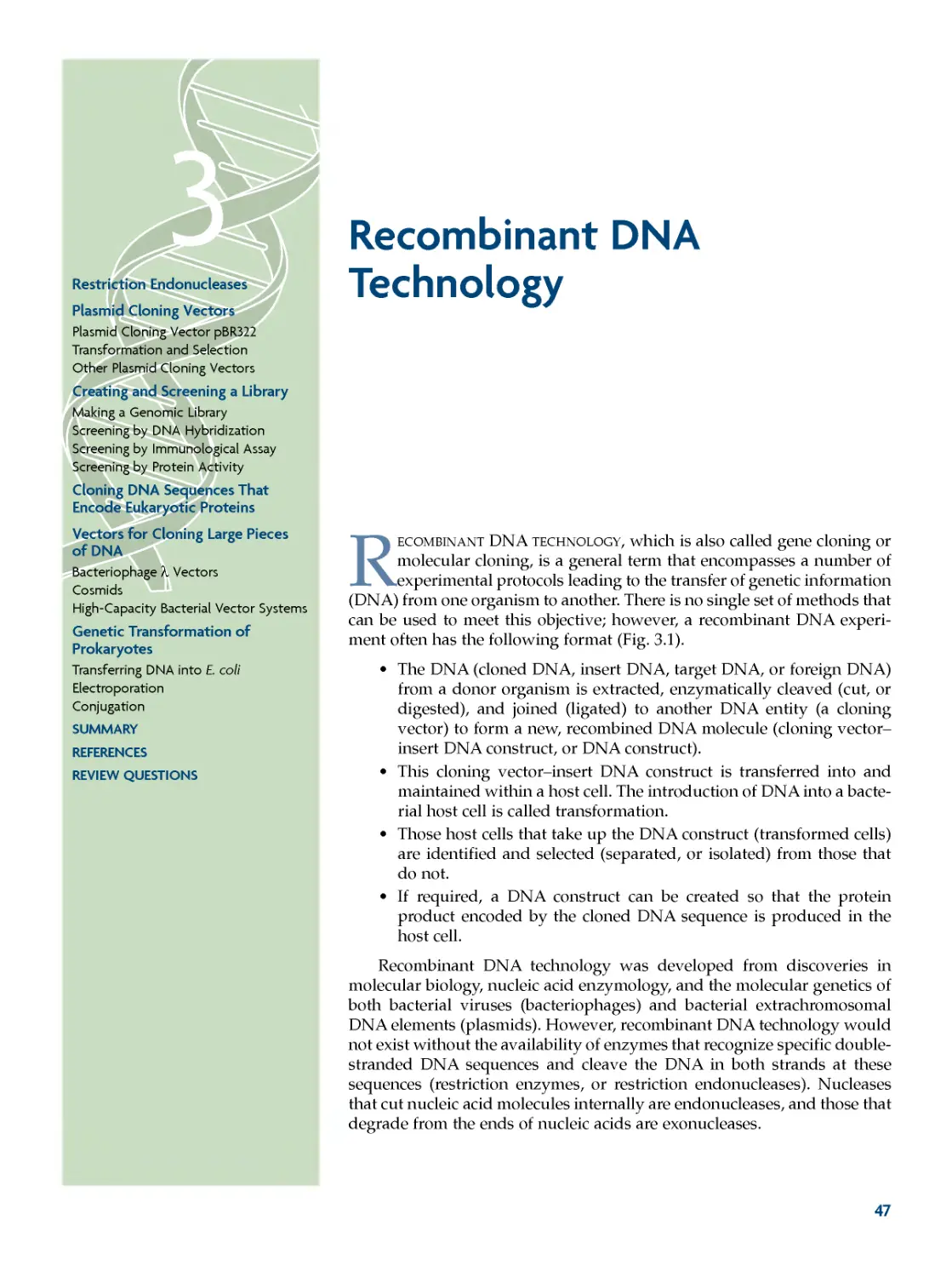
restriction endonucleases 49
plasmid cloning vectors 57
Plasmid Cloning Vector pBR322 59
Transformation and Selection 60
Other Plasmid Cloning Vectors 63
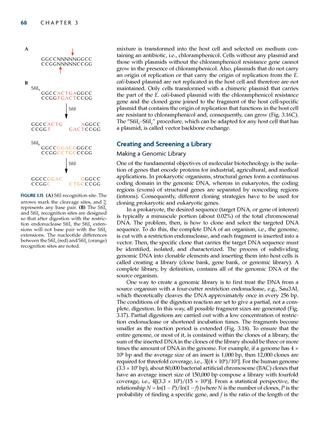
creating and screening a Library 68
Making a Genomic Library 68
Screening by DNA Hybridization 70
Screening by Immunological Assay 76
Screening by Protein Activity 78
cloning DNa sequences that encode
eukaryotic proteins 80
vectors for cloning Large pieces of
DNa 86
Bacteriophage λ Vectors 86
Cosmids 90
High-Capacity Bacterial Vector
Systems 92
Genetic transformation of
prokaryotes 92
Transferring DNA into E. coli 92
Electroporation 93
Conjugation 94
suMMary 95
refereNces 96
review QuestioNs 97
chapter 4
chemical synthesis,
amplification, and
sequencing of DNa 98
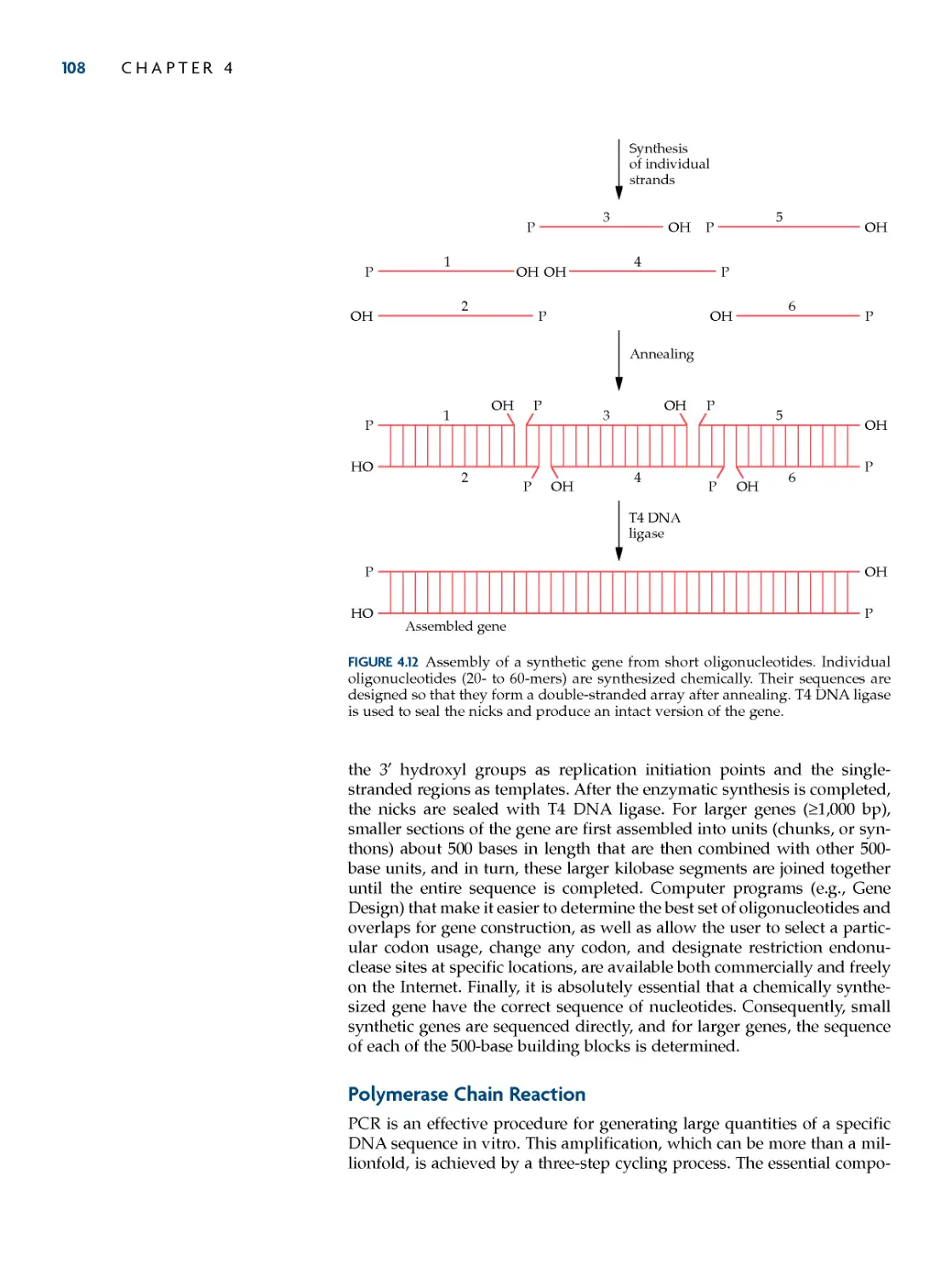
chemical synthesis of DNa 98
The Phosphoramidite Method 99
Uses of Synthesized
Oligonucleotides 103
polymerase chain reaction 108
PCR Amplification of Full-Length
cDNAs 113
Gene Synthesis by PCR 113
DNa-sequencing techniques 117
Dideoxynucleotide Procedure for
Sequencing DNA 118
Primer Walking 124
Pyrosequencing 125
Sequencing Using Reversible Chain
Terminators 128
Sequencing by Ligation 131
Large-scale DNa sequencing 133
part i
fuNDaMeNtaLs of MoLecuLar BiotechNoLoGy 1
Preface xiii
Preface to the First Edition xv
contents
viii coNteNts
Shotgun Cloning Strategy for
Sequencing Genomes 133
Cyclic Array Sequencing 136
suMMary 142
refereNces 143
review QuestioNs 145
chapter 5
Bioinformatics, Genomics,
and proteomics 146
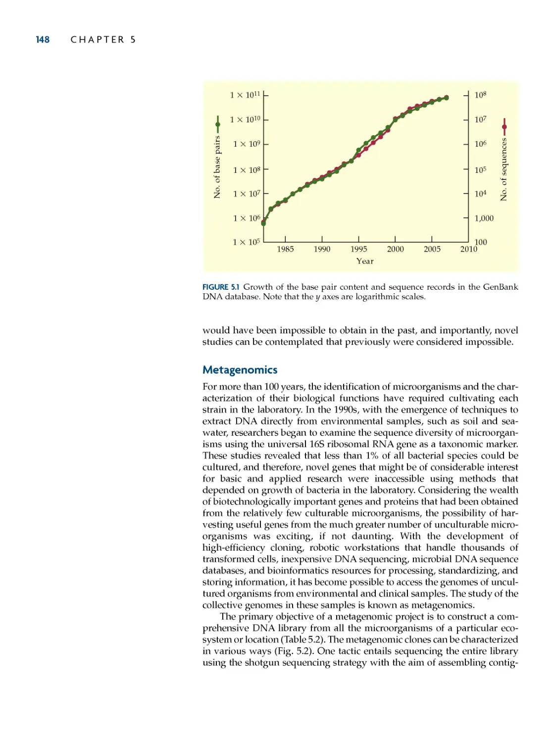
Molecular Databases 146
Metagenomics 148
functional Genomics 154
DNA Microarray Technology 155
Serial Analysis of Gene Expression 160
proteomics 164
Separation and Identification of
Proteins 165
Protein Expression Profiling 169
Protein Microarrays 172
Protein--Protein Interaction
Mapping 181
suMMary 189
refereNces 190
review QuestioNs 193
chapter 6
Manipulation of
Gene expression in
prokaryotes 195
Gene expression from strong and
regulatable promoters 196
Regulatable Promoters 196
Increasing Protein Production 201
Large-Scale Systems 201
Expression in Other
Microorganisms 204
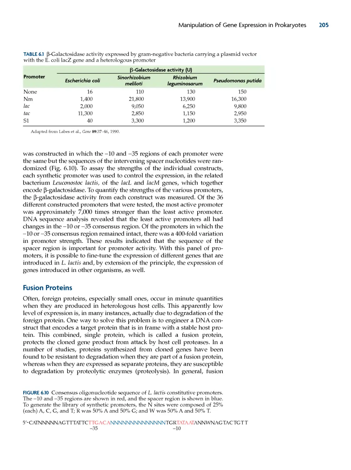
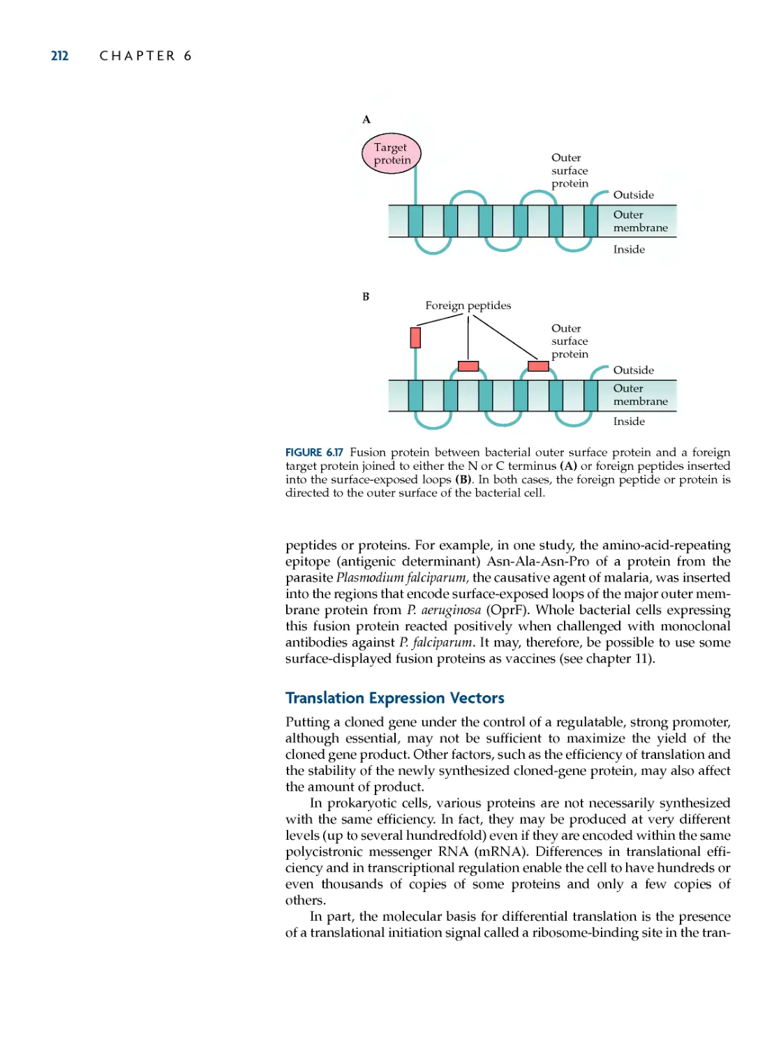
fusion proteins 205
Uses of Fusion Proteins 206
Cleavage of Fusion Proteins 208
Surface Display 210
translation expression vectors 212
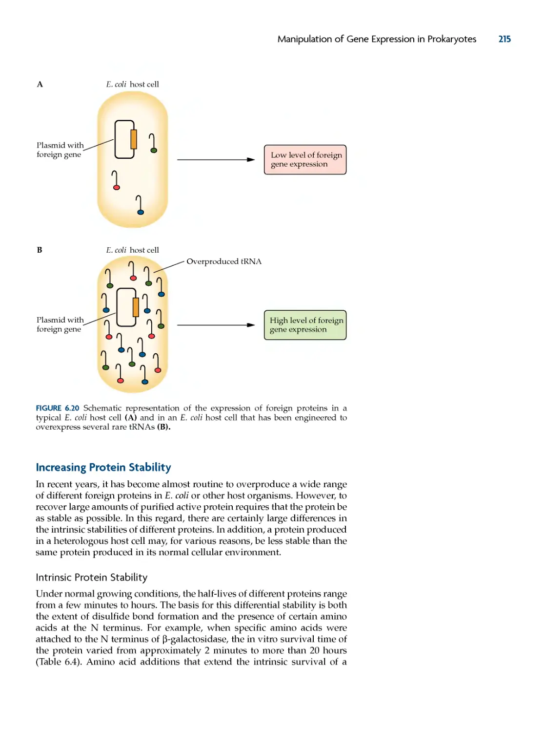
increasing protein stability 215
Intrinsic Protein Stability 215
Facilitating Protein Folding 217
Coexpression Strategies 219
overcoming oxygen Limitation 220
Use of Protease-Deficient Host
Strains 220
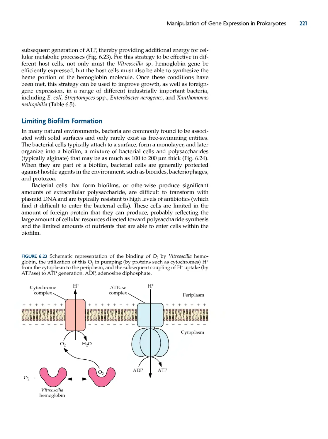
Bacterial Hemoglobin 220
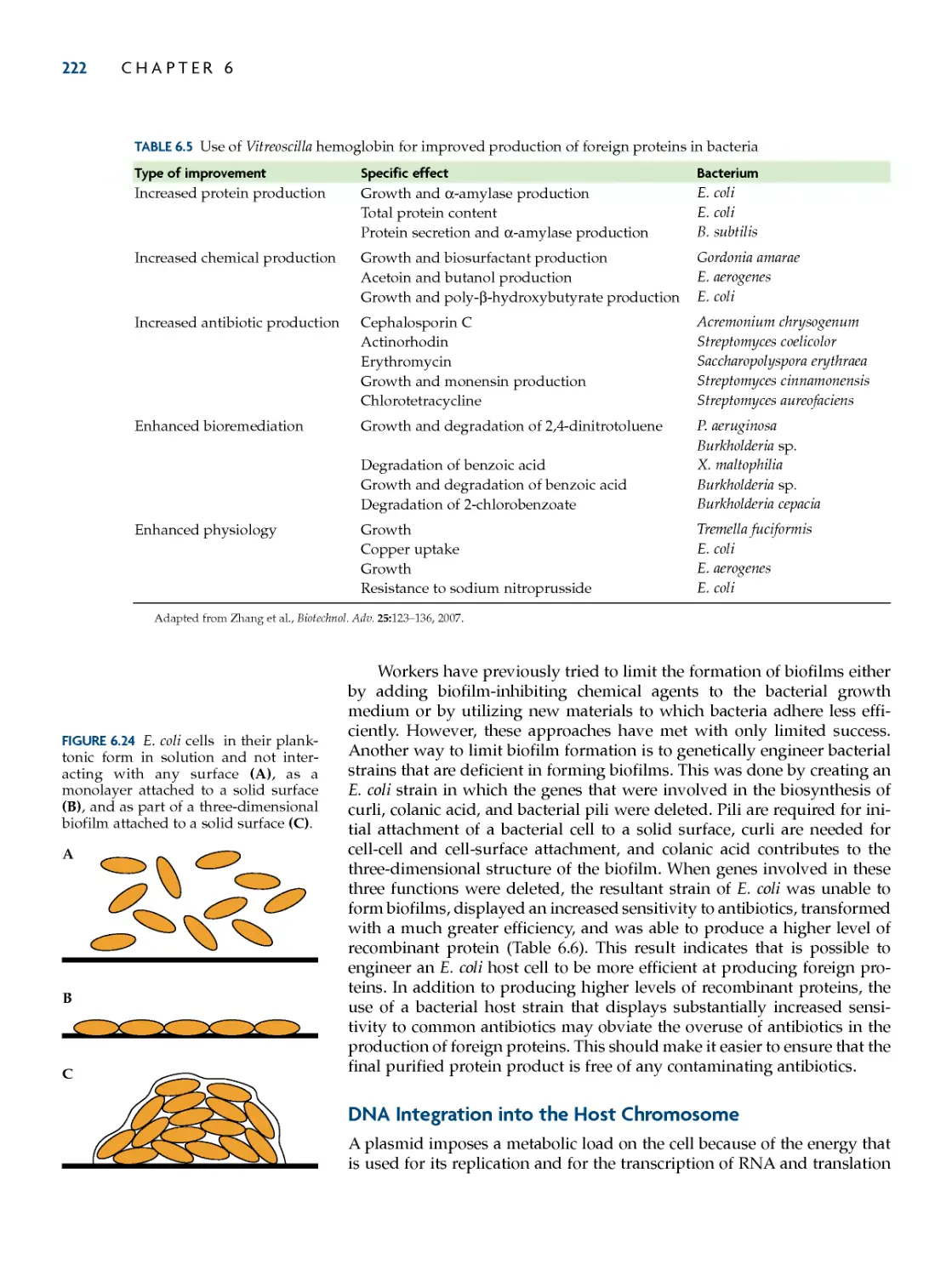
Limiting Biofilm formation 221
DNa integration into the host
chromosome 222
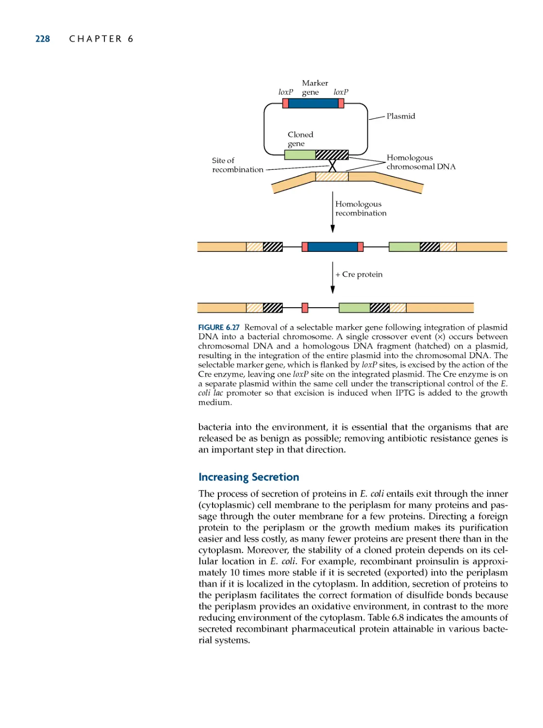
Removing Selectable Marker
Genes 227
increasing secretion 228
Secretion into the Periplasm 229
Secretion into the Medium 230
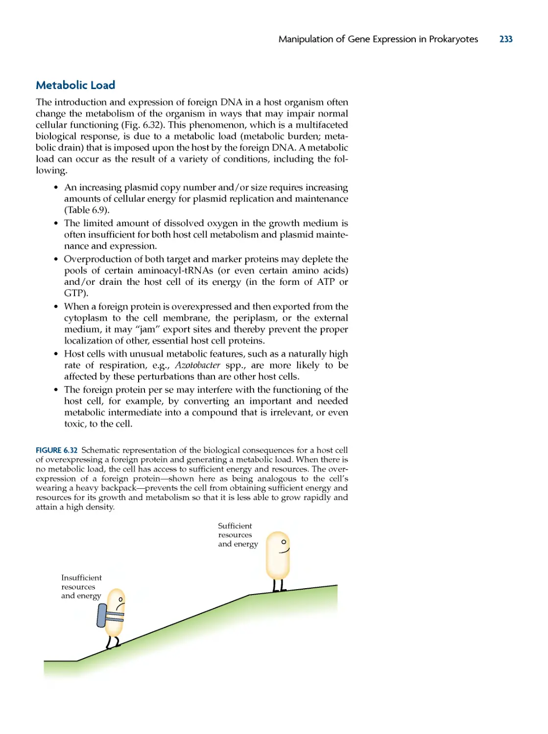
Metabolic Load 233
suMMary 235
refereNces 236
review QuestioNs 238
chapter 7
heterologous protein
production in eukaryotic
cells 240
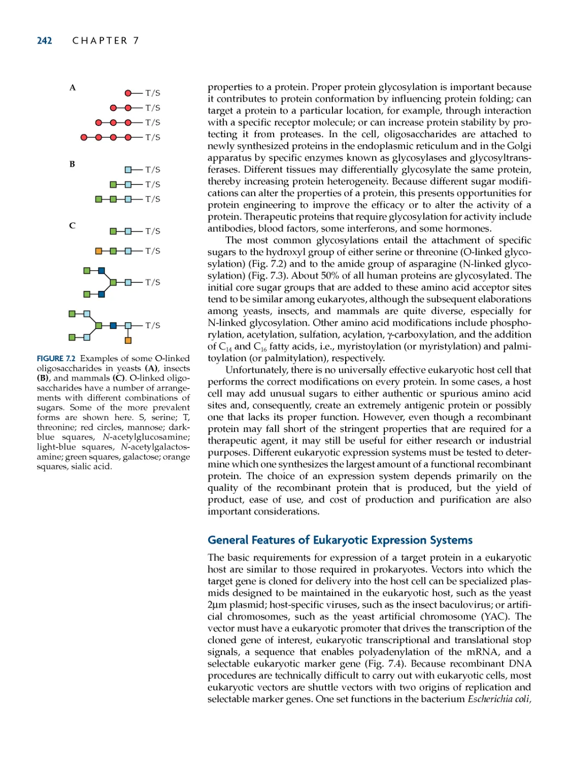
posttranslational Modification of
eukaryotic proteins 240
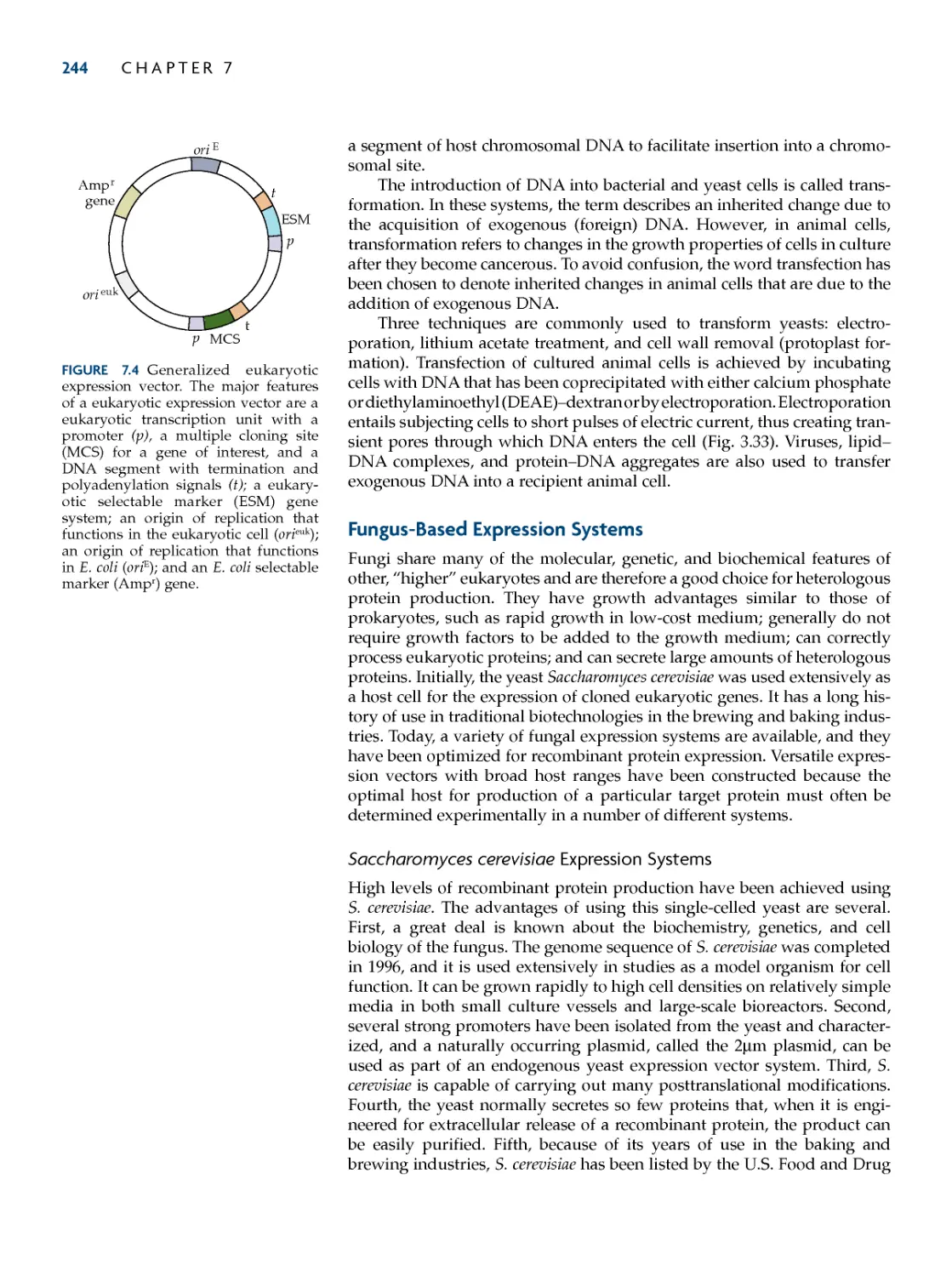
General features of eukaryotic
expression systems 242
fungus-Based expression
systems 244
Saccharomyces cerevisiae Expression
Systems 244
Pichia pastoris Expression Systems 253
Other Yeast Systems 255
Filamentous Fungal Systems 259
Baculovirus–insect cell expression
systems 261
Baculovirus Expression Vectors 263
Increasing the Yield of Recombinant
Baculovirus 264
Integration of Target Genes into
Baculovirus by Site-Specific
Recombination 265
Mammalian Glycosylation and
Processing of Precursor Proteins in Insect
Cells 267
Production of Multiprotein Complexes
Using Baculovirus 270
Mammalian cell expression
systems 27 1
Vector Design 272
Baculovirus Vectors for Protein
Production in Mammalian Cells 275
Selectable Markers for Mammalian
Expression Vectors 278
Engineering Mammalian Cell Hosts for
Enhanced Productivity 279
Plasmid Integration and Chromosomal
Environment 282
suMMary 286
refereNces 287
review QuestioNs 288
chapter 8
Directed Mutagenesis and
protein engineering 290
Directed Mutagenesis
procedures 291
Oligonucleotide-Directed Mutagenesis
with M13 DNA 292
Oligonucleotide-Directed Mutagenesis
with Plasmid DNA 295
PCR-Amplified Oligonucleotide-Directed
Mutagenesis 297
Error-Prone PCR 298
Random Mutagenesis with Degenerate
Oligonucleotide Primers 298
Random Insertion/Deletion
Mutagenesis 301
DNA Shuffling 303
Mutant Proteins with Unusual Amino
Acids 304
protein engineering 305
Adding Disulfide Bonds 305
Changing Asparagine to Other Amino
Acids 310
Reducing the Number of Free Sulfhydryl
Residues 311
Increasing Enzymatic Activity 312
Modifying Metal Cofactor
Requirements 316
Decreasing Protease Sensitivity 317
Modifying Protein Specificity 318
Increasing Enzyme Stability and
Specificity 321
Altering Multiple Properties 325
suMMary 327
refereNces 327
review QuestioNs 328
coNteNts ix
part ii
MoLecuLar BiotechNoLoGy of
MicroBiaL systeMs 331
chapter 9
Molecular
Diagnostics 333
immunological Diagnostic
procedures 334
ELISA 335
Monoclonal antibodies 337
Formation and Selection of Hybrid
Cells 337
Identification of Specific Antibody-
Producing Hybrid Cell Lines 339
Biofluorescent and Bioluminescent
systems 341
Colored Fluorescent Proteins 341
Luciferase 342
Microbial Biosensors 343
Nucleic acid Diagnostic systems 345
Hybridization Probes 346
Diagnosis of Malaria 347
Detection of T. cruzi 349
Nonradioactive Hybridization
Procedures 350
Molecular Beacons 352
DNA Fingerprinting 353
RAPD 355
Real-Time PCR 358
Immunoquantitative Real-Time
PCR 359
Ancestry Determination 361
Animal Species Determination 364
Automated DNA Analysis 364
Molecular Diagnosis of Genetic
Disease 365
Screening for Cystic Fibrosis 366
Sickle-Cell Anemia 367
The PCR/OLA Procedure 368
Padlock Probes 374
Genotyping with Fluorescence-Labeled
PCR Primers 374
TaqMan Assay 375
suMMary 375
refereNces 376
review QuestioNs 377
chapter 10
protein therapeutics 379
pharmaceuticals 380
Isolation of Interferon cDNAs 380
Human Interferons 381
Human Growth Hormone 383
Tumor Necrosis Factor Alpha 388
enzymes 389
DNase I 389
Alginate Lyase 390
Phenylalanine Ammonia Lyase 392
α1-Antitrypsin 393
Glycosidases 394
Lactic acid Bacteria 395
Interleukin-10 396
Leptin 397
An HIV Inhibitor 398
Monoclonal antibodies 399
Structure and Function of
Antibodies 400
Preventing Rejection of Transplanted
Organs 402
recombinant antibodies 403
Hybrid Human--Mouse Monoclonal
Antibodies 403
Human Monoclonal Antibodies 406
Antibody Fragments 408
Combinatorial Libraries of Antibody
Fragments 411
A Combinatorial Library of Full-Length
Antibodies 415
Shuffling CDR Sequences 416
Chemically Linked Monoclonal
Antibodies 417
Dual-Variable-Domain Antibodies 420
Anticancer Antibodies 421
suMMary 422
refereNces 422
review QuestioNs 424
chapter 11
Nucleic acids as
therapeutic agents 426
antisense rNa 426
Antisense Oligonucleotides 428
ribozymes 434
Deoxyribozymes 436
chimeric rNa–DNa Molecules 43 7
aptamers 437
interfering rNas 440
Principles 440
Applications 442
antibody Genes 443
Nucleic acid Delivery 444
Human Gene Therapy 444
Targeting Systems 451
suMMary 456
refereNces 456
review QuestioNs 458
chapter 12
vaccines 459
subunit vaccines 463
Herpes Simplex Virus 463
Foot-and-Mouth Disease 464
Cholera 466
SARS 466
Staphylococcus aureus 467
Human Papillomavirus 468
peptide vaccines 469
Foot-and-Mouth Disease 470
Malaria 472
Genetic immunization: DNa
vaccines 472
Delivery 472
Dental Caries 479
attenuated vaccines 481
Cholera 481
Salmonella Species 482
Leishmania Species 484
Herpes Simplex Virus 485
vector vaccines 486
Vaccines Directed against Viruses 486
Vaccines Directed against Bacteria 492
Bacteria as Antigen Delivery
Systems 494
suMMary 497
refereNces 497
review QuestioNs 499
chapter 13
synthesis of commercial
products by recombinant
Microorganisms 501
restriction endonucleases 501
Lipase 505
small Biological Molecules 506
Synthesis of L-Ascorbic Acid 507
Microbial Synthesis of Indigo 512
Synthesis of Amino Acids 514
Microbial Synthesis of Lycopene 519
Increasing Succinic Acid Production 519
antibiotics 521
Cloning Antibiotic Biosynthesis
Genes 523
Modulating Gene Expression in
Streptomycetes 526
Synthesis of Novel Antibiotics 527
Engineering Polyketide Antibiotics 529
Improving Antibiotic Production 531
Designer Antibiotics 534
Biopolymers 535
Xanthan Gum 535
Melanin 538
Adhesive Protein 539
Rubber 541
Polyhydroxyalkanoates 542
Hyaluronic Acid 545
suMMary 547
refereNces 547
review QuestioNs 549
chapter 14
Bioremediation and
Biomass utilization 551
Microbial Degradation of
Xenobiotics 551
Genetic engineering of
Biodegradative pathways 556
Manipulation by Transfer of
Plasmids 557
Manipulation by Gene Alteration 559
utilization of starch and sugars 569
Commercial Production of Fructose and
Alcohol 570
Altering Alcohol Production 571
Improving Fructose Production 575
Silage Fermentation 576
Isopropanol Production 577
Engineering Yeast Transcription 578
utilization of cellulose 580
Lignocellulosics 581
Components of Lignocellulose 582
Isolation of Prokaryotic Cellulase
Genes 583
Isolation of Eukaryotic Cellulase
Genes 586
Manipulation of Cellulase Genes 586
Zymomonas mobilis 589
hydrogen production 595
suMMary 596
refereNces 596
review QuestioNs 598
chapter 15
plant Growth-promoting
Bacteria 599
Growth promotion by free-Living
Bacteria 600
Decreasing Plant Stress 604
Increasing Phosphorus Availability 606
Biocontrol of pathogens 608
Siderophores 608
Antibiotics 612
Enzymes 614
Ice Nucleation and Antifreeze
Proteins 614
Ethylene 617
Root Colonization 618
Nitrogen fixation 619
Nitrogenase 621
Components of Nitrogenase 621
Genetic Engineering of the Nitrogenase
Gene Cluster 622
Engineering Improved Nitrogen
Fixation 628
hydrogenase 630
Hydrogen Metabolism 631
Genetic Engineering of Hydrogenase
Genes 632
Nodulation 635
Competition among Nodulating
Organisms 635
Genetic Engineering of Nodulation
Genes 635
Nodulation and Ethylene 640
phytoremediation 641
Engineering Strains That Facilitate
Growth 641
Engineering Degradative
Plasmids 643
Engineering Bacterial
Endophytes 644
Metals in the Environment 646
suMMary 648
refereNces 649
review QuestioNs 651
chapter 16
Microbial
insecticides 652
insecticidal toxin of
B. thuringiensis 653
Mode of Action and Use 653
Toxin Gene Isolation 658
engineering of B. thuringiensis toxin
Genes 660
Synthesis during Vegetative
Growth 660
Broadening the Spectrum of Target
Insects 663
Improving Delivery of a Mosquitocidal
Toxin 666
Protecting Plant Roots 668
Protoxin Processing 670
Preventing the Development of
Resistance 671
Improved Biocontrol 674
Baculoviruses as Biocontrol
agents 67 7
Mode of Action 677
Genetic Engineering for Improved
Biocontrol 679
suMMary 681
refereNces 682
review QuestioNs 684
x
coNteNts
chapter 18
Genetic engineering of
plants: Methodology 725
plant transformation with the ti
plasmid of A. tumefaciens 726
ti plasmid-Derived vector
systems 730
physical Methods of transferring
Genes to plants 735
Microprojectile Bombardment 736
chloroplast engineering 738
use of reporter Genes in
transformed plant cells 741
Manipulation of Gene expression in
plants 743
Isolation and Use of Different
Promoters 743
Gene Targeting 745
Targeted Alterations in Plant RNA 747
Facilitating Protein Purification 748
production of Marker-free transgenic
plants 750
Removing Marker Genes from Nuclear
DNA 752
Removing Marker Genes from
Chloroplast DNA 753
suMMary 755
refereNces 756
review QuestioNs 757
chapter 19
engineering plants to
overcome Biotic and
abiotic stress 759
insect resistance 759
Increasing Expression of the
B. thuringiensis Protoxin 760
Other Strategies for Protecting Plants
against Insects 764
Preventing the Development of
B. thuringiensis-Resistant
Insects 770
virus resistance 773
Viral Coat Protein-Mediated
Protection 773
Protection by Expression of Other
Genes 779
herbicide resistance 782
fungus and Bacterium
resistance 787
oxidative stress 792
salt and Drought stress 793
fruit ripening and flower
wilting 796
suMMary 799
refereNces 800
review QuestioNs 802
chapter 20
engineering plant Quality
and proteins 803
Modification of plant Nutritional
content 803
Amino Acids 803
Lipids 805
Vitamins 808
Iron 813
Phosphorus 814
Modification of food plant taste and
appearance 815
Preventing Discoloration 815
Sweetness 817
Starch 818
Genetic Manipulation of flower
pigmentation 821
plants as Bioreactors 825
Antibodies 827
Polymers 830
edible vaccines 830
plant yield 832
Increasing Iron Content 833
Altering Lignin Content 834
Erect Leaves 836
Increasing Oxygen Content 837
phytoremediation 838
suMMary 841
refereNces 841
review QuestioNs 843
coNteNts
xi
part iii
MoLecuLar BiotechNoLoGy
of eukaryotic systeMs 723
chapter 17
Large-scale production of
proteins from
recombinant
Microorganisms 685
principles of Microbial Growth 687
Batch Fermentation 687
Fed-Batch Fermentation 689
Continuous Fermentation 691
Maximizing the efficiency of the
fermentation process 692
High-Density Cell Cultures 693
Increasing Plasmid Stability 695
Quiescent E. coli Cells 696
Protein Secretion 696
Reducing Acetate 698
Bioreactors 701
typical Large-scale fermentation
systems 705
Two-Stage Fermentation in Tandem
Airlift Reactors 706
Two-Stage Fermentation in a Single
Stirred-Tank Reactor 708
Batch versus Fed-Batch
Fermentation 708
harvesting Microbial cells 711
Disrupting Microbial cells 714
Downstream processing 717
Protein Solubilization 718
Large-scale production of plasmid
DNa 719
suMMary 720
refereNces 720
review QuestioNs 722
chapter 22
regulating the use of
Biotechnology 897
regulating recombinant DNa
technology 898
Deliberate release of Genetically
Modified Microorganisms 900
regulating food and food
ingredients 903
Food Ingredients Produced by
Genetically Engineered
Microorganisms 903
Genetically Modified Crops 907
Genetically Engineered Livestock 910
patenting Biotechnology 911
Patenting in Different Countries 915
Patenting DNA Sequences 916
Patenting Multicellular Organisms 917
Patenting and Fundamental
Research 918
suMMary 920
refereNces 920
review QuestioNs 921
chapter 23
societal issues in
Biotechnology 923
concerns about the safety of
consuming Genetically Modified
foods 923
Alteration of the Nutritional Content of
Food 924
Potential for Introducing Toxins or
Allergens into Food 927
Potential for Transferring Transgenes
from Food to Humans or Intestinal
Microorganisms 930
Controversy about the Labeling of
Genetically Modified Foods 931
concerns about the impact of
Genetically Modified organisms on
the environment 932
Impact on Biodiversity 932
Impact of the Bt Toxin on Nontarget
Insects 933
Environmental Benefits of Genetically
Modified Organisms 934
economic issues 935
Who Benefits from Molecular
Biotechnology? 935
How Do Views about Genetically
Engineered Food Affect Trade? 936
suMMary 937
refereNces 938
review QuestioNs 939
xii coNteNts
part iv
MoLecuLar BiotechNoLoGy aND society 895
amino acids of proteins and their Designations 941
Glossary 943
index 973
chapter 21
transgenic animals 845
transgenic Mice: Methodology 847
The Retroviral Vector Method 848
The DNA Microinjection Method 850
The Engineered Embryonic Stem Cell
Method 851
Genetic Modification with the Cre--loxP
Recombination System 856
RNA Interference 859
Transgenesis with High-Capacity
Vectors 861
transgenic Mice: applications 863
Transgenic Disease Models: Alzheimer
Disease 863
Using Transgenic Mice as Test
Systems 865
Conditional Regulation of Transgene
Expression 866
Conditional Control of Cell Death 870
cloning Livestock by Nuclear
transfer 871
transgenic Livestock 873
Production of Pharmaceuticals 873
Production of Donor Organs 875
Disease-Resistant Livestock 876
Improving Milk Quality 879
Improving Animal Production
Traits 880
transgenic poultry 885
transgenic fish 886
suMMary 890
refereNces 890
review QuestioNs 893
SINCE THE EARLY 1970S, when recombinant DNAtechnologywasfirst
developed, there has been a veritable explosion of knowledge in the
biological sciences. Since that time, with the advent of PCR, chemical
DNA synthesis, DNA sequencing, monoclonal antibodies, directed muta-
genesis, genomics, proteomics, and metabolomics, our understanding of
and ability to manipulate the biological world have grown exponentially.
When the first edition of Molecular Biotechnology: Principles and Applications
of Recombinant DNA was published in 1994, nearly all of the transgenic
organisms that were produced included only a single introduced gene. Just
15 years later, it is not uncommon for researchers to engineer organisms by
modifying both the activity and the regulation of existing genes while at
the same time introducing entire new pathways. In 1994, only a handful of
products produced by this new technology were available in the market-
place. Today, molecular biotechnology has given us several hundred new
therapeutic agents, with many more in the pipeline, as well as dozens of
transgenic plants. The use of DNA has become a cornerstone of modern
forensics, paternity testing, and ancestry determination. Several new
recombinant vaccines have been developed, with many more on the
horizon. The list goes on and on. Molecular biotechnology really has lived
up to its promise, to all of the original hype. It has been estimated that
worldwide there are currently several thousand biotechnology companies
employing tens of thousands of scientists. When the exciting science being
done at universities, government labs, and research institutes around the
world is factored in, the rate of change and of discovery in the biological
sciences is astounding. This fourth edition of Molecular Biotechnology,
building upon the fundamentals that were established in the previous three
editions, endeavors to provide readers with a window on some of the
major developments in this growing field in the past several years. Of
necessity, we have had to be highly selective in the material that is included
in this edition. Moreover, the window that we are looking through is
moving. This notwithstanding, we both expect and look forward to the
commercialization of many of these discoveries as well as to the develop-
ment of new approaches, insights, and discoveries. BE R N A R D R. GL I C K
JACKJ.PASTERNAK
CHERYLL.PATTEN
xiii
preface
This page intentionally left blank
MOLECULAR BIOTECHNOLOGY EMERGED as a new research field that
arose as a result of the fusion in the late 1970s of recombinant
DNA technology and traditional industrial microbiology. Whether
one goes to the movies to see Jurassic Park with its ingenious but scientifi-
cally untenable plot of cloning dinosaurs, reads in the newspaper about the
commercialization of a new "biotech" tomato that has an extended shelf
life, or hears one of the critics of molecular biotechnology talking about the
possibility of dire consequences from genetic engineering, there is a sig-
nificant public awareness about recombinant DNA technology. In this
book, we introduce and explain what molecular biotechnology actually is
as a scientific discipline, how the research in the area is conducted, and
how this technology may realistically impact on our lives in the future.
We have written Molecular Biotechnology: Principles and Applications of
Recombinant DNA to serve as a text for courses in biotechnology, recombi-
nant DNA technology, and genetic engineering or for any course intro-
ducing both the principles and the applications of contemporary molecular
biology methods. The book is based on the biotechnology course we have
offered for the past 12 years to advanced undergraduate and graduate stu-
dents from the biological and engineering sciences at the University of
Waterloo. We have written this text for students who have an under-
standing of basic ideas from biochemistry, molecular genetics, and micro-
biology. We are aware that it is unlikely that students will have had all of
these courses before taking a course on biotechnology. Thus, we have tried
to develop the topics in this text by explaining their broader biological
context before delving into molecular details.
This text emphasizes how recombinant DNA technology can be used
to create various useful products. We have, wherever possible, used exper-
imental results and actual methodological strategies to illustrate basic con-
cepts, and we have tried to capture the flavor and feel of how molecular
biotechnology operates as a scientific venture. The examples that we have
selected---from a vast and rapidly growing literature---were chosen as case
studies that not only illustrate particular points but also provide the reader
with a solid basis for understanding current research in specialized areas of
molecular biotechnology. Nevertheless, we expect that some of our exam-
ples will be out of date by the time the book is published, because molec-
ular biotechnology is such a rapidly changing discipline.
xv
preface to the first edition
xvi preface to the first eDitioN
For the ease of the day-to-day practitioners, scientific disciplines often
develop specialized terms and nomenclature. We have tried to minimize
the use of technical jargon and, in many instances, have deliberately used
a simple phrase to describe a phenomenon or process that might otherwise
have been expressed more succinctly with technical jargon. In any field of
study, synonymous terms that describe the same phenomenon exist. In
molecular biotechnology, for example, recombinant DNA technology, gene
cloning, and genetic engineering, in a broad sense, have the same meaning.
When an important term or concept appears for the first time in this text, it
is followed in parentheses with a synonym or equivalent expression. An
extensive glossary can be found at the end of the book to help the reader
with the terminology of molecular biotechnology.
Each chapter opens with an outline of topics and concludes with a
detailed summary and list of review questions to sharpen students' critical
thinking skills. All of the key ideas in the book are carefully illustrated by
the more than 200 full-color diagrams in the pedagogical belief that a pic-
ture is indeed worth a thousand words. After introducing molecular bio-
technology as a scientific and economic venture in Chapter 1, the next five
chapters (2 to 6) deal with the methodologies of molecular biotechnology.
The chapters of Part I act as a stepping-stone for the remainder of the book.
Chapters 7 to 12 in Part II present examples of microbial molecular biotech-
nology covering such topics as the production of metabolites, vaccines,
therapeutics, diagnostics, bioremediation, biomass utilization, bacterial
fertilizers, and microbial pesticides. Chapter 13 describes some of the key
components of large-scale fermentation processes using genetically engi-
neered (recombinant) microorganisms. In Part III, we deal with the molec-
ular biotechnology of plants and animals (Chapters 14 and 15). The
isolation of human disease-causing genes by using recombinant DNA tech-
nology and how, although it is in its early stages, genetic manipulation is
being currently contemplated for the treatment of human diseases are pre-
sented in Chapters 16 and 17. The book concludes with coverage of the
regulation of molecular biotechnology and patents in Part IV.
A brief mention should be made about the reference sections that
follow each chapter. Within many of the chapters we have relied upon the
published work of various researchers. In all cases, although not cited
directly in the body of a chapter, the original published articles are noted in
the reference section of the appropriate chapter. In some cases, we have
taken "pedagogic license" and either extracted or reformulated data from
the original publications. Clearly, we are responsible for any distortions or
misrepresentations from these simplifications, although we hope that none
has occurred. The reference sections also contain other sources that we
used in a general way, which might, if consulted, bring the readers closer
to a particular subject.
acknowledgments
We express our appreciation to the following people who reviewed various
parts of the manuscript as it was being developed. The comments of these
expert scientists and teachers helped us immeasurably: Arthur I. Aronson,
Purdue University; Ronald M. Atlas, University of Louisville; Fred Ausubel,
Massachusetts General Hospital; David R. Benson, University of
Connecticut; Jean E. Brenchley, Pennsylvania State University; A. M.
Chakrabarty, University of Illinois at Chicago; Stan Gelvin, Purdue
preface to the first eDitioN xvii
University; Janet H. Glaser, University of Illinois at Urbana-Champaign;
David Gwynne, Cambridge NeuroScience; George D. Hegeman, Indiana
University; James B. Kaper, University of Maryland at Baltimore; Donald R.
Lightfoot, Eastern Washington University at Cheney and Spokane; Cynthia
Moore, Washington University; William E. Newton, Virginia Polytechnic
University; Danton H. O'Day, University of Toronto in Mississauga;
Richard D. Palmiter, University of Washington; David H. Persing, Mayo
Clinic; William S. Reznikoff, University of Wisconsin; Campbell W.
Robinson, University of Waterloo; Marc Siegel, University of Waterloo;
Aaron J. Shatkin, Center for Advanced Biotechnology and Medicine at
Rutgers University; Jim Schwartz, Genentech; Daniel C. Stein, University
of Maryland at College Park; Dean A. Stetler, University of Kansas; and
Robert T. Vinopal, University of Connecticut.
The following professionals at ASM Press worked on the book and
deserve our thanks: Susan Birch, senior production editor; Ruth Siegal,
developmental editor; Jodi Simpson, copy editor; Susan Schmidler, designer
and art director; Peg Markow at Ruttle, Shaw & Wetherill, Inc., senior
project manager; and Network Graphics, illustrators. Finally we are
indebted to Patrick Fitzgerald, Director of ASM Press, who, in all possible
ways, helped transform our original efforts into an acceptable final form.
His encouragement as a persistent and friendly "torturer" was deeply
appreciated.
BERNARDR.GLICK
JACKJ.PASTERNAK
This page intentionally left blank
1
MOLECULAR BIOTECHNOLOGY isanexcitingscientificdisciplinethatis
based on the ability of researchers to transfer specific units of
genetic information from one organism to another. This convey-
ance of a gene or genes relies on the techniques of genetic engineering
(recombinant DNA technology). The objective of recombinant DNA tech-
nology is often to create a useful product or a commercial process. In part I,
the concept of molecular biotechnology, some fundamentals of molecular
biology, and recombinant DNA procedures are presented. Essential molec-
ular biotechnology laboratory techniques, including chemical synthesis of
genes, the polymerase chain reaction (PCR), and DNA sequencing, are dis-
cussed. Developments in sequencing technologies have led to the sequencing
of the entire genomes of many organisms, and this has enabled researchers
to begin to understand organisms from their sequences and to identify
novel genes with potentially useful functions. In addition to isolation
(cloning) of genes, it is important that these genes function properly in a
host organism. To this end, strategies for optimizing the expression of a
cloned gene in either prokaryotic or eukaryotic cells are reviewed. Finally,
procedures for modifying cloned genes by the introduction of specific
nucleotide changes (in vitro mutagenesis) to enhance the properties of the
target proteins are examined. Together, the chapters in part I provide the
conceptual and technical underpinnings for understanding the applications
of molecular biotechnology that are described in the ensuing chapters.
1 The Development of Molecular Biotechnology
2 DNA, RNA, and Protein Synthesis
3 Recombinant DNA Technology
4 Chemical Synthesis, Amplification, and Sequencing of DNA
5 Bioinformatics, Genomics, and Proteomics
6 Manipulation of Gene Expression in Prokaryotes
7 Heterologous Protein Production in Eukaryotic Cells
8 Directed Mutagenesis and Protein Engineering
FUNDAMENTALS OF
MOLECULAR
BIOTECHNOLOGY
I
This page intentionally left blank
3
The Emergence of Molecular
Biotechnology
Recombinant DNA Technology
Commercialization of Molecular
Biotechnology
Concerns and Consequences
SUMMARY
REFERENCES
REVIEW QUESTIONS
The Development of
Molecular Biotechnology
The Emergence of Molecular Biotechnology
LONGBEFOREWEKNEWTHATMICROORGANISMSEXISTEDorthatgeneswere
the units of inheritance, humans looked to the natural world to
develop methods to increase food production, preserve food, and
heal the sick. Our ancestors discovered that grains could be preserved
through fermentation into beer; that storing horse saddles in a warm, damp
corner of the stable resulted in the growth of a saddle mold that could heal
infected saddle sores; and that intentional exposure to a "contagion" could
somehow provide protection from an infectious disease on subsequent
exposure. Since the discovery of the microscopic world in the 17th century,
microorganisms have been employed in the development of numerous
useful processes and products. Many of these are found in our households
and backyards. Lactic acid bacteria are used to prepare yogurt and probi-
otics, insecticide-producing bacteria are sprayed on many of the plants
from which the vegetables in our refrigerator were harvested, nitrogen-
fixing bacteria are added to the soil used for cultivation of legumes, the
enzymatic stain removers in laundry detergent came from a microor-
ganism, and antibiotics derived from common soil microbes are used to
treat infectious diseases. These are just a few examples of traditional bio-
technologies that have improved our lives. Up to the early 1970s, however,
traditional biotechnology was not a well-recognized scientific discipline,
and research in this area was centered in departments of chemical engi-
neering and occasionally in specialized microbiology programs.
In a broad sense, biotechnology is concerned with the production of
commercial products generated by the metabolic action of microorganisms.
More formally, biotechnology may be defined as "the application of scien-
tific and engineering principles to the processing of material by biological
agents to provide goods and services." The term "biotechnology" was first
used in 1917 by a Hungarian engineer, Karl Ereky, to describe an integrated
process for the large-scale production of pigs by using sugar beets as the
source of food. According to Ereky, biotechnology was "all lines of work by
which products are produced from raw materials with the aid of living
4
CHAPTER 1
things." This fairly precise definition was more or less ignored. For a
number of years, the term biotechnology was used to describe two very
different engineering disciplines. On one hand, it referred to industrial
fermentation. On the other, it was used for the study of efficiency in the
workplace---what is now called ergonomics. This ambiguity ended in 1961
when the Swedish microbiologist Carl Göran Hedén recommended that
the title of a scientific journal dedicated to publishing research in the fields
of applied microbiology and industrial fermentation be changed from the
Journal of Microbiological and Biochemical Engineering and Technology to
Biotechnology and Bioengineering. From that time on, biotechnology has
clearly and irrevocably been associated with the study of "the industrial
production of goods and services by processes using biological organisms,
systems, and processes," and it has been firmly grounded in expertise in
microbiology, biochemistry, and chemical engineering.
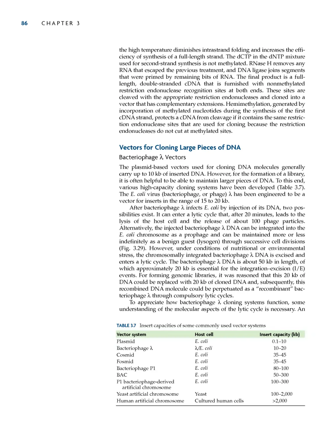
An industrial biotechnology process that uses microorganisms for pro-
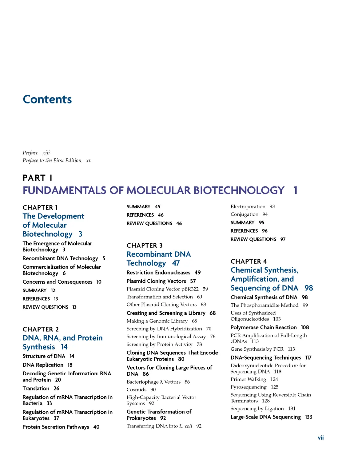
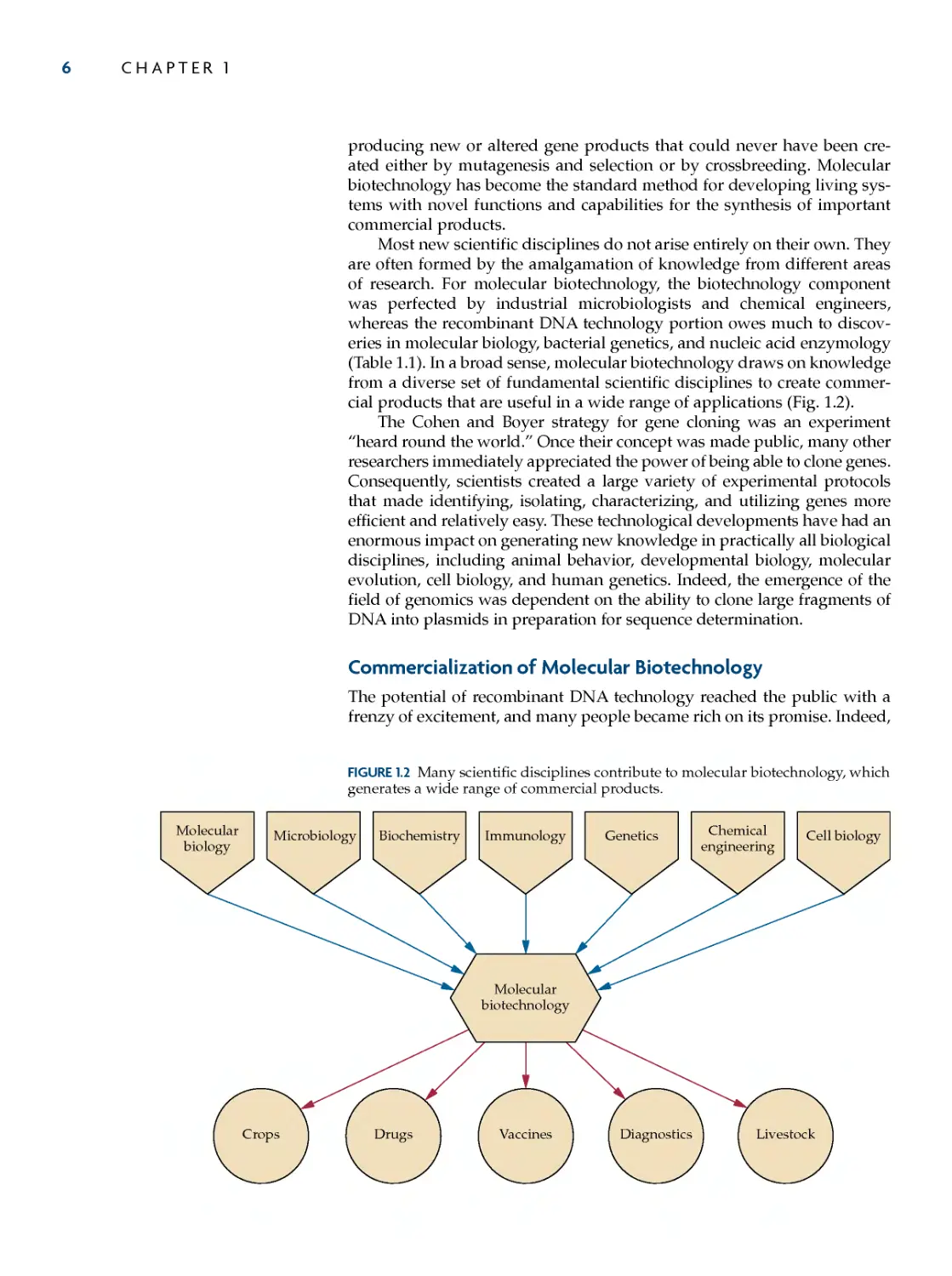
ducing a commercial product typically has three key stages (Fig. 1.1):
1. Upstream processing: preparation of the microorganism and the
raw materials required for the microorganism to grow and pro-
duce the desired product
2. Fermentation and transformation: growth (fermentation) of the
target microorganism in a large bioreactor (usually >100 liters)
with the consequent production (biotransformation) of a desired
compound, which can be, for example, an antibiotic, an amino
acid, or a protein
3. Downstream processing: purification of the desired compound
from either the cell medium or the cell mass
Biotechnology research is dedicated to maximizing the overall effi-
ciency of each of these steps and to finding microorganisms that make
products that are useful in the preparation of foods, food supplements, and
drugs. During the 1960s and 1970s, this research focused on upstream pro-
cessing, bioreactor design, and downstream processing. These studies led
to enhanced bioinstrumentation for monitoring and controlling the fer-
mentation process and to efficient large-scale growth facilities that increased
the yields of various products.
The biotransformation component of the overall process was the most
difficult phase to manipulate. Commodity production by naturally occur-
ring microbial strains on a large scale was often considerably less than
optimal. Initial efforts to enhance product yields focused on creating vari-
ants (mutants) by using chemical mutagens or ultraviolet radiation to
induce changes in the genetic constitution of existing strains. However, the
level of improvement that could be achieved in this way was usually lim-
ited biologically. If a mutated strain, for example, synthesized too much of
a compound, other metabolic functions often were impaired, thereby
causing the strain's growth during large-scale fermentation to be less than
desired. Despite this constraint, the traditional "induced mutagenesis and
selection" strategies of strain improvement were extremely successful for a
number of processes, such as the production of antibiotics.
The traditional genetic improvement regimens were tedious, time-
consuming, and costly because of the large numbers of colonies that had to
be selected, screened, and tested. Moreover, the best result that could be
expected with this approach was the improvement of an existing inherited
property of a strain rather than the expansion of its genetic capabilities.
Fermentation and
biotransformation
Downstream
processing
Upstream
processing
Raw
material
Pure
product
FIGURE 1.1 Principal steps of a bioengi-
neered biotechnology process. Paren-
thetically, Karl Ereky's scheme entailed
using inexpensive sugar beets (raw
material) to feed pigs (biotransforma-
tion) for the production of pork (down-
stream processing).
The Development of Molecular Biotechnology
5
Despite these limitations, by the late 1970s, effective processes for the mass
production of a wide range of commercial products had been perfected.
Today, we have acquired sufficient knowledge of the biochemistry,
genetics, and molecular biology of microorganisms to accelerate the devel-
opment of useful and improved biological products and processes and to
create new products that would not otherwise occur. Distinct from tradi-
tional biotechnology, the modern methods require knowledge of and
manipulation of genes, the functional units of inheritance, and the discipline
that is concerned with the manipulation of genes for the purpose of pro-
ducing useful goods and services using living organisms is known as
molecular biotechnology. The pivotal development that enabled this tech-
nology was the establishment of techniques to isolate genes and to transfer
them from one organism to another. This technology is known as recombi-
nant deoxyribonucleic acid (DNA) technology, and it began as a lunchtime
conversation between two scientists working in different fields who met at
a scientific conference in 1973. In his laboratory at Stanford University in
California, Stanley Cohen had been developing methods to transfer plas-
mids, small circular DNA molecules, into bacterial cells. Meanwhile, Herbert
Boyer of the University of California at San Francisco was working with
enzymes that cut DNA at specific nucleotide sequences. Over lunch at a
scientific meeting, they reasoned that Boyer's enzyme could be used to
splice a specific segment of DNA into a plasmid and then the recombinant
plasmid could be introduced into a host bacterium using Cohen's method.
Recombinant DNA Technology
It was clear to Cohen and Boyer and others that recombinant DNA tech-
nology had far-reaching possibilities. As Cohen noted at the time, "It may be
possible to introduce in E. coli, genes specifying metabolic or synthetic func-
tions such as photosynthesis or antibiotic production indigenous to other
biological classes." The first commercial product produced using recombi-
nant DNA technology was human insulin, which is used in the treatment of
diabetes. The DNA sequence that encodes human insulin was synthesized,
a remarkable feat in itself at the time, and was transplanted into a plasmid
that could be maintained in the common bacterium Escherichia coli. The bac-
terial host cells acted as biological factories for the production of the two
peptide chains of human insulin, which, after being combined, could be
purified and used to treat diabetics who were allergic to the commercially
available porcine (pig) insulin. In the previous decade, this achievement
would have seemed absolutely impossible. By today's standards, however,
this type of genetic engineering is considered commonplace.
The nature of biotechnology was changed forever by the development
of recombinant DNA technology. With these techniques, the maximization
of the biotransformation phase of a biotechnology process was achieved
more directly. Genetic engineering provided the means to create, rather
than merely isolate, highly productive strains. Not long after the production
of the first commercial preparation of recombinant human insulin, bacteria
and then eukaryotic cells were used for the production of insulin, inter-
feron, growth hormone, viral antigens, and a variety of other therapeutic
proteins. Recombinant DNA technology could also be used to facilitate the
biological production of large amounts of useful low-molecular-weight
compounds and macromolecules that occur naturally in minuscule quanti-
ties. Plants and animals became targets to act as natural bioreactors for
6
CHAPTER 1
producing new or altered gene products that could never have been cre-
ated either by mutagenesis and selection or by crossbreeding. Molecular
biotechnology has become the standard method for developing living sys-
tems with novel functions and capabilities for the synthesis of important
commercial products.
Most new scientific disciplines do not arise entirely on their own. They
are often formed by the amalgamation of knowledge from different areas
of research. For molecular biotechnology, the biotechnology component
was perfected by industrial microbiologists and chemical engineers,
whereas the recombinant DNA technology portion owes much to discov-
eries in molecular biology, bacterial genetics, and nucleic acid enzymology
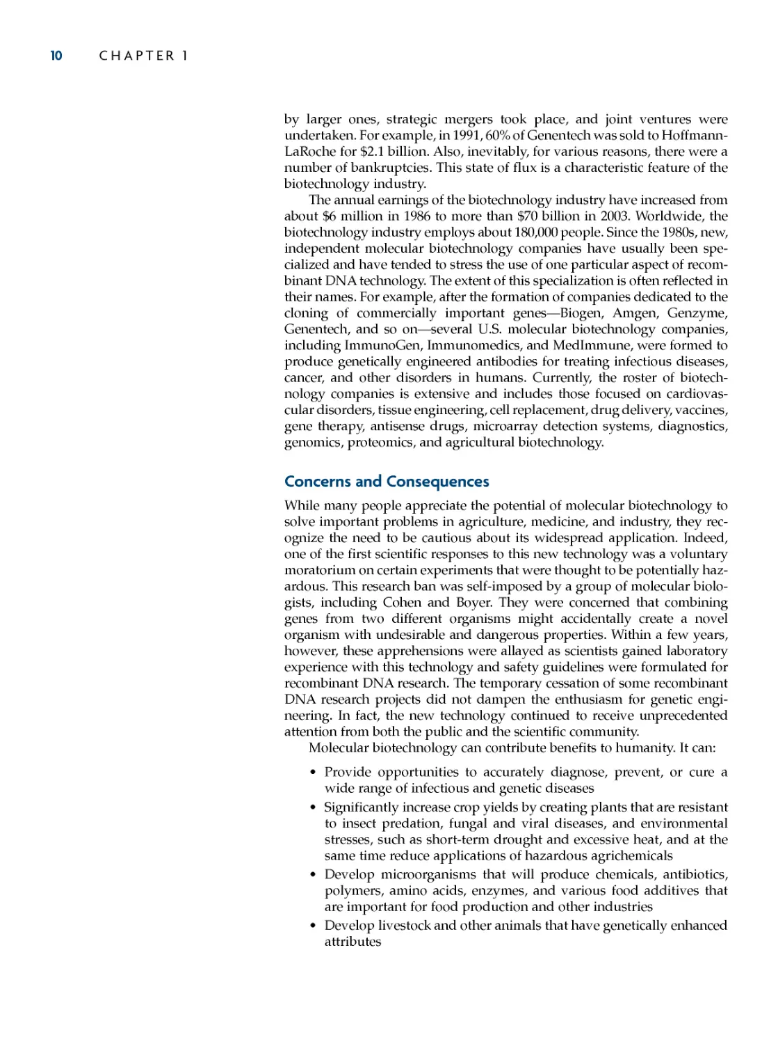
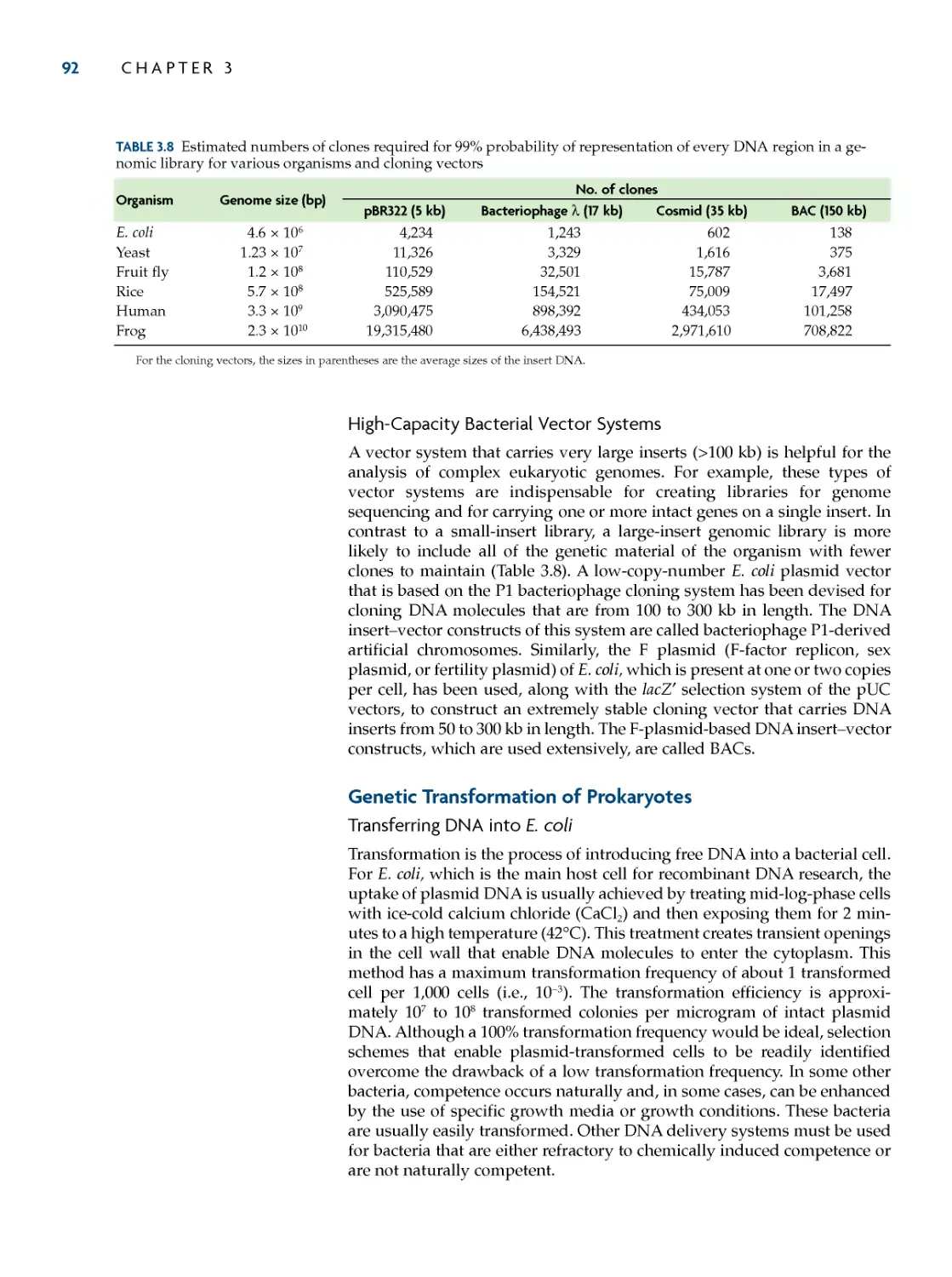
(Table 1.1). In a broad sense, molecular biotechnology draws on knowledge
from a diverse set of fundamental scientific disciplines to create commer-
cial products that are useful in a wide range of applications (Fig. 1.2).
The Cohen and Boyer strategy for gene cloning was an experiment
"heard round the world." Once their concept was made public, many other
researchers immediately appreciated the power of being able to clone genes.
Consequently, scientists created a large variety of experimental protocols
that made identifying, isolating, characterizing, and utilizing genes more
efficient and relatively easy. These technological developments have had an
enormous impact on generating new knowledge in practically all biological
disciplines, including animal behavior, developmental biology, molecular
evolution, cell biology, and human genetics. Indeed, the emergence of the
field of genomics was dependent on the ability to clone large fragments of
DNA into plasmids in preparation for sequence determination.
Commercialization of Molecular Biotechnology
The potential of recombinant DNA technology reached the public with a
frenzy of excitement, and many people became rich on its promise. Indeed,
Microbiology
Molecular
biology
Biochemistry
Genetics
Chemical
engineering
Immunology
Cell biology
Vaccines
Livestock
Diagnostics
Crops
Drugs
Molecular
biotechnology
FIGURE 1.2 Many scientific disciplines contribute to molecular biotechnology, which
generates a wide range of commercial products.
The Development of Molecular Biotechnology
7
TABLE 1.1 Selected developments in the history of molecular biotechnology
Date
Event
1917
Karl Ereky coins the term "biotechnology"
1940
A. Jost coins the term "genetic engineering"
1943
Penicillin is produced on an industrial scale
1944
Avery, MacLeod, and McCarty demonstrate that DNA is the genetic material
1953
Watson and Crick determine the structure of DNA
1961
The journal Biotechnology and Bioengineering is established
1961--1966 Entire genetic code is deciphered
1970
First restriction endonuclease is isolated
1972
Khorana and coworkers synthesize an entire tRNA gene
1973
Boyer and Cohen establish recombinant DNA technology
1975
Kohler and Milstein describe the production of monoclonal antibodies
1976
First guidelines for the conduct of recombinant DNA research are issued
1976
Techniques are developed to determine the sequence of DNA
1978
Genentech produces human insulin in E. coli
1980
U.S. Supreme Court rules in the case of Diamond v. Chakrabarty that genetically manipulated
microorganisms can be patented
1981
First commercial, automated DNA synthesizers are sold
1981
First monoclonal antibody-based diagnostic kit is approved for use in the United States
1982
First animal vaccine produced by recombinant DNA methodologies is approved for use in Europe
1983
Engineered Ti plasmids are used to transform plants
1988
U.S. patent is granted for a genetically engineered mouse susceptible to cancer
1988
PCR method is published
1990
Approval is granted in the United States for a trial of human somatic cell gene therapy
1990
Human Genome Project is officially initiated
1990
Recombinant chymosin is used for cheese making in the United States
1994--1995 Detailed genetic and physical maps of human chromosomes are published
1994
FDA announces that genetically engineered tomatoes are as safe as conventionally bred tomatoes
1995
First genome sequence of a cellular organism, the bacterium Haemophilus influenzae, is completed
1996
First recombinant protein, erythropoietin, exceeds $1 billion in annual sales
1996
Complete DNA sequence of all the chromosomes of a eukaryotic organism, the yeast Saccharomyces cerevisiae,
is determined
1996
Commercial planting of genetically modified crops begins
1997
Nuclear cloning of a mammal (a sheep) with a differentiated cell nucleus is accomplished
1998
FDA approves first antisense drug
1999
FDA approves recombinant fusion protein (diphtheria toxin--interleukin-2) for cutaneous T-cell lymphoma
2000
Arabidopsis genome is sequenced
2000
Monoclonal antibodies exceed $2 billion in annual sales
2000
Development of "golden rice" (provitamin-A-producing rice) is announced
2000
Over $33 billion is invested in U.S. biotechnology companies
2001
Human genome is sequenced
2002
Complete human gene microarrays (gene chips) become commercially available
2002
FDA approves first nucleic acid test system to screen whole blood from donors for HIV and HCV
2004
Large-scale sequencing of the Sargasso Sea metagenome begins
2005
NCBI announces that there are 100 gigabases of nucleotides in the GenBank sequence database
2006
Recombinant cancer vaccine becomes available to protect against cervical cancer
2008
Two-billionth acre of genetically engineered crops is planted
2009
FDA approves first drug produced in a genetically engineered animal (a goat)
FDA, Food and Drug Administration; HCV, hepatitis C virus; HIV, human immunodeficiency virus; NCBI, National Center for Biotechnology Information;
PCR, polymerase chain reaction; tRNA, transfer ribonucleic acid.
8
CHAPTER 1
within 20 minutes of the start of trading on the New York Stock Exchange
on 14 October 1980, the price of shares in Genentech, the company, founded
by Cohen and Boyer with chemist and entrepreneur Robert Swanson, that
produced recombinant human insulin, went from $35 to $89. This was the
fastest increase in the value of any initial public offering in the history of
the market. It was predicted that some genetically engineered microorgan-
isms would replace chemical fertilizers and others would eat up oil spills,
plants with inherited resistance to a variety of pests and exceptional nutri-
tional content would be created, and livestock would have faster growing
times, more efficient feed utilization, and meat with low fat content. Many
were convinced that as long as a biological characteristic was genetically
determined by one or a few genes, organisms with novel genetic constitu-
tions could be readily created. Today we see that, despite the commercial
hype that dominated reality in the beginning, this infatuation with recom-
binant DNA technology was not totally unfounded. A number of the more
sensible versions of the initial claims, although trimmed in scope, have
become realities.
In the 25 years since the commercial production of recombinant human
insulin, more than 200 new drugs produced by recombinant DNA tech-
nology have been used to treat over 300 million people for diseases such as
cancer, multiple sclerosis, cystic fibrosis, and strokes and to provide protec-
tion against infectious diseases. Over 400 new drugs are in the process of
being tested in human trials to treat Alzheimer disease and heart disease
(to name only two). Similarly, many new molecular biotechnology prod-
ucts for enhancing crop and livestock yields, decreasing pesticide use, and
improving industrial processes, such as the manufacture of pulp and paper,
food, energy, and textiles, have been created and are being marketed.
The impact on agriculture has been tremendous. According to the Food
and Agriculture Organization of the United Nations, yield improvements
of all major crops have decreased due to poor agricultural management
practices, decreased acreage of arable land, and increased reliance on fertil-
izers and pesticides that diminish soil quality. To produce more food on
less land, 13 million farmers in 25 countries are now planting genetically
engineered crops on 300 million acres of land. These crops are predomi-
nantly corn, cotton, canola, and soybeans that are resistant to herbicides
and insects. Over the last 10 years in the United States, genetically engi-
neered crops contributed to $44 million in economic gains due to increased
yields and lower production costs. The global market value of genetically
modified crops is currently $7.5 billion. Small resource-poor farmers are
among the beneficiaries of agricultural biotechnology. In a comparative
study of small cotton farms in South Africa, it was found that the yield of
cotton from plants that were genetically engineered to produce a bacterial
insecticide was on average about 70% greater than those from non-geneti-
cally modified plants over three seasons. Higher yields and reduced pesti-
cide and labor costs translated into doubled revenues despite the slightly
higher costs of the transgenic seeds. Similarly, in India, farmers who
planted genetically modified cotton increased their yields by 31% in 2008
while decreasing insecticide use by 39%. This resulted in an 88% increase
in profits for small farmers.
The ultimate objective of all biotechnology research is the development
of commercial products. Consequently, molecular biotechnology is driven,
to a great extent, by economics. Not only does financial investment cur-
rently sustain molecular biotechnology, but clearly the expectation of finan-
The Development of Molecular Biotechnology 9
cial gain was responsible for the considerable interest and excitement
during the initial stages of its development. By nightfall on 14 October
1980, the principal shareholders of Genentech stock were worth millions of
dollars. The unprecedented enthusiastic public response to Genentech
encouraged others to follow. Between 1980 and 1983, about 200 small bio-
technology companies were founded in the United States with the help of
tax incentives and funding from both stock market speculation and private
investment. Like Herbert Boyer, who was first a research scientist at the
University of California at San Francisco and then a vice president of
Genentech, university professors started many of the early companies.
Much of the commercial development of molecular biotechnology has
been centered in the United States. By 1985, there were over 400 biotech-
nology companies, including many with names that contained variants of
the word "gene" to emphasize their expertise in gene cloning: Biogen,
Amgen, Calgene, Engenics, Genex, and Cangene. Today, there are about
1,500 biotechnology companies in the United States, 3,000 in Europe, and
more than 8,000 worldwide, most in the health care sector. All large mul-
tinational chemical and pharmaceutical companies, including Monsanto,
Du Pont, Pfizer, Eli Lilly, GlaxoSmithKline, Merck, Novartis, and
Hoffmann-LaRoche, to name but a few, have made significant research
commitments to molecular biotechnology. During the rapid proliferation
of the biotechnology business in the 1980s, small companies were absorbed
The landmark study of Cohen et
al. established the foundation
for recombinant DNA tech-
nology by showing how genetic infor-
mation from different sources could be
joined to create a novel, replicatable
genetic structure. In this instance, the
new genetic entities were derived
from bacterial autonomously repli-
cating extrachromosomal DNA struc-
tures called plasmids. In a previous
study, Cohen and Chang (Proc. Natl.
Acad. Sci. USA 70:1293--1297, 1973)
produced a small plasmid from a large
naturally occurring plasmid by
shearing the larger plasmid into
smaller random pieces and intro-
ducing the mixture of pieces into a
host cell, the bacterium E. coli. By
chance, one of the fragments that was
about 1/10 the size of the original
plasmid was perpetuated as a func-
tional plasmid. To overcome the ran-
domness of this approach and to make
the genetic manipulation of plasmids
more manageable, Cohen and his
coworkers decided to use an enzyme
(restriction endonuclease) that cuts a
DNA molecule at a specific site and
produces a short extension at each
end. The extensions of the cut ends of
a restriction endonuclease-treated
DNA molecule can combine with the
extensions of another DNA molecule
that has been cleaved with the same
restriction endonuclease.
Consequently, when DNA mole-
cules from different sources are
treated with the same restriction endo-
nuclease and mixed together, new
DNA combinations that never existed
before can be formed. In this way,
Cohen et al. not only introduced a
gene from one plasmid into another
plasmid, but also demonstrated that
the introduced gene was biologically
active. To their credit, these authors
fully appreciated that their strategy
was "potentially useful for insertion of
specific sequences from prokaryotic or
eukaryotic chromosomes or extrachro-
mosomal DNA into independently
replicating bacterial plasmids." In
other words, any gene from any
organism could theoretically be cloned
into a plasmid, which, after introduc-
tion into a host cell, would be main-
tained indefinitely and, perhaps,
produce the protein encoded by the
cloned gene. By demonstrating the
feasibility of gene cloning, Cohen et al.
provided the experimental basis for
recombinant DNA technology; estab-
lished that plasmids could act as vehi-
cles (vectors) for maintaining cloned
genes; motivated others to pursue
research in this area that rapidly led to
the development of more sophisti-
cated vectors and gene-cloning strate-
gies; engendered concerns about the
safety and ethics of this kind of
research that, in turn, was responsible
for the establishment of official guide-
lines and governmental agencies for
conducting and regulating recombi-
nant DNA research, respectively; and
contributed to the formation of the
molecular biotechnology industry.
Construction of Biologically Functional Bacterial
Plasmids In Vitro
S.N.COHEN,A.C.Y.CHANG,H.W.BOYER,andR.B.HELLING
Proc. Natl. Acad. Sci. USA 70:3240--3244, 1973
MILESTONE
10 CHAPTER 1
by larger ones, strategic mergers took place, and joint ventures were
undertaken. For example, in 1991, 60% of Genentech was sold to Hoffmann-
LaRoche for $2.1 billion. Also, inevitably, for various reasons, there were a
number of bankruptcies. This state of flux is a characteristic feature of the
biotechnology industry.
The annual earnings of the biotechnology industry have increased from
about $6 million in 1986 to more than $70 billion in 2003. Worldwide, the
biotechnology industry employs about 180,000 people. Since the 1980s, new,
independent molecular biotechnology companies have usually been spe-
cialized and have tended to stress the use of one particular aspect of recom-
binant DNA technology. The extent of this specialization is often reflected in
their names. For example, after the formation of companies dedicated to the
cloning of commercially important genes---Biogen, Amgen, Genzyme,
Genentech, and so on---several U.S. molecular biotechnology companies,
including ImmunoGen, Immunomedics, and MedImmune, were formed to
produce genetically engineered antibodies for treating infectious diseases,
cancer, and other disorders in humans. Currently, the roster of biotech-
nology companies is extensive and includes those focused on cardiovas-
cular disorders, tissue engineering, cell replacement, drug delivery, vaccines,
gene therapy, antisense drugs, microarray detection systems, diagnostics,
genomics, proteomics, and agricultural biotechnology.
Concerns and Consequences
While many people appreciate the potential of molecular biotechnology to
solve important problems in agriculture, medicine, and industry, they rec-
ognize the need to be cautious about its widespread application. Indeed,
one of the first scientific responses to this new technology was a voluntary
moratorium on certain experiments that were thought to be potentially haz-
ardous. This research ban was self-imposed by a group of molecular biolo-
gists, including Cohen and Boyer. They were concerned that combining
genes from two different organisms might accidentally create a novel
organism with undesirable and dangerous properties. Within a few years,
however, these apprehensions were allayed as scientists gained laboratory
experience with this technology and safety guidelines were formulated for
recombinant DNA research. The temporary cessation of some recombinant
DNA research projects did not dampen the enthusiasm for genetic engi-
neering. In fact, the new technology continued to receive unprecedented
attention from both the public and the scientific community.
Molecular biotechnology can contribute benefits to humanity. It can:
• Provide opportunities to accurately diagnose, prevent, or cure a
wide range of infectious and genetic diseases
• Significantly increase crop yields by creating plants that are resistant
to insect predation, fungal and viral diseases, and environmental
stresses, such as short-term drought and excessive heat, and at the
same time reduce applications of hazardous agrichemicals
• Develop microorganisms that will produce chemicals, antibiotics,
polymers, amino acids, enzymes, and various food additives that
are important for food production and other industries
• Develop livestock and other animals that have genetically enhanced
attributes
The Development of Molecular Biotechnology 11
• Facilitate the removal of pollutants and waste materials from the
environment
Although it is exciting and important to emphasize the positive aspects
of new advances, there are also social concerns and consequences that must
be addressed. The following are some examples.
• Will some genetically engineered organisms be harmful either to
other organisms or to the environment?
• Will the development and use of genetically engineered organisms
reduce natural genetic diversity?
• Should humans be genetically engineered?
• Will new diagnostic procedures undermine individual privacy?
• Will financial support for molecular biotechnology constrain the
development of other important technologies?
• Will the emphasis on commercial success mean that the benefits of
molecular biotechnology will be available only to wealthy nations?
• Will agricultural molecular biotechnology undermine traditional
farming practices?
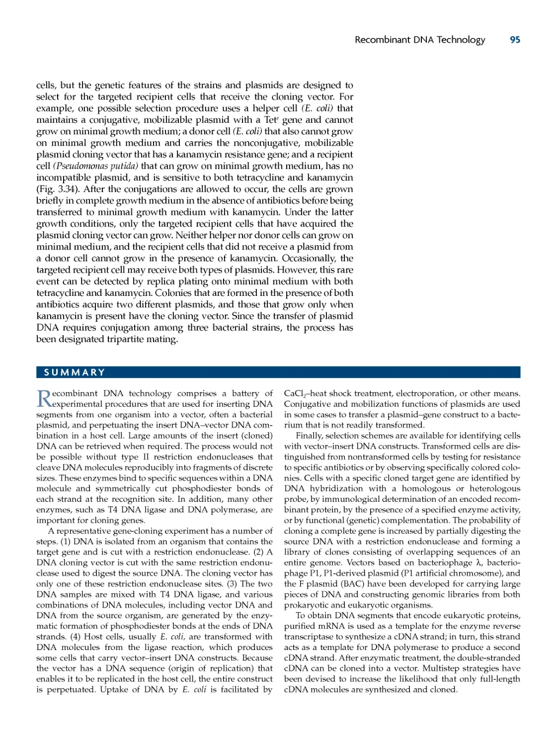
FIGURE 1.3 The Farm, by Alexis Rockman. According to the artist, "The Farm explores
the iconography of agriculture. The Farm is set on a wide-angled field with all its
usual trappings---animals, fruits, and vegetables. The situation, however familiar,
is far from predictable. A disproportionately enormous and savage cow has an
overabundance of teats. The pig is a human organ factory. And the chicken, which
boasts three pairs of wings and no feathers, is ready for basting. The fruit fly, the
workhorse of many a genetic study, is present as is a mouse with a human ear car-
tilage projecting from its back....Past, present, and future states are threaded
together here with barbed wire, woven baskets and DNA....The Farm shows how
the bodies of these animals have been---and may one day be---transformed to suit
our aesthetic, medical, gastronomic needs." © Alexis Rockman, 2000. Reprinted
with the permission of the artist.
12
CHAPTER 1
• Will medical therapies based on molecular biotechnology supersede
equally effective traditional treatments?
• Will the quest for patents inhibit the free exchange of ideas among
research scientists?
These and many other issues have been considered by government
commissions, discussed extensively at conferences, and thoughtfully
debated and analyzed by individuals in both popular and academic publi-
cations. On this basis, rules and regulations have been formulated, guide-
lines have been established, and policies have been created. There has been
active and extensive participation by both scientists and the general public
in deciding how molecular biotechnology should proceed, although some
controversies still remain.
Molecular biotechnology, with much fuss and fanfare, became a com-
prehensive scientific and commercial venture in a remarkably short time.
Many scientific and business publications are now devoted to the subject,
and graduate and undergraduate programs and courses are available at
universities throughout the world to teach it. Even artists have depicted
their perception of molecular biotechnology (Fig. 1.3). It could be debated
whether the early promise of biotechnology has been fulfilled in the way
that was predicted in a 1987 document published by the U.S. Office of
Technology Assessment, which declared that molecular biotechnology is "a
new scientific revolution that could change the lives and futures of . . . citi-
zens as dramatically as did the Industrial Revolution two centuries ago and
the computer revolution today. The ability to manipulate genetic material
to achieve specified outcomes in living organisms . .
. promises major
changes in many aspects of modern life." It does, however, offer solutions
to some serious global problems, including the spread of infectious dis-
eases, the burden of waste accumulation, and food shortages. The potential
of molecular biotechnology to solve some of these imminent problems is
the subject of this book.
In 1973, Stanley Cohen, Herbert Boyer, and their coworkers
devised a method for transferring genetic information
(genes) from one organism to another. This procedure, which
became known as recombinant DNA technology, enabled
researchers to isolate specific genes and to perpetuate them in
host organisms. Recombinant DNA technology has been ben-
eficial to many different areas of study. However, its impact on
biotechnology has been extraordinary.
Biotechnology, for the most part, uses microorganisms on a
large scale for the production of commercially important
products. Before the advent of recombinant DNA technology,
the most effective way of increasing the productivity of an
organism was to induce mutations and then use selection pro-
cedures to identify organisms with superior traits. This pro-
cess was not foolproof; it was time-consuming, labor-intensive,
and costly; and only a small set of traits could be enhanced in
this way. Recombinant DNA technology, however, provided a
rapid, efficient, and powerful means for creating microorgan-
isms with specific genetic attributes. Moreover, the tools of
recombinant DNA technology enable not only microorgan-
isms, but also plants and animals, to be genetically engineered.
Combining recombinant DNA technology with biotechnology
created a dynamic and exciting discipline called molecular
biotechnology.
From its beginning, molecular biotechnology captured the
imagination of the public. Many small companies dedicated to
gene cloning (recombinant DNA technology) were established
with funding from private investors. Although these biotech-
nology companies took somewhat longer than expected to
bring their products to the marketplace, a large number of
recombinant DNA-based products are currently available,
and many more are expected soon.
Because of its broad impact, molecular biotechnology has
been scrutinized carefully for its potential effects on society.
Some of the concerns that have been raised are its safety, its
possible negative effects on the environment, and the private
or public ownership of genetically engineered organisms.
The Development of Molecular Biotechnology 13
REVIEW QUESTIONS
1. What is biotechnology?
2. Distinguish between traditional biotechnology and molec-
ular biotechnology.
3. Describe the basic steps of a bioengineered biotechnology
process.
4. What are the shortcomings of the "mutation and selection"
method for developing enhanced organisms for commercial
purposes?
5. Why was the work reported by Cohen and Boyer and their
coworkers in 1973 considered important?
6. How did recombinant DNA technology enable the produc-
tion of human insulin?
7. What are some of the problems that molecular biotech-
nology has the potential to solve?
8. Discuss the statement "molecular biotechnology is a diverse
science."
9. Discuss some of the social concerns that have been raised
about molecular biotechnology.
10. Go to http://www.nytimes.com, http://news.yahoo.com,
or an equivalent news website and conduct a search with the
word "biotechnology." Describe and discuss three recent bio-
technology news stories.
REFERENCES
Anonymous. 1987. New Developments
in Biotechnology---Background Paper:
Public Perceptions of Biotechnology.
Office of Technology Assessment, U.S.
Congress, U.S. Government Printing
Office, Washington, DC.
Bud, R. 1991. Biotechnology in the
twentieth century. Soc. Stud. Sci.
21:415--457.
Bud, R. 1993. The Uses of Life: a History
of Biotechnology. Cambridge University
Press, Cambridge, United Kingdom.
Busch, L., W. B. Lacy, J. Burkhardt,
and L. R. Lacy. 1992. Plants, Power, and
Profit: Social, Economic and Ethical
Consequences of the New Biotechnologies.
Blackwell Publishers, Cambridge, MA.
Cohen, S. N., and A. C. Chang. 1973.
Recircularization and autonomous
replication of a sheared R-factor DNA
segment in Escherichia coli transfor-
mants. Proc. Natl. Acad. Sci. USA
70:1293--1297.
Cohen, S. N., A. C. Y. Chang, H. W.
Boyer, and R. B. Helling. 1973.
Construction of biologically functional
bacterial plasmids in vitro. Proc. Natl.
Acad. Sci. USA 70:3240--3244.
Davis, B. D. (ed.). 1991. The Genetic
Revolution: Scientific Prospects and
Public Perceptions. Johns Hopkins
University Press, Baltimore, MD.
Grace, E. S. 1997. Biotechnology
Unzipped: Promises & Realities.
Trifolium Press, Inc., Toronto, Canada.
Morse, S., R. Bennett, and Y. Ismael.
2004. Why Bt cotton pays for small-
scale producers in South Africa. Nat.
Biotechnol. 22:379-380.
Robbins-Roth, C. 2000. From Alchemy
to IPO: the Business of Biotechnology.
Perseus Publishing, Cambridge, MA.
Van Beuzekom, B., and A. Arundel.
2006. OECD Biotechnology Statistics
2006. OECD Publishing, Paris, France.
14
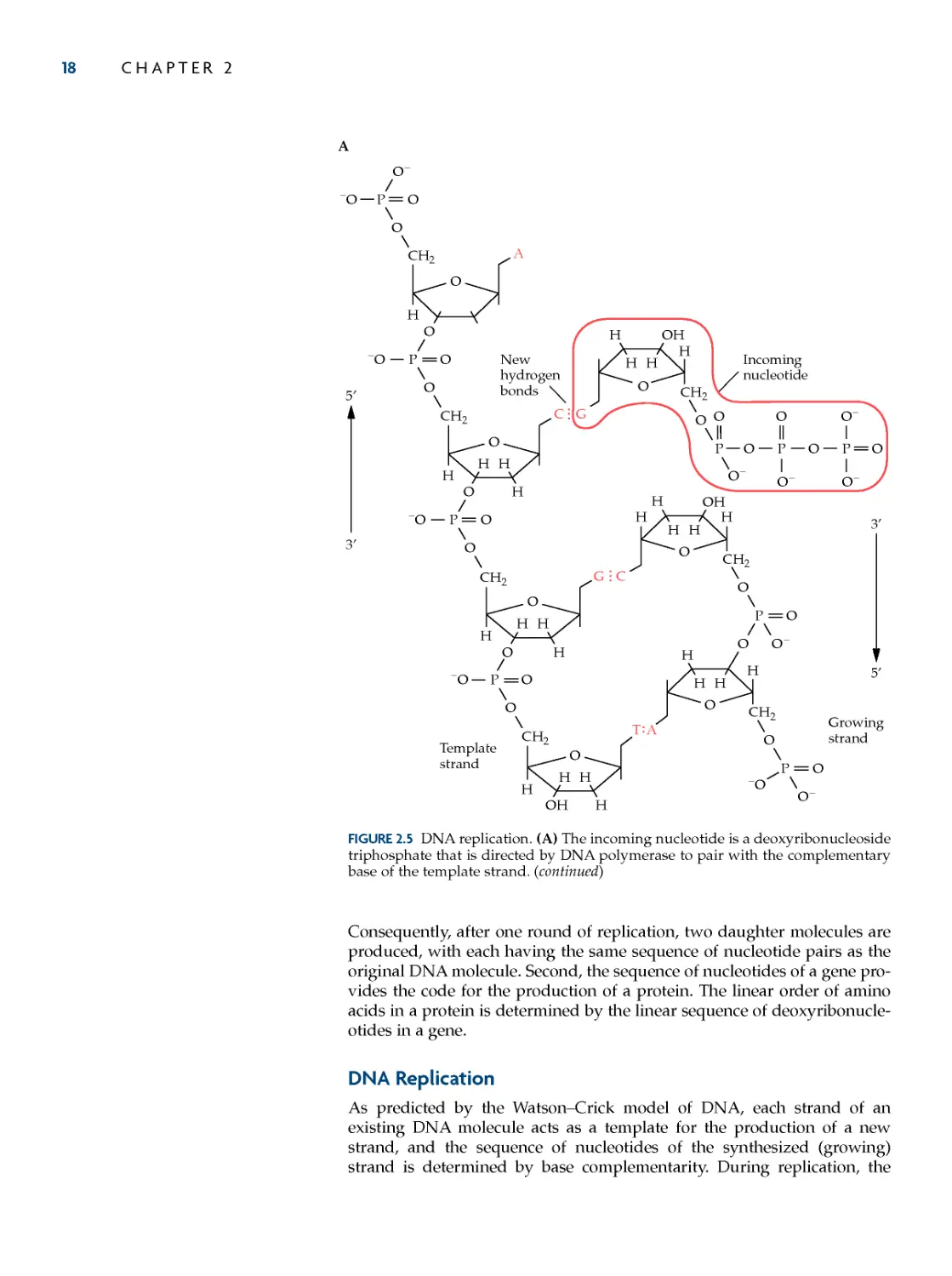
Structure of DNA
DNA Replication
Decoding Genetic Information:
RNA and Protein
Translation
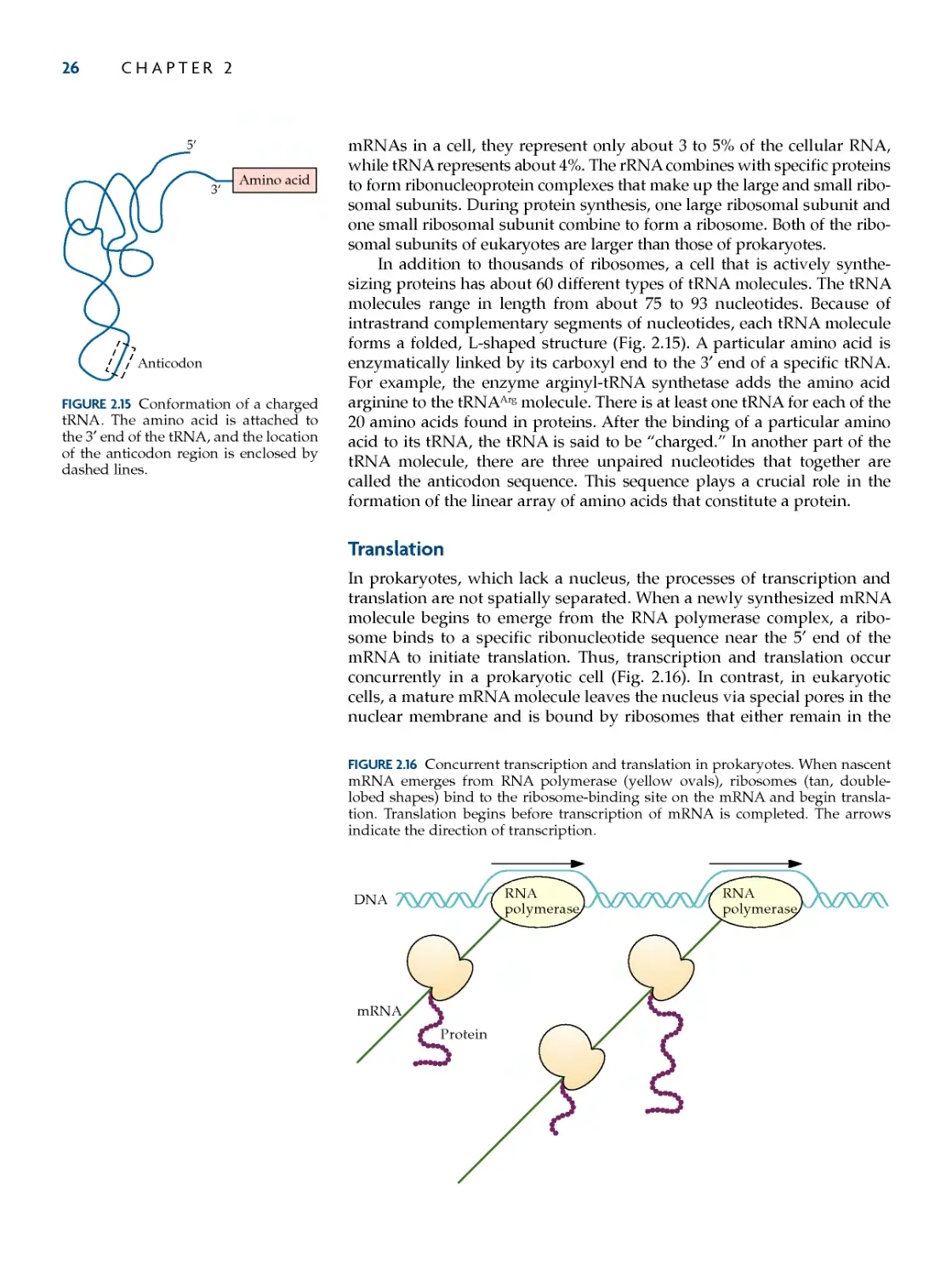
Regulation of mRNA Transcription
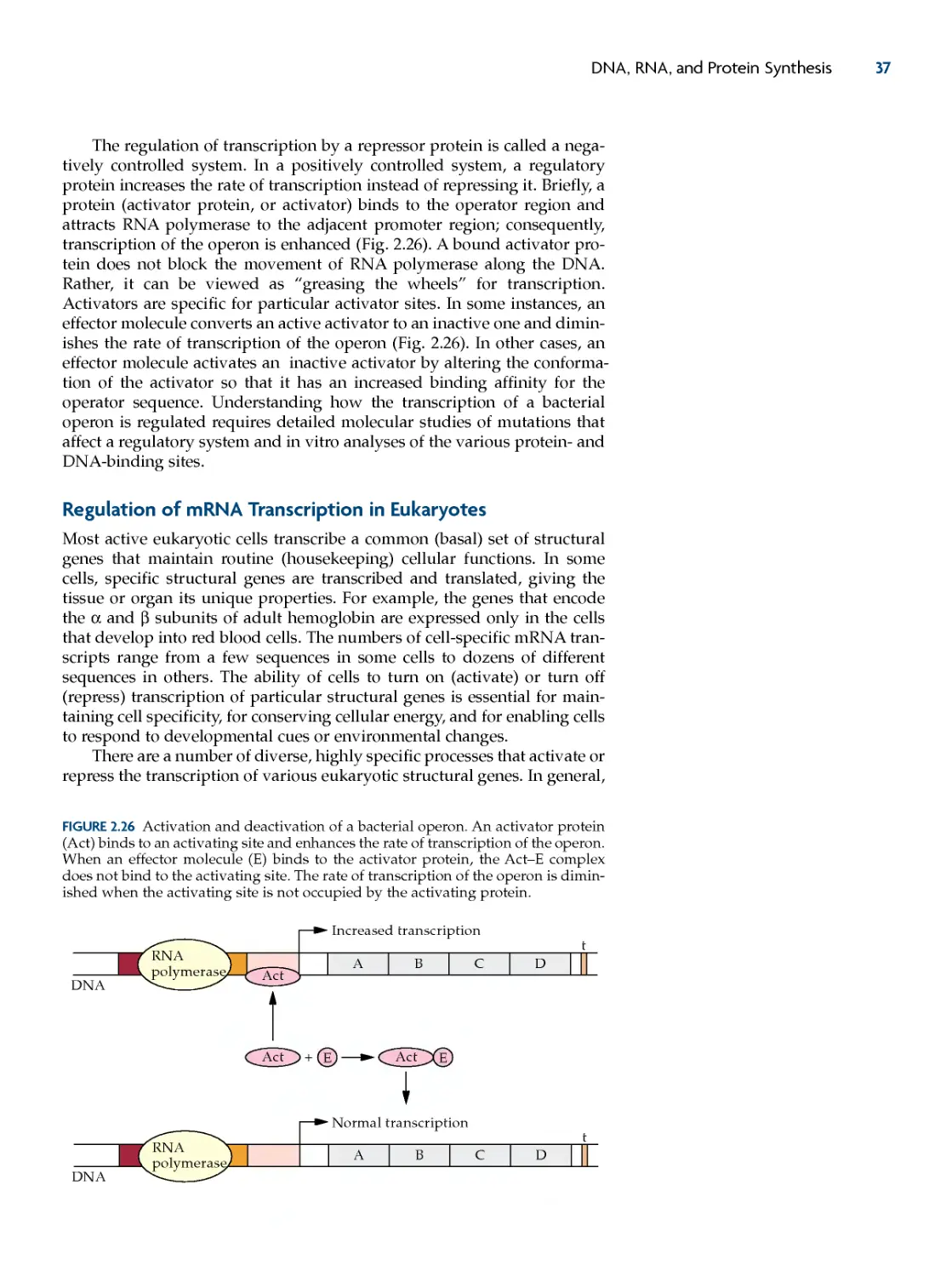
in Bacteria
Regulation of mRNA Transcription
in Eukaryotes
Protein Secretion Pathways
SUMMARY
REFERENCES
REVIEW QUESTIONS
DNA, RNA, and Protein
Synthesis
THE INFORMATION ENCODED INGENETIC MATERIALisresponsibleforestab-
lishing and maintaining the cellular and biochemical functions of an
organism. In most organisms, the genetic material is a long double-
stranded DNA polymer. The sequence of units (deoxyribonucleotides) of
one DNA strand is complementary to the deoxyribonucleotides of the other
strand. This complementarity enables new DNA molecules to be synthe-
sized with the same linear order of deoxyribonucleotides in each strand as
an original DNA molecule. The process of DNA synthesis is called replica-
tion. A specific order of deoxyribonucleotides determines the information
content of an individual genetic element (gene). Some genes encode pro-
teins, and others encode only ribonucleic acid (RNA) molecules. The pro-
tein-coding genes (structural genes) are decoded by two successive major
cellular processes: RNA synthesis (transcription) and protein synthesis
(translation). First, a messenger RNA (mRNA) molecule is synthesized
from a structural gene using one of the two DNA strands as a template.
Second, an individual mRNA molecule interacts with other components,
including ribosomes, transfer RNAs (tRNAs), and enzymes, to produce a
protein molecule. A protein consists of a precise sequence of amino acids,
which is essential for its activity.
Although the deoxyribonucleotide sequences are different in genes
encoding different functions, and for genes encoding similar functions in
different organisms, the chemical compositions are the same. This enables
molecular biotechnologists to transfer genes among a variety of organisms
to create beneficial products. To understand how this is accomplished, it is
helpful to know about the structure of DNA, replication, transcription, and
translation.
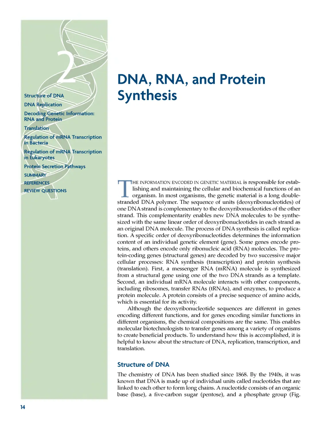
Structure of DNA
The chemistry of DNA has been studied since 1868. By the 1940s, it was
known that DNA is made up of individual units called nucleotides that are
linked to each other to form long chains. A nucleotide consists of an organic
base (base), a five-carbon sugar (pentose), and a phosphate group (Fig.
DNA, RNA, and Protein Synthesis 15
2.1A). The sugar of DNA is 2′-deoxyribose because it does not have a
hydroxyl (OH) group on the 2′ carbon; rather, it has a hydroxyl group only
on the 3′ carbon of the sugar moiety. By contrast, in mRNA, the five-carbon
sugar ribose has hydroxyl groups at both the 2′ and 3′ carbons of the pentose
ring. In both DNA and RNA, the phosphate group and base are attached to
the 5′ carbon and 1′ carbon atoms of the sugar moiety, respectively. In DNA,
there are four kinds of bases: adenine (A), guanine (G), cytosine (C), and
thymine (T) (Fig. 2.1B). The nucleotide subunits of DNA are joined by phos-
phodiester bonds, with the phosphate group of the 5′ carbon of one nucle-
otide linked to the 3′ OH group of the deoxyribose of the adjacent nucleotide
(Fig. 2.2). A polynucleotide strand has a 3′ OH group at one end (the 3′ end)
and a 5′ phosphate group at the other (the 5′ end).
CH3
N
CH
N
C
C
N
HC
NC
NH2
Adenine
H
N
CH
N
C
C
N
C
C
H
HN
O
N
C
C
Cytosine
H2N
H
O
N
NH2
CH
CH
N
C
C
Thymine
H
O
CH
HN
O
P
O
O
O--
CH2
5'
O
4'
H
Base
Deoxyribose
1'
H
OH
H
H
H
3' 2'
A
O
B
Guanine
C
FIGURE 2.1 Chemical structures of the components of DNA. (A) A representative
nucleotide. The term "base" denotes any of the four bases (adenine, guanine, cyto-
sine, and thymine) that are found in DNA. The deoxyribose sugar is enclosed by
dashed lines. The numbers with primes mark the carbon atoms of the deoxyribose
moiety. (B) The bases of DNA. The circled nitrogen atom is the site of attachment of
the base to the 1′ carbon atom of the deoxyribose moiety.
16 CHAPTER 2
In 1953, James Watson and Francis Crick, using X-ray diffraction anal-
ysis of crystallized DNA, discovered that DNA consists of two long chains
(strands) that form a double-stranded helix (Fig. 2.3). The two polynucle-
otide chains of DNA are held together by hydrogen bonds between the
bases of the opposite strands. Base pairing occurs only between specific,
complementary bases (Fig. 2.4). A pairs only with T, and G pairs only with
C. The A⋅T base pairs are held together by two hydrogen bonds, and the
G⋅C base pairs are held together by three. The number of complementary
base pairs is often used to characterize the length of a double-stranded
DNA molecule. For DNA molecules with thousands or millions of base
pairs, the designations are kilobase pairs and megabase pairs, respectively.
For example, the DNA of human chromosome 1 is one double-stranded
helix that has about 263 megabase pairs (Mb).
P
CH2
H
O
O
O
O
O
H
H
H
O
O
CH2
O
HH
H
O
O
CH2 O
HH
H
H
OH
H
H
H
O--
O--
H
H
H
OP
O
CH2
O--
OP
O--
Adenine
Cytosine
Thymine
Guanine
H
5'
3'
5'
3'
3'
3'
5'
5'
5' end
3' end
Phosphodiester
linkage
OP
O--
FIGURE 2.2 Chemical structure of a single strand of DNA.
FIGURE 2.3 A rod--ribbon model of
double-helical DNA. The rods repre-
sent the complementary base pairs,
and the ribbons represent the deoxyri-
bose--phosphate backbones.
DNA, RNA, and Protein Synthesis
17
The A⋅T and G⋅C base pairs lie within the interior of the molecule, and
the 5′-to-3′-linked phosphate and deoxyribose components form the back-
bone of each strand (Fig. 2.4). The two strands of a duplex DNA molecule
run in opposite directions to each other (antiparallel chains). One chain is
oriented in a 3′-to-5′ direction, and the other is oriented in a 5′-to-3′ direc-
tion. Because of the base-pairing requirements, when one strand of DNA
has, for example, the base sequence 5′-TAGGCAT-3′, the complementary
strand must be 3′-ATCCGTA-5′. In this case, the double-stranded form
would be 5′-TAGGCAT-3′
3′-ATCCGTA-5′ . By convention, when DNA is drawn on a horizontal
plane, the 5′ end of the upper strand is on the left.
Genetic material has two major functions. It encodes the information
for the production of proteins, and it is reproduced (replicated) with a high
degree of accuracy to pass the encoded information to new cells. The
Watson--Crick model of DNA fully meets these important requirements.
First, because of base complementarity, each preexisting DNA strand can
act as a template for the production of a new complementary strand.
P
O
CH2
H
O
O
--O
O
PO
--O
O
CH2
O
HH
H
H
O
PO
--O
O
CH2
H
O
HH
H
O
PO
--O
O
CH2
H
O
HH
H
OH
HHH
O
H
O
CH2
HHH
O
H
O
CH2
P
O
HH
O CH2
H
H
3'
5'
H
H
H
H
H
O
H
O
O
CH2
HO
H
HH
O--
H
O--
O--
O--
HH
P
O
H
H
P
O
O--
O
O
P
O
O--
O
TA
C
G
CG
AT
5'
3'
FIGURE 2.4 Chemical structure of double-stranded DNA.
18 CHAPTER 2
Consequently, after one round of replication, two daughter molecules are
produced, with each having the same sequence of nucleotide pairs as the
original DNA molecule. Second, the sequence of nucleotides of a gene pro-
vides the code for the production of a protein. The linear order of amino
acids in a protein is determined by the linear sequence of deoxyribonucle-
otides in a gene.
DNA Replication
As predicted by the Watson--Crick model of DNA, each strand of an
existing DNA molecule acts as a template for the production of a new
strand, and the sequence of nucleotides of the synthesized (growing)
strand is determined by base complementarity. During replication, the
A
CG
TA
GC
P
CH2
H
--O
O--
P
--O
CH2
HH
H
H
P
--O
CH2
HHH
H
P
--O
CH2
HHH
H
HH
O CH2
P
HO
H
OPOPO
Incoming
nucleotide
Template
strand
New
hydrogen
bonds
O
O
O
O
O
O
O
O
O
O
O
O
O
O
O
OH
O
O
O
O--
O--
H
HHH
CH2
P
--O O--
HHHH
CH2
P
O--
OH
H
Growing
strand
O
O
O
O
O
O
O
A
O--
O--
H
3'
5'
5'
3'
FIGURE 2.5 DNA replication. (A) The incoming nucleotide is a deoxyribonucleoside
triphosphate that is directed by DNA polymerase to pair with the complementary
base of the template strand. (continued)
DNA, RNA, and Protein Synthesis 19
phosphate group of each incoming nucleotide is enzymatically joined by a
phosphodiester linkage to the 3′ OH group of the last nucleotide that was
incorporated in the growing strand (Fig. 2.5A). The nucleotides that are
used for DNA replication are triphosphate deoxyribonucleotides that have
three consecutive phosphate groups attached to the 5′ carbon of the deoxy-
ribose sugar moiety. The phosphate that is attached to the 5′ carbon is des-
ignated the α phosphate, the next phosphate is the β phosphate, and the
third one is the γ phosphate (Fig. 2.6). During the replication process, the β
and γ phosphates are cleaved off as a unit, and the α phosphate is linked to
the 3′ OH group of the previously incorporated nucleotide (Fig. 2.5B). The
DNA synthesis machinery of prokaryotes and eukaryotes includes a large
number of different proteins. Of these, DNA polymerases are responsible
for binding deoxyribonucleotides, fitting the correct nucleotide into place
P
O
CH2
H
O
O
--O
A
O
PO
--O
O
CH2
O
HH
H
H
O
PO
--O
O
CH2
H
O
HH
H
O
PO
--O
O
CH2
H
O
HH
T
H
OH
A
HHH
O
H
O
CH2
P
HHH
O
H
O
CH2
PO
O
CG
HH
O
O
CH2
P
HO
H
O
O
H
New phosphodiester
linkage
H
H
H
H
H
O
O--
O--
T
HH
O
O
CH2
HO
H
HH
POPOPO
O--
O--
O--
O
O
O--
Next incoming
nucleotide
O--
H
O--
O--
HH
B
H
H
C
G
3'
5'
5'
3'
FIGURE 2.5 (continued) (B) The α phosphate of the incoming nucleotide forms a
phosphodiester bond with the 3′ hydroxyl group of the growing strand. The next
incoming nucleotide of the growing strand that is complementary to the nucleotide
of the template strand is positioned by DNA polymerase.
20 CHAPTER 2
according to the base-pairing requirement of the template strand, and
forming the phosphodiester linkage.
In bacteria, DNA replication is initiated at a specific region of the (usu-
ally circular) chromosome called the origin of replication (or origin) and, in
Escherichia coli, proceeds at the rate of about 1,000 nucleotides per second. In
eukaryotes, a chromosome has many different sites of initiation of replica-
tion. Because of these multiple origins of replication, part of the eukaryotic
replication process includes enzymatically joining (ligating) segments of
newly synthesized DNA together with phosphodiester bonds. Furthermore,
in eukaryotes, a special replication enzyme called telomerase is used for the
completion of the linear ends (telomeres) of each chromosome.
Decoding Genetic Information: RNA and Protein
The vast majority of genes encode information for the production of pro-
tein chains. Proteins are essential polymers that are involved in almost all
biological functions. They catalyze chemical reactions; transport molecules
within cells; escort molecules between cells; control membrane permea-
bility; give support to cells, organs, and body structures; cause movement;
provide protection against infectious agents and toxins; and regulate the
differential production of other gene products. A protein chain consists of
The elucidation of the structure of
the genetic material (DNA) was
undoubtedly one of the most
important scientific breakthroughs of
the 20th century. James Watson and
Francis Crick, who, with Maurice
Wilkins, were awarded the Nobel
Prize in physiology or medicine in
1962 for this work, suggested "a struc-
ture for the salt of deoxyribonucleic
acid (D. N. A.). This structure has
novel features which are of consider-
able biological interest....This struc-
ture has two helical chains each coiled
around the same axis....Both chains
follow right handed helices, but . . .
the sequences of the atoms in the two
chains run in opposite directions....
The novel feature of the structure is
the manner in which the two chains
are held together by the purine and
pyrimidine bases....They are joined in
pairs, a single base from one chain
being hydrogen bonded to a single
base from the other chain....One of the
pair must be a purine and the other a
pyrimidine for bonding to occur....If
adenine forms one member of a pair,
on either chain, then...the other
member must be a thymine; similarly
for guanine and cytosine. The
sequence of bases on a single chain
does not appear to be restricted in any
way....It has not escaped our notice
that the specific pairing we have pos-
tulated immediately suggests a pos-
sible copying mechanism for the
genetic material." In another article a
few months later (Nature 171:964--967,
1953), Watson and Crick discussed
more implications for their model.
"The phosphate-sugar backbone of
our model is completely regular but
any sequence of the pairs of bases can
fit into the structure. It follows that in
a long molecule many different per-
mutations are possible, and it there-
fore seems likely that the precise
sequence of bases is the code which
carries the genetic information....Our
model suggests possible explanations
for a number of other phenomena. For
example, spontaneous mutations may
be due to a base occasionally in one of
its less likely tautomeric forms. Again,
the pairing between homologous chro-
mosomes at meiosis may depend on
pairing between specific bases."
Within a decade of the demonstra-
tion of the double-helical nature of
DNA with its complementary base
pairs, the molecular aspects of DNA
replication were known, the cellular
processes that are responsible for both
decoding and regulating the synthesis
of gene products were understood,
and many of the kinds of changes that
lead to altered gene products were
recognized. From the time of its publi-
cation to the present, the scientific
impact of the Watson--Crick discovery
has been pervasive and, for the most
part, inestimable. As one small
example, recombinant DNA tech-
nology would not exist without
knowledge of the structure of DNA.
A Structure for Deoxyribose Nucleic Acid
J.D.WATSON ANDF.H.C.CRICK
Nature 171:737--738, 1953
MIlESTONE
DNA, RNA, and Protein Synthesis 21
a specific sequence of units called amino acids. All amino acids have the
same basic chemical structure. There is a central carbon atom (the α carbon)
that has a hydrogen (H), a carboxyl group (COO−), an amino group (NH3+),
and an R group attached to it (Fig. 2.7A). An R group can be any 1 of 20
different side chains (groups) that make up the 20 different amino acids
found in proteins. When R, for example, is a methyl group (CH3), then the
amino acid is alanine. The amino acids of proteins are designated by either
a three- or a one-letter notation (see the table following chapter 23). For
example, alanine is abbreviated Ala or A. In a protein, each amino acid is
linked to an adjacent amino acid by a peptide bond that joins the carboxyl
group of one amino acid to the amino group of the adjacent one (Fig. 2.7B).
The first amino acid of a protein has a free amino group (N terminus), and
the last amino acid in the polypeptide chain has a free carboxyl group (C
terminus).
Proteins range in length from about 40 to more than 1,000 amino acid
residues. A protein folds into a particular shape (configuration, or confor-
mation) depending on the locations of specific amino acid residues and
the overall amino acid composition. Individual amino acids have different
characteristics that are determined by the properties of their side chains,
and these influence the folding of the protein into a particular three-
dimensional shape. The shape of a protein in turn helps to determine its
function. Also, many functional proteins consist of two or more polypep-
tide chains. In some cases, multiples of the same polypeptide chain are
required for an active protein molecule (homomeric protein). In other
instances, a set of different protein chains (subunits) assembles to form a
functional protein (heteromeric protein). Finally, large protein complexes
that are made up of many different subunits often perform important cel-
lular functions.
The decoding of genetic information is carried out through interme-
diary RNA molecules that are transcribed from discrete regions of the
DNA. RNA molecules are linear polynucleotide chains that differ from
DNA in two important respects. First, the sugar moiety of the nucleotides
of RNA is ribose, which has hydroxyl groups on both the 2′ and 3′ carbons
of the sugar. Second, instead of thymine, the base uracil (U) is found in
RNA. Most RNA molecules are single stranded, although often there are
segments of nucleotides within a single chain that are complementary to
each other and form double-stranded regions (intrastrand pairing) (Fig.
2.8). The base pairing within a single RNA strand is the same as the base
O
H
H
H
H
H
β
γO
O
O
O
O
O
O--
O--
O--
O--
α
CH2
Base
OH
P
PP
FIGURE 2.6 Structure of a deoxyribonucleoside triphosphate. The phosphate units
are designated α, β, and γ. The α phosphate is directly attached to the 5′ carbon of
the deoxyribose sugar. During DNA synthesis, the α phosphate participates in the
formation of the phosphodiester bond, and the β and γ phosphate groups are
released as a unit (pyrophosphate).
+H3NC
H
R
COO--
COO--
A
+H3NC
H
R1
CN
O
C
H
R2
H
Peptide bond
B
FIGURE 2.7 Generalized structures of an
amino acid and a peptide bond. (A) An
amino acid. The R represents the loca-
tion of the side chain. (B) A peptide
bond. The peptide bond is encircled,
and R1 and R2 represent different side
chains.
22 CHAPTER 2
pairing between complementary sequences of DNA, except that uracil
pairs with adenine. Base pairing can occur between two RNA molecules if
they contain complementary sequences of base pairs.
The major kinds of RNA molecules that are essential for the decoding
of genetic information are mRNA, ribosomal RNA (rRNA), and tRNA. The
production of RNA from DNA is called transcription. In most prokaryotes,
a single RNA polymerase is responsible for the transcription of all types of
RNA. In eukaryotic organisms, mRNA, rRNA, and tRNA are each tran-
scribed by a different RNA polymerase.
In many of its features, transcription resembles replication. Briefly, one
strand of the DNA of a specific region acts as a template for the synthesis
of a polymer of ribonucleotides. RNA polymerase sequentially joins, via
3′-5′ phosphodiester linkages, ribonucleotides that are complementary to
the nucleotides of the template DNA strand (Fig. 2.9). As transcription pro-
ceeds, the newly synthesized RNA is released from the DNA and the DNA
helix re-forms. Since only specific segments of DNA molecules are tran-
scribed, sets of short stretches of base pairs within the DNA are required to
ensure that transcription is initiated at the correct nucleotide and that it
terminates at a specific nucleotide. The sequences that control the initiation
of transcription usually precede the coding sequence, and the termination
signal sequences follow it. The DNA segment that precedes a gene is called
the 5′-flanking or upstream region, and the one following a gene is the
A
C
G
G
C
A
U
G
C
A
U
G
CG
GU
U
C
G
G
A
A
G
3'
5'
GC
C
A
G
G
U
G
C
C
C
G
G
C
G
C
C
G
C
A
A
A
C
U
G
A
CC
GA
A
U
A
C
U
C
U
G
A
C
C
G
GUU
C
AG
C
U
FIGURE 2.8 Secondary structure of an RNA molecule. The lines represent hydrogen
bonding between complementary base pairs. The ribose--phosphate backbone is
omitted.
DNA, RNA, and Protein Synthesis 23
3′-flanking or downstream region. To initiate transcription, RNA poly-
merase binds to a specific sequence of nucleotides upstream of the coding
sequence known as the promoter. Similarly, a specific sequence of nucle-
otides downstream of the coding sequence, known as the transcriptional
terminator, signals RNA polymerase to stop RNA synthesis.
From a molecular perspective, a gene is a specific nucleotide sequence
that is transcribed into RNA. Structural genes, which make up the vast
majority of transcribed DNA sequences, encode proteins; however, the ini-
tial transcription product of a structural gene is an mRNA. In prokaryotes,
a contiguous DNA segment forms a structural gene (the coding region).
Prokaryotic transcription entails the binding of RNA polymerase to a pro-
moter region, the initiation of transcription at a nucleotide upstream of the
structural gene, and the cessation of transcription at a termination sequence
that lies downstream from the coding region (Fig. 2.10). In eukaryotic organ-
isms, a structural gene usually consists of several coding regions (exons)
that are separated by noncoding regions (introns, or intervening sequences).
After RNA polymerase has bound to the promoter and the entire eukaryotic
structural gene is transcribed, the introns are removed from the primary
transcript, and the exons, in the correct order, are linked (spliced) together
to form a functional mRNA (Fig. 2.11 and 2.12). In general, exons tend to be
150 to 300 bases in length, and introns can vary from as few as 40 to over
10,000 bases. A small number of eukaryotic structural genes lack introns,
3'
5'
DNA
C
GA
UA
UT
AG
CATGCAATGCTACGC
U
ppp
OH
GT TACTACGT TACGATGCG
5'
3'
FIGURE 2.9 Schematic representation of transcription. The arrow indicates the direc-
tion of transcription.
Structural gene
p
t
Transcription
mRNA
Translation
DNA
Protein
FIGURE 2.10 Schematic representation of a prokaryotic structural gene. The pro-
moter region (p), the site of initiation and direction of transcription (the right-
angled arrow), and the termination sequence for RNA polymerase (t) are depicted.
A prokaryotic structural gene is transcribed into mRNA and then directly into
protein.
and in some instances, the introns in a primary transcript may be legiti-
mately removed in more than one way in a process known as alternate
splicing. For example, in one kind of tissue, all the exons of the primary
transcript may be spliced together to form a functional mRNA, whereas in
another tissue, the initial transcript may undergo a different pattern of exon
splicing, with an exon being skipped during the process of intron removal
and a novel functional mRNA being produced. The exon-skipping mecha-
nism generates different gene products in different tissues from the same
t
5
p
Intron a Intron b Intron c Intron d
Transcription
RNA processing
AAAAAAAAA
AAAAAAAAA
Primary transcript
Functional transcript
DNA
Protein
Translation
1234
G
G2
13
45
FIGURE 2.11 Schematic representation of a eukaryotic structural gene. The promoter
region (p), the site of initiation and direction of transcription (the right-angled
arrow), and the termination sequence for RNA polymerase (t) are depicted. The
numbers 1 to 5 mark the exons of the structural gene, and the letters a to d mark the
introns. The primary transcript is polyadenylated at the 3′ end and capped with a
modified guanine (G) nucleotide at the 5′ end. Processing of the primary transcript
removes the introns. The functional RNA is translated into protein.
Exon 1
DNA
Intron a
Exon 2
Exon 3
Intron b
Exon 1
Exon 1
Functional mRNA
Primary RNA transcript
Intron a
Intron b
Exon 2
Exon 2
Exon 3
Exon 3
Intron b
Intron a
FIGURE 2.12 Splicing of a eukaryotic primary RNA transcript. The bracketing arrows
mark the sites that are spliced together after the removal of the intervening RNA
regions. In this example, introns a and b are spliced out of the primary transcript,
and exons 1, 2, and 3 are spliced together to form a functional mRNA.
DNA, RNA, and Protein Synthesis 25
structural gene (Fig. 2.13). For example, in Drosophila, the fruit fly that is
commonly used in genetic studies, two different mRNAs are produced from
the doublesex (dsx) gene as a consequence of alternate splicing of the exons
contained in the gene (Fig. 2.14). One form is produced exclusively in female
flies and the other only in male flies, and each encodes a protein that has a
different activity. The protein produced in female flies prevents the develop-
ment of some male-specific characteristics, including male genitalia, and
conversely, the protein produced in male flies prevents the development of
female-specific traits.
Most (>90%) of the RNA in a metabolically active cell is rRNA found
in ribosomes. Although there can be hundreds to thousands of different
Exon 1
DNA
Intron a
Exon 2
Exon 3
Intron b
Exon 1
Exon 1
Functional mRNA
Intron a
Intron a
Exon 2
Exon 2
Exon 3
Exon 3
Intron b
Intron b
Primary RNA transcript
FIGURE 2.13 Alternative splicing of a eukaryotic primary RNA transcript. The brack-
eting arrows mark the sites that are spliced together after the removal of the inter-
vening RNA region. In this example, exon 2, flanked by introns a and b, is spliced
out of the primary transcript, and exons 1 and 3 are spliced together to form a func-
tional mRNA transcript.
1
1
2
3
4
1
2
3
5
6
2
56
34
Exon splicing in
male fruit flies
pA
Exon splicing in
female fruit flies
dsx primary transcript
dsx mRNA
pA
FIGURE 2.14 Alternative splicing of the doublesex (dsx) primary transcript produces
two different mRNAs in the fruit fly Drosophila melanogaster. The first three exons
are spliced together in both male and female flies; however, in male flies, exon 4 is
skipped, resulting in the splicing of exon 3 to exons 5 and 6. In female flies, exon 3
is spliced to exon 4, which contains a signal for the addition of a poly(A) tail (pA),
resulting in a shortened mRNA. The numbered boxes indicate exons, while the red
and blue lines indicate intron sequences that are spliced out of the primary tran-
script in female and male flies, respectively. Adapted from Maniatis and Tasic,
Nature 418:236--243, 2002.
26 CHAPTER 2
mRNAs in a cell, they represent only about 3 to 5% of the cellular RNA,
while tRNA represents about 4%. The rRNA combines with specific proteins
to form ribonucleoprotein complexes that make up the large and small ribo-
somal subunits. During protein synthesis, one large ribosomal subunit and
one small ribosomal subunit combine to form a ribosome. Both of the ribo-
somal subunits of eukaryotes are larger than those of prokaryotes.
In addition to thousands of ribosomes, a cell that is actively synthe-
sizing proteins has about 60 different types of tRNA molecules. The tRNA
molecules range in length from about 75 to 93 nucleotides. Because of
intrastrand complementary segments of nucleotides, each tRNA molecule
forms a folded, L-shaped structure (Fig. 2.15). A particular amino acid is
enzymatically linked by its carboxyl end to the 3′ end of a specific tRNA.
For example, the enzyme arginyl-tRNA synthetase adds the amino acid
arginine to the tRNAArg molecule. There is at least one tRNA for each of the
20 amino acids found in proteins. After the binding of a particular amino
acid to its tRNA, the tRNA is said to be "charged." In another part of the
tRNA molecule, there are three unpaired nucleotides that together are
called the anticodon sequence. This sequence plays a crucial role in the
formation of the linear array of amino acids that constitute a protein.
Translation
In prokaryotes, which lack a nucleus, the processes of transcription and
translation are not spatially separated. When a newly synthesized mRNA
molecule begins to emerge from the RNA polymerase complex, a ribo-
some binds to a specific ribonucleotide sequence near the 5′ end of the
mRNA to initiate translation. Thus, transcription and translation occur
concurrently in a prokaryotic cell (Fig. 2.16). In contrast, in eukaryotic
cells, a mature mRNA molecule leaves the nucleus via special pores in the
nuclear membrane and is bound by ribosomes that either remain in the
Anticodon
Amino acid
3'
5'
FIGURE 2.15 Conformation of a charged
tRNA. The amino acid is attached to
the 3′ end of the tRNA, and the location
of the anticodon region is enclosed by
dashed lines.
DNA
mRNA
Protein
RNA
polymerase
RNA
polymerase
FIGURE 2.16 Concurrent transcription and translation in prokaryotes. When nascent
mRNA emerges from RNA polymerase (yellow ovals), ribosomes (tan, double-
lobed shapes) bind to the ribosome-binding site on the mRNA and begin transla-
tion. Translation begins before transcription of mRNA is completed. The arrows
indicate the direction of transcription.
DNA, RNA, and Protein Synthesis 27
cytoplasm or associate with the endoplasmic reticulum (Fig. 2.17). In addi-
tion to removal of introns, before the mRNA leaves the nucleus, it is
capped with a modified guanine nucleotide at the 5′ end and a polymer of
adenine nucleotides is added to the 3′ end to form a poly(A) tail (Fig. 2.18).
The 5′ cap and the 3′ poly(A) tail aid in the binding of ribosomes to the
mRNA to begin translation.
Translation requires the interaction of mRNA, charged tRNAs, ribo-
somes, and a large number of proteins (factors) that facilitate the initiation,
RNA
polymerase
RNA
polymerase
DNA
Primary
transcripts
mRNA
mRNA
Nucleus
Cytoplasm
Nuclear
pore
Endoplasmic
reticulum
FIGURE 2.17 Transcription and translation are spatially separated in eukaryotes.
Transcription occurs in the nucleus. Primary transcripts produced by RNA poly-
merase (yellow ovals) are processed to remove introns (grey lines), and the resulting
mRNA (green lines) is exported from the nucleus via nuclear pores. Ribosomes (tan,
double-lobed shapes) in the cytoplasm or associated with the endoplasmic retic-
ulum translate the mRNA to produce proteins that remain in the cytoplasm or are
translocated into the lumen of the endoplasmic reticulum for further processing.
28 CHAPTER 2
elongation, and termination of the polypeptide chain. In prokaryotes,
translation is initiated by the binding of a small ribosomal subunit to an
mRNA by base pairing between a sequence of about 8 nucleotides (a Shine-
Dalgarno sequence) that is located near the 5′ end of the mRNA and a
complementary sequence near the 3′ end of the rRNA of the small ribo-
somal subunit. The 3′-UAC-5′ anticodon of a specific initiator tRNA, fMet-
tRNAfMet, where f represents a formyl moiety that is bound to the
methionine residue, binds to a 5′-AUG-3′ codon (start codon) of the mRNA.
Proteins (initiation factors) facilitate the binding of the initiator tRNA to the
mRNA--small ribosomal subunit complex. A large ribosomal subunit then
combines with the fMet-tRNAfMet--mRNA--small subunit complex to form
the initiation complex (Fig. 2.19).
In eukaryotes, translation is initiated by the binding of a particular
charged initiator tRNA, Met-tRNAMet, along with initiation factors, to a
small ribosomal subunit. Next, the 5′ capped end of an mRNA, which is
combined with specific proteins, associates with the initiator tRNA--small
ribosomal subunit complex, and the complex migrates along the mRNA
until an AUG sequence (initiator, or start codon) is encountered. The 3′
poly(A) tail of the mRNA facilitates the interaction between the mRNA and
the ribosome. When the UAC anticodon sequence of the initiator Met-
tRNAMet base pairs with the AUG sequence of the mRNA, the migration
stops, and the large ribosomal subunit joins the complex to form the initia-
tion complex (Fig. 2.20).
The elongation and termination phases of translation are very similar in
prokaryotes and eukaryotes. The elongation process entails the formation of
a peptide bond between adjacent amino acids, with the order of the amino
acids being determined by the order of codons of the mRNA (Fig. 2.21).
More specifically, after the initiation complex is formed, the second set of
three nucleotides (triplet, or codon) in the mRNA that immediately follows
the AUG codon dictates the anticodon sequence and, therefore, the charged
tRNA that will bind to the ribosome complex. Uncharged tRNAs do not
bind efficiently to ribosomes. For example, if the second nucleotide triplet in
the mRNA is CUG, then the charged tRNA with the anticodon sequence
3′-GAC-5′ will bind. This charged tRNA carries the amino acid leucine.
Once this charged tRNA is in place, a peptide bond is formed between the
carboxyl group of the methionine and the amino group of the leucine. The
leucine remains bound to its tRNA. Peptide bond formation is catalyzed by
activity exclusively associated with the large rRNA. The formation of the
peptide bond "discharges" the initiator tRNA because the bond between the
rRNA
SD
Small
ribosomal
subunit
mRNA
Large
ribosomal
subunit
Met
Met
Initiation
complex
Initiator
fMet-tRNAfMet
UAC
AUG
UAC
AUG
AUG
G-P-P-P
AAAAAAAAAA
AAUAAA
Polyadenylation
signal
5' capP
oly(A) tail
3'
5'
mRNA
FIGURE 2.18 Modification of the ends of a primary RNA transcript in the nucleus. A
modified guanine nucleotide cap is added to the 5′ end of the transcript, and a
polyadenylation signal in the RNA sequence specifies the addition of a polymer of
50 to 250 adenine (A) nucleotides to the 3′ end to form a poly(A) tail. The modified
ends aid in the transport of the mature mRNA from the nucleus and in the binding
of ribosomes to the mRNA and increase the stability of the mRNA.
FIGURE 2.19 Schematic representation of
the initiation of translation in prokary-
otes. The mRNA binds to the small
ribosomal subunit. For some mRNAs,
the Shine-Dalgarno sequence near the
5′ end of the mRNA base pairs with a
sequence near the 3′ end of the rRNA of
the small ribosomal subunit. The anti-
codon (UAC) of the initiator fMet-
tRNAfMet base pairs with the start codon
(AUG) of the mRNA. The large ribo-
somal subunit combines with the initi-
ator tRNA--mRNA--small ribosomal
subunit complex to form the initiation
complex. The amino acid methionine of
the initiator tRNA is formylated (CHO)
at the amino group in prokaryotes (not
depicted). After translation, the initial
formyl-methionine is removed from
the protein chain.
DNA, RNA, and Protein Synthesis 29
carboxyl group of methionine and its tRNA is cleaved to make the carboxyl
group available for peptide bond formation. The uncharged tRNA is ejected
from the ribosomal complex. The methionine--leucine--tRNALeu--mRNA
combination shifts (translocates) along the ribosome to the site vacated by
the initiator tRNA, and as a consequence, the next codon of the mRNA is
available for binding by another charged tRNA with the appropriate anti-
codon sequence. If the third codon is UUU, then the charged tRNA with an
AAA anticodon will bind. In this case, the tRNA with an AAA anticodon
carries the amino acid phenylalanine. Once this charged tRNA is in place,
the linkage between the carboxyl group of leucine and its tRNA is broken
and a peptide bond is formed between the carboxyl group of the leucine and
the amino group of the phenylalanine. After ejection of the uncharged
tRNALeu, the "peptidyl" tRNAPhe, with the attached methionine--leucine--
phenylalanine amino acid polymer and the mRNA, is translocated to the
peptidyl site (P site), and the next codon is available for binding by the
appropriately charged tRNA in the aminoacyl site (A site).
The succession of operations that includes binding of a charged tRNA
by means of anticodon--codon pairing, peptide bond formation, ejection of
an uncharged tRNA, and translocation continues until all the amino acids
that are encoded by the mRNA are linked together. Translation occurs in a
5′-to-3′ direction along the mRNA at a rate of about 15 amino acids per
second. When the 5′ end of the mRNA is free of a ribosome, it can combine
with another initiation complex. A single mRNA can be translated simulta-
neously by a number of ribosomes, with each ribosome producing a poly-
peptide chain. In rapidly growing E. coli cells, the entire population of
approximately 20,000 ribosomes per cell is capable of producing about
30,000 polypeptides per minute. Parenthetically, the average bacterial
structural gene with about 1,000 base pairs (bp) encodes a protein with 333
amino acids because 3 bases code for each amino acid. With a mean molec-
ular weight of an amino acid being about 105, the molecular weight of an
average bacterial protein is about 35,000.
The elongation process continues until a UAA, UAG, or UGA codon
(stop codon, or termination codon) is encountered (Fig. 2.22). There are no
naturally occurring tRNAs with anticodons that are complementary to these
codons. However, a protein(s) (termination factor, or release factor) recog-
nizes a stop codon and binds to the ribosome. After binding of a termination
factor, the bond between the last tRNA, which has the complete chain of
amino acids linked to it, and its amino acid is broken, resulting in dissocia-
tion of the uncharged tRNA, the complete protein, and the mRNA from the
ribosome. In addition, a ribosome-releasing factor separates the ribosomal
subunits so that they can be recycled for the translation of other mRNAs.
After translation, a protein may be modified in various ways. In both
prokaryotes and eukaryotes, the methionine at the N terminus is cleaved
from most proteins, leaving the second encoded amino acid as the
N-terminal moiety. In eukaryotes, certain proteins are selectively cleaved at
defined sites (processed) to make smaller protein chains that have discrete
functions. In other instances, especially in eukaryotes, phosphate groups,
lipids, carbohydrates, or other low-molecular-weight groups are enzymati-
cally added to certain amino acids of a protein. These chemical additions
(posttranslational modifications) create proteins that mediate specific cel-
lular activities.
Small
ribosomal
subunit
mRNA
Initiator Met-tRNAMet
Large
ribosomal
subunit
A
UU
AG
C
Met
A
U
U
A
G
C
Met
mRNA
Met
UAC
FIGURE 2.20 Schematic representation
of the initiation of translation in eukary-
otes. The initiator tRNA (Met-tRNAMet)
binds to a small ribosomal subunit, and
then the mRNA moves along the com-
plex until the anticodon (UAC) of the
initiator tRNA base pairs with the start
codon (AUG) of the mRNA. The large
ribosomal subunit combines with the
mRNA--initiator tRNA--small ribo-
somal subunit complex to form the
initiation complex.
30 CHAPTER 2
The complete genetic code consists of 64 codons. Three of these codons
are reserved for stops, and one (AUG) is used for initiation (Table 2.1).
When a methionine residue occurs internally in a protein, the codon AUG
is recognized by another Met-tRNAMet that is neither formylated nor the
initiator tRNA. There is one codon (UGG) for the amino acid tryptophan.
For the rest of the amino acids that are found in proteins, there are at least
two, usually four, and sometimes as many as six codons. For example, there
are six codons (UUA, UUG, CUU, CUC, CUA, and CUG) for the amino
acid leucine. Different codons are used to different extents in different
organisms (Table 2.1). Of the four codons for glycine, GGA is used about
26% of the time by human structural genes and about 9% of the time by
protein-coding genes of E. coli. The stop codons are also used to different
extents in different organisms. In humans, the frequencies of usage of
UAA, UAG, and UGA are 0.22, 0.17, and 0.61, respectively, whereas in E.
coli, they are 0.62, 0.09, and 0.30, respectively. The differences in codon
usage notwithstanding, the genetic code, with a few rare exceptions, is the
same in all organisms.
A
U
U
A
G
C
C
G
U
A
G UUU
C
ABC
Met Leu
DEF
AUGC
G
U
A
G UUU
C
UAC
Leu
AU
Met
GC
G
U
A
G UUU
C
Met
Leu
AU
Met
GC
G
U
A
G UUU
C
Leu
AAA
Phe
AUGC
G
U
A
G UUU
C
AAA
Leu
Met
Phe
AUGCUG UUU
AAA
Leu
Met
Phe
FIGURE 2.21 Schematic representation of the elongation phase of translation. (A) The
second codon (CUG) base pairs with the anticodon (GAC) of Leu-tRNALeu. (B) The
methionine of the initiator tRNA is joined to the leucine of Leu-tRNALeu by a pep-
tide bond, and the uncharged initiator tRNA is ejected from the ribosome. (C)
Translocation of the peptidyl-tRNA and the mRNA to the peptidyl site from the
aminoacyl site opens the aminoacyl site for the next codon (UUU). (D) The third
codon (UUU) base pairs with the anticodon (AAA) of Phe-tRNAPhe. (E) The leucine
of the peptidyl-tRNA is joined to the phenylalanine of the Phe-tRNAPhe by a peptide
bond, and the uncharged Leu-tRNALeu is ejected from the ribosome. (F) Translocation
of the peptidyl-tRNA and mRNA to the peptidyl site from the aminoacyl site opens
the aminoacyl site for the next codon and codon--anticodon interaction.
DNA, RNA, and Protein Synthesis
31
G
C
U
A
U
A
UAG
AUG
H2N Met Leu Phe Asp Tyr Ala Gly Ala Cys Val COOH
Asp Phe Leu
Cys
Val
Ala
Termination
factor
Gly
Ala Tyr
Met NH2
CAA
FIGURE 2.22 Schematic representation of the termination of translation. The stop
codon (UAG) interacts with a termination factor that leads to the termination of
translation. The last tRNA is cleaved from the peptide chain and ejected. The
mRNA and the finished peptide are released. The ribosomes are prepared for recy-
cling by a ribosome-releasing factor.
TABlE 2.1 Genetic code and codon usage in E. coli and humans
Codon
Amino acid
Frequency of use in:
E. coli
Humans
GGG
Glycine
0.13
0.23
GGA
Glycine
0.09
0.26
GGU
Glycine
0.38
0.18
GGC
Glycine
0.40
0.33
GAG
Glutamic acid
0.30
0.59
GAA
Glutamic acid
0.70
0.41
GAU
Aspartic acid
0.59
0.44
GAC
Aspartic acid
0.41
0.56
GUG
Valine
0.34
0.48
GUA
Valine
0.17
0.10
GUU
Valine
0.29
0.17
GUC
Valine
0.20
0.25
GCG
Alanine
0.34
0.10
GCA
Alanine
0.22
0.22
GCU
Alanine
0.19
0.28
(continued)
32 CHAPTER 2
TABlE 2.1 Genetic code and codon usage in E. coli and humans (continued)
Codon
Amino acid
Frequency of use in:
E. coli
Humans
GCC
Alanine
0.25
0.40
AAG
Lysine
0.24
0.60
AAA
Lysine
0.76
0.40
AAU
Asparagine
0.39
0.44
AAC
Asparagine
0.61
0.56
AUG
Methionine
1.00
1.00
AUA
Isoleucine
0.07
0.14
AUU
Isoleucine
0.47
0.35
AUC
Isoleucine
0.46
0.51
ACG
Threonine
0.23
0.12
ACA
Threonine
0.12
0.27
ACU
Threonine
0.21
0.23
ACC
Threonine
0.43
0.38
UGG
Tryptophan
1.00
1.00
UGU
Cysteine
0.43
0.42
UGC
Cysteine
0.57
0.58
UGA
Stop
0.30
0.61
UAG
Stop
0.09
0.17
UAA
Stop
0.62
0.22
UAU
Tyrosine
0.53
0.42
UAC
Tyrosine
0.47
0.58
UUU
Phenylalanine
0.51
0.43
UUC
Phenylalanine
0.49
0.57
UCG
Serine
0.13
0.06
UCA
Serine
0.12
0.15
UCU
Serine
0.19
0.17
UCC
Serine
0.17
0.23
AGU
Serine
0.13
0.14
AGC
Serine
0.27
0.25
CGG
Arginine
0.08
0.19
CGA
Arginine
0.05
0.10
CGU
Arginine
0.42
0.09
CGC
Arginine
0.37
0.19
AGG
Arginine
0.03
0.22
AGA
Arginine
0.04
0.21
CAG
Glutamine
0.69
0.73
CAA
Glutamine
0.31
0.27
CAU
Histidine
0.52
0.41
CAC
Histidine
0.48
0.59
CUG
Leucine
0.55
0.43
CUA
Leucine
0.03
0.07
CUU
Leucine
0.10
0.12
CUC
Leucine
0.10
0.20
UUG
Leucine
0.11
0.12
UUA
Leucine
0.11
0.06
CCG
Proline
0.55
0.11
CCA
Proline
0.20
0.27
CCU
Proline
0.16
0.29
CCC
Proline
0.10
0.33
DNA, RNA, and Protein Synthesis 33
Regulation of mRNA Transcription in Bacteria
In bacteria, the production of amino acids, nucleotides, and other essential
metabolites; replication; transcription; translation; cell growth; catabolic
pathways; energy-generating systems; and responses to environmental
changes all depend on proteins. However, the energy resources of a cell are
not sufficient to support the transcription and translation (expression) of all
of its structural genes at the same time. Consequently, only those genes that
encode proteins that maintain basic cellular functions are expressed con-
tinuously. The transcription of the remaining structural genes is regulated.
If a protein(s) is required by a cell, then a signaling system initiates tran-
scription (the "on" state) of the pertinent structural gene(s). Alternatively,
if a protein(s) is not needed, transcription of the protein-coding gene(s) is
turned off (the "off" state).
Frequently, bacterial structural genes that encode proteins required for
several steps in a single metabolic pathway are contiguous in the chromo-
some. This arrangement is called an operon. Generally, an operon is under
the control of a single promoter, and its transcription gives rise to one large
mRNA. The placement of a stop codon for one protein close to the start
codon of the next protein within a multigene mRNA generates a set of dis-
crete proteins during translation. Note that a ribosome-binding site (a
Shine-Dalgarno sequence) precedes each start codon.
For many of the structural genes of E. coli, the promoter region has
two DNA-binding sites for RNA polymerase; more specifically, the
binding sites are recognized by the component of the RNA polymerase
complex known as the sigma factor. Frequently, one of these sites tends to
have the sequence TATAAT
ATATTA (a Pribnow box), and the other is usually TTGACA
AACTGT .
The Pribnow box and the TTGACA sequence are located about 10 bp (the
−10 region) and 35 bp (the −35 region), respectively, upstream from the
site of initiation of transcription (the +1 nucleotide) (Fig. 2.23). A promoter
containing a Pribnow box and the TTGACA sequence is recognized by the
sigma factor RpoD (also called sigma-70 [σ70] because it has a molecular
mass of 70 kilodaltons). Many bacteria are capable of producing several
different sigma factors, each of which recognizes a different promoter
sequence. For example, E. coli can produce seven different sigma factors,
each of which initiates the transcription of a specific subset of genes,
although there is some overlap among these sigma factors in the promoter
sequences that they recognize (Table 2.2). RpoD, together with RNA poly-
merase, binds to the promoters of genes that encode proteins or RNA
molecules that are required for essential or "housekeeping" processes.
Other sigma factors direct RNA polymerase to the promoters of genes that
encode more specialized functions, such as proteins required for adapta-
tion to environmental stresses (RpoS) or for nitrogen metabolism
(RpoN).
Nucleotide sequences in and around the RNA polymerase-binding site
often play an essential role in determining whether an operon is transcribed.
This regulatory region is usually referred to as the operator region. A plethora
of elaborate regulatory systems that control the on and off states of various
operons have evolved. For example, when a regulatory protein called a
repressor binds to an operator region and prevents RNA polymerase from
binding to the promoter or moving along the DNA, transcription is blocked
34 CHAPTER 2
(Fig. 2.24). However, in some cases, specific low-molecular-weight com-
pounds (effectors) bind to a particular repressor protein and change its
conformation, thus preventing it from binding to its operator region. When
an effector-repressor complex fails to bind to an operator region, RNA poly-
mRNA
DNA
Translation
Transcription
--35
--10
+1
ABCD
t
αβγδProteins
o
p
A
B
mRNA
DNA
Translation
Transcription
+1
ABCD
t
α
δ Proteins
o
RNA
polymerase
βγ
--35
--10
FIGURE 2.23 Schematic representation of a bacterial transcription unit. (A) The struc-
tural genes (A, B, C, and D) of an operon are under the transcriptional control of an
operator (o) and a promoter (p) region. RNA polymerase binds to regions that are
10 (--10) and 35 (--35) bp from the site of initiation of transcription (+1) (right-angled
arrow). The t denotes a transcription termination signal sequence. After transcrip-
tion of the operon, the proteins of the operon (α, β, γ, and δ) are produced during
translation. (B) Same as panel A, except that binding of RNA polymerase to the
promoter region is shown.
TABlE 2.2 Sigma factors produced by E. coli
Sigma factor Synonym(s) −35 region
−10 region
Function of genes controlled by sigma factor
RpoD
σ70
TTGACA
TATAAT
Most genes required during growth phase
RpoS
σ38
---
CTACACT
Stationary phase and stress response
RpoN
σ54
YTGGCAC (-24 region) TTGCW (-12 region) Nitrogen metabolism
RpoH
σ32
TCTCNCCCTTGAA
CCCCATNTA
Heat shock response
RpoF
σ28, FliA CTAAA
CCGATAT
Flagellum synthesis and chemotaxis
RpoE
σ24
GAANTT
YCTGA
Response to misfolded proteins in the
periplasm
RpoFecI
σ19
GAAAAT
---
Iron transport
The DNA-binding sequences shown are the consensus sequences for each sigma factor recognition site. A consensus sequence represents the nucleotides most
frequently found at each position as determined by comparing the nucleotide sequences of many different promoters recognized by the sigma factor. Rarely, how-
ever, are all of the nucleotides in a given recognition site exactly as represented by the consensus sequence. N = A, T, G, or C; Y = T or C; W = A or T; ---, no consensus
sequence.
DNA, RNA, and Protein Synthesis 35
merase can bind to the promoter and move along the DNA, and the operon
is transcribed. Effector molecules that block repression are generally broken
down by cellular activity. When the levels of an effector molecule are
reduced, the repressor proteins can bind to the operator region and the off
state is reestablished. In many cases an operator region is specific for its
particular operon; however, there are many examples of different operons
with similar operator sequences that are controlled by the same regulatory
protein. The proteins encoded in these operons are usually involved in
related cellular processes.
By way of illustration, if a cell has the enzymatic capability of catabo-
lizing a particular sugar, it is a waste of cellular resources to synthesize the
enzymes that break down the sugar if the sugar is not present in the
medium. On the other hand, if the sugar is available and is the only
carbon source, then the enzymes that are responsible for its cellular utili-
zation are essential. In this case, the sugar acts as an effector, preventing
the repressor from binding to the operator region and thus enabling the
operon to be transcribed. When the amount of sugar is depleted, the
By the early 1960s, due in large
part to the efforts of Watson and
Crick, it was known that the
sequence of bases in DNA contained
the genetic code. However, an out-
standing question was how nucleotide
sequences were decoded to produce
proteins with precise sequences of
amino acids. Specifically, how could
the arrangement of four different
nucleotides (i.e., A, T, G, and C) in a
sequence determine the combination
and linear order of 20 different amino
acids that were known to be found in
proteins? In a series of papers pub-
lished in 1961 and 1962, Marshall W.
Nirenberg and his colleague J.
Heinrich Matthaei at the National
Institutes of Health described a cell-
free system that allowed them to add
various components that might be
required to synthesize a protein to a
reaction under controlled conditions.
Included in the reaction mixture were
ribosomes and soluble RNA (later
determined to be the source of amino-
acyl-tRNAs) extracted from E. coli and
radiolabeled amino acids. Only when
a simple, synthetic RNA polymer,
polyuridylic acid [poly(U)] was added
to the reaction, was radiolabeled
L-phenylalanine incorporated into a
polypeptide identified as poly-L-phe-
nylalanine. "The synthetic polynucle-
otide appears to contain the code for
the synthesis of a protein containing
only one amino acid....Polyuridylic
acid appears to function as a synthetic
template or messenger RNA."
Polyphenylalanine could not be pro-
duced from any other polynucleotide
tested, e.g., poly(A), poly(C), or
poly(A·U). Furthermore, phenylalanyl-
tRNA was required for the transfer of
phenylalanine to the polypeptide
chain. Nirenberg, Matthaei, and Jones
suggested that, "since a sequence of
one or more uridylic acid residues in
poly-U is the code for phenylalanine
in this system, it is probable that phe-
nylalanine-sRNA [tRNA] contains a
complementary sequence of one or
more adenylic acid residues which
base-pair with the template." Not only
had the first "word" of the genetic
code been discovered, but also a
mechanism was proposed by which
the "letters" in mRNA were inter-
preted to produce a protein. In a sub-
sequent paper (Proc. Natl. Acad. Sci.
USA 48:666--677, 1962), Matthaei et al.
showed that an amino acid coding
unit contains a minimum of three
nucleotides and that the genetic code
is at least partially degenerate (two
coding units composed of different
nucleotides specified the same amino
acid) and contains "nonsense words"
that do not encode amino acids but
likely "serve as periods." Nirenberg,
together with H. G. Khorana, who
showed that polyribonucleic acids
with precise sequences of nucleotides
could produce polypeptides with pre-
dicted sequences of amino acids, and
R. W. Holley, who worked out the
structure and function of tRNA, was
awarded the Nobel Prize in physi-
ology or medicine in 1968 for the
cracking of the genetic code.
The Dependence of Cell-Free Protein Synthesis in
E. coli upon Naturally Occurring or Synthetic
Polyribonucleotides
M.W.NIRENBERG andJ.H.MATTHAEI
Proc. Natl. Acad. Sci. USA 47:1588--1602, 1961
An Intermediate in the Biosynthesis of
Polyphenylalanine Directed by Synthetic Template RNA
M.W.NIRENBERG,J.H.MATTHAEI,andO.W.JONES
Proc. Natl. Acad. Sci. USA 48:104--109, 1962
MIlESTONE
36 CHAPTER 2
repressor protein binds to the operator region and prevents transcription
of the operon.
For other operons, transcription may be the normal state because the
repressor protein is inactive. In these cases, a specific effector molecule
(corepressor) attaches to an inactive repressor and causes a conformational
change that enables the repressor--corepressor complex to bind to its cor-
responding operator region and turn off the transcription of the operon
(Fig. 2.25). When the concentration of the corepressor decreases, the on
state for the operon resumes because the repressor by itself is unable to
bind to the operator.
+
DNA
No transcription
ABCD
t
RNA
polymerase
DNA
Transcription
ABCD
t
RNA
polymerase
R
R
R
E
E
FIGURE 2.24 Induction of the on state for transcription of a bacterial operon. The
repressor protein (R) binds to the operator region and blocks transcription. The
binding of an effector molecule (E) to the repressor protein changes the conforma-
tion of the repressor protein. The repressor protein--effector (RE) complex cannot
bind to the operator region; thus, RNA polymerase can transcribe the operon.
+
IR
IR
DNA
No transcription
ABCD
t
RNA
polymerase
DNA
Transcription
ABCD
t
RNA
polymerase
C
C
C
IR
FIGURE 2.25 Induction of the off state for transcription of a bacterial operon. The
binding of a corepressor molecule (C) to an inactive repressor protein (IR) changes
the conformation of the repressor protein. The corepressor--repressor protein com-
plex (IR-C) binds to the operator region and blocks transcription of the operon by
RNA polymerase.
DNA, RNA, and Protein Synthesis 37
The regulation of transcription by a repressor protein is called a nega-
tively controlled system. In a positively controlled system, a regulatory
protein increases the rate of transcription instead of repressing it. Briefly, a
protein (activator protein, or activator) binds to the operator region and
attracts RNA polymerase to the adjacent promoter region; consequently,
transcription of the operon is enhanced (Fig. 2.26). A bound activator pro-
tein does not block the movement of RNA polymerase along the DNA.
Rather, it can be viewed as "greasing the wheels" for transcription.
Activators are specific for particular activator sites. In some instances, an
effector molecule converts an active activator to an inactive one and dimin-
ishes the rate of transcription of the operon (Fig. 2.26). In other cases, an
effector molecule activates an inactive activator by altering the conforma-
tion of the activator so that it has an increased binding affinity for the
operator sequence. Understanding how the transcription of a bacterial
operon is regulated requires detailed molecular studies of mutations that
affect a regulatory system and in vitro analyses of the various protein- and
DNA-binding sites.
Regulation of mRNA Transcription in Eukaryotes
Most active eukaryotic cells transcribe a common (basal) set of structural
genes that maintain routine (housekeeping) cellular functions. In some
cells, specific structural genes are transcribed and translated, giving the
tissue or organ its unique properties. For example, the genes that encode
the α and β subunits of adult hemoglobin are expressed only in the cells
that develop into red blood cells. The numbers of cell-specific mRNA tran-
scripts range from a few sequences in some cells to dozens of different
sequences in others. The ability of cells to turn on (activate) or turn off
(repress) transcription of particular structural genes is essential for main-
taining cell specificity, for conserving cellular energy, and for enabling cells
to respond to developmental cues or environmental changes.
There are a number of diverse, highly specific processes that activate or
repress the transcription of various eukaryotic structural genes. In general,
DNA
t
RNA
polymerase
ABCD
Increased transcription
DNA
t
RNA
polymerase
ABCD
Normal transcription
+
Act
Act
Act
E
E
FIGURE 2.26 Activation and deactivation of a bacterial operon. An activator protein
(Act) binds to an activating site and enhances the rate of transcription of the operon.
When an effector molecule (E) binds to the activator protein, the Act--E complex
does not bind to the activating site. The rate of transcription of the operon is dimin-
ished when the activating site is not occupied by the activating protein.
38 CHAPTER 2
the control of transcription in eukaryotes is mediated by proteins that are
collectively classified as transcription factors. Many transcription factors
bind directly to DNA sequences that are frequently less than 10 bp in
length. The naming of these protein-binding sites is idiosyncratic. However,
for the most part, they are called boxes, DNA modules, initiator elements,
or response elements. Unlike the situation in prokaryotes, operons are
almost never found in the genomes of eukaryotes. Consequently, each
eukaryotic structural gene has its own set of response elements. Moreover,
in addition to DNA--protein interactions, protein--protein associations are
important for regulating eukaryotic transcription.
In addition to specific response elements, a representative eukaryotic
structural gene has a promoter sequence that binds to a core set of proteins
that are minimally required for transcription initiation. A eukaryotic pro-
moter consists of a TATA sequence (TATA box, or Hogness box), a CCAAT
sequence ("cat" box), and a sequence of repeated GC nucleotides (GC box)
that lie about −25, −75, and −90 bp, respectively, from the site of initiation
of transcription (+1) (Fig. 2.27). The first step in the initiation of transcrip-
tion of eukaryotic structural genes with a TATA promoter is the binding of
transcription factor IID (TFIID, or TATA-binding protein [TBP]), which is a
complex of at least 14 proteins, to an available TATA sequence. Subsequently,
other transcription factors bind to TFIID and the DNA adjacent to the TATA
box. Then, RNA polymerase II, which is oriented toward the structural
gene, binds to the transcription complex. With the aid of additional tran-
scription factors, transcription is initiated at the correct starting point (the
+1 nucleotide) (Fig. 2.28). Clearly, if a TATA sequence is deleted or grossly
altered, then transcription of the structural gene cannot occur. Transcription
factors that are specific for the CCAAT and GC response elements have
been identified. In addition, enhancer sequences that increase the rate of
transcription of structural genes are located hundreds or even thousands of
base pairs from the +1 base pair. Folding, looping, or bending of the chro-
mosomal DNA may bring DNA regions, which in the elongated state are
far apart, close to one another. Also, transcription factors that bind to cer-
tain enhancers or response elements may form a chain of proteins that
create bridges from one DNA site to another.
Some repressed (nonexpressed) structural genes are activated by a cas-
cade of events that is triggered by a specific extracellular signal, such as a
temperature increase or the presence of a hormone. For example, a hor-
mone that is released into the circulatory system comes into contact with a
specific cell type that has a receptor on its outer surface that binds the hor-
mone and facilitates the entry of the hormone into the cell. Once inside the
GC box
CCAAT box
TATA box
--90
--75
--25
+1
FIGURE 2.27 Promoter and initiator elements of some eukaryotic structural genes.
The negative numbers designate the locations of nucleotide pairs in the DNA that
lie upstream from the site of initiation (+1) of transcription. The right-angled arrow
indicates the site of initiation and the direction of transcription. The locations of the
transcription elements are not drawn to scale.
DNA, RNA, and Protein Synthesis 39
cell, the hormone interacts with a cytoplasmic protein and changes the
conformation of the protein. In this altered state, the protein is now able to
enter the nucleus, where it binds to an exclusive response element that
initiates transcription of the target gene.
Some proteins bind to response elements and prevent transcription.
For example, there is a class of about 18 vertebrate genes that are actively
transcribed in nerve cells (neurons) and turned off in nonneuronal cells.
Each of these neuron-active genes has a 24-bp response element that lies
upstream of its transcription initiation site. This DNA sequence is called a
neuron-restrictive silencer element (NRSE). In nonneuronal cells, a protein
called neuron-restrictive silencer factor (NRSF) is synthesized, binds to
each NRSE, and prevents transcription of each member of this set of genes.
TATA box
TFIID
TFIIB
RNA polymerase II
TFIIA
TFIIF
TFIIETFIIH
TFIIJ
FIGURE 2.28 Formation of an RNA polymerase II transcription initiation complex at
a TATA box. Transcription factor TFIID binds to a TATA box, and in sequence, other
transcription factors and RNA polymerase II bind to form a protein aggregate that
is responsible for initiating transcription. The right-angled arrow indicates the site
of initiation and the direction of transcription.
40 CHAPTER 2
Conversely, NRSF is not produced by neuronal cells, and therefore, each
gene with an NRSE is transcribed.
On the whole, the regulation of transcription in eukaryotes is complex.
A structural gene may have a number of different response elements that
can be activated in different cell types by different signals at different times
in the life cycle of an organism. Alternatively, some structural genes are
under the preferential control of a unique transcription factor. For the off
state, specific proteins can interact with certain response elements and pre-
vent transcription, or in a more general way, some proteins obstruct tran-
scription by binding to the transcription complex either before initiation or
during the elongation process.
More generalized control of gene expression that influences larger
regions of the chromosomes is mediated by the state of chromosome struc-
ture. A very large amount of chromosomal DNA must be packaged into
the nucleus of a eukaryotic cell. To facilitate this, the DNA is bound by
specific proteins called histones that interact with each other to compact
(condense) the chromosomes into a smaller volume. DNA with its associ-
ated packaging proteins is known as chromatin. Some regions of the chro-
mosomes are tightly packed (heterochromatin), while other regions are
less condensed (euchromatin). Highly condensed DNA is less accessible to
regulatory proteins that activate transcription, and therefore, the genes in
these regions are usually not expressed or are expressed only at a low
level. Chromatin structure, however, is dynamic, and condensed regions
can be "relaxed" by the addition of chemical groups, such as an acetyl
group to amino acids in the packaging proteins or methyl groups to spe-
cific sites in the nucleotide sequences to which the proteins bind.
Unpacking of the chromatin generally increases transcription of genes in
the region.
Protein Secretion Pathways
Bacteria and eukaryotic cells have specialized systems for exporting certain
proteins (secretory proteins) to the external environment. Generally, secre-
tory proteins are required for acquiring nutrients, cell-to-cell communica-
tion, protection, and structures that reside on the outer surface of the cell
membrane. The primary impediment to the release of a secretory protein is
a membrane. The processes that facilitate secretion of proteins through
such a formidable barrier are similar among all organisms, although there
are significant differences between organisms. For example, gram-negative
and gram-positive bacteria do not have the same secretory pathways. A
secreted protein in gram-negative bacteria must pass through an inner
membrane, a periplasmic space, and an outer membrane to exit the cell,
whereas in gram-positive bacteria, secretory proteins are transported only
across a single cytoplasmic membrane. In contrast, the secretory system in
higher organisms is more complex. Unlike prokaryotic proteins, many
eukaryotic proteins require a number of highly specific modifications, such
as glycosylation, acetylation, sulfation, and phosphorylation, to produce
functional secretory proteins. Some of these protein modifications and
various processing steps are carried out in the endoplasmic reticulum, and
others take place in the Golgi apparatus, where proteins are also sorted
according to their final cellular destinations, including those that exit
DNA, RNA, and Protein Synthesis 41
through the cell membrane. The property that distinguishes a protein that
remains in the cytoplasm from one that is secreted is often an amino acid
sequence (a signal peptide, signal sequence, leader sequence, or leader
peptide) at its N terminus.
In gram-positive bacteria, the signal peptide of some secretory proteins
makes direct contact with a membrane-bound assembly of proteins (a
secretion complex, or Sec complex) that facilitates the passage of these pro-
teins through the membrane and their release to the external environment
(Fig. 2.29). Alternatively, for other secretory proteins, a group of proteins
called a signal recognition complex binds to a signal peptide, and this com-
bination attaches to a membrane-bound signal recognition complex
receptor before making contact with the Sec complex (Fig. 2.29). In both
cases, the secretory protein is translocated through a channel formed by the
Sec complex, and its release depends on removal of the signal peptide by a
membrane-bound enzyme called a signal peptidase. Subsequently, pro-
teins that have crossed the cytoplasmic membrane readily pass through the
porous cell wall, where they encounter metal ions and other components
that promote proper folding and molecular stabilization.
Gram-negative bacteria have multiple pathways for the secretion of
various proteins. Some of these systems (Sec-dependent pathways) use the
Cytoplasmic membrane
Cytoplasm
Cell wall
Ribosome
Membrane
protein
Sec complex
Signal peptidase
SRP
Signal peptide
Ribosome
1
2
3
4
FIGURE 2.29 Schematic representation of secretion in gram-positive bacteria. A
signal recognition particle (SRP) binds to the signal peptide of a secretory protein,
and this complex binds to a membrane protein that directs the secretory protein (1)
to the Sec complex. There is also an SRP-independent pathway (2), where a signal
peptide alone makes contact with the Sec complex. The secretory protein is translo-
cated through a channel within the Sec complex (3), and the signal peptide is
removed by a signal peptidase(s). Proper folding of the secretory protein occurs as
it passes through the cell wall (4).
42 CHAPTER 2
same membrane-bound Sec complex for transmitting a secretory protein
through the inner membrane into the periplasm. Collectively, the Sec-
dependent pathways are designated the general secretion pathway. In
these instances, a cytoplasmic protein (SecB) binds an amino acid sequence
(domain) of a secretory protein that has a signal peptide. In turn, the SecB
protein combines with a protein (SecA) of the membrane-bound Sec com-
plex. The secretory protein is translocated into the periplasm, and the
signal peptide is removed. At this point, the secretory protein encounters
various periplasmic proteins that ensure proper folding. Thereafter, Sec-
dependent secretory proteins exit through the outer membrane by different
routes. A region of some proteins is capable of forming a channel in the
outer membrane that allows part of the remaining protein to be selectively
Periplasm
Inner membrane
Outer membrane
Cytoplasm
3
2
1
4
5
SecA
SecB
Sec complex
Gsp complex
Signal peptidase
6
FIGURE 2.30 Schematic representation of a type II secretion pathway in gram-nega-
tive bacteria. The SecB protein binds to a secretory protein in the cytoplasm (1),
SecB attaches to the SecA protein that is part of the Sec complex of the inner mem-
brane (2), and the secretory protein is translocated through the inner membrane (3).
A signal peptidase removes the signal peptide, and the secretory protein is properly
folded in the periplasm (4); the secretory protein combines with the Gsp complex
(5); and it is translocated to the external environment (6).
DNA, RNA, and Protein Synthesis 43
extruded (autotransporter pathway). In these cases, proteolytic cleavage
releases the functional portion of the protein to the external environment.
Other proteins are able to pass through an outer membrane channel that is
formed by a separate protein (single accessory pathway). Another pathway
(chaperone/usher pathway) is used by specific proteins that form fimbriae
on the surface of the bacterial cell. A fourth general secretion pathway
branch called the type II secretion pathway consists of a protein complex
(the Gsp complex) that spans the periplasmic space and forms a channel
through the outer membrane. Most secreted proteins pass through the type
II pathway. In these cases, secretory proteins earmarked for the type II
pathway are first transported to the periplasmic space via the Sec-
dependent pathway, where they bind to the Gsp complex and are shunted
through the outer membrane (Fig. 2.30). Other Sec-dependent pathways
have been found in various gram-negative bacteria. In contrast to the type
II pathway, the type I and type III secretion pathways are Sec independent,
and each has its own protein complex that extends from the inner to the
outer membrane, forming a continuous channel from the bacterial cyto-
plasm to the external environment. For example, bacterial flagellar proteins
reach the outer surface of the cell by means of a type III secretion pathway.
Type III secretion pathways are often used by bacterial pathogens to secrete
bacterial proteins into the cytoplasm of eukaryotic host cells (Fig. 2.31).
Signal peptides are recognized by the Sec-independent systems but are not
necessarily cleaved during the secretion process.
Periplasm
Inner membrane
Outer membrane
Cytoplasm
FIGURE 2.31 The type III secretion system is made up of about 20 different proteins
that form a continuous channel through the inner and outer membranes of gram-
negative bacteria. The type III secretion system is used by bacterial pathogens to
secrete toxins and other proteins into plant and animal host cells. A hollow needle-
like protein structure extends from the bacterial surface into the host cell.
44 CHAPTER 2
Protein secretion is basically the same in all eukaryotic organisms from
yeast to plant and animal cells. Briefly, the signal sequence of a secretory
protein is bound by a signal recognition particle during protein synthesis;
the signal recognition particle attaches to a receptor on the membrane of
the endoplasmic reticulum, and the secretory protein passes through a
channel in the membrane as translation proceeds; a signal peptidase
removes the signal sequence; and the secretory protein is released into the
lumen of the endoplasmic reticulum, where it is folded and, if required,
glycosylated. A vesicle containing a processed secretory protein buds off
from the endoplasmic reticulum and is transported to and fuses with the
cis face of the Golgi apparatus (Fig. 2.32). Additional processing, glycosyla-
tions, and posttranslation modifications take place in the Golgi stack. The
secretory protein then emerges from the trans face of the Golgi apparatus
enclosed in a vesicle that is transported to and fuses with the plasma mem-
brane, where the contents are released to the external environment (Fig.
2.32). In eukaryotic organisms, some proteins are secreted continuously
(constitutive secretion). Others remain in vesicles (mature secretory gran-
ules) near the plasma membrane and are released only after a hormone or
membrane depolarization signal is received.
A
DE
BC
Ribosome
Signal
sequence
SRP
SRP
receptor
complex
Signal
peptidase
ER lumen
ER membrane
ER membrane
Vesicle
Golgi
network
Plasma
membrane
FIGURE 2.32 Schematic representation of the secretion pathway in eukaryotes. (A)
A signal recognition particle (SRP) binds to the signal sequence of a secretory pro-
tein. (B) The SRP attaches to an SRP receptor on the endoplasmic reticulum (ER)
membrane. (C) The secretory protein is translocated into the lumen of the ER, and
a signal peptidase removes the signal sequence. (D) The secretory protein is
folded, partially modified, and packaged in a transport vesicle intended for the
Golgi network. (E) The ER-released vesicle carrying the secretory protein enters
the Golgi network at the cis face and passes through the Golgi stack, where it is
further modified; after it is sorted, a plasma membrane-specific vesicle is formed
at the trans face of the Golgi network. The secretory transport vesicle fuses with the
plasma membrane and releases the secretory protein to the extracellular environ-
ment.
DNA, RNA, and Protein Synthesis 45
ADNA molecule has two polynucleotide strands that form
an antiparallel double helix. The monomeric unit of a
DNA strand is a nucleotide that consists of an base, a deoxyri-
bose sugar, and a phosphate group. The successive nucle-
otides of a DNA strand are linked by phosphodiester bonds,
and the two strands of DNA are held together by hydrogen
bonds between specific complementary pairs of bases. During
replication, which is mediated by a number of different pro-
teins, including DNA polymerases, each DNA strand acts as a
template for the production of a complementary strand.
Proteins are vital for the maintenance of all biological func-
tions. A protein consists of a specific sequence of amino acids
that are linked by peptide bonds. The sequence of amino acids
for a protein is encoded in the DNA. The process of decoding
genetic information is carried out by RNA molecules, including
mRNA, tRNA, and rRNA; various enzymes; and an assort-
ment of protein factors. All RNA is transcribed from DNA.
Sequences of DNA in combination with protein factors ensure
that transcription is initiated at a precise starting point, that
the appropriate strand is used as the template, and that termi-
nation occurs at a specified nucleotide site. In eukaryotic
organisms, most structural genes consist of coding regions
(exons) separated by noncoding segments (introns). Primary
transcripts contain both exons and introns. However, a pro-
cessing system removes the introns and joins the exons, in the
proper order, to form a functional mRNA. An mRNA carries
the code for the sequence of amino acids of a protein.
Translation of mRNA to produce a protein occurs on ribo-
somes that are composed of a large and a small subunit, each
containing rRNA and a large number of specific proteins.
Translation in prokaryotes is initiated by the joining of an
mRNA with a small ribosomal subunit. As a result of codon-
to-anticodon complementary base pairing, the initiator tRNA,
fMet-tRNAfMet, attaches to the mRNA--small ribosomal sub-
unit complex, which then combines with the large ribosomal
subunit to form an initiation complex. Translation in eukary-
otes is initiated by the combining of a unique initiator tRNA
which carries the amino acid methionine, Met-tRNAMet, with a
small ribosomal subunit and then by the threading of an
mRNA through the initiator tRNA--small ribosomal subunit
complex until the first AUG sequence in the mRNA pairs with
the anticodon of the initiator tRNA. The large ribosomal sub-
unit joins the initiator tRNA--small ribosomal subunit--mRNA
complex to form an initiation complex that is ready for the
translation of the mRNA sequence.
After the formation of the initiation complex, the elonga-
tion phase of translation is very similar in prokaryotes and
eukaryotes. The next three nucleotides in the mRNA pair with
the anticodon of a tRNA that carries its specific amino acid.
The first amino acid, methionine, is cleaved from the initiator
tRNA and joined to the second amino acid by a peptide bond.
The "empty" initiator tRNA is ejected from the ribosome, the
ribosome complex shifts, and the tRNA to which the growing
peptide is attached occupies the site vacated by the ejected
initiator tRNA. As a consequence of the shift (translocation),
the next codon of the mRNA is available to pair with the
appropriate anticodon of a tRNA that carries its specific amino
acid that will be joined to the growing peptide. By repeating
these steps, a polypeptide with a specific sequence of amino
acids is formed. Translation is terminated when one of three
stop codons is encountered in the mRNA on a ribosome. A
termination factor, rather than a tRNA, recognizes the stop
codon, and the bond between the last tRNA and the com-
pleted amino acid chain is cleaved, causing the tRNA, mRNA,
and completed protein to be released.
Only the RNAs and proteins that are essential for main-
taining routine cellular functions are synthesized continu-
ously. To conserve cellular resources, transcription of the
remaining genes occurs only when a particular protein is
required and is turned off when the protein is no longer
needed. In prokaryotes, transcription is initiated by the
binding of RNA polymerase to the −10 and −35 elements of the
promoter region of an operon. Regulatory proteins that bind
to operator sequences in and around the promoter region con-
trol the activity of RNA polymerase at the promoter and
thereby control transcription initiation. Repressors prevent
transcription initiation by blocking RNA polymerase binding
to the promoter or movement along the DNA, while activators
enhance the binding of RNA polymerase to a promoter
sequence. The activities of regulatory proteins are controlled
by small effector molecules that increase or decrease their
binding to the operator sequence. In eukaryotes, RNA poly-
merase II, which transcribes structural genes, binds to an
array of proteins called transcription factors that attach, in
sequence, to a TATA sequence of a promoter region. Other
transcription factors that bind to DNA elements of eukaryotic
structural genes are responsible for turning on or turning off
transcription. The expression of eukaryotic genes is also influ-
enced by the local conformation of chromosomal DNA.
Regions that are highly compacted by specific DNA-associated
proteins are generally not transcribed, while more loosely
packed regions contain genes that are transcriptionally
active.
Prokaryotes and eukaryotes have specialized systems for
exporting proteins across a cytoplasmic membrane. Secreted
prokaryotic proteins have a sequence of amino acids at their
N-terminal ends that targets the protein either to the general
secretory pathway or to more specialized protein complexes
that transport specific proteins. Eukaryotic proteins that are to
be secreted are synthesized on ribosomes associated with the
endoplasmic reticulum and are first secreted into the lumen of
the endoplasmic reticulum via an N-terminal signal sequence,
where they are cleaved, folded, and chemically modified.
After further processing in the Golgi apparatus, the proteins
are transported to the cytoplasmic membrane in membrane
vesicles and, following fusion of the vesicle and cytoplasmic
membranes, are released into the external environment.
46 CHAPTER 2
REFERENCES
Browning, D. F., and S. J. Busby. 2004.
The regulation of bacterial transcrip-
tion initiation. Nat. Rev. Microbiol.
2:57--65.
Dever, T. E. 2002. Gene-specific regu-
lation by general translation factors.
Cell 108:545--556.
Ghosh, P. 2004. Process of protein
transport by the type III secretion
system. Microbiol. Mol. Biol. Rev.
68:771--795.
Henkin, T. M. 2000. Transcription ter-
mination control in bacteria. Curr.
Opin. Microbiol. 3:149--153.
Juven-Gershon, T., J.-Y. Hsu, J. W. M.
Theisen, and J. T. Kadonaga. 2008.
The RNA polymerase II core pro-
moter---the gateway to transcription.
Curr. Opin. Cell Biol. 20:253--259.
Koenigsberger, C., J. J. Chicca II,
M. C. Amoureux, G. M. Edelman, and
F. S. Jones. 2000. Differential regula-
tion by multiple promoters of the gene
encoding the neuron-restrictive
silencer factor. Proc. Natl. Acad. Sci.
USA 97:2291--2296.
Kozak, M. 2005. Regulation of transla-
tion via mRNA structure in prokary-
otes and eukaryotes. Gene 361:13--37.
Maniatis, T., and B. Tasic. 2002.
Alternative pre-mRNA splicing and
proteome expansion in metazoans.
Nature 418:236--243.
Morse, R. H. 2007. Transcription factor
access to promoter elements. J. Cell.
Biochem. 102:560--570.
Petry, S., A. Weixlbaumer, and V.
Ramakrishnan. 2008. The termination
of translation. Curr. Opin. Struct.
Biol.18:70--77.
Saier, M. H., Jr. 2006. Protein secretion
and membrane insertion systems in
gram-negative bacteria. J. Membr. Biol.
214:75--90.
Schoenherr, C. J., and D. J. Anderson.
1995. The neuron-restrictive silencer
factor (NRSF): a coordinate repressor
of multiple neuron-specific genes.
Science 267:1360--1363.
Watson, J. D., and F. H. C. Crick. 1953.
Genetical implications of the structure
of deoxyribonucleic acid. Nature
171:964--967.
Williams, S. K., and J. K. Tyler. 2007.
Transcriptional regulation by chro-
matin disassembly and reassembly.
Curr. Opin. Genet. Dev. 17:88--93.
Wilson, D. N., and K. H. Nierhaus.
2006. The E-site story: the importance
of maintaining two tRNAs on the ribo-
some during protein synthesis. Cell.
Mol. Life Sci. 63:2725--2737.
REVIEW QUESTIONS
1. Discuss the basic features of DNA replication.
2. Compare and contrast DNA and RNA.
3. Describe the differences and similarities between prokary-
otic and eukaryotic structural genes.
4. Describe the elongation phase of translation.
5. Deduce the most likely DNA coding sequence for the fol-
lowing human protein: MAGGTWYQLFPRKMWNDSTLHP
FILPMNVAG.
6. Determine the amino acid sequence encoded by the fol-
lowing mRNA: GCGAUCGACGAUGUUUCUAAAAGUAUC
UCAUCGAAAUGAGGGUUCGUAAUAGCGACC
CGGGCGG.
7. What is an operon? What is the biological significance of an
operon?
8. How is transcription initiation controlled in bacterial cells?
9. Describe the major DNA elements that are responsible for
the transcription of eukaryotic structural genes.
10. How are proteins transported across the cytoplasmic
membrane of gram-positive bacterial cells?
11. Describe the type II secretion system of gram-negative
bacterial cells.
12. How are secretory proteins processed in eukaryotic cells?
47
Restriction Endonucleases
Plasmid Cloning Vectors
Plasmid Cloning Vector pBR322
Transformation and Selection
Other Plasmid Cloning Vectors
Creating and Screening a Library
Making a Genomic Library
Screening by DNA Hybridization
Screening by Immunological Assay
Screening by Protein Activity
Cloning DNA Sequences That
Encode Eukaryotic Proteins
Vectors for Cloning Large Pieces
of DNA
Bacteriophage λ Vectors
Cosmids
High-Capacity Bacterial Vector Systems
Genetic Transformation of
Prokaryotes
Transferring DNA into E. coli
Electroporation
Conjugation
SUMMARY
REFERENCES
REVIEW QUESTIONS
Recombinant DNA
Technology
RECOMBINANT DNATECHNOLOGY, whichisalsocalledgenecloningor
molecular cloning, is a general term that encompasses a number of
experimental protocols leading to the transfer of genetic information
(DNA) from one organism to another. There is no single set of methods that
can be used to meet this objective; however, a recombinant DNA experi-
ment often has the following format (Fig. 3.1).
• The DNA (cloned DNA, insert DNA, target DNA, or foreign DNA)
from a donor organism is extracted, enzymatically cleaved (cut, or
digested), and joined (ligated) to another DNA entity (a cloning
vector) to form a new, recombined DNA molecule (cloning vector--
insert DNA construct, or DNA construct).
• This cloning vector–insert DNA construct is transferred into and
maintained within a host cell. The introduction of DNA into a bacte-
rial host cell is called transformation.
• Those host cells that take up the DNA construct (transformed cells)
are identified and selected (separated, or isolated) from those that
do not.
• If required, a DNA construct can be created so that the protein
product encoded by the cloned DNA sequence is produced in the
host cell.
Recombinant DNA technology was developed from discoveries in
molecular biology, nucleic acid enzymology, and the molecular genetics of
both bacterial viruses (bacteriophages) and bacterial extrachromosomal
DNA elements (plasmids). However, recombinant DNA technology would
not exist without the availability of enzymes that recognize specific double-
stranded DNA sequences and cleave the DNA in both strands at these
sequences (restriction enzymes, or restriction endonucleases). Nucleases
that cut nucleic acid molecules internally are endonucleases, and those that
degrade from the ends of nucleic acids are exonucleases.
48 CHAPTER 3
Cloning
vector
Enzymatically
linearize
Target
DNA
Source
DNA
Join target DNA
and cloning vector
DNA construct
Introduce DNA into host cell
Isolate cells with cloned gene
Produce protein from cloned gene
Protein encoded
by cloned gene
Vector DNA
Host cell
Enzymatic
fragmentation
FIGURE 3.1 Recombinant DNA-cloning procedure. DNA from a source organism is
cleaved with a restriction endonuclease and inserted into a cloning vector. The
cloning vector--insert (target) DNA construct is introduced into a host cell, and
those cells that carry the construct are identified and grown. If required, the cloned
gene can be expressed (transcribed and translated) in the host cell, and the protein
(recombinant protein) can be harvested.
Recombinant DNA Technology 49
Restriction Endonucleases
For molecular cloning, both the source DNA that contains the target
sequence and the cloning vector must be consistently cut into discrete and
reproducible fragments. It was only after bacterial enzymes that cut DNA
molecules internally at specific base pair sequences were discovered that
molecular cloning became feasible. These enzymes are formally designated
type II restriction endonucleases. Despite the fact that there are other kinds
of restriction endonucleases (type I, type III, and type IV), the type II
restriction endonucleases are commonly called restriction endonucleases or
simply restriction enzymes.
One of the first type II restriction endonucleases to be characterized
was from the bacterium Escherichia coli, and it was originally designated
EcoRI. More recently, it has been proposed that the use of italics for naming
restriction endonucleases be abandoned. Here, we have implemented this
recommendation. EcoRI is a homodimeric protein (it is made up of two
identical proteins) that binds to a DNA region with a specific palindromic
sequence (recognition site, or binding site). In other words, the sequences
of nucleotides in the two strands of the binding site are identical when
either is read in the same polarity, i.e., 5′ to 3′. The EcoRI recognition
sequence consists of 6 base pairs (bp) and is cut between the guanine and
adenine residues on each strand (Fig. 3.2). EcoRI specifically cleaves the
internucleotide bond between the oxygen of the 3′ carbon of the sugar of
one nucleotide and the phosphate group attached to the 5′ carbon of the
sugar of the adjacent nucleotide. The symmetrical staggered cleavage of
DNA by EcoRI produces two single-stranded, complementary cut ends,
each with extensions of 4 nucleotides, known as sticky ends. In this case,
PSP
T
A
SP
SP
G
C
SP
SP
A
T
SP
SP
A
T
SP
SP
T
A
SP
SP
T
A
SP
SP
C
G
SP
S
T
A
SP
OH
HO
Staggered
cleavage
5'-phosphate
group extension
S
P
P
T
A
SP
HO
S
G
C
SP
T
SP
T
SP
A
SP
A
SP
OH
S
P
P
A
SP
T
SP
T
SP
C
G
SP
HO
S
T
A
SP
OH
S
P
A
Recessed 3'-hydroxyl
group
FIGURE 3.2 Symmetrical, staggered cleavage of a short fragment of DNA by the type
II restriction endonuclease EcoRI. The large arrows show the sites of cleavage in the
DNA backbone. S, deoxyribose sugar; P, phosphate group; OH, hydroxyl group.
The EcoRI recognition sequence is highlighted by the dashed line.
50 CHAPTER 3
each single-stranded extension terminates with a 5′ phosphate group, and
the 3′ hydroxyl group of the opposite strand is recessed.
In addition to EcoRI, more than 3,700 type II restriction endonucleases
with about 250 different recognition sites have been isolated from various
bacteria. The naming protocol for these enzymes is the same as that for
EcoRI; the genus is the capitalized letter, and the first two letters of the spe-
cies name are in lowercase letters. The strain designation is occasionally
added to the name, such as R in EcoRI, or the serotype of the source bacte-
rium is sometimes noted, such as d in HindIII. The Roman numerals are
used to designate the order of characterization of different restriction endo-
nucleases from the same organism. For example, HpaI and HpaII are the
first and second type II restriction endonucleases that were isolated from
Haemophilus parainfluenzae.
The palindromic sequences where most type II restriction endonu-
cleases bind and cut a DNA molecule are within the recognition sites. Some
restriction endonucleases digest (cleave) DNA, leaving 5′ phosphate exten-
sions (protruding ends, or sticky ends) with recessed 3′ hydroxyl ends;
some leave 3′ hydroxyl extensions with recessed 5′ phosphate ends; and
some cut the backbones of both strands within a recognition site to produce
blunt-ended (flush-ended) DNA molecules (Fig. 3.3). The lengths of the
recognition sites for different enzymes can be four, five, six, eight, or more
nucleotide pairs (Table 3.1). Because of the frequency with which their rec-
ognition sites occur in DNA, restriction endonucleases that cleave within
sites of four (four-cutters) and six (six-cutters) nucleotide pairs are used for
most of the common molecular-cloning protocols. The importance of the
type II restriction endonucleases for gene cloning cannot be overstated.
S
P
T
A
SP
P
HO
S
G
C
SP
S
P
P
T
A
SP
SP
G
C
SP
SP
T
A
SP
SP
T
A
SP
SP
A
T
SP
SP
A
T
SP
SP
C
G
SP
S
T
A
SP
OH
HO
Blunt-end
cleavage
PSP
T
A
SP
S
T
A
SP
OH
S
P
P
A
T
SP
HO
S
A
T
SP
PSP
C
G
SP
S
T
A
SP
OH
FIGURE 3.3 Blunt-end cleavage of a short fragment of DNA by the type II restriction
endonuclease HindII. The large arrows show the sites of cleavage in the DNA back-
bone. For abbreviations, see the legend to Fig. 3.2. The HindII recognition sequence
is highlighted.
Recombinant DNA Technology 51
When a DNA sample is treated with one of these enzymes, the same set of
fragments is always produced, assuming that all of the recognition sites are
cleaved. In addition, ready access to a variety of restriction endonucleases
adds versatility to gene-cloning strategies.
Type IIS restriction endonucleases form a subgroup of the type II cat-
egory of restriction enzymes and are occasionally used for cloning and
other molecular studies, such as multiplex polony sequencing, that are
discussed in chapter 4. These enzymes have the fascinating feature of cut-
ting DNA, usually in both strands, a fixed number of nucleotides away
from one end of the recognition site. Moreover, any particular sequence of
nucleotides may be present between the binding sequence and the cut sites.
The cleavages for most type IIS restriction enzymes are staggered. For
example, the FokI restriction endonuclease binds to GGATG
CCTAC and cuts 9 nucle-
otides downstream on the upper strand and 13 nucleotides downstream
on the lower strand, producing a recessed 3′ hydroxyl end and a 4-nucle-
otide extension at the 5′ phosphate end. One representation of the recogni-
tion sequence and cut sites of the FokI restriction endonuclease is
GGATGNNNNNNNNN
CCTACNNNNNNNNNNNNN, where N denotes A, C, G, or T. Of course, with this
single-letter code, it is understood that the nucleotides (N) opposite each
other are base paired. A simpler notation is GGATG(N)9
CCTAC(N)13, and perhaps the sim-
plest is GGATG(9/13). Examples of some type IIS restriction endonucleases are
shown in Table 3.2. It should be noted that a few type IIS restriction endo-
nucleases cleave DNA both upstream and downstream from their recogni-
tion sites.
Under natural conditions, bacteria use restriction endonucleases to
cleave foreign DNA, such as that of infecting bacterial viruses (bacterio-
phages), and have developed systems that protect their own DNA from
being degraded. Most often, methylation of the cytosine residues of a
restriction endonuclease site in the host DNA prevents restriction endonu-
cleases from cutting at these sites, but the nonmethylated sites of foreign
DNA are vulnerable to attack. With the characterization of large numbers
of restriction endonucleases from various bacteria, interesting relationships
have been observed. In some instances, different phosphodiester bonds
TABLE 3.1 Recognition sequences of some restriction endonucleases
Enzyme
Recognition site
Type of cut end
EcoRI
G↓A---A---T---T---C
C---T---T---A---A↑G
5′ phosphate extension
BamHI
G↓G---A---T---C---C
C---C---T---A---G↑G
5′ phosphate extension
PstI
C---T---G---C---A↓G
G↑A---C---G---T---C
3′ hydroxyl extension
Sau3AI
↓G---A---T---C
C---T---A---G↑
5′ phosphate extension
PvuII
C---A---G↓C---T---G
G---T---C↑G---A---C
Blunt end
HpaI
G---T---T↓A---A---C
C---A---A↑T---T---G
Blunt end
HaeIII
G---G↓C---C
C---C↑G---G
Blunt end
NotI
G↓C---G---G---C---C---G---C
C---G---C---C---G---G---C↑G
5′ phosphate extension
Arrows denote cleavage sites.
52 CHAPTER 3
within the same recognition site are cleaved by restriction endonucleases
from different organisms. For example, the restriction enzymes XmaI and
SmaI both recognize the sequence CCCGGG
GGGCCC, but XmaI cleaves after the first 5′
cytosine in each strand and produces 5′ phosphate extensions whereas
SmaI generates blunt ends by cleaving between the CG
GC base pair in the
middle of the recognition site. The first restriction endonuclease that is
discovered to bind to a particular recognition site is designated the proto-
type. Any additional restriction endonucleases that attack the same
sequence as the prototype are called isoschizomers. For example, the
restriction endonucleases XhoI and PaeR7I from different organisms both
have the same recognition sequences and cleavage locations. Isoschizomers
that cleave at different positions within the same recognition site are neo-
schizomers (Fig. 3.4). On the other hand, restriction endonucleases that
produce the same nucleotide extensions but have different recognition sites
are designated isocaudomers, e.g., BamHI and Sau3AI (Table 3.1). In some
cases, a restriction endonuclease will cleave a sequence only if the cytosines
of the recognition site are not methylated whereas another restriction endo-
nuclease will cut the same sequence if these cytosines are methylated. For
example, HpaII cleaves only nonmethylated CCGG
GGCC sites, and MspI, an iso-
schizomer of HpaII, cuts this sequence regardless of cytosine methylation.
This pair of restriction endonucleases is often used to determine the meth-
ylation status of genomic DNA. If a DNA molecule is not cut by HpaII but
is cut by MspI, then the recognition site is methylated. If both restriction
endonucleases cleave a DNA molecule, then the site(s) is not methylated.
Physical maps that designate the relative positions of restriction endo-
nuclease sites on a specific piece of DNA can be constructed by treating the
DNA molecule singly with different restriction endonucleases and then
with combinations of the same restriction endonucleases. The positions of
the cleavage sites can be deduced from an analysis of fragment sizes,
which are determined by agarose gel electrophoresis (Box 3.1). By way of
illustration, the fragment sizes produced by various digestions are shown
TABLE 3.2 Some examples of type IIS restriction endonucleases
Restriction endonuclease
Recognition sequence
AcuI
5′ CTGAAG(N)16
3′ GACTTC(N)14
BfuAI
5′ ACCTGC(N)4
3′ TGGACG(N)8
BsmBI
5′ CGTCTC(N)1
3′ GCAGAG(N)5
EciI
5′ GGCGGA(N)11
3′ CCGCCT(N)9
FokI
5′ GGATG(N)9
3′ CCTAC(N)13
HgaI
5′ GACGC(N)5
3′ CTGCG(N)10
MlyI
5′ GAGTC(N)5
3′ CTCAG(N)5
MmeI
5′ TCCRAC(N)20
3′ AGGYTG(N)18
Recognition sequences are shown with locations of downstream cleavage sites. N = A, T, G, or C; R = A
or G; Y = T or C. The single-letter codes R
Y represent the base pair A
TorG
C,andN
N represents A
T,T
A,G
C,orC
G.
G
CG
KasI
KasI
CC
GG
CC
GC
G
NarI
NarI
SfoI
SfoI
BbeI
BbeI
FIGURE 3.4 Neoschizomers. Four restric-
tion endonucleases bind to the same
recognition site and cleave at different
positions. The restriction endonucleases
and cleavage sites (arrows) are color
coded: KasI, red; NarI, blue; SfoI, black;
BbeI, green. A number of other restric-
tion endonucleases, such as NdaI,
Mly113I, MchI, BinSII, DinI, EgeI, and
EheI, that bind to and cleave this
sequence are not shown.
Recombinant DNA Technology 53
in Fig. 3.5A. It can be deduced that this linear piece of DNA has two
BamHI sites and two EcoRI sites in a definite order with a specified
number of base pairs separating the sites. More specifically, the sizes of
fragments produced in each single digestion can be compared with those
from double digestions to determine the positions of the restriction endo-
nuclease sites and to generate a restriction endonuclease site map (restric-
tion endonuclease map). In the example shown in Fig. 3.5, the analysis
goes as follows. Because each single digestion produces three fragments
from a linear DNA molecule, the original piece of DNA must contain two
sites for each of the restriction endonucleases. The 3,000-bp fragment that
Gel Electrophoresis
Gel electrophoresis is a commonly
used technique for resolving pro-
teins or nucleic acids. In general, a
sample of one particular type of mac-
romolecule (protein, DNA, or RNA) is
placedinawellatorneartheendofa
gel matrix (gel). The composition of an
electrophoresis gel is a semisolid open
meshwork of interlinked linear
strands. A gel is cast as a thin slab
with a number of sample wells. After
the wells of a gel are loaded with
sample, an electric field is applied
across the gel, and charged macromol-
ecules of the same size are driven
together in the direction of the anode
through the gel as discrete invisible
bands of material. The distance that a
band moves into a gel depends on the
mass of its macromolecules and the
size of the openings (pore size) of the
gel. The smaller macromolecules
travel further than the larger ones.
The progress of gel electrophoresis
is monitored by observing the migra-
tion of a visible dye (tracking dye)
through the gel. The tracking dye is a
charged, low-molecular-weight com-
pound that is loaded into each sample
well at the start of a run. When the
tracking dye reaches the end of the
gel, the run is terminated. The bands,
which are aligned in a lane under each
well, are visualized by staining the gel
with a dye that is specific for protein,
DNA, or RNA. Discrete bands are
observed when there is enough mate-
rial present in a band to bind the dye
to make the band visible and when
the individual macromolecules of a
sample have distinctly different sizes.
Otherwise, a band is not detected. If
there is little or no difference among
the sizes of the macromolecules in a
concentrated sample, a smear of
stained material is observed. The
intensity of a stained band reflects the
frequency of occurrence of a macro-
molecule in a sample.
The molecular mass (molecular
weight) of a gel-fractionated macro-
molecule (band) is determined from a
standard curve that is based on a set
of macromolecules of known molec-
ular mass (size markers) that covers
the separation range of the gel system
and is run in one or both of the out-
side lanes (calibrator lanes) of the
same gel as the samples. The loga-
rithm of the molecular mass of a size
marker is related to its relative
mobility (Rf) through a gel. The value
of Rf is defined as the distance trav-
eled by a band divided by the distance
traveled by the tracking dye (ion
front). The relationship between the
logarithm of the molecular mass of
each size marker and its Rf value is
plotted. Then, with this standard
curve, a molecular mass is calculated
for each band in a lane. The units of
molecular mass for proteins and
double-stranded and single-stranded
nucleic acids are daltons, base pairs,
and bases, respectively. The size
markers are included in the same gel
as the samples because the extent of
mobility of a macromolecule(s) varies
from one electrophoretic run to the
next.
Polyacrylamide is the preferred gel
system for separating proteins.
Copolymerization of monomeric
acrylamide and the cross-linker
bisacrylamide forms a lattice of cross-
linked, linear polyacrylamide strands.
The pore size of a polyacrylamide gel
is determined by the concentration of
acrylamide and the ratio of acryl-
amide to bisacrylamide. For many
applications, a protein sample is
treated with the anionic detergent
sodium dodecyl sulfate (SDS) before
electrophoresis. The SDS binds to pro-
teins and dissociates most multichain
proteins. Each SDS-coated protein
chain has a similar charge-to-mass
ratio. Consequently, during electro-
phoresis, the separation of the SDS--
protein chains is based primarily on
size, and the effect of conformation is
eliminated. SDS--polyacrylamide gel
electrophoresis with a 10% polyacryl-
amide gel resolves proteins that range
from 20 to 200 kilodaltons (kDa).
Agarose, which is a polysaccharide
from seaweed, is used routinely as the
gel matrix for the electrophoretic sepa-
ration of medium-size nucleic acid
molecules. A 1.0% agarose gel can
resolve duplex DNA chains that
range from about 600 to 10,000 bp.
Specialized agarose gel electrophoresis
systems are available for fractionating
DNA molecules with millions of base
pairs, denatured DNA, and denatured
RNA. In addition, for specific pur-
poses, polyacrylamide gels are used
for separating DNA molecules. For
example, DNA chains that are as small
as 6 bases and that differ from each
other by 1 nucleotide can be resolved
with a 20% polyacrylamide gel.
BOX 3.1
54 CHAPTER 3
is produced by the single EcoRI digestion remains intact after the double
digestion, whereas the 8,500- and 5,000-bp EcoRI fragments are cleaved.
Therefore, the two EcoRI sites are 3,000 bp apart with no intervening
BamHI site, and there is a single BamHI site within each of the 8,500- and
5,000-bp EcoRI fragments. The 9,500-bp fragment that is produced by the
single BamHI digestion is cleaved by EcoRI in the double digestion into
three pieces (2,500 + 3,000 + 4,000 = 9,500 bp). Therefore, the two BamHI
sites lie 2,500 and 4,000 bp to either side of the EcoRI sites. Digestion with
BamHI cleaves the 8,500-bp EcoRI fragment into 2,500- and 6,000-bp frag-
ments, and one of the EcoRI sites is 2,500 bp from a BamHI site, so the
6,000-bp region must include one of the ends of the original molecule.
Using the same logic, we also note that digestion with BamHI cuts the
5,000-bp EcoRI fragment into 1,000- and 4,000-bp fragments and that one
of the EcoRI sites is 4,000 bp from a BamHI site; therefore, the 1,000-bp
EcoRI
digestion
3,000
5,000
8,500
BamHI
digestion
9,500
1,000
6,000
EcoRI and BamHI
digestion
1,000
2,500
3,000
4,000
6,000
1,000
1,000
B
A
5,000
3,000
8,500
9,500
6,000
4,000
3,000
2,500
6,000
BamHI
BamHI
EcoRI
Eco RI
FIGURE 3.5 Mapping of restriction endonuclease sites. (A) Restriction endonuclease
digestions and electrophoretic separation of fragments. A purified, linear piece of
DNA is cut with EcoRI and BamHI separately (single digestions) and then with
both enzymes together (double digestion). The horizontal lines under the digestion
conditions represent schematically the locations of the DNA fragments (bands) in
the lanes of the gel after electrophoresis and staining of the DNA with ethidium
bromide. The numbers denote the lengths of the digestion products (fragments) in
base pairs. (B) Restriction endonuclease map derived from the digestions and elec-
trophoretic separation shown in panel A.
Recombinant DNA Technology 55
region must include the other end of the original molecule. In the final
map (Fig. 3.5B), the assigned locations of the restriction endonuclease sites
are consistent with the fragment lengths that were observed in each of the
digestion reactions.
The process of formulating a restriction endonuclease map for circular
DNA is, in principle, the same as with linear DNA, except that each
cleavage produces a fragment. In other words, three pieces are formed
when three sites are cut, and so on. With the data in Table 3.3, the deduction
of a restriction endonuclease map of a circular plasmid is as follows. The
source DNA is a 12-kilobase-pair (kb) circle with single EcoRI, BamHI, and
HindIII sites and three HaeII sites. Digestion with EcoRI, BamHI, or HindIII
separately produces a single 12-kb fragment, while digestion with HaeII
produces three fragments. The results of the EcoRI and HaeII double diges-
tion indicate that the EcoRI site lies within the 6.0-kb HaeII fragment,
because the 2.0-kb and 4.0-kb HaeII fragments remain intact and the sum
of the two new pieces (5.0 kb + 1.0 kb) is 6.0 kb. Based on the BamHI and
HaeII double digestion, the BamHI site lies within the 2.0-kb HaeII region.
The BamHI and EcoRI double digestion places these sites 1.5 kb apart;
therefore, the 6.0-kb and 2.0-kb HaeII fragments are adjacent. The data do
not support any other positions for the BamHI and EcoRI sites. The order
of the HaeII segments around the circular molecule is 6.0 kb-4.0 kb-2.0 kb.
The same reasoning localizes the HindIII site to the 4.0-kb HaeII fragment,
and the results from the BamHI and HindIII and/or EcoRI and HindIII
double digestions complete the restriction endonuclease map (Fig. 3.6).
For some restriction endonuclease mapping experiments, the sum of
the fragments of some multiple digestions is less than the total length of the
starting DNA because the fortuitous locations of some sites produce frag-
ments of the same size. Under these conditions, two different fragments
with the same length that migrate to the same location in a gel after electro-
phoresis often stain more heavily than a band with only one kind of frag-
ment. This difference in staining intensity gives an indication that
coincidental fragments have been produced by restriction endonuclease
digestion. Generally, computer programs are used to configure restriction
endonuclease maps for large DNA molecules with many single and mul-
tiple digestions. Also, for very large DNA molecules, specialized electro-
phoresis systems are used to separate the large number of restriction
endonuclease digestion products.
The resolution of fragments for restriction endonuclease mapping can
be enhanced by labeling the pieces of DNA, usually at the 5′ ends, with a
radioactive compound or fluorescent dye and determining their lengths
after electrophoretic separation with autoradiography or fluorography,
respectively. A common 5′-end-labeling procedure entails dephosphoryla-
tion of the 5′ ends of a linear DNA molecule with calf intestine alkaline
TABLE 3.3 DNA fragment sizes (in kilobase pairs) after single and double restriction endonuclease digestions of a
plasmid
EcoRI
BamHI HindIII HaeII EcoRI +
HaeII BamHI +
HaeII
HindIII +
HaeII
EcoRI +
HindIII EcoRI +
BamHI BamHI +
HindIII
12.0
12.0
12.0
6.0
5.0
6.0
6.0
6.5
10.5
8.0
4.0
4.0
4.0
2.5
5.5
1.5
4.0
2.0
2.0
1.5
2.0
1.0
0.5
1.5
Bam HI (11.5) Hae II
(0/12.0)
Hae II
(10.0)
Hae II
(6.0)
12.0 kb
Hin dIII
(7.5)
Eco RI (1.0)
FIGURE 3.6 Restriction endonuclease
map for the digestion products pre-
sented in Table 3.3. The circular DNA
has 12,000 bp (12 kb). With one of the
HaeII sites arbitrarily placed at posi-
tion 0/12.0 kb, the locations of the
other mapped restriction endonuclease
sites are marked. The nucleotide posi-
tions of the sites are in parentheses.
56 CHAPTER 3
phosphatase and the addition of radiolabeled γ-phosphate from adenosine
triphosphate (ATP) to the 5′ OH ends by T4 polynucleotide kinase. The
labeled DNA fragments are separated from unincorporated label by
column chromatography before gel electrophoresis. Parenthetically, recom-
binant DNA technology requires many different enzymes with various
activities. Some of these are listed in Table 3.4.
Restriction endonuclease cleavage is important in molecular cloning for
inserting target DNA into a cloning vector. When two different DNA sam-
ples are digested with the same restriction endonuclease that produces a
staggered cut, i.e., the same 5′ or 3′ extension or sticky end, and then mixed
together, new DNA combinations can be formed as a result of base pairing
between the extension (overhang) regions (Fig. 3.7). However, restriction
enzymes alone are not sufficient for molecular cloning. First, when the
extended ends that are created by restriction enzyme (e.g., BamHI) cleavage
are aligned, the hydrogen bonds of the four bases that pair are not strong
enough to keep two DNA molecules together. A means of re-forming the
internucleotide linkage between the 3′ hydroxyl group and the 5′ phosphate
group in the backbone at the two broken bond sites (nicks) is required. This
problem is resolved by using the enzyme DNA ligase, usually from bacte-
riophage T4. This enzyme catalyzes the formation of phosphodiester bonds
at the ends of DNA strands that are already held together by the base
pairing of two extensions. DNA ligase also joins blunt ends that come in
contact when they both bind to the enzyme (Fig. 3.8). The reaction condi-
tions for DNA ligations depend on whether the DNA molecules have exten-
sions or blunt ends. With protruding ends, the reaction is often carried out
at low temperatures for long periods to ensure that the extensions remain
base paired. Blunt-end ligations require 10 to 100 times more T4 DNA ligase
TABLE 3.4 Some of the enzymes used for recombinant DNA technology
Enzyme
Activity
Alkaline phosphatase
Removes 5′ phosphate groups of DNA molecules; bacterial alkaline phosphatase is more stable
but less active than calf intestinal alkaline phosphatase
DNase I
Degrades double-stranded DNA by hydrolyzing internal phosphodiester linkages
E. coli exonuclease III
Sequentially removes nucleotides from 3′ OH ends of DNA molecules, except from protruding
3′ OH termini
Klenow fragment
Proteolytic product of E. coli DNA polymerase I that has both polymerase and 3′ exonuclease
activities and no 5′ exonuclease activity because fractionation of the digestion products
removes the fragment with the 5′ exonuclease activity; a Klenow fragment with only DNA
polymerase activity due to a mutation in the 3′ exonuclease sequence is also available
Mung bean nuclease
Single-stranded DNA and RNA endonuclease
Nuclease BAL 31
Degrades both 3′ and 5′ ends of DNA without internal cleavages
Poly(A) polymerase
Adds AMP from ATP to the 3′ end of mRNA
Reverse transcriptase
Retroviral RNA-directed DNA polymerase
RNase H
Degrades the RNA strand of a DNA--RNA hybrid molecule
S1 nuclease
Degrades single-stranded DNA
T4 polynucleotide kinase Catalyzes the transfer of the terminal (γ) phosphate from a nucleoside 5′ triphosphate to a 5′
hydroxyl group of a polynucleotide
T4 DNA polymerase
DNA polymerase and 3′ exonuclease activities
T7 DNA polymerase
DNA polymerase and 3′ exonuclease activities
Taq DNA polymerase
Heat-stable DNA polymerase from Thermus aquaticus
β-Agarase I
Digests agarose; is used to retrieve separated DNA molecules from agarose gels
Recombinant DNA Technology 57
than do ligations of DNA molecules with extensions and are conducted at
room temperature because stable base pairing is not required.
Second, the ability to join different DNA molecules is not by itself useful
unless the new DNA combination (i.e., recombinant DNA) can be perpetu-
ated in a host cell. Thus, the ligated construct must contain the biological
information for cellular maintenance. This requirement is usually provided
on cloning vectors that were developed to overcome this problem.
Third, digestion of the source DNA containing the gene of interest with
a restriction endonuclease produces a mixture of DNA molecules, and a
number of different DNA constructs are formed after ligation with a
cloning vector. Consequently, there has to be a way of identifying the DNA
combination in a host cell that contains the target DNA sequence. Screening
procedures have been devised to detect host cells carrying a specific
cloning vector--DNA insert construct.
Plasmid Cloning Vectors
Plasmids are self-replicating, double-stranded, circular DNA molecules that
are maintained in bacteria as independent extrachromosomal entities.
OH
OHP
P
P
PNick
OH
C
GGAT C
C
CA
GG
TACAAA
AC TG
TTGAC
AG
TT
T
T
C
GGAT C
C
CA
GG
TACAAA
AC TG
TTGAC
AG
TT
T
T
C
GG
ATC
C
CA
GG
TACAAA
AC TG
TTGAC
AG
TT
T
T
Cleave
Anneal
Nick
HO
BamHI recognition site
OH
OHP
P
P
PNick
OH
T
GATC T
C
CG
GA
GATACT
CCGG
AGGCC
CA
TG
T
A
GATC
C
CA
GG
GATAC
AACTG
TTGAC
CA
TG
T
T
T
GATC
C
CG
G
GATACA
TCCG
TAGGC G
C
CA
TG
T
A
Cleave
Mix and anneal
Nick
HO
BamHI recognition site
FIGURE 3.7 Annealing complementary extensions after staggered cleavage with a
type II restriction endonuclease. Two different DNA fragments are cut with the
restriction endonuclease BamHI, mixed, and annealed. Not all of the possible com-
binations of annealed DNAs are shown. The four fragments that are generated by
the BamHI digestion can anneal to one another to form any of six different DNA
molecules. A break in the phosphodiester bond in one strand of duplex DNA is
called a nick. The hydrogen bonds of the four base pairs between nicks on opposite
strands are not strong enough to hold DNA molecules together for long periods in
solution. A, C, G, and T represent nucleotides.
58 CHAPTER 3
Virtually all bacterial genera have natural plasmids. Some plasmids carry
information for their own transfer from one cell to another (e.g., F plasmids),
others encode resistance to antibiotics (R plasmids), others carry specific
sets of genes for the utilization of unusual metabolites (degradative plas-
mids), and some have no apparent functional coding genes (cryptic plas-
mids). Although they are not typically essential for bacterial cell survival
under laboratory conditions, plasmids often carry genes that are advanta-
geous under particular conditions. Plasmids can range in size from less than
1 kb to more than 500 kb. Each plasmid has a sequence that functions as an
origin of DNA replication; without this site, it cannot replicate in a host
cell.Some plasmids are represented by 10 to 100 copies per host cell; these
are called high-copy-number plasmids. Others maintain one to four copies
per cell and are called low-copy-number plasmids. Seldom does the popu-
lation of plasmids in a bacterium make up more than approximately 0.1 to
5.0% of the total DNA. When two or more different plasmids cannot coexist
in the same host cell, they are said to belong to the same incompatibility
group, but plasmids from different incompatibility groups can be main-
tained together in the same cell. This coexistence is independent of the copy
numbers of the individual plasmids. Some microorganisms have been
found to contain as many as 8 to 10 different plasmids. In these instances,
each plasmid can carry out different functions and have its own unique
copy number, and each belongs to a different incompatibility group. Some
plasmids, because of the specificity of their origin of replication, can repli-
cate in only one species of host cell. Other plasmids have less specific ori-
OHP
PNick
OH
A
C
GGAT C
C
CA
GG
TACAAA
AC TG
TTGAC
AG
TT
T
T
T4 DNA
ligase
O--
C
GGAT C
C
CA
GG
TACAAA
AC TG
TTGAC
AG
TT
T
OO
P
O
--O
T
OO
P
O
B
Nick
OH
P
P
OH
A
GTAC T
G
CGCA
ACAT GT
CTAT
AGAT A
TA
TT
AC
G
T
OO
--O
OO
T4 DNA
ligase
A
GTAC T
G
CG
CA
ACAT GT
CTAT
AGAT A
TA
TT
AC
G
T
Phosphodiester
bond
Phosphodiester
bond
Phosphodiester
bond
Phosphodiester
bond
P
O
O--
P
O
FIGURE 3.8 Mode of action of T4 DNA ligase. The enzyme T4 DNA ligase forms
phosphodiester bonds by joining 5′ phosphate and 3′ hydroxyl groups at nicks in
the backbone of double-stranded DNA. (A) Ligation of sticky-ended DNA; (B) liga-
tion of blunt-ended DNA. A, C, G, and T represent nucleotides.
Recombinant DNA Technology 59
gins of replication and can replicate in a number of bacterial species. These
plasmids are called narrow- and broad-host-range plasmids, respectively.
As autonomous, self-replicating genetic elements, plasmids have the
basic attributes to make them potential vectors for carrying cloned DNA.
However, naturally occurring (unmodified, or nonengineered) plasmids
often lack several important features that are required for a high-quality
cloning vector. The more important features are (1) a choice of unique
(single) restriction endonuclease recognition sites into which the insert
DNA can be cloned and (2) one or more selectable genetic markers for iden-
tifying recipient cells that carry the cloning vector–insert DNA construct. In
other words, plasmid cloning vectors have to be genetically engineered.
Plasmid Cloning Vector pBR322
In the 1980s, one of the best-studied and most often used “general-pur-
pose” plasmid cloning vectors was pBR322. In general, plasmid cloning
vectors are designated by a lowercase p, which stands for plasmid, and
some abbreviation that may be descriptive or, as is the case with pBR322,
anecdotal. The “BR” of pBR322 recognizes the work of the researchers F.
Bolivar and R. Rodriguez, who created the plasmid, and 322 is a numerical
designation that has relevance to these workers. Plasmid pBR322 contains
4,361 bp. As shown in Fig. 3.9, pBR322 carries two antibiotic resistance
genes. One confers resistance to ampicillin (Ampr), and the other confers
resistance to tetracycline (Tetr). This plasmid also has unique BamHI,
HindIII, and SalI recognition sites within the Tetr gene; a unique PstI site in
the Ampr gene; a unique EcoRI site that is not within any coding DNA; and
Recombinant DNA technology
requires a vector to carry
cloned DNA, the specific
joining of vector and cloned (insert)
DNA molecules to form a vector--
insert DNA construct, the introduction
of the vector--insert DNA construct
into a host cell, and the identification
of host cells that acquired the cloned
DNA. Without type II restriction
endonucleases, it would be impossible
to do recombinant DNA technology
routinely. These enzymes facilitate the
development of vectors (see, e.g.,
Bolivar et al., Gene 2:95–113, 1977) and
are essential for cloning genes into
vectors. In 1968, M. Meselson and R.
Yuan (Nature 217:1110–1114) showed
that the capability of a strain of E. coli
to prevent (restrict) the development
of a bacterial virus (bacteriophage)
was due to a host cell enzyme that
cleaved the DNA of the infecting bac-
teriophage. The study done by Mertz
and Davis established that the RI
restriction endonuclease from E. coli,
which is now called EcoRI, cut DNA
at a specific site and produced com-
plementary extensions. Briefly, they
showed that after circular DNA was
linearized by treatment with EcoRI,
some of the molecules formed
hydrogen-bonded circular DNA mole-
cules, which were converted to cova-
lently closed circular DNA molecules
by treating the sample with a DNA
ligase. The extensions of all of the cut
DNA molecules were the same and
were estimated to be 4 to 6 nucleotides
long, with the recognition site being
six nucleotide pairs. Mertz and Davis
concluded that “any two DNA mole-
cules with RI sites can be ‘recombined’
at their restriction sites by the sequen-
tial action of RI endonuclease and
DNA ligase to generate hybrid DNA
molecules.” The discovery that EcoRI
created cohesive ends was one of the
most important contributions to the
development of recombinant DNA
technology because it provided,
according to Mertz and Davis, a
“simple way…to generate specifically
oriented recombinant DNA molecules
in vitro."
Cleavage of DNA by RI Restriction Endonuclease
Generates Cohesive Ends
J.E.MERTZ andR.W.DAV
I
S
Proc. Natl. Acad. Sci. USA 69:3370–3374, 1972
MILESTONE
60 CHAPTER 3
an origin of DNA replication that functions only in E. coli, is maintained at
a high copy number in E. coli, and cannot be readily transferred to other
bacteria.
How does pBR322 work as a cloning vector? Purified, closed circular
pBR322 molecules are cut with a restriction enzyme that lies within either
of the antibiotic resistance genes and cleaves the plasmid DNA only once
to create single, linear, sticky-ended DNA molecules. These linear mole-
cules are combined with prepared target DNA from a source organism.
This DNA has been cut with the same restriction enzyme, which generates
the same sticky ends as those on the plasmid DNA. The DNA mixture is
then treated with T4 DNA ligase in the presence of ATP. Under these condi-
tions, a number of different ligated combinations are produced, including
the original closed circular plasmid DNA. To reduce the amount of this
particular unwanted ligation product, the cleaved plasmid DNA prepara-
tion is treated with the enzyme alkaline phosphatase to remove the 5′
phosphate groups from the linearized plasmid DNA. As a consequence, T4
DNA ligase cannot join the ends of the dephosphorylated linear plasmid
DNA (Fig. 3.10). However, the two phosphodiester bonds that are formed
by T4 DNA ligase after the ligation and circularization of alkaline phos-
phatase-treated plasmid DNA with restriction endonuclease-digested
source DNA, which provides the phosphate groups, are sufficient to hold
the two molecules together, despite the presence of two nicks (Fig. 3.10).
After transformation, these nicks are sealed by the host cell DNA ligase
system. Digested fragments from the source DNA are also joined to each
other by T4 DNA ligase. However, these unwanted ligation products do
not contain an origin of replication and therefore will not replicate fol-
lowing introduction into a host cell.
Transformation and Selection
The next step in a recombinant DNA experiment requires the uptake of the
cloned plasmid DNA by a bacterial cell, usually E. coli. The process of intro-
ducing purified DNA into a bacterial cell is called transformation, and a cell
that is capable of taking up DNA is said to be competent. Competence
occurs naturally in many bacteria. In different bacterial species, usually
when cell density is high or starvation is impending, a set of proteins is
produced that facilitates the uptake of DNA molecules. This phenomenon
allows genes to be transferred between different bacteria. A natural transfor-
mation process often entails (1) the binding of double-stranded DNA to
components of the cell wall; (2) entry of the DNA into an inner compartment
(periplasm), where it is protected from enzymes that degrade nucleic acids
(nucleases); (3) transmission of one strand into the cytoplasm while the
other one is degraded; and (4) if the DNA is a linear molecule, integration
into the host chromosome. If the introduced DNA is a plasmid, it is main-
tained in the cytoplasm after the second strand is synthesized. Competence
and transformation are not intrinsic properties of E. coli. However, compe-
tence can be induced in E. coli by various special treatments, such as cold
calcium chloride, which in turn enhance the acquisition of DNA by the cell.
A brief heat shock facilitates the uptake of exogenous DNA molecules.
Two parameters—transformation frequency and transformation effi-
ciency---are used to assess the success of DNA transformation. The transfor-
mation frequency is the ratio of transformed cells to the total number of
Tet r
gene
HindIII
Sal I
Pst I
Ampr
gene
pBR322
Origin of
replication
BamHI
EcoRI
A
B
FIGURE 3.9 Plasmid pBR322. (A) Genetic
map of the plasmid cloning vector
pBR322. Unique HindIII, SalI, BamHI,
and PstI recognition sites are present in
the Ampr and Tetr genes. The unique
EcoRI site is just outside the Tetr gene.
The origin of replication functions in
the bacterium E. coli. The complete
DNA sequence of pBR322 consists of
4,361 bp. (B) Electron micrograph of
plasmid pBR322. Magnificatio n,
×100,000. Source: K. G. Murti. © Visuals
Unlimited.
Recombinant DNA Technology 61
treated cells. The transformation efficiency is the number of transformed
cells as a function of the amount of DNA that was originally added to the
cells. Generally, transformation is an inefficient process, with typically no
more than 1 cell in 1,000 being transformed. After transformation, most of the
cells have not acquired a new plasmid. Furthermore, a few cells are trans-
formed by recircularized plasmid DNA that escaped dephosphorylation by
OH
HO
OH
HO
Restriction endonuclease
cleavage
Alkaline phosphatase
treatment
Plasmid DNA
T4 DNA ligase
OH
P
P
HO
Restriction endonuclease
cleavage
Ta rget DNA
Transformation
Host cell
Phosphodiester
bond
Phosphodiester
bond
Nick
OO
P
O
--O
OH OH
OO
P
O
OH HO
O--
Nick
FIGURE 3.10 Cloning foreign DNA into a plasmid vector. After restriction endonu-
clease cleavage and alkaline phosphatase treatment, the plasmid DNA is ligated to
the restriction endonuclease-digested target DNA, and two of the four nicks are
sealed. This molecular configuration is stable, and the two DNA molecules are
covalently joined. After introduction into a host cell, ensuing replication cycles
produce new complete circular DNA molecules with no nicks.
62 CHAPTER 3
alkaline phosphatase, others acquire ligated and nonligated nonplasmid
DNA, and a few are transformed by the plasmid--insert DNA construct.
As noted earlier, extrachromosomal DNA that lacks an origin of repli-
cation cannot be maintained within a bacterial cell. Thus, the uptake of
nonplasmid DNA is usually of no consequence in a recombinant DNA
experiment. To ensure that a plasmid--cloned DNA construct is perpetu-
ated in its original form, the E. coli host cells should have certain features.
For example, the absence of restriction endonucleases ensures that DNA
constructs will not be degraded after transformation. In addition, the integ-
rity of DNA constructs is more likely to be maintained in host cells that are
unable to carry out exchanges between DNA molecules because the host
cells are recombination negative (RecA-). Also, cells that do not produce
the endonuclease encoded by the endA1 gene have increased transforma-
tion frequencies.
After the transformation step, it is necessary to identify, as easily as
possible, those cells that contain plasmids with cloned DNA. In a pBR322
system in which the target DNA was inserted into the BamHI site, this
specific identification is accomplished using the two antibiotic resistance
markers that are carried on the plasmid. Following transformation, the
cells are incubated in medium without antibiotics to allow the antibiotic
resistance genes to be expressed, and then the transformation mixture is
plated onto medium that contains the antibiotic ampicillin. Cells that carry
pBR322 with or without insert DNA can grow under these conditions
because the Ampr gene on pBR322 is intact. The nontransformed cells are
sensitive to ampicillin.
The BamHI site of pBR322 is within the Tetr gene (Fig. 3.9), so the inser-
tion of DNA into this gene disrupts the coding sequence and tetracycline
resistance is lost. Therefore, cells with these plasmid--cloned DNA con-
structs are resistant to ampicillin and sensitive to tetracycline. Cells with
recircularized pBR322 DNA, however, have an intact Tetr gene and are
resistant to both ampicillin and tetracycline. The second step in the selec-
tion scheme distinguishes between these two possibilities. Cells that grow
on the ampicillin-containing medium are transferred to a tetracycline-con-
taining medium. The relative positions of the cells transferred to the tetra-
cycline--agar plate are the same as those of the colonies from which they
were transferred on the original ampicillin--agar plate. Cells that form colo-
nies on the tetracycline--agar plates carry recircularized pBR322 without
insert DNA, because as noted above, these cells are resistant to both ampi-
cillin and tetracycline. Those cells that do not grow on the tetracycline--agar
plates, however, are sensitive to tetracycline and carry pBR322--cloned
DNA constructs (Fig. 3.11).
Individual cultures that are sensitive to tetracycline are established
from each of the colonies on the ampicillin--agar plates. Later, additional
screening procedures can be conducted to verify that these cells, called
transformants, carry the desired pBR322--cloned DNA construct. The
HindIII and SalI sites in the tetracycline resistance gene and the PstI site in
the ampicillin resistance gene of pBR322 provide alternative potential
cloning locations. When the PstI recognition site is used for cloning, the
principle of the selection scheme is the same but the antibiotic sensitivities
are reversed; thus, the first set of plates contains tetracycline and the second
set contains ampicillin.
The pBR322 selection scheme for identifying transformed cells with
insert DNA–vector constructs relies on replica plating. This technique,
Recombinant DNA Technology 63
which can be used in many different ways for various purposes, was
originally devised to isolate mutant bacterial colonies that require a supple-
ment for growth, i.e., auxotrophic mutants (Fig. 3.12).
Other Plasmid Cloning Vectors
The plasmid pBR322 was a well-conceived cloning vector. However, it has
only a few unique cloning sites, and the selection procedure is time-con-
suming. Thus, inevitably, other systems were developed. For example, the
No plasmid
Amps Tets
Plate on complete
medium with ampicillin
Only Ampr cells
survive and grow
Only Tetr cells
survive and grow
Cells with pBR322-cloned
DNA inserts
1
Pick, pool, and grow
cells from Ampr Tets
colonies
3
Intact plasmid
Ampr Tetr
Cloned plasmid
Ampr Tets
Replica plate on
complete medium
with tetracycline
2
FIGURE 3.11 Strategy for selecting host cells that have been transformed with pBR322.
(1) The transformation mixture, which contains three cell types, viz., nontrans-
formed cells, cells with the intact original plasmid, and cells with DNA cloned into
the BamHI site of pBR322, is plated on complete medium with ampicillin. (2) The
mixture is diluted beforehand to ensure separate colonies are formed on the agar.
The nontransformed cells (Amps) are killed. The cells with the intact plasmid and
cloned DNA--plasmid constructs are Ampr and therefore form colonies. Samples of
the surviving colonies on the ampicillin plate are transferred to a plate with com-
plete medium and tetracycline, keeping the same position of each colony on the
second plate, i.e., replica plating. Only cells with intact plasmids (Tetr) will form
colonies in the presence of tetracycline. (3) The colonies that did not grow on the
tetracycline plate (dashed circles) but grew on the ampicillin plate carry pBR322
with DNA that was cloned into the BamHI site. The colonies with cloned DNA
inserts are picked from the original plate, pooled, and grown. The red square rep-
resents an orientation marker that keeps the master and replica plates aligned.
64 CHAPTER 3
plasmid pUC19 is 2,686 bp long and contains an Ampr gene; a segment of
the -galactosidase gene (lacZ′) of the lactose operon of E. coli under the
control of the regulatable lac promoter; a lacI gene that produces a repressor
protein that regulates the expression of the lacZ′ gene from the lac pro-
moter; a short DNA sequence with many unique cloning sites (e.g., EcoRI,
SacI, KpnI, XmaI, SmaI, BamHI, XbaI, SalI, HincII, AccI, BspMI, PstI, SphI,
and HindIII), which is called a multiple cloning site (multiple cloning
Handle
Orientation
marker
Disk
Velveteen
1
A
B
2
3
4
6
5
FIGURE 3.12 Screening bacterial colonies for mutant strains by replica plating. (A)
Replica-plating (colony transfer) device; (B) replica-plating technique. Cells from
each separated colony on a master plate (1) adhere to the velveteen of the replica-
plating device after it is gently pressed against the agar surface (2). The adhering
cells are transferred (3), in succession, to a petri plate with complete medium (4)
and to one with selective medium (5). The pattern of the colonies is consistent
among the replicated plates because the orientation markers (red squares) are
aligned for each transfer. In this example, minimal medium is the selective medium
used to identify colonies that require a nutritional supplement for growth, i.e., aux-
otrophic mutants. The missing colony (dashed circle) on the minimal medium (5)
denotes an auxotrophic mutation. The equivalent location on the plate with com-
plete medium (4) has the colony with the auxotrophic mutation that can be picked
and grown (6). Further analysis of the isolated strain is necessary to determine the
nature of the auxotrophic mutation.
Recombinant DNA Technology 65
sequence, multicloning site, or polylinker); and the origin of DNA replica-
tion from pBR322 (Fig. 3.13).
The pUC19 selection procedure has the following rationale. When cells
carrying unmodified pUC19 are grown in the presence of isopropyl-β-D-
thiogalactopyranoside (IPTG), which is an inducer of the lac operon, the
protein product of the lacI gene can no longer bind to the promoter--oper-
ator region of the lacZ′ gene, so the lacZ′ gene in the plasmid is transcribed
and translated. The LacZ′ protein combines with a protein (LacZα) that is
encoded by chromosomal DNA to form an active hybrid β-galactosidase.
In pUC19, the multiple cloning site is incorporated into the lacZ′ gene in the
plasmid without interfering with the production of the functional hybrid
β-galactosidase. Finally, if the substrate 5-bromo-4-chloro-3-indolyl-β-D-
galactopyranoside (X-Gal) is present in the medium, it is hydrolyzed by
this hybrid β-galactosidase to a blue product. Under these conditions, colo-
nies containing unmodified pUC19 appear blue.
For a pUC19 cloning experiment, DNA from a source organism is cut
with one of the restriction endonucleases for which there is a recognition
site in the multiple cloning site (Fig. 3.14). This source DNA is mixed with
pUC19 plasmid DNA that has been treated with the same restriction endo-
nuclease and then with alkaline phosphatase. After ligation with T4 DNA
ligase, the reaction mixture is introduced by transformation into a host cell
which can synthesize that part of β-galactosidase (LacZα) that combines
with the product of the lacZ′ gene to form a functional enzyme. The treated
host cells are plated onto medium that contains ampicillin, IPTG, and
X-Gal.
Nontransformed cells cannot grow in the presence of ampicillin. Cells
with recircularized plasmids can grow with ampicillin in the medium, and
because they can form functional β-galactosidase, they produce blue colo-
nies. In contrast, host cells that carry a plasmid–cloned DNA construct
produce white colonies on the same medium. The reason for this is that,
usually, DNA inserted into a restriction endonuclease site within the mul-
tiple cloning site disrupts the correct sequence of DNA codons (reading
frame) of the lacZ′ gene and prevents the production of a functional LacZ′
protein, so no active hybrid β-galactosidase is produced (Fig. 3.14). In the
Origin of replication
Multiple cloning sequence
lacZ ' gene
pUC19
lacI gene
Ampr
gene
FIGURE 3.13 Genetic map of the plasmid cloning vector pUC19. The multiple cloning
site contains unique sites for the restriction endonucleases that are used for the
insertion of cloned DNA. The plasmid contains an Ampr gene, an origin of replica-
tion that functions in E. coli, and the lacI gene, which produces a repressor that
blocks the transcription of the lacZ′ gene in the absence of the inducer IPTG. The
complete DNA sequence of pUC19 is 2,686 bp long.
66 CHAPTER 3
absence of β-galactosidase activity, the X-Gal in the medium is not con-
verted to the blue compound, so these colonies remain white. The white
(positive) colonies subsequently must be screened to identify those that
carry a specific target DNA sequence.
In addition to ampicillin and tetracycline, other antibiotics are used as
selective agents in various cloning vectors (Table 3.5). Moreover, a number
of inventive selection systems have been devised to identify cells with
insert--vector constructs. For example, a vector that is derived from the
pUC series carries a gene that, when expressed, encodes a protein that kills
the cell (suicide protein). This cell-killing gene is fused in the correct
reading frame to the lacZ′ gene so that it is transcribed from the regulatable
lacZ′ gene promoter. A cell with an intact plasmid and no IPTG in the
medium does not synthesize the suicide protein. Cells with a plasmid and
no insert, in the presence of IPTG, synthesize the suicide protein and are
killed. With an insert and IPTG, a nonfunctional suicide protein is pro-
duced because the insert, in all likelihood, disrupts the reading frame of the
suicide gene. Nontransformed cells are sensitive to an antibiotic, whereas
transformed cells have as part of the vector a gene that confers resistance
to the antibiotic. In other words, in this case, the only surviving cells in the
presence of IPTG and antibiotic are those that carry a plasmid with a DNA
insert.
Although a number of vectors have ingenious designs, in principle they
all retain the two basic requirements of recombinant DNA technology. There
is both a choice of cloning sites and an easy way of identifying cells with
plasmid–cloned DNA constructs. It should be noted that unique restriction
endonuclease sites have a dual function in recombinant DNA research. They
are required for inserting DNA into a cloning vector, and they allow an
inserted DNA sequence to be recovered from the vector. In other words,
after a piece of DNA has been cloned into a plasmid that was cut with the
same restriction endonuclease, it can be retrieved by cutting the purified
plasmid--cloned DNA construct with that restriction endonuclease because
the insertion event recreates the recognition site at each end of the cloned
DNA sequence. A recovered DNA fragment can be cloned into specialized
cloning vectors for DNA sequencing or vectors that have been specifically
agtgaattCGAGCTCGGTACCCG
...AlaLeuSerAsnSerSerProValArgProAspGluLeuThrSerArgCysAlaHisLeuSerProThrIleMetThr
GGATCCTCTAGAG TCGACCTGCAGGCATGCAAGCTTGGcgtaa tcatggtcat
Eco RI
ApoI
KpnI
Acc 65I
Ban II
Ecl136II
SacI
AvaI
XmaI
SmaI
XbaI
Sbf I
Pst I
Hind III
BamHIS
alI
Hin cII
AccI
SphI
BspMI
26
1
FIGURE 3.14 Plasmid pUC19 multiple cloning site. The multiple cloning site (upper-
case nucleotides) is inserted into the lacZ′ gene (lowercase nucleotides). Some of the
unique restriction endonuclease sites of the multiple cloning site are named and
demarcated by horizontal lines. The double arrows mark the lacZ′--multiple cloning
site DNA sequence that encodes the first 26 amino acids of the hybrid LacZ′ protein.
Insertion of DNA into any of the unique restriction endonuclease sites of the mul-
tiple cloning site changes the reading frame of the lacZ′ gene and prevents the cor-
rect translation of the LacZ′ protein.
Recombinant DNA Technology 67
designed to achieve high levels of expression (transcription and translation)
of the cloned gene. In fact, thousands of vectors have been developed for a
variety of purposes and for many different organisms.
Even though E. coli, which is well-known as a laboratory organism, is
used for all routine molecular-cloning procedures, other bacteria, such as
Bacillus subtilis and Agrobacterium tumefaciens, often act as the final host
cells. For many applications, cloning vectors that function in E. coli may be
provided with a second origin of replication that enables the plasmid to
replicate in the alternative host cell. With these shuttle cloning vectors, the
initial cloning steps are conducted with E. coli before the fully developed
construct is introduced into a different host cell. In addition, a number of
plasmid vectors have been constructed with a single broad-host-range
origin of DNA replication instead of a narrow-host-range origin of replica-
tion. These vectors can be used with a variety of microorganisms.
Shuttle vectors have some drawbacks. The addition of a segment of
DNA containing the second origin of replication increases the size of the
vector and reduces the amount of DNA that can be inserted, and in some
instances, shuttle vectors are not efficiently propagated in the host cell.
Also, broad-host-range cloning vectors can be unstable and can be lost
from a preferred host cell. To overcome these limitations, a system was
devised for cloning a DNA insert into an E. coli-based plasmid and then
combining the part of the plasmid that carries both the cloned DNA and an
antibiotic resistance gene with a part of a host cell-specific plasmid carrying
its own origin of replication. The first step in the creation of this shuttle
vector requires engineering two different recognition sites for the restric-
tion endonuclease SfiI. The sequence of this site is GGCCNNNNNGGCC
CCGGNNNNNCCGG, where N
N
represents any base pair (Fig. 3.15A). Two different SfiI sites (SfiIx and SfiIy)
are designed to have different variable sequences so that after digestion
with SfiI, the extensions of the SfiIx site are not complementary to those of
the SfiIy site (Fig. 3.15B). Next, the two SfiI sequences are inserted into both
the E. coli-based and host cell-specific plasmids so that they flank the region
containing the antibiotic resistance gene and the cloning site on the E. coli-
based plasmid (Fig. 3.16A) and the origin of replication on the host cell-
specific plasmid (Fig. 3.16B). After a DNA sequence is cloned into the E.
coli-based plasmid and the construct is grown in E. coli, the E. coli-based
and host cell-specific plasmids are purified separately, mixed, and digested
with SfiI. The SfiIx--SfiIx and SfiIy--SfiIy extensions base pair, and after liga-
tion, several different “chimeric” circular DNA molecules are formed. The
TABLE 3.5 Some antibiotics commonly used as selective agents
Antibiotic (abbreviations) Description
Ampicillin (Ap, Amp) Inhibits cell wall formation; inactivated by β-lactamase
Hygromycin B (HygB) Blocks translocation from amino acyl site to peptidyl site; inactivated by a phosphotransferase
Kanamycin (Km, Kan) Binds to 30S subunit and prevents translocation from aminoacyl-tRNA site to peptidyl site;
inactivated by a phosphotransferase
Neomycin (Nm, Neo) Binds to 30S subunit and inhibits protein synthesis; inactivated by a phosphotransferase
Streptomycin (Sm, Str) Blocks protein initiation complex formation and causes misreading during translation; inacti-
vated by a phosphotransferase
Tetracycline (Tc, Tet)
Prevents binding of aminoacyl-tRNA to 30S ribosomal subunit; resistance gene encodes an
inner cell membrane protein that passes the antibiotic out of the cell and blocks the passage
of the antibiotic through the cell wall
68 CHAPTER 3
mixture is transformed into the host cell and selected on medium con-
taining an antibiotic, i.e., chloramphenicol. Cells without any plasmid and
those with plasmids without the chloramphenicol resistance gene cannot
grow in the presence of chloramphenicol. Also, plasmids that do not carry
an origin of replication or that carry the origin of replication from the E.
coli-based plasmid are not replicated in the host cell and therefore are not
maintained. Only cells transformed with a chimeric plasmid that carries
the part of the E. coli-based plasmid with the chloramphenicol resistance
gene and the cloned gene joined to the fragment of the host cell-specific
plasmid that contains the origin of replication that functions in the host cell
are resistant to chloramphenicol and, consequently, can grow (Fig. 3.16C).
The “SfiIx--SfiIy" procedure, which can be adapted for any host cell that has
a plasmid, is called vector backbone exchange.
Creating and Screening a Library
Making a Genomic Library
One of the fundamental objectives of molecular biotechnology is the isola-
tion of genes that encode proteins for industrial, agricultural, and medical
applications. In prokaryotic organisms, structural genes form a continuous
coding domain in the genomic DNA, whereas in eukaryotes, the coding
regions (exons) of structural genes are separated by noncoding regions
(introns). Consequently, different cloning strategies have to be used for
cloning prokaryotic and eukaryotic genes.
In a prokaryote, the desired sequence (target DNA, or gene of interest)
is typically a minuscule portion (about 0.02%) of the total chromosomal
DNA. The problem, then, is how to clone and select the targeted DNA
sequence. To do this, the complete DNA of an organism, i.e., the genome,
is cut with a restriction endonuclease, and each fragment is inserted into a
vector. Then, the specific clone that carries the target DNA sequence must
be identified, isolated, and characterized. The process of subdividing
genomic DNA into clonable elements and inserting them into host cells is
called creating a library (clone bank, gene bank, or genomic library). A
complete library, by definition, contains all of the genomic DNA of the
source organism.
One way to create a genomic library is to first treat the DNA from a
source organism with a four-cutter restriction endonuclease, e.g., Sau3AI,
which theoretically cleaves the DNA approximately once in every 256 bp.
The conditions of the digestion reaction are set to give a partial, not a com-
plete, digestion. In this way, all possible fragment sizes are generated (Fig.
3.17). Partial digestions are carried out with a low concentration of restric-
tion endonuclease or shortened incubation times. The fragments become
smaller as the reaction period is extended (Fig. 3.18). To ensure that the
entire genome, or most of it, is contained within the clones of a library, the
sum of the inserted DNA in the clones of the library should be three or more
times the amount of DNA in the genome. For example, if a genome has 4 ×
106 bp and the average size of an insert is 1,000 bp, then 12,000 clones are
required for threefold coverage, i.e., 3[(4 × 10⁶)/103]. For the human genome
(3.3 × 10⁹ bp), about 80,000 bacterial artificial chromosome (BAC) clones that
have an average insert size of 150,000 bp compose a library with fourfold
coverage, i.e., 4[(3.3 × 10⁹)/(15 × 104)]. From a statistical perspective, the
relationship N = ln(1 − P)/ln(1 - f) (where N is the number of clones, P is the
probability of finding a specific gene, and f is the ratio of the length of the
GGCCNNNNNGGCC
CCGGNNNNNCCGG
A
G
SfiIx GCC AC TGAGGCC
CCGG TGAC T CCGG
GGCCAC TG
AGGCC
CCGG TG
AC T CCGG
B
SfiI
G
SfiIy GCC GGACG GGCC
CCGGCCTGC CCGG
GGCCGGAC
GGGCC
CCGGCC
TGC CCGG
SfiI
FIGURE 3.15 (A) SfiI recognition site. The
arrows mark the cleavage sites, and N
N
represents any base pair. (B) The SfiIx
and SfiIy recognition sites are designed
so that after digestion with the restric-
tion endonuclease SfiI, the SfiIx exten-
sions will not base pair with the SfiIy
extensions. The nucleotide differences
between the SfiIx (red) and SfiIy (orange)
recognition sites are noted.
Recombinant DNA Technology 69
average insert to the size of the entire genome) provides an estimate of the
number of clones that is necessary for a comprehensive genomic library. On
this basis, about 700,000 clones are required for a 99% chance of discovering
a particular sequence in a human genomic library with an average insert
size of 20 kb. Finally, because restriction endonuclease sites are not ran-
domly located, some fragments may be too large to be cloned. When this
occurs, it may be difficult or even impossible to find a specific target DNA
sequence because the library is incomplete. This problem can be overcome
by forming libraries with different restriction endonucleases. Clearly, the
SfiIx
SfiIy
Cloned gene
AB
C
E. coli-based
plasmid
Cmr
oriE
SfiIx
SfiIy
Cloned gene
Cmr
oriH
SfiIx
SfiIy
Host cell-specific
plasmid
Eryr
Mix and digest with SfiI
Ligate and transform host cells
oriH
FIGURE 3.16 Vector backbone exchange. Shown are an E. coli-based plasmid with a
cloned DNA sequence, a chloramphenicol resistance gene (Cmr), and an E. coli-spe-
cific origin of replication (oriE) (A) and a host cell-specific plasmid with a host-spe-
cific origin of replication (oriH) and an erythromycin resistance gene (Eryr) (B) that
have each been engineered with SfiIx and SfiIy recognition sites. Treatment of the
plasmids shown in panels A and B with SfiI generates two fragments from each
plasmid with SfiIx and SfiIy extensions. Several different circular chimeric DNA mol-
ecules are formed after base pairing between complementary extensions and liga-
tion (not shown). (C) After transformation of the mixture of chimeric DNA molecules
into the host cell, only cells carrying a chimeric plasmid that has an origin of replica-
tion that functions in the host cell, as well as the cloned DNA sequence and the
chloramphenicol resistance gene from the E. coli-based plasmid, will be selected on
medium containing chloramphenicol.
70 CHAPTER 3
number of clones in a genomic library depends on the extent of the cov-
erage, the size of the genome of the organism (Table 3.6), and the average
size of the insert in the vector.
After a library is created, the clone(s) with the target sequence must be
identified. Four popular methods of identification are used: DNA hybrid-
ization with a labeled DNA probe followed by radiographic screening for
the probe label, immunological screening for the protein product, assaying
for protein activity, and functional (genetic) complementation.
Screening by DNA Hybridization
The presence of a target nucleotide sequence in a DNA sample can be
determined with a DNA probe. This procedure, called DNA hybridization,
depends on the formation of stable base pairs between the probe and the
target sequence. DNA hybridization is feasible because naturally occurring
double-stranded DNA can be converted into single-stranded DNA by heat
or alkali treatment. Heating DNA breaks the hydrogen bonds that hold the
RE1
site
ab
cd
RE1
site
RE1
site
ab
cd
a+bcd
d
a
ad
ab
a+b+c
b+c+d
a+bc
+d
b+c
c+d
Partial digestion
with a restriction
endonuclease
Source
DNA
FIGURE 3.17 Partial digestion of a fragment of DNA with a type II restriction endo-
nuclease. Partial digestions are usually performed by varying either the length of
time or the amount of enzyme used for the digestion. In some of the DNA mole-
cules, the restriction endonuclease has cut at all sites (each labeled RE1). In other
molecules, fewer cleavages have occurred. The desired outcome is a sample with
DNA molecules of all possible lengths.
Recombinant DNA Technology 71
B
A
12345
FIGURE 3.18 Effect of increasing the time of restriction endo-
nuclease digestion of a DNA sample. (A) The restriction
endonuclease sites (arrows) of a DNA molecule are shown.
(B) As the duration of restriction endonuclease treatment is
extended, cleavage occurs at an increased number of sites
(lanes 1 to 5). Lane 1 represents the size of the DNA mole-
cule at the time of addition of restriction endonuclease.
Lanes 2 to 5 depict the extents of DNA cleavage after
increasing exposures to restriction endonuclease.
TABLE 3.6 DNA contents of various organisms
Organism
Genome size (millions of base pairs)
Mycoplasma genitalium
0.58
Methanococcus jannaschii
1.66
Haemophilus influenzae Rd
1.83
Neisseria meningitidis
2.27
Lactococcus lactis
2.36
Mycobacterium leprae
3.26
Vibrio cholerae
4.00
B. subtilis
4.20
E. coli K-12
4.60
E. coli O157:H7
5.50
Pseudomonas aeruginosa
6.30
Mesorhizobium loti
7.59
Saccharomyces cerevisiae
13
Caenorhabditis elegans
97
Arabidopsis thaliana
125
Drosophila melanogaster
165
Fugu rubripes
400
Solanum tuberosum
840
Zea mays
2,700
Mus musculus
3,000
Homo sapiens
3,300
Hordeum vulgare
5,500
Triticum aestivum
17,300
72 CHAPTER 3
bases together (denaturation) but does not affect the phosphodiester bonds
of the DNA backbone. If the heated solution is rapidly cooled, the strands
remain single stranded. However, if the temperature of a heated DNA solu-
tion is lowered slowly, the double-stranded, helical conformation of DNA
can be reestablished because of the base pairing of complementary nucle-
otides (renaturation). The process of heating and slowly cooling double-
stranded DNA is called annealing. When DNA fragments from different
sources with some shared (homologous) sequences are mixed, heated to
100°C, and slowly cooled, there will be some hybrid DNA molecules
among the annealed products, that is, double-stranded DNA in which the
strands come from the different sources.
In general, for a DNA hybridization assay, the target DNA is denatured
and the single strands are irreversibly bound to a matrix, e.g., nitrocellulose
or nylon. Then, the single strands of a DNA probe, which are labeled with
either a radioisotope or another tagging system, are incubated with the
A
TT
AC
GG
CT
AA
TG
CT
AC
GG
CT
AA
TG
CG
CT
AC
GG
CG
CT
AT
AA
TG
CC
GT
AT
AG
CA
TA
TC
GC
G
ATCGTAGTCGTAGGTCGGTTAGCTTGAACC
TTTCCCCAAAAGGGGGCCCCCTTTTAAAA
AAAGGGGTTTTCCCCCGGGGGAAAA TTTT
TTTCCCCAAAAGGGGGCCCCCTTTTAAAA
AAAGGGG TTTTCCCCCGGGGGAAAA TTTT
TTTCCCCAAAAGGGGGCCCCCTTTTAAAA
AAAGGGGTTTTCCCCCGGGGGAAAA TTTT
TAGCATCAGCATCCAGCCAA TCGAAC TTGG
ATCGTAGTCGTAGGTCGG TTAGCTTGAACC
TAGCATCAGCATCCAGCCAA TCGAAC TTGG
1 Prepare target DNA
2 Prepare probe DNA
3 Hybridization
Extract
Denature
Immobilize
Label
Denature
TAGG TCGG
TAGG TCGG *
TAGGTCGG*
ATCCAGCC
ATCCAGCC*
ATCCAGCC*
FIGURE 3.19 DNA hybridization. (1) The DNA of samples containing the putative
target DNA is denatured, and the single strands are kept apart, usually by binding
them to a solid support, such as a nitrocellulose or nylon membrane. (2) The probe,
which is often 100 to 1,000 bp in length, is labeled, denatured, and mixed with the
denatured putative target DNA under hybridization conditions. (3) After the
hybridization reaction, the membrane is washed to remove nonhybridized probe
DNA and assayed for the presence of any hybridized labeled tag. If the probe does
not hybridize, no label is detected. The asterisks denote the labeled tags (signal) of
the probe DNA.
Recombinant DNA Technology 73
bound DNA sample. If the sequence of nucleotides in the DNA probe is
complementary to a nucleotide sequence in the sample, then base pairing,
i.e., hybridization, occurs (Fig. 3.19). The hybridization can be detected by
autoradiography (Box 3.2) or other visualization procedures depending on
the nature of the probe label. If the nucleotide sequence of the probe does
not base pair with a DNA sequence in the sample, then no hybridization
occurs and the assay gives a negative result. Generally, probes range in
length from 100 to more than 1,000 bp, although both larger and smaller
probes can be used. Depending on the conditions of the hybridization reac-
tion, stable base pairing requires a match of >80% within a segment of 50
bases.
DNA probes can be labeled in various ways. One strategy, which is
called the random-primer method, utilizes a mixture of synthetic random
oligonucleotides (oligomers) containing all possible combinations of
sequences of 6 nucleotides (hexamers) that act as primers for DNA syn-
thesis. On the basis of the chance occurrence of complementary sequences,
some of the oligomers in the sample will hybridize to complementary
sequences on the unlabeled probe DNA template (Fig. 3.20). After the oli-
gomer sample is mixed with the denatured probe template DNA, the four
deoxyribonucleotides (deoxyribonucleoside triphosphates [dNTPs]) and a
portion of E. coli DNA polymerase I called the Klenow fragment are added.
The dNTPs are deoxyadenosine triphosphate (dATP), deoxyribosylthy-
mine triphosphate (dTTP), deoxyguanosine triphosphate (dGTP), and
Autoradiography
Autoradiography is used to detect
the location of a radiolabeled
entity in a cell or sample of fraction-
ated macromolecules. In principle,
autoradiography consists of placing a
radioactive source next to a radiosen-
sitive photographic film that contains
silver bromide. The energy from the
decay of the radioisotope hits the pho-
tographic emulsion and produces elec-
trons that are trapped by specks of
silver bromide crystals in the emul-
sion. The negatively charged specks
attract silver ions, and metallic silver
is formed. The grains of metallic silver
are visualized by developing the pho-
tographic film. Thus, an exposed dark
region on a developed film indicates
that the underlying material was radi-
olabeled. Parenthetically, fluorography
is the term used for the exposure of
light-sensitive photographic film to
molecules that directly or indirectly
generate light as the source of energy
that reduces silver in the photographic
emulsion.
Proteins and nucleic acids that are
radiolabeled and separated by gel
electrophoresis can be visualized by
placing an X-ray film on a dried gel
and developing the film after a suit-
able exposure time. All autoradio-
graphic steps are carried out in the
dark to avoid inadvertent exposure of
the X-ray film to light. A number of
autoradiographic techniques have
been devised for the quantitative and
qualitative analysis of proteins and
nucleic acids.
One of the major applications of
autoradiography is the detection of
the hybridization of a radiolabeled
DNA probe to a DNA molecule that
has been electrophoretically fraction-
ated. However, DNA molecules in a
gel are not accessible to hybridization
with a DNA probe. Consequently, the
DNA molecules in the gel are trans-
ferred by blotting or electrotransfer to
a nitrocellulose or nylon membrane.
The transfer process retains the same
positions on the membrane as the
DNA molecules had in the gel. The
DNA molecules that are transferred to
a membrane are denatured, bound to
the membrane, and hybridized with a
radiolabeled DNA probe. Auto-
radiography of the membrane reveals
whether the probe hybridized to a
particular DNA band(s).
The transfer of DNA from a gel to a
membrane is called Southern blotting
(Southern DNA blotting) after Edwin
Southern, who devised the original
DNA blotting strategy. Northern blot-
ting and Western blotting are methods
for the transfer of RNA and protein,
respectively, from a gel to a mem-
brane. The terms “Northern” and
“Western” have nothing to do with
direction and were coined by molec-
ular biologists both to give Edwin
Southern further credit for developing
the notion of blotting macromolecules
from a gel to a membrane and to dis-
tinguish the macromolecules that are
transferred. The designations
“Northern” and “Western” are also
examples of molecular biology humor.
BOX 3.2
74 CHAPTER 3
Oligonucleotide
primers added
DNA synthesis with
Klenow fragment and
four dNTPs
*
Labeled
probe
DNA
Source
DNA
Denaturation
**
*
**
*
*
*
*
*
**
*
5'
Hybridization
Denaturation
*
*
*
*
*
**
*
*
*
**
3'
5'
3'
5'
3'
5'
3'
5'
5'
3'
5'
3'
5'
3'
5'
3'
3'
3'
5'
5'
3'
5'
3'
3'
5'
5'
3'
FIGURE 3.20 Production of labeled probe DNA by the random-primer method. The
duplex DNA containing the sequence that is to act as the probe is denatured, and
an oligonucleotide sample containing all possible sequences of 6 nucleotides is
added. It is a statistical certainty that some of the molecules of the oligonucleotide
mixture will hybridize to the unlabeled, denatured probe DNA. In the presence of
Klenow fragment and the four dNTPs, one of which is labeled with a tag (*), the
base-paired oligonucleotides act as primers for DNA synthesis. The synthesized
DNA is labeled and used as a probe to detect the presence of a DNA sequence in a
DNA sample. In this case, the labeled probe consists of a number of separate DNA
molecules that together constitute almost the entire sequence of the original unla-
beled template DNA.
Recombinant DNA Technology 75
deoxycytidine triphosphate (dCTP). The Klenow fragment retains both
DNA polymerase and 3′ exonuclease activities but lacks the 5′ exonuclease
activity that is normally associated with E. coli DNA polymerase I (Fig.
3.21). The 3′ exonuclease is retained because it reduces the misincorpora-
tion of erroneous dNTPs during the synthesis of the new DNA strand;
however, 5′ exonuclease activity is abolished because it would degrade
some of the newly synthesized DNA. With the available 3′ hydroxyl groups
provided by the base-paired random primers and the strands of the probe
as templates, new DNA synthesis occurs (Fig. 3.20). If a radioactive label is
used, then one of the dNTPs contains the isotope 32P in the α-position phos-
phate. Autoradiography is used to determine whether the labeled probe
sequences hybridize to sequences of a target DNA sample. Often today, the
deoxyribonucleotides, including the labeled dNTP, are incorporated into
the probe sequence using the polymerase chain reaction (PCR), generating
high yields of labeled probe DNA (see chapter 4).
For nonisotopic detection of hybridization, biotin can be attached to
one of the four dNTPs that are incorporated during the DNA synthesis
step. When a biotin-labeled probe hybridizes to the sample DNA, detection
5'P
5'P
C
3' HO
3' OH
OH
OH
OH 3'
P5'
OH 3'
P5'
B
A
FIGURE 3.21 Schematic representation of the enzymatic activities of E. coli DNA poly-
merase I. (A) The polymerase (red) adds deoxyribonucleotides to the 3′ hydroxyl
groups of the growing chains. (B) The 5′ exonuclease (blue) removes successive
nucleotides from 5′ phosphate ends. (C) The 3′ exonuclease (yellow) removes suc-
cessive nucleotides from 3′ hydroxyl ends.
76 CHAPTER 3
is based on the binding of an intermediary compound (e.g., streptavidin) to
biotin (see chapter 9). The intermediary compound carries an appropriate
enzyme that, depending on the assay system, may either form a chro-
mogenic (colored) molecule that can be visualized directly or produce a
chemiluminescent response that can be detected by autoradiography.
There are at least two possible sources of probes for screening a
genomic library. First, cloned DNA from a closely related organism (a het-
erologous probe) can be used. In this case, the conditions of the hybridiza-
tion reaction can be adjusted to permit considerable mismatch between the
probe and the target DNA to compensate for the natural differences
between the two sequences. Second, a probe can be produced by chemical
synthesis. The nucleotide sequence of a synthetic probe is based on the
probable nucleotide sequence that is deduced from the known amino acid
sequence of the protein encoded by the target gene.
Genomic libraries are often screened by plating out the transformed
cells on the growth medium of a master plate and then transferring sam-
ples of each colony to a solid matrix, such as a nitrocellulose or nylon
membrane. The cells on the membrane are broken open (lysed), the protein
is removed, and the DNA is bound to the membrane. At this stage, a
labeled probe is added, and if hybridization occurs, signals are observed on
an autoradiograph. The colonies from the master plate that correspond to
samples containing hybridized DNA are then isolated and cultured (Fig.
3.22). Because most libraries are created from partial digestions of genomic
DNA, a number of colonies may give a positive response to the probe. The
next task is to determine which clone, if any, contains the complete
sequence of the target gene. Preliminary analyses that use the results of gel
electrophoresis and restriction endonuclease mapping reveal the length of
each insert and identify those inserts that are the same and those that share
overlapping sequences. If an insert in any one of the clones is large enough
to include the full gene, then the complete gene can be recognized after
DNA sequencing because it will have start and stop codons and a contig-
uous set of nucleotides that code for the target protein. Alternatively, a gene
can be assembled by using overlapping sequences from different clones.
Unfortunately, there is no guarantee that the complete sequence of a
target gene will be present in a particular library. If the search for an intact
gene fails, then another library can be created with a different restriction
endonuclease and screened with either the original probe or probes derived
from the first library. On the other hand, as discussed below, libraries that
contain DNA fragments larger than the average prokaryotic gene can be
created with specialized vectors to increase the chance that some members
of the library will carry a complete version of the target gene.
Screening by Immunological Assay
Alternative methods are used to screen a library when a DNA probe is not
available. For example, if a cloned DNA sequence is transcribed and trans-
lated, the presence of the protein, or even part of it, can be determined by
an immunological assay. Technically, this procedure has much in common
with a DNA hybridization assay. All the clones of the library are grown on
several master plates. A sample of each colony is transferred to a known
position on a matrix, where the cells are lysed and the released proteins are
attached to the matrix. The matrix with the bound proteins is treated with
an antibody (primary antibody) that specifically binds to the protein
Recombinant DNA Technology 77
encoded by the target gene. Following the interaction of the primary anti-
body with the target protein (antigen), any unbound antibody is washed
away, and the matrix is treated with a second antibody (secondary anti-
body) that is specific for the primary antibody. In many assay systems, the
secondary antibody has an enzyme, such as alkaline phosphatase, attached
to it. After the matrix is washed, a colorless substrate is added. If the sec-
ondary antibody has bound to the primary antibody, the colorless substrate
is hydrolyzed by the attached enzyme and produces a colored compound
that accumulates at the site of the reaction (Fig. 3.23).
The colonies on the master plate that correspond to positive results
(colored spots) on the matrix contain either an intact gene or a portion of
the gene that is large enough to produce a protein product that is recog-
3 Hybridize
2 Lyse cells;
denature DNA
and bind to matrix
1 Transfer
cells
Master
plate
Probe
Colony
Cells
Matrix
Bound
DNA
Matrix
Matrix
Hybridized
probe
4 Subculture
cells from
master plate
FIGURE 3.22 Screening a library with a labeled DNA probe (colony hybridization).
Cells from the transformation reaction are plated onto solid agar medium under
conditions that permit transformed, but not nontransformed, cells to grow. (1)
From each discrete colony formed on the master plate, a sample is transferred to a
solid matrix, such as a nitrocellulose or nylon membrane. The pattern of the colo-
nies on the master plate is retained on the matrix. (2) The cells on the matrix are
lysed, and the released DNA is denatured, deproteinized, and irreversibly bound
to the matrix. (3) A labeled DNA probe is added to the matrix under hybridization
conditions. After the nonhybridized probe molecules are washed away, the matrix
is processed by autoradiography to determine which cells have bound labeled
DNA. (4) A colony on the master plate that corresponds to the region of positive
response on the X-ray film is identified. Cells from the positive colony on the
master plate are subcultured because they may carry the desired plasmid--cloned
DNA construct.
78 CHAPTER 3
nized by the primary antibody. After detection by immunoassay of genomic
DNA libraries, the positive clones must be characterized to determine
which, if any, carry a complete gene.
Screening by Protein Activity
DNA hybridization and immunological assays work well for many kinds
of genes and gene products. If the target gene produces an enzyme that is
not normally made by the host cell, a direct (in situ) plate assay can be
Secondary
antibody
3 Tr eat matrix
with primary
antibody
Matrix
6 Subculture
cells from
master
plate
1 Transfer
cells
2 Lyse cells
and bind
proteins
to matrix
Primary
antibody
Bound
protein
Matrix
Master
plate
Protein product
from positive
colony
5 Wash away
unbound
secondary
antibody
and examine
matrix for
color reaction
E
Matrix
Matrix
4 Wash away
unbound
primary
antibody
and treat
matrix with
secondary
antibody
E
E
E
Matrix
E
E
E
FIGURE 3.23 Immunological screening of a gene library (colony immunoassay). Cells
from the transformation reaction are plated onto solid agar medium under condi-
tions that permit transformed, but not nontransformed, cells to grow. (1) From the
discrete colonies formed on this master plate, a sample from each colony is trans-
ferred to a solid matrix, such as a nitrocellulose or nylon membrane. (2) The cells on
the matrix are lysed, and their proteins are bound to the matrix. (3) The matrix is
treated with a primary antibody that binds only to the target protein. (4) Unbound
primary antibody is washed away, and the matrix is treated with a secondary anti-
body that binds only to the primary antibody. (5) Any unbound secondary antibody
is washed away, and a colorimetric reaction is carried out. The reaction can occur
only if the secondary antibody, which is attached to an enzyme (E) that performs
the reaction, is present. (6) A colony on the master plate that corresponds to a posi-
tive response on the matrix is identified. Cells from the positive colony on the
master plate are subcultured because they may carry the plasmid--insert DNA con-
struct that encodes the protein that binds the primary antibody.
Recombinant DNA Technology 79
devised to identify members of a library that carry the particular gene
encoding that enzyme. The genes for α-amylase, endoglucanase,
β-glucosidase, and many other enzymes from various organisms have been
isolated in this way. This approach has proven effective for isolating genes
encoding biotechnologically useful enzymes from microorganisms present
in environmental samples. Most of the organisms contained in these sam-
ples cannot be grown in the laboratory, outside of their natural environ-
ment. However, the total genomic DNA from these organisms can be
extracted directly from the sample, for example, a soil sample, and used to
prepare a metagenomic library that can be expressed in a host bacterium,
such as E. coli, and screened for target protein activity. This technique has
enabled the isolation of many novel proteins with interesting properties
without the need to first culture the natural host microorganism.
In some cases, the cells of a genomic library are plated onto medium
supplemented with a specific substrate; if the substrate is hydrolyzed, a
colorimetric reaction identifies the colony that carries the target gene (Fig.
3.24). For example, to detect a cloned bacterial lipase gene, transformed
cells are grown in the presence of trioleoglycerol and the fluorescent dye
rhodamine B. As a result of hydrolysis of the substrate, positive colonies
have orange fluorescent halos when viewed under ultraviolet light. Other
detection systems do not rely on a colorimetric reaction for discovering a
particular gene. For example, a transformed cell with a conjugated bile acid
hydrolase gene from Lactobacillus plantarum was detected by growing the
members of the genomic library in the presence of bile salts. In this case, a
hydrolase-positive colony was easily identified because it became sur-
rounded with a ring of precipitated, free bile acids.
Functional (genetic) complementation is another useful way of isolating
genes that encode enzymes. In this procedure, the host cell does not have the
enzyme activity of interest because the gene encoding the enzyme carries a
mutation that abolishes the activity of the enzyme. Next, a DNA library is
constructed that carries fragments of genomic DNA from an organism that
has the desired enzyme activity. Host cells with the genetic deficiency are
transformed with plasmids of the DNA library, and transformed cells that
have restored normal enzyme function are selected (Fig. 3.25). The genomic
DNA that is used to prepare the library can be from a variety of donor organ-
isms, such as the wild-type strain of the host bacterium that carries a func-
tional copy of the gene encoding the enzyme, a different organism that can
be either another prokaryote or perhaps a eukaryote, or uncultured organ-
isms that are present in an environmental sample. E. coli and yeast cells with
mutations that affect various biochemical pathways have frequently been
used as host cells for functional complementation gene cloning. In many of
these experiments, the protein derived from the cloned gene enables the host
cell to grow on minimal medium; whereas growth of the mutant cells
requires the addition of a specific compound to the medium. Furthermore,
genes that play a role in antibiotic biosynthesis, root nodulation, and other
processes have been isolated in this way.
In practice, the availability of genomic sequences from a great number
of organisms, in which the protein coding regions have been identified and
in many cases assigned a known or predicted function, has rendered library
screening unnecessary for some applications. Where the nucleotide sequence
of a gene of interest is known, the gene can be cloned by designing short
oligonucleotide primers that bind specifically to complementary sequences
Colony expressing functional
enzyme from cloned gene
produces a colored product
Medium containing
substrate for enzyme
FIGURE 3.24 Screening a genomic library
for enzyme activity. Cells of a genomic
library are plated onto solid medium
containing the substrate for the enzyme
of interest. If a functional enzyme is
produced by a colony that carries a
cloned gene encoding the enzyme, the
substrate is converted to a colored
product that can be easily detected.
Note that other, noncolored colonies on
the medium also contain fragments of
the genomic library, but they do not
carry the gene for the enzyme of
interest.
80 CHAPTER 3
within the target gene in a sample of genomic DNA and from which DNA
synthesis can be initiated in a reaction known as the PCR (see chapter 4).
Cloning DNA Sequences That Encode Eukaryotic Proteins
Special strategies are required for cloning and expressing eukaryotic
coding regions in prokaryotic cells. Basically, a eukaryotic structural gene
will not function in a prokaryotic organism because there is no mechanism
for removing introns from transcribed RNA. Moreover, a eukaryotic DNA
sequence needs prokaryotic transcriptional and translational control
sequences to be properly expressed. Parenthetically, a cloned prokaryotic
A--
A--
A+
A-- A+
A+
E+
B+C+A+
D+
E+
A--
A--
A--
Transformation
Genomic library
Growth on
minimal medium
Retrieve and characterize complementing gene
No complementation
No growth
No complementation
No growth
FIGURE 3.25 Gene cloning by functional complementation. Host cells that are defec-
tive in a certain function, e.g., A-, are transformed with plasmids from a genomic
library derived from cells that are normal with respect to function A, i.e., A+. Only
transformed cells that carry a cloned gene that confers the A+ function will grow on
minimal medium. The cells that show complementation are isolated, and the insert
of the vector is studied to characterize the gene that corrects the defect in the mutant
host cells.
Recombinant DNA Technology 81
gene also has the same constraint unless the insert carries regulatory
sequences that are compatible with the transcription and translation sys-
tems of the host cell. For eukaryotic genes, the “intron problem” is over-
come by synthesizing double-stranded DNA copies (complementary DNA
[cDNA]) of purified messenger RNA (mRNA) molecules that lack introns
and cloning the cDNA molecules into a vector to create a cDNA library.
Often, a cDNA library represents the mRNA sequences from a single spe-
cific tissue.
Functional eukaryotic mRNA does not have introns because they have
been removed by the splicing machinery of the eukaryotic cell. The mRNA
has a G cap at the 5′ end and, usually, a string of up to 200 adenine residues
[poly(A) tail] at the 3′ end. The poly(A) tail provides the means for sepa-
rating the mRNA fraction of a tissue from the more abundant ribosomal
RNA (rRNA) and transfer RNA (tRNA). Short chains of 15 thymidine resi-
dues (oligodeoxythymidylic acid [oligo(dT), or dT15]) are attached to cellu-
lose beads, and the oligo(dT)–cellulose beads are packed into a column.
Total RNA extracted from eukaryotic cells or tissues is passed through the
oligo(dT)--cellulose column, and the poly(A) tails of the mRNA molecules
bind by base pairing to the oligo(dT) chains. The tRNA and rRNA molecules,
which lack poly(A) tails, pass through the column. The mRNA is removed
(eluted) from the column by treatment with a buffer that breaks the A:T
hydrogen bonds, thereby releasing the base-paired mRNAs (Fig. 3.26).
Before the mRNA molecules can be cloned into a vector, they must be
converted to double-stranded DNA. This synthesis is accomplished by
using, in succession, two different kinds of nucleic acid polymerases.
Reverse transcriptase synthesizes the first DNA strand, and E. coli DNA
polymerase I synthesizes the second (Fig. 3.27). After the mRNA fraction is
purified, short unattached sequences of oligo(dT) molecules are added to
the sample, along with the enzyme reverse transcriptase and the four
dNTPs. An oligo(dT) molecule base pairs with the adenine residues of the
poly(A) tail of an mRNA and provides an available 3′ hydroxyl group to
prime the synthesis of the first cDNA strand.
Reverse transcriptase, which is encoded by certain RNA viruses (retro-
viruses), uses an RNA strand as a template while directing deoxyribonucle-
otides into the growing chain. Thus, when an A, G, C, or U nucleotide of
the template RNA strand is encountered, the complementary deoxyribo-
nucleotide (i.e., T, C, G, or A) is incorporated into the growing DNA strand.
Unfortunately, full-length first cDNA strands are not always produced by
reverse transcriptase in vitro. Incomplete first DNA strands are due to the
inability of reverse transcriptase to proceed to the ends of long mRNA tem-
plates, frequent pausing of the enzyme during synthesis, and intrastrand
base-paired configurations (secondary structure) impeding synthesis.
The second, complementary DNA strands are generated by treating the
RNA--DNA (heteroduplex) molecules with ribonuclease H (RNase H),
which nicks the mRNA strands, thereby providing a free 3′ hydroxyl group
for initiation of DNA synthesis. As synthesis of the second strand progresses
from the nicks, the 5′ exonuclease activity of DNA polymerase I removes
any nucleotides that are encountered. The eventual length of the second
strand depends on the length of the first DNA strand and the location of the
nick in the mRNA molecule relative to the 3′ end of the first DNA strand.
The synthesis of second strands is often initiated several nucleotides from
the 5′ end of the mRNA. However, obtaining some full-length cDNAs for
cloning is usually not a problem because most eukaryotic mRNAs have
82 CHAPTER 3
noncoding leader sequences that range from 40 to 80 nucleotides in length
and precede the coding sequence. In other words, a few of the cDNAs will
have a complete protein coding sequence and a truncated leader sequence.
After the second DNA strand synthesis is terminated, the ends of the cDNA
molecules are blunt-ended (end repaired, or polished) with T4 DNA poly-
merase, which removes 3′ extensions and fills in from 3′ recessed ends.
Chemically synthesized adaptors with extensions for a restriction endonu-
clease recognition sequence are ligated to the ends of the cDNA molecules
to facilitate cloning of the cDNAs into a vector (Fig. 3.27).
A cDNA library is screened by DNA hybridization to identify clones
that carry a specific plasmid--cDNA construct. Positive clones must be
examined further to determine which one(s) carries the complete coding
sequence for the target protein. Once a full-length cDNA is discovered, the
sequence can be retrieved and cloned into a vector that is designed to sup-
port its expression in a prokaryotic cell.
As noted above, the standard cDNA synthesis protocols produce both
complete (full-length) and incomplete molecules. Unfortunately, much
time and effort can be spent on identifying clones of a cDNA library with
full-length sequences. Various strategies have been devised to overcome
FIGURE 3.26 Schematic representation of oligo(dT)--cellulose separation of polyade-
nylated mRNA from total cellular RNA.
A
G
AAAAAAAAAAAAAAA
A
G
AAAAAAAAAAAAAAA
AG
AAAAAAAAAAAAAAA
TTTTTTTTTTT
A
G
AAAAAAAAAAAAAAA
A
G
AAAAAAAAAAAAAAA
TTTTTTTTTTT
TTTTTTTTTTT
AG
AAAAAAAAAAAAAAA
TTTTTTTTTTT
T TTTTTTTTTT
rRNA
rRNA
tRNA
mRNA
Wash
Oligo(dT)-cellulose
Denature
Recombinant DNA Technology 83
A
m7Gppp
AAAA
A
m7Gppp
AAAA
TTTTT
AAAAA
TTTTT
AAAAA
TTTTT
A
m7Gppp
AAAA
TTTTT
A
B
Reverse transcriptase
RNase H
Nick
Oligo(dT)
primer
mRNA
First-strand synthesis
Second-strand synthesis
Hairpin loop
A
m7Gppp
AAAA
TTTTT
A
m7Gppp
AAAA
TTTTT
E. coli DNA polymerase I
Adaptor
Adaptor
Adaptor
T4 DNA ligase
A
m7Gppp
AAAA
TTTTT
A
m7Gppp
AAAA
TTTTT
T4 DNA polymerase
cDNA
AAAAA
TTTTT
AAAAA
TTTTT
FIGURE 3.27 Synthesis of cDNA. (A)
Oligo(dT) primer is added to a purified
mRNA preparation, and reverse tran-
scriptase with the four dNTPs is used for
the production of a complementary
(cDNA) strand from the RNA template.
Reverse transcriptase does not always
produce full-length cDNA copies from
every mRNA template due to mRNA
secondary structure, i.e., hairpin loops or
other factors. For second-strand DNA
synthesis, the mRNA is nicked by RNase
H, which creates initiation sites for E. coli
DNA polymerase I. The 5′ exonuclease
activity of DNA polymerase I removes
both RNA and DNA sequences that are
encountered as synthesis from the nick
closest to the 5′ end of the mRNA pro-
gresses. (B) The cDNA molecules are end
repaired with T4 DNA polymerase, and
adaptors containing restriction enzyme
recognition sequences are ligated to the
ends to increase the efficiency of
cloning.
84 CHAPTER 3
this inconvenience. A method for generating full-length cDNA molecules
that is based on PCR is presented in chapter 4. Here, a multistep procedure
for capturing full-length first-strand cDNAs that are used as templates for
the synthesis of second strands is described (Fig. 3.28). Briefly, the primer
for first-strand DNA synthesis is a polydeoxythymidylic acid [poly(dT)]
sequence at the 3′ end of a synthetic nucleic acid sequence that also con-
tains a recognition site for a restriction endonuclease. This dual-function
oligonucleotide is called a primer--adaptor. The disaccharide trehalose is
added to the reverse transcriptase reaction to stabilize the enzyme and
allow DNA synthesis to proceed at a high temperature. Secondary struc-
ture (due to intrastrand base pairing) in the mRNAs is disrupted by high
temperature, and the likelihood that complete molecules will be synthe-
sized is increased. In addition, one of the four dNTPs in the reverse tran-
scriptase reaction mixture is 5-methyl-dCTP, which is incorporated into the
growing strand. The presence of methyl groups in one strand (hemimethy-
lation) of double-stranded DNA protects the DNA from being cleaved by
certain restriction endonucleases. This DNA modification is important for
the final step of the procedure.
After the first strand is synthesized, biotin is chemically attached to the
ribose sugars of the cap nucleotide at the 5′ end and the nucleotide at the 3′
end of the mRNA molecules. Deoxyribose is not biotinylated under these
conditions. Next, the hybrid RNA--DNA molecules are treated with RNase
I. This enzyme cleaves single-stranded RNA; it does not attack RNA that is
base paired with DNA or DNA strands. As a result, both the 5′ single-
stranded regions of the mRNAs with incomplete cDNAs and the nonpaired
segments of the poly(A) tails of the mRNA molecules are degraded. The
mRNA strands of completely synthesized cDNA strands are not affected by
this enzyme. The sample is then mixed with streptavidin-coated magnetic
beads. Biotin has a high affinity for streptavidin. After RNase I treatment,
the only biotinylated RNA--DNA hybrid molecules that remain are those
with a biotinylated cap. In other words, only full-length cDNAs are cap-
tured because the 5′ end of the mRNA is base paired with the cDNA and is
therefore protected from cleavage by RNase I, leaving the biotin molecule
attached. A magnet is used to separate out the beads from solution. Next,
the RNA of the streptavidin-bound RNA--DNA hybrids is hydrolyzed with
RNase H, which cuts base-paired RNA and releases the full-length cDNA
strands into solution.
Since the sequences at the 3′ ends of the first-strand cDNAs are not
known, a string of guanine nucleotides is added to the 3′ hydroxyl ends to
provide a complementary sequence for a DNA primer that initiates the
synthesis of second cDNA strands. The addition of the guanine nucleotides
is performed by the enzyme terminal deoxynucleotidyl transferase, which
adds dNTPs sequentially by phosphodiester bond formation to the 3′
hydroxyl end of a polynucleotide in the absence of a template strand. If
only one type of dNTP is present in the terminal transferase reaction mix-
ture, in this case dGTP, a homopolymeric tail is formed. After polydeox-
yguanylic acid [poly(dG)] tailing, a polydeoxycytidylic acid [poly(dC)]
primer--adaptor is added that base pairs with the poly(dG) tail and pro-
vides an available 3′ hydroxyl group for second-strand cDNA synthesis.
The adaptor portion of this oligonucleotide contains the sequence for
another restriction endonuclease site. Second-strand cDNA synthesis is car-
ried out at high temperatures with thermostabilized DNA polymerase,
RNase H, and DNA ligase. As with the synthesis of the first cDNA strand,
A
G
AAAAAAAAAAAAAAAAAAAA
TTTTTTT
Primer-adaptor
1
A
G
AAAAAAAAAAAAAAAAAAAA
Reverse transcriptase
Complete first strand
2
TTTTTTT
A
G
AAAAAAAAAAAAAAAAAAAA
TTTTTTT
A
G
BB
AAAAAAAAAAAAAAAAAAAA
TTTTTTT
A
G
B
AAAAAA
TTTTTTT
G
BB
TTTTTTT
A
G
B
AAAAAA
G
BB
AAAAAAA
TTTTTTT
A
G
BB
AAAAAAAAAAAAAAAAAAAA
Incomplete first strand TTTTTTT
A
G
AAAAAAAAAAAAAAAAAAAA
Biotinylation
3
RNase I
4
Bind to streptavidin
5
RNase H
6
TTTTTTT
GGGGGG
TTTTTTT
CCCCCCC
GGGGGG
DNA polymerase
RNase H
DNA ligase
9
TTTTTTT
CCCCCCC
GGGGGG
TTTTTTT
CCCCCCC
GGGGGG
Restriction endonucleases
Clone into vector
10
Primer-adaptor
8
TTTTTTT
Poly(dG) tail
7
FIGURE 3.28 Schematic representation of a method for selecting and
cloning full-length cDNA molecules. (1) Purified mRNA (blue) is mixed
with an oligonucleotide (primer–adaptor) with an oligo(dT) sequence
and restriction endonuclease site (yellow). (2) Reverse transcriptase
synthesizes the first cDNA strand (red) with 5-methyl-dCTP (red boxes)
as one of the four dNTPs. Both incomplete and complete DNA strands
are synthesized. (3) Biotin (light-blue boxes labeled B) is attached to the
ends of mRNA molecules. RNase I-susceptible regions are marked by
square brackets. (4) Single-stranded segments of RNA are degraded by
RNase I. (5) Biotinylated molecules bind to streptavidin-coated mag-
netic beads (pink). After RNase I treatment, full-length RNA–DNA
hybrids are biotinylated, and therefore, they bind to streptavidin.
Incomplete cDNAs are not biotinylated and do not bind to streptavidin.
(6) RNase H treatment degrades the RNA of streptavidin-bound
RNA--DNA hybrids and releases full-length, first-strand cDNA mole-
cules. (7) A poly(dG) tail is added to the 3′ hydroxyl end of the first
cDNA strand. (8) An oligonucleotide (primer--adaptor) with an oligode-
oxycytidylic acid [oligo(dC)] sequence and a restriction endonuclease
site (green) pairs with the oligodeoxyguanylic acid [oligo(dG)] tail and
provides a 3′ hydroxyl group for synthesis of the second DNA strand by
DNA polymerase. (9) During the synthesis of the second cDNA strand
by DNA polymerase, none of the dNTPs are methylated; RNase H
removes any remaining base-paired RNA, and DNA ligase joins DNA
segments that were synthesized internally from bits of mRNA that
escaped degradation. The oligo(dC) primer–adaptor sequence acts as a
template for DNA synthesis from the 3′ hydroxyl group at the end of the
poly(dG) tail. (10) The final full-length cDNAs are cut with two restric-
tion endonucleases, one for each end, and cloned into a vector that has
complementary extensions. Hemimethylation protects the cDNA from
cleavage by the restriction endonucleases that are used for cloning
because these enzymes cannot cut methylated restriction endonuclease
sites.
86 CHAPTER 3
the high temperature diminishes intrastrand folding and increases the effi-
ciency of synthesis of a full-length strand. The dCTP in the dNTP mixture
used for second-strand synthesis is not methylated. RNase H removes any
RNA that escaped the previous treatment, and DNA ligase joins segments
that were primed by remaining bits of RNA. The final product is a full-
length, double-stranded cDNA that is furnished with nonmethylated
restriction endonuclease recognition sites at both ends. These sites are
cleaved with the appropriate restriction endonucleases and cloned into a
vector that has complementary extensions. Hemimethylation, generated by
incorporation of methylated nucleotides during the synthesis of the first
cDNA strand, protects a cDNA from cleavage if it contains the same restric-
tion endonuclease sites that are used for cloning because the restriction
endonucleases do not cut at methylated sites.
Vectors for Cloning Large Pieces of DNA
Bacteriophage λ Vectors
The plasmid-based vectors used for cloning DNA molecules generally
carry up to 10 kb of inserted DNA. However, for the formation of a library,
it is often helpful to be able to maintain larger pieces of DNA. To this end,
various high-capacity cloning systems have been developed (Table 3.7).
The E. coli virus (bacteriophage, or phage) λ has been engineered to be a
vector for inserts in the range of 15 to 20 kb.
After bacteriophage λ infects E. coli by injection of its DNA, two pos-
sibilities exist. It can enter a lytic cycle that, after 20 minutes, leads to the
lysis of the host cell and the release of about 100 phage particles.
Alternatively, the injected bacteriophage λ DNA can be integrated into the
E. coli chromosome as a prophage and can be maintained more or less
indefinitely as a benign guest (lysogen) through successive cell divisions
(Fig. 3.29). However, under conditions of nutritional or environmental
stress, the chromosomally integrated bacteriophage λ DNA is excised and
enters a lytic cycle. The bacteriophage λ DNA is about 50 kb in length, of
which approximately 20 kb is essential for the integration–excision (I/E)
events. For forming genomic libraries, it was reasoned that this 20 kb of
DNA could be replaced with 20 kb of cloned DNA and, subsequently, this
recombined DNA molecule could be perpetuated as a “recombinant” bac-
teriophage λ through compulsory lytic cycles.
To appreciate how bacteriophage λ cloning systems function, some
understanding of the molecular aspects of the lytic cycle is necessary. An
TABLE 3.7 Insert capacities of some commonly used vector systems
Vector system
Host cell
Insert capacity (kb)
Plasmid
E. coli
0.1--10
Bacteriophage λ
λ/E. coli
10--20
Cosmid
E. coli
35--45
Fosmid
E. coli
35--45
Bacteriophage P1
E. coli
80--100
BAC
E. coli
50--300
P1 bacteriophage-derived
artificial chromosome
E. coli
100--300
Yeast artificial chromosome
Yeast
100--2,000
Human artificial chromosome Cultured human cells
>2,000
Recombinant DNA Technology 87
infective bacteriophage λ consists of a tubular protein tail with a protein
tail fiber and a protein head packed with 50 kb of DNA. The production
and assembly of the heads and tails and the packaging of DNA are a highly
coordinated sequence of events. The DNA within the head of a λ particle is
a 50-kb linear molecule with a 12-base single-stranded extension at the 5′
end of each strand. These extensions are called cohesive (cos) ends because
they contain sequences that are complementary to each other. After the
injection of the λ DNA through the tail into E. coli, the cos ends base pair to
form a circular DNA molecule. During the early phase of the lytic cycle,
DNA replication from the circular molecule creates a linear form of λ DNA
that is composed of several contiguous lengths of 50-kb units, i.e., concate-
mers (Fig. 3.30A). Each newly assembled head is filled with one 50-kb unit
of DNA, and finally, the tail assembly is added to complete the formation
of an infective particle (Fig. 3.30B). The volume of the bacteriophage λ head
is sufficient for about 50 kb. If less than 38 kb of DNA is packed into a head,
FIGURE 3.29 Life cycle of bacteriophage λ. Lysogeny occurs when the bacteriophage
λ genome (red) becomes integrated into the host chromosome (blue); otherwise, the
lytic cycle is initiated, resulting in the production and release by cell lysis of about
100 bacteriophage particles about 20 minutes after infection.
Lysogeny
Lytic cycle
Integration
Infection
Bacteriophage
production
Lysis
88 CHAPTER 3
a noninfective bacteriophage particle is produced. More than 52 kb of DNA
cannot fit into a head. The location of the cos sequences, which are 50 kb
apart in the multiple-length linear λ DNA, ensures that each head receives
the correct amount of DNA. Located at the opening of the head is an
enzyme that recognizes the double-stranded cos sequence and cuts the
DNA at this site as the DNA is inserted into the head. By mixing purified
empty heads, bacteriophage λ DNA (50 kb), and tail assemblies, infective
particles are produced in a reaction tube.
One of the many bacteriophage λ cloning vectors that have been
devised has two BamHI sites that flank the I/E region. When purified DNA
from this bacteriophage is cut with BamHI, three segments are created. The
left arm (L region) contains the genetic information for the production of
heads and tails, the right arm (R region) carries the genes for DNA replica-
tion and cell lysis, and the middle (I/E) fragment has the genes for the
integration and excision processes. The objective of this genetic-engineering
FIGURE 3.30 Packaging of bacteriophage λ DNA into heads during the lytic cycle.
(A) DNA replication from the circular form of bacteriophage λ creates a linear form
that has contiguous, multiple lengths (concatemers) of bacteriophage DNA with
units of approximately 50 kb each. (B) Each newly assembled head is filled with a
50-kb unit of λ DNA before the tail assembly is attached.
Tail fiber
Bacteriophage
head
cos sequence
Multiple-length
bacteriophage
DNA
DNA
Tail
A
B
λ
λ
Head
Recombinant DNA Technology 89
protocol is to replace the middle, I/E, segment of the λ DNA with cloned
DNA that is approximately 20 kb in length (Fig. 3.31). The BamHI-treated
bacteriophage λ DNA sample is enriched for L and R arms by size fraction-
ation and removal of I/E segments. The source DNA is cut with either
BamHI or Sau3AI, and DNA pieces that are 15 to 20 kb in length are iso-
lated. The digested source DNA and the L and R regions are combined and
incubated with T4 DNA ligase. Then, empty bacteriophage heads and tail
assemblies are added. Under these conditions, 50-kb units of DNA, with
insert DNA flanked by L and R regions with cos ends, are packaged into
the heads, and infective bacteriophage particles are formed. Other products
from the ligation reaction cannot be packaged because they are either too
large (>52 kb) or too small (<38 kb). Also, any 50-kb DNA molecules
without a functional origin of replication and cos ends cannot be perpetu-
ated. Recombinant bacteriophage λ undergoes lytic cycles and is main-
tained by growth in E. coli.
To identify the recombinant bacteriophage that carries the target gene
in the bacteriophage λ library, the individual zones of lysis (plaques), each
FIGURE 3.31 A bacteriophage λ cloning
system. Bacteriophage λ is engineered to
have two BamHI sites that flank the I/E
region. For cloning, the source DNA is
cut with BamHI and fractionated by size
to isolate pieces that are about 15 to 20 kb
long. The bacteriophage λ DNA is also
cut with BamHI, and size fractionation
removes the I/E segment. The L and R
arms, plus the 15- to 20-kb source DNA
molecules, are mixed with T4 DNA
ligase. The ligation reaction produces a
number of different DNA molecules,
including ligated source DNA only, com-
bined L and R arms only, and molecules
that have a source DNA molecule flanked
by L and R arms. The last molecules are
packaged into bacteriophage heads in
vitro, and infective particles are formed
after the addition of tail assemblies. The
recombined bacteriophage λ is perpetu-
ated by infection of E. coli. Some 50-kb
source DNA ligation products may be
packaged into heads, but since this DNA
lacks both a functional origin of replica-
tion and cos ends, it cannot be perpetu-
ated. Other ligation products are either
too small or too large to be packaged. For
some bacteriophage λ cloning systems
(not shown here), high packaging effi-
ciency is achieved by setting the condi-
tions of ligation to favor concatemer
formation to imitate how the phage
heads are normally filled.
BamHI BamHI
Bacteriophage λ
BamHI
BamHI
LR
I/E
L
L
L
R
R
R
I/E
I/E
cos
cos
BamHI
BamHI
Source DNA
Fractionate
Retain
Discard
Fractionate
Retain
15–20 kb
T4 DNA ligase
Arms only
Source DNA only
Plaques
In vitro packaging
Infect E. coli
Discard
90 CHAPTER 3
of which contains a recombinant bacteriophage, are lifted onto a matrix
and screened with either DNA probes or antibodies. For DNA hybridiza-
tion, the bacteriophage proteins are removed and the DNA is denatured
and bound to a matrix. For immunological assays, the proteins encoded by
cloned genes that are synthesized during the lytic cycle, along with bacte-
riophage and bacterial proteins, are transferred with the plaque and subse-
quently bound to the matrix. On the basis of the sites on the matrix that
give positive responses, corresponding plaques on the original plate are
subcultured to provide a source of selected recombinant bacteriophage that
is individually cultured in E. coli.
Cosmids
Cloning vectors called cosmids can carry about 45 kb of cloned DNA and
are maintained as plasmids in E. coli. Cosmids combine the properties of
plasmids and bacteriophage λ vectors. For example, the commonly used
cosmid pLFR-5 has two cos sites (cos ends) from bacteriophage λ flanking
a ScaI restriction endonuclease site, a multiple cloning site with six unique
recognition sites (HindIII, PstI, SalI, BamHI, SmaI, and EcoRI), an origin of
DNA replication, and a Tetr gene (Fig. 3.32). Pieces of DNA that are approx-
imately 45 kb in length are purified by sucrose density gradient centrifuga-
tion from a partial BamHI digestion of the source DNA (Fig. 3.32). The
pLFR-5 DNA (~6 kb) is cleaved initially with ScaI and then with BamHI.
The final two DNA samples are mixed and ligated. Some of the ligated
products have an ~45-kb DNA piece inserted between the two fragments
that are derived from the digestions of the pLFR-5 DNA. These molecules
are about 50 kb long and have cos sequences that are about 50 kb apart.
Consequently, these DNA constructs are successfully packaged into bacte-
riophage λ heads in vitro. Reconstituted pLFR-5 without inserted DNA is
not packaged. After the assembly of bacteriophage particles, the DNA is
delivered by infection into E. coli (Fig. 3.32). Once inside the host cell, the
cos ends, which were cleaved during the in vitro packaging, base pair and
enable the linear DNA to circularize. This circular form is stable, and the
cloned DNA is maintained as a plasmid--insert DNA construct because the
vector DNA contains a complete set of plasmid functions. Moreover, the
Tetr gene allows colonies that carry the cosmid to grow in the presence of
tetracycline. Nontransformed cells are sensitive to tetracycline and die.
A fosmid is a kind of cosmid vector that carries up to 40 kb of insert
DNA and a cos site for in vitro bacteriophage λ packaging. The difference
between a cosmid and a fosmid is that the origin of replication of a fosmid
is derived from the E. coli F factor (sex plasmid), hence the name. The
advantage of fosmids is that they are very stable single-copy vectors,
whereas cosmids are maintained at higher copy numbers, which often
leads to deletions or rearrangements of parts of the insert DNA.
Other E. coli bacteriophages have been used for creating vectors. For
example, the genome of the P1 bacteriophage is 115 kb long. The P1 vector
system can carry 80 to 100 kb of inserted DNA. The advantages of using
cosmids and other vectors derived from bacteriophages are twofold. First,
because the capacity of these vectors is greater than that of plasmids, gene
clusters and large genes can be cloned. Second, a larger insert in the cloning
vehicle means that fewer clones of a genomic library have to be screened
for a specific gene.
T4 DNA ligase
Source
DNA
50 kb
In vitro phage
packaging
BamHI;
fractionate by
size ~45 kb
cos
Te tr gene
ori
cos end
Head
Plasmid
form
BamHI
ScaI
cos
ScaI
cos
cos BamHI
BamHI
cos
cos
Infection
ori
cos
Tetr gene
E. coli
50-kb packaged
vector DNA
Tail fiber
Tail
FIGURE 3.32 A cosmid cloning system. The
cosmid contains an E. coli origin of replica-
tion (ori) that allows the cosmid to be
maintained as a plasmid in E. coli; two
intact cos sites closely flanking a unique
ScaI site; a unique BamHI site near, but
outside, one of the cos sites; and a Tetr
gene. The source DNA is cut with BamHI
and fractionated by size to isolate mole-
cules that are about 45 kb long. The plasmid
DNA is cut with ScaI and BamHI. The two
DNA samples are mixed and treated with
T4 DNA ligase. After ligation, some of the
joined DNA molecules will have a 45-kb
piece of DNA inserted into the BamHI site
of the plasmid; when this happens, the two
cos sequences are about 50 kb apart. These
molecules are packaged into bacteriophage
λ heads in vitro, and infective particles are
formed after the addition of tail assem-
blies. Infective bacteriophage λ delivers a
linearized DNA molecule with cos exten-
sions into E. coli. After entry into the host
cell, the cos ends base pair and the DNA
ligase of the host cell seals the nicks. The
circular DNA molecule that is created in
this way persists as a plasmid in the host
cell. In this case, transformed cells can be
identified because they are resistant to the
antibiotic tetracycline.
92 CHAPTER 3
High-Capacity Bacterial Vector Systems
A vector system that carries very large inserts (>100 kb) is helpful for the
analysis of complex eukaryotic genomes. For example, these types of
vector systems are indispensable for creating libraries for genome
sequencing and for carrying one or more intact genes on a single insert. In
contrast to a small-insert library, a large-insert genomic library is more
likely to include all of the genetic material of the organism with fewer
clones to maintain (Table 3.8). A low-copy-number E. coli plasmid vector
that is based on the P1 bacteriophage cloning system has been devised for
cloning DNA molecules that are from 100 to 300 kb in length. The DNA
insert--vector constructs of this system are called bacteriophage P1-derived
artificial chromosomes. Similarly, the F plasmid (F-factor replicon, sex
plasmid, or fertility plasmid) of E. coli, which is present at one or two copies
per cell, has been used, along with the lacZ′ selection system of the pUC
vectors, to construct an extremely stable cloning vector that carries DNA
inserts from 50 to 300 kb in length. The F-plasmid-based DNA insert–vector
constructs, which are used extensively, are called BACs.
Genetic Transformation of Prokaryotes
Transferring DNA into E. coli
Transformation is the process of introducing free DNA into a bacterial cell.
For E. coli, which is the main host cell for recombinant DNA research, the
uptake of plasmid DNA is usually achieved by treating mid-log-phase cells
with ice-cold calcium chloride (CaCl2) and then exposing them for 2 min-
utes to a high temperature (42°C). This treatment creates transient openings
in the cell wall that enable DNA molecules to enter the cytoplasm. This
method has a maximum transformation frequency of about 1 transformed
cell per 1,000 cells (i.e., 10-3). The transformation efficiency is approxi-
mately 107 to 108 transformed colonies per microgram of intact plasmid
DNA. Although a 100% transformation frequency would be ideal, selection
schemes that enable plasmid-transformed cells to be readily identified
overcome the drawback of a low transformation frequency. In some other
bacteria, competence occurs naturally and, in some cases, can be enhanced
by the use of specific growth media or growth conditions. These bacteria
are usually easily transformed. Other DNA delivery systems must be used
for bacteria that are either refractory to chemically induced competence or
are not naturally competent.
TABLE 3.8 Estimated numbers of clones required for 99% probability of representation of every DNA region in a ge-
nomic library for various organisms and cloning vectors
Organism
Genome size (bp)
No. of clones
pBR322 (5 kb) Bacteriophage λ (17 kb) Cosmid (35 kb) BAC (150 kb)
E. coli
4.6 × 10⁶
4,234
1,243
602
138
Yeast
1.23 × 10⁷
11,326
3,329
1,616
375
Fruit fly
1.2 × 10⁸
110,529
32,501
15,787
3,681
Rice
5.7 × 10⁸
525,589
154,521
75,009
17,497
Human
3.3 × 10⁹
3,090,475
898,392
434,053
101,258
Frog
2.3 × 10¹⁰
19,315,480
6,438,493
2,971,610
708,822
For the cloning vectors, the sizes in parentheses are the average sizes of the insert DNA.
Recombinant DNA Technology 93
Electroporation
The uptake of free DNA can be induced by subjecting bacteria to a high-
voltage electric field in the presence of DNA. This procedure is called elec-
troporation, a term that is a contraction of the descriptive phrase
“electric-field-mediated membrane permeabilization.” The experimental
protocols for electroporation are different for various bacterial species. For
E. coli, the cells (~50 microliters) and DNA are placed in a chamber fitted
with electrodes (Fig. 3.33A), and a single pulse of approximately 25 micro-
farads, 2.5 kilovolts, and 200 ohms is administered for about 4.6 millisec-
onds. This treatment yields transformation efficiencies of 109 transformants
per microgram of DNA for small plasmids (~3 kb) and 106 for large plas-
mids (~136 kb). Similar conditions are used to introduce BAC vector DNA
into E. coli. Thus, electroporation is an effective way to transform E. coli
with plasmids containing inserts that are longer than 100 kb. Because an
appropriate set of electroporation conditions can be found for nearly all
bacterial species, this procedure has become standard for transforming
many different types of bacteria.
Very little is known about the mechanism of DNA uptake during elec-
troporation (Fig. 3.33B). It has been deduced, along the lines of the explana-
tion of chemically induced transformation, that transient pores are formed
in the cell wall as a result of the electroshock and that, after contact with the
lipid bilayer of the cell membrane, the DNA is taken into the cell.
FIGURE 3.33 Electroporation. (A) Electroporation cuvette with a cell suspension
between two electrodes. (B) (1) Cells (yellow) and DNA (red) in suspension in an
electroporation cuvette prior to the administration of high-voltage electric field
(HVEF) pulses. (2) HVEF pulses induce openings in the cells (dashed lines) that
allow entry of DNA into the cells. (3) After HVEF pulsing, some cells acquire exog-
enous DNA, and the HVEF-induced openings are resealed.
Cell
suspension
Electrode
Electrode
123
Cuvette
A
B
94 CHAPTER 3
Conjugation
For some bacteria, the natural system of transmitting plasmids from one
strain to another has been used to transport a plasmid--insert DNA con-
struct from a donor cell to a recipient cell that is not readily transformed.
Some plasmids are genetically equipped to form cell-to-cell junctions
through which plasmid DNA is transferred from one cell to another.
Effective contact between a donor cell and a recipient cell depends on
plasmid genes that encode conjugative functions. Moreover, the mechan-
ical transfer of the DNA requires plasmid genes that encode mobilizing
functions. Most of the plasmids that are used for recombinant DNA
research lack conjugative functions, and therefore, they are not passed to
recipient cells by conjugation. However, some nonconjugative plasmid
cloning vectors can be mobilized and transferred if the conjugative func-
tions are supplied by a second plasmid in the same cell. In other words, by
introducing a plasmid with conjugative functions into a bacterial cell that
carries a mobilizable plasmid cloning vector, it is possible to transfer the
plasmid cloning vector to a recipient cell that is difficult to transform by
other means.
The typical experimental protocol for this procedure entails mixing three
strains together. When the cells are close to each other, the conjugative
plasmid, which in this case is also mobilizable, can be self-transferred to the
cell with the mobilizable plasmid cloning vector. Then, with the help of the
conjugative plasmid, the plasmid cloning vector is transferred to a targeted
recipient cell. All possible combinations of plasmid transfer occur among the
FIGURE 3.34 Tripartite mating. A helper cell self-transfers a conjugative, mobilizing
plasmid with a Tetr gene to a donor cell. The plasmid from the helper cell provides
conjugative functions for the nonconjugative, mobilizable plasmid of the donor cell,
which carries a kanamycin resistance (Kanr) gene and enables transport of the latter
plasmid into the recipient cell. Unlike the recipient cells, neither helper nor donor
cells can grow on minimal medium, and they are resistant to kanamycin and sensi-
tive to tetracycline. The selection strategy identifies cells that are able to grow on
minimal medium. In this example, the cloning vector is transferred from E. coli to
P. putida. Although it is not shown, each plasmid has an origin of replication. The
plasmid from the donor cell replicates in the recipient cell.
Helper
Tetr gene
E. coli
Donor
Tetr gene
E. coli
Kanr gene
Recipient
P. putida
Kanr gene
Conjugative, mobilizable
plasmid
No growth on minimal
medium
Mobilizable plasmid
No growth on minimal
medium
Transferred plasmid
Grows on minimal
medium
Recombinant DNA Technology 95
cells, but the genetic features of the strains and plasmids are designed to
select for the targeted recipient cells that receive the cloning vector. For
example, one possible selection procedure uses a helper cell (E. coli) that
maintains a conjugative, mobilizable plasmid with a Tetr gene and cannot
grow on minimal growth medium; a donor cell (E. coli) that also cannot grow
on minimal growth medium and carries the nonconjugative, mobilizable
plasmid cloning vector that has a kanamycin resistance gene; and a recipient
cell (Pseudomonas putida) that can grow on minimal growth medium, has no
incompatible plasmid, and is sensitive to both tetracycline and kanamycin
(Fig. 3.34). After the conjugations are allowed to occur, the cells are grown
briefly in complete growth medium in the absence of antibiotics before being
transferred to minimal growth medium with kanamycin. Under the latter
growth conditions, only the targeted recipient cells that have acquired the
plasmid cloning vector can grow. Neither helper nor donor cells can grow on
minimal medium, and the recipient cells that did not receive a plasmid from
a donor cell cannot grow in the presence of kanamycin. Occasionally, the
targeted recipient cell may receive both types of plasmids. However, this rare
event can be detected by replica plating onto minimal medium with both
tetracycline and kanamycin. Colonies that are formed in the presence of both
antibiotics acquire two different plasmids, and those that grow only when
kanamycin is present have the cloning vector. Since the transfer of plasmid
DNA requires conjugation among three bacterial strains, the process has
been designated tripartite mating.
Recombinant DNA technology comprises a battery of
experimental procedures that are used for inserting DNA
segments from one organism into a vector, often a bacterial
plasmid, and perpetuating the insert DNA--vector DNA com-
bination in a host cell. Large amounts of the insert (cloned)
DNA can be retrieved when required. The process would not
be possible without type II restriction endonucleases that
cleave DNA molecules reproducibly into fragments of discrete
sizes. These enzymes bind to specific sequences within a DNA
molecule and symmetrically cut phosphodiester bonds of
each strand at the recognition site. In addition, many other
enzymes, such as T4 DNA ligase and DNA polymerase, are
important for cloning genes.
A representative gene-cloning experiment has a number of
steps. (1) DNA is isolated from an organism that contains the
target gene and is cut with a restriction endonuclease. (2) A
DNA cloning vector is cut with the same restriction endonu-
clease used to digest the source DNA. The cloning vector has
only one of these restriction endonuclease sites. (3) The two
DNA samples are mixed with T4 DNA ligase, and various
combinations of DNA molecules, including vector DNA and
DNA from the source organism, are generated by the enzy-
matic formation of phosphodiester bonds at the ends of DNA
strands. (4) Host cells, usually E. coli, are transformed with
DNA molecules from the ligase reaction, which produces
some cells that carry vector--insert DNA constructs. Because
the vector has a DNA sequence (origin of replication) that
enables it to be replicated in the host cell, the entire construct
is perpetuated. Uptake of DNA by E. coli is facilitated by
CaCl2–heat shock treatment, electroporation, or other means.
Conjugative and mobilization functions of plasmids are used
in some cases to transfer a plasmid--gene construct to a bacte-
rium that is not readily transformed.
Finally, selection schemes are available for identifying cells
with vector--insert DNA constructs. Transformed cells are dis-
tinguished from nontransformed cells by testing for resistance
to specific antibiotics or by observing specifically colored colo-
nies. Cells with a specific cloned target gene are identified by
DNA hybridization with a homologous or heterologous
probe, by immunological determination of an encoded recom-
binant protein, by the presence of a specified enzyme activity,
or by functional (genetic) complementation. The probability of
cloning a complete gene is increased by partially digesting the
source DNA with a restriction endonuclease and forming a
library of clones consisting of overlapping sequences of an
entire genome. Vectors based on bacteriophage λ, bacterio-
phage P1, P1-derived plasmid (P1 artificial chromosome), and
the F plasmid (BAC) have been developed for carrying large
pieces of DNA and constructing genomic libraries from both
prokaryotic and eukaryotic organisms.
To obtain DNA segments that encode eukaryotic proteins,
purified mRNA is used as a template for the enzyme reverse
transcriptase to synthesize a cDNA strand; in turn, this strand
acts as a template for DNA polymerase to produce a second
cDNA strand. After enzymatic treatment, the double-stranded
cDNA can be cloned into a vector. Multistep strategies have
been devised to increase the likelihood that only full-length
cDNA molecules are synthesized and cloned.
96 CHAPTER 3
REFERENCES
Ammiraju, J. S., M. Luo, J. L.
Goicoechea, W. Wang, D. Kudrna, C.
Mueller, J. Talag, H. Kim, N. B.
Sisneros, B. Blackmon, E. Fang, J. B.
Tomkins, D. Brar, D. MacKill, S.
McCouch, N. Kurata, G. Lambert, D.
W. Galbraith, K. Arumuganathan, K.
Rao, J. G. Walling, N. Gill, Y. Yu, P.
SanMiguel, C. Soderlund, S. Jackson,
and R. A. Wing. 2006. The Oryza bac-
terial artificial chromosome library
resource: construction and analysis of
12 deep-coverage large-insert BAC
libraries that represent the 10 genome
types of the genus Oryza. Genome Res.
16:140–147.
Bayer, E. A., and M. Wilchek. 1990.
Protein biotinylation. Methods Enzymol.
184:138–160.
Berger, S. L., and A. R. Kimmel (ed.).
1987. Methods in Enzymology, vol. 152.
Guide to Molecular Cloning Techniques.
Academic Press, London, United
Kingdom.
Bolivar, F., R. L. Rodriguez, P. J.
Greene, M. C. Betlach, H. L.
Heyneker, and H. W. Boyer. 1977.
Construction and characterization of
new cloning vehicles. II. A multipur-
pose cloning system. Gene 2:95--113.
Carninci, P., and Y. Hayashizaki. 1999.
High-efficiency full-length cDNA
cloning. Methods Enzymol. 303:19--44.
Christiaens, H., R. J. Leer, P. H.
Pouwels, and W. Verstraete. 1992.
Cloning and expression of a conju-
gated bile acid hydrolase gene from
Lactobacillus plantarum by using a
direct plate assay. Appl. Environ.
Microbiol. 58:3792–3798.
Frengen, E., D. Weichenhan, B. Zhao,
K. Osoegawa, M. van Geel, and P. J.
de Jong. 1999. A modular, positive
selection bacterial artificial chromo-
some vector with multiple cloning
sites. Genomics 58:250--253.
Garfin, D. E. 1995. Electrophoretic
methods, p. 53--109. In J. A. Glasel and
M. P. Deutscher (ed.), Introduction to
Biophysical Methods for Protein and
Nucleic Acid Research. Academic Press,
Inc., San Diego, CA.
Geertsma, E. R., and B. Poolman.
2007. High-throughput cloning and
expression in recalcitrant bacteria. Nat.
Methods 4:705–707.
Grinstead, J., and P. M. Bennett (ed.).
1988. Methods in Microbiology, vol. 21.
Plasmid Technology. Academic Press,
London, United Kingdom.
Ioannou, A. P., C. T. Amemiya, J.
Garnes, P. M. Kroisel, H. Shizuya, C.
Chen, M. A. Batzer, and P. J. de Jong.
1994. A new bacteriophage-derived
vector for the propagation of large
human DNA fragments. Nat. Genet.
6:84--89.
Kim, U.-J., B. W. Birren, T. Slepak,
V. Mancino, C. Boysen, H.-L. Kang,
M. I. Simon, and H. Shizuya. 1996.
Construction and characterization of a
human bacterial artificial chromosome
library. Genomics 34:213--218.
Kouker, G., and K. E. Jaeger. 1987.
Specific and sensitive plate assay for
bacterial lipases. Appl. Environ.
Microbiol. 53:211--213.
Krzywinski, M., I. Bosdet, D.
Smailus, R. Chiu, C. Mathewson, N.
Wye, S. Barber, M. Brown-John, S.
Chan, S. Chand, A. Cloutier, N. Girn,
D. Lee, A. Masson, M. Mayo, T.
Olson, P. Pandoh, A. L. Prabhu, E.
Schoenmakers, M. Tsai, D. Albertson,
W. Lam, C. O. Choy, K. Osoegawa, S.
Zhao, P. J. de Jong, J. Schein, S. Jones,
and M. A. Marra. 2004. A set of BAC
clones spanning the human genome.
Nucleic Acids Res. 32:3651–3660.
Martin, C., L. Bresnick, R.-R. Juo,
J. C. Voyta, and I. Bronstein. 1991.
Improved chemiluminescent DNA
sequencing. BioTechniques 11:110--114.
Meselson, M., and R. Yuan. 1968.
DNA restriction enzyme from E. coli.
Nature 217:1110--1114.
Pingoud, A., M. Fuxreiter, V.
Pingoud, and W. Wende. 2005. Type II
restriction endonucleases: structure
and mechanism. Cell. Mol. Life Sci.
62:685-707.
Radnedge, L., and H. Richards. 1999.
The development of plasmid vectors.
Methods Microbiol. 29:51--95.
Roberts, R. J. 2005. How restriction
enzymes became the workhorses of
molecular biology. Proc. Natl. Acad. Sci.
USA 102:5905--5908.
Roberts, R. J., M. Belfort, T. Bestor, A.
S. Bhagwat, T. A. Bickle, J. Bitinaite,
R. M. Blumenthal, S. Degtyarev, D. T.
Dryden, K. Dybvig, K. Firman, E. S.
Gromova, R. I. Gumport, S. E.
Halford, S. Hattman, J. Heitman, D. P.
Hornby, A. Janulaitis, A. Jeltsch, J.
Josephsen, A. Kiss, T. R.
Klaenhammer, I. Kobayashi, H.
Kong, D. H. Kruger, S. Lacks, M. G.
Marinus, M. Miyahara, R. D. Morgan,
N. E. Murray, V. Nagaraja, A.
Piekarowicz, A. Pingoud, E. Raleigh,
D. N. Rao, N. Reich, V. E. Repin, E. U.
Selker, P. C. Shaw, D. C. Stein, B. L.
Stoddard, W. Szybalski, T. A.
Trautner, J. L. Van Etten, J. M. Vitor,
G. G. Wilson, and S. Y. Xu. 2003. A
nomenclature for restriction enzymes,
DNA methyltransferases, homing
endonucleases and their genes. Nucleic
Acids Res. 31:1805--1812.
Roberts, R. J., T. Vincze, J. Posfai, and
D. Macelis. 2007. REBASE—enzymes
and genes for DNA restriction and
modification. Nucleic Acids Res.
35:D269–D270.
Sambrook, J., and D. Russell. 2001.
Molecular Cloning: a Laboratory Manual,
3rd ed. Cold Spring Harbor Labora-
tory Press, Cold Spring Harbor, NY.
Winnacker, E.-L. 1987. From Genes to
Clones: Introduction to Gene Technology.
VCH, New York, NY.
Zeng, C., N. Kouprina, B. Zhu, A.
Cairo, M. Hoek, G. Cross, K.
Osoegawa, V. Larionov, and P. de
Jong. 2001. Large-insert BAC/YAC
libraries for selective re-isolation of
genomic regions by homologous
recombination in yeast. Genomics
77:27–34.
Recombinant DNA Technology 97
REVIEW QUESTIONS
1. What are type II restriction endonucleases? Why are they
important for recombinant DNA technology?
2. When circular double-stranded DNA from the plasmid
pCEL1 is digested by various restriction endonucleases and
combinations thereof, the following bands (with lengths given
in kilobase pairs) are observed: EcoRI, 6.0; BamHI, 6.0; HindIII,
6.0; HaeII, 3.0, 2.0, and 1.0; EcoRI and HaeII, 2.0 and 1.0; EcoRI
and HindIII, 3.5 and 2.5; EcoRI and BamHI, 4.5 and 1.5;
BamHI and HindIII, 5.0 and 1.0; BamHI and HaeII, 3.0, 1.5, 1.0,
and 0.5; and HindIII and HaeII, 3.0, 1.5, 1.0, and 0.5. Construct
a restriction endonuclease enzyme site map with this informa-
tion.
3. Describe how plasmid pBR322 is used as a cloning vector.
What are its special features?
4. Describe the principal features of the pUC cloning system.
5. A genomic library of a prokaryotic organism is often con-
structed by cloning the products of a Sau3AI partial digest of
the genomic DNA into a BamHI site of the vector.
a. Why are two different enzymes used in this experi-
ment?
b. What is a partial digestion, and how is it performed?
c. Why is partial digestion often used for constructing
genomic libraries?
6. For gene-cloning experiments, why is the cleaved plasmid
DNA often treated with alkaline phosphatase prior to the liga-
tion step?
7. Suggest a few different ways that recombinant plasmids
can be introduced into a gram-negative bacterium, such as E.
coli.
8. Outline some of the strategies that are used to detect a
cloned target gene within a library in E. coli. What conditions
must be satisfied for each type of assay?
9. What is a cDNA library?
10. Why would you use a plasmid, bacteriophage λ, cosmid,
or BAC as a cloning vector?
11. What is a common mode of action of a type IIS restriction
endonuclease?
12. What is replica plating? Outline the basic methodology for
replica plating.
13. What is a fosmid?
14. What is a tripartite mating?
15. What is the role of RNase H during cDNA synthesis?
98
Chemical Synthesis,
Amplification, and
Sequencing of DNA
Chemical Synthesis of DNA
The Phosphoramidite Method
Uses of Synthesized Oligonucleotides
Polymerase Chain Reaction
PCR Amplification of Full-Length
cDNAs
Gene Synthesis by PCR
DNA-Sequencing Techniques
Dideoxynucleotide Procedure for
Sequencing DNA
Primer Walking
Pyrosequencing
Sequencing Using Reversible Chain
Terminators
Sequencing by Ligation
Large-Scale DNA Sequencing
Shotgun Cloning Strategy for
Sequencing Genomes
Cyclic Array Sequencing
SUMMARY
REFERENCES
REVIEW QUESTIONS
TECHNOLOGICAL ADVANCES IN ANY AREA OF SCIENCE haveprofound
effects on research. New protocols spawn novel experiments, and
laboratory procedures that were at one time difficult to implement
become much easier to perform. The very essence of molecular biotech-
nology is rooted in a wide range of technical developments, many of which
have become commonplace and accessible to both large and small research
facilities. For example, it is now standard to chemically synthesize a DNA
molecule, amplify DNA using the polymerase chain reaction (PCR), and
obtain the nucleotide sequence of DNA. Each of these procedures is derived
from basic studies of the structure of DNA and the mechanism of DNA
replication. Moreover, these experimental methods are essential for iso-
lating, characterizing, and expressing cloned genes.
Chemical Synthesis of DNA
The ability to chemically synthesize a strand of DNA with a specific
sequence of nucleotides easily, inexpensively, and rapidly has contributed
significantly to the methodologies of molecular cloning and DNA charac-
terization. Chemically synthesized single-stranded DNA oligonucleotides
are used for assembling whole genes, amplifying specific DNA sequences,
introducing mutations into cloned genes, screening gene libraries,
sequencing DNA, and facilitating gene cloning.
Machines that automate the chemical reactions for DNA synthesis
(DNA synthesizers, or "gene machines") have made the production of
single-stranded oligonucleotides (≤50 nucleotides) into a more or less rou-
tine procedure. Generally, DNA synthesizers consist of a set of valves and
pumps that are programmed to introduce, in the correct order, specified
nucleotides and the reagents required for the coupling of each consecutive
nucleotide to the growing chain. Chemical DNA synthesis does not follow
the biological direction of DNA synthesis; rather, during the chemical pro-
cess, each incoming nucleotide is coupled to the 5′ hydroxyl terminus of the
growing chain. All the reactions are carried out in succession in a single
Chemical Synthesis, Amplification, and Sequencing of DNA 99
reaction column, and both the duration of each reaction and the washing
steps are computer controlled.
The Phosphoramidite Method
Currently, the phosphoramidite method of chemical DNA synthesis is the
procedure of choice. Before their introduction into the reaction column, the
amino groups of the bases adenine, guanine, and cytosine are derivatized
by the addition of benzoyl, isobutyryl, and benzoyl groups, respectively, to
prevent undesirable side reactions during chain growth. Thymine is not
treated because it lacks an amino group. Solid-phase synthesis, i.e., attach-
ment of the growing DNA strand to a solid support, is used so that all the
Linking first
nucleotide to
column
n cycles
Washing
Detritylation
Activation and
coupling
Washing
Washing
Capping
Oxidation
Purifying
oligonucleotide
Removing
oligonucleotide
from column
FIGURE 4.1 Flowchart for the chemical synthesis of DNA oligonucleotides. After n
coupling reactions (cycles), a single-stranded piece of DNA with n + 1 nucleotides
is produced.
100 CHAPTER 4
reactions can be conducted in one reaction vessel, the reagents from one
reaction step can be readily washed away before the reagents for the next
step are added, and the reagents can be used in excess in an attempt to
drive the reactions to completion.
The chemical synthesis of DNA is a multistep process (Fig. 4.1). The
initial nucleoside (base and sugar only), which will be the 3′-terminal
nucleotide of the synthesized strand, is attached to a spacer molecule by its
3′ hydroxyl terminus, and the spacer molecule is covalently attached to an
inert support, which is often a controlled-pore glass (CPG) bead (a glass
bead with uniformly sized pores) (Fig. 4.2). A dimethoxytrityl (DMT)
group is attached to the 5′ terminus of the first nucleoside to prevent the 5′
hydroxyl group from reacting nonspecifically before the addition of the
second nucleotide. Each nucleotide that is added to the growing chain has
a 5′ DMT protective group and also a diisopropylamine group attached to
a 3′ phosphite group that is protected by a β-cyanoethyl (CH2CH2CN)
group (Fig. 4.3). This molecular assembly is called a phosphoramidite.
After the first nucleoside is bound to the CPG beads, the cycle begins.
First, the reaction column is washed extensively with an anhydrous
reagent, e.g., acetonitrile, to remove water and any nucleophiles that may
be present. The column is flushed with argon to purge the acetonitrile.
Next, the 5′ DMT group is removed from the attached nucleoside by treat-
ment with trichloroacetic acid (TCA) to yield a reactive 5′ hydroxyl group
(Fig. 4.4). After this detritylation step, the reaction column is washed with
acetonitrile to remove the TCA and then with argon to remove the ace-
tonitrile. The machine is programmed to introduce the next prescribed base
(phosphoramidite) and tetrazole simultaneously for the activation and
coupling steps. The tetrazole activates the phosphoramidite so that its 3′
phosphite forms a covalent bond with the 5′ hydroxyl group of the initial
nucleoside (Fig. 4.5). Unincorporated phosphoramidite and tetrazole are
removed by flushing the column with argon.
Because not all of the support-bound nucleosides are linked to a phos-
phoramidite during the first coupling reaction, the unlinked residues must
be prevented from linking to the next nucleotide during the following
cycle. To do this, acetic anhydride and dimethylaminopyridine are added
to acetylate the unreacted 5′ hydroxyl groups (Fig. 4.6). If this capping step
O
H
H
O
H
H
H
CH2
O
Base 1
CPG bead
DMT
CO
(CH2)2
CO
NH
(CH2)6
NH
CO
O
(CH2)2
O
(CH2)3
Si
Spacer
Initial
nucleoside
FIGURE 4.2 Starting complex for the
chemical synthesis of a DNA strand.
The initial nucleoside has a protective
DMT group attached to the 5′ hydroxyl
group of the deoxyribose moiety and a
spacer molecule attached to the
hydroxyl group of the 3′ carbon of the
deoxyribose. The spacer unit is attached
to a solid support, which is usually a
CPG bead.
O
H
H
O
H
H
H
CH2
O
DMT
Methylated
3'- phosphite
group
O
Me
Nucleoside
Diisopropylamine
group
CH2
N
P
H
C CH2
C
H
CH2
CH2
Base
FIGURE 4.3 Structure of a phosphoramidite. Phosphoramidites are available for each
of the four bases (A, C, G, and T) that are used for the chemical synthesis of a DNA
strand. A diisopropylamine group is attached to the 3′ phosphite group of the
nucleoside. A β-cyanoethyl group protects the 3′ phosphite group, and a DMT
group is bound to the 5′ hydroxyl group of the deoxyribose sugar.
O
H
H
H
H
H
CH2
O
Base 2
DMT
O
H
H
H
H
H
CH2
O
Base 2
DMT
Activation
O
H
H
O
H
H
H
CH2
HO
Base 1
Spacer
O
H
H
O
H
H
H
CH2
O
Base 2
DMT
P
O
Me
O
H
H
O
H
H
H
CH2
O
Base 1
Spacer
Coupling
CPG
bead
CPG
bead
N
C
H
N+
H
CH2
CH
H2C
CH2
CH2
O
Me
O
P
N
C
H
CH2
H2C
O
Me
O
P
CH
CH2
CH2
O
H
H
O
H
H
H
CH2
O
Spacer
O
H
H
H
H
H
CH2
HO
Base 1
CPG
bead
DMT
Detritylation
TCA
DMT
O
Spacer
CPG
bead
Base 1
FIGURE 4.4 Detritylation. The 5′ DMT group is
removed by treatment with TCA. In this
example, the detritylation of the first nucleoside
is depicted.
FIGURE 4.5 Activation and coupling. The activation of a phosphoramidite enables
its 3′ phosphite group to attach to the 5′ hydroxyl group of the bound detritylated
nucleoside.
102 CHAPTER 4
is not carried out, then, after a number of cycles, the growing chains will
differ in both length and nucleotide sequence.
At this stage of the process, the linkage between the nucleotides is in
the form of a phosphite triester bond, which is unstable and prone to
breakage in the presence of either acid or base. Therefore, the phosphite
triester is oxidized with an iodine mixture to form the more stable pentava-
lent phosphate triester (Fig. 4.7). After this oxidation step and a subsequent
wash of the reaction column, the cycle of detritylation, phosphoramidite
activation, coupling, capping, and oxidation is repeated (Fig. 4.1). This
O
H
H
H
H
H
CH2
HO
Base 1
Unreacted
nucleoside
O
H
H
H
H
H
CH2
O
Base 1
Capping
CH3C O
O
Spacer
CPG
bead
O
Spacer
CPG
bead
Cap
FIGURE 4.6 Capping. The available 5′ hydroxyl groups of unreacted detritylated
nucleosides are acetylated to prevent them from participating in the coupling reac-
tion of the next cycle.
O
H
H
H
H
H
CH2
O
Base 2
DMT
P
O
H
H
H
H
H
CH2
O
Base 1
OMe
Oxidation
O
H
H
O
H
H
H
CH2
O
Base 2
DMT
P
O
H
H
H
H
H
CH2
O
Base 1
OMe
O
O
O
Spacer
CPG
bead
O
Spacer
CPG
bead
FIGURE 4.7 Oxidation. The phosphite triester internucleotide linkage is oxidized to
the pentavalent phosphate triester. This reaction stabilizes the phosphodiester bond
and makes it less susceptible to cleavage under either acidic or basic conditions.
Chemical Synthesis, Amplification, and Sequencing of DNA 103
cycling continues with each successive phosphoramidite until the last pro-
grammed residue has been added to the growing chain. When the final
cycle is completed, the newly synthesized DNA strands are bound to the
CPG beads; each phosphate triester contains a β-cyanoethyl group; every
guanine, cytosine, and adenine carries its amino-protecting group; and the
5′ terminus of the last nucleotide has a DMT group.
The β-cyanoethyl groups are removed by a chemical treatment in the
reaction column. The DNA strands are then cleaved from the spacer mol-
ecule, leaving a 3′ hydroxyl terminus. The DNA is eluted from the reaction
column, and in succession, the benzoyl and isobutyryl groups are stripped
away and the DNA is detritylated. The 5′ terminus of the DNA strand is
phosphorylated either by a T4 polynucleotide kinase reaction or by a
chemical procedure. Phosphorylation can also be carried out after detrity-
lation while the oligonucleotide is still bound to the support.
To achieve a reasonable overall yield of an oligonucleotide, the coupling
efficiency should be greater than 98% at each step. The coupling efficiency
of each cycle is determined by spectrophotometrically monitoring released
trityl groups. If, for example, the efficiency is 99% at each cycle during the
production of a 20-unit oligonucleotide (20-mer), which entails 19 coupling
reactions, since the first base is bound to the spacer and is not involved in a
coupling step, then 83% (i.e., 0.9919 × 100) of the product will be 20 nucle-
otides long. If a 60-mer is synthesized with 99% efficiency at each cycle, then
about 55% of the final product will contain all 60 of the nucleotides. With an
average coupling efficiency consistently less than 98%, the yield of full-
length oligonucleotides diminishes as a function of the required number of
cycles (Table 4.1). The coupling efficiency for most commercial DNA synthe-
sizers averages about 99.5% for each step. However, depending on the
length and stringency of the end use of an oligonucleotide, it may be neces-
sary to purify the final product using either reverse-phase high-pressure
liquid chromatography or gel electrophoresis. These methods separate the
longer target oligonucleotides from the shorter "failure" sequences.
Uses of Synthesized Oligonucleotides
Chemically synthesized oligonucleotides (<100-mers) have a myriad of
functions. Single-stranded hybridization oligonucleotide probes (20- to
40-mers) can be formulated by deducing the codons from the amino acid
sequence of a protein and then used to screen a genomic library for the
gene (Fig. 4.8A). Since the actual codons representing a conserved amino
acid sequence are unknown because of codon redundancy, especially at the
third position, a single arbitrary synthetic probe may not contain sufficient
complementary bases (matches) to produce significant hybridization with
TAbLE 4.1 Overall yields of chemically synthesized oligonucleotides with
different coupling efficiencies
Coupling
efficiency (%)
Overall yield of oligonucleotide (%)
20-mer 40-mer 60-mer 80-mer 100-mer
90
14
1.6
0.2
0.02
0.003
95
38
14
4.8
1.7
0.6
98
68
46
30
20
14
99
83
68
55
45
37
99.5
91
82
74
67
61
104 CHAPTER 4
a heterologous sequence. For this reason, a set of mixed probes is often
used to screen a genomic library. The formation of a sample of mixed
probes is straightforward. Briefly, during chemical DNA synthesis, instead
of providing a specific phosphoramidite for a particular nucleotide site, a
mixture of different bases is added to the reaction. For example, with the
addition of equal concentrations of four different bases for one nucleotide
position, four different probes are produced. If two sites are treated this
way, 16 (42) different probes will be synthesized (Fig. 4.8B); for n sites, there
are 4n different oligonucleotides. Moreover, the frequencies of various
probes in the mixture can be skewed by varying the proportions of bases in
the reaction mixture for specific sites. As a consequence, in contrast to a
single probe, a set of mixed probes is likely to contain sequences that are
highly complementary to a heterologous gene. As discussed below, single-
stranded oligonucleotides (~17- to 24-mers, or primers) are also required
for DNA sequencing and PCR. In some cases, additional sequences are
added to the PCR primers as part of the synthesis process to create mole-
cules with restriction endonuclease sites for cloning or sequences that con-
tain regulatory elements for transcription and translation of the amplified
DNA after it is inserted into a vector.
Oligonucleotides are extremely useful for both cloning DNA molecules
and creating unique cloning sites. For example, linkers are short, double-
stranded, blunt-ended DNA molecules with self-complementary (palin-
A
Deduced DNA
sequences
TAACAACC
A
U
Putative codons
AU
C UGU
C UGG
Amino acid
sequence
Tyr Cys Trp
C
Mixed oligonucleotide
probes
CCCCC
C=A=G=T
at the third
nucleotide
C=A=G=T
at the third and
sixth nucleotides
B
C
Putative DNA
sequence
C?CCC
ATAACGACC
CCACCC
ATGACAACC
CCG CCC
ATGACGACC
CCT CCC
Amino acid
sequence
ProP
ro
CCC CCC
CC ?CC?
CCC CCA
CCC CCG
CCC CC
CCACC
CCACC
CCACC
CCACC
CCG CC
CCG CC
CCG CC
CCG CC
CCT CC
CCT CC
CCT CC
CCT CC
ProP
ro
T
C
A
G
T
C
A
G
T
C
A
G
T
FIGURE 4.8 (A) All possible DNA sequences deduced from a protein sequence. For
simplicity, only 3 amino acids, 2 of which have twofold degeneracy and 1 that has
a single codon, are shown. (B) Formation of sets of mixed probes. A question mark
denotes a site where equal concentrations of A, C, G, and T are supplied during
chemical synthesis. The remaining sites have the same deoxynucleotide (C). Four
and 16 different oligonucleotides are produced when there is a 25% probability that
any 1 of the 4 nucleotides will be incorporated at one or two sites, respectively.
Chemical Synthesis, Amplification, and Sequencing of DNA 105
dromic) strands that contain a restriction endonuclease site (Fig. 4.9A and
B). Typically, after complementary single-stranded oligonucleotides are
annealed, the resulting double-stranded linker is blunt-end ligated to DNA
molecules that are unclonable because they lack a suitable restriction endo-
nuclease site. In practice, during the ligation reaction, the linker molecules
are successively added to the ends of the source DNA and to each other.
Then, after treatment with the restriction endonuclease corresponding to
the linker sequence, the large DNA fragments, i.e., the source DNA with
restriction endonuclease extensions, are separated from the remnants of
linker DNA and cloned into the comparable site of a cloning vector (Fig.
4.10).Adaptors are variants of linkers that are often used to create novel
cloning sites in vectors. They are short double-stranded DNA molecules
that contain one or more restriction endonuclease sites and may have either
one blunt end and one extended end or two extended ends. For example,
the BamHI--SmaI adaptor (Fig. 4.9C) is inserted into a unique BamHI site
of a cloning vector to create a novel SmaI site. The DNA fragment that is to
be cloned can be blunt-end ligated into the SmaI site, and after selection,
the insert can be retrieved by treating the construct with BamHI (Fig. 4.11).
For this procedure to be effective, the cloned DNA fragment must not have
the same restriction endonuclease sites as the linker sequence; otherwise,
the insert will be cut during restriction endonuclease treatment.
Oligonucleotides are the key components for assembling genes. There
are a number of applications for synthetic genes, including large-scale pro-
duction of proteins, testing protein function after changing specific codons,
and creating nucleotide sequences that encode proteins with novel proper-
ties. The production of short genes (60 to 80 base pairs [bp]) is technically
straightforward and can be accomplished by synthesizing the complemen-
tary strands and then annealing them. For the production of longer genes
(>300 bp), however, special strategies must be devised because the cou-
pling efficiency of each cycle during chemical DNA synthesis is never
100%. For example, if a gene contains 1,000 bp and the average coupling
efficiency is 99.5%, then the proportion of full-length single DNA strands
after the last cycle is a minuscule 0.007%. To overcome this problem, syn-
thetic (double-stranded) genes are assembled in modular fashion from
oligonucleotides that are about 60 nucleotides in length.
One method for building a synthetic gene requires the initial produc-
tion of a set of overlapping, complementary oligonucleotides, each of
which is usually between 20 and 60 nucleotides long. Each internal section
of the gene is made up of a set of oligonucleotides with complementary
3′- and 5′-terminal extensions that are designed to base pair precisely with
a different oligonucleotide that has complementary terminal extensions
(Fig. 4.12). The oligonucleotides that make up the two ends of a gene are
aligned to give blunt ends. Thus, after the gene is assembled, the only
remaining requirement to complete the process is sealing the nicks along
the backbones of the two strands with T4 DNA ligase. In addition to the
protein coding sequence, synthetic genes can be designed with restriction
endonuclease sites at their ends to facilitate insertion into a cloning vector
and, if necessary, with additional sequences that contain signals for the
proper initiation and termination of transcription and translation. To opti-
mize translation, the codons of a gene from one organism can be changed
to those that are preferred by the host cell without altering the amino acid
sequence.
GAAT C
T
CTTAAG
GAAT C
T
CTTAAG
PO4
5'
OH
PO4
OH
A
B
PO4
OH
PO4
OH
G
C
C
G
EcoRI
CCCC G
GC
GGGC C
C
PO4
OH
PO4
OH
C
G
SmaI
TAG
GAT
BamHI
cohesive
end
BamHI
cohesive
end
3'
5'
3'
5'
3'
5'
3'
5'
3'
5'
3'
FIGURE 4.9 Typical linker and adaptor
sequences. (A) A 6-mer EcoRI linker;
(B) an 8-mer EcoRI linker; (C) a BamHI--
SmaI adaptor. Both the BamHI cohe-
sive ends and the SmaI recognition
sequence are indicated.
106 CHAPTER 4
Another way to prepare a full-size gene is to synthesize a specified set
of overlapping oligonucleotides that are about 60 nucleotides in length
with approximately 20-base overlaps (Fig. 4.13). After the 3′ and 5′ exten-
sions are annealed, large gaps still remain, but the base-paired regions are
both long enough and stable enough to hold the structure together. After
all the oligonucleotides are combined, the gaps are filled by enzymatic
DNA synthesis with Escherichia coli DNA polymerase I. This enzyme uses
CGGATCCGCGGATCCGCGGATCCG 3'
GCCTAGGCGCCTAGGCGCCTAGGC 5'
5'CGGATCCGCGGATCCGCGGA TCCG 3'
3'GCCTAGGCGCCTAGGCGCCTAGGC 5'
5'CGGATCCG
CGGATCCG 3'
3'GCCTAGGC
GCCTAGGC 5'
5'CGGATCCGCGGATCCGCGGATCCG
3'GCCTAGGCGCCTAGGCGCCTAGGC
CG 3'
GCCTAG 5'
5'GATCCG
3'GC
CG 3'
GCCTAG 5'
5'GATCCG
3'GC
CG 3'
GCCTAG 5'
5'GATCCG
3'GC
CG 3'
GCCTAG 5'
5'GATCCG
3'GC
CG 3'
GCCTAG 5'
5'GATCCG
3'GC
CG 3'
GCCTAG 5'
5'GATCCG
3'GC
BamHI
Fractionation
Clone into BamHI site of a vector
Ligation
5'CGGATCCG 3'
3'GCCTAGGC 5'
5'CGGATCCG 3' 5'CGGATCCG3'
Linker formation
Source DNA (e.g., cDNA)
FIGURE 4.10 Cloning with a linker. A palindromic oligonucleotide (red) with a
BamHI recognition site base pairs to itself to form a BamHI linker. During the liga-
tion reaction, linkers are successively attached to the source DNA and to each other.
The ligation mixture is treated with BamHI. The large DNA molecules are purified
and cloned into the BamHI site of a cloning vector. DNA molecules without linkers
are not cloned.
Chemical Synthesis, Amplification, and Sequencing of DNA 107
5'GA T CCCCGGG 3'
5'GA T CCCCGGG 3'
3'GGGCCCCTAG5'
GG
ATCC
CCTAGG
G
GG
ATCC
ATCCCCGGG
GGGCC
CCTAG
CCTAGG
G
GG
ATCC
ATCCCC
GGG
GGG
CC
CCTAG
CCTAGG
Adaptor formation
5'GA T CCCCGGG 3'
BamHI
BamHI
SmaI
SmaI
G
GG
ATCC
ATCCCC
GGG
GGG
CC
CCTAG
CCTAGG
Clone into SmaI site
GAT CCCC
GGG
GGG
CCCCTAG
BamHI
Insert adaptor into BamHI site
Excise insert with BamHI
Vector
FIGURE 4.11 Creating a restriction endonuclease site in a vector with an adaptor.
After self-hybridization, an adaptor molecule (red) with two BamHI 5′ extensions
and a SmaI site (BamHI--SmaI adaptor) is formed. The BamHI--SmaI adaptor is
inserted into a unique BamHI site of a vector to create a unique SmaI site. DNA
(blue) is cloned into the SmaI site by blunt-end ligation. Although the SmaI site is
destroyed by the insertion of a DNA molecule, the insert can be retrieved by cutting
the vector with BamHI.
108 CHAPTER 4
the 3′ hydroxyl groups as replication initiation points and the single-
stranded regions as templates. After the enzymatic synthesis is completed,
the nicks are sealed with T4 DNA ligase. For larger genes (≥1,000 bp),
smaller sections of the gene are first assembled into units (chunks, or syn-
thons) about 500 bases in length that are then combined with other 500-
base units, and in turn, these larger kilobase segments are joined together
until the entire sequence is completed. Computer programs (e.g., Gene
Design) that make it easier to determine the best set of oligonucleotides and
overlaps for gene construction, as well as allow the user to select a partic-
ular codon usage, change any codon, and designate restriction endonu-
clease sites at specific locations, are available both commercially and freely
on the Internet. Finally, it is absolutely essential that a chemically synthe-
sized gene have the correct sequence of nucleotides. Consequently, small
synthetic genes are sequenced directly, and for larger genes, the sequence
of each of the 500-base building blocks is determined.
Polymerase Chain Reaction
PCR is an effective procedure for generating large quantities of a specific
DNA sequence in vitro. This amplification, which can be more than a mil-
lionfold, is achieved by a three-step cycling process. The essential compo-
Synthesis
of individual
strands
OH
P
HO
P
P
OH
6
P
OH
4
OH
5
P
OH
2
Annealing
T4 DNA
ligase
OHP
1
OH
P
35
24
6
OH
P
HO
P
Assembled gene
OH
P
1
OH
P
3
P
OHP
OH
P
FIGURE 4.12 Assembly of a synthetic gene from short oligonucleotides. Individual
oligonucleotides (20- to 60-mers) are synthesized chemically. Their sequences are
designed so that they form a double-stranded array after annealing. T4 DNA ligase
is used to seal the nicks and produce an intact version of the gene.
Chemical Synthesis, Amplification, and Sequencing of DNA 109
nents for PCR amplification are (1) two synthetic oligonucleotide primers
(~20 nucleotides each) that are complementary to regions on opposite
strands that flank the target DNA sequence and that, after annealing to the
source DNA, have their 3′ hydroxyl ends oriented toward each other; (2) a
template sequence in a DNA sample that lies between the primer-binding
sites and that can be from 100 to ~35,000 bp in length; (3) a thermostable
DNA polymerase that can withstand being heated to 95°C or higher and
that copies the DNA template with high fidelity; and (4) the four deoxyri-
bonucleotides.
A typical PCR process entails a number of cycles for amplifying a spe-
cific DNA sequence. Each cycle has three successive steps.
Synthesis of
oligonucleotides
OH
P
1
P
OH
4
P
OH
2
Annealing
OH
P
3
OH
P
OH
P
OH
P
OH
P
1
2
3
4
DNA synthesis with
DNA polymerase I
OH
P
OH
P
OHP
OH
P
T4 DNA
ligase
OH
P
OH
P
Complete gene
Enzymatically
synthesized gene
FIGURE 4.13 Assembly and in vitro enzymatic DNA synthesis of a gene. Individual
oligonucleotides are synthesized chemically. Their sequences are designed to
enable them to form a stable molecule, with base-paired regions separated by open
regions (gaps). The gaps are filled in by in vitro enzymatic DNA synthesis. The
nicks are sealed with T4 DNA ligase.
110 CHAPTER 4
1. Denaturation. The first step in the PCR amplification system is the
thermal denaturation of the DNA sample by raising the tempera-
ture within a reaction tube to 95°C. In addition to the source tem-
plate DNA, this reaction tube contains a vast molar excess of the
two oligonucleotide primers, a thermostable DNA polymerase
(e.g., Taq DNA polymerase, isolated from the bacterium Thermus
aquaticus), and four deoxyribonucleotides. The temperature is
maintained for about 1 minute.
2. Renaturation. For the second step, the temperature of the mixture
is slowly lowered to ~55°C. During this step, the primers base pair
with their complementary sequences in the DNA sample.
3. Synthesis. In the third step, the temperature is raised to ~75°C,
which is optimum for the catalytic functioning of Taq DNA poly-
merase. DNA synthesis is initiated at the 3′ hydroxyl end of each
primer and uses the source DNA as a template (Fig. 4.14).
All steps in a PCR cycle are carried out in an automated block heater
that is programmed to change temperatures after a specified period of
time. One cycle generally lasts from 3 to 5 minutes.
To understand how the PCR protocol succeeds in amplifying a discrete
segment of DNA, it is important to keep in mind the location of each
primer-annealing site and its complementary sequence within the strands
that are synthesized during each cycle. During the synthesis phase of the
first cycle, the newly synthesized DNA from each primer is extended
New techniques are the life-
blood of science. They enable
researchers to acquire infor-
mation that was previously inacces-
sible, and that, in turn, generates
insights that stimulate new research
and lead to new discoveries. For
molecular biotechnology, DNA
sequencing and PCR are powerful
procedures that have become labora-
tory mainstays.
DNA sequencing by enzymatic
synthesis with chain elongation inhibi-
tors is a relatively simple, accurate,
and reliable method. The most defini-
tive form of molecular characteriza-
tion of a cloned piece of DNA is its
sequence. Among other things, the
coding content of an insert, potential
primer sequences for a PCR, and the
presence of a gene mutation can be
determined by DNA sequencing. At
the time the Sanger (dideoxy) method
was published, most DNA sequencing
was carried out by the base-specific
chemical-cleavage method devised by
A. M. Maxam and W. Gilbert (Proc.
Natl. Acad. Sci. USA 74:560--564, 1977).
Before the development of these tech-
niques, nucleic acid sequencing was
more or less limited to RNA mole-
cules. The sequencing of a DNA mole-
cule required transcribing a DNA
fragment into RNA with RNA poly-
merase and then sequencing the RNA
product. In general, RNA sequencing
entailed treating a radiolabeled RNA
molecule with different ribonucleases,
chromatographically separating the
digestion products, redigesting the
separated products, hydrolyzing the
products of the second digestion with
alkali, chromatographically separating
the hydrolysis products, determining
the sequence of the oligonucleotides,
and constructing the sequence based
on overlapping stretches of nucle-
otides. This approach was time-con-
suming and tedious. With the advent
of the dideoxy method, it became
obsolete. Now, RNA molecules are
usually sequenced by converting them
into DNA molecules. The Sanger
method superseded the Maxam and
Gilbert sequencing procedure when
the M13 bacteriophage cloning
system, which provided the single-
stranded DNA templates required for
sequencing, was developed. Sanger
and Gilbert received the Nobel Prize
in Chemistry in 1980 for this work.
The ability to sequence DNA mole-
cules has been, directly and indirectly,
responsible for both the dramatic
upsurge in studies of the molecular
basis of human diseases and the
development of diagnostic and thera-
peutic treatments for these disorders.
DNA Sequencing with Chain-Terminating Inhibitors
F.SANGER,S.NICKLEN,andA.R.COULSON
Proc. Natl. Acad. Sci. USA 74:5463--5467, 1977
MILESTONE
Chemical Synthesis, Amplification, and Sequencing of DNA 111
beyond the endpoint of the sequence that is complementary to the second
primer. These new strands form "long templates" that are used in the
second cycle (Fig. 4.14).
During the second cycle, the original DNA strands and the new strands
synthesized in the first cycle (long templates) are denatured and then
Excess P1
and P2
Source
DNA
Ta q DNA polymerase
dNTPs
Denaturation
Renaturation
Original strand
Long template
Original strand
First cycle
2
12
'
1'
P1
2
1'
P2
2'
1
Original strand
Synthesis
2
1'
1
2
2'
1
Original strand
P2
P1
Long template
FIGURE 4.14 First PCR cycle. The target DNA lies between sequences 1′ and 2 on one
strand and between sequences 1 and 2′ on the complementary strand. Two primers
(P1 and P2) are mixed with the sample DNA. The mixture, which also contains Ta q
DNA polymerase and the four deoxyribonucleotides (deoxyribonucleoside triphos-
phates [dNTPs]), is heated to 95°C to denature the DNA and then slowly cooled to
55°C. The primers, which are present in excess, base pair to the original DNA
strands of the sample during the renaturation (annealing) step. The temperature is
raised to about 75°C, and DNA synthesis commences from the 3′ hydroxyl end of
each primer sequence and continues past the region of the template DNA strand
that is complementary to the other primer sequence. The products of this reaction
are two long strands of DNA (long templates) that serve as additional DNA tem-
plates during the second PCR cycle.
112 CHAPTER 4
Denaturation
Renaturation
Synthesis
Second cycle
2
1
2'
Short template
Short template
P1
2
2'
1'
1
P2
2'
P2
2
P1
1
1
2'
2
12
'
2
1'
1
2'
2
1'
1'
1'
FIGURE 4.15 Second PCR cycle. The templates for this cycle are the long templates
synthesized during the first PCR cycle and the original DNA strands. The primers
hybridize to complementary regions in both the original strands and the long tem-
plate strands, and DNA synthesis produces more long template strands from the
original strands and short template strands from the long template strands. A short
template has a primer sequence at one end and the sequence complementary to the
other primer at its other end.
Chemical Synthesis, Amplification, and Sequencing of DNA 113
hybridized with the primers. The large molar excess of primers in the reac-
tion mixture ensures that they will hybridize to the template DNA before
complementary template strands have the chance to reanneal to each other.
A second round of synthesis produces long templates from the original
strands, as well as some DNA strands that have a primer sequence at one
end and terminate with a sequence complementary to the other primer at
the other end ("short templates") that were generated from the long tem-
plates (Fig. 4.15).
During the third cycle, short templates, long templates, and original
strands all hybridize with the primers and are replicated (Fig. 4.16). In sub-
sequent cycles, the short templates preferentially accumulate, and by the
30th cycle, these strands are about a million times more abundant than
either the original or long template strands (Fig. 4.17). PCR has become a
pervasive technique that is used for innumerable purposes, some of which
are described here and many others in the ensuing chapters.
PCR Amplification of Full-Length cDNAs
A PCR-based method that enriches for full-length cDNA molecules entails
adding an oligonucleotide that consists of an oligo(dT) sequence followed
by a PCR primer sequence at the 5′ end to a purified poly(A) mRNA prep-
aration (Fig. 4.18). The first cDNA strand is synthesized with the enzyme
reverse transcriptase, which catalyzes the synthesis of a DNA strand using
RNA as a template. When reverse transcriptase reaches the 5′ end of an
RNA template, its terminal transferase activity, which does not require a
template, adds additional nucleotides that consist predominantly of cyto-
sines. A second oligonucleotide in the reaction mixture that has a poly(dG)
sequence at its 3′ end and a PCR primer sequence at the 5′ end base pairs
with the poly(dC) tract at the end of each full-length cDNA first strand.
Reverse transcriptase uses the sequence of the second oligonucleotide,
including the primer sequence, as a template to extend the cDNA first
strand at the 3′ end. The reaction conditions prevent tandem repeats from
forming at the 5′ ends of the full-length first cDNA strands. Next, reverse
and forward PCR primers are added to the reaction mixture, and full-
length double-stranded cDNA molecules are generated by PCR. Incomplete
cDNA molecules do not have oligo(dC) tracts at their 5′ ends. Consequently,
they lack the necessary complementary sequence for the forward primer,
and as a result, they are not amplified. Moreover, sequences for restriction
endonuclease sites that facilitate cloning into a vector may be included as
part of the original oligo(dT)--primer and primer--oligo(dG) oligonucle-
otide sequences. This PCR amplification strategy has been dubbed SMART
(which stands for switching mechanism at 5′ end of RNA transcript) cDNA
synthesis by its developers.
Gene Synthesis by PCR
The assembly of a gene by PCR is faster and more economical than filling
in overlapping oligonucleotides using DNA polymerase and then sealing
the nicks with T4 DNA ligase. One PCR-based protocol for total gene
construction starts with two overlapping oligonucleotides (A and B) that
represent sequences from the center of the gene (Fig. 4.19). After being
annealed, these oligonucleotides have recessed 3′ hydroxyl groups that
provide a starting point for DNA synthesis during the elongation phase
of a PCR cycle. A double-stranded DNA molecule is formed by a filling-in
Denaturation
Renaturation
Third cycle
12
'
P2
2'
1'
P1
1
P2
P2
2'
P1
2
1'
1
2'
2
1'
Synthesis
2
1'
P1
1
2'
2
1'
1
2'
1
2'
2
1'
2
1'
12
'
2
1'
2'
1
2
1'
1
2'
FIGURE 4.16 Third PCR cycle. During the renaturation
step, the primer sequences hybridize to complementary
regions of original, long-template, and short-template
strands, and DNA synthesis produces long templates
from the original strands and short templates from both
the long and short templates.
Chemical Synthesis, Amplification, and Sequencing of DNA 115
reaction. This 4-minute cycle is repeated 20 times to maximize the amount
of the product that is formed. Next, two additional oligonucleotides (C
and D) are added to the mixture. Oligonucleotide C overlaps at its 3′ end
with the 5′ end of oligonucleotide A and represents the nucleotide
sequence of the gene immediately upstream of the oligonucleotide A
sequence. Oligonucleotide D overlaps at its 3′ end with the 5′ end of oli-
gonucleotide B and represents the nucleotide sequence of the gene imme-
diately downstream of the oligonucleotide B sequence. After 20
denaturation, renaturation, and synthesis cycles, a double-stranded DNA
with a specific sequence order (CABD) is produced.
Thereafter, pairs of oligonucleotides are added, one of the pair overlap-
ping the upstream sequence of the DNA molecule formed in the previous
round and the other overlapping the downstream sequence, and subjected
to 20 PCR cycles for each pair added until the entire gene is formed.
Generally, the oligonucleotides are about 50 nucleotides long. Thus, the 10
Synthesis
Denaturation
Renaturation
2
1'
1
2'
1
2'
P2
2
1'
P1
2
1'
1
2'
Thirtieth cycle
FIGURE 4.17 Thirtieth PCR cycle. By the
30th cycle, the population of DNA mol-
ecules in a reaction tube consists almost
entirely of short template strands.
AAAAA
TTT
GGG
CCC
TT
3
TTT
CCC
TT
4
5
AAAAA
TTT
GGG
CCC
TT
2
AAAAA
TTT
CCC
TT
Oligo(dT)-primer
Reverse primer
Primer-dG
oligomer
mRNA
Forward
primer
1
AAAAA
FIGURE 4.18 PCR amplification of full-length cDNAs. An oligonucleotide with
oligo(dT) and an added sequence [oligo(dT)--primer] is used by reverse tran-
scriptase to initiate first-strand cDNA synthesis from poly(A) mRNA templates (1).
The terminal transferase activity of reverse transcriptase adds mostly deoxycyti-
dines (dCs) to the end of each full-length first-strand cDNA molecule (1). A primer-
deoxyguanosine (dG) oligonucleotide that base pairs with the dC tail (2) acts as a
template for reverse transcriptase to extend the first-strand cDNA at the 3′ ends (3).
Forward and reverse primers that have the same sequences as the primer--dG and
oligo(dT)--primer oligonucleotides, respectively, are added to the first-strand cDNA
mixture (4), and full-length double-stranded cDNAs are generated by PCR amplifi-
cation (5).
116 CHAPTER 4
rounds of 20 4-minute PCR cycles that are required to synthesize a gene
with 1,000 bp can be carried out in 1 day. In addition, as with other methods
for assembling genes, the last pair of oligonucleotides (i.e., the 5′ and 3′
5'
A
B
A
A'
B'
B
3'
3'
3'
5'
3'
5'
5
PCR amplification
Add oligonucleotides C and D
Denaturation-renaturation
PCR amplification
Denaturation-renaturation
PCR amplification
5'A
AB
'D
'
A'
BD
D
B' 3'
3'
5'
5'
C
D
AB
'
A'
B
3'
3'
5'
3'
C'
D'
AB
'
A'
B
5'
5'
3'
3'
5'
5'
3'
CA B'
C'
A'
B
3'
5'
5'
3'
5'
C
A'
B
3'
3'
5'
FIGURE 4.19 Gene synthesis by PCR. Overlapping oligonucleotides (A and B) are filled
in from the recessed 3′ hydroxyl ends during DNA synthesis. Oligonucleotides (C
and D) that are complementary to the ends of the product of the first PCR cycle are
added to a sample, overlapping molecules are formed after denaturation and rena-
turation, and the recessed ends are filled in during DNA synthesis. Next, oligonucle-
otides (E and F) that overlap the ends of the second-cycle PCR product are added to
a sample, and a third PCR cycle is initiated. The final PCR product is a double-
stranded DNA molecule with a specified sequence of nucleotides. The pairs of letters
with or without a prime (e.g., A′ and A) represent complementary oligonucleotides.
Each oligonucleotide corresponds to a sequence from a particular DNA strand.
Chemical Synthesis, Amplification, and Sequencing of DNA 117
ends of the gene) can be made with supplementary sequences outside the
coding region that facilitate the cloning of the gene into a vector and, at the
5′ end, with sequences that enable the gene to be expressed in a host cell.
DNA-Sequencing Techniques
In molecular terms, the definitive understanding of a DNA molecule comes
from determining its nucleotide sequence. The function of a gene can often
be deduced from its nucleotide sequence. For example, a presumptive
amino acid sequence, determined from the nucleotide sequence, can be
compared with protein sequences of known genes, and significant sequence
5'
5'
5'
5'
3'
3'
3'
3'
5'
5'
3'
3'
5'
5'
3'
3'
5'
5'
5'
5'
3'
3'
3'
3'
5'
3'
5'
5'
5'
3'
3'
3'
C
CA
D
B
F
C'
C'
C'
C'
C
C'
C
C'
C'
A
A'
F'
F'
F'
A'
A'
A'
A'
A'
A'
A
B'
B'
B'
E'
E
E'
E'
C
E
B'
B'
B'
B'
B
D'
D'
D'
D'
D'
D'
D'
D
Add oligonucleotides E and F
Denaturation-renaturation
CA
BDF
CA
BD
E
A
BD
PCR amplification
Denaturation-renaturation
PCR amplification
E
BDF
A
BF
D
FIGURE 4.19 (continued)
118 CHAPTER 4
similarity generally indicates a protein with an equivalent function. Also,
distinctive coding regions, such as DNA-binding sites, receptor recognition
sites, and transmembrane domains, can be ascertained. The nucleotide
sequences in noncoding regions (regions that do not encode a protein or
RNA molecule) may provide information about the regulation of a gene. In
addition, nucleotide sequence information is essential for molecular-
cloning studies and for characterizing gene activity.
For more than 3 decades, the dideoxynucleotide procedure developed
by Fred Sanger has been used for DNA sequencing. This includes
sequencing of DNA fragments containing one to a few genes and also
many whole genomes, including the human genome. However, the interest
in sequencing large numbers of DNA molecules in less time and at a lower
cost has driven the recent development of new sequencing technologies
that can process thousands to millions of sequences concurrently (a term
often used to describe this is massive parallelization). Many different
sequencing technologies are currently being explored; however, those that
have reached the commercialization stage are largely based on the princi-
ples of sequencing by synthesis, which includes pyrosequencing and
sequencing using reversible chain terminators, and on sequencing by liga-
tion. In general, these new, second-generation methods involve repeated
cycles of (1) enzymatic addition of nucleotides to a primer based on com-
plementarity to a template DNA fragment and (2) detection and identifica-
tion of the nucleotide(s) added. The techniques differ in the method by
which the nucleotides are extended, employing either DNA polymerase to
catalyze the addition of a single nucleotide or ligase to add a short, comple-
mentary oligonucleotide, and in the method by which the addition is
detected. The development of these promising new technologies notwith-
standing, the Sanger dideoxynucleotide procedure is still the most com-
monly used method today and is well suited for small-scale sequencing
projects (in the kilobase-to-megabase range).
Dideoxynucleotide Procedure for Sequencing DNA
A dideoxynucleotide is a human-made molecule that lacks a hydroxyl
group at both the 2′ and 3′ carbons of the sugar moiety (Fig. 4.20A). In con-
O
H
H
HH
CH2
Base
3'
H2'
O
P
O
P
O
P
O--
O--
O
O
O
O--
O--
A
O
H
H
HH
CH2
Base
3'
OH 2'
O
P
O
P
O
P
O--
O--
O
O
O
O--
O--
B
H
H
FIGURE 4.20 (A) A dideoxynucleotide. The 2′ and 3′ carbons of the sugar moiety lack
hydroxyl groups. (B) A deoxyribonucleotide.
Chemical Synthesis, Amplification, and Sequencing of DNA 119
trast, a natural deoxyribonucleotide has a 3′ hydroxyl group on the sugar
unit (Fig. 4.20B). Normally, during DNA replication, an incoming natural
nucleoside triphosphate is linked by its 5′ α-phosphate group to the 3′
hydroxyl group of the last nucleotide of the growing chain (Fig. 4.21).
However, if a dideoxynucleotide is incorporated at the end of the growing
chain, DNA synthesis stops because a phosphodiester bond cannot be
formed with the next incoming nucleotide (Fig. 4.22). The termination of
DNA synthesis is the quintessential feature of the dideoxynucleotide DNA-
sequencing method, although other experimental conditions must be met
before a DNA sequence can be determined.
In principle, the first step in the standard laboratory procedure for
dideoxynucleotide DNA sequencing entails annealing a synthetic oligo-
nucleotide (17- to 24-mer; primer) to a predetermined segment of a strand
of the DNA to be sequenced, for example, to a segment of a cloning vector
near the insertion site of the cloned DNA. The oligonucleotide acts as a
primer by providing a 3′ hydroxyl group for the initiation of DNA syn-
thesis. In the original method, the primed DNA sample is partitioned into
O
H
H
HH
O
H
PO
O--
O
Growing strand
Template strand
O
H
H
HH
CH2
OH
H
C
G
AT
O
H
H
HH
CH2
OH
H
Phosphodiester
linkage
PO
O
O--
O
P
O
P
O--
O--
O--
O
O
T
Incoming
dNTP
G
C
PO
O--
O
CH2
FIGURE 4.21 Normal DNA synthesis. An incoming deoxyribonucleotide (deoxyribo-
nucleoside triphosphate [dNTP]) base pairs with the complementary nucleotide of
the template strand. The internucleotide linkage occurs between the 3′ hydroxyl
group of the last nucleotide of the growing strand and the α-phosphate group of the
incoming nucleotide.
120 CHAPTER 4
four separate reaction tubes. Each tube contains four deoxyribonucleotides
(dATP, dCTP, dGTP, and dTTP), one of which is radiolabeled, and one of
the four dideoxynucleotides (dideoxyadenosine triphosphate [ddATP],
dideoxycytidine triphosphate [ddCTP], dideoxyguanosine triphosphate
[ddGTP], or dideoxythymidine triphosphate [ddTTP]). The concentration
of each dideoxynucleotide in each reaction tube is carefully adjusted to
ensure that it is incorporated into the mixture of growing chains at every
possible site. Recall that chain growth stops as soon as a dideoxynucleotide
is incorporated, so each growing chain will eventually contain a dideoxy-
nucleotide at its 3′ terminus. This experimental condition is met in each of
the four reaction tubes. Consequently, after enzymatic DNA synthesis with
DNA polymerase, each reaction tube will contain a unique set of single-
stranded DNA molecules of all possible lengths, each of which includes the
primer sequence at its 5′ end (Fig. 4.23).
The synthesis reactions are stopped by the addition of formamide,
which prevents DNA strands from base pairing, and the DNA molecules,
including the newly synthesized DNA molecules, are separated by poly-
acrylamide gel electrophoresis. This separation procedure resolves pieces
of DNA that differ in size by as little as a single nucleotide. An autoradio-
graph of the gel shows only the radiolabeled DNA fragments that were
produced during the enzymatic DNA synthesis step. Each of the four lanes
in the gel and the autoradiograph corresponds to a reaction tube that con-
tained one of the four dideoxynucleotides.
The sequence of a segment of DNA is determined by noting the order
of the bands, as accurately as possible, from the bottom to the top of the
O
O
H
H
HH
O
H
AT
P
O--
O
O
H
H
HH
CH2
H
H
GC
Dideoxynucleotide
G
Stops
synthesis
PO
O--
O
CH2
FIGURE 4.22 Blocked DNA synthesis. Chain growth is stopped by the addition of a
dideoxynucleotide to the end of the growing strand. The internucleotide linkage
between the last nucleotide, which is a dideoxynucleotide, and the next incoming
nucleotide cannot be formed because there is no 3′ hydroxyl group on the dideoxy-
nucleotide sugar.
Chemical Synthesis, Amplification, and Sequencing of DNA 121
autoradiograph. In the example shown in Fig. 4.24, the first 6 bases of the
sequenced DNA are AGCTGC. In this case, the fastest-migrating band (the
radiolabeled fragment closest to the bottom), which corresponds to the
smallest DNA fragment, is in the ddATP lane, the next band is in the
ddGTP lane, the next is in the ddCTP lane, the next is in the ddTTP lane,
and so on. Up to 500 bands can be resolved reliably on most autoradio-
graphs. Usually, the primer sequence is positioned about 10 to 20 nucle-
otides away from the target DNA so that the researcher can recognize the
known sequence at the start of the autoradiograph and thereby identify
precisely the first nucleotide of the target DNA.
Although the procedure described above is still used for some special-
ized applications, in practice, the entire procedure has been automated and
typically uses nucleotides labeled with fluorescent dyes that are detected
using a laser, rather than radiolabeled nucleotides which are visualized on
an autoradiograph. Automated DNA sequencing minimizes manual
manipulations and increases the rate of acquiring sequence data, which is
essential for assembling vast amounts of nucleotide sequence data from
whole prokaryotic and eukaryotic genomes. Automated sequence analysis
can be carried out with four different fluorescent dyes, one for each dide-
oxynucleotide reaction, or with the same fluorescent dye for each dideoxy-
nucleotide in each reaction mixture. In some cases, the primer, rather than
a dideoxynucleotide, is labeled with a fluorescent dye. With a four-fluores-
cent-dye system, the samples at the completion of each reaction are pooled,
OH
Primer
Template
strand
Contents of
reaction tube Size of primer
and extension
Primer and sequence
of extension
ddATP +
four dNTPs
Primer + 3
Primer + 7
Primer + 8
Primer -- dGdCddA
Primer -- dGdCdAdTdCdGddA
Primer -- dGdCdAdTdCdGdAddA
ddCTP +
four dNTPs
Primer + 2
Primer + 5
Primer -- dGddC
Primer -- dGdCdAdTddC
ddGTP +
four dNTPs
Primer + 1
Primer + 6
Primer -- ddG
Primer -- dGdCdAdTdCddG
ddTTP +
four dNTPs
Primer + 4
Primer + 9
Primer -- dGdCdAddT
Primer -- dGdCdAdTdCdGdAdAddT
P
C1G
2T3A
4G
5C6T7T8A
9
FIGURE 4.23 Primer extension during DNA synthesis in the presence of dideoxy-
nucleotides. Each of the four reaction tubes contains a unique set of nucleotide
extensions attached to the primer, because when a dideoxynucleotide is incorpo-
rated into the growing strand, it terminates the synthesis. A few full-length DNA
molecules will be synthesized in each reaction tube. dNTP, deoxyribonucleoside
triphosphate.
122 CHAPTER 4
and the fragments are separated in a single lane of a polyacrylamide gel or
polymer-filled capillary tube. This type of analysis is called "four-color,
one-lane" detection. Alternatively, with one fluorescent dye marker, each
sample is run in a separate lane; this is "one-color, four-lane" detection.
Each fluorescent dye emits a narrow spectrum of light with a distinc-
tive peak when it is struck by an argon ion laser beam. The beam scans a
fixed location near the bottom of the electrophoretic matrix. As each succes-
sive labeled fragment passes through the beam, excitation by the laser
causes an emission with specific spectral features that is detected by a pho-
tomultiplier tube. The emission data are recorded and stored in a computer.
After a run is completed, the succession of fluorescent signals is converted
to nucleotide sequence information. For a four-color, one-lane system, each
fluorescent dye emits a different wavelength, and the order of spectral
responses in a single lane corresponds to the sequence of nucleotides (Fig.
4.25 and 4.26A to C). In other words, each dye represents a particular nucle-
otide. With a one-color, four-lane system, the fluorescent signals from the
dideoxynucleotide-terminated fragments are recorded in succession across
the four lanes. In this case, the overall order of the fluorescent signals as a
function of each lane corresponds to a nucleotide sequence. Parenthetically,
DNA sequencing with radioactive label is equivalent to the one-color, four-
lane format (Fig. 4.26D).
3'
T
G
C
A
A
C
G
T
A
A
G
C
C
C
G
T
C
G
A
5'
ddATP ddCTP ddGTP ddTTP
FIGURE 4.24 Simulated autoradiograph of a dideoxynucleotide DNA-sequencing
gel. Each lane of the gel was loaded with the contents of one of the four reaction
tubes, which contained either ddATP, ddCTP, ddGTP, or ddTTP. By convention, the
bands of the autoradiograph are read from the bottom to the top. In this example,
the results of the sequence determination are shown on the right.
Chemical Synthesis, Amplification, and Sequencing of DNA 123
Generally, automated DNA-sequencing systems can read with high
accuracy about 500 bases per run; under optimal conditions, one instru-
ment can resolve about 20,000 bases per hour. The electrophoretic matrix
for separating dideoxynucleotide-terminated products may be a slab gel or
a liquid polymer in a capillary tube. Automated capillary DNA-sequencing
machines handle larger numbers of samples with faster separations than
do units that analyze slab gels.
To produce large amounts of dideoxynucleotide-terminated fragments
from small amounts of template DNA, PCR-based cycle sequencing is com-
monly used. The setup and components for this method are the same as
those for the standard automated dideoxynucleotide sequencing protocol
except that a thermostable DNA polymerase is required because the pro-
cess entails 25 or more cycles of denaturation, annealing, and elongation
AGACT
Primer
AATGCCGTAAG C C A A
TCTGA
Template
Synthesis with fluorescent
dideoxynucleotides
TTACGGCATTC G G TT
AGACTAA TGCCGTAAG C C A
AGACTAA TGCCGTAAG C C
AGACTAA TGCCGTAAG C
AGACTAA TGCCGTAAG
AGACTAA TGCCGTA A
AGACTAA TGCCGTA
AGACTAA TGCCG T
AGACTAA TGCCG
AGACTAA TGC C
CGTAAGCCAA
Capillary electrophoresis
Nucleotide sequence
FIGURE 4.25 Automated fluorescent-dye terminator Sanger DNA sequencing.
Fragments that are synthesized with a primer (highlighted in pale green) from a
DNA template (highlighted in pale blue) and terminated with fluorescent dideoxy-
nucleotides are separated by capillary electrophoresis. A detector records each suc-
cessive laser-induced fluorescent signal (not shown). The successive fluorescent
signals are represented as a sequencing chromatogram (colored peaks). In this case,
the fluorescence for dideoxyadenosine (ddA), dideoxyribosylthymine (ddT), dide-
oxyguanosine (ddG), and dideoxycytidine (ddC) aregreen, purple, orange, and blue,
respectively. Commercial automated DNA sequencers run 96 (Applied Biosystems
3730xl) and 384 (GE Healthcare MegaBACE 4550) samples concurrently.
124 CHAPTER 4
(Fig. 4.27). Since there is only a single primer in each dideoxynucleotide
reaction, the amplification of fragments is linear. The high temperature
both reduces intrastrand base pairing (secondary structure), which blocks
strand elongation, and minimizes false priming due to incomplete base
pairing (mismatching) between the primer and DNA template. After the
last cycle, formamide-treated samples are run in either one or four lanes,
depending on the format of the experiment, and the sequence is deter-
mined. Under optimal conditions, cycle sequencing resolves between 600
and 800 nucleotides at a time.
Primer Walking
The entire sequence of pieces of DNA longer than about 500 bp cannot
typically be obtained in a single run in most sequencing systems, and there-
fore, a number of different strategies are used to obtain the complete
sequence of large DNA fragments. In one commonly used strategy, the dide-
oxynucleotide sequencing reactions are carried out to determine the identity
and order of the first 500 or so nucleotides of the DNA. On the basis of this
analysis, a second primer that is designed to hybridize to a region about 20
nucleotides upstream from the end of the acquired sequence is chemically
synthesized and then used to determine the sequence of the next 500 nucle-
otides. In a similar manner, a third primer-binding site is selected, another
oligonucleotide is synthesized, and the sequence of the next 500 bases is
determined (Fig. 4.28). This "primer-walking" strategy proceeds until the
entire cloned DNA is sequenced. To ensure that the overall sequence is cor-
rect and that there is no ambiguity regarding the identity of any nucleotide,
both strands of the DNA must be sequenced. Different initial primers enable
sequencing of both strands; one primer binds to the strand at one end of the
insert, and the other primer binds to the opposite strand at the other end of
the insert. False priming of DNA synthesis can give erroneous and ambig-
A
B
C
DT
C
G
A
ATTCCACACAACATACGAGCCGGAAG
180
190
200CATAAAG
FIGURE 4.26 DNA sequence determination by the dideoxynucleotide procedure. (A
and B) Computer readout (A) of the nucleotide sequence from a four-color, one-
lane sequencing gel (B). The numbers denote nucleotide positions. (C) Electrophoretic
separation of the fluorescently labeled dideoxynucleotide fragments in a single lane
that correspond to the nucleotide sequence shown in panel A. (D) Autoradiograph
of a four-lane, one-label sequencing gel for the nucleotide sequence shown in panel
A. Redrawn from a figure supplied by Applied Biosystems.
Chemical Synthesis, Amplification, and Sequencing of DNA 125
uous results. This situation may arise if the primer binds to more than one
region within the target DNA. To avoid this problem, the primers used for
this procedure are generally at least 24 nucleotides long. In addition, strin-
gent annealing conditions do not permit spurious binding of the primer to
similar but nonidentical sequences. Primer walking has been used to
sequence pieces of DNA that have been cloned into bacteriophage λ (~20
kilobase pairs [kb]) or a cosmid vector (~45 kb).
Pyrosequencing
Pyrosequencing was the first of the second-generation sequencing technolo-
gies to be made commercially available and has contributed to the recent
rapid output of large amounts of sequence data by the scientific community.
The basis of the technique is the detection of pyrophosphate that is released
during DNA synthesis. As part of the structure of a deoxynucleoside
triphosphate, the phosphate attached to the 5′ carbon of the deoxyribose
sugar moiety is designated the α-phosphate, the next phosphate is the
β-phosphate, and the third is the γ-phosphate (Fig. 4.29). During replication,
the α-phosphate of each incoming complementary nucleotide is joined
enzymatically by a phosphodiester linkage to the 3′ OH group of the last
Renaturation (~55°C, 30 s)
~25 cycles
DNA sequence analysis
Elongation (72°C, 60 s)
Denaturation
(95°C, 30 s)
FIGURE 4.27 Cycle sequencing. The approximate temperatures and durations of
each step are noted. The primer is green, the template DNA is blue, and the dide-
oxynucleotide-terminated fragments are red.
126 CHAPTER 4
nucleotide of the growing strand, and the β- and γ-phosphates are cleaved
off as a unit that is called pyrophosphate (Fig. 4.29).
The unambiguous detection of pyrophosphate during DNA strand syn-
thesis forms the basis for determining the DNA sequence of a template
strand. Specifically, the release of pyrophosphate is correlated with the
incorporation of a known nucleotide in the growing DNA strand. A DNA
fragment of unknown sequence is engineered at one end with a sequence
that is complementary to a primer, and then, after the primer is added, one
deoxynucleotide is introduced at a time in the presence of DNA polymerase.
Pyrophosphate is formed only when the complementary nucleotide is incor-
porated at the end of the growing strand. Obviously, nucleotides that are not
complementary to the nucleotide in the template strand are not incorporated
and no pyrophosphate is formed. Thus, for this system, it was necessary to
develop an accurate and rapid method for detecting pyrophosphate.
The strategy for pyrosequencing entails a series of enzymatic reactions
(Fig. 4.30). Briefly, the pyrophosphate generated by the incorporation of a
nucleotide is combined with adenosine-5′-phosphosulfate by the enzyme
1
OH
P1
Plasmid DNA
Cloned DNA
2
P1
3
OH
P2
4
P2
5
OH
P3
6
P3
FIGURE 4.28 DNA sequencing by primer walking. (1) DNA sequencing is initiated
with a primer (P1) that is complementary to a site on a plasmid near the point of
insertion of the cloned DNA. (2) Based on the segment of the cloned DNA that has
just been sequenced, a second primer (P2) that is complementary to a stretch of
about 20 nucleotides near the end of that segment is synthesized. (3) P2 is used to
sequence the next segment of cloned DNA. (4) Based on the segment of the cloned
DNA that has just been sequenced, a third primer (P3) that is complementary to a
stretch of about 20 nucleotides near the end of that segment is synthesized. (5) P3 is
used to sequence the next segment of cloned DNA. (6) Based on the segment of the
cloned DNA that has just been sequenced, a fourth primer that is complementary
to a stretch of about 20 nucleotides near the end of that segment is synthesized. The
process of successively synthesizing and using new primers continues until the
entire insert is sequenced.
Chemical Synthesis, Amplification, and Sequencing of DNA 127
O
H
H
HHH
Growing strand
Pyrophosphate
Template strand
O
H
H
HH
CH2
OH
OH
H
C
G
AT
PO
O
O--
O
Pβ
γ
γβ
α
O
P
O--
O--
O--
O
O
PO
O--
O
CH2
O--
P
O
P
O--
O--
O
O
O--
FIGURE 4.29 Phosphodiester bond formation and release of pyrophosphate during
the incorporation of a nucleotide at the end of a growing DNA strand. Phosphodiester
bond formation occurs between the 3′ OH of the deoxyribose sugar of the last incor-
porated nucleotide and the α-phosphate of the incoming nucleotide (blue arrow).
The bond between the α- and β-phosphates is cleaved (green arrow), and pyrophos-
phate is released (black arrow).
dXTP
DNA polymerase (DNA)n+1 + PPi
(DNA)n
dXMP + 2P
H2O
Apyrase
AT P sulfurylase
Luciferase
ATP
Adenosine-5'-phosphosulfate
Oxyluciferin
Luciferin
AMP+2P
H2O
Apyrase
Light
Nucleotide determination
FIGURE 4.30 Pyrosequencing enzyme reac-
tions. Black, nucleotide incorporation and
release of pyrophosphate; red, generation
of light from pyrophosphate with ATP
sulfurylase and luciferase. The DNA
sequence is determined by correlating the
extent of light emission with a particular
nucleotide. Green, breakdown of unincor-
porated deoxynucleoside triphosphate
(dXTP) and any remaining ATP to mono-
phosphates (dXMP) by apyrase. AMP,
adenosine monophosphate.
128 CHAPTER 4
ATP sulfurylase to form ATP. In turn, ATP drives the conversion of luciferin
to oxyluciferin by the enzyme luciferase, which generates light that is
recorded by a photon detector. Before the next nucleotide is added to the
mixture, ATP and any unincorporated deoxynucleoside triphosphate is
degraded to its monophosphate form by the enzyme apyrase, which is an
ADP diphosphohydrolase that removes the γ-phosphate from nucleoside
triphosphates and the β-phosphate from nucleoside diphosphates (Fig.
4.30). Because the natural nucleotide dATP can participate in the luciferase
reaction, deoxyadenosine α-thiotriphosphate (dATPαS), which is used by
DNA polymerase but not luciferase, is substituted for dATP in the reaction
mixture.
The amount of light generated after the addition of a particular nucle-
otide tends to be proportional to the number of nucleotides that are incor-
porated in the growing strand (Fig. 4.31). The incorporation of any single
nucleotide produces an amount of light that falls within a limited range that
represents the incorporation of one nucleotide, i.e., a 1-mer. A dinucleotide
sequence, e.g., AA, TT, CC, or GG, generates a more intense signal that falls
within the 2-mer range. This more or less linear relationship holds only for
stretches of about eight identical nucleotides in a row (homopolymer tracts).
Generally, regardless of the DNA-sequencing chemistry, homopolymer
tracts are difficult to sequence accurately.
During each incubation, the amount of the light signal is measured and
recorded. At the end of the sequencing run, the light emissions are repre-
sented as a bar on a graph (pyrogram) where the y axis reveals whether the
incorporation is equivalent to one or more nucleotide units (mers) and the
x axis presents the sequence of the strand that is complementary to the
template sequence (Fig. 4.31C).
Sequencing Using Reversible Chain Terminators
For pyrosequencing, in which DNA is sequenced by synthesis, that is, by
nucleotide extension of a growing DNA strand, each of the 4 nucleotides
must be added to the reaction sequentially in separate cycles. This process
PCR, which is the invention of
Kary Mullis (U.S. patent
4,683,202), has had a tremen-
dous impact on many research areas,
including molecular biotechnology.
The capability of generating large
amounts of DNA by amplification
from segments of cloned or genomic
DNA has facilitated the cloning of
DNA versions of rare mRNA mole-
cules, screening gene libraries, diag-
nostic testing for gene mutations,
physical mapping of chromosomes,
and a myriad of other applications. In
fact, the first study using PCR
described a diagnostic test for sickle-
cell anemia (Saiki et al., Science
230:1350--1354, 1985). PCR was a
unique idea that did not replace any
existing technology. The power of the
method is in its simplicity, sensitivity,
and specificity. It utilizes a mechanism
similar to that used by our cells to
accurately replicate a DNA template,
it can detect and produce millions of
copies from a single template mole-
cule in a few hours, and under appro-
priate conditions, it can be used to
amplify a specific sequence in a com-
plex mixture of DNA molecules even
when other, similar sequences are
present. Mullis received the Nobel
Prize in Chemistry for his work on
PCR in 1993. Since 1986, more than
200,000 published studies have used
PCR in one way or another. Moreover,
a Google search with the phrase
"polymerase chain reaction" yields
more than 17,000,000 hits! Its status as
an indispensable method is well estab-
lished, and considering the large
number of PCR applications that have
already been devised, there seems to
be no end to its potential uses.
Specific Enzymatic Amplification of DNA In Vitro: the
Polymerase Chain Reaction
K.B.MULLIS,F.A.FALOONA,S.J.SCHARF,R.K.SAIKI,G.T.HORN,and
H.A.ERLICH
Cold Spring Harbor Symp. Quant. Biol. 51:263--273, 1986
MILESTONE
Chemical Synthesis, Amplification, and Sequencing of DNA 129
would be considerably faster if, for each cycle, all the nucleotides were
added together. For this, it is necessary to ensure that the growing DNA
strands are extended by only a single nucleotide during each cycle and that
the incorporated nucleotides are recognized individually. These objectives
can be met with reversible chain terminators and four-color fluorescence,
which form the basis of some new sequencing technologies.
One approach entails capping the 3′ carbon of the deoxyribose sugar
with a chemical group that blocks subsequent addition of nucleotides and
attaching a different fluorophore to each nucleotide at positions that do
not interfere with either base pairing or phosphodiester bond formation
TATCG
ATAGC
Template strand
Primer
A
TCAGAT TCGGG G G C
Round Deoxynucleotide* Signal
12345678
dTTP
dATPαS
dCTP
dGTP
dTTP
dATPαS
dCTP
dGTP
*Apyrase is added after each round.
1-mer
--
+
--
+
+
--
+
--
A
Round Deoxynucleotide* Signal
9
10
11
12
13
14
15
16
dTTP
dATPαS
dCTP
dGTP
dTTP
dATPαS
dCTP
dGTP
+
++
--
+
--
--
+++++
+
B
C
2-mer
3-mer
Light
intensity
4-mer
5-mer
GTCTAAGCCCCCG
FIGURE 4.31 DNA sequence determination by pyrosequencing. (A) Template strand
(blue background) with primer sequence (blue letters). (B) Signal (light) intensities
with four pyrosequencing cycles based on the template sequence in panel A. A
pyrosequencing cycle consists of four rounds, where one round represents the addi-
tion of one of the four deoxynucleotides. Each round is followed by treatment with
apyrase. A minus sign denotes no light emission, and the plus signs indicate the
relative amount of light released after the introduction of a particular deoxynucle-
otide. (C) Pyrogram based on the data in panel B and the deduced nucleotide
sequence of the strand that is complementary to the template sequence in panel A.
A
O
O
3' blocking group
Cleavage site
Cleavage site
Linker
Fluorescent dye
Primer
sequence
CH2
H2N
O--
P
O
CH CH
O
O
O
O
O
N
N
N
N
H
H
N
O
HN
O
O
N
H
()5
NN
FF
S
B
Fluorescent dye
NN
FF
B
B
O
O
3' blocking group
Cleavage site
Cleavage site
Linker
Fluorescent dye
Primer
sequence
CH2
NH2
O--
P
O
CO2--
CH3
CH3
O
O
O
O
N
N
N
N
H
H
N
O
N
Cleavage site
N
N+
O
CO2--
H3C
H3C
CH3
CH3
O
D
O
O
O
3' blocking group
Cleavage site
Cleavage site
Linker
Primer
sequence
CH2
NH2
O--
P
O
O
O
O
N
N
H
H
N
O
N
O
Fluorescent dye
C
O
O
O
3' blocking group
Cleavage site
Linker
Primer
sequence
CH2
O--
P
O
O
O
O
N
N
H
H
N
H
N
H
N+
O
HN
O
O
Chemical Synthesis, Amplification, and Sequencing of DNA 131
(Fig. 4.32). It is essential that both the 3′ blocking group and the fluorescent
dye be quickly and easily removed. Also, the decapping step must restore
a hydroxyl group at the 3′ position of the deoxyribose sugars to provide a
site for phosphodiester bond formation with the next nucleotide. After
fluorescent emissions are recorded, only 30 seconds is required to remove
both the 3′ blocking group and the fluorescent dye from each incorporated
nucleotide and to leave a hydroxyl group at the 3′ position of the deoxyri-
bose sugars. Cycles of nucleotide addition to the growing DNA strand by
DNA polymerase, acquisition of fluorescence data, and chemical cleavage
of the blocking and dye groups are repeated to generate short sequence
determinations (reads) of up to 36 nucleotides per run. Currently, read
lengths are limited by difficulties in incorporating the fluorescent nucle-
otides and incomplete cleavage of the dye or blocking groups.
Sequencing by Ligation
In contrast to pyrosequencing and sequencing using reversible terminators,
which extend the growing DNA strand by a single base during each cycle,
sequencing by ligation extends the DNA strand by ligation of short oligo-
nucleotides in a template-dependent fashion and utilizes the enzyme ligase
rather than DNA polymerase. This procedure requires oligonucleotides
that are usually 8 (octamers) or 9 (nonamers) nucleotides in length and are
partially degenerate. That is, they contain a known (fixed) nucleotide at a
specific (query) position and any nucleotide in the other positions. As
shown in Fig. 4.33A, one set of nonamers has a fixed nucleotide (A, C, G,
or T) in query position 1 and any nucleotide in positions 2 to 9; another set
has a fixed nucleotide in query position 2 and any nucleotide in the other
eight positions, i.e., 1 and 3 to 9, and so on. Moreover, all the nonamers with
the same fixed nucleotide, regardless of position, are tagged at their 3′ ends
with the same fluorescent dye (Fig. 4.33A). Each of the four different fluo-
rescent dyes emits a distinctive wavelength that can be used to identify the
nucleotide in the query position.
A short nucleotide adaptor is joined to the ends of the DNA fragments
that are to be sequenced. The adaptor sequences serve as binding sites for
anchor primers (Fig. 4.33B). An anchor primer provides a 3′ end for ligation
FIGURE 4.32 Addition of reversible terminators to a primer sequence. A blocking
group that prevents chain extension is attached to the oxygen of the 3′ position of
the deoxyribose sugars of each nucleotide. A different fluorescent dye (colored
structures) is linked to each nucleotide by means of a blocking group and a linker
sequence at position 9 of the purines (guanine [A] and adenine [B]) and position 5
of the pyrimidines (cytosine [C] and uracil [D]). Palladium-catalyzed cleavage
(arrows) removes the 3′ blocking group and the fluorescent dyes with the restora-
tion of a hydroxyl group at the 3′ carbon of each deoxyribose sugar. During the next
cycle, the growing strands are extended by one reversible terminator. The template
strand is not shown here. Note that the difference between deoxyuridine mono-
phosphate (dUMP) and deoxythymidine monophosphate (dTMP) is the absence of
the methyl group at position 5 of the pyrimidine ring of dUMP. The uracil of the
modified deoxyuridine triphosphate (dUTP) pairs with adenine of the template
strand, and dUMP is joined to the last nucleotide of the growing strand by DNA
polymerase. Adapted, with permission, from Fig. 7 of the online Supporting
Information supplement at http://www.pnas.org/cgi/content/full/0609513103/
DC1 accompanying J. Ju et al., Proc. Natl. Acad. Sci. USA 103:19635--19640, 2006.
132 CHAPTER 4
to the 5′ end of an adjacent completely hybridized nonamer. Ligation does
not occur if there is a single noncomplementary base pair (mismatch)
between a nonamer and template sequence.
A sequencing cycle consists of the following steps. (1) The template
DNA is denatured, and the anchor primer binds to its complementary
sequence in the adaptor at the 3′ end of the single-stranded template DNA.
(2) A pool of nonamers with fixed nucleotides A, G, C, and T at the same
query position, say position 1, is added and incubated for a brief period
with T4 DNA ligase. (3) The nonligated nonamers and other components
are washed away, and the fluorescent signal is recorded. If the nonamer
sequence is exactly complementary to the template sequence, then T4 DNA
ligase will join the nonamer to the anchor primer (Fig. 4.33B). The identity
of the nucleotide in position 1 is determined by the fluorescence produced.
(4) The ligated anchor primer--nonamer sequence is removed by increasing
Genomic sequence
Adaptor sequence
Anchor primer
Nonamer
Cycle 1
etc.
A
A TCAGCAGT
TAGTCG TCAG TATGC
CAT
B
ANNNNNNNN
GNNNNNNNN
CNNNNNNNN
T NNNNNNNN
ANNN
GNNN
CNNN
T NNN
NNNNN
NNNNN
NNNNN
NNNNN
ANNNN
GNNNN
CNNNN
T NNNN
NNNN
NNNN
NNNN
NNNN
ANNNNN
GNNNNN
CNNNNN
T NNNNN
NNN
NNN
NNN
NNN
ANNNNNN
GNNNNNN
CNNNNNN
T NNNNNN
NN
NN
NN
NN
ANNNNNNN
1
6
5
4
Query position
3
2
GNNNNNNN
CNNNNNNN
T NNNNNNN
N
N
N
N
Genomic sequence
Adaptor sequence
Anchor primer
Nonamer
Cycle 2
A T CAGCAGT
TAGTCG TCAG TATGC
CAT
Genomic sequence
Adaptor sequence
Anchor primer
Nonamer
Cycle 3
ATC AGCAGT
TAGTCG TCAG TATGC
CAT
FIGURE 4.33 Sequencing by ligation. (A) Sets of nonamers. The fixed position of each
nucleotide is denoted by a boldface colored letter. The colored dots represent dif-
ferent fluorescent dyes. The letter N signifies that any nucleotide can occupy that
position in the nonamer. There are more than 65,000 different nonamer sequences
when one position is fixed and the other eight sites are filled by any of the four
nucleotides. (B) Representation of the first three cycles of sequencing by ligation of
a template sequence. During cycle 1, the anchor primer anneals to the adaptor
sequence at the 3′ end of the template sequence. Nonamers with A, T, C, and G in
the first query position are added, and the complementary nonamer with A in the
first position hybridizes and is ligated to the anchor primer. After the fluorescent
signal is recorded, the ligated anchor primer--nonamer strand is released by
melting. During the following two cycles, nonamers with T and C in the second and
third positions, respectively, are ligated to the anchor primer. In this example, the
successive color signals from the fluorescent dyes are red, blue, and orange, which
correspond to A, T, and C.
Chemical Synthesis, Amplification, and Sequencing of DNA 133
the temperature, and the cycle is repeated using another pool of nonamers
with fixed nucleotides A, G, C, and T in query position 2 to identify the
nucleotide in the second position on the template. These cycles are repeated
to determine the sequences of short regions of DNA.
Large-Scale DNA Sequencing
Generally, DNA-sequencing projects fall into two categories: de novo
genome sequencing and resequencing. Sequencing entire genomes that
have not been previously sequenced is de novo genome sequencing,
whereas resequencing entails comparing a newly determined sequence
with a known reference sequence. Some of the applications for both de
novo sequencing and resequencing include the identification of pathogenic
strains, drug discovery, tests for disease-related mutations, forensic anal-
yses, and development of biological products for commercial and indus-
trial purposes.
Over the past 30 years, the cost of sequencing using the Sanger dideoxy-
nucleotide procedure has been lowered from more than $10 to less than
$0.05 per base. In addition, the speed of acquisition of DNA sequences has
been increased dramatically by the introduction of automated DNA
sequencers that run, depending on the instrument, either 96 or 384 capillary
electrophoretic separations concurrently. Generally, these machines gen-
erate about 2 × 10⁶ to 3 × 10⁶ bases per day from more than 2,500 different
DNA templates. The success of the Sanger dideoxynucleotide method is due
to the highly accurate, long sequence reads of about 700 bases per sequencing
reaction. These read lengths make it computationally easier to definitively
determine overlapping sequences and thereby to assemble the nucleotide
sequence of a genome, chromosome, or large gene, and for now, it remains
the method of choice for de novo sequencing. However, the method is time-
consuming, mainly due to the requirement for electrophoretic separation of
the fragments, and expensive due to the relatively large volumes of chemi-
cals that are used. Substantial research funds have been granted by the U.S.
National Human Genome Research Institute for developing novel, inexpen-
sive technologies to facilitate large-scale sequencing projects. The nominal
objective of these efforts has been christened the "$1,000 genome." In other
words, the goal is to reduce the cost of sequencing any human genome to
$1,000. The National Human Genome Research Institute has targeted a
"$100,000 genome" for 2009 and the "$1,000 genome" for 2014. In 2005, the
sequencing of a human genome required about 10 months at a cost of about
$15 × 106, which is obviously less than the 15 years and the $2.7 × 10⁹ that
were required to assemble the first human genome sequence (completed in
2003). The expectation is that a "$1,000 genome" would allow assessment of
the risks of genetic diseases on an individual basis, as well as optimization
of drug treatments for each patient, and of course, a "$1,000 genome" would
mean that genome sequencing of microorganisms would cost a few dollars
and that of single genes would cost pennies.
Shotgun Cloning Strategy for Sequencing Genomes
The shotgun cloning strategy has been used for de novo sequencing of thou-
sands of prokaryotic, eukaryotic, and viral genomes. First, genomic DNA is
randomly fragmented by sonication, nebulization, or hydrodynamic
shearing. Breaking DNA by a physical method leaves extended (frayed)
134 CHAPTER 4
ends that make cloning extremely inefficient. Consequently, the fragments
are enzymatically blunt ended (end repaired, or polished) by filling in 3′
recessed ends with T4 DNA polymerase in the presence of the four deoxyri-
bonucleotides and removing protruding 3′ ends with 3′ exonuclease activity
(Fig. 4.34A and B). To facilitate ligation, the 5′ ends of the polished genomic
fragments are phosphorylated with T4 polynucleotide kinase (Fig. 4.34C).
Next, the fragments are separated into small (~1-kb), medium (~8-kb),
and large (~40-kb) fractions with which three libraries are created by cloning
the small- and medium-size pieces into plasmids and the large, 40-kb frag-
ments into a fosmid vector. Alternatively, the different-size DNA fractions
may be isolated first and then end repaired separately before being cloned
into different vectors. After transformation of E. coli with the library, colo-
nies with cloned DNA are picked and grown. Vector DNA is purified from
each library and amplified. Depending on the DNA sequencer, either 96 or
384 sequencing templates are analyzed concurrently. To achieve a high
degree of accuracy, each nucleotide site in a genome should be sequenced at
least 6 to 10 different times from different fragments. The extent of
sequencing redundancy is called coverage, or depth of coverage.
A computer program, called a base caller, assesses the fluorescence
chromatogram of each read and designates the nucleotides that most likely
correspond to unambiguous peaks. After ignoring primer and vector
sequences and removing low-quality base calls that commonly occur at the
beginnings and ends of the chromatograms, another program, called an
assembler, finds extensive overlapping segments. The process of gener-
ating successive overlapping sequences produces long, contiguous stretches
of nucleotides (contigs).
Although assemblers are extremely effective, repetitive elements that
in actuality occupy different genomic locations may be assigned to the
same genomic region. This creates false contigs. This problem is overcome
by using sequence information from both ends of an insert (paired ends, or
mate pairs). Paired ends from the three libraries are situated ~1, ~8, or ~40
kb apart, respectively. The assembler identifies sets of paired ends that, in
turn, are used to order and orient contigs. This process is called scaffolding
(Fig. 4.35). Linking contigs together may also be confounded by incomplete
genomic libraries that are often due to certain sequences not being repli-
cated in E. coli. Additional cloning and sequencing may be required to
complete the overall sequence. After the assembly process is completed,
any small gaps between contigs that remain are resolved (gap closure, or
finishing) either by PCR of high-molecular-weight genomic DNA across
each gap, followed by sequencing the amplification product, or by primer
walking. The sequences adjacent to the gaps provide the information for
the primer sequences.
The genomes of more than 800 organisms, mostly bacteria, have been
entirely sequenced by the shotgun strategy (Fig. 4.36), and incomplete (draft)
versions of the genomes of more than 2,000 organisms are in the process of
A
3' 5'
5' 3'
DNA polymerase
3'
5'
5'
3'
B
3'
5'
5'
3'
3'--5' exonuclease
5'
5'
3'
5'
5'
3'
C
T4 polynucleotide
kinase
3'
5'
5'
3'
3'
P
P
3'
FIGURE 4.34 Repair of the ends of frayed
DNA (end repair) and phosphorylation
of blunt-end DNA. (A) DNA poly-
merase fills in from recessed 3′ ends.
(B) 3′ exonuclease degrades 3′ exten-
sions. (C) T4 polynucleotide kinase
phosphorylates the 5′ ends of the blunt-
end fragments. Note that fragments
with different combinations of exten-
sions and recessed ends are not shown
here. In all of these cases, the poly-
merase and/or 3′-exonuclease activity
produces blunt-end DNA molecules.
Paired ends
Contigs
FIGURE 4.35 Scaffolding. A scaffold of three contigs (black) is detected by paired end
sequences. The paired ends are matched by color. The dashed lines represent the
different distances between the paired ends.
Chemical Synthesis, Amplification, and Sequencing of DNA 135
Genomic DNA
Shear
DNA fragments
~8 kb
Size fractionate
Assemble
nucleotide reads
Scaffolding
Gap closure
Clone
Template
Raw sequence data from each template
~1 kb
Clone
Template
~40 kb
Clone
Contigs
Finished sequence
Template
End repair
Phosphorylate
Ligate
Transform
Grow bacteria
Pick colonies
Extract DNA
Sequencing reaction
Capillary array electrophoresis
Fluorescence detection
FIGURE 4.36 Flow diagram for whole-genome shotgun DNA sequencing. Samples
of genomic DNA may be fragmented under different conditions to enhance the
yield of small (~1-kb), medium (~8-kb), and large (~40-kb) pieces. It takes about 4
weeks to go from genomic DNA to purified templates. The time from the acquisi-
tion of raw sequence data to a finished (completed) sequence depends on the size
of the genome, the extent of coverage, and the number of DNA sequencers that are
used for the project. The procedures for cloning, colony selection, and template
preparation apply to each library.
136 CHAPTER 4
being finished. One major DNA-sequencing center (Joint Genome Institute,
Walnut Creek, CA [http://www.jgi.doe.gov]) has 70 instruments that each
analyze 96 samples at a time and run 24 hours a day, 7 days a week, along
with another 40 instruments that each process 384 sequencing samples con-
currently and run 24 hours a day, 5 days a week. The monthly output is about
2.8 × 10⁹ bases. Under these conditions, with six-times coverage and 1 month
to prepare the sequencing samples, it would take about 8 months to deter-
mine the genomic sequence of a single person.
Cyclic Array Sequencing
Cloning of large numbers of DNA fragments into plasmids, transforming
the plasmids into bacterial cells, and then picking and extracting the plas-
mids from thousands to millions of colonies for sequencing using the
Sanger dideoxynucleotide method is very time-consuming and expensive,
even though many of these steps have been automated. To reduce the time
and cost of large-scale sequencing, cyclic array sequencing strategies have
been developed that (1) prepare libraries of DNA fragments for sequencing
in vitro, (2) immobilize the sequencing templates in a dense array on a
surface so that very small volumes of reagents are required for the
sequencing reactions, and (3) utilize a sequencing-by-synthesis approach
so that hundreds of millions of sequences can be acquired simultaneously
(in parallel). In comparison to the 8 months required to sequence a human
genome using the shotgun cloning--Sanger sequencing approach, cyclic
array sequencing can provide the sequence of a human genome in 2
months.
Cell-free methods have been developed to construct libraries of
sequencing templates that circumvent the requirement for preparation of
clone libraries in bacterial cells. Instead, PCR is used to produce clonal
clusters containing millions of copies of each template DNA molecule that
are separated from other sequences. The term "polony," which is a contrac-
Key sequence
(4 nucleotides)
Left-specific
sequencing
primer site
(20 nucleotides)
PCR primer site
(20 nucleotides)
Adaptor A
Biotin
Key sequence
(4 nucleotides) Right-specific
sequencing
primer site
(20 nucleotides)
PCR primer site
(20 nucleotides)
Adaptor B
FIGURE 4.37 Features of adaptors A and B that are used for template preparation,
PCR amplification, and sequencing using a cyclic array-sequencing strategy with
the 454 sequencing platform.
Chemical Synthesis, Amplification, and Sequencing of DNA 137
tion of polymerase colony, has been coined to represent multiple copies of
the same nucleotide sequence that are confined to an entity, such as within
a bead or on a solid or gel surface. One cyclic array-sequencing strategy
entails capturing the PCR-amplified sequencing templates on the surface of
a small bead, arraying the beads in wells of a plate, and then using pyrose-
quencing to determine the sequences of the captured templates. This
strategy is often referred to as 454 sequencing after the company (454 Life
Sciences) that developed the technology.
The source DNA is fragmented to an average size of about 350 bp. The
frayed ends are blunt ended, and the 5′ ends are phosphorylated. Two non-
phosphorylated adaptor sequences (A and B) are ligated to the polished,
phosphorylated genomic DNA fragments (Fig. 4.37). The adaptors have
sequences for PCR amplification of the genomic sequence, for priming the
sequencing reaction, and for calibrating the signal output of the sequencing
reaction. The elements of the long arm of adaptor A, reading from the 3′
end, consist of a 4-nucleotide calibration (key) sequence, a 20-nucleotide
sequencing primer site (left-template-specific primer; left-specific
sequencing primer), and a 20-nucleotide PCR primer site (primer A).
Adaptor B has a key sequence, a sequencing primer site (right-template-
specific primer; right-specific sequencing primer), a PCR primer site
(primer B), and a biotin tag on the 5′ end (Fig. 4.37). Because the adaptors
are not phosphorylated, during ligation, the 5′ phosphate end of the
genomic DNA is joined to the 3′ hydroxyl group of an adaptor with a nick
(gap) remaining at the 3′ hydroxyl end of the genomic DNA. Complete
double-stranded DNA molecules are formed by filling in from the 3′ ends
of the genomic DNA with a DNA polymerase that lacks exonuclease
activity. The short adaptor strands are displaced during replication, and the
long arms of the adaptors act as the templates (Fig. 4.38A).
After ligation and filling in, a genomic DNA fragment may be flanked
on each end by (1) an A and B adaptor, (2) only adaptor A, or (3) only
adaptor B (Fig. 4.38B). The next step is designed to recover single-stranded
DNA with adaptors A and B on each end of the genomic DNA fragment,
i.e., A--genomic DNA--B. Streptavidin beads are added to the DNA sample,
and the biotin on adaptor B binds to the streptavidin beads. Thus, both the
A--genomic DNA--B and B--genomic DNA--B molecules are immobilized.
On the other hand, the A--genomic DNA--A molecules do not bind and are
subsequently washed away. Next, after denaturation, only the nonbiotiny-
lated strands of the A--genomic DNA--B molecules are released and col-
lected. The biotinylated strands that remain bound to the streptavidin
beads under these conditions are discarded (Fig. 4.38B).
During the next phase of the procedure, each collected genomic
sequence is individually amplified. To this end, 20-nucleotide oligomers
with a sequence that is complementary to the PCR primer site of adaptor B
are bound at their 5′ ends to beads (DNA capture beads). Each DNA cap-
ture bead carries more than 107 of these oligomers. Then, the isolated
A--genomic DNA--B strands are mixed with the DNA capture beads in a
ratio that allows only one DNA strand to base pair with one of the oli-
gomers on a bead (Fig. 4.39). Next, the beads and PCR reagents, including
PCR primers that anneal to sequences that are part of adaptors A and B, are
stirred vigorously with oil to create a water-in-oil emulsion. The conditions
are set so that there is a single bead, along with PCR components, in a
water droplet within an oil globule (Fig. 4.40A). In other words, each oil
globule is a separate reaction chamber. There may be as many as 1,000 of
A
Adaptor B
End-repaired, phosphorylated genomic DNA
Ligation
Fill in from gaps with
DNA polymerase
Bind to
streptavidin-coated beads
Gap
Gap
Adaptor A
P
P
Biotin
Biotin
Biotin
Biotin
Biotin
Biotin
Do not bind to streptavidin and are eluted
Bind to streptavidin-coated beads
Released from streptavidin-coated beads by melting
Streptavidin-coated bead
B
1
2
3
Biotin
Biotin
Biotin
Remain bound to streptavidin-coated beads
4
Biotin
Biotin
Biotin
Chemical Synthesis, Amplification, and Sequencing of DNA 139
these PCR "microreactors" per microliter. This form of PCR has been des-
ignated emulsion PCR.
During PCR cycling in each microreactor, strands with the same
sequence as the isolated A--genomic DNA--B molecule are synthesized and
base pair with the immobilized oligomers. This is followed by the syn-
thesis of a complete complementary strand from the 3′ ends of the oli-
gomers (Fig. 4.40B to D). After 20 to 30 cycles, each bead has about 107
bound DNA molecules that are complementary to the original hybridized
A--genomic DNA--B strand. Following PCR, the emulsion is broken, the
beads are collected, and all the free DNA molecules are washed away.
DNA strands that are base paired with the bound DNA strands are
removed by melting them. Since not all of the DNA capture beads become
enclosed in a PCR microreactor, it is necessary to concentrate the beads
with attached full-length DNA extensions. These beads are enriched by
hybridization to oligomers that are bound to magnetic beads and are
complementary to the adaptor A sequence at the free 3′ end of each full-
length immobilized DNA molecule. All the DNA capture beads that do not
hybridize are discarded. The DNA capture beads that are held by the oli-
gomer attached to the magnetic beads are released by melting them.
Finally, the magnetic beads are removed with a magnet. At this point, in
preparation for DNA sequencing, a protein that binds to single-stranded
FIGURE 4.38 Preparation of single-stranded genomic DNA fragments for the 454
sequencing platform. (A) The frayed ends of genomic fragments are polished (end
repaired), phosphorylated, and ligated with adaptors A and B. (B) The ligation
products are mixed with streptavidin-coated beads. (1) The adaptor A--genomic
DNA--adaptor A molecules do not bind to the streptavidin-coated beads and are
washed out. (2) The DNA molecules with biotin tags bind to the streptavidin-coated
beads. (3) The adaptor A--genomic DNA--adaptor B strands without a biotin tag are
released by melting, concentrated, and retained for sequencing. (4) DNA molecules
with biotin tags remain bound to the streptavidin-coated beads and are discarded.
Immobilized sequence
complementary to PCR
primer site of adaptor B
Adaptor A--genomic
DNA--adaptor B
Hybridized adaptor
A--genomic
DNA--adaptor B
DNA capture bead
3'
FIGURE 4.39 DNA capture bead. Oligomers that are complementary to the PCR
amplification sequence of adaptor B are attached at their 5′ ends to a bead. Each
DNA capture bead hybridizes with only one adaptor A--genomic DNA--adaptor B
strand. The inset highlights the available 3′ end of the immobilized oligomer.
140 CHAPTER 4
A
Emulsion oil
Extend DNA from
3' end of immobilized
oligomer sequence
during emulsion PCR
Released DNA capture bead with multiple copies of
the same genomic DNA sequence
B
CD
Water
droplet
PCR microreactor
PCR microreactor
FIGURE 4.40 Emulsion PCR. (A) Representation of a DNA capture bead with a
hybridized A--genomic DNA--B strand and PCR amplification primers in a water-
in-oil droplet (PCR microreactor) before the initiation of the PCR cycles. (B) DNA
capture bead with the original hybridized adaptor A--genomic DNA--adaptor B
strand and the complementary sequence extended from the 3′ end of the immobi-
lized oligomer. Blue, adaptor A; green, genomic DNA; red, adaptor B. (C)
Representation of a DNA capture bead during PCR cycling with many immobilized
full-length sequences that are complementary to the originally hybridized DNA
strand. (D) A DNA capture bead with many copies of the same genomic sequence.
Chemical Synthesis, Amplification, and Sequencing of DNA 141
DNA, a sequencing primer that is complementary to the left-template-
specific sequence of adaptor A, and DNA polymerase are added to the
DNA capture beads. Although sample preparation may seem excessive,
the actual process takes only 2 to 3 days. By contrast, cloning, growing
bacteria, and preparing vector DNA for conventional whole-genome
shotgun sequencing requires from 20 to 30 days.
Pyrosequencing can be used to determine the nucleotide sequence by
cycles of single-nucleotide addition followed by detection of light emission
to determine which incorporated nucleotide releases pyrophosphate. The
sequencing reactions are carried out in wells that hold picoliter volumes.
(One picoliter is one millionth of a millionth of a liter.) Two other types of
beads, in addition to the DNA capture beads (~28-μm diameter), are used
to enhance the efficiency of the sequencing reaction. First, for pyrose-
quencing, luciferase and ATP sulfurylase are bound to small beads (2.8-μm
diameter). Second, microparticles (0.8-μm diameter), without any immobi-
lized molecules, are used to maintain uniformity within the reaction wells
and to support the DNA capture beads. The beads are mixed and applied
to a plate that has 2.6 × 10⁶ or 0.8 × 10⁶ wells, depending on the size of the
plate. Each hexagonal well is only large enough to accommodate one DNA
capture bead (Fig. 4.41). Optic fibers mounted next to the plate transmit the
light signal, which corresponds to the incorporation of a particular nucle-
otide, from each of the wells to a sensor. The succession of signal outputs
(flow signals) that corresponds to the sequence of incorporated nucleotides
from each well is captured and stored in a computer.
A single sequencing round consists of flooding the wells, in succession,
with one of the four deoxyribonucleotides, pyrosequencing reagents, and
finally apyrase. This process is repeated for each nucleotide. The duration
of a round is about 60 seconds. The "key" sequence on the adaptors is used
to locate the wells with DNA capture beads and to calibrate the signal
44µm
FIGURE 4.41 Wells of a PicoTiterPlate used for 454 sequencing. Reproduced with the
kind permission of Roche Diagnostics North America, Indianapolis, IN.
142 CHAPTER 4
output. Generally, high-quality sequence information is generated from 200
to 300 bases before the accuracy of the read drops off. At the end of a run,
the flow signal data from the wells are combined, and an assembler deter-
mines the most likely sets of contiguous nucleotides. This platform gener-
ates about 25 × 106 raw base reads in 4.5 hours, which is about 10 times
coverage for a genome with 2 × 10⁶ bp. A range of large-scale sequencing
projects have demonstrated the utility of this approach, for example, the
sequencing of a human genome (the genome of the Nobel laureate James
Watson was used as a test case) and the sequencing of DNA in complex
environmental samples that contain many microbial genomes with the aim
of identifying candidate pathogens responsible for the mysterious collapse
of honeybee colonies. Sequencing of the Neanderthal genome showed that
PCR amplification of sequencing templates is especially useful when only
small amounts of low-quality template are available, as is usually the case
for ancient DNA samples.
Other cyclic array-sequencing strategies use emulsion PCR, or another
PCR-based method, for clonal amplification of sequencing templates in
combination with sequencing by ligation or sequencing by base extension
using reversible chain terminators. Methods for sequencing single DNA
molecules without amplification are also being explored. One of these
approaches envisions translocating a DNA molecule through a very small
channel (nanopore) and reading the nucleotide sequence from the succes-
sive perturbations of electrical conductance caused by specific base pairs.
An advantage of nanopore sequencing is that extensive template prepara-
tion is not required. Moreover, hypothetically, sequence information could
be generated in microseconds. Finally, notwithstanding the advances in
large-scale DNA sequencing, it will be remarkable if any group ever wins
the daunting $10 million challenge, called the X Prize, for devising a plat-
form that will sequence 100 human genomes with 99.999% accuracy in 10
days for no more than $10,000 per genome.
In addition to the various gene-cloning techniques, a number
of other procedures, such as the chemical synthesis, ampli-
fication, and sequencing of DNA, are fundamental tools of
molecular biotechnology. The chemical synthesis of DNA
using phosphoramidites is a stepwise method that produces
single-stranded DNA molecules (oligonucleotides). A high
efficiency of phosphodiester bond formation (coupling) is
mandatory. Otherwise, at the end of the process, the sample
contains very few full-length molecules. The most commonly
used chemically synthesized oligonucleotides range from
about 10- to 30-mers. Although the yields are low, for special
applications, oligonucleotides with 80 to 100 nucleotides can
be produced. To make double-stranded DNA molecules, the
complementary strands are synthesized separately and then
mixed together. Oligonucleotides are used as probes for
screening gene libraries, as linkers and adaptors for cloning
genes and adding novel restriction endonuclease sites to vec-
tors, as primers for dideoxynucleotide DNA sequencing and
PCR, and for assembling genes.
PCR is an invaluable procedure that has innumerable
applications. With this technique, specific segments of DNA
are amplified over a millionfold. This amplification is achieved
with two primers that hybridize to segments of DNA on oppo-
site strands and have their 3′ hydroxyl ends facing each other.
The primers flank the target sequence. The process entails 30
or more successive cycles, with each cycle consisting of dena-
turation, renaturation, and in vitro DNA synthesis. Because it
would be tedious and costly to add DNA polymerase at the
end of each cycle, a DNA polymerase that is not inactivated at
the high denaturation temperature (95°C) is used throughout.
During the DNA renaturation step, the primer sequences,
which are present in excess, hybridize to the sample DNA in
the first cycle and to primer sites in the amplified DNA
product molecules in subsequent cycles. In the DNA synthesis
step, a new DNA strand that is complementary to its template
strand grows from the 3′ end of the primer. Among its various
applications, PCR can be used to detect a specific nucleotide
sequence in a biological sample, to obtain large amounts of a
Chemical Synthesis, Amplification, and Sequencing of DNA 143
particular DNA sequence either for cloning or for DNA
sequencing, or to assemble a synthetic gene.
Among other uses, knowledge of the complete sequence of
a gene enables researchers to both optimize the function of a
coding sequence in a particular host cell and maximize the
production of an economically important protein. The dide-
oxynucleotide method that was developed by Sanger and his
colleagues is routinely used to sequence DNA. This technique
relies on the in vitro incorporation of a dideoxynucleotide into
a growing DNA strand. A dideoxynucleotide terminates the
synthesis of a strand because it does not have a 3′ hydroxyl
group. In a sequencing experiment, four DNA synthesis reac-
tions, each with a different dideoxynucleotide, are carried out
independently. The DNA fragments of different lengths that
are produced during each reaction are separated electropho-
retically in an individual lane of a polyacrylamide gel. The
pattern of separation of the synthesized radiolabeled frag-
ments is used to determine the nucleotide sequence of the
growing strand. Automated DNA sequencers that detect fluo-
rescent dyes are now used routinely for both small- and large-
scale sequencing projects. A common format entails using
dideoxynucleotides that are each labeled with a different fluo-
rescent dye, i.e., a one-lane, four-color detection system. In
this case, the dideoxynucleotide reaction products are gener-
ated using a thermostable polymerase and a PCR-based
approach, mixed, and then separated in a single lane of a poly-
acrylamide gel or a polymer-filled capillary tube. The sequence
of fluorescent signals is recorded and converted to the corre-
sponding nucleotide sequence.
Pyrosequencing entails correlating the release of pyrophos-
phate, which is recorded as the extent of an emission of light,
with the incorporation of a particular nucleotide into a
growing DNA strand. Pyrophosphate is enzymatically com-
bined with adenosine-5′-phosphosulfate to form ATP, which
in turn acts as a substrate for luciferase, which causes the
emission of light. Sequencing using reversible chain termina-
tors also reveals the sequence of a DNA fragment by detecting
single-nucleotide extensions; however, in contrast to pyrose-
quencing, the 4 nucleotides are added to the reaction together
in each cycle, and after the unincorporated nucleotides are
washed away, the nucleotide incorporated by DNA poly-
merase is distinguished by its fluorescent signal. The fluores-
cent dye and a blocking group that prevents addition of more
than 1 nucleotide during each cycle are chemically cleaved,
and the cycle is repeated. In another method, short sequences
can be determined by ligating degenerate oligonucleotides
that have a known nucleotide at the query position to an
anchor primer in a DNA template-dependent fashion. The
identity of the nucleotide in the query position is determined
by the corresponding fluorescent signal.
Traditionally, large-scale DNA sequencing of genomes has
relied on the four-color Sanger dideoxynucleotide method
and instruments that run 94 or 364 capillary gels in parallel.
The Sanger method generates sequence lengths from 600 to
800 bases, and effective software exists for assembling long
contigs from the sequence data. Also, mate pairs and other
strategies facilitate the ordering of the contigs. Finally, gaps
are closed by various methods, such as primer walking, to
produce a finished sequence. Although the whole-genome
shotgun strategy has been the method of choice for sequencing
genomes, novel, massively parallel DNA-sequencing plat-
forms have been developed to significantly reduce the cost of
sequencing and to enhance the speed of sequence data acqui-
sition. The ostensible objective of these "next-generation"
technologies is the $1,000 genome, which denotes the antici-
pated cost of sequencing a human genome. To date, massive
parallelization has been achieved with cyclic array-sequencing
approaches that array clusters, each containing millions of
copies of a single PCR-amplified sequence, on a surface and
then acquire hundreds of millions of sequences simultane-
ously using one of the non-Sanger sequencing methods
described above. In addition, assemblers have been developed
that efficiently and accurately construct contigs from data
based on short nucleotide reads from about 25 to 200 nucle-
otides in length. Various strategies for sequencing single DNA
molecules, such as nanopore DNA sequencing, are in the early
stages of development.
REFERENCES
Alphey, L. 1997. DNA Sequencing: from
Experimental Methods to Bioinformatics.
BIOS Scientific Publishers Ltd., New
York, NY.
Blazej, R. G., P. Kumaresan, and R. A.
Mathies. 2006. Microfabricated biopro-
cessor for integrated nanoliter-scale
Sanger DNA sequencing. Proc. Natl.
Acad. Sci. USA 103:7240--7245.
Chan, E. Y. 2005. Advances in
sequencing technology. Mutat. Res.
573:13--40.
Chen, E. Y., and P. H. Seeburg. 1985.
Supercoil sequencing: a fast and
simple method for sequencing plasmid
DNA. DNA 4:165--170.
Climie, S., and D. V. Santi. 1990.
Chemical synthesis of the thymidylate
synthase gene. Proc. Natl. Acad. Sci.
USA 87:633--637.
Cox-Foster, D. L., S. Conlan, E. C.
Holmes, G. Palacios, J. D. Evans,
N. A. Moran, P. L. Quan, T. Briese, M.
Hornig, D. M. Geiser, V. Martinson,
D. vanEngelsdorp, A. L. Kalkstein, A.
Drysdale, J. Hui, J. Zhai, L. Cui, S. K.
Hutchison, J. F. Simons, M. Egholm,
J. S. Pettis, and W. I. Lipkin. 2007. A
metagenomic survey of microbes in
honey bee colony collapse disorder.
Science 318:283--287.
Di Donato, A., M. de Nigris, N.
Russo, S. Di Biase, and G. D'Alessio.
1993. A method for synthesizing genes
and cDNAs by the polymerase chain
reaction. Anal. Biochem. 212:291--293.
Dressman, D., H. Yan, G. Traverso, K.
W. Kinzler, and B. Vogelstein. 2003.
144 CHAPTER 4
Transforming single DNA molecules
into fluorescent magnetic particles for
detection and enumeration of genetic
variations. Proc. Natl. Acad. Sci. USA
100:8817--8822.
Erlich, H. A., D. Gelfand, and J. J.
Sninsky. 1991. Recent advances in the
polymerase chain reaction. Science
252:1643--1651.
Fakhrai-Rad, H., N. Pourmand, and
M. Ronaghi. 2002. Pyrosequencing: an
accurate detection platform for single
nucleotide polymorphisms. Hum.
Mutat. 19:479--485.
Fan, J. B., M. S. Chee, and K. L.
Gunderson. 2006. Highly parallel
genomic assays. Nat. Rev. Genet. 7:632--
644.
Feng, L., W. W. Chan, S. L. Roderick,
and D. E. Cohen. 2000. High-level
expression and mutagenesis of recom-
binant human phosphatidylcholine
transfer protein using a synthetic gene:
evidence for a C-terminal membrane
binding domain. Biochemistry
39:15399--15409.
Green, R. E., J. Krause, S. E. Ptak, A.
W. Briggs, M. T. Ronan, J. F. Simons,
L. Du, M. Egholm, J. M. Rothberg, M.
Paunovic, and S. Pääbo. 2006.
Analysis of one million base pairs of
Neanderthal DNA. Nature 444:330--
336.
Imanishi, M., Y. Hori, M. Nagaoka,
and Y. Sugiura. 2001. Design of novel
zinc finger proteins: towards artificial
control of specific gene expression.
Eur. J. Pharm. Sci. 13:91--97.
Itakura, K., J. J. Rossi, and R. B.
Wallace. 1984. Synthesis and use of
synthetic oligonucleotides. Annu. Rev.
Biochem. 53:323--356.
Jarvie, T. 2005. Next generation
sequencing technologies. Drug Discov.
Today: Technol. 2:255--260.
Jones, H. M., A. Kubo, and R. S.
Stephens. 2000. Design, expression
and functional characterization of a
synthetic gene encoding the Chlamydia
trachomatis major outer membrane
protein. Gene 258:173--181.
Ju, J., D. H. Kim, L. Bi, Q. Meng, X.
Bai, Z. Li, X. Li, M. S. Marma, S. Shi,
J. Wu, J. R. Edwards, A. Romu, and
N. J. Turro. 2006. Four-color DNA
sequencing by synthesis using cleav-
able fluorescent nucleotide reversible
terminators. Proc. Natl. Acad. Sci. USA
103:19635--19640.
Leamon, J. H., M. S. Braverman, and
J. M. Rothberg. 2007. High-
throughput, massively parallel DNA
sequencing technology for the era of
personalized medicine. Gene Ther.
Regul. 3:15--31.
Levene, M. J., J. Korlach, S. W.
Turner, M. Foquet, H. G. Craighead,
and W. W. Webb. 2003. Zero-mode
waveguides for single-molecule anal-
ysis at high concentrations. Science
299:682--686.
Margulies, M., M. Egholm, W. E.
Altman, S. Attiya, J. S. Bader, L. A.
Bemben, J. Berka, M. S. Braverman,
Y. J. Chen, Z. Chen, S. B. Dewell, L.
Du, J. M. Fierro, X. V. Gomes, B. C.
Godwin, W. He, S. Helgesen, C. H.
Ho, G. P. Irzyk, S. C. Jando, M. L.
Alenquer, T. P. Jarvie, K. B. Jirage, J.
B. Kim, J. R. Knight, J. R. Lanza, J. H.
Leamon, S. M. Lefkowitz, M. Lei, J.
Li, K. L. Lohman, H. Lu, V. B.
Makhijani, K. E. McDade, M. P.
McKenna, E. W. Myers, E. Nickerson,
J. R. Nobile, R. Plant, B. P. Puc, M. T.
Ronan, G. T. Roth, G. J. Sarkis, J. F.
Simons, J. W. Simpson, M.
Srinivasan, K. R. Tartaro, A. Tomasz,
K. A. Vogt, G. A. Volkmer, S. H.
Wang, Y. Wang, M. P. Weiner, P. Yu, R.
F. Begley, and J. M. Rothberg. 2005.
Genome sequencing in microfabri-
cated high-density picolitre reactors.
Nature 437:376--380.
Maxam, A. M., and W. Gilbert. 1977.
A new method for sequencing DNA.
Proc. Natl. Acad. Sci. USA 74:560--564.
Metzker, M. L. 2005. Emerging tech-
nologies in DNA sequencing. Genome
Res. 15:1767--1776.
Mullis, K. B., F. Ferré, and R. A.
Gibbs (ed.). 1994. The Polymerase Chain
Reaction. Birkhäuser, Boston, MA.
Rhee, M., and M. A. Burns. 2006.
Nanopore sequencing technology:
research trends and applications.
Trends Biotechnol. 24:580--586.
Richardson, S. M., S. J. Wheelan, R.
M. Yarrington, and J. D. Boeke. 2006.
GeneDesign: rapid, automated design
of multikilobase synthetic genes.
Genome Res. 16:550--556.
Rothberg, J. M., and J. H. Leamon.
2008. The development and impact of
454 sequencing. Nat. Biotechnol.
26:1117--1124.
Saiki, R. K., D. H. Gelfand, S. Stoffel,
S. Scharf, R. Higuchi, G. T. Horn,
K. B. Mullis, and H. A. Erlich. 1988.
Primer-directed enzymatic amplifica-
tion of DNA with a thermostable DNA
polymerase. Science 239:487--491.
Saiki, R. K., S. Scharf, F. Faloona, K.
B. Mullis, G. T. Horn, H. A. Erlich,
and N. Arnheim. 1985. Enzymatic
amplification of beta-globin genomic
sequences and restriction site analysis
for diagnosis of sickle cell anemia.
Science 230:1350--1354.
Sanger, F., S. Nicklen, and A. R.
Coulson. 1977. DNA sequencing with
chain-terminating inhibitors. Proc.
Natl. Acad. Sci. USA 74:5463--5467.
Shendure, J., and H. Ji. 2008. Next-
generation DNA sequencing. Nat.
Biotechnol. 26:1135--1145.
Shendure, J., R. D. Mitra, C. Varma,
and G. M. Church. 2004. Advanced
sequencing technologies: methods and
goals. Nat. Rev. Genet. 5:335--344.
Shendure, J., G. J. Porreca, N. B.
Reppas, X. Lin, J. P. McCutcheon,
A. M. Rosenbaum, M. D. Wang, K.
Zhang, R. D. Mitra, and G. M.
Church. 2005. Accurate multiplex
polony sequencing of an evolved bac-
terial genome. Science 309:1728--1732.
Wheeler, D. A., M. Srinivasan, M.
Egholm, Y. Shen, L. Chen, A.
McGuire, W. He, Y. J. Chen, V.
Makhijani, G. T. Roth, X. Gomes, K.
Tartaro, F. Niazi, C. L. Turcotte, G. P.
Irzyk, J. R. Lupski, C. Chinault, X. Z.
Song, Y. Liu, Y. Yuan, L. Nazareth, X.
Qin, D. M. Muzny, M. Margulies, G.
M. Weinstock, R. A. Gibbs, and J. M.
Rothberg. 2008. The complete genome
of an individual by massively parallel
DNA sequencing. Nature 452:872--877.
Chemical Synthesis, Amplification, and Sequencing of DNA 145
REVIEW QUESTIONS
1. If your new DNA synthesizer has an average coupling
efficiency of 98.5%, what overall synthesis yield would you
expect after the synthesis of a 50-mer DNA hybridization
probe?
2. Suggest two different strategies for synthesizing a 0.5-kb
gene. Discuss the advantages and disadvantages of both
methods.
3. What is a linker? How is it used?
4. What are the essential components of a PCR used to
amplify a specific sequence of DNA?
5. Outline the steps in a PCR cycle.
6. Discuss the significance of long templates and short tem-
plates and their prevalence as the number of cycles increases
during PCR.
7. Discuss how PCR is used to synthesize a gene.
8. What is a primer? What are the key requirements of an
effective primer? Describe some of the techniques that
require primers.
9. What is a dideoxynucleotide? How is it used to determine
the sequence of a DNA molecule?
10. Why is it necessary to make DNA single stranded before
determining its sequence?
11. Draw the autoradiograph derived from dideoxynucle-
otide sequencing of CCTGATCTTAGCCAT.
12. Outline the basic features of pyrosequencing.
13. How are incorporated nucleotides recognized after each
cycle of sequencing by synthesis using reversible chain ter-
minators? How does this differ from pyrosequencing?
14. How are short sequences of a DNA fragment determined
using oligonucleotides and DNA ligase?
15. Describe the basic features of whole-genome shotgun
sequencing.
16. What are the advantages of cycle array sequencing com-
pared to the shotgun cloning method for large-scale
sequencing?
17. Describe how a million or more copies of the same DNA
molecule are attached to a single bead.
146
Molecular Databases
Metagenomics
Functional Genomics
DNA Microarray Technology
Serial Analysis of Gene Expression
Proteomics
Separation and Identification of
Proteins
Protein Expression Profiling
Protein Microarrays
Protein-Protein Interaction Mapping
SUMMARY
REFERENCES
REVIEW QUESTIONS
Bioinformatics, Genomics,
and Proteomics
Molecular Databases
SCIENTIFIC INFORMATION IS GENERALLY PRESENTED for expertscrutinyin
peer-reviewed articles that are published in professional periodicals.
However, in the mid- to late 1980s, molecular biology journals were
devoting more and more pages to DNA, RNA, and protein sequences
derived from individually cloned genes. Moreover, anyone who wanted to
conduct comparative analyses of nucleotide or amino acid sequences
among related genes or proteins, as well as other kinds of analyses, had the
unenviable task of typing out all the sequences from the relevant publica-
tions. The GenBank database was established in 1982 in anticipation of the
increasing availability of DNA sequences. Its purpose was the collection,
management, storage, and distribution of sequence data. At first, submis-
sions to GenBank were sporadic, but almost complete compliance was
achieved when many journals made database submission a prerequisite for
publication. From that point on, the database became an integral part of
sequence-based research. Initially, access to the GenBank database was
through servers that were linked to NSFnet (National Science Foundation
Network). By current standards, the original GenBank interface was prim-
itive, and the download times were interminable. From 1990 to 1995, large-
scale projects, such as genetic and physical mapping of the human genome,
partial sequencing of thousands of complementary DNAs (cDNAs)
(expressed sequence tags [ESTs]), and sequencing of entire genomes,
required additional databases and the expansion of the existing databases
for storing and retrieving information. NSFnet was replaced in 1995 by the
Internet (World Wide Web). Thereafter, submissions, access, and especially
retrieval (data mining) of stored molecular information became rapid and
easy. The visual aspects of the online sites improved dramatically, and links
among relevant databases were established. Ready access through the
Internet led to the creation of specialized databases for gene-specific muta-
tions, regulatory sequences, mitochondrial genes and functions, specific
Bioinformatics, Genomics, and Proteomics 147
human genetic diseases, protein--protein interactions, protein structures,
gene expression data, and many other types of data (Table 5.1).
Generally, the area of research that generates, analyzes, and manages
the mountains of information about genome sequences and all the genes
that are transcribed in various cell types and tissues has been designated
genomics. Studies of the entire protein populations of various cell types
and tissues and the numerous protein--protein interactions has been
dubbed proteomics. As new methods were implemented and research tar-
gets became more focused, other "omics," such as metagenomics, func-
tional genomics (transcriptomics), and metabolomics, emerged. Generally,
the suffix "omics" implies large-scale, whole-genome experimentation,
with the analysis of many samples at one time. As a consequence of this
high throughput, there is heavy reliance on computers and computer pro-
grams for assembling, analyzing, archiving, and distributing genomic data.
Diverse computer resources have been developed to handle the various
kinds of genomic information.
The amount of information generated from massively parallel experi-
mental systems is huge. For example, since its inception, the sequence
information in GenBank has been doubling every 18 months (Fig. 5.1).
More specifically, after 25 years, GenBank contained more than 80 billion
nucleotide bases from about 80 million sequences derived from more than
100,000 different organisms. Currently, there are more than 900 molecular
databases that range from major DNA and protein sequence repositories
(e.g., GenBank, Ensembl, UCSC Gene Browser, Genome Database, Universal
Protein Resource, and the Ribosomal Database Project) that provide
genomic and proteomic data to bibliographic and informational resources
(e.g., OMIM, PubMed, RefSeq, and Gene Clinics).
Clearly, the proliferation of molecular databases would not have been
possible without either high-speed computers or the programmers who
develop the means whereby the information can be used by the scientific
community. Because of its distinctive nature, the extensive use of computers
for storage and analysis of molecular data has become known as bioinfor-
matics. Broadly speaking, bioinformatics is the development and applica-
tion of computational tools for the submission, storage, organization,
archiving, acquisition, analysis, and visualization of biological and medical
data. Database contents are routinely accessed through Internet sites that
have tutorials (e.g., http://www.ncbi.nlm.nih.gov/Education/) with
instructions for accessing the information and explanations of how to use
the software tools for sequence similarity searches, gene prediction, and
many other kinds of analyses. Most molecular databases are designed to
meet the needs of researchers and are not meant for curious visitors. There
are, however, many useful websites that provide overviews of genomics,
proteomics, bioinformatics, and other related topics.
In sum, the adage "necessity is the mother of invention," which was
coined by Richard Franck (1624--1708), aptly encapsulates how bioinfor-
matics has advanced both genomic and proteomic research. Programs are
written to analyze and visualize the experimental data, and accessible data-
bases have been developed that are augmented with additional descriptive
features (annotations) and linked to other sources of data to combine as
much relevant material as possible. Information is now accessible that
TABLE 5.1 Types of molecular databases
Bibliographic resources
Cellular regulation
Chromosome aberrations
Comparative genomics
Gene expression
Gene identification and structure
Gene mutation
Gene sequences
Genetic and physical maps
Genetic disorders
Genomic variants
Genomic sequences
Intermolecular interactions
Metabolic pathways
Protein motifs
Protein sequences
Protein structure
Proteome resources
RNA sequences
Transgenic organisms
148 CHAPTER 5
would have been impossible to obtain in the past, and importantly, novel
studies can be contemplated that previously were considered impossible.
Metagenomics
For more than 100 years, the identification of microorganisms and the char-
acterization of their biological functions have required cultivating each
strain in the laboratory. In the 1990s, with the emergence of techniques to
extract DNA directly from environmental samples, such as soil and sea-
water, researchers began to examine the sequence diversity of microorgan-
isms using the universal 16S ribosomal RNA gene as a taxonomic marker.
These studies revealed that less than 1% of all bacterial species could be
cultured, and therefore, novel genes that might be of considerable interest
for basic and applied research were inaccessible using methods that
depended on growth of bacteria in the laboratory. Considering the wealth
of biotechnologically important genes and proteins that had been obtained
from the relatively few culturable microorganisms, the possibility of har-
vesting useful genes from the much greater number of unculturable micro-
organisms was exciting, if not daunting. With the development of
high-efficiency cloning, robotic workstations that handle thousands of
transformed cells, inexpensive DNA sequencing, microbial DNA sequence
databases, and bioinformatics resources for processing, standardizing, and
storing information, it has become possible to access the genomes of uncul-
tured organisms from environmental and clinical samples. The study of the
collective genomes in these samples is known as metagenomics.
The primary objective of a metagenomic project is to construct a com-
prehensive DNA library from all the microorganisms of a particular eco-
system or location (Table 5.2). The metagenomic clones can be characterized
in various ways (Fig. 5.2). One tactic entails sequencing the entire library
using the shotgun sequencing strategy with the aim of assembling contig-
1 105 1985 1990 1995 2000 2005 2010
No. of base pairs
Year
1108
1107
1106
11011
11010
1109
100
105
104
1,000
108
107
106
No. of sequences
FIGURE 5.1 Growth of the base pair content and sequence records in the GenBank
DNA database. Note that the y axes are logarithmic scales.
Bioinformatics, Genomics, and Proteomics 149
uous sequences of DNA (contigs) from as many different genomes as pos-
sible and identifying both novel and homologous gene sequences. For
example, a massive study that included 50 ocean samples from locations in
the North Atlantic through the Panama Canal to the South Pacific yielded
6.3 × 10⁹ bases of sequence. Analysis of the assembled and nonassembled
sequences indicated that there might be as many as 400 new bacterial spe-
cies among the samples, with about 1 × 10⁶ genes that lack significant
sequence similarity with any known gene. The analysis also revealed
sequences encoding potentially novel forms of many proteins, including
proteins for repair of ultraviolet light-induced DNA damage and RuBisCO
(ribulose bisphosphate carboxylase), an enzyme that is important for
carbon fixation.
Sequence-based metagenomic projects are especially effective with
microbial communities that have relatively few species. For example, some
bacteria are able to thrive on pyrite (iron sulfide) ore sediments and are
associated with extremely acidic runoff from metal and coal mines (acid
mine drainage). Not only is this acidic water (often pH <1.0) harmful to
aquatic and terrestrial ecosystems, but it leaches out environmentally haz-
ardous heavy metal contaminants, such as copper, lead, zinc, and cadmium,
from the ore sediments and mine tailings. The toxic runoff often continues
long after the mining operation has been abandoned. Thus, there is consid-
erable interest in learning more about the metabolic pathways of the micro-
organisms found in these environments and how they survive under such
conditions. This information may contribute to more effective control mea-
sures for curtailing the production of acid. In one metagenomic study of an
abandoned mine site in California, the nearly complete genomes of the two
major bacterial species (Leptospirillum group II and Ferroplasma type II) and
partial genomes of three other microbes were cloned and assembled.
Although more research is required to determine the metabolic dynamics of
this microbial community, gene assignments for the different genomes indi-
cated that a rare member of the assemblage, Leptospirillum group III, plays a
critical role. It may be the only organism that fixes atmospheric nitrogen in
this environment, and as a result, it acts as the primary source of nitroge-
nous compounds and ammonia for the other microorganisms.
Metagenomic libraries are frequently screened for enzyme activity to
identify novel enzymes with biotechnological potential (Fig. 5.2). Selection
for growth of transformed Escherichia coli cells on particular substrates,
complementation tests, and, most often, simple indicator systems are used
TABLE 5.2 Sources of some metagenomic libraries
Abandoned metal and coal mines
Arctic plankton
Freshwater lakes and rivers
High-temperature springs and mudholes
Intestinal microbial communities from insects, humans, and mice
Marine sponges
Oceans
Sediments of all types
Sewage sludge
Soils of all types
150 CHAPTER 5
for these studies. In one example, a metagenomic library was screened for
cloned lipase genes by growing transformed cells on agar plates that were
supplemented with various triglyceride substrates, such as tricaprylin, and
isolating colonies that were surrounded by a clear zone, i.e., a halo. The halo
indicated that the colony produced and secreted an enzyme that digested
tricaprylin. Relatively large DNA fragments (typically between 5 and 40
kilobase pairs [kb]) must be cloned to ensure that all genes encoding pro-
teins in the pathway for catabolism of a substrate, often encoded in a poly-
cistronic operon, are present in a transformed cell. These function-based
metagenomic projects have identified a myriad of enzymes (Table 5.3).
The availability of robotic systems for maintaining and transferring
transformed cells has been extremely helpful for expression-based studies,
because on average, about 104 to 106 metagenomic clones must be screened
to detect one or two positive colonies. However, there is an inherent limita-
Sample
Extract DNA
Clone DNA fragments into a vector
Concentrate
microorganisms by
filtration
Concentrate
microorganisms by
centrifugation
Soil, mud, intestine, etc.
Marine or freshwater
Shotgun sequencing to
obtain whole or partial
genomic sequences
Screen libraries for
novel genes or operons
Transform E. coli with a
metagenomic library
FIGURE 5.2 Construction of metagenomic libraries from various sources. Bacteria
from marine or freshwater samples are concentrated by using a 0.1- to 0.8-µm-pore-
size filter, and viruses are concentrated by using a <0.1-µm filter before the DNA is
extracted and then fragmented. Libraries containing small DNA fragments, inserted
into a plasmid, are screened for novel genes, while libraries containing larger DNA
fragments, inserted into a fosmid, cosmid, or bacterial artificial chromosome vector,
are screened for operons encoding protein complexes or metabolic pathways.
Bioinformatics, Genomics, and Proteomics 151
tion with the host cell, usually E. coli, for selection schemes that depend on
transcription and translation of the cloned gene. Computer modeling using
codon usage and other transcription and translation features from the
genes of many different microorganisms suggests that only 40% of the het-
erologous genes will be expressed in E. coli. Consequently, to increase the
likelihood of identifying additional novel genes, broad-host-range vectors
and other host cells are being used for constructing and maintaining meta-
genomic libraries.
Specialized gene expression systems aid in detecting metagenomic
clones that carry genes with certain functions. One example of this type of
strategy has been called substrate-induced gene expression (SIGEX)
screening (Fig. 5.3). As the name suggests, this procedure identifies catabolic
genes that are expressed when their promoters are activated in the presence
of particular substrates and relies on the cloning of regulatory elements that
are often found upstream of the catabolic genes that they control. The
system utilizes a vector that contains the green fluorescent protein (gfp) gene
under the control of the lac promoter (plac) in a pUC-based plasmid, desig-
nated p18GFP (Fig. 5.3A). The cloning site lies between the lac promoter and
the gfp gene. DNA from a microbial community is fragmented and cloned
into p18GFP. The cells are grown in the presence of ampicillin, to prevent
the growth of untransformed cells, and IPTG (isopropyl-β-D-thio-
galactopyranoside), which induces the expression of green fluorescent pro-
tein from the lac promoter. Cells that produce green fluorescent protein in
the presence of IPTG are those that carry plasmids without inserts (i.e., self-
ligated plasmids), plasmids with inserts that do not prevent transcription of
gfp from the lac promoter (for example, the insert does not contain a tran-
scriptional terminator), or plasmids with inserts containing promoters that
are constitutively active. Transformed cells of interest in this procedure are
those that do not produce green fluorescent protein in the presence of IPTG
TABLE 5.3 Some of the gene-encoded enzymes identified in
metagenomic libraries
α-Amylases
Antibiotic resistance enzymes
Antibiotic synthesis enzymes (e.g., polyketide synthases)
Aromatic hydrocarbon catabolic enzymes
Cellulases
Chitinases
Dehydrogenases
1,4-α-Glucan branching enzymes
Lipases, esterases
Nitrilases
Oxidoreductases
Pectin lyases
Proteases
Vitamin biosynthesis enzymes
Xylanases
152 CHAPTER 5
plac gfp
MCS
IPTGS
ubstrate
None
GFP
ND
plac
Constitutive
promoter or plac
activity not
disrupted
GFP
ND
plac
No promoter
No GFP
No GFP
plac
Substrate-inducible
promoter
No GFP
GFP
gfp
gfp
gfp
Environmental
sample
Extract
DNA
No insert
Insert with
constitutive
promoter or plac
not disrupted
Insert with
no promoter
Insert with
substrate-inducible
promoter
Remove GFP-
expressing cells
Collect GFP-expressing cells
Collect GFP-
negative cells
Insert DNA fragments into MCS of p18GFP
Transform E. coli
Induce
with
IPTG
FACS
Induce
with
substrate
FACS
Analyze inserted DNA
fragments for catabolic
operons and
regulatory elements
Relevant feature carried on
metagenomic insert sequence
Response to inducer
Ampr
Ampr
Ampr
Ampr
Bioinformatics, Genomics, and Proteomics 153
because they carry plasmids with inserts that do not induce expression of
gfp under these conditions. To remove cells that produce green fluorescent
protein in the presence of IPTG, the transformed cells are subjected to fluo-
rescence-activated cell sorting (FACS). Briefly, FACS consists of streaming
cells in single file past a laser beam that detects the excitation of a fluoro-
chrome that is either attached to or, as in this example, inside the cell and
sorts fluorescent and nonfluorescent cells into separate collection vessels
(Fig. 5.3B). Accordingly, with SIGEX screening, the cells with green fluores-
cent protein, which fluoresces green when exposed to blue light, are sepa-
rated from the cells that do not synthesize green fluorescent protein and,
therefore, do not fluoresce.
The green fluorescent protein-negative cells are then grown in the pres-
ence of a low-molecular-weight substrate, for example, benzoate. The pur-
pose of this step is to identify the metagenomic clones that carry cloned
DNA segments that are required for activation of catabolic genes by the
interaction of the target substrate, e.g., benzoate. Regulatory elements,
including genes encoding regulatory proteins and the DNA elements that
they bind to in the presence of the target substrate, are usually close to the
promoter of the catabolic operon, and therefore, at least a portion of the
catabolic operon is likely to also be present on the cloned segment. If a
catabolic operon is activated by the substrate and transcription continues
through the gfp gene, then green fluorescent protein will be produced.
Consequently, a second round of FACS is carried out, and the cells that
express green fluorescent protein in the presence of the substrate are
retained (Fig. 5.3B). This procedure enables rapid, high-throughput
screening of a metagenomic library with various substrates. An initial test
of SIGEX with a groundwater metagenomic library yielded seven different
operons that were induced by benzoate and two induced by naphthalene.
Bioinformatics systems are being developed to handle the vast amount
of data derived from metagenomic projects. For example, an Internet
resource that has been called CAMERA, which stands for Community
Cyberinfrastructure for Advanced Marine Microbial Ecology and Analysis
(http://camera.calit2.net), provides extensive sequence and ecological data-
bases, as well as the computational tools for analyzing marine metagenomic
FIGURE 5.3 SIGEX system for isolating catabolic genes from metagenomic libraries.
(A) A SIGEX vector, designated p18GFP, contains the gfp gene encoding green fluo-
rescent protein (GFP) under the control of the IPTG-inducible promoter plac (top
plasmid). Metagenomic DNA fragments are inserted into the multiple cloning site
(MCS) between plac and gfp. If an inserted fragment contains a constitutive promoter
that is active in the host cell and drives expression of gfp or a sequence that does not
disrupt the activity of plac (e.g., a sequence carrying a transcriptional terminator),
then the cells carrying these constructs will emit green fluorescence in the presence
of the inducer, IPTG. If the insert carries a promoter and regulatory elements that
are activated only in the presence of a substrate of interest, e.g., benzoate, and drive
gfp expression, then these cells will emit green fluorescence when the substrate is
added. ND, not determined because cells that express gfp in the presence of IPTG
are removed in the first SIGEX screening step. (B) Metagenomic libraries are pre-
pared in p18GFP and initially screened using FACS to remove clones that emit
green fluorescence in the absence of a catabolic substrate. Nonfluorescent cells are
collected, grown in the presence of a substrate of interest, and then screened again
by FACS to identify clones that carry substrate-inducible regulatory elements. The
inserts from clones that emit green fluorescence only in the presence of the substrate
are analyzed to identify full or partial sequences encoding catabolic enzymes.
Additional experiments may be required to isolate entire catabolic operons.
154 CHAPTER 5
libraries. Researchers will also be able to correlate species abundance and
gene frequencies with environmental and physicochemical information for
a better appreciation of the dynamics of marine microbial communities. In
a broader context, algorithms have been developed to detect prokaryotic
gene sequences, to recognize DNA sequences that are specific for particular
microbial species, and to distinguish members of known gene families. In
sum, the metagenomics approach has begun to reveal an immense amount
of information about the vast microbial world that barely a decade ago was
considered beyond reach.
Functional Genomics
Genomics encompasses the study of all features of genomes and individual
genes at the DNA level, including mutations, polymorphisms, and phylo-
genetic relationships that are based on sequence differences. Another
aspect of genomics that is often called functional genomics (or transcrip-
tomics) is concerned with the patterns of transcription, either qualitatively
to determine which genes are expressed or quantitatively to measure
changes in the levels of transcription of genes. Transcription at the whole-
genome level is assessed as a function of clinical conditions, as a conse-
quence of mutations, in response to natural or toxic agents, in different cells
or tissues, or at different times during biological processes, such as cell
division or the development of an organism. One of the aims of gene
expression studies is to discover the genes that are up- and downregulated
under specific conditions (the transcriptome). In the past, the transcription
of only one or a few genes could be followed at a time. Currently, func-
tional genomics methodology can track the simultaneous transcription of
thousands of genes (gene expression profiling) of either a cell or a tissue
sample. The main experimental approaches for determining gene expres-
Fodor et al. described the basic
features of an oligonucleotide
microarray fabricated by photo-
lithography. In the words of the
authors, "Recently, we have devel-
oped new technologies to synthesize
and assay biological molecules in a
miniaturized format. The method uses
light to direct the combinatorial chem-
ical synthesis of biopolymers on a
solid support. The identity and loca-
tion of each biopolymer is known, and
its interaction with a molecular
binding agent can be measured. These
miniature biological arrays, or chips,
can then be used for a variety of mul-
tiplexed biochemical assays...." In a
later article (S. P. A. Fodor, Science
277:393--395, 1997), Fodor noted that
"the applications [of oligonucleotide
microarrays] appear to be only limited
by imagination." Over a short period,
DNA microarrays have become a stan-
dard component in the tool kits of
both molecular biologists and biotech-
nologists.
Schena et al. showed the utility of a
robotic printed (spotted) cDNA
microarray for analyzing gene expres-
sion on a large scale. By current stan-
dards, a 45-cDNA microarray seems
modest. However, in 1995, it was a
spectacular demonstration of a new
technology.
In sum, although not necessarily
the first articles using or describing
multiple assay formats, the Fodor et
al. and Schena et al. publications
showed that DNA microarray plat-
forms had come of age.
Multiplexed Biochemical Assays with Biological Chips
S.P.FODOR,R.P.RAVA,X.C.HUANG,A.C.PEASE,C.P.HOLMES,
andC.L.ADAMS
Nature 364:555--556, 1993
Quantitative Monitoring of Gene Expression Patterns
with a Complementary DNA Microarray
M.SCHENA,D.SHALON,R.W.DAV
I
S,andP.O.BROWN
Science 270:467--470, 1995
MILESTONE
Bioinformatics, Genomics, and Proteomics 155
sion profiles are DNA microarrays and serial analysis of gene expression
(SAGE). Because of the large amount of data that is generated from these
experiments, special computational tools are required for obtaining,
storing, and analyzing the results.
DNA Microarray Technology
A DNA microarray (DNA chip, or gene chip) experiment consists of hybrid-
izing a nucleic acid sample (target) derived from the messenger RNAs
(mRNAs) of a cell or tissue to single-stranded DNA sequences (probes) that
are bound in an ordered arrangement to a solid platform. One type of DNA
microarray is constructed by spotting polymerase chain reaction (PCR)-
amplified cDNA sequences from the mRNAs of a single cell or all or specific
sets of the coding sequences of an organism onto a glass slide or nylon
membrane. In this case, about 10,000 different probes can be arrayed in a
1-cm2 area.
An alternative microarray system utilizes sets of oligonucleotides as
probes, usually representing thousands of genes. For one commonly used
platform, the probes are synthesized directly (in situ) on a solid surface
(quartz wafer) by a light-directed process known as photolithography.
Thousands of copies of an oligonucleotide with the same specific nucle-
otide sequence are synthesized in a predefined position (probe cell or fea-
ture) on the array surface. For this type of microarray, the probes are
typically 10 to 40 nucleotides, and several probes with different sequences
for each gene will be synthesized on the microarray. Longer oligonucle-
otides up to 100 nucleotides can also be used. A complete whole-genome
oligonucleotide array may contain more than 500,000 probes representing
as many as 30,000 genes.
Generally, the design of the probes (probe set) for a microarray depends
on the objective of the experiment and the degree of resolution that is
required. Computer programs determine probe sequences that are specific
for their target sequences, are least likely to hybridize with nontarget
sequences (cross-hybridize), have no secondary structure (foldback) that
would prevent hybridization with the target sequence, and have similar
melting (annealing) temperatures, so that all target sequences can bind to
their complementary probe sequences under the same conditions.
Oligonucleotide microarrays may consist of probes that represent an entire
genome, a single chromosome, selected genomic regions, or selected
coding regions from one or several different organisms. Repetitive sequences
are not included in genomic DNA microarrays.
Typically, for most gene expression profiling experiments that utilize
microarrays, mRNA is extracted from cells or tissues and purified, and
cDNA is synthesized using reverse transcriptase and the extracted mRNA
as a template. Usually, mRNA is extracted from two or more sources whose
expression profiles are compared; for example, from diseased versus
normal tissue or from cells grown under different conditions. The cDNA
from each source is labeled with a different fluorophore by incorporating
fluorescently labeled nucleotides during cDNA synthesis. For example, a
green-emitting fluorescent dye (Cy3) is used for the normal (reference)
sample and a red-emitting fluorescent dye (Cy5) for the test sample (Fig.
5.4). After being labeled, the cDNA samples are mixed and hybridized to
abcdef
Laser scanning
Hybridization
cDNA-Cy5 ( )
DNA microarray
gh ij
1
2
3
4
cDNA-Cy3 ( )
mRNA
mRNA
Sample 2
Sample 1
FIGURE 5.4 Gene expression profiling
with a DNA microarray. mRNA is
extracted from two samples (sample 1
and sample 2), and during reverse tran-
scription, the first cDNA strands are
labeled with the fluorescent dyes Cy3
and Cy5, respectively. The cDNA sam-
ples are mixed and hybridized to an
ordered array of either gene sequences
or gene-specific oligonucleotides. After
the hybridization reaction, each probe
cell is scanned for both fluorescent
dyes and the separate emissions are
recorded. Probe cells that produce only
a green or red emission represent genes
that are transcribed only in samples 1
and 2, respectively; yellow emissions
denote genes that are active in both
samples; and no emissions (black) rep-
resent genes that are not transcribed in
either sample.
156 CHAPTER 5
the same microarray. A laser scanner determines the intensities of Cy5 and
Cy3 for each probe cell. The probe cells have different colors depending on
the amounts of Cy3 and Cy5 that are present, and the ratio of red (Cy5) to
green (Cy3) fluorescence intensity of a probe cell indicates the relative
expression levels of the represented gene in the two samples (Fig. 5.5). To
avoid variation due to inherent and sequence-specific differences in
labeling efficiencies between Cy3 and Cy5, reference and test samples are
often reverse labeled and hybridized to another microarray. In the above
example, reverse labeling (dye swapping) would entail labeling the refer-
ence sample with Cy5 and the test sample with Cy3. Alternatively, for some
microarray platforms, the target sequences from each source are labeled
with the same fluorescent dye, and reference and test samples are hybrid-
ized to different microarrays.
In an alternative strategy, mRNA is purified with an oligo(dT) sequence
that binds to the poly(A) tail of eukaryotic mRNA and has a short extension
(T7 primer) containing the sequence for the bacteriophage T7 RNA poly-
merase promoter (Fig. 5.6). The oligo(dT)--T7 sequence primes the synthesis
of cDNA from mRNA using reverse transcriptase. Synthesis of the second
DNA strand using DNA polymerase results in double-stranded cDNA that
contains the T7 RNA polymerase promoter. Next, T7 RNA polymerase is
used to produce RNA copies (complementary RNA [cRNA] or antisense
RNA) from the second cDNA strand as a template in the presence of biotin-
labeled ribonucleotides. This reaction results in linear amplification and
biotinylation of cRNA, which is then fragmented into pieces from 50 to 100
nucleotides in length that are optimal for hybridization. After hybridiza-
tion, the microarray is treated with streptavidin that is bound to the fluo-
rescent protein phycoerythrin. Streptavidin binds specifically to the biotin
residues of hybridized cRNA, and hybridization can be detected by emis-
sions from phycoerythrin that are elicited during laser scanning.
Because of the vast amount of data generated by microarray experi-
ments, specialized software has been developed to maximize the output of
information. Analyses of two-dye and one-dye hybridized microarrays are
similar. A common method by which information is extracted from two-
dye hybridized microarrays is summarized here. Each probe cell of a two-
dye hybridized microarray is scanned using a confocal scanning
microscope. Following laser excitation of the dye, fluorescence emitted
from each probe cell, detected at both 532 and 635 nm for Cy3 and Cy5,
respectively, is collected through the microscope's objective lens and con-
verted to an electrical signal via a photomultiplier tube. The intensities of
fluorescence emitted by both dyes for each probe cell, along with back-
ground readings for the microarray, are recorded and stored. Background
fluorescence is determined by measuring the fluorescence from blank
areas where probe cells have not been spotted and is subtracted from the
fluorescence intensities measured for each probe cell. Microarrays are
designed with internal controls, that is, specific probes that are used to
evaluate the reliability of the hybridization procedure and to ensure that
the laser scanner performed properly. At this stage, the microarray data
(i.e., the collection of fluorescence intensities of each probe cell) is normal-
ized to correct for variations (systematic errors) caused by technical factors
that contribute to the fluorescence intensities of a probe cell and enable
comparison among the microarrays of an experiment. To minimize errors,
multiple probe cells for each gene are included on a single microarray, and
replicate samples are independently prepared under the same conditions
FIGURE 5.5 Fluorescence image of a
DNA microarray hybridized with Cy3-
and Cy5-labeled cDNA. Reproduced
from http://biotech.biology.arizona.
edu/Resou rces/DNA_ analysis .html.
Courtesy of N. Anderson, University of
Arizona.
Bioinformatics, Genomics, and Proteomics 157
AAAAA
TTTTT
Second-strand cDNA synthesis
Synthesis of biotinylated cRNA
Purification of cRNA
Fragmentation of cRNA
Hybridization
AAAAA
TTTTT
First-strand cDNA synthesis
AAAAA
TTTTT T7 primer
Total RNA
Purification of mRNA
AAA
TTT
AAAAA
UUUUU
AAA
UUU
AAA
TTT
AAA
TTT
UUUUU
UUU
UUUUU
UUU
UUUUU
UUU
Scanning
Oligonucleotide
microarray
Streptavidin-phycoerythrin staining
FIGURE 5.6 Gene expression profiling
with an oligonucleotide microarray.
mRNA is purified with a poly(dT)
sequence that has a T7 RNA polymerase
primer sequence extension. After two-
stranded cDNA synthesis, the second
cDNA strand acts as a template for syn-
thesis of cRNA by T7 RNA polymerase in
the presence of ATP, cytidine triphos-
phate (CTP), guanosine triphosphate
(GTP), uridine triphosphate (UTP), bioti-
nylated CTP, and biotinylated UTP. The
gray circles represent incorporated bioti-
nylated nucleotides. The biotinylated
cRNA is purified, fragmented into pieces
from 50 to 100 nucleotides in length, and
hybridized to an oligonucleotide micro-
array. The microarray is treated with
streptavidin--phycoerythrin, and the
probe cells (black squares) are scanned
for emission (yellow) from the biotin-
bound streptavidin--phycoerythrin.
158 CHAPTER 5
and hybridized to different microarrays. Misleading interpretations are
caused, in part, by variations in sample preparation, such as growth condi-
tions, RNA extraction, cDNA synthesis and labeling efficiencies, differ-
ences in efficiencies of hybridization of the target sequences among
replicate microarrays or across a single microarray, variations in the con-
centrations of probes on different microarrays, or unequal amounts of
target sequences applied to different microarrays or unequal distribution
of targets on a single microarray. Several methods for normalization are
used to calibrate the data among replicate microarrays, such as using the
fluorescence intensity of a gene that is not differentially expressed under
different conditions as a reference point, including spiked control sequences
that are sufficiently different from the target sequences and therefore bind
only to a corresponding control probe cell, and adjusting the total fluores-
cence intensity for each microarray to a similar value under the assump-
tion that a relatively small number of genes are expected to change under
different conditions.
One of the major purposes of a microarray experiment is to identify
genes whose expression changes in response to a particular biological con-
dition. The response to a biological condition is determined by comparing
the fluorescence intensity for each gene (each probe cell), averaged among
replicates, under two different conditions and calculating the ratio, com-
monly expressed as an n-fold change. For effective comparisons, the raw
data of the dye emissions of each probe cell of a microarray are often con-
verted to log2 ratios (Table 5.4). The sign indicates the dye with the higher
intensity. Generally, positive log ratios represent more Cy5 than Cy3 and,
therefore, greater expression of the gene in the test sample than in the refer-
ence sample. Negative values (more Cy3 than Cy5) indicate a lower level
of expression in the test sample than in the reference sample. The log ratios
for all probe cells are compiled into a table called an expression matrix.
Microarray analysis is also used to identify genes that are coexpressed
under different conditions or over a period of time, with the goal of deter-
mining which gene products function in a given pathway. A number of
computational strategies are available that organize the data into related
groups (clusters). For a clear presentation of the clustered data, ranges of
log ratio values are assigned arbitrary colors. Usually, black is designated
for a log ratio of zero, dark to bright red for increasing positive log ratios,
and dark to bright green for decreasing negative log ratios. In other words,
red is frequently used to denote gene overexpression and green to denote
underexpression. A visualized representation of a clustered microarray is
called a gene expression profile, where the rows represent the reordered
genes and the columns represent either conditions or samples (Fig. 5.7).
The gene expression profile in Fig. 5.7, determined by microarray
analysis, clearly shows that different genes are transcribed in patients with
cirrhosis of the liver than in healthy individuals and in patients with eth-
anol-induced cirrhosis than in those with cirrhosis induced by the hepatitis
TABLE 5.4 Converting Cy3 and Cy5 intensities to log2 ratios
Feature (gene) Cy3 intensity Cy5 intensity
Cy5/Cy3 log2(Cy5/Cy3)
1
180
10,000
56
+5.81
2
5,400
5,400
1
0
3
8,400
400
0.05
-4.39
Bioinformatics, Genomics, and Proteomics 159
1* 2* 3* 45678910 11 12* 13* 14* 15*
--0.3
+0.3
log2 ratio
FIGURE 5.7 Gene expression profile of cirrhotic liver tissue. Columns 1 to 7 and 8 to
15 are liver samples from patients with ethanol- and hepatitis C virus-induced cir-
rhosis of the liver, respectively. Each patient's sample was compared to normal liver
tissue. The cluster consists of 2,965 genes. The asterisks denote patients with severe
cirrhosis of the liver. Adapted from Lederer et al., Virol. J. 3:98, 2006.
1* 2* 3* 45678910 11 12* 13* 14* 15*
FIGURE 5.8 Gene expression profile of lymphocyte-specific genes from cirrhotic
liver tissue. Columns 1 to 7 and 8 to 15 are liver samples from the patients shown
in Fig. 5.7 with ethanol- and hepatitis C virus-induced cirrhosis of the liver, respec-
tively. Each patient's sample was compared to normal liver tissue. The cluster con-
sists of about 70 genes. The asterisks denote patients with severe cirrhosis of the
liver. Adapted from Lederer et al., Virol. J. 3:98, 2006.
160 CHAPTER 5
C virus. Moreover, there is a difference between the genes that are turned
on during advanced ethanol-induced liver damage and those in patients
with less severe ethanol-induced cirrhosis (Fig. 5.7). No such distinction is
evident among individuals with different severities of virus-induced cir-
rhosis (Fig. 5.7). In addition, information about the transcription of genes
that contribute to a particular pathway or cellular activity can be extracted
from a gene expression profile. For example, genes that are transcribed
during lymphocyte proliferation and activation are highly expressed in
virus-induced liver cirrhosis and to a much lesser extent in ethanol-associ-
ated cirrhotic samples (Fig. 5.8).
The importance and pervasiveness of DNA microarrays cannot be over-
stated. In 2007, for example, there were more than 13,000 published journal
articles that either used this technology or described methods for enhancing
data analysis. Clinical applications for DNA microarrays are being devel-
oped. The U.S. Food and Drug Administration granted permission in 2007
for the first commercial diagnostic assay based on a DNA microarray. In this
case, a 70-gene expression profile (MammaPrint) distinguishes between
patients with breast cancer that is likely to migrate to other sites (metasta-
size) and those whose cancer has a low risk of metastasis. The reliability and
reproducibility of microarrays with different formats and from various labo-
ratories have been major concerns. However, standards for both running
microarray experiments and analyzing the data have been proposed by a
number of international groups, which should alleviate these problems.
Finally, it should be noted that in addition to gene expression studies, DNA
microarrays are used to determine the binding sites for DNA-binding pro-
teins (e.g., ChIP-on-chip assays, which use chromatin immunoprecipitation
[ChIP] to identify proteins bound to a DNA microarray), the sites where the
transcription of genes starts and stops, and many other aspects of genome
architecture. This research has shown that a much larger proportion of the
eukaryotic genome is transcribed than was previously thought; a number of
genes have multiple start and termination sites, some of which are hun-
dreds of kilobases from known sites for many genes; both strands of many
genomic regions are transcribed; splicing occurs between RNA molecules;
and some transcription factors bind to dozens of sites scattered throughout
the genome. In short, whole-genome microarray analysis has revealed
greater complexity in the processes that control transcription in a eukaryotic
organism than could be predicted through smaller-scale transcriptional
analyses.
Serial Analysis of Gene Expression
Unlike DNA microarrays that rely on hybridization and signal detection,
SAGE uses recombinant DNA techniques to clone randomly linked short
sequences of cDNA prepared from extracted cellular mRNA that can be
efficiently sequenced to identify expressed genes (Fig. 5.9). Polyadenylated
mRNA is captured by an oligo(dT) sequence that is labeled with biotin and
attached to a streptavidin-coated magnetic bead. Double-stranded cDNA
is synthesized from the purified mRNA using reverse transcriptase to syn-
thesize the first strand of cDNA from the oligo(dT) primer and mRNA
template and then DNA polymerase to synthesize the second, comple-
mentary, strand. A strong magnet is used to retain the magnetic beads with
attached cDNAs during successive treatments and washings. The cDNAs
are cut with the restriction endonuclease NlaIII, which recognizes the
Bioinformatics, Genomics, and Proteomics 161
sequence CATG
GTAC and cuts outside the G:C base pairs, leaving a 3′ GTAC
extension. Since NlaIII cuts, on average, 1 in 256 base pairs (bp), there is a
high probability that each cDNA will have at least one NlaIII recognition
site. Because the cDNAs are bound to beads, each NlaIII cut site that is
closest to the 3′ end of a cDNA is retained, and all unbound fragments are
washed away. In the nomenclature developed for SAGE, NlaIII is called an
anchoring enzyme. Next, the NlaIII-digested cDNA sample is divided in
two. One aliquot is ligated with adaptor A and the other with adaptor B.
Both adaptors have a CATG extension that is complementary to the exten-
sion produced by NlaIII digestion, a 5-bp recognition site for the restric-
tion endonuclease BsmFI, and a sequence for priming a PCR. The adaptors
have different primer sequences to prevent intrastrand base pairing (snap
back) during subsequent PCR steps. After the adaptors are ligated to
NlaIII-digested cDNA, the products are treated with BsmFI. Unlike NlaIII
and other type II restriction endonucleases, this type IIs restriction endo-
nuclease cuts 10 nucleotides downstream from its recognition site in one
DNA strand and 14 nucleotides in the other strand regardless of the inter-
vening nucleotide sequence. In SAGE, BsmFI is known as a tagging
enzyme, and the segment of cDNA produced by the BsmFI treatment is
called a tag. BsmFI digestion releases the adaptor--tag molecules from the
beads into solution, from which they can be recovered. The 4-nucleotide
extension of the BsmFI-cut DNA is filled in to form a blunt-end molecule,
the pools of adaptor A-- and adaptor B--tags are mixed, and T4 DNA ligase
is added to the mixture. Under these conditions, the blunt ends of two tags
are joined to form a two-tag molecule (a ditag) that is flanked by primer
sequences. Since ditag formation is completely random, tags from dif-
ferent cDNAs are joined during ligation, and the ditags are readily ampli-
fied during PCR using primer sequences present in the adaptors. The
amplified ditags are treated with NlaIII to release the adaptor sequences
and produce ditags with an NlaIII extension at each end. The NlaIII-
digested ditags are ligated to form multiple, randomly joined combina-
tions (concatemers) of ditags. Concatemers that are about 500 bp in length
are isolated and cloned in an E. coli plasmid. The concatemers are
sequenced, the sequence of each tag is recorded, and a specialized "tag to
gene" database is searched to identify the corresponding gene. The
sequenced tag is derived from the 3′ end of the mRNA and therefore cor-
responds to the 3′ end of the gene. The number of times each tag is
sequenced, which represents its abundance in the initial sample, is deter-
mined. Up- and downregulated mRNAs can be identified by comparing
the frequencies of tags in different samples. Generally, more than 10,000
unique tags are collected from a single experiment. Over 30 million SAGE
tags have been assembled from humans and another 35 million from var-
ious organisms. In principle, SAGE can detect all the transcripts in a
sample, whereas with a DNA microarray, only the sequences that corre-
spond to probes on the array are identified.
Additional SAGE protocols, such as LongSAGE and SuperSAGE, have
been developed to produce longer tags (Table 5.5). Also, other anchoring
enzymes, e.g., Sau3AI and other restriction endonucleases that recognize
and cleave at specific 4-bp sequences (4-base cutters), have been used to
identify transcribed genes that do not have a convenient NlaIII site. An
online resource called SAGE Genie (http://cgap.nci.nih.gov/SAGE) is avail-
able for matching tags to likely genes, determining the frequency of a tag
among various SAGE libraries, and providing other pertinent information.
162 CHAPTER 5
AAA
TTT
cDNA synthesis
AAA
TTT BS
A
Total RNA
Poly(A)+ mRNA purification
AAA
TTT
AAA
TTT
ACATG
T GTAC
GGG
CCC
BS
A
AAA
TTT
Cut with NlaIII
Cut with BsmFI
Split sample
Ligate adaptors
Adaptor B
Adaptor A
AAA
TTT
TAC
G
BS
A
AAA
TTT
AAA
TTT
BS
A
AAA
TTT
AAA
TTT
TAC
G
BS
A
AAA
TTT
AAA
TTT
TAC
G
B
Primer B
ACATG
T GTAC
GGG
CCC
AAA
TTT
AAA
TTT
BS
A
Primer A
ACATG
T GTAC
GGG
CCC
Primer A
10
14
Tag 23
ACATG
T
GGG
CCC
Primer B BsmFI
ACATG
T
GGG
CCC
Primer A
SA
BsmFI
ACATG
T GTAC
GGG
CCC
Primer B
10
14
Tag 106
FIGURE 5.9 SAGE. Poly(A)+ mRNA transcripts are isolated by poly(dT) hybridiza-
tion. The poly(dT) sequence is biotinylated (B) and bound to streptavidin (SA)-
coated magnetic beads (yellow circles), or the poly(dT) is attached directly to
magnetic beads. Double-stranded cDNA is synthesized from the captured mRNAs
and then cut with the restriction endonuclease NlaIII. The fragments of cDNA that
are not bound to the magnetic beads are eluted. The NlaIII-cut cDNA sample that
is attached to the magnetic beads is divided in two, and one sample is ligated with
adaptor A and the other with adaptor B. Each adaptor contains a 4-base extension
that is complementary to the 4-base extension produced by cleavage with NlaIII, a
recognition site for the restriction endonuclease BsmFI, and its specific primer
sequence (primer A and primer B). The ligated "adaptor--NlaIII-cut" molecules are
treated with BsmFI, which cleaves the DNA 10 and 14 nucleotides downstream
from its recognition site (open arrows). The extensions of the BsmFI fragments are
filled in by DNA synthesis, and the mixture is blunt-end ligated. Some of the ligated
molecules consist of two joined segments from different cDNAs (ditags) that are
flanked by sequences for primers A and B. The ditags are amplified by PCR and
then treated with NlaIII to remove the primer sequences and generate sticky ends.
Ligation of NlaIII-cut ditags forms concatemers of various ditags. Concatemers
with about 20 ditags (~500 bp) are purified and cloned into a plasmid vector. The
concatemers are sequenced, and the individual tags are identified. The likely cor-
responding gene is determined by a similarity search, and the frequency of each tag
in the sample is recorded.
Bioinformatics, Genomics, and Proteomics 163
TABLE 5.5 Different SAGE systems
System
Tagging enzyme
Recognition site
Tag (bp)
SAGE
BsmFI
GGGAC(N)10
10--14
CCCTG(N)14
LongSAGE
MmeI
TCCRAC(N)21
17--21
AGGYTG(N)19
SuperSAGE
EcoP15I
CAGCAG(N)25
26
GTCGTC(N)27
Fill in tags
Blunt-end ligation
Concatemer formation
Clone into plasmid and maintain in E. coli
DNA sequencing of tags
Counts of individual tags
Identification of tags
Size separation of concatemers (~500 bp)
PCR
Cut with NlaIII
ACATG
T GTAC
GGG
CCC
Primer A
14
Tag 23
ACATG
T GTAC
GGG
CCC
Primer B
14
14
14
Tag 106
ACATG
T GTAC
GGG
CCC
Primer A
Ditag
Tag 23
GTCCC
CAGGG
CAT
GTA
Primer B
Tag 106
GTAC Tag 23
G
C
CAT
GTA
Tag 106
Tag 69
G
C
CAT
GTA
Tag 237
Tag 23
G
CAT
Tag 254
GTAC Tag 23
G
CAT
Tag 106
FIGURE 5.9 (continued)
164 CHAPTER 5
Proteomics
Proteomics is the comprehensive study of all the proteins, i.e., the pro-
teome, of a cell, tissue, body fluid, or organism from a variety of perspec-
tives, including structure, function, expression profiling, and protein--protein
interactions. There are a number of good reasons to study the protein
complement of cells or tissues. First, proteins comprise the active compo-
nent of cells. They are the molecular machines that catalyze the synthesis
of important metabolites and molecules, monitor the internal and external
environment of the cell and mediate responses to environmental perturba-
tions, and make up the structural components of cells. Thus, insight into
the proteins that are present in a cell or tissue under particular biological
conditions can aid in our understanding of the cell's activities. Although
protein-coding sequences can often be identified in genomic sequences,
some annotated open reading frames (ORFs) are subsequently found not to
encode proteins, and others encode proteins whose functions cannot be
predicted from the sequence. Furthermore, posttranslational modifications
that influence the protein function and cellular localization of proteins
often cannot be predicted from the sequence. On the other hand, a protein's
function can sometimes be inferred by determining the conditions under
which it is expressed and active. Although expression profiles of protein-
How Many Genes Do We
Have?
The exact number of genes in the
human genome has been difficult
to pin down. Before the release of the
draft sequences of the human genome
in February 2001, it was thought that
we have between 80,000 and 100,000
genes. Surprisingly, estimates based
on analyses of the draft sequences
ranged from 23,000 to 49,000, with a
consensus value of about 30,000 genes.
By comparison, yeast (S. cerevisiae),
fruit flies (D. melanogaster), and round-
worms (C. elegans) have about 6,000,
13,600, and 20,000 genes, respectively.
Considering our apparent biological
complexity, it was perplexing that
humans have only 50% more genes
than a simple invertebrate and about
as many as a tomato. To confound the
gene number controversy, some
researchers estimated that there were
as many as 60,000, 89,000, or, quite
possibly, 120,000 human genes.
Clearly, a number of questions
come to mind. How are gene numbers
predicted? Why do gene counts vary?
What is the likely number of human
genes? What is the significance of a
low gene number?
One of the obstacles to an accurate
human gene count is that protein-
coding genes make up only 3 to 5% of
the total genome. Although it is a
belabored cliché, gene counters are
looking for a handful of needles
hidden in a large haystack. There
must be a precise identification pro-
cess for any realistic tally of the
total number of human genes.
Unfortunately, eukaryote gene struc-
ture confounds gene enumeration.
Exons are usually about 140 nucle-
otides and are separated by introns
that may be thousands of nucleotides
in length. Also, the sequences at the 5′
and 3′ ends (5′ and 3′ untranslated
regions) of mRNAs are not translated
and vary from gene to gene. However,
there are some consistent features that
aid in the identification of protein-
coding regions (ORFs) and that can be
readily recognized. The translation
start codon ATG distinguishes the first
exon at the beginning of a protein-
coding gene and is preceded by a
transcription start site. At the 3′ end of
most genes, there are a translation
stop codon, transcription termination
site, and sequence that signals poly(A)
tailing. Finally, the splice sites that
precede and follow the internal exons
are regular motifs.
A number of gene prediction com-
puter programs based on the common
gene structures have been devised to
scan the genome for possible genes.
Generally, these programs are desig-
nated ab initio, which means "from
the beginning" or, in the context of
gene finding, an approach "based on
first principles." The ab initio pro-
grams predict coding nucleotides with
high accuracy and recognize exons
with good efficiency but are less than
50% effective at finding complete
genes. In addition, the numbers of
falsely identified genes (false posi-
tives) are high.
Alignments between a comprehen-
sive set of cDNAs and the human
genome are another way of identi-
fying genes. Full-length cDNAs,
which are derived from extracted cel-
lular mRNA, are an excellent resource
BOX 5.1
(continued)
Bioinformatics, Genomics, and Proteomics 165
coding sequences can be determined using transcriptomics, mRNA levels
do not always correlate with protein levels, and interactions between pro-
teins cannot be assessed by these methods. From a practical standpoint,
proteomics can be used to track clinical disorders and detect targets for
therapeutic treatments.
A number of factors complicate the study of proteins. First, in eukary-
otes, there are many more proteins than genes due to alternative splicing,
posttranslation modifications, and, to a lesser extent, posttranscriptional
modifications to RNA (RNA editing). With about 60 to 70% of the estimated
30,000 human genes (Box 5.1) undergoing alternative splicing, the human
proteome may consist of 85,000 or more different proteins. Second, it is
impossible to account experimentally for every member of a proteome with
a single technique because proteins are susceptible to degradation; have dif-
ferent properties, including different solubilities; and range considerably in
abundance. In spite of these drawbacks, effective procedures have been
devised for examining most of the components of many proteomes.
Separation and Identification of Proteins
Ordinarily, the study of the proteome of an entire multicellular organism is
difficult due to the diverse functions of the many cells/tissues that comprise
(continued) for finding genes because
they contain all the exons of a gene.
However, variation in the splicing of
exons makes accurate gene identifica-
tion somewhat arduous. More impor-
tantly, relatively few complete human
cDNAs have been synthesized and
sequenced, although techniques have
been developed for capturing intact
capped mRNAs. The major sources of
cDNA sequences are EST databases.
These cDNAs are incomplete and con-
centrated at the 3′ ends of the mRNAs.
In addition, the databases contain
about 10 to 15 ESTs for each gene. In
some studies, a single representative
sequence for each cDNA was deter-
mined to winnow down the overall
number of cDNA sequences that
would be needed for scanning genome
sequences. The cDNA approach effec-
tively locates exons. However, final
gene counts are determined by com-
bining the information about cDNA
alignments with results from ab initio
analyses. A number of genes are over-
looked with this approach, because
not all genes are represented in the
cDNA databases. Also, miscounting
splice variants as a number of indi-
vidual genes instead of a single gene
can inflate a gene count.
Theoretically, the alignment of the
complete genomic sequence of an
organism, such as the mouse or puffer
fish, to the entire human genome
should locate highly conserved
sequences that would likely be exons
and regulatory elements and produce
poor similarity scores for introns and
the DNA between genes (intergenic
DNA). The principle underlying this
comparative genome strategy for gene
enumeration is that introns and inter-
genic DNA are not under the same
biological constraint as exons and reg-
ulatory regions. Thus, nucleotide
changes accumulate in introns and
intergenic DNA, with the result that
over a long period of time, these
sequences in different species diverge
from one another. By contrast, the
sequence similarity of exons and regu-
latory elements is maintained between
relatively closely related organisms
because gene mutations lower repro-
ductive fitness, which makes these
sequence deviations less likely to be
passed on from one generation to the
next. Generally, on the basis of dif-
ferent gene identification strategies,
we probably have about 22,000 genes.
The ENCODE (Encyclopedia of
DNA Elements) project is a large col-
laborative effort that is dedicated to
elaborating in fine detail the molecular
aspects of the human genome. Some
important and exciting discoveries
have been made by the ENCODE pilot
project, which covered about 1% of the
human genome. For example, virtu-
ally all the DNA segments under
study were transcribed, many coding
regions have multiple transcription
start sites, and, surprisingly, transcrip-
tion often goes in both directions
within the same segment of DNA.
More studies are required to get a
broader view of how the human
genome functions. However, not only
does the ENCODE pilot study point to
more human genes being discovered,
but the apparent complexity that has
been observed strongly suggests that
it may be necessary to redefine the
current concept of the gene at the
molecular level.
BOX 5.1 (continued)
166 CHAPTER 5
the organism. For convenience, the complexity is often reduced by exam-
ining the protein complement of a cellular component or organelle, such as
the nucleolus, nuclear matrix, lysosome, or endoplasmic reticulum. These
protein subsets have been dubbed subproteomes. Because of its high
resolving power, two-dimensional polyacrylamide gel electrophoresis (2D
PAGE) is frequently used to separate a population of proteins in a sample
(Fig. 5.10). Briefly, the proteins in a sample are first separated on the basis of
their net charge by electrophoresis through an immobilized pH gradient in
one dimension (the first dimension). Amino acids in a polypeptide have
ionizable groups that contribute to the net charge of a protein; the degree of
ionization (protonation) is influenced by the pH of the solution. In a gel to
which an electric current is applied, proteins migrate through a pH gradient
until they reach a specific pH (the isoelectric point) where the overall charge
of the protein is zero and they no longer move (Fig. 5.10A). However, some
proteins migrate to the same position in the pH gradient because they have
the same net charge, although they have different molecular weights. These
are further separated according to their molecular wieghts by electropho-
resis at right angles to the first dimension (the second dimension) through a
sodium dodecyl sulfate-polyacrylamide gel (Fig. 5.10B). The separated pro-
A
B
10
--+
+
--
9
8
7
High
Low
pH gradient
Proteins separate on the basis of their charge
6
5
Electric
current
Proteins
separate on
the basis of
their MW
4
FIGURE 5.10 2D PAGE for separation of proteins. (A) First dimension. Isoelectric
focusing is performed to first separate the proteins in a mixture on the basis of their
net charge. The protein mixture is applied to a pH gradient gel. When an electric
current is applied, proteins will migrate toward either the anode (+) or cathode (−),
depending on their net charge. As proteins move through the pH gradient, they will
gain or lose protons until they reach a point in the gel where their net charge is zero.
The pH in this position of the gel is known as the isoelectric point and is character-
istic of a given protein. At that point, a protein no longer moves in the electric cur-
rent. (B) Second dimension. Several proteins in a sample may have the same
isoelectric point and therefore migrate to the same position in the gel in the first
dimension. Therefore, proteins are further separated on the basis of differences in
their molecular weights (MW) by electrophoresis, at a right angle to the first dimen-
sion, through a sodium dodecyl sulfate-polyacrylamide gel.
Bioinformatics, Genomics, and Proteomics 167
teins form an array of spots in the gel that is visualized by staining the spots
with Coomassie blue or silver protein stain. A 2D polyacrylamide gel can
resolve up to 2,000 different proteins. The pattern of stained spots is cap-
tured by densitometric scanning. Databases have been established with
images of 2D polyacrylamide gels from different cell types. Software pack-
ages are available for detecting spots, matching patterns between gels, and
quantifying the protein content of the spots. Proteins with either low or high
molecular weights, those that are found in cellular membranes, and those
that are present in small amounts are not readily resolved by 2D PAGE.
Also, highly charged proteins, such as ribosomal proteins and histone pro-
teins, are not separated by standard conditions. The next task after separa-
tion of most of the proteins of a proteome or subproteome is to excise the
individual proteins from the gel, often using robotics to extract large num-
bers of proteins, and to identify as many of the proteins as possible. Mass
spectrometry (MS) is commonly used for this purpose.
In principle, a mass spectrometer detects the mass of the ionized form
of a molecule. For protein identification, proteins are fragmented into pep-
tides that are ionized and separated according to their mass-to-charge (m/z)
ratios, and then the abundances and m/z ratios of the ions are measured.
The results are presented as a spectrum, with the x axis representing m/z
ratios and the y axis representing the abundance of each ion relative to the
most abundant ion. In practice, mass spectrometers have different configu-
rations according to the nature (wet or dry) of the sample (analyte), the
mode of ionization of the analyte, how the electric field(s) is established for
accelerating the ions in order to separate and sort them, and the method for
detecting the different masses. Mass spectrometric studies of proteins and
peptides have been facilitated by effective ionization methods, such as
electrospray ionization (ESI) and matrix-assisted laser desorption ioniza-
tion (MALDI). Peptide masses are usually determined by MALDI--MS and
amino acid sequences by ESI--tandem MS (ESI--MS-MS). MS is an impor-
tant proteomic tool because analyses are rapid and accurate and require
small amounts of starting material. Moreover, computational protocols are
available for processing large amounts of MS data.
Protein identification is straightforward because particular databases
can be easily searched with either peptide mass or amino acid sequence
data. For whole-proteome analysis, the cellular proteins must first be sepa-
rated, typically by excision of individual protein spots from a 2D polyacryl-
amide gel following electrophoresis. Each excised protein is then treated
with the proteolytic enzyme trypsin, which cleaves on the C-terminal side
of lysine and arginine residues. Contaminating salts, polyacrylamide, and
other compounds are removed before the peptides are concentrated.
MALDI--MS is used regularly to determine the mass (m/z value) of each
peptide fragment generated from the excised protein. Briefly, peptides are
ionized by mixing them with a matrix consisting of an organic acid and
then using a laser to promote ionization. The ions are accelerated through
a tube using a high-voltage current, and the time required to reach the ion
detector is determined by their molecular masses, with lower-mass ions
reaching the detector first. The values of the observed peptide masses are
matched with the expected masses of tryptic peptides for all known pro-
teins (Fig. 5.11). This type of analysis is called peptide mass fingerprinting.
Online sites (e.g., http://www.expasy.ch/tools/aldente/) are available
that rapidly find the set of peptide masses of a known protein that most
likely corresponds to those of the unknown protein.
168 CHAPTER 5
MW
Unknown
protein
Known
protein
pH
Two-dimensional
polyacrylamide gel
Excise spot
Trypsin
treatment
Peptide mass
determination
Peptide masses
Peptide masses
Search
Match of peptide masses
2,917.3229
2,593.2425
2,433.2635
2,404.1709
2,203.0012
2,045.0953
1,915.7731
.
.
.
517.2980
509.3194
508.2514
503.2936
2,917.3229
2,593.2425
2,433.2635
2,404.1709
2,203.0012
2,045.0953
1,915.7731
.
.
.
517.2980
509.3194
508.2514
503.2936
MALDI-TOF MS
Protein database
Computer-derived
tryptic peptides
FIGURE 5.11 Peptide mass fingerprinting. A spot containing an unknown protein that
was separated by 2D PAGE is excised from the gel and treated with trypsin.
Purified trypsin peptides are separated by MALDI--time of flight (TOF) MS. The set
of peptide masses from the unknown protein are used to search a database that
contains the masses of tryptic peptides for every known sequenced protein, and the
best match is determined. The trypsin cleavage sites of known proteins are deter-
mined from the amino acid sequence, and consequently, the masses of the tryptic
peptides are easy to calculate. Only some of the tryptic peptide masses for the
unknown protein are listed in this example.
Bioinformatics, Genomics, and Proteomics 169
Alternatively, the amino acid sequence of a peptide can be obtained by
ESI--MS-MS and used to search a protein database to identify an unknown
protein (Fig. 5.12). With this approach, the peptides derived from a protein
spot in a 2D polyacrylamide gel are first separated by mass, and then one
of the peptides is selected for sequencing. Fragmentation and ionization of
a peptide occur by cleavage along the peptide backbone at amide bonds
(peptide bonds) between amino acids. When the charge from ionization is
retained at the N terminus, it is designated a b ion, and when it is retained
at the C terminus, it is a y ion. Each ion type forms a ladder of subsequences
that differ in size and consist of one, two (dipeptide), three (tripeptide), and
more amino acids up to the full-length peptide. An amino acid sequence is
determined from the mass values of ions of the same type (i.e., b ion or y
ion) by calculating the difference in mass (∆m) between subsequences. This
difference for members of a y-ion ladder represents the successive loss of
an amino acid from the N terminus. For the y-ion subsequences VFDEFK,
FDEFK, DEFK, EFK, FK, and K, the difference from one subsequence to the
next is the removal of one amino acid from the N terminus. In other words,
for amino acid sequencing, the y ions form a reverse mass ladder. The con-
verse holds true for a b-ion spectrum. Each difference of mass is equivalent
to the mass of a known amino acid, except for leucine and isoleucine,
which have the same mass. For example, with part of a y-ion series, the
mass/charge ratios (m/z values) for five consecutive peaks from large to
small are 1,171.50, 1,056.48, 942.43, 813.39, and 684.35, and the calculated
successive differences are 115.02, 114.05, 129.04, and 129.04. Thus, based on
known amino acid masses, the sequence is Asp-Asn-Glu-Glu. Automated
programs are available that distinguish the ion types of a scan, remove as
much background noise as possible, and calculate the most likely amino
acid sequence. Protein identification does not require complete sequencing
of all of the peptides. Frequently, partial sequences of two or three peptides
are sufficient for effective similarity searches of protein databases.
A system called shotgun proteomics that circumvents cumbersome 2D
separation of proteins in a gel uses liquid chromatography combined with
MS-MS (LC--MS-MS) for analyzing the proteins of a proteome. In this case,
the entire mixture of proteins in a sample is initially treated with a protease.
Then, the peptides are separated by LC, and the amino acid sequence of
each peptide is determined by MS-MS. Finally, the proteins are identified
by database searches. Hundreds and, in some cases, thousands of proteins,
including those not well resolved by 2D PAGE, have been recorded for
proteomes and subproteomes with this approach.
Protein Expression Profiling
Protein expression profiling is important for pinpointing changes during
disease processes, cataloging differences between normal cells and cancer
cells that can be used for diagnosis, and tracking cellular responses to toxic
agents. Both gel and nongel methods have been developed for comparing
the proteins of different samples.
2D differential in-gel electrophoresis is very similar to 2D PAGE; how-
ever, rather than separating proteins from different samples on individual
gels and then comparing the maps of the separated proteins, proteins from
two different samples are differentially labeled and then separated on the
same 2D polyacrylamide gel. Typically, proteins of one sample are labeled
with the fluorescent dye Cy3 and those of a second sample with Cy5 (Fig.
5.13); the labeled samples are mixed and then run together in the same gel,
170 CHAPTER 5
which overcomes the variability between separate gel runs. The two dyes
carry the same mass and charge, and therefore, a protein labeled with Cy3
migrates to the same position as the identical protein labeled with Cy5. The
MW
Relative abundance
pH
Two-dimensional
polyacrylamide gel
Excise spot
Trypsin
treatment
Peptide mass
determination
Peptide masses
Mass (m/z)
Identify protein
Computer
search
Select
peptide
MS profile
ESI-MS
Amino acid sequence
Protein database
MS
Relative abundance
Mass (m/z)
y1
y2
y3
y4
y5
y6
y7
y8
y9
y10
y11
y12
FIGURE 5.12 Protein identification by amino acid sequencing using ESI--MS-MS. A
spot containing an unknown protein from a 2D polyacrylamide gel is excised and
treated with trypsin to produce peptides. The tryptic peptides are separated
according to their mass/charge (m/z) ratios, and the amino acid sequence of a
selected peptide is determined with a mass spectrometer (MS). The selected peptide
is fragmented and ionized to generate a ladder of smaller peptides that differ by
single amino acids (only the y-ion ladder [y1, y2, etc.] is depicted here). The differ-
ences in the masses of the fragmented peptides correspond to the characteristic
masses of the amino acids. The unknown protein is identified by searching a pro-
tein database with the amino acid sequences from two or more peptides.
Bioinformatics, Genomics, and Proteomics 171
Cy3 and Cy5 protein patterns are visualized separately by fluorescence
excitation. The images are compared, and any differences are recorded. In
addition, the ratio of Cy3 to Cy5 fluorescence for each spot is determined
to detect proteins that are either up- or downregulated. Unknown proteins
are identified by MS.
The isotope-coded affinity tag (ICAT) method combined with
LC--MS-MS is another way of comparing proteins from different sources
(Fig. 5.14). An ICAT reagent consists of an affinity tag (biotin), a carbon
chain (mass-encoded linker) that is labeled with either eight hydrogen
(light form; H) or eight deuterium (heavy form; D) atoms, and a chemical
Proteome 2
-labeled proteins
Cy5
Cy5
Cy5
Proteome 1
-labeled proteins
Cy3
imaging
Cy3
imaging
2D PAGE
Determination of relative
abundances of individual
proteins
Protein identification by
tandem mass spectrometry
Cy3
FIGURE 5.13 The 2D differential in-gel electrophoresis method for quantitative anal-
ysis of protein expression. The proteins of two proteomes are labeled with the fluo-
rescent dyes Cy3 and Cy5, respectively. The fluorescence-labeled samples are
combined and separated by 2D PAGE. The gel is scanned for each fluorescent dye,
and the relative levels of the two dyes in each protein spot are recorded. The gel is
stained with a protein dye, and each spot with an unknown protein is excised and
treated with trypsin. The peptides are separated by ESI--MS-MS, and the amino acid
sequences of some selected peptides are determined. The protein is identified by
searching a protein database for a likely match with amino acid sequences from two
or more peptides.
172 CHAPTER 5
group that covalently binds to an amino acid, usually cysteine. Deuterium
is a stable isotope of hydrogen that is twice its mass, i.e., 2 daltons. Thus,
the mass difference between a peptide that is labeled with a light and a
heavy ICAT is 8 daltons. This difference is readily detected by MS.
The proteins of one proteome are labeled with a light ICAT and those
of another with a heavy ICAT. The samples are mixed, treated with trypsin,
fractionated, and passed through an avidin column to capture biotin,
which is present only on ICAT-labeled peptides (both the light and heavy
versions). The purified ICAT-labeled peptides are separated by LC before
their introduction into a mass spectrometer. The key feature of this tech-
nique is that the same ICAT-labeled peptides from the two samples pro-
duce a pair of signals with a defined difference in mass due to the light and
heavy versions of the labels. The relative amounts of each pair of light and
heavy peptides reflect the relative abundances of the source proteins in the
original samples. Finally, with ESI--MS-MS, the amino acid sequences of the
peptides are determined, and the proteins that match the sequences can be
identified. Hundreds of proteins from different samples can be compared
in this way.
Protein Microarrays
Conceptually, protein microarrays are similar to DNA microarrays; how-
ever, rather than arrays of oligonucleotides or genes, protein microarrays
consist of large numbers of proteins individually immobilized in known
positions on the coated surface of a glass slide or silicon chip. The proteins
arrayed on the surface can be antibodies specific for each protein in an
organism, purified recombinant proteins, or short synthetic peptides. There
are many ways of attaching a protein to a support surface. The major objec-
tive of any coupling system is maintenance of protein structure and func-
tion. Some systems bind the proteins to a chemical group that coats the
surface of the support. With other protocols, recombinant proteins are pre-
pared with a short amino acid sequence (tag) at the N or C terminus that
binds to a recognition sequence on the support. In this case, all the protein
molecules are uniformly oriented. In addition, instead of spotting proteins
on a flat surface, some microarrays are engineered with tiny depressions
(nanowells) that keep each protein moist and prevent mixing with adjacent
proteins.
The purpose of protein microarray analyses, for the most part, is to
detect, on a large scale, the molecules that a protein interacts with. These
interacting molecules can be other proteins, nucleic acid sequences, or low-
molecular-weight compounds. Protein populations from different samples
can be compared, for example, in control versus treated samples or in
normal versus diseased tissues. There are a number of methods for visual-
izing the interactions on protein microarrays. One common approach is to
label the test samples directly with a fluorescent dye and then detect the
labeled molecules that bind to the proteins of a microarray with a laser
scanner (Fig. 5.15A). A two-dye strategy (e.g., Cy3 or Cy5), similar to that
employed for DNA microarray analyses, can be used to compare proteins
in two different samples on a single array. "Sandwich style" assays are also
performed (Fig. 5.15B). Briefly, proteins in a sample are biotinylated and
applied to the microarray. After the microarrays are washed to remove
unbound proteins, streptavidin with a conjugated fluorescent dye is added,
and the sample proteins that bind to the proteins on the microarray are
Bioinformatics, Genomics, and Proteomics 173
A
B
Proteome 2
Heavy ICAT-
labeled proteins
Proteome 1
Light ICAT-
labeled proteins
Light (H) ICAT reagent
Heavy (D) ICAT reagent
Peptides
B
HHHH
HHHH
B
Biotin
Cysteine-specific
reactive group
Treat proteins with trypsin
ICAT-labeled peptides
Avidin affinity chromatography
Quantitate peptide levels
Mass spectrometry
Protein identification
Tandem mass spectrometry
Separate ICAT-labeled peptides
Liquid chromatography
Isotope-tagged linker
(X=HorD)
XXXX
XXXX
B
DDDD
DDDD
FIGURE 5.14 ICAT method for quantitative analysis of protein expression. (A)
Schematic representation of an ICAT reagent. An ICAT reagent has either all
hydrogen (H) or deuterium (D) atoms at sites (X) of a linker sequence. (B) ICAT
protocol. Proteins are extracted from two proteomes, and one proteome is labeled
with the light (H) hydrogen-only ICAT reagent and the other with the heavy (D)
deuterium-only ICAT reagent. The samples are combined and treated with trypsin.
The ICAT-labeled peptides are captured by affinity chromatography using avidin,
which binds to biotin, and fractionated by LC. The ratio of light to heavy (H:D) ver-
sions of the same peptide is determined by MS, which provides an estimate of the
relative amounts of the protein in the two proteomes. The protein represented by a
pair of heavy and light peptides is identified by amino acid sequencing with ESI--
MS-MS and searching a protein database with this sequence for a likely match.
174 CHAPTER 5
detected by laser scanning. In principle, the interpretation of the signals
from a protein microarray is very similar to analysis of DNA microarrays.
Broadly speaking, there are three types of protein microarrays: analyt-
ical (capture), reverse phase, and functional. Analytical microarrays are
used for protein profiling, that is, the detection and quantification of pro-
teins present in a sample, and consist of either protein samples applied to
immobilized antibodies or antibody samples applied to immobilized pro-
teins. Antibody microarrays are often probed with proteins from biological
sources, such as serum or plasma, or proteins that are secreted from cells in
culture to determine disease-specific profiles. For example, antibody
microarrays that specifically detect cytokines have been formulated.
Cytokines, of which there are a large number, are small, secreted proteins
(signaling proteins) that mediate and regulate immune and inflammatory
responses, cell death (apoptosis), cell growth, blood vessel formation, and
differentiation in humans and other animals. Cytokine antibody microar-
rays are used to examine cytokines in both normal and diseased states and
from a variety of sources after various treatments. A sandwich immuno-
assay is often used to detect cytokines that bind to immobilized antibodies
(Fig. 5.16). After the microarray is treated with a sample containing cyto-
kines, biotinylated cytokine antibodies are added. A biotinylated cytokine
antibody will bind to a cytokine that is bound to an immobilized antibody.
Streptavidin with an attached fluorescent dye is added next. Then, the sig-
nals are detected with a laser scanner and the data are analyzed.
In one study, plasma samples from individuals with Alzheimer disease
and those from individuals with no dementia were applied to a microarray
made up of antibodies against 120 cytokines. Eighteen cytokines were
found to be associated with Alzheimer disease. The levels of 7 of these were
A
Microarray support
Immobilized proteins
Bound
labeled
sample
Fluorescent dye
B
Streptavidin
B
Microarray support
Immobilized proteins
Bound
biotinylated
sample
Fluorescent dye
Biotin
B
FIGURE 5.15 Protein microarray detection methods. (A) Direct labeling. The sample
molecules are labeled with a detector reagent, e.g., fluorescent dye. (B) Sandwich
style assay. The sample molecules are biotinylated, and after the initial incubation,
a streptavidin--fluorescent-dye conjugate that binds to biotin to facilitate the detec-
tion of sample molecules is applied.
Bioinformatics, Genomics, and Proteomics 175
higher and 11 were lower in individuals with Alzheimer disease than in the
subjects without dementia. The Alzheimer disease-associated cytokines
adversely affect blood cell formation, immune responses, apoptosis, and
nerve cell signaling. The Alzheimer disease cytokine pattern was also
present in individuals with mild cognitive impairment. Currently, there is
no definitive method for either predicting Alzheimer disease or deter-
mining whether a patient has the disease. Possibly, the Alzheimer disease-
specific cytokine signature may provide the basis for a diagnostic test for
this degenerative disease that afflicts more than 4 million people in the
United States.
In another type of analytical microarray, proteins (antigens) are
attached to a solid support and then probed with antibodies, mostly in
serum samples. The purpose of these studies is to discover whether the
production of antibodies against specific proteins correlates with particular
diseases or biological processes. By way of illustration, a microarray with
about 5,000 different human proteins was created and used to determine if
1
Microarray support
Immobilized antibodies
Streptavidin
Biotinylated
secondary
antibody
Fluorescent
dye
B
B
B
2
Cytokine
3
B
B
B
4
FIGURE 5.16 Detection of cytokines with a cytokine antibody microarray. A cytokine
antibody microarray (1) is incubated with a sample, and cytokines (solid circles)
bind to specific antibodies (2). Free biotinylated cytokine antibodies are added and
bind to the corresponding captured cytokine (3). For visualization, a streptavidin--
fluorescent-dye conjugate attaches to the biotin of the secondary antibody (4).
176 CHAPTER 5
serum from ovarian cancer patients has a distinctive set of antibodies in
comparison to the antibody population of healthy individuals. The initial
results revealed 94 proteins that were specifically recognized by antibodies
in the sera from the ovarian cancer patients. With further testing, three
proteins were consistently found to be specific for ovarian cancer. The ear-
lier ovarian cancer is diagnosed, the better the chance of survival.
Unfortunately, ovarian cancer is currently identified in a late stage. The
ovarian-cancer-specific proteins may help in the early detection of the dis-
ease.Antibody microarrays have also been used to determine whether par-
ticular posttranslational protein modifications, such as phosphorylation of
tyrosine or glycosylation, are associated with specific diseases. To screen
for proteins that contain phosphotyrosine, proteins are first captured by
primary antibodies immobilized on a microarray, and then the microarray
is flooded with biotinylated anti-phosphotyrosine antibody. Next, strepta-
vidin conjugated with a fluorescent dye is added, and the protein spot with
the fluorescence is detected (Fig. 5.17A). In a similar manner, glycosyl
groups (glycans) present on proteins can be visualized by adding a biotiny-
lated lectin to proteins that are bound to immobilized antibodies on an
array (Fig. 5.17B). Lectins are plant glycoproteins that bind to specific car-
bohydrate moieties on the surfaces of proteins or cell membranes, and
many different lectins with affinities for different glycans are available. The
use of lectins can facilitate the detection of specific protein glycosylation
patterns.
The utility of antibody microarrays has been enhanced by the expan-
sion of libraries that produce highly specific antibodies. For example,
clones making antibodies against more than 1,800 human proteins have
been isolated, characterized, and validated. The long-term objective of this
project is to have available one antibody for each protein from every coding
sequence in the human genome, that is, a total of about 22,000 antibodies.
With a reverse-phase microarray, a multiprotein sample, for example,
from a cell lysate or tissue specimen, is immobilized in a single spot on a
support. Several such multiprotein samples are spotted on the microarray,
which is then probed with a single target molecule. This format contrasts
with analytical and functional microarrays, in which immobilized spots
containing single proteins are probed with multiple targets; hence, the term
"reverse" is used. The advantage of the reverse-phase microarray is that a
large number of samples can be compared at one time. With a reverse-
phase microarray, the presence of specific proteins in multiple complex
samples can be readily determined (Fig. 5.18).
Functional protein microarrays feature large sets of individual proteins
that are used predominately to determine interactions with other proteins
or low-molecular-weight compounds, such as lipids, drugs, and metabo-
lites (Fig. 5.19). Enzymes, such as oxidoreductases, kinases, proteases, and
glycosidases, with novel activities that are useful for biotechnology and
medicine are likely to be discovered using functional microarrays. Ideally,
a functional protein microarray should consist of all possible proteins of a
proteome under study. To obtain comprehensive representation of a pro-
teome, a library containing all of the protein coding sequences is first con-
structed. The term ORF, which stands for open reading frame, is used to
represent any protein-coding sequence, and a library of cloned protein-
encoding ORFs has been dubbed an ORFeome. Large-scale expression of
Bioinformatics, Genomics, and Proteomics 177
the library is followed by purification of each of the proteins, which are
subsequently arrayed on a solid support.
The starting point for producing an ORFeome is usually PCR amplifi-
cation of the coding sequences for cloning into a vector. For prokaryotic
organisms, the protein-coding sequences can often be readily identified
from genomic sequences. On the other hand, full-length cDNA libraries
are the primary sources of the coding sequences of a eukaryotic pro-
teome.
1
Microarray support
Immobilized antibodies
Streptavidin
Biotinylated
phosphotyrosine
antibody
Fluorescent dye
Streptavidin
Fluorescent dye
B
B
B
B
B
B
2
Protein
Phosphotyrosine
3
4
A
1
Microarray support
Immobilized antibodies
Biotinylated
lectin
B
2
Protein
Glycan
3
4
B
B
B
B
B
B
FIGURE 5.17 Detection of post-translational modifications with antibody microar-
rays. (A) Detection of tyrosine phosphorylation. An antibody microarray (1) is
incubated with a protein sample (2). Biotinylated phosphotyrosine antibody is
added (3), and for visualization, a streptavidin--fluorescent-dye conjugate attaches
to the biotin of the phosphotyrosine antibody (4). (B) Detection of glycan groups.
An antibody microarray (1) is incubated with a protein sample (2). A biotinylated
molecule (e.g., lectin) that binds to a specific glycan is added (3), and for visualiza-
tion, a streptavidin--fluorescent-dye conjugate attaches to the biotin of the lectin
(4).
178 CHAPTER 5
A rapid and versatile system for efficient cloning of PCR-generated
ORFs without costly and time-consuming restriction endonuclease and
ligation reactions is known as recombinational cloning (also known as
Gateway cloning technology) and exploits the bacteriophage λ system for
integration and excision of viral DNA into the host bacterial genome.
Briefly, as background for understanding recombinational cloning, the inte-
gration of bacteriophage λ in the E. coli chromosome requires a specific
attachment sequence in the bacteriophage genome (243-bp attachment
phage [attP] site) and another sequence in the bacterial genome (25-bp
attachment bacteria [attB] site), plus an E. coli-encoded protein called inte-
gration host factor and the bacteriophage λ recombination protein inte-
grase (Fig. 5.20A). Recombination between the attP and attB sequences
results in insertion of the phage genome into the bacterial genome to create
a prophage with the attachment sites attL (100 bp) and attR (168 bp) at the
left and right ends of the integrated bacteriophage λ DNA, respectively. For
subsequent excision of the bacteriophage λ DNA from the bacterial chro-
mosome, recombination between the attL and attR sites is mediated by
integration host factor, integrase, and bacteriophage λ excisionase (Fig.
5.20B). The recombination events occur at precise locations without either
the loss or gain of nucleotides.
For recombinational cloning, bacterial λ attachment sites are added to
each ORF during PCR amplification. The PCR primers carry modified attB
sequences that recombine only with specific attP sequences. For example,
attB1 recombines only with attP1, and attB2 recombines with attP2. One of
the primers has the attB1 sequence, and the other primer has attB2 (Fig.
5.21). If required, translation start and termination codons can be added to
1
Microarray support
Streptavidin
Biotinylated
antibody
Fluorescent dye
B
2
B
3
Lysate 1
Lysate 2
FIGURE 5.18 Reverse-phase microarray format. Multiprotein samples, e.g., cell
lysates, are spotted on a solid support (1) and incubated with a known biotinylated
antibody (2). A streptavidin--fluorescent-dye conjugate is used to identify samples
with bound antibody (3).
Bioinformatics, Genomics, and Proteomics 179
an ORF by including the corresponding nucleotides on the PCR primers. A
nucleotide sequence that encodes a short amino acid sequence (affinity tag)
that is in frame with each ORF is also included on one of the primers. The
affinity tag enables a protein to be selectively purified and may be added
to the N and/or C terminus of the protein. Another short sequence
encoding an in-frame peptide provides a cut site that is used to remove the
affinity tag after the protein is purified.
Following amplification of a specific ORF by PCR using the primer
pair, the PCR products carrying the ORF with flanking attB1 and attB2
Protein
Fluorescent dye
Microarray support
Immobilized proteins
Functional protein microarray
Protein-protein interaction
Lipid
Fluorescent dye
Protein-lipid interaction
Drug
Fluorescent dye
Protein-drug interaction
Substrate
Fluorescent dye
Enzyme activity
FIGURE 5.19 Functional protein microarray platform. The individual proteins of a
functional protein microarray can be examined for interactions with proteins,
lipids, and drugs, among other compounds, and tested for substrate binding and
enzyme activities. Here, for convenience, direct labeling of the input sample is
depicted as the detection method. However, there are a variety of visualization
protocols for different types of samples.
180 CHAPTER 5
sequences are mixed with a vector (donor vector) that has attP1 and attP2
sites flanking a negative selection gene (ccdB) (Fig. 5.22A). When present
and expressed, the product of the ccdB gene interferes with DNA replica-
tion and is toxic to bacterial cells. Integration host factor and integrase are
added to the mixture of DNA molecules to catalyze in vitro recombination
between the attB1 and attP1 sites and between the attB2 and attP2 sites. As
a consequence of the two recombination events, the ccdB gene sequence
between the attP1 and attP2 sites on the donor vector is replaced by the
ORF. The recombination events create new attachment sites flanking the
ORF sequence (designated attL1 and attL2), and the plasmid with the
attL1-ORF-attL2 sequence is referred to as an entry clone. The mixture of
original and recombinant DNA molecules is transformed into E. coli, and
cells that are transformed with donor vectors that have not undergone
recombination retain the ccdB gene and therefore do not survive. Host cells
carrying the entry clone are positively selected by the presence of a select-
able marker. This procedure is repeated to clone each of the ORFs in the
proteome.
The next step to obtain functional proteins is the expression of each
cloned ORF. For expression, the ORF is transferred from the entry vector to
a destination vector that carries a promoter and other expression signals.
An entry clone is mixed with a destination vector that has attR1 and attR2
sites flanking a ccdB gene (Fig. 5.22B). In the presence of integration host
factor, integrase, and bacteriophage λ excisionase, the attL1 and attL2 sites
on the entry clone recombine with the attR1 and attR2 sites, respectively, on
the destination vector. This results in the replacement of the ccdB toxin gene
on the destination vector with the ORF from the entry clone, and the resul-
tant plasmid is designated an expression clone. The reaction mixture is
transformed into E. coli, and a selectable marker is used to isolate trans-
formed cells that carry an expression clone. Cells that carry an intact desti-
nation vector or the exchanged entry plasmid (known as a by-product
plasmid) will not survive because they carry the ccdB gene. Destination
vectors are available for maintenance and expression of the ORF in various
host cells, such as E. coli and yeast, insect, and mammalian cells. For con-
struction of a microarray, each protein encoded by an ORF is isolated by
affinity purification using the affinity tag that was encoded on the initial
A
B
E. coli chromosome
E. coli chromosome
E. coli chromosome
Prophage
Prophage
Bacteriophage λ
Bacteriophage λ
Integration
Excision
attB
attB
attL
attR
attP
attP
attL attR
FIGURE 5.20 Integration (A) and exci-
sion (B) of bacteriophage λ into and
from the E. coli genome via recombina-
tion between attachment (att) sites in
the bacterial and bacteriophage DNAs.
attB1
Primer 1
5'
Translation
signals
Tag cut
site
Affinity
tag
ORF-specific
sequence
attB2
Primer 2
5'
Translation
signals
ORF-specific
sequence
FIGURE 5.21 Primer pair used to amplify ORFs for recombinational cloning to gen-
erate an ORFeome.
Bioinformatics, Genomics, and Proteomics 181
PCR primer used to amplify the ORFs (Fig. 5.21). Protocols have been
developed to isolate thousands of proteins in parallel to facilitate the cre-
ation of proteomic microarrays.
Protein–Protein Interaction Mapping
Proteins seldom act alone. On average, one protein interacts with five
others. Some protein--protein interactions are short-lived, others form
A
B
ccdB
attP1
attP2
ORF
attB1
attB2
SM1
Donor vector
Entry clone
Destination vector
Expression clone
ORF
By-product
attL1
attL2
ccdB
attR1
attR2
SM1
ccdB
attR1
attR2
ORF
attL1
attL2
ORF
By-product
Entry clone
attB1
attB2
ccdB
attP1
attP2
SM1
SM1
SM2
SM2
FIGURE 5.22 Recombinational cloning. (A) Recombination (thin vertical lines)
between a PCR-amplified ORF with attachment sites (attB1 and attB2) and a donor
vector with attP1 and attP2 sites on either side of the ccdB gene results in an entry
clone in which the ORF is flanked by attL1 and attL2 sites. The selectable marker
(SM1) selects transformed cells with an entry clone. The protein encoded by ccdB is
toxic to transformed cells with nonrecombined donor vector molecules. The origin
of replication of the donor vector is not shown. (B) Recombination (thin vertical
lines) between the entry clone with attL1 and attL2 sites and a destination vector
with attR1 and attR2 sites results in an expression clone with attB1 and attB2 sites
flanking the ORF. The selectable marker (SM2) selects transformed cells with an
expression clone. The second plasmid, designated a by-product, has the ccdB gene
flanked by attP1 and attP2 sites. Cells with an intact destination vector that did not
undergo recombination or that retain the by-product plasmid are killed by the CcdB
protein. Transformed cells with an entry clone, which lacks the SM2 selectable
marker, are selected against. The origins of replication and the sequences for expres-
sion of the ORF are not shown.
182 CHAPTER 5
A
DNA
Reporter gene
No transcription
DNA
DNA-
binding
domain
Activation
domain
Gene
Transcription
Transcription
B
Hybrid gene (bait)
DNA-binding
domain
DNA-binding
domain
Activation
domain
DNA X
Hybrid gene (prey)
Activation
domain
DNA Y
Protein X
No interaction
Protein Y
mRNA
mRNA
DNA
Reporter gene
C
Hybrid gene (bait)
DNA-binding
domain
DNA-binding
domain
Activation
domain
DNA X
Hybrid gene (prey)
Activation
domain
DNA Z
Protein X
Interaction
Protein Y
mRNA
mRNA
FIGURE 5.23 Two-hybrid assay for
detecting pairwise protein interactions.
(A) The DNA-binding domain of a tran-
scription factor binds to a specific
sequence in the regulatory region of a
gene, which orients and localizes the
activation domain that is required for the
initiation of transcription of the gene by
RNA polymerase. (B) The coding
sequences for the DNA-binding domain
and the activation domain are fused to
DNA X and DNA Y, respectively, and
both constructs (hybrid genes) are intro-
duced into a cell. After translation, the
DNA-binding domain--protein X fusion
protein binds to the regulatory sequence
of a reporter gene. However, protein Y
(prey) does not interact with protein X
(bait), and the reporter gene is not tran-
scribed because the activation domain
does not, on its own, associate with RNA
polymerase. (C) The coding sequence for
the activation domain is fused to the
DNA for protein Z (DNA Z) and trans-
formed into a cell containing the DNA-
binding domain--DNA X fusion construct.
The proteins encoded by the cDNAs of
the hybrid genes interact, and the activa-
tion domain is properly oriented to ini-
tiate transcription of the reporter gene,
demonstrating a specific protein--protein
interaction.
Bioinformatics, Genomics, and Proteomics 183
stable multicomponent complexes, and at a higher level of cellular organi-
zation, complexes interact with one another. Determining the functional
interconnections among the members of a proteome is not an easy task. The
strategies for examining protein--protein interactions on a large scale
require a number of experimental manipulations, with no guarantee that
all potential interactions will be recognized. Notwithstanding the limita-
tions of existing protocols, thousands of protein--protein interactions for
proteomes of single-celled and multicellular organisms have been cata-
logued.
The two-hybrid method that was originally devised for studying the
yeast proteome has been used extensively to determine pairwise protein--
protein interactions in both eukaryotes and prokaryotes. The underlying
principle of this assay is that the physical connection between two proteins
reconstitutes an active transcription factor that initiates the expression of a
reporter gene. Generally, transcription factors have two domains. One
region is required for binding to a specific DNA site (DNA-binding
domain), and the other region activates transcription (activation domain)
(Fig. 5.23A). These two domains need not be part of the same protein to
function as an effective transcription factor. However, the activation
domain alone will not bind to RNA polymerase to activate transcription.
Connection with the DNA-binding domain is necessary to place the activa-
tion domain in the correct orientation and location to initiate transcription
by RNA polymerase.
For a two-hybrid system, the coding regions of the DNA-binding and
activation domains of a specific transcription factor are isolated and
cloned into separate vectors. Often, the Gal4 transcriptional factor from
Saccharomyces cerevisiae or the bacterial LexA transcription factor is used. A
cDNA sequence is cloned in frame with the DNA-binding domain
sequence to form a fusion gene that produces a hybrid protein. A protein
attached to the DNA-binding domain is called the "bait," or target. This is
the target protein for which interacting proteins are to be identified.
Another cDNA sequence is cloned adjacent to the activation domain
coding sequence. A protein attached to the activation domain is called the
"prey" and potentially interacts with the target, or bait, protein. Host cells
are transformed with both bait and prey DNA constructs. After expression
and translation, if the bait and prey do not interact, then there is no tran-
scription of the reporter gene (Fig. 5.23B). However, if the reporter gene is
transcribed, then a physical connection occurred between the bait and
prey proteins that brought the DNA-binding and activation domains
together and enabled the activation domain to make contact with RNA
polymerase (Fig. 5.23C). In other words, there was a specific interaction
between the bait and prey proteins. The product of an active reporter gene
may either allow a host cell to proliferate in a specific medium or produce
a colorimetric response.
A variant of the two-hybrid system has been developed for studying
protein interactions in mammalian cells. With this scheme, two genetically
altered subunits, α and ω, of an indicator enzyme (β-galactosidase) are
unable to associate under normal cellular conditions, and as a result, there
is no β-galactosidase activity (Fig. 5.24A). cDNAs are cloned in frame with
184 CHAPTER 5
B
Gene α
Protein ω
Protein α
cDNA X
Protein X
No interaction
No activity
Protein Y
mRNA
mRNA
Gene ω
cDNA Y
C
Gene α
Protein ω
Protein α
cDNA X
Protein X
Interaction
Protein Z
mRNA
mRNA
Activity
Gene ω
cDNA Z
A
Gene α
Protein ω
Protein α
No interaction
No activity
mRNA
mRNA
Gene ω
FIGURE 5.24 Complementation assay for
detecting pairwise protein interactions
in mammalian cells. (A) Proteins α and ω
must combine to produce an active
enzyme but are not able to interact spon-
taneously due to mutations. (B) DNA
fusion constructs of the gene encoding
protein α (gene α) with a cDNA (cDNA
X) and the gene encoding protein ω
(gene ω) with a cDNA (cDNA Y) are
introduced into a cell. Since proteins X
(bait) and Y (prey) do not interact, pro-
teins α and ω do not associate, and the
activity specified by the α:ω combination
is not observed. (C) DNA fusion con-
structs of the gene encoding protein α
(gene α) with a cDNA (cDNA X) and the
gene encoding protein ω (gene ω) with a
cDNA (cDNA Z) are introduced into a
cell. Proteins X and Z interact, bringing
together proteins α and ω, and the
activity specified by the α:ω combination
is observed.
Bioinformatics, Genomics, and Proteomics 185
the genes for proteins α and ω, and the constructs are tested by trans-
forming both into a mammalian cell (Fig. 5.24B and C). If two cDNA-encoded
proteins interact, then proteins α and ω are brought into close proximity,
and the formation of a functional β-galactosidase is detected with a colori-
metric assay (Fig. 5.24C).
As a first step in a large-scale protein--protein interaction study, two
libraries are prepared, each containing thousands of cDNAs generated
from total cellular mRNA (or genomic DNA fragments in a study of pro-
teins from a prokaryote). In one case, the cDNAs are cloned into the vector
adjacent to the DNA sequence for the DNA-binding domain of the tran-
scription factor Gal4 to form a library of hybrid bait genes. In the other
case, the cDNAs are cloned into a different vector containing the sequence
for the activation domain to form the prey library. The libraries are typi-
cally screened for bait--prey protein interactions in one of two ways. In one
method, a library of yeast cells containing the prey--activation domain con-
structs is arrayed on a grid. The library is then screened for the production
of proteins that interact with a hybrid bait protein by introducing indi-
vidual bait--DNA-binding domain constructs to the arrayed clones by
mating (Fig. 5.25A). Alternatively, each yeast clone in a bait library is mated
en masse with a mixture of strains in the prey library, and then positive
interactions are identified by screening for activation of the reporter gene
(Fig. 5.25B). Challenges with using the two-hybrid system for large-scale
determination of protein--protein interactions include the inability to clone
all possible ORFs in the libraries in frame with the activation and DNA-
binding domains, which leads to missed interactions (false negatives) and
the detection of interactions that do not normally occur in their natural
environments within the original cells and therefore are not biologically
relevant (false positives). Nonetheless, this approach has been used to suc-
cessfully identify interacting proteins in several organisms, including bac-
teria, viruses, yeast, the fruit fly Drosophila melanogaster, the roundworm
Caenorhabditis elegans, and humans.
Many schemes have been devised to streamline the acquisition of
results. Regardless of the protocol, vast numbers of interactions are scored.
Specialized computer programs are required to categorize and map all the
relationships. As part of this analysis, stringent statistical criteria are used
to minimize the numbers of possible false-positive interactions in the final
data set. With D. melanogaster, an overall protein interaction (interactome)
map of 3,000 interactions with 3,522 proteins was delineated. In addition,
the nuclear, cytoplasmic, and extracellular locations of 2,268 interactions
with 2,346 proteins were mapped. Finally, smaller interacting sets of pro-
teins within cellular regions were noted (Fig. 5.26). Another study with
Drosophila revealed 710 protein--protein interactions with 641 proteins.
Surprisingly, there was little congruence between the two studies. Clearly,
the technical reasons for this difference need to be determined. A network
of 5,500 protein interactions was constructed for C. elegans, and a similar
number was determined for S. cerevisiae. Protein interaction maps place
proteins with unknown functions into contexts that provide clues about
186 CHAPTER 5
A
B
Mate with each yeast
strain in arrayed library
Reporter gene
activation indicates
yeast strains with
interacting bait and
prey proteins
Arrayed prey library
Individual yeast strain
from bait library
Mate with mixture of yeast
strains in prey library
Reporter gene
activation indicates
yeast strains with
interacting bait and
prey proteins
Individual yeast strain
from bait library
Mixture of yeast strains
containing prey library
FIGURE 5.25 Large-scale screens for pro-
tein interactions using the yeast two-
hybrid system. Two libraries are
prepared, one containing genomic DNA
fragments or cDNAs fused to the coding
sequence for the DNA-binding domain
of a transcription factor (bait library) and
another containing genomic DNA frag-
ments or cDNAs fused to the activation
domain of the transcription factor (prey
library). Two methods are commonly
used to screen for pairwise protein inter-
actions. (A) Individual yeast strains in
the bait library are mated with each
yeast strain in an arrayed prey library.
The resulting strains in the array that
produce bait and prey proteins that
interact are detected by assaying for
reporter gene activation (activated cells
growing in a multiwell plate are indi-
cated in green). (B) Yeast strains in the
prey library are mated en masse with
individual strains in the bait library. The
mixture of strains is screened for reporter
gene activity, which identifies strains
with interacting bait and prey proteins
(green).
Bioinformatics, Genomics, and Proteomics 187
their roles in cellular processes and identify proteins with multiple func-
tions.
Instead of studying pairwise protein interactions, the tandem affinity
purification (TAP) tag procedure is designed to capture multiprotein clus-
ters and then identify the components by MS (Fig. 5.27). In this case, a
cDNA sequence that encodes the target (bait) protein is fused to a DNA
sequence that encodes two short amino acid sequences (tags). An amino
acid sequence tag binds with high affinity to a specific molecule and facili-
tates purification of the target protein. A "two-tag" system allows two suc-
cessive rounds of affinity binding to ensure that the target and its associated
proteins are free of any nonspecific proteins. Alternatively, a "one-tag"
system with a small protein tag that is immunoprecipitated with a specific
antibody requires only a single purification step. In a number of trials, the
tags did not alter the functions of various test proteins.
A cDNA--two-tag construct is introduced into a host cell, where it is
expressed, and a tagged protein is synthesized. The underlying assumption
is that the cellular proteins that normally interact with the native protein in
vivo will also combine with the tagged protein. After the cells are lysed, the
target protein and any interacting proteins are purified using the affinity
tags. The proteins of the cluster are separated according to their molecular
weights by PAGE. Individual bands are excised and treated with trypsin,
and the proteins are identified by MS. Computer programs are available for
generating maps of clusters with common proteins, assigning proteins with
shared interrelationships to specific cellular activities, and establishing the
links between multiprotein complexes.
Cell surface transporters
Calmodulin-dependent phosphatases
Calmodulin-
binding
proteins
Calmodulin and
related proteins
FIGURE 5.26 Protein interaction map of calcium signaling protein clusters of D.
melanogaster. Individual proteins (solid circles) are not named. The thickness of a
connecting line denotes the extent of the interaction rating. Adapted from Giot et
al., Science 302:1727--1736, 2003.
188 CHAPTER 5
cDNA X
Protein X
Tag
2 Tag
1
Tag
2 Tag
1
mRNA
Cell lysis
Affinity purification
with tag 1
Affinity purification
with tag 2
Polyacrylamide gel
electrophoresis
Excise band
Trypsin treatment
Gel
Peptide mass fingerprinting
and protein identification
Tandem mass spectrometry
amino acid sequencing and
protein identification
FIGURE 5.27 TAP tag procedure for detecting protein--protein interactions. Two
DNA sequences (tag 1 and tag 2), each encoding a short amino acid sequence with
high affinity for a specific molecule, are cloned together and fused in frame to the
3′ end of the coding region of a cDNA (cDNA X). The tagged cDNA construct is
introduced into a host cell, where it is transcribed and the mRNA is translated.
Other cellular proteins bind to the protein encoded by cDNA X (protein X). The
cluster consisting of protein X and its associated proteins is separated from cell
components (squares) by the binding of tag 1 to its affinity partner, which is usually
fixed to a column that retains the cluster while allowing all noninteracting proteins
to flow through. The cluster is eluted from the affinity partner, typically by cleaving
off tag 1, and a second purification step is carried out with tag 2 and its affinity
partner. The proteins of the cluster are separated by one-dimensional PAGE. Single
bands are excised from the gel and treated with trypsin. The protein represented by
the tryptic peptides is identified by either peptide mass fingerprinting or searching
a protein database with peptide amino acid sequences obtained with ESI--MS-MS.
Bioinformatics, Genomics, and Proteomics 189
Bioinformatics grew as a field of study, for the most part,
from the efforts to maintain, organize, analyze, and make
accessible large amounts of gene and genomic sequence infor-
mation. GenBank, a gene sequence database, was established
in the early 1980s to cope with the influx of DNA sequences in
the scientific literature. By the mid-1990s, a myriad of data-
bases had been developed for genomic sequences, genetic and
physical maps, ESTs, and many other types of molecular data.
The expansion of the Internet and the availability of browsers
led to enhanced pictorial presentation of information stored in
databases, and the development of computational tools
enabled rapid searching of DNA or protein databases with
query sequences and other kinds of sequence analyses.
Currently, there are hundreds of public online molecular data-
bases.
Metagenomics is the study of the genome sequences of a
complex population of microorganisms. DNA consisting of
many different microbial genomes can be extracted directly
from environmental samples and sequenced, and the sequences
of as many complete genomes as possible can be assembled
and analyzed. With sequence database comparisons, the
organisms are placed among their phylogenetic relatives, if
any exist. By determining the genetic capacities of annotated
sequences, physiological profiles can be predicted for organ-
isms that cannot currently be grown in the laboratory.
Moreover, variations in the nucleotide sequences of various
genes are determined to discover proteins with unusual, and
potentially useful, characteristics.
The expression of thousands of genes in cells or tissues can
be tracked simultaneously by hybridization of target sequences
in extracted cellular mRNA to the bound probes of cDNA or
oligonucleotide microarrays. DNA microarrays are used to
quantify differences in gene expression among samples, such
as between diseased and normal tissues or mutant and wild-
type cells, and among cells exposed to different internal or
external conditions. Diagnostic tests based on DNA microarray
patterns are being developed that detect the expression of
genes known to be associated with a diseased state (molecular
markers). SAGE relies on sequence information from short
segments at the ends of cDNAs derived from a population of
mRNAs to assess which genes are up- or downregulated
under various conditions.
The comprehensive study of all the proteins of a cell, tissue,
body fluid, or organism, i.e., a proteome, is called proteomics.
High-throughput methods have been developed to identify
most members of a proteome, compare the levels of individual
proteins among proteomes, and characterize thousands of
protein--protein interactions within a proteome. There are two
main ways of obtaining comparative protein expression pro-
files. With these approaches, proteomes from two different
sources are mixed and analyzed together. For 2D differential
in-gel electrophoresis, the proteins of two samples are labeled
with two different fluorescent dyes and separated by 2D
PAGE. Then, the specific fluorescent emissions from the indi-
vidual proteins are recorded to determine the relative propor-
tions of the proteins in each sample. In another method,
peptides from two different samples are labeled with a heavy
form (containing eight deuterium atoms) or a light form (with
no deuterium atoms) of an ICAT. The same ICAT-labeled pep-
tides in the two samples produce a pair of signals with a
defined difference in mass due to the light and heavy versions
of the labels. MS is used to measure the relative abundances of
the proteins in the original proteomes.
Protein microarrays consist of a large number of proteins
immobilized in a small area on a support to facilitate mas-
sively parallel analyses of interactions between proteins,
between proteins and low-molecular-weight compounds, and
between antibodies and antigens. Based on the immobilized
proteins, there are three kinds of protein microarrays: analyt-
ical, reverse phase, and functional. Analytical microarrays use
immobilized antibodies to capture proteins or immobilized
proteins to bind antibodies. Multiprotein complexes, such as
cell lysates, are bound to the support for reverse-phase
microarrays. Functional protein microarrays contain as many
members of a proteome or subproteome as possible to study
the activities of proteins by their interactions with other pro-
teins or small molecules. Recombinational cloning is often
used to generate libraries containing the ORFs for all of the
proteins encoded in a genome.
Protein interaction maps can reveal novel functions for
proteins, place proteins with unknown functions among those
with known cellular roles, and elucidate new biological path-
ways and cellular machines. The yeast two-hybrid system
detects interactions between two proteins (bait and prey) that
bring together the activation and DNA-binding domains of a
transcription factor required for the initiation of transcription
of a reporter gene. Large networks of protein--protein interac-
tions are revealed by many thousands of pairwise assays. The
TAP tag procedure is used to isolate clusters of proteins that
associate with a test (bait) protein in vivo. In this method, the
individual proteins of a purified protein complex are sepa-
rated by one-dimensional PAGE and identified by either pep-
tide mass fingerprinting or MS amino acid sequencing and a
database similarity search.
190 CHAPTER 5
REFERENCES
Benson, D. A., I. Karsch-Mizrachi,
D. J. Lipman, J. Ostell, and D. L.
Wheeler. 2007. GenBank. Nucleic Acids
Res. 35:D21--D25.
Bertone, P., and M. Snyder. 2005.
Advances in functional protein
microarray technology. FEBS J.
272:5400--5411.
Brasch, M. A., J. L. Hartley, and
M. Vidal. 2004. ORFeome cloning and
systems biology: standardized mass
production of the parts from the parts-
list. Genome Res. 14:2001--2009.
Buyse, M., S. Loi, L. van't Veer, G.
Viale, M. Delorenzi, A. M. Glas, M.
S. d'Assignies, J. Bergh, R. Lidereau,
P. Ellis, A. Harris, J. Bogaerts, P.
Therasse, A. Floore, M. Amakrane, F.
Piette, E. Rutgers, C. Sotiriou, F.
Cardoso, and M. J. Piccart. 2006.
Validation and clinical utility of a
70-gene prognostic signature for
women with node-negative breast
cancer. J. Natl. Cancer Inst. 98:1183--
1192.
Charboneau, L., H. Tory, T. Chen,
M. Winters, E. F. Petricoin III, L. A.
Liotta, and C. P. Paweletz. 2002.
Utility of reverse phase protein arrays:
applications to signalling pathways
and human body arrays. Brief Funct.
Genomic Proteomic 1:305--315.
Chen, C. S., E. Korobkova, H. Chen, J.
Zhu, X. Jian, S. C. Tao, C. He, and H.
Zhu. 2008. A proteome chip approach
reveals new DNA damage recognition
activities in Escherichia coli. Nat.
Methods 5:69--74.
Chung, C. H., S. Levy, P. Chaurand,
and D. P. Carbone. 2007. Genomics
and proteomics: emerging technolo-
gies in clinical cancer research. Crit.
Rev. Oncol. Hematol. 61:1--25.
Cochrane, G., R. Akhtar, P. Aldebert,
N. Althorpe, A. Baldwin, K. Bates, S.
Bhattacharyya, J. Bonfield, L. Bower,
P. Browne, M. Castro, T. Cox, F.
Demiralp, R. Eberhardt, N. Faruque,
G. Hoad, M. Jang, T. Kulikova,
A. Labarga, R. Leinonen, S. Leonard,
Q. Lin, R. Lopez, D. Lorenc,
H. McWilliam, G. Mukherjee,
F. Nardone, S. Plaister, S. Robinson,
S. Sobhany, R. Vaughan, D. Wu,
W. Zhu, R. Apweiler, T. Hubbard,
and E. Birney. 2008. Priorities for
nucleotide trace, sequence and annota-
tion data capture at the Ensembl Trace
Archive and the EMBL Nucleotide
Sequence Database. Nucleic Acids Res.
36:D5--D12.
Committee on Metagenomics:
Challenges and Functional
Applications, National Research
Council. 2007. The New Science of
Metagenomics: Revealing the Secrets of
Our Microbial Planet. National
Academies Press, Washington, DC.
Daniel, R. 2005. The metagenomics of
soil. Nat. Rev. Microbiol. 3:470--478.
DeLong, E. F. 2005. Microbial commu-
nity genomics in the ocean. Nat. Rev.
Microbiol. 3:459--469.
Droit, A., G. G. Poirier, and J. M.
Hunter. 2005. Experimental and bioin-
formatic approaches for interrogating
protein-protein interactions to deter-
mine protein function. J. Mol.
Endocrinol. 34:263--280.
Emanuelsson, O., U. Nagalakshmi, D.
Zheng, J. S. Rozowsky, A. E. Urban, J.
Du, Z. Lian, V. Stolc, S. Weissman, M.
Snyder, and M. B. Gerstein. 2007.
Assessing the performance of different
high-density tiling microarray strate-
gies for mapping transcribed regions
of the human genome. Genome Res.
17:886--897.
Ewing, R. M., P. Chu, F. Elisma, H. Li,
P. Taylor, S. Climie, L. McBroom-
Cerajewski, M. D. Robinson, L.
O'Connor, M. Li, R. Taylor, M.
Dharsee, Y. Ho, A. Heilbut, L. Moore,
S. Zhang, O. Ornatsky, Y. V.
Bukhman, M. Ethier, Y. Sheng, J.
Vasilescu, M. Abu-Farha, J. P.
Lambert, H. S. Duewel, I. I. Stewart,
B. Kuehl, K. Hogue, K. Colwill, K.
Gladwish, B. Muskat, R. Kinach, S. L.
Adams, M. F. Moran, G. B. Morin, T.
Topaloglou, and D. Figeys. 2007.
Large-scale mapping of human pro-
tein-protein interactions by mass spec-
trometry. Mol. Syst. Biol. 3:89.
Fields, S. 2005. High-throughput two-
hybrid analysis. The promise and the
peril. FEBS J. 272:5391--5399.
Fodor, S. P., R. P. Rava, X. C. Huang,
A. C. Pease, C. P. Holmes, and C. L.
Adams. 1993. Multiplexed biochemical
assays with biological chips. Nature
364:555--556.
Fodor, S. P. A. 1997. TECH.SIGHT/
Techwire. Science 277:393--395.
Formstecher, E., S. Aresta, V. Collura,
A. Hamburger, A. Meil, A. Trehin, C.
Reverdy, V. Betin, S. Maire, C. Brun,
B. Jacq, M. Arpin, Y. Bellaiche, S.
Bellusci, P. Benaroch, M. Bornens, R.
Chanet, P. Chavrier, O. Delattre, V.
Doye, R. Fehon, G. Faye, T. Galli, J.
A. Girault, B. Goud, J. de Gunzburg,
L. Johannes, M. P. Junier, V. Mirouse,
A. Mukherjee, D. Papadopoulo, F.
Perez, A. Plessis, C. Rossé, S. Saule,
D. Stoppa-Lyonnet, A. Vincent, M.
White, P. Legrain, J. Wojcik, J.
Camonis, and L. Daviet. 2005. Protein
interaction mapping: a Drosophila case
study. Genome Res. 15:376--384.
Gentleman, R., F. Hahne, and W.
Huber. 2006. Visualizing Genomic Data.
Bioconductor Project Working Papers,
working paper 10. http://www.
bepress.com/bioconductor/paper10.
Giot, L., J. S. Bader, C. Brouwer, A.
Chaudhuri, B. Kuang, Y. Li, Y. L. Hao,
C. E. Ooi, B. Godwin, E. Vitols, G.
Vijayadamodar, P. Pochart, H.
Machineni, M. Welsh, Y. Kong, B.
Zerhusen, R. Malcolm, Z. Varrone, A.
Collis, M. Minto, S. Burgess, L.
McDaniel, E. Stimpson, F. Spriggs, J.
Williams, K. Neurath, N. Ioime, M.
Agee, E. Voss, K. Furtak, R. Renzulli,
N. Aanensen, S. Carrolla, E.
Bickelhaupt, Y. Lazovatsky, A.
DaSilva, J. Zhong, C. A. Stanyon,
R. L. Finley, Jr., K. P. White, M.
Braverman, T. Jarvie, S. Gold, M.
Leach, J. Knight, R. A. Shimkats,
M. P. McKenna, J. Chant, and J. M.
Rothberg. 2003. A protein interaction
map of Drosophila melanogaster. Science
302:1727--1736.
Glas, A. M., A. Floore, L. J. Delahaye,
A. T. Witteveen, R. C. Pover, N. Bakx,
J. S. Lahti-Domenici, T. J. Bruinsma,
M. O. Warmoes, R. Bernards, L. F.
Wessels, and L. J. Van't Veer. 2006.
Converting a breast cancer microarray
signature into a high-throughput diag-
nostic test. BMC Genomics 7:278.
Haab, B. B. 2006. Applications of anti-
body array platforms. Curr. Opin.
Biotechnol. 17:415--421.
Hall, D. A., J. Ptacek, and M. Snyder.
2007. Protein microarray technology.
Mech. Ageing Dev. 128:161--167.
Bioinformatics, Genomics, and Proteomics 191
Handelsman, J. 2004. Metagenomics:
application of genomics to uncultured
microorganisms. Microbiol. Mol. Biol.
Rev. 68:669--685.
Hartley, J. L., G. F. Temple, and M. A.
Brasch. 2000. DNA cloning using in
vitro site-specific recombination.
Genome Res. 10:1788--1795.
Higgs, P. G., and T. K. Attwood. 2005.
Bioinformatics and Molecular Evolution.
Blackwell Publishing, Malden, MA.
Hoheisel, J. D. 2006. Microarray tech-
nology: beyond transcript profiling
and genotype analysis. Nat. Rev. Genet.
7:200--210.
Huber, W., A. von Heydebreck, and
M. Vingron. 2005. An Introduction to
Low-Level Analysis Methods of DNA
Microarray Data. Bioconductor Project
Working Papers, Working Paper 9.
http://www.bepress.com/biocon-
ductor/paper9.
Hudson, M. E., I. Pozdnyakova, K.
Haines, G. Mor, and M. Snyder. 2007.
Identification of differentially
expressed proteins in ovarian cancer
using high-density protein microar-
rays. Proc. Natl. Acad. Sci. USA
104:17494--17499.
Huson, D. H., A. F. Auch, J. Qi, and
S. C. Schuster. 2007. MEGAN analysis
of metagenomic data. Genome Res.
17:377--386.
Ito, T., T. Chiba, R. Ozawa, M.
Yoshida, M. Hattori, and Y. Sakaki.
2001. A comprehensive two-hybrid
analysis to explore the yeast protein
interactome. Proc. Natl. Acad. Sci. USA
98:4569--4574.
Kawasaki, E. S. 2006. The end of the
microarray Tower of Babel: will uni-
versal standards lead the way? J.
Biomol. Tech. 17:200--206.
Köcher, T., and G. Superti-Furga.
2007. Mass spectrometry-based func-
tional proteomics: from molecular
machines to protein networks. Nat.
Methods 4:807--815.
LaBaer, J., and N. Ramachandran.
2005. Protein microarrays as tools for
functional proteomics. Curr. Opin.
Chem. Biol. 9:14--19.
Lederer, S. L., K. A. Walters, S. Proll,
B. Paeper, S. Robinzon, L. Boix, N.
Fausto, J. Bruix, and M. G. Katze.
2006. Distinct cellular responses differ-
entiating alcohol- and hepatitis C
virus-induced liver cirrhosis. Virol. J.
3:98.
Luan, C. H., S. Qiu, J. B. Finley, M.
Carson, R. J. Gray, W. Huang, D.
Johnson, J. Tsao, J. Reboul, P. Vaglio,
D. E. Hill, M. Vidal, L. J. Delucas,
and M. Luo. 2004. High-throughput
expression of C. elegans proteins.
Genome Res. 14:2102--2110.
Lutz, M. W., P. V. Warren, R. W. Gill,
and D. B. Searls. 2005. Managing
genomic and proteomic knowledge.
Drug Discov. Today 2:197--204.
Marcotte, E. M. 2007. How do shotgun
proteomics algorithms identify pro-
teins? Nat. Biotechnol. 25:755--757.
Matsumura, H., A. Ito, H. Saitoh, P.
Winter, G. Kahl, M. Reuter, D. H.
Krüger, and R. Terauchi. 2005.
SuperSAGE. Cell Microbiol. 7:11--18.
Mockler, T. C., S. Chan, A.
Sundaresan, H. Chen, S. E. Jacobsen,
and J. R. Ecker. 2005. Applications of
DNA tiling arrays for whole-genome
analysis. Genomics 85:1--15.
Morris, J. S., B. N. Clark, and H. B.
Gutstein. 2008. Pinnacle: a fast, auto-
matic and accurate method for
detecting and quantifying protein
spots in 2-dimensional gel electropho-
resis data. Bioinformatics 24:529--536.
Mulder, N., and R. Apweiler. 2007.
InterPro and InterProScan: tools for
protein sequence classification and
comparison. Methods Mol. Biol. 396:59--
70.
Mulder, N. J., R. Apweiler, T. K.
Attwood, A. Bairoch, A. Bateman, D.
Binns, P. Bork, V. Buillard, L. Cerutti,
R. Copley, E. Courcelle, U. Das, L.
Daugherty, M. Dibley, R. Finn, W.
Fleischmann, J. Gough, D. Haft, N.
Hulo, S. Hunter, D. Kahn, A.
Kanapin, A. Kejariwal, A. Labarga,
P. S. Langendijk-Genevaux, D.
Lonsdale, R. Lopez, I. Letunic, M.
Madera, J. Maslen, C. McAnulla, J.
McDowall, J. Mistry, A. Mitchell,
A. N. Nikolskaya, S. Orchard, C.
Orengo, R. Petryszak, J. D. Selengut,
C. J. Sigrist, P. D. Thomas, F. Valentin,
D. Wilson, C. H. Wu, and C. Yeats.
2007. New developments in the
InterPro database. Nucleic Acids Res.
35:D224--D228.
Nesvizhskii, A. I., O. Vitek, and R.
Aebersold. 2007. Analysis and valida-
tion of proteomic data generated by
tandem mass spectrometry. Nat.
Methods 4:787--797.
Nishizuka, S., L. Charboneau, L.
Young, S. Major, W. C. Reinhold, M.
Waltham, H. Kouros-Mehr, K. J.
Bussey, J. K. Lee, V. Espina, P. J.
Munson, E. Petricoin III, L. A. Liotta,
and J. N. Weinstein. 2003. Proteomic
profiling of the NCI-60 cancer cell
lines using new high-density reverse-
phase lysate microarrays. Proc. Natl.
Acad. Sci. USA 100:14229--14234.
Noguchi, H., J. Park, and T. Takagi.
2006. MetaGene: prokaryotic gene
finding from environmental genome
shotgun sequences. Nucleic Acids Res.
34:5623--5630.
Nuwaysir, E. F., W. Huang, T. J.
Albert, J. Singh, K. Nuwaysir, A.
Pitas, T. Richmond, T. Gorski, J. P.
Berg, J. Ballin, M. McCormick, J.
Norton, T. Pollock, T. Sumwalt, L.
Butcher, D. Porter, M. Molla, C. Hall,
F. Blattner, M. R. Sussman, R. L.
Wallace, F. Cerrina, and R. D. Green.
2002. Gene expression analysis using
oligonucleotide arrays produced by
maskless photolithography. Genome
Res. 12:1749--1755.
Parrish, J. R., K. D. Gulyas, and R. L.
Finley, Jr. 2006. Yeast two-hybrid con-
tributions to interactome mapping.
Curr. Opin. Biotechnol. 17:387--393.
Patterson, T. A., E. K. Lobenhofer,
S. B. Fulmer-Smentek, P. J. Collins,
T. M. Chu, W. Bao, H. Fang, E. S.
Kawasaki, J. Hager, I. R. Tikhonova,
S. J. Walker, L. Zhang, P. Hurban, F.
de Longueville, J. C. Fuscoe, W. Tong,
L. Shi, and R. D. Wolfinger. 2006.
Performance comparison of one-color
and two-color platforms within the
MicroArray Quality Control (MAQC)
project. Nat. Biotechnol. 24:1140--1150.
Quackenbush, J. 2006. Microarray
analysis and tumor classification. N.
Engl. J. Med. 354:2463--2472.
Rapp, B. A., and D. L. Wheeler. 2005.
Bioinformatics resources from the
National Center for Biotechnology
Information: an integrated foundation
for discovery. J. Am. Soc. Inf. Sci.
Technol. 56:538--550.
Ray, S., M. Britschgi, C. Herbert,
Y. Takeda-Uchimura, A. Boxer,
K. Blennow, L. F. Friedman, D. R.
Galasko, M. Jutel, A. Karydas, J. A.
Kaye, J. Leszek, B. L. Miller,
L. Minthon, J. F. Quinn,
192 CHAPTER 5
G. D. Rabinovici, W. H. Robinson, M.
N. Sabbagh, Y. T. So, D. L. Sparks, M.
Tabaton, J. Tinklenberg, J. A.
Yesavage, R. Tibshirani, and T. Wyss-
Coray. 2007. Classification and predic-
tion of clinical Alzheimer 's diagnosis
based on plasma signaling proteins.
Nat. Med. 13:1359--1362.
Riesenfeld, C. S., P. D. Schloss, and J.
Handelsman. 2004. Metagenomics:
genomic analysis of microbial commu-
nities. Annu. Rev. Genet. 38:525--552.
Rual, J. F., T. Hirozane-Kishikawa, T.
Hao, N. Bertin, S. Li, A. Dricot, N. Li,
J. Rosenberg, P. Lamesch, P. O.
Vidalain, T. R. Clingingsmith, J. L.
Hartley, D. Esposito, D. Cheo, T.
Moore, B. Simmons, R. Sequerra, S.
Bosak, L. Doucette-Stamm, C. Le
Peuch, J. Vandenhaute, M. E. Cusick,
J. S. Albala, D. E. Hill, and M. Vidal.
2004. Human ORFeome version 1.1: a
platform for reverse proteomics.
Genome Res. 14:2128--2135.
Rusch, D. B., A. L. Halpern, G.
Sutton, K. B. Heidelberg, S.
Williamson, S. Yooseph, D. Wu, J. A.
Eisen, J. M. Hoffman, K. Remington,
K. Beeson, B. Tran, H. Smith, H.
Baden-Tillson, C. Stewart, J. Thorpe,
J. Freeman, C. Andrews-Pfannkoch,
J. E. Venter, K. Li, S. Kravitz, J. F.
Heidelberg, T. Utterback, Y. H.
Rogers, L. I. Falcón, V. Souza, G.
Bonilla-Rosso, L. E. Eguiarte, D. M.
Karl, S. Sathyendranath, T. Platt, E.
Bermingham, V. Gallardo, G.
Tamayo-Castillo, M. R. Ferrari, R. L.
Strausberg, K. Nealson, R. Friedman,
M. Frazier, and J. C. Venter. 2007. The
Sorcerer II Global Ocean Sampling
Expedition: northwest Atlantic
through eastern tropical Pacific. PLoS
Biol. 5:e77.
Saha, S., A. B. Sparks, C. Rago, V.
Akmaev, C. J. Wang, B. Vogelstein,
K. W. Kinzler, and V. E. Velculescu.
2002. Using the transcriptome to anno-
tate the genome. Nat. Biotechnol.
20:508--512.
Sanchez-Carbayo, M. 2006. Antibody
arrays: technical considerations and
clinical applications in cancer. Clin.
Chem. 52:1651--1659.
Schena, M., D. Shalon, R. W. Davis,
and P. O. Brown. 1995. Quantitative
monitoring of gene expression pat-
terns with a complementary DNA
microarray. Science 270:467--470.
Schloss, P. D., and J. Handelsman.
2005. Metagenomics for studying
unculturable microorganisms: cutting
the Gordian knot. Genome Biol. 6:229.
Seshadri, R., S. A. Kravitz, L. Smarr,
P. Gilna, and M. Frazier. 2007.
CAMERA: a community resource for
metagenomics. PLoS Biol. 5:e75.
Shi, L., L. H. Reid, W. D. Jones, R.
Shippy, J. A. Warrington, S. C. Baker,
P. J. Collins, F. de Longueville, E. S.
Kawasaki, K. Y. Lee, Y. Luo, Y. A.
Sun, J. C. Willey, R. A. Setterquist, G.
M. Fischer, W. Tong, Y. P. Dragan, D.
J. Dix, F. W. Frueh, F. M. Goodsaid, D.
Herman, R. V. Jensen, C. D. Johnson,
E. K. Lobenhofer, R. K. Puri, U. Schrf,
J. Thierry-Mieg, C. Wang, M. Wilson,
P. K. Wolber, L. Zhang, S. Amur, W.
Bao, C. C. Barbacioru, A. B. Lucas, V.
Bertholet, C. Boysen, B. Bromley, D.
Brown, A. Brunner, R. Canales, X. M.
Cao, T. A. Cebula, J. J. Chen, J.
Cheng, T. M. Chu, E. Chudin, J.
Corson, J. C. Corton, L. J. Croner, C.
Davies, T. S. Davison, G. Delenstarr,
X. Deng, D. Dorris, A. C. Eklund, X.
H. Fan, H. Fang, S. Fulmer-Smentek,
J. C. Fuscoe, K. Gallagher, W. Ge, L.
Guo, X. Guo, J. Hager, P. K. Haje, J.
Han, T. Han, H. C. Harbottle, S. C.
Harris, E. Hatchwell, C. A. Hauser,
S. Hester, H. Hong, P. Hurban, S. A.
Jackson, H. Ji, C. R. Knight, W. P.
Kuo, J. E. LeClerc, S. Levy, Q. Z. Li,
C. Liu, Y. Liu, M. J. Lombardi, Y. Ma,
S. R. Magnuson, B. Maqsodi, T.
McDaniel, N. Mei, O. Myklebost, B.
Ning, N. Novoradovskaya, M. S. Orr,
T. W. Osborn, A. Papallo, T. A.
Patterson, R. G. Perkins, E. H. Peters,
R. Peterson, K. L. Philips, P. S. Pine,
L. Pusztai, F. Qian, H. Ren, M. Rosen,
B. A. Rosenzweig, R. R. Samaha, M.
Schena, G. P. Schroth, S. Shchegrova,
D. D. Smith, F. Staedtler, Z. Su, H.
Sun, Z. Szallasi, Z. Tezak, D. Thierry-
Mieg, K. L. Thompson, I. Tikhonova,
Y. Turpaz, B. Vallanat, C. Van, S. J.
Walker, S. J. Wang, Y. Wang, R.
Wolfinger, A. Wong, J. Wu, C. Xiao,
Q. Xie, J. Xu, W. Yang, L. Zhang, S.
Zhong, Y. Zong, and W. Slikker, Jr.
2006. The MicroArray Quality Control
(MAQC) project shows inter- and
intraplatform reproducibility of gene
expression measurements. Nat.
Biotechnol. 24:1151--1161.
Singh-Gasson, S., R. D. Green, Y.
Yue, C. Nelson, F. Blattner, M. R.
Sussman, and F. Cerrina. 1999.
Maskless fabrication of light-directed
oligonucleotide microarrays using a
digital micromirror array. Nat.
Biotechnol. 17:974--978.
Steele, H. L., and W. R. Streit. 2005.
Metagenomics: advances in ecology
and biotechnology. FEMS Microbiol.
Lett. 247:105--111.
Tyson, G. W., J. Chapman, P.
Hugenholtz, E. E. Allen, R. J. Ram,
P. M. Richardson, V. V. Solovyev,
E. M. Rubin, D. S. Rokhsar, and
J. F. Banfield. 2004. Community struc-
ture and metabolism through recon-
struction of microbial genomes from
the environment. Nature 428:37--43.
Uchiyama, T., T. Abe, T. Ikemura, and
K. Watanabe. 2005. Substrate-induced
gene-expression screening of environ-
mental metagenome libraries for isola-
tion of catabolic genes. Nat. Biotechnol.
23:88--93.
Uetz, P., L. Giot, G. Cagney, T. A.
Mansfield, R. S. Judson, J. R. Knight,
D. Lockshon, V. Narayan, M.
Srinivasan, P. Pochart, A. Qureshi-
Emili, Y. Li, B. Godwin, D. Conover,
T. Kalbfleisch, G. Vijayadamodar, M.
Yang, M. Johnston, S. Fields, and J.
M. Rothberg. 2000. A comprehensive
analysis of protein-protein interactions
in Saccharomyces cerevisiae. Nature
403:623--627.
University of Arizona. 22 January
2008, accession date. The University of
Arizona BIOTECH Project. http://bio-
tech.biology.arizona.edu/Resources/
DNA_analysis.html.
Vasilescu, J., and D. Figeys. 2006.
Mapping protein-protein interactions
by mass spectrometry. Curr. Opin.
Biotechnol. 17:394--399.
Velculescu, V. E., L. Zhang, B.
Vogelstein, and K. W. Kinzler. 1995.
Serial analysis of gene expression.
Science 270:484--487.
Walhout, A. J., G. F. Temple, M. A.
Brasch, J. L. Hartley, M. A. Lorson, S.
van den Heuvel, and M. Vidal. 2000.
GATEWAY recombinational cloning:
application to the cloning of large
numbers of open reading frames or
ORFeomes. Methods Enzymol. 328:575--
592.
Bioinformatics, Genomics, and Proteomics 193
Wang, S. M. 2007. Understanding
SAGE data. Trends Genet. 23:42--50.
Wheeler, D. L., T. Barrett, D. A.
Benson, S. H. Bryant, K. Canese, V.
Chetvernin, D. M. Church, M.
DiCuccio, R. Edgar, S. Federhen, L. Y.
Geer, Y. Kapustin, O. Khovayko, D.
Landsman, D. J. Lipman, T. L.
Madden, D. R. Maglott, J. Ostell, V.
Miller, K. D. Pruitt, G. D. Schuler, E.
Sequeira, S. T. Sherry, K. Sirotkin,
A. Souvorov, G. Starchenko, R. L.
Tatusov, T. A. Tatusova, L. Wagner,
and E. Yaschenko. 2007. Database
resources of the National Center for
Biotechnology Information. Nucleic
Acids Res. 35:D5--D12.
Whitfield, E. J., M. Pruess, and R.
Apweiler. 2006. Bioinformatics data-
base infrastructure for biotechnology
research. J. Biotechnol. 124:629--639.
Xia, X. 2007. Bioinformatics and the Cell:
Modern Computational Approaches in
Genomics, Proteomics, and
Transcriptomics. Springer, New York,
NY.
Zhou, J., D. K. Thompson, Y. Xu, and
J. M. Tiedje. 2004. Microbial Functional
Genomics. Wiley-Liss, Hoboken, NJ.
REVIEW QUESTIONS
1. The Basic Local Alignment Search Tool (BLAST) finds
regions of similarity between sequences. For a brief introduc-
tion to a BLAST nucleotide search, go to http://www.ncbi.
nlm.nih.gov/blast/Blast.cgi and click on the link labeled
"nucleotide blast." Type accession number NM_000492.3,
which is the unique identifier assigned to a specific DNA
sequence, into the "Enter accession number..." box. In the
pull-down menu in the "Database" field, select "Nucleotide
collection (nr/nt)." Check the "Show results in a new window"
box. Click on "Algorithm parameters," and select 10 from the
"Max target sequences" menu. Click on BLAST. From the
results of the nucleotide sequence, determine the identity of
the gene that has been given accession number NM_000492.3.
2. What is the likely protein that contains the amino
acid sequence LSPQMSGEEEDSDLAAKLGMCNREIVR
RGA? To answer this question, go to http://www.ncbi.nlm.
nih.gov/blast/Blast.cgi and click on the link labeled "Protein
blast." Enter the amino acid sequence into the "Enter accession
number..." field. Name the job "test" or whatever appeals to
you. Check "Show results in a new window." Click on
"Algorithm parameters," and select 10 from the "Max target
sequences" menu. Click on BLAST. The results should reveal
the identity of the protein containing the amino acid
sequence.
3. What is a DNA microarray?
4. Describe a cDNA microarray gene expression profiling
system.
5. How is the target sample prepared for an oligonucleotide
microarray experiment?
6. What is an affinity tag?
7. What are the objectives of gene expression profiling experi-
ments?
8. Describe the SAGE procedure.
9. Find the best gene for the following SAGE tags:
ATCGCTTTCT, TTTTGTCATT, and GGCCCCAGTT. Go to the
SAGE Anatomic Viewer (http://cgap.nci.nih.gov/SAGE/
AnatomicViewer), and under the "Find the Best Gene for a
Tag" heading, enter one of the query tag sequences at a time
in the "10 bp tag" box, and then click on Go.
10. Why are there so many more proteins than genes in
humans?
11. What are the principal features of 2D PAGE?
12. Describe how unknown members of a proteome can be
readily identified.
13. What is a peptide mass fingerprint?
14. Are you curious about the unknown protein in Fig. 5.11?
If so, type the peptide masses of the unknown protein as a
column in a word processor. Omit the dots. Copy the column
to the clipboard. Go to http://www.expasy.org/cgi-bin/
aldente/form.cgi. Paste the list of peptide masses into the
"Peak list" box. Change "Minimum number of Hits" from
four to nine. Enter your e-mail address in the appropriate box.
Click on "Submit." An e-mail message should arrive within 10
minutes with the results. What is the unknown protein? You
may also want to check out the peptide mass fingerprint pro-
gram at http://www.matrixscience.com. Click on the link
labeled "Mascot." Click on "Peptide Mass Fingerprint." Enter
your name and e-mail address. Give a name to the project in
the "Search title" field. Paste the peptide masses into the
"Query" box. Select 5 in the "Report top" field. Click on "Start
Search...." Note the "Top Score" and other information on the
Mascot Search Results page in your browser.
15. The following ladder of y-ion masses (m/z ratios) was
observed with MS after fragmentation and ionization of a
peptide: 1,136, 980, 851, 723, 609, 512, 411, 324, 196, and 99.
What is the likely amino acid sequence of the peptide? A list
of average amino acid masses can be obtained from a number
of online sites. Simply do an online search with the phrase
"amino acid masses."
16. What is the ICAT method?
17. Describe the major features of the two-hybrid assay.
18. What is the TAP tag system?
19. What is a protein microarray?
194 CHAPTER 5
20. Compare and contrast analytical and reverse-phase
microarrays.
21. Describe how a library of ORFs that represents a proteome
is constructed by recombinational cloning.
22. What types of molecules can be captured by a functional
protein microarray?
23. What are the objectives of protein--protein interaction
studies?
195
Gene Expression from Strong and
Regulatable Promoters
Regulatable Promoters
Increasing Protein Production
Large-Scale Systems
Expression in Other Microorganisms
Fusion Proteins
Uses of Fusion Proteins
Cleavage of Fusion Proteins
Surface Display
Translation Expression Vectors
Increasing Protein Stability
Intrinsic Protein Stability
Facilitating Protein Folding
Coexpression Strategies
Overcoming Oxygen Limitation
Use of Protease-Deficient Host Strains
Bacterial Hemoglobin
Limiting Biofilm Formation
DNA Integration into the Host
Chromosome
Removing Selectable Marker Genes
Increasing Secretion
Secretion into the Periplasm
Secretion into the Medium
Metabolic Load
SUMMARY
REFERENCES
REVIEW QUESTIONS
Manipulation of Gene
Expression in Prokaryotes
THE PRIMARY OBJECTIVE OF GENE CLONING forbiotechnologicalapplica-
tions is the expression of the cloned gene in a selected host organism.
Unfortunately, the insertion of a gene into a cloning vector does not
necessarily ensure that it will be successfully expressed. Moreover, for
many commercial purposes, a high rate of production of the protein
encoded by the cloned gene is required. In response to the need for a high
expression rate, many specialized expression vectors have been created
that provide genetic elements for controlling transcription, translation,
protein stability, and secretion of the product of the cloned gene from the
host cell. The molecular biological features that have been manipulated to
modulate gene expression include the promoter and transcription termi-
nator sequences, the strength of the ribosome-binding site, the number of
copies of the cloned gene and whether the gene is plasmid borne or inte-
grated into the genome of the host cell, the final cellular location of the
synthesized foreign protein, the efficiency of translation in the host
organism, and the intrinsic stability within the host cell of the protein
encoded by the cloned gene. There is no single strategy for obtaining
maximal expression of every cloned gene. Consideration of the distinctive
features of a cloned sequence is usually required before an optimal level of
expression is found.
The level of foreign-gene expression also depends on the host organism.
Currently, although a wide range of both prokaryotic and eukaryotic
organisms can express foreign genes, many of the commercially important
proteins produced by recombinant DNA technology are synthesized in
Escherichia coli. The extensive use of E. coli is understandable in light of the
vast amount of research that has been carried out on its genetics, molecular
biology, biochemistry, and physiology. Moreover, foreign proteins can usu-
ally be produced by this organism rapidly and inexpensively. However,
other host systems, such as Bacillus subtilis, yeasts, fungi, and animal, plant,
and insect cells, are used to express certain cloned genes. Nevertheless, the
strategies that have been elaborated for E. coli, in principle, are applicable
to all systems.
196 CHAPTER 6
Gene Expression from Strong and Regulatable Promoters
The minimal requirement for an effective gene expression system is the
presence of a strong and regulatable promoter sequence upstream from a
cloned gene. A strong promoter is one that has high affinity for RNA poly-
merase, with the consequence that the adjacent downstream region is fre-
quently transcribed. The ability to regulate a promoter enables the cell (and
the researcher) to control the extent of transcription in a precise manner.
The promoter from the well-studied lac (lactose) operon of E. coli has been
used extensively for expressing cloned genes. However, other promoters
have distinctive properties that make them useful for controlling expres-
sion. Thus, many different promoters have been isolated from a range of
organisms.
It might seem that a good way to optimize the expression of a cloned
gene would be to insert it into a plasmid under the control of a continu-
ously activated strong promoter. However, a high level of continuous
expression of a cloned gene is often detrimental to the host cell because it
creates an energy drain, thereby impairing essential host cell functions. In
addition, all or a portion of the plasmid carrying a continuously (constitu-
tively) expressed cloned gene may be lost after several division cycles,
since cells without a plasmid grow faster and eventually take over the cul-
ture. Such plasmid instability is a major problem that may prevent the
efficient production of a plasmid-borne gene product on a large scale. To
overcome this drawback, it is desirable to control transcription in such a
way that a cloned gene is expressed only at a specific stage in the host cell
growth cycle and only for a specified duration. This objective is achieved
by using a strong regulatable promoter. The plasmids constructed to
accomplish this task are called expression vectors.
Regulatable Promoters
The most widely used strong regulatable promoters are those from the E.
coli lac and trp (tryptophan) operons; the tac promoter, which is constructed
from the −10 region (i.e., 10 nucleotide pairs upstream from the site of ini-
tiation of transcription) of the lac promoter and the −35 region of the trp
promoter; the leftward, or pL, promoter from bacteriophage λ; and the gene
10 promoter from bacteriophage T7. Each of these promoters interacts with
regulatory proteins (e.g., repressors or inducers), which provide a control-
lable switch for either turning on or turning off specific transcription of
adjacent cloned genes. In addition, each of these promoters is recognized
by the major form of the E. coli RNA polymerase holoenzyme. This holoen-
zyme is formed when a protein, called sigma factor, combines with the core
proteins of RNA polymerase. Sigma factor directs the binding of the
holoenzyme to promoter regions on the DNA.
In the absence of lactose in the growth medium, the E. coli lac promoter
is repressed (turned off) by the lac repressor protein, which prevents the lac
operon from being transcribed (Fig. 6.1). Induction (turning on) of the lac
promoter is achieved by the addition of either lactose or isopropyl-β-D-
thiogalactopyranoside (IPTG), a synthetic inducer, to the medium (Fig. 6.2).
Either of these substances prevents the lac repressor from binding to the lac
operator, thereby enabling transcription to occur. In practice, lactose must
be converted to allolactose, by low levels of β-galactosidase that are synthe-
sized when the system is repressed, before it can act as an inducer. The
enzyme β-galactosidase is encoded by the lacZ gene of the lac operon, and
Manipulation of Gene Expression in Prokaryotes 197
it is primarily involved in the cleavage of lactose into glucose and galac-
tose.Transcription from the lac promoter is also regulated by the binding of
the catabolite activator protein (CAP) (also sometimes referred to as the
cyclic AMP [cAMP] repressor protein, or CRP) to a region of the DNA (the
CAP box) just upstream of the promoter region (Fig. 6.1). When CAP binds
to the CAP box, it increases the affinity of the promoter for RNA poly-
merase, thereby increasing transcription of the genes downstream from the
promoter. The affinity of CAP for its binding site on the DNA is enhanced
by its association with cAMP, whose level is highest when the amount of
glucose in the medium is lowest. Thus, when inducer (lactose or IPTG) is
present and there is no repressor bound to the operator, a high intracellular
concentration of cAMP can lead to a high level of transcription of the genes
downstream of the lac promoter. In practice, lacUV5, a variant of the lac
promoter that contains an altered nucleotide sequence in the −10 region
and is a stronger promoter than the wild-type lac promoter, is usually used
in plasmid expression vectors.
The trp promoter regulates transcription of the genes that are necessary
for the biosynthesis of the amino acid tryptophan. This strong promoter is
negatively regulated (turned off) by the trp repressor protein complexed
with tryptophan, which binds to the trp operator and prevents transcrip-
tion of the trp operon. Derepression (turning on) of the trp promoter is
achieved either by removing tryptophan or by adding 3-indoleacrylic acid
to the growth medium. Unfortunately, repression of this promoter is not
very efficient: it is "leaky," which leads to a continuous low level of tran-
scription, even when the gene should be turned off. Because of this, this
promoter--operator cannot be used to express genes that might be toxic or
otherwise deleterious to the growth of E. coli.
The tac and trc promoters are commonly used hybrid constructs that are
similar to one another, differing by only a single base pair. Both promoters
RNA
polymerase
CAP
cAMP
Low
High
High
Low
Low
Low
High
High
High
Low
Low
High
Low
Low
Low
High
Operator
Gene
Promoter Operator
Gene
Promoter Operator
Gene
Gene
Glucose
concentration Lactose
concentration
cAMP
concentration
Level of
transcription
CAP Promoter
Promoter Operator
lac regulatory region
CAP box
CAP box
CAP box
CAP box
cAMP
lac repressor
lac repressor
FIGURE 6.1 Diagrammatic representation of the effects of the concentrations of glu-
cose, lactose, and cAMP in the growth medium on the level of transcription from
the E. coli lac promoter. The arrow indicates the direction of transcription. The lac
repressor is a tetramer. The cAMP--CAP complex binds to a CAP recognition site
(CAP box) on the DNA.
198 CHAPTER 6
contain the −35 region from the trp promoter and the −10 region from the lac
promoter separated by either 16 (tac) or 17 (trc) base pairs (bp). Both of the
promoters are repressed by the lac repressor and can be induced (in the same
way as the lac promoter) by the addition of lactose or IPTG to the medium.
It has been estimated that the tac and trc promoters are three times as strong
as the trp promoter and 10 times more effective than the lac promoter.
The pL promoter is controlled by the cI repressor protein of bacterio-
phage λ (Fig. 6.3). In practice, a temperature-sensitive mutant of the cI
repressor, cI857, is generally used to regulate pL-directed transcription.
Cells carrying the temperature-sensitive cI repressor are first grown at 28 to
30°C, a temperature at which the cI repressor prevents transcription
directed by the pL promoter. When the cell culture reaches the desired stage
of growth, often the mid-log phase, the temperature is shifted to 42°C. At
this temperature, the thermosensitive cI repressor is inactivated and tran-
scription can proceed.
The bacteriophage T7 gene 10 promoter requires T7 RNA polymerase
for transcription (Fig. 6.4). To utilize this promoter, the T7 RNA polymerase
gene is inserted in the E. coli chromosome on a bacteriophage λ lysogen
under the control of the E. coli lac promoter. After cells are transformed by
a plasmid with a cloned gene under the control of the T7 promoter, IPTG is
added to the medium. Under these conditions, the T7 RNA polymerase
gene is induced and synthesized, and the T7 RNA polymerase transcribes
the cloned gene. There is often a lag of an hour or more from the time that
the T7 RNA polymerase gene is induced until the cloned (target) gene is
transcribed. A series of plasmids called pET vectors have been developed
to exploit the strength of the T7 promoter.
The effectiveness of deactivating a repressor protein and thereby acti-
vating transcription depends on the ratio of the number of repressor pro-
tein molecules to the number of copies of the promoter sequences. If there
are too many repressor protein molecules, then it is difficult to induce tran-
AB
CH2OH
CH2OH
H
OHH
OH
H
OH
H
OHH
H
HO
H
H
H
H
HO
O
O
O
CH2OH
CH3
CH3
H
OHH
OH
H
H
H
HO
O SCH
CH2OH
H
OHH
OH
H
H
H
HO
O
O CH2
H
OHH
OH
H
HO
H
O
H
OH
Lactose
IPTG
Allolactose
β-Galactosidase
FIGURE 6.2 Inducers of the lac promoter. (A) Lactose, which must be converted to
allolactose to be effective; (B) IPTG.
Manipulation of Gene Expression in Prokaryotes 199
scription. Conversely, with too few repressor protein molecules, even when
there are more repressor molecules than copies of the promoter, transcrip-
tion occurs in the absence of induction. In these cases, the promoters are
said to be leaky. Various means have been devised to keep these regulatable
systems under complete control. For example, the repressor protein gene
and the promoter that it regulates may be placed on two different plasmids
that maintain different numbers of copies per cell; this arrangement main-
tains the appropriate ratio between the repressor protein and the promoter.
Usually, the repressor gene is placed on a low-copy-number plasmid that
maintains about 1 to 8 copies per cell, and the cloned gene with its pro-
moter sequence is inserted into a high-copy-number plasmid that main-
tains about 30 to 100 copies per cell. Alternatively, the repressor protein
gene may be carried as a single gene in the chromosomal DNA, an arrange-
ment that keeps repressor protein levels low. In systems that use the lac
promoter, a mutant form of the lacI gene (lacIq) that produces much higher
levels of the lac repressor is often used to decrease transcriptional leakiness
under noninduced conditions, i.e., transcription of a cloned gene in the
absence of inducer.
Active c I protein
cI gene TT
mRNA
30°C
pcI
Target gene TT
oL pL
oL pL
A
Active c I protein
Inactive protein
cI gene TT
mRNA
42°C
pcI
Target gene TT
B
FIGURE 6.3 Regulation of gene expression controlled by the pL promoter. (A) At
30°C, the cI repressor, which is synthesized constitutively under the control of its
own promoter (pcI), binds to the operator region (oL) of the pL promoter, thereby
preventing the target gene from being transcribed. (B) At 42°C, the temperature-
sensitive cI repressor is synthesized and then inactivated so that it no longer inter-
feres with transcription of the target protein. TT, transcription termination
sequence.
200 CHAPTER 6
It is generally believed that the spacer region between the −35 and the
−10 regions of E. coli promoters does not have any specific sequence
requirements and acts primarily to position the binding sites for optimal
interaction between the sigma factor of RNA polymerase and the promoter
T7 RNA polymerase
mRNA
IPTG
Target gene TT
pT7
olac plac
lacI gene
lac repressor
TT
mRNA
placI
T7 RNA
polymerase gene TT
FIGURE 6.4 Regulation of gene expression controlled by the promoter for gene 10
from bacteriophage T7 (pT7). In the absence of the inducer IPTG, the constitutively
produced lac repressor, the product of the lacI gene, which is under the control of
the lacI promoter, placI, represses the synthesis of the T7 RNA polymerase that is
transcriptionally controlled by the lac operator (olac) and lac promoter (plac). In the
absence of T7 RNA polymerase, the target gene, which is under the transcriptional
control of the T7 gene 10 promoter (pT7), is not transcribed. When lactose or IPTG is
added to the medium, it binds to the lac repressor, thereby preventing it from
repressing the transcription of T7 RNA polymerase. In the presence of T7 RNA
polymerase, the target gene is transcribed. TT, transcription termination sequence.
plac
T**
TTA
--35 region
--10 region
--20 to --13
region
CA
TAT GTT
CCGGCTCG
pmut
TTTACA
TAT GTT
TTTATGTT
FIGURE 6.5 A portion of the DNA sequence of the E. coli lac promoter (plac) and its
mutated, more active, form (pmut). The −10 and −35 residues are indicated by aster-
isks. The −10, −20 to −13, and −35 regions are indicated.
Manipulation of Gene Expression in Prokaryotes 201
DNA. However, this spacer region can in fact contribute to the strength of
the promoter. When a portion of the spacer region from the E. coli lac pro-
moter was deliberately mutated and constructs that yielded the most active
promoters were selected, one construct displayed a >40-fold increase in lac
promoter-directed RNA synthesis in the absence of CRP (and an ~8-fold
increase in the presence of CRP, where CRP stimulated RNA synthesis
10-fold). In the mutated promoter with increased activity, the −20 to −13
region of the promoter was altered from a GC-rich (7 out of 8 bp) to an
AT-rich (7 out of 8 bp) region (Fig. 6.5). Moreover, when the −20 to −13
region was excised from the mutated lac promoter and inserted into the pR
promoter from bacteriophage λ, transcription was enhanced twofold. This
is important, because while the lac promoter is normally relatively weak in
the absence of CRP, the pR promoter is one of the strongest promoters in E.
coli. Thus, by substituting an AT-rich region for a GC-rich region, and pos-
sibly enabling the promoter region to form a stronger complex with RNA
polymerase, the intrinsic strengths of various promoters, even very strong
ones, may be increased. These altered promoters may then be utilized as
parts of an expression vector.
Increasing Protein Production
Plasmid pCP3 (Fig. 6.6) was created in an effort to obtain the highest pos-
sible level of foreign-protein production in a recombinant E. coli strain. This
plasmid contains the strong pL promoter, the β-lactamase gene (ampicillin
resistance gene) as a selectable marker, a multiple cloning sequence imme-
diately downstream from the promoter, and a temperature-sensitive origin
of DNA replication that increases the plasmid's copy number 5- to 10-fold
when the growth temperature is increased to 42°C (Fig. 6.7).
E. coli cells that carry the plasmid pCP3 are first grown at 28°C and
then shifted to 42°C. At the lower temperature, the cI repressor, which is
integrated into the host E. coli chromosomal DNA, is functional, the pL pro-
moter is turned off, and the plasmid copy number is normal (about 60
copies per cell). At the higher temperature, the temperature-sensitive cI
repressor is inactivated, the pL promoter is active, and the plasmid copy
number increases to around 600 copies per cell. These properties make
pCP3 a particularly effective expression vector. When the gene for the
enzyme T4 DNA ligase is inserted into the multiple cloning site of pCP3,
about 20% of the cellular protein produced at 42°C is T4 DNA ligase. This
level of expression is much higher than that for even the most abundant
native E. coli proteins, such as the elongation factor EF-Tu, which have an
expression level of about 2%.
Large-Scale Systems
In small culture vessels (1 to 5 liters), induction is readily achieved either by
shifting the temperature or by adding a chemical inducer. In pilot plant-size
MCS
Ampr
pL
ori ts
FIGURE 6.6 Linear representation of E. coli expression vector pCP3 with a tempera-
ture-sensitive origin of DNA replication (orits) that causes an increase in the plasmid
copy number at 42°C and a pL promoter controlled by a temperature-sensitive
repressor protein, a multiple cloning site (MCS), and an ampicillin resistance
(Ampr) gene.
202 CHAPTER 6
(20 to 200 liters) and industrial-size (>200 liters) bioreactors, however, a shift
in temperature requires time (30 to 60 minutes) and energy, both of which
can be costly. Similarly, the cost of a chemical inducer, such as IPTG, that is
required for the expression of a cloned gene in a large-scale bioreactor can
make the overall process uneconomical. To overcome some of the problems
associated with the use of the pL promoter for large-scale fermentations, a
two-plasmid system has been developed. The cI repressor was placed under
the control of the trp promoter and inserted into a low-copy-number
plasmid (Fig. 6.8). The use of a low-copy-number plasmid ensures that
excess cI repressor molecules are not produced. A second plasmid carries a
cloned gene under the control of the pL promoter. As shown in Fig. 6.8A, the
trp promoter is turned on in the absence of tryptophan, so the cI repressor
protein is synthesized and the pL promoter is turned off. In contrast, as
shown in Fig. 6.8B, the trp promoter is turned off in the presence of trypto-
phan, so the cI repressor protein is not synthesized and the pL promoter is
fully active.
With this two-plasmid system, cells can be grown on an inexpensive
medium consisting of molasses and casein hydrolysate, which contains
only very small amounts of free tryptophan, and then induced to express
the cloned gene by the addition of tryptone to the medium. Tryptone con-
tains enough free tryptophan for efficient induction of transcription. In trial
runs of this system, cloned β-galactosidase and citrate synthase genes, after
induction by addition of tryptone to the medium, represented 21 and 24%
of the cellular protein, respectively. Thus, this system provides a potentially
inexpensive means of producing proteins from recombinant microorgan-
isms on a large scale.
Several considerations may limit the choice of promoters for the large-
scale production of foreign proteins. Chemical inducers can be costly, toxic,
or difficult to remove; thermal induction of promoters may induce the pro-
duction of heat shock proteins, including proteases; nutrient promoters
limit the types of media that can be used for cell growth and induction; and
oxygen-regulated promoters often have significant basal levels of activity
as a consequence of the inherent difficulty in precisely controlling dis-
solved oxygen levels in the growth medium. On the other hand, promoters
that are induced when cells enter stationary phase may be useful in the
design of expression vectors that are useful for large-scale applications.
In E. coli, the housekeeping RNA polymerase sigma factor, σD, recog-
nizes and binds to a well-characterized consensus DNA sequence at
approximately −10 and −35 bp (upstream) from the site where transcription
28°C
42°C
FIGURE 6.7 E. coli cells that contain a plasmid with a temperature-sensitive origin of
DNA replication contain a limited number of plasmid copies at 28°C, which is
greatly amplified when the temperature is shifted to 42°C.
Manipulation of Gene Expression in Prokaryotes 203
is initiated (Fig. 6.9). The stationary-phase sigma factor, σS, recognizes a
similar, but not identical, sequence of nucleotides in the same region
upstream of the start of transcription. Despite the similarities between
these two types of promoter sequences, the −35 region appears not to be
important for the functioning of the known stationary-phase promoters.
Assuming that the sequences around the −35 region affect promoter
activity, in one study, researchers generated more than 150 different sta-
tionary-phase promoters in which the DNA sequences upstream of the −10
consensus sequence were partially randomized. These workers found that
a number of these synthetic promoters had three to four times the level of
activity of naturally occurring stationary-phase promoters and had no, or
only a very low level of, background gene expression during exponential
growth. Thus, it is possible to use these promoters as parts of an expression
vector. In this case, the cells would be grown to a high density without
A No tryptophan in the medium
ptrp
c I protein
synthesized
(derepressed)
No cloned
gene protein
synthesized
(repressed)
B Tryptophan in the medium
Cloned
gene protein
synthesized
(derepressed)
c I protein
not synthesized
(repressed)
cI
pL Gene
pL Gene
cI
ptrp
FIGURE 6.8 Dual-plasmid system for controlling the λ pL promoter by regulating the
cI repressor with tryptophan. The tryptophan promoter (ptrp) is inserted next to the
cI gene on one plasmid, and the pL promoter is placed adjacent to the cloned gene
(gene) on a second plasmid. The wavy arrows denote transcription. (A) With no
tryptophan in the medium, the cI gene is transcribed and translated, and the cI
repressor protein binds to the pL promoter, thereby blocking the transcription of the
cloned gene. (B) In the presence of tryptophan, the cI gene is repressed, no cI
product is made, and the cloned gene is transcribed and translated.
204 CHAPTER 6
expressing a foreign gene of interest, and as the cells entered stationary
phase, gene expression would be rapidly and efficiently induced. Despite
its success on a laboratory scale, the effectiveness of this system remains to
be demonstrated on a large scale.
Expression in Other Microorganisms
E. coli is not necessarily the microorganism of choice for the expression of
all foreign proteins. However, our understanding of the genetics and
molecular biology of most other microorganisms is not as well developed.
Unfortunately, there is no one vector or promoter--repressor system that
gives optimal levels of gene expression in all bacteria, or even in all gram-
negative bacteria. Fortunately, many of the strategies that have been devel-
oped for E. coli are also useful with a variety of other microorganisms. With
this in mind, the abilities of various promoters to support transcription in
other gram-negative bacteria have been tested. In one study, a set of
plasmid expression vectors containing either the lac, tac, Nm (from the
neomycin resistance gene), or S1 (from the ribosomal protein S1 gene of
Sinorhizobium meliloti) promoter was constructed. The expression of
β-galactosidase under the control of each of these promoters was examined
(Table 6.1). The results indicated that (1) all of the promoters were active to
some extent in each of the bacteria tested, (2) the tac promoter was the most
active promoter in E. coli and the least active promoter in the other bacteria,
and (3) Nm was the second least active promoter in E. coli and the most
active promoter in the other bacteria. Clearly, even though gram-negative
organisms may utilize similar DNA sequences to promote transcription,
the best promoter for use in a particular organism is not necessarily the one
that is most efficient in E. coli. Nevertheless, depending on the application,
known E. coli promoters may be satisfactory for regulating the expression
of cloned genes in other gram-negative bacteria.
Lactic acid bacteria, e.g., Lactococcus spp., are widely used in the pro-
duction of foods such as cheese and yogurt. Genetic manipulation of these
bacteria is desirable to increase yields or add to the quality of the product.
However, any changes must not affect the production process, product
palatability and appearance, or other features. For these reasons, it is not
acceptable to modulate gene expression by adding chemical inducers or
significantly modifying process conditions, such as temperature. In addi-
tion, the range of attainable foreign-gene expression is intrinsically limited
when only the available constitutive promoters are used. To overcome this
limitation, a plasmid library of synthetic promoters for Lactococcus lactis
A
TTT
TT
TT
AA
T
AT-rich
NNNNNNNNNNN
12 bp
----
----
W
W
W
W
WG
G
TT
T
G
AA
CC
C
A
TTT
TT
TT
AA
T*
*
4--9 bp
NNNAAANNNNN
12 bp
----
----
W
T
W
W
AG
G
TT
T
G
A
--35 region
--10 region
A
CN
A
FIGURE 6.9 Consensus sequences of σD-dependent (top) and σS-dependent (bottom)
promoters. The −10 region is shown, including the −13 residue, since it is important
for σS binding/recognition. The AT-rich region downstream of the −10 region and
the AWWWWWTTTT sequence contribute to σS selectivity. N indicates any one of
A, C, G, or T, whereas W indicates an A or a T at that position. The −10 and −35
residues are indicated by an asterisk. Modified from Miksch et al., J. Biotechnol.
120:25--37, 2005.
Manipulation of Gene Expression in Prokaryotes 205
was constructed in which the −10 and −35 regions of each promoter were
the same but the sequences of the intervening spacer nucleotides were ran-
domized (Fig. 6.10). To assay the strengths of the individual constructs,
each synthetic promoter was used to control the expression, in the related
bacterium Leuconostoc lactis, of the lacL and lacM genes, which together
encode β-galactosidase. To quantify the strengths of the various promoters,
the β-galactosidase activity from each construct was measured. Of the 36
different constructed promoters that were tested, the most active promoter
was approximately 7,000 times stronger than the least active promoter.
DNA sequence analysis revealed that the least active promoters all had
changes in the −10 or −35 consensus region. Of the promoters in which the
−10 or −35 consensus region remained intact, there was a 400-fold variation
in promoter strength. These results indicated that the sequence of the
spacer region is important for promoter activity. With this panel of pro-
moters, it is possible to fine-tune the expression of different genes that are
introduced in L. lactis and, by extension of the principle, the expression of
genes introduced in other organisms, as well.
Fusion Proteins
Often, foreign proteins, especially small ones, occur in minute quantities
when they are produced in heterologous host cells. This apparently low
level of expression is, in many instances, actually due to degradation of the
foreign protein. One way to solve this problem is to engineer a DNA con-
struct that encodes a target protein that is in frame with a stable host pro-
tein. This combined, single protein, which is called a fusion protein,
protects the cloned gene product from attack by host cell proteases. In a
number of studies, proteins synthesized from cloned genes have been
found to be resistant to degradation when they are part of a fusion protein,
whereas when they are expressed as separate proteins, they are susceptible
to degradation by proteolytic enzymes (proteolysis). In general, fusion
5'- CATNNNNNAG TTT --35
--10
AT TCTTGACA
TATA AT
NNNNNNNNNNNNNN TG RA
NNWNAG TA CTGT T
FIGURE 6.10 Consensus oligonucleotide sequence of L. lactis constitutive promoters.
The −10 and −35 regions are shown in red, and the spacer region is shown in blue.
To generate the library of synthetic promoters, the N sites were composed of 25%
(each)A,C,G, andT;Rwas50%Aand50%G;andWwas50%Aand50%T.
TABLE 6.1 β-Galactosidase activity expressed by gram-negative bacteria carrying a plasmid vector
with the E. coli lacZ gene and a heterologous promoter
Promoter
-Galactosidase activity (U)
Escherichia coli
Sinorhizobium
meliloti
Rhizobium
leguminosarum Pseudomonas putida
None
16
110
130
150
Nm
1,400
21,800
13,900
16,300
lac
2,000
9,050
6,250
9,800
tac
11,300
2,850
1,150
2,950
S1
40
3,300
1,200
3,350
Adapted from Labes et al., Gene 89:37--46, 1990.
206 CHAPTER 6
proteins are stable because the target proteins are fused with proteins that
are not especially susceptible to proteolysis.
Fusion proteins are constructed at the DNA level by ligating a portion
of the coding regions of two or more genes. In its simplest form, a fusion
vector system entails the insertion of a target gene or gene segment into the
coding region of a cloned host gene. Knowledge of the nucleotide sequences
of the various coding segments that are joined at the DNA level is essential
to ensure that the ligation product maintains the correct reading frame. If
the combined DNA has an altered reading frame, i.e., a sequence of succes-
sive codons that yields either an incomplete or an incorrect translation
product, then a functional version of the protein encoded by the cloned
target gene will not be produced. Many strategies have been devised to
ensure that a proper reading frame is achieved.
Uses of Fusion Proteins
For some applications, a fusion protein can be a satisfactory end product.
For example, a specific antigenic site that is required in large amounts and
is part of a fusion protein may be used for research or diagnostic purposes
Ampr gene
AbcI lac Z
ompF
Abc I
AbcI
Gene
Cut with
AbcI
Ligate
AbcI
lacZ
ompF
AbcI
Ampr gene
Gene
FIGURE 6.11 A fusion protein cloning vector. The plasmid contains an ampicillin
resistance (Ampr) gene as the selectable marker, a DNA sequence encoding the
N-terminal segment of the E. coli outer membrane protein (ompF), a restriction
endonuclease site (AbcI) for cloning, and a truncated β-galactosidase gene (lacZ).
The cloned gene (Gene) is inserted into the AbcI site. After transcription and trans-
lation, a tribrid protein is produced consisting of OmpF--target protein--LacZ.
Manipulation of Gene Expression in Prokaryotes 207
as long as the stabilizing protein does not interfere with the correct folding
of the antigenic site. In this case, the fusion protein can be used as an
antigen, and any antibodies that are directed against the stabilizing protein
can be removed by absorption with this protein alone, thus leaving in the
antiserum only those antibodies that bind to the targeted protein
sequence.
In one instance, a fusion cloning vector that included the 5′-terminal
segment of the E. coli ompF gene, which directs the synthesis of an outer
membrane protein, and a portion of the E. coli lacZ (β-galactosidase) gene
was constructed and used to generate antibodies against selected target
proteins (Fig. 6.11). The ompF gene segment contributed the signals for the
initiation of both transcription and translation and for secretion of the
fusion protein. Even though the truncated lacZ gene lacks the codons for
the first 8 amino acids, the shortened protein encoded by this gene frag-
ment is still enzymatically active. This form of the enzyme β-galactosidase
is able to function with almost any peptide fused to its N terminus. The
lacZ gene was cloned on the vector at a location that put it in an altered
reading frame with respect to the ompF leader sequence. Therefore, no
functional β-galactosidase was produced. However, any cloned target
DNA that had both ompF and lacZ in frame would produce a three-part
hybrid protein that comprised a portion of the OmpF amino acid sequence,
the protein encoded by the cloned target gene, and the functional C-terminal
portion of β-galactosidase, whose activity is readily visualized on plates.
Such a hybrid protein can be used either as an antigen to produce anti-
bodies that will cross-react with the protein encoded by the cloned gene or
as a means of producing large amounts of small, important portions of
specific proteins.
In addition to reducing the degradation of polypeptides, a number of
fusion proteins have been developed to simplify the purification of recom-
binant proteins (Table 6.2). This approach is useful for purification of pro-
teins expressed in either prokaryotic or eukaryotic host organisms. For
TABLE 6.2 Some protein fusion systems used to facilitate the purification of foreign proteins in E. coli and other host organisms
Fusion partner
Size
Ligand
Elution conditions
ZZ
14 kDa
Immunoglobulin G
Low pH
Histidine tail
6--10 amino acids Ni2+
Imidazole
Strep tag
10 amino acids
Streptavidin
Iminobiotin
Pinpoint
13 kDa
Streptavidin
Biotin
Maltose-binding protein
40 kDa
Amylose
Maltose
GST
26 kDa
Glutathione
Reduced glutathione
Flag
8 amino acids
Specific monoclonal antibody
(MAb)
EDTA or low pH
Poly-arginine
5--6 amino acids SP-Sephadex
High salt at pH >8.0
c-myc
11 amino acids
Specific MAb
Low pH
S tag
15 amino acids
S fragment of RNase A
Low pH
Calmodulin-binding peptide 26 amino acids
Calmodulin
EGTA and high salt
Cellulose-binding domain
4--20 kDa
Cellulose
Urea or guanidine hydrochloride
Chitin-binding domain
51 amino acids
Chitin
SDS or guanidine hydrochloride
SBP tag
38 amino acids
Streptavidin
Biotin
ZZ, a fragment of Staphylococcus aureus protein A; Strep tag, a peptide with affinity for streptavidin; Pinpoint, a protein fragment that is biotinylated and binds
streptavidin; GST, glutathione S-transferase; Flag, a peptide recognized by enterokinase; c-myc, a peptide from a protein that is overexpressed in many cancers;
S tag, a peptide fragment of ribonuclease (RNase) A; SBP (streptavidin-binding protein), a peptide with affinity for streptavidin; SP-Sephadex, a cation-exchange
resin composed of sulfopropyl groups covalently attached to Sephadex beads.
208 CHAPTER 6
example, a vector that contains the human interleukin-2 gene joined to DNA
encoding the marker peptide sequence Asp-Tyr-Lys-Asp-Asp-Asp-Asp-Lys
has the dual function of reducing the degradation of the expressed inter-
leukin-2 gene product and then enabling the product to be purified.
Interleukin-2 is a biological factor that stimulates both T-cell growth and
B-cell antibody synthesis. Following expression of this construct (Fig. 6.12),
the secreted fusion protein can be purified in a single step by immunoaf-
finity chromatography, in which monoclonal antibodies against the marker
peptide have been immobilized on a polypropylene (or other solid) support
and act as ligands to bind the fusion protein (Fig. 6.13). Because the marker
peptide is relatively small, it does not significantly decrease the amount of
host cell resources that are available for the production of interleukin-2;
thus, the yield of interleukin-2 is not affected by the concomitant synthesis
of the marker peptide. In addition, while the fusion protein has the same
biological activity as native interleukin-2, to satisfy the government agen-
cies that regulate the use of pharmaceuticals, it is still necessary to remove
the marker peptide if the product is to be used for human immunotherapy
or other medical purposes. In this system, the marker sequence may be spe-
cifically removed by treatment of the fusion protein with bovine intestinal
enterokinase (which is a highly specific protease, despite its name).
In many instances antigen-antibody complexes that form during the
immunoaffinity process are difficult to separate without the use of dena-
turing chemicals. As an alternative, it has become very popular to generate
a fusion protein containing six or eight histidine residues attached to either
the N- or C-terminal end of the target protein. The histidine-tagged protein,
along with other cellular proteins, is then passed over an affinity column of
nickel--nitrilotriacetic acid. The histidine-tagged protein, but not the other
cellular proteins, binds tightly to the column. The bound protein is eventu-
ally eluted from the column by the addition of imidazole (the side chain of
histidine). With this protocol, some cloned and overexpressed proteins
have been purified up to 100-fold with greater than 90% recovery in a
single step. In addition, this system can be utilized to purify denatured
proteins, for example, following solubilization of inclusion bodies and
before the solubilized proteins are renatured.
Cleavage of Fusion Proteins
Depending on its end use, it may be undesirable to produce a fusion pro-
tein as the final product. For example, the presence of the host protein seg-
ment makes most fusion proteins unsuitable for clinical use and may affect
the biological functioning of the target protein. In addition, fusion proteins
require more extensive testing before being approved by regulatory agen-
N-Asp-Tyr-Lys -Asp-Asp-Asp-Asp-Lys -C
Promoter Signal
peptide Marker
peptide
Interleukin-2
gene
FIGURE 6.12 Schematic representation of the genetic construct used to produce a
secreted fusion protein consisting of a marker peptide and interleukin-2. The amino
acid sequence of the marker peptide is shown.
Manipulation of Gene Expression in Prokaryotes 209
cies, such as the U.S. Food and Drug Administration. Thus, strategies have
been developed to remove the unwanted amino acid sequence from the
target protein. One way to do this is to join the gene for the target protein
to all or a portion of the gene for another protein (the stabilizing fusion
partner) with oligonucleotides that encode short stretches of amino acids
that are recognized by a specific nonbacterial protease. The oligonucleotide
linkers that code for the protease recognition site may be ligated upstream
of the cloned gene (so that the linker peptide will be synthesized at the
N-terminual end of the target protein) before the construct is inserted into
1 Concentrate secreted protein mixture
Marker
peptide
Interleukin-2
Other proteins
2 Prepare immunoaffinity column
Polypropylene
support
Anti-marker
peptide antibody
3 Add secreted protein mixture
to the column
Marker peptide
binds to antibody
FIGURE 6.13 Immunoaffinity chromatographic purification of a fusion protein. An
antibody that binds to the marker peptide of the fusion protein (anti-marker pep-
tide antibody) is attached to a solid polypropylene support. The secreted proteins
are passed through the column containing the bound antibody. The marker peptide
portion of the fusion protein is bound to the antibody, and the other proteins pass
through. The immunopurified fusion protein can then be selectively eluted from
the column by the addition of pure marker peptide.
210 CHAPTER 6
a fusion expression vector. For example, an oligonucleotide linker encoding
the amino acid sequence Ile-Glu-Gly-Arg can be joined to the cloned gene.
Following synthesis and purification of the fusion protein, a blood coagula-
tion factor called Xa can be used to release the target protein from the fusion
partner, because factor Xa is a specific protease that cleaves peptide bonds
uniquely on the C-terminal side of the Ile-Glu-Gly-Arg sequence (Fig. 6.14).
Moreover, because this peptide sequence occurs rather infrequently in
native proteins, this approach can be used to recover many different cloned
gene products.
The proteases most commonly used to cleave a fusion partner/affinity
tag from a protein of interest are enterokinase, tobacco etch virus protease,
thrombin, and factor Xa. However, following this cleavage, it is necessary
to perform additional purification steps in order to separate both the pro-
tease and the fusion protein from the protein of interest. In addition, pro-
teases may cleave the protein of interest at unintended sites, and the
cleavage reaction may not go to completion, leaving a portion of the pro-
tein of interest still attached to its fusion partner. One way around this
problem is through the use of self-splicing inteins. An intein may be
defined as an internal segment of a protein that, under specific conditions,
catalyzes its own cleavage into two separate polypeptides. Of the more
than 100 known inteins, the majority contain a cysteine or serine residue at
the N-terminal side of an asparagine residue, with cleavage occurring
between these two amino acids. When the N-terminal residue at the
cleavage site is cysteine, protein chain cleavage may be initiated by the
addition of a sulfhydryl reagent, such as dithiothreitol. This approach
typically includes a gene fusion with the gene of interest (encoding the
target protein), an intein, and a protein tag (Fig. 6.15). One inexpensive way
to commercialize this system is to utilize a chitin-binding domain as the
protein tag. In practice, a mixture of cellular proteins is passed through a
chitin column to which only the fusion protein binds. The rest of the pro-
teins pass through the column. Then, the column is treated with dithiothre-
itol, the protein of interest is cleaved at the intein junction and eluted, and
the inexpensive used column is discarded. This system has been used to
purify Cre recombinase (a tool for chromosome engineering), α-1-antitrypsin
(a therapeutic protein), and basic human fibroblast growth factor (a poten-
tial therapeutic protein).
Surface Display
Specialized fusion protein systems have been devised for screening com-
plementary DNA (cDNA) libraries that contain very large numbers of dif-
. . . Thr-Ala-Glu-Gly-Gly-Ser-Ile-Glu-Gly-Arg-Val-His-Leu . . .
Xa linker sequence
Site of
cleavage of
factor Xa
FIGURE 6.14 Proteolytic cleavage of a fusion protein by blood coagulation factor Xa.
The factor Xa recognition sequence (Xa linker sequence) lies between the amino acid
sequences of two different proteins. A functional cloned-gene protein (with Val at
its N terminus) is released after cleavage.
Manipulation of Gene Expression in Prokaryotes 211
ferent clones (sometimes up to 5 × 10¹⁰) for proteins that are encoded by
rarely occurring cDNAs. Generally, for these libraries, cDNA molecules are
cloned into a surface protein (filament protein, or pilus protein) gene of
either a filamentous bacteriophage, such as M13, or a bacterium. After tran-
scription and translation, the fusion protein is incorporated into a surface
structure of the bacteriophage or bacterium, where it can be identified by
an immunological assay. More specifically, fusions are often made with the
protein pIII gene from bacteriophage M13. Protein pIII is normally found
at the tip of this tubular bacteriophage and is responsible for initiating
phage infection of E. coli by binding to F pili. A plasmid that contains a
small piece of M13 DNA that allows the plasmid (phagemid) to be pack-
aged in vitro into M13 phage particles; a protein pIII gene under the control
of a regulatable bacterial promoter, such as the E. coli lac promoter; and a
cloning site near the 5′ end of the pIII gene for insertion of a cDNA or other
coding sequence is constructed. The expressed target protein is fused to
bacteriophage M13 protein pIII near its N terminus (Fig. 6.16). After M13
replication in E. coli cells, the plaques are assayed immunologically using
antibodies that detect the presence of the target protein. Recombinant
phagemids isolated from positive plaques can then be used as a source of
the target cDNA. This is an extremely powerful selection system that has
the capability of finding cDNAs for very rarely expressed but important
proteins. Finally, it is also possible, although less straightforward and
therefore less common, to create recombinant phage in which a target pep-
tide is fused to protein pVIII, the bacteriophage coat protein.
An alternative to the phage surface display of proteins described
above is the use of libraries with bacterial surface structures composed of
fusion proteins that can be screened for clones that carry specific coding
sequences. To export proteins to the surface of a gram-negative bacterium,
such as E. coli, fusions between the genes for the target protein and for an
outer surface protein are created. Bacterial fusion partners that have been
used in these types of constructs include outer membrane protein A
(OmpA) and peptide-glycan-associated lipoprotein (PAL) from E. coli, as
well as Pseudomonas aeruginosa outer membrane protein F (OprF). With
most bacterial surface fusion proteins, the target protein is located at either
the N or C terminus of the fusion protein. However, in some instances,
short stretches of a target protein can be expressed in the middle of the
fusion partner (Fig. 6.17).
In addition to facilitating the screening of large cDNA libraries, sur-
face-displayed proteins can provide an effective means of overexpressing
A
B
POI Intein CBD Chitin
POI
Intein
+
CBD Chitin
FIGURE 6.15 (A) Purification of a protein
of interest (POI) from an intein-con-
taining fusion protein bound to a chitin
chromatography column through a
chitin-binding domain(CBD). Cleavage,
which is indicated by the arrow, occurs
upon the addition of dithiothreitol. (B)
Products following intein cleavage,
including the released protein of
interest.
M13 coat protein
Phagemid
Protein pIII
Target protein
FIGURE 6.16 Recombinant bacteriophage M13 displaying a fusion protein consisting
of the target protein and phage protein pIII.
212 CHAPTER 6
peptides or proteins. For example, in one study, the amino-acid-repeating
epitope (antigenic determinant) Asn-Ala-Asn-Pro of a protein from the
parasite Plasmodium falciparum, the causative agent of malaria, was inserted
into the regions that encode surface-exposed loops of the major outer mem-
brane protein from P. aeruginosa (OprF). Whole bacterial cells expressing
this fusion protein reacted positively when challenged with monoclonal
antibodies against P. falciparum. It may, therefore, be possible to use some
surface-displayed fusion proteins as vaccines (see chapter 11).
Translation Expression Vectors
Putting a cloned gene under the control of a regulatable, strong promoter,
although essential, may not be sufficient to maximize the yield of the
cloned gene product. Other factors, such as the efficiency of translation and
the stability of the newly synthesized cloned-gene protein, may also affect
the amount of product.
In prokaryotic cells, various proteins are not necessarily synthesized
with the same efficiency. In fact, they may be produced at very different
levels (up to several hundredfold) even if they are encoded within the same
polycistronic messenger RNA (mRNA). Differences in translational effi-
ciency and in transcriptional regulation enable the cell to have hundreds or
even thousands of copies of some proteins and only a few copies of
others.
In part, the molecular basis for differential translation is the presence
of a translational initiation signal called a ribosome-binding site in the tran-
Target
protein
A
Outer
surface
protein Outside
Inside
Outer
membrane
Outer
surface
protein
B
Outside
Inside
Outer
membrane
Foreign peptides
FIGURE 6.17 Fusion protein between bacterial outer surface protein and a foreign
target protein joined to either the N or C terminus (A) or foreign peptides inserted
into the surface-exposed loops (B). In both cases, the foreign peptide or protein is
directed to the outer surface of the bacterial cell.
Manipulation of Gene Expression in Prokaryotes 213
scribed mRNA. A ribosome-binding site is a sequence of 6 to 8 nucleotides
(e.g., UAAGGAGG) in mRNA that can base pair with a complementary
sequence (AUUCCUCC for E. coli) on the RNA component of the small
ribosomal subunit.
Generally, the stronger the binding of the mRNA to the ribosomal
RNA, the greater the efficiency of translational initiation. For this reason,
many E. coli expression vectors have been designed to ensure that the
mRNA of a cloned gene contains a strong ribosome-binding site. In effect,
this means that heterologous prokaryotic and eukaryotic genes can be
translated readily in E. coli. However, certain other conditions must be sat-
isfied for this approach to function properly. First, the ribosome-binding
sequence must be located a precise distance from the translational start
codon of the cloned gene. (At the RNA level, the translational codon is usu-
ally AUG. At the DNA level, the strand that has the ATG sequence is called
the coding strand, and the complementary strand, which acts as the tem-
plate for transcription, is the noncoding strand. By convention, a start
codon at the DNA level is designated ATG.) Second, the DNA sequence
that includes the ribosome-binding site through the first few codons of the
gene of interest must not contain nucleotide sequences that have regions of
complementarity and therefore after transcription can fold back, i.e., form
intrastrand loops (Fig. 6.18), thereby thwarting the interaction of the mRNA
with the ribosome. The local secondary structure of the mRNA, which can
either shield or expose the ribosome-binding site, determines the extent to
De Boer and his colleagues
began their efforts to construct
the tac promoter with the idea
of combining portions of two different
strong and regulatable promoters to
create an even stronger promoter that
would direct very high levels of for-
eign-gene expression. When they
undertook their studies, although the
DNA sequences of a number of
prokaryotic promoters, mostly from E.
coli, were known, the precise features
that enabled a promoter to be efficient
at directing transcription were not
well understood. It was known that
almost all mutations that affected the
strength of a prokaryotic promoter
were found in either the −10 region or
the −35 region, which are approxi-
mately 10 or 35 bp upstream of the
mRNA transcription start site, respec-
tively. Moreover, only mutations that
made an existing promoter more like
the consensus sequences for each of
these regions, i.e., 5′-TATAAT-3′ for
the −10 region and 5′-TTGACA-3′ for
the −35 region, increased the strength
of the promoter. The consensus
sequences had been deduced by com-
paring the DNA sequences of all
known promoters and determining
which nucleotides occurred most
often. de Boer and his colleagues also
knew that the lacUV5 promoter, which
is a stronger variant of the lac pro-
moter, had a consensus sequence at its
−10 but not its −35 region, while the
trp promoter, which normally controls
the transcription of genes involved in
the biosynthesis of tryptophan, has a
consensus sequence at its −35 but not
its −10 region. They decided to create
a fusion promoter which included the
−10 region from the lac promoter and
the −35 region from the trp promoter.
They tested this new "tac" promoter,
as they called it, for its ability to direct
the synthesis of the enzyme galactose
kinase in E. coli and compared it in the
same assay system with the trp and lac
promoters. In agreement with their
initial idea, the tac promoter was
found to be approximately 5 times
stronger than the trp promoter and 10
times stronger than the lac promoter.
In addition, like the lac promoter, the
tac promoter was repressed by the lac
repressor and derepressed by IPTG.
Thus, this new promoter was not only
strong, but also regulatable.
The tac Promoter: a Functional Hybrid Derived from the
trp and lac Promoters
H.A.DEBOER,L.J.COMSTOCK,andM.VASSER
Proc. Natl. Acad. Sci. USA 80:21--25, 1983
MILESTONE
214 CHAPTER 6
which the mRNA can bind to the appropriate sequence on the ribosome
and initiate translation. Thus, for each cloned gene, it is important to estab-
lish that the ribosome-binding site is properly placed and that the sec-
ondary structure of the mRNA does not prevent its access to the
ribosome.
A number of convenient vector systems that incorporate both tran-
scriptional and translational signals for the expression of cloned eukaryotic
genes in E. coli have been developed. One such system is called expression
vector pKK233-2 and includes a number of elements, including the fol-
lowing (Fig. 6.19): an ampicillin resistance gene as a selectable marker, the
tac promoter, the lacZ ribosome-binding site, an ATG start codon located 8
nucleotides downstream from the ribosome-binding site, and the transcrip-
tion terminators T1 and T2 from bacteriophage λ. The cloned gene is
inserted into an NcoI, PstI, or HindIII site that lies between the ribosome-
binding site and the transcription terminators so that it is in the same
reading frame as the ATG start codon. After induction and transcription,
the mRNA of a cloned gene is efficiently translated. However, since the
nucleotide sequences that encode the amino acids in the N-terminal region
of the target protein vary from one gene to another, it is not possible to
design a vector that will eliminate the possibility of mRNA fold-back in all
instances. Therefore, no single optimized translational initiation region can
guarantee a high rate of translation initiation for all cloned genes.
Consequently, the expression vectors described above are merely starting
points for the optimization of translation initiation.
A cellular incompatibility that can interfere with efficient translation
occurs when a cloned gene has codons that are rarely used by the host cell.
For example, AGG, AGA, AUA, CUA, and CGA are the least-used codons
in E. coli. In these cases, the host cell may not produce enough of the
transfer RNAs (tRNAs) that recognize these rarely used codons, and the
yield of the cloned-gene protein is much lower than expected. An insuffi-
cient supply of certain tRNAs may lead to either a reduction in the amount
of protein synthesized or the incorporation of incorrect amino acids into
the protein. Any codon that is used less than 5 to 10% of the time by the
host organism may cause problems. Particularly detrimental to high levels
of expression are places where two or more rarely used codons are close
or adjacent or appear in the N-terminal portion of the protein. There are
several experimental approaches that can be used to alleviate this problem.
(1) If the target gene is eukaryotic, it may be cloned and expressed in a
eukaryotic host cell. (2) A new version of the target gene containing codons
that are more commonly used by the host cell may be chemically synthe-
sized (codon optimization). (3) A host cell that has been engineered to
overexpress several rare tRNAs may be employed (Fig. 6.20). In fact, an E.
coli strain that overproduces the tRNAs argU, ileY, and leuW, which are
specific for the codons AGG/AGA, AUA, and CUA, respectively, is avail-
able commercially. This cell line is sold for the explicit purpose of
expressing a high level of foreign proteins that use these rare E. coli
codons. With this commercially available E. coli cell line, it was possible to
overexpress the Ara h2 protein, a peanut allergen, approximately 100-fold
over the amount that was synthesized in conventional E. coli cells. Using
the approach described, it should be possible to produce large quantities
of a variety of heterologous proteins that are otherwise difficult to express
in E. coli hosts (Table 6.3).
CG
A
GAACCG
5'
3'
GAACAC
U
G
A
C
U
A
G
U
AA
AU
AU
AU
GA
GU
GU
GU
FIGURE 6.18 Example of secondary
structure of the 5′ end of an mRNA that
would prevent efficient translation. The
ribosome-binding site is GGGGG, the
initiator codon is AUG (shown in red),
and the first few codons are CAG-
CAU-GAU-UUA-UUU. The mRNA is
oriented with its 5′ end to the left and
its 3′ end to the right. Note that in addi-
tion to the traditional A·U and G·C base
pairs in mRNA, G can also base pair to
some extent with U.
Ampr
gene
rbs
ptac
T1
T2
pKK233-2
NcoI
PstI
HindIII
FIGURE 6.19 The expression vector
pKK233-2. The plasmid pKK233-2
codes for the ampicillin resistance
(Ampr) gene as a selectable marker
gene, the tac promoter (ptac), the lacZ
ribosome-binding site (rbs), three
restriction endonuclease cloning sites
(NcoI, PstI, and HindIII), and two tran-
scription termination sequences (T1
and T2). The arrow indicates the direc-
tion of transcription. The plasmid is not
drawn to scale.
Manipulation of Gene Expression in Prokaryotes 215
Increasing Protein Stability
In recent years, it has become almost routine to overproduce a wide range
of different foreign proteins in E. coli or other host organisms. However, to
recover large amounts of purified active protein requires that the protein be
as stable as possible. In this regard, there are certainly large differences in
the intrinsic stabilities of different proteins. In addition, a protein produced
in a heterologous host cell may, for various reasons, be less stable than the
same protein produced in its normal cellular environment.
Intrinsic Protein Stability
Under normal growing conditions, the half-lives of different proteins range
from a few minutes to hours. The basis for this differential stability is both
the extent of disulfide bond formation and the presence of certain amino
acids at the N terminus. For example, when specific amino acids were
attached to the N terminus of β-galactosidase, the in vitro survival time of
the protein varied from approximately 2 minutes to more than 20 hours
(Table 6.4). Amino acid additions that extend the intrinsic survival of a
E. coli host cell
Plasmid with
foreign gene
Overproduced tRNA
E. coli host cell
A
B
Plasmid with
foreign gene
Low level of foreign
gene expression
High level of foreign
gene expression
FIGURE 6.20 Schematic representation of the expression of foreign proteins in a
typical E. coli host cell (A) and in an E. coli host cell that has been engineered to
overexpress several rare tRNAs (B).
216 CHAPTER 6
protein can be readily incorporated into cloned genes. Often the presence
of a single extra amino acid at the N-terminal end is sufficient to stabilize a
target protein, probably by making it less susceptible to degradation by
certain cellular proteases. Long-lived proteins can accumulate in cells and
thereby increase the yield of the product. This phenomenon occurs in both
prokaryotes and eukaryotes.
In contrast to the amino acids at the N terminus, which can increase the
stability of a protein, there are internal amino acid sequences that make a
protein more susceptible to proteolytic degradation. These regions of the
protein, which are called PEST sequences, are rich in proline (P), glutamic
acid (E), serine (S), and threonine (T). They are often, but not always,
flanked by clusters of positively charged amino acids, such as lysine and
arginine, and may act to mark proteins for degradation within the cell. In
some instances, it is possible to enhance the stability of a protein by altering
its PEST regions by genetic manipulation. Such changes, of course, must
not alter the function of the target protein.
TABLE 6.3 Increases in gene expression that result from altering the
codon usage of the wild-type gene (or cDNA) to more closely cor-
respond to the host E. coli cell
Protein
Improvement (fold)
Human interleukin-2
16
Clostridium tetani tetanus toxin fragment C
4
Human cardiac troponin T
10--40
Mouse c-FOS protein
>200
Spinach plastocyanin
1.2
Human neurofibromin
3
Human glutathione transferase M2-2
140
Human phosphatidylcholine transfer protein
>100
Human interleukin-6
3
Human interleukin-18
5
Plasmodium vaccine candidate antigen
4
In some cases, only a small number of codons were altered, while in others, the entire
gene was synthesized with the optimal codon usage for expression in E. coli.
TABLE 6.4 Stability of β-galactosidease with certain
amino acids added to its N terminus
Amino acid added
Half-life
Met, Ser, Ala
>20 h
Thr, Val, Gly
>20 h
Ile, Glu
>30 min
Tyr, Gln
~10 min
Pro
~7 min
Phe, Leu, Asp, Lys
~3 min
Arg
~2 min
Adapted from Bachmair et al., Science 234:179--186, 1986.
Manipulation of Gene Expression in Prokaryotes 217
Facilitating Protein Folding
Many of the proteins that are produced in E. coli accumulate in the form of
insoluble, intracellular, biologically inactive inclusion bodies. Although a
small amount of biologically active protein can often be recovered from
inclusion bodies, the extraction procedure requires expensive and time-
consuming protein solubilization and refolding procedures. Since the in
vivo insolubility of proteins is often a consequence of their incorrect
A
B
c I repressor gene
Protein synthesis
Chromosomal DNA
otrp
Thioredoxin gene
Target gene
Plasmid DNA
No protein synthesis
oL pL
ptrp
TT
TT
c I repressor gene
No protein synthesis
Chromosomal DNA
Thioredoxin gene
Target gene
Plasmid DNA
Protein synthesis
oL pL
ptrp
TT
TT
o trp
FIGURE 6.21 Regulation of the synthesis of a thioredoxin--target protein fusion in the
absence (A) or presence (B) of tryptophan in the growth medium. The arrows
labeled ptrp and pL indicate the direction of transcription. otrp, the operator region
where the trp repressor protein binds; oL, the operator region where the cI repressor
binds; ptrp, the trp promoter; pL, the leftward promoter from bacteriophage λ; TT,
transcription termination region. The box between the thioredoxin and target genes
indicates the DNA region that codes for the peptide that acts as the enterokinase
cleavage site; the horseshoes indicate the binding of a repressor protein to its
operator region.
218 CHAPTER 6
folding, various strategies have been devised to avoid this problem. For
example, fusion proteins that contain thioredoxin, an 11.7-kilodalton (kDa)
protein, as the fusion partner remain soluble even when up to 40% of the
cellular protein consists of the fusion protein. With this system, the target
gene is cloned into a multiple cloning site just downstream from the thiore-
doxin gene. Both of these genes are under the control of the pL promoter on
an E. coli plasmid vector (Fig. 6.21). The E. coli host cell for this system has
a cI repressor protein construct that consists of a copy of the gene for the cI
repressor protein under the transcriptional control of the trp promoter inte-
grated into the chromosomal DNA. In the absence of tryptophan (Fig.
6.21A), a sufficient amount of cI repressor protein is synthesized to prevent
transcription from the pL promoter and therefore prevent the production of
the fusion protein. When tryptophan is added to the growth medium (Fig.
6.21B), the trp promoter is turned off by the trp repressor, the cI repressor
protein is not synthesized, and the DNA construct encoding the fusion
protein is transcribed from the pL promoter on the plasmid and then trans-
lated. In this case, the fusion protein, which consists of thioredoxin and the
target protein, is synthesized and accumulates preferentially at osmotically
sensitive sites called adhesion zones at the inner periphery of the host E.
coli cytoplasmic membrane. The soluble fusion protein is selectively
released by osmotic shock from E. coli cells into the growth medium. If
required, the native form of the target protein can be released from the
fusion protein by treatment with the enzyme enterokinase. Finally, since
thioredoxin is stable at temperatures as high as 80°C, in those instances
where the target protein is also stable at high temperatures, the fusion pro-
tein may be purified by high-temperature incubation, a condition that
denatures most of the other cellular proteins.
In E. coli and other gram-negative bacteria, disulfide bond formation
takes place in the periplasmic space, which is topologically equivalent to
the endoplasmic reticulum in eukaryotes but is a much more oxidizing
environment. Disulfide bond formation in E. coli requires the participation
of two soluble periplasmic enzymes (DsbA and DsbC) and two membrane-
bound enzymes (DsbB and DsbD). Unfortunately, foreign proteins that
contain three or more disulfides generally do not fold correctly in bacteria
and often form inclusion bodies. In these instances, the foreign protein is
usually produced in animal or plant cells. However, researchers have
devised some strategies to overcome this problem for recalcitrant proteins
with a large number of disulfide bonds.
Human tissue plasminogen activator is an important therapeutic pro-
tein that is used to remove blood clots. The active form of this protein is a
527-amino-acid serine protease that folds into five distinct structural
domains with 35 cysteine residues that participate in the formation of 17
disulfide bonds. The cDNA for human tissue plasminogen activator was
cloned downstream of a DNA sequence that encodes a leader peptide that
was previously used to facilitate the secretion (to the periplasm) of other
eukaryotic proteins. However, in this case, only trace amounts of human
tissue plasminogen activator were produced. This low level of activity is
thought to reflect the fact that this complex protein is unable to fold prop-
erly in E. coli. In an attempt to increase the yield of human tissue plasmi-
nogen activator, the gene was coexpressed with the gene for either rat or
Manipulation of Gene Expression in Prokaryotes 219
yeast protein disulfide isomerase, both of which had been found to assist
protein folding in other cases; however, this strategy did not affect the
amount of active human tissue plasminogen activator that could be
obtained. On the other hand, the coexpression of high levels of DsbC (Fig.
6.22) resulted in more than a 100-fold increase in the production of func-
tional human tissue plasminogen activator. Alterations in the levels of the
other Dsb proteins did not affect the amount of active human tissue plas-
minogen activator that could be recovered. To realize the maximum benefit
from DsbC overproduction, it was necessary to induce the synthesis of this
protein approximately 30 minutes prior to the induction of human tissue
plasminogen activator.
Similar research in another laboratory found that while DsbC overpro-
duction had the largest impact on promoting the correct folding of horse-
radish peroxidase in E. coli and preventing its aggregation, the simultaneous
overproduction of all four Dsb proteins yielded the greatest amount of
properly folded and active horseradish peroxidase. Thus, the experience of
several researchers clearly demonstrates that the correct folding and disul-
fide bond formation of eukaryotic proteins produced in E. coli require high
levels of the Dsb proteins or possibly other protein disulfide isomerases.
Coexpression Strategies
The expression of some thermolabile foreign proteins in E. coli host strains,
which are typically grown at 37°C, often results in the formation of inclu-
sion bodies of inactive protein. This occurs because the foreign protein
misfolds when it cannot attain its native active conformation. A variety of
strategies have been developed, albeit with limited success, to circumvent
this problem. Cultivation of recombinant strains at low temperatures,
which is beneficial to proper protein folding, often significantly increases
the amount of recoverable active protein. However, mesophilic bacteria
like E. coli grow extremely slowly at low temperatures. In one study, the
chaperonin 60 gene (cpn60) and the cochaperonin 10 gene (cpn10) from the
psychrophilic bacterium Oleispira antarctica were introduced into a host
strain of E. coli with the result that the E. coli strain gained the ability to
grow at a high rate at low temperatures (4 to 10°C). This strain was subse-
quently transformed with a plasmid encoding a temperature-sensitive
esterase. The expression of the temperature-sensitive esterase in the E. coli
strain carrying the two chaperone genes at 4 to 10°C yielded esterase spe-
cific activity that was 180-fold higher than the activity from the native E.
coli strain (without chaperonins) grown at 37°C. Interestingly, and con-
trary to expectations, the psychrophile chaperonins do not facilitate the
proper folding of the esterase and do not affect its catalytic properties.
Although very high levels of expression of the cloned esterase were not
attained, this work is an important first step in the development of expres-
sion systems for proteins that are sensitive to high temperature and might
otherwise be difficult to produce. The next logical step in the development
of this system would likely be the construction of an E. coli host cell that
contains stably integrated copies of these chaperonin genes in the chromo-
some.
A
B
Inactive tPA
Active tPA
DsbC
Periplasm
Periplasm
FIGURE 6.22 Schematic representation
of the effect of overproduction of the E.
coli disulfide bond-forming protein
(DsbC) on the synthesis of active
human tissue plasminogen activator
(tPA). (A) When too few molecules of
DsbC are available, the tPA is folded
incorrectly and is inactive. (B) Over-
production of DsbC results in correctly
folded and active human tPA. In both
instances, the tPA is secreted into the
periplasm.
220 CHAPTER 6
Overcoming Oxygen Limitation
E. coli and most other microorganisms that are used to express foreign pro-
teins generally require oxygen for optimal growth. Unfortunately, oxygen
has only limited solubility in aqueous media. Thus, as the cell density of a
growing culture increases, the cells rapidly deplete the growth medium of
dissolved oxygen. When cells become oxygen limited, exponential growth
slows and the culture rapidly enters a stationary phase, during which cel-
lular metabolism changes. One consequence of the stationary phase is the
production by the host cells of proteases that can degrade foreign proteins.
Oxygen dissolves into the growth medium very slowly, so this problem is
not always alleviated when large amounts of air or oxygen are added to the
growth medium, even with high stirring rates. Modification of the fer-
menter configuration to optimize the aeration and agitation of cells, and
addition of chemicals to the growth medium to increase the solubility of
oxygen have been tried in an effort to deal with the limited amount of dis-
solved oxygen. However, these efforts have met with only limited success.
Use of Protease-Deficient Host Strains
One possible way to stabilize foreign proteins produced in E. coli is to
develop host strains that are deficient in the production of proteolytic
enzymes. However, this is not as simple as it might appear. E. coli has at
least 25 different proteases, and only a few of them have been studied at the
genetic level. Moreover, these proteases are important for the degradation
of abnormal or defective proteins, which is a housekeeping function that is
necessary for the continued viability of the cells. In one study, strains with
mutations in one or more protease genes were constructed. Generally, the
strains that were most deficient in overall protease activity grew most
slowly. Thus, decreasing protease activity caused cells to be debilitated.
However, an E. coli strain with mutations in both the gene for the RNA
polymerase sigma factor that is responsible for heat shock protein synthesis
(rpoH) and the gene for a protease that is required for cell growth at high
temperatures (degP) secreted target proteins that had a 36-fold-greater spe-
cific activity than when they were produced in wild-type host cells. This
increase in activity reflects a decrease in the proteolytic degradation of
these secreted proteins.
Bacterial Hemoglobin
Some strains of the Vitreoscilla bacterium, a gram-negative obligate aerobe,
normally live in oxygen-poor environments, such as stagnant ponds. To
obtain a sufficient amount of oxygen for their growth and metabolism,
these organisms synthesize a hemoglobin-like molecule that binds oxygen
from the environment and increases the level of available oxygen inside
cells. When the gene for this protein was cloned and expressed in E. coli, the
transformants displayed higher levels of synthesis of both cellular and
recombinant proteins, higher levels of cellular respiration, a higher ATP
production rate, and higher ATP contents, especially at low levels of
oxygen in the growth medium, than did nontransformed cells. In these
transformants, the Vitreoscilla hemoglobin increases the intracellular effec-
tive oxygen concentration, which raises the activities of both cytochrome d
and cytochrome o. This causes an increase in proton pumping, with the
Manipulation of Gene Expression in Prokaryotes 221
subsequent generation of ATP, thereby providing additional energy for cel-
lular metabolic processes (Fig. 6.23). For this strategy to be effective in dif-
ferent host cells, not only must the Vitreoscilla sp. hemoglobin gene be
efficiently expressed, but the host cells must also be able to synthesize the
heme portion of the hemoglobin molecule. Once these conditions have
been met, this strategy can be used to improve growth, as well as foreign-
gene expression, in a range of different industrially important bacteria,
including E. coli, Streptomyces spp., Enterobacter aerogenes, and Xanthomonas
maltophilia (Table 6.5).
Limiting Biofilm Formation
In many natural environments, bacteria are commonly found to be associ-
ated with solid surfaces and only rarely exist as free-swimming entities.
The bacterial cells typically attach to a surface, form a monolayer, and later
organize into a biofilm, a mixture of bacterial cells and polysaccharides
(typically alginate) that may be as much as 100 to 200 µm thick (Fig. 6.24).
When they are part of a biofilm, bacterial cells are generally protected
against hostile agents in the environment, such as biocides, bacteriophages,
and protozoa.
Bacterial cells that form biofilms, or otherwise produce significant
amounts of extracellular polysaccharide, are difficult to transform with
plasmid DNA and are typically resistant to high levels of antibiotics (which
find it difficult to enter the bacterial cells). These cells are limited in the
amount of foreign protein that they can produce, probably reflecting the
large amount of cellular resources directed toward polysaccharide synthesis
and the limited amounts of nutrients that are able to enter cells within the
biofilm.
+
--
+
--
+
--
+
--
+
--
+
--
+
--
+
--
+
--
+
--
+
--
+
--
+
--
+
--
+
--
+
--
O2
O2
Vitreoscilla
hemoglobin
O2
+
H+
H+
H2O
Cytochrome
complex
ATPase
complex
ADP
ATP
+
--
+
--
+
--
+
--
+
Periplasm
Cytoplasm
--
+
--
FIGURE 6.23 Schematic representation of the binding of O2 by Vitreoscilla hemo-
globin, the utilization of this O2 in pumping (by proteins such as cytochromes) H+
from the cytoplasm to the periplasm, and the subsequent coupling of H+ uptake (by
ATPase) to ATP generation. ADP, adenosine diphosphate.
222 CHAPTER 6
Workers have previously tried to limit the formation of biofilms either
by adding biofilm-inhibiting chemical agents to the bacterial growth
medium or by utilizing new materials to which bacteria adhere less effi-
ciently. However, these approaches have met with only limited success.
Another way to limit biofilm formation is to genetically engineer bacterial
strains that are deficient in forming biofilms. This was done by creating an
E. coli strain in which the genes that were involved in the biosynthesis of
curli, colanic acid, and bacterial pili were deleted. Pili are required for ini-
tial attachment of a bacterial cell to a solid surface, curli are needed for
cell-cell and cell-surface attachment, and colanic acid contributes to the
three-dimensional structure of the biofilm. When genes involved in these
three functions were deleted, the resultant strain of E. coli was unable to
form biofilms, displayed an increased sensitivity to antibiotics, transformed
with a much greater efficiency, and was able to produce a higher level of
recombinant protein (Table 6.6). This result indicates that is possible to
engineer an E. coli host cell to be more efficient at producing foreign pro-
teins. In addition to producing higher levels of recombinant proteins, the
use of a bacterial host strain that displays substantially increased sensi-
tivity to common antibiotics may obviate the overuse of antibiotics in the
production of foreign proteins. This should make it easier to ensure that the
final purified protein product is free of any contaminating antibiotics.
DNA Integration into the Host Chromosome
A plasmid imposes a metabolic load on the cell because of the energy that
is used for its replication and for the transcription of RNA and translation
TABLE 6.5 Use of Vitreoscilla hemoglobin for improved production of foreign proteins in bacteria
Type of improvement
Specific effect
Bacterium
Increased protein production Growth and α-amylase production
E. coli
Total protein content
E. coli
Protein secretion and α-amylase production
B. subtilis
Increased chemical production Growth and biosurfactant production
Gordonia amarae
Acetoin and butanol production
E. aerogenes
Growth and poly-β-hydroxybutyrate production E. coli
Increased antibiotic production Cephalosporin C
Acremonium chrysogenum
Actinorhodin
Streptomyces coelicolor
Erythromycin
Saccharopolyspora erythraea
Growth and monensin production
Streptomyces cinnamonensis
Chlorotetracycline
Streptomyces aureofaciens
Enhanced bioremediation
Growth and degradation of 2,4-dinitrotoluene P. aeruginosa
Burkholderia sp.
Degradation of benzoic acid
X. maltophilia
Growth and degradation of benzoic acid
Burkholderia sp.
Degradation of 2-chlorobenzoate
Burkholderia cepacia
Enhanced physiology
Growth
Tremella fuciformis
Copper uptake
E. coli
Growth
E. aerogenes
Resistance to sodium nitroprusside
E. coli
Adapted from Zhang et al., Biotechnol. Adv. 25:123--136, 2007.
A
B
C
FIGURE 6.24 E. coli cells in their plank-
tonic form in solution and not inter-
acting with any surface (A), as a
monolayer attached to a solid surface
(B), and as part of a three-dimensional
biofilm attached to a solid surface (C).
Manipulation of Gene Expression in Prokaryotes 223
of the proteins that it encodes. For the most part, high-copy-number plas-
mids impose a greater metabolic load than do low-copy-number plasmids.
As a consequence, a fraction of the cell population often loses its plasmids
during cell growth. Also, cells that lack plasmids generally grow faster than
those that retain them, so plasmidless cells eventually dominate the cul-
ture. After a number of generations of cell growth, the loss of plasmid-
containing cells diminishes the yield of the cloned gene product.
Investigators have devised at least two methods of combating this loss. On
a laboratory scale, plasmid-containing cells are maintained by growing the
cells in the presence of either an antibiotic or an essential metabolite that
enables only plasmid-bearing cells to thrive. But the addition of either anti-
biotics or metabolites to pilot plant- or industrial-scale fermentations can
be extremely costly, and it is imperative that anything that is added to the
fermentation, such as an antibiotic or a metabolite, be completely removed
before the product is certified as fit for human use. Moreover, for geneti-
cally engineered microorganisms that are designed to be released into the
environment to remain both effective and environmentally safe, it is essen-
tial that the cloned DNA be retained and be neither easily lost nor trans-
ferred to other microorganisms. For these reasons, the introduction of
cloned DNA directly into the chromosomal DNA of the host organism can
overcome the problem of plasmid loss. When DNA is part of the host chro-
mosomal DNA, it is relatively stable and consequently can be maintained
for many generations in the absence of selective agents.
The chromosomal integration site of a cloned gene must not be within
an essential coding gene. Consequently, the input DNA sequence must be
targeted to a specific nonessential site within the chromosome. In addition,
to ensure efficient production of the target protein, the input gene should
be under the control of a regulatable promoter.
For integration of DNA into a chromosomal site, the input DNA must
share some sequence similarity, usually at least 50 nucleotides, with the
chromosomal DNA, and there must be a physical exchange (recombina-
tion) between the two DNA molecules. Briefly, a generalized protocol for
DNA integration includes the following steps.
1. Identify the desired chromosomal integration site, i.e., a segment of
DNA on the host chromosome that can be disrupted without
affecting the normal functions of the cell.
2. Isolate and clone part or all of the chromosomal integration site.
3. Ligate a cloned gene and a regulatable promoter either into (Fig.
6.25A) or adjacent to (Fig. 6.25B) the cloned chromosomal integra-
tion site.
TABLE 6.6 Performance and behavior of wild-type compared to biofilm-minus E. coli
Strain
% Adherence to a
polystyrene
surface
Sensitivity to
streptomycin
Sensitivity to
rifampin
Transformation
efficiency (no. of
transformants/g
of plasmid DNA)
Intracellular
endoxylanase
activity (U/mL)
Extracellular lipase
activity (U/mL)
Wild type
30
15
20
3.4 × 10⁵
6.4
7.9
Biofilm-minus
mutant
3
1
1
7.9 × 10⁶
7.3
10.0
Antibiotic sensitivity is reported as the relative amount of antibiotic needed to obtain the same amount of bacterial killing. Foreign endoxylanase and lipase
genes were introduced on plasmids.
224 CHAPTER 6
4. Transfer the chromosomal integration fragment--cloned-gene con-
struct into the host cell as part of a plasmid that cannot replicate in
the host cell.
5. Select and perpetuate host cells that express the cloned gene.
Propagation of the cloned gene can occur only if it has been inte-
grated into the chromosomal DNA of the host cell.
When a host cell is transformed with a nonreplicating plasmid that car-
ries the cloned gene in the middle of a portion of the cloned chromosomal
integration site, the DNA on the plasmid can base pair with identical
sequences on the host chromosome (Fig. 6.25A). The integration occurs as
a result of a host enzyme-catalyzed double crossover. Alternatively, a single
crossover that incorporates the entire input plasmid into the host chromo-
some may occur (not shown). Similarly, insertion of the entire input
plasmid DNA occurs when the cloned gene is inserted next to the cloned
chromosomal integration site (Fig. 6.25B).
The effectiveness of integration of a cloned gene was examined in B.
subtilis. The investigators constructed an E. coli plasmid that contained an
α-amylase gene from Bacillus amyloliquefaciens that had been inserted into
the middle of a chromosomal DNA fragment from B. subtilis but could not
replicate in B. subtilis. This construct, however, could transform B. subtilis.
Transformants expressing α-amylase, an enzyme involved in the hydro-
lysis of starch, were recovered, indicating that the α-amylase gene had been
integrated into the B. subtilis chromosome and was functioning. The
selected transformants were resistant to ampicillin and chloramphenicol.
Because both of the antibiotic resistance genes were on the input plasmid,
this result indicated that a single recombination event must have occurred
and caused the integration of the entire plasmid into the B. subtilis chromo-
somal DNA.
To increase the number of copies of the α-amylase gene that were
present on the B. subtilis chromosome, the original transformants were
grown in the presence of high levels of chloramphenicol. Only cells in
which spontaneous DNA duplication of the integrated plasmid had
occurred could survive under these conditions. The cells selected for a high
level of chloramphenicol resistance were then assayed for α-amylase
activity (Table 6.7). With this selection procedure, cells with up to nine
copies of the α-amylase gene were identified. The level of enzyme activity
expressed from the chromosomally integrated genes far exceeded the
activity levels that occurred when the α-amylase gene was present on a
multicopy (about 20 to 40 copies per cell) B. subtilis plasmid, probably
reflecting either the instability of the multicopy plasmid or the energy
drain on the transformed cells imposed by the synthesis of the chloram-
phenicol resistance gene and α-amylase.
In one study, several copies of a foreign gene were inserted into dif-
ferent predetermined sites on the B. subtilis chromosome. To do this, a
two-step procedure was used for each copy of the foreign gene to be
inserted (Fig. 6.26). First, a selectable marker gene, such as an antibiotic
resistance gene, was inserted into the middle of a defined but nonessential
piece of B. subtilis chromosomal DNA on a plasmid vector that could not
replicate in B. subtilis (Fig. 6.26, step 1). Following transformation of B.
subtilis with this construct, cells expressing the marker gene were selected.
These transformants carried the selectable marker gene integrated at the
specified site in the B. subtilis chromosomal DNA. Second, the target gene
Manipulation of Gene Expression in Prokaryotes 225
with its transcriptional and translational control sequences was inserted
into the middle of the piece of B. subtilis chromosomal DNA that was used
with the marker gene and introduced into the cell on a nonreplicatable
A
Plasmid
Cloned gene
Cloned
chromosomal
DNA
Cloned
chromosomal
DNA
Chromosomal DNA
Site of
recombination
Chromosomal
DNA
Cloned gene
B
Site of
recombination
c
c'
Cloned gene
Plasmid
DNA
Cloned gene
ab
a'
b'
Plasmid
FIGURE 6.25 Integration of a cloned gene into a chromosomal site. (A) The cloned
gene has been inserted, on a plasmid, in the middle of a cloned segment of DNA
(ab) from the host chromosome. Homologous DNA pairing occurs between
plasmid-borne DNA regions a and b and host chromosome DNA regions a′ and b′,
respectively. A double-crossover event (×---×) results in the integration of the cloned
gene. (B) The cloned gene is inserted adjacent to the cloned DNA from the host
chromosome (c). Homologous DNA pairing occurs between plasmid DNA region c
and host chromosome DNA region c′. A single recombination event (×) within the
paired c--c′ DNA region results in the integration of the entire plasmid, including
the cloned gene.
226 CHAPTER 6
plasmid (Fig. 6.26, step 2). Following transformation of B. subtilis with the
target gene--plasmid construct, cells that no longer expressed the marker
gene were selected after replica plating. These transformants carried the
target gene and not the marker gene integrated into the B. subtilis chromo-
somal DNA. To integrate additional copies of the target gene into the host
TABLE 6.7 α-Amylase gene copy number and acitvity in B. subtilis
No. of copies/genome
Activity (U/mL of mid-log-phase cells)
2
500
5
2,300
7
3,100
8
3,400
9
4,400
Multicopy plasmid
700
Adapted from Kallio et al., Appl. Micrtobiol. Biotechnol. 27:64--71, 1987.
Chromosomal DNA
Chromosomal DNA
Chromosomal DNA
Target gene
Marker gene
Target gene
Marker gene
1
2
Plasmid DNA
Plasmid DNA
FIGURE 6.26 Insertion of a foreign gene into a unique predetermined site on the B.
subtilis chromosome. In step 1, a marker gene is integrated into the host cell chro-
mosomal DNA by homologous recombination. In step 2, the selectable marker gene
is replaced by the target gene. The process may then be repeated with different
nonessential regions of B. subtilis chromosomal DNA.
Manipulation of Gene Expression in Prokaryotes 227
cell chromosomal DNA, the entire procedure was repeated several times
with different defined but nonessential regions of B. subtilis chromosomal
DNA.
Removing Selectable Marker Genes
The integration of a selectable marker gene along with a gene of interest is
helpful for identifying transformed cells under laboratory conditions.
However, the presence of a selectable marker gene for antibiotic resistance
in a genetically modified organism that is released into the environment is
not desirable. For organisms that are intended for deliberate release into
the environment (e.g., to degrade pollutants), it is desirable to avoid the
concomitant proliferation of antibiotic resistance genes in the environment.
To ensure that only the cloned gene is integrated into the host chromosome,
the cloned gene may be inserted into a DNA fragment (on a plasmid) that
is identical to DNA on the host chromosome. Moreover, the input DNA
sequence must be targeted to a specific nonessential site within the chro-
mosome or important host functions will be disrupted. However, when no
nonessential host genes have been identified, a cloned gene may be intro-
duced into the host chromosome by means of a single crossover that incor-
porates the entire input plasmid into the host chromosome. This occurs
when the cloned gene is inserted (on a plasmid) next to the cloned chromo-
somal integration site. In this case, any selectable markers on the plasmid
(including antibiotic resistance genes) will be inserted into the host chro-
mosomal DNA. To avoid the problems associated with these approaches,
several groups of workers have developed "insertion--removal" systems
for the selective removal of marker genes from host cell chromosomes. An
overview of one of these systems is described below.
When a marker gene is flanked by certain short specific DNA sequences
and then inserted into either a plasmid or chromosomal DNA, the gene
may be excised by treatment of the construct with an enzyme that recog-
nizes the flanking DNA sequences and removes them (Fig. 6.27). One com-
bination of an enzyme and DNA sequence that is useful for this sort of
manipulation is the Cre--loxP recombination system, which consists of the
Cre recombinase enzyme and two 34-bp loxP recombination sites. The
marker gene to be removed is flanked by loxP sites, and after integration of
the plasmid into the chromosomal DNA, the marker gene is removed by
the Cre enzyme. A gene encoding the Cre enzyme is located on its own
plasmid, which can be introduced into the chromosomally transformed
host cells. Marker gene excision is triggered by the addition of IPTG to the
growth medium; this derepresses the lacI gene (encoding the lac repressor),
which turns on the E. coli lac promoter--operator, which was present
upstream of the Cre gene, and causes the Cre enzyme to be synthesized.
Once there is no longer any need for the Cre enzyme, the plasmid that
contains the gene for this enzyme under the control of the lac promoter may
be removed from the host cells merely by raising the temperature. This
plasmid has a temperature-sensitive replicon that allows it to be main-
tained in the cell at 30°C but not above 37°C.
Although protocols for the excision of marker genes have not as yet
been widely implemented, most of the details have been worked out.
Given the unease that exists among certain segments of the public in var-
ious locales regarding the deliberate release of genetically engineered
228 CHAPTER 6
bacteria into the environment, it is essential that the organisms that are
released be as benign as possible; removing antibiotic resistance genes is
an important step in that direction.
Increasing Secretion
The process of secretion of proteins in E. coli entails exit through the inner
(cytoplasmic) cell membrane to the periplasm for many proteins and pas-
sage through the outer membrane for a few proteins. Directing a foreign
protein to the periplasm or the growth medium makes its purification
easier and less costly, as many fewer proteins are present there than in the
cytoplasm. Moreover, the stability of a cloned protein depends on its cel-
lular location in E. coli. For example, recombinant proinsulin is approxi-
mately 10 times more stable if it is secreted (exported) into the periplasm
than if it is localized in the cytoplasm. In addition, secretion of proteins to
the periplasm facilitates the correct formation of disulfide bonds because
the periplasm provides an oxidative environment, in contrast to the more
reducing environment of the cytoplasm. Table 6.8 indicates the amounts of
secreted recombinant pharmaceutical protein attainable in various bacte-
rial systems.
Homologous
recombination
+ Cre protein
Homologous
chromosomal DNA
Plasmid
Marker
gene
Cloned
gene
Site of
recombination
loxP
loxP
FIGURE 6.27 Removal of a selectable marker gene following integration of plasmid
DNA into a bacterial chromosome. A single crossover event (×) occurs between
chromosomal DNA and a homologous DNA fragment (hatched) on a plasmid,
resulting in the integration of the entire plasmid into the chromosomal DNA. The
selectable marker gene, which is flanked by loxP sites, is excised by the action of the
Cre enzyme, leaving one loxP site on the integrated plasmid. The Cre enzyme is on
a separate plasmid within the same cell under the transcriptional control of the E.
coli lac promoter so that excision is induced when IPTG is added to the growth
medium.
Manipulation of Gene Expression in Prokaryotes 229
Secretion into the Periplasm
Normally, an amino acid sequence called the signal peptide (also called the
signal sequence, or leader peptide), located at the N-terminal end of a pro-
tein, facilitates its export by enabling the protein to pass through the cell
membrane (Fig. 6.28). It is sometimes possible to engineer a protein for
secretion to the periplasm by adding the DNA sequence encoding a signal
peptide to the cloned gene. When the recombinant protein is secreted into
the periplasm, the signal peptide is precisely removed by the cell's secre-
tion apparatus so that the N-terminal end of the target protein is identical
to that of the natural protein.
However, the presence of a signal peptide sequence does not neces-
sarily guarantee a high rate of secretion. When the fusion of a target gene
to a DNA fragment encoding a signal peptide is ineffective in producing a
secreted protein product, alternative strategies need to be employed. One
approach that was found to be successful for the secretion of the protein
interleukin-2 was the fusion of the interleukin-2 gene downstream from the
gene for the entire propeptide maltose-binding protein, rather than just the
maltose-binding protein signal sequence, with DNA encoding the factor Xa
recognition site as a linker peptide separating the two genes (Fig. 6.29).
When this genetic fusion, on a plasmid vector, was used to transform E. coli
cells, as expected, a large fraction of the fusion protein was found to be
localized in the host cell periplasm. Functional interleukin-2 could then be
released from the fusion protein by digestion with factor Xa.
In many instances, when foreign proteins engineered for secretion are
overproduced in E. coli, the precursor form is only partially processed, with
about half of the secreted proteins retaining the leader peptide and the other
half being fully processed to the mature form. This is probably the result of
overloading some of the components involved in the secretion process. If
this is the case, then it might be possible to increase the ratio of processed to
unprocessed proteins by increasing the level of expression of some of the
limiting components of the protein secretion pathway. This hypothesis was
tested in a series of experiments in which a plasmid containing both the
TABLE 6.8 Yields of several secreted recombinant proteins produced in different bacteria
Protein
Yield (per liter) Host bacterium
Hirudin
>3g
E. coli
Human antibody fragment
1--2 g
E. coli
Human insulin-like growth factor
8.5 g
E. coli
Monoclonal antibody 5T4
700 mg
E. coli
Humanized anti-CD18 F(ab′)2
2.5 g
E. coli
Human epidermal growth factor
325 mg
E. coli
Alkaline phosphatase
5.2 g
E. coli
Staphylokinase
340 mg
B. subtilis
Human proinsulin
1g
B. subtilis
Human calcitonin precursor
2g
Staphylococcus carnosus
Organophosphohydrolase
1.2 g
Ralstonia eutropha
Human CD4 receptor
200 mg
Streptomyces lividans
Human insulin
100 mg
Streptomyces lividans
230 CHAPTER 6
prlA4 and secE genes, which encode major components of the molecular
apparatus that physically moves proteins across the membrane, was intro-
duced into E. coli host cells. Following this augmentation of the host cell
secretory machinery, the fraction of the cloned protein (in this case, the
cytokine interleukin-6) that was secreted to the periplasm as the processed
form with the signal peptide removed increased from about 50% to more
than 90%.
In many cases, secretion of heterologous proteins in E. coli is dependent
on the translational level of the protein. The foreign proteins that were
translated most efficiently were not necessarily secreted to the greatest
extent. Sometimes too high a level of translation of a foreign protein can
overload the cell's secretion machinery and inhibit the secretion of that
protein. Thus, one way to ensure that secretion of a target protein occurs
most efficiently may be to lower the level of expression of that protein.
Secretion into the Medium
E. coli and other gram-negative microorganisms generally cannot secrete
proteins into the surrounding medium because of the presence of an outer
membrane that restricts this process. There are at least two solutions to this
problem. The first is to use as host organisms gram-positive prokaryotes or
eukaryotic cells, both of which lack an outer membrane and therefore can
Interleukin-2
MBP signal
No secretion
Secretion
Cleavage with Xa
A
Interleukin-2
Linker
peptide
Maltose-binding protein
MBP signal
B
FIGURE 6.29 Engineering the secretion of interleukin-2. (A) Interleukin-2 fused to
the E. coli maltose-binding protein signal peptide (MBP signal) is not secreted. (B)
When interleukin-2 is fused to the E. coli maltose-binding protein and its signal
peptide, with the two proteins joined by a linker peptide, secretion occurs.
Subsequently, the maltose-binding protein and the linker peptide are removed by
digestion with factor Xa.
Ribosome
mRNA
Growing
peptide
chain
Signal peptide
Aminoacyl-
tRNA
Cytoplasm
Periplasm
Cell membrane
FIGURE 6.28 Schematic representation
of protein secretion. The ribosome is
attached to a cellular membrane, and
the signal peptide (signal sequence;
leader peptide) at the N terminus is
transported, by the secretion apparatus,
across the cytoplasmic membrane, fol-
lowed by the rest of the amino acids
that constitute the mature protein or
peptide. Once the signal peptide has
crossed the membrane, it is cleaved by
an enzyme, called a signal peptidase,
associated with the membrane.
Membrane proteins, as well as secreted
proteins, generally contain a signal
peptide (prior to its removal by pro-
cessing).
Manipulation of Gene Expression in Prokaryotes 231
secrete proteins directly into the medium. The second solution entails the
use of genetic manipulation to engineer gram-negative bacteria that can
secrete proteins directly into the growth medium.
In general, relatively few proteins pass through the outer membrane of
E. coli. However, some gram-negative bacteria can secrete a bacteriocidal
protein called a bacteriocin into the medium. A cascade mechanism is
responsible for this specific secretion. A bacteriocin release protein activates
phospholipase A, which is present in the bacterial inner membrane, and
cleaves membrane phosopholipids so that both the inner and outer mem-
branes are permeabilized. Some cytoplasmic and periplasmic proteins are
released into the culture medium. Thus, by putting the bacteriocin release
protein gene onto a plasmid under the control of a strong regulatable pro-
moter, E. coli cells may be permeabilized at will. E. coli cells that carry the
bacteriocin release protein gene are transformed with another plasmid car-
rying a cloned gene that has been fused to a secretion signal peptide
sequence. The cloned gene is placed under the same transcriptional-regu-
latory control as the bacteriocin release protein gene so that the two genes
can be induced simultaneously, with the target protein being secreted into
the medium (Fig. 6.30).
Although secretion of E. coli proteins to the growth medium is quite
rare, the small protein YebF is naturally secreted to the medium without
lysing the cells or permeabilizing the membranes. When various proteins
A
Target gene
B
Target gene
Bacteriocin
release factor
gene
Target protein
FIGURE 6.30 E. coli cells engineered to secrete a foreign protein to the periplasm by
fusing the gene of interest (green) to a secretion signal (A) and to the growth
medium by permeabilizing cell membranes with a bacteriocin release protein
encoded on another plasmid (red) (B).
232 CHAPTER 6
are fused to the C-terminal end of YebF, following the removal of the signal
peptide, the entire fusion construct is secreted to the medium. It is believed
that the pre-YebF--foreign-protein fusion is first secreted across the E. coli
cytoplasmic membrane to the periplasm with the concomitant removal of
the signal peptide (Fig. 6.31). Next, the leaderless YebF--foreign-protein
fusion is secreted from the periplasm to the medium by an unknown pro-
cess that involves specific amino acids that are part of YebF that interact
with an unidentified receptor on the E. coli outer membrane. To date,
researchers have reported facilitating the secretion to the medium of
human interleukin-2 (a 15-kDa hydrophobic protein), bacterial α-amylase
(a 48-kDa hydrophilic protein), and alkaline phosphatase (94 kDa), demon-
strating that a wide range of proteins may be secreted to the medium using
this system. The next step in the development of this system will likely
involve engineering a readily cleavable linker region between YebF and the
protein of interest so that the protein of interest can be recovered in its
native form. In addition, this approach should make it easier to avoid con-
taminating purified recombinant proteins with E. coli lipopolysaccharide,
which is a pyrogenic toxin that may be released upon lysis of the bacterial
outer membrane and is therefore a serious concern when the proteins are
produced for use as therapeutic agents.
YebF is not the only E. coli protein that is secreted to the growth
medium. The flagellar type III secretion apparatus secretes the protein
flagellin to the growth medium. Thus, it is possible to fuse a 173-bp
untranslated DNA fragment upstream of the gene encoding flagellin, as
well as a transcriptional terminator from the same gene, to a gene encoding
a protein of interest, with the result that the protein of interest is efficiently
secreted into the medium. However, this system is not applicable to all
proteins.
Signal peptide
YebF
Interleukin-2
FIGURE 6.31 Secretion, following expression in E. coli, of YebF--interleukin-2 fusion
protein into the growth medium. The protein synthesized in the cytoplasm includes
a signal peptide (yellow) that is excised when the fusion protein is secreted to the
periplasm. The YebF--interleukin-2 fusion protein is then secreted from the
periplasm, across the outer membrane, to the growth medium. YebF is shown in
blue and interleukin-2 in green.
Manipulation of Gene Expression in Prokaryotes 233
Metabolic Load
The introduction and expression of foreign DNA in a host organism often
change the metabolism of the organism in ways that may impair normal
cellular functioning (Fig. 6.32). This phenomenon, which is a multifaceted
biological response, is due to a metabolic load (metabolic burden; meta-
bolic drain) that is imposed upon the host by the foreign DNA. A metabolic
load can occur as the result of a variety of conditions, including the fol-
lowing.
• An increasing plasmid copy number and/or size requires increasing
amounts of cellular energy for plasmid replication and maintenance
(Table 6.9).
• The limited amount of dissolved oxygen in the growth medium is
often insufficient for both host cell metabolism and plasmid mainte-
nance and expression.
• Overproduction of both target and marker proteins may deplete the
pools of certain aminoacyl-tRNAs (or even certain amino acids)
and/or drain the host cell of its energy (in the form of ATP or
GTP).
• When a foreign protein is overexpressed and then exported from the
cytoplasm to the cell membrane, the periplasm, or the external
medium, it may "jam" export sites and thereby prevent the proper
localization of other, essential host cell proteins.
• Host cells with unusual metabolic features, such as a naturally high
rate of respiration, e.g., Azotobacter spp., are more likely to be
affected by these perturbations than are other host cells.
• The foreign protein per se may interfere with the functioning of the
host cell, for example, by converting an important and needed
metabolic intermediate into a compound that is irrelevant, or even
toxic, to the cell.
Insufficient
resources
and energy
Sufficient
resources
and energy
FIGURE 6.32 Schematic representation of the biological consequences for a host cell
of overexpressing a foreign protein and generating a metabolic load. When there is
no metabolic load, the cell has access to sufficient energy and resources. The over-
expression of a foreign protein---shown here as being analogous to the cell's
wearing a heavy backpack---prevents the cell from obtaining sufficient energy and
resources for its growth and metabolism so that it is less able to grow rapidly and
attain a high density.
234 CHAPTER 6
One of the most commonly observed consequences of a metabolic load
is a decrease in the rate of cell growth after the introduction of foreign DNA
(Table 6.9). Sometimes, a metabolic load may result in plasmid-containing
cells losing a portion of the plasmid DNA. Even in the presence of selective
pressure, all or part of the recombinant gene may be deleted from the
plasmid. Since cells growing in the presence of a metabolic load generally
have a decreased level of energy for cellular functions, energy-intensive
metabolic processes, such as nitrogen fixation and protein synthesis, are
invariably adversely affected by a metabolic load. A metabolic load may
also lead to changes in the host cell size and shape and to increases in the
amount of extracellular polysaccharide produced by the bacterial host cell.
This extracellular carbohydrate causes the cells to stick together, making
harvesting, e.g., by cross-flow microfiltration procedures, and protein puri-
fication more difficult.
Translational errors occur in growing E. coli cells at a rate of about 2 ×
10−4 to 2 × 10⁻³ errors per cell per generation. However, when a particular
aminoacyl-tRNA becomes limiting, as is often the case when a foreign pro-
tein is overexpressed in E. coli, there is an increased probability that an incor-
rect amino acid will be inserted in place of the limiting amino acid. In
addition, translational accuracy, which depends upon the availability of
GTP as part of a proofreading mechanism, is likely to be further decreased
as a consequence of a metabolic load from foreign-protein overexpression.
For example, a high level of expression of mouse epidermal growth factor in
E. coli caused about 10 times the normal amount of incorrect amino acids to
be incorporated into the recombinant protein. This frequency of errors
diminishes the usefulness of the protein as a therapeutic agent, since (1) the
specific activity and stability of the target protein are significantly lowered
and (2) the incorrect amino acids may cause the protein to be immunogenic
in humans.
A well-designed experiment can minimize the impact of the metabolic
load, optimize the yield of the recombinant protein, and enhance the sta-
bility of the transformed host cell. For example, the extent of the metabolic
load can be reduced by using a low-copy-number rather than a high-copy-
number plasmid vector. An even better strategy might be to avoid the use
of plasmid vectors altogether and integrate the introduced foreign DNA
directly into the chromosomal DNA of the host organism. In this case,
plasmid instability will not be a problem. With an integrated cloned gene,
without the plasmid vector, the transformed host cell will not waste its
resources synthesizing unwanted and unneeded antibiotic resistance
TABLE 6.9 Effect of plasmid copy number on host cell growth rate
E. coli HB101 with plasmid Plasmid copy no. Relative specific growth rate
None
0
1.00
A
12
0.92
B
24
0.91
C
60
0.87
D
122
0.82
E
408
0.77
Adapted from Seo and Bailey, Biotechnol. Bioeng. 27:1668--1674, 1985.
The different plasmids, designated A, B, C, D, and E, encode only β-lactamase and are all the
same size. The growth rates were normalized to the growth rate value for E. coli HB101 without a
plasmid.
Manipulation of Gene Expression in Prokaryotes 235
marker gene products. Chromosome integration is particularly important
when a genetically engineered organism is destined to be released directly
into the environment. The use of strong but regulatable promoters is also
an effective means of reducing the metabolic load. In this case, the fermen-
tation process is performed in two stages. During the first, or growth, stage,
the promoter controlling the transcription of the target gene is turned off,
while during the second, or induction, stage, this promoter is turned on
(see chapter 17).
When the codon usage of the foreign gene is different from the codon
usage of the host organism, depletion of specific aminoacyl-tRNA pools
may be avoided by either completely or partially synthesizing the target
gene to better reflect the codon usage of the host organism. However, since
this is not a simple procedure, such an approach is likely to be used in only
a limited number of instances. Nevertheless, in one study, it was found that
levels of the protein streptavidin were 10-fold higher in E. coli when expres-
sion was directed by a synthetic gene with a G+C content of 54% than when
it was directed by the natural gene with a G+C content of 69%.
Although it may at first seem counterintuitive, one way to increase the
amount of foreign protein produced during the fermentation process is to
accept a modest level of foreign-gene expression---perhaps 5% of the total
cell protein---and instead focus on attaining a high host cell density. An
organism with a 5% foreign-protein expression level and a low level of
metabolic load that can be grown to a density of 40 grams (dry weight) per
liter produces more of the target protein than one with a 15% expression
level for the same protein and a cell density of only 5 to 10 grams (dry
weight) per liter.
The production of a protein requires that the gene be prop-
erly transcribed and then that the mRNA be translated. In
prokaryotes, a promoter region is necessary for the initiation
of transcription at the correct nucleotide site, and a sequence
at the end of the gene (a terminator) is essential for the cessa-
tion of transcription. Cloned genes often lack these signals.
Consequently, for expression of a cloned gene in a prokaryotic
host cell, the appropriate signals recognized by the host must
be provided in the correct locations. Moreover, the aim of
many biotechnology applications is to produce large amounts
of protein, so it is necessary to use a promoter that supports
transcription at a high level (strong promoter) and that is com-
patible with the RNA polymerase of the host cell. However,
continuous transcription of a cloned gene drains the energy
reserves of the host cell, so it is also necessary to use a pro-
moter system whose activity can be regulated either by the
addition of a low-molecular-weight compound or by changing
the growth temperature. In addition to utilizing promoters in
their native forms, it is possible to alter the DNA to produce
promoters with a wide range of activities.
Efficient protein synthesis from a gene depends on specific
sequences in its mRNA, and often for cloned genes, other
manipulations are required to ensure the stability of the pro-
tein and, if necessary, its secretion. As part of the gene-engi-
neering process, a ribosome-binding site is placed in the DNA
segment that precedes the translation initiation site (start
codon), which also may need to be added. Finally, a termina-
tion sequence may be placed at the end of the cloned gene to
ensure that translation stops at the correct amino acid. If secre-
tion of the protein is desired, the DNA sequence preceding the
cloned gene should include a signal sequence in the same
reading frame as the target gene.
Lack of stability of the protein that is encoded by a cloned
gene is another complication that is often encountered. For
example, the recombinant protein can be degraded by prote-
olytic enzymes of the host cell. One strategy to overcome this
problem is to alter the cloned gene so that it encodes one or
more additional amino acids at its N terminus. In this form,
the recombinant protein is no longer rapidly degraded. In
addition, the amino acids that are added to the recombinant
protein can sometimes be used for purifying the fusion pro-
tein by, for example, immunoaffinity column chromatography.
In these cases, the junction point of a fusion protein is usually
designed to be cleaved in vitro either chemically or enzymati-
cally. Fusion proteins may also be used to facilitate the purifi-
cation of the target protein.
Most microorganisms that are used to express foreign pro-
teins require oxygen for growth. However, oxygen has only
limited solubility in water and is rapidly depleted from the
growth media of actively growing cultures, especially when
236 CHAPTER 6
the cultures attain a high cell density. Attempts to deal with
the limited amount of dissolved oxygen available for cell
growth and maintenance have included (1) utilizing microbial
host strains deficient in the production of some proteolytic
enzymes that are produced during stationary phase and (2)
introducing the gene for the Vitreoscilla sp. hemoglobin that
binds oxygen from the environment, creating a higher level of
intracellular oxygen and thereby causing increases in both
host and foreign-protein synthesis.
By increasing the numbers of copies of the cloned gene, the
production of the protein is increased, and under these condi-
tions, the stability of the product is often enhanced. However,
during scale-up of plasmid-based systems, all or parts of the
plasmid can be lost. This plasmid instability is undesirable for
commercial systems. To overcome this problem, researchers
have developed protocols for integrating a cloned gene into a
chromosomal site of the host organism. Under these condi-
tions, the gene is maintained stably as part of the DNA of the
host organism.
The introduction and expression of foreign DNA in a host
organism often change the metabolism of the organism and
thereby impair its normal functioning. This phenomenon is
called a metabolic load. A variety of strategies have been
developed to minimize the extent of the perturbations caused
by a metabolic load and at the same time optimize the yield
of the target protein and the stability of the transformed
cell.
Proteins produced by gram-negative bacteria may be engi-
neered so that they are secreted to either the periplasm or the
external medium by including a sequence encoding a peptide
that marks the protein for secretion. This generally facilitates
the purification of the protein and ensures that its expression
does not interfere with cellular functioning.
Many bacterial cells used to produce recombinant proteins
tend to bind to solid surfaces and form biofilms. In this state,
bacteria are resistant to high levels of antibiotics and limited
in the amount of foreign protein that they can produce. To
remedy this, it is possible to genetically engineer bacteria that
are unable to form biofilms.
An ideal (plasmid) expression system might include a con-
venient multiple cloning site in three different reading frames
in which to insert the target gene, a strong but regulatable
promoter, a selectable marker (to eliminate the large back-
ground of nontransformed cells), a replication origin for
plasmid maintenance or a site(s) for homologous recombina-
tion for integration of the plasmid into the host chromosomal
DNA, a secretory signal peptide, and a removable (cleavable)
fusion tag that facilitates protein stability and eventual purifi-
cation. However, both target proteins and expression systems
are quite diverse, and investigators tend to develop the set of
conditions that optimizes the production of a specific protein
in a particular host cell. The differences in detail notwith-
standing, the same fundamental strategies may be used to
create a variety of different expression systems.
REFERENCES
Amann, E., and J. Brosius. 1985. "ATG
vectors" for regulated high-level
expression of cloned genes in
Escherichia coli. Gene 40:183--190.
Bachmair, A., D. Finley, and A.
Varshavsky. 1986. In vivo half-life of a
protein is a function of its amino-ter-
minal residue. Science 234:179--186.
Bagdasarian, M. M., E. Amann, R.
Lurz, B. Ruckert, and M. Bagdasarian.
1983. Activity of the hybrid trp-lac (tac)
promoter of Escherichia coli in
Pseudomonas putida. Construction of
broad-host-range, controlled-expres-
sion vectors. Gene 26:273--282.
Berlin, Y. A. 1999. DNA splicing by
directed ligation (SDL). Curr. Issues
Mol. Biol. 1:21--30.
Chaitan, K., J. E. Curtis, J. DeModena,
U. Rinas, and J. E. Bailey. 1990.
Expression of intracellular hemoglobin
improves protein synthesis in oxygen-
limited Escherichia coli. Bio/Technology
8:849--853.
Chong, S. R., F. B. Mersha, D. G.
Comb, M.E. Scott, D. Landry, L. M.
Vence, F. B. Perler, J. Benner, R. B.
Kucera, C. A. Hirvonen, J. J. Pelletier,
H. Paulus, and M. Q. Xu. 1997. Single-
column purification of free recombi-
nant proteins using a self-cleavable
affinity tag derived from a protein
splicing element. Gene 192:271--281.
Chou, C. H., G. N. Bennett, and K. Y.
San. 1994. Effect of modified glucose
uptake using genetic engineering tech-
niques on high-level recombinant pro-
tein production in Escherichia coli
dense cultures. Biotechnol. Bioeng.
44:952--960.
Cserjan-Puschmann, M., W. Kramer,
E. Duerrschmid, G. Striedner, and K.
Bayer. 1999. Metabolic approaches for
the optimisation of recombinant fer-
mentation processes. Appl. Microbiol.
Biotechnol. 53:43--50.
de Boer, H. A., L. J. Comstock, and M.
Vasser. 1983. The tac promoter: a func-
tional hybrid derived from the trp and
lac promoters. Proc. Natl. Acad. Sci.
USA 80:21--25.
Donovan, R. S., C. W. Robinson, and
B. R. Glick. 1996. Optimizing inducer
and culture conditions for expression
of foreign proteins under the control
of the lac promoter. J. Ind. Microbiol.
16:145--154.
Ernst, J. F. 1988. Codon usage and
gene expression. Trends Biotechnol.
6:196--199.
Ferrer, M., T. N. Chernikova, K. N.
Timmis, and P. N. Golyshin. 2004.
Expression of a temperature-sensitive
esterase in a novel chaperone-based
Escherichia coli strain. Appl. Environ.
Microbiol. 70:4499--4504.
Geisow, M. J. 1991. Both bane and
blessing---inclusion bodies. Trends
Biotechnol. 9:368--369.
Georgiou, G., and L. Segatori. 2005.
Preparative expression of secreted pro-
teins in bacteria: status report and
future prospects. Curr. Opin. Biotechnol.
16:538--545.
Glick, B. R. 1995. Metabolic load and
heterologous gene expression.
Biotechnol. Adv. 13:247--261.
Goldstein, M. A., and R. H. Doi. 1995.
Prokaryotic promoters in biotech-
nology, p. 105--128. In M. R. El-Gewely
Manipulation of Gene Expression in Prokaryotes 237
(ed.), Biotechnology Annual Review, vol.
1. Elsevier Science B.V., Amsterdam,
The Netherlands.
Gwynne, D. I., F. P. Buxton, S. A.
Williams, S. Garven, and R. W.
Davies. 1987. Genetically engineered
secretion of active human interferon
and a bacterial endoglucanase from
Aspergillus nidulans. Bio/Technology
5:713--719.
Halfmann, G., H. Brailly, A.
Bernadac, F. A. Montero-Julian, C.
Lazdunski, and D. Baty. 1993.
Targeting of interleukin-2 to the
periplasm of Escherichia coli. J. Gen.
Microbiol. 139:2465--2473.
Hannig, G., and S. C. Makrides. 1998.
Strategies for optimizing heterologous
protein expression in Escherichia coli.
Trends Biotechnol. 16:54--60.
Hockney, R. C. 1994. Recent develop-
ments in heterologous protein produc-
tion in Escherichia coli. Trends
Biotechnol. 12:456--463.
Hopp, T. P., K. S. Prickett, V. L. Price,
R. T. Libby, C. J. March, D. P. Cerretti,
D. L. Urdal, and P. J. Conlon. 1988. A
short polypeptide marker sequence
useful for recombinant protein identi-
fication and purification. Bio/
Technology 6:1204--1210.
Hsiung, H. M., A. Cantrell, J. Luirink,
B. Oudega, A. J. Veros, and G. W.
Becker. 1989. Use of bacteriocin
release protein in E. coli for excretion
of human growth hormone into the
culture medium. Bio/Technology 7:267--
271.
Jensen, P. R., and K. Hammer. 1998.
The sequence of spacers between the
consensus sequences modulates the
strength of prokaryotic promoters.
Appl. Environ. Microbiol. 64:82--87.
Jespers, L. S., J. H. Messens, A. De
Keyser, D. Eeckhout, I. Van Den
Brande, Y. G. Gansemans, M. J.
Lauwereys, G. P. Vlasuk, and P. E.
Stanssens. 1995. Surface expression
and ligand-based selection of cDNAs
fused to filamentous phage gene VI.
Bio/Technology 13:378--382.
Kallio, P., A. Palva, and I. Palva. 1987.
Enhancement of α-amylase production
by integrating and amplifying the
α-amylase gene of Bacillus amylolique-
faciens in the genome of Bacillus sub-
tilis. Appl. Microbiol. Biotechnol.
27:64--71.
Kurokawa, Y., H. Yanagi, and T. Yura.
2000. Overexpression of protein disul-
fide isomerase DsbS stabilizes mul-
tiple-disulfide-bonded recombinant
protein produced and transported to
the periplasm in Escherichia coli. Appl.
Environ. Microbiol. 66:3960--3965.
Labes, M., A. Pühler, and R. Simon.
1990. A new family of RSF1010-
derived expression and lac-fusion
broad-host-range vectors for gram-
negative bacteria. Gene 89:37--46.
Lee, S. Y., J. H. Choi, and Z. Xu. 2003.
Microbial cell-surface display. Trends
Biotechnol. 21:45--52.
Leemans, R., E. Remaut, and W. Fiers.
1987. Broad-host-range expression
vector based on the pL promoter of
coliphage 1: regulated synthesis of
human interleukin 2 in Erwinia and
Serratia species. J. Bacteriol. 169:1899--
1904.
Little, M., F. Breitling, B. Micheel,
and S. Dübel. 1994. Surface display of
antibodies. Biotechnol. Adv. 12:539--555.
Liu, M., M. Tolstorukov, V. Zhurkin,
S. Garges, and S. Adhaya. 2004. A
mutant spacer sequence between −35
and −10 elements makes the Plac pro-
moter hyperactive and cAMP receptor
protein-independent. Proc. Natl. Acad.
Sci. USA 101:6911--6916.
Liu, S. C., D. A. Webster, M. L. Wei,
and B. C. Stark. 1996. Genetic engi-
neering to contain the Vitreoscilla
hemoglobin gene enhances degrada-
tion of benzoic acid by Xanthomonas
maltophilia. Biotechnol. Bioeng. 49:101--
105.
Looman, A. C., J. Bodlaender, M. de
Gruyter, A. Vogelaar, and P. H. van
Knippenberg. 1986. Secondary struc-
ture as primary determinant of the
efficiency of ribosomal binding sites in
Escherichia coli. Nucleic Acids Res.
14:5481--5497.
Majander, K., L. Anton, J.
Antikainen, H. Lang, M. Brummer,
T. K. Korhonen, and B. Westerlund-
Wikström. 2005. Extracellular secre-
tion of polypeptides using a modified
Escherichia coli flagellar secretion appa-
ratus. Nat. Biotechnol. 23:475--481.
Martinez-Morales, F., A. C. Borges,
A. Martinez, K. T. Shanmugam, and
L. O. Ingram. 1999. Chromosomal
integration of heterologous DNA in
Escherichia coli with precise removal of
markers and replicons used during
construction. J. Bacteriol. 181:7143--
7148.
Meerman, H. J., and G. Georgiou.
1994. Construction and characteriza-
tion of a set of E. coli strains deficient
in all known loci affecting the prote-
olytic stability of secreted recombinant
proteins. Bio/Technology 12:1107--1110.
Mieschendahl, M., and B. Müller-
Hill. 1985. F9-coded, temperature-sen-
sitive λ cI857 repressor gene for each
construction and regulation of λ pro-
moter-dependent expression systems.
J. Bacteriol. 164:1366--1369.
Mieschendahl, M., T. Petris, and U.
Hanggi. 1986. A novel prophage inde-
pendent trp regulated λ pL expression
system. Bio/Technology 4:802--808.
Miksch, G., F. Bettenworth, K. Friehs,
E. Flaschel, A. Saalbach, T.
Twellmann, and T. W. Nattkemper.
2005. Libraries of synthetic stationary-
phase and stress promoters as a tool
for fine tuning of expression of recom-
binant proteins in Escherichia coli. J.
Biotechnol. 120:25--37.
Murby, M., M. Uhlén, and S. Ståhl.
1996. Upstream strategies to minimize
proteolytic degradation upon recombi-
nant production in Escherichia coli.
Protein Expr. Purif. 7:129--136.
Nilsson, J., S. Ståhl, J. Lundeberg, M.
Uhlén, and P.-Å. Nygren. 1997.
Affinity fusion strategies for detection,
purification, and immobilization of
recombinant proteins. Protein Expr.
Purif. 11:1--16.
Nygren, P. Å., S. Ståhl, and M. Uhlén.
1994. Engineering proteins to facilitate
bioprocessing. Trends Biotechnol.
12:184--188.
O'Neil, K. T., and R. H. Hoess. 1995.
Phage display: protein engineering by
directed evolution. Curr. Opin. Struct.
Biol. 5:443--449.
Palmeros, B., J. Wild, W. Szybalski, S.
LeBorgne, G. Hernández-Chávez, G.
Gosset, F. Valle, and F. Bolivar. 2000.
A family of removal cassettes designed
to obtain antibiotic-resistance-free
genomic modifications of Escherichia
coli and other bacteria. Gene 247:255--
264.
Pérez-Pérez, J., G. Márquez, J. L.
Barbero, and J. Gutiérrez. 1994.
Increasing the efficiency of protein
export in Escherichia coli. Bio/Technology
12:178--180.
238 CHAPTER 6
Qiu, J., J. R. Swartz, and G. Georgiou.
1998. Expression of active human
tissue-type plasminogen activator in
Escherichia coli. Appl. Environ. Microbiol.
64:4891--4896.
Remaut, E., H. Tsao, and W. Fiers.
1983. Improved plasmid vectors with
thermoinducible expression and tem-
perature-regulated runaway regula-
tion. Gene 22:103--113.
Rogers, S., R. Wells, and M.
Rechsteiner. 1986. Amino acid
sequences common to rapidly
degraded proteins: the PEST hypoth-
esis. Science 234:364--368.
Sassenfeld, H. M. 1990. Engineering
proteins for purification. Trends
Biotechnol. 8:88--93.
Schmidt, F. R. 2004. Recombinant
expression systems in the pharmaceu-
tical industry. Appl. Microbiol.
Biotechnol. 65:363--372.
Seo, J. H., and J. E. Bailey. 1985.
Effects of recombinant plasmid con-
tent on growth properties and cloned
gene product formation in Escherichia
coli. Biotechnol. Bioeng. 27:1668--1674.
Sharma, S. S., S. Chong, and S. W.
Harcum. 2006. Intein-mediated protein
purification of fusion proteins
expressed under high-cell density con-
ditions in E. coli. J. Biotechnol. 125:48--
56.
Simmons, L. C., and D. G. Yansura.
1996. Translational level is a critical
factor for the secretion of heterologous
proteins in Escherichia coli. Nat.
Biotechnol. 14:629--634.
Sung,B.H.,C.H.Lee,B.J.Yu,J.H.
Lee,J.Y.Lee,M.S.Kim,F.R.
Blattner, and S. C. Kim. 2006.
Development of a biofilm production-
deficient Escherichia coli strain as a host
for biotechnological applications. Appl.
Environ. Microbiol. 72:3336--3342.
Talmadge, K., and W. Gilbert. 1982.
Cellular location affects protein sta-
bility in Escherichia coli. Proc. Natl.
Acad. Sci. USA 79:1830--1833.
Terpe, K. 2003. Overview of tag pro-
tein fusions: from molecular and bio-
chemical fundamentals to commercial
systems. Appl. Microbiol. Biotechnol.
60:523--533.
Tobias, J. W., T. E. Schrader, G.
Rocap, and A. Varshavsky. 1991. The
N-end rule in bacteria. Science
254:1374--1377.
Weinstock, G. M., C. A. Rhys, M. L.
Berman, B. Hampar, D. Jackson, T. J.
Silhavy, J. Weisemann, and M. Zweig.
1983. Open reading frame expression
vectors: a general method for antigen
production in Escherichia coli using
protein fusion to β-galactosidase. Proc.
Natl. Acad. Sci. USA 80:4432--4436.
Wilkinson, D. L., and R. G. Harrison.
1991. Predicting the solubility of
recombinant proteins in Escherichia
coli. Bio/Technology 9:443--448.
Wong, R. S. Y., R. A. Wirtz, and R. E.
W. Hancock. 1995. Pseudomonas aerugi-
nosa outer membrane protein OprF as
an expression vector for foreign
epitopes: the effects of positioning and
length on the antigenicity of the
epitope. Gene 158:55--60.
Zhang, G., S. Brokx, and J. H. Weiner.
2006. Extracellular accumulation of
recombinant proteins fused to the car-
rier protein YebF in Escherichia coli.
Nat. Biotechnol. 24:100--104.
Zhang, L., Y. Li, Z. Wang, Y. Xia, W.
Chen, and K. Tang. 2007. Recent
developments and future prospects of
Vitreoscilla hemoglobin application in
metabolic engineering. Biotechnol. Adv.
25:123--136.
REVIEW QUESTIONS
1. Suggest several ways that the expression of a cloned gene
can be manipulated for optimal expression.
2. What is lacIq, and how is it used?
3. Why is the plasmid that contains the strongest promoter
not always the best expression vector?
4. What is the tac promoter, and how is it regulated?
5. The pL promoter is derived from E. coli bacteriophage λ,
which cannot infect other bacteria, yet it is sometimes used
as part of a broad-host-range expression vector. Explain how
the pL promoter can be used to promote transcription in
organisms other than E. coli.
6. Sometimes the strategy for the expression of a target pro-
tein in a host organism involves synthesizing the protein as
part of a fusion protein. Why is this approach useful? How is
a fusion protein created?
7. What are inclusion bodies, and how can their formation
be avoided?
8. Why would you want to express a foreign protein on the
surface of a bacterium or bacteriophage? How would you do
this?
9. How would you manipulate the DNA sequences
upstream of a target gene to modulate expression of that
gene?
10. How would you avoid some of the problems associated
with the limited amount of oxygen that is available to
growing E. coli cells when a foreign protein is overproduced?
11. A specific target DNA fragment to be integrated into the
chromosomal DNA of the host organism can include (1) only
the target gene sequence or (2) the entire plasmid, including
the target sequence. Explain how each of these results might
occur. What are the advantages or disadvantages of the
plasmid vector becoming incorporated into the host chromo-
somal DNA?
12. What factors are responsible for a metabolic load?
13. Suggest a number of different strategies to limit the
extent of metabolic load on E. coli cells that are designed to
overproduce a recombinant protein.
14. During the course of integrating a target gene into the
chromosomal DNA of the host bacterium, a marker gene
may also be inserted into the chromosomal DNA. What
strategy could be used to excise only the marker gene?
15. How do biofilms limit the production of recombinant
proteins? How might this limitation be overcome?
16. How can E. coli host cells be engineered so that complex
proteins with a large number of disulfide bonds are properly
folded and therefore produced in an active form rather than
as part of an inclusion body?
Manipulation of Gene Expression in Prokaryotes 239
17. How can E. coli host cells be engineered to yield high
levels of expression of foreign proteins that contain signifi-
cant numbers of rare E. coli codons?
18. What is the T7 expression system, and how does it work?
19. What features affect the strength of a bacterial promoter?
20. How can a protein of interest be engineered to be
secreted to the medium by E. coli?
21. What is an intein, and how is it used to purify foreign
proteins expressed in E. coli?
240
Posttranslational Modification of
Eukaryotic Proteins
General Features of Eukaryotic
Expression Systems
Fungus-Based Expression Systems
Saccharomyces cerevisiae Expression
Systems
Pichia pastoris Expression Systems
Other Yeast Systems
Filamentous Fungal Systems
Baculovirus–Insect Cell Expression
Systems
Baculovirus Expression Vectors
Increasing the Yield of Recombinant
Baculovirus
Integration of Target Genes into
Baculovirus by Site-Specific
Recombination
Mammalian Glycosylation and
Processing of Precursor Proteins in
Insect Cells
Production of Multiprotein Complexes
Using Baculovirus
Mammalian Cell Expression
Systems
Vector Design
Baculovirus Vectors for Protein
Production in Mammalian Cells
Selectable Markers for Mammalian
Expression Vectors
Engineering Mammalian Cell Hosts for
Enhanced Productivity
Plasmid Integration and Chromosomal
Environment
SUMMARY
REFERENCES
REVIEW QUESTIONS
Heterologous
Protein Production
in Eukaryotic Cells
HETEROLOGOUS (RECOMBINANT)PROTEINS FROM CLONED DNAorigi-
nating from a wide variety of organisms have been successfully
produced using prokaryotic expression systems. Although expres-
sion of any gene from any source organism in a prokaryotic host is theo-
retically possible, in practice, the eukaryotic proteins produced in bacteria
do not always have the desired biological activity or stability. In addition,
despite careful purification procedures, bacterial compounds that are toxic
or that cause a rise in body temperature in humans and animals (pyrogens)
may contaminate the final product. To avoid these problems, investigators
have developed eukaryotic expression systems in fungal, insect, and mam-
malian cells for the production of uncontaminated therapeutic agents for
either humans or animals; large quantities of stable, biologically active
proteins for biochemical, biophysical, and structural studies; and proteins
for industrial processes. Moreover, any human protein intended for med-
ical use must be identical to the natural protein in all its properties. The
inability of prokaryotic organisms to produce authentic versions of eukary-
otic proteins is, for the most part, due to improper posttranslational protein
processing, including improper protein cleavage and folding, and to the
absence of appropriate mechanisms that add chemical groups to specific
amino acid acceptor sites.
Posttranslational Modification of Eukaryotic Proteins
In prokaryotes, the steps in protein synthesis are not compartmentalized,
and therefore, translation of messenger RNA (mRNA) occurs concurrently
with transcription; as soon as the nascent transcript emerges from RNA
polymerase, it is accessible to the ribosome to begin translation. With the
aid of folding proteins, known as chaperones, that bind to polypeptides as
they are being synthesized, proteins are folded into their proper three-
dimensional configuration during synthesis. In contrast, eukaryotes trans-
port mRNA from the nucleus to ribosomes in the cytoplasm or on the
endoplasmic reticulum, where translation occurs. Proteins produced on
ribosomes associated with the endoplasmic reticulum either are inserted in
Heterologous Protein Production in Eukaryotic Cells 241
the membrane of the endoplasmic reticulum or are secreted into the lumen
of the endoplasmic reticulum during synthesis, where they are processed
further.
Many proteins, including most of those that are of interest as thera-
peutic agents for the treatment of human or animal diseases, undergo some
type of posttranslational processing that is required for protein activity and
stability. Some proteins are produced as inactive precursor polypeptides
that must be cleaved by proteases at specific sites to produce the active
form of the protein. For example, the small peptide hormone insulin is
produced in animal pancreatic cells as a single polypeptide, preproinsulin,
that is cleaved to produce two shorter peptides that are joined by disulfide
bonds (Fig. 7.1). Production of inactive preproinsulin ensures that the pep-
tide is not active in the pancreatic cells that produce it, but upon secretion,
cleaved mature insulin can act on other cells. Similarly, the digestive
enzyme trypsinogen, which degrades proteins, is produced as an inactive
polypeptide to avoiding digestion of components of the producing cell.
Upon secretion into the small intestines, trypsinogen is cleaved by an
enteropeptidase to yield the active enzyme trypsin.
Similar to prokaryotes, proper folding of proteins in eukaryotic cells
requires the assistance of chaperones. In the endoplasmic reticulum, the
chaperones BiP and calnexin bind nascent polypeptides, and protein disul-
fide isomerases catalyze the formation of disulfide bonds between adjacent
cysteine residues. Proper folding is important, not only for the protein to
attain a configuration for optimal activity, but also to protect regions of the
protein that would otherwise be recognized by proteases that destroy the
protein. Quality control systems ensure that only correctly folded proteins
are released from the endoplasmic reticulum and transported within vesi-
cles to the Golgi apparatus for further processing. Proteins intended for
secretion from the cell are subsequently transported to the cell membrane
within specific transport vesicles and released by exocytosis.
The addition of specific sugars (glycosylation) to certain amino acids
is a major modification that provides stability and distinctive binding
S
S
S
S
A
C
C
L
L
Preproinsulin polypeptide (inactive)
Insulin (active)
Proteolytic cleavage
S
S
S
S
A
FIGURE 7.1 Cleavage of inactive preproinsulin to yield active mature insulin.
Proteases remove the leader peptide (L) and an internal peptide (C), yielding a
peptide that consists of chains A and B.
242 CHAPTER 7
properties to a protein. Proper protein glycosylation is important because
it contributes to protein conformation by influencing protein folding; can
target a protein to a particular location, for example, through interaction
with a specific receptor molecule; or can increase protein stability by pro-
tecting it from proteases. In the cell, oligosaccharides are attached to
newly synthesized proteins in the endoplasmic reticulum and in the Golgi
apparatus by specific enzymes known as glycosylases and glycosyltrans-
ferases. Different tissues may differentially glycosylate the same protein,
thereby increasing protein heterogeneity. Because different sugar modifi-
cations can alter the properties of a protein, this presents opportunities for
protein engineering to improve the efficacy or to alter the activity of a
protein. Therapeutic proteins that require glycosylation for activity include
antibodies, blood factors, some interferons, and some hormones.
The most common glycosylations entail the attachment of specific
sugars to the hydroxyl group of either serine or threonine (O-linked glyco-
sylation) (Fig. 7.2) and to the amide group of asparagine (N-linked glyco-
sylation) (Fig. 7.3). About 50% of all human proteins are glycosylated. The
initial core sugar groups that are added to these amino acid acceptor sites
tend to be similar among eukaryotes, although the subsequent elaborations
among yeasts, insects, and mammals are quite diverse, especially for
N-linked glycosylation. Other amino acid modifications include phospho-
rylation, acetylation, sulfation, acylation, γ-carboxylation, and the addition
of C14 and C16 fatty acids, i.e., myristoylation (or myristylation) and palmi-
toylation (or palmitylation), respectively.
Unfortunately, there is no universally effective eukaryotic host cell that
performs the correct modifications on every protein. In some cases, a host
cell may add unusual sugars to either authentic or spurious amino acid
sites and, consequently, create an extremely antigenic protein or possibly
one that lacks its proper function. However, even though a recombinant
protein may fall short of the stringent properties that are required for a
therapeutic agent, it may still be useful for either research or industrial
purposes. Different eukaryotic expression systems must be tested to deter-
mine which one synthesizes the largest amount of a functional recombinant
protein. The choice of an expression system depends primarily on the
quality of the recombinant protein that is produced, but the yield of
product, ease of use, and cost of production and purification are also
important considerations.
General Features of Eukaryotic Expression Systems
The basic requirements for expression of a target protein in a eukaryotic
host are similar to those required in prokaryotes. Vectors into which the
target gene is cloned for delivery into the host cell can be specialized plas-
mids designed to be maintained in the eukaryotic host, such as the yeast
2µm plasmid; host-specific viruses, such as the insect baculovirus; or artifi-
cial chromosomes, such as the yeast artificial chromosome (YAC). The
vector must have a eukaryotic promoter that drives the transcription of the
cloned gene of interest, eukaryotic transcriptional and translational stop
signals, a sequence that enables polyadenylation of the mRNA, and a
selectable eukaryotic marker gene (Fig. 7.4). Because recombinant DNA
procedures are technically difficult to carry out with eukaryotic cells, most
eukaryotic vectors are shuttle vectors with two origins of replication and
selectable marker genes. One set functions in the bacterium Escherichia coli,
T/S
T/S
T/S
T/S
T/S
T/S
T/S
T/S
T/S
T/S
T/S
A
B
C
FIGURE 7.2 Examples of some O-linked
oligosaccharides in yeasts (A), insects
(B), and mammals (C). O-linked oligo-
saccharides have a number of arrange-
ments with different combinations of
sugars. Some of the more prevalent
forms are shown here. S, serine; T,
threonine; red circles, mannose; dark-
blue squares, N-acetylglucosamine;
light-blue squares, N-acetylgalactos-
amine; green squares, galactose; orange
squares, sialic acid.
Heterologous Protein Production in Eukaryotic Cells 243
and the other set functions in the eukaryotic host cell. If a eukaryotic
expression vector is to be used as a plasmid, i.e., as extrachromosomal rep-
licating DNA, then it must also have a eukaryotic origin of replication.
Alternatively, if the vector is designed for stable integration into the host
chromosomal DNA, then it must have a sequence that is complementary to
X
N
T/S
X
N
T/S
X
N
T/S
C
X
N
T/S
X
N
T/S
X
N
T/S
A
X
N
T/S
B
FIGURE 7.3 Examples of some N-linked oligosaccharides in yeasts (A), insects (B),
and mammals (C). All N-linked glycosylations in eukaryotes start with the same
initial group, which is subsequently trimmed and then elaborated in diverse ways
within and among species. Some yeast sites have 15 or fewer mannose units (core
series), and others have more (outer-chain family). In S. cerevisiae, the chains fre-
quently have 50 or more mannose units. An asparagine (N) residue next to any
amino acid (X) followed by either threonine (T) or serine (S) can be targeted for
glycosylation. Red circles, mannose; dark blue squares, N-acetylglucosamine;
yellow triangles, glucose; green squares, galactose; orange squares, sialic acid;
maroon triangle, fucose.
244 CHAPTER 7
a segment of host chromosomal DNA to facilitate insertion into a chromo-
somal site.
The introduction of DNA into bacterial and yeast cells is called trans-
formation. In these systems, the term describes an inherited change due to
the acquisition of exogenous (foreign) DNA. However, in animal cells,
transformation refers to changes in the growth properties of cells in culture
after they become cancerous. To avoid confusion, the word transfection has
been chosen to denote inherited changes in animal cells that are due to the
addition of exogenous DNA.
Three techniques are commonly used to transform yeasts: electro-
poration, lithium acetate treatment, and cell wall removal (protoplast for-
mation). Transfection of cultured animal cells is achieved by incubating
cells with DNA that has been coprecipitated with either calcium phosphate
or diethylaminoethyl (DEAE)--dextran or by electroporation. Electroporation
entails subjecting cells to short pulses of electric current, thus creating tran-
sient pores through which DNA enters the cell (Fig. 3.33). Viruses, lipid--
DNA complexes, and protein--DNA aggregates are also used to transfer
exogenous DNA into a recipient animal cell.
Fungus-Based Expression Systems
Fungi share many of the molecular, genetic, and biochemical features of
other, "higher" eukaryotes and are therefore a good choice for heterologous
protein production. They have growth advantages similar to those of
prokaryotes, such as rapid growth in low-cost medium; generally do not
require growth factors to be added to the growth medium; can correctly
process eukaryotic proteins; and can secrete large amounts of heterologous
proteins. Initially, the yeast Saccharomyces cerevisiae was used extensively as
a host cell for the expression of cloned eukaryotic genes. It has a long his-
tory of use in traditional biotechnologies in the brewing and baking indus-
tries. Today, a variety of fungal expression systems are available, and they
have been optimized for recombinant protein expression. Versatile expres-
sion vectors with broad host ranges have been constructed because the
optimal host for production of a particular target protein must often be
determined experimentally in a number of different systems.
Saccharomyces cerevisiae Expression Systems
High levels of recombinant protein production have been achieved using
S. cerevisiae. The advantages of using this single-celled yeast are several.
First, a great deal is known about the biochemistry, genetics, and cell
biology of the fungus. The genome sequence of S. cerevisiae was completed
in 1996, and it is used extensively in studies as a model organism for cell
function. It can be grown rapidly to high cell densities on relatively simple
media in both small culture vessels and large-scale bioreactors. Second,
several strong promoters have been isolated from the yeast and character-
ized, and a naturally occurring plasmid, called the 2µm plasmid, can be
used as part of an endogenous yeast expression vector system. Third, S.
cerevisiae is capable of carrying out many posttranslational modifications.
Fourth, the yeast normally secretes so few proteins that, when it is engi-
neered for extracellular release of a recombinant protein, the product can
be easily purified. Fifth, because of its years of use in the baking and
brewing industries, S. cerevisiae has been listed by the U.S. Food and Drug
Ampr
gene
t
ESM
p
t
MCS
p
ori E
ori euk
FIGURE 7.4 Generalized eukaryotic
expression vector. The major features
of a eukaryotic expression vector are a
eukaryotic transcription unit with a
promoter (p), a multiple cloning site
(MCS) for a gene of interest, and a
DNA segment with termination and
polyadenylation signals (t); a eukary-
otic selectable marker (ESM) gene
system; an origin of replication that
functions in the eukaryotic cell (orieuk);
an origin of replication that functions
in E. coli (oriE); and an E. coli selectable
marker (Ampr) gene.
Heterologous Protein Production in Eukaryotic Cells 245
Administration as a "generally recognized as safe" organism. It does not
harbor human pathogens or produce fever-stimulating pyrogens. Therefore,
the use of the organism for the production of human therapeutic agents
(drugs or pharmaceuticals) does not require the same extensive experimen-
tation demanded for unapproved host cells. A number of proteins that
have been produced in S. cerevisiae are currently being used commercially
as vaccines, pharmaceuticals, and diagnostic agents (Table 7.1). For
example, at present, more than 50% of the world supply of insulin is pro-
duced by S. cerevisiae. Engineered S. cerevisiae strains are also major pro-
ducers of a hepatitis B vaccine.
S. cerevisiae vectors. There are three main classes of S. cerevisiae expression
vectors: episomal, or plasmid, vectors (yeast episomal plasmids [YEps]);
integrating vectors (yeast integrating plasmids [YIps]); and YACs. Of these,
episomal vectors have been used extensively for the production of either
intra- or extracellular heterologous proteins. Typically, the vectors contain
features that allow them to function in both bacteria and S. cerevisiae. An E.
coli origin of replication and bacterial antibiotic resistance genes are usually
included on the vector, enabling all manipulations to first be performed in
E. coli before the vector is transferred to S. cerevisiae for expression.
The YEp vectors are based on the high-copy-number 2µm plasmid, a
small, circular plasmid found in most natural strains of S. cerevisiae. The
vector replicates independently of the host chromosome via a single origin
of replication (autonomous replicating sequence [ARS]/STB loci), and is
maintained in more than 30 copies per cell. Many S. cerevisiae selection
TABlE 7.1 Recombinant proteins produced by
S. cerevisiae expression systems
Vaccines
Hepatitis B virus surface antigen
Malaria circumsporozoite protein
HIV-1 envelope protein
Diagnostics
Hepatitis C virus protein
HIV-1 antigens
Human therapeutic agents
Epidermal growth factor
Insulin
Insulin-like growth factor
Platelet-derived growth factor
Proinsulin
Fibroblast growth factor
Granulocyte-macrophage colony-stimulating factor
a1 -Antitrypsin
Blood coagulation factor XIIIa
Hirudin
Human growth factor
Human serum albumin
HIV-1, human immunodeficiency virus type 1.
246 CHAPTER 7
schemes rely on mutant host strains that require a particular amino acid
(histidine, tryptophan, or leucine) or nucleotide (uracil) for growth. Such
strains are said to be auxotrophic because minimal growth medium must
be supplemented with a specific nutrient. In practice, the vector is equipped
with a functional (wild-type) version of a gene that complements the
mutated gene in the host strain. For example, when a YEp with a wild-type
LEU2 gene is transformed into a mutant leu2 host cell and plated onto
medium that lacks leucine, only cells that carry the plasmid will grow.
A number of promoters derived from S. cerevisiae genes are available for
engineering efficient transcription of heterologous genes in yeast vectors
(Table 7.2). Generally, tightly regulatable, inducible promoters are preferred
for producing large amounts of recombinant protein at a specific time
during large-scale growth. In this context, the galactose-regulated pro-
moters respond rapidly to the addition of galactose with a 1,000-fold
increase in transcription. Repressible, constitutive, and hybrid promoters
that combine the features of different promoters are also available. Maximal
expression depends on efficient termination of transcription. Often, for YEp
vectors, the terminator sequence is from the same gene as the promoter.
Many heterologous genes are provided with a DNA coding sequence
for an amino acid segment (signal sequence, signal peptide, or leader
sequence) that facilitates the passage of the recombinant protein through
cell membranes and its release to the external environment. The main
reason for this modification is that it is much easier to purify a secreted
protein than one from a cell lysate. The most commonly used signal
sequence for S. cerevisiae is derived from the mating factor a gene. Also,
synthetic leader sequences have been created to increase the amount of
secreted protein. Other sequences that stabilize the recombinant protein,
protect it from proteolytic degradation, and provide a specific amino acid
sequence (affinity tag) that is used for selective purification can be fused
onto the coding sequence of the heterologous gene. These extra amino acid
sequences are equipped with a protease cleavage site so that they can be
removed from the recombinant protein after it is purified.
Plasmid-based yeast expression systems are often unstable under large-
scale (≥10 liters) growth conditions even in the presence of selection pres-
sure. To remedy this problem, a heterologous gene is integrated into the host
genome to provide a more reliable production system. Different approaches
have been devised for the integration of a cloned gene together with a
TABlE 7.2 Promoters for S. cerevisiae expression vectors
Promoter
Expression conditions
Status
Acid phosphatase (PH05)
Phosphate-deficient medium Inducible medium
Alcohol dehydrogenase I (ADHI)
2--5% Glucose
Constitutive
Alcohol dehydrogenase II (ADHII) 0.1--0.2% Glucose
Inducible
Cytochrome c1 (CYC1)
Glucose
Repressible
Gal-1-P Glc-1-P uridyltransferase
Galactose
Inducible
Galactokinase (GAL1)
Galactose
Inducible
Glyceraldehyde-3-phosphate
dehydrogenase (GAPD, GAPDH) 2--5% Glucose
Constitutive
Metallothionein (CUP1)
0.03--0.1 mM Copper
Inducible
Phosphoglycerate kinase (PGK)
2--5% Glucose
Constitutive
Triosephosphate isomerase (TPI)
2--5% Glucose
Constitutive
UDP-galactose epimerase (GAL10) Galactose
Inducible
Heterologous Protein Production in Eukaryotic Cells 247
selectable marker gene into an S. cerevisiae chromosome. Briefly, a functional
selectable marker gene and a heterologous gene equipped with yeast-spe-
cific transcription and translation control sequences are inserted between
two DNA segments derived from the ends of a nonessential yeast gene. In
this instance, the plasmid does not usually carry an origin of replication that
functions in yeast cells. The plasmid is cleaved, and the linear fragment is
transformed into S. cerevisiae. A double recombination event between
homologous sequences on the linearized plasmid and a chromosome in the
host inserts the piece of DNA with both target and marker genes into a spe-
Restriction
endonuclease
Transformation
Recombination
Chromosome
Chromosome
Ampr gene
GOI
RE
RE
ori E
LEU2
A2
A
A1
FIGURE 7.5 Schematic representation of integration of DNA with a YIp vector. A
selectable marker gene (LEU2) and a gene of interest (GOI) with transcription and
translation control elements (not shown) are inserted into a YIp vector between two
segments from the ends of a nonessential yeast gene (A1 and A2). The ampicillin
resistance (Ampr) gene and the origin of replication (oriE) function in E. coli. A leu-
cine-requiring (leu2) yeast strain is transformed with restriction endonuclease-
digested (RE) vector DNA because chromosomal DNA is more likely to recombine
with linearized DNA than with circular DNA. The restriction endonuclease sites
flank the segments from the nonessential gene. The DNA sequences at the ends of
nonessential gene A undergo recombination (×) that leads to the incorporation of
both the gene of interest and the LEU2 gene into the corresponding chromosome
site. Transformants grow on medium that is not supplemented with leucine.
Nonrecombined DNA is degraded.
248 CHAPTER 7
cific chromosome site (Fig. 7.5). The plasmid DNA is linearized because
DNA in this form is more likely than circular DNA to recombine with chro-
mosome DNA. The DNA that is not integrated is lost during successive cell
divisions. The major drawback of this strategy is the low yield of recombi-
nant protein from a single gene copy.
To increase the number of copies of an integrated heterologous gene
and thereby increase the overall yield of the recombinant protein, the gene
can be targeted to yeast repetitive DNA sequences, such as the sequences
(ribosomal DNA [rDNA]) encoding ribosomal RNA that are present in
approximately 200 copies. Alternatively, the heterologous gene can be inte-
grated into several of the 400 copies of δ sequences in the S. cerevisiae
genome. The δ sequences are parts of nonessential DNA elements derived
from retrotransposons. Briefly, retrotransposons are chromosomal DNA
sequences that are transcribed but not translated into protein. The RNA
molecules act as templates for the synthesis of DNA by the enzyme reverse
transcriptase, and the copied DNA sequences integrate into the chromo-
some at different sites. In one study, 10 copies of a heterologous gene were
inserted into δ sequences and produced a significant amount of the recom-
binant protein.
A YAC is designed to clone a large segment of DNA (100 kilobase pairs
[kb]), which is then maintained as a separate chromosome in the host yeast
cell. The YAC system is highly stable and has been used for the physical
mapping of human genomic DNA, the analysis of large transcription units,
and the formation of genomic libraries containing DNA from individual
human chromosomes. A YAC vector mimics a chromosome because it has
a sequence that acts as an origin of DNA replication (ARS), a yeast cen-
tromere sequence to ensure that after cell division each daughter cell
receives a copy of the YAC, and telomere sequences that are present at both
ends after linearization of the YAC DNA for stability (Fig. 7.6). In some
cases, the input DNA is cloned into a site that disrupts a yeast marker gene.
In the absence of the product of the marker gene, a colorimetric response is
observed when recipient cells are grown on a specialized medium.
Alternatively, some YAC vectors contain a selectable marker gene that is
independent of the cloning site. To date, YACs have not been used as
expression systems for the commercial production of heterologous pro-
teins, although they have the potential to produce large amounts of either
a single protein from multiple copies of the same gene or a heterologous
protein with different subunits.
Intracellular production of heterologous proteins in S. cerevisiae. Most S.
cerevisiae intracellular expression systems have the same basic features.
Here, the production of the human enzyme superoxide dismutase (SOD)
will be used to illustrate the process. Superoxide anion is a by-product of
oxygen utilization in aerobic organisms. In humans, this anion helps both
to stimulate the inflammatory response of phagocytes and to direct leuko-
cytes to the site of an infection. However, too much of the molecule and its
derivatives can cause cellular damage. To minimize these potentially cyto-
toxic effects, the naturally occurring cytoplasmic enzyme Cu/Zn-SOD
scavenges the superoxide radical and combines it with a hydrogen ion to
form hydrogen peroxide, which in turn is degraded to water and oxygen
by catalase or peroxidase. Superoxide anion is also produced when blood
is allowed to reenter an organ (reperfusion) after it has been deprived of
blood during a surgical procedure. To prevent this source of superoxide
Heterologous Protein Production in Eukaryotic Cells 249
anion damage, clinicians have speculated that Cu/Zn-SOD could be
administered to an organ as it is being reperfused. In addition, Cu/Zn-SOD
might act as a therapeutic agent against inflammatory diseases, such as
osteoarthritis, rheumatoid arthritis, scleroderma, and ankylosing spon-
dylitis. For both of these uses, an authentic human form of Cu/Zn-SOD is
preferred to avoid any adverse immunological responses that might result
from using an enzyme from another species.
Initially, a complementary DNA (cDNA) for human Cu/Zn-SOD was
cloned into an E. coli expression system. As expected, the E. coli host cells
removed the initiator N-terminal methionine from the Cu/Zn-SOD pro-
Ampr
gene
Amp r
gene
Ampr
gene
ori E
pYAC
T
TRP1
BamHI
ARS CEN Sma I
URA3
BamHI
SmaI
Alkaline phosphatase
ori E
T
T
TRP1 ARS CEN
URA3
T
T
TRP1 ARS CEN
URA3
Input DNA (>100 kb)
Ligate
Insert DNA
BamHI
T
ori E
FIGURE 7.6 YAC cloning system. The YAC plasmid (pYAC) has an E. coli selectable
marker (Ampr) gene; an origin of replication that functions in E. coli (oriE); and yeast
DNA sequences, including URA3, CEN, TRP1, and ARS. CEN provides centromere
function, ARS is a yeast autonomous replicating sequence that is equivalent to a
yeast origin of replication, URA3 is a functional gene of the uracil biosynthesis
pathway, and TRP1 is a functional gene of the tryptophan biosynthesis pathway.
The T regions are yeast chromosome telomeric sequences. The SmaI site is the
cloning insertion site. pYAC is first treated with SmaI, BamHI, and alkaline phos-
phatase and then ligated with size-fractionated (100-kb) input DNA. The final
construct carries cloned DNA and can be stably maintained in double-mutant ura3
and trp1 cells.
250 CHAPTER 7
tein. However, the next amino acid (alanine) was not acetylated, as it is in
human cells. To produce a fully functional protein, the human Cu/Zn-SOD
cDNA was cloned into a YEp vector (Fig. 7.7). This YEp vector contained
(1) a 2µm plasmid origin of DNA replication, (2) a yeast gene for leucine
biosynthesis (LEU2) for selection in yeast, (3) an E. coli origin of replication,
(4) the ampicillin resistance (Ampr) gene for selection in E. coli, and (5) the
human Cu/Zn-SOD cDNA inserted between the promoter region of the
yeast glyceraldehyde phosphate dehydrogenase gene (GAPDp) and a
sequence containing the signals for transcription termination and polyade-
nylation of mRNA from the same gene (GAPDt). A leucine-defective (leu2)
yeast strain was transformed with the vector, and the cells were plated onto
medium that lacked leucine. Only cells with the functional LEU2 gene,
which was supplied by the vector, could grow under these conditions. The
GAPD promoter is transcribed continuously (constitutively) during cell
growth. In this experiment, the yeast cells produced high levels of intracel-
lular Cu/Zn-SOD that, like the authentic protein from human cells, had an
acetylated N-terminal alanine residue.
Secretion of heterologous proteins by S. cerevisiae. All glycosylated pro-
teins of S. cerevisiae are secreted, and each must have a leader sequence to
pass through the secretory system. Consequently, the coding sequences of
recombinant proteins that require either O-linked or N-linked sugars for
biological activity must be equipped with a leader sequence. Usually, the
leader sequence from the yeast mating type a-factor gene (prepro-a-factor)
is inserted immediately in front (upstream) of the cDNA of the gene of
interest. Under these conditions, correct disulfide bond formation, prote-
olytic removal of the leader sequence, and appropriate posttranslational
modifications often occur, and an active recombinant protein is secreted.
During this process, the leader peptide is removed by an endoprotease that
recognizes the dipeptide Lys-Arg. The Lys-Arg codons must be located
adjacent to the cDNA sequence so that, following removal of the leader
peptide, the recombinant protein will have the correct amino acid residue
Ampr
gene
ori E
2µm plasmid DNA
LEU2
2µm
plasmid
DNA
GAPDt
Cu /Zn-SOD cDNA
GAPD p
FIGURE 7.7 S. cerevisiae expression vector. The cDNA for human Cu/Zn-SOD was
cloned between the promoter (GAPDp) and termination--polyadenylation sequence
(GAPDt) of the S. cerevisiae glyceraldehyde phosphate dehydrogenase gene. The
LEU2 gene that was cloned between segments of the yeast 2µm plasmid DNA
encodes a functional enzyme of the leucine biosynthesis pathway. The yeast origin
of replication is included in the 2µm plasmid DNA. The ampicillin resistance
(Ampr) gene and the E. coli origin of replication (oriE) are derived from plasmid
pBR322.
Heterologous Protein Production in Eukaryotic Cells 251
at its N terminus. For example, a properly processed and active form of the
protein hirudin was synthesized and secreted by an S. cerevisiae strain con-
taining a YEp vector that had the prepro-a-factor sequence added to the
hirudin coding sequence. The gene for hirudin is from an invertebrate, the
leech Hirudo medicinalis. This protein is a powerful blood anticoagulant that
is not immunogenic in humans.
Over the last 10 years, the amount of heterologous protein that can be
produced per liter of yeast culture has increased 100-fold (from about 0.02
to 2 g/liter). This increase is mainly due to improvements in growing cul-
tured cells to high cell densities; the level of protein produced per cell has
remained largely unchanged. Although there have been significant advances
in techniques to increase the number of copies of a target gene in a host cell
and to increase the expression levels of these genes, the overexpressed pro-
teins tend to form intracellular aggregates, often associated with molecular
chaperones, rather than to be secreted into the medium, which facilitates
purification. Major problems that must be addressed to increase heterolo-
gous-protein secretion in yeast cells are the incorrect folding of the poly-
peptide, the activation of cellular mechanisms to cope with the stress of
protein overproduction, and the aberrant processing and release of the
protein of interest from the endoplasmic reticulum.
One of the major reasons for producing a recombinant protein for use
in human therapeutics in yeast rather than in bacteria is to ensure that the
protein is processed correctly following synthesis. Correct protein folding
occurs in the endoplasmic reticulum in eukaryotes and is facilitated by a
number of different proteins, including molecular chaperones, enzymes for
disulfide bond formation, signal transduction proteins that monitor the
demand and capacity of the protein-folding machinery, and proteases that
clear away improperly folded or aggregated proteins (Fig. 7.8). The eukary-
otic enzyme protein disulfide isomerase is instrumental in forming the
correct disulfide bonds within a protein. Aberrant disulfide bond forma-
tion changes a protein's configuration, which abolishes protein activity and
causes instability. Poor yields of overexpressed proteins often occur because
Conceptually, the development
of a eukaryotic expression
system appears to be a rela-
tively simple matter of assembling the
appropriate regulatory sequences,
cloning them in the correct order into
a vector, and then putting the gene of
interest into the precise location that
enables it to be expressed. In reality,
the development of the first genera-
tion of eukaryotic expression vectors
was a painstaking process following a
trial-and-error approach. Before
Mulligan, Howard, and Berg's study, a
number of genes had been cloned into
the mammalian SV40 vectors, but
mature, functional mRNAs were never
detected after infection of host cells.
This problem was overcome by
inserting the rabbit cDNA for β-globin
into an SV40 gene that had nearly all
of its coding region deleted but
retained "all the regions implicated in
transcriptional initiation and termina-
tion, splicing and polyadenylation...."
Both rabbit β-globin mRNA and pro-
tein were synthesized in cells that
were transfected with this β-globin
cDNA--SV40 construct. Mulligan et al.
concluded, "The principal conceptual
innovation is the decision to leave
intact the regions of the vector impli-
cated in...mRNA processing." This
study established that an effective
eukaryotic expression system could be
created by placing the cloned gene
under the control of transcription and
translation regulatory sequences. It
also stimulated additional research
that pinpointed in detail the structural
prerequisites for the next generation of
eukaryotic expression vectors.
Synthesis of Rabbit β-Globin in Cultured Monkey
Kidney Cells Following Infection with a SV40 β-Globin
Recombinant Genome
R.C.MULLIGAN,B.H.HOWARD,andP.BERG
Nature 277:108--114, 1979
MIlESTONE
252 CHAPTER 7
the capacity of the cell to properly fold and secrete proteins has been
exceeded. Several strategies have therefore been implemented to increase
the host cell's capacity to process higher than normal levels of proteins. For
example, the overproduction of molecular chaperones and protein disul-
fide isomerases might increase the yield of recombinant proteins, especially
those with disulfide bonds. To test this hypothesis, the yeast protein disul-
fide isomerase gene was cloned between the constitutive glyceraldehyde
phosphate dehydrogenase promoter and a transcription terminator
sequence in a YIp vector, and the entire construct was integrated into a
chromosomal site. The modified strain showed a 16-fold increase in protein
disulfide isomerase production compared with the wild-type strain. When
protein disulfide isomerase-overproducing cells were transformed with a
YEp vector carrying the gene for human platelet-derived growth factor B,
there was a 10-fold increase in the secretion of recombinant protein over
that of transformed cells with normal levels of protein disulfide isomerase.
The overproduction of protein disulfide isomerase specifically increases
the secretion of proteins with disulfide bonds. Higher levels of secreted
products are also obtained for the recombinant proteins human erythropoi-
etin, bovine prochymosin, and leech hirudin in S. cerevisiae cells that over-
express the chaperone BiP.
Overexpression of the molecular chaperone BiP or protein disulfide
isomerase increased the secretion of some heterologous protein; however,
overexpression of a single chaperone may not have the desired outcome
Nucleus
Hac1
UPR
Ribosomes
mRNA
Cytoplasm
ER
BiP
PDI
Correctly
folded
protein
Protein released to
Golgi apparatus for
further processing
Misfolded
protein
FIGURE 7.8 Summary of protein folding in the endoplasmic reticulum of yeast cells.
During synthesis on ribosomes associated with the endoplasmic reticulum (ER),
nascent proteins are bound by the chaperones BiP and calnexin, which aid in the
correct folding of the protein. Protein disulfide isomerases (PDI) catalyze the forma-
tion of disulfide bonds between cysteine amino acids that are nearby in the folded
protein. Quality control systems ensure that only correctly folded proteins are
released from the ER. Proteins released from the ER are transported to the Golgi
apparatus for further processing. Prolonged binding of BiP to misfolded proteins
leads to activation of the S. cerevisiae transcription factor Hac1, which controls the
expression of several proteins that mediate the unfolded-protein response (UPR).
Adapted from Gasser et al., Microb. Cell Fact. 7:11--29, 2008.
Heterologous Protein Production in Eukaryotic Cells 253
and, in some instances, may increase the degradation of the target protein.
This is because proper protein folding requires the coordinated efforts of
many interacting factors (Fig. 7.8). Even when levels of one chaperone are
adequate, the levels of cochaperones or cofactors may be limiting. The
unfolded-protein response of yeast cells coordinates the expression of sev-
eral chaperones, as well as cochaperones. When the demand for protein
folding exceeds the folding capacity of the endoplasmic reticulum, the
unfolded-protein response increases the expression of chaperones, protein
disulfide isomerase, and other proteins involved in protein secretion.
Engineering the proteins of the unfolded-protein response may be a better
approach to increase the overall capacity of the cell to fold proteins in a
coordinated way by maintaining appropriate ratios of all factors required.
Accumulation of unfolded proteins in the endoplasmic reticulum activates
the S. cerevisiae transcription factor Hac1, which activates the expression of
proteins of the unfolded-protein response, and therefore expression of
Hac1 was targeted for genetic manipulation. Overexpression of Hac1 in S.
cerevisiae improved secretion of the important industrial enzyme a-amylase,
which is used for starch hydrolysis in a wide range of processes, such as
alcohol production, paper recycling, and oil drilling.
Pichia pastoris Expression Systems
Recombinant proteins have been produced successfully in S. cerevisiae from
cloned genes from many sources. However, in many cases, expression
levels are low and protein yields are modest. One of the major drawbacks
of using S. cerevisiae is the tendency for the yeast to hyperglycosylate heter-
ologous proteins by typically adding 50 to 150 mannose residues in
N-linked oligosaccharide side chains that often alter protein function.
Although the initial stages of addition of glycan chains to proteins in the
lumen of the endoplasmic reticulum are similar in yeast and humans, fol-
lowing transfer of the protein to the Golgi apparatus, further processing
differs significantly. The outcome is the production of a sialylated protein
in humans and a hypermannosylated protein in yeast, with a-1,3 bonds
between the sugar residues that can make the heterologous protein anti-
genic (Fig. 7.3). Also, proteins that are designed for secretion frequently are
retained in the periplasmic space, increasing the time and cost of purifica-
tion. Finally, S. cerevisiae produces ethanol at high cell densities, which is
toxic to the cells (the Crabtree effect) and, as a consequence, lowers the
quantity of secreted protein. For these reasons, researchers have examined
other yeast species and eukaryotic cells that could act as effective host cells
for recombinant protein production.
P. pastoris is a methylotrophic yeast that is able to utilize methanol as a
source of energy and carbon. It is an attractive host for recombinant protein
production because glycosylation occurs to a lesser extent and the linkages
between sugar residues are of the a-1,2 type, which are not allergenic to
humans. With these natural characteristics as a starting point, a P. pastoris
strain was extensively engineered with the aim of developing a "human-
ized" strain that glycosylates proteins in a manner identical to that of
human cells. Both human and yeast cells add the same small (10-residue),
branched oligosaccharide to nascent proteins in the endoplasmic reticulum
(Fig. 7.9). However, this is the last common precursor between the two cell
types, because once the protein is transported to the Golgi apparatus, fur-
ther processing is different. In the Golgi apparatus, yeast cells add an a-1,6
254 CHAPTER 7
mannose residue to the oligosaccharide, which subsequently leads to
hypermannosylation. Mammalian cells, on the other hand, remove some
mannose residues from the precursor (trimming) and then sequentially
add specific sugars to yield a glycoprotein with an oligosaccharide that
terminates in sialic acid. To create a "humanized" strain, the enzyme
responsible for addition of the a-1,6 mannose was first eliminated from P.
pastoris to prevent hypermannosylation. Next, the gene encoding a man-
nose-trimming enzyme (a mannosidase) from the filamentous fungus
Trichoderma reesei was inserted into the yeast genome and was found to trim
the oligosaccharide to a human-like precursor. Genes encoding enzymes
for the sequential addition of sugar residues that terminate the oligosac-
charide chains in galactose were also added. It should be noted that the
coding sequences for all engineered genes contained a secretion signal for
localization of the encoded protein to the Golgi apparatus. Finally, several
genes for proteins that catalyze the synthesis, transport to the Golgi appa-
ratus, and addition of sialic acid to the terminal galactose on the protein
A
BC
D
E
FIGURE 7.9 Differential processing of glycoproteins in P. pastoris, humans, and
"humanized" P. pastoris. Initial additions of sugar residues to glycoproteins in the
endoplasmic reticulum are similar in human and P. pastoris cells (A). However,
further N glycosylation in the Golgi apparatus differs significantly between the two
cell types. N-glycans are hypermannosylated in P. pastoris (B), while in humans,
mannose residues are trimmed and specific sugars are added, leading to termina-
tion of the oligosaccharide in sialic acid (C). P. pastoris cells have been engineered to
produce enzymes that process glycoproteins in a manner similar to that of human
cells. In "humanized" P. pastoris, a recombinant glycoprotein produced in the endo-
plasmic reticulum (D) is transported to the Golgi apparatus, where it is further
processed to yield a properly sialylated glycoprotein (E). Blue squares,
N-acetylglucosamine; red circles, mannose; green squares, galactose; orange
squares, sialic acid. Adapted from Hamilton and Gerngross, Curr. Opin. Biotechnol.
18:387--392, 2007.
Heterologous Protein Production in Eukaryotic Cells 255
precursor were inserted into the P. pastoris genome. Properly sialylated
recombinant proteins that can be used as human therapeutic agents have
been produced by the "humanized" P. pastoris, including erythropoietin
and antibodies.
During growth on methanol, enzymes required for catabolism of this
substrate are expressed at very high levels with alcohol oxidase, the first
enzyme in the methanol utilization pathway, encoded by the gene AOX1,
representing as much as 30% of the cellular protein. Transcription of AOX1
is tightly regulated; in the absence of methanol, the AOX1 gene is com-
pletely turned off but responds rapidly to the addition of methanol to the
medium. Therefore, the AOX1 promoter is an excellent candidate for pro-
ducing large amounts of recombinant protein under controlled conditions.
Moreover, the induction of the cloned gene can be timed to maximize
recombinant protein production during large-scale fermentations. In con-
trast to S. cerevisiae, P. pastoris does not synthesize ethanol, which can limit
cell yields; therefore, very high cell densities of P. pastoris are attained, with
the concomitant secretion of large quantities of protein. P. pastoris normally
secretes very few proteins, thus simplifying the purification of secreted
recombinant proteins.
Many P. pastoris expression vectors have been devised, each one having
more or less the same format. The basic features include a gene of interest
under the control of promoter and transcription termination sequences
from the P. pastoris AOX1 gene, an E. coli origin of replication and selectable
marker gene, and a yeast selectable marker gene (Fig. 7.10). The addition of
a signal sequence from either the P. pastoris phosphatase PHO1 gene or
another yeast gene facilitates the secretion of a recombinant protein. To
avoid the problems of plasmid instability during long-term growth, most
P. pastoris vectors are designed to be integrated into the host genome, usu-
ally within the AOX1 gene, the HIS4 gene for histidine biosynthesis, or
rDNA. Both the engineered gene of interest and a yeast selectable marker
gene are inserted together into a specific chromosome site by either a single
(Fig. 7.11A) or a double (Fig. 7.11B) recombination event. The P. pastoris
expression system has been used to produce more than 100 different bio-
logically active proteins from bacteria, fungi, invertebrates, plants, and
mammals, including humans. Many of these proteins, such as the hepatitis
B virus surface antigen, human serum albumin, and bovine lysozyme, are
identical to the native proteins and thus authentic.
Other Yeast Systems
Authentic heterologous proteins for industrial and pharmaceutical uses
have also been generated in other yeasts. For example, the cDNAs for the
a- and β-globin chains of human hemoglobin A were each cloned between
the methanol oxidase promoter (MOXp) and transcription terminator
(MOXt) sequences of the methylotrophic yeast Hansenula polymorpha and
placed in tandem in an expression vector. Fortuitous integration into a
chromosome yielded an isolate that produced functional hemoglobin A
that had the correct tetrameric organization of the two a- and two β-globin
chains (a2β2). Also, large amounts of the animal feed enzyme supplement
phytase have been produced by transformed H. polymorpha.
The thermotolerant dimorphic yeasts Arxula adeninivorans and Yarrowia
lipolytica have demonstrated promising potential as hosts for high levels of
heterologous-protein expression. These yeasts can grow at temperatures up
Amp r
gene
AOX1p
AOX1t
HIS4
3' AOX1
ori E
GOI
FIGURE 7.10 P. pastoris integrating
expression vector. The gene of interest
(GOI) is cloned between the promoter
(AOX1p) and termination-polyadeny-
lation sequence (AOX1t) of the P. pas-
toris alcohol oxidase 1 gene. The HIS4
gene encodes a functional histidinol
dehydrogenase of the histidine biosyn-
thesis pathway. The ampicillin resis-
tance (Ampr) gene and an origin of
replication (oriE) function in E. coli. The
segment marked 3′ AOX1 is a piece of
DNA from the 3′ end of the alcohol
oxidase 1 gene of P. pastoris. A double
recombination event between the
AOX1p and 3′ AOX1 regions of the
vector and the homologous segments
of chromosome DNA results in the
insertion of the DNA carrying the gene
of interest and the HIS4 gene.
256 CHAPTER 7
to 48°C and can survive at higher temperatures (55°C) for several hours. At
higher temperatures, the fungi grow in a mycelial form and revert to bud-
ding cells below 42°C. Some secreted proteins, such as glucoamylase and
invertase, are produced at higher levels in mycelia. Cell morphology also
influences posttranslational modification, with O-linked glycosylation pre-
dominating in budding cells while N glycosylation occurs in both mycelial
and budding cells. An additional advantage of A. adeninivorans is the ability
to grow on a wide range of inexpensive carbon and nitrogen sources.
Stable chromosomal integration systems have been developed for A.
adeninivorans, including a promising system based on complementation of
Chromosome
Chromosome
Chromosome
Chromosome
GOI TT
A
B
HIS4
5' AOX1
GOI TT
HIS4
his4
his4
5' AOX1
GOI TT
RE
RE
HIS4
5' AOX1
5' AOX1
3' AOX1
GOI TT
HIS4
5' AOX1
3' AOX1
3' AOX1
AOX1
FIGURE 7.11 Integration of DNA into a specific P. pastoris chromosome site by single
(A) or double (B) recombination. (A) A single recombination (dashed line) between
the HIS4 gene of an intact circular plasmid and a chromosome his4 mutant gene
results in the integration of the entire vector, including the gene of interest (GOI)
with the AOX1 promoter in the 5′ AOX1 DNA segment and the transcription--
polyadenylation sequence from the AOX1 gene (TT), into the chromosome. The
inserted DNA is flanked by recombined mutant his4 and functional HIS4 genes. The
dot in the his4 gene represents the mutation. (B) A double recombination (dashed
lines) between the cloned 5′ AOX1 and 3′ AOX1 DNA segments of a restriction
endonuclease (RE) linearized DNA fragment from the vector and the corresponding
chromosome regions results in the integration of the gene of interest (GOI) with the
AOX1 promoter in the 5′ AOX1 segment, the termination--polyadenylation sequence
from the AOX1 gene (TT), and a functional HIS4 gene. The chromosome AOX1
coding region is lost as a result of the recombination event.
Heterologous Protein Production in Eukaryotic Cells 257
a tryptophan auxotroph. An expression cassette was constructed with the
target a-amylase gene (amyA) from the bacterium Bacillus amyloliquefaciens
flanked by the A. adeninivorans TEF1 promoter, the S. cerevisiae PHO5 termi-
nator, and the selectable marker ATRP1 that restores tryptophan biosyn-
thesis in an A. adeninivorans tryptophan-auxotrophic strain carrying a
deletion in the chromosomal copy of ATRP1 (Fig. 7.12A). The 25S rDNA
sequence was included on the vector with the expression cassette for tar-
geted integration into the yeast chromosome. Following transformation of
A. adeninivorans with the vector and selection on a medium that did not
contain tryptophan, transformants were isolated that synthesized trypto-
phan, produced high levels of a-amylase, and carried a single copy of the
vector with the expression cassette within a chromosomal 25S rDNA site.
To increase the number of copies of the expression cassette integrated into
the chromosome, a defective promoter was used to drive expression of the
ATRP1 selectable marker (Fig. 7.12B). The integration of multiple copies of
the cassette compensated for the low levels of ATRP1 expressed from this
amyA
ATRP1
Strong
promoter
TEF1p
PHO5t
A
Ampr
ori E
t
p
25S rDNA
t
ATRP1
Defective
promoter
B
Ampr
ori E
amyA
TEF1p
PHO5t
t
p
25S rDNA
25S rDNA
t
FIGURE 7.12 Constructs for stable integration of target genes into a chromosome of
the yeast A. adeninivorans. (A) The target gene (e.g., the amyA gene) is inserted into
a vector between the TEF1 promoter (p) and the PHO5 terminator (t), and the vector
is introduced into a strain of A. adeninivorans that is unable to synthesize trypto-
phan. The vector is integrated into a chromosome by homologous recombination
between chromosomal and vector 25S rDNA sequences, and expression of the
ATRP1 gene driven by a strong promoter restores tryptophan biosynthesis, enabling
survival of the yeast on media lacking tryptophan. (B) Expression of low levels of
ATRP1 from a defective promoter results in chromosomal integration of multiple
copies of the expression cassette. In this construct, the expression cassette, which
consists of the target gene and the ATRP1 gene, is inserted in the middle of the 25S
rDNA sequence so that, following a double recombination event, only the expres-
sion cassette is integrated into the A. adeninivorans genome. Sequences for maintain-
ance (oriE) and selection (Ampr) in E. coli are included on the vector.
258 CHAPTER 7
promoter and resulted in sufficient synthesis of tryptophan to enable
growth on medium lacking tryptophan, integration of eight copies of
amyA, and up to five-fold-higher levels of a-amylase activity. Moreover, by
inserting the ATRP1 and amyA genes in the middle of the 25S rDNA
sequence on the vector, following a double recombination event, only the
expression cassette was integrated into the host chromosome; sequences
present on the vector that are required for initial manipulation in E. coli
were excluded.
It is often necessary to try several host types in order to find the one
that produces the highest levels of a biologically active recombinant pro-
tein. Differences in the processing and productivity of a particular protein
can occur among different yeast strains. For example, both S. cerevisiae and
H. polymorpha produced a truncated version of the heterologous protein
interleukin-6 (IL-6), whereas A. adeninivorans produced a full-length ver-
sion of the protein. The construction of a wide-range yeast vector for
expression in several fungal species has facilitated this trial-and-error pro-
cess (Fig. 7.13). The basic vector contains features for propagation and
selection in E. coli and a multiple cloning site for insertion of interchange-
able modules that are chosen for a particular yeast host, including a
sequence for vector integration into the fungal genome, a suitable origin of
replication, a promoter to drive expression of the heterologous gene, and
selectable markers to complement a range of nutritional auxotrophies or to
confer resistance to antifungal compounds, such as hygromycin B (Table
7.3). In other words, by selecting from a range of available modules, cus-
MCS
Ampr
ori E
rDNA module
ARS module Expression module Selection module
FIGURE 7.13 A wide-range yeast vector system for expression of heterologous genes
in several different yeast hosts. The basic vector contains a multiple cloning site
(MCS) for insertion of selected modules containing appropriate sequences for chro-
mosomal integration (rDNA module), replication (ARS module), selection (Selection
module), and expression (Expression module) of a target gene in a variety of yeast
host cells (Table 7.3 shows examples of interchangeable modules). Sequences for
maintenance (oriE) and selection (Ampr) of the vector in E. coli are also included.
Heterologous Protein Production in Eukaryotic Cells 259
tomized vectors can be rapidly and easily constructed for expression of the
same gene in several different yeast cells to determine which host is optimal
for heterologous-protein production.
Filamentous Fungal Systems
Distinct from unicellular yeasts, filamentous fungi are multicellular, micro-
scopic fungi that produce long, branching strands of cells called hyphae.
This group of fungi includes the common mold genera Penicillium, Rhizopus,
Trichoderma, and Aspergillus. Many species of these genera of filamentous
fungi are a rich natural resource for commercially important metabolites
and enzymes, such as the antibiotic penicillin, the cancer chemotherapeutic
agent taxol, and the cellulose-degrading enzyme cellulase, and have also
been used as cell factories for the production of recombinant proteins for the
food, beverage, pulp and paper, and pharmaceutical industries (Table 7.4).
Similar to yeast, filamentous fungi can grow rapidly on inexpensive media,
secrete large amounts of proteins, process eukaryotic mRNA, and carry out
many posttranslational modifications. However, an additional advantage of
using filamentous fungi as hosts for the production of mammalian proteins
is their ability to add mammalian-like sugars to proteins, in contrast to
yeasts, which typically add sugars with high mannose content.
Several vectors have been constructed with appropriate transcription
and translation control elements for the expression of recombinant proteins
in filamentous fungi, and some of these are commercially available. To
achieve high yields, multiple copies of the target gene are expressed under
the control of a strong promoter. Commonly used promoters include the
regulatable promoter from the cellobiohydrolase I gene (cbhI) from T. reesei
or the glucoamylase A gene (glaA) from Aspergillus niger, or the strong con-
stitutive promoter from the glyceraldehyde-3-phosphate dehydrogenase
gene (gpdA) from Aspergillus nidulans. However, yields of recombinant pro-
teins are low, owing in part to degradation by extracellular proteases. The
use of protease-deficient strains and gene fusion constructs has improved
the output of recombinant proteins to a limited extent. The fusion partner
is often a secreted protein that can carry the target protein with it to the
secretory apparatus. It may also protect the target protein from degrada-
tion. When the human IL-6 gene was initially expressed in A. nidulans from
the A. niger glaA promoter, protein production was very low; however,
when it was expressed from the same promoter as a fusion with the
secreted glucoamylase A protein, IL-6 yields increased 200-fold, although
the yields were still too low for commercial viability, possibly due to deg-
radation by host proteases (Table 7.5). Expression of IL-6 from the strong,
constitutive gpdA promoter in a protease-deficient host improved the pro-
tein yields, but only in a mutant host that was unable to acidify the culture
medium. The increased yields of recombinant protein at higher pH were
attributed to reduced protease activity and altered fungal morphology. At
higher pH, the fungus formed small mycelial pellets that contained more
live cells.
This system has been used successfully to produce another human
protein, the a1-proteinase inhibitor, which blocks the activity of neutrophil
elastase in the lungs. Individuals deficient in a1-proteinase inhibitor can
develop fatal emphysema because they are unable to prevent elastase
TABlE 7.3 Examples of modules
available for wide-range yeast vector
systems
Module and gene Donor
Integration
25S rDNA
A. adeninivorans
18S rDNA
A. adeninivorans
18S rDNA
H. polymorpha
Replication
2µm ARS
S. cerevisiae
ARS1
S. cerevisiae
HARS
H. polymorpha
Selection
URA3
S. cerevisiae
LEU2
S. cerevisiae
ALEU2m
A. adeninivorans
ATRP1m
A. adeninivorans
HIS4
P. pastoris
Expression
MOX promoter H. polymorpha
AOX1 promoter P. pastoris
TEF1 promoter A. adeninivorans
GAA promoter A. adeninivorans
RPS7 promoter Y. lipolytica
Adapted from Gellissen et al., FEMS Yeast
Res., 5:1079--1096, 2005.
HARS, Hansenula ARS.
260 CHAPTER 7
from damaging lung tissue. Purified a1-proteinase inhibitor is used for
replacement therapy; however, it is difficult to obtain sufficient quantities
from blood plasma, where it is normally found, and there is a risk of trans-
ferring infectious agents with the blood product, making production in
fungal hosts an attractive alternative. A 1,230-base-pair cDNA encoding
the mature human a1-proteinase inhibitor gene was cloned into an expres-
sion vector under the control of the gpdA promoter and fused to the glu-
coamylase A gene to facilitate the secretion of a1-proteinase inhibitor (Fig.
7.14). The nucleotide sequence encoding the recognition site for a specific
host endoprotease was included between the a1-proteinase inhibitor gene
and the fusion partner for in vivo cleavage of the fusion protein and
release of a1-proteinase inhibitor. Expression of this construct in protease-
deficient, nonacidifying A. niger resulted in the secretion of active, stable,
glycosylated a1-proteinase inhibitor to the culture medium, demonstrating
the efficacy of using filamentous fungal hosts to produce human proteins
of therapeutic value.
Transformation of filamentous fungi may be achieved by a variety of
methods, including (1) using protoplasts that have had their cell walls
removed to facilitate DNA uptake, (2) Agrobacterium-mediated transfer of a
vector carrying the target gene in a manner similar to that used to transform
plants, (3) electroporation, and (4) biolistic transformation, which "shoots"
the target DNA into the cell on a gold or tungsten particle. Not all of these
TABlE 7.4 Some recombinant proteins produced by filamentous fungal expression systems
Recombinant protein
Host cell
Main application
a-Amylase
A. niger, Aspergillus oryzae Starch processing, food industry
Aspartyl protease
A. nidulans, A. oryzae
Food industry
Cellulase
T. reesei
Textile, pulp and paper industries
Chymosin
A. niger
Food industry
Immunoglobulin G
A. niger
Pharmaceutical industry
Insulin
A. niger
Pharmaceutical industry
Interleukin-6
A. niger
Pharmaceutical industry
Laccase
A. niger, T. reesei
Textile, pulp and paper industries
Manganese peroxidase A. niger
Chemical industry
Lactoferrin
A. oryzae
Pharmaceutical industry
Lipase, thermophilic
A. oryzae
Detergent
Lysozyme
A. niger
Pharmaceutical industry
Phytase
T. reesei
Food industry
Xylanase
A. niger, T. reesei
Textile, pulp and paper, food industries
TABlE 7.5 Production of human interleukin-6 in filamentous fungi
Host cell Relevant host trait Promoter (donor) Fusion partner Yield (mg/liter)
A. nidulans
glaA (A. niger)
<0.1
glaA (A. niger) glaA
5
A. niger
glaA
<0.1
Protease deficient gpdA
<0.1
Protease deficient gpdA
glaA
2
Protease deficient,
nonacidifying gpdA
glaA
10
Adapted from Punt et al., Trends Biotechnol. 20:200--206, 2002.
Heterologous Protein Production in Eukaryotic Cells 261
methods have been successful for all filamentous fungi, and the best strategy
to transform a given species must be determined experimentally.
While targeted integration of a foreign gene into the genome of the
yeast S. cerevisiae via recombination between homologous nucleotide
sequences on the vector and in the host genome usually occurs at a high
frequency, similar approaches in filamentous fungi have been less suc-
cessful. An alternative approach exploits another recombination system
found in many eukaryotes, including filamentous fungi, known as the
nonhomologous-end-joining pathway. In this pathway, the Ku protein
binds to the ends of introduced linear DNA and, with the DNA ligase
IV-Xrcc4, integrates the fragment carrying the foreign gene at random sites
in the fungal host chromosome. This system does not require homology
between the introduced DNA and the integration site. Interestingly, when
the nonhomologous-end-joining pathway is disrupted through mutations
in Ku or the DNA ligase, targeted integration through homologous recom-
bination is improved.
In sum, fungal expression systems play an important role in the pro-
duction of heterologous proteins for research, industrial, and medical
applications. However, experience has shown that no one system is able to
produce an authentic version of every heterologous protein. For this and
other reasons, gene expression systems that use insect or mammalian cells
have been developed.
Baculovirus–Insect Cell Expression Systems
Baculoviruses are a large, diverse group of viruses that specifically infect
arthropods, including many insect species, and are not infectious to other
animals. During the infection cycle, two forms of baculovirus are produced
(Fig. 7.15). The infection is initiated by the occluded form of the virus. In
this form, the viral nucleocapsids (virions) are clustered in a matrix that is
made up of the protein polyhedrin. The occluded virions packaged in this
protein matrix are referred to as a polyhedron and are protected from inac-
tivation by environmental agents. Once the virus is taken up into the
midgut of the insect, usually through ingestion of contaminated plant
material, the polyhedrin matrix dissolves due to the alkaline gut environ-
ment, and the virions enter midgut cells to begin the infection cycle in the
nucleus. Within the insect midgut, the infection can spread from cell to cell
as viral particles (single nucleocapsids) bud off from an infected cell and
infect other midgut cells. This form of the virus, known as the budding
form, is not embedded in a polyhedrin matrix and is not infectious to other
individual insect hosts, although it can infect cultured insect cells. Plaques
produced in insect cell cultures by the budding form of baculovirus have a
KEX2
cleavage
TrpCt
Human a1-proteinase
inhibitor cDNA
Glycoamylase
cDNA
gdpAp
FIGURE 7.14 Construct for expression and secretion of the human a1-proteinase
inhibitor in the filamentous fungus A. niger. The expression cassette includes the
strong constitutive promoter gpdAp, the transcriptional terminator from the TrpC
gene (TrpCt), the cDNA encoding glucoamylase to facilitate secretion, and the
coding sequence for the Kex2 recognition site for in vivo removal of the glucoamy-
lase fusion protein by the host Kex2 endoprotease.
262 CHAPTER 7
different morphology from the occluded form. During the late stages of the
infection cycle in the insect host, about 36 to 48 hours after infection, the
polyhedrin protein is produced in massive quantities and continues for 4
to 5 days, until the infected cells rupture and the host organism dies.
Occluded virions are released and can infect new hosts.
The promoter for the polyhedrin (polyh) gene is exceptionally strong,
and transcription from this promoter can account for as much as 25% of the
mRNA produced in cells infected with the virus. Moreover, the polyhedrin
protein is not required for virus production. Consequently, it was reasoned
that replacement of the polyhedrin gene with a coding sequence for a het-
erologous protein, followed by infection of cultured insect cells, would
result in the production of large amounts of the heterologous protein.
Furthermore, because of the similarity of posttranslational modification
systems between insects and mammals, it was thought that the recombi-
nant protein would mimic closely, if not precisely, the authentic form of the
original protein. On the basis of these premises, a baculovirus--insect cell
expression vector system was devised. Baculoviruses have been highly suc-
cessful as delivery systems for introducing target genes for production of
heterologous proteins in insect cells. More than a thousand different pro-
teins have been produced using this system, including enzymes, transport
proteins, receptors, and secreted proteins, for a variety of applications
(Table 7.6).
A
B
Cell membrane
Nucleocapsid
Nucleocapsid
Polyhedron
Polyhedrin
FIGURE 7.15 Budded (A) and occluded (B) forms of AcMNPV. During budding, a
nucleocapsid becomes enveloped by the membrane of an infected cell. A polyhe-
dron consists of clusters of nucleocapsids (occluded virions) embedded in various
orientations in a polyhedrin matrix.
Heterologous Protein Production in Eukaryotic Cells 263
The specific baculovirus that has been used extensively as an expres-
sion vector is Autographa californica multiple nuclear polyhedrosis virus
(AcMNPV). A. californica (the alfalfa looper) and over 30 other insect spe-
cies are infected by AcMNPV. This virus also grows well on many insect
cell lines. The most commonly used cell line for genetically engineered
AcMNPV is derived from the fall armyworm, Spodoptera frugiperda. In these
cells, the polyhedrin promoter is exceptionally active, and during infec-
tions with wild-type baculovirus, high levels of polyhedrin are synthe-
sized. To a lesser extent, the Bombyx mori nuclear polyhedrosis virus has
been used to express heterologous proteins in silkworm larvae.
Baculovirus Expression Vectors
The first step in the production of a recombinant AcMNPV that will be
used to deliver the gene of interest into the insect host cell is to create a
transfer vector. The transfer vector is an E. coli-based plasmid that carries a
segment of DNA from AcMNPV (Fig. 7.16) consisting of the polyhedrin
promoter region and an adjacent portion of upstream AcMNPV DNA, a
multiple cloning site, the polyhedrin termination and polyadenylation
signal regions, and an adjacent portion of downstream AcMNPV DNA.
The coding region for the polyhedrin gene has been deleted from this block
of DNA. The upstream and downstream AcMNPV DNA segments included
on the transfer vector provide regions for homologous recombination with
AcMNPV. A gene of interest is inserted into the multiple cloning site
between the polyhedrin promoter and termination sequences, and the
transfer vector is propagated in E. coli.
Next, insect cells in culture are cotransfected with AcMNPV DNA and
the transfer vector carrying the cloned gene. Within some of the doubly
transfected cells, a double-crossover recombination event occurs at homol-
ogous polyhedrin gene sequences on the transfer vector and in the
AcMNPV genome, and the cloned gene with polyhedrin promoter and
termination regions becomes integrated into the AcMNPV DNA (Fig. 7.17)
with the concomitant loss of the polyhedrin gene. Virions lacking the poly-
hedrin gene produce distinctive zones of cell lysis (occlusion-negative
plaques), from which recombinant baculovirus is isolated.
DNA hybridization or a polymerase chain reaction (PCR) assay can be
used to confirm the presence of recombinant baculovirus. To facilitate the
TABlE 7.6 Some of the recombinant proteins that have been produced by the baculovirus expression
vector system
a-Interferon
Adenosine deaminase
Anthrax antigen
β-Amyloid precursor protein
β-Interferon
Bovine rhodopsin
Cystic fibrosis transmembrane
conductance regulator
Dengue virus type 1 antigen
Erythropoietin
G-protein-coupled receptors
HIV-1 envelope protein
HSV capsid proteins
Human alkaline phosphatase
Human DNA polymerase a
Human pancreatic lipase
Influenza virus hemagglutinin
Interleukin-2
Lassa virus protein
Malaria proteins
Mouse monoclonal antibodies
Multidrug transporter protein
Poliovirus proteins
Pseudorabies virus glycoprotein 50
Rabies virus glycoprotein
Respiratory syncytial virus antigen
Simian rotavirus capsid antigen
Tissue plasminogen activator
HIV-1, human immunodeficiency virus type 1; HSV, herpes simplex virus.
264 CHAPTER 7
detection of recombinant plaques, the E. coli lacZ gene, which encodes
β-galactosidase, is put under the control of a baculovirus promoter that is
turned on during the early to late stages of the infection cycle, and this
construct is made part of the DNA unit that is incorporated into the
AcMNPV genome. Recombinant plaques turn blue when a chromogenic
substrate for β-galactosidase is added to the medium. Additional rounds of
infection of insect cells with viruses from occlusion-negative plaques
increase the concentration of recombinant viruses. Heterologous protein is
harvested 4 to 5 days after host insect cells are infected with a high-titer
recombinant baculovirus stock.
Increasing the Yield of Recombinant Baculovirus
The identification of occlusion-negative plaques is subjective, and purifica-
tion of recombinant baculovirus is tedious due to the low frequency of
recombination (~0.1%) between the AcMNPV DNA and the transfer
plasmid; therefore, improvements have been made to the original proce-
dure to increase the frequency of recombinant viruses. A very simple yet
effective procedure that increases the frequency of recombinant plaques to
about 30% involves linearization of the AcMNPV genome before transfec-
tion into insect cells. Linearized baculovirus genomes have reduced infec-
tivity, decreasing the number of nonrecombinant plaques, and a
double-crossover event between a linearized AcMNPV DNA molecule and
a circular transfer vector establishes a closed circular DNA molecule that is
infective.
The linearization method was further developed to generate a system
that produces an even higher frequency of recombinant baculovirus
plaques. The AcMNPV genome was engineered with two Bsu36I sites that
were placed on either side of the polyhedrin gene (Fig. 7.18). One is in gene
603 (open reading frame 603 [ORF603]), and the other is in a gene (ORF1629)
that is essential for viral replication. When DNA from this modified bacu-
lovirus is treated with Bsu36I and transfected into insect cells, no viral
replication occurs because a segment of the essential gene (ORF1629) is
missing. As part of this system, a transfer vector is constructed with the
gene of interest between intact versions of gene 603 and gene 1629. This
transfer vector is introduced into insect cells that were previously trans-
fected with linearized, replication-defective AcMNPV DNA that is missing
the segment between the two Bsu36I sites. A double-crossover event both
reestablishes a functional version of ORF1629 and incorporates the cloned
3' AcMNPV DNA
Pp MCS Pt
5' AcMNPV
DNA
Vector
DNA
FIGURE 7.16 Organization of the expression unit of a baculovirus (AcMNPV) transfer
vector. The gene of interest is inserted into the multiple cloning site (MCS) that lies
between the polyhedrin gene promoter (Pp) and polyhedrin gene transcription
termination (Pt) sequences. The AcMNPV DNA upstream from the polyhedrin
promoter (5′ AcMNPV DNA) and downstream from the polyhedrin transcription
termination sequence (3′ AcMNPV DNA) provides sequences for integration of the
expression unit by homologous recombination into an AcMNPV genome.
Heterologous Protein Production in Eukaryotic Cells 265
gene into the AcMNPV genome (Fig. 7.18). With this system, over 90% of
the baculovirus plaques are recombinant.
Integration of Target Genes into Baculovirus by Site-Specific
Recombination
To eliminate the need to use plaque assays to identify and purify recombi-
nant viruses, several methods have been developed that introduce the
target gene into the baculovirus genome at a specific nucleotide sequence
by recombination, either in an intermediate bacterial host, such as E. coli, or
in vitro outside of a host cell. Transfection of insect cells is required only for
the production of the heterologous protein. AcMNPV DNA can be main-
tained in E. coli as a plasmid known as a bacmid, which is a baculovirus--
plasmid hybrid molecule. In addition to AcMNPV genes, the bacmid
contains an origin of replication for propagation in E. coli, a kanamycin
resistance gene for selection of the bacmid, and an integration site (attach-
ment site) that is inserted into the lacZ′ gene without impairing its function
(Fig. 7.19A). Another component of this system is the transfer vector that
carries the gene of interest cloned between the polyhedrin promoter and a
terminator sequence. In the transfer vector, the target gene expression unit
(expression cassette) and a gentamicin resistance gene are flanked by DNA
5' DNA
3' DNA
GOI
Polyhedrin gene
Transfer vector
AcMNPV genome
Recombinant AcMNPV
Pp
Pt
5' DNA
3' DNA
GOI
Pp
Pt
FIGURE 7.17 Replacement of the AcMNPV polyhedrin gene with an expression unit
from a transfer vector. A double crossover event (×) between homologous DNA
segments of the transfer vector and the AcMNPV genome results in the integration
of the expression unit into the AcMNPV genome. GOI, gene of interest; Pp, polyhe-
drin promoter; Pt, polyhedrin polyadenylation and termination sequence.
266 CHAPTER 7
attachment sequences that can bind to the attachment site in the bacmid
(Fig. 7.19B). An ampicillin resistance gene lies outside the expression cas-
sette for selection of the transfer vector.
Bacterial cells carrying a bacmid are cotransformed with the transfer
vector and a helper plasmid that encodes the specific proteins (transposi-
Modified baculovirus
Gene 603
Polyhedrin
gene
Gene 1629
Treat with
Bsu36I
Bsu36I
Bsu36I
Bsu36I fragment
Cleaved baculovirus
Recombinant baculovirus
Transfer vector
Bsu36I
Bsu36I
Gene 603 Gene of interest
Gene 1629
Gene of interest
Gene 1629
Gene 603
p
t
p
t
FIGURE 7.18 Production of recombinant baculovirus. Single Bsu36I sites are engi-
neered into gene 603 and a gene (1629) that is essential for AcMNPV replication.
These genes flank the polyhedrin gene in the AcMNPV genome. After a baculovirus
with two engineered Bsu36I sites is treated with Bsu36I, the segment between the
Bsu36I sites is deleted. Insect cells are cotransfected with a Bsu36I-treated baculo-
virus DNA and a transfer vector with a gene of interest under the control of the
promoter (p) and terminator (t) elements of the polyhedrin gene and the complete
sequences of both genes 603 and 1629. A double crossover event (dashed lines)
generates a recombinant baculovirus with a functional gene 1629. With this system,
almost all of the progeny baculoviruses are recombinant.
Heterologous Protein Production in Eukaryotic Cells 267
tion proteins) that mediate recombination between the attachment sites on
the transfer vector and on the bacmid and that carries a tetracycline resis-
tance gene. After recombination, the DNA segment that is bounded by the
two attachment sites on the transfer vector (the expression cassette carrying
the target gene) is transposed into the attachment site on the bacmid,
destroying the reading frame of the lacZ′ gene (Fig. 7.19C). Consequently,
bacteria with recombinant bacmids produce white colonies in the presence
of isopropyl-β-D-thiogalactopyranoside (IPTG) and 5-bromo-4-chloro-3-
indolyl-β-D-galactopyranoside (X-Gal). Moreover, white colonies that are
resistant to kanamycin and gentamicin and sensitive to both ampicillin and
tetracycline carry only a recombinant bacmid and no transfer and helper
plasmids. After all of these manipulations, the integrity of the cloned gene
can be confirmed by PCR. Finally, recombinant bacmid DNA can be trans-
fected into insect cells, where the cloned gene is transcribed and the heter-
ologous protein is produced.
Another method exploits the bacteriophage λ integration system for
site-specific recombination of λ DNA into a host bacterial genome. All
genetic manipulations to construct the baculovirus expression vector are
carried out in vitro before transferring the recombinant virus into the insect
host for selection and production of the heterologous protein. First, the
AcMNPV genome was engineered to include two different integration
sequences (attachment sites attR1 and attR2) derived from bacteriophage λ
that flank the tk gene encoding the enzyme thymidine kinase from herpes
simplex virus (Fig. 7.20A). Thymidine kinase converts the synthetic nucle-
otide analogue ganciclovir into a product that is toxic to insect cells and
was included in the engineered AcMNPV construct for subsequent selec-
tion against nonrecombinant viruses. The attR1-tk-attR2 DNA segment
(cassette) was placed downstream of the polyhedrin gene promoter. Next,
the target gene was inserted into a donor plasmid between two attachment
sites, attL1 and attL2, that correspond to the attachment sites in the engi-
neered AcMNPV genome (Fig. 7.20B). Integration of the target gene into
the AcMNPV genome occurs by recombination between the attachment
sites flanking the target gene on the plasmid and flanking the tk gene in the
AcMNPV genome. Site-specific recombination between the attachment
sequences is mediated by the addition of integration enzymes (integrase,
excisionase, and integration host factor) derived from bacteriophage λ to
the in vitro reaction mixture (Fig. 7.20C). Insect cells are then transfected
with the recombinant baculovirus for selection and propagation. Because
recombination results in excision of the thymidine kinase gene from the
baculovirus genome, only cells transfected with viruses carrying the target
gene will survive in the presence of ganciclovir.
Mammalian Glycosylation and Processing of Precursor Proteins in
Insect Cells
Although insect cells can process proteins in a manner similar to that of
higher eukaryotes, some mammalian proteins produced in S. frugiperda cell
lines are not authentically glycosylated. For example, insect cells do not
normally add galactose and terminal sialic acid residues to N-linked glyco-
proteins. Where these residues are normally added to mannose residues
during the processing of some proteins in mammalian cells, insect cells will
268 CHAPTER 7
Polyhedrin gene
lacZ'
lacZ'
att lacZ'
oriE
ori E
3'
5'
5'
5'
3'
3'
Kanr
gene
att
attR
attL
pt
GOI
attL
pt
lacZ'
lacZ'
lacZ'
attR
ori E
Ampr gene
Tetr gene
Transposition genes
Helper plasmid
Transfer vector
AcMNPV genome
Recombinant bacmid
Bacmid
E. coli plasmid
A
B
C
**
Kanr
gene
Genr gene
GOI
Genr
gene
Kanr
gene
FIGURE 7.19 Construction of a recombinant bacmid. (A) An E. coli plasmid is incor-
porated into the AcMNPV genome by a double crossover event (dashed lines)
between DNA segments (5′ and 3′) that flank the polyhedrin gene to create a shuttle
vector (bacmid) that replicates in both E. coli and insect cells. The gene for resistance
to kanamycin (Kanr), an attachment site (att) that is inserted in frame in the lacZ′
sequence, and an E. coli origin of replication (oriE) are introduced as part of the
plasmid DNA. (B) The transposition proteins encoded by genes of the helper
plasmid facilitate the integration (transposition) of the DNA segment of the transfer
vector that is bounded by two attachment sequences (attR and attL). The gene for
resistance to gentamicin (Genr) and a gene of interest (GOI) that is under the control
of the promoter (p) and transcription terminator (t) elements of the polyhedrin gene
are inserted into the attachment site (att) of the bacmid. The helper plasmid and
transfer vector carry the genes for resistance to tetracycline (Tetr) and ampicillin
(Ampr), respectively. (C) The recombinant bacmid has a disrupted lacZ′ gene (*).
The right-angled arrow denotes the site of initiation of transcription of the cloned
gene after transfection of the recombinant bacmid into an insect cell. Cells that are
transfected with a recombinant bacmid are not able to produce functional
β-galactosidase.
Heterologous Protein Production in Eukaryotic Cells 269
trim the oligosaccharide to produce paucimannose (Fig. 7.21). Consequently,
because these residues are usually important for the proper functioning of
a protein and improperly glycosylated mammalian proteins may elicit an
allergic response when used as human therapeutic agents, the baculovirus
system cannot be used for the production of several important mammalian
glycoproteins. Host insect cell lines have been constructed with an inte-
grated mammalian β-1,4-galactotransferase gene and a mammalian a-2,6-
sialyltransferase gene under the control of a promoter that is active during
the early stages of the baculovirus infection cycle. Under test conditions,
this system synthesized N-linked glycans with both galactose and sialic
acid; however, the position of the sugar residues in the oligosaccharide
chain was not identical to that found on the natural human glycoprotein.
To ensure the production of "humanized" glycoproteins with accurate gly-
cosylation patterns, an insect cell line was constructed to express five dif-
ferent mammalian glycosyltransferases (Fig. 7.21). Because insect cells do
not normally produce cytidine monophosphate (CMP)--sialic acid, the sub-
strate for sialyltransferase and donor molecules for sialic acid residues, cell
lines have also been constructed to express sialic acid synthases that pro-
duce CMP--sialic acid from N-acetylmannosamine provided in the culture
medium. Although cell lines have been improved by the addition of mam-
attR1
att R2
tk
Pp
att
att
Target gene
Pp
A AcMNPV genome
B Donor plasmid
C Recombinant
AcMNPV DNA
attL1
att L2
Target gene
Addition of
integration enzyme
to reaction in vitro
FIGURE 7.20 Construction of a recombinant baculovirus expression vector in vitro.
(A) The AcMNPV genome was engineered to carry the thymidine kinase gene (tk)
between attachment sequences attR1 and attR2 and downstream of the baculovirus
polyhedrin gene promoter (Pp). (B) The target gene is cloned into a donor plasmid
between attachment sequences attL1 and attL2. (C) The AcMNPV DNA carrying
the tk gene and the donor plasmid are combined in a test tube, and integration
enzymes are added to catalyze the recombination between corresponding attR and
attL sites. Recombination results in the replacement of the tk gene with the target
gene in the AcMNPV DNA so that, following transfection with the recombinant
AcMNPV DNA, the insect cells are resistant to the toxic effects of ganciclovir and
express the target gene from the polyhedrin gene promoter.
270 CHAPTER 7
malian N-glycan-processing capabilities, in some cases it may be preferable
to express the protein of interest in a mammalian host.
Further improvements to prevent undesirable processing of heterolo-
gous proteins in insect cells using the baculovirus expression system are
the removal of the genes encoding chitinase and the protease v-cathepsin
from the AcMNPV genome. v-Cathepsin is normally produced late in the
infection cycle to facilitate the release of new virions from the insect host. It
also reduces the yield of heterologous proteins through proteolytic
cleavage. Chitinase is produced in conjunction with v-cathepsin and is
thought to function in the proper folding of v-cathepsin and in the degra-
dation of the host exoskeleton. It is secreted at very high levels from bacu-
lovirus-infected insect host cells and can compete with secreted target
proteins for the secretory apparatus, thereby reducing yields of the target
protein. Coexpression of chaperones to ensure proper folding of the target
protein has also resulted in increased yields of functional heterologous
proteins.
Production of Multiprotein Complexes Using Baculovirus
In many cases, the baculovirus--insect expression system is used to express
a single protein of interest. For example, it has been recently tested for the
production of hemagglutinin, the dominant antigenic protein on the sur-
face of the influenza virus that has potential to be used as a vaccine against
influenza infection (subunit vaccines are discussed in chapter 12). In mice
and humans, the purified recombinant hemagglutinin vaccine produced
using baculovirus in insect cells was found to be safe and to provide a high
level of protection against influenza virus infection. Influenza vaccines in
current use are inactivated or attenuated forms of the whole virus; how-
ever, protection against influenza virus using these vaccines is short-lived
"Humanized"
insect cell lines
Humans
Insects
Paucimannose
Sialylated N-glycans
FIGURE 7.21 N glycosylation of proteins in the Golgi apparatus of insect, human, and
"humanized" insect cells. While the sugar residues added to N-glycoproteins in the
endoplasmic reticulum are similar in insect and human cells, further processing in
the Golgi apparatus yields a trimmed oligosaccharide (paucimannose) in insect
cells and an oligosaccharide that terminates in sialic acid in human cells. To pro-
duce recombinant proteins for use as human therapeutic agents, "humanized"
insect cells have been engineered to express several enzymes that process human
glycoproteins accurately. Blue squares, N-acetylglucosamine; red circles, mannose;
green squares, galactose; orange squares, sialic acid.
Heterologous Protein Production in Eukaryotic Cells 271
because the antigenic surface proteins, such as hemagglutinin, change rap-
idly, giving rise to new influenza virus strains against which existing vac-
cines are not effective. An important advantage of producing a recombinant
subunit vaccine using baculovirus is the relative ease with which genes
such as the hemagglutinin gene can be cloned and expressed. This allows
the rapid and flexible production of vaccines that change on a frequent
basis. Currently, whole-virus vaccines must be propagated in chicken eggs
and therefore have long production times and are less amenable to change
once production has begun. Moreover, because production using eggs is
avoided, baculovirus vaccines do not contain egg proteins that can stimu-
late an allergic response in some individuals.
The simultaneous expression of two or more cloned genes can lead to
the formation of functional multimeric protein complexes. This can be
accomplished by cotransfecting insect cells with multiple baculoviruses,
each engineered to express one target protein, or, using a more manageable
approach, by transfection with a single recombinant virus expressing mul-
tiple proteins. AcMNPV is particularly amenable to carrying large inser-
tions, up to 38 kb, with several foreign genes due to its flexible envelope.
An important example is the production of vaccines known as virus-like
particles using AcMNPV. Virus-like particles are comprised of the assem-
bled protein coat of the virus but without the nucleic acid genome (Fig.
7.22A), and while these particles are often infectious (i.e., they can enter
cells), they cannot replicate and therefore do not cause disease. One of the
shortcomings of using single antigenic proteins as vaccines is that they
often have poor immunogenicity, that is, they do not elicit a strong immune
response. Researchers have shown that protein vaccines that more closely
mimic the overall structure of a virus particle, such as virus-like particles,
evoke a stronger response and therefore provide greater protection against
subsequent infection. Generally, multiple genes encoding the proteins that
make up the virus-like particle are cloned into a transfer vector, and these
are then incorporated into the AcMNPV DNA via site-specific recombina-
tion either in vitro, as described above, or in E. coli. In the latter case, rather
than supplying purified enzymes required for recombination in vitro, the
recombinase is produced from a gene in the E. coli genome. In one study,
the genes for three different envelope structural proteins from the human
severe acute respiratory syndrome coronavirus (SARS-CoV) were expressed
simultaneously at a high level from a single baculovirus vector (Fig. 7.22B).
The proteins were found to assemble spontaneously and stably into virus-
like particles. Although the envelope proteins were also expressed from
three separate baculovirus vectors in a single host cell, stable virus-like
particles were not recovered, possibly due to asynchronous expression of
sufficient structural components.
Mammalian Cell Expression Systems
Mammalian cell expression systems are important for the production of
heterologous proteins with a full complement of posttranslational modifi-
cations. Currently, about half of the commercially available therapeutic
proteins are produced in mammalian cells. However, there are several
major challenges to the production of high levels of heterologous proteins
in mammalian cell lines. Generally, these cells are slow growing, have more
fastidious growth requirements, and can become contaminated with
animal viruses. A number of established mammalian cell lines have been
272 CHAPTER 7
developed as hosts for heterologous-protein production. Cells derived
from African green monkey kidney (COS), baby hamster kidney (BHK),
and human embryonic kidney (HEK 293) are used for short-term (tran-
sient) gene expression for either rapid production of small amounts of
heterologous proteins for evaluating their potential as drug candidates or
testing the integrity of constructs during various stages of vector develop-
ment. Important characteristics of these cells are their receptiveness to
transfection, their ability to grow in suspension cultures without addition
of serum that contains animal proteins, and their suitability for high-den-
sity, large-scale production. The ability to grow in serum-free medium not
only reduces costs, but also facilitates purification of the target protein and
reduces the risk of contamination with animal-derived material. Chinese
hamster ovary (CHO) cells and mouse myeloma cells are most commonly
used for long-term (stable) gene expression and when high yields of heter-
ologous proteins are required. About 140 recombinant proteins are cur-
rently approved for therapeutic use, most produced in CHO cells that have
been adapted for growth in high-density suspension cultures, and many
more are in clinical trials. Although mammalian cells have been used for
some time to produce therapeutic proteins, especially antibodies, and vec-
tors carrying suitable expression signals have been developed, current
efforts are aimed at improving productivity through the development of
high-producing cell lines, increasing the stability of production over time,
and increasing expression by manipulating the chromosomal environment
in which the recombinant genes are integrated.
Vector Design
Most cloning vectors constructed for the expression of heterologous genes
in mammalian cells are based on the genomes of viruses that infect mam-
malian cells. The first vector was based on a simian virus (simian virus 40
[SV40]) that can replicate in several mammalian species. The genome of
this virus is a double-stranded DNA molecule of 5.2 kb that carries genes
expressed early in the infection cycle that function in the replication of viral
DNA (early genes) and genes expressed later in the infection cycle that
function in the production of viral capsid proteins (late genes). For use as a
S
Pp
E
Pp
M
Recombinant baculovirus
10p
AB
FIGURE 7.22 (A) Virus-like particles are made up of viral coat (capsid) or envelope
proteins that assemble to form a structure that resembles the original virus but does
not contain the viral genetic material. (B) Viral spike (S), membrane (M), and enve-
lope (E) proteins, which comprise the envelope of the human SARS-CoV, can be
produced in insect cells using a single recombinant baculovirus vector carrying all
three viral genes. Following expression, the S, M, and E proteins self-assemble to
form a SARS-CoV virus-like particle that is a candidate vaccine for protection
against SARS. Pp, polyhedrin promoter; 10p, baculovirus p10 promoter.
Heterologous Protein Production in Eukaryotic Cells 273
cloning vector, some of the early and late genes are removed and replaced
with a target gene under the control of appropriate mammalian expression
signals. Although many cloning vectors are based on SV40 DNA, its use is
restricted to small inserts because only a limited amount of DNA can be
packaged into the viral capsid. Other vectors that can accommodate larger
amounts of cloned DNA are derived from adenovirus; bovine papilloma-
virus, which can be maintained as a multicopy plasmid in some mamma-
lian cells; and adeno-associated virus, which can integrate into specific sites
in the host chromosome.
While hundreds of mammalian expression vectors have been devel-
oped, they all tend to have the same shared features and are not very dif-
ferent in design from other eukaryotic expression vectors. A representative
mammalian expression vector (Fig. 7.23) contains a eukaryotic origin of
replication, usually from an animal virus, such as SV40. The promoter
sequences that drive expression of the cloned gene(s) and the selectable
marker gene(s), and the transcription termination sequences (polyadenyla-
tion signals), must be eukaryotic and are frequently taken from either
human viruses (cytomegalovirus [CMV], SV40, or herpes simplex virus) or
mammalian genes (β-actin, metallothionein, thymidine kinase, or bovine
growth hormone). Strong constitutive promoters and efficient polyadeny-
lation signals are preferred. Inducible promoters are often used when con-
tinuous synthesis of the heterologous protein is toxic to the host cell.
Expression of a gene of interest is often increased by placing the sequence
for an intron between the promoter and the multiple cloning site within the
transcribed region. The sequences that are required for selection and prop-
agation of a mammalian expression vector in E. coli are derived from a
standard E. coli cloning vector, such as pBR322.
For the best results, a gene of interest must be equipped with transla-
tion control sequences (Fig. 7.24). Initiation of translation in higher eukary-
otic organisms depends on a specific sequence of nucleotides surrounding
the start (AUG) codon in the mRNA called the Kozak sequence, e.g.,
GCCGCC(A or G)CCAUGG in vertebrates. The corresponding DNA
sequence for the Kozak sequence, which is often followed by a signal
sequence to facilitate secretion, a protein sequence (tag) to enhance the
purification of the heterologous protein, and a proteolytic cleavage
sequence that enables the tag to be removed from the heterologous protein,
SMG
MCS
ori euk
ori E Ampr
gene
TT
TT
I
p
pp
ap
a
FIGURE 7.23 Generalized mammalian expression vector. The multiple cloning site
(MCS) and selectable marker gene (SMG) are under the control of eukaryotic pro-
moter (p), polyadenylation (pa), and termination of transcription (TT) sequences.
An intron (I) enhances the production of heterologous protein. Propagation of the
vector in E. coli and mammalian cells depends on the origins of replication oriE and
orieuk, respectively. The ampicillin resistance (Ampr) gene is used for selecting trans-
formed E. coli.
274 CHAPTER 7
is placed at the 5′ end of the gene of interest. A stop codon is added to
ensure that translation ceases at the correct location. Finally, the sequence
content of the 5′ and 3′ untranslated regions (UTRs) is important for effi-
cient translation and mRNA stability. Either synthetic 5′ and 3′ UTRs or
those from the human β-globin gene are used in mammalian expression
vectors. The codon content of the gene of interest may also require modifi-
cation to suit the translational preferences of the host cell.
The majority of mammalian cell expression vectors carry a single gene
of interest that encodes a functional polypeptide. However, the active form
of some commercially important proteins consists of two different protein
chains. For example, human thyroid-stimulating hormone is a two-chain
protein (heterodimer), and both hemoglobin and antibodies are tetramers
with two copies of each subunit, a2β2 and H2L2, respectively. It is possible
to clone the gene or cDNA for each subunit of a multimeric protein, synthe-
size and purify each subunit separately, and then mix the chains together
in a test tube. Unfortunately, relatively few multichain proteins are prop-
erly assembled in vitro. By contrast, in vivo assembly of dimeric and tetra-
meric proteins is quite efficient. Consequently, various strategies have been
devised for the production of two different recombinant proteins within
the same cell.
Two mammalian expression vectors, each with the gene or cDNA for
one of the subunits and a different selectable marker gene, can be cotrans-
fected into host cells (Fig. 7.25). The transfected cells are treated with both
selecting agents, and the cells that survive carry both vectors. Two-vector
systems have been used successfully for the production of authentic
dimeric and tetrameric recombinant proteins. However, loss of one of the
two vectors in doubly transfected cells is common. Moreover, the two vec-
tors are not always maintained with the same copy number, so one sub-
unit is overproduced relative to the other and yields of the final product
are reduced. To overcome these problems, single vectors that carry two
cloned genes have been developed. In some instances, the two genes are
placed under the control of independent promoters and polyadenylation
signals (double-cassette vectors) (Fig. 7.26). Alternatively, to ensure that
equal amounts of the recombinant proteins are synthesized, vectors (bicis-
tronic vectors) have been constructed with the two cloned genes separated
from each other by a DNA sequence that contains an internal ribosomal
entry site (IRES) (Fig. 7.27). Parenthetically, IRESs are found in mamma-
lian virus genomes, and after transcription, they allow simultaneous
translation of different proteins from a polycistronic mRNA molecule.
Transcription of a "gene a--IRES--gene β" construct is controlled by one
promoter and polyadenylation signal. Under these conditions, a single
"two-gene" (bicistronic) transcript is synthesized, and translation pro-
ceeds from the 5′ end of the mRNA to produce one of the chains (chain a)
Gene of interest
KS TP
5' UTR
3' UTR
SC
FIGURE 7.24 Translation control elements. A gene of interest can be fitted with var-
ious sequences that enhance translation and facilitate both secretion and purifica-
tion, such as a Kozak sequence (K), signal sequence (S), protein affinity tag (T),
proteolytic cleavage site (P), and stop codon (SC). The 5′ and 3′ UTRs increase the
efficiency of translation and contribute to mRNA stability.
Heterologous Protein Production in Eukaryotic Cells 275
and internally from the IRES element to produce the second chain (chain
β) (Fig. 7.27). Generally, constructing an effective mammalian expression
vector is time-consuming and demands considerable effort to achieve
optimum protein production.
Baculovirus Vectors for Protein Production in Mammalian Cells
It is possible to use some of the baculovirus delivery systems that have
already been developed to express target proteins in mammalian cells, such
as HEK 293, CHO, COS, and human HeLa cells. Although baculoviruses
cannot replicate in mammalian cells, they can be transduced into these cells
with a transduction efficiency reaching 100% in some cases, where they
enter the nucleus and express heterologous genes that were inserted in the
viral genome. Both adherent cells growing in a single layer on a solid sur-
face and cells suspended in culture can be transduced. Because baculovi-
ruses cannot replicate in mammalian cells and the polyhedron-deficient
strains employed as vectors cannot infect insects, this system presents a
safe alternative to some of the other approaches to heterologous-protein
expression in mammalian cells.
Gene β
TT
SMG2
ori euk
oriE pp
a
pp
a/TT
Gene a
a
a
β
β
TT
SMG1
RNA
Protein subunits
Assembled protein
ori euk
ori E
Ampr
gene
Ampr
gene
pp
a
pp
a/TT
FIGURE 7.25 Two-vector expression system. The cloned genes (gene a and gene β)
encode subunits of a protein dimer (aβ). After cotransfection, both subunits (a and
β) are synthesized and assembled into a functional protein dimer. Both vectors
carry origins of replication for E. coli (oriE) and mammalian cells (orieuk) and a
marker (Ampr) gene for selecting transformed E. coli. The selectable marker genes
(SMG1 and SMG2) and the cloned genes (gene a and gene β) are each under the
control of promoter (p), polyadenylation (pa), and termination of transcription (TT)
sequences.
276 CHAPTER 7
Appropriate promoter, polyadenylation, and transcription termina-
tion sequences that are functional in mammalian cells must be included
on the recombinant baculovirus delivery system, as they are for other
mammalian expression systems. First, a recombinant AcMNPV vector is
constructed to carry the target gene under the control of mammalian cell
expression signals, using methods such as site-specific recombination
(shown in Fig. 7.20). Next, the titer of the recombinant virus is increased
by replication in insect cell lines. Finally, purified recombinant AcMNPV
DNA is transduced into mammalian cells. The mechanism of baculovirus
uptake by mammalian cells is unclear but seems to involve specific pro-
tein interactions. The baculovirus envelope glycoprotein 64 (gp64) may
be important, because increasing the levels of this protein, for example,
by including additional copies of the gp64 gene in the AcMNPV vector,
results in increased transduction. Transduction efficiency and, subse-
quently, target gene expression, are often variable but can be optimized
by incubating the baculovirus and mammalian cells for a longer time and
at a lower temperature, by adding the viral inoculum over a period of
several days, or by addition of the chemical butyrate or trichostatin A,
Gene a
a
a
β
β
TT
SMG
RNA
Protein subunits
Assembled protein
ori euk
ori E Ampr
gene
p
pa
p1
pa1/TT
Gene β
p2
pa2/TT
FIGURE 7.26 Two-gene expression vector. The cloned genes (gene a and gene β)
encode subunits of a protein dimer (aβ). The cloned genes are inserted into a vector
and are under the control of different eukaryotic promoter (p), polyadenylation (pa),
and termination of transcription (TT) sequences. Each subunit is translated from a
separate mRNA, and a functional protein dimer (aβ) is assembled. The vector has
origins of replication for E. coli (oriE) and mammalian cells (orieuk), a marker gene
(Ampr) for selecting transformed E. coli, and a selectable marker gene (SMG) that is
under the control of eukaryotic promoter (p), polyadenylation (pa), and termination
of transcription (TT) sequences.
Heterologous Protein Production in Eukaryotic Cells 277
which prevents modification of chromatin structure that can inhibit gene
expression.
Expression of a heterologous gene from the baculovirus vector in
mammalian host cells is typically transient, although it can be sustained
for several days to weeks. For stable long-term expression, the target
gene is integrated into the host genome. Stable cell lines can be generated
by including a selectable marker, such as the gene encoding neomycin
phosphotransferase (see below) under the control of an appropriate
mammalian promoter, on the AcMNPV vector. Under selective condi-
tions, fragments of baculovirus DNA ranging in size from 5 to 18 kb and
containing the target gene were found to be randomly integrated into the
mammalian host cell genome, albeit at a relatively low frequency.
Integration into host DNA using baculoviruses can be increased signifi-
cantly by engineering the AcMNPV vector to carry the adeno-associated
virus inverted terminal repeat regions (145 base pairs) flanking the target
Gene a
a
a
β
β
TT
SMG
RNA
Protein subunits
Assembled protein
ori euk
ori E Ampr
gene
p
pa
p
Gene β
IRES
pa/TT
FIGURE 7.27 Bicistronic expression vector. The cloned genes (gene a and gene β)
encode subunits of a protein dimer (aβ). Each cloned gene is inserted into a vector
on either side of a sequence for an IRES. The two genes and the IRES sequence form
a transcription unit under the control of a single eukaryotic promoter (p), polyade-
nylation (pa), and termination of transcription (TT) sequence. Translation of the
mRNA occurs from the 5′ end and internally (right-angled arrows). Both subunits
(a and β) are synthesized and assembled into a functional protein dimer (aβ). The
vector carries origins of replication for E. coli (oriE) and mammalian cells (orieuk), a
selectable marker (Ampr) for selecting transformed E. coli, and a selectable marker
gene (SMG) that is under the control of eukaryotic promoter (p), polyadenylation
(pa), and termination of transcription (TT) sequences.
278 CHAPTER 7
gene (Fig. 7.28). Similar to the attachment (att) sequences used by bacte-
riophage λ to integrate into specific sites in the bacterial genome
(described above), the inverted terminal repeat sequences are used by
adeno-associated virus for integration into the host genome. The rep gene
must also be included, as the encoded protein directs site-specific inte-
gration.
Selectable Markers for Mammalian Expression Vectors
For the most part, the systems that are used to select transfected mamma-
lian cells are the same as those for other eukaryotic host cells (Table 7.7). In
fact, a number of bacterial marker genes have been adapted for eukaryotic
cells. For example, the bacterial neo gene, which encodes neomycin phos-
photransferase, is often used to select transfected mammalian cells.
However, in eukaryotic cells, G-418 (Geneticin), which is phosphorylated
by neomycin phosphotransferase, replaces neomycin as the selective agent
because neomycin is not an effective inhibitor of eukaryotic protein syn-
thesis.
Some selection schemes are designed not only to identify transfected
cells, but to increase heterologous-protein production by amplifying the
copy number of the expression vector (Fig. 7.29). The dihydrofolate
reductase--methotrexate system falls into this category. Briefly, dihydrofo-
late reductase catalyzes the reduction of dihydrofolate to tetrahydrofolate,
which is required for the production of purines. Methotrexate is a com-
petitive inhibitor of dihydrofolate reductase. Sensitivity to methotrexate
can be overcome if the cell produces excess dihydrofolate reductase, and as
the methotrexate concentration is increased over a period of time, the dihy-
drofolate reductase gene in cultured cells is amplified. It is not unusual for
methotrexate-resistant cells to have hundreds of dihydrofolate reductase
genes. The standard dihydrofolate reductase--methotrexate protocol entails
transfecting dihydrofolate reductase-deficient cells with an expression
vector carrying a dihydrofolate reductase gene as the selectable marker
gene and treating the cells with methotrexate. After the initial selection of
transfected cells, the concentration of methotrexate is gradually increased,
and eventually cells with very high copy numbers of the expression vector
are selected.
Target gene
t
p
ITR
ITR
rep
FIGURE 7.28 Baculovirus vector for stable expression of target genes in mammalian
cells. The target gene with appropriate promoter (p) and terminator (t) sequences is
inserted into an AcMNPV vector between sequences for adeno-associated virus
inverted terminal repeats (ITR). Following the transfection of mammalian cells, the
protein encoded by the rep gene from adeno-associated virus mediates site-directed
integration of the target gene into the host genome via the ITR sequences.
Heterologous Protein Production in Eukaryotic Cells 279
Engineering Mammalian Cell Hosts for Enhanced Productivity
Several improvements have been made to mammalian cell lines to enhance
their productivity by increasing cell growth, vector stability, gene expres-
sion, and protein secretion. Conditions in large-scale bioreactors can be
stressful for mammalian cells. Depleted nutrients and accumulation of
toxic cell waste can limit the viability and density of cells as they respond
to stress by inducing cell death, also known as apoptosis. Often chemical
inhibitors of cell death pathways are utilized, but recently, genetic means
have been explored to construct cell lines in which the apoptotic pathways
are inhibited. When cells perceive a variety of stresses, an initial response
is the activation of the tumor suppressor protein p53, which is a transcrip-
tion factor that induces the expression of genes that encode proteins in the
apoptotic pathway. One method to improve cell growth and viability under
culture conditions in bioreactors is to prevent p53 from activating the cell
death response pathway. The mouse double-mutant 2 protein (MDM2)
binds to protein p53 and prevents it from acting as a transcription factor
(Fig. 7.30). MDM2 also marks p53 for degradation. HEK 293 and CHO cells
were transfected with plasmids containing a regulatable MDM2 gene and
cultured under conditions that mimicked the late stages of cell culture and
in nutrient-limited medium. Cultures expressing MDM2 had higher cell
densities and delayed cell death compared to nontransfected cells, espe-
cially in nutrient-deprived medium.
Many cultured mammalian cells are unable to achieve high cell densi-
ties in cultures because toxic metabolic products accumulate in the culture
medium and inhibit cell growth. Although efforts are made to optimize the
culture conditions, inevitably nutrients essential for optimal cell growth,
including oxygen, are reduced. Under low-oxygen conditions, many cells,
TABlE 7.7 Selective marker gene systems for mammalian cells
Selective agent
Action of selective agent
Marker gene
Action of marker gene protein
Xyl-A
Damages DNA
Adenine deaminase (ada)
Deaminates Xyl-A
Blasticidin S
Inhibits protein synthesis Blasticidin S deaminases (Bsr,
BSD)
Deaminates blasticidin S
Bleomycin
Breaks DNA strands
Bleomycin-binding protein (Ble) Binds to bleomycin
G-418 (Geneticin) Inhibits protein synthesis Neomycin phosphotransferase
(neo)
Phosphorylates G-418
Histidinol
Produces cytotoxic effects Histidinol dehydrogenase (hisD) Oxidizes histidinol to histidine
Hygromycin B
Inhibits protein synthesis Hygromycin B phosphotrans-
ferase (Hph)
Phosphorylates hygromycin B
MSX
Inhibits glutamine synthesis Glutamine synthetase (GS)
Cells that produce excess glutamine
synthetase survive.
MTX
Inhibits DNA synthesis
Dihydrofolate reductase (dhfr) Cells that produce excess dihydro-
folate reductase survive.
PALA
Inhibits purine synthesis
Cytosine deaminase (codA)
Lowers cytosine levels in the
medium by converting cytosine
to uracil
Puromycin
Inhibits protein synthesis Puromycin N-acetyltransferase
(Pac)
Acetylates puromycin
MSX, methionine sulfoximine; MTX, methotrexate; PALA, N-(phosphoacetyl)-L-aspartate; Xyl-A, 9-β-D-xylofuranosyl adenine.
280 CHAPTER 7
including CHO cells, secrete the acidic waste product lactate as they
struggle to obtain energy from glucose. Under these conditions, pyruvate,
an intermediate compound produced during the metabolism of glucose, is
converted to lactate by lactate dehydrogenase rather than entering into the
tricarboxylic acid cycle, where it is further oxidized through the activity of
pyruvate carboxylase (Fig. 7.31). Pyruvate carboxylase has a lower level of
activity in the absence of oxygen. To counteract the acidification of the
medium from lactate secretion, alkaline compounds are typically added;
however, they also contribute to reduced cell growth by increasing the
osmolality of the medium. A more effective approach may be to either
decrease the expression of lactate dehydrogenase or increase the expression
Inhibitor [5x]
Inhibitor [4x]
Inhibitor [3x]
Inhibitor [2x]
Inhibitor [x]
No inhibitor
10
8
6
4
2
1
10
8
6
4
2
1
12
8
6
4
2
1
10
8
6
6
2
1
10
8
6
4
4
2
2
1
10
8
6
4
2
10
8
6
4
2
1
10
8
8
4
2
1
10
8
6
4
1
10
10
6
4
2
1
10
8
6
4
2
1
10
8
6
4
2
1
FIGURE 7.29 Schematic representation of the process of selecting for an increased
copy number of a gene in cultured cells. As the concentration of an inhibitor of a
vital enzyme ([x]) is increased, cells with extra copies of the gene that encodes this
enzyme survive. The increments of the gene copy number occur by chance among
thousands of cells. The circles indicate cells; the numerals indicate numbers of gene
copies.
Heterologous Protein Production in Eukaryotic Cells 281
and/or the activity of pyruvate carboxylase in host cells. The human pyru-
vate decarboxylase gene was cloned into an expression vector under the
control of the CMV promoter and the SV40 polyadenylation signals and
transfected into CHO cells. Under selective conditions, the pyruvate car-
boxylase gene was stably integrated into the CHO genome and expressed,
and the enzyme was detected in the mitochondria, where glucose is
degraded. After 7 days in culture, 85% of the CHO cells without the human
pyruvate carboxylase gene were viable. In contrast, 96% of the cells that
expressed human pyruvate carboxylase were still viable. The rate of lactate
production decreased by up to 40% in the engineered cells.
Many of the eukaryotic DNA viruses from which the vectors used in
mammalian cells are derived maintain their genomes as multicopy epi-
somal DNA (plasmids) in the host cell nucleus. These viruses produce
proteins, such as the large-T antigen in SV40 and the nuclear antigen 1
protein in Epstein-Barr virus, that help to maintain the plasmids in the host
nucleus and to ensure that each host cell produced after cell division
receives a copy of the plasmid. To increase the copy number of the target
gene by increasing the plasmid copy number, HEK 293 cells and other cell
lines have been engineered to express the SV40 large-T antigen or Epstein-
Barr nuclear antigen 1.
Many heterologous proteins of therapeutic value, such as antibodies
and interferon, are secreted. However, the high levels of these proteins that
are desirable from a commercial standpoint can quickly overwhelm the
capacity of the cell secretory system. Thus, protein processing is a major
limiting step in the achievement of high target protein yields. Although
high levels of recombinant protein production have been found to increase
Target
gene
MDM2
gene
Apoptosis
genes
p53 MDM2
Target
gene
Apoptosis
genes
Cell death
Delayed cell death
Higher cell densities
p53
Stress
FIGURE 7.30 Strategy to increase yields of recombinant mammalian cells. Cell death
(apoptosis), stimulated by the transcription factor p53, can lead to decreased yields
of recombinant mammalian cells grown under stressful conditions in large bioreac-
tors. To prevent cell death, the gene encoding MDM2 is introduced into mammalian
cells. The MDM2 protein binds to p53 and prevents it from inducing expression of
proteins required for apoptosis. Engineered cells not only showed delayed cell
death, but also achieved higher cell densities in bioreactors.
282 CHAPTER 7
the levels of proteins associated with proper protein folding and secretion
in the endoplasmic reticulum, the levels are usually not sufficient for
optimal protein processing. Researchers have therefore devised methods to
increase the capacity for protein secretion by engineering cell lines with
enhanced production of components of the secretion apparatus. Although
this can be achieved in yeast and insects by transforming host cells with
additional copies of the endoplasmic reticulum chaperone proteins BiP and
protein disulfide isomerase (described above), they are not very effective in
mammalian host cells, possibly because these engineered cell lines overex-
press only a single component of the secretion pathway. A more effective
strategy may be to simultaneously overexpress several, if not all, of the
proteins that make up the secretory mechanism. Simultaneous upregula-
tion of the genes encoding these proteins can be achieved through the
enhanced production of the transcription factor X box protein 1 (Xbp-1), a
key regulator of the secretory pathway. Normally, full-length, unspliced
xbp-1 mRNA is found in nonstressed cells and is not translated into a stable,
functional protein (Fig. 7.32A). However, when unfolded or misfolded pro-
teins accumulate in the endoplasmic reticulum, a ribonuclease is activated
that specifically cleaves xbp-1 mRNA (Fig. 7.32B). This results in the pro-
duction of a functional transcription factor that activates the expression of
a number of proteins of the secretion apparatus. A truncated xbp-1 gene
that encodes an actively translated form of xbp-1 mRNA (Fig. 7.32C) was
overexpressed under the control of the CMV promoter in recombinant
CHO cell lines that were previously constructed to express human erythro-
poietin, human γ-interferon, and human monoclonal antibodies either
stably or transiently. Expression of the genes encoding proteins of the secre-
tion apparatus that are controlled by Xbp-1 was found to increase in
response to elevated levels of Xbp-1. Although overexpression of Xbp-1 did
not increase the production of recombinant proteins in stable cell lines, a
significant increase was observed in cell lines engineered to express the
target proteins transiently.
Plasmid Integration and Chromosomal Environment
A major consideration for high levels and long-term stability of heterolo-
gous-protein production is the site of integration of the gene of interest into
the mammalian cell chromosome. Expression of high levels of protein from
plasmid vectors is transient and inevitably results in loss of the vector,
which cannot be propagated in mammalian cells, or death of the host cell.
Stable cell lines in which the target gene is integrated into the chromosome
have been generated to overcome this problem. However, the site of inte-
gration can have a significant impact on the levels of target protein pro-
duced. Genomic DNA is associated with a great number of proteins,
including the major histone proteins, around which the DNA is coiled, that
compact (condense) the DNA so that it can fit inside the nucleus. The DNA
and associated packaging proteins are known as chromatin. While much of
the genome is highly condensed (heterochromatin) and contains silent
genes or genes with low levels of expression, other regions are less con-
densed (euchromatin) and contain actively transcribed genes. For enhanced
expression and stability, the target gene should be integrated into euchro-
matin, rather than heterochromatin. Because a larger portion of the genome
is in the heterochromatin form, there is a greater chance that the target gene
Pyruvate
TCA cycle
Lactate
Lactate
dehydrogenase
Pyruvate
carboxylase
Glucose
Glycolysis
FIGURE 7.31 When oxygen is present,
pyruvate, which is formed from glu-
cose during glycolysis, is converted by
the enzyme pyruvate carboxylase to an
intermediate compound in the tricar-
boxylic acid (TCA) cycle. This meta-
bolic pathway is important for the
generation of cellular energy and for
the synthesis of biomolecules required
for cell proliferation. However, under
low-oxygen conditions, such as those
found in large bioreactors, pyruvate
carboxylase has a low level of activity.
Under these conditions, lactate dehy-
drogenase converts pyruvate into lac-
tate, which yields a lower level of
energy. Cultured cells secrete lactate,
thereby acidifying the medium.
Heterologous Protein Production in Eukaryotic Cells 283
will be inserted into one of these regions. Therefore, genetic engineering
strategies to prevent the surrounding DNA from decreasing transcription
of inserted genes are being explored.
These strategies exploit natural cellular processes (known as epigenetic
modifications) that contribute to the dynamic state of chromatin. Chromatin
structure, that is, the degree of DNA packing, is altered in two general
ways. Histone proteins are modified by addition of chemical groups, such
as an acetyl group, to specific amino acids, and cytosines at specific sites in
the chromosomal DNA can be methylated. Techniques to relax chromatin
structure and thereby increase the expression of introduced genes include
modifying host strains to express proteins that alter chromatin structure at
the site of vector integration or inserting DNA elements that prevent chro-
mosome condensation together with the target gene.
One approach to alter the epigenetic environment surrounding the
inserted gene is to increase histone acetylation. The extent of histone
acetylation is determined by the relative activities of two host cell enzymes,
A Unstressed cells
B Stressed cells
C Engineered cells
Active Xbp-1 transcription
factor not produced
Production of
functional Xbp-1
transcription factor
Overproduction of
functional Xbp-1
transcription factor
xbp-1 DNA
xbp-1 RNA
xbp-1 mRNA
Expression of
chaperones and other
proteins of the
secretion apparatus
Overexpression of
chaperones and other
proteins of the
secretion apparatus
FIGURE 7.32 Strategy to increase yields of secreted recombinant proteins from mam-
malian cells by simultaneously upregulating the expression of several proteins in
the secretion apparatus. The expression of chaperones and other proteins of the
secretion apparatus is controlled by the transcription factor Xbp-1. (A) In unstressed
cells, the intron (green box) is not cleaved from the xbp-1 transcript, and therefore,
functional Xbp-1 transcription factor is not produced. (B) However, in stressed cells
that have accumulated misfolded proteins, an endoribonuclease cleaves the tran-
script to yield mature xbp-1 mRNA (the red and blue boxes represent exons) that is
translated into a stable, functional transcription factor. (C) Recombinant CHO cells
were transfected with a truncated gene including only the xbp-1 exons and overpro-
duced a functional Xbp-1 transcription factor that directed the production of high
levels of proteins required for protein secretion.
284 CHAPTER 7
histone acetyltransferase, which adds acetyl groups to lysine residues on
histone proteins, and histone deacetylase, which removes acetyl groups
from the histone. The relative influences of these two enzymes at a given
promoter are determined by specific transcription factors that recruit one
or the other of the enzymes to the promoter. Increased histone acetylation,
which leads to increased gene transcription, can be accomplished either by
increasing the expression of histone acetyltransferase or by decreasing the
activity of histone deacetylase. Although chemical inhibitors of histone
deacetylation, such as butyrate, are often added to the culture medium,
they have a broad, genomewide effect on cellular histones and may inhibit
cell growth and stimulate apoptosis. A better strategy is to target histone
acetyltransferase specifically to the site of target gene insertion to ensure
that the target gene is actively and continuously transcribed. One group of
researchers created a stable CHO cell line in which histone acetyltrans-
ferase was produced as a fusion protein with the LexA protein that binds
to specific DNA sequences (Fig. 7.33). To test this fusion protein for the
ability to specifically increase expression of integrated target genes, the
green fluorescent protein (GFP) reporter gene was employed as a target
gene and was integrated into a CHO chromosome under the control of the
CMV promoter with the LexA-binding sequence inserted upstream. A
gene encoding resistance to the antibiotic Zeocin (a member of the bleo-
mycin/phleomycin family of antibiotics) was coupled to the reporter gene
by an IRES element and therefore was also under the control of the CMV
promoter. Stable cells with an active CMV promoter were established by
addition of Zeocin to the culture medium. Production of GFP, determined
by measuring the emission of green fluorescence in a spectrophotometer,
was severalfold higher in cells that expressed the LexA--histone acetyl-
transferase fusion protein than in those that expressed the LexA protein
alone (Fig. 7.33A). The LexA protein specifically binds to the LexA recog-
nition site upstream of the gene encoding GFP and brings with it the fused
histone acetyltransferase protein that acetylates histones in the promoter
region and promotes a higher level of GFP transcription. Moreover,
expression remained stable, although at a lower level, for at least 4 months
in some of the clones.
To improve expression levels over a longer period, the construct was
further modified to include a DNA segment known as a stabilizing and
antirepressor (STAR) element on both sides of the expression cassette to
block repression (Fig. 7.33B). Repression can occur when heterochromatin
forms due to the association of the heterochromatin protein HP1 with
methylated histones. This stimulates further histone deacetylation and
methylation and, consequently, greater HP1 activity. Insertion of the rela-
tively small (<2-kb) STAR elements was found to counteract the activity of
HP1 and other heterochromatin-associated repressor proteins. Flanking
the expression cassette with the antirepressor elements resulted in higher
levels of GFP expression that were maintained over a longer period of
time.Other DNA elements that improve heterologous-protein expression
by modifying heterochromatin structure are the ubiquitous chromatin-
opening elements and matrix-associated regions. Ubiquitous chromatin-
opening elements are sequences of DNA normally found near the
Heterologous Protein Production in Eukaryotic Cells 285
promoters of housekeeping genes that are constitutively expressed at high
levels due to enhanced histone acetylation. Inclusion of the ubiquitous
chromatin-opening element from the promoter of the highly expressed
CHO elongation factor 1 alpha gene in an expression vector increased
recombinant protein expression in CHO cells 6- to 35-fold. Matrix-
associated regions were also found to enhance the production of heterolo-
gous protein in CHO cells. These elements, found in the chromosomes of
many eukaryotes, bind to protein complexes in the nucleus that arrange
regions of the chromosome into loops. It is thought that these DNA loops
contain transcriptionally active genes that are regulated in a coordinated
fashion. Although matrix-associated regions from the human β-globin
gene and the chicken lysozyme gene were found to increase expression of
A
B
LexA
IRES
LexA-BS
GFP
GFP
Zeocin
PSV40
PCMV
GFP
GFP
LexA
LexA
IRES
LexA-BS
GFP
6
10
Zeocin
PSV40
PCMV
HAT
LexA
HAT
LexA
IRES
LexA-BS
GFP
Zeocin
PSV40
PCMV
HAT
STAR
STAR
LexA
HAT
FIGURE 7.33 Strategies to increase expression of recombinant proteins in mammalian
cells by altering chromatin structure. Local "relaxation" of chromosome condensa-
tion, which leads to increased transcription of genes in the region, can be achieved
by the addition of an acetyl group to DNA-packing proteins known as histones.
Histone acetylation is catalyzed by the enzyme histone acetyltransferase (HAT). (A)
To increase the expression of a recombinant protein, HAT was directed to the site of
target gene (GFP gene) insertion in a mammalian chromosome. HAT was expressed
as a fusion protein with the LexA protein that binds to a specific DNA sequence
(LexA-BS) inserted upstream of the CMV promoter (PCMV) that directs expression of
GFP. Production of the HAT-LexA fusion protein under the control of the SV40
promoter (PSV40) increased expression of GFP sixfold compared to production of the
LexA protein alone. (B) Insertion of STAR elements on both sides of the expression
cassette further increased GFP expression. The gene encoding resistance to the anti-
biotic Zeocin was included as a selectable marker and was expressed from an IRES.
The arrows above the promoter boxes indicate the direction of transcription.
286 CHAPTER 7
a target gene, not all matrix-associated regions have a positive effect on
gene expression.
In sum, mammalian cell expression systems are as versatile and effec-
tive as other eukaryotic expression systems. However, industrial produc-
tion of a recombinant protein with engineered mammalian cells is costly.
Consequently, less expensive expression systems are favored unless
authenticity of an important recombinant protein can be obtained only
with mammalian cells.
Anumber of heterologous proteins have been successfully
synthesized in prokaryotic host cells. However, many
proteins require eukaryote-specific posttranslational modifi-
cations, such as glycosylation, to be functional. Consequently,
expression systems were devised for fungal, insect, and mam-
malian cells. With respect to the ease and likelihood of
obtaining an authentic protein from a cloned gene, each of
these systems has distinct merits and shortcomings. In other
words, there is no single eukaryotic host cell that is capable of
producing an authentic protein from every cloned gene.
All eukaryotic expression vectors have the same basic
format. The gene of interest, which may be equipped with
sequences that facilitate the secretion and purification of the
heterologous protein, is under the control of eukaryotic pro-
moter and polyadenylation and transcription terminator
sequences. To simplify both maintenance and recombinant
DNA manipulations, eukaryotic expression vectors are rou-
tinely maintained in E. coli.
Several different fungus-based expression systems have
been developed for the production of heterologous proteins.
The yeast S. cerevisiae, which is well characterized genetically
and can be grown in large fermenters, has been used exten-
sively for this purpose. Both episomal and integrating expres-
sion vectors have been constructed. However, with S. cerevisiae
as the host cell, a number of recombinant proteins are hyper-
glycosylated, and in some cases, protein yields are low
because the capacity of the cell to properly fold and secrete
proteins has been exceeded. Other yeast and filamentous
fungal systems have been developed for the production of
heterologous proteins. Of these, the methylotrophic yeast P.
pastoris has been used successfully because of the low occur-
rence of hyperglycosylation, the ease of obtaining high cell
densities, and the rapid and strong response of the AOX1 pro-
moter (usually used to drive the gene of interest) to methanol.
A "humanized" strain of P. pastoris has been genetically
altered to produce glycoproteins with glycosylation patterns
that are identical to those found on the same proteins pro-
duced in human cells.
A large number of biologically active heterologous pro-
teins have also been produced in insect cells grown in culture
using baculoviruses to deliver the gene of interest into the
insect host cell. This system is advantageous because post-
translational protein modification is similar in insects and
mammals, and the baculoviruses used in these systems do
not infect humans or other insect cells. The baculovirus most
commonly used as a vector is AcMNPV. A gene of interest is
inserted into the AcMNPV genome by homologous or site-
specific recombination between sequences on a transfer
vector carrying the target gene and the AcMNPV DNA.
Recombination occurs either in insect cells doubly transfected
with the transfer vector and viral DNA, in E. coli as an inter-
mediate host, or in an in vitro reaction catalyzed by purified
integration enzymes. The last two methods eliminate the
need to identify and purify recombinant baculoviruses using
plaque assays. Once the target gene has been inserted, recom-
binant AcMNPV DNA is introduced into insect cells for het-
erologous-protein production. Improved insect host cells
have been developed through genetic engineering to increase
protein yields and to ensure that target proteins are properly
glycosylated. In addition to production of a single protein of
interest, the baculovirus--insect expression system is particu-
larly amenable to producing functional multimeric protein
complexes, such as virus-like particles, which are effective
vaccines.
Many therapeutic proteins that require a full complement
of posttranslational modifications are now produced in cul-
tured mammalian cells, such as CHO cells. Most of the vec-
tors that have been developed to introduce foreign genes into
mammalian cells are based on mammalian viruses, especially
SV40. The viral genome has been altered to remove some
viral genes required for replication and viral-protein produc-
tion and to include suitable mammalian transcription and
translation signals to drive expression of the cloned gene.
Baculoviruses have also been used to deliver target genes into
mammalian cells, although expression of the target gene is
usually transient, unless the gene is integrated into the host
cell genome to generate a stable cell line. Expression of inte-
grated target genes can be increased by altering the epige-
netic state of the insertion site through histone acetylation or
insertion of chromatin-relaxing DNA elements. A major chal-
lenge for production of high levels of heterologous proteins
in mammalian cell lines is preventing cell death, which is
often induced by the stressful conditions of large-scale biore-
actors. Strategies to improve cell growth and protein yields
Heterologous Protein Production in Eukaryotic Cells 287
REFERENCES
Ansari, A., and V. C. Emery. 1998.
Baculoviruses, p. 219--233. In R. Rabley
and J. M. Walker (ed.), Molecular
Biomethods Handbook. Humana Press
Inc., Totowa, NJ.
Barnes, L. M., and A. J. Dickson.
2006. Mammalian cell factories for effi-
cient and stable protein expression.
Curr. Opin. Biotechnol. 17:381--386.
Berger, I., D. J. Fitzgerald, and T. J.
Richmond. 2004. Baculovirus expres-
sion system for heterologous multipro-
tein complexes. Nat. Biotechnol.
22:1583--1587.
Bowman, K. K., J. Clark, L. Yu, K.
Mortara, K. Radika, J. Wang, and H.
Zhan. 2000. Expression, purification,
and characterization of deglycosylated
human pro-prostate-specific antigen.
Protein Expr. Purif. 20:405--413.
Cole, E. S., K. Lee, K. Lauziere, C.
Kelton, S. Chappel, B. Weintraub, D.
Ferrara, P. Peterson, R. Bernasconi, T.
Edmunds, S. Richards, L. Dickrell,
J. M. Kleeman, J. H. McPherson, and
B. M. Pratt. 1993. Recombinant human
thyroid stimulating hormone: devel-
opment of a biotechnology product for
detection of metastatic lesions of thy-
roid carcinoma. Bio/Technology 11:1014--
1024.
Condreay, J. P., and T. A. Kost. 2007.
Baculovirus vectors for insect and
mammalian cells. Curr. Drug Targets
8:1126--1131.
Cox, M. M. 2008. Progress on baculo-
virus-derived influenza vaccines. Curr.
Opin. Mol. Ther. 10:56--61.
Cregg, J. M. 1999. Expression in the
methylotrophic yeast Pichia pastoris, p.
157--191. In J. Fernandez and J. P.
Hoeffler (ed.), Gene Expression Systems:
Using Nature for the Art of Expression.
Academic Press, Inc., San Diego, CA.
Figler, R. A., H. Omote, R. K.
Nakamoto, and M. K. Al-Shawi. 2000.
Use of chemical chaperones in the
yeast Saccharomyces cerevisiae to
enhance heterologous membrane pro-
tein expression: high-yield expression
and purification of human
P-glycoprotein. Arch. Biochem. Biophys.
376:34--46.
Gasser, B., M. Saloheimo, U. Rinas,
M. Dragosits, E. Rodríguez-Carmona,
K. Baumann, M. Giuliani, E. Parrilli,
P. Branduardi, C. Lang, D. Porro, P.
Ferrer, M. L. Tutino, D. Mattanovich,
and A. Villaverde. 2008. Protein
folding and conformational stress in
microbial cells producing recombinant
proteins: a host comparative overview.
Microb. Cell Fact. 7:11--29.
Gellissen, G., G. Kunze, C.
Gaillardin, J. M. Cregg, E. Berardi, M.
Veenhuis, and E. van der Klei. 2005.
New yeast expression platforms based
on methylotrophic Hansenula poly-
morpha and Pichia pastoris and on
dimorphic Arxula adeninivorans and
Yarrowia lipolytica---a comparison.
FEMS Yeast Res. 5:1079--1096.
Gurtu, V., G. Yan, and G. Zhang.
1996. IRES bicistronic expression vec-
tors for efficient creation of stable
mammalian cell lines. Biochem. Biophys.
Res. Commun. 229:295--298.
Hallewell, R. A., R. Mills, P. Tekamp-
Olson, R. Blacher, S. Rosenberg,
F. Otting, F. R. Masiarz, and C. J.
Scandella. 1987. Amino terminal
acetylation of authentic human Cu,Zn
superoxide dismutase produced in
yeast. Bio/Technology 5:363--366.
Hamilton, S. R., and T. U. Gerngross.
2007. Glycosylation engineering in
yeast: the advent of fully humanized
yeast. Curr. Opin. Biotechnol. 18:387--
392.
Jarvis, D. L. 2003. Developing baculo-
virus-insect cell expression systems for
humanized recombinant glycoprotein
production. Virology 310:1--7.
Karnaukhova, E., Y. Ophir, L. Trinh,
N. Dalal, P. J. Punt, B. Golding, and J.
Shiloach. 2007. Expression of human
a1-protease inhibitor in Aspergillus
niger. Microb. Cell Fact. 6:34--44.
Kim, M.-D., S.-K. Rhee, and J.-H. Seo.
2001. Enhanced production of antico-
agulant hirudin in recombinant
Saccharomyces cerevisiae by chromo-
somal δ-integration. J. Biotechnol.
85:41--48.
Kitts, P. A., and R. D. Possee. 1993. A
method for producing recombinant
baculovirus expression vectors at high
frequency. BioTechniques 14:810--817.
Kjeldsen, T., M. Hach, P. Balschmidt,
S. Havelund, A. F. Pettersson, and J.
Markussen. 1998. Prepro-leaders
lacking N-linked glycosylation for
secretory expression in the yeast
Saccharomyces cerevisiae. Protein Expr.
Purif. 14:309--316.
Kjeldsen, T., A. F. Pettersson, and M.
Hach. 1999. Secretory expression and
characterization of insulin in Pichia
pastoris. Biotechnol. Appl. Biochem.
29:79--86.
Kost, T. A., J. P. Condreay, and D. L.
Jarvis. 2005. Baculovirus as versatile
vectors for protein expression in insect
and mammalian cells. Nat. Biotechnol.
23:567--575.
Kwaks, T. H. J., and A. P. Otte. 2006.
Employing epigenetics to augment the
expression of therapeutic proteins in
mammalian cells. Trends Biotechnol.
24:137--142.
Kwaks, T. H. J., R. G. A. B. Sewalt,
R. van Blokland, T. J. Siersma, M.
Kasiem, A. Kelder, and A. P. Otte.
2005. Targeting of a histone acetyl-
transferase domain to a promoter
enhances protein expression levels in
mammalian cells. J. Biotechnol. 115:35--
46.
Leahy, D. J., C. E. Dann III, P. Longo,
B. Perman, and K. X. Ramyar. 2000. A
mammalian expression vector for
include genetically engineering host cells to block the tran-
scription factor that induces apoptosis, to prevent accumula-
tion of toxic metabolites in the culture medium, and to
increase expression of proteins required for proper protein
folding and secretion.
288 CHAPTER 7
expression and purification of secreted
proteins for structural studies. Protein
Expr. Purif. 20:500--506.
Liang, M., S. Dübel, D. Li, I.
Queitsch, W. Li, and E. K. Bautz.
2001. Baculovirus expression cassette
vectors for rapid production of com-
plete human IgG from phage display
selected antibody fragments. J.
Immunol. Methods 247:119--130.
Liu, X., S. N. Constantinescu, Y. Sun,
J. S. Bogan, D. Hirsch, R. A.
Weinberg, and H. F. Lodish. 2000.
Generation of mammalian cells stably
expressing multiple genes at prede-
termined levels. Anal. Biochem.
280:20--28.
Lucas, B. K., L. M. Giere, R. A.
DeMarco, A. Shen, V. Chisholm, and
C. W. Crowley. 1996. High-level pro-
duction of recombinant proteins in
CHO cells using a dicistronic DHFR
intron expression vector. Nucleic Acids
Res. 24:1774--1779.
Lucknow, V. A., S. C. Lee, G. F. Barry,
and P. O. Olins. 1993. Efficient genera-
tion of infectious recombinant baculo-
viruses by site-specific transposon-
mediated insertion of foreign genes
into a baculovirus genome propagated
in Escherichia coli. J. Virol. 67:4566--4579.
Makrides, S. C. 1999. Components of
vectors for gene transfer and expres-
sion in mammalian cells. Protein Expr.
Purif. 17:183--202.
Malissard, M., S. Zeng, and E. G.
Berger. 1999. The yeast expression
system for recombinant glycosyltrans-
ferases. Glycoconj. J. 16:125--139.
Mayer, A. F., K. Hellmuth, H.
Schlieker, R. Lopez-Ulibarri, S.
Oertel, U. Dahlems, A. W. Strasser,
and A. P. van Loon. 1999. An expres-
sion system matures: a highly efficient
and cost-effective process for phytase
production by recombinant strains of
Hansenula polymorpha. Biotechnol.
Bioeng. 63:373--381.
Mielke, C., M. Tümmler, D.
Schübeler, I. von Hoegen, and H.
Hauser. 2000. Stabilized, long-term
expression of heterodimeric proteins
from tricistronic mRNA. Gene 254:1--8.
Peng, S., M. Sommerfelt, J. Logan, Z.
Huang, T. Jilling, K. Kirk, E. Hunter,
and E. Sorscher. 1993. One-step
affinity isolation of recombinant pro-
tein using the baculovirus/insect cell
expression system. Protein Expr. Purif.
4:95--100.
PharMingen. 1999. Baculovirus
Expression Vector System Manual, 6th
ed. PharMingen, San Diego, CA.
Punt, P. J., N. van Biezen, A. Conesa,
A. Albers, J. Mangnus, and C. van
den Hondel. 2002. Filamentous fungi
as cell factories for heterologous pro-
tein production. Trends Biotechnol.
20:200--206.
Robinson, A. S., V. Hines, and K. D.
Wittrup. 1994. Protein disulfide
isomerase overexpression increases
secretion of foreign proteins in
Saccharomyces cerevisiae. Bio/Technology
12:381--384.
Roy, P., M. Mikhailov, and D. H.
Bishop. 1997. Baculovirus multigene
expression vectors and their use for
understanding the assembly process of
architecturally complex virus particles.
Gene 190:119--129.
Schröder, M. 2008. Engineering
eukaryotic protein factories. Biotechnol.
Lett. 30:187-196.
Shi, X., and D. L. Jarvis. 2007. Protein
N-glycosylation in the baculovirus-
insect cell system. Curr. Drug Targets
8:1116--1125.
Steinborn, G., T. Wartmann, G.
Gellissen, and G. Kunze. 2007.
Construction of an Arxula adeniniv-
orans host-vector system based on trp1
complementation. J. Biotechnol.
127:392--401.
Steinborn, G., G. Gellissen, and G.
Kunze. 2007. A novel vector element
providing multicopy vector integra-
tion in Arxula adeninivorans. FEMS
Yeast Res. 7:1197--1205.
Takahashi, K., T. Yuuki, T. Takai, C.
Ra, K. Okumura, T. Yokota, and Y.
Okumura. 2000. Production of human-
ized Fab fragment against human high
affinity IgE receptor in Pichia pastoris.
Biosci. Biotechnol. Biochem. 64:2138--
2144.
Whittaker, M. M., and J. W.
Whittaker. 2000. Expression of recom-
binant galactose oxidase by Pichia pas-
toris. Protein Expr. Purif. 20:105--111.
Wolff, A. M., O. C. Hansen, U.
Poulsen, S. Madrid, and P. Stougaard.
2001. Optimization of the production
of Chondrus crispus hexose oxidase in
Pichia pastoris. Protein Expr. Purif.
22:189--199.
REVIEW QUESTIONS
1. What are the major posttranslational modifications of
eukaryotic proteins in the endoplasmic reticulum and Golgi
apparatus?
2. In general, how do N glycosylation patterns differ among
yeast, insects, and mammalian proteins?
3. Describe the features of a eukaryotic expression vector.
4. What are the advantages and disadvantages of the different
classes of yeast vectors for producing a biotechnology
product?
5. Describe some of the strategies that have been used to
increase proper folding and secretion of recombinant proteins
from yeast cells.
6. Discuss the salient features of a P. pastoris high-expression
integrating vector system. How has P. pastoris been "human-
ized"?
7. What are baculoviruses? Describe the strategy of the orig-
inal baculovirus expression system and how this protocol has
been enhanced.
8. Describe two strategies that can be used to insert a target
gene into the baculovirus genome that do not require transfec-
tion of insect cells.
9. Describe the main features of an extrachromosomal mam-
malian-cell expression vector.
Heterologous Protein Production in Eukaryotic Cells 289
10. Describe at least two selectable marker systems that are
used with mammalian expression vectors.
11. How does a stable mammalian cell line differ from a tran-
sient cell line? Describe one way in which a stable cell line can
be generated.
12. Why are yields of recombinant proteins produced by
mammalian cells in large bioreactors generally low? How can
yields be improved?
13. What is chromatin, and how does it affect gene expres-
sion? Describe some of the strategies that have been devel-
oped to increase expression levels of a target gene that is
integrated into a chromosome.
14. What criteria are used to decide if a particular recombi-
nant protein should be produced in a yeast, insect, or mam-
malian cell system?
290
ITISPOSSIBLE WITH RECOMBINANTDNATECHNOLOGYtoisolatethegene(or
complementary DNA [cDNA]) for any protein that exists in nature, to
express it in a specific host organism, and to produce a purified product
that can be used commercially. However, the physical and chemical proper-
ties of these "naturally occurring" proteins are often not well suited to an
industrial application. In some instances, a protein that is better suited to a
particular task may be obtained by using a gene from an organism that
grows in an unusual, often extreme, environment. For example, when an
enzyme, such as α-amylase, was required to function at a high tempera-
ture, the gene for that enzyme was isolated from Bacillus stearothermophilus,
an organism whose natural niche is a 90°C hot spring. In this case, the
cloned gene produced α-amylase that was stable at the high temperatures
used in the industrial production of alcohol from starch. In addition to
isolating natural genes that encode proteins with useful properties, con-
ventional mutagenesis and selection schemes can be used in an attempt to
create and perpetuate a mutant form of a gene that encodes a protein with
the desired properties. However, the number of mutant proteins (each with
a different amino acid change) that are possible after the alteration of indi-
vidual nucleotides within a structural gene by conventional mutagenesis is
extremely large. In practice, the mutagenesis--selection strategy rarely
results in any significant beneficial changes to the targeted protein because
most amino acid changes decrease the activity of an enzyme.
By using a set of techniques that change specific amino acids encoded
by a cloned gene, proteins with properties that are better suited than those
of naturally occurring counterparts can be created for therapeutic and
industrial applications. Such "directed evolution" of genes encoding pro-
teins of interest has emerged as a key technology to generate proteins with
new and improved properties. For example:
• By altering both the Michaelis constant (Km), which reflects the
"tightness" of substrate binding to the enzyme, and the maximal
rate of conversion of the substrate into product under defined condi-
tions (Vmax) for an enzyme-catalyzed reaction to improve the overall
Directed Mutagenesis and
Protein Engineering
Directed Mutagenesis Procedures
Oligonucleotide-Directed Mutagenesis
with M13 DNA
Oligonucleotide-Directed Mutagenesis
with Plasmid DNA
PCR-Amplified Oligonucleotide-
Directed Mutagenesis
Error-Prone PCR
Random Mutagenesis with Degenerate
Oligonucleotide Primers
Random Insertion/Deletion
Mutagenesis
DNA Shuffling
Mutant Proteins with Unusual Amino
Acids
Protein Engineering
Adding Disulfide Bonds
Changing Asparagine to Other Amino
Acids
Reducing the Number of Free
Sulfhydryl Residues
Increasing Enzymatic Activity
Modifying Metal Cofactor
Requirements
Decreasing Protease Sensitivity
Modifying Protein Specificity
Increasing Enzyme Stability and
Specificity
Altering Multiple Properties
SUMMARY
REFERENCES
REVIEW QUESTIONS
Directed Mutagenesis and Protein Engineering 291
catalytic efficiency (Vmax/Km) of the reaction, where Vmax equals the
total amount of enzyme present (E0) times the catalytic rate constant
(kcat)
• By changing the thermal tolerance, pH stability, or both of a protein,
enabling the altered protein to be used under conditions that would
inactivate the native version
• By modifying the reactivity of an enzyme in nonaqueous solvents so
that chemical reactions can be catalyzed under nonphysiological
conditions
• By changing an enzyme so that a cofactor is no longer required for
certain continuous industrial production processes in which the
cofactor must be supplied on a regular basis
• By modifying the substrate-binding site of an enzyme to increase its
specificity, thereby decreasing the extent of undesirable side reac-
tions
• By increasing the resistance of a protein to cellular proteases, which
simplifies purification and increases the recoverable yield
• By altering the allosteric regulation of an enzyme to diminish the
impact of metabolite feedback inhibition and increase the product
yield
Directed Mutagenesis Procedures
It is not a simple matter to produce a new protein with specified predeter-
mined properties. However, it is quite feasible to modify the existing prop-
erties of known proteins. Theoretically, these changes can be carried out at
either the protein or the gene level. However, chemical modifications of
proteins generally are harsh, nonspecific, and required repeatedly, for each
batch of protein, so it is preferable to manipulate the DNA sequence of a
cloned gene to create an altered protein with novel properties. Unfortunately,
it is not always possible to know in advance which individual amino acids
or short sequences of amino acids contribute to a particular physical,
kinetic, or chemical property. For example, a particular property of a pro-
tein may be the consequence of two or more amino acid residues that are
far apart from each other in the linear sequence but are juxtaposed as a
result of the folding of the protein. In this case, two or more amino acid
residues may have to be changed to produce a protein with the desired
properties. In the not too distant future, computer programs may be able to
make accurate predictions of protein function on the basis of deduced
amino acid sequences, thereby simplifying the task of producing a protein
with specific predetermined properties. At present, although it is relatively
straightforward to introduce new coding information into cloned genes,
large numbers of novel proteins must often be assayed to determine
whether a particular property has been created.
The process for generating amino acid coding changes at the DNA
level is called directed mutagenesis. Determining which amino acids of a
protein should be changed to attain a specific property is much easier if the
three-dimensional structure of the protein, or a similar protein, has been
well characterized by X-ray crystallographic analysis and other analytical
procedures. However, for many proteins, such detailed information is often
lacking, so directed mutagenesis becomes a trial-and-error strategy in
which changes are made to those nucleotides that are most likely to yield a
292 CHAPTER 8
particular change in a protein property. Then, of course, the protein
encoded by each mutated gene has to be tested to ascertain whether the
mutagenesis process has indeed generated the desired change.
A large number of experimental approaches have been devised for the
directed mutagenesis of cloned genes. Some of the methods are designed
to mutate, delete, or add specific nucleotides of a cloned gene. Others intro-
duce mutations randomly over a short segment of the cloned gene, thereby
creating a panel of mutated proteins among which one or more may have
the desired activity.
Oligonucleotide-Directed Mutagenesis with M13 DNA
Oligonucleotide-directed mutagenesis (site-specific mutagenesis) is a
straightforward method for producing defined point mutations in a
cloned gene (Fig. 8.1). For this procedure, the investigator must know (1)
the precise nucleotide sequence in the region of DNA that encodes the
messenger RNA (mRNA) codon that is to be changed and (2) the amino
acid changes that are being introduced. In an early version of this method,
the cloned gene was inserted into the double-stranded form of an M13
bacteriophage vector. The single-stranded form (M13 plus-strand) of the
recombinant vector was isolated and mixed with a synthetic oligonucle-
otide. The oligonucleotide had, except for 1 nucleotide, the sequence
exactly complementary to a segment of the cloned gene. The nucleotide
difference (i.e., mismatch) coincided precisely with the nucleotide of the
mRNA codon that was targeted for change. In Fig. 8.1, the sequence ATT,
which encodes the isoleucine codon, AUU, is to be changed to CTT, which
encodes the leucine codon, CUU. The oligonucleotide hybridizes to the
complementary region of the cloned gene if it is added in an amount much
in excess of that of the M13 DNA, if the mismatch is near the middle of the
oligonucleotide, and if the mixing is done at a low temperature in the pres-
ence of a high concentration of salt. The 3′ end of the hybridized oligo-
nucleotide acts as a primer site for the initiation of DNA synthesis that
uses the intact M13 strand as the template. The replication, which uses the
four deoxyribonucleoside triphosphates, is catalyzed by the Klenow frag-
ment of Escherichia coli DNA polymerase I. T4 DNA ligase is added to
ensure that the last nucleotide of the synthesized strand is joined to the 5′
end of the primer. Since in vitro DNA synthesis is often incomplete, par-
tially double-stranded M13 molecules must be removed from the mixture
by sucrose gradient centrifugation.
Each complete double-stranded M13 molecule, now containing the
mismatched nucleotide, is introduced into E. coli cells by transformation.
The infected cells produce M13 virus particles, which eventually lyse the
cells and form plaques. Theoretically, because plasmid DNA is replicated
semiconservatively, half of the phage that are formed carry the wild-type
sequence and the other half contain the mutated sequence that has the
specified nucleotide change. Phage produced in the initial transformation
step are propagated in E. coli, and particles that contain only the mutated
gene are identified by DNA hybridization under highly stringent condi-
tions. The original oligonucleotide containing the mismatched nucleotide
is the probe in these hybridization experiments and will bind only to the
mutated gene under these conditions. After the double-stranded form of
M13 is isolated, the mutated gene is excised by digestion with restriction
enzymes and then spliced onto an E. coli plasmid expression vector. For
Directed Mutagenesis and Protein Engineering 293
further study, the altered protein is expressed in and purified from the E.
coli cells.
In actuality, when oligonucleotide-directed mutagenesis is used, the
expected 50% of the M13 viruses carrying the mutated form of the target
gene are not recovered. Rather, for a variety of technical reasons, only
around 1% of the plaques actually contain phage carrying the mutated
gene. Consequently, the oligonucleotide-directed mutagenesis method
has been modified in several ways to enrich for the number of mutant
C
G
AACC
AT
GG
T
M13 + strand
Oligonucleotide Mismatched nucleotide
Klenow fragment
dNTPs
C
G
AACC
AT
GG
T
G
Synthesized
strand
G
T4 DNA ligase
Transform E. coli
Produce M13 plaques
C
AT
GG
T
C
C TGG
T
M13 + strand
5'P
3’ OH
FIGURE 8.1 Oligonucleotide-directed mutagenesis. Single-stranded bacteriophage
M13 (M13 + strand), carrying a cloned gene, is annealed with a complementary
synthetic oligonucleotide containing one mismatched base, i.e., one base that is not
complementary to its counterpart in the target DNA. With the oligonucleotide as
the primer, DNA synthesis is catalyzed by the Klenow fragment of E. coli DNA
polymerase I; the cloned gene and the M13 vector are the templates. Synthesis con-
tinues until the entire strand is copied. The newly synthesized DNA strand is circu-
larized by T4 DNA ligase. The ligation reaction mixture is used to transform E. coli.
Both the target DNA with its original sequence and the mutated sequence are
present in the progeny M13 phage. dNTPs, deoxynucleoside triphosphates.
294 CHAPTER 8
phage plaques that can be obtained. For example, one approach is to
introduce the M13 viral vector carrying the gene that is to be mutagen-
ized into an E. coli strain that has two defective enzymes of DNA metabo-
lism (Fig. 8.2). One enzyme is a defective form of dUTPase (dut). Cells
without a functional dUTPase have an elevated intracellular level of
M13
replicative
form
Transform dut ung
strain of E. coli
Cloned gene
M13 + strand
U
U
U
U
U
U
U
U Incorporated
uracil
Add
oligonucleotide
U
U
U
U
U
U
U
U
Synthesize
second
strand
U
U
U
U
U
U
U
U
T4 DNA ligase
Transform
wild- type E. coli
M13 + strand
FIGURE 8.2 Enrichment of mutated M13 by passage of the parental DNA through a
dut ung strain of E. coli. The target DNA is cloned into the double-stranded replica-
tive form of bacteriophage M13, which is then used to transform a dut ung strain of
E. coli. The dut mutation causes the intracellular level of dUTP to be elevated; the
high level of nucleotide leads to the incorporation of a few dUTP residues (U). The
ung mutation prevents the removal of any incorporated uracil residues. Following
in vitro oligonucleotide-directed mutagenesis, the double-stranded M13 vector
with the mutated DNA is introduced into wild-type E. coli. The wild-type ung gene
product (uracil N-glycosylase) removes any uracil residues from the parental
strand, so a significant portion of the parental strand is degraded. The mutated
strand remains intact because it does not contain uracil. It serves as a template for
DNA replication, thereby enriching the yield of M13 bacteriophage carrying the
mutated gene.
Directed Mutagenesis and Protein Engineering 295
dUTP, which in turn causes a few dUTP residues to be incorporated into
DNA during replication instead of dTTP. The other enzyme is a defective
uracil N-glycosylase (ung). In the absence of functional uracil N-glycosylase,
the dUTP residues that were spuriously incorporated into DNA cannot be
removed. The single-stranded M13 DNA that is produced by this E. coli
strain has approximately 1% of its thymidines replaced by uridines. A
mismatched oligonucleotide primer is hybridized to this substitution-
containing M13 DNA, and the second strand of M13 is prepared by in
vitro synthesis and ligation. The double-stranded bacteriophage DNA is
next introduced into an E. coli strain that contains a functional ung gene.
The active uracil N-glycosylase in the host cells then removes uridine
residues from the transforming M13 DNA (Fig. 8.2). As a result, the
original M13 template strand is degraded and only the mutated strand,
which contains no dUTP, is replicated. In this way, the yield of M13 bac-
teriophage carrying a gene with a site-specific mutation is significantly
increased.
Oligonucleotide-Directed Mutagenesis with Plasmid DNA
The major drawback to bacteriophage M13 oligonucleotide-directed muta-
genesis is the large number of time-consuming steps that need to be per-
formed before the mutated form of the target gene is eventually isolated.
As an alternative to the M13 system, a number of protocols that allow oli-
gonucleotide-directed mutagenesis to be performed with plasmid rather
than M13 DNA have been developed. With this approach, the need to sub-
clone a target gene from a plasmid into M13 and then, after mutagenesis,
clone it back into a plasmid is avoided. In one of the plasmid-based muta-
genesis protocols, the target DNA is inserted into a multiple cloning site on
a plasmid vector that contains a functional tetracycline resistance gene and
a nonfunctional ampicillin resistance (Ampr) gene as the result of a single
nucleotide substitution in the middle of the Ampr gene (Fig. 8.3). The vector
carrying the target DNA is transformed into E. coli host cells to increase the
amount of DNA through plasmid replication. Following growth of the
transformed cells, the double-stranded plasmid DNA is extracted and then
denatured by treatment with an alkaline solution to form single-stranded
circular DNA molecules. Three different oligonucleotides that anneal to
one of the single-stranded circular DNA molecules are added to the sample
of denatured plasmid DNA. One oligonucleotide is designed specifically to
alter the target DNA, another is designed to correct the substituted nucle-
otide in the nonfunctional ampicillin resistance gene, and the third is
designed to change a single nucleotide in the tetracycline resistance gene so
that the gene will become nonfunctional. The four deoxyribonucleoside
triphosphates and T4 DNA polymerase, which has the same activity as the
Klenow fragment of E. coli DNA polymerase I, are added to the reaction
mixture, and the 3′ ends of the annealed oligonucleotides act as primers for
DNA synthesis with the intact circular DNA molecule as the template. The
nicks in the synthesized strand are sealed by T4 DNA ligase. After syn-
thesis and ligation are complete, the reaction mixture is used to transform
E. coli cells. Transformants are selected for ampicillin resistance and tetra-
cycline sensitivity. With this procedure, about 90% of the selected transfor-
mants have the specified mutation in the target gene. In the remaining
transformants, the target gene is unchanged because the oligonucleotide
296 CHAPTER 8
MCS
Amps gene
Tetr gene
pALTER
Target DNA
Amps gene
Tetr gene
Ampr gene
Tets gene
DNA synthesis
Transformation
Selection
Mutated target DNA
Clone target DNA
into vector
Target DNA
+
Make single stranded
Add oligonucleotides
Mutagenic
oligonucleotide
Target DNA
Ampr
oligonucleotide
Tets
oligonucleotide
Amps gene
Tetr gene
FIGURE 8.3 Oligonucleotide-directed mutagenesis with plasmid DNA. The target
DNA is inserted into the multiple cloning site (MCS) on the vector pALTER.
Plasmid DNA isolated from E. coli cells is alkaline denatured before the mutagenic
oligonucleotide, the ampicillin resistance (Ampr) oligonucleotide, and the tetracy-
cline sensitivity (Tets) oligonucleotide are annealed. The oligonucleotides act as
primers for DNA synthesis by T4 DNA polymerase with the original strand as the
template. The gaps between the synthesized pieces of DNA are sealed by T4 DNA
ligase. The reaction mixture is used to transform E. coli host cells, and cells that are
Ampr and Tets are selected.
Directed Mutagenesis and Protein Engineering 297
did not anneal to the target gene or it was bypassed during DNA synthesis.
The cells with the specified mutation in the target gene are identified by
DNA hybridization. All of the plasmids, host bacterial strains, enzymes,
oligonucleotides (other than the one needed to alter the target gene), and
buffers for this method are sold as a kit, facilitating its widespread use.
PCR-Amplified Oligonucleotide-Directed Mutagenesis
Researchers continually try to develop simpler and faster protocols. For
site-specific mutagenesis, the polymerase chain reaction (PCR) can be
exploited both to introduce the desired mutation and to enrich for the
mutated gene. In fact, kits are often available to simplify the process; a
researcher merely adds the target plasmid carrying the gene of interest and
forward and reverse PCR primers that are typically 24 to 30 nucleotides in
length, and following PCR, a high percentage of the plasmids produced
will have the desired mutation. In this case, no special plasmid vectors are
required; any plasmid up to approximately 10 kb in length is acceptable.
For PCR-based mutagenesis point mutations, nucleotide changes are intro-
duced in the middle of the primer sequence (Fig. 8.4). To create deletion
mutations, primers must border the region of target DNA to be deleted on
both sides and be perfectly matched to their annealing (or template)
sequences. To create mutations with long insertions, a stretch of mis-
matched nucleotides is added to the 5′ end of one or both primers, while
for mutations with short insertions, a stretch of nucleotides is designed in
the middle of one of the primers. In all of these procedures, the only abso-
lute requirements are that (1) the nucleotide sequence of the target DNA
must be known and (2) the 5′ ends of the primers must be phosphorylated.
Following PCR amplification, the linear DNA is circularized by ligation
with T4 DNA ligase. The circularized plasmid DNA is then used to trans-
form E. coli by any standard procedure. Since this protocol yields a very
high frequency of plasmids with the desired mutation, it is not necessary to
utilize any enrichment procedures. Rather, screening three or four clones
by sequencing the target DNA should be sufficient to find the desired
Point mutation
Deletion mutation
Large insertion mutation
Small insertion mutation
FIGURE 8.4 Overview of the basic methodology to introduce point mutations, inser-
tions, or deletions into DNA cloned into a plasmid. The forward and reverse
primers are shown in red and green, respectively. The solid circles represent tem-
plate DNA. The dotted lines represent newly synthesized DNA. The X indicates an
altered nucleotide(s).
298 CHAPTER 8
mutation. In sum, this procedure introduces a specified mutation (point,
insertion, or deletion) into a cloned gene without the need to insert the
cloned gene into bacteriophage M13; to use enrichment procedures, such as
the dut ung system; or to subclone the mutated gene from M13 onto an
expression plasmid vector. Given its simplicity and effectiveness, this pro-
cedure has come to be widely used.
Error-Prone PCR
Some of the temperature-stable DNA polymerases that are used to amplify
target DNA by PCR occasionally insert incorrect nucleotides into the rep-
licating DNA. If one is attempting to amplify a DNA with high fidelity,
this is obviously a problem. On the other hand, if the construction of a
library of mutants of the target gene is the objective, then this approach is
a very powerful method for random mutagenesis. Moreover, with DNA
up to 10 kilobase pairs (kb) in size, it is possible to vary the number of
alterations per gene from about 1 to about 20 by modifying the DNA tem-
plate concentration (Fig. 8.5). When error-prone PCR is performed using
Ta q DNA polymerase, which lacks proofreading activity, the error rate
may be increased by adding Mn2+, by increasing the concentration of Mg2+,
and by adding unequal amounts of the four deoxynucleoside triphos-
phates to the reaction buffer. Alternatively, high error rates may be
achieved with other temperature-stable DNA polymerases in the absence
of Mn2+ and with balanced amounts of the four deoxynucleoside triphos-
phates. Following error-prone PCR, the randomly mutagenized DNA is
cloned into expression vectors and screened for altered or improved pro-
tein activity. The DNA from those clones that encode the desired activity
is isolated and sequenced so that the relevant changes to the target DNA
may be elaborated. Error-prone PCR has been used to create enzymes with
improved solvent and temperature stability and with enhanced specific
activity.
Random Mutagenesis with Degenerate Oligonucleotide Primers
Unfortunately, investigators seldom know which specific nucleotide
changes need to be introduced into a cloned gene to modify the properties
of the target protein. Consequently, they must use methods that generate
all the possible amino acid changes at one particular site. For example,
oligonucleotide primers can be synthesized with any of the four nucle-
otides at defined positions. This pattern of sequence degeneracy is gener-
ally achieved by programming an automated DNA synthesis reaction to
add a low level (usually a few percent) of each of the three alternative
nucleotides each time a particular nucleotide is added to the chain (Fig.
8.6). In this way, the oligonucleotide primer preparation contains a hetero-
geneous set of DNA sequences that will generate a series of mutations that
are clustered in a defined portion of the target gene.
This approach has two advantages. (1) Detailed information regarding
the roles of particular amino acid residues in the functioning of the protein
is not required. (2) Unexpected mutants encoding proteins with a range of
interesting and useful properties may be generated because the introduced
changes are not limited to one amino acid. Of course, should none of the
mutants yield a protein with the properties that are being sought, then it
PCR
FIGURE 8.5 Error-prone PCR of a target
gene yields a variety of mutated forms
of the gene. Mutations are shown in
blue. The horizontal arrows represent
PCR primers.
Directed Mutagenesis and Protein Engineering 299
may be necessary to repeat the entire procedure with a set of degenerate
primers that is complementary to a different region of the gene.
In practice, partially degenerate oligonucleotides may be incorporated
into a target gene by a variety of procedures. One strategy entails inserting
a target gene into a plasmid between two unique restriction endonuclease
sites and using PCR, in separate reactions, to amplify overlapping frag-
ments (Fig. 8.7). The primer pair that is used to amplify the left fragment
consists of mismatched oligonucleotides that were synthesized to contain
degenerate oligonucleotides and that bind to the lower strand of the target
DNA, along with a regular, completely complementary primer that hybrid-
izes to a region of the upper strand that flanks the left unique restriction
endonuclease site. For the right fragment, the PCR primers are the mis-
matched oligonucleotides that were synthesized to contain degenerate oli-
gonucleotides and that bind to the upper strand of the target DNA, along
with a primer that is complementary to a region of the lower strand that lies
outside the second (right) unique restriction endonuclease site. After PCR
amplification, the products are purified and combined. Denaturation and
reannealing of the DNA in the mixture produce some DNA molecules that
overlap in the target region. DNA polymerase is then used to form complete
double-stranded DNA molecules. These molecules are amplified by PCR
with a pair of primers that bind to opposite ends of the DNA molecule. The
Direction
of flow
Synthesis
column
Synthesized
oligonucleotide
Controller valve
Input phosphoramidites
G---A---G---C---C---T---G---A---A
2%A
2%C
2%T
2%A
2%C
2%T
2%A
2%C
2%T
94% G
2%A
2%C
2%T
100% A
100% C
100% T
ACGT
FIGURE 8.6 Chemical synthesis of oligonucleotide primers with any of the four
nucleotides at defined positions. In this case, the flask with G phosphoramidite
consists of a mixture of nucleotides, such as 94% G, 2% A, 2% C, and 2% T, leading
to a mixture of oligonucleotides that may have A, C, or T at the sites where G is the
specified nucleotide.
300 CHAPTER 8
amplified DNA is then treated with the two restriction enzymes for which
there are unique sites at the ends of the fragment, and the DNA is then
cloned into a suitable plasmid vector. This procedure results in the produc-
tion of an altered gene that has mutated sites in the region of the overlap of
the original oligonucleotides.
Restriction site A
Target DNA
Restriction site B
Synthesis of
left fragment
Synthesis of
right fragment
Purify,
denature,
renature
DNA polymerase
PCR with
outer primers
Digest with restriction
enzymes A and B
FIGURE 8.7 Random mutagenesis of a target DNA by using degenerate oligonucle-
otides and PCR. The left and right portions of the target DNA are amplified sepa-
rately byPCR. The primer pairs are shown by horizontalarrows.Amutation-producing
oligonucleotide is shown as a line with three spikes; each spike denotes a position
that contains a nucleotide that is not found in the native gene. The amplified frag-
ments are purified, denatured to make them single stranded, and then reannealed.
Complementary regions of overlap are formed between complementary mutation-
producing oligonucleotides. The single-stranded regions are made double stranded
with DNA polymerase, and then the entire fragment is amplified by PCR. The
resultant product is digested with restriction endonucleases A and B and then
cloned into a vector that has been digested with the same enzymes.
Directed Mutagenesis and Protein Engineering 301
Random Insertion/Deletion Mutagenesis
The technique of error-prone PCR, which is quite commonly used to intro-
duce random changes into a target gene, is somewhat limited in the types
of changes that can be introduced. That is, since errors are typically intro-
duced into DNA at no more than one or two per 1,000 nucleotides, only
single nucleotides are replaced within a triplet codon, yielding only a lim-
ited number of amino acid changes. As an alternative to error-prone PCR,
researchers have developed the technique of random insertion/deletion
mutagenesis. With this approach, it is possible to delete a small number of
nucleotides at random positions along the gene and, at the same time,
insert either specific or random sequences into that position. This method
entails the following steps (Fig. 8.8).
1. An isolated gene fragment with different restriction endonuclease
sites at each end is ligated at one end to a short nonphosphorylated
linker that leaves a small gap in the DNA. The gap is a consequence
of the fact that the 5′ nucleotide from the linker is not phosphory-
lated and therefore cannot be ligated to an adjacent 3′-OH group.
2. After restriction enzyme digestion that creates compatible sticky
ends, the gene fragment is cyclized with T4 DNA ligase to create a
circular double-stranded gene fragment with a nick in the anti-
sense strand.
3. The nicked strand is degraded by digestion with the enzyme T4
DNA polymerase (which has exonuclease activity).
4. The single-stranded DNA is randomly cleaved at single positions
by treating it with a cerium(IV)--ethylenediaminetetraacetic acid
(EDTA) complex.
5. The linear single-stranded DNAs are ligated to a linker (containing
several additional nucleotides selected for insertion at one end),
and the entire mutagenesis library is PCR amplified.
The technique of oligonucleotide-
directed mutagenesis (site-spe-
cific mutagenesis) was
developed mainly in the laboratory of
Michael Smith as incremental changes
to the technique of "marker rescue."
In the marker rescue procedure, a
mutation in a bacteriophage genomic
DNA is corrected after the mutant
DNA is annealed to a fragment of
complementary wild-type DNA.
Subsequently, it was demonstrated
that a chemically synthesized oligonu-
cleotide annealed to bacteriophage
genomic DNA could produce a spe-
cific mutation. Unfortunately, these
and other early procedures for oligo-
nucleotide-directed mutagenesis
required specialized skills and initially
could be performed in only a few
research laboratories. However, the
procedure using bacteriophage M13
described by Zoller and Smith made it
a relatively straightforward matter for
thousands of laboratories throughout
the world to specifically and rapidly
alter the DNA sequence of any cloned
gene. The key to the success of the
protocol developed by Zoller and
Smith lay in the use of E. coli bacterio-
phage M13. It was possible to clone
foreign DNA into the double-stranded
form of the virus, add an oligonucle-
otide with a specified change to the
single-stranded form to produce a
mutated DNA copy, and then recover
the mutated double-stranded form in
a relatively high yield. Since it was
originally described, this procedure
has been enhanced, simplified, and
optimized and has been used by a
large number of researchers to specifi-
cally modify thousands of different
genes.
Oligonucleotide-Directed Mutagenesis Using
M13-Derived Vectors: an Efficient and General
Procedure for the Production of Point Mutations in
Any Fragment of DNA
M.J.ZOLLER andM.SMITH
Nucleic Acids Res. 10:6487--6500, 1982
MIlESTONE
302 CHAPTER 8
T4 DNA
polymerase
Add linkers
Ce(IV)-EDTA
PCR amplify
Remove linkers
Fill in ends and cyclize
T4 DNA
ligase
HindIII
EcoRI
HindIII
HindIII EcoRI
FIGURE 8.8 Schematic representation of
random insertion/deletion protocol to
introduce random mutations into a gene
of interest. The inserted DNA is shown in
yellow and the linkers in green. Adapted
from Murakami et al., Nat. Biotechnol.
20:76--81, 2002.
Directed Mutagenesis and Protein Engineering 303
6. The linkers are removed by restriction enzyme digestion.
7. The constructs are made blunt ended by filling in the single-
stranded overhangs using the Klenow fragment of E. coli DNA
polymerase I and then cyclized again by T4 DNA ligase.
8. The amplified products are digested with appropriate restriction
enzymes, cloned into a plasmid vector, and then tested for activity.
With this approach, it is possible to insert any small DNA fragment (car-
ried on a linker) into the randomly cleaved single-stranded DNA, with
the result that a much greater number of modified genes may be gener-
ated than by error-prone PCR. The mutations that are developed by this
procedure may be used to select protein variants with a wide range of
activities.
DNA Shuffling
Some biologically important proteins, such as α-interferon (IFN-α) (see
chapter 10), are encoded by a family of several related genes, with each
protein having slightly different biological activity. If all, or at least several,
of the genes or cDNAs for a particular protein have been isolated, it is pos-
sible to recombine portions of these genes or cDNAs to produce hybrid or
chimeric forms (Fig. 8.9). This "DNA shuffling" is done in the hope that
some of the hybrid proteins will have unique properties or activities that
were not encoded in any of the original sequences. Also, some of the hybrid
proteins may combine important attributes of two or more of the original
proteins, e.g., high activity and thermostability.
The simplest way to shuffle portions of similar genes is through the
use of common restriction enzyme sites (Fig. 8.10). Digestion of two or
more of the DNAs that encode the native forms of similar proteins with
one or more restriction enzymes that cut the DNAs in the same place, fol-
lowed by ligation of the mixture of DNA fragments, can potentially gen-
erate a large number of hybrids. For example, two DNAs, each of which
has three unique restriction enzyme sites, can be recombined (shuffled) to
produce 14 different hybrids in addition to the original DNA (Fig. 8.10).
Another way to shuffle DNA involves combining several members of a
gene family, fragmenting the mixed DNA with deoxyribonuclease I
(DNase I), selecting smaller DNA fragments, and PCR amplifying these
fragments. During PCR, gene fragments from different members of a gene
family cross-prime each other after DNA fragments bind to one another in
regions of high homology/complementarity. The final full-length prod-
ucts are obtained by including "terminal primers" in the PCR. After 20 to
30 PCR cycles, a panel of hybrid (full-length) DNAs will be established
(Fig. 8.11). The hybrid DNAs are then used to create a library that can be
screened for the desired activity (a task that may be the most difficult and
labor-intensive part of the entire process). Although DNA shuffling works
well with gene families---it is sometimes called molecular breeding---or
with genes from different families that nevertheless have a high degree of
homology, the technique is not especially useful when proteins have little
or no homology. Thus, the DNAs must be very similar to one another or
the PCR will not proceed. To remedy this situation and combine the genes
of dissimilar proteins, several variations of the DNA-shuffling protocol
have been described.
Wild type
Random
mutagenesis
or error-prone PCR
DNA shuffling
FIGURE 8.9 Schematic representation of
the changes introduced into a protein
by either random mutagenesis or error-
prone PCR, both of which cause single-
amino-acid substitutions, and by DNA
shuffling, in which genes are formed
with large regions from different
sources.
304 CHAPTER 8
One procedure that was developed to combine the genes of dissimilar
proteins and that does not rely on PCR amplification of DNA fragments is
called nonhomologous random recombination. In this procedure (Fig.
8.12), DNAs from different sources (either defined or random DNA
sequences or a mixture of both) are combined and then partially digested
with DNase I. These DNA fragments, which include a wide variety of sizes,
are made blunt ended by digestion with the enzyme T4 DNA polymerase.
This enzyme both fills in 5′ overhanging nucleotides and degrades 3′ over-
hanging nucleotides. The DNA fragments are then mixed with a synthetic
DNA fragment that forms a hairpin loop and contains a specific restriction
enzyme site before the entire mixture is ligated by the addition of the
enzyme T4 DNA ligase to form extended mosaic DNA hairpin molecules of
variable lengths. The average length of these hairpin structures is dictated
by the ratio between the blunt-ended DNA and the added DNA hairpins,
which prevent further concatemerization once they are ligated to the ends.
Finally, restriction enzyme digestion removes the hairpin loops so that the
resulting sticky-ended DNA fragments can be inserted into plasmid vec-
tors and tested for various activities. Because this process randomly recom-
bines DNA fragments, only a very small fraction of the recombined DNAs
are likely to encode the desired activity.
Mutant Proteins with Unusual Amino Acids
Essentially any protein can be altered by substituting one amino acid for
another using directed mutagenesis. However, this approach is limited to
the 20 amino acids that are normally used in protein synthesis. One way to
increase the diversity of the proteins formed after mutagenesis is to intro-
duce synthetic amino acids with unique side chains at specific sites. To do
this, E. coli was engineered to produce both a novel transfer RNA (tRNA)
that is not recognized by any of the existing E. coli aminoacyl-tRNA syn-
thetases but nevertheless functions in translation and a new aminoacyl-
tRNA synthetase that aminoacylates only that novel tRNA. A novel tRNA
and unique aminoacyl-tRNA synthetase pair from the archaebacterium
Methanococcus jannaschii was used as a starting point for this system. The
tyrosine-tRNA synthetase from M. jannaschii can add an amino acid to an
amber suppressor tRNA that is a mutant of its tyrosine-tRNA. An amber
suppressor tRNA is a modified tRNA that can insert an amino acid into a
protein in places where the mRNA contains an amber codon, UAG, which
normally codes for a stop, i.e., the cessation of protein synthesis. To prevent
the translational fusion of proteins whose mRNAs normally code for a stop
with a UAG with downstream proteins, suppression is always less than
100% and is often dependent upon the nucleotides surrounding the stop
codon. The amino acid specificity of the tyrosine-tRNA synthetase from M.
jannaschii is altered by random mutagenesis of its gene so that, instead of
tyrosine, it places O-methyl-L-tyrosine onto the tRNA. Specifically, a cloned
version of the target gene is modified by oligonucleotide-directed muta-
genesis so that it contains a 5′-TAG-3′ in that portion of the DNA that
encodes the amino acid that is targeted for change to O-methyl-L-tyrosine.
Once the modified DNA has been selected, it is used to transform an E. coli
strain that was previously engineered to produce the O-methyl-L-tyrosine-
tRNA. The engineered E. coli strain inserts O-methyl-L-tyrosine into pro-
teins that contain a UAG stop codon, resulting in a full-length target
Native
genes
Start
codon
Stop
codon
RE1RE2 RE3
Hybrid genes
FIGURE 8.10 The 14 different hybrid
genes that can be generated by com-
bining restriction enzyme fragments
from two genes from the same gene
family that have three different restric-
tion sites in common. RE, restriction
enzyme.
Directed Mutagenesis and Protein Engineering 305
protein containing the modified amino acid. Had the mutant gene been
expressed in wild-type E. coli, a truncated version of the protein would
have been produced (Fig. 8.13). This system may be manipulated to insert
a variety of different amino acid analogues into specified sites within pro-
teins in the hope of producing functional proteins with altered activities
compared with the native form. In a similar approach to this problem,
researchers modified a portion of the valine-tRNA synthetase gene so that
the altered enzyme was able to insert the nonstandard amino acid amin-
obutyrate into proteins. While the full potential of these approaches has yet
to be realized, it is nevertheless clear that it is now possible to produce
proteins containing unusual chemical structures and possibly having
unique properties.
Protein Engineering
About 20 of the many thousands of enzymes that have been studied and
characterized biochemically account for over 90% of the total amount of
enzymes that is currently being used industrially. Table 8.1 lists some of the
most important enzymes and their primary uses. A major reason why addi-
tional enzymes are not used to any great extent in industrial processes is
that an activity that has evolved to perform a particular function for a
microorganism, animal, or plant under natural conditions usually is not
well suited for a highly specialized industrial application. Most enzymes
are easily denatured by exposure to the conditions, such as high tempera-
ture and the presence of organic solvents, that are used in many industrial
processes. Although thermotolerant enzymes can be isolated from thermo-
philic microorganisms, these organisms often lack the particular enzyme
that is required for use in industrial processes. However, with the avail-
ability of directed mutagenesis and gene cloning, these constraints are no
longer significant.
Adding Disulfide Bonds
The thermostability of a protein can be increased by creating a molecule
that will not readily unfold at elevated temperatures. In addition, these
thermostable enzymes are often resistant to denaturation by organic sol-
vents and nonphysiological conditions, such as extremes of pH. The addi-
tion of disulfide bonds (through the introduction of specifically placed
cysteine residues) can usually significantly increase the stability of a pro-
tein (Fig. 8.14). The problem is whether extra disulfide bonds perturb the
normal functioning of a protein.
T4 lysozyme. In one study, six variants of the enzyme T4 lysozyme were
constructed by oligonucleotide-directed mutagenesis, which introduced
new internal disulfide bonds. Specifically, two, four, or six amino acid resi-
dues at a time were changed to cysteine, thereby generating one, two, or
three disulfide bonds, respectively (Table 8.2). The side chains of the amino
acid residues that were targeted to become cysteine residues were known
to be spatially close to each other in the active enzyme. This proximity
ensured that the overall conformation of the molecule would remain essen-
tially unaffected by the formation of the new disulfide linkages. The tar-
geted amino acids were not involved in the active site of the enzyme, which
Native genes
DNA fragmentation
and PCR
Hybrid genes
FIGURE 8.11 Some of the hybrid DNAs
that can be generated during PCR
amplification of three members of a
gene family.
306 CHAPTER 8
is the portion of the enzyme molecule that is probably most sensitive to
small changes in conformation. The newly introduced cysteines created
disulfide bonds between positions 3 and 97, 9 and 164, and 21 and 142 of
the enzyme, where the numbers denote the amino acid positions in the
polypeptide, starting from the N terminus.
After mutagenesis, each mutated gene was identified and expressed in
E. coli. The engineered enzymes were purified, and the enzymatic activity
1. DNase I
2. T4 DNA polymerase
1. Add DNA hairpin
2. Ligate
Restriction enzyme digestion
Size fractionation
FIGURE 8.12 Schematic representation of nonhomologous random recombination.
Different DNAs (shown in different colors) are mixed together, partially digested
with DNase I, blunt ended by digestion with T4 DNA polymerase, size fractionated,
ligated with synthetic hairpin DNAs to form extended hairpins, restriction enzyme
digested to remove the hairpin ends and generate sticky ends, and then ligated into
plasmid vectors.
Directed Mutagenesis and Protein Engineering 307
Mutant
gene
Transcription
Translation in
wild-type E. coli
Truncated protein
Translation in
modified E. coli
ATG
TAC
AAG
TTC
TAA
ATT
ATG
TAC
TAG
ATC
TAA
AUGU
AG
UAA
ATT
Wild-type
gene
Oligonucleotide-
directed mutagenesis
Mutant
mRNA
Full-length protein
Amino
acid
analogue
FIGURE 8.13 Schematic representation of the production of a protein with a modified
(nonstandard amino acid) side chain. The start codon is highlighted in green, and
the stop codons are in red. The inserted amino acid analogue is shown in blue.
TAblE 8.1 Some industrial enzymes and their commercial uses
Enzyme
Industrial use(s)
α-Amylase
Beer making, alcohol production
Aminoacylase
Preparation of L-amino acids
Bromelain
Meat tenderizer, juice clarification
Catalase
Antioxidant in prepared foods
Cellulase
Alcohol and glucose production
Ficin
Meat tenderizer, juice clarification
Glucoamylase
Beer making, alcohol production
Glucose isomerase Manufacture of high-fructose syrups
Glucose oxidase
Antioxidant in prepared foods
Invertase
Sucrose inversion
Lactase
Whey utilization, lactose hydrolysis
Lipase
Cheese making, preparation of flavorings
Papain
Meet tenderizer, juice clarification
Pectinase
Clarifying fruit juices, alcohol production
Protease
Detergent, alcohol production
Rennet
Cheese making
308 CHAPTER 8
and thermostability of each were determined (Table 8.2). The thermostability
of a protein is often defined as the temperature at which the overall structure
of the protein is 50% denatured; the state of denaturation can be assessed by
monitoring the circular dichroism of the protein in solution. The wild-type
(native) form of the enzyme T4 lysozyme has two free cysteine residues that
both exist as free sulfhydryl groups, neither of which is involved in a disul-
fide bond. In the so-called pseudo-wild-type enzyme, these cysteines were
changed by oligonucleotide-directed mutagenesis to threonine (Thr) and
alanine (Ala) without altering either the activity or the thermostability of the
enzyme. Consequently, the pseudo-wild-type sequence provided a standard
for comparing variants with potentially thermostabilizing disulfide bonds
and also prevented spurious disulfide bonding between the introduced
cysteine residues and the naturally occurring ones. The constructed
lysozyme derivatives had from one to three disulfide bonds.
The results of this experiment indicate that the thermal stability of the
enzyme increases as a result of the presence of disulfide bonds, with the
most thermostable variant being the one with the largest number of disul-
fide bonds, and that some variants (C, E, and F in Table 8.2), which are
more thermostable than the wild type or pseudo-wild type, have lost their
enzymatic activity. The loss of enzymatic activity in three of the variants
probably reflects a distortion of the peptide backbone of the molecule con-
taining a disulfide linkage between residues 21 and 142. Often, the engi-
neering of a protein is a trial-and-error process. Hence, the precise amino
acid changes that yield the "best" variant are not always obvious. However,
from this experiment, it is clear that increasing disulfide bonds to enhance
protein stability is feasible.
Xylanase. In a similar study, the development of a temperature-stable
mutant of the enzyme xylanase from Bacillus circulans was undertaken.
During the making of paper, wood pulp is chemically treated to remove the
hemicellulose that would otherwise contribute to the discoloration of the
paper product. Unfortunately, this step results in the creation of large
amounts of potentially toxic effluent. From an environmental perspective,
treatment of wood pulp with xylanase, which degrades hemicellulose, is
preferred to pulping. Treatment of wood pulp with this enzyme could lower
the amount of bleaching chemical that would otherwise be required as a
part of this process. However, the step at which xylanase would be added
Native protein
Engineered protein
FIGURE 8.14 Schematic representation of
the engineering of a protein that con-
tains two engineered disulfide bridges
(colored lines in the bottom diagram)
that hold together and may stabilize
regions of the protein that are often
separated in the primary amino acid
sequence.
TAblE 8.2 Properties of T4 lysozyme and six engineered variants
Enzyme
Amino acid at position:
No. of -S-S- % Activity
Tm (°C)
3
9
21
54
97
142
164
wt
Ile
Ile
Thr
Cys
Cys
Thr
Leu
0
100
41.9
pwt
Ile
Ile
Thr
Thr
Ala
Thr
Leu
0
100
41.9
A
Cys
Ile
Thr
Thr
Cys
Thr
Leu
1
96
46.7
B
Ile
Cys
Thr
Thr
Ala
Thr
Cys
1
106
48.3
C
Ile
Ile
Cys
Thr
Aka
Cys
Leu
1
0
52.9
D
Cys
Cys
Thr
Thr
Cys
Thr
Cys
2
95
57.6
E
Ile
Cys
Cys
Thr
Ala
Cys
Cys
2
0
58.9
F
Cys
Cys
Cys
Thr
Cys
Cys
Cys
3
0
65.5
Adapted from Matsumura et al., Nature 342:291–293, 1989.
wt, wild-type T4 lysozyme; pwt, pseudo-wild-type enzyme; A through F, six engineered cysteine variants; -S-S-, disulfide bonds; Tm, "melting" temperature
(a measure of thermostability).
Directed Mutagenesis and Protein Engineering 309
follows the hot-alkali treatment of the pulp. While it is possible to lower the
pH of this material by adding acid, current industry practice is directed
toward using less water to cool the pulp, so if xylanase were to be used in
this process, it must function efficiently at relatively high temperatures.
Computer modeling of the three-dimensional structure of xylanase
was used to predict sites along the polypeptide chain where one, two, or
three disulfide bridges could be introduced in order to stabilize the enzyme
without disrupting its catalytic activity. All of the eight derivatives of the B.
circulans xylanase that were generated showed an increase in thermosta-
bility compared with the wild type, and three of the eight mutants were as
enzymatically active as the wild type at 60°C. Moreover, one mutant, in
which a disulfide bridge between the N- and C-terminal ends of the
enzyme was introduced, was nearly twice as active as the wild type at 60°C
and retained more than 85% of its activity after a 2-hour incubation at 60°C,
whereas the wild type lost all of its activity after only 30 minutes at this
temperature (Fig. 8.15). These results indicate that the thermostability of
other enzymes can be enhanced, provided that sufficiently detailed X-ray
crystallographic information is available. The success of the protein engi-
neering and the laboratory testing notwithstanding, it remains to be deter-
mined whether a thermostable xylanase can be effectively incorporated
into a commercial process for the manufacture of paper.
Human pancreatic RNase. Ribonuclease (RNase) from bull semen can act as
an antitumorigenic agent. In both in vitro and in vivo experiments, a dimeric
form of this protein is internalized into tumor cells by non-receptor-medi-
ated endocytosis, and when it reaches the cytosol, it selectively degrades
ribosomal RNA (rRNA), thereby blocking protein synthesis and causing cell
death. The dimeric form of this enzyme consists of two identical subunits
covalently joined by two adjacent intersubunit disulfide bridges and stabi-
lized by a small number of noncovalent interactions. The antitumor activity
of bull semen RNase is dependent on the dimeric structure of the protein,
Activity at room temperature
Activity after a 2-h incubation at 60°C
0%
100%
S
S
85%
200%
FIGURE 8.15 Addition of a disulfide bond between the N and C termini of B. circulans
xylanase stabilizes the protein. Its activity at room temperature is doubled, and at
60°C it is largely protected against heat inactivation.
310 CHAPTER 8
the only dimeric RNase from the pancreatic-like RNase superfamily of pro-
teins. However, human antibodies against bull semen RNase are often pro-
duced following repeated or prolonged use of this therapeutic protein,
thereby eventually negating the usefulness and effectiveness of bull semen
RNase in treating tumor cells. For this reason, monomeric human pancreatic
RNase was engineered to be an antitumorigenic agent.
The amino acid sequence of bull semen RNase is more than 70% iden-
tical to the amino acid sequence of human pancreatic RNase and therefore
could be used as a model to determine which amino acids might be altered
in the human enzyme to participate in dimer formation (Fig. 8.16). When
dimeric human pancreatic RNase was formed in E. coli, the protein was
localized in an insoluble inclusion body (Fig. 8.17). Solubilization of the
inclusion body and renaturation of the unfolded protein yielded dimeric
human pancreatic RNase that displayed a slightly lower level of antitum-
origenic activity than bull semen RNase. Depending on the tumor cell line,
up to twice as much of the engineered protein was required. Since the
dimeric human pancreatic RNase did not impair the functioning of normal
human diploid fibroblast cells, this engineered protein is a good candidate
to become an important human therapeutic agent.
Changing Asparagine to Other Amino Acids
When proteins are exposed to high temperatures, asparagine and glu-
tamine residues may undergo deamidation, a reaction that releases
ammonia. With the loss of the amide moiety, these amino acids become
aspartic acid and glutamic acid, respectively, which have different chemical
properties at physiological pH than asparagine and glutamine, resulting in
localized changes in the folding of the peptide chain that may lead to a loss
of activity.
In one study, the effect of changing some asparagine residues in the
Saccharomyces cerevisiae enzyme triosephosphate isomerase was examined.
This enzyme consists of two identical subunits; each subunit has two aspar-
agine residues that may contribute to the thermosensitivity of the enzyme
because they are located at the subunit interface. By oligonucleotide-
directed mutagenesis, the asparagine residues at positions 14 and 78 were
targeted for change (Table 8.3). Converting either of these asparagine resi-
Gln
Arg+
Arg+
Lys+
Asn
Leu
Leu
Cys
Cys
Lys+
A
Cys
Cys
B
FIGURE 8.16 Schematic representation of native monomeric human pancreatic
RNase (A) and engineered dimeric human pancreatic RNase (B). The native protein
was modified by changing glutamine 28 to leucine, arginine 31 to cysteine, arginine
32 to cysteine, and asparagine 34 to lysine. The monomeric enzyme is approxi-
mately 13.6 kDa, while the dimeric form is approximately 27.2 kDa.
Inclusion body
1. 6 M guanidine HCl
2. Reduced glutathione
1. Dilute 20-fold
2. Reoxidize
Soluble, unfolded enzyme
Enzyme-glutathione
mixed disulfide
Active dimeric enzyme
1. Dithiothreitol
2. Dialysis
FIGURE 8.17 Flowchart showing the sol-
ubilization and renaturation of the
dimeric human pancreatic RNase from
E. coli inclusion bodies.
Directed Mutagenesis and Protein Engineering 311
dues to threonine or isoleucine enhanced the thermostability of the enzyme,
whereas as predicted, changing one of the asparagine residues to aspartic
acid reduced thermostability. When both asparagine residues were changed
to aspartic acid residues, the resulting enzyme was unstable even at
ambient temperature and had low enzymatic activity (data not shown).
The engineered proteins were also tested for their sensitivities to pro-
teolytic digestion. A positive correlation between thermostability and resis-
tance to proteolysis was observed. On the basis of these results, it should
be possible to generate thermostable forms of other enzymes by mutating
nonessential asparagine codons. In fact, a long-acting human insulin ana-
logue was produced by replacing an aspartic acid residue with glycine.
This analogue was recently approved for use as a human therapeutic
agent.
Reducing the Number of Free Sulfhydryl Residues
Occasionally, an expressed foreign protein is less active than expected.
Protein engineering can be used to increase this activity. For example, when
the cDNA for human IFN-β was initially cloned and expressed in E. coli, the
protein product showed only a disappointing 10% of the antiviral activity
of the authentic, glycosylated form. And although reasonable amounts of
IFN-β were synthesized, most of the IFN-β was found to exist as dimers
and higher oligomers that were inactive.
Analysis of the DNA sequence of the IFN-β gene revealed that the
native protein has three cysteine residues, so one or more of these residues
could be involved in the intermolecular disulfide bonding that produced
the dimers and oligomers in E. coli but not in human cells. It was reasoned
that conversion of one or more of the cysteine codons into serine codons
might result in an IFN-β derivative that would not form oligomers. Serine
was chosen to replace cysteine because the structures of the two amino
acids are identical, except that serine contains oxygen instead of sulfur and
as a result cannot form disulfide bonds.
When this study was undertaken, detailed information about the struc-
ture of IFN-β was lacking, so the researchers were forced to rely on data
derived from related proteins. In other words, they did not know which of
the three cysteine residues contributed to intermolecular disulfide bond
formation. However, the locations of the cysteine residues that form the
internal disulfide bonds in IFN-α, a structurally similar molecule, were
known, so a comparison of the primary amino acid sequence of this mole-
TAblE 8.3 Stability at 100°C of the yeast enzyme triosephosphate
isomerase and its engineered derivatives
Enzyme
Amino acid at position:
Half-life (min)
14
78
Wild type
Asn
Asn
13
Variant A
Asn
Thr
17
Variant B
Asn
Ile
16
Variant C
Thr
Ile
25
Variant D
Asp
Asn
11
Adapted from Ahern et al., Proc. Natl. Acad. Sci. USA 84:657--679, 1987.
Enzyme stability is expressed as the half-life, or rate of enzyme inactivation, at
100°C. A longer half-life indicates a more stable enzyme.
312 CHAPTER 8
cule with that of IFN-β was made (Fig. 8.18). Alignment of the amino acid
sequences of the two protein molecules indicated that Cys-31 and Cys-141
in IFN-β are located in positions similar to those of Cys-29 and Cys-138 in
IFN-α. Because Cys-29 and Cys-138 in IFN-α are involved in the formation
of an intramolecular disulfide bond, it seemed reasonable to assume that
Cys-17 of IFN-β was not involved in intramolecular disulfide bonding and
was therefore a good candidate for modification.
This deduction proved to be correct. No multimeric complexes were
formed when a Ser-17 variant of IFN-β (Ser-17 IFN-β) was expressed in E.
coli. Moreover, the Ser-17 IFN-β had a specific activity similar to that of
authentic, native IFN-β and was more stable during long-term storage than
the native form.
Increasing Enzymatic Activity
In addition to stabilizing an enzyme by directed mutagenesis, it may be
feasible to modify its catalytic function. Currently, detailed information
about the geometry of the active site of a well-characterized enzyme is
required in order to predictably alter enzymatic activity. With such data,
researchers are able to deduce which specific changes might be necessary
to modulate the substrate-binding specificity of an enzyme (Box 8.1).
Tyrosyl-tRNA synthetase. In an early demonstration of how directed muta-
genesis could enhance enzyme activity, the enzyme tyrosyl-tRNA synthetase
Cys
98
Known: IFN- α
Cys
29
Cys
138
Cys
1
Cys
31
Cys
141
Cys
17
Deduced: IFN- β
FIGURE 8.18 Positions of the cysteine residues and disulfide bonds of IFN-α and
IFN-β. The known intramolecular disulfide bond in IFN-α is indicated by a dashed
line, and the deduced intramolecular disulfide bond in IFN-β is indicated by a
dotted line.
Directed Mutagenesis and Protein Engineering 313
from B. stearothermophilus was modified with respect to substrate binding.
Tyrosyl-tRNA synthetase catalyzes the aminoacylation of a tRNA that spe-
cifically accepts tyrosine (tRNATyr) in a two-step process:
(1)Tyr+ATP→Tyr-A+PPi
(2) Tyr-A + tRNATyr → Tyr-tRNATyr + AMP
In step 1, tyrosine (Tyr) is activated by ATP to yield enzyme-bound
tyrosyl adenylate (Tyr-A), with the concomitant formation of pyrophos-
phate (PPi). In step 2, Tyr-A is hydrolyzed by the free 3′ hydroxyl of the
incoming tRNA molecule, so the tyrosine moiety becomes attached to the
tRNA, and AMP is released. Both of these reactions take place while the
substrates are bound to tyrosyl-tRNA synthetase.
The three-dimensional structure of tyrosyl-tRNA synthetase from B.
stearothermophilus had already been determined, and the active site had
been mapped. With the aid of computer graphics, predictions were made
about the effects of changing one or more amino acid residues in the active
site on the interaction of the enzyme with the reaction substrates. To test
whether these predictions were correct, the gene for tyrosyl-tRNA syn-
thetase was specifically modified by oligonucleotide-directed mutagenesis.
A threonine residue at position 51 (Thr-51) was replaced with either an
alanine or a proline residue. In the native enzyme, the hydroxyl group of
Thr-51 forms a long hydrogen bond with the ring oxygen of the ribose
moiety of tyrosine adenylate. It was deduced that the removal of this weak
hydrogen bond might improve the affinity of the enzyme for ATP.
An Overview of Enzyme
Kinetics
In its simplest form, an enzyme-cata-
lyzed reaction may be described by
the equation
E
+S↔ES→E+P (1)
where the symbols represent concen-
trations, E is the enzyme not bound to
substrate, S is the unbound substrate,
ES is the enzyme--substrate complex,
and P is the product of the enzyme-
catalyzed reaction. The interaction of
E with S to form ES is controlled by
the forward rate constant k1, the disso-
ciation of ES to E and S is controlled
by k--1, and the formation of P from ES
is controlled by k2. Thus, the overall
rate of an enzyme-catalyzed reaction
may be described as follows:
dP/dt=v=k2ES
(2)
Since it is usually quite difficult to
directly measure the concentration of
ES, it is necessary to express the rate
of the enzyme-catalyzed reaction in
terms of parameters that can be
readily quantified. The system may
also be described, in part, by the fol-
lowing equations:
E
0=E+ES
(3)
S
0=S+ES+P
(4)
v= --dS/dt=k1S--k−1ES+k2ES (5)
where E0 and S0 are the total amount
of enzyme and substrate in the
system, respectively. From equations
3, 4, and 5, it is possible to derive an
expression for the reaction rate in
terms of measurable parameters.
Thus:
v
= VmaxS/(S + Km) (6)
and
V
max = E0kcat
(7)
where Vmax is the maximum rate that a
particular enzyme-catalyzed reaction
can attain, Km is the dissociation con-
stant of the ES complex (generally
called the Michaelis constant), and kcat
is the catalytic rate constant. Each of
these values is dependent upon condi-
tions, including the temperature, pH,
and salt concentration. From these
equations, it becomes apparent that
the greater the value of Vmax or kcat, the
higher the rate of the reaction.
Conversely, the greater the value of
Km, the lower the rate of the reaction.
Despite differences in mechanisms of
action, all enzyme-catalyzed reactions
can be characterized by values for Vmax
(or kcat) and Km under a particular set
of conditions. This makes it easy for
scientists to rapidly compare the
behaviors of different enzymes and
substrates. For example, an enzyme
with a Km of 10 mM for a particular
substrate has a low affinity for that
substrate, whereas an enzyme with a
Kmof1µMhasahighaffinityfora
particular substrate.
bOX 8.1
314 CHAPTER 8
The resultant enzyme variants were characterized by determining their
kinetic constants, and some of the observed changes were more dramatic
than anticipated (Table 8.4). For the Ala-51 variant, the binding affinity (Km)
of the enzyme for ATP increased approximately twofold, without any sig-
nificant change in the catalytic rate constant (kcat). By contrast, the enzyme
that contained a proline in position 51 bound ATP more than 100-fold more
tightly than did the native enzyme. The catalytic efficiency (kcat/Km) of the
aminoacylation reaction was increased with both of the variants. The result
obtained with the Pro-51 variant was unexpected, because theoretically, the
addition of a proline residue should distort, at least locally, the α-helical
polypeptide backbone found in this region. Thus, one might have expected
that this conformational change would reduce substrate binding.
This study demonstrates that, even though it may be difficult to predict
the precise effect that a particular amino acid change will have on the reac-
tion kinetics, this approach can correctly identify those amino acid side
chains that might be altered to improve the kinetic behavior of an enzyme.
Moreover, as a consequence of these kinds of experiments, it is clear that
the affinity of an enzyme for its substrate, as well as the catalytic efficiency
of the enzyme-catalyzed reaction, can be improved by in vitro manipula-
tion of a cloned gene.
Endoprotease. It is often difficult to engineer an enzyme that has a high
level of enzymatic activity for one reaction to perform a new and different
function that is also at a high level. The enzyme amino acid residues that
are optimized for the original activity may interfere with the ability of the
enzyme to function optimally for the new activity. To remedy this, one
group of researchers argued that, following mutagenesis, it is necessary to
simultaneously select for the new/modified activity and against the orig-
inal enzyme activity. This approach was then used to modify a target gene
encoding an endoprotease specific for cleaving between adjacent arginine
residues. The gene was first subjected to error-prone PCR, and the modi-
fied genes were then cloned so that the encoded modified enzyme was
fused to a peptide that displayed the construct on the surfaces of E. coli cells
(Fig. 8.19A). Two different substrates were then added. The first substrate
was a peptide that contained three arginine residues, each with a positive
charge, and two fluorescent dyes, one near each end of the peptide (Fig.
8.19B). The second substrate consisted of a peptide that also contained
three arginine residues but only one fluorescent dye near its C terminus.
The positive charges guaranteed that the substrate molecules and their
cleavage products would become associated with the negatively charged
surfaces of the E. coli cells displaying the modified enzymes. The protease
TAblE 8.4 Aminoacylation activity of native (Thr-51) and
modified (Ala-51 and pro-51) tyrosyl-tRNA synthetases
Enzyme
kcat (s–1) Km (mM) kcat/Km (s–1 M–1)
Thr-51
4.7
2.5
1,860
Ala-51
4.0
1.2
3,200
Pro-51
1.8
0.019
95,800
Adapted from Wilkenson et al., Nature 307:187--188, 1984.
The units for Km, the binding constant of the enzyme for ATP, are milli-
molar units (mM); the units for kcat, the catalytic rate constant, are reciprocal
seconds (s--1); and the units for kcat/Km, the catalytic efficiency, are s--1 M--1.
Directed Mutagenesis and Protein Engineering 315
activity being selected for cleaves a peptide bond between arginine and
alanine, amino acid residues that are located in the middle of substrate 1.
The activity being selected against cleaves a peptide bond between argi-
nine and arginine, amino acid residues found in the middle of substrate 2.
The cleavage of substrate 1, the new activity being selected for, results in
the production of a peptide with three positive charges and carrying a
green fluorescent dye (Fig. 8.19B). This peptide binds to the surfaces of cells
and is readily detectable using a fluorescent cell sorter. The cleavage of
substrate 2, by the original enzyme activity, results in the production of a
peptide with two positive charges carrying a red fluorescent dye (Fig.
8.19B) that also binds to cells and is detectable using a fluorescent cell
sorter. Following cell sorting, cells that showed both an increase in green
fluorescence and a decrease in red fluorescence were isolated and tested
further. Cells that displayed this activity encoded the modified enzyme
within their plasmid DNA. In this way, one clone was isolated that dis-
played a >3 million-fold selectivity for cleavage of Ala-Arg bonds com-
pared to Arg-Arg bonds, an enormous change in substrate specificity
without any significant decrease in the high catalytic activity used to cleave
the original substrate.
A
B
Before cleavage
Substrate 1
+
+
+
Substrate 2
+
+
+
After cleavage
+
+
+
+
+
+
FIGURE 8.19 (A) E. coli displaying a cloned endoprotease on its surface. The negative
charge on the surface of the bacterial cell is shown. (B) Positively charged peptide
substrates before and after cleavage containing an Ala-Arg bond and two different
fluorescent markers (shown in red and green) (substrate 1) and containing an
Arg-Arg bond and one fluorescent marker (substrate 2). The arrows indicate the
point of cleavage.
316 CHAPTER 8
Modifying Metal Cofactor Requirements
Subtilisins are a group of serine proteases that are secreted into growth
medium by gram-positive bacteria and are widely used as biodegradable
cleaning agents in laundry detergents. All subtilisins bind tightly (affinity
constant [Ka] = ~107 M) to one or more molecules of calcium per molecule
of enzyme. Calcium binding stabilizes the enzyme. Unfortunately, since
subtilisins are used in industrial settings where there are a large number of
metal-chelating agents that can bind to and effectively remove calcium,
these enzymes are rapidly inactivated under these conditions. To circum-
vent this problem, it is necessary first to abolish completely the ability of a
subtilisin to bind calcium and then to attempt to increase the stability of
this modified enzyme in the absence of bound calcium.
The starting point for the development of a modified subtilisin was the
isolated subtilisin BPN′ gene from Bacillus amyloliquefaciens. Prior to this
work, the subtilisin BPN′ protein had been well characterized, and its high-
resolution X-ray crystallographic structure had been determined.
Oligonucleotide-directed mutagenesis was used to construct a mutant
form of the gene for this enzyme by deleting the nucleotides encoding the
portion of the protein—amino acid residues 75 to 83—that is responsible
for binding to calcium (Fig. 8.20). The protein without this stretch of amino
acids does not bind calcium and, surprisingly, retains an overall conforma-
tion that is similar to that of the native form.
The next steps in the development of a stable subtilisin from one that
lacked a calcium-binding domain entailed determining which sites might
contribute to stability and which amino acid residues should be placed at
these sites. The researchers assumed that any of the amino acids that had
previously interacted with the calcium-binding loop in the native form of
the enzyme were potential candidates for change. In total, 10 amino acids
were considered to be candidates for modification. Moreover, since it was
not known a priori which particular amino acid residues might best con-
tribute to stabilizing the enzyme molecule, random mutagenesis was used
for each of these sites.
The amino acids selected for mutagenesis came from four separate
regions of the protein: the N terminus (residues 2 to 5), the omega loop
(residues 36 to 44), an α-helical region (residues 63 to 85), and a β-pleated
region (residues 202 to 220). To identify the best amino acid at a particular
position, mutant clones were grown in the wells of microtiter plates, heated
to 65°C for 1 hour, allowed to cool, and then assayed for subtilisin activity.
It was necessary to express the active calcium-free subtilisin in Bacillus sub-
tilis because it was lethal when expressed in E. coli.
After the initial screening, stabilizing mutations were identified at 7 of
the 10 positions that were examined (Table 8.5). When these stabilizing
mutations were combined into a single gene, the enzyme that was pro-
duced had kinetic properties that were very similar to those of the native
form of subtilisin. Moreover, the modified form of subtilisin was nearly 10
times more stable than the native form of the enzyme in the absence of
calcium and, surprisingly, about 50% more stable than the native enzyme
in the presence of calcium. Although this work was somewhat labor-inten-
sive and painstaking, it demonstrated that complex properties of enzymes
that involve a large number of different amino acid residues can be geneti-
cally engineered.
Directed Mutagenesis and Protein Engineering 317
Decreasing Protease Sensitivity
Streptokinase, a 47-kilodalton (kDa) protein produced by pathogenic
strains of Streptococcus bacteria, is a blood clot-dissolving agent. Streptokinase
forms a complex with plasminogen that results in the conversion of plas-
minogen to plasmin, the active protease that degrades fibrin in the blood
Low
activity
High
activity
High
activity
No
activity
Ca
Deleted region
FIGURE 8.20 Genetic engineering of calcium-independent subtilisin. The native cal-
cium-containing enzyme is highly active but loses almost all of its activity when the
loop that binds the calcium is deleted. After several rounds of random mutagenesis,
mutants of the deleted enzyme, each with stabilizing mutations and a low level of
activity, are selected. Several of these mutations are combined into a single deriva-
tive with the result that a subtilisin that does not require calcium and that has a high
level of activity is produced.
318 CHAPTER 8
clot. Unfortunately, plasmin also rapidly degrades streptokinase, making it
necessary for medical personnel to administer streptokinase as a 30- to
90-minute infusion so that a sufficient level of intact and active streptoki-
nase is maintained. Since it is essential that individuals suffering a heart
attack be treated as quickly as possible, a long-lived streptokinase could be
administered as a single injection before a person is transported to a hos-
pital. This early treatment might contribute to saving the lives of heart
attack victims by quickly restoring blood flow and limiting damage to
heart muscles.
Plasmin is a trypsin-like protease that specifically cleaves the peptide
bond after a lysine or arginine residue. Plasmin rapidly digests the
414-amino-acid streptokinase protein by cleaving it at lysine 59, near the N
terminus, and at lysine 386, near the C terminus. The 328-amino-acid pep-
tide that remains following the digestion by plasmin has approximately
16% of the activity of intact streptokinase in activating plasminogen, and it
is slowly degraded by plasmin until no activity remains. To make strepto-
kinase less susceptible to proteolysis by plasmin, the lysine residues at
positions 59 and 386 were changed to glutamine by site-specific mutagen-
esis (Fig. 8.21). Glutamine was chosen to replace lysine because the length
of its side chain is similar to that of lysine, so that the three-dimensional
structure would not be disturbed, and because glutamine does not have a
positive charge. Both single mutants, as well as the double mutant, had the
same ability to bind to and activate plasminogen as did the native form of
streptokinase. At the same time, in the presence of plasmin, the half-lives
of all three mutants were increased compared with native streptokinase,
with the double mutant being approximately 21-fold more protease resis-
tant. This work is an important first step in the development of variants of
streptokinase with significantly longer half-lives.
Modifying Protein Specificity
FokI endonuclease. Although protein-engineering studies using oligonu-
cleotide-directed mutagenesis have focused on modifying and enhancing
existing properties of specialized enzymes, it is conceivable that an enzyme
TAblE 8.5 Effects of random mutations of selected amino acid residues
on the stability of subtilisin BPN lacking a calcium-binding domain
Region of protein Amino acid
residue
Stabilizing
mutation
Fold increase
in half-life
N terminus
2 Gln→Lys
2.0
3 Ser→Cys
17.0
4 None found
None
5 Pro→Ser
1.2
Omega loop
41
Asp→Ala
1.2
44
Lys→Asn
1.2
α-Helix
73
Ala→Leu
2.6
74
None found
None
β-Pleat structure
206
Gln→Cys
17.0
214 None found
None
Adapted from Strausberg et al., Bio/Technology 13: 669–673, 1995.
The mutations at positions 3 and 206 to Cys occur in the same clone and provide such
a high level of stability because of the formation of the disulfide bridge between these
residues.
Directed Mutagenesis and Protein Engineering 319
could be redesigned to produce a unique catalytic entity. For example, new
site-specific endonucleases have been created from the relatively nonspe-
cific enzyme FokI endonuclease.
To date, more than 2,500 restriction-modification enzymes from a
large number of different organisms have been discovered. Since many of
these enzymes recognize the same DNA sequence, there are only about
200 different recognition sites, with the vast majority of the known
NH2
NH2
COOH
A
+
+
+
NH2
COOH
COOH
Processed
size (kDa) Activity
37
Low
COOH
B
+
NH2
42
Low
NH2
COOH
C
NH2
COOH
40
Low
COOH
D
NH2
COOH
47
High
NH2
FIGURE 8.21 Schematic representation of protease (plasmin) sensitivity of streptoki-
nase and some engineered plasmin-resistant derivatives. The green circles indicate
positively charged lysine residues where plasmin cuts the peptide chain. The red
circles indicate glutamine residues where plasmin does not cut the peptide chain.
The horizontal arrows indicate plasmin digestion of streptokinase. The protein size
and activity following plasmin digestion are indicated for each derivative. (A)
Native protein; (B) the derivative in which glutamine replaces lysine 386; (C) the
derivative in which glutamine replaces lysine 59; (D) the derivative in which glu-
tamine residues replace lysine 59 and lysine 386.
320 CHAPTER 8
enzymes recognizing DNA sequences that are 4 to 6 base pairs (bp) in
length. Restriction enzymes that recognize 4 to 6 bp cut DNA relatively
frequently and are not as useful for producing large DNA fragments as are
restriction endonucleases that recognize DNA sequences that are 8 bp or
longer, i.e., rare cutters. Since the discovery of new restriction enzymes is
tedious, time-consuming, and unlikely to yield many new enzymes that
recognize DNA sequences that are 8 bp or longer, an alternative means of
obtaining new rare cutters is to develop them by protein engineering.
The class of proteins that contain unique structural domains that each
bind a molecule of Zn2+ are called zinc finger proteins. These proteins bind
to DNA in a sequence-specific manner by inserting a protein α-helical
region into the major groove of the DNA double helix. For example, a
mouse protein called Zif268 has three separate zinc finger domains, and
each zinc finger interacts with a specific DNA triplet codon. Moreover,
since the zinc fingers bind to the DNA independently of one another, they
can be linked together in a peptide by genetic engineering so that they will
bind to a predetermined site on a DNA fragment. Thus, it is possible to
engineer nucleases that can cut DNA at specific sites by fusing several zinc
finger-encoding sequences with the portion of the gene for the nonspecific
nuclease FokI from the bacterium Flavobacterium okeanokoites. To test this
idea, a hybrid gene that included six consecutive histidine residues at the
N terminus to facilitate purification of the fusion protein, three zinc fingers,
a (Gly4Ser3) linker peptide to confer flexibility on the fusion protein, and
the portion of the FokI gene that encodes the nuclease activity was con-
structed (Fig. 8.22). After purification of the expressed protein, the
N-terminal histidine residues may be removed by treatment with
thrombin.
Bacteria that produce restriction enzymes protect their own DNA from
being cleaved by these restriction enzymes by synthesizing enzymes that
bind to and methylate the restriction enzyme recognition sites on the DNA.
However, a host genome would not be protected from digestion by the
synthetic FokI hybrid restriction endonuclease. Consequently, during cell
growth, the expression of the hybrid enzyme was prevented by placing it
under the control of the bacteriophage T7 expression system.
Two FokI hybrid restriction endonucleases, each designed to cleave
bacteriophage λ DNA at different single sites, were produced. One cleaved
bacteriophage λ DNA at its target site, and the other cleaved λ at the
expected site and to a lesser extent at two other sites. The latter result is not
entirely surprising, since zinc fingers recognize triplet codons by inter-
acting primarily with two of the three bases. Thus, although these hybrid
enzymes are not yet ready for routine laboratory use, the strategy of using
zinc finger motifs and a nuclease domain to create unique restriction endo-
nucleases appears promising.
Antibodies. With antibody molecules, large portions of the protein are
identical from one antibody to the next. However, a small portion of the
amino acids in the peptide chain are hypervariable, giving an antibody
pT7 His tag Zinc fingers Linker
Nuclease domain
FIGURE 8.22 Gene construct for a zinc finger--FokI hybrid restriction endonuclease.
Directed Mutagenesis and Protein Engineering 321
molecule a high degree of specificity for the antigenic determinant to which
it binds (see chapter 10). By modifying these hypervariable regions of the
protein, it should be possible to generate antibodies in vitro that are
directed against a wide range of antigenic determinants. The portion of an
antibody molecule that contains the ability to bind to an antigenic determi-
nant is sometimes called a Fab fragment; it can bind in the absence of the
rest of the antibody molecule and consists of two peptides each with dif-
ferent hypervariable complementarity-determining regions (CDRs) sepa-
rated by relatively invariant framework regions (Fig. 8.23). Together, the six
CDRs (three from the variable part of the light chain and three from the
variable part of the heavy chain) determine the specificity of an antibody
molecule. Consequently, altering one or more of the amino acid residues in
one of the CDRs changes the specificity of the antibody.
Using random mutagenesis with degenerate (mixed) oligonucleotide
primers, it was possible to introduce a range of different mutations into the
three CDRs of the variable region of an antibody heavy-chain gene (Fig.
8.24). First, one of the CDRs was modified by PCR. Then, in a second PCR,
the other two CDRs were modified. Finally, the three altered CDRs were
combined in a single DNA fragment. With this approach, the changes could
have just as easily been introduced into the gene for the variable portion of
an antibody light chain. In one instance, a Fab fragment of a monoclonal
antibody that was specific for the compound 11-deoxycortisol was altered
as described above to produce a Fab fragment that was specific for cortisol
and no longer bound 11-deoxycortisol. Depending on the method used to
screen the library of mutagenized Fab genes, this approach can facilitate
the creation of Fab fragments directed toward any antigenic determinant.
Increasing Enzyme Stability and Specificity
tPa. The enzyme tissue plasminogen activator (tPA) is a multidomain
serine protease that is medically useful for the dissolution of blood clots.
However, like streptokinase, tPA is rapidly cleared from the circulation, so
that it must be administered by infusion. Therefore, to be effective with this
form of delivery, high initial concentrations of tPA must be used.
Unfortunately, under these conditions, tPA can cause nonspecific internal
bleeding. Thus, a long-lived tPA that has an increased specificity for fibrin
in blood clots and is not prone to induce nonspecific bleeding would be
desirable. It was found that these three properties could be separately
introduced by directed mutagenesis into the gene for the native form of
tPA. First, changing Thr-103 to Asn causes the enzyme to persist in rabbit
plasma approximately 10 times longer than the native form. Second,
FR1 CDR1 FR2 CDR2
CDR3
FR3
FR4
CH1
2HN
COOH
S
FR1 CDR1 FR2 CDR2
CDR3
FR3
FR4
CL
2HN
COOH
S
FIGURE 8.23 Structure of a Fab molecule. FR, framework region. CH1 and CL are con-
stant domains from the heavy and light chains of the antibody molecule, respec-
tively. The N-terminal (NH2) and C-terminal (COOH) ends of each polypeptide, as
well as a disulfide bridge (-S-S-), are indicated.
322 CHAPTER 8
changing amino acids 296 to 299 from Lys-His-Arg-Arg to Ala-Ala-Ala-Ala
produces an enzyme that is much more specific for fibrin than is the native
form. Third, changing Asn-117 to Gln causes the enzyme to retain the level
of fibrinolytic activity found in the native form. Moreover, combining these
three mutations in a single construct allows all three activities to be
expressed simultaneously (Table 8.6). Additional work is needed to deter-
mine whether a modified form of tPA is an acceptable replacement for
native tPA.
Fructosyl-amino acid oxidase. Glycation, the nonenzymatic addition of
glucosyl residues on the surfaces of blood proteins, such as hemoglobin
and albumin, is typically increased in diabetics with high blood glucose
levels. Also, since the glycation of blood proteins is not affected by the
changes in blood glucose levels that occur following food intake, the
levels of glycated proteins serve as good indices for monitoring diabetes
patients during therapy. In particular, the hemoglobin A1c (HbA1c) value,
an index of the medical condition of diabetes patients, measures the
amount of the valine residue at the amino end of the hemoglobin β-subunit
that is glycated. One way to measure HbA1c employs the enzyme fruc-
tosyl-amino acid oxidase isolated from a strain of Corynebacterium. This
PCR
A
PCR
PCR
B
C
FIGURE 8.24 Schematic representation of the method used to introduce mutations
into the three CDR genes of the variable region of a heavy antibody chain. The
framework region sequences are in green, and the CDR sequences are in blue. (A)
The first PCR introduces random mutations into the DNA encoding CDR1. (B) The
second PCR introduces random mutations into the DNA encoding CDR2 and
CDR3. (C) The third PCR combines the DNA that was amplified in panels A and B.
The circled portion of the DNA indicates the place where random mutations were
introduced.
Directed Mutagenesis and Protein Engineering 323
enzyme is specific for D-fructosyl-L-valine and does not have any activity
toward Nε-fructosyl-L-lysine, the glycated amino acid associated with
serum albumin. Thus, the high level of substrate specificity of the
Corynebacterium enzyme is essential for the accurate enzymatic determina-
tion of the amount of HbA1c. The one problem with the Corynebacterium
enzyme is that it is relatively unstable. To remedy this problem, E. coli cells
were transformed with a plasmid containing the Corynebacterium sp. gene
for fructosyl-amino acid oxidase. The transformed E. coli strain was then
subjected to repeated rounds of in vivo mutagenesis and screening for
stable enzyme activity by measuring the enzyme activity following a
10-minute incubation of the enzyme at a temperature of 47°C. In each
round of in vivo mutagenesis, cells were grown in culture and 10,000 cells
were plated on medium containing D-fructosyl-L-valine. The fructosyl-
amino acid oxidase gene from the most stable clone was isolated and
sequenced, and that clone became the starting point for the next round of
in vivo mutagenesis. As can be seen in Table 8.7, each successive round
produced a more stable enzyme with additional amino acid changes.
Importantly, in each round, the enzyme activity remained essentially
unchanged; in fact, the mutant selected following four rounds of in vivo
mutagenesis had slightly increased activity compared to the starting
enzyme. Thus, this simple directed-evolution procedure resulted in an
enzyme with significantly increased stability and the possibility of greater
practical utility.
Enteropeptidase. The enzyme enteropeptidase (sometimes referred to as
enterokinase) is a membrane-bound serine protease from the duodenal
mucosa consisting of two polypeptide chains that converts the inactive
TAblE 8.6 Stabilities and activities of various modified versions of tPA
tPA
variant Modification(s)
Stability
in plasma Fibrin
binding Activity
in plasma Activity
vs. clots
1
Thr(103)→Asn
10
0.34
0.68
0.56
2
LysHisArgArg(296--299)→AlaAlaAlaAla
0.85
0.93
0.13
1.01
3
Thr(103)→Asn,KysHisargArg(296--299)→AlaAlaAlaAla
5.3
0.33
0.13
0.65
4
Thr(103)→Asn, Ans(117)→Gln
3.4
1.0
1.13
1.17
5
LysHisArgArg(296--299)→AlaAlaAlaAla, Ans(17) Gln
1.2
1.33
0.16
1.38
6
Thr(103)→Asn, LysHisArgArg(296--299)→AlaAlaAlaAla, Asn(117)→Gln 8.3
0.87
0.06
0.85
Adapted from Keyt et al., Proc. Natl. Acad. Sci. USA 91:3670–3674, 1994.
All of the values shown are normalized to the wild type. Plasma stability is the reciprocal of the time it takes for plasma clearance, larger numbers indicate a
more stable derivative. Fibrin specificity is reflected by a high activity versus clots and a lows activity in plasma.
TAblE 8.7 Increasing thermostability of fructosyl-amino acid oxidase with increasing rounds of in vivo mutagenesis
Round no.
% Activity
remaining after
10 min at 47˚C
Changes to wild-type amino acid sequence
0
1
None
1
30
Ala-188→Gly-188, Met-244→Leu-244
2
70
Ala-60→Thr-60, Ala-188→Gly-188, Met-244→Leu-244
3
80
Ala-60→Thr-60, Ala-188→Gly-188, Met-244→Leu-244, Leu-261→Met-261
4
90
Ala-60→Thr-60, Ala-188→Gly-188, Met-244→Leu-244, Asn-27→Ser-257, Leu-261→Met-261
324 CHAPTER 8
precursor trypsinogen to active trypsin. The bovine or porcine version of
this enzyme is often used to excise polyhistidine tags from recombinant
proteins produced in E. coli. This is achieved by including a recognition
site for enteropeptidase (Asp-Asp-Asp-Asp-Lys) between the polyhisti-
dine tag and the target protein (Fig. 8.25). While this strategy works rea-
sonably well, the enteropeptidases that are currently used to remove
polyhistidine tags digest proteins at other amino acid sequences to a sig-
nificant extent, leading to varying amounts of hydrolysis of the target
protein. While this may not be a significant problem when proteins are
purified on a laboratory scale, on an industrial scale, anything that lowers
the final yield of the target protein is a problem. Therefore, researchers
have sought to identify enzymes that recognize the same site as bovine
and porcine enteropeptidases but lack their nonspecific peptidase activity.
One group of researchers isolated and expressed cDNA encoding entero-
peptidase from the medaka (Oryzias latipes), a freshwater teleost fish. The
enzyme encoded by this organism has a level of activity comparable to
those of the bovine and porcine enzymes toward synthetic substrates con-
taining the Asp-Asp-Asp-Asp-Lys sequence and only about 1/10 the level
of activity of the bovine and porcine enzymes against peptide substrates
lacking this sequence. To better understand the strict specificity of the
medaka enzyme compared to the mammalian enzymes, several mutations
were created in the gene for the medaka enzyme by site-specific mutagen-
esis. Amino acid residues that were conserved in four different mamma-
lian versions of the enzyme but not in the medaka enzyme were identified
at five different sites. These sequences were altered in the medaka enzyme
to reflect the amino acids used in the mammalian enzymes. When these
mutant enzymes were tested, one had about 90% of the activity of the
native medaka enzyme against synthetic peptides containing the Asp-
Asp-Asp-Asp-Lys sequence and as little as a fifth of the medaka native
enzyme's activity against peptides lacking this sequence. In other words,
one of the mutant enzymes was significantly better at reducing unwanted
nonspecific activities than the native enzyme. This mutant was tested with
several different fusion proteins with a histidine tag and an Asp-Asp-Asp-
Asp-Lys sequence. In all cases, the enzyme efficiently excised the histidine
tag and Asp-Asp-Asp-Asp-Lys sequence without degrading the target
protein to any detectable extent. This was in marked contrast to the mam-
malian enzymes, which in all cases significantly degraded the target pro-
teins. Thus, by changing a single amino acid residue, it was possible to
generate an enzyme with an altered specificity that was more beneficial
for biotechnological processes.
His-His-His-His-His-His-Asp-Asp-Asp-Asp-Lys
Cleavage
His tag
EP recognition site Target protein
FIGURE 8.25 A target protein with an N-terminal polyhistidine tail, which may be
specifically excised by digestion with an enteropeptidase (EP) that recognizes the
Asp-Asp-Asp-Asp-Lys sequence.
Directed Mutagenesis and Protein Engineering 325
Altering Multiple Properties
Subtilisin. Generally, directed mutagenesis has been used to alter a single
property of a protein. However, changing one property of an enzyme often
disrupts other important characteristics. One possible solution to this
problem is the "molecular breeding" of new proteins starting from several
similar genes. Portions of these genes are recombined by DNA shuffling to
produce a large number of new proteins. This approach does not require
any prior knowledge of the structure and function of the target protein.
The validity of this concept was tested starting with 26 different sub-
tilisin genes isolated from different Bacillus strains. After the DNA was
shuffled, a library of chimeras was constructed and transformed into B.
subtilis. The library of 654 clones that was generated represented only a
small portion of the total number of chimeras that could be produced. The
enzyme secreted by each clone in the library, as well as each of the 26
parent enzymes, was assayed (in a microtiter plate) for activity at 23°C,
thermostability, solvent stability, and pH dependence (Fig. 8.26), traits that
are useful in one or more industrial applications. Of the 654 clones tested,
77 produced enzymes that performed as well as or better than the best
parent strain at 23°C. For each parameter that was tested, a number of
enzymes with improved performance were detected. When these "supe-
rior" clones were sequenced, all of the genes were chimeric. In one instance,
one of the chimeric genes included eight crossovers that produced a pro-
tein with 15 amino acid substitutions compared with the most similar
parent. This library, produced by molecular breeding, contains functional
genes that are more altered in sequence from the starting sequence than can
be achieved by multiple rounds of mutagenesis of a single parent.
Often, the properties that are desirable for use in an industrial process
do not exist among naturally occurring enzymes because these properties
are not especially useful in nature. For example, an enzyme that is both
highly active at 23°C and stable at 70°C is unlikely to have been selected for
under natural conditions. However, using molecular breeding should
make it easier to develop enzymes with properties that can be used as com-
ponents of industrial processes.
Peroxidase. The enzyme peroxidase from the ink cap mushroom Coprinus
cinereus has been used as a dye transfer inhibitor in laundry detergent. This
enzyme acts by oxidizing, and therefore decolorizing, free dyes that have
leached out of clothing, thereby preventing their uptake by other garments.
Unfortunately, under wash conditions using bleach-containing detergents,
high pH (10.5), high temperature (50°C), and high peroxide concentration
(5 to 10 mM) rapidly inactivate C. cinereus peroxidase. To use this enzyme
as a dye transfer inhibitor in laundry detergent, it is essential that the
enzyme withstand high pH, temperature, and peroxide levels; therefore, a
strategy for engineering this enzyme had to be developed.
While it is a relatively straightforward matter to shuffle DNA and
isolate hybrid genes with unique properties when two or more similar
genes are available, this approach cannot be used when starting with a
single gene. Rather, the most effective strategy is probably either random
mutagenesis or error-prone PCR. However, one group of researchers uti-
lized an approach that combined either random mutagenesis or error-
prone PCR with DNA shuffling (Fig. 8.27). Based on their knowledge of
Shuffled DNA clone bank
Screen for
protease activity
1. Pick colonies
that form a zone
of clearance
2. Assay activity
under different
conditions
FIGURE 8.26 Screening and testing a
shuffled DNA library of the subtilisin
gene family. First, the entire library is
plated in a grid formation on agar
plates containing 2% skim milk, and
the clones that generate a zone of clear-
ance because they have digested the
milk proteins are picked, numbered,
and cultured. Second, the chosen colo-
nies are grown under different condi-
tions in 96-well microtiter plates before
a chromogenic substrate is added.
Enzyme activity is quantified by the
intensity of the color that is formed.
326 CHAPTER 8
the three-dimensional structure of peroxidase from C. cinereus, scientists
used site-directed mutagenesis to replace solvent-exposed amino acids
with those with nonoxidizable side chains and to introduce stabilizing
features, such as disulfide bridges, into the protein. To identify other
areas of the protein that it might be beneficial to change, the gene was
subjected to error-prone PCR, and several beneficial mutants were iso-
lated. When all of the mutations that were successful in improving the
properties of the enzyme were combined into a single genetic construct,
the resultant enzyme had a 114-fold improvement in thermal stability and
a 2.8-fold improvement in oxidative stability. Unfortunately, although
these changes were in the right direction, they were insufficient to ade-
quately protect the enzyme against actual wash conditions. Following
another round of random mutagenesis, the genes of all of the beneficial
mutants were used as a starting point for a round of DNA shuffling.
Eventually, an enzyme with a 174-fold increase in thermal stability and a
100-fold increase in oxidative stability compared with the native enzyme
was isolated. Not only was the enzyme that was engineered suitable for
use as a dye transfer inhibitor in laundry detergent, but this approach
may also be used with a variety of enzymes to improve two or more prop-
erties at the same time.
Site-directed mutagenesis
and error-prone PCR
DNA shuffling
FIGURE 8.27 Simplified version of a scheme used to generate mutants in which sev-
eral traits are altered at once. The stars indicate point mutations. The colored
regions represent segments generated by site-directed or random mutagenesis after
DNA shuffling. Only a small number of protein variants are shown.
Directed Mutagenesis and Protein Engineering 327
The proper functioning of a protein is due to its conforma-
tion, which is a consequence of its amino acid sequence
and subunit structure. Certain amino acids in a polypeptide
chain play important roles in determining the specificity, ther-
mostability, and other properties of a protein. Changing even
a single nucleotide of the gene encoding a target protein can
result in the incorporation of an amino acid that can either
disrupt the normal activity or enhance a specific property of
the protein. With the emergence of recombinant DNA tech-
nology, it has become possible to replace nucleotides of a
cloned gene and to produce proteins with specific amino acids
at defined sites. This procedure is called directed mutagenesis,
and it can be performed in various ways. To change a partic-
ular amino acid within a protein, the target gene is first sub-
cloned onto bacteriophage M13 DNA. The single-stranded
form of this bacteriophage is then copied by using an oligo-
nucleotide primer that is designed to introduce a specified
nucleotide into the target gene. E. coli cells are transformed
with the double-stranded M13, and some of the M13 bacterio-
phage progeny carry a variant of the cloned gene that contains
the mutation. These bacteriophages are identified, the altered
gene is subcloned into an expression vector, and the expressed
protein is tested for activity. There are also plasmid- and PCR-
based strategies for introducing these kinds of changes into
cloned genes. In many instances, the amino acid change(s)
that might enhance a particular property of a target protein is
not known a priori. In these cases, random mutagenesis, error-
prone PCR, or DNA shuffling, rather than oligonucleotide-
directed mutagenesis, is preferred.
The choice of which amino acid to change is often based on
knowledge of the role of a particular amino acid in the func-
tional protein. This knowledge comes from genetic studies or
X-ray crystallographic data of the three-dimensional organiza-
tion of the protein. Specific sites or regions can be altered, or
combined, to improve the thermostability, pH tolerance,
specificity, allosteric regulation, cofactor requirements, and
other properties of enzymes that are used in industrial pro-
cesses. For example, thermostability has been enhanced by
changing amino acids at two sites in the enzyme triosephos-
phate isomerase, and the protease sensitivity of streptokinase
has been decreased by changing two lysine residues to glu-
tamine. Not only are these approaches helpful in engineering
new properties for existing proteins, but they can also be used
to design unique enzymes.
REFERENCES
Ahern, T. J., J. I. Casal, G. A. Petsko,
and A. M. Klibanov. 1987. Control of
oligomeric enzyme thermostability by
protein engineering. Proc. Natl. Acad.
Sci. USA 84:675--679.
Andersson, C. I. J., N. Holmberg,
J. Farrés, J. E. Bailey, L. Bülow, and
P. T. Kallio. 2000. Error-prone PCR of
Vitreoscilla hemoglobin (VHb) to sup-
port the growth of microaerobic
Escherichia coli. Biotechnol. Bioeng.
70:446--455.
Bittker, J. A., B. V. Le, and D. R. Liu.
2002. Nucleic acid evolution and mini-
mization by nonhomologous random
recombination. Nat. Biotechnol.
20:1024--1029.
Brange, J., U. Ribel, J. F. Hansen, G.
Dodson, M. T. Hansen, S. Havelund,
S. G. Melberg, F. Norris, K. Norris, L.
Snel, A. R. Sorensen, and H. O. Voigt.
1988. Monomeric insulins obtained by
protein engineering and their medical
implications. Nature 333:679--682.
Chatterjee, R. and L. Yuan. 2006.
Directed evolution of metabolic path-
ways. Trends Biotechnol. 24:28–38.
Chen, K., and F. H. Arnold. 1991.
Enzyme engineering for nonaqueous
solvents: random mutagenesis to
enhance activity of subtilisin E in
polar organic media. Bio/Technology
9:1073–1077.
Cherry, J. R., M. H. Lamsa, P.
Schneider, J. Vind, A. Svendsen, A.
Jones, and A. H. Pedersen. 1999.
Directed evolution of a fungal peroxi-
dase. Nat. Biotechnol. 17:379–384.
Döring, V., H. D. Mootz, L. A.
Nangle, T. L. Hendrickson, V. de
Crécy-Lagard, P. Schimmel, and P.
Marlière. 2001. Enlarging the amino
acid set of Escherichia coli by infiltra-
tion of the valine coding pathway.
Science 292:501--504.
Eijsink, V. G. H., A. Bjørk, S.
Gåseidnes, R. Sirevåg, B. Synstad, B.
van den Burg, and G. Vriend. 2004.
Rational engineering of enzyme sta-
bility. J. Biotechnol. 113:105--120.
Herlitze, S., and M. Koenen. 1990. A
general and rapid mutagenesis
method using polymerase chain reac-
tion. Gene 91:143–147.
Hermes, J. D., S. M. Parekh, S. C.
Blacklow, H. Köster, and J. R.
Knowles. 1989. A reliable method for
random mutagenesis: the generation
of mutant libraries using spiked oli-
godeoxyribonucleotide primers. Gene
84:143–151.
Kaur, J., and R. Sharma. 2006.
Directed evolution: an approach to
engineer enzymes. Crit. Rev. Biotechnol.
26:165--199.
Keyt, B. A., N. F. Paoni, C. J. Refino,
L. Berleau, H. Nguyen, A. Chow, J.
Lai, L. Peña, C. Pater, J. Ogez, T.
Etcheverry, D. Botstein, and W. F.
Bennett. 1994. A faster-acting and
more potent form of tissue plasmi-
nogen activator. Proc. Natl. Acad. Sci.
USA 91:3670–3674.
Kim, Y.-G., J. Cha, and S. Chandra-
segaran. 1996. Hybrid restriction
enzymes: zinc finger fusions to FokI
cleavage domain. Proc. Natl. Acad. Sci.
USA 93:1156--1160.
Kirchhoff, F., and R. C. Desrosiers.
1995. Random mutagenesis of short
target DNA sequences via PCR with
degenerate oligonucleotides. Methods
Mol. Biol. 57:323–333.
Landt, O., H.-P. Grunert, and U. Hahn.
1990. A general method for rapid site-
directed mutagenesis using the poly-
merase chain reaction. Gene 96:125--128.
Mark, D. F., S. D. Lu, A. A. Creasey,
R. Yamamoto, and L. S. Lin. 1984.
Site-specific mutagenesis of the human
fibroblast interferon gene. Proc. Natl.
Acad. Sci. USA 81:5662--5666.
328 CHAPTER 8
Matsumura, M., G. Signor, and B. W.
Mathews. 1989. Substantial increase of
protein stability by multiple disulfide
bonds. Nature 342:291–293.
Michnick, S. W., and F. H. Arnold.
1999. "Itching" for new strategies in
protein engineering. Nat. Biotechnol.
17:1159--1160.
Miyazaki, C., Y. Iba, Y. Yamada, H.
Takahashi, J. Sawada, and Y.
Kurosawa. 1999. Changes in the speci-
ficity of antibodies by site-specific
mutagenesis followed by random
mutagenesis. Protein Eng. 12:407--415.
Murakami, H., T. Hohsaka, and M.
Sisido. 2002. Random insertion and
deletion of arbitrary number of bases
for codon-based random mutation of
DNAs. Nat. Biotechnol. 20:76--81.
Ness, J. E., M. Welch, L. Giver, M.
Bueno, J. R. Cherry, T. V. Borchert, W.
P. C. Stemmer, and J. Minshull. 1999.
DNA shuffling of subgenomic
sequences of subtilisin. Nat. Biotechnol.
17:893–896.
Nosoh, Y., and T. Sekiguchi. 1990.
Protein engineering for thermosta-
bility. Trends Biotechnol. 8:16--20.
Ogiwara, K., and T. Takahashi. 2007.
Specificity of the medaka enteropepti-
dase serine protease and its usefulness
as a biotechnological tool for fusion-
protein cleavage. Proc. Natl. Acad. Sci.
USA 104:7021--7026.
Piccoli, R., S. Di Gaetano, C. De
Lorenzo, M. Grauso, C. Monaco, D.
Spalletti-Cernia, P. Laccetti, J. Cinátl,
J. Matousek, and G. D'Alessio. 1999.
A dimeric mutant of human pancreatic
ribonuclease with selective cytotox-
icity toward malignant cells. Proc. Natl.
Acad. Sci. USA 96:7768–7773.
Piechocki, M. P., and R. N. Hines.
1994. Oligonucleotide design and opti-
mized protocol for site-directed muta-
genesis. BioTechniques 16:702--707.
Sakaue, R., and N. Kajiyama. 2003.
Thermostabilization of bacterial fruc-
tosyl-amino acid oxidase by directed
evolution. Appl. Environ. Microbiol.
69:139–145.
Smith, M. 1985. In vitro mutagenesis.
Annu. Rev. Genet. 19:423–462.
Stemmer, W. P. C. 1994. DNA shuf-
fling by random fragmentation and
reassembly: in vitro recombination for
molecular evolution. Proc. Natl. Acad.
Sci. USA 91:10747--10751.
Strausberg, S. L., P. A. Alexander,
D. T. Gallagher, G. L. Gilliland,
B. L. Barnett, and P. N. Bryan. 1995.
Directed evolution of a subtilisin with
calcium-independent stability. Bio/
Technology 13:669–673.
Varadarajan, N., J. Gam, M. J. Olsen,
G. Georgiou, and B. L. Iverson 2005.
Engineering of protease variants
exhibiting high catalytic activity and
exquisite substrate selectivity. Proc.
Natl. Acad. Sci. USA 102:6855--6860.
Wakarchuk, W. W., W. L. Sung, R. L.
Campbell, A. Cunningham, D. C.
Watson, and M. Yaguchi. 1994.
Thermostabilization of the Bacillus cir-
culans xylanase by the introduction of
disulfide bonds. Protein Eng. 7:1379–
1386.
Wang, L., A. Brock, B. Herberich, and
P. G. Schultz. 2001. Expanding the
genetic code of Escherichia coli. Science
292:498--500.
Wetzel, R., L. J. Perry, and C. Veilleux.
1991. Mutations in human interferon
gamma affecting inclusion body for-
mation identified by a general immu-
nochemical screen. Bio/Technology
9:731–737.
Wilkinson, A. J., A. R. Fersht, D. M.
Blow, P. Carter, and G. Winter. 1984. A
large increase in enzyme-substrate
affinity by protein engineering. Nature
307:187--188.
Wu, X.-C., R. Ye, Y. Duan, and S.-L.
Wong. 1998. Engineering of plasmin-
resistant forms of streptokinase and
their production in Bacillus subtilis:
streptokinase with longer functional
half-life. Appl. Environ. Microbiol.
64:824--829.
Zoller, M. J., and M. Smith. 1982.
Oligonucleotide-directed mutagenesis
using M13-derived vectors: an efficient
and general procedure for the produc-
tion of point mutations in any frag-
ment of DNA. Nucleic Acids Res.
10:6487--6500.
REVIEW QUESTIONS
1. What physical and chemical properties of enzymes are tar-
gets for enhancement by directed mutagenesis?
2. You have cloned a bacterial gene that is expressed in E. coli,
and now you want to alter its activity. However, because of
technical problems with the original M13 protocol, only a very
small fraction of the mutagenized clones of this gene actually
carry the modified gene; the vast majority of the clones con-
tain the unaltered form of the gene. How would you perform
site-specific mutagenesis so that a much larger proportion of
the clones have the desired mutation?
3. You have isolated a gene for an enzyme that is expressed in
E. coli. Describe how you would alter the catalytic activity of
the enzyme. Assume that you know the DNA sequence of the
gene but do not know anything about which regions of the
enzyme are important for catalytic activity.
4. Discuss the advantages and disadvantages of oligonucle-
otide-directed mutagenesis using either bacteriophage M13 or
PCR.
5. Describe a strategy for oligonucleotide-directed mutagen-
esis that uses plasmid DNA containing the gene of interest.
6. How can degenerate oligonucleotides be used to generate
random mutations within a cloned DNA fragment?
7. Describe a strategy for increasing the stability of a protein
that has (1) no cysteine residues or (2) an odd number of
cysteine residues.
8. How might replacing asparagine with another amino acid
residue affect the stability of a protein?
9. How can the cofactor requirements of an enzyme be
altered?
Directed Mutagenesis and Protein Engineering 329
10. Describe how you would change the catalytic activity or
substrate specificity of an enzyme whose gene you have iso-
lated. Why would you want to do this?
11. What is error-prone PCR, and why is it useful?
12. Outline two ways in which DNA shuffling may be used to
generate hybrid genes.
13. How can unusual amino acids be incorporated into pro-
teins, thereby producing an altered form of the target pro-
tein?
14. How would you engineer human pancreatic RNase to be
an antitumorigenic agent?
15. How would you engineer streptokinase so that it was less
sensitive to proteolytic digestion?
16. How can the gene(s) encoding a Fab fragment of a mono-
clonal antibody be modified so that the specificity of the anti-
body is altered?
17. What is molecular breeding? How can this approach be
used to simultaneously engineer several properties of a pro-
tein?
18. Starting from a single enzyme-encoding gene, how can
DNA shuffling be used to engineer an enzyme in which sev-
eral separate and distinct properties have been modified?
19. How would you use PCR-amplified oligonucleotide-
directed mutagenesis to create insertion mutants? Deletion
mutants?
20. How would you generate random insertion/deletion
mutations in a target gene on a plasmid?
21. What is nonhomologous random recombination, and how
would you use it to generate modified enzymes?
22. How would you engineer an enzyme with a high level of
activity toward one substrate to have a high level of activity
for a different substrate and a low level of activity toward the
original substrate?
23. How would you ensure that an enteropeptidase cleaved
only at the target site within the protein of interest and
nowhere else?
This page intentionally left blank
331
WITH THE ADVENT OF RECOMBINANTDNATECHNOLOGY,manyofthe
properties of microorganisms that might be useful in a variety of
applications can be more readily exploited. In part II, we examine
some of the uses for genetically engineered microbial systems.
Currently, bacteria are being genetically manipulated to act as biolog-
ical factories for the production of pharmaceutical proteins, nucleic acid
therapeutic agents, restriction endonucleases, chemical compounds, amino
acids, antibiotics, and biopolymers. In some applications, cloned genes
have been introduced into bacterial host cells to create novel biosynthetic
pathways that produce novel metabolites. Genes and DNA fragments from
pathogenic organisms have been isolated and used as probes for the diag-
nosis of disease in both animals and humans. In other instances, isolated
genes and DNA fragments have been used to produce safer and more effi-
cacious vaccines.
Genetic manipulation of microbial systems also entails enhancing the
natural ability of certain bacterial strains to carry out specific biological
processes. For example, researchers have developed bacterial strains that
can degrade environmental pollutants, improve the growth of plant crops,
degrade cellulosic biomass into utilizable low-molecular-weight com-
pounds, and prevent the proliferation of specific insect pests.
It is often assumed that the growth of large quantities of microbes is a
routine procedure. Successful large-scale production of proteins synthesized
9 Molecular Diagnostics
10 Protein Therapeutics
11 Nucleic Acids as Therapeutic Agents
12 Vaccines
13 Synthesis of Commercial Products by Recombinant
Microorganisms
14 Bioremediation and Biomass Utilization
15 Plant Growth-Promoting Bacteria
16 Microbial Insecticides
17 Large-Scale Production of Proteins from Recombinant
Microorganisms
MoLeCULAR
BIoTeChNoLoGy of
MICRoBIAL SySTeMS
II
332 PART II
by recombinant microorganisms, however, requires that many different fac-
tors be controlled during both the growth phase of the microorganism and
the purification process to ensure that high yields of a pure product are
obtained.
333
THE SUCCESS OF MODERN MEDICINEandagricultureoftendependsonthe
ability of workers in these fields to detect the presence of specific
viruses, bacteria, fungi, parasites, proteins, and small molecules in
humans, animals, plants, water, and soil. For example, the prevention, con-
trol, or treatment of infectious disease is generally facilitated by the early
and accurate identification of the causative pathogenic organism. Many of
these detection procedures require the growth in culture of the potential
pathogen and then the analysis of a spectrum of physiological properties
that facilitate its identification. Although tests of this type are effective and
reasonably specific, they are often slow and costly. These constraints apply
to the identification of both bacterial and parasitic (Table 9.1) organisms. In
addition, if the pathogenic organism does not grow well or cannot be cul-
tivated, the opportunity to detect the disease-causing organism is severely
limited. For example, Chlamydia trachomatis, an obligately intracellular bac-
terium, causes a sexually transmitted disease prevalent in North America
and Europe. Clinical diagnosis of chlamydial infection is difficult, because
long-term cell culture is required. Frequently, false-negative results (i.e., the
diagnosis of the absence of the organism is erroneous) are obtained, and
consequently, adequate treatment procedures are not implemented.
Certainly, if growth were required for detection, then at best only a few of
all known pathogenic organisms could ever be routinely identified. To
overcome this major constraint, molecular diagnostic procedures using
either immunological or DNA detection methodologies have been
devised.
In general, any useful detection strategy must be specific, sensitive,
and simple. Specificity means that the assay must yield a positive response
for only the target organism or molecule. Sensitivity means that the diag-
nostic test must identify very small amounts of the target organism or
molecule, even in the presence of other potentially interfering organisms or
substances. Simplicity is required for the test to be run efficiently, effec-
tively, and inexpensively on a routine basis.
Molecular Diagnostics
Immunological Diagnostic
Procedures
ELISA
Monoclonal Antibodies
Formation and Selection of Hybrid
Cells
Identification of Specific Antibody-
Producing Hybrid Cell Lines
Biofluorescent and Bioluminescent
Systems
Colored Fluorescent Proteins
Luciferase
Microbial Biosensors
Nucleic Acid Diagnostic Systems
Hybridization Probes
Diagnosis of Malaria
Detection of Trypanosoma cruzi
Nonradioactive Hybridization
Procedures
Molecular Beacons
DNA Fingerprinting
Random Amplified Polymorphic DNA
Real-Time PCR
Immunoquantitative Real-Time PCR
Ancestry Determination
Animal Species Determination
Automated DNA Analysis
Molecular Diagnosis of Genetic
Disease
Screening for Cystic Fibrosis
Sickle-Cell Anemia
The PCR/OLA Procedure
Padlock Probes
Genotyping with Fluorescence-Labeled
PCR Primers
TaqMan Assay
SUMMARy
RefeReNCeS
ReVIeW QUeSTIoNS
334 CHAPTER 9
It is estimated that worldwide sales of immunodiagnostics accounted
for approximately $7.7 billion in 1999, and this figure continues to increase
by 5 to 10% per year. The market for DNA-based diagnostic procedures
was around $500 million in 1999 and is increasing at around 20 to 30% per
year, so that in 2004 it was worth approximately $2 billion. In this chapter,
the principles behind some of these molecular diagnostic procedures and
the use of these procedures for a variety of applications are discussed.
Immunological Diagnostic Procedures
Many immunological detection methods are sensitive, specific, and simple.
They can be used for a wide range of applications, including drug testing,
assessment and monitoring of various cancers, detection of specific metab-
olites, pathogen identification, and monitoring infectious agents. However,
there are limitations. For example, if the target is a protein, then the use of
antibodies requires that the genes contributing to the presence of the target
site be expressed and that the target site not be masked or blocked in any
way that would prevent the binding of the antibody.
In principle, traditional diagnostic procedures for infectious agents rely
on either a discrete set of traits characteristic of the pathogenic agent or,
preferably, one unique, readily distinguishable feature. The clinical micro-
biologist searches for the smallest number of biological characteristics that
TABLe 9.1 A comparison of some of the methods used to diagnose parasite infection
Method
Advantages
Disadvantages
Microscopic examination
Simple
Direct detection of parasite
Differentiates morphologically
distinct organisms
Slow, laborious, and tedious
Low sensitivity
Cannot discriminate between similar
organisms
Requires a high skill level
In vitro culture and mouse
inoculation
Detects only viable parasites
Measures virulence and infectivity
Slow and expensive
Different strains show a range of
responses
Parasite may lose its viability in the
specimen
Uses animals
Detection of antibodies in
serum
Simple and fast
Automatable
Can be used to screen a large number
of samples
Not always specific
Does not distinguish between active
and latent infections
DNA hybridization
and PCR
Fast, sensitive, and specific
Detects parasite directly
Can distinguish different species
Independent of previous infections
Parasites need not be viable
Automatable
Expensive and multistep
Does not distinguish between live
and dead organisms
Possible false positives and false
negatives
Adapted from Weiss, Clin. Microbiol Rev. 8:113--130, 1995.
Molecular Diagnostics 335
can, with complete certainty, reveal the presence and precise identity of a
pathogenic agent. For example, some infectious agents produce distinctive
biochemical molecules. The problem is how to determine when the identi-
fying component is present in a biological sample. Often, such a marker
molecule can be identified directly in a specialized biochemical assay that
is very specific for the marker molecule. The problem with this approach is
that it can potentially lead to a proliferation of highly individualized detec-
tion systems for different pathogenic organisms. A standardized method of
identifying any key marker molecule, regardless of its chemical nature, is
preferred. Because antibodies bind with high specificity to discrete target
sites (antigens), assays based solely on identifying specific antibody--
antigen complexes have abolished the need to devise a unique identifica-
tion procedure for each particular marker molecule.
ELISA
There are a number of different ways to determine whether an antibody has
bound to its target antigen. The enzyme-linked immunosorbent assay
(ELISA) is one method, and it is frequently used for diagnostic detection.
The ELISA procedure may be either indirect (Fig. 9.1A) or direct (Fig. 9.1B).
A generalized indirect ELISA protocol (Fig. 9.1A) has the following steps.
1. Bind the sample being tested for the presence of a specific molecule
or organism to a solid support, such as a plastic microtiter plate,
which usually contains 96 sample wells. Wash the support to remove
unbound molecules.
2. Add a marker-specific antibody (primary antibody directed against
the target antigen) to the bound material, and then wash the support
to remove unbound primary antibody.
3. Add a second antibody (secondary antibody) that binds specifi-
cally to the primary antibody and not to the target molecule. Bound
(conjugated) to the secondary antibody is an enzyme, such as alka-
line phosphatase, peroxidase, or urease, that can catalyze a reaction
that converts a colorless substrate into a colored product. Wash
the mixture to remove any unbound secondary antibody--enzyme
conjugate.
4. Add the colorless substrate.
5. Observe or measure the amount of colored product.
If the primary antibody does not bind to a target site in the sample, the
second washing step removes it. Consequently, the secondary antibody--
enzyme conjugate has nothing to bind to and is removed during the third
washing step, and the final mixture remains colorless. Conversely, if the
target site is present in the sample, then the primary antibody binds to it,
the secondary antibody binds to the primary antibody, and the attached
enzyme catalyzes the reaction to form an easily detected colored product.
Since secondary antibodies that are complexed with an enzyme are avail-
able commercially, each new diagnostic test requires only a unique primary
antibody. In addition, several secondary antibody molecules, each with
several enzyme molecules attached, bind to one primary antibody mole-
cule, thereby amplifying the intensity of the signal.
With a direct ELISA protocol (Fig. 9.1B), a monoclonal antibody specific
for the target antigen is first bound to the surface of the microtiter plate. To
336 CHAPTER 9
assess the amount of a particular antigen in a sample, the sample is added
to the well of the microtiter plate and allowed to interact with the bound
antibody. This is followed by a wash to remove any unbound molecules.
Then, the primary antibody and the secondary antibody conjugated to an
enzyme are added, as described above, before the presence of bound
antigen is visualized.
The principal feature of an ELISA system is the specific binding of the
primary antibody to the target site. If the target molecule is, for example, a
protein, then a purified preparation of this protein is generally used to gen-
erate the antibodies that will be used to detect the target. The resulting
antibody mixture, which is found in the serum (antiserum) of an inoculated
animal, usually a rabbit, contains a number of different antibodies that
would each bind to a different antigenic determinant (epitope) on the target
molecule. Such a mixture of antibodies is called a polyclonal preparation.
For some diagnostic assays, the use of polyclonal antibodies has two draw-
backs: (1) the amounts of the different antibodies within a polyclonal prepa-
ration may vary from one batch to the next, and (2) polyclonal antibodies
cannot be used to distinguish between two similar targets, e.g., when the
difference between the pathogenic form (target) and the nonpathogenic one
(nontarget) is a single determinant. However, these problems can be over-
come, because it is now possible to generate an antibody preparation that is
directed against a single antigenic determinant, namely, a monoclonal anti-
body. Also, despite these drawbacks, diagnostic assays employing poly-
clonal antibodies are widely used for a variety of purposes.
B
Primary
antibody
Secondary
antibody
A
Colorless substrate
Colored product
Microtiter plate
E
E
E
E
Microtiter plate
E
fIGURe 9.1 Generalized ELISA protocol for detecting a target antigen. The primary
antibody is often obtained from rabbits that have been immunized with the target
antigen, while the secondary antibody is from goats immunized with rabbit anti-
bodies. The enzyme (E) is conjugated to the secondary antibody. (A) Indirect ELISA;
(B) direct ELISA.
Molecular Diagnostics 337
Monoclonal Antibodies
In mammals, a complex set of cellular systems has evolved to protect the
body from toxic substances and invasion by infectious agents. As part of
the defensive response, cells of the lymphatic system can be induced to
produce specific proteins (antibodies) that bind to foreign substances (anti-
gens) and---with the help of other immune system proteins, including the
complement system---neutralize their biological impact. In response to an
immunological challenge, each antibody-producing cell synthesizes and
secretes a single antibody that recognizes with high affinity a discrete
region (epitope, or antigenic determinant) of the immunizing antigen.
Because an antigen generally has several different epitopes, normally sev-
eral cells of the immune system each produce a different antibody against
one of the many epitopes of the antigen. Such a set of antibodies, all of
which react with the same antigen, is designated a polyclonal antibody
(Fig. 9.2).
Early in the 20th century, although the polyclonal nature of antibodies
was not appreciated, it was realized that antibody specificity could be used
to prevent infections. Later, antibodies were used as diagnostic agents to
determine the presence of toxic substances in clinical samples. Unfortunately,
the effectiveness of a polyclonal antibody preparation varies from batch to
batch because, in some immunizations, certain antigenic determinants of a
particular antigen are strong stimulators of antibody-producing cells,
whereas at other times, the immune system responds more actively to dif-
ferent epitopes of the same antigen. Also, individual animals often respond
differently to a particular antigen. This variation can affect the abilities of
different preparations to neutralize antigens because different epitopes
have different potencies (stimulating abilities). Hence, one batch of poly-
clonal antibody may have a low level of antibody molecules directed
against a major epitope and not be as effective as a previous antibody
preparation.
Consequently, a fundamental objective for the applied use of anti-
bodies, as diagnostic agents or as components of therapeutic agents, was
to discover how to create a cell line that could be grown in culture and
that would produce a single type of antibody molecule (monoclonal anti-
body) with a high affinity for a specific target antigen. Such a cell line
would provide a consistent and continuous source of identical antibody
molecules. Unfortunately, the B lymphocytes (B cells) that synthesize anti-
bodies do not reproduce in culture. However, it was envisioned that a
hybrid cell type could be created to solve this problem. This hybrid would
have the B-cell genetic components for producing antibodies and the cell
division functions of a compatible cell type to enable the cells to grow in
culture. It was known that normal B lymphocytes sometimes become
cancer cells (myelomas) that acquire the ability to grow in culture while
retaining many of the attributes of B cells. Thus, myeloma cells, especially
those that did not produce antibody molecules, became candidates for
fusion with antibody-producing B cells. In the mid-1970s, these ideas
became reality.
Formation and Selection of Hybrid Cells
The initial step leading to the production of a hybrid cell line that produces
a single antibody entails the inoculation of mice with an antigen. After
76
4
5
3
2
1
fIGURe 9.2 Schematic representation of
a target antigen. The surface of the
antigen depicted has seven (numbered
1 to 7) different antigenic determinants
(epitopes). When this antigen is used to
immunize an animal, each antigenic
determinant elicits the synthesis of a
different antibody. Together, the dif-
ferent antibodies that interact with an
antigen constitute a polyclonal anti-
body directed against that antigen.
338 CHAPTER 9
several inoculations over a period of a few weeks, the animals are tested,
generally using an ELISA or similar system, to determine whether they
have developed an immune response. If they have, they are killed and their
spleens are removed, washed, minced, and gently agitated to release indi-
vidual cells, some of which are antibody-producing B cells. The splenic cell
suspension is mixed with a suspension of myeloma cells that are geneti-
cally defective for the enzyme hypoxanthine-guanine phosphoribosyl-
transferase (HGPRT−). The combined cell suspensions are mixed with 35%
polyethylene glycol for a few minutes and then transferred to a growth
medium containing hypoxanthine, aminopterin, and thymidine (HAT
medium).
The polyethylene glycol treatment facilitates fusion between cells.
Nevertheless, the fusion events are rare and random. There will be
myeloma cells, spleen cells, myeloma--spleen fusion cells, myeloma--
myeloma fusion cells, and spleen--spleen fusion cells in the mixture. The
HAT medium, however, allows only the myeloma--spleen fusion cells to
grow, because none of the other cell types can proliferate in this medium.
Unfused spleen cells and spleen--spleen fusion cells cannot grow in any
culture medium. The HGPRT− myeloma and the myeloma--myeloma fusion
cells cannot use hypoxanthine as a precursor for the biosynthesis of the
purines guanine and adenine, which are, of course, essential for nucleic
acid synthesis. However, they have a second, naturally occurring pathway
for purine biosynthesis that utilizes the enzyme dihydrofolate reductase.
Therefore, aminopterin is included in the medium because it inhibits dihy-
drofolate reductase activity. Hence, HGPRT− myeloma and myeloma--
myeloma fusion cells are unable to synthesize purines in HAT medium, so
they die (Fig. 9.3).
Nonfused spleen cells (S)
Nonfused myeloma cells (M)
Fused spleen-spleen cells (S -S)
Fused myeloma-myeloma cells (M-M)
Fused hybrid spleen-myeloma cells (S -M)
HAT medium
Immunize mouse
Isolate spleen cells
Myeloma cells
(HGPRT--)
M and M-M cannot grow
S and S-S cannot divide
S-M grow and divide
fIGURe 9.3 The HAT procedure for selecting hybrid spleen--myeloma (hybridoma)
cells.
Molecular Diagnostics 339
The spleen--myeloma fusion cells survive in HAT medium because the
spleen cell contributes a functional HGPRT, which can utilize the exoge-
nous hypoxanthine in the medium even though purine production by
means of dihydrofolate reductase is blocked by aminopterin, and because
the cell division functions of the myeloma cell are active. Thymidine is
provided to overcome the block in pyrimidine production that is caused by
the inhibition of dihydrofolate reductase by aminopterin. About 10 to 14
days after the fusion treatment, only spleen--myeloma fusion cells have
survived and grown in the HAT medium. These cells are then distributed
into the wells of plastic microtiter plates and grown on complete culture
medium without HAT.
Identification of Specific Antibody-Producing Hybrid Cell Lines
The next task is to identify those hybrid cells that produce antibody
against the immunizing antigen. One common screening procedure uses
the culture medium, which contains secreted antibodies. The medium is
collected from the wells that have growing cells and is added to a well of
another microtiter plate that has been precoated with the target antigen. If
the culture medium contains an antibody (primary antibody) that recog-
nizes an epitope of the antigen, it will bind to the antigen and not be
washed away during a subsequent washing step. A second antibody (sec-
ondary antibody) that is specific for mouse antibodies is added to the
wells of the test plate. It will bind to any primary antibody that is bound
to the antigen.
Before its use in the immunoassay, the secondary antibody is conju-
gated to an enzyme that can convert a colorless substrate to a colored com-
pound. The presence of color in one of the test wells indicates that the
original culture medium contained an antibody that was specific for the
antigen (Fig. 9.4). If the culture medium does not contain an antibody that
binds to the antigen, then the first wash will remove the primary antibody.
Therefore, when the secondary antibody is added, it has nothing to bind to
and is removed by the second washing step. In a well where such a
sequence of events occurs, the substrate solution remains colorless.
Those wells of the original microtiter plate whose media give a posi-
tive (color) response in the immunoassay may contain a mixture of cell
fusions. These cells are therefore diluted with culture medium and seeded
into fresh wells to establish cell lines from single cells (clones). After the
clones have been cultured, their media are tested again to determine
which cell lines (hybrid spleen--myeloma cells, or hybridomas) produce
monoclonal antibody molecules that recognize the target antigen. If more
than one specific hybridoma is isolated, further tests are conducted to
determine whether the different clones produce antibody against the
same antigenic determinant. Each monoclonal antibody-producing clone
can be maintained, more or less indefinitely, in culture. In addition,
samples can be frozen in liquid nitrogen to provide a source of cells for
future use.
Because a monoclonal antibody binds to a single discrete site, the
specificity of an ELISA protocol can be considerably enhanced by using a
monoclonal rather than a polyclonal antibody. Many monoclonal antibodies
have been developed for use as immunodiagnostic agents for a variety of
compounds and pathogenic organisms (Table 9.2). As an alternative to the
340 CHAPTER 9
Spleen
cells
Myeloma
cells
Fusion
Growth on
HAT medium
Immunoassay
Positive
response
Immunoassay
Positive
Negative
Separate
hybridoma
clone
Separate
hybridoma
clone
fIGURe 9.4 Screening for the production of a monoclonal antibody. Spleen cells from
a mouse that was immunized with a specific antigen are isolated and fused in cul-
ture with myeloma cells that do not produce antibodies. Fused cells are selected for
the ability to grow on HAT medium, which contains hypoxanthine, aminopterin,
and thymidine. Cells that produce a specific antibody to the immunizing antigen
(hybridomas) are identified by an immunoassay and individually subcultured. A
hybridoma, which grows in culture and secretes a single type of antibody mole-
cules, is the source of a monoclonal antibody.
Molecular Diagnostics 341
isolation and synthesis of monoclonal antibodies in hybridoma cells in cul-
ture, monoclonal antibodies and parts of antibodies (Fab or Fv fragments)
directed against a target antigen may be selected and produced in Escherichia
coli (see chapter 10).
Biofluorescent and Bioluminescent Systems
Proteins that naturally fluoresce or luminesce, or that can be easily induced
to do so, may be used as biological reporters in a variety of ways. For
example, genes encoding these bioreporter proteins may be used to engi-
neer cells to produce a measurable signal in response to a particular
chemical or physical agent in their environment. In one version of this
system, a gene for a fluorescent or luminescent protein is placed under the
control of a promoter that responds to certain environmental signals so
that when the promoter is activated, a fluorescent or luminescent signal is
produced (Fig. 9.5).
Colored Fluorescent Proteins
Green fluorescent protein. The 238-amino-acid-long photoprotein green
fluorescent protein, isolated from the jellyfish Aequorea victoria, fluoresces
green when it is exposed to ultraviolet light. While many fluorescent dyes
are phototoxic, the incorporation of green fluorescent protein into cells
allows intact living cells to be monitored in real time. The use of this
reporter molecule has revolutionized fluorescence microscopy. Among its
many uses, researchers have used green fluorescent protein to monitor
tumor cells in gene therapy protocols, to assess the responses of specific cell
types to various therapeutic drugs and treatments, to monitor protein-
protein interactions, and to monitor the fates of individual proteins in dif-
ferent therapies.
Red fluorescent protein. Following the discovery and subsequent suc-
cessful employment of green fluorescent protein, scientists began to search,
both in nature and by directed mutagenesis, for other colored fluorescent
proteins. Having multiple colored fluorescent proteins would enable sev-
eral biological processes to be monitored at the same time. For the practical
use of these proteins, it is essential that they be both as stable and as bright
as possible. One problem with many of the naturally occurring colored
fluorescent proteins is that they often have a tendency to form homodimers
or homotetramers. Such multimeric structures can adversely influence the
subcellular localization of the proteins, potentially leading to intracellular
aggregation and other artifacts. One group of researchers isolated a gene
for a red fluorescent protein from Discosoma coral and, by means of mul-
tiple random mutations, generated a mutant that existed exclusively as a
monomer instead of as a tetramer. With each iterative cycle of random
mutagenesis, proteins that yielded red fluorescence that was both bright
and stable were selected. The production of monomeric red fluorescent
protein required 33 mutations. This success notwithstanding, monomeric
red fluorescent protein had several drawbacks compared to the native
tetrameric form of the protein, including decreased brightness and reduced
photostability.
TABLe 9.2 Targets for diagnostic
monoclonal antibodies
Polypeptide hormones
Chorionic gonadotropin
Growth hormone
Luteinizing hormone
Follicle-stimulating hormone
Thyroid-stimulating hormone
Prolactin
Tumor markers
Carcinoembryonic antigen
Prostate-specific antigen
Interleukin-2 receptor
Epidermal growth factor receptor
Cytokines
Interleukins 1--8
Colony-stimulating factor
Drug monitoring
Theophylline
Gentamicin
Cyclosporin
Miscellaneous targets
Thyroxine
Vitamin B12
Ferritin
Fibrin degradation products
Tau protein
Infectious disease
Chlamydia
Herpes
Rubella
Hepatitis B
Legionella
Human immunodeficiency virus
342 CHAPTER 9
To address some of the above-mentioned problems with monomeric red
fluorescent protein, as well as to expand the repertoire of available fluores-
cent proteins, additional rounds of mutagenesis were performed. First,
DNA encoding 7 amino acids from the N terminus of green fluorescent pro-
tein was added to the gene for red fluorescent protein (Fig. 9.6). Then, DNA
for the 6 amino acids from the green fluorescent protein C terminus was
added to the gene for the red fluorescent protein. This construct then
became the starting point for several additional rounds of random mutagen-
esis and directed evolution. Eventually, seven different monomeric colored
fluorescent proteins were produced (Fig. 9.7). It is argued that there is no
one best colored fluorescent protein. Some are brighter than others, some
are more photostable, and some are more sensitive to changes in pH. Thus,
various fluorescent proteins may be used for different applications. Moreover,
using two or three of these proteins, it is possible to label several different
cellular components or cell types at once, thereby increasing the utility of
this approach.
Luciferase
The luciferase enzyme, which catalyzes a light-emitting reaction, may be
produced by a variety of different organisms, including bacteria, algae,
fungi, jellyfish, insects, shrimp, and squid. Luciferase genes from bacteria
are termed lux genes, while those from other organisms---the most widely
studied and utilized being the firefly---are termed luc genes. The lux system
includes five genes, luxCDABE, and produces a peak of light at 490 nm. In
some applications, all five lux genes are utilized as a means of monitoring
the presence and concentrations of various compounds in the environment,
such as organic compounds, including phenol, salicylate, benzene, trichlo-
roethylene, ammonia, xylene, toluene, and ethylbenezene, or metals,
including cobalt, copper, iron, lead, mercury, nickel, and zinc. When all five
genes are used, the light-generating system does not require the addition of
any other compounds. Therefore, following the addition of a contaminant-
containing sample to bacterial cells carrying luxCDABE, a quantifiable
Promoter
Reporter gene
Reporter protein
Fluorescent or
luminescent signal
Environmental
signal
fIGURe 9.5 Schematic representation of a bacterial cell, engineered to respond to a
particular environmental compound, producing a detectable fluorescent or biolu-
minescent signal.
Molecular Diagnostics 343
amount of bioluminescence is produced within a period of a few minutes
to no more than a few hours. In some cases, the reporter system includes
only the luxAB genes. While luxA and luxB are together responsible for
generating the light signal, this system requires that the substrate decanal
be added during the assay procedure.
The firefly luciferase-catalyzed reaction results in the production of
light at 550 to 575 nm. Moreover, the system requires the addition of the
low-molecular-weight organic compound luciferin as a substrate for the
light reaction. Typically, luc genes are used in conjunction with eukaryotic
cells.
Microbial Biosensors
There is a need for methods that can easily and rapidly detect the large
numbers of potentially toxic compounds that contaminate the environ-
ment. Once the contaminated sites have been identified and their range has
been delineated, there are a number of highly sophisticated analytical tech-
niques available to identify and quantify specific pollutants. Bacteria that
are constitutively bioluminescent (i.e., unlike the situation mentioned
above, the bioluminescence does not need to be induced) are good candi-
dates for pollutant detectors. In the presence of pollutants, the biolumines-
cence decreases, providing a clear indication of the presence of the
pollutants. Naturally bioluminescent bacteria, such as the marine bacte-
rium Vibrio fischeri, require saline conditions and a particular pH range and
are therefore not useful for testing terrestrial groundwater. However, struc-
tural genes encoding enzymes that lead to bioluminescence (luxCDABE)
may be inserted into random sites in the chromosomal DNA of a soil bac-
terium, such as Pseudomonas fluorescens. These genes do not contain a tran-
scriptional promoter, so after insertion into the chromosomal DNA of P.
fluorescens, the only luminescent colonies (visualized in a darkroom) are
those in which the lux genes are inserted downstream from a constitutive
P. fluorescens promoter (without disrupting any important bacterial genes).
The cells that luminesce to the greatest extent and have a growth rate sim-
ilar to that of the wild-type strain are selected for testing with various
environmental pollutants. To screen water samples for the presence of
various pollutants (both metals and organic compounds), a suspension of
bioluminescent P. fluorescens is mixed with the solution being tested, and
after a 15-minute incubation together, the luminescence of the suspension
N
C
C Modified mRFP
N
C
N
C
GFP
mRFP
fIGURe 9.6 Construction of a modified monomeric red fluorescent protein (modi-
fied mRFP). The regions of the gene encoding the N and C termini of the green
fluorescent protein (GFP) were spliced onto the gene for mRFP following the
removal of the portion of the mRFP gene encoding the N terminus and the addition
of an oligonucleotide encoding a short linker peptide (L).
344 CHAPTER 9
is measured in a luminometer (Fig. 9.8). When a test sample contains a low
to moderate level of certain pollutants, the cell luminescence is inhibited,
presumably because the pollutant directly interferes with bacterial metabo-
lism. Since this procedure is rapid, simple, and inexpensive, it is a good
first screen for assessing the presence of pollution at a particular site. After
a positive response with a bacterial biosensor, the actual pollutants can be
determined by other methods.
In the United States, it has been estimated that there are approximately
87,000 different chemical compounds that need to be tested for estrogenic
activity, i.e., steroid-like activity that can disrupt the endocrine system in
vertebrates. While a number of different methods exist that could be used
to test these compounds, they are too slow for this sort of large-scale
screening. Therefore, scientists have developed a simple and sensitive
system for the rapid initial screening of these 87,000 compounds. With this
system, yeast (Saccharomyces cerevisiae) cells have been genetically engi-
neered to produce measurable quantities of light in the presence of
extremely low levels of estrogenic compounds (Fig. 9.9). Using this method,
in which luminescence is induced, to test for estrogenic compounds, light
production could be detected in as little as 1 hour. Moreover, following 6
hours of incubation, the assay attained maximum bioluminescence when
the engineered yeast cells were exposed to as little as 5 × 10⁻¹¹ M
17β-estradiol, a common estrogen. Of course, many estrogenic compounds
required higher concentrations in order to be detected. The main drawback
of this approach is that the yeast cell wall and transport system facilitate
the entry of some compounds into the cell and inhibit the uptake of other
compounds. This can skew the results and in some instances may suggest
that a compound is not estrogenic when it is unable to efficiently enter
yeast cells. Nevertheless, this technique is likely to identify a large number
of estrogenic compounds that were deemed nonestrogenic until they were
tested with this protocol.
At the same time that some groups of scientists are working to develop
and perfect cells as biosensors, others have focused their efforts on auto-
mating these systems. Such an automated system might include genetically
engineered cells that emit blue-green light (~490 nm) in response to specific
Emission
maximum
(nm)
Derivative
number
537, 562
1
553
2
562
3
581
4
585
5
596
6
610
7
fIGURe 9.7 Various colored monomeric fluorescent proteins derived from mono-
meric red fluorescent protein showing their emission wavelengths and color
maxima following excitation. The colors have been called, from derivative 1
through 7, honeydew, banana, orange, tomato, tangerine, strawberry, and cherry,
respectively. Adapted from Shaner et al., Nat. Biotechnol. 22:1567--1572, 2004.
Molecular Diagnostics 345
compounds, an environment that sustains the cells, a light-tight enclosure,
and an integrated circuit for light detection and signal processing. While
simple prototypes have been constructed, the difficulties associated with
utilizing living cells remain major impediments.
Nucleic Acid Diagnostic Systems
The genetic material of an organism contains the essential information that
contributes to its various features and characteristics. For example, bacte-
rial pathogenicity may be due to the presence of a specific gene or set of
genes. Similarly, alteration of a gene may cause an inherited genetic disease
in humans. In theory, the sequence of nucleotides that contributes to a par-
ticular biological characteristic is a distinctive signature that, if detectable,
can be used as a definitive diagnostic determinant.
Nucleic acid hybridization is the basis for rapid and reliable assays.
The physical basis of these systems is precise nucleotide base pairing and
hydrogen bonding between one string of nucleotides and a complementary
nucleotide sequence. A general laboratory nucleic acid hybridization
scheme is as follows.
1. Bind single-stranded DNA (the target) to a membrane support.
2. Add single-stranded labeled DNA (the probe) under appropriate
conditions of temperature and ionic strength to promote base
pairing between the probe and the target DNAs.
3. Wash the support to remove excess unbound labeled probe DNA.
4. Detect the hybrid sequences that form between the probe and target
DNA.
Chromosomal DNA
Wild-type bacterium
No
bioluminescence
Engineered bacterium
Engineered bacterium
Bioluminescence
No or low-level
bioluminescence
+ Pollutants
lux genes in the
chromosomal DNA
fIGURe 9.8 Assaying for the presence of pollutants with genetically engineered bio-
luminescent P. fluorescens.
346 CHAPTER 9
A nucleic acid hybridization diagnostic test has three critical elements:
probe DNA, target DNA, and signal detection. This type of detection
system can be both extremely specific and highly sensitive.
Hybridization Probes
To be effective, a nucleic acid hybridization probe must have a high degree
of specificity. In other words, the probe must hybridize exclusively to the
selected target nucleic acid sequence. False positives (i.e., responses in the
luxD
luxE
luxC frp
luxA
luxB
fIGURe 9.9 Schematic representation of a strain of the yeast Saccharomyces cerevisiae
that has been engineered to detect low levels of estrogenic substances in the envi-
ronment. Each yeast cell contains the human estrogen receptor (hER) gene inte-
grated into its chromosomal DNA, one plasmid (pUTK404) that contains
constitutively expressed bacterial luxCDE genes and the flavin oxidoreductase (frp)
gene from the bacterium Vibrio harveyi, and one plasmid (pUTK407) that contains
luxA and luxB genes under the regulatory control of estrogen response elements
(ERE) and constitutive bacterial promoters (not shown). Following the binding of
the hER protein to an estrogenic compound, the complex binds to an ERE and acti-
vates luxAB transcription. Expression of the luxAB, luxCDE, and frp genes leads to
the production of a measurable bioluminescent signal. Adapted from Sanseverino
et al., Appl. Environ. Microbiol. 71:4455--4460, 2005.
Molecular Diagnostics 347
absence of the target sequence) and false negatives (i.e., no response when
the target is present) severely undermine the utility of a diagnostic proce-
dure. Probes can be specific at different organismic levels. They can distin-
guish between two or more species, determine particular strains within a
given species, or identify differences between genes. Depending on the
requirements of the test protocol, probes can be DNA or RNA, long (>100
nucleotides) or short (<50 nucleotides), and chemically synthesized, cloned
intact genes, or isolated regions of a gene.
Sequences that might make effective probes can be isolated in a number
of ways. For example, the DNA from a pathogenic organism can be cut
with a restriction endonuclease and cloned into a plasmid vector.
Recombinant plasmids are screened with the genomic DNA from both
pathogenic and nonpathogenic strains. Those plasmids that contain
sequences that hybridize only to the pathogenic strain form the basis for
species-specific, and even strain-specific, probes (Fig. 9.10). Additional
hybridization tests with DNA from a wide range of organisms are then
conducted to ensure that the candidate probe sequences do not cross-
hybridize. Each potential probe is also tested under simulated sample
conditions, including the presence of mixed cultures, to determine its level
of sensitivity. It is important to note that knowledge of the genomic
sequence of a large number of bacterial pathogens (currently several hun-
dred) has facilitated the identification of unique stretches of DNA that
could be used as probes.
The ability to perform nucleic acid probe diagnostic assays directly on
available samples without either additional culturing or time-consuming
extraction procedures is extremely desirable, especially with clinical speci-
mens. Researchers have successfully used probes that hybridize to target
DNA from fecal samples, urine, blood, throat washes, and tissue samples
without extensive DNA purification. If a target sequence is rare in the
working sample, the polymerase chain reaction (PCR) can be used to
amplify it.
Diagnosis of Malaria
An example of a diagnostic protocol that utilizes a DNA probe as a means
of detection is the procedure developed for the detection of Plasmodium
falciparum. This parasite causes malaria, a disease that affects about one-
third of the world's population. The parasite infects and destroys red blood
cells, leading to fever and, in severe cases, damage to the brain, kidneys,
and other organs. Sensitive, simple, and inexpensive methods are required
to identify the source(s) of the parasite in various localities, to assess the
progress of eradication programs, and to facilitate early treatment. Currently,
malarial infections are diagnosed by either microscopic examination of
blood smears or immunological detection of parasite antigens, effective but
labor-intensive and time-consuming processes, especially given the large
numbers of samples that need to be examined. Although immunological
procedures for Plasmodium detection, such as ELISAs, are rapid and ame-
nable to automation, they do not always discriminate between current and
past infections, because they are designed simply to detect anti-Plasmodium
antibodies in the blood of affected individuals.
A DNA diagnostic procedure that selectively measures only current
infections, i.e., the presence of DNA-containing organisms, was developed
348 CHAPTER 9
by using highly repeated DNA (DNA that is present in many copies) from
P. falciparum. First, a genomic library of the parasite DNA was screened
with labeled whole-genome parasite DNA. The most intensely labeled
hybridizing colonies were selected because they were expected to contain
repetitive DNA. The DNA from each of the selected colonies was then
tested for its ability to hybridize with DNA from several other Plasmodium
species that do not cause malaria. The DNA sequence that was chosen as a
specific probe hybridized with P. falciparum but not with Plasmodium vivax,
Plasmodium cynomolgi, or human DNA, despite the fact that P. vivax causes
a less severe form of malaria. This probe can detect as little as 10 picograms
of purified P. falciparum DNA or 1 nanogram (ng) of P. falciparum DNA in
blood.
More than 100 different DNA diagnostic probes have been isolated and
characterized for the detection of various pathogenic strains of bacteria,
viruses, and parasites. For example, probes have been developed for the
diagnosis of human bacterial infections caused by Legionella pneumophila
(respiratory failure), Salmonella enterica serovar Typhi (food poisoning),
Campylobacter hyointestinalis (gastritis), and enterotoxigenic E. coli (gastro-
enteritis). Clearly, this is just the "tip of the iceberg," because in principle,
nearly all pathogenic organisms can be detected by this procedure.
Pathogenic
organism
Chromosomal
DNA
Introduce the plasmid into E. coli
Grow E. coli cells
Isolate plasmid DNA
Isolate target DNA
1.
2.
3.
4.
1.
2.
Label probe
Hybridize
Generate single-stranded target DNA
Probe
Bind single-stranded
sample DNAs to a
solid support
Probe hybridized
to target DNA
Isolate target DNA from the
pathogen and clone onto an
E. coli plasmid
fIGURe 9.10 Overview of the development and use of a DNA hybridization probe.
Molecular Diagnostics 349
Detection of T. cruzi
The protozoan parasite Trypanosoma cruzi is the causative agent of Chagas
disease. In this disease, the parasites invade the liver, spleen, lymph
nodes, and central nervous system, where they multiply and destroy the
infected cells. T. cruzi is quite common in Latin America. It is spread by
insects and is responsible for approximately 50,000 deaths per year.
Diagnosis of acute Chagas disease is usually made by microscopic exami-
nation of a fresh blood sample. Alternatively, a test that takes a longer time
but ensures that the parasite has not been overlooked entails feeding a
patient's blood to uninfected insects and then examining with a micro-
scope the contents of the insects' intestines for parasites about 30 to 40
days later. Both of these tests are laborious, time-consuming, and costly.
Chagas disease can also be diagnosed by immunological tests; however,
these tests are notorious for producing false-positive responses. As a pos-
sible alternative to these less than satisfactory procedures, several PCR-
based assays have been developed. At present, PCR assays for Chagas
disease are used as adjuncts to the traditional diagnostic procedures that
are currently in widespread use.
In one of the PCR-based assay procedures, a 188-base-pair (bp) DNA
fragment that is present in multiple copies in the T. cruzi genome but is
absent from the genomic DNA of several related parasites is the target
sequence. The presence of the amplified 188-bp DNA fragment is readily
detected by polyacrylamide gel electrophoresis. In general, with minor
variations in the methodology, such as the primer sequences, PCR can
facilitate the detection of a wide range of bacteria, viruses, and parasites.
Currently, there are several PCR-based diagnostic kits that have been
The development of techniques
in the early 1980s for rapid, effi-
cient, and inexpensive chemical
synthesis of DNA oligonucleotides
opened up the possibility of using
radiolabeled oligonucleotides as
hybridization probes for the detection
of a variety of DNAs, including muta-
tions in human genes. At the time, rel-
atively few human genes or even
cDNAs had been isolated. Thus, it was
not always easy to find a homologous
hybridization probe for a particular
human gene. Moreover, even if the rel-
evant cDNAs had been isolated, it was
not possible to discriminate by DNA
hybridization between wild-type and
mutant human genes that had only a
single-base-pair difference when the
hybridization probe was large (≥100
bp). However, Conner et al. synthe-
sized specific oligonucleotides that
could recognize either the wild-type
or the mutant DNA. Moreover, oligo-
nucleotides complementary to the
sequences of both the wild type and
the mutant were synthesized so that
it could be determined whether a
person was heterozygous, as well as
whether a person was homozygous.
For sickle-cell anemia, two 19-base-
long oligonucleotides were used, one
complementary to the normal β-globin
gene (βA) and the other complemen-
tary to the sickle-cell gene (βS). DNA
from normal individuals (βAβA)
hybridized only with the βA probe,
DNA from individuals with sickle-cell
anemia (βSβS) hybridized only with the
βS probe, and DNA from heterozygous
individuals (βAβS) hybridized with
both probes. This model system was
the first demonstration of the feasi-
bility of determining genotypes by
DNA hybridization and opened up
the possibility of detecting a range of
human genetic disorders by hybrid-
ization with oligonucleotide probes.
This possibility has been realized with
the development of a large number of
DNA-based gene mutation tests.
Although the original strategy has
been largely supplanted by newer
techniques, such as PCR and OLA,
this work was important in estab-
lishing that single DNA base pair
mutations could be accurately and
easily detected.
Detection of Sickle Cell βS-Globin Allele by
Hybridization with Synthetic Oligonucleotides
B.J.CONNER,A.A.REYES,C.MORIN,K.ITAKURA,R.L.TEPLITZ,
andR.B.WALLACE
Proc. Natl. Acad. Sci. USA 80:278--282, 1983
MILeSToNe
350 CHAPTER 9
approved for use by the U.S. Food and Drug Administration for the detec-
tion and quantitation of human immunodeficiency virus, Mycobacterium
tuberculosis (the causative agent of tuberculosis), and C. trachomatis.
Nonradioactive Hybridization Procedures
In many research laboratories, nucleic acid hybridization is routinely
detected by labeling the probe with a radioactive isotope, commonly phos-
phorus-32. High specific activity ensures an excellent signal-to-noise ratio.
In a standard detection system, a radiolabeled probe is mixed with target
DNA that is bound to a membrane support. After the support is washed
free of nonhybridized probe DNA, the presence of radioactivity is deter-
mined by laying the membrane on X-ray film (autoradiography).
However, phosphorus-32 is short-lived, is potentially dangerous, and
requires special laboratory equipment for handling and safe disposal, so
nonradioactive systems for indicating hybrid DNA formation have also
been developed. The nonradioactive detection systems achieve signal
amplification by enzymatic conversion of either chromogenic or chemilu-
minescent substrates. Chromogenic substrates change color and chemilu-
minescent substrates give off light when they are converted into a specific
product by an appropriate enzyme. The signal is detected, in most of these
systems, by incorporating biotin-labeled nucleotides into the DNA probe
and following a more or less standard procedure:
1. The biotin-labeled probe is hybridized to the target DNA (Fig.
9.11A).
2. Either avidin, a chicken egg white protein, or streptavidin, a bacte-
rial analogue of avidin, is added (Fig. 9.11B).
3. A biotin-labeled enzyme, such as alkaline phosphatase or peroxi-
dase, is added (Fig. 9.11C).
4. Depending on which biotin-labeled enzyme was used in the pre-
vious step, either a chromogenic or a chemiluminescent substrate
is added, and either the color change or the light produced as a
consequence of the conversion of substrate into product is mea-
sured (Fig. 9.11D).
Alternatively, following hybridization with a biotin-labeled probe in
step 2 above, a streptavidin--enzyme complex with an available biotin-
binding site can be added.
Both avidin and streptavidin bind very tightly (Kd [dissociation con-
stant] = ~10--15) to biotin; in addition, each of these proteins has four sepa-
rate biotin-binding sites, so a single molecule of avidin or streptavidin can
bind both a biotin-labeled enzyme and a biotin-labeled probe. Enzymatic
activity is not impaired by biotin labeling or binding to streptavidin. In
chromogenic detection systems, the action of the enzyme on the substrate
creates a colored insoluble dye that remains at the site of the hybrid DNA.
In chemiluminescent systems, enzymatic alteration of the substrate gener-
ates a product that emits light at the site of the hybrid DNA.
Nonradioactive systems have other advantages: biotin-labeled DNA is
stable for at least 1 year at room temperature, devices that detect chemilu-
minescence are as sensitive as those that detect radioactive signals, and
detection of the emitted light with either X-ray film or a luminometer, or
scoring of a color change, can be completed within a few hours. The use of
Molecular Diagnostics 351
chemiluminescence, which is more sensitive than chromogenic dyes, is
becoming the detection signal system of choice for many nucleic acid probe-
based diagnostic assays. For PCR-based assays, the amplification product
can be labeled by a fluorescent dye that is bound to the 5′ end of each
primer. A fluorescent compound emits light of a longer wavelength after it
absorbs light of a shorter wavelength. Fluorescein, which appears green
under certain wavelengths of light, and rhodamine, which appears red, are
often used for this purpose. After PCR amplification of a target DNA with
fluorescence-labeled primers, the primers are separated from the amplifica-
tion product and the presence of the label is detected (Fig. 9.12). If the target
DNA is not present in the sample, then no fluorescent product will be
B
B
B
Streptavidin
B
A
Biotin
Probe
C
B
AP
AP
AP
B
B
B
Biotin-labeled
alkaline phosphatase
B
AP
AP
AP
B
B
B
D
B
AP
AP
AP
B
B
B
Substrate
B
AP
AP
AP
B
B
B
Light-emitting
product
Target
DNA
B
fIGURe 9.11 Chemiluminescent detection of target DNA. (A) A biotin-labeled probe
is bound to the target DNA. (B) Streptavidin is bound to the biotin molecules. (C)
Biotin-labeled alkaline phosphatase binds to the streptavidin. (D) Alkaline phos-
phatase converts the substrate into a light-emitting product. B, biotin; AP, alkaline
phosphatase.
352 CHAPTER 9
observed. This system is not only sensitive, it is also quite rapid, since it is
not necessary to run a gel to separate the amplified target DNA.
Molecular Beacons
A novel nonradioactive method for detecting specific sequences of nucleic
acids involves using "molecular beacon" probes (Fig. 9.13). A typical molec-
ular beacon probe is 25 nucleotides long. The 15 nucleotides in the middle
are complementary to the target DNA and are designed so that this single-
stranded molecule does not does not form a structure in which these nucle-
otides base pair with one another. However, the 5 nucleotides at each end
are complementary to each other and not to the target DNA. A fluorescent
molecule (fluorophore) is attached to the 5′ end, and a nonfluorescent mol-
ecule (quencher) that can absorb the energy emitted by the fluorophore
before it fluoresces is attached to the 3′ end. In solution at room temperature,
the conformation of the molecular beacon ensures that the fluorophore and
quencher are close to one another, and the fluorophore is quenched (does
not fluoresce). On the other hand, when the 15 middle nucleotides of the
molecular beacon probe hybridize to a target DNA or RNA sequence, the
fluorophore and quencher are separated from each other and the fluoro-
phore is not quenched, i.e., it fluoresces. With this procedure, care must be
taken to maintain the reaction mixture at near-ambient temperatures, since
high temperatures can also cause the nucleotides in the intrastrand (hairpin)
stem of the molecular beacon to become unpaired, with the result that the
molecule fluoresces. For this procedure to be effective, all 15 nucleotides in
the molecular beacon probe must be perfectly complementary to the target
PCR
Chromatographic
separation
Primers
Detection by
fluorescence
Fluorescent dye
Amplified
product
Fluorescent dye
DNA
P1
P2
fIGURe 9.12 Use of fluorescent dyes that are attached to primers for detecting ampli-
fied PCR products. The primers are marked P1 and P2.
Molecular Diagnostics 353
DNA or RNA. The sensitivity of this procedure can be improved dramati-
cally if the target DNA is first amplified by PCR.
A number of variations of the basic molecular beacon protocol have
been developed. For example, combinations of molecular beacon probes
may be used simultaneously provided that each one is complementary to
a different target DNA and contains a different-color light-emitting fluoro-
phore (Fig. 9.14). For example, when one wants to determine the genotype
of an individual, two different molecular beacon probes are added to a
biological sample, such as blood, that contains DNA from that individual.
The first molecular beacon probe is labeled with one of the fluorophores
(e.g., fluorescein), and all 15 probe nucleotides are exactly complementary
to the wild-type sequence (Fig. 9.15). One nucleotide difference is sufficient
to prevent hybridization. The second molecular beacon probe is labeled
with a different fluorophore (e.g., Texas red), and the 15 probe nucleotides
are complementary to the sequence from the mutant form (Fig. 9.15).
Following hybridization, the appearance of green fluorescence indicates a
homozygous normal genotype, red fluorescence indicates a homozygous
mutant genotype, and green and red fluorescence indicates a heterozygous
genotype.
DNA Fingerprinting
The DNA from a biological sample left at the scene of a crime can be ana-
lyzed and compared with the DNAs of likely suspects. A match between
evidence and a particular individual is helpful to the prosecution. In addi-
tion, DNA comparisons are used to determine whether individuals have
Molecular
beacon
probe
Hybridization
Fluorophore
Target DNA--probe hybrid
Quencher
Target DNA
fIGURe 9.13 Hybridization of a molecular beacon probe to target DNA. If the target
sequence is present, the unpaired single-stranded portion of the molecular beacon
base pairs with its complementary sequence, thereby separating the fluorophore
and quencher moieties. As a result, the fluorescence of the fluorophore is not
quenched. Adapted from Tyagi and Kramer, Nat. Biotechnol. 14:303--308, 1996.
354 CHAPTER 9
been wrongly convicted of a crime. In other instances, DNA analyses help
determine paternity and identify victims of disasters. Distinguishing indi-
viduals with DNA analysis is called DNA fingerprinting (DNA typing).
One approach for determining DNA relationships among humans relies on
DNA hybridization to undegraded minisatellite DNA. The probes for this
type of analysis consists of human minisatellite DNAs, sequences that
occur throughout the human genome and consist of tandemly repeated
sequences (Fig. 9.16). The lengths of the repeats range from 9 to 40 bp, and
the numbers of repeats in the minisatellites range from about 10 to 30. A
minisatellite DNA sequence at a specific chromosome location can have
different lengths in different individuals. This variability is due to either a
gain or a loss of tandem repeats, probably during DNA replication. These
changes do not have any biological effect because minisatellite DNA does
not encode any proteins. Unrelated individuals generally have minisatel-
lites that differ in length, but children inherit one set of minisatellite DNA
sequences from each parent. For minisatellite DNA typing, the sample
DNA is digested with a restriction enzyme, and the fragments are sepa-
rated on an agarose gel and transferred by blotting them onto a nylon
membrane. The membrane is hybridized sequentially with four or five
separate labeled minisatellite DNA probes, each of which recognizes a dis-
tinct DNA sequence. After each hybridization reaction, the bands in which
the probe has bound to the digested DNA sample are visualized by autora-
diography, and the banding pattern for each sample is noted (Fig. 9.17).
Before the next probe is used, the first probe is completely removed
Coumarin
475 nm
Fluorescein
515 nm
Tetramethylrhodamine
575 nm
Texas red
615 nm
fIGURe 9.14 Molecular-beacon probes with different fluorophores. Beneath each
molecular beacon probe is the name of the fluorophore and the wavelength of its
maximum emission. Each fluorophore is represented (on the left) by the color of its
fluorescence.
Homozygous wild type
Homozygous mutant
Heterozygote
fIGURe 9.15 Use of two different molec-
ular beacon probes to distinguish
between different genotypes. Once the
probe is bound to target DNA, the fluo-
rescence of the fluorophore is no longer
quenched.
Molecular Diagnostics 355
(stripped) from the membrane. Since each hybridization and autoradiog-
raphy step can take up to 10 to 14 days, the entire process may take many
weeks, and even several months.
A minisatellite DNA pattern (fingerprint) represents the repertoire of
the lengths of some of these sequences in an individual. Because of the
extensive variability in human minisatellite DNA sequences, the chance of
finding two individuals in the population with the same DNA fingerprint
is about 1 in 105 to 1 in 108. Therefore, individuals' DNA banding patterns
based on minisatellite DNA sequences are almost as unique as their finger-
prints.
RAPD
Not only are DNA banding patterns important for forensic analyses, they
are also useful in distinguishing among different plant cultivars. Random
amplified polymorphic DNA (RAPD) markers may be used for this pur-
pose. With this procedure, an arbitrary oligonucleotide primer, usually 9
to 10 bp long, that does not contain any palindromic sequences and has a
G+C content of 50 to 80% is added to a sample of plant chromosomal
DNA; virtually any oligonucleotide sequence will suffice. Because of its
short sequence, the added oligonucleotide will pair with the chromosomal
DNA at many sites, sometimes including opposite strands on the target
DNA. When the 3′ ends of the oligonucleotides on opposite strands of the
DNA face each other, the DNA in between can be amplified (Fig. 9.18).
Although the sequence of each primer is known, it is not known which
oligonucleotide, if any, will be effective in priming the PCR. Whenever a
primer can hybridize to both strands of the target DNA in the proper ori-
entation and the two sites are about 100 to 3,000 bp from each other, the
intervening DNA region will be amplified via PCR. The DNA fragments
of characteristic size that are produced can be visualized following poly-
acrylamide gel electrophoresis. The number of amplified DNA fragments
in a sample is dependent on the primer and the genomic DNA used. Each
time that the same primer is used with the same target DNA, the amplified
products will be the same. A single nucleotide substitution in a primer will
result in a complete change in the RAPD pattern. Thus, the RAPD finger-
prints of different plant cultivars can be compared when the same set of
oligonucleotide primers is used. To fingerprint the DNAs of two very
similar plant strains or cultivars, it is often necessary to perform the RAPD
procedure with a number of different arbitrary primers with known
ACCGTTCACACCGTTCACACCGTTCACACCGTTCACACCGTTCACACCGTTCACACCGTTCAC
ACCGTTCACACCGTTCACACCGTTCACACCGTTCACACCGTTCACACCGTTCAC
ACCGTTCACACCGTTCACACCGTTCACACCGTTCACACCGTTCAC
fIGURe 9.16 Schematic representation of human minisatellite DNA. Only one DNA
strand is shown. In this example, the repeating unit is 9 bp, and there are 5, 6, and
7 repeating units per cluster (although 10 to 30 repeating units are more common).
356 CHAPTER 9
sequences until differences are revealed (Fig. 9.19). Like other molecular
markers, RAPDs can be used to characterize whole genomes, individual
chromosomes, or, less commonly, specific genes. Although the procedure
was originally developed for plants, it is also useful in the characterization
of microorganisms.
In comparison with other procedures for characterizing complex DNA,
the RAPD procedure has a number of advantages. (1) The same (universal)
set of oligonucleotide primers can be used for all plant species. (2) No
genomic libraries, radioactivity, Southern transfers, or DNA hybridization
reactions are required, so a large number of samples may be easily and
rapidly characterized. (3) The process can be automated. Moreover, with
conventional PCR analysis it is necessary to know the sequence of a specific
gene or gene segment that is the target for amplification. On the other
hand, amplification in RAPD analysis occurs anywhere in a genome where
Defendant's
shirt
Victim's
blood
Defendant's
blood
11
10
9
8
7
6
5
4
3
2
1
DNA sizing
standards, kb
fIGURe 9.17 Southern blot of a forensic DNA sample. The DNA samples from the
victim, the defendant's shirt, and the defendant were treated with the same restric-
tion enzyme. Here, the banding pattern of the DNA extracted from the blood on the
defendant's shirt is identical to the victim's DNA banding pattern and different
from the defendant's pattern. The sizes of the DNA molecules in these bands are
estimated by comparison with the positions of the sizing standards.
Molecular Diagnostics 357
there are two sequences complementary to the primer that are within the
length limits of the PCR.
With this technology, scientists were able to distinguish six inbred
maize lines from each other, and maize hybrids produced by genetic cross-
ings of these inbred lines were shown to have the PCR products of their
parental inbred lines. RAPD markers have also been used to screen dif-
ferent strains of the fungus Leptosphaeria maculans, which is the causal agent
of blackleg disease in crucifers. Differences between avirulent (non-disease-
causing) and virulent (disease-causing) strains could be distinguished on
the basis of specific RAPD markers, making it easier for scientists to pro-
duce an avirulent strain that could be used as a biological control agent that
helps to prevent blackleg disease.
Chromosomal DNA
Region of DNA that
is amplified
fIGURe 9.18 Binding of a single random oligonucleotide (arrow) to the chromosomal
DNA of an animal, plant, or microbe. When two of the oligonucleotides on opposite
strands are oriented facing one another and are 100 to 3,000 bp apart, the inter-
vening DNA is amplified by PCR.
Cultivar 1 Cultivar 2
Oligonucleotide A
Oligonucleotide B
Oligonucleotide C
Cultivar 1 Cultivar 2 Cultivar 1 Cultivar 2
fIGURe 9.19 Ethidium bromide-stained bands following polyacrylamide gel electro-
phoresis of PCR-amplified plant DNA. Three separate oligonucleotide primers
were used to amplify fragments from each of the two cultivars. Cultivars 1 and 2
show identical patterns of bands with oligonucleotides A and C. However, they
have different patterns when oligonucleotide B is used; hence, oligonucleotide B
can be used to distinguish between cultivars 1 and 2.
358 CHAPTER 9
Real-Time PCR
By labeling the DNA that is amplified in a PCR with a fluorescent dye and
monitoring the fluorescence that results when the dye bound to double-
stranded DNA is irradiated with light of a certain wavelength, it is possible
to "watch" the production of PCR products. Moreover, this approach
allows one to quantify the amount of a specific DNA fragment in the
starting material. Labeling the DNA is achieved using any one of a variety
of protocols. In the simplest approach to labeling DNA, researchers add
dyes that bind to double-stranded DNA and emit fluorescence, and the
fluorescence intensity increases in proportion to the concentration of
double-stranded DNA (Fig. 9.20).
Real-time PCR may be described as occurring in four phases (Fig.
9.21). In the first, or linear, phase (Fig. 9.21, phase 1), which generally takes
about 10 to 15 cycles, fluorescence emission at each cycle has not yet risen
above the background level. In the early exponential phase (Fig. 9.21,
phase 2), the amount of fluorescence reaches a threshold at which it is
significantly higher than the background. The cycle at which this occurs is
known as the threshold cycle (CT, or CP, depending upon the manufac-
turer of the PCR equipment). The CT value is inversely correlated with the
amount of target DNA in the original sample. During the exponential
phase (Fig. 9.21, phase 3), the amount of product doubles in each cycle
under ideal conditions, while in the plateau stage (Fig. 9.21, phase 4), the
reaction components become limited and measurements of the fluores-
cence intensity are no longer useful. To quantitate the amount of target
DNA in a sample, a standard curve is first generated by serially diluting a
sample with a known number of copies of the target DNA, and assuming
all samples are amplified with equal efficiency, the CT values for each dilu-
tion are plotted against the starting amount of sample (Fig. 9.22). The
number of copies of a target DNA in a sample can be determined by
obtaining the CT value for the sample and extrapolating the starting
amount from the standard curve. In addition, since during the exponential
phase the DNA doubles with each cycle, a sample that has four times the
number of starting copies of the target sequence compared to another
sample would require two fewer cycles of amplification to generate the
same number of product strands.
Among its many other uses, real-time PCR has been used to monitor
Cryptosporidium parvum (a waterborne protozoan parasite that is the caus-
ative agent of a range of human diseases, including persistent diarrhea and
severe infections, in infected individuals). This approach is likely to replace
the more imprecise and time-consuming traditional methods of monitoring
C. parvum infections, such as histological staining. Similarly, other
researchers have reported using real-time PCR to quantitate S. enterica con-
tamination in food samples. In this case, food samples (chicken and mung
beans were tested) were rinsed with 100 to 250 mL of water or with a
physiological saline solution. The liquid was filtered to remove particulate
matter and then filtered to capture the Salmonella sp. cells. The cells were
removed from the filter membrane, lysed, and subjected to real-time PCR.
In this case, the entire procedure took only approximately 3 hours and was
able to detect and quantitate cell numbers as low as 7 × 10² colony-forming
units (i.e., cells) per 100 mL of liquid. Compared to the existing method-
ology, real-time PCR offers a dramatic improvement in both the sensitivity
of detection and the time that it takes to complete the analysis.
Molecular Diagnostics 359
In addition to its use in the measurement of pathogenic agents in the
environment, a variant of real-time PCR may be used to quantitate the
levels of a variety of mRNAs in different eukaryotic tissues or prokaryotic
cells. In this case, since the initial target is RNA and not DNA, a reverse
transcription (RT) step is needed before the real-time PCR. In the first step
of real-time RT-PCR, the mRNA sample is reverse transcribed to generate
complementary DNA (cDNA). This may be done in the same tube as the
subsequent PCR, or the RT reaction and PCR may be carried out in separate
tubes. Many of the more traditional methods of monitoring gene expres-
sion, including Northern hybridization, ribonuclease (RNase) protection
assays, and RNA dot blot hybridizations, are both limited in sensitivity and
difficult to quantify. However, with the increasingly popular technique of
real-time RT-PCR, it is possible to detect and quantify mRNA levels that are
about 10,000- to 100,000-fold lower than those measurable by traditional
techniques. With real-time RT-PCR, even a single copy of a transcript may
be detected. The very low levels of RNA that are required for this proce-
dure make it the method of choice for monitoring mRNA levels.
Immunoquantitative Real-Time PCR
The detection limits of many commercially available immunological methods
for measuring levels of pathogenic microorganisms are often insufficient to
A
B
C
fIGURe 9.20 The fluorescent dye SYBR green does not bind to single-stranded DNA
(A), binds to double-stranded DNA as it is synthesized (B), and is bound to the
double-stranded amplified DNA (C). Only the bound DNA fluoresces.
360 CHAPTER 9
01
0
--0.5
0
0.5
1
1.5
2
2.5
3
3.5
∆Rn
20
Cycle
4
3
2
1
30
40
50
fIGURe 9.21 A plot of ∆Rn (normalized fluorescence) versus cycle number in a real-
time PCR experiment. Four phases of PCR are shown. (1) A linear phase, where
fluorescence emission is not yet above background level. (2) An early exponential
phase, where the fluorescence intensity becomes significantly higher than the back-
ground. The cycle at which this occurs is generally known as CT. (3) An exponential
phase, where the amount of product doubles in each cycle. (4) A plateau phase,
where reaction components are limited and amplification slows down.
01
10
15
20
25
30
CT
10
Starting amount of target DNA
100 1,000 104
105
106
fIGURe 9.22 Plot of CT versus the starting amount of a target nucleotide sequence.
Fluorescence detection is linear over several orders of magnitude.
Molecular Diagnostics 361
perceive low but still potentially dangerous levels of these organisms.
Notwithstanding the high degree of specificity that antibodies provide, it
would be advantageous to be able to increase the sensitivity of various
immunological assay procedures. One way to do this is to develop a pro-
tocol that combines the specificity of antibodies with the sensitivity of PCR.
Figure 9.23 compares an ELISA-type protocol with an immunoquantitative
real-time PCR procedure. With the ELISA method (Fig. 9.23A), the first anti-
body is coupled to the surface of a microtiter plate. The added antigen binds
to the first antibody. When the second antibody is added, it binds to a dif-
ferent epitope on the antigen. The bound antigen is visualized by the action
of alkaline phosphatase, bound to the second antibody, which turns a color-
less substrate into a colored product. With the immunoquantitative real-time
PCR procedure (Fig. 9.23B), instead of alkaline phosphatase, a streptavidin-
biotin complex links the second antibody to a 246-bp DNA fragment with a
known sequence. Once the immunological complex has formed, it may be
visualized and quantified by performing real-time PCR in the well of the
microtiter plate, thereby significantly amplifying the signal from the immu-
nological complex. In fact, it has been estimated that this procedure is
approximately 1,000-fold more sensitive than an ELISA.
Ancestry Determination
By examining a number of different single-nucleotide polymorphisms
(SNPs) (i.e., minor variations in DNA sequence) in an individual and com-
paring the pattern of the SNPs to those of other individuals in the popula-
tion, it is possible to infer information regarding an individual's ancestry.
For an analysis of an individual's ancestry, three different types of DNA can
be examined: autosomal DNA (which includes all of a person's DNA
except for the X and Y chromosomes and mitochondrial DNA), which
originates from a combination of a person's parents' DNA; paternal DNA
(i.e., the Y chromosome), which is passed on from father to son; and
maternal DNA (i.e., mitochondrial DNA and the X chromosome), which is
passed on from a mother to all of her children.
To perform an analysis of an individual's ancestry (or for paternity
testing or forensic analysis), DNA is typically extracted and purified from
buccal swabs (i.e., from cheek cells) or from blood samples. Stretches of
DNA are then amplified by PCR using primers that target specific regions
of the genome. The DNA samples are then separated by size on a small
column by a technique known as capillary electrophoresis (this method has
generally replaced gel electrophoresis, which was previously used to sepa-
rate these small DNA fragments but was slower and less amenable to auto-
mation). If the PCR primers are labeled with fluorescent dyes before the
PCR amplification reaction, fluorescent samples are eluted from the capil-
lary column in a characteristic pattern of bands (in much the same way that
DNA bands form a pattern unique to a particular fragment following gel
electrophoresis). The sizes of the amplified DNA bands in specific regions
are determined and are referred to as alleles. With autosomal DNA, each
person should have two alleles at each site, one from the father and one
from the mother. Each known allele has a determined frequency of occur-
rence in the general population and among various ethnic groups. After
testing for approximately 150 to 350 different SNPs, the frequency of certain
362 CHAPTER 9
alleles in an individual is compared to the frequency of those alleles in
various ethnic groups. This provides an indication of the ethnic background
or ancestry of that individual. For example, this type of testing may indicate
that an individual is genetically 70% Northern European, 17% Middle
Eastern, and 13% Native American.
Since mitochondrial DNA changes only very little over many genera-
tions, characterization of mitochondrial DNA is an ideal means of tracking
migrations over many hundreds of generations of human genetic history.
Analysis of mitochondrial DNA indicates that the observed genetic varia-
tion in human populations may be divided into divisions of ethnically
similar individuals called haplotypes. Table 9.3 summarizes the currently
accepted mitochondrial DNA haplotypes and the groups associated with
these haplotypes. The root of all human lineages is the L groups in Africa.
All other groups diverged from these groups after early humans began to
migrate out of Africa around 150,000 years ago.
Analysis of parental (Y chromosome) haplotypes has been used to
examine the claim that all members of a priestly line of Jewish males called
"Kohanim" are descended from the family of the biblical Aaron, the brother
of Moses. According to Jewish tradition, membership in this priestly line
may be acquired only by males whose biological fathers are Kohanim.
B
B
B
Streptavidin
First antibody
Second antibody
A
AP
Biotin
246-bp DNA
Real-time
PCR
Microtiter plate
Antigen
First antibody
Second antibody
Alkaline phosphatase
Microtiter plate
Antigen
fIGURe 9.23 Antigen detection by ELISA with the substrate color change detected
spectrophotometrically (A) and by immunoquantitative real-time PCR with the
amplified DNA detected by measuring the fluorescence of a dye bound to the
double-stranded DNA (B).
Molecular Diagnostics 363
When Y-linked genetic markers were examined among Jews claiming to be
Kohanim, as well as men who either were not Jewish or did not claim to be
Kohanim, the limited variation in the markers examined was entirely con-
sistent with a 3,300-year-old origin of this priestly line in a single male or a
small group of related males. Interestingly, these same Y chromosome
genetic markers are found among the Lemba, a South African tribal group
claiming paternal kinship with the Jews of Yemen.
By mid-2008, there were nearly 30 companies marketing genetic
ancestry kits; each test costs from $100 to $900. Moreover, in the few years
that they have been available, more than 500,000 people have purchased
these tests, and there is every indication that the demand will continue to
grow. Some geneticists, anthropologists, and epidemiologists have publicly
expressed concern that naïve laypersons, anxious for definitive informa-
tion about their personal genetic ancestry, often misinterpret the results of
these tests. For example, the fact that a particular allele or haplotype is
TABLe 9.3 Human mitochondrial haplotypes and the groups associated with these haplotypes
haplotype group Associated traits
A
Originated in Asia ~60,000 years ago; currently found widely in Asia; a precursor of Native Americans
B
Originated in Asia ~50,000 years ago; subgroup B2 is one precursor of Native Americans
C
Originated in Asia ~60,000 years ago; includes the Siberian region of northern Asia and is a precursor of
Native Americans
CZ
Found in modern Eurasian populations in northern and eastern Asia, including Siberia
D
Originated in Asia ~60,000 years ago; along with groups A, B, and C, this group is thought to have pro-
duced Native Americans; currently found in northern and eastern Asia
E
Not a well-characterized group; currently found among some people in Argentina
F
Originated in eastern Asia; from haplotype group R1; currently found in China and Japan
G
Currently found in northeastern Siberia; also found among indigenous people of Kamchatka
H
Common in the Middle East and northern Africa; ancestor to about half of all Europeans; a prominent
subgroup of HV
HV
Originated ~20,000 years ago; a progenitor of groups H and V; commonly found in modern western
Europeans
I
Originated ~30,000 years ago; currently found in both southern Europe and northern Africa
J
Originated in Mesopotamia ~10,000 years ago; currently found in Russia and eastern Europe
JT
Derived from group R and a progenitor of groups J and T
K
Originated ~18,000 years ago in Eurasia; currently found in some parts of western Europe
L1
Originated ~150,000 years ago in Africa; this haplotype group represents the group from which all of
humankind is thought to descend; currently found in West and Central Africa
L2
Originated from haplotype group L1 in Africa ~70,000 years ago; currently commonly found in sub-
Saharan Africa and among American blacks
L3
Originated from haplotype group L1; gave rise to haplotype groups M and N; currently commonly found
in East Africa
M
Originated from haplotype group L3 ~80,000 years ago; this group is thought to have migrated into
Eurasia ~60,000 years ago; currently found throughout southern Asia
M1
Believed to be the result of migration from North Africa and parts of Asia to sub-Saharan Africa
N
Originated from haplotype group L3 ~80,000 years ago; this haplotype group is believed to be the
progenitor of nearly all European haplotype groups; groups B, U, F, HV, H, and V all descend from
haplotype group N
N1a
Currently found in Iran and other parts of western and central Asia
N1b
Common in Near and Middle East regions of Asia, as well as among Ashkenazi Jewish people
Pre-HV
Widely represented in the Middle East and parts of eastern Africa; the ancestor of haplotype groups HV,
H,andV
Q
Currently found in the southern Pacific region, especially New Guinea and Melanesia
R
Descended from haplotype group N; currently found throughout Asia and Eastern Europe
364 CHAPTER 9
quite common in certain populations does not mean that its presence is a
definitive diagnostic of membership in those populations. There is often
high genetic diversity within populations, and gene flow can readily occur
between populations. Thus, a particular allele could have been inherited
from a population in which it is less common rather than from the popula-
tion in which it appears to be "diagnostic," leading some individuals to
mistakenly believe that they are genetically part of a particular ethnic or
racial group.
Animal Species Determination
In many parts of the world, numerous large mammalian species have been
hunted to the verge of extinction because of the trade in their skin, bones,
horns, or other body parts. As part of an effort to stem the illegal traffic in
wild-animal remains and enforce international conventions designed to
prevent this trade, a number of laboratories have been set up to determine
the species of origin from powdered animal remains (so prepared because
of their supposed medicinal properties). Currently, the method of choice
for animal species determination involves DNA typing. More specifically,
using PCR, a portion of the animal's cytochrome b gene, which is found in
the mitrochondrial DNA, is amplified and then sequenced (Fig. 9.24). This
locus was chosen because it is both sufficiently conserved so that it is
present in all mammals and sufficiently polymorphic that members of dif-
ferent, but closely related, species can be distinguished. The primers used
in this method amplify a 486-bp DNA fragment, which is sufficient for
DNA sequencing and to identify most mammals despite the fact that the
sample DNA is often somewhat degraded. To ensure that high-quality data
are obtained, it is necessary to (1) start with 20 to 50 ng of DNA template
and (2) use a control DNA sample (usually mouse or cow DNA) that is
treated in parallel to the target DNA.
Automated DNA Analysis
More than 1,400 different organisms (bacteria, fungi, viruses, and protozoa)
have been identified as being pathogenic to humans. Moreover, new patho-
B
A
ATGACCAACA
5'
3'
TTCGAAAATC CCACCCACTA ATAAAAATTA TAAACAACTC
Cytochrome b
fIGURe 9.24 (A) PCR amplification of a portion of mammalian mitochondrial DNA.
(B) The consensus sequence of the first 50 nucleotides of the cytochrome b gene.
Minor variations from the consensus sequence are characteristic of specific genera
and species of various mammals. Adapted from Hsieh et al., Forensic Sci. Int.
122:7--18, 2001.
Molecular Diagnostics 365
gens are constantly being identified and characterized. To limit the damage
from either a natural epidemic or a bioterrorist attack, it is necessary to
rapidly identify the organism(s) that is the source of the infectious outbreak
so that appropriate public health measures may be instituted as rapidly as
possible. In addition to human diseases, it is also important to rapidly iden-
tify the causative agents of outbreaks of animal and plant diseases.
By performing a combination of PCR and electrospray ionization mass
spectrometry (Fig. 9.25), it is possible to rapidly and accurately identify a
wide range of human pathogens. Small aliquots of the DNA that is isolated
from a test sample are placed into a number of different wells of a micro-
titer plate. Each well contains a pair of PCR primers that have been
designed to amplify a gene product from a broad group of organisms
within a selected domain of microbial life. For example, workers have
reported using 12 primers that are targeted to universally conserved
sequences and 6 primers that are targeted to broad divisions of microbial
life (such as bacilli). The products of each of the PCRs are electrosprayed
into a mass spectrometer, and the DNA base sequences of the various
samples are determined. This technique allows scientists to very rapidly
hone in on the nature of an infectious agent. Of course, this technique is
facilitated by the fact that the genomic DNA sequences of a large number
of microbes have already been determined. Moreover, with the very rapid
progress that has been made recently in DNA sequence analysis, it is rea-
sonable to expect that all known pathogens will be fully sequenced by
2015.
Molecular Diagnosis of Genetic Disease
The ability to diagnose the occurrence of specific inherited diseases in
humans at the genetic level makes it possible for individuals to discover
whether they or their offspring are at risk. DNA analysis can be used for
the identification of carriers of hereditary disorders, for prenatal diagnosis
of serious genetic conditions, and for early diagnosis before the onset of
symptoms.
Tests at the DNA level are definitive for determining the existence of
specific genetic mutations. Previously, genetic testing relied almost exclu-
sively on biochemical assays that scored either the presence or the absence
of a gene product. A DNA-based test does not, however, require expression
for detection of the mutant gene, thereby making it theoretically possible to
develop screening assays for all single-gene diseases.
Extract DNA
and distribute
into the wells of
a microtiter plate
Amplify DNA by PCR
with different primers
in each well
Analyze by
electrospray mass
spectrometry
fIGURe 9.25 Flowchart of an automated system to identify the pathogenic microbes
in an environmental sample.
366 CHAPTER 9
Screening for Cystic Fibrosis
Often, screening for genetic diseases can be rather complex. This reflects
the fact that instead of a disease being the consequence of a single altera-
tion to the wild-type DNA, as is the case with sickle-cell anemia (see
below), many diseases are caused by any one of a large number of genetic
alterations to the normal DNA for that gene. For example, cystic fibrosis,
one of the most common lethal autosomal recessive disorders in Europeans
and their descendants, with an incidence of approximately 1 in every
2,500 live births and a carrier frequency of approximately 1 in 29, is
caused by mutations to the cystic fibrosis transmembrane conductance
regulator (CFTR) gene that result in defects in chloride ion transport.
There are currently nearly 1,400 known mutations to the CFTR gene that
can result in the development of cystic fibrosis. Screening individuals
who may be at risk for cystic fibrosis for 1,400 different mutations is a
daunting task. Fortunately, some of the mutations that cause cystic
fibrosis are much more common than others (Table 9.4). In fact, over 90%
of cystic fibrosis patients carry at least one ∆F508 allele, and nearly 50%
of cystic fibrosis cases are individuals who are homozygous for ∆F508.
Despite the fact that separate tests are required for each mutation, it is
estimated that screening individuals for ∆F508 and for the next 20 most
common mutations should identify approximately 98% of cystic fibrosis-
affected individuals and carriers.
Current diagnostic tests for cystic fibrosis include several different
techniques. One of the most widely used methods is allele-specific oligo-
nucleotide dot blots (also called allele-specific hybridization). With this
technique, genomic DNA or cDNA from an individual is amplified by
PCR and, following transfer to a membrane, is hybridized (separately) to
labeled oligonucleotide probes for the mutant (usually ∆F508) and wild-
type genes (Fig. 9.26). In this way, it is possible to distinguish between
normal individuals, cystic fibrosis carriers, and cystic fibrosis-affected
individuals (Fig. 9.27). With this technique, the probe or the probe--target
complex may be labeled in a variety of ways, including the use of radioac-
tivity, enzymes that produce color change when acting on certain sub-
strates (see the discussion of the ELISA procedure above), and fluorescent
dyes. This technique may be automated and is currently commercially
TABLe 9.4 The most common mutations of the CFTR protein that lead to cystic
fibrosis
Mutation designation Amino acid change to the CfTR protein
∆F508
Deletion of phenylalanine at position 508
G542X
Replacement of glycine at position 542 by a stop codon
W1282X
Replacement of tryptophan at position 1282 by a stop
codon
N1303K
Replacement of asparagine at position 1303 by lysine
1717-1G>A
Replacement of glycine by alanine at the last nucleotide
in the intron proceeding nucleotide 1717 in the cDNA
R553X
Replacement of arginine at position 553 by a stop codon
I148T
Replacement of isoleucine at position 148 by threonine
3120+1G>A
Replacement of glycine by alanine at the first nucleotide
in the intron following nucleotide 3120 in the cDNA
Adapted from Eshaque and Dixon, Biotechnol. Adv. 24:86--93, 2006.
Amino acids are numbered starting at the N-terminal end of the protein.
Molecular Diagnostics 367
available in a kit form that can detect 12 frequent and 17 rare cystic fibrosis
mutations.
Another method that has been marketed as a kit is based on the PCR
amplification of specific alleles. Using this protocol, several PCRs are per-
formed simultaneously for each DNA sample---the primers anneal to dif-
ferent regions for different mutations. Following amplification, the presence
of a DNA band of a particular size indicates that a specific mutation is
present. In this case, different-size DNA fragments are typically separated
either by gel electrophoresis or by capillary electrophoresis. This test is
quite rapid, and it has the ability to detect a variety of different mutations.
However, it does not distinguish between homozygotes and heterozygotes,
so a positive response must be followed up by additional tests to determine
whether a positive test is indicative of a cystic fibrosis gene carrier or
affected individual.
The PCR/OLA procedure (described in detail below) is also com-
monly used to detect cystic fibrosis mutations. This technique is consid-
ered to be highly accurate compared to many other protocols and has the
highest detection rate of any of the diagnostic tests for this disease.
Moreover, it is readily amenable to automation. Notwithstanding the suc-
cess with all of the procedures mentioned above, researchers continue to
refine and develop these and other approaches to the diagnosis of cystic
fibrosis.
Sickle-Cell Anemia
Sickle-cell anemia is a genetic disease that is the result of a single-nucle-
otide change in the codon for the sixth amino acid of the β chain of the
hemoglobin molecule. In individuals homozygous for the defect (S/S), the
shape of the red blood cells is irregular (sickle shaped) because the confor-
mation of the hemoglobin molecule is distorted by a single amino acid
change from glutamic acid to valine. The biological ramifications of this
genetic alteration are severe anemia and progressive damage to the heart,
lungs, brain, joints, and major organ systems. The anemia is caused by the
inability of the mutated hemoglobin to carry sufficient oxygen. The life
expectancy for S/S homozygotes is quite short. Heterozygous individuals
(A/S) (genetic carriers) have normal-shape red blood cells and no symp-
toms unless they are subjected to extreme conditions, such as high altitude
G
C
Genomic DNA
Probe
Hybridization
A
C
Probe
A
Genomic DNA
No hybridization
B
fIGURe 9.26 Labeled oligonucleotide probe hybridizes to a completely complemen-
tary genomic DNA but, under stringent conditions (A), not to a DNA sequence in
which one of the bases in the middle has been altered (B). In this case, the probe was
designed to hybridize to wild-type DNA. Other probes, designed to hybridize to
the genomic DNA of known mutations, would not bind to the wild-type DNA.
368 CHAPTER 9
or extremes of temperature, that lower the oxygen supply. If both parents
are heterozygous, there is a 25% chance that a child of theirs will have
sickle-cell anemia, i.e., will be an S/S homozygote. The sickle-cell anemia
gene occurs with high frequency among black Africans and their descen-
dants and in Hispanic populations. Carrier screening for the sickle-cell
anemia gene is routinely conducted in the United States so that those indi-
viduals who are at risk for transmitting the gene to their offspring can be
identified. One of the test systems is described below.
The single-nucleotide change in the β-globin gene that causes sickle-
cell anemia by chance abolishes a CvnI restriction endonuclease site. This
restriction enzyme recognizes the sequence CCTNAGG and cleaves the
DNA between the C and the T. (The letter N indicates that any one of the
four nucleotides can occupy this position.) In the normal gene, the DNA
sequence is CCTGAGG, whereas in the sickle-cell anemia gene, the
sequence is CCTGTGG. This difference forms the basis for a DNA diag-
nostic assay (Fig. 9.28A).
After two oligonucleotide primer sequences that flank the CvnI site
are added, a small amount of sample DNA can be amplified by PCR (Fig.
9.28B). The amplified DNA is digested with CvnI (Fig. 9.28C), and the
cleavage products are separated by gel electrophoresis and visualized by
ethidium bromide staining of the DNA in the gel. If the CvnI site is
present, a specific set of DNA fragments is observed (Fig. 9.28D). A dif-
ferent profile of DNA fragments occurs if the CvnI site is absent. By this
procedure, the genetic makeup of a tested person can be determined
quickly, directly, and easily. Moreover, because of the fortuitous loss of the
CvnI site, this assay functions without the need for a target--probe hybrid-
ization reaction.
The PCR/OLA Procedure
Obviously, not all genetic changes that produce defective genes affect
existing restriction endonuclease sites. Therefore, other strategies for
detecting single-nucleotide changes are required. One of these procedures
Normal
individual
Wild-type
oligonucleotide
Mutant
oligonucleotide
Carrier
Affected
individual
fIGURe 9.27 Allele-specific oligonucleotide dot blot to diagnose individuals who are
either carriers of a mutant CFTR gene (heterozygotes) or affected by the disease
(homozygotes). The dark dot blot indicates that the labeled oligonucleotide has
bound to the individual's DNA.
Molecular Diagnostics 369
combines PCR with an oligonucleotide ligation assay (OLA); not surpris-
ingly, it is called PCR/OLA.
Let us assume that in a normal gene at a specific site (say, nucleotide
number 106) the nucleotide pair is A·T; in the mutant form, the nucleotide
pair at this site is G·C. Knowledge of the sequence of nucleotides on both
sides of position 106 enables the design and use of two short (20-nucle-
otide) adjacent oligonucleotide sequences that are complementary to one of
the two native DNA strands (Fig. 9.29). The essential feature of this pair of
oligonucleotides is that one of them (probe X) has as its last base at the 3′
B
A
Primer
CCTGAG
HbA
GAG
Normal sequence
Primer
Primer Sickle - cell anemia
sequence
Primer
C Amplified normal sequence
Amplified sickle - cell
anemia sequence
256bp 201bp 181bp 88bp
CvnI CvnI CvnI
256 bp
88 bp
CvnI
CvnI
382 bp
D
Size, bp
Genotype
AS
AA
SS
382
256
201
181
88
Pro-5 Glu-6 Glu-7
CCTGTG
HbS
GAG
Pro-5 Val-6 Glu-7
fIGURe 9.28 Detection of the sickle-cell anemia gene at the DNA level. (A) A portion
of the sequence of the wild-type (HbA) and sickle-cell (HbS) human β-globin gene.
The amino acids (numbered from the N-terminal end of the peptide chain) encoded
by this portion of the DNA are shown above the DNA sequence. (B) PCR amplifica-
tion of the portion of the β-globin gene containing the CvnI recognition site that is
altered in the mutant gene. (C) CvnI digestion of the PCR products. The normal
(wild-type) gene has three CvnI sites between the PCR primers, and the mutant
gene has two. (D) Size distribution of fragments following gel electrophoresis of
CvnI-digested PCR-amplified β-globin DNA. AA, homozygous condition for the
normal β-globin gene; AS, heterozygous condition; SS, homozygous condition for
the sickle-cell anemia β-globin gene.
370 CHAPTER 9
A Synthesize a pair of oligonucleotide probes
A
C
Probe X
Probe Y
B Hybridize probes to PCR-amplified DNA
A
TG
C
Normal DNA
A
GG
C
Mutant DNA
C Add ligase to hybridized DNA
A
TG
C
Normal DNA
A
GG
C
Mutant DNA
Ligation
Mismatch
No ligation
D Bind probes to streptavidin; wash
B
B
B
B
D
D
D
D
B
D
B
D
Results with normal DNA
Results with mutant DNA
AC
B
A
E Add antidigoxigenin antibody--alkaline
phosphatase conjugate; wash; add substrate
Well
Results with normal DNA
Results with mutant DNA
B
D
AC
B
A
Colorless
substrate
Insoluble
colored
product
SA
AP
SA
SA
SA
Colorless
substrate
fIGURe 9.29 PCR/OLA procedure. B, biotin; D, digoxigenin; AP, alkaline phos-
phatase; SA, streptavidin; A, adenine; C, cytosine; G, guanine; T, thymine.
Molecular Diagnostics 371
end the nucleotide that is complementary to the nucleotide at position 106
of the normal sequence. The other oligonucleotide (probe Y) starts at its 5′
end with a nucleotide that is complementary to the nucleotide immediately
adjacent to position 106. When these two probes are hybridized with target
DNA containing the normal sequence (which has been amplified by PCR),
the nucleotide at the 3′ end of probe X base pairs with the target DNA, and
probe Y is aligned so that its 5′ end lies next to the 3′ end of probe X. The
addition of DNA ligase to the reaction covalently joins probe X and probe
Y. By contrast, when these two probes are hybridized to mutant DNA in
which the nucleotide at position 106 is altered, the nucleotide at the 3′ end
of probe X is mismatched and is not able to pair with nucleotide 106 in the
target DNA sequence; probe Y, however, is perfectly aligned. In this case,
DNA ligase cannot join probe X and probe Y because of the single-nucle-
otide misalignment.
Other oligonucleotides (probes) can also be chemically synthesized to
give a perfect base pair match when nucleotide 106 is mutated. Obviously,
with this second set of probes, ligation occurs when they are hybridized to
target DNA that contains the mutant nucleotide, whereas with normal
target DNA, the single nucleotide pair mismatch prevents the ligation of
the probes. In short, PCR/OLA is designed to distinguish between two
possibilities: ligation and no ligation of two input probes.
To determine whether ligation has occurred between two indicator
probes, probe X is labeled at its 5′ end with biotin and probe Y is labeled at
its 3′ end with digoxigenin. The low-molecular-weight compound digoxi-
genin serves as an antibody-binding indicator. After the hybridization and
ligation steps are carried out, the DNA is denatured to release the hybrid-
ized probes, and the entire mixture is transferred to a small plastic well that
has been coated with streptavidin. The well is then washed to remove
unbound material, so only the biotin-labeled probe DNA remains bound.
Next, antidigoxigenin antibodies, which have been previously coupled to
alkaline phosphatase, are added to the well. After an additional washing
step to remove unbound conjugated antidigoxigenin antibodies, a colorless
chromogenic substrate is added. The appearance of color in the well indi-
cates that antidigoxigenin antibodies have bound to digoxigenin and that
the digoxigenin-labeled probe was ligated to the biotinylated probe. If no
color appears, then no ligation occurred.
With two pairs of probes, it is possible to ascertain the genetic makeup
of any tested individual at a particular site. For example, heterozygous
individuals yield positive results with both pairs of probes. The DNA from
people with two copies of the normal gene gives a positive response only
with the set of probes that contains the nucleotide complementary to the
nucleotide at the normal site. Finally, DNA from individuals with two
altered gene copies will give a positive response only with the set of probes
that is designed to detect the mutant site. To minimize the amount of the
original sample DNA that is required for the assay, the segment of the
target DNA sequence that contains the nucleotide site to be tested is ampli-
fied by PCR before the hybridization reaction.
Overall, the PCR/OLA system is rapid, sensitive, and highly specific.
It has even been automated with a robotic workstation to carry out the
steps of the assay procedures. Under these conditions, as many as 1,200
ligation reactions can be conducted per day.
The ligase chain reaction assay is a simpler, albeit less sensitive, variant
of the PCR/OLA system. Sample DNA is mixed with an excess amount of
372 CHAPTER 9
a pair of OLA indicator probes (as described above) in the presence of a
heat-resistant DNA ligase. After an initial ligation reaction at 65°C, the
temperature is raised to 94°C to denature the probe--target DNA hybrid
and then lowered to 65°C to allow hybridization of the free, nonligated
OLA indicator probes to the target DNA. The cycle is repeated 20 times. If
the OLA indicator probes match the target DNA perfectly, then ligation will
occur at 65°C during each cycle, and after 20 cycles, enough ligation
product (probe X joined to probe Y) will accumulate to be observed by
either gel electrophoresis or an ELISA detection system. If no ligation
5'
5'
3'
3'
A
T
Reporter
molecule
Matched
sequence
Ligation
No ligation
5'
5'
3
3'
A
A
Mismatched
sequence
Hybridization
No hybridization
5' 3'
A
A
B
fIGURe 9.30 Schematic representation of the functioning of a padlock probe. (A)
When the bases at the 5′ and 3′ ends of the probes are completely paired to the
target DNA, ligation can take place. When there is a single-base mismatch at the 3′
end of the probe, ligation cannot occur and the probe assumes a conformation that
does not allow hybridization. (B) Under stringent conditions, the ligated probe
remains bound to the target DNA, which is bound to the surface of a 96-well micro-
titer plate. The nonligated probe is removed during washing. The bound probe is
detected by interaction with the reporter molecules. If the reporter is biotin, then
avidin and a biotinylated enzyme, such as alkaline phosphatase, are added sequen-
tially. A colored well indicates that the probe is present and bound to the target
DNA or RNA.
Molecular Diagnostics 373
PCR
P1T
A
T
P2
P2
P2
P2
PCR
PCR
PCR
No product
No product
Wild-type DNA (+)
Wild-type DNA (+)
Mutant DNA (--)
Mutant DNA (--)
3'
P1
T
G
C
3'
P3C
G
C
3'
P3C
A
T
3'
B
A
fIGURe 9.31 Detection of a single-base mutation with fluorescence-labeled PCR
primers. (A) Primers P1 and P2 amplify DNA from the wild-type sequence. The
same primers cannot amplify DNA from the mutant sequence because primer P1 is
mismatched with this DNA. Primer P1 is labeled at its 5′ end with rhodamine (red).
Primer P2 is unlabeled. (B) Primers P3 and P2 amplify DNA from the mutant but
not the wild-type sequence. Primer P3 is labeled at its 5′ end with fluorescein
(green). Primer P2 is unlabeled. The plus and minus signs denote wild-type and
mutant sites, respectively. The genotypes 1/1, 1/2, and 2/2 produce PCR products
that contain rhodamine only, rhodamine and fluorescein, and fluorescein only and
that fluoresce red, yellow, and green, respectively.
374 CHAPTER 9
occurs because of a mismatch, then no joined probe product will be pro-
duced or detected.
Padlock Probes
A padlock probe is an oligonucleotide that is complementary to a target
(DNA or RNA) sequence at its 5′ and 3′ ends but not in its middle region
(Fig. 9.30). When a padlock probe hybridizes to its target sequence, the 5′
and 3′ ends of the probe come into close proximity with one another and
the middle portion loops out. Following hybridization, if the probe is
exactly complementary to the target sequence, the 5′ and 3′ ends of the
probe can be joined to one another by DNA ligase. The fact that two
sequences (actually two ends of the same oligonucleotide) must bind to
the target ensures a high "specificity of detection." For DNA ligation to
occur, both sequences must hybridize perfectly to the target; this makes it
possible to easily detect allelic sequence variants. If there is a mismatch at
either end, no ligation occurs. Following the ligation reaction, the probe--
target hybrid can be detected because of reporter molecules, such as
biotin or digoxigenin, that are attached to the middle (linker) portion of
the padlock probe. Padlock probes typically have sequences approxi-
mately 15 to 20 nucleotides in length at the 5′ and 3′ ends that are comple-
mentary to the target sequence and a middle region of approximately 50
nucleotides. This procedure has become popular with researchers, as it is
simpler, with fewer steps than the OLA procedure. In addition, the proce-
dure requires one oligonucleotide compared to two for OLA, and it is
amenable to automation.
Genotyping with Fluorescence-Labeled PCR Primers
PCR primers labeled with different fluorescent dyes can be used in the
development of nonradioactive color-based detection systems. To distin-
guish between mutant and wild-type DNAs, PCR is performed with two
different primers. One is exactly complementary to the wild-type DNA
and is labeled at its 5′ end with rhodamine (red). The other is complemen-
tary to the mutant DNA and is labeled at its 5′ end with fluorescein (green)
(Fig. 9.31). In both cases, amplification is programmed by a third, unla-
beled primer that is complementary to the opposite strand. Since PCR
amplification can occur only when the primer is exactly complementary to
the target DNA, the presence of these three primers in the same reaction
mixture will result in the amplification of either the wild-type or the
mutant DNA or both, depending on which target DNAs are initially
present to act as PCR templates. If an individual is homozygous for the
wild-type DNA, after PCR and removal of unincorporated primer, the
reaction mixture will fluoresce red; if he or she is homozygous for the
mutant DNA, the reaction mixture will fluoresce green; and if he or she
has both mutant and wild-type DNA (i.e., is heterozygous), the reaction
mixture will fluoresce yellow. This assay can be automated and adapted
for any single-nucleotide target site of any gene that has been sequenced.
The problem with this technique is that it is limited to detecting an SNP.
Analysis of multiple loci is not possible, since the presence of many dif-
ferent PCR primers in one reaction tube could lead to large numbers of
cross-reactions among primer pairs, with a large number of non-specific
PCR products being formed.
A
B
C
fIGURe 9.32 TaqMan assay. (A) A
TaqMan probe that is complementary
to the wild-type DNA is added prior to
PCR amplification of the DNA sequence.
The probe contains a fluorescent dye
attached to its 5′ end (green) and a
quencher attached to its 3′ end (blue).
When the probe is intact, the quencher
interacts with the fluorophore,
quenching its fluorescence. The PCR
primers are indicated by arrows. (B) In
the extension phase of PCR, the TaqMan
probe is displaced by the growing DNA
strand. (C) Subsequently, the 5′ fluores-
cent dye is cleaved from the probe by
the 5′ nuclease activity of the Ta q poly-
merase, leading to a dramatic increase
in the fluorescence of the reporter dye
(shown as a starburst of color from the
dye).
Molecular Diagnostics 375
TaqMan Assay
The TaqMan protocol is used to check individuals for the presence of SNPs
that are indicative of any of a variety of genetic diseases (Fig. 9.32). Made
popular by one particular company, it is based on the 5′ nuclease activity of
Taq polymerase, which is commonly used to amplify DNA in PCR applica-
tions. To simultaneously monitor wild-type and mutant alleles, two
TaqMan probes are utilized. Each probe is exactly complementary to either
the wild-type or the mutant DNA sequence, and each probe has a different
fluorescent dye attached to its 5′ end. Intact probes, whether bound or
unbound to cDNA, do not fluoresce because of the presence of the quencher
molecule at the 3′ end of the probe. As PCR proceeds from primers flanking
the probe hybridization site, the TaqMan probe is displaced by the growing
DNA strand, and the 5′ nuclease activity of the Taq polymerase degrades
the 5′ end of the TaqMan probe, thereby releasing the fluorescent dye and
removing it from the proximity of the quencher molecule. Thus, only
TaqMan probes that were previously bound to target DNA will be degraded
and subsequently fluoresce. Any mismatched probes, due to mutations in
the region where the TaqMan probe binds, will be displaced but not
cleaved, so they will not fluoresce. By monitoring the fluorescence at two
different wavelengths (one for each TaqMan probe), it is possible to distin-
guish the wild type, heterozygotes (carrying one mutant and one wild-type
gene), and individuals that are homozygous for the target mutation. In fact,
this technique may be used to assay for two or three mutations at the same
time. The only requirements for the successful employment of the tech-
nique are that (1) the precise DNA sequences of the target DNAs must be
known and (2) the fluorescent dyes must have well-separated, nonoverlap-
ping fluorescence maxima.
To be effective, a diagnostic test must be (1) specific for the
target molecule, (2) sensitive enough to detect minute
levels of the target, and (3) technically simple, with unequiv-
ocal results that can be obtained readily. There are two catego-
ries of molecular diagnostic techniques. One category relies on
the specificity of an antibody for a particular antigen. The
other uses nucleic acid hybridization or PCR to detect a spe-
cific nucleic acid sequence.
A common assay that uses antibodies is the ELISA. In this
procedure, (1) a sample is bound to a solid support, (2) a pri-
mary antibody specific for the target antigen is added and
binds to the target antigen, (3) a secondary antibody--enzyme
conjugate that binds to the primary antibody is added, and (4)
a colorless substrate that is transformed into a colored com-
pound by the enzyme in the conjugate is added. The appear-
ance of a color response in an ELISA indicates the presence of
the target molecule in the sample.
ELISAs have been used for detecting various proteins,
identifying viruses and bacteria, and determining the pres-
ence of low-molecular-weight compounds in a wide range of
biological samples. To increase the specificity of the primary
antibody and to ensure the reliability of the antibody prepara-
tion, monoclonal antibodies are often used for diagnostic
ELISAs.
Nucleic acid hybridization can be a highly sensitive and
specific method for detecting the presence of a nucleic acid
sequence in a biological sample. This method has been used to
develop diagnostic assays for disease-causing organisms in a
clinical setting and other organisms in the environment.
Because a nucleic acid detection assay is directed toward a
known DNA sequence, primers for PCR can be synthesized
and then used to amplify the target sequence. The detection
assay can be run in a nonradioactive system, such as the
biotin--streptavidin--chemiluminescence protocol, or the
amplified PCR product can be scored by gel electrophoresis.
Also, a PCR product can be labeled with a fluorescent dye that
is attached to the 5′ end of the primer.
One way to characterize forensic samples is by DNA fin-
gerprinting. In this technique, human minisatellite DNA,
which does not encode any proteins and is highly variable in
sequence, is usually used as a hybridization probe. The exten-
sive variability of human minisatellite DNA sequences means
that each human being produces a unique set of hybridized
DNA bands.
376 CHAPTER 9
RefeReNCeS
Banér, J., M. Nilsson, A. Isaksson, M.
Mendel-Hartvig, D.-O. Antson, and
U. Landegren. 2001. More keys to pad-
lock probes: mechanisms for high-
throughput nucleic acid analysis. Curr.
Opin. Biotechnol. 12:11--15.
Barker, R. H., L. Suebsaeng, W.
Rooney, G. C. Alecrim, H. V.
Dourado, and D. F. Wirth. 1986.
Specific DNA probe for the diagnosis
of Plasmodium falciparum malaria.
Science 231:1434--1436.
Bolnick, D. A., D. Fullwiley, T.
Duster, R. S. Cooper, J. H. Fujimura,
J. Kahn, J. S. Kaufman, J. Marks, A.
Morning, A. Nelson, P. Ossorio, J.
Reardon, S. M. Reverby, and K.
TallBear. 2007. The science and busi-
ness of genetic ancestry testing. Science
318:399--400.
Boonham, N., J. Tomlinson, and R.
Mumford. 2007. Microarrays for rapid
identification of plant viruses. Annu.
Rev. Phytopathol. 45:307--328.
Bugawan, T. L., R. K. Saiki, C. H.
Levenson, R. M. Watson, and H. A.
Erlich. 1988. The use of non-radioac-
tive oligonucleotide probes to analyze
enzymatically amplified DNA for pre-
natal diagnosis and forensic HLA
typing. Bio/Technology 6:943--947.
Campbell, R. E., O. Tour, A. E.
Palmer, P. A. Steinbach, G. S. Baird,
D. A. Zacharias, and R. Y. Tsien. 2002.
A monomeric red fluorescent protein.
Proc. Natl. Acad. Sci. USA 12:7877--
7882.
Cao, W. 2004. Recent developments in
ligase-mediated amplification and
detection. Trends Biotechnol. 22:38--44.
Carlson, D. P., C. Superko, J. Mackey,
M. E. Gaskill, and P. Hansen. 1990.
Chemiluminescent detection of nucleic
acid hybridization. Focus 12:9--12.
Caskey, C. T. 1987. Disease diagnosis
by recombinant DNA methods. Science
236:1223--1229.
Chehab, F. F., and Y. W. Kan. 1989.
Detection of specific DNA sequences
by fluorescence amplification: a color
complementation assay. Proc. Natl.
Acad. Sci. USA 86:9178--9182.
Claustres, M. 2005. Frequency and
nature of mutations and methods to
detect them, p. 9--31. In G. R. Taylor
and I. N. M. Day (ed.), Guide to
Mutation Detection. John Wiley & Sons,
Inc., New York, NY.
Debenham, P. G. 1992. Probing iden-
tity: the changing face of DNA finger-
printing. Trends Biotechnol. 10:96--102.
Erlich, H. A., D. Gelfand, and J. J.
Sninsky. 1991. Recent advances in the
polymerase chain reaction. Science
252:1643--1651.
Eshaque, B., and B. Dixon. 2006.
Technology platforms for molecular
diagnosis of cystic fibrosis. Biotechnol.
Adv. 24:86--93.
Gillam, I. C. 1987. Non-radioactive
probes for specific DNA sequences.
Trends Biotechnol. 5:332--334.
Godiwala, N. T., A. Vandewalle,
H. D. Ward, and B. A. Leav. 2006.
Quantification of in vitro and in vivo
Cryptosporidium parvum infection by
using real-time PCR. Appl. Environ.
Microbiol. 72:4484--4488.
Hardenbol, P., J. Banér, M. Jain, M.
Nilsson, E. A. Namsaraev, G. A.
Karlin-Neumann, H. Fakhari-Rad, M.
Ronaghi, T. D. Willis, U. Landegren,
and R. W. Davis. 2003. Multiplexed
genotyping with sequence-tagged
molecular inversion probes. Nat.
Biotechnol. 21:673--678.
Hsieh, H.-M., H.-L. Chiang, L.-C.
Tsai, S.-Y. Lai, N.-E. Huang, A.
Linacre, and J. C.-I. Lee. 2001.
Cytochrome b gene for species identifi-
cation of conservation animals.
Forensic Sci. Int. 122:7--18.
Jeffreys, A. J., A. MacLeod, K.
Tamaki, D. L. Neil, and D. G.
Monckton. 1991. Minisatellite repeat
coding as a digital approach to DNA
typing. Nature 354:204--209.
Kingsbury, D. T. 1987. DNA probes in
the diagnosis of genetic and infectious
diseases. Trends Biotechnol. 5:107--111.
Klevan, L., and G. Gebeyehu. 1990.
Biotinylated nucleotides for labeling
and detecting DNA. Methods Enzymol.
184:561--577.
Kohler, G., and C. Milstein. 1975.
Continuous cultures of fused cells
secreting antibody of predefined speci-
ficity. Nature 256:495--497.
To characterize plant DNA, a set of arbitrary oligonucle-
otide primers can be used to amplify random segments of the
plant DNA by PCR and, after electrophoresis, to produce a
specific set of DNA bands. This procedure is called the RAPD
procedure. Any particular set of primer sequences will pro-
duce a unique collection of amplified DNA fragments that is
characteristic of the genomic DNA of a particular plant cul-
tivar.
DNA diagnostic assays can also be used to detect the pres-
ence of a single-nucleotide change in a particular gene. One of
these methods distinguishes between the ligation and the
absence of ligation of two oligonucleotides. A single-nucle-
otide mismatch at the junction of the hybridized oligonucle-
otides prevents ligation. In general, the use of PCR increases
the resolution of nucleic acid diagnostic tests and should also
decrease the overall costs of these assays.
The development of molecular diagnostic assays is a
growing and dynamic field. Although the technical details of
various tests may differ, the general principles have been
established. At present, PCR has contributed significantly to
overcoming the problem of the limited availability of target
DNA. The use of PCR for probe systems has eliminated most
concerns about the sensitivity of the detection signal, with the
result that nonradioactive chromogenic, chemiluminescent, or
fluorescent systems can be used reliably for certain assays.
Moreover, in a number of tests, PCR treatment and electro-
phoretic analysis are sufficient to determine the presence of
either a genetic mutation or an infectious agent in the targeted
sample. Undoubtedly, many novel DNA-based systems will
be created for the diagnosis of most, if not all, of the common
genetic, infectious, and malignant diseases.
Molecular Diagnostics 377
Kostrikis, L. G., S. Tyagi, M. M.
Mhlanga, D. D. Ho, and F. R. Kramer.
1998. Spectral genotyping of human
alleles. Science 279:1228--1229.
Kuppuswamy, M. N., J. W.
Hoffmann, C. K. Kasper, S. G.
Spitzer, S. L. Groce, and S. P. Bajaj.
1991. Single nucleotide primer exten-
sion to detect genetic diseases: experi-
mental application to hemophilia B
(factor IX) and cystic fibrosis genes.
Proc. Natl. Acad. Sci. USA 88:1143--1147.
Nickerson, D. A., R. Kaiser, S.
Lappin, J. Stewart, L. Hood, and U.
Landegren. 1990. Automated DNA
diagnostic using an ELISA-based oli-
gonucleotide ligation assay. Proc. Natl.
Acad. Sci. USA 87:8923--8927.
Nivens, D. E., T. E. McKnight, S. A.
Moser, S. J. Osbourn, M. L. Simpson,
and G. S. Sayler. 2004. Bioluminescent
bioreporter integrated circuits: poten-
tially small, rugged and inexpensive
whole-cell biosensors for remote envi-
ronmental monitoring. J. Appl.
Microbiol. 96:33--46.
Persing, D. H., T. F. Smith, F. C.
Tenover, and T. J. White (ed.). 1993.
Diagnostic Molecular Microbiology:
Principles and Applications. American
Society for Microbiology, Washington,
DC.
Pollard-Knight, D., A. C. Simmonds,
A. P. Schaap, H. Akhavan, and M. A.
W. Brady. 1990. Nonradioactive DNA
detection on Southern blots by enzy-
matically triggered chemilumines-
cence. Anal. Biochem. 185:353--358.
Rafalski, J. A., and S. V. Tingey. 1993.
Genetic diagnostics in plant breeding:
RAPDs, microsatellites and machines.
Trends Genet. 9:275--279.
Rajkovic, A., B. El Moualij, M.
Uyttendaele, P. Brolet, W. Zorzi, E.
Heinen, E. Foubert, and J. Debevere.
2006. Immunoquantitative real-time
PCR for detection and quantification
of Staphylococcus aureus enterotoxin B
in foods. Appl. Environ. Microbiol.
72:6593--6599.
Sanseverino, J., R. K. Gupta, A. C.
Layton, S. S. Patterson, S. A. Ripp, L.
Saidak, M. L. Simpson, T. W. Schultz,
and G. S. Sayler. 2005. Use of
Saccharomyces cerevisiae BLYES
expressing bacterial bioluminescence
for rapid detection of estrogenic com-
pounds. Appl. Environ. Microbiol.
71:4455--4460.
Sayler, G. S., and A. C. Layton. 1990.
Environmental application of nucleic
acid hybridization. Annu. Rev.
Microbiol. 44:625--648.
Shaner, N. C., R. E. Campbell, P. A.
Steinbach, B. N. G. Giepmans, A. E.
Palmer, and R. Y. Tsien. 2004.
Improved monomeric red, orange and
yellow fluorescent proteins derived
from Discosoma sp. red fluorescent
protein. Nat. Biotechnol. 22:1567--1572.
Tyagi, S., and F. R. Kramer. 1996.
Molecular beacons: probes that fluo-
resce upon hybridization. Nat.
Biotechnol. 14:303--308.
Tyagi, S., D. P. Bratu, and F. R.
Kramer. 1998. Multicolor molecular
beacons for allele discrimination. Nat.
Biotechnol. 16:49--53.
Waldmann, T. A. 1991. Monoclonal
antibodies in diagnosis and therapy.
Science 252:1657--1662.
Weiss, J. B. 1995. DNA probes and
PCR for diagnosis of parasitic infec-
tions. Clin. Microbiol. Rev. 8:113--130.
Weitz, H. J., J. M. Ritchie, D. A.
Bailey, A. M. Horsburgh, K. Killham,
and L. A. Glover. 2001. Construction of
a modified mini-Tn5 luxCDABE trans-
poson for the development of bacterial
biosensors for ecotoxicity testing.
FEMS Microbiol. Lett. 197:159--165.
White, T. J., R. Madej, and D. H.
Persing. 1992. The polymerase chain
reaction: clinical applications. Adv.
Clin. Chem. 29:161--196.
Wolffs, P. F. G., K. Glencross, R.
Thibaudeau, and M. W. Griffiths.
2006. Direct quantitation and detection
of salmonellae in biological samples
without enrichment using two-step fil-
tration and real-time PCR. Appl.
Environ. Microbiol. 72:3896--3900.
Wong, M. L., and J. F. Medrano. 2005.
Real-time PCR for mRNA quantita-
tion. BioTechniques 39:75--85.
Yu, K. F., A. Van Deynze, and K. P.
Pauls. 1993. Random amplified poly-
morphic DNA (RAPD) analysis, p.
287--301. In B. R. Glick and J. E.
Thompson (ed.), Methods in Plant
Molecular Biology and Biotechnology.
CRC Press, Inc., Boca Raton, FL.
ReVIeW QUeSTIoNS
1. Briefly describe how the change in the human β-globin
gene that gives rise to sickle-cell anemia can be detected by
using PCR.
2. Describe and discuss the PCR/OLA detection protocol.
3. What is an ELISA? How does it work?
4. Describe several types of nonradioactive DNA labels. What
are the advantages of nonradioactive detection procedures?
5. You have been given the task of developing a simple, sensi-
tive, and reproducible diagnostic procedure for a double-
stranded DNA virus that is devastating a local cattle
population. Because effective treatment of this disease depends
on early and correct diagnosis, you need to be able to detect
the very low levels of this virus that are present in infected
animals before the onset of disease symptoms. Briefly explain
how you would proceed and why you have chosen a partic-
ular course of action.
6. For diagnostic assays, what is meant by sensitivity, speci-
ficity, and simplicity?
7. How is Chagas disease currently diagnosed? How might
the existing procedures be improved?
8. What is a molecular beacon probe, and how does it work?
9. What is DNA fingerprinting, and how is it used to charac-
terize traces of DNA in forensic samples?
378 CHAPTER 9
10. What is the RAPD procedure, and how can it be used to
characterize plant cultivars?
11. What is a padlock probe, and how is it used?
12. What are monoclonal antibodies? How are they different
from polyclonal antibodies?
13. Briefly, explain how the HAT selection for hybridomas
works.
14. How can molecular beacon probes be used to (1) detect
several genes in the same sample and (2) characterize an indi-
vidual's genotype for a particular genetic disease, such as
sickle-cell anemia?
15. Why is it difficult to screen an individual's chromosomal
DNA to assess whether he or she carries a mutation of the
CFTR gene that leads to cystic fibrosis?
16. Why is it useful to simultaneously employ several dif-
ferent-color fluorescent proteins?
17. How would you develop microbial biosensors to detect
environmental contaminants?
18. What is real-time PCR? What is it used for? How does it
work?
19. What is immunoquantitative real-time PCR, and how
does it work?
20. What is a padlock probe, and how does it work?
21. What is the TaqMan assay procedure, and how can it be
used to assay SNPs?
379
PRIOR TO THE DEVELOPMENT OF RECOMBINANT DNATECHNOLOGY, most
human protein pharmaceuticals were available in only limited quanti-
ties, they were extremely costly to produce, and, in a number of cases,
their biological modes of action were not well characterized. When recom-
binant DNA technology was first developed, it was heralded as a means of
producing a whole range of possible human therapeutic agents in sufficient
quantities for both efficacy testing and eventual human use. This forecast
has turned out to be true. Today, the "genes" (mostly complementary DNAs
[cDNAs]) for several thousand different proteins that are potential human
therapeutic agents have been cloned. Most of these sequences have been
expressed in mammalian as well as bacterial host cells, and currently more
than 500 are undergoing clinical testing with human subjects for the treat-
ment of various diseases. More than 250 of these "biotechnology drugs"
have been approved for use in the United States or the European Union
(Table 10.1). However, it will be several years before many of the other pro-
teins are commercially available, because medical products must first be
tested rigorously in animals and then undergo thorough human trials,
which can last for several years, before being approved for general use.
However, the financial incentive for pharmaceutical companies is consider-
able. It has been estimated that in 2006 the annual global market for human
recombinant protein drugs was about $60 billion. Ten "blockbuster" drugs
constitute nearly half of these sales. For example, in 2006, rituximab
(Rituxan), a monoclonal antibody used to treat individuals with non-
Hodgkin lymphoma, generated nearly $4 billion in sales, while various
forms of recombinant human insulin generated around $2.5 billion.
The development of preventive procedures and treatments for human
diseases was the outstanding contribution of medicine and science to
human well-being in the 20th century. This process, however, is a contin-
uous one. So-called old diseases (e.g., tuberculosis) can reappear if preven-
tive measures are relaxed or if antibiotic-resistant organisms arise. The idea
of using antibodies as therapeutic agents has come to fruition in the past
several years, and specific antibodies are being tested to attack toxins, bac-
teria, viruses, and even cancer cells. An antibody may be viewed as a
Protein Therapeutics
Pharmaceuticals
Isolation of Interferon
Human Interferons
Human Growth Hormone
Tumor Necrosis Factor Alpha
Enzymes
DNase I
Alginate Lyase
Phenylalanine Ammonia Lyase
α1-Antitrypsin
Glycosidases
Lactic Acid Bacteria
Interleukin-10
Leptin
An HIV Inhibitor
Monoclonal Antibodies
Structure and Function of Antibodies
Preventing Rejection of Transplanted
Organs
Recombinant Antibodies
Hybrid Human–Mouse Monoclonal
Antibodies
Human Monoclonal Antibodies
Antibody Fragments
Combinatorial Libraries of Antibody
Fragments
A Combinatorial Library of Full-Length
Antibodies
Shuffling CDR Sequences
Chemically-Linked Monoclonal
Antibodies
Dual-Variable Domain Antibodies
Anticancer Antibodies
SUMMARY
REFERENCES
REVIEW QUESTIONS
380 CHAPTER 10
target-seeking missile or as a magic bullet that either can directly neutralize
an offending agent or, if equipped with a warhead or poison arrow, can
destroy a specific target cell.
Pharmaceuticals
Isolation of Interferon cDNAs
A number of different strategies have been used to isolate either the genes
or cDNAs for human proteins. In some cases, the target protein is isolated
and a portion of the amino acid sequence is determined. From this informa-
tion, a DNA coding sequence is deduced. The appropriate oligonucleotide
is synthesized and used as a DNA hybridization probe to isolate the gene
or cDNA from either a genomic or a cDNA library. Alternatively, antibodies
are raised against the purified protein and used to screen a gene expression
library. For human proteins that are synthesized primarily in a single
tissue, a cDNA library from the messenger RNA (mRNA) of that tissue is
enriched for the target DNA sequence. For example, the major protein syn-
thesized by the islets of Langerhans of the pancreas is insulin; 70% of the
mRNA fraction isolated from these cells encodes insulin.
Before the completion of sequencing of the human genome in 2001, it
was often necessary to devise innovative approaches to isolate human
genes or cDNAs, especially when the proteins encoded were found in very
low concentrations or when the site of synthesis was not known. The
human interferon (IFN) proteins, which include IFN-α, IFN-β, and IFN-γ,
are naturally occurring proteins, each one with somewhat different bio-
logical activity. When the IFN cDNAs were initially isolated in the early
1980s, very little was known about the encoded proteins (IFN was origi-
nally thought to be a single protein), so a novel scheme had to be devised
to overcome the scarcity of both the mRNAs and the proteins. Also, at the
time, Escherichia coli expression vectors for eukaryotic cDNAs were not
readily available, so it was necessary to devise an indirect scheme to iso-
TABLE 10.1 Examples of recombinant proteins that have been approved for human use in the United States or the
European Union
Alglucosidase α
Anakinra
Antihemophilic factor
Darbepoetin α
Dibotermin
DNase I
Drotrecogin α
Erythropoietin
Factor VIIa
Factor VIII
Factor IX
Follicle-stimulating hormone
α-Galactosidase
Galsulfase
Glucagon
β-Glucocerebrosidase analogue
Granulocyte-macrophage
colony-stimulating factor
Hirudin
Human growth hormone
Human growth hormone analogue
Hyaluronidase
Insulin
Insulin analogue
Insulin-like growth factor 1
Interferon-α2a
Interferon-α2b
Interferon-αN3
Interferon-β1a
Interferon-β1b
Interferon-γ1b
Interferon-N
Interferon analogues
Interleukin-2
Interleukin-2 analogues
Interleukin-11
Interleukin-11 analogue
Keratinocyte growth factor
Laronidase
Novel erythropoiesis-stimulating
protein
Osteogenic protein
Platelet-derived growth factor
Stem cell factor
Tissue plasminogen activator
Thyrotropin-α
Truncated tissue plasminogen
activator
Protein Therapeutics 381
late IFN cDNA. The isolation of IFN cDNAs included the following steps
(Fig. 10.1).
1. Size-fractionated mRNA was isolated from human leukocytes,
reverse transcribed, and inserted into the PstI site of plasmid
pBR322.
2. The approximately 6,000 clones that were produced following
transformation of E. coli were divided into 12 pools of 512 clones
each. Pools of clones, rather than individual clones, were tested to
speed up the identification process.
3. The plasmid DNA from each pool was hybridized to a crude IFN
mRNA preparation.
4. The input mRNA that hybridized to the plasmid DNA was sepa-
rated from the cloned DNA--mRNA hybrids and translated in a
cell-free protein synthesis system.
5. Each translation mixture was then assayed for IFN antiviral
activity. The pools that showed IFN activity contained at least one
clone with a cDNA that hybridized to IFN mRNA.
6. Positive pools were divided into eight subgroups of 64 clones each
and retested (i.e., steps 3 to 5 were repeated). This subgrouping
process was repeated until a clone with the complete cDNA for a
human IFN was identified.
Subsequently, whenever large quantities of the IFN were required, the
IFN cDNAs could be subcloned into an E. coli expression vector and
expressed at high levels.
Human Interferons
After the isolation of the first IFN gene, researchers found that there are a
number of different IFNs. On the basis of chemical and biological proper-
ties, the IFNs can be classified, as noted above, into three different groups:
IFN-α, IFN-β, and IFN-γ. The proteins IFN-α and IFN-β are synthesized in
cells that have been exposed to viruses or viral RNA; IFN-γ is synthesized
in response to cell growth-stimulating agents. IFN-α is encoded by a family
Human
leukocytes
mRNA
cDNA
Amps Tetr
cells
Clone into Pst I
site of pBR322
Transform
into E. coli
12 pools of
512 clones
each
AAAA
AAAA
AAAA
Release hybridized
mRNA and translate
into protein in a
cell-free system
Hybridize plasmid
DNA from each
pool to crude IFN
mRNA
Divide positive pools
into 8 subgroups of
64 colonies each and
retest
Subclone isolated
cDNA onto an
expression vector
Assay
proteins
for IFN
activity
FIGURE 10.1 Overview of the protocol used to isolate IFN cDNA.
382 CHAPTER 10
of 13 different (but similar) genes, IFN-β is encoded by two genes, and
IFN-γ is encoded by a single gene. The IFN-α subtypes have different
specificities. For example, the antiviral activities of IFN-α2 and IFN-α1 are
approximately the same when assessed with a virus-challenged bovine cell
line, but IFN-α2 is seven times more effective than IFN-α1 when human
cells are treated with virus. IFN-α2 is 30 times less effective than IFN-α1
when mouse cells are used in this assay.
Interferon gene shuffling. Several research groups have attempted to engi-
neer IFNs with combined properties based on different members of the
IFN-α gene family that vary in the extents and specificities of their antiviral
activities. Theoretically, this can be achieved by splicing a portion of one
IFN-α gene with a DNA sequence from a different IFN-α gene to create,
after translation, a hybrid protein that exhibits novel properties, i.e., prop-
erties different from either of the contributing genes.
In one study, hybrid genes from IFN-α2 and IFN-α3 were constructed
in an effort to create proteins with novel IFN activities. Comparison of the
sequences of the two IFN-α cDNAs indicated that they had common
restriction sites at positions 60, 92, and 150. Digestion of both cDNAs at
these sites and ligation of the DNA fragments yielded a number of hybrid
derivatives of the original genes (Fig. 10.2). These hybrids were expressed
in E. coli, and the resultant proteins were purified and examined for various
biological functions. When tested for the extent of protection of mamma-
lian cells in culture against viral infection, some of the hybrid IFNs were
found to have greater activity than the parental molecules. In addition,
many of the hybrid IFNs induced test cells to synthesize (2′-5′)-oligoisoad-
enylate synthetase. This enzyme generates (2′-5′)-linked oligonucleotides,
which in turn activate a latent cellular endoribonuclease that cleaves viral
mRNA. Other hybrid IFNs had an antiproliferative activity against various
human cancers that was greater than that of either of the parental mole-
cules. More recently, additional hybrid IFN molecules have been generated
by a variation of the above-mentioned procedure. In this case, the entire
IFN-α cDNA family was PCR amplified and then digested with DNase into
small DNA fragments (~50 to 60 nucleotides long) before the fragments
were shuffled and amplified by PCR (Fig. 10.3). This procedure works
because the PCR mixture contains many overlapping single-stranded
DNAs that can act as PCR primers (see "Chemical Synthesis of DNA" in
chapter 4). Following testing of the many shuffled IFN cDNAs, it is pos-
sible to select hybrid IFNs with vastly improved antiviral or antiprolifera-
tive activities. In fact, some hybrid IFNs have recently undergone successful
clinical trials (Box 10.1) and have been approved for use as human thera-
peutic agents. The strategy for creating hybrid IFNs can also be applied to
other gene families whose products have therapeutic potential.
Longer-acting interferons. Hepatitis C virus infection is one of the most
common causes of liver disease, which affects nearly 200 million people
worldwide. Many of these individuals eventually develop either cirrhosis
of the liver or hepatocellular carcinoma. Therapeutic agents that maximize
early antiviral response and maintain viral suppression throughout the
course of therapy have the best chance of achieving lasting eradication of
the virus from an infected individual. One effective treatment for hepatitis
C includes the combined use of the antiviral chemical compound ribavirin
with IFN-α. Longer-acting IFNs are needed to minimize the side effects
Protein Therapeutics 383
from IFN treatment, lower the required dosage, and decrease the required
frequency of the treatments. One approach to creating long-acting IFNs
includes PEGylation. PEGylation entails covalently attaching polyethylene
glycol (PEG) to proteins. The binding is typically achieved by incubation of
a reactive derivative of PEG with the target protein molecule. PEGylation
increases the size of IFN in solution, thereby prolonging its circulatory time
by reducing its renal clearance. A simpler means of generating longer-
acting IFNs is to fuse an IFN gene with the gene for a stable protein, such
as human serum albumin, that, after translation, produces a stable hybrid
protein. This combination has been called the albumin--interferon hybrid
molecule (Zalbin, formerly Albuferon), and it retains all of the biological
activity of the native IFN molecule (Fig. 10.4). Native IFN levels in the
blood of a treated patient typically decrease rapidly, so that 2 days after
administration, they are undetectable. On the other hand, with the albumin-
interferon hybrid molecule, the drug (in this case, the fusion protein) in
serum remains at a therapeutically effective level for a much longer time,
so that it needs to be administered no more than once every 2 weeks. The
initial clinical trials of the albumin-interferon hybrid molecule have all
been positive. Phase III clinical trials of the albumin-interferon hybrid mol-
ecule began in late 2006. If these trials are successful, then the albumin-
interferon hybrid molecule may be available for general use some time in
2010.
Human Growth Hormone
Human growth hormone (somatotropin) is a 191-amino-acid pituitary
protein with a molecular mass of 22,125 daltons (Da) that stimulates the
IFN 2
gene
IFN 3
gene
RE1
RE2
RE3
RE1
RE2
RE3
Hybrid
genes
α
α
FIGURE 10.2 Structure of the IFN-α2 and IFN-α3 genes and four hybrid genes.
Comparison of the sequences of the IFN-α2 and IFN-α3 genes shows shared restric-
tion enzyme sites (RE1, RE2, and RE3). Digestion of the genes at the indicated
restriction sites and ligation of the resulting fragments generate a number of dif-
ferent hybrid IFN genes, of which four possibilities are shown.
384 CHAPTER 10
production of insulin-like growth factor 1. Insulin-like growth factor 1 is
an essential component of the promotion of growth in children, and in
adults, it controls metabolism. Human growth hormone was one of the
first therapeutic proteins in the world to be approved for human use. The
recombinant form of the protein is produced in E. coli and is identical to
native pituitary-derived human growth hormone. Infants and children
who lack sufficient endogenous levels of human growth hormone, patients
with chronic renal insufficiency (defective kidneys), and individuals with
Turner syndrome respond to treatment with growth hormone, which
stimulates tissue and bone growth, increases protein synthesis and min-
eral retention, and decreases body fat storage.
The first recombinant growth hormone was called somatrem (Protropin);
it was produced and marketed by Genentech beginning in 1985. It had an
amino acid sequence that was identical to that of human growth hormone,
except that there was an extra methionine residue at the N-terminal end of
the peptide chain (which was thought to prolong its half-life). It was dis-
continued in the late 1990s.
Treatment of children with human growth hormone typically entails
daily injections during the years when the child is growing. The cost of the
treatment varies depending on the country and the size of the child but is
generally approximately $10,000 to $30,000 per year. In addition, in 2004,
the U.S. Food and Drug Administration (FDA) approved the use of recom-
binant human growth hormone for individuals whose short stature was
1. PCR amplify IFN cDNAs
2. Partially digest IFN cDNAs with DNase
3. Assemble chimeric cDNAs by PCR synthesis
FIGURE 10.3 Construction of hybrid IFN-α genes. The resultant IFN-α gene-shuffled
libraries are tested for antiproliferative and antiviral activities.
Protein Therapeutics 385
caused by a variety of medical conditions other than human growth hor-
mone deficiency.
The strategy of designing a protein by either functional domain shuf-
fling or directed mutagenesis can be used to augment or constrain its mode
of action. For example, native human growth hormone binds to both
growth hormone and prolactin receptors that occur on a number of dif-
ferent cell types. To avoid unwanted side effects during therapy, it is desir-
able that human growth hormone bind only to growth hormone receptors.
Because the segment of the growth hormone molecule that binds to the
growth hormone receptor overlaps but is not identical to the portion of the
Clinical Trials
After the discovery of a new drug
or course of treatment, and before
it is made available to the public, it is
essential that extensive studies and
analysis of its safety and efficacy be
conducted and then reviewed by an
impartial agency. Although a large
number of countries have developed
their own approaches to test new ther-
apeutics, the "gold standard" for clin-
ical trials is the set of requirements
established by the FDA. This process
is briefly described here.
The preclinical phase of therapeutic
drug development (i.e., the initial
stage of the process of bringing a new
therapeutic agent to market) entails
thorough and extensive laboratory
research on the mode of action, struc-
ture, and other biochemical and phys-
ical properties of a potential new
drug. Scientists working at universi-
ties, research institutes, and drug and
biotechnology companies are continu-
ally discovering and testing new mol-
ecules, as well as new uses for known
compounds. However, it is impossible
to know with any certainty which ave-
nues of research will eventually bear
fruit. Once a promising result has been
obtained in the laboratory, and it has
been shown to be reproducible, suffi-
cient quantities of a highly purified
version of the potential therapeutic
compound must be produced so that
it can be tested on small animals, such
as mice. If the animal tests are positive
and there is no evidence of any
serious side effects, the organization
seeking to commercialize the research
files an "investigational new drug"
application with the FDA. This is an
application to begin the process of
clinical trials. Based on the preclinical
research data that are provided, about
85% of these applications are
approved.
Clinical trials are conducted in
three distinct phases (described
below), generally requiring a total of
about 7 to 9 years at a cost of approxi-
mately $75 million to $100 million to
complete. At each stage, various com-
pounds are dropped from consider-
ation based on the results obtained.
Eventually, approximately 20% of the
compounds that looked promising
based on preclinical results will, after
a careful review of all the data, finally
be approved. This slow and expensive
process is claimed to be "the most
effective method ever devised to
assess the efficacy of a treatment."
The three phases of the FDA review
process are as follows.
Phase I: With between 10 and 100
healthy people, the safety of the
drug and, starting with very low
doses, the highest dosages that can
be administered are assessed.
When there is a chance that serious
side effects may result, individuals
affected with the disorder that the
drug is designed to alleviate may
be used.
Phase II: With 50 to 500 affected
patients, the optimal dosing reg-
imen is determined. A control
group is used so that it is possible
to clearly distinguish between the
effects of the drug and the natural
remission of the disease. The use of
a control group also helps to delin-
eate real from apparent side effects
of the treatment.
Phase III: Depending upon the dis-
ease, approximately 300 to 30,000
patients who have the disease are
tested. After it is established that
the drug is not harmful and the
optimal dosing regimen has been
determined, the effectiveness of the
treatment needs to be proven.
The requirement for careful and
thorough clinical trials ensures both
the safety and efficacy of approved
drugs. However, since the costs of
both the preclinical research and the
clinical trials are borne by pharmaceu-
tical companies, this system makes it
difficult for small companies that dis-
cover a new product to eventually
bring that product to market without
the involvement of a large corporation
with significant financial resources.
Furthermore, the high cost of clinical
trials and the low probability of a new
drug's being approved mean that it is
unlikely that therapeutic agents will
even be considered for clinical trials
unless there is a strong possibility that
there will be significant financial gains
from the sale of that agent. This finan-
cial disincentive may discourage
research on therapeutic agents for dis-
eases that either affect only a relatively
small number of people or affect only
populations in poor, underdeveloped
countries.
BOX 10.1
386 CHAPTER 10
molecule that binds to the prolactin receptor, it should be possible to selec-
tively decrease the binding to the prolactin receptor.
Site-specific mutagenesis of the cloned human growth hormone cDNA
was used to change some of the amino acid side chains that act as ligands
for Zn2+ (i.e., His-18, His-21, and Glu-174), because the ion is required for
the high-affinity binding of human growth hormone to the prolactin
receptor (Fig. 10.5). As hoped, these modifications yielded human growth
hormone derivatives that bound to the growth hormone receptor but not to
the prolactin receptor. These derivatives are being tested for safety and
efficacy in humans.
HSA gene
IFN-α gene
IFN-α
Human serum albumin
FIGURE 10.4 Schematic representation of the synthesis of the albumin--interferon
fusion protein (Zalbin, formerly Albuferon), which includes human serum albumin
(HSA) (red) at the N terminus and human IFN-α2b (blue) at the C terminus.
Modified from http://www.hgsi.com/albinterferon-alfa-2b.html with permission.
Altered prolactin
receptor-binding
site
Prolactin receptor-
binding site
Growth hormone
receptor-binding
site
Native growth hormone
Modified growth hormone
Growth hormone
receptor-binding
site
FIGURE 10.5 Schematic representation of native and modified human growth hor-
mone. Oligonucleotide-directed mutagenesis was used to alter human growth
hormone so that it no longer bound to the prolactin receptor but retained its speci-
ficity for the growth hormone receptor.
Protein Therapeutics 387
As a consequence of its relatively short half-life in plasma, human
growth hormone therapy currently requires subcutaneous injection once a
day. This treatment is both inconvenient and expensive. Therefore, it would
be advantageous to have a long-lasting form of human growth hormone.
To this end, the extracellular domain of the human growth hormone
receptor was fused to human growth hormone using a 20-amino-acid-long
linker peptide consisting of four repeats of the amino acids Gly4Ser (Fig.
10.6). This construct has a very strong tendency to dimerize as the growth
hormone moiety from one molecule binds with the receptor portion of
another molecule. When this growth hormone construct was tested in rats,
a single injection promoted growth for 10 days (compared to the usual
requirement in rats for daily injections). It is thought that the dimerization
of the growth hormone construct stabilizes human growth hormone in
vivo so that it is cleared from plasma approximately 300 times more slowly
than free human growth hormone. Under these conditions, the active
monomeric form (Fig.10.6A) is slowly released from the inactive dimeric
growth hormone (Fig.10.6B), allowing it to bind to the growth hormone
receptor (Fig.10.6C). This experiment is certainly intriguing. It remains to
be determined whether humans respond in a similar manner to the
dimerized complex.
Another method that has been devised to prolong the active lifetime of
human growth hormone includes fusing the coding sequences for the
C-terminal end of human growth hormone (~22 kDa) with the N-terminal
end of human serum albumin (~67 kDa). This fusion protein is called
Albutropin (Fig. 10.7); it has a molecular mass of ~89 kDa and is produced
by a strain of yeast that has been genetically modified so that the proteins
that it produces have a minimal number of posttranslational modifications.
The stabilization of the human growth hormone portion of Albutropin
reflects the stability of human serum albumin, which has a half-life in
serum of about 19 days. Albutropin has been shown to be effective in both
C
AB
Peptide linker
Growth
hormone
Growth
hormone
receptor
Receptor
fragment
Cell membrane
FIGURE 10.6 Derivatization of growth hormone by coupling it to a portion of the
growth hormone receptor using a 20-amino-acid peptide. (A) Monomeric deriva-
tive; (B) dimeric derivative; (C) monomeric derivative bound to a growth hormone
receptor.
388 CHAPTER 10
rats and monkeys, in which high levels of the protein in serum were
observed 5 days after it was administered (Fig. 10.8). Moreover, Albutropin
has successfully completed phase I clinical trials.
Tumor Necrosis Factor Alpha
While a number of studies have clearly shown that tumor necrosis factor
alpha (TNF-α) is a potent antitumor agent, it has not been widely used in
this capacity because of its severe toxicity. If TNF-α could be delivered
directly to its site of action, i.e., the tumor, then lower doses could be used
and the unwanted side effects would be diminished. To develop a version
of TNF-α with tumor specificity, DNA encoding the peptide Cys-Asn-Gly-
Arg-Cys-Gly (which targets a tumor cell surface protein) was fused to
TNF-α DNA. The fusion protein contained a 6-amino-acid extension at its
N-terminal end (Fig. 10.9). In mice, the cytotoxic activities of Cys-Asn-Gly-
Arg-Cys-Gly--TNF-α and TNF-α were identical, indicating that the addi-
tional amino acids did not prevent protein folding, combining of three
subunits to form a trimer, or binding to receptors. However, the modified
version of TNF-α was 12 to 15 times more effective at inhibiting tumor
growth than the unmodified form. Moreover, a higher percentage of mice
with lymphoma survived after treatment with the modified factor (Fig.
10.10). In addition, all the mice that were treated with the modified factor
and survived for 30 days survived a second and third challenge with
mouse lymphoma cells. These data indicate that there is a significant ben-
efit, at least in mice, to fusing TNF-α with a short targeting peptide.
Nevertheless, this work must be regarded as preliminary until its efficacy
is demonstrated in humans.
Human
serum
albumin
Human
growth
hormone
H2N
COOH
FIGURE 10.7 Schematic representation of
the fusion protein Albutropin, which
includes human serum albumin (red)
at the N terminus and human growth
hormone (blue) at the C terminus.
Growth hormone concentration, ng/ml
1 0123
Time, days
Human growth hormone
Albutropin
456
10
100
1,000
FIGURE 10.8 Intravenous concentration in monkeys of either human growth hor-
mone or Albutropin following subcutaneous injection.
Protein Therapeutics 389
Enzymes
DNase I
Cystic fibrosis is one of the most common fatal hereditary diseases among
Europeans and their descendants, with approximately 30,000 diagnosed
cases in the United States and another 23,000 cases in Canada and Europe.
It is estimated that a mutant cystic fibrosis gene is carried by 1 in 29
Europeans, 1 in 65 African Americans, and 1 in 150 Asians. Individuals
with cystic fibrosis are highly susceptible to bacterial infections in their
lungs. Antibiotic treatment of patients who have these recurring infections
eventually leads to the selection of antibiotic-resistant bacteria. The pres-
ence of bacteria, some alive and some lysed, contributes to the accumula-
tion of a thick mucus in the lungs of these patients, making breathing very
difficult and acting as a source for further infection. The thick mucus in the
AB
FIGURE 10.9 Schematic representation of TNF-α (blue) without (A) and with (B) a
6-amino-acid peptide (red) fused to its N terminus. The protein structure shown is
hypothetical; only the numbers of amino acid residues (shown as circles) are accu-
rately depicted.
Days after treatment
Survival, %
100
80
60
40
20
0
5
01
01
520
25
30
TNF
CNGRCG-TNF
FIGURE 10.10 Survival of lymphoma-bearing mice following treatment with 3 µg of
either TNF-α or Cys-Asn-Gly-Arg-Cys-Gly--TNF-α (CNGRCG--TNF) as a function
of the number of days after treatment.
390 CHAPTER 10
lungs is the result of the combination of the alginate that is secreted by the
living bacteria, the DNA that is released from lysed bacterial cells, and
degenerating leukocytes that accumulate in response to the infection, as
well as filamentous actin derived from the cytoskeletons of damaged epi-
thelial cells (Fig. 10.11). To address this problem, scientists at the U.S. bio-
technology company Genentech isolated the gene for the human enzyme
deoxyribonuclease I (DNase I) and subsequently expressed the gene in
Chinese hamster ovary (CHO) cells in culture. DNase I can hydrolyze long
polymeric DNA chains into much shorter oligonucleotides. The purified
enzyme was delivered in an aerosol mist to the lungs of patients with cystic
fibrosis. The DNase I decreased the viscosity and adhesivity of the mucus
in the lungs and made it easier for these patients to breathe. While this
treatment is not a cure for cystic fibrosis, it nevertheless relieves the most
severe symptom of the disease in most patients. The enzyme was approved
for use by the FDA in 1994; it had sales of approximately $100 million in
2000.The monomeric form of actin binds very tightly to DNase I (inhibitor
constant [Ki] = ~1 nM) and inhibits its ability to cleave DNA (Fig. 10.12).
This interaction limits the effectiveness of DNase I as a therapeutic agent.
On the basis of X-ray crystallographic studies, it was possible to predict
which amino acid residues of DNase I interacted with actin and were there-
fore possible targets for change by directed mutagenesis. For example,
changing amino acid 144 from alanine to arginine or amino acid 65 from
tyrosine to arginine decreased the binding of DNase I to actin up to 10,000-
fold. In addition, the actin-resistant mutants had 10- to 50-fold more DNase
I activity than the native enzyme. It is not known whether any additional
benefit might be realized by combining the amino acid changes from sev-
eral actin-resistant mutants. The clinical efficacy of a DNase I mutant
enzyme that does not bind actin still remains to be demonstrated.
Alginate Lyase
Alginate is a polysaccharide polymer that is produced by a wide range of
seaweeds and both soil and marine bacteria. Alginate is composed of
chains of the sugars β-D-mannuronate and α-L-guluronate. The properties
of a particular alginate depend on the relative amounts and distribution of
these two saccharides. For example, stretches of α-L-guluronate residues
form both interchain and intrachain cross-links by binding calcium ions,
and the β-D-mannuronate residues bind other metal ions. The cross-linked
alginate polymer forms an elastic gel. In general, the structure of an alg-
Alginase or DNase
Blocked airway
Open airway
FIGURE 10.11 Schematic representation of a portion of a human lung occluded by
a combination of live alginate-secreting bacterial cells, lysed bacterial cells, and
leukocytes and their released DNA. This matrix may be digested by alginate lyase
or DNase I.
Protein Therapeutics 391
inate polymer is related to its viscosity, which is in turn directly propor-
tional to its molecular size.
The excretion of alginate by mucoid strains of Pseudomonas aeruginosa
that infect the lungs of patients with cystic fibrosis significantly contributes
to the viscosity of the mucus in the airways. Once mucoid strains of P.
aeruginosa have become established in the lungs of cystic fibrosis patients,
it is almost impossible to eliminate them by antibiotic treatment. This is
because the bacteria form biofilms in which the alginate prevents the anti-
biotics from coming into contact with the bacterial cells. In one experiment,
it was shown that the addition of alginate lyase, which can liquefy bacterial
alginate, together with or prior to antibiotic treatment, significantly
decreased the number of bacteria found in biofilms (Fig. 10.13). This result
suggests that, in addition to the DNase I treatment, depolymerization of
the alginate would help clear blocked airways of individuals with cystic
fibrosis.
An alginate lyase gene has been isolated from a Flavobacterium species,
a gram-negative soil bacterium that is a strong producer of this enzyme. A
Flavobacterium clone bank was constructed in E. coli and screened for alg-
inate lyase-producing clones by plating the entire clone bank onto solid
medium containing alginate. Following growth, colonies that produced
alginate lyase formed a halo around the colony when calcium was added
to the plate (Fig. 10.14). In the presence of calcium, all of the alginate in the
medium, except in the immediate vicinity of an alginate lyase-positive
clone, becomes cross-linked and opaque. Since hydrolyzed alginate chains
do not form cross-links, the medium surrounding an alginate lyase-positive
clone is transparent. Analysis of a cloned DNA fragment from one of the
positive colonies revealed an open reading frame encoding a polypeptide
with a molecular mass of approximately 69,000 Da. Detailed biochemical
Actin
DNase I
DNA
FIGURE 10.12 Schematic representation of
the ternary complex of human DNase I,
actin, and DNA.
No. of bacteria in biofilm
1 0123
Incubation time, days
Antibiotic
+
alginate lyase
Antibiotic
456
1010
108
106
104
102
FIGURE 10.13 Time courses of the killing of bacteria in a biofilm with and without the
addition of alginate lyase.
392 CHAPTER 10
and genetic studies indicated that this polypeptide is a precursor of the
three different alginate lyases produced by the Flavobacterium sp. (Fig.
10.15). After the 69,000-Da precursor is produced, a proteolytic enzyme
cleaves off an N-terminal peptide of about 6,000 Da. The 63,000-Da protein
can lyse both bacterial and seaweed alginates. Cleavage of the 63,000-Da
protein yields a 23,000-Da enzyme that depolymerizes seaweed alginate
and a 40,000-Da enzyme that is effective against bacterial alginate. To pro-
duce large amounts of the 40,000-Da enzyme, the DNA corresponding to
the enzyme was amplified by the polymerase chain reaction (PCR) and
then inserted into a Bacillus subtilis plasmid vector fused to a B. subtilis
α-amylase leader peptide to direct the secretion of the protein and placed
under the transcriptional control of a penicillinase gene promoter (Fig.
10.16). Transformation of B. subtilis cells with this construct yielded colo-
nies with large halos on solid medium containing alginate after calcium
was added. When these transformants were grown in liquid medium, the
recombinant alginate lyase was secreted into the culture broth. Further
tests showed that the enzyme efficiently liquefied alginates that were pro-
duced by mucoid strains of P. aeruginosa that had been isolated from the
lungs of patients with cystic fibrosis. Additional studies are necessary to
determine whether recombinant alginate lyase is an effective therapeutic
agent.
Phenylalanine Ammonia Lyase
The human genetic disease phenylketonuria results from the impaired
functioning of the enzyme phenylalanine hydroxylase. In the United States,
approximately 1 of every 12,000 newborns has phenylketonuria. When
phenylalanine hydroxylase, which oxidizes phenylalanine to tyrosine, is
deficient, the normal cognitive development of an individual is impaired
and mental retardation ensues due to a buildup of phenylalanine. Following
diagnosis of phenylketonuria, either prenatally or shortly after birth, the
Calcium
added
Alginate lyase
producer
FIGURE 10.14 Schematic representation
of the detection of an alginate lyase-
producing clone from a clone bank of a
Flavobacterium sp. in E. coli. The alg-
inate that is present in the growth
medium is digested by alginase secreted
by an E. coli clone. The alginate in the
vicinity of such a colony is not cross-
linked when calcium is added and
instead produces a clear zone (halo)
surrounding the colony.
Lyses bacterial alginate
Lyses seaweed alginate
Lyses both bacterial
and seaweed
alginates
6 kDa
40 kDa
23 kDa
N terminus
C terminus
FIGURE 10.15 Processing of the recombinant Flavobacterium alginate lyase protein
precursor in E. coli. A 6-kDa peptide is removed from the N terminus of the 69-kDa
precursor to yield a 63-kDa protein that can depolymerize alginate from both sea-
weed and bacteria. A second cleavage event converts the 63-kDa protein into a
23-kDa protein that is active against seaweed alginate and a 40-kDa protein that
hydrolyzes bacterial alginate.
Protein Therapeutics 393
treatment entails a controlled semisynthetic diet with low levels of pheny-
lalanine through infancy and possibly for life. A possible alternative treat-
ment would be the administration of the enzyme phenylalanine hydroxylase.
Unfortunately, phenylalanine hydroxylase is a multienzyme complex that
is not very stable and requires a cofactor for activity. On the other hand,
phenylalanine ammonia lyase, which converts phenylalanine to ammonia
and trans-cinnamic acid (Fig. 10.17), is a stable enzyme that does not require
a cofactor and could potentially prevent the accumulation of phenylalanine
in phenylketonuria patients. To test this concept, the gene for phenylala-
nine ammonia lyase from the yeast Rhodosporidium toruloides was cloned
and overexpressed in E. coli. Preclinical studies were conducted with mice
that were defective in producing phenylalanine ammonia lyase and there-
fore accumulated phenylalanine. With these mice, plasma phenylalanine
levels were lowered when phenylalanine ammonia lyase was injected
intravenously or encapsulated enzyme was administered orally. Thus, at
least in mice, phenylalanine ammonia lyase is an effective substitute for
phenylalanine hydroxylase, and the orally delivered enzyme is sufficiently
stable to survive the mouse gastrointestinal tract and still function.
Although this report is preliminary, a combination of oral enzyme therapy
with phenylalanine ammonia lyase and a less stringent low-phenylalanine
diet might serve to improve the quality of life of individuals affected with
this disease.
α1-Antitrypsin
The processing of a number of different pathogenic bacterial or viral pre-
cursor proteins by human proteases occurs when the protease recognizes
the amino acid sequence --Arg-X-Lys/Arg-Arg↓--, with peptide bond
cleavage on the C-terminal side of the C-terminal Arg (as indicated by the
arrow), where X is any of the 20 common amino acids. Since this pro-
cessing step is common to several infectious agents, a therapeutic agent
Penicillinase
promoter
α-Amylase
leader peptide
40,000-Da alginate lyase
FIGURE 10.16 DNA construct encoding the 40,000-Da alginate lyase. The leader pep-
tide from a B. subtilis α-amylase gene is fused to the N terminus of the alginate lyase
coding sequence. The construct is under the transcriptional control of a B. subtilis
penicillinase gene expression system.
Phenylalanine
ammonia lyase
Phenylalanine
hydroxylase
Phenylalanine
Tyrosine
Ammonia and
trans-cinnamic acid
FIGURE 10.17 Products of the conversion of phenylalanine by phenylalanine hydrox-
ylase and phenylalanine ammonia lyase.
394 CHAPTER 10
that targeted the processing enzyme and blocked its activity might act as
a broad-spectrum antibacterial and antiviral agent (Fig. 10.18). When a
variant of human α1-antitrypsin was genetically engineered and tested in
tissue culture experiments, the protein blocked the processing of human
immunodeficiency virus (HIV) type 1 glycoprotein gp160, as well as
measles virus protein F0, and consequently, in both cases, the production
of infectious viruses. When the α1-antitrypsin variant was added to cell
cultures, it blocked the production of human cytomegalovirus, a major
cause of illness and death in organ transplant recipients and AIDS patients.
The α1-antitrypsin variant is both potent and selective. Against human
cytomegalovirus, it is at least 10-fold more effective than any currently
used viral inhibitory agent. Its efficacy has been demonstrated in cell cul-
ture, but it remains to be determined if the strategy is effective with whole
animals.
Glycosidases
The ABO blood group system is based upon the presence or absence of
specific carbohydrate residues on the surfaces of erythrocytes, endothelial
cells, and some epithelial cells. The monosaccharide that determines blood
group A is a terminal α-1,3-linked N-acetylgalactosamine, while the corre-
sponding monosaccharide of blood group B is α-1,3-linked galactose (Fig.
10.19). Group O cells lack both of these monosaccharides at the ends of
Inactive
precursor
Proteolytic
cleavage
Antitrypsin
Active
protein
FIGURE 10.18 Schematic representation
of α1-antitrypsin inhibiting the prote-
olytic cleavage of pathogenic precursor
proteins by human proteases.
A
H
B
AcNH
CH2OH
HO
HO
O
O
CH2OH
CH3
O
O
O
R
OH
OH
OH
OH
CH2OH
HO
HO
O
O
CH2OH
CH3
O
O
O
R
OH
OH
OH
OH
OH
HO
HO
CH2OH
CH3
O
O
O
R
OH
OH
OH
FIGURE 10.19 Digestion of the monosaccharides that determine blood groups A and
B to obtain the H antigen (i.e., blood group O) with specific glucosidases. AcNH
stands for an acetyl moiety covalently bound to a nitrogen atom.
Protein Therapeutics 395
their oligosaccharide chains and instead contain α-1,2-linked fucose, which
is designated the H antigen. Plasma from blood group A individuals con-
tains antibodies against the B antigen, blood group B individuals have
antibodies against the A antigen, and blood group O individuals have anti-
bodies against both the A and B antigens. In practice, this means that indi-
viduals with either anti-A or anti-B antibodies cannot safely receive a blood
transfusion containing the incompatible antigen, since this is likely to cause
a severe immune response (Table 10.2). As a consequence, blood group AB
individuals are said to be universal recipients while those from blood
group O are universal donors. Thus, when a blood transfusion is required,
it is advantageous to have a large supply of plasma that is from blood
group O (e.g., in an emergency situation, there may not be sufficient time
to check a patient's blood group). Fortunately, digestion of blood cells from
either type A or B with specific glycosidases can cause types A, B, and AB
to be converted into type O (Fig. 10.19). These enzymes were found fol-
lowing an extensive screening process of 2,500 fungal and bacterial isolates.
Eventually, an active α-N-acetylgalactosamidase, which converts group A
to group O, was found in the gram-negative bacterium Elizabethkingia
meningosepticum and one with α-galactosidase A, which converts group B
to group O, was found in Bacteroides fragilis (also a gram-negative bacte-
rium). The genes were isolated, and the proteins were characterized. Both
of the enzymes have high specificity for cleaving the appropriate monosac-
charide under conditions that maintain the integrity and functioning of the
treated red blood cells. Moreover, each enzyme could readily be removed
from the treated red blood cells following treatment. While this is a very
recent and still preliminary experiment, if this novel approach works effec-
tively in a clinical setting, then it should become a boon for all types of
blood transfusions.
Lactic Acid Bacteria
Lactic acid bacteria are widely used in the production and preservation of
fermented foods, and many have been given the designation "generally
regarded as safe" within the food industry. Many of these organisms are
members of the indigenous microflora of the human gut and have been
recognized for their health-promoting properties. Some strains of lactic acid
bacteria, notably lactobacilli, are used in probiotic products. A probiotic is a
live microorganism that is claimed to confer a health benefit by altering the
indigenous microflora of the intestinal tract. Lactic acid bacteria have also
been used to treat several gastrointestinal disorders, including lactose intol-
erance, traveler 's diarrhea, antibiotic-associated diarrhea, infections caused
TABLE 10.2 Compatible and incompatible blood groups
Donor blood
type
Recipient blood type
A
B
AB
O
A
Compatible Incompatible Compatible Incompatible
B
Incompatible Compatible Compatible Incompatible
AB
Incompatible Incompatible Compatible Incompatible
O
Compatible Compatible Compatible Compatible
Individuals from one blood group may safely receive a blood transfusion from individuals from a
compatible blood group but not from someone from an incompatible blood group.
396 CHAPTER 10
by various bacterial and viral pathogens, and immunopathological disor-
ders, such as Crohn disease and ulcerative colitis.
In the past few years, lactic acid bacteria have been used as a host
system to express various foreign genes with the idea that these bacteria
facilitate the delivery of the proteins encoded by the genes to the human
gut. In particular, Lactococcus lactis has been developed as a host for this
purpose. L. lactis is a nonpathogenic, noninvasive, noncolonizing gram-
positive bacterium that is often used in the production of fermented foods.
Moreover, L. lactis has been used for many years as a human probiotic.
Interleukin-10
Ulcerative colitis and Crohn disease, both diseases of the intestinal tract,
affect approximately 1 in every 500 to 1,000 people in the developed coun-
tries of the world. Ulcerative colitis is associated with excess type 2 T helper
cell cytokines, including interleukin-4 and interleukin-5, whereas in Crohn
disease, type 1 T helper cell cytokines, including TNF-α, IFN-α, and inter-
leukin-2, are overproduced. The treatment for Crohn disease often includes
trying to lower the levels of cytokines, especially TNF-α. One approach has
been the administration of antibodies against TNF-α. Other workers have
targeted interleukin-10 as a means of controlling Crohn disease because it
modulates the regulatory T cells that control inflammatory responses to
intestinal antigens. However, interleukin-10 is not clinically acceptable
because it needs to be administered by either frequent injections or rectal
enemas. To overcome this problem, the bacterium L. lactis was engineered
to synthesize and secrete interleukin-10.
Experiments were performed with mice to test whether interleukin-10-
secreting L. lactis could be used to treat inflammatory bowel disease (Fig.
10.20). First, interleukin-10-secreting L. lactis was fed to mice with ulcer-
ative colitis that had been induced by 5% dextran sulfate in their drinking
water. Second, strains of mice that are genetically incapable of synthesizing
interleukin-10 and provide an animal model for ulcerative colitis were
tested. In both of these cases, the engineered L. lactis significantly alleviated
IL-10-deficient mouse
Ulcerative colitis
Ulcerative colitis
Prevented onset of
ulcerative colitis
+ 5% dextran sulfate
+ engineered IL-10-producing L. lactis
+ 5% dextran sulfate
(which inflames the intestine)
IL-10-deficient mouse
+ engineered IL-10-producing L. lactis
50% reduction in
ulcerative colitis
FIGURE 10.20 Schematic representation of the effects of intestinal interleukin-10
(IL-10)-secreting bacteria on inflammatory bowel disease in mice.
Protein Therapeutics 397
the symptoms of the disease, establishing that this approach works in prin-
ciple. However, these mouse models for inflammatory bowel disease are
not identical to the disease in humans, and a large number of questions
remain before the treatment is used with humans.
One concern about the use of an interleukin-10-secreting L. lactis strain
as a therapeutic approach is the possibility that the genetically modified
bacterium will be released to the environment. If this were to happen, the
plasmid carrying the interleukin-10 gene and any plasmid-borne antibiotic
resistance marker genes could be spread to other bacteria in the environ-
ment. To prevent this from occurring, a synthetic human interleukin-10
gene that replaced the L. lactis thymidylate synthase gene, thyA, which is
essential for the growth of the bacterium, was inserted into the bacterial
chromosome of L. lactis by homologous recombination (Fig. 10.21). This
strain produced interleukin-10 and grew well in the laboratory when either
thymidine or thymine was added to the medium. However, when it was
deprived of thymidine and thymine, the viability of the bacterium declined
by several orders of magnitude. When this modified bacterium was tested
in pigs, whose digestive tract is similar to that of humans, it thrived and
actively produced interleukin-10. In addition, laboratory experiments dem-
onstrated that the modified L. lactis was extremely unlikely to acquire a
thymidylate synthase gene from other bacteria in the environment, con-
firming both the safety and efficacy of this approach.
Recently, clinical trials with this L. lactis strain were initiated. To date,
10 patients with Crohn disease have been treated. So far, a significant
decrease in disease activity has been observed, with only minor adverse
events. Moreover, bacteria isolated from the patients' feces were not able to
grow without the addition of thymidine. In other words, the engineered L.
lactis did not acquire a thymidylate synthase gene, indicating that the con-
tainment strategy was effective. Thus, initial indications are that this
strategy appears to be working as well in humans as it did with small ani-
mals.
Leptin
It has been estimated that approximately 30% of the North American and
20% of the European populations are overweight. Moreover, North
Americans annually spend tens of billions of dollars on various weight
reduction schemes, most of which are unsuccessful. However, real weight
reduction may be obtained by administration of the protein leptin. Leptin,
the product of the obese (ob) gene, is a 167-amino-acid protein with a molec-
ular mass of approximately 16 kDa. Leptin is synthesized as a precursor
with a 21-amino-acid-long signal peptide that is removed when leptin is
secreted. Treatment with recombinant leptin can reduce food intake and
Flanking region
Flanking region
Interleukin-10 gene
Leader
peptide
pthyA
FIGURE 10.21 The genetic construct integrated into the chromosomal DNA of L. lactis
in place of its thymidylate synthase gene. The promoter (pthyA) is from the thymidy-
late synthase gene. The interleukin-10 gene was chemically synthesized so that its
codon usage was optimized for L. lactis, thereby ensuring a high level of protein
expression.
398 CHAPTER 10
correct metabolic perturbations in (homozygous) leptin-deficient mice.
Leptin also helps to overcome human congenital leptin deficiency. However,
when it is introduced subcutaneously, leptin is not particularly effective in
obese patients unless their serum leptin concentrations reach levels 20- to
30-fold higher than normal. This response has been attributed to the inef-
ficient transport of leptin across the blood-brain barrier. To overcome this
problem, a scheme for the intranasal delivery of leptin has been devised.
When leptin is produced in E. coli, it typically forms insoluble inclusion
bodies that must be solubilized and renatured before the active protein is
generated. This is a time-consuming, inefficient, and expensive process. In
one study, the 462-bp cDNA for human leptin without its signal peptide
was cloned and expressed under the control of the nisin promoter in L.
lactis (Fig. 10.22). Nisin is a 34-amino-acid-residue polycyclic peptide that
has antibacterial activity and is used as a food preservative. In L. lactis,
leptin was produced efficiently without the formation of an inclusion body
and was secreted from the recombinant bacteria. Intranasal administration
of the leptin-producing L. lactis strain significantly reduced food intake and
body weight in obese mice. This approach opens up the possibility that, if
delivered properly, leptin might act as an effective weight loss treatment in
humans.
An HIV Inhibitor
Worldwide, the predominant mode of HIV transmission is by heterosexual
contact. One possible way to protect women, who currently comprise
about half of all new cases of HIV/AIDS, against HIV infection is a topical
microbicide, delivered by a live vaginal Lactobacillus strain, that prevents
HIV infection directly at mucosal surfaces. This strategy seems reasonable
because naturally occurring vaginal Lactobacillus strains play a protective
role in preventing urogenital infections.
The compound cyanovirin N, isolated from the cyanobacterium Nostoc
ellipsosporum, blocks several steps of HIV infection, preventing virus entry
into human cells. Consequently, cyanovirin N is a candidate for a topical
microbiocide to prevent HIV infections. To ensure that cyanovirin N would
be expressed at a sufficiently high level in a vaginal strain of Lactobacillus
jensenii, the gene was chemically synthesized to reflect the codon usage
found in the bacterium. Typically, the GC content of lactobacilli is about
36%. In addition, during the synthesis of the gene, proline 51 was replaced
by a glycine residue to stabilize the cyanovirin N, and four amino acids
were added to the N terminus to ensure proper cleavage of the signal
sequence (Fig. 10.23). The modified cyanovirin N gene was fused to a
strong and constitutive Lactobacillus promoter. The final construct was inte-
grated into the chromosomal DNA of a strain of L. jensenii and, when it was
tested for efficacy, was found to be highly effective at preventing HIV infec-
Leptin cDNA
Signal peptide
Nisin
promoter
FIGURE 10.22 Genetic construct used to secrete leptin from L. lactis.
Protein Therapeutics 399
tions in mice. Under these conditions, about 4 µg of cyanovirin N per mL
was released into the culture medium.
Monoclonal Antibodies
About 100 years ago, horses were inoculated with the bacterium
Corynebacterium diphtheriae, which causes diphtheria in humans. The
resulting crude horse antiserum was used to treat this often fatal childhood
disease. In those days, mortality sometimes reached 45%. C. diphtheriae
infects the throat or tonsils and produces an exotoxin that is lethal to
human cells. This exotoxin enters the bloodstream and damages organs
that are distant from the primary site of infection. The administration of
horse antiserum containing antibodies against the exotoxin provided pas-
sive immunity, protecting the patient from a fatal outcome when the anti-
serum was given within the first few days after the onset of infection.
Unfortunately, this kind of antibody therapy carries considerable risk
and is not widely used today. Patients often develop antibodies against the
foreign proteins of either whole or partially purified horse antiserum. After
a second treatment, the sensitized patient may go into anaphylactic shock
and die. As a result, the use of antibodies as therapeutic agents was consid-
ered too dangerous for patients and was used only rarely.
However, with the development of hybridoma technology, antibodies
are once again seen as potential therapeutic agents. One reason for the
renewed interest in therapeutic antibodies is that it is now possible to engi-
Test the activity of the
protein secreted by
Lactobacillus in mice
Synthesize the CV-N gene
with Lactobacillus codons
Replace Pro51
with Gly
Place CV-N gene
under the control of
a strong promoter
Isolate a strong
Lactobacillus
promoter
Add amino acids to the
N terminus to ensure
proper processing
Ensure that Asn
is not glycosylated
FIGURE 10.23 Flowchart of the scheme used to develop a Lactobacillus strain that
produces and secretes cyanovirin N (CV-N).
400 CHAPTER 10
neer antibodies with a greatly reduced level of immunogenicity in humans.
In addition, this technique can be used to maintain a continuous supply of
pure monospecific antibody. However, the problems of cross-reactivity
leading to an immune response and anaphylaxis have not been completely
overcome. Thus, the recipient might still produce antibodies to a mono-
clonal antibody that carries mouse (murine) determinants. To avoid this
problem, human monoclonal antibodies with both specific immunothera-
peutic properties and lowered potential for immunogenicity have been
produced. In fact, a number of monoclonal antibodies have been approved
for treating human diseases (Table 10.3, Box 10.2, and Box 10.3).
Structure and Function of Antibodies
An antibody molecule (immunoglobulin) consists of two identical light (L)
protein chains and two identical heavy (H) protein chains held together by
both hydrogen bonding and precisely localized disulfide linkages. The
N-terminal regions of the L and H chains together form the antigen recog-
nition site of each antibody. Antibody genes can be readily manipulated
because the various functions of an antibody molecule are confined to dis-
crete domains (regions) (Fig. 10.24). The sites that recognize and bind anti-
gens consist of three complementarity-determining regions (CDRs) that lie
TABLE 10.3 Some therapeutic monoclonal antibodies that have been approved for human use in either the United
States or the European Union
Approval
date
Antibody
Drug name Antibody type
Therapeutic use
1986
Muromomab Orthoclone Murine
Prevention of acute kidney transplant rejection
1994
Abciximab ReoPro
Chimeric
Prevention of blood clots
1997
Daclizumab Zenapax Humanized
Prevention of acute kidney transplant rejection
1998
Rituximab
Rituxan
Chimeric
Treatment of non-Hodgkin lymphoma
1998
Infliximab
Remicade Chimeric
Treatment of Crohn disease, psoriasis,
rheumatoid arthritis
1998
Basiliximab Simulect Chimeric
Preventation of transplantation
rejection
1998
Palivizumab Synagis
Humanized
Treatment of viral infections in
children
1998
Trastuzumab Herceptin Humanized
Treatment of metastatic breast cancer
2000
Gemtuzumab Mylotarg Humanized
Treatment of acute myeloid leukemia
2001
Alemtuzumab Leukosite Humanized
Treatment of chronic lymphocytic leukemia
2002
Adalimumab Humira
Human
Treatment of rheumatoid arthritis
2002
Ibritumomab Zevalin
Chimeric
Treatment of non-Hodgkin lymphoma
2003
Efalzumab Raptiva
Humanized
Treatment of severe plaque psoriasis
2003
Omalizumab Xolair
Humanized
Treatment of severe persistent asthma
2003
Tositumomab Bexxar
Murine + iodine-131 Treatment of non-Hodgkin lymphoma
2004
Cetuximab Erbitux
Chimeric
Treatment of various cancers
2004
Natalizumab Tysabri
Humanized
Treatment of multiple sclerosis
2004
Bevacizumab Avastin
Humanized
Treatment of various cancers
2006
Panitumumab Vectibix
Human
Treatment of colorectal cancer
2009? Denosumab
Human
Treatment of osteoporosis
In addition to the monoclonal antibodies listed here, a number of monoclonal antibodies have been approved for diagnostic and imaging pur-
poses. Phase III clinical trials of denosumab were successfully completed in 2008, and in early 2009 the manufacturer applied for FDA approval of
denosumab.
Protein Therapeutics 401
within the variable (VH and VL) regions at the N-terminal ends of the two
H and two L chains. The CDRs are the part of an antibody molecule with
the greatest variability in amino acid sequence. In addition to the variable
regions, each L chain contains one constant region, or domain (CL), and
each H chain has three constant regions, or domains (CH1, CH2, and CH3).
When antibodies are digested with the proteolytic enzyme papain, three
fragments are released: two identical (Fab) fragments, each of which con-
tains an intact L chain linked by a disulfide bond to the CH1 region of the H
chain, and one Fc fragment, which consists of two H chain fragments, each
containing the CH2 and CH3 domains and joined by a disulfide bond (Fig.
10.24). The Fab fragment retains the antigen-binding activity. In fact, the
N-terminal half of the Fab fragment, which is called the Fv fragment, con-
tains all of the antigen-binding activity of the intact antibody molecule (Fig.
10.24). The amino acid sequence of this portion of the antibody varies con-
siderably from one molecule to another. Each of the constant and variable
regions consists of approximately 110 amino acid residues. A complete
antibody molecule has a molecular mass of approximately 150 kDa, a Fab
fragment is around 50 kDa, and an Fv fragment is about 25 kDa.
In an intact antibody molecule, the Fc portion elicits several immuno-
logical responses after antigen--antibody binding occurs.
• The complement cascade is activated. The components of this
system break down cell membranes, activate phagocytes, and gen-
Trastuzumab: the First
Humanized Monoclonal
Antibody Approved for the
Treatment of Breast Cancer
In 25 to 30% of women with aggres-
sive metastatic breast cancer, there is
a genetic alteration in the HER2 gene
that results in the production of an
increased amount of human epidermal
growth factor receptor 2 (HER2) pro-
tein on the surface of the tumor.
Overexpression of the HER2 protein
can readily be determined by using an
immunohistochemistry-based assay.
Some years ago, researchers at
Genentech isolated a mouse mono-
clonal antibody with high affinity for
the HER2 protein and then (using pro-
cedures similar to those described in
this chapter) humanized it. The
humanized anti-HER2 monoclonal
antibody, trastuzumab (Herceptin),
contains human FRs and mouse CDRs
and is produced commercially using
mammalian (CHO) cells grown in sus-
pension culture as the host for the
expression of the antibody. Antibodies
produced in CHO cells are glycosy-
lated similarly to bona fide human
antibodies. After humanization, tras-
tuzumab bound to the HER2 protein
with a dissociation constant of approx-
imately 5 × 10⁻⁹ M, indicating that the
high level of specificity for the sub-
strate had been maintained through
the process of humanization.
In the laboratory, and then in initial
clinical trials with more than 800
patients, trastuzumab mediated anti-
body-dependent cellular cytotoxicity
(i.e., it told the immune system to
target the cancerous cells) and inhib-
ited the proliferation of human tumor
cells that overexpressed HER2 (i.e., it
stopped the cancerous cells from
growing). Trastuzumab was most
effective when it was administered
together with some of the chemicals
that are currently used for the treat-
ment (chemotherapy) of breast cancer,
provided that the breast cancer was at
a later stage of development. In two
large clinical trials that included over
3,700 women, those who received tras-
tuzumab and chemotherapy had a
52% higher chance that the cancer
would not return than those who were
treated with chemotherapy alone.
Trastuzumab is provided by the man-
ufacturer as a sterile white to pale
yellow powder containing 440 mg per
vial, and after reconstitution, it is typi-
cally administered intravenously over
a period of 30 minutes and is taken
weekly for 52 weeks. Since a small
number of individuals treated with
trastuzumab develop heart problems,
it is necessary to carefully monitor the
cardiac functions of all patients on this
therapy, especially older patients and
those with a family history of heart
problems. In the relatively short time
that it has been available, trastuzumab
has become a blockbuster drug, with
annual sales above $1 billion. In 2006,
in the United States, trastuzumab
treatment for one individual cost
approximately $40,000 for the year.
BOX 10.2
402 CHAPTER 10
erate signals to mobilize other components of the immunological
response system.
• Antibody-dependent cell-mediated cytotoxicity (ADCC), which is
the result of the binding of the Fc portion of the antibody to an Fc
receptor of an ADCC effector cell, is produced. The bound effector
cell releases substances that lyse the foreign cell to which the Fab
portion of the antibody molecule is bound.
• After the Fab region binds to a soluble antigen, the Fc portion of an
antibody can be bound to Fc receptors of phagocytic cells, which
engulf and destroy the antibody--antigen complex.
Preventing Rejection of Transplanted Organs
In the 1970s, passive immunization was reconsidered as a way of pre-
venting immunological rejection of a transplanted organ. The rationale was
to administer to patients a specific antibody that would bind to certain
lymphocytes and diminish the immune response directed against the
transplanted organ. The mouse monoclonal antibody OKT3 was approved
Rituximab and
Ibritumomab: Therapeutic
Monoclonal Antibodies
That Treat Non-Hodgkin
Lymphoma
Non-Hodgkin lymphoma is a
malignant growth of B or T cells
of the lymph system. It has been esti-
mated by the American Cancer Society
that in 2007 alone approximately
63,000 new cases of non-Hodgkin lym-
phoma were diagnosed, resulting in
approximately 19,000 deaths. In fact,
about 5 million people worldwide
have non-Hodgkin lymphoma, 5 to
10% of these people die every year,
and the incidence of the disease is
growing. It is the fifth most common
cancer (although there are about 29
different lymphomas in this category),
with an individual's chance of devel-
oping the disease in their lifetime
being about 1 in 50.
There are a variety of treatments
for patients with non-Hodgkin lym-
phoma, including radiation therapy,
chemotherapy, immunotherapy, bone
marrow transplantation, and "watch
and wait" for slowly growing cases. In
1997, the FDA approved the use of
rituximab (Rituxan) for the treatment
of non-Hodgkin lymphoma.
Rituximab is a genetically engineered
chimeric (murine/human) monoclonal
antibody directed against the CD20
antigen (a protein on the surfaces of B
lymphocytes). Following binding of
the antibody to CD20, the body's
defenses attack and kill the antibody-
marked B cells. Stem cells in bone
marrow lack CD20, so they are unin-
hibited by this treatment. Healthy B
cells can regenerate from those stem
cells, after the completion of the
course of rituximab treatment (given
once a week for 4 to 8 weeks), and
return to normal levels within several
months. In 2006, the FDA approved
the use of rituximab in combination
with CHOP (cyclophosphamide, dox-
orubicin, vincristine, and prednisone)
and other anthracycline-based chemo-
therapy regimens. In addition, the use
of rituximab in combination with the
chemical compound methotrexate was
approved for the treatment of moder-
ately to severely active rheumatoid
arthritis in patients who had been
refractory to other treatments.
Notwithstanding some severe side
effects in some patients, rituximab has
been enormously successful.
Hundreds of thousands of people
worldwide who did not respond well
to conventional chemotherapy have
been successfully treated with ritux-
imab. In fact, while the incidence of
non-Hodgkin lymphoma continues to
increase, since the introduction of
rituximab, mortality from the disease
in the United States has declined at a
rate of approximately 2.3% a year. In
2002, the FDA approved the use of
ibritumomab tiuxetan (Zevalin)
together with rituximab. Ibritumomab
is also a monoclonal antibody that tar-
gets B cells. However, ibritumomab is
linked to a chemical chelator molecule
(tiuxetan) that binds tightly to radio-
active indium-111 or yttrium-90. Thus,
a therapeutic regimen with ibritu-
momab tiuxetan targets tumor cells
with a high dose of radiation. In late
2007, treatment with ibritumomab
tiuxetan was priced at approximately
$24,000 per month, with treatments
typically lasting 1 or 2 months.
Treatment with ibritumomab tiuxetan
is quite toxic, and around half of the
treated individuals experience side
effects. Therefore, ibritumomab tiux-
etan is approved only for patients who
have failed to respond to other
treatments.
BOX 10.3
Protein Therapeutics 403
in 1986 by the FDA for use as an immunosuppressive agent after organ
transplantation in humans (Table 10.3). Lymphocytes that differentiate in
the thymus are called T cells. Various members of the T-cell population act
as immunological helper and effector cells and are responsible for organ
rejection. The OKT3 monoclonal antibody binds to a cell surface receptor
called CD3, which is present on all T cells. As a result, a full immunological
response is blocked, and the transplanted organ is not rejected.
Immunosuppression by this means was reasonably effective, although as
anticipated, because the antibody was from a mouse, there were some side
effects, including fever and rash formation.
Recombinant Antibodies
Hybrid Human–Mouse Monoclonal Antibodies
The modular nature of antibody functions has made it possible to convert
a mouse monoclonal antibody into one that has some human segments but
still retains its original antigen-binding specificity. This hybrid molecule is
called a chimeric antibody (Fig. 10.25), or, with more human sequences, a
"humanized" antibody (Fig. 10.26). The difference between a chimeric and
a humanized mouse monoclonal antibody depends on which portions of
the mouse antibody are removed. The first portion of a mouse monoclonal
antibody that was targeted for replacement with a human sequence was
NH2
NH2
NH2
CDR1
CDR2
CDR3
CDR1
CDR2
CDR3
CH1
CL
COOH
S
S
S
S
COOH
CH1
CL
S
S
Fab
H
L
CH2
NH2
Fv
COOH COOH
CH3
CH2
CH3
Fc
VL
VH
FIGURE 10.24 Structure of an antibody molecule. The H and L chains contain vari-
able regions (VL and VH) with their CDRs (CDR1, CDR2, and CDR3) and constant
domains (CL, CH1, CH2, and CH3). The Fv, Fab, and Fc portions of an antibody mole-
cule are delineated. The N-terminal (NH2) and C-terminal (COOH) ends of each
polypeptide chain are indicated.
404 CHAPTER 10
the mouse Fc fragment. The mouse Fc fragment was chosen because it
functions poorly as an effector of immunological responses in humans. It is
also the most likely fragment to elicit the production of human antibodies.
To diminish immunogenicity and to introduce human Fc effector capabili-
ties, the DNA coding sequences for the Fv regions of both the L and the H
chains of a human immunoglobulin were substituted for the Fv DNA
sequences for the L and H chains from a specific mouse monoclonal anti-
body (Fig. 10.25). This replacement of Fv coding regions can be accom-
plished by using either oligonucleotides with in vitro DNA replication or
cloned DNA segments. The DNA constructs for both chimeric chains were
cloned into an expression vector and transfected into cultured B lympho-
cytes, from which the chimeric antibody was collected. Chimeric antibodies
are composed of approximately 70% human and 30% mouse DNA
sequences.
When a chimeric antibody that contained the binding site from a
mouse monoclonal antibody directed against the surfaces of human colon
cancer cells was tested in patients with colorectal cancer, it remained in the
blood system about six times longer than the complete mouse monoclonal
antibody, thereby extending the period of effectiveness. Only 1 patient of
the 10 developed a mild immunological reaction to the chimeric antibody.
However, in this trial, no antitumor effects were observed, an outcome that
S
S
Fv (mouse)
H
L
VL
VH
S
S
S
S
Mouse
Human
Fc (human)
FIGURE 10.25 Genetically engineered chimeric antibody. The VL and VH DNA regions
from the immunoglobulin L and H genes that encode part of a mouse monoclonal
antibody were substituted for the VL and VH DNA regions of a human immuno-
globulin molecule. The product of the constructed gene is a chimeric (partially
humanized) immunoglobulin with the antigen-binding specificity of the mouse
monoclonal antibody and both lowered immunogenicity in humans and human Fc
effector capabilities.
Protein Therapeutics 405
may be due to the low dose levels or to the advanced stage of the cancer in
the subjects.
The "humanizing" of mouse and rat monoclonal antibodies has been
taken one step further than the formation of chimeric molecules by substi-
tuting into human antibodies only the CDRs of the rodent monoclonal
antibodies (Fig. 10.26). Humanized antibodies consist of approximately
95% human and 5% mouse DNA sequences. Because these engineered
human antibodies have antigen-binding affinities similar to those of the
original rodent monoclonal antibodies, they may be more effective thera-
peutic agents.
The humanizing of rodent monoclonal antibodies may be performed as
follows. Starting with a rodent hybridoma cell line, cDNAs for the L and H
chains are isolated. The variable regions of these cDNAs are amplified by
PCR. The oligonucleotide primers that are used for this amplification are
complementary to the sequences at the 5′ and 3′ ends of the DNA encoding
the variable regions. From the nucleotide sequences of the cDNAs for the L
and H regions (VL and VH), it is possible to delineate the limits of the CDRs.
It is usually straightforward to determine where the CDRs begin and end,
because these regions are highly variable in sequence while the sequences
of the framework regions (FRs) are relatively conserved. On the basis of the
sequences of the DNAs encoding the rodent CDRs, six pairs of oligonucle-
otide PCR primers are synthesized. Each pair of primers is designed to
initiate the synthesis of the DNA for one of the six rodent CDRs---three
from the L chain and three from the H chain. In addition, each primer
includes an extra 12 nucleotides at its 5′ end, complementary to the flanking
regions within the human framework DNA into which the DNA for the
rodent CDRs is targeted (Fig. 10.27). Oligonucleotide-directed mutagenesis
is then used to replace, one at a time, the complete DNA sequence for each
S
S
S
S
S
S
Mouse
Human
CDR1
CDR2
CDR3
CDR1
CDR2
CDR3
FIGURE 10.26 Genetically engineered humanized antibody. The CDRs (CDR1, CDR2,
and CDR3) from the genes for H and L immunoglobulin chains of a mouse mono-
clonal antibody replace the CDRs of the genes for a human antibody. The product
of this constructed gene is an immunoglobulin with the antigen-binding specificity
of the mouse monoclonal antibody and all the other properties of a human antibody
molecule.
406 CHAPTER 10
of the human CDRs with the amplified DNA for the rodent CDRs. Thus, it
is necessary to carry out six cycles of oligonucleotide-directed mutagenesis,
one cycle to replace each CDR. This procedure, in effect, "grafts" the rodent
CDRs onto the human antibody framework. The humanized variable-
region cDNAs are then cloned into expression vectors, which are then
introduced into appropriate host cells, usually either E. coli or mammalian
cells, for the production of antibodies.
To date, more than 50 different monoclonal antibodies have been
humanized. While this technology is clearly effective and widely appli-
cable, it is nevertheless time-consuming and expensive. Probably (as
described below) in the future other strategies will be used to produce
human antibodies and antibody fragments, such as (1) phage display com-
binatorial libraries that are constructed from mRNA from human B cells
from nonimmunized donors and (2) transgenic mice that express the entire
human antibody repertoire.
Human Monoclonal Antibodies
Although most of the immunotherapeutic agents that have been developed
have been effective, there are drawbacks to the use of monoclonal anti-
bodies that contain nonhuman sequences. For example, if multiple treat-
ments are required, which is often the case, it is desirable that the antibody
contain no or only a very limited amount of nonhuman sequences to pre-
vent immunological cross-reactivity and sensitization of the patient.
Unfortunately, it is very difficult to create human monoclonal antibodies
for a number of reasons. The human chromosomes of fused human lym-
phocyte--mouse myeloma cells during hybridoma formation are unstable,
and cells that produce a human monoclonal antibody are rarely formed. To
date, no human myeloma cell line has been discovered that can replace the
mouse myeloma cell line in this procedure. Even if it were possible to form
human hybridoma cell lines, it is contrary to accepted norms of medical
research to inject humans with a specific antigen for nontherapeutic pur-
Signal
peptide FR1 CDR1
CDR1
CDR2 FR3
FR4
CDR3
FR2
P1
P2
FIGURE 10.27 PCR amplification of CDR1 from a rodent monoclonal antibody L
chain cDNA. The PCR primers P1 and P2 contain oligonucleotides complementary
to the rodent CDR1 DNA. In addition, P1 and P2 each contain 12 nucleotides at
their 5′ ends that are complementary to the FRs of human monoclonal L chain
cDNAs. Using six separate pairs of oligonucleotide primers---three for the VL region
and three for the VH region---each of the rodent CDRs is separately amplified by
PCR. Then, by PCR, the amplified rodent CDRs are spliced into human antibody
genes in place of the resident CDRs. This grafting is made possible by the presence
of DNA complementary to the human FRs on the amplified rodent CDR DNAs.
Protein Therapeutics 407
poses and to perform a partial splenectomy to collect antibody-producing
cells. Therefore, it has been necessary to devise other approaches for
obtaining human monoclonal antibodies.
To address this need, researchers constructed a "XenoMouse" in which
(1) the mouse antibody production machinery was inactivated and (2) all
of the human immunoglobulin loci (both light and heavy chains) are inte-
grated into a mouse chromosome (Fig. 10.28). The human heavy chain
genes and the human κ and λ light chain genes (where κ and λ are different
classes of light chain genes) were cloned onto yeast artificial chromosomes
(YACs) that can carry very large amounts of foreign DNA. The YACs with
the human immunoglobulin genes were then introduced into mouse
embryonic stem cells by fusing YAC-containing yeast spheroplasts with the
Inactivate mouse IgH
and Igκ chain genes
Introduce human IgL loci and
IgH locus into a cell that has
mouse IgH and Igκ genes
XenoMouse
FIGURE 10.28 Generation of a XenoMouse. Mouse antibody genes are inactivated by
specific deletions in embryonic stem cells, which are subsequently used to generate
transgenic mice unable to make antibodies. The human genes encoding immuno-
globulin light and heavy chains are introduced on a YAC into mouse embryonic
stem cells. These cells are used to generate transgenic mice able to synthesize both
mouse and human antibodies. The mice generated from these two types of manip-
ulation are cross-bred, and mice that can synthesize only human immunoglobulins
are selected, immunized, and used to make hybridomas producing human anti-
bodies.
408 CHAPTER 10
embryonic stem cells. This procedure yields a large number of embryonic
stem cells in which all of the introduced human immunoglobulin genes
have become stably integrated into the chromosomal DNA. These trans-
fected cells were used to generate mice containing human immunoglobulin
gene loci. Cross-breeding of two mouse lines, one carrying both mouse and
human immunoglobulin genes and the other carrying the deleted mouse
immunoglobulin genes, produced a mouse strain (called XenoMouse) that
expresses only human immunoglobulins. It is now possible, after immuni-
zation of a XenoMouse with a particular antigen, to produce a fully human
immunoglobulin. The large human antibody repertoire in the XenoMouse
has enabled researchers to produce a number of fully human antibodies,
many of which are currently at various stages of clinical development. For
example, the first fully human monoclonal antibody produced using this
technology is panitumumab, which was approved in September 2006 for
the treatment of certain forms of colorectal cancer (Table 10.3).
Antibody Fragments
Naturally occurring antibodies are highly specific targeting reagents that
provide animals with a powerful means of defending themselves against a
wide range of pathogenic organisms and toxins. Immunoglobulin G (IgG)
is the main antibody found in mammalian serum, and it is the native form
that is almost exclusively used in therapeutic antibodies (Fig. 10.29). The
fact that IgG molecules have two identical sites that bind to two identical
antigens (i.e., they are bivalent) generally increases their effectiveness in
vivo. While the Fc portion of the IgG molecule is important in recruiting
cytotoxic effector functions through complement or interaction with spe-
cific receptors, Fc-mediated effects are not necessary for all applications
and may even sometimes be undesirable. Over the past several years, by
manipulating portions of the IgG light and heavy chain cDNAs, researchers
have constructed a variety of IgG derivatives or fragments that may be
used instead of whole antibody molecules (Fig. 10.29). Some of these mol-
ecules, because of their small size, bind more efficiently to targets that are
inaccessible to conventional whole antibodies. Others have multiple sites
Fc domains
Intact IgG
Bivalent diabody
Bispecific diabody
Fab
scFv
FIGURE 10.29 Schematic representation of active antibodies and antibody fragments.
Protein Therapeutics 409
for binding to the same antigen, while others have binding specificities for
two or more target antigens.
Initially, based on Fab and Fv fragments of antibodies, antigen-binding
single protein chains (scFv) consisting of only VL and VH domains were
developed. Single-chain antibodies may be used for a variety of therapeutic
and diagnostic applications in which Fc effector functions are not required
and when small size is an advantage. Single-chain antibodies have a molec-
ular mass of approximately 27 kDa compared with approximately 150 kDa
for IgG molecules. Because of their small size, single-chain antibodies can
penetrate and distribute in large tumors more readily than intact anti-
bodies. In addition, a protein-coding sequence can be linked to a single-
chain antibody sequence to create a dual-function molecule that can both
bind to a specific target and deliver a toxin or some other specific activity
to a cell (Fig. 10.30).
Computer simulations of the three-dimensional structure of a potential
single-chain antibody showed that the VL and VH domains have to be sepa-
rated by a linker peptide to assume the correct conformation for antigen
binding. On the basis of this design constraint, DNA constructs of VL and
VH sequences from a cDNA of a cloned monoclonal antibody were each
ligated to a chemically synthesized DNA linker fragment in the order
VL--linker--VH. After expression in E. coli, the single-chain protein was puri-
fied, and both its affinity and specificity were found to be equivalent to
those of the original intact monoclonal antibody. Moreover, instead of
linking the VH and VL chains with a short peptide, amino acids in the FR
can be modified to form a disulfide linkage between the two peptides (Fig.
10.30B). The effectiveness of this disulfide-stabilized Fv molecule (VL-S-
S-VH) coupled to a cancer cell toxin was compared with that of an scFv
molecule coupled to the same toxin. The disulfide-stabilized and scFv
immunotoxins had the same activity and specificity. However, the former
molecule was severalfold more stable than the latter. This suggests that
disulfide-stabilized Fv molecules may be more useful than scFv molecules
in some therapeutic applications. The two types of molecules have been
used in different ways. For example, by altering the number of amino acids
in the linker in an scFv molecule (usually to five or fewer amino acids) it is
possible to direct the self-assembly of these molecules into either bivalent
dimers, called diabodies (Fig. 10.29), trimers (triabodies), or tetramers (tet-
rabodies). Shortening the linker affects not only the multimerization, but
also the stability of the molecule, with molecules with a shorter linker
-S-S-
Toxin
Toxin
Linker peptide
Disulfide bridge
VH
VL
VH
VL
AB
FIGURE 10.30 Schematic representation of an scFv immunotoxin (A) and a disulfide-
stabilized Fv immunotoxin (B).
410 CHAPTER 10
tending to be more stable. In addition, it is possible to combine two dif-
ferent antigen specificities into a single bispecific diabody (Fig. 10.29).
Most recombinant antibody--toxin combinations (immunotoxins) have
been constructed using Pseudomonas exotoxin A, which is a 66-kDa protein
with three separate domains. Domain I is responsible for cell binding,
domain II for translocation of the protein into the cell, and domain III for
ADP-ribosylation (Fig. 10.31A). Other protein toxins that have been used
include bacterially derived diphtheria toxin and the plant-derived toxin
ricin. An immunotoxin is generally synthesized by replacing the N-terminal
domain of the toxin, e.g., Pseudomonas exotoxin A (domain I), with the
single-chain antibody sequence, thereby creating molecules very similar in
size to the original toxin with the ability to bind, enter, and kill a specific
cell (Fig. 10.31B). A number of immunotoxins that have antitumor activity
in vitro and in animal models have been constructed. These include anti-
bodies directed against the p55 subunit of the interleukin-2 receptor, the
transferrin receptor, carbohydrate antigens, the epidermal growth factor
receptor, and some cancer cell surface proteins. Toxin molecules may also
be directed to cancer cells by using a dispecific diabody that is engineered
to bind to a surface-specific tumor-associated antigen and then to a toxin
molecule, thereby directing the toxin molecule to the tumor (Fig. 10.32). A
number of different engineered immunotoxins are currently in clinical
trials.
It may be possible to create peptides that are smaller than scFvs and
still retain the ability to bind to a specific antigen. The rationale for devel-
oping smaller antibody--toxin complexes is that they are more likely to
penetrate a tumor and more completely stop tumor growth. Recently, a
group of researchers constructed a short peptide (28 amino acids long) that
retained the binding specificity of the monoclonal antibody from which it
The late 1970s and early 1980s
were a time of tremendous
excitement in molecular biotech-
nology. The promise of this new tech-
nology was being touted to both the
public and large institutional inves-
tors. One of the products of the new
biotechnology that captured the imag-
ination of a large number of people
was IFN, which at the time was seen
by many as a possible miracle cure for
a wide range of diseases caused by
viruses and cancer. Thus, the isolation
of a human IFN cDNA and its subse-
quent expression in E. coli were
reported in newspapers and maga-
zines around the world.
Several features of IFN made it par-
ticularly difficult to synthesize and
isolate a cDNA encoding the polypep-
tide. First, although IFN had been
purified more than 80,000-fold, only
minuscule amounts were available, so
researchers did not even have an accu-
rate estimate of its molecular mass.
Second, unlike many other proteins,
IFN did not have a chemical or biolog-
ical activity that was easy to monitor.
At the time, its activity was measured
by the reduction in the cytopathic
effect of an animal virus on cells in
culture, which was an extremely com-
plex and time-consuming process.
Third, unlike insulin, researchers had
no idea if there was one particular
human cell that produced high levels
of IFN and therefore could serve as a
source of mRNA that was enriched for
IFN mRNA. These problems notwith-
standing, a cDNA encoding IFN was
eventually isolated and characterized.
Since that time, researchers have dis-
covered that there are several different
types of IFNs. Unfortunately, IFN is
not the panacea that was dreamed of
by both investors and the press.
However, the genes for several IFNs
have been isolated, and clinical trials
have shown that they are effective
treatments for a variety of viral dis-
eases.
Synthesis in E. coli of a Polypeptide with Human
Leukocyte Interferon Activity
S.NAGATA,H.TAIRA,A.HALL,L.JOHNSRUD,M.STREULI,J.ECSÖDI,
W.BOLL,K.CANTELL,andC.WEISSMANN
Nature 284:316--320, 1980
MILESTONE
Protein Therapeutics 411
was derived. It is well established that antibody-binding specificity resides
within the six hypervariable loops called CDRs, three from the variable
region of the heavy chain and three from the variable region of the light
chain (Fig. 10.24 and 10.33). In all antibody molecules, the CDRs are flanked
by FRs. Moreover, it was speculated, at least for some antibodies, that the
major portion of the antigen-binding site might reside primarily within two
CDRs, one from the heavy chain and the other from the light chain. To test
this possibility, starting with genes for the variable portion of a monoclonal
antibody against a surface protein from Epstein-Barr virus, eight different
peptide combinations were synthesized. Each peptide contained at least
one CDR3 loop (known to be the major antigen-contacting segment), as
well as one other CDR loop and a linker peptide (usually an FR spacer). All
eight of these peptides were tested in vitro for the ability to compete with
the parental antibody for binding to the Epstein-Barr virus (thought to be
the causative agent of Burkitt lymphoma and other cancers) surface pro-
tein. One of the peptide combinations, VHCDR1--VHFR2--VLCDR3, appeared
to be promising (Fig. 10.33). Next, this peptide was coupled to a toxin mol-
ecule, colicin Ia, and the combination was tested both with cells in culture
and with mice. In mice, the peptide--colicin adduct efficiently traveled
through the circulation and then found and killed the tumor cells expressing
the target antigen. Colicin Ia by itself does not affect these tumors to any
significant extent. Also, the original monoclonal antibody is unable to pen-
etrate into the tumor. On the other hand, the peptide--colicin adduct accu-
mulated at the cores of the targeted tumors. This very exciting work is at
an early stage of development, so a large number of issues remain to be
addressed before it can become an effective human therapeutic measure.
Nevertheless, the demonstration that a small peptide can mimic the
binding specificity of an entire antibody molecule and successfully deliver
a cellular toxin to targeted cells may provide the basis for a whole new
approach for treating tumors.
Combinatorial Libraries of Antibody Fragments
Hybridoma cells, like most other animal cells in culture, grow relatively
slowly, do not attain high cell densities, and require complex and expen-
sive growth media. The cost of monoclonal antibody production is an
impediment to their more widespread use as therapeutic agents. To cir-
cumvent this problem, attempts have been made to genetically engineer
bacteria, plants, and animals to act as "bioreactors" for the production of
Translocation
Domain II
Binding
Domain I
Domain II
Translocation
Binding
ADP-ribosylation
ADP-ribosylation
Domain III
Domain III
A
B
VH chain--linker --VL chain
FIGURE 10.31 Domain structure of Pseudomonas exotoxin A (A) and a single-chain
antibody--Pseudomonas exotoxin A (B). The functions of the various domains are
shown.
412 CHAPTER 10
monoclonal antibodies. For effective delivery and function of some immu-
notherapeutic agents, only the antigen-binding region of an antibody (the
Fab or Fv fragment) is required. In other words, the Fc portion of an anti-
body is dispensable for some applications.
An elaborate series of manipulations makes it possible to select, as well
as produce, functional antibodies in E. coli (Fig. 10.34).
1. cDNA is synthesized from mRNA isolated from mouse antibody-
producing cells (B lymphocytes).
2. The H and L chain sequences in the cDNA preparation are ampli-
fied separately by PCR.
3. Each amplified cDNA preparation is treated with a specific set of
restriction endonucleases and cloned into a bacteriophage λ vector.
The cDNA sequences of the H and L chains each have distinctive
restriction endonuclease recognition sites, an arrangement that
facilitates the directional cloning of each sequence into a separate
bacteriophage λ vector. At this stage of the process, many different
H and L chain sequences are cloned (Fig. 10.35A and B).
4. The cDNAs of one H and one L chain are cloned into a single "com-
binatorial" vector, thereby enabling the bacteriophage to coexpress
FR1 CDR1 FR2
CDR2
CDR1 FR2
Peptide that binds
to target antigen
CDR3
FR3
CDR3
FR4
FR1 CDR1 FR2
CDR2
FR3
CDR3
FR4
FIGURE 10.33 Organization of VH and VL regions of a monoclonal antibody and the
development of a peptide, based on a portion of the CDR and FR of the VH and VL
regions of the antibody molecule, with a similar binding specificity.
Cell surface
protein
Cell membrane
Bispecific diabody
Toxin
FIGURE 10.32 Schematic representation of the binding of a diabody to a protein mol-
ecule on the surface of a cancerous cell, as well as the binding of a toxin protein
molecule to the other portion of the diabody.
Protein Therapeutics 413
both chains, thus forming an assembled antibody Fv fragment (Fig.
10.35C).
5. The H and L chains are expressed during the lytic cycle of bacterio-
phage λ, so that the library of combinatorial bacteriophage clones
can be screened for the presence of antigen-binding activity.
The step in which L and H chain cDNAs are combined on one vector
creates a vast array of diverse antibody genes, some of which encode unique
target-binding sites whose isolation would never have been possible by
standard hybridoma procedures. The mammalian antibody repertoire has
the potential to produce approximately 106 to 108 different antibodies. A
phage library contains approximately this number of clones, so one combi-
Combine H and
L chains into one
bacteriophage vector
Clone L chain sequences
into "L chain"
bacteriophage vector
Clone H chain sequences
into "H chain"
bacteriophage vector
Screen plaques
for antigen binding
Excise H and
L chains as
part of a plasmid
Transform E. coli with
plasmid --H--L
chain DNA construct
Harvest antigen-binding
Fv fragment from E. coli
Extract mRNA from
isolated lymphocytes
Convert mRNA to cDNA
Amplify H and
L chains by PCR
Cut with specific set of
restriction endonucleases
FIGURE 10.34 Procedure to create a combinatorial library of the VL and VH regions of
antibody chains in E. coli. Note that the H and L chains are amplified in separate
PCRs.
414 CHAPTER 10
natorial library can be expected to produce as many different antibodies (in
this case, Fv molecules) as any mammal. In addition, once an initial combi-
natorial library has been constructed, it is possible to shuffle the L and H
chains to obtain Fv molecules that recognize unusual epitopes, and even
greater variation may be achieved by random mutagenesis of the DNAs in
the combinatorial library (see chapter 8). Because millions of bacteriophage
plaques can be screened in a relatively short period, the identification of Fv
molecules with the desired specificity takes only about 7 to 14 days. By con-
trast, screening hybridoma cell lines is a slow, time-consuming process.
Because they lyse bacterial host cells, bacteriophage λ vectors are not
particularly useful for the production of large quantities of protein. To
overcome this drawback, the bacteriophage λ vector was engineered so
that the H and L chain DNA sequences were inserted into a site that was
flanked by plasmid DNA sequences. This plasmid DNA, containing an H
and L chain DNA combination, can be excised from the bacteriophage λ
vector and transformed into E. coli (Fig. 10.34). As part of a plasmid, large
numbers of Fv fragments can be produced in E. coli cells.
As an alternative to the use of bacteriophage λ, filamentous bacterio-
phages, such as M13 and fd, have been used for the production of combi-
natorial libraries (Fig. 10.36). In these cases, the antibody fragment is
synthesized as part of a fusion protein that is located on the outer surface
of the bacteriophage. A combinatorial library of antibody fragments dis-
played on the surface of a filamentous bacteriophage can be screened by an
enzyme-linked immunosorbent assay-like system. Briefly, samples (ali-
quots) of the library are added to the wells of a multiwell plate that are
coated with the target antigen (Fig. 10.37). The wells are rinsed thoroughly
to remove any unbound bacteriophage. Next, an antibody that binds to the
A
B
C
EcoRI
EcoRI
EcoRI
NotI
NotI
NotI
NotI
plac
plac
plac
RBS
RBS
RBS
Leader
peptide
Leader
peptide
Leader
peptide H chain fragment
H chain fragment
L chain fragment
RBS Leader
peptide L chain fragment
FIGURE 10.35 DNA constructs of an Fv combinatorial gene library cloned into bacte-
riophage λ DNA. (A and B) Portions of the cDNAs of the L (A) and H (B) chains are
separately cloned into bacteriophage λ vectors. (C) Each of these libraries is
digested with EcoRI, and then the fragments from the H chain library are ligated to
the fragments from the L chain library, thereby creating a combinatorial library that
contains all possible combinations of L and H chain fragments. plac, the E. coli lac
promoter; RBS, ribosome-binding site.
Protein Therapeutics 415
bacteriophage coat protein and is conjugated with an enzyme is added to
each well. The wells are rinsed to remove any unbound antibody--enzyme
complex. The phage particles bound to the target antigen are recognized by
the antibody--enzyme complex. A chromogenic substrate that is cleaved by
the bound enzyme is then used to determine which wells contain a phage
carrying antibodies to the target antigen. This approach is easier than using
plaque assays with bacteriophage λ to select and subsequently purify a
bacteriophage producing an antibody fragment that binds to a specific
antigen. Once a desired antibody fragment-producing bacteriophage has
been isolated, using either bacteriophage λ or M13, the DNA can be iso-
lated and subcloned into an expression vector. These procedures are used
to produce mouse, chimeric, or humanized antibodies.
A Combinatorial Library of Full-Length Antibodies
Until recently, all of the combinatorial libraries of antibodies included either
single-chain antibodies or Fab fragments and not full-length antibodies.
Linker
Heavy chain cDNA
Light chain cDNA
PCR
PCR
Ligate
Insert into gene 3
of bacteriophage
M13
Package modified
M13 DNA into
M13 phage
VL DNA
VH DNA Linker
VL DNA
VL DNA
VH DNA
VH DNA
Linker
Linker
VL
VH
FIGURE 10.36 Formation of an Fv antibody combinatorial library in the filamentous
bacteriophage M13. Following extraction of mRNA and its conversion to cDNA, the
cDNAs for the VL and VH regions are amplified (separately) by PCR and then ligated
to DNA that encodes a short linker peptide. Each single-chain antibody--DNA con-
struct is cloned into gene 3 of bacteriophage M13. There are three copies of the phage
gene 3 protein, which is a phage surface protein, per M13 bacteriophage.
416 CHAPTER 10
However, for many applications it is advantageous for therapeutic anti-
bodies to be full length. With this in mind, researchers cloned and expressed
complete antibody molecules (using either two separate vectors, one
encoding a light chain and the other encoding a heavy chain, or a single
operon controlling the expression of both the light and heavy chain on a
single vector) in E. coli.
The process of selecting antibodies with specified affinities would be
greatly facilitated if an E. coli library could be screened directly for binding
to various antigens. To do this, a combinatorial library of full-length anti-
bodies was generated and engineered so that the antibodies were secreted
into the periplasm between the inner and outer membranes. In addition,
prior to the expression of the antibody molecules, the host E. coli was engi-
neered to express a fusion protein that became anchored within the inner
membrane (lipoprotein fragment) and also contained a portion of a protein
from the Staphylococcus aureus protein A that binds specifically to Fc regions
(Fig. 10.38). When an IgG molecule is secreted into the periplasm, the
fusion protein binds to the Fc region. The tightly bound IgG--fusion protein
complex remains intact when the E. coli cells are treated with EDTA and
lysozyme to remove a portion of the outer membrane. Then, a fluorescently
labeled target antigen is added to detect those cells that have expressed an
antibody directed against that target antigen. By cell sorting, fluorescently
labeled cells are selected. Then, the plasmid DNA that encodes the selected
IgG molecules is isolated and expressed in an E. coli strain that does not
synthesize the membrane-anchored fusion protein. This simple, yet pow-
erful, technique simplifies the selection and production in E. coli of full-
length monoclonal antibodies.
Shuffling CDR Sequences
Very large libraries of single-chain antibodies can be the sources of a wide
range of highly specific human antibodies so that one does not have to
resort to using mice for the initial monoclonal antibody. Theoretically, such
libraries contain a greater diversity of antibodies than is normally found in
the human immune system. To construct a library of this sort, B cells and
other antibody-producing cells, from different, nonimmunized individuals
Awellofa
multiwell
plate
Recombinant
bacteriophage
Bound
antigen
FIGURE 10.37 Immunological screening of a bacteriophage M13 combinatorial
library.
Protein Therapeutics 417
and from different tissues and organs, are collected and pooled before the
mRNA is isolated (Fig. 10.39). The isolated mRNA is used to program the
synthesis of cDNA, which then becomes a template for the specific PCR
amplification of each CDR (separately). The amplified CDRs are mixed
with oligonucleotides encoding FRs, a linker, and DNA sequences encoding
the variable L and H domains. Overlap extension PCR (see chapter 4) is
used to order, join, and amplify H and L antibody chain genes. In addition
to being entirely human, the 2 × 10⁹ different single-chain antibodies that
have been produced in this way have a very wide range of specificities,
reflecting the fact that the CDR sequences have been incorporated in
random order and from a variety of sources, i.e., they have been shuffled.
There are single-chain antibodies against β-galactosidase, the β subunit of
cholera toxin, fluorescein isothiocyanate, human cell surface antigen,
human leptin, human prostrate-specific antigen, and streptavidin, among
others. Moreover, the dissociation constants (Kds) for the interaction of the
selected antibodies with their target antigens ranged from 0.9 × 10⁻⁹ to 420
× 10−9 M−1 (where tight binding is represented by a low number). For anti-
bodies, Kds of around 0.1 × 10⁻⁸ to 10 × 10−8 M−1 reflect a high degree of
specificity for the target antigen. The fact that a very large library of single-
chain antibodies, all with high affinity for their target antigens, could easily
be generated by this procedure means that the goal of being able to select
virtually any antibody from a nonimmunized library has been achieved.
Chemically Linked Monoclonal Antibodies
Drugs that are very effective when tested in cell culture are often much less
potent in a whole organism. This difference in potency is typically due to
Inner membrane
Antigen
Antigen
Outer membrane
E. coli
lipoprotein
fragment
Fc-binding
domain
FIGURE 10.38 Schematic representation of the selection of a full-length antibody that
binds to a fluorescently labeled target antigen. The Fc portion of the antibody is
bound by a fragment of S. aureus protein A fused to a portion of an E. coli lipopro-
tein anchored in the inner membrane.
418 CHAPTER 10
Add framework oligonucleotides to the
CDR pool and combine by PCR
1. Extract mRNA
2. cDNA synthesis
Amplification of CDRs
Combine cells
Linker
Antibody-encoding cells
FIGURE 10.39 Construction of a large library of single-chain antibodies. B cells from
several nonimmunized individuals were collected and pooled, the mRNA was iso-
lated and used to program the synthesis of cDNA, oligonucleotide primers con-
taining DNA sequences that included small portions of the FR sequence were
added to the cDNA preparation, and all six CDRs were amplified separately by
PCR. The amplified CDRs from all six PCRs were mixed together with oligonucle-
otides encoding the FRs and the linker, and genes encoding the variable L and H
domains were synthesized by overlap extension PCR. One of the many possible
single-chain antibodies that were synthesized is shown.
Protein Therapeutics 419
the drug not being able to reach the targeted site in the whole animal at a
concentration sufficient to be effective. Increasing the dose of a drug is not
the answer to this problem, because high drug concentrations often have
deleterious side effects. A number of different strategies are used to enhance
the delivery of a drug to its target site. (1) Drugs may be encapsulated in
liposomes, i.e., particles in which the drug is surrounded by a specific lipid
surface, that can be targeted to certain organs. (2) Certain toxin genes may
be incorporated into tumor-infiltrating lymphocytes. These cells can deliver
the incorporated toxin directly to the site of a tumor. (3) A drug can be
coupled to a monoclonal antibody that is specific for proteins found only
on the surfaces of certain cells, e.g., tumor cells (Fig. 10.40). (4) A prodrug
is an inert form of a drug that requires a specific enzyme to be activated. To
ensure that the drug is released only in the vicinity of the target cells, the
activating enzyme is coupled to a monoclonal antibody directed against
specific cell surface antigens (Fig. 10.40).
For this type of therapeutic system to be effective, the monoclonal anti-
body or single-chain antibody that is complexed with the prodrug-con-
verting enzyme must be available in quantity in a relatively pure form,
bind to a protein that is highly specific to the target cell, be stable under
physiological conditions but cleared rapidly from circulation, and, when
necessary, be able to penetrate into tumor masses so that all of the cells can
Human gene therapy has
always been a controversial
subject. However, most scien-
tists agreed with T. Friedmann and R.
Roblin, who in 1972 (Science 175:949--
955) stated, "In our view, gene therapy
may ameliorate some human genetic
diseases in the future. For this reason,
we believe that research directed at
the development of techniques for
gene therapy should continue." The
essential features of human gene
therapy are the delivery of a remedial
gene and its expression in a cell type
or tissue that cures a disease without
risk to either those administering the
therapeutic agent or those receiving it.
Initially, vectors derived from human
viruses seemed likely to be the prin-
cipal mode for delivering remedial
genes because they have specialized
mechanisms for entering specific cells.
In particular, of the various potential
viral vectors, those based on retrovi-
ruses were considered the most prom-
ising. However, a native retrovirus is
an infectious agent that can cause cell
damage and, in some instances,
induce cancer. The most significant
advance that made human gene
therapy possible was the development
of a system for packaging a remedial
gene into a noninfectious virus par-
ticle that retains the capability of
attaching to its host cell.
Mann et al. constructed the first
retroviral packaging cell line. In
essence, they integrated a viral
genome from which they had
removed a DNA segment that con-
tained the packaging signal into a
chromosome of a cell line. Under
these conditions, the cell line pro-
duced noninfectious virus particles.
However, after transfection of these
cells with a DNA construct that had a
packaging signal and a remedial gene
but no retroviral genes, the construct
was packaged into virus particles that
in turn could be used to deliver a
remedial gene to a particular cell
type. This clever strategy was imme-
diately adopted by many other
researchers who were working on
human gene therapy. Over the years,
the original retrovirus cell-packaging
line has been enhanced, and the con-
cept has been successfully applied to
other viral vectors.
In 1985, as a result of the work of
Mann et al. and others, W. F.
Anderson, who has been a persistent
advocate for human gene therapy,
noted, "It now appears that effective
delivery-expression systems are
becoming available that will allow
reasonable attempts at human gene
therapy." On 22 May 1989, Anderson
and his colleagues initiated the first
clinical trial using a gene therapy
strategy. To date, gene therapy in gen-
eral has not been particularly suc-
cessful; however, as more information
accumulates from ongoing studies, it
is inevitable that it will become the
standard mode of treatment for many
diseases.
Construction of a Retrovirus Packaging Mutant and Its
Use To Produce Helper-Free Defective Retrovirus
R.MANN,R.C.MULLIGAN,andD.BALTIMORE
Cell 33:153--159, 1983
MILESTONE
420 CHAPTER 10
be exposed to the drug. With this approach, only specifically targeted cells
are exposed to the drug, permitting the use of a much lower concentration
than if it were administered directly.
Dual-Variable-Domain Antibodies
In some instances, an antibody (particularly one that is conjugated to a
toxin or radiochemical) is able to destroy a tumor or pathogen cell. In such
cases, it is often advantageous to use antibody fragments, since the Fc por-
tion of the molecule is not only not needed, it may impede or prevent the
rest of the molecule from binding to relatively inaccessible antigens.
Despite the usefulness of antibody fragments in a variety of applications, a
major limitation of using them as therapeutic agents is that, since they lack
the Fc portion of the molecule, they are unable to mount a complete
immune response. To increase the utility of naturally existing antibodies, as
well as to ensure that they are effective initiators of a complete immune
response, researchers have created what they have termed "dual-variable-
domain immunoglobulins" (Fig. 10.41). These constructs are essentially
IgG molecules containing two tandem Fv regions, each with a different
specificity. Dual-variable-domain immunoglobulins are bispecific and tet-
ravalent, and they consist entirely of human immunoglobulin sequences.
That is, each molecule contains four Fv regions, two identical Fv regions
directed against one antigen and two Fv regions directed against another
antigen. A dual-variable-domain immunoglobulin specific for both inter-
leukin-12 and interleukin-18 produced in CHO cells showed binding to
both of these cytokines, binding each with an affinity similar to that of the
original monospecific antibody. In addition, in a biological assay, using a
severe combined immunodeficient mouse model engrafted with human
peripheral blood mononuclear cells, the interleukin-12/interleukin-18 anti-
body was as effective at inhibiting induced IFN-γ production as was a
combination of the original two monospecific antibodies. Using this
strategy, it should be possible to generate full-size bispecific antibodies for
a variety of therapeutic purposes.
Specific for IL-12
Specific for IL-18
FIGURE 10.41 Dual-variable-domain immunoglobulin directed against both inter-
leukin-12 (IL-12) and interleukin-18.
Drug
Drug
Prodrug
Enzyme
Monoclonal antibody
Cell surface protein
Target cell
Monoclonal antibody
Cell surface protein
Target cell
A
B
FIGURE 10.40 Schematic representation
of a monoclonal antibody-based drug
delivery system. (A) The drug is cou-
pled to a monoclonal antibody. (B) An
enzyme that converts an inactive
prodrug to an active drug is attached to
a monoclonal antibody. The active drug
is formed only in the immediate vicinity
of the target cells. In both cases, the
monoclonal antibody binds to a spe-
cific protein on the surface of the target
cell.
Protein Therapeutics 421
Anticancer Antibodies
A number of therapeutic antibodies that are directed against protein anti-
genic determinants on the surfaces of cancer cells have been selected
because the proteins are overexpressed compared to those on noncancerous
cells. Unfortunately, (1) antibodies directed against these proteins may also
bind to some noncancerous cells expressing the same or a similar antigen
and (2) this approach presents researchers with only a limited number of
targets for therapeutic antibodies. One way to select for additional cell sur-
face targets would be to identify proteins whose expression is selectively
induced in tumor cells exposed to chemotherapeutic drugs. When col-
orectal cancer cells were treated with the drug irinotecan, which is a topoi-
somerase inhibitor and is commonly used to treat this type of cancer,
several newly synthesized proteins were found on the surfaces of those
cells. (Topoisomerases are enzymes that unwind DNA during either DNA
replication or mRNA transcription.) The new cell surface proteins were
expressed early, prior to any major effects of the chemotherapeutic com-
pound on cell viability. Monoclonal antibodies directed against one newly
synthesized cell surface protein (called LY6D/E48) were generated, and
then the antibodies were complexed with the cellular toxin auristatin E.
The antibody--toxin conjugate was then used to treat tumor cells that were
Antibody-toxin
conjugate
Irinotecan
FIGURE 10.42 Targeting tumor cells for destruction by monoclonal antibody--toxin
conjugates. Tumor cells are first treated with the chemotherapeutic agent irino-
tecan, which induces the synthesis of a unique cell surface protein. Then, a mono-
clonal antibody directed against the cell surface protein and conjugated to a toxin
molecule is added. After binding of the antibody to the cell surface, the toxin is
internalized, thereby killing the tumor cell.
422 CHAPTER 10
first treated with irinotecan (Fig. 10.42). Following binding to the cell sur-
face protein, the antibody--toxin conjugate was internalized inside the
tumor cell. With this strategy, in six out of eight mice tumors disappeared
entirely, while in the other two mice, the tumors were dramatically
decreased in size. This exciting approach will have to be tried with larger
numbers of animals before it can begin clinical trials. However, provided
that it is possible to identify one or more proteins that are specifically
induced by chemotherapeutic agents and are not found on the surfaces of
nontumor cells, this procedure could become a general strategy that is used
to treat a variety of different types of human cancer.
Alarge number of proteins that have potential as thera-
peutic agents have been synthesized from cloned genes in
bacteria. Because most of these proteins are from eukaryotic
organisms, the strategy for the isolation of a target gene often
involves isolating mRNA enriched in the messenger of interest,
synthesizing a cDNA library, and subcloning the selected
target cDNA into an appropriate expression vector. In some
instances, novel and useful variants of these proteins can be
constructed either by shuffling functional domains of related
genes or by directed replacement of functional domains of the
cloned gene. In addition, long-acting and stable variants of
some therapeutic proteins have been synthesized.
In some instances, genetically engineered enzymes may be
used as therapeutic agents. For example, both recombinant
DNase I and alginate lyase have been used in an aerosol form
to decrease the viscosity of the mucus found in the lungs of
patients with cystic fibrosis. In addition, phenylalanine
ammonia lyase may help patients with phenylketonuria as a
replacement for phenylalanine hydroxylase, α1-antitrypsin
may be used to limit some infections, and glycosidases may be
utilized to convert blood groups A, B, and AB to type O.
The development of recombinant DNA and monoclonal
antibody technologies, combined with an understanding of
the molecular structure and function of immunoglobulin mol-
ecules, has provided specific antibodies as therapeutic agents
to treat various diseases. Antibody genes can be readily
manipulated because the various functions of an antibody
molecule are confined to discrete domains.
Drugs, prodrugs, or enzymes can be coupled to mono-
clonal antibodies or Fv fragments that are specific for proteins
found only on the surfaces of certain cells, e.g., tumor cells.
These antibody--drug or antibody--enzyme combinations act
as therapeutic agents. However, if the therapy requires mul-
tiple treatments, the antibody component should be from a
human source to prevent immunological cross-reactivity and
sensitization of the patient. To achieve this, rodent monoclonal
antibodies are "humanized" by substituting into human anti-
bodies only the CDRs of the rodent monoclonal antibodies. In
addition, it has become possible to produce and select human
monoclonal antibodies in E. coli and in transgenic mice.
REFERENCES
Adams, G. P. and L. M. Weiner. 2005.
Monoclonal antibody therapy of
cancer. Nat. Biotechnol. 23:1147--1157.
Alkawash, M. A., J. S. Soothill, and
N. L. Schiller. 2006. Alginate lyase
enhances antibiotic killing of mucoid
Pseudomonas aeruginosa in biofilms.
APMIS 114:131--138.
Barbas, C. F., III, and D. R. Burton.
1996. Selection and evolution of high-
affinity human anti-viral antibodies.
Trends Biotechnol. 14:230--234.
Bermúdez-Humarán, L. G., S.
Nouaille, V. Zilberfarb, G. Corthier,
A. Gruss, P. Langella, and T. Issad.
2007. Effect of intranasal administra-
tion of a leptin-secreting Lactococcus
lactis recombinant on food intake,
body weight, and immune response of
mice. Appl. Environ. Microbiol. 73:5300-
5307.
Bird, R. E., and B. W. Walker. 1991.
Single chain antibody variable regions.
Trends Biotechnol. 9:132--137.
Braat, H., P. Rottiers, D. W. Hommes,
N. Huyghebaert, E. Remaut, J.-P.
Remon, S. J. H. van Deventer, S.
Neirynck, M. P. Peppelenbosch, and
L. Steidler. 2006. A phase I trial with
transgenic bacteria expressing inter-
leukin-10 in Crohn's disease. Clin.
Gastroenterol. Hepatol. 4:754--759.
Brideau-Andersen, A. D., X. Huang,
S.-C. C. Sun, T. T. Chen, D. Stark, I. J.
Sas, L. Zadik, G. N. Dawes, D. R.
Guptill, R. McCord, S. Govindarajan,
A. Roy, S. Yang, J. Gao, Y. H. Chen,
N. J. Ø. Skartved, A. K. Pedersen, D.
Lin, C. P. Locher, I. Rebbarpragada,
A. D. Jensen, S. H. Bass, T. L. S.
Nissen, S. Viswanathan, G. R. Foster,
J. A. Symons, and P. A. Patten. 2007.
Directed evolution of gene-shuffled
IFN-α molecules with activity profiles
tailored for treatment of chronic viral
diseases. Proc. Natl. Acd. Sci. USA
104:8269--8274.
Brinkmann, U., L. H. Pai, D. J.
FitzGerald, M. Willingham, and I.
Pastan. 1991. B3(Fv)-PE38KDEL, a
single-chain immunotoxin that causes
complete regression of a human carci-
noma in mice. Proc. Natl. Acad. Sci.
USA 88:8616--8620.
Burton, D. R. 1991. Human and
Protein Therapeutics 423
mouse monoclonal antibodies by rep-
ertoire cloning. Trends Biotechnol.
9:169--175.
Chamow, S. M., and A. Ashkenazi.
1996. Immunoadhesins: principles and
applications. Trends Biotechnol. 14:52--
60.
Chester, K. A., and R. E. Hawkins.
1995. Clinical issues in antibody
design. Trends Biotechnol. 13:294--300.
Collet, T. A., P. Roben, R. O'Kennedy,
C. F. Barbas III, D. R. Burton, and
R. A. Lerner. 1992. A binary plasmid
system for shuffling combinatorial
antibody libraries. Proc. Natl. Acad. Sci.
USA 89:10026--10030.
Cunningham, B. C., and J. A. Wells.
1991. Rational design of receptor-spe-
cific variants of human growth hor-
mone. Proc. Natl. Acad. Sci. USA
88:3407--3411.
Curnis, F., A. Sacchi, L. Borgna, F.
Magni, A. Gasparri, and A. Corti.
2000. Enhancement of tumor necrosis
factor α antitumor immunotherapeutic
properties by targeted delivery to
aminopeptidase N (CD13). Nat.
Biotechnol. 18:1185--1190.
de Vos, W. M. and J. Hugenholtz.
2004. Engineering metabolic highways
in lactococci and other lactic acid bac-
teria. Trends Biotechnol. 22:72--79.
Dübel, S. 2007. Recombinant thera-
peutic antibodies. Appl. Microbiol.
Biotechnol. 74:723--729.
Gram, H., L. A. Marconi, C. F. Barbas
III, T. A. Collet, R. A. Lerner, and A.
S. Kang. 1992. In vitro selection and
affinity maturation of antibodies from
a naive combinatorial immunoglob-
ulin library. Proc. Natl. Acad. Sci. USA
89:3576--3580.
Hanniffy, S., U. Wiedermann, A.
Repa, A. Mercenier, C. Daniel, J.
Fioamonti, H. Tlaskolova, H.
Kozakova, H. Israelsen, S. Madsen,
A. Vrang, P. Hols, J. Delcour, P. Bron,
M. Kleerebezem, and J. Wells. 2004.
Potential and opportunities for use of
recombinant lactic acid bacteria in
human health. Adv. Appl. Microbiol.
56:1--64.
Harris, W. J. 1994. Humanizing mono-
clonal antibodies for in vivo use. Anim.
Cell Biotechnol. 6:259--279.
Hodgson, J. 1991. Making monoclo-
nals in microbes. Bio/Technology 9:421--
425.
Holliger, P. and P. J. Hudson. 2005.
Engineered antibody fragments and
the rise of single domains. Nat.
Biotechnol. 23:1126--1136.
Huennekens, F. M. 1994. Tumor tar-
geting: activation of prodrugs by
enzyme-monoclonal antibody conju-
gates. Trends Biotechnol. 12:234--239.
Huse, W. D., L. Sastry, S. A. Iverson,
A. S. Kang, M. Alting-Mees, D. R.
Burton, S. J. Benkovic, and R. A.
Lerner. 1989. Generation of a large
combinatorial library of the immuno-
globulin repertoire in phage lambda.
Science 246:1275--1281.
Jakobovits, A., R. G. Amado, X. Yang,
L. Roskos, and G. Schwab. 2007.
From XenoMouse technology to pani-
tumumab, the first fully human anti-
body product from transgenic mice.
Nat. Biotechnol. 25:1134--1143.
Jean, F., L. Thomas, S. S. Molloy, G.
Liu, M. A. Jarvis, J. A. Nelson, and G.
Thomas. 2000. A protein-based thera-
peutic for human cytomegalovirus
infection. Proc. Natl. Acad. Sci. USA
97:2864--2869.
Kreitman, R. J. 1999. Immunotoxins in
cancer therapy. Curr. Opin. Immunol.
11:570--578.
Kufer, P., R. Lutterbrüse, and P. A.
Baeuerle. 2004. A revival of bispecific
antibodies. Trends Biotechnol. 22:238--
244.
Little, M., F. Breitling, S. Dübel, P.
Fuchs, and M. Braunagel. 1995.
Human antibody libraries in
Escherichia coli. J. Biotechnol. 41:187--
195.
Liu, Q. P., G. Sulzenbacher, H. Yuan,
E. P. Bennett, G. Pietz, K. Saunders, J.
Spence, E. Nudelman, S. B. Levery, T.
White, J. M. Neveu, W. S. Lane, Y.
Bourne, M. L. Olsson, B. Henrissat,
and H. Clausen. 2007. Bacterial gly-
cosidases for the production of uni-
versal red blood cells. Nat. Biotechnol.
25:454--464.
Liu, X., L. A. Lagenaur, D. A.
Simpson, K. P. Essenmacher, C. L.
Frazier-Parker, Y. Liu, S. S. Rao, D. H.
Hamer, T. P. Parks, P. P. Lee, and Q.
Xu. 2006. Engineered vaginal
Lactobacillus strain for mucosal
delivery of the human immunodefi-
ciency virus inhibitor cyanovirin-N.
Antimicrob. Agents Chemother. 50:3250--
3259.
LoBuglio, A. G., R. H. Wheeler, J.
Trang, A. Haynes, K. Rogers, E. B.
Harvey, L. Sun, J. Ghrayeb, and M. B.
Khazaeli. 1989. Mouse/human chi-
meric monoclonal antibody in man:
kinetics and immune response. Proc.
Natl. Acad. Sci. USA 86:4220--4224.
Lonberg, N. 2005. Human antibodies
from transgenic animals. Nat.
Biotechnol. 23:1117--1125.
Lorenzen, N., P. M. Cupit, K. Einer-
Jensen, E. Lorenzen, P. Ahrens, C. J.
Secombes, and C. Cunningham. 2000.
Immunoprophylaxis in fish by injec-
tion of mouse antibody genes. Nat.
Biotechnol. 18:1177--1180.
Marks, J. D., A. D. Griffiths, M.
Malmqvist, T. P. Clackson, J. M. Bye,
and G. Winter. 1992. By-passing
immunization: building high affinity
antibodies by chain shuffling. Bio/
Technology 10:779--783.
Mazor, Y., T. Van Blarcom, R. Mabry,
B. L. Iverson and G. Georgiou. 2007.
Isolation of engineered full-length
antibodies from libraries expressed in
Escherichia coli. Nat. Biotechnol. 25:563--
565.
Molina, A. 2008. A decade of ritux-
imab: improving survival outcomes in
non-Hodgkin's lymphoma. Annu. Rev.
Med. 59:237--250.
Mullinax, R. L., E. A. Gross, J. R.
Amberg, B. N. Hay, H. H. Hogrefe,
M. M. Kubitz, A. Greener, M. Alting-
Mees, D. Ardourel, J. M. Short,
J. A. Sorge, and B. Shopes. 1990.
Identification of human antibody frag-
ment clones specific for tetanus toxoid
in a bacteriophage λ immunoexpres-
sion library. Proc. Natl. Acad. Sci. USA
87:8095--8099.
Murata, K., T. Inose, T. Hisano, S.
Abe, Y. Yonemoto, T. Yamashita, M.
Takagi, K. Sakaguchi, A. Kimura, and
T. Imanaka. 1993. Bacterial alginate
lyase: enzymology, genetics and appli-
cation. J. Ferment. Bioeng. 76:427--437.
Nagata, S., H. Taira, A. Hall, L.
Johnsrud, M. Streuli, J. Ecsödi, W.
Boll, K. Cantell, and C. Weissmann.
1980. Synthesis in E. coli of a polypep-
tide with human leukocyte interferon
activity. Nature 284:316--320.
Osborn, B. L., L. Sekut, M. Corcoran,
C. Poortman, B. Sturm, G. Chen, D.
Mather, H. L. Lin, and T. J. Parry.
2002. Albutropin: a growth hormone-
albumin fusion with improved phar-
424 CHAPTER 10
macokinetics and pharmacodynamics
in rats and monkeys. Eur. J. Pharmacol.
456:149--158.
Plückthun, A. 1991. Antibody engi-
neering: advances from the use of E.
coli expression systems. Bio/Technology
9:545--551.
Qiu, X.-Q., H. Wang, B. Cai, L.-L.
Wang, and S.-T. Yue. 2007. Small anti-
body mimetics comprising two com-
plementarity-determining regions and
a framework region for tumor tar-
geting. Nat. Biotechnol. 25:921--929.
Queen, C., W. P. Schneider, H. E.
Selick, P. W. Payne, N. F. Landolf, J. F.
Duncan, N. M. Avdalovic, M. Levitt,
R. P. Junghans, and T. A. Waldmann.
1989. A humanized antibody that
binds to the interleukin 2 receptor.
Proc. Natl. Acad. Sci. USA 86:10029--
10033.
Reichert, J. M. 2006. Trends in US
approvals: new biopharmaceuticals
and vaccines. Trends Biotechnol. 24:293--
298.
Reichert, J. M., C. J. Rosensweig, L. B.
Faden, and M. C. Dewitz. 2005.
Monoclonal antibody success in the
clinic. Nat. Biotechnol. 23:1073--1078.
Reiter, Y., U. Brinkmann, K. O.
Webber, S.-H. Jung, and I. Pastan.
1994. Engineering interchain disulfide
bonds into conserved framework
regions of Fv fragments: improved
biochemical characteristics of recombi-
nant immunotoxins containing disul-
fide-stabilized Fv. Protein Eng.
7:697--704.
Rubinfeld, B., A. Upadhyay, S. L.
Clark, S. E. Fong, V. Smith, H.
Koeppen, S. Ross, and P. Polakis.
2006. Identification and immunothera-
peutic targeting of antigens induced
by chemotherapy. Nat. Biotechnol.
24:205--209.
Sampson, J. H., L. E. Crotty, S. Lee,
G. E. Archer, D. M. Ashley, C. J.
Wikstrand, L. P. Hale, C. Small, G.
Dranoff, A. H. Friedman, H. S.
Friedman, and D. D. Bigner. 2000.
Unarmed, tumor-specific monoclonal
antibody effectively treats brain
tumors. Proc. Natl. Acad. Sci. USA
97:7503--7508.
Sarkissian, C. H., Z. Shao, F. Blain,
R. Peevers, H. Su, R. Heft, T. M. S.
Chang, and C. R. Scriver. 1999. A dif-
ferent approach to treatment of phe-
nylketonuria: phenylalanine
degradation with recombinant pheny-
lalanine ammonia lyase. Proc. Natl.
Acad. Sci. USA 96:2339--2344.
Söderlind, E., L. Strandberg, P.
Jirholt, N. Kobayashi, B. Alexeiva,
A.-M. Åberg, A. Nilsson, B. Jansson,
M. Ohlin, C. Wingren, L. Danielsson,
R. Carlsson, and C. A. K. Borrebaeck.
2000. Recombining germline-derived
CDR sequences for creating diverse
single-framework antibody libraries.
Nat. Biotechnol. 18:852--856.
Steidler, L., W. Hans, L. Schotte, S.
Neirynck, F. Obermeier, W. Falk, W.
Fiers, and E. Remaut. 2000. Treatment
of murine colitis by Lactococcus lactis
secreting interleukin-10. Science
289:1352--1355.
Steidler, L., S. Neirynck, N.
Huyghebaert, V. Snoeck, A. Vermeire,
B. Goddeeris, E. Cox, J. P. Remon,
and E. Remaut. 2003. Biological con-
tainment of genetically modified
Lactococcus lactis for intestinal delivery
of human interleukin 10. Nat.
Biotechnol. 21:785--789.
Subramanian, G. M., M. Fiscella, A.
Lamousé-Smith, S. Zeuzem, and J. G.
McHutchison. 2007. Albuferon α-2b: a
generic fusion protein for the treat-
ment of chronic hepatitis C. Nat.
Biotechnol. 25:1411--1419.
Taniguchi, T., Y. Fujii-Kuriyama, and
M. Muramatsu. 1980. Molecular
cloning of human interferon cDNA.
Proc. Natl. Acad. Sci. USA 77:4003--
4006.
Ulmer, G. S., A. Herzka, K. J. Toy, D.
L. Baker, A. H. Dodge, D. Sinicropi,
S. Shak, and R. A. Lazarus. 1996.
Engineering actin-resistant human
DNase I for treatment of cystic
fibrosis. Proc. Natl. Acad. Sci. USA
93:8225--8229.
Vaughan, T. J., A. J. Williams, K.
Pritchard, J. K. Osbourn, A. R. Pope,
J. C. Earnshaw, J. McCafferty, R. A.
Hodits, J. Wilton, and K. S. Johnson.
1996. Human antibodies with sub-
nanomolar affinities isolated from a
large non-immunized phage display
library. Nat. Biotechnol. 14:309--314.
Wilkenson, I. R., E. Ferrandis, P. J.
Artymiuk, M. Teillot, C. Soulard, C.
Touvay, S. L. Pradananga, S. Justice,
Z. Wu, K. C. Leung, C. J. Strasburger,
J. R. Sayers, and R. J. Ross. 2007. A
ligand-receptor fusion of growth hor-
mone forms a dimer and is a potent
long-acting agonist. Nat. Med. 13:1108--
1113.
Wu, C., H. Ying, C. Grinnell, S.
Bryant, R. Miller, A. Clabbers, S.
Bose, D. McCarthy, R.-R. Zhu, L.
Santora, R. Davis-Taber, Y. Kunes, E.
Fung, A. Schwartz, P. Sakorafas, J.
Gu, E. Tarcsa, A. Murtaza, and T.
Ghayur. 2007. Simultaneous targeting
of multiple disease mediators by a
dual-variable-domain immunoglobin.
Nat. Biotechnol. 25:1290--1297.
Zahm, J.-M., C. Debordeaux, C.
Maurer, D. Hubert, D. Dusser, N.
Bonnet, R. A. Lazarus, and E.
Puchelle. 2001. Improved activity of
an actin-resistant DNase I variant on
the cystic fibrosis airway secretions.
Am. J. Respir. Crit. Care Med. 163:1153--
1157.
REVIEW QUESTIONS
1. Before the sequencing of the human genome, how would
you have cloned and expressed a cDNA sequence encoding
human IFN? You do not have a DNA hybridization probe for
human IFN, although you have isolated a human cell line that
can be induced to synthesize IFN approximately 100-fold over
background levels. Explain your strategy.
2. What is the Fc portion of an antibody molecule? The Fab
portion? The Fv portion? The CDR portion?
3. How are antibody light and heavy chains coordinately syn-
thesized in E. coli?
4. How would you modify growth hormone to make it
longer-acting?
5. Why would DNase I and alginate lyase be useful for
treating cystic fibrosis?
Protein Therapeutics 425
6. How is the production of alginate lyase from a cloned gene
detected in E. coli transformants?
7. What is a combinatorial cDNA library?
8. How is bacteriophage M13 used to select Fv fragments that
bind to specific target antigens?
9. What are disulfide-stabilized and scFv molecules?
10. How are enzymes coupled to monoclonal antibodies or Fv
fragments used as therapeutic agents?
11. How are mouse monoclonal antibodies "humanized"?
Discuss the reasons for creating humanized monoclonal anti-
bodies.
12. Describe a protocol for producing a therapeutic agent that
targets and kills a specific cell type.
13. How would you engineer TNF-α to be a more specific and
effective anticancer agent?
14. What would you do to make interleukin-10 more effective
for treating inflammatory bowel disease?
15. How can the gene for DNase I be manipulated so that the
enzyme becomes more effective for treating cystic fibrosis
patients?
16. How would you develop a strategy to protect at-risk
women from HIV infection?
17. How might low levels of phenylalanine be attained, other
than with a phenylalanine-free diet, in patients with the
human genetic disease phenylketonuria?
18. What types of genetic manipulations can be used to gen-
erate a very large bacterial library of highly specific single-
chain human monoclonal antibodies?
19. How would you engineer a mouse so that it produces
only human antibodies?
20. What is a bispecific diabody?
21. How would you design a short peptide so that it retains
the antigen-binding specificity of an entire immunoglobulin
molecule?
22. How would select antibodies against specific antigens in
E. coli?
23. What are dual-variable-domain immunoglobulins?
24. How can you use a chemotherapy agent to facilitate the
targeting of tumor cells with monoclonal antibodies?
426
OFTEN,HUMAN DISORDERS, suchas cancer,inflammatoryconditions,
and both viral and parasitic infections, result from the overproduc-
tion of a normal protein. Therapeutic systems using nucleotide
sequences are being devised to treat these types of conditions. Theoretically,
a small single-stranded nucleotide sequence (oligonucleotide) could
hybridize to a specific gene or messenger RNA (mRNA) and diminish tran-
scription or translation, respectively, thereby decreasing the amount of
protein that is synthesized. An oligonucleotide that is designed to bind to
a gene and block transcription is called an antigene oligonucleotide, and
one that base pairs with a specific mRNA is called an antisense oligonucle-
otide. The binding of an oligonucleotide to a transcription factor that is
responsible for the expression of a specific gene could lower both transcrip-
tion and translation of the target gene. Double-stranded oligonucleotides
that attach to DNA-binding proteins could prevent the activation of tran-
scription of specific genes. Also, some synthetic RNA/DNA molecules
called aptamers that bind to proteins that are not naturally nucleic acid-
binding proteins and prevent them from functioning can be created.
Ribozymes, which are natural RNA sequences that bind and cleave specific
RNA molecules, could be engineered to target an mRNA and subsequently
decrease the amount of a particular protein that is synthesized. In addition,
interfering RNAs, small double-stranded RNA molecules that direct the
sequence-specific degradation of mRNA, may be used instead of either
antisense RNAs (or oligonucleotides) or ribozymes. The potential for
nucleic acid therapeutic agents is considerable and is just now beginning to
be realized.
Antisense RNA
To be an effective therapeutic agent, an antisense RNA must bind to a
specified mRNA and prevent translation of the protein (Fig. 11.1A). The
possibility of using an expression vector to produce an antisense RNA that
suppresses a pathogenic condition has been examined. For example, episo-
mally based expression vectors that carry the complementary DNA (cDNA)
Nucleic Acids as
Therapeutic Agents
Antisense RNA
Antisense Oligonucleotides
Ribozymes
Deoxyribozymes
Chimeric RNA–DNA Molecules
Aptamers
Interfering RNAs
Principles
Applications
Antibody Genes
Nucleic Acid Delivery
Human Gene Therapy
Targeting Systems
SUMMARY
REFERENCES
REVIEW QUESTIONS
Nucleic Acids as Therapeutic Agents 427
p AScDNA pa
2
1
Transcription
Antisense
RNA
mRNA--antisense
RNA complex
p1
pa
2
A
DNA
Transcription
2
1
A
RNA processing
Primary
transcript
2
1
mRNA
No translation
A
2
1
mRNA--antisense
RNA complex
pa
2
A
DNA
Transcription
2
1
A
2
1
RNA processing
Primary
transcript
p1
Antisense
oligonucleotide
No translation
B
FIGURE 11.1 Inhibition of translation of specific mRNAs by antisense (AS) nucleic
acid molecules. The promoter and polyadenylation regions are marked by p and pa,
respectively; the intron is indicated by the letter A; and the exons are indicated by
numbers (1 and 2). (A) A cDNA (AS gene) is cloned into an expression vector in
reverse orientation, and the construct is transfected into a cell, where the AS RNA
is synthesized. The AS RNA hybridizes to the target mRNA, and translation is
blocked. (B) An AS oligonucleotide is introduced into a cell, and after it hybridizes
with the target mRNA, translation is blocked.
428 CHAPTER 11
sequence for either insulin-like growth factor 1 or insulin-like growth factor
1 receptor were constructed with the cloned sequences oriented so that the
transcripts were antisense rather than mRNA (sense) sequences. Insulin-
like growth factor 1 is prevalent in malignant glioma, which is the most
common form of human brain tumor. Excess production of insulin-like
growth factor 1 receptor occurs in prostate carcinoma, which is a significant
type of cancer in males. In both vectors, the reverse-oriented cDNAs are
under the control of the metallothionein promoter, which is induced by low
levels of ZnSO4.
Cultured glioma cells were transfected with the vector that produces
the antisense version of the insulin-like growth factor 1 mRNA. In the
absence of ZnSO4, the tumorous properties were retained; in contrast, when
ZnSO4 was added to the culture medium, these distinctive features were
lost (Fig. 11.2). In another experiment, nontransfected glioma cells caused
tumors after they were injected into rats, whereas glioma cells that had
been transfected with antisense insulin-like growth factor 1 cDNA did not
develop tumors.
When mice were injected with rat prostate carcinoma cells that were
transfected with the insulin-like growth factor 1 receptor cDNA in the anti-
sense orientation, they developed either small or no tumors, whereas large
tumors were formed when mice were treated with either nontransfected or
control-transfected rat prostate carcinoma cells. It was assumed that in both
cases the antisense RNA hybridized with its complementary mRNA
sequence and hindered translation of insulin-like growth factor 1 and
insulin-like growth factor 1 receptor, thus preventing the proliferation of
the cancer cells.
Antisense Oligonucleotides
The sequence-specific effectiveness of chemically synthesized antisense
oligodeoxynucleotides (Fig. 11.1B) relies on hybridization to an accessible
nucleotide sequence on the target mRNA, resistance to degradation by cel-
lular nucleases, and ready delivery into cells. Oligonucleotides with about
15 to 24 nucleotides have sufficient specificity to hybridize to a unique
mRNA. Potential mRNA target sites are determined by testing a set of anti-
sense oligonucleotides with cells in culture that produce the target mRNA.
Those antisense oligonucleotides that diminish the translation of the speci-
fied protein are selected. Proteomic analysis of cellular proteins that are
labeled with fluorescent dyes during translation can be used to determine
if the production of a particular protein is reduced in the presence of an
antisense oligonucleotide. There are no general rules for predicting the best
target sites in various RNA transcripts. Antisense oligonucleotides that are
directed to the 5′ and 3′ ends of mRNAs, intron--exon boundaries, and
regions that are naturally double stranded have all been effective.
Since oligodeoxynucleotides are susceptible to degradation by intracel-
lular nucleases, it was important to find ways to synthesize molecules that
are resistant to attack by nucleases without affecting the ability of the anti-
sense oligonucleotide to hybridize to a target sequence. To this end, the
backbone, pyrimidines, and sugar moiety have been modified (Fig. 11.3).
Currently, the most extensively used antisense oligonucleotide has a sulfur
group in place of the free oxygen of the phosphodiester bond (Fig. 11.3B).
This modification is called a phosphorothioate linkage. Phosphorothioate
antisense oligonucleotides are water soluble, polyanionic, and resistant to
Transfect construct into
tumor-causing cells
Cells lose
tumorous
properties
Cells retain
tumorous
properties
MTp
ILGF-1
--ZnSO4
+ZnSO4
FIGURE 11.2 A cDNA for human insulin-
like growth factor 1 (ILGF-1) cloned on
a vector in the antisense orientation
under the transcriptional control of
a metallothionein promoter (MTp).
Following transfection into tumor-
causing cells, when low levels of ZnSO4
are added, the cells have decreased
tumorigenicity. The arrows above the
gene and promoter indicate the normal
direction of transcription. The origin of
replication and other plasmid sequences
have been omitted for clarity.
Nucleic Acids as Therapeutic Agents 429
endogenous nucleases. In addition, when a phosphorothioate antisense
oligonucleotide hybridizes to its target site, the RNA--DNA duplex acti-
vates the endogenous enzyme ribonuclease H (RNase H), which cleaves
the mRNA component of the hybrid molecule. Clinical trials with several
phosphorothioate antisense oligonucleotides, which are considered to be
"first-generation" therapeutic agents, have been initiated. Second-
generation antisense oligonucleotides typically contain alkyl modifications
at the 2′ position of the ribose (Fig. 11.3E) and are generally less toxic and
more specific than phosphorothioate-modified molecules. Third-generation
antisense oligonucleotides contain a variety of modifications within the
ribose ring and/or the phosphate backbone, as well as being less toxic than
either first- or second-generation antisense oligonucleotides. One phospho-
rothioate antisense oligonucleotide has been approved by the U.S. Food
and Drug Administration (FDA) to treat cytomegalovirus infections of the
retina in patients with acquired immune deficiency syndrome (AIDS). This
particular antisense oligonucleotide, called fomivirsen and sold as Vitravene,
is administered by injection of 330 µg in a volume of 50 µl directly into an
affected eye after the application of a topical or local anesthetic. Fomivirsen
treatment is typically once every 2 weeks for 4 weeks, followed by once
The nematode worm
Caenorhabditis elegans is studied
as a model eukaryotic organism
in part because the strains are inex-
pensive to breed and can be frozen.
When the cells are thawed, they
remain viable, allowing long-term
storage. C. elegans has the advantage
of being a multicellular eukaryotic
organism that is simple enough to be
studied in great detail. The develop-
mental fate of every single somatic cell
(959 in the adult hermaphrodite; 1,031
in the adult male) has been mapped
out, and these patterns of cell lineage
are largely invariant between individ-
uals. In addition, C. elegans is one of
the simplest organisms with a nervous
system.
In the late 1990s, Andrew Fire,
Craig Mello, and their colleagues were
investigating how gene expression is
regulated in C. elegans. When they
injected worms with mRNA molecules
encoding a C. elegans muscle protein,
they did not observe any changes in
the behavior of the worms. Injecting
the antisense version of this mRNA
also had no effect. However, when
they injected sense and antisense
RNAs together, they observed that the
worms displayed peculiar twitching
movements. Similar movements were
seen in worms that completely lacked
a functioning gene for the muscle pro-
tein.
Somehow the added double-
stranded RNA molecule was silencing
the expression of the gene carrying the
same genetic information as that par-
ticular RNA. When double-stranded
RNA molecules containing portions of
the mRNA sequences for several other
worm proteins were injected, the
expression of these genes was also
silenced.
From these experiments, Fire and
Mello deduced that double-stranded
RNA can silence genes, that this RNAi
is specific for the gene whose
sequence matches that of the injected
RNA molecule, and that RNAi can
spread between cells and even be
inherited. In addition, since the injec-
tion of even tiny amounts of double-
stranded RNA was sufficient to
achieve an effect, Fire and Mello pro-
posed that RNAi is a catalytic process.
Fire and Mello's discovery clarified
many earlier confusing and appar-
ently contradictory experimental
observations and revealed a natural
mechanism for controlling the flow of
genetic information. Soon after this
original report, other workers found
interfering RNAs in a number of dif-
ferent systems from worms to mam-
mals to plants. This work opened up a
whole new field of research. Workers
soon discovered that RNAi can regu-
late gene expression in hundreds of
genes in our genome and that these
small RNAs play an important role in
animal and plant development and
the control of cellular functions. RNAi
also appeared to protect the genome
against transposons and viruses, and
it opened up exciting possibilities for
use as a therapeutic agent. In 2006,
Fire and Mello received the Nobel
Prize in Physiology or Medicine for
their pioneering work.
Potent and Specific Genetic Interference by Double-
Stranded RNA in Caenorhabditis elegans
A.FIRE,S.-Q.XU,M.K.MONTGOMERY,S.A.KOSTAS,S.E.DRIVER,
andC.C.MELLO
Nature 391:806-811, 1998
MIlESTONE
430 CHAPTER 11
every 4 weeks. Before treatment with fomivirsen is started, it is essential
that the presence of cytomegalovirus be absolutely confirmed, since several
other infective agents produce similar symptoms.
Antisense oligonucleotides with phosphoramidate and polyamide
(peptide) linkages have been synhesized in the expectation that these mol-
ecules should be very resistant to nuclease degradation (Fig. 11.3C and D).
Furthermore, as mentioned above, chemical groups have been added to the
2′ carbon of the sugar moiety and the 5 carbon (C-5) of pyrimidines to both
enhance stability and facilitate the binding of the antisense oligonucleotide
to its target site (Fig. 11.3E and F).
In one set of experiments, phosphoramidite antisense oligonucleotides
were delivered by injecting muscle, followed by a short (less than a second)
O
O
B
A
O
O
OP
O
B
O
O
B
C
O
N
OP
C
O
B
E
O
B
O
CH3
CH3
O
O
B
B
S
O
OP
O
B
D
F
N
N
H
N
H
O
O
O
O
N
B
B
NH2
N
N
O
H
C
H
FIGURE 11.3 Modifications to antisense oligonucleotides. (A) Phosphodiester linkage;
(B) phosphorothioate linkage; (C) phosphoramidite linkage; (D) polyamide linkage
(peptide nucleic acid); (E) 2′-O-methyl ribose; (F) C-5 propynylcytosine.
Nucleic Acids as Therapeutic Agents 431
electrical pulse. In this case, the antisense oligonucleotide formed highly
stable duplexes with the target RNA and did not induce RNase H activity.
Since only oligonucleotides targeted to the 5′ untranslated region, and not
to the coding portion, of the mRNA were inhibitory, it was surmised that
phosphoramidite antisense oligonucleotides prevented translation of the
target mRNA.
Several preclinical trials have shown the usefulness of antisense oligo-
nucleotides as therapeutic agents. For example, the narrowing (stenosis) of
coronary and carotid arteries that leads to heart attacks and strokes, respec-
tively, is often alleviated by angioplasty, which is a procedure that widens
arteries by the insertion of an inflated balloon. However, arterial blockage
recurs (restenosis) in about 40% of patients within 6 months because angio-
plasty induces a healing reaction, which stimulates the proliferation of
smooth muscle cells and the secretion of an extracellular matrix in the inner
layer of the artery at the site of the treatment. When phosphorothioate anti-
sense oligonucleotides that targeted mRNAs for proteins that are essential
for the mammalian cell cycle were applied to rat carotid arteries after
angioplasty, restenosis was reduced by about 90%. In addition to postan-
gioplasty restenosis, smooth muscle cell proliferation is implicated in ath-
erosclerosis, hypertension, diabetes mellitus, and the failure of coronary
bypass grafts. Presumably, these conditions might be controlled by similar
antisense therapeutics.
In another study, a 20-nucleotide phosphorothioate antisense oligonu-
cleotide complementary to the coding region of human apolipoprotein B
was used to lower the level of low-density lipoprotein cholesterol in
humans. High levels of low-density lipoprotein cholesterol have long been
considered a significant risk factor for cardiovascular disease; high levels of
apolipoprotein B are also likely associated with cardiovascular risk.
Apolipoprotein B, which is produced in the liver, is an essential structural
and receptor-binding component of all atherogenic (plaque-causing) lipo-
proteins. It plays a key role in low-density lipoprotein cholesterol transport
and removal. Typically, statins, the most prescribed drug class in the world,
are used to lower low-density lipoprotein cholesterol levels. While statins
are effective for many individuals, some people continue to have high
levels of both low-density lipoprotein cholesterol and apolipoprotein B.
Therefore, a 20-nucleotide phosphorothioate antisense oligonucleotide
complementary to the coding region of human apolipoprotein B was devel-
oped to be an adjunct to statin treatment. While this antisense oligonucle-
otide has been tested on only 36 individuals (Fig. 11.4), the results to date
Lipid test
Lipid test
Observation period
Multidosing
FIGURE 11.4 Dosing regimen of an antisense oligonucleotide designed to lower low-
density lipoprotein cholesterol. The entire procedure took approximately 10 weeks.
The arrows below the horizonal line indicate the times when the antisense oligo-
nucleotide was administered (subcutaneously). Doses of the antisense oligonucle-
otide ranged from 50 to 400 mg per injection.
432 CHAPTER 11
are encouraging, and there is an expectation that this approach may lead to
significant reductions in adverse cardiovascular events.
Antisense oligonucleotides have been tested to determine if they can
control psoriasis, a disease of uncontrolled epidermal growth that causes
red scaly, itchy patches to appear on the skin. Insulin-like growth factor I
has been implicated in the pathogenesis of psoriasis because insulin-like
growth factor I receptors are present in excess in psoriatic lesions. Thus,
antisense oligonucleotide lowering of the mRNA for insulin-like growth
factor I receptor might form the basis of a psoriasis therapy. In preliminary
experiments, all of the different insulin-like growth factor receptor anti-
sense oligonucleotides tested were 15 nucleotides in length. The potential
antisense oligonucleotides were transfected into keratinocytes by using
liposomes to facilitate antisense oligonucleotide cellular uptake (Fig. 11.5),
and then the level of insulin-like growth factor I receptor mRNA was
assessed. The three most active antisense oligonucleotides reduced the
insulin-like growth factor I receptor protein concentration by 45 to 65%,
while a random oligonucleotide had no effect on the amount of the protein.
The selected antisense oligonucleotides were tested with athymic nude
mice carrying human psoriatic lesional grafts. When the grafts were
injected every 2 days for 20 days with antisense oligonucleotides comple-
mentary to the insulin-like growth factor I receptor mRNA, there was a
significant reduction (58 to 69%) in both epidermis thickness and the cross-
sectional area of the skin lesions. This result is very encouraging and sug-
gests that skin diseases in which a normal protein is overproduced may be
appropriate targets for antisense oligonucleotides that can be delivered
topically.
Antisense oligonucleotides have also been used to inhibit the synthesis
of the transcription factor forkhead box O1 (also called FOXO1) in mice.
This protein increases the expression of phosphoenolpyruvate carboxyki-
nase (PEPCK) and glucose-6-phosphatase (G6Pase), both of which are key
enzymes in gluconeogenesis. Antisense oligonucleotides cause a reduction
in the expression of these genes (approximately 50 to 60%) in both liver and
fat tissues, but not in cardiac or skeletal muscle. Thus, the introduction of
antisense oligonucleotides complementary to FOXO1 mRNA essentially
mimics insulin action and therefore may bypass some of the defects in
insulin signaling common among diabetics. To choose the most effective
antisense oligonucleotide sequence, 80 different 20-nucleotide-long
sequences complementary to various portions of mouse FOXO1 mRNA
were tested, using mouse primary hepatocytes in culture, for the ability to
inhibit FOXO1 mRNA expression. The positive effects that were reported
represent the results of studies with the most effective of the original 80
oligonucleotides. It will now be of interest to determine whether the prom-
ising results observed in mice can be extended to humans.
Aberrant splicing of an mRNA occurs when a mutation in an intron is
recognized by the RNA-processing system as an authentic splice site, and
consequently, a portion of the intron is included as part of the processed
mRNA (Fig. 11.6A). The presence of part of an intron disrupts the reading
frame, and a truncated protein is produced. As a result, a disease condition
may result from a diminished level of normal protein.
It was reasoned that an antisense oligonucleotide that targeted an aber-
rant splice site could likely prevent splicing at that site and increase the
number of joining events between the correct intron--exon splice sequences.
This notion was tested with a splice mutation in the second intron of the
Hydrophilic
Hydrophobic
FIGURE 11.5 Schematic representation of
a liposome carrying a nucleic acid.
Nucleic Acids as Therapeutic Agents 433
β-globin gene (Fig. 11.6B). This mutation is responsible for one form of
β-thalassemia, which is an inherited blood disorder that leads to loss of red
blood cells (anemia). After cells that are homozygous for the intron 2 splice
site mutation were transfected with a 2′-O-methyl phosphorothioate anti-
sense oligonucleotide that targeted the mutant splice site, the number of
normal β-globin chains was increased by about 50%, which theoretically
would be beneficial to patients with this genetic defect. Further studies are
required to determine if antisense rectification of splice site mutations is an
effective therapeutic strategy for thalassemia and other conditions due to
similar mutations.
It has recently been shown that it is possible to protect mice against
retroviruses by injecting them, intravenously or intraperitoneally, with
phosphorothioated antisense oligonucleotides that prevent the conversion
of the viral RNA genome into double-stranded DNA. In this system, the
added antisense oligonucleotide effectively blocks replication of the retro-
virus. When an added antisense oligonucleotide binds to the junction of the
polypurine tract (which is present in a broad range of different retrovi-
ruses) and the U3 element, a structure is formed which mimics the normal
substrate for the virus-encoded enzyme RNase H (Fig. 11.7). This causes
2
3
1
2
3
1
b
B2
2
3
1
A
B2
B1
ba
A
a
RNA processing
2
3
1
B
2
3
1
A
B2
B1 AS
RNA processing
FIGURE 11.6 Correction of a mutant splice site with an antisense oligonucleotide. (A)
The outcome of a mutation in a splice site is depicted. The numbers denote exons.
The first intron is marked with the letter A, and the second intron contains a splice
mutation (red dot) that divides the intron into two parts (B1 and B2). The dotted
lines span the RNA segments that are removed during RNA processing. There are
two possible splicing events: pathway a leads to a functional mRNA, and pathway
b leads to an RNA that includes part of the second intron (B2). (B) An antisense oli-
gonucleotide (AS) (shown as a red bar) that binds to the mutant splice site prevents
RNA processing at the site, and consequently, only functional mRNA is produced.
434 CHAPTER 11
premature cleavage of the viral RNA, resulting in the virus being destroyed
before reverse transcription occurs. This strategy has been shown to be
effective in protecting mice from retroviruses. In principle, it should also
work in humans and on a range of different retroviruses. However, a
number of technical obstacles must be overcome before this approach is
ready for testing in humans.
Ribozymes
Ribozymes are naturally occurring catalytic RNA molecules (RNA metal-
loenzymes) that are ~40 to 50 nucleotides in length and have separate cata-
lytic and substrate-binding domains. Compared with protein therapeutics,
an important advantage of ribozymes is that they are unlikely to evoke an
immune response in a treated animal or human. The substrate-binding
sequence combines by nucleotide complementarity and, possibly, non-
hydrogen-bond interactions with its target sequence. The catalytic portion
cleaves the target RNA at a specific site. By altering the substrate-binding
domain, a ribozyme can be engineered to specifically cleave any mRNA
sequence (Fig. 11.8). For therapeutic purposes, either hammerhead or
hairpin ribozymes---named after the appearance of their secondary struc-
ture that results from intrastrand base pairing---may be used. However,
some workers have suggested that hammerhead ribozymes are preferable
because of their ability to more efficiently recognize, bind to, and cleave a
range of different mRNAs.
In practice, an indirect strategy is often used for creating a therapeutic
ribozyme, since the large-scale production of synthetic RNA molecules is
1
2
3
4
U3
U5
PPT
U3
U5
PPT
U3
U5
PPT
U3
U5
PPT
FIGURE 11.7 Schematic representation of the reverse transcription of retroviral RNA
(red) to produce double-stranded DNA (the minus strand is light blue, and the plus
strand is dark blue). In step 1, a transfer RNA (tRNA) (shown as a blue circle)
primes synthesis of the minus strand. The RNA shows U3 and U5 elements and a
polypurine tract (PPT). In step 2, a complete RNA--DNA duplex is formed. In step
3, RNase H digests the viral RNA in the RNA--DNA duplex into small pieces (the
arrows indicate digestion sites). In step 4, a double-stranded DNA copy of the viral
RNA is produced, which can exist as a provirus integrated into a cell's DNA. To
block the formation of the minus strand and hence the synthesis of a double-
stranded DNA version of the virus, an antisense oligonucleotide that hybridizes to
the PPT region is added.
Nucleic Acids as Therapeutic Agents 435
difficult and RNA molecules are susceptible to degradation after delivery
to a target cell. One approach to overcome these drawbacks entails chemi-
cally synthesizing a double-stranded oligodeoxyribonucleotide with a
ribozyme catalytic domain (~20 nucleotides) flanked by sequences that
hybridize to the target mRNA after it is transcribed. The double-stranded
form of the ribozyme oligodeoxyribonucleotide is cloned into a eukaryotic
expression vector (usually a retrovirus). Cells are transfected with the con-
struct, and the transcribed ribozyme cleaves the target mRNA, thereby
suppressing the translation of the protein that is responsible for a disorder.
Since most of the vectors that have been used cannot infect nondividing
cells, target cells may be removed from a patient and then grown and trans-
fected in culture before they are returned to the original tissue.
As an alternative to intracellular ribozyme production, ribozymes may
be delivered directly to cells by injection or with liposomes, i.e., endoge-
nous delivery. Directly delivered ribozymes may be chemically modified to
protect them from rapid breakdown by nucleases. For example, the 2
hydroxyl groups may be modified by alkylation or by substitution with
either an amino group or a fluorine atom. These modifications increase the
half-life of ribozymes in serum from minutes to days.
Under laboratory conditions, ribozymes can inhibit the expression of a
variety of viral genes and significantly inhibit the proliferation of numerous
organisms. For example, in cell culture, ribozymes inhibit the expression of
(1) human cytomegalovirus transcriptional regulatory proteins, resulting
in a 150-fold decrease in viral growth; (2) human herpes simplex virus type
1 transcriptional activator, resulting in a reduction of around 1,000-fold in
viral growth; and (3) a reovirus mRNA encoding a protein required for
viral proliferation. Moreover, a hammerhead ribozyme was designed to
UH
N'
N'
A
A
A
G
N
NN'
N'
N
N
N'
N
N'
N
N'
N
N'
... 3'
... 5'
... 5'
N
C
U
G
A
Y
RAGN
N
N
N
N
A
Ribozyme
N
N
N'
N'
N'
NNAN
A
U
U
A
C
NN
N
N
A
C
A
A
AGA N'
N'
N'
N
N
NN'
N'
N
N
N
N'
N
N'
N
N'
3'...
5'...
N...3'
N
R
Y
B
V
B
V
N
A
B
G
A
H
G
Y
N
N
Ribozyme
Substrate
Substrate
5'
.
.
.
3'
.
.
.
FIGURE 11.8 Two-dimensional representation of hammerhead (A) and hairpin (B)
ribozyme--mRNA substrate complexes. The mRNA substrates and ribozymes are
shown in red and blue, respectively. A, adenosine; C, cytosine; G, guanosine; U,
uridine; Y, a pyrimidine nucleotide (C or U); R, a purine nucleotide (A or G); H, any
nucleotide except G; B, any nucleotide except A; V, any nucleotide except U; N and
N′, any complementary nucleotides. The arrows indicate the points of mRNA
cleavage.
436 CHAPTER 11
treat collagen-induced arthritis in mice. A plasmid-encoded ribozyme
directed against the mRNA for tumor necrosis factor alpha, which is
involved in rheumatoid arthritis, was intravenously injected into affected
mice. Following this treatment, there was a significant reduction in the
level of the tumor necrosis factor alpha mRNA, as well as a decrease in the
collagen-induced arthritis. This type of ribozyme, used so far only with
mice, has the potential to become a human therapeutic agent.
The development of resistance in humans to various chemical treat-
ments is a persistent problem for the pharmaceutical industry. Generally, a
single mutation that alters the target site is sufficient to void the action of a
drug. However, with appropriate ribozyme-based therapeutics, ribozymes
for a number of different sites could be used simultaneously, thereby
cleaving an mRNA at different sites. The ability to cleave multiple sites on
a single viral gene should make it less likely that any single viral mutation
will confer resistance.
Deoxyribozymes
To date, no naturally occurring DNA equivalent of ribozymes, i.e., DNA
enzymes (deoxyribozymes), has been discovered. However, oligodeoxynu-
cleotides with catalytic activity have been synthesized. The best character-
ized and most studied of these deoxyribozymes is 10-23 RNase (Fig. 11.9).
As a therapeutic agent, a catalytic oligodeoxynucleotide has some advan-
tages over a ribozyme. DNA is approximately 1,000-fold more stable against
hydrolytic destruction than protein and is nearly 100,000-fold more stable
than RNA. In addition, deoxyribozymes are more efficient at binding and
cutting mRNAs than are ribozymes. However, a deoxyribozyme cannot be
produced continuously after the vector that encodes it is introduced into a
particular tissue, because only ribozymes are produced from the DNA
sequence. Therefore, deoxyribozymes must be delivered directly to affected
cells. Proof-of-principle experiments have shown that deoxyribonucleotides
are effective with cells in culture. For example, deoxyribozymes have been
used to cleave mRNA transcribed from the growth-stimulating gene myc,
which limits the growth of leukemia cells in culture. A deoxyribozyme has
also been used to prevent mRNA from a gene called Egr-1 from being
expressed. Production of this mRNA is one reason for the failure of angio-
plasty---a procedure in which a balloon at the end of a catheter is used to
N'
N
N'
N
N'
N
N'
... 3'
... 5'
N
N'
N
N'
N
N'
N
N'
N
N'
N
N'
N
N'
N
N'
N
N'
N
RG
AG
G
C
C
T
A
A
A
G
CC
T
A
Y
R
5'...
3'...
Substrate
Deoxyribozyme
FIGURE 11.9 Two-dimensional representation of deoxyribozyme 10-23 RNase--mRNA
substrate complex. The mRNA substrate and deoxyribozyme are shown in red and
blue, respectively. A, adenosine; C, cytosine; G, guanosine; U, uridine; Y, a pyrimi-
dine nucleotide (C or U); R, a purine nucleotide (A or G); N and N′, any comple-
mentary nucleotides. The arrow indicates the point of mRNA cleavage.
Nucleic Acids as Therapeutic Agents 437
unclog arteries that contain atherosclerotic plaques. When the Egr-1 mRNA
is expressed, the recently unclogged artery is rapidly closed.
Chimeric RNA–DNA Molecules
The ability to convert a mutant base pair of a gene to the wild-type (normal,
or correct) version would reverse the consequences of many different
genetic conditions. A strategy using a modified RNA--DNA oligonucleotide
with 68 nucleotides (chimeric oligonucleotide) has been devised for this
purpose. The composition of the chimeric oligonucleotide includes a single
mixed oligonucleotide with ribonucleotides and deoxyribonucleotides in a
duplex conformation with hairpin caps at the ends of the complementary
segments and methylation of the oxygen of the 2′ carbon of the ribose
sugars (Fig. 11.10). The rationale for this particular arrangement is based on
various experimental observations. First, combined RNA--DNA molecules
participate more readily than duplex DNA in homologous nucleic acid
pairing reactions. Second, hairpin caps, which do not interfere with the
pairing of homologous nucleic acid molecules, protect the molecule from
exonucleases. Third, the 2′-O methylation of the ribose units shields the
molecule from degradation by RNase H. In addition, the organization of
the nucleotides of a chimeric oligonucleotide is important. Ten ribonucle-
otides flank a central core of five deoxyribonucleotides, and except for the
correct base pair, this segment of the chimeric oligonucleotide has the same
sequence as the target.
In cell culture, the feasibility of base pair correction with a chimeric
oligonucleotide was examined with both a mutated cDNA sequence car-
ried by a plasmid and a mutant site within a chromosomal sequence. In
both instances, with high frequencies, the mutant sites were replaced by the
correct base pair. However, more studies are required before chimeric oli-
gonucleotides will become effective therapeutic agents.
Aptamers
Aptamers are nucleic acid sequences, RNA or DNA, that bind tightly to
proteins, amino acids, drugs, or other molecules. They are typically 15 to 40
Target sequence
T GGGGG TCGCGGCGGATGTGGGTGAGCCGA
ACCCCCAGCGCCGCCTACACCCACTCGGCT
CTGGCC
GACCGG
T CG CGCAGCGCCGCCTACGCCCAC TCGGCT
T G CGCGu c gcggcggaTGCGG gugagc cg T T
T
T
T
T
Chimeric oligonucleotide
3' 5'
ac
G
FIGURE 11.10 Correction of a single-base-pair mutation by a chimeric oligonucle-
otide. The double arrow points to the mutant site in the target sequence and the
correct base pair in the chimeric oligonucleotide. The mutant and correct nucle-
otides are underlined. The uppercase letters represent deoxyribonucleotides, and
the lowercase letters represent ribonucleotides. The nucleotides of the hairpin caps
are shown in red. The vertical line and the 3′ and 5′ designations mark the 3′ and 5′
ends of the chimeric oligonucleotide. Adapted from Yoon et al., Proc. Natl. Acad. Sci.
USA 93:2071--2076, 1996.
438 CHAPTER 11
nucleotides long, have highly organized secondary and tertiary structures,
and bind with high affinity (10−12 < Kd < 10−9, where Kd is the dissociation
constant) to their target molecules. Aptamers are attractive as potential
therapeutic agents because of their high specificity, relative ease of produc-
tion, low or no immunogenicity, and long-term stability.
Aptamers that are directed against specific targets are typically selected
by a procedure known as SELEX (systematic evolution of ligands by expo-
nential enrichment), in which DNA or RNA ligands that bind to the target
molecule are selectively enriched (Fig. 11.11). In this procedure, a random
DNA sequence is cloned between two particular DNA sequences. The 3′
region contains an attachment site for reverse transcriptase primers, and
the 5′ region contains an attachment site for a polymerase chain reaction
(PCR) primer. The double-stranded DNA is converted to RNA using T7
RNA polymerase. The SELEX procedure combines several rounds of
binding, partitioning, and amplification of selected nucleotide sequences
from an initial pool of up to 1016 nucleotide sequence variants. The end
5' region
Random nucleotide sequence
3' region
DNA
RNA
Folded RNA aptamers with
5' and 3' flanking regions
Target molecules
Aptamer-target complex
Transcription
Dissociate
aptamer-target
complex
Reverse transcribe
selected aptamers
PCR amplify
aptamer cDNAs
Rescreen selected
aptamers against
target molecules to
find high-affinity
aptamers
FIGURE 11.11 Overview of the SELEX procedure for selecting aptamers with high
affinity to a target molecule (often a protein). The selected aptamers are typically
cycled through this procedure 5 to 15 times.
Nucleic Acids as Therapeutic Agents 439
result of this procedure is the selection of aptamers that bind to the target
molecule with high affinity. Ultimately, the SELEX procedure yields one (or
just a few) unique nucleic acid sequence(s) from the original mixture with
high affinity for the target molecule. To make aptamers less sensitive to
nuclease digestion, OH residues at the 2′ positions of purines may be
replaced with 2′-O-methyl residues. In addition, aptamers may be capped
at their 3′ end with a deoxythymidine residue. Table 11.1 lists some of the
proteins against which aptamers have been generated, as well as the range
of affinities of the aptamer for the target protein.
An aptamer known as pegaptanib received approval from the U.S. FDA
in December of 2004 for use as a human therapeutic agent. Pegaptanib is a
30-nucleotide-long aptamer that targets vascular endothelial growth factor
(VEGF) and binds to the protein with extremely high affinity (Kd = 0.05 nM).
This secreted protein promotes the growth of new blood vessels by stimu-
lating the endothelial cells that not only form the walls of the blood vessels,
but also transport nutrients and oxygen to the tissues. When retinal pigment
epithelial cells begin to senesce from lack of nutrition (ischemia), VEGF acts
to stimulate the synthesis of new blood vessels (neovascularization).
TAblE 11.1 Some proteins against which aptamers have been
generated and the affinities of the aptamers for the proteins
Protein
Kd (nM)
Keratinocyte growth factor
0.0003
HIV type 1 reverse transcriptase
0.02
Transforming growth factor β1
0.03
P-Selectin
0.04
VEGF receptor
0.05
Platelet-derived growth factor
0.09
Immunoglobulin E
0.1
Extracellular signal-regulated kinase
0.2
CD4 antigen
0.5
HIV type 1 RNase H
0.5
Factor IXa
0.58
Angiogenin
0.7
Complement factor 5
1.0
Transforming growth factor β2
1.0
Secretory phospholipase A2
2.0
Thrombin
2.0
Angiopoietin 2
2.2
γ-Interferon
2.7
L-Selectin
3.0
Human neutrophil elastase
5.0
Tenascin C
5.0
Integrin
8.0
Hepatitis C virus NS3 protease
10.0
Factor VIIa
11.0
Yersinia pestis tyrosine phosphatase
18.0
Anti-insulin receptor antibody MA20
30.0
Trypanosoma cruzi cell adhesion receptor
172.0
440 CHAPTER 11
However, this process is imperfect, and often the blood vessels do not form
properly so that leakage results, causing scarring in the macular region of
the retina with the eventual loss of central vision. These physiological
changes contribute to age-related macular degeneration, a leading cause of
blindness. Pegaptanib was selected to bind to one of the four isoforms of
VEGF (VEGF165) that is responsible for age-related macular degeneration.
The drug is injected directly into the eye every 6 weeks, or about nine times
a year. All four forms of VEGF have a receptor-binding domain (Fig. 11.12),
while only VEGF165 has a heparin-binding domain, which is the specific
target for pegaptanib. Pegaptanib is a new type of therapeutic agent with an
unusual specificity that can effectively suppress age-related macular degen-
eration---to date, approximately 95% of the patients receiving pegaptanib
were 65 years of age or older.
The safety of aptamers used in clinical trials is a concern, especially
when the optimal dose of a particular aptamer is not known. One way to
overcome this problem is through the use of aptamer "antidotes." These
molecules consist of short oligonucleotides whose sequences are comple-
mentary to the aptamers being tested. When antidotes are added to
aptamers, they hybridize to the aptamers and inhibit their binding to the
clinical target.
Interfering RNAs
Principles
The addition of double-stranded RNA to animal and plant cells reduces the
expression of the gene from which the double-stranded RNA sequence is
derived. This "gene silencing," which specifically reduces the concentra-
tion of a target mRNA by up to 90%, is reversible, since there is no change
in the target cells' DNA. This phenomenon has been termed RNA interfer-
ence (or "RNAi") and occurs naturally in virtually all eukaryotic organ-
isms. RNAi appears to be the same phenomenon as gene silencing in
animals or cosuppression in plants. Although all of its biological roles
remain to be established, RNAi may protect both animals and plants from
viruses and from the accumulation of transposons. A working model for
RNAi has been formulated based on experimental analyses (Fig. 11.13).
Following the introduction of a double-stranded RNA molecule into a cell,
the double-stranded RNA is cleaved by the RNase III-like enzyme Dicer
into single-stranded pieces of RNA, approximately 21 to 23 nucleotides in
length, that have been called small interfering RNAs (siRNAs). The anti-
sense strand of an siRNA is incorporated into an RNA-induced silencing
complex (RISC) that binds to and then cleaves the mRNA. The specific
binding of the siRNA to the mRNA that occurs is based on the complemen-
tarity of the two RNA sequences. The site of cleavage of the targeted mRNA
is between nucleotides 10 and 11 relative to the 5′ end of the siRNA (anti-
sense) guide strand. Consistent with this model, the transfection of mam-
malian cells in culture with duplexes of 21-nucleotide RNA can also
mediate RNAi.
Despite the fact that many aspects of RNAi are still not completely
understood, it could form the basis for new therapeutic agents. The use of
short RNA duplexes may eventually provide an alternative approach to the
use of antisense oligonucleotides or ribozymes.
Receptor-binding
domain
Heparin-binding
domain
FIGURE 11.12 Schematic representation
of protein VEGF, which contains both a
receptor-binding domain and a hep-
arin-binding domain.
Nucleic Acids as Therapeutic Agents 441
The phenomenon of RNAi is expected to facilitate the development of a
wide range of antiviral compounds and therapies utilizing specific siRNAs
delivered to the appropriate target cell. Similarly, the expression of endog-
enous eukaryotic genes may be inhibited by plasmid-driven expression of
short hairpin RNAs (shRNAs), which are similar in structure to the micro-
RNAs that often normally regulate gene expression in eukaryotic cells. In
fact, there are a number of reports of the use of either siRNA or shRNA to
suppress virus replication in tissue culture (Table 11.2). Moreover, it has
been successfully demonstrated that RNAi is effective in vivo (with mice),
suggesting that, in principle, all viruses may be inactivated by RNAi.
Independently of how an siRNA or shRNA is introduced into a cell, it
may have nonspecific effects. For example, introduction of these molecules
may inadvertently activate innate cellular immune responses, such as the
interferon response. In addition, siRNA or shRNA may also be complemen-
tary to nontarget mRNAs. However, several experimental approaches may
be utilized to avoid these problems. (1) Off-target effects are most often
observed when the siRNA or shRNA concentration is ≥100 nM. By lowering
the concentration as much as possible (often to 20 nM or less), off-target
AAAAAAA
RNA-induced
silencing complex
(RISC)
Antisense strand
Sense strand
Strand selection
Argonaute
mRNA
siRNA
dsRNA
Dicer-mediated
cleavage
Helicase
dsRNA-binding protein
Dicer
RISC-mediated
mRNA cleavage
AAAAAAA
FIGURE 11.13 Overview of the process of RNA interference. Following introduction of
double-stranded RNA (dsRNA) into a cell, the Dicer complex binds to the RNA and
cleaves it into an siRNA containing approximately 21 bp. The antisense strand (red)
becomes part of the RISC, directing the cleavage of the complementary mRNA.
442 CHAPTER 11
effects are often avoided. (2) Since the interferon response can be induced by
double-stranded RNAs with as few as 11 bp that are perfectly complemen-
tary, siRNA or shRNA is designed to contain at least a 1-nucleotide bulge
(where the bases on opposing strands are noncomplementary) near the
center of the molecule (typically around 21 bp). (3) Since siRNA or shRNA
can exert a toxic effect when it contains the sequence 5′-UGGC-3′, this
sequence should be avoided. (4) Blunt-ended 27-bp RNA duplexes or 29-bp
shRNAs with 2 nucleotides overhanging at the 3′ end are much more potent
inducers of RNAi than 21-mer siRNAs. The greater level of effectiveness of
the slightly longer RNAs may reflect the fact that they are first bound and
cleaved by Dicer, which facilitates their entry into the RISC. Using these
slightly longer RNAs at low concentrations should avoid side reactions
associated with 21-mer siRNAs.
Unlike siRNAs, shRNAs are expressed in vivo as part of a genetic con-
struct that includes a promoter sequence. This means that shRNAs need to
be introduced by using strategies different than those used with siRNAs.
Thus, shRNAs are typically delivered to their target cells by using viral vec-
tors. Viral vectors that integrate into the chromosomal DNA are generally
used when persistent long-term knockdown of gene expression is desired;
the most popular choice is lentiviruses. However, with the use of all virus-
based vectors, there are serious safety concerns that need to be addressed,
and the type of promoter that is used also needs to be optimized.
Applications
Interfering RNAs have already found widespread use as tools in research
that is directed toward understanding how gene expression is regulated in
natural systems. One company that specializes in producing RNAi directed
against human mRNAs advertises, "For each target (human) gene, we pro-
vide four plasmids each with a different short hairpin RNAi sequence
(shRNA). Our experimentally verified design algorithm minimizes the risk
of off target effects and ensures the maximum knock-down. At least one of
the four shRNA plasmids will reduce the target mRNA levels in the trans-
fected cells by >70%." With the ready availability of human shRNA
libraries, new insights and understanding of many fundamental and dis-
ease processes should be rapidly forthcoming. It is hoped that this in turn
will lead to a variety of new therapeutic agents and approaches.
In addition to the many reports of successful modification of the gene
expression of cells in culture with RNAi, there are an increasing number of
reports of the in vivo effectiveness of RNAi. By 2008, siRNAs against a
wide range of proteins, viruses, and diseases had been successfully
expressed in mice, including siRNAs directed against herpes simplex virus
type 2, hepatitis B virus, hepatitis C virus, Huntington disease, metastatic
Ewing sarcoma (a form of cancer), respiratory syncytial virus, hepatic
cancer, transforming growth factor receptor 2, severe acute respiratory syn-
drome, heme oxygenase 1, keratinocyte-derived chemokine, tumor necrosis
factor alpha, and human epidermal growth factor receptor 2. In addition,
in 2008, three different RNAi therapeutics were being tested in clinical
trials. All three of these therapeutics target VEGF or its receptor, a cause of
age-related macular degeneration (see "Aptamers" above), and at that
time, all had successfully completed either phase I or phase II clinical
trials.
TAblE 11.2 Some examples of sup-
pression of viral replication in tissue
culture by RNAi
Severe acute respiratory syndrome-
associated coronavirus
Hepatitis C virus
West Nile virus
Coxsackievirus B3
Foot-and-mouth disease virus
Hepatitis A virus
Human rhinovirus 6
Poliovirus
Respiratory syncytial virus
Human parainfluenza virus 3
Vesicular stomatitis virus
Influenza virus
Hepatitis delta virus
Rotavirus
HIV type 1
Hepatitis B virus
Herpes simplex virus type 1
Human cytomegalovirus
Epstein-Barr virus
Human herpesvirus 6B
Murine herpesvirus 68
Human papillomavirus type 18
JC virus
Nucleic Acids as Therapeutic Agents 443
Antibody Genes
A major problem with aquaculture is the loss of fish due to bacterial and
viral infections. The chemical treatments that protect fish against these dis-
ease-causing pathogens are extremely costly and not particularly effective.
One way to address this problem is to develop fish that synthesize protec-
tive antibodies against particular pathogens. The impetus for this approach
came from the observation that rainbow trout could be protected against
hemorrhagic septicemia virus by passive immunization through the injec-
tion of a monoclonal antibody against the G protein from the virus.
Subsequently, a gene encoding a single-chain antibody directed against the
hemorrhagic septicemia virus G protein was synthesized. The synthetic
gene encoded variable regions from the mouse L and H chains of an anti-G
protein monoclonal antibody with a human antibody constant domain
fused to the 3′ end of the construct (Fig. 11.14). To mediate secretion in fish
cells, the DNA was fused to the gene sequence encoding the signal sequence
of rainbow trout transforming growth factor beta. The DNA construct was
inserted into a eukaryotic expression vector under the transcriptional con-
trol of a constitutive cytomegalovirus promoter. The cloned gene--vector
construct was injected into the circulatory system of rainbow trout. When
the fish were challenged with the hemorrhagic septicemia virus 11 days
after injection of the DNA construct, nearly all of them survived, whereas
A
B
PCMV TGF
VH
VL
CL
Spacer
VL
NH2
COOH
CL
FIGURE 11.14 (A) DNA construct of a single-chain antibody against the hemorrhagic
septicemia virus G protein. PCMV, cytomegalovirus promoter; TGF, rainbow trout
transforming growth factor beta signal peptide; VH, variable domain from mouse
hybridoma heavy chain; spacer, 42 nucleotides encoding a 14-amino-acid spacer
connecting VH and VL; VL, variable domain from mouse hybridoma light chain; CL,
constant part of the human light chain gene. (B) Schematic representation of the
single-chain antibody against the hemorrhagic septicemia virus G protein. The
curved black line represents the 14-amino-acid spacer.
444 CHAPTER 11
the control fish did not (Fig. 11.15). Thirty-nine days after the DNA injection,
the plasma concentration of the single-chain antibody remained at a high
level. Thus, instead of administering a purified antibody to provide an
animal with passive immunity, it is possible to inject an animal with DNA
encoding the antibody. Giving an animal the ability to synthesize a specific
antibody may be more efficient than depending upon the animal's immune
system to produce a similar antibody. However, since it may not be practical
to inject large numbers of fish, the next step will be to create transgenic fish
with antibody genes that confer passive immunity to various diseases.
Nucleic Acid Delivery
The ultimate effectiveness of any therapeutic agent depends upon the
ability to deliver that agent to the tissues where it is required. Systemic
introduction of a therapeutic agent often leads to the accumulation of very
high levels in tissues where the agent is not required and sometimes results
in serious side effects. To this end, viral vectors that deliver small nucleic
acids to specific cellular targets have been developed. However, although
virus-based gene delivery has been successful, a number of safety concerns
have arisen in regard to the use of these vectors. Several approaches have
recently been developed as an alternative to virus-based systems, as well
as to the systemic introduction of target nucleic acids. There are several
methods that have been used to deliver relatively small nucleic acids to
animal cells. They include (1) intravenous injection; (2) local injection at the
site of the pathology; (3) packaging into cationic liposomes (Fig. 11.5); (4)
physical methods, like electroporation, sonoporation, or hydrodynamic
pressure; and (5) a number of systems in which the nucleic acid is chemi-
cally conjugated to another molecule.
Human Gene Therapy
Most of the experience of managing genetic diseases has been gathered
from inherited inborn errors of metabolism and, to a lesser extent, from
Days after viral challenge
Cumulative mortality, %
80
60
40
20
0
5
01
01
52
0
-- DNA
+ DNA
FIGURE 11.15 Survival of fish exposed to hemorrhagic septicemia virus 11 days after
being injected with DNA encoding an antibody directed against the hemorrhagic
septicemia virus G protein (+DNA) compared with control fish that did not receive
this DNA construct (--DNA).
Nucleic Acids as Therapeutic Agents 445
disorders with defective structural proteins. The strategies for treating
genetic disorders include restrictive or supplemented diets, inhibition of
enzyme reactions to prevent the accumulation of toxic molecules, removal
of toxic molecules, replacement of defective or absent proteins, restoration
of protein activity, selective protein removal, organ and bone marrow
transplantation, and nucleic acid-based therapies (Table 11.3).
With the development of recombinant DNA technology, large num-
bers of enzymes and structural proteins are now available for therapeutic
use. Infusions of β-glucosidase, β-galactosidase, α-L-iduronidase, and
adenosine deaminase (ADA) in clinical trials have significantly reduced
the adverse effects of Gaucher disease, Fabry disease, mucopolysacchari-
dosis I, and severe combined immunodeficiency disorder (SCID), respec-
tively. This type of treatment, called enzyme replacement therapy, works
well when either the enzyme or the structural protein (protein replace-
ment therapy) is delivered to its biological site of action through the
bloodstream.
Since the 1940s, when it was discovered that a gene from one strain of
bacteria could be transferred to and expressed in another strain, researchers
have contemplated the possibility that human genetic diseases might be
cured in an analogous manner. Introduction of a normal gene into a cell
with a defective gene ought to correct the disorder because the transferred
TAblE 11.3 Strategies for treating genetic disorders
Specially formulated diets
Restrictive diet to lower the intracellular level of a toxic
molecule
Supplemented diet to replace a metabolic deficiency
Inhibition of enzyme reactions
Enzyme inhibitor to prevent the accumulation of a toxic
molecule by blocking a step in a metabolic pathway
that precedes the reaction with a defective enzyme
Removal of toxic molecules
Dialysis
Removal of excess cations (chelation)
Facilitation of excretion by binding a toxic molecule to a
low-molecular-weight compound
Replacement of defective or missing product
Enzyme replacement therapy
Protein replacement therapy
Cofactor supplementation
Alteration of defective protein by small molecules
Restoration of partial protein function
Directed proteolytic degradation of defective protein
Transplantation
Replacement of a nonfunctional organ transplantation
Providing a required protein synthesized by blood cells
(bone marrow transplantation)
Gene therapy
Rectification of a genetic defect with a functional gene
Nucleic acid therapy
Blocking translation of mRNA from a mutant gene with
an oligonucleotide (antisense, ribozyme)
Correction of a gene mutation with an oligonucleotide
446 CHAPTER 11
gene provides the required gene product. In theory, gene therapy should
provide a persistent in vivo treatment, either in the tissue that is primarily
affected by a gene mutation or in deficient cells that acquire a recombinant
protein from distant cells that release the therapeutic protein into the circu-
latory system. Although gene therapy was intended as a cure for genetic
disorders, gene products can also be used to treat cancers, infections, and
various degenerative disorders.
In 1990, after exhaustive reviews by many different regulating panels
in the United States, the first human gene therapy trial was initiated. Two
young girls with ADA-deficient SCID received large doses of their own
cells that had been engineered to carry a functional ADA gene. In both
instances, the adverse symptoms were alleviated, indicating that this form
of therapy is feasible. One of the patients has been free of SCID for more
than 10 years, although she was regularly administered polyethylene
glycol--ADA. The second ADA-deficient patient from the original trial and
others from additional experiments have not had long-lasting cures. After
the initial trials with gene therapy for ADA, a number of gene-based clin-
ical protocols for various conditions were conducted. Unfortunately, these
trials failed to establish the effectiveness of any of the treatments.
Notwithstanding this lack of success, the trials provided a great deal of
information about the methods of gene delivery, the duration of gene
expression, and other technical features of gene therapy. Generally, despite
the failure to correct a genetic disorder with an exogenous functional gene,
this type of clinical research was considered safe. However, in September
1999, the attitude toward gene therapy dramatically changed. Jesse
Gelsinger, a healthy 18-year-old with ornithine transcarbamylase defi-
ciency, was given a large dose of a virus carrying the OTC gene as part of a
clinical trial. Tragically, he died 4 days later of a massive immune response.
In another trial, injection of a gene into the heart muscle of a patient with
severe coronary artery disease was fatal. As a consequence, although these
disastrous outcomes were not predictable, the rigorous requirements for
human gene therapy experiments became even more stringent. Researchers,
in addition, became disinclined to initiate new trials. However, in 2000, two
infants with an X-linked form of SCID (SCID-X1) were successfully treated
with the gene encoding the subunit (γc) that is part of various cytokine
receptors. These patients were free of symptoms for 10 months and are
being monitored to determine if the correction is permanent; of the 11
patients, 4 developed cancer. Also, hemophilic patients expressed an input
gene encoding the blood coagulation factor IX for long periods and, impor-
tantly, enough of the protein was produced to reduce the extent of the
condition from severe to mild.
Although in the broadest sense, the concept of human somatic cell gene
therapy is straightforward, there are a number of critical biological consid-
erations. For example, how will the cells that are to be targeted for correc-
tion be accessed? How will the therapeutic (remedial) gene be delivered?
What proportion of the target cells must acquire the input gene to coun-
teract the disease? Does transcription of the input gene need to be precisely
regulated to be effective? Will overexpression of the input gene cause alter-
native physiological problems? Will the cells with the input gene be main-
tained indefinitely, or will repeated treatments be required?
Much of the research effort in human gene therapy has been directed
to developing efficient and nonimmunogenic systems that deliver a thera-
peutic gene to a specified cell type. Both viral and nonviral strategies have
Nucleic Acids as Therapeutic Agents 447
been examined in detail. Different types of viruses, including retroviruses,
adenovirus, adeno-associated virus, herpes simplex virus, and vaccinia
virus, have been engineered as gene transfer vectors. A virus-mediated
gene delivery system uses the cell receptor recognition system of the virus
for binding to a specific cell type, which is followed by the internalization
of the vector DNA and its transport to the cell nucleus. Some viruses have
mechanisms for integration of the vector DNA into a chromosome site, and
with others, the input DNA is maintained as an extrachromosomal ele-
ment. Since high efficiency of vector DNA transfer is important, the admin-
istered sample of viruses should contain mostly vector viruses with very
few, and preferably no, infectious virus particles. To meet this goal, pack-
aging cell lines were constructed for some virus vectors. These cultured
cells carry genes that express viral proteins that are necessary for the for-
mation of virus particles but are not capable of producing replication-
competent (infectious) viruses. After transfection of a packaging cell line
with a vector construct that is equivalent to the length of the wild-type
virus genome and that carries the appropriate DNA sequence for pack-
aging into a virus particle, the input DNA is replicated, assembled into
viruses, and released into the cell medium. The noninfectious vector
viruses are concentrated and prepared for use. Packaging cell lines have
been devised for retrovirus vectors (Fig. 11.16) and other viral delivery
systems. In some cases, disarmed viral and vector DNAs are cotransfected
and only viruses with vector DNA are produced. In other instances, proto-
cols allow the formation of both vector and infectious viruses with the
separation of the two types of viruses before use. There are advantages and
disadvantages to each of the major viral gene delivery systems. Some
vector viruses transduce DNA at high efficiencies but the size of the insert
is limited. Retrovirus vectors infect only dividing cells, which, without
genetic modification, makes them ineligible for treating disorders of nondi-
viding cells. Some viruses lack cell specificity. However, this particular
shortcoming has been countered by designing viruses with cell-specific
receptor-binding sites. Many vector viruses are immunogenic, which nulli-
fies repeated treatment with the same viral strain. To overcome this
problem, vector viruses with different antigenic determinants may be used
for successive treatments. Currently, third- and fourth-generation varieties
of vector viruses are being developed with distinctive features for specific
illnesses.
Some successful gene therapy experiments using viral vectors have
been reported. In phase I clinical trials, a small number of patients have been
treated with a recombinant adeno-associated virus vector carrying a human
cDNA encoding retinal-pigment epithelium-specific 65-kDa protein (RPE65).
The protein encoded by the RPE65 gene is an important part of the visual
cycle, forming part of a pathway that regenerates the visual pigment after
exposure to light. Individuals who lack this protein become deficient in
11- cis retinal, and their rod photoreceptors are unable to respond to light,
eventually leading to blindness. Prior to undertaking these experiments, a
strain of Baird dogs with a spontaneous defect in the RPE65 gene was suc-
cessfully treated, with a restoration of visual function in the treated dogs. In
all instances, the recombinant viral vector carrying the restorative gene was
introduced by surgery of the eye into the subretinal space. Although the
phase I human experiments were aimed only at determining the safety of
this treatment, an improvement in the vision of four of the six patients was
observed. This encouraging result notwithstanding, a longer follow-up
448 CHAPTER 11
Cytoplasm
Nucleus
Viral proteins
∆ψ gag
3'-LTR
5'-LTR
3'-LTR
5'-LTR
∆ψ pol env
3'-LTR
5'-LTR ψ+ Gene X Neor
Retrovirus vector DNA
Viral
proteins
∆ψ
∆ψ
ψ+
Packaging cell line
Transfection
ψ+
Full-length
packageable
vector RNA
Producer
cell line
Packaged
retroviral
vector RNA
Cytoplasm
Nucleus
FIGURE 11.16 Production of packaged
retrovirus vector RNA. The packaging
cell line has two separate retroviral
gene regions on its chromosomes; one
contains the gag gene, and the other
contains the pol and env genes. In each
of these inserts, transcription is driven
by sequences within the 5′ long ter-
minal repeat (5′-LTR) region. Both virus
DNA segments lack the encapsidation
sequence (ψ+) that is required for pack-
aging a retroviral genome into a viral
capsid. The packaging cell line synthe-
sizes viral proteins, but because there is
no encapsi dation sequence within
either of the retroviral mRNAs, empty
viral capsids are produced. The viral
proteins continue to be synthesized
after the transfection of a packaging
cell line with a full-length retroviral
vector carrying a remedial (therapeutic)
gene (Gene X) and a selectable marker
gene (Neor). The full-length RNAs from
the retrovirus vector sequence are rep-
licated, and because they have an
encapsidation region (ψ+), they are
packaged into viral capsids. The
released viral particles are replication
defective because they do not have a
pol gene.
Nucleic Acids as Therapeutic Agents 449
period and clinical trials with many more subjects are required before this
procedure can gain FDA approval. However, on the positive side, if the
procedure is used to treat younger patients with less severe disease, greater
improvements of visual function might be expected.
Notwithstanding the advantages of viral vectors, they are often immu-
nogenic, costly to maintain, and difficult to produce on a large scale
without high-level expertise. Consequently, various nonviral gene transfer
systems have been devised. The least complicated nonviral gene delivery
system is the introduction of pure (naked) DNA constructs directly into the
cells of a target tissue. When plasmid DNA was injected into mouse skel-
etal muscle, some of the cells took up the DNA and a reporter gene was
expressed for more than 50 days. However, this approach is limited to
accessible tissues and requires large amounts of DNA. Pure DNA con-
structs that cover the surfaces of 1- to 3-µm-diameter gold particles have
been propelled with a gene gun into skin cells (see chapter 18) and into
subcutaneous tumor cells. Therapeutic genes delivered in this way were
expressed in the targeted tissues. Surrounding a DNA construct with arti-
ficial lipid layers that form a lipid sphere with an aqueous core (liposome)
facilitates the passage of the DNA through a cell membrane.
To avoid degradation of introduced DNA, DNA--molecular conjugates
have been developed. With this approach, poly-L-lysine is chemically
linked (conjugated) to a molecule that binds to a specific cell receptor. Next,
DNA is added and combines with the poly-L-lysine to form a tightly com-
pacted, twisted, solid ring. With the cell receptor-binding sites arrayed on
the outside of the DNA--molecular conjugate, the complexes bound exclu-
sively to the specified cells, but the frequency of transfection was low. To
remedy this problem, in addition to a cell receptor-binding amino acid
sequence, other short amino acid sequences that facilitate cell membrane
fusion and internalization of the DNA--molecular conjugate and both pro-
tect the DNA from degradation and direct it to the cell nucleus have been
combined into a single polypeptide. The addition of such a multifunctional
protein to a DNA--molecular conjugate (Fig. 11.17) could enhance the effi-
ciency of transfection. The current nonviral gene delivery systems have
two major limitations: (1) the frequency of transfection is often too low to
create a therapeutic effect and (2) the duration of therapeutic gene expres-
sion is too brief to provide an effective treatment.
A human artificial chromosome (HAC) would be an exciting thera-
peutic vector. The DNA-carrying capacity would be very large, which
would allow the inclusion of several genes, each with a complete set of
regulatory elements. This type of vector should have long-term stability
and sustained expression of a therapeutic gene(s) within either a prolifer-
ating or a quiescent target cell. HACs (also called human engineered chro-
mosomes) have been created in two ways (Fig. 11.18). First, HACs were
assembled by ligation of individual chromosome components, including
the chromosome ends (telomeres), centromere, and origins of replication.
Telomere and centromere sequences were mixed with high-molecular-
weight human DNA that had both origins of replication and a selectable
gene marker in the presence of ligase. Cells were transfected with the DNA
from the ligation mixture, and those with HACs were selected and main-
tained. A second method of forming a HAC entails paring down an existing
human chromosome by deleting material from within each chromosome
arm to form a "minichromosome." HACs have been formed that range in
size from 0.7 to 400 megabases. However, before HACs are used for gene
450 CHAPTER 11
therapy, certain issues must be addressed. For example, will HACs be effi-
ciently introduced into the nuclei of target cells? Will effective levels of
therapeutic gene expression be maintained for extended periods of time?
Before therapeutic genes are introduced in human beings, the efficacy
of using a particular gene along with a specific delivery system is tested
on small animals, typically mice. This is intended to ensure not only that
the added gene relieves a particular ailment, but also that there are no
unexpected side effects that occur as a consequence of the treatment.
Recently, researchers reported the administration of a gene to dystrophic
and normal mice that helped them to increase both muscle biomass and
strength. The growth factor myostatin plays a critical role in regulating
skeletal muscle mass. It negatively regulates both the number of myofibers
formed in development and the postnatal growth of muscles. It was previ-
ously suggested that a number of neuromuscular disorders, including
muscular dystrophies and age-related muscle disorders, might be "treated"
using gene therapy approaches that prevent or lessen the inhibition of
muscle growth by myostatin (Fig. 11.19). This could be achieved by the
knockout of myostatin gene expression or by the overexpression of
insulin-like growth factor 1, which can increase muscle size and strength.
To this end, transgenic mice were created, from both dystrophic and
normal mice, by a single postnatal intramuscular injection of adeno-asso-
ciated virus that resulted in the overexpression of a gene encoding the
myostatin inhibitor protein follistatin (Fig. 11.19). This single treatment
enhanced muscle mass and strength in normal and dystrophic mice for
Multifunctional protein
Therapeutic gene
DNA--molecular conjugate
FIGURE 11.17 Schematic representation of a DNA--molecular conjugate delivery
system. Short peptide sequences (motifs) that facilitate cell-specific binding (red),
fusion with the cell membrane and internalization (yellow), protection of the DNA
by preventing it from being routed to a lysosome (orange), and entry into the
nucleus (dark blue) are attached to poly-L-lysine (light blue). The poly-L-lysine com-
ponent combines with the DNA containing a therapeutic gene to form a condensed
DNA--polylysine ring (medium blue) with the protein motifs facing outward.
Nucleic Acids as Therapeutic Agents 451
more than 2 years. This therapeutic strategy warrants serious consider-
ation for clinical trials in the treatment of human muscle diseases. One
potential concern, other than the safety of the viral vector, is that gene
therapy approaches that are intended to treat muscle diseases by increasing
muscle mass and strength might also be used for "gene doping" of healthy
individuals to enhance athletic performance.
Targeting Systems
lipids. While the effectiveness of siRNAs for specifically inhibiting gene
expression in cultured cells has been demonstrated on numerous occa-
sions, it is difficult to efficiently deliver these RNAs to tissues in vivo. One
approach to overcome this difficulty has been to chemically couple an
siRNA (at the terminal hydroxyl group of the sense strand RNA) to choles-
terol (Fig. 11.20). The siRNA in question is complementary to an mRNA
that encodes apolipoprotein B, a molecule involved in the metabolism of
Ligate
Transfect
Select
Selectable
marker
gene
Human artificial chromosome
Human chromosome
Minichromosome
Telomere
A
Centromere
High-molecular-weight
human DNA
In situ truncation
B
FIGURE 11.18 Construction of human artificial chromosomes (HACs). (A) Formation
of a HAC in vitro by ligation. Chromosome elements (telomere [red] and cen-
tromere [blue]) and high-molecular-weight human DNA (green) that has origins of
replication and a selectable marker gene (green rectangle) are placed in a ligation
reaction mixture. After ligation, the DNA is introduced into cells in culture, and the
selectable marker system is used to identify the cells with a functional HAC. The
ligation products that do not form a complete chromosome are not maintained
through successive cell divisions. (B) Formation of a HAC by truncation of a chro-
mosome in situ. The square brackets denote DNA sections that are removed from
the chromosome arms to produce a truncated chromosome (minichromosome).
Chromosome DNA can be deleted by radiation of human cells and recovered as a
minichromosome in a cell hybrid after fusion of rodent and irradiated human
cells.
452 CHAPTER 11
cholesterol. When the siRNA--cholesterol complex is intravenously injected
into mice, it is taken up by the liver, jejunum (part of the small intestine),
heart, kidney, lungs, and fat tissue cells. Once this complex is inside the
tissue, the sense strand is destroyed and the antisense strand binds to the
target mRNA.
With this approach, the level of apolipoprotein B was reduced by more
than 50% in the liver and by 70% in the jejunum. This resulted in a signifi-
cant decrease in the plasma apolipoprotein B level, as well as the total
amount of cholesterol. This strategy is an important first step in the devel-
opment of a method to therapeutically lower cholesterol levels in
humans.
A number of other molecules, including some long-chain fatty acids
and bile acids, may be used in place of cholesterol to mediate the uptake of
siRNAs into cells. A critical factor in mediating the interaction between
fatty acid-conjugated siRNAs and lipoprotein particles is the length of the
fatty acid alkyl chain. Thus, docosanyl (C22) and stearoyl (C18) conjugates
bind more tightly to high-density lipoprotein and subsequently silence
gene expression more effectively in vivo than lauroyl (C12) and myristoyl
(C14) conjugates. Studies are under way to improve the delivery of lipid-
conjugated siRNAs to treat a wide range of diseases.
bacteria. Bacteria that are normally found in association with various
mammalian tissues and cells may be genetically engineered to produce
therapeutic shRNAs. The engineered bacteria may then be used as vectors
to deliver the therapeutic agent directly to the affected tissues. For example,
a nonpathogenic strain of Escherichia coli was transformed with the plasmid
vector TRIP containing the gene for the protein invasin, which permits
AAAAAAA
RISC
Strand selection
Argonaute
mRNA
siRNA
HO
Cholesterol
H
H
H
FIGURE 11.20 A conjugate of cholesterol and an siRNA in which the cholesterol is
coupled through the 5′-OH of the sense strand of the siRNA. The cholesterol facili-
tates uptake of the siRNA into specific tissues. The antisense strand becomes part
of the RISC and specifies where the mRNA is to be cleaved.
A
B
Muscle growth
Dystrophic
Myostatin
Muscle growth
Follistatin
Normal growth
Myostatin
FIGURE 11.19 Schematic representation
of the regulation of muscle growth and
development by myostatin in nontrans-
genic mice (A) and in transgenic mice
(B) that overexpress the protein fol-
listatin.
Nucleic Acids as Therapeutic Agents 453
E. coli to enter β1-integrin-positive mammalian cells, and the gene HlyA,
which encodes listeriolysin O, a protein that enables genetic material to
escape from entry vesicles (Fig. 11.21). In addition, the TRIP vector carries
an shRNA molecule under the control of a bacterial promoter directed
against the mRNA produced by the cancer gene CTNNB1. As long as a
bacterium is able to enter target mammalian cells and release shRNAs, the
shRNA may be directed against any specific mRNAs. The E. coli cells act as
a vector to transport the shRNAs to where they are required, e.g., cancer
cells. This approach has been shown to work both for cancer cells in culture
and with mice. With whole animals, the bacteria can be administered
orally.
Collagen. The protein polymer collagen, isolated from calf dermis, can be
digested, under acidic conditions, by the proteolytic enzyme pepsin to
form subunits of approximately 300 kDa each. These rod-like proteins
(approximately 300 nm in length by 1.5 nm in diameter) are positively
charged and therefore readily interact with and bind to negatively charged
siRNAs (Fig. 11.22). These "atelocollagen" particles protect siRNAs from
nuclease digestion and also can be injected locally for tissue-targeting
delivery of the siRNAs. For example, siRNA--atelocollagen complexes have
been efficiently delivered to tumor cells in mice and, after injection, can
exist in an intact form for at least 3 days. Furthermore, in mice, siRNA--
atelocollagen complexes have been found to inhibit tumor growth in bone
cells. This method of packaging siRNA promises to be both reliable and
safe, depending on the tissue involved.
Engineered bacterium
β1-integrin
Vesicle
Mammalian cell
Lysed vesicle
shRNAs
FIGURE 11.21 Use of a nonpathogenic strain of E. coli to deliver siRNAs to certain tis-
sues. The bacterium was engineered to produce the protein invasin, which permits
E. coli to enter β1-integrin-positive mammalian cells, as well as the gene HlyA,
encoding listeriolysin O, which permits the shRNAs synthesized by the bacterium
to be released inside the mammalian cell.
454 CHAPTER 11
Antibodies. It has become relatively straightforward to generate mono-
clonal antibodies against nearly any target protein; to humanize those
antibodies, or their variable regions; and then to produce them in heterolo-
gous host cells. Moreover, at the DNA level, it is easy to fuse the antibody
gene with the gene for another protein. With this in mind, the gene
encoding single-chain Fab fragments that bind specifically to a protein
called ErbB2, which is found on the surfaces of breast cancer cells, was
fused to the gene for the positively charged nucleic acid-binding protein
protamine. The fusion protein binds to the surfaces of cells expressing
ErbB2 and at its C terminus carries the 51-amino-acid-long protamine,
which readily binds to added siRNAs (Fig. 11.23). In one test of this system,
siRNA
Protamine
scFab
VL
VH
+
+
+
+
+
+
+
+
+
+
++++
FIGURE 11.23 A single-chain Fab fragment directed against a mammalian cell surface
protein is fused to the positively charged polypeptide protamine, which binds non-
covalently to negatively charged siRNAs. The Fab fragment acts to deliver the
siRNA to specific cells. Note that a conventional (two-chain) Fab fragment has also
been used to deliver siRNAs.
Atelocollagen
Atelocollagen-siRNA complex
siRNA
FIGURE 11.22 Negatively charged siRNAs bind to positively charged atelocollagen.
The complex greatly facilitates the delivery of siRNAs to specific tissues.
Nucleic Acids as Therapeutic Agents 455
an anti-human immunodeficiency virus (HIV) envelope Fab and an siRNA
that is designed to cleave the HIV gag mRNA were employed. Using cells
in culture, it was possible to reduce the amount of secreted Gag protein (the
protein of the nucleocapsid shell around the RNA of a retrovirus) by >70%.
This system also works in vivo when the construct is injected either intra-
venously or directly into tumors. The hope is that by using a combination
of specific antibodies (or antibody fragments) that direct siRNAs only to
certain cells, and siRNAs that selectively cleave specific target mRNAs, this
system can be used to treat a wide range of diseases.
Aptamers. The binding specificity to a target antigen that is a central fea-
ture of the functioning of antibodies is also a property of aptamers. Thus,
conjugating aptamers, which bind to specific cell surface proteins, to
siRNAs that are designed to reduce the expression of certain mRNAs
should provide another method of targeting siRNAs to specific tissues or
cells. Also, since both aptamers and siRNAs are chemically synthesized
RNA oligonucleotides, it should be simple and straightforward to synthe-
size chimeric RNA molecules that include both the binding specificity of an
aptamer and an siRNA that targets a specific mRNA (Fig. 11.24). For
example, an aptamer that binds selectively to a prostate-specific membrane
antigen (found on prostate cancer cells) was first selected. Then, a 21-bp
siRNA directed against mRNAs encoded by either of two genes that are
necessary for prostate cells to survive was added to the aptamer sequence.
Both activities (i.e., aptamer binding and RNAi) were maintained in the
U
G
C
C
CC
G
C
G
C
A
A
AA
U
U
U
U
U
U
G
C
G
C
G
G
G
C
C
C
CC
C
G
C
C
GCC
C
G
U
A
A
A
A
C
C
GG
G
G
A
U
G
C
U
A
A
A
A
UUUUU
A
A
G
G
G
A
TA
TG
CG
CG
CC
GG
CG
CC
GU
AU
AU
AG
CC
GC
GA
TA
TG
CU
AG
CC
G
siRNA
Aptamer-binding site
FIGURE 11.24 Schematic representation of the secondary structure of a chimeric RNA
molecule consisting of an aptamer and an siRNA. The portion of the aptamer that
binds to the target protein and the siRNA portion of the molecule are shaded.
456 CHAPTER 11
chimeric molecule. The targeting aptamer did not impair the ability of the
siRNA to silence the target gene, and the presence of the siRNA did not
affect the ability of the aptamer to bind to its target. This simple but highly
effective approach should be amenable to treating a wide range of human
diseases provided that (1) silencing specific genes in a population produces
therapeutic benefits and (2) there are surface receptors (usually proteins)
that distinguish the target cell population and allow the siRNA to be inter-
nalized by the cell.
Anumber of human disorders that result from the overpro-
duction of a normal protein may be treated by using (1)
nucleotide sequences that bind to a specific mRNA and pre-
vent its translation, i.e., an antisense oligonucleotide; (2) RNA
sequences that bind and cleave specific RNA molecules, i.e.,
ribozymes; (3) small RNA molecules, i.e., aptamers, that
assume a highly organized secondary and tertiary structure
and bind tightly to a wide range of molecules, including pro-
teins, amino acids, and drugs; or (4) small double-stranded
RNA molecules that direct the sequence-specific degradation
of mRNA, i.e., interfering RNAs. These techniques may also
be used to lessen or prevent diseases caused by pathogenic
viruses and other disease-causing organisms.
The greatest impediment to the development of nucleic
acid-based therapeutic agents is the difficulty in delivering
these agents to their target tissue(s). Initially, workers used
virus-based delivery systems with some success, although
some safety concerns exist in regard to the use of these vec-
tors. Other approaches for the delivery of nucleic acid-based
therapeutic agents include intravenous injection; local injec-
tion at the site of the pathology; packaging the nucleic acid
into cationic liposomes; physical methods, like electropora-
tion, sonoporation, or hydrodynamic pressure; and conju-
gating the nucleic acid to another molecule, such as a lipid
molecule, cholesterol, collagen, an antibody fragment, or an
aptamer.
The development of effective treatments for genetic dis-
eases has been elusive because, in many instances, the appro-
priate gene product cannot be provided to a patient. However,
when a normal version of a gene has been identified and
cloned, it may be possible that either it or a cDNA derivative
can be used to correct the defect in affected individuals. Viral
and nonviral systems have been developed for the delivery of
therapeutic genes. Viral vectors take advantage of the ability
of a virus to penetrate a specific cell, protect the DNA from
degradation, and direct it to the cell nucleus. A number of
viruses have been engineered for gene therapy applications.
Packaging cell lines for some viral systems ensure that virtu-
ally no infectious viruses are present in a sample of vector
viruses. Nonviral gene delivery systems include injection of
pure DNA, bombardment of a target tissue with DNA-coated
particles, and cellular uptake of DNA that is enclosed within a
lipid envelope. In addition, HACs may find use as vectors for
the long-term maintenance and expression of therapeutic
genes in human cells. Generally, the major drawbacks of the
current generation of gene therapy vector systems are immu-
nogenicity, lack of cell specificity, inefficient gene transfer, and
limited therapeutic gene expression.
REFERENCES
Agrawal, S. (ed.). 1996. Antisense
Therapeutics. Humana Press Inc.,
Totowa, NJ.
Alton, E., and C. Kitson. 2000. Gene
therapy for cystic fibrosis. Exp. Opin.
Investig. Drugs 9:1523--1535.
Bainbridge, J. W. B., A. J. Smith, S. S.
Barker, S. Robbie, R. Henderson,
K. Balaggan, A. Viswanathan, G. E.
Holder, A. Stockman, N. Tyler, S.
Petersen-Jones, S. S. Bhattacharya, A.
J. Thrasher, F. W. Fitzke, B. J. Carter,
G. S. Rubin, A. T. Moore, and R. R.
Ali. 2008. Effect of gene therapy on
visual function in Leber 's congenital
amaurosis. N. Engl. J. Med. 358:2231--
2239.
Bennett, J., and A. M. Maguire. 2000.
Gene therapy for ocular disease. Mol.
Ther. 1:501--505.
Bramlage, B., E. Luzi, and F. Eckstein.
1998. Designing ribozymes for the
inhibition of gene expression. Trends
Biotechnol. 16:434--438.
Burfeind, P., C. L. Chernicky, F.
Rininsland, J. Ilan, and J. Ilan. 1996.
Antisense RNA to the type I insulin-
like growth factor receptor suppresses
tumor growth and prevents invasion
by rat prostate cancer cells in vivo.
Proc. Natl. Acad. Sci. USA 93:7263--
7268.
Cairns, M. J., T. M. Hopkins, C.
Witherington, L. Wang, and L.-Q.
Sun. 1999. Target site selection for an
RNA-cleaving catalytic DNA. Nat.
Biotechnol. 17:480--486.
Cavazzana-Calvo, M., S. Hacein-Bey,
G. de Saint Basile, F. Gross, E. Yvon,
P. Nusbaum, F. Selz, C. Hue, S.
Certain, J. L. Casanova, P. Bousso, F.
L. Deist, and A. Fischer. 2000. Gene
therapy of human severe combined
immunodeficiency (SCID)-X1 disease.
Science 288:669--672.
Nucleic Acids as Therapeutic Agents 457
Choo, K. H. 2001. Engineering human
chromosomes for gene therapy
studies. Trends Mol. Med. 7:235--237.
Chu, T. C., K. Y. Twu, A. D. Ellington,
and M. Levy. 2006. Aptamer mediated
siRNA delivery. Nucleic Acids Res.
34:e73.
Chuah, M. K., D. Collen, and T.
Vanden Driessche. 2001. Gene therapy
for hemophilia. J. Gene Med. 3:3--20.
Cole-Strauss, A., K. Yoon, Y. Xiang, B.
C. Bryne, M. C. Rice, J. Gryn, W. K.
Holloman, and E. B. Kmiec. 1996.
Correction of the mutation responsible
for sickle cell anemia by an RNA-DNA
oligonucleotide. Science 273:1386--1389.
Cullen, B. R. 2006. Enhancing and
confirming the specificity of RNAi
experiments. Nat. Methods 3:677--681.
Davidson, B. L. 2006. All in the family.
Nat. Biotechnol. 24:951--952.
De Fougerolles, A., H.-P. Vornlocher,
J. Maraganore, and J. Lieberman.
2007. Interfering with disease: a prog-
ress report on siRNA-based therapeu-
tics. Nat. Rev. 6:443--453.
Dillon, C. P., P. Sandy, A. Nencioni, S.
Kissler, D. A. Rubinson, and L. Van
Parijs. 2005. RNAi as an experimental
and therapeutic tool to study and reg-
ulate physiological and disease pro-
cesses. Annu. Rev. Physiol. 67:147--173.
Elbashir, S. M., J. Harborth, W.
Lendeckel, A. Yalcin, K. Weber, and
T. Tuschi. 2001. Duplexes of 21-nucle-
otide RNAs mediate RNA interference
in cultured mammalian cells. Nature
411:494--498.
Elbashir, S. M., W. Lendeckel, and
T. Tuschi. 2001. RNA interference is
mediated by 21- and 22-nucleotide
RNAs. Genes Dev. 15:188--200.
Eng, C. M., M. Banikazemi, R. E.
Gordon, M. Goldman, R. Phelps, L.
Kim, A. Gass, J. Winston, S. Dikman,
J. T. Fallon, S. Brodie, C. B. Stacy,
D. Mehta, R. Parsons, K. Norton, M.
O'Callaghan, and R. J. Desnick. 2001.
A phase 1/2 clinical trial of enzyme
replacement in Fabry disease: pharma-
cokinetic, substrate clearance, and
safety studies. Am. J. Hum. Genet.
68:711--722.
Faria, M., D. G. Spiller, C. Dubertret,
J. S. Nelson, M. R. H. White, D.
Scherman, C. Hélène, and C.
Giovannangeli. 2001. Phosphor-
amidate oligonucleotides as potent
antisense molecules in cells and in
vivo. Nat. Biotechnol. 19:40--44.
Fichou, Y., and C. Férec. 2006. The
potential of oligonucleotides for thera-
peutic applications. Trends Biotechnol.
24:563--570.
Field, A. K. 1998. Viral targets for anti-
sense oligonucleotides: a mini review.
Antivir. Res. 37:67--81.
Haasnoot, E., M. Westerhout, and
B. Berkhout. 2007. RNA interference
against viruses: strike and counter-
strike. Nat. Biotechnol. 25:1435--1443.
Haidet, A. M., L. Rizo, C. Handy, P.
Umapathi, A. Eagle, C. Shilling, D.
Boue, P. T. Martin, Z. Sahenk, J. R.
Mendell, and B. K. Kaspar. 2008.
Long-term enhancement of skeletal
muscle mass and strength by single
gene administration of myostatin
inhibitors. Proc. Natl. Acad. Sci. USA
105:4318-4322.
Isaacs, F. J., D. J. Dwyer, and J. J.
Collins. 2006. RNA synthetic biology.
Nat. Biotechnol. 24:545--554.
Johnson, W. E. 2007. Assisted suicide
for retroviruses. Nat. Biotechnol.
25:643--644.
Kay, M. A., C. S. Manno, M. V. Ragni,
P. J. Larson, L. B. Couto, A.
McClelland, B. Glader, A. J. Chew, S.
J. Tai, R. W. Herzog, V. Arruda, F.
Johnson, C. Scallan, E. Skarsgard, A.
W. Flake, and K. A. High. 2000.
Evidence for gene transfer and expres-
sion of factor IX in haemophilia B
patients treated with an AAV vector.
Nat. Genet. 24:257--261.
Kim, D.-H., M. A. Behlke, S. D. Rose,
M.-S. Chang, S. Choi, and J. J. Rossi.
2005. Synthetic dsRNA Dicer sub-
strates enhance RNAi potency and
efficacy. Nat. Biotechnol. 23:222-226.
Kono, K., Y. Torikoshi, M. Mitsutomi,
T. Itoh, N. Emi, H. Yanagie, and T.
Takagishi. 2001. Novel gene delivery
systems: complexes of fusigenic
polymer-modified liposomes and lipo-
plexes. Gene Ther. 8:5--12.
Lee, J.-H., M. D. Canny, A. De
Erkenez, D. Krilleke, Y.-S. Ng, D. T.
Shima, A. Pardi, and F. Jucker. 2005. A
therapeutic aptamer inhibits angiogen-
esis by specifically targeting the hep-
arin binding domain of VEGF165.
Proc. Natl. Acad. Sci. USA 102:18902--
18907.
Maguire, A. M., F. Simonelli, E. A.
Pierce, E. N. Pugh, F. Mingozzi, J.
Bennicelli, S. Banfi, K. A. Marshall,
F. Testa, E. M. Surace, S. Rossi, A.
Lyubarsky, V. R. Arruda, B. Konkle,
E. Stone, J. Sun, J. Jacobs, L.
Dell'Osso, R. Hertle, J.-X. Ma, T. M.
Redmond, X. Zhu, B. Hauck,
O. Zelenaia, K. S. Shindler, M. G.
Maguire, J. F. Wright, N. J. Volpe, J.
W. McDonnell, A. Auricchio, K. A.
High, and J. Bennett. 2008. Safety and
efficacy of gene transfer for Leber 's
congenital amaurosis. N. Engl. J. Med.
358:2240--2248.
Matzen, K., L. Elzaouk, A. A.
Matskevich, A. Nitzsche, J. Heinrich,
and K. Moelling. 2007. RNase
H-mediated retrovirus destruction in
vivo triggered by oligodeoxynucle-
otides. Nat. Biotechnol. 25:669--674.
McNamara, J. O., II, E. R. Andrechek,
Y. Wang, K. D. Viles, R. E. Rempel, E.
Gilboa, B. A. Sullenger, and P. H.
Giangrande. 2006. Cell type-specific
delivery of siRNAs with aptamer-
siRNA chimeras. Nat. Biotechnol.
24:1005--1015.
Morris, M. C., L. Chaloin, F. Heitz,
and G. Divita. 2000. Translocating
peptides and proteins and their use for
gene delivery. Curr. Opin. Biotechnol.
11:461--466.
Nettelbeck, D. M., V. Jérôme, and R.
Müller. 2000. Gene therapy: designer
promoters for tumour targeting. Trends
Genet. 16:174--181.
Nimjee, S. M., C. P. Rusconi, and B.
A. Sullenger. 2005. Aptamers: an
emerging class of therapeutics. Annu.
Rev. Med. 56:555--583.
Oliveira, S., G. Storm, and R. M.
Schiffelers. 2006. Targeted delivery of
siRNA. J. Biomed. Biotechnol. 2006:1--9.
Pendergast, P. S., H. N. Marsh, D.
Grate, J. M. Healy, and M. Stanton.
2005. Nucleic acid aptamers for target
validation and therapeutic applica-
tions. J. Biomol. Technol. 16:224--234.
Putnam, D. A. 1996. Antisense strate-
gies and therapeutic applications. Am.
J. Health Syst. Pharm. 53:151--160.
Rossi, J. J. 2004. A cholesterol connec-
tion in RNAi. Nature 432:155--156.
Rossi, J. J. 2005. Receptor-targeted
siRNAs. Nat. Biotechnol. 23:682--683.
Rubanyi, G. M. 2001. The future of
human gene therapy. Mol. Aspects Med.
22:113--142.
458 CHAPTER 11
Sampson, T. 2003. Aptamers and
SELEX: the technology. World Patent
Info. 25:123--129.
Samuel, V. T., C. S. Choi, T. G.
Phillips, A. J. Romanelli, J. G.
Geisler, S. Bhanot, R. McKay, B.
Monia, J. R. Shutter, R. A. Lindberg,
G. I. Shulman, and M. M. Veniant.
2006. Targeting Foxo1 in mice using
antisense oligonucleotide improves
hepatic and peripheral insulin action.
Diabetes 55:2042--2050.
Scherer, L. J., and J. J. Rossi. 2003.
Approaches for the sequence-specific
knockdown of mRNA. Nat. Biotechnol.
21:1457--1464.
Shahi, S., G. K. Shanmugasundaram,
and A. C. Banerjea. 2001. Ribozymes
that cleave reovirus genome segment
Si also protect cells from pathogenesis
caused by reovirus infection. Proc.
Natl. Acad. Sci. USA 98:4101--4106.
Siolas, D., C. Lerner, J. Burchard, W.
Ge, P. S. Linsley, P. J. Paddison, G. J.
Hannon, and M. A. Cleary. 2005.
Synthetic shRNAs as potent RNAi
triggers. Nat. Biotechnol. 23:227--231.
Sioud, M., and D. R. Sorensen. 1998.
A nuclease-resistant protein kinase Cα
ribozyme blocks glioma cell growth.
Nat. Biotechnol. 16:556--561.
Snøve, O., and J. J. Rossi. 2006.
Expressing short hairpin RNAs in vivo.
Nat. Methods 3:689--695.
Song, E., P. Zhu, S.-K. Lee, D.
Chowdhury, S. Kussman, D. M.
Dykxhoorn, Y. Feng, D. Palliser, D. B.
Weiner, P. Shankar, W. A. Marasco,
and J. Lieberman. 2005. Antibody
mediated in vivo delivery of small
interfering RNAs via cell-surface
receptors. Nat. Biotechnol. 23:709--717.
Soutschek, J., A. Akinc, B. Bramlage,
K. Charisse, R. Constien, M.
Donoghue, S. Elbashir, A. Geick,
P. Hadwiger, J. Harborth, M. John,
V. Kesavan, G. Lavine, R. K. Pandey,
T. Racie, K. G. Rajeev, I. Röhl, I.
Toudjarska, G. Wang, S. Wuschko, D.
Bumcrot, V. Koteliansky, S. Limmer,
M. Manoharan, and H.-P. Vornlocher.
2004. Therapeutic silencing of an
endogenous gene by systematic
administration of modified siRNAs.
Nature 432:173-178.
Takeshita, F., Y. Minakuchi, S.
Nagahara, K. Honma, H. Sasaki,
K. Hirai, T. Teratani, N. Namatame,
Y. Yamamoto, K. Hanai, T. Kato,
A. Sano, and T. Ochiya. 2005. Efficient
delivery of small interfering RNA to
bone-metastatic tumors by using atelo-
collagen in vivo. Proc. Natl. Acad. Sci.
USA 102:12177--12182.
Trang, P., A. Kilani, J. Kim, and F. Liu.
2000. A ribozyme derived from the
catalytic subunit of RNase P from
Escherichia coli is highly effective in
inhibiting replication of herpes sim-
plex virus 1. J. Mol. Biol. 301:817--826.
Trang, P., M. Lee, E. Nepomuceno,
J. Kim, H. Zhu, and F. Liu. 2000.
Effective inhibition of human cyto-
megalovirus gene expression and rep-
lication by a ribozyme derived from
the catalytic RNA subunit of RNase P
from Escherichia coli. Proc. Natl. Acad.
Sci. USA 97:5812--5817.
Van Rij, R. P., and R. Andino. 2006.
The silent treatment: RNAi as a
defense against virus infection in
mammals. Trends Biotechnol. 24:186--
193.
Wolfrum, C., S. Shi, N. K.
Jayaprakash, M. Jayaraman,
G. Wang, R. J. Pandey, K. G. Rajeev,
T. Nakayama, K. Charrise,
E. M. Ndungo, T. Zimmermann, V.
Koteliansky, M. Manoharan, and M.
Stoffel. 2007. Mechanisms and optimi-
zation of in vivo delivery of lipophilic
siRNAs. Nat. Biotechnol. 25:1149--1157.
Wraight, C. J., P. J. White, S. C.
McKean, R. D. Fogarty, D. J.
Venables, I. J. Liepe, S. R.
Edmondson, and G. A. Werther. 2000.
Reversal of epidermal hyperprolifera-
tion in psoriasis by insulin-like growth
factor I receptor antisense oligonucle-
otides. Nat. Biotechnol. 18:521--526.
Xiang, S., J. Fruehauf, and C. J. Li.
2006. Short hairpin RNA-expressing
bacteria elicit RNA interference in
mammals. Nat. Biotechnol. 24:697--702.
Yoon, K., A. Cole-Strauss, and E. B.
Kmiec. 1996. Targeted gene correction
of episomal DNA in mammalian cells
mediated by a chimeric RNA·DNA oli-
gonucleotide. Proc. Natl. Acad. Sci.
USA 93:2071--2076.
Zamore, P. D., T. Tuschi, P. A. Sharp,
and D. P. Bartel. 2001. RNAi: double-
stranded RNA directs the ATP-
dependent cleavage of mRNA at 21 to
23 nucleotide intervals. Cell 101:25--33.
REVIEW QUESTIONS
1. How can antisense oligonucleotides be used to treat psori-
asis?
2. What are ribozymes, and how can they be used as human
therapeutic agents?
3. What are interfering RNAs, and how might they be used as
human therapeutic agents?
4. What is an aptamer, and how is it used as a therapeutic
agent?
5. How can antibody genes be used to confer passive immu-
nity?
6. How can interfering RNAs be delivered to specific cells?
7. In developing a new nucleic acid-based therapeutic agent,
how would you decide between antisense oligonucleotides,
ribozymes, aptamers, and interfering RNA?
8. What are the key attributes of a therapeutic gene delivery
system for humans?
9. How can the interferon response, which is usually induced
by double-stranded RNA, be avoided when utilizing siRNAs
as therapeutic agents?
10. What are some of the advantages and disadvantages of
deoxyribozymes compared to ribozymes?
11. How can the progression of age-related macular degener-
ation be limited using RNA therapeutics?
459
VACCINATION PROTECTS A RECIPIENT frompathogenic agentsbyestab-
lishing an immunological resistance to infection. An injected or oral
vaccine induces the host to generate antibodies against the disease-
causing organism; therefore, during future exposures, the infectious agent
is inactivated (neutralized, or killed), its proliferation is prevented, and the
disease state is not established.
Over 200 years ago, in 1796, Edward Jenner experimentally tested the
folklore-based notion that human infection with a mild cattle disease called
cowpox would protect infected individuals against the human disease
smallpox. Smallpox is an extremely virulent disease with a high death rate.
If one survives, permanent disfigurement, mental derangement, and blind-
ness often follow. Jenner inoculated James Phipps, an 8-year-old boy, with
exudate from a cowpox pustule. In two separate trials after the initial vac-
cination, the boy was fully protected against human smallpox. This country
doctor had discovered the principle of vaccination.
Communicable diseases such as tuberculosis, smallpox, cholera,
typhus, bubonic plague, and poliomyelitis, have in the past been a scourge
for humankind. With the advent of vaccination, antibiotics, and effective
public health measures, these epidemic diseases have, for the most part,
been brought under control (Table 12.1). Occasionally, however, protective
measures become ineffective, and devastating new outbreaks occur. In
1991, a cholera epidemic struck Peru, producing, over the next 3 years,
approximately 1 million infections and several thousand deaths. Also, for
many current human and animal diseases, there are no vaccines. Today,
more than 2 billion humans suffer from diseases that theoretically could be
curtailed by vaccination. In addition, new diseases for which vaccines
might be useful continue to emerge.
In recent years, in some developed countries, a small but vocal minority
of individuals have refused to have their children vaccinated. These indi-
viduals argue that many of the previously common illnesses have been
vanquished, and they fear the potential side effects of the vaccinations
more than the disease itself. In addition, many of these people question
modern medicine and instead prefer to rely upon so-called traditional or
Vaccines
Subunit Vaccines
Herpes Simplex Virus
Foot-and-Mouth Disease
Cholera
SARS
Staphylococcus aureus
Human Papillomavirus
Peptide Vaccines
Foot-and-Mouth Disease
Malaria
Genetic Immunization: DNA
Vaccines
Delivery
Dental Caries
Attenuated Vaccines
Cholera
Salmonella Species
Leishmania Species
Herpes Simplex Virus
Vector Vaccines
Vaccines Directed against Viruses
Vaccines Directed against Bacteria
Bacteria as Antigen Delivery Systems
SUMMARY
REFERENCES
REVIEW QUESTIONS
460 CHAPTER 12
natural therapies. In fact, the small number of individuals who are not vac-
cinated benefit from the fact that the vast majority of other people in society
have been immunized, thereby making it difficult for many diseases to
spread through a community. However, in communities where vaccination
levels decrease below a certain level, there is a real danger of some tradi-
tional diseases making a comeback.
Modern vaccines typically consist of either a killed (inactivated) or a
live, nonvirulent (attenuated) form of an infectious agent. Traditionally, the
infectious agent is grown in culture, purified, and either inactivated or
attenuated without, of course, losing the ability to evoke an immune
response that is effective against the virulent form of the infectious
organism. Notwithstanding the considerable success that has been achieved
in creating effective vaccines against diseases such as German measles,
diphtheria, whooping cough, tetanus, smallpox, and poliomyelitis, there
are a number of limitations to the current mode of vaccine production.
• Not all infectious agents can be grown in culture, so no vaccines
have been developed for a number of diseases.
• Production of animal and human viruses requires animal cell cul-
ture, which is expensive.
• Both the yield and rate of production of animal and human viruses
in culture are often quite low, making vaccine production costly.
• Extensive safety precautions are necessary to ensure that laboratory
and production personnel are not exposed to a pathogenic agent.
• Batches of vaccine may not be killed or may be insufficiently attenu-
ated during the production process, thereby introducing virulent
organisms into the vaccine and inadvertently spreading the disease.
• Attenuated strains may revert, a possibility that requires continual
testing to ensure that the reacquisition of virulence has not
occurred.
• Not all diseases (e.g., acquired immune deficiency syndrome
[AIDS]) are preventable through the use of traditional vaccines.
• Most current vaccines have a limited shelf life and often require
refrigeration to maintain potency. This requirement creates storage
problems in countries with large, unelectrified rural areas.
Within the last 2 decades, recombinant DNA technology has provided a
means of creating a new generation of vaccines that overcome the drawbacks
TAblE 12.1 Annual cases in Canada from various diseases before and after the
introduction of vaccines against the causative agents of the diseases
Disease
Annual no. of cases before
vaccine was introduced
No. of cases in
2002
Polio
20,000
0
Diphtheria
9,000
0
Rubella
69,000
16
Mumps
52,000
197
Haemophilus influenzae
type b infection
2,000
48
Whooping cough
25,000
2,557
Measles
300,000
7
Vaccines 461
of traditional vaccines. The availability of gene cloning has enabled research-
ers to contemplate various novel strategies for vaccine development.
• Virulence genes could be deleted from an infectious agent that
retains the ability to stimulate an immunological response. In this
case, the genetically engineered agent could be used as a live vaccine
without concern about reversion to virulence, because it is impos-
sible for a whole gene to be reacquired spontaneously during
growth in pure culture.
• Live nonpathogenic carrier systems that carry discrete antigenic
determinants of an unrelated pathogenic agent could be created. In
this form, the carrier system facilitates the induction of a strong
immunological response directed against the pathogenic agent.
• For infectious agents that cannot be maintained in culture, the genes
for the proteins that have critical antigenic determinants can be iso-
lated, cloned, and expressed in an alternative host system, such as
Escherichia coli or a mammalian cell line. These cloned gene proteins
can be formulated into a vaccine.
• There are some infectious agents that do not damage host cells
directly; instead, the disease condition results when the host immune
system attacks its own (infected) cells. For these diseases, it may be
possible to create a targeted cell-specific killing system. Although
not a true vaccine, this type of system attacks only infected cells,
thereby removing the source of the adverse immunological response.
In these cases, the gene for a fusion protein is constructed. First, one
part of this fusion protein binds to an infected cell. Then, the other
part kills the infected cell.
Because of less stringent regulatory requirements, the first vaccines
that were produced by recombinant DNA techniques were for animal dis-
eases, such as foot-and-mouth disease, rabies, and scours, a diarrheal dis-
ease of pigs and cattle. In addition, many more animal vaccines are
currently being developed. For human diseases, a large number of recom-
binant vaccines are currently in various stages of development, including
clinical trials (Table 12.2).
Unfortunately, in comparison to the number of new therapeutic agents,
very few recombinant-DNA-based vaccines have been developed. Why,
according to the vaccine producers, does it take so long for new vaccines to
come to the marketplace? First, while there were 25 major vaccine manufac-
turers worldwide in 1970, in 2005 there were only 5. Second, vaccines are
currently viewed as "almost a commodity," with little financial incentive to
develop new vaccines; in 2005, the worldwide market for preventive vac-
cines was approximately $8 billion. Third, the U.S. government is a major
purchaser of vaccines, forcing discount prices and thereby decreasing the
potential profit. Fourth, in 1980 in the United States, “good manufacturing
practices" were introduced into vaccine production, causing manufacturing
costs to increase dramatically. Fifth, the transition from conventional to
newer processes for vaccine production is expensive and time-consuming
(not including clinical trials), so that it is preferable to continue using a more
established technology. On the positive side, the focus of the larger vaccine
manufacturers on large-scale products has provided smaller biotechnology
companies with a number of niche opportunities to develop and market
new products. Finally, since most vaccines are intended to protect large
462 CHAPTER 12
populations, the very large amounts of money that companies often charge
to treat a single individual with some of the newer therapeutic agents (see
chapter 10) are unrealistic for pricing of a new vaccine. In fact, it is precisely
TAblE 12.2 Human disease agents for which recombinant vaccines are currently
being developed
Pathogenic agent
Disease
Viruses
Varicella-zoster viruses
Chicken pox
Cytomegalovirus
Infection in infants and immuno-
compromised patients
Dengue virus
Hemorrhagic fever
Hepatitis A virus
High fever, liver damage
Hepatitis B virus
Long-term liver damage
Herpes simplex virus type 2
Genital ulcers
Influenza A and B viruses
Acute respiratory disease
Japanese encephalitis
Encephalitis
Parainfluenza virus
Inflammation of the upper respiratory
tract
Rabies virus
Encephalitis
Respiratory syncytial virus
Upper and lower respiratory tract
lesions
Rotavirus
Acute infantile gastroenteritis
Yellow fever virus
Lesions of heart, kidney, and liver
Human immunodeficiency virus AIDS
Bacteria
Vibrio cholerae
Cholera
E. coli enterotoxin strains
Diarrheal disease
Neisseria gonorrhoeae
Gonorrhea
Haemophilus influenzae
Meningitis, septicemic conditions
Mycobacterium leprae
Leprosy
Neisseria meningitidis
Meningitis
Bordetella pertussis
Whooping cough
Shigella strains
Dysentery
Streptococcus group A
Scarlet fever, rheumatic fever, throat
infection
Streptococcus group B
Sepsis, urogenital tract infection
Streptococcus pneumoniae
Pneumonia, meningitis
Clostridium tetani
Tetanus
Mycobacterium tuberculosis
Tuberculosis
Salmonella enterica serovar Typhi
Typhoid fever
Parasites
Onchocerca volvulus
River blindness
Leishmania spp.
Internal and external lesions
Plasmodium spp.
Malaria
Schistosoma mansoni
Schistosomiasis
Trypanosoma spp.
Sleeping sickness
Wuchereria bancrofti
Filariasis
Vaccines 463
in many poorer countries, where most individuals cannot afford to pay very
much for treatment or immunization, that vaccines are needed the most.
Subunit Vaccines
Vaccines generally consist of either killed or attenuated forms of the whole
pathogenic agent. The antibodies elicited by these vaccines initiate an
immune response to inactivate (neutralize) pathogenic organisms by
binding to proteins on the outer surface of the agent. So, do vaccines need
to contain the whole organism, or will specific portions of pathogenic
organisms suffice? For disease-causing viruses, it has been shown that
purified outer surface viral proteins, either capsid or envelope proteins
(Fig. 12.1), are often sufficient for eliciting neutralizing antibodies in the
host organism. Vaccines that use components of a pathogenic organism
rather than the whole organism are called "subunit" vaccines; recombinant
DNA technology is very well suited for developing new subunit vaccines.
There are advantages and disadvantages to the use of subunit vaccines.
On the positive side, using a purified protein(s) as an immunogen ensures
that the preparation is stable and safe, is precisely defined chemically, and
is free of extraneous proteins and nucleic acids that can initiate undesirable
side effects in the host organism. On the negative side, purification of a
specific protein can be costly, and in certain instances, an isolated protein
may not have the same conformation as it does in situ (within the viral
capsid or envelope), with the result that its antigenicity is decreased.
Obviously, the decision to produce a subunit vaccine depends on an assess-
ment of several biological and economic factors.
Herpes Simplex Virus
Herpes simplex virus (HSV) has been implicated as a cancer-causing (onco-
genic) agent, in addition to its more common roles in causing sexually
transmitted disease, severe eye infections, and encephalitis, so prevention
of HSV infection by vaccination with either killed or attenuated virus may
Envelope
proteins
Envelope
Capsid
Nucleic acid
FIGURE 12.1 Schematic representation of an animal virus. Viruses generally consist of
a relatively small nucleic acid genome (3 to 200 kb of either double- or single-
stranded DNA or RNA) within a viral protein capsid that is sometimes, depending
on the virus, surrounded by a protein-containing viral envelope (membrane).
464 CHAPTER 12
put the recipient at risk for cancer. Thus, protection against HSV would be
best achieved by a subunit vaccine, which would not be oncogenic.
The primary requirement for creating any subunit vaccine is identifica-
tion of the component(s) of the infectious agent that elicits antibodies that
react against the intact form of the infectious agent. The HSV type 1 (HSV-1)
envelope glycoprotein D (gD) is such a component, because after injection
into mice, it elicits antibodies that neutralize intact HSV. The HSV-1 gD
gene was isolated and then cloned into a mammalian expression vector and
expressed in Chinese hamster ovary (CHO) cells (Fig. 12.2), which, unlike
the E. coli system, properly glycosylate foreign eukaryotic proteins. The
complete sequence of the gD gene encodes a protein that becomes bound
to the mammalian host cell membrane (Fig. 12.3A). However, a membrane-
bound protein is much more difficult to purify than a soluble one.
Consequently, the gD gene was modified by removing the nucleotides
encoding the C-terminal transmembrane-binding domain (Fig. 12.3B). The
modified gene was then transformed into CHO cells, where the product
was glycosylated and secreted into the external medium (Fig. 12.2). In
laboratory trials, the modified form of gD was effective against both HSV-1
and HSV-2.
Foot-and-Mouth Disease
Foot-and-mouth disease virus (FMDV) has a devastating impact on cattle
and swine and is extremely virulent, but for the most part, it has been pos-
sible to keep the negative effects of the virus to a minimum by using form-
alin-killed FMDV preparations as a vaccine. Approximately 1 billion doses
of this killed-virus vaccine are used worldwide each year. The availability
of the vaccine notwithstanding, in 2001, there was a major outbreak of foot-
and-mouth disease in Europe in which tens of thousands of cattle were
Inject
Infect
Not protected
Protected
Infect
Purify and
concentrate
Clone
Transfect
HSV
Cloned viral gD gene
Transfected CHO cell
Secreted gD protein
FIGURE 12.2 Schematic representation of the development of a subunit vaccine
against HSV. The isolated HSV gD protein gene is used to transfect CHO cells.
Then, the transfected cells are grown in culture and produce gD protein. Mice
inoculated with the purified gD protein are protected against infection by HSV.
Vaccines 465
slaughtered and their carcasses were incinerated in an effort to prevent the
virus from spreading.
Research on FMDV found that the major antigenic determinant that
induces neutralizing antibodies is capsid viral protein 1 (VP1). Although
purified VP1 is a much less potent antigen than intact viral particles, it can
still elicit neutralizing antibodies by itself and therefore can protect animals
from infection by FMDV. Thus, the gene for VP1 became a target for
cloning.
The genome of FMDV is composed of single-stranded RNA (approxi-
mately 8,000 nucleotides long). Therefore, for recombinant DNA manipula-
tions, it was necessary first to synthesize a double-stranded complementary
DNA (cDNA) of the entire genome (Fig. 12.4). This cDNA was then
digested with restriction enzymes, and the fragments were cloned in an E.
coli expression vector. The product of the VP1 coding sequence was identi-
fied immunologically as part of a fusion protein under the control of the pL
promoter--cI repressor system. The fusion protein was 396 amino acids long
and consisted of a portion of a stabilizing carrier protein, i.e., the bacterio-
phage MS2 replicase protein, as well as the entire coding sequence of the
FMDV VP1 protein (Fig. 12.4). The fusion protein containing the VP1 pro-
tein fragment was able to generate neutralizing antibodies to FMDV.
A fusion protein, however, faces more government regulatory hurdles
than intact VP1 would because of the potential immunogenic effects of the
A
COOH
NH2
HSV glycoprotein D
Exterior
Membrane
Interior
Transmembrane
domain
B
Modified HSV
glycoprotein D
H2N
COOH
Exterior
Interior
Membrane
FIGURE 12.3 (A) Location in the envelope of HSV-1 gD with the transmembrane
domain. (B) Extracellular location of a soluble gD without the transmembrane
domain.
466 CHAPTER 12
non-VP1 component. Therefore, the VP1 sequence alone will have to be
subcloned onto a different expression vector. Nevertheless, a subunit vac-
cine for foot-and-mouth disease could soon be ready for preclinical trials.
Cholera
The bacterium Vibrio cholerae, the causative agent of cholera, colonizes the
small intestine and secretes large amounts of a hexameric enterotoxin,
which is the actual pathogenic agent. This protein consists of one subunit,
the A subunit, that has ADP ribosylation activity and stimulates adenylate
cyclase, and five identical B subunits that bind specifically to an intestinal
mucosal cell receptor (Fig. 12.5). The A subunit has two functional domains:
the A1 peptide, which contains the toxic activity, and the A2 peptide, which
joins the A subunit to the B subunits. Until a few years ago, a traditional
cholera vaccine consisting of phenol-killed V. cholerae was in common use.
This vaccine generated only moderate protection, typically lasting from
about 3 to 6 months. More recently, a vaccine (Dukoral) consisting of heat-
inactivated V. cholerae Inaba classic strain, heat-inactivated Ogawa classic
strain, formalin-inactivated Inaba El Tor strain, formalin-inactivated Ogawa
classic strain, and a recombinant cholera toxin B subunit, has come into use.
The vaccine is taken orally (two doses 1 week apart), and it is claimed that
an additional booster immunization is not required for about 2 years.
SARS
In 2003, there were more or less simultaneous outbreaks in several major
cities, including Hong Kong, Singapore, and Toronto, of a new, unknown
disease. The first case of this disease, severe acute respiratory syndrome, or
SARS, was reported in Guangdong Province, southern People's Republic of
China, in November 2002. Given the enormous frequency of air travel, the
disease rapidly spread to 29 countries on five continents. With the assis-
tance of the World Health Organization, authorities in affected regions
immediately implemented strict infection control procedures, so that by
mid-July 2003, the outbreak was effectively contained. However, this was
not before a total of 8,096 SARS cases and 774 associated deaths were
reported. Within a very short time, scientists had identified a novel corona-
virus as the causative agent of the disease. The SARS virus contains a
single-stranded plus-sense RNA genome of approximately 30 kb. In nature,
the viral spike protein, which is inserted into the viral membrane, binds to
Clone
cDNA for VP1 fused to
MS2 replicase gene
Immunogenic
fusion protein
cDNA
Viral RNA
FIGURE 12.4 Schematic representation of the development of a subunit vaccine
against foot-and-mouth disease. The entire viral RNA is made into cDNA, which is
then digested with restriction enzymes. The DNA fragments are cloned into an
expression vector in frame with the gene for the E. coli bacteriophage MS2 replica-
tive protein. The plasmid constructs are used to transform E. coli, and then the
stable fusion protein is isolated and used to inoculate animals.
B subunit
A1 peptide
A2 peptide
FIGURE 12.5 Schematic representation of
hexameric cholera toxin. The A peptide
is shown in blue, and the B peptide is
shown in green.
Vaccines 467
a receptor protein that is present on the surfaces of mammalian host cells
(Fig. 12.6). Following the binding of the virus to the receptor, the viral and
cell membranes can fuse, thereby facilitating the entry of the virus into the
cell. The spike protein (or the external portion of the molecule) is an attrac-
tive candidate for the development of a subunit vaccine. In practice, it was
found that the external portion of the spike protein (i.e., amino acids 318 to
510) could bind efficiently to the host cell receptor protein. Following the
determination of the complete nucleotide sequence of the SARS virus in
2003, it was relatively straightforward to express a codon-optimized ver-
sion of this 192-amino-acid peptide in CHO cells. In addition to encoding
the 192-amino-acid spike peptide, the DNA construct introduced into the
CHO cells also included a mammalian secretion signal, an N-terminal
(Staphylococcus aureus) protein A purification tag, and a tobacco etch virus
protease cleavage site (Fig. 12.7). The recombinant protein synthesized in
CHO cells was secreted into the growth medium, purified by affinity chro-
matography on a column containing immobilized immunoglobulin G, and
then digested with tobacco etch virus protease to remove the protein A
purification tag. Using this construct, the spike protein fragment was
readily synthesized and purified. To date, the fully glycosylated form of
this subunit vaccine candidate has been shown to elicit a strong immune
response in mice. It still remains to be seen whether it can protect immu-
nized animals against infection with the SARS virus.
Staphylococcus aureus
The gram-positive bacterium S. aureus is a major cause of hospital-acquired
infection. This bacterium produces a pore-forming toxin; is a leading cause
of infections of the bloodstream, lower respiratory tract, and skin; and,
because of the emergence of antibiotic-resistant strains, is a serious public
health threat. To address the challenge of treating S. aureus infections,
Spike
protein
Carbohydrate
moiety
Receptor
Viral membrane
Mammalian cell membrane
FIGURE 12.6 Schematic representation of the binding of the SARS virus glycosylated
spike protein to a cellular outer surface protein receptor.
Signal
peptide
Protease
cleavage
site
Protein A
fragment
Spike protein
fragment
FIGURE 12.7 The main features of a portion of the recombinant plasmid construct
used to transfect CHO cells and to produce the fragment of the SARS spike protein
that interacts with the host cell receptor.
468 CHAPTER 12
whole-cell attenuated or killed vaccines have been developed. However,
these vaccines have not been particularly effective. Similarly, subunit vac-
cines composed of individual bacterial surface proteins generate immune
responses that afford only partial protection when tested in experimental
animals. However, a more effective subunit vaccine has recently been
developed to protect individuals against S. aureus by combining several of
the bacterium’s antigens (Fig. 12.8). Starting with one disease-causing
strain of S. aureus, 23 bacterial outer surface proteins were identified from
genomic DNA sequence data. Then, the coding regions of these proteins,
minus the signal sequences, were polymerase chain reaction (PCR) ampli-
fied and cloned into plasmid vectors that enabled the proteins to be
expressed in E. coli with a poly-His tag at the N terminus of the protein (to
facilitate the purification of the overexpressed protein). The proteins were
expressed and purified, and mice were separately immunized with each of
the 23 purified proteins. The immunized mice were subsequently chal-
lenged by injections of live disease-causing S. aureus. Many of the recombi-
nant surface proteins generated an immune response that afforded partial
protection against staphylococcal disease, with some proteins affording
more protection than others. However, over the long term, immunization
with individual surface proteins afforded only modest protection. A mix-
ture of the four proteins that individually generated the most effective
antibodies was used to immunize mice and was found to completely pro-
tect against the pathogen. The experimental design ensured that only
common, and not strain-specific, S. aureus surface proteins were used to
immunize mice. Thus, it is not surprising that the tetravalent subunit vac-
cine that was developed was effective against five different clinical isolates
(strains) of S. aureus. This work represents an important first step in the
development of an S. aureus vaccine.
Human Papillomavirus
Human papillomavirus is the causative agent of many common sexually
transmitted diseases. While most of these infections are benign and often
Inject
individual
proteins
Infect
Not protected
Protected
Infect
Clone genes for
surface proteins
Purify and
concentrate
surface proteins
FIGURE 12.8 Development of a vaccine against S. aureus.
Vaccines 469
asymptomatic, persistent infection with some strains of human papilloma-
virus is associated with the development of cervical and related cancers, as
well as genital warts. Since human papillomavirus type 16 is associated
with approximately 50% of cervical cancers, a vaccine that prevents human
papillomavirus type 16 infection could significantly reduce the incidence of
cervical cancer. Moreover, a vaccine that is directed against several dif-
ferent types of human papillomaviruses could effectively prevent nearly all
human papillomavirus-induced cervical cancers. To put this into perspec-
tive, cervical cancer is the second most commonly diagnosed cancer among
women worldwide, accounting for more than 250,000 deaths per year.
In June 2006, the U.S. Food and Drug Administration approved a vac-
cine that protects women against infection by human papillomavirus types
6, 11, 16, and 18, the types most frequently associated with cervical cancer
and genital warts. By the end of 2006, this vaccine had been approved for
use in more than 50 countries worldwide. The vaccine, called Gardasil, is
quadrivalent, i.e., it contains virus-like particles assembled from the major
capsid (L1) proteins of the above-mentioned four types of human papillo-
mavirus (Fig. 12.9 and Box 12.1). It was previously shown that the L1 pro-
tein can self-assemble into virus-like particles that resemble papillomavirus
virions, and these particles are highly immunogenic, inducing neutralizing
antibodies directed against the whole live virus. The gene for the L1 protein
from each of the four virus types was cloned and expressed in a recombi-
nant Saccharomyces cerevisiae (yeast) strain. Following separate fermenta-
tions of the four yeast strains, the viral capsid proteins assembled into
virus-like particles (i.e., the viral capsid without any other viral proteins or
the viral nucleic acid). These virus-like particles were then purified and
combined to form the quadrivalent vaccine.
Peptide Vaccines
The question arises as to whether a small discrete portion (domain) of a
protein can act as an effective subunit vaccine and induce the production
of neutralizing antibodies. Intuitively, one would expect that only the por-
tions, or domains, of a protein that are accessible to antibody binding, that
is, those on the exterior surface of the virus, would be immunologically
important and that those located in inaccessible regions inside the virus
particle could be ignored if they do not contribute to the conformation of
Type 6
Type 11
Type 16
Type 18
FIGURE 12.9 Schematic representation of the virus-like particles assembled from
cloned and overproduced L1 proteins from the capsids of four different strains of
human papillomavirus. These virus-like particles are the constituents of a commer-
cial subunit vaccine against the virus.
470 CHAPTER 12
the immunogenic domain (Fig. 12.10). If this argument has validity, it is
possible that short peptides that mimic epitopes (antigenic determinants)
will be immunogenic and could be used as vaccines (peptide vaccines).
However, there are certain limitations to using short peptides as vac-
cines:
• To be effective, an epitope must consist of a short stretch of contig-
uous amino acids, which does not always occur naturally.
• The peptide must be able to assume the same conformation as the
epitope in the intact viral particle.
• A single epitope may not be sufficiently immunogenic.
Foot-and-Mouth Disease
Potential epitopes of the soluble antigenic FMDV VP1 were identified from
the X-ray crystallographic structure, and chemically synthesized domains
of the protein were tested as candidate peptide vaccines. Peptides corre-
sponding to amino acids 141 to 160, 151 to 160, and 200 to 213, which are
located near the C-terminal end of VP1, and amino acids 9 to 24, 17 to 32,
and 25 to 41, which are located near the N-terminal end of VP1, were each
bound to a separate inert carrier protein (keyhole limpet hemocyanin) and
injected into guinea pigs (Fig. 12.11). Very small peptides are usually rap-
A Vaccine To Prevent
Cervical Cancer
On 15 September 2007, the head-
line on the front page of The Globe
and Mail, a Toronto, Canada, news-
paper, read, "Should your daughter
get the needle?" The article that fol-
lowed related how the Canadian fed-
eral government, in conjunction with
the Ontario provincial government,
was funding a program that would
offer free vaccinations against human
papillomavirus to girls in grade 8
(typically 12- and 13-year-olds). The
vaccine, which had received approval
in the United States a year earlier, pro-
vides inoculated women with immu-
nity against the viruses that are
responsible for approximately 70% of
all cervical cancers and 90% of genital
warts. Grade 8 was chosen because,
according to officials, it is before most
girls become sexually active. The vac-
cine, sold under the brand name
Gardasil, is given by needle in three
doses over 6 months and is approved
for females between the ages of 9 and
26. The rationale for giving the vac-
cine at such a young age is related to
the fact that once a woman has been
exposed to the four strains of the virus
for which the vaccine provides protec-
tion, the vaccine will no longer be
effective. The three doses of vaccine
cost about $300 to $400, although
those inoculated through this program
received the vaccine free of charge.
Boys can also get human papilloma-
virus infections, but testing is still
under way to determine whether the
vaccine works as well for them.
According to The Globe and Mail,
“For many parents it’s a no-brainer:
anything that will protect their daugh-
ters from cancer ... is worth the risks."
However, at the same time, a small
but vocal minority has expressed
serious reservations about this pro-
gram. On one hand, there are individ-
uals who do not trust the medical
establishment, the pharmaceutical
companies, and/or the government.
Others have expressed concerns that
some girls will naively believe that
this vaccine will protect them against
any and all sexually transmitted dis-
eases and use this as a rationale or
excuse for becoming sexually active at
an early age. Still others have ques-
tioned the potential side effects from
the vaccine, despite the fact that exten-
sive clinical trials have shown that
they are quite rare. Notwithstanding
the concerns of some individuals, the
vaccine was initially offered through
school inoculation programs to young
females in the Canadian provinces of
Newfoundland and Labrador, Prince
Edward Island, Nova Scotia, and
Ontario. However, by September 2008,
all of the other provinces in Canada
had decided to implement this pro-
gram. The real benefits of the program
(hopefully an enormous reduction in
cervical cancer) may not be known for
several decades; in the meantime, the
debate will continue. Also, since the
vaccine does not protect against all
strains of human papillomavirus, it is
essential that women continue to get
an annual Pap test.
bOX 12.1
Exterior
Interior
Membrane
1
2
3
4
5
FIGURE 12.10 Generalized envelope-
bound protein with external epitopes
(1 to 5) that might elicit an immune
response.
Vaccines 471
idly degraded unless they are bound to the surface of a larger carrier mol-
ecule. A single inoculation with peptide 141 to 160 elicited sufficient
antibody to protect animals against subsequent challenges with FMDV. By
contrast, inoculation with complete VP1 or peptide 9 to 24, 17 to 32, or 25
to 41 yielded lower levels of neutralizing antibodies.
In an additional experiment, a longer peptide consisting of amino acids
141 to 158 joined to amino acids 200 to 213 by two proline residues elicited
high levels of neutralizing antibodies in guinea pigs, even when it was
injected without any carrier protein. This "two-peptide" molecule was
more effective than either of the single peptides alone and prevented
FMDV proliferation in cattle, as well as in guinea pigs.
Although these results were promising, the amount (dose) of peptide
material that had to be used to elicit an immunological response was
approximately 1,000 times the amount of inactivated FMDV needed to
elicit the same response. To overcome this problem, DNA encoding FMDV
VP1 peptide 142 to 160 was linked to the gene encoding a highly immuno-
genic carrier molecule, hepatitis B virus core antigen (HBcAg). When the
gene for this fusion protein was expressed in either E. coli or animal cells in
culture, the protein molecules self-assembled into stable "27-nm particles,"
with the FMDV VP1 peptide located on the outer surface of the particle.
These particles are highly immunogenic in laboratory animals. Therefore,
HBcAg may be an effective carrier molecule for such short synthetic pep-
tides. A comparison of the immunogenicities in guinea pigs of a variety of
FMDV peptide vaccines, all of which contained the VP1 peptide 142 to 160
sequence, revealed that a fusion protein containing HBcAg and FMDV VP1
amino acids 142 to 160 was approximately 1/10 as immunogenic as inacti-
vated FMDV particles, 35 times more immunogenic than a fusion protein
containing E. coli β-galactosidase and FMDV VP1 amino acids 137 to 162,
and 500 times more immunogenic than the free synthetic peptide com-
posed of amino acids 142 to 160. Because synthetic peptides fused to
HBcAg do not interfere with the assembly of the 27-nm hepatitis B virus-
like particles, and because these particles are nearly as immunogenic as the
intact virus from which the synthetic peptide was derived, this approach
may become a general method for the delivery of peptide vaccines.
Carrier
protein
Linker
Short peptides
FIGURE 12.11 Structure of a peptide vaccine composed of identical short peptides
bound to a carrier protein.
472 CHAPTER 12
Malaria
The genus Plasmodium consists of approximately 125 known species of
parasitic protozoa, 5 of which are known to infect humans and cause
malaria. The Plasmodium life cycle is very complex. Sporozoites from the
saliva of a biting female mosquito are transmitted to either the blood or the
lymphatic system and then migrate to the liver and invade liver cells (hepa-
tocytes) (Fig. 12.12). The parasite buds off the hepatocytes in merosomes
containing hundreds or thousands of merozoites. These merosomes lodge
in pulmonary capillaries and slowly disintegrate there, generally over 2 or
3 days, releasing merozoites. The merozoites invade the red blood cells,
where the parasite divides several times to produce new merozoites, which
then leave the red blood cells and travel within the bloodstream to invade
new red blood cells. The parasite eventually forms gametocytes, which
may be ingested by feeding mosquitoes. Fusion of the gametes that develop
from gametocytes leads to the formation of new sporozoites in the mos-
quito that can infect new individuals, spreading the disease.
In the life cycle of the malaria parasite, it is the asexual blood-stage
multiplication that is responsible for most of the acute symptoms of the
disease. In areas where malaria is endemic, some individuals show consid-
erable resistance to the disease despite the fact that when their blood is
examined they are found to carry the parasite. This resistance to the worst
symptoms of malaria was shown to be a result of an "antibody-dependent
cellular-inhibition" mechanism that inhibits parasite development. In other
words, some individuals who were infected with the malaria parasite
made antibodies against a parasite protein that prevented the growth of the
parasite. Following a detailed study, it was determined that the protective
antibodies targeted merozoite surface protein 3. When this protein was
examined in different strains of Plasmodium, it was observed that while the
N-terminal part of the protein varied considerably from one strain to
another, the C-terminal end of the protein was highly conserved among the
various isolates of the parasite. It was therefore decided to chemically syn-
thesize peptides that corresponded to small portions of the C terminus of
merozoite surface protein 3. Human antibodies from individuals who were
resistant to the parasite were affinity purified based upon their interaction
with one or more of these peptides. The antibodies that bound to the pep-
tides were then tested in an antibody-dependent cellular-inhibition assay.
Antibodies directed against peptides B, C, and D (Fig. 12.13) had a major
inhibitory effect on parasite growth. Based on the ability of peptides B, C,
and D to bind to and select protective antibodies, a peptide representing
amino acid residues 181 to 276 of merozoite surface protein 3 was chemi-
cally synthesized. This peptide is currently being tested in clinical trials as
a novel malaria vaccine. While more research needs to be done, in the
future, synthetic peptide vaccines could become highly specific, relatively
inexpensive, safe, and effective alternatives to traditional vaccines.
Genetic Immunization: DNA Vaccines
Delivery
A novel strategy that elicits an antibody response without the introduction
of an antigen has been developed. In this case, the gene encoding an anti-
genic protein is introduced into cells of a target animal, where the antigen
is synthesized (Table 12.3). In the initial experiments, gold microprojectiles
Vaccines 473
were coated with E. coli plasmid DNA carrying an antigen gene under the
transcriptional control of an animal virus promoter. A biolistic system was
used to deliver the microprojectiles into cells in the ears of mice (see chapter
18 for a more detailed description of the biolistic system). Other workers
introduced cloned cDNAs into mouse cells by injecting large amounts of
the plasmid carrying the target DNA directly into the muscles of test ani-
mals. However, effective "genetic immunization" by direct injection into
muscles (100 µg per mouse) requires 3 to 4 orders of magnitude more DNA
than the biolistic delivery system (10 to 100 ng per mouse). One distinctive
feature of genetic immunization is that the costly and time-consuming pro-
cedure of either purifying an antigen or creating a recombinant vaccine
delivery vehicle is bypassed. Moreover, proteins produced by this proce-
dure are more likely to be correctly posttranslationally modified than are
proteins that are produced by different host organisms.
An advantage of genetic immunization, besides bypassing the need for
purified protein antigens, is that it can trigger a response against only the
Gametocytes
Liver cells
Mosquito
Sporozoites
Merozoites
Merozoites in
red blood cells
FIGURE 12.12 Infection of an individual with Plasmodium falciparum (a malaria-
causing parasite) introduced by a mosquito.
474 CHAPTER 12
protein encoded on the plasmid and not against the plasmid itself. In addi-
tion, when plasmid DNA is introduced into a mammalian system, only
those genes (or cDNAs) under the control of eukaryotic regulatory signals
will be transcribed and translated. Antibiotic resistance genes for main-
taining the plasmid in E. coli will not be transcribed or translated, and the
same vector can be used to deliver different proteins to an individual at the
same time, or the administration of the same gene can be repeated a
number of times.
The feasibility of genetic immunization has been examined in detail. In
one series of experiments, mice were injected in the quadriceps of both legs
with an E. coli plasmid carrying the cDNA for influenza A virus nucleopro-
tein under the transcriptional control of either a Rous sarcoma virus or a
cytomegalovirus promoter. Although the expression of the nucleoprotein
was too low to detect, nucleoprotein-specific antibodies were observed in
the blood of the test mice 2 weeks after the initial injection. In comparison
to control mice, the nucleoprotein-injected mice were significantly pro-
tected against the lethal effects of influenza virus infection (Fig. 12.14).
Moreover, the nucleoprotein-injected mice were also protected against a
different strain of influenza virus. This cross-protection is in sharp contrast
to traditional influenza virus vaccines, which are directed against surface
antigens of the virus, so that each vaccine is specific to a single strain of
Plasmodium falciparum merozoite surface protein 3 (C terminus)
A
BF
C
D
E
Final peptide
1
150
200
250
300
350
FIGURE 12.13 Schematic representation of Plasmodium falciparum merozoite surface
protein 3 and peptides corresponding to portions of the C terminus. The peptides,
labeled A to F, are drawn to scale, with the numbers above the whole protein indi-
cating the amino acid number (counting from the N terminus). The "final peptide"
is currently being tested in clinical trials for efficacy as a malaria vaccine.
TAblE 12.3 Advantages of genetic immunization over conventional vaccines
Cultivation of dangerous agents is not required.
Since genetic immunization does not utilize any viral or bacterial strains, there is
no chance that an attenuated strain will revert to virulence.
Since no organisms are used, attenuated organisms that many cause disease in
young or immunocompromised animals are not a problem.
Approach is independent of whether the microorganism is difficult to grow or
attenuate.
Production is inexpensive because protein does not need to be produced or
purified.
Storage is inexpensive because of the stability of DNA.
One plasmid could encode several antigens/vaccines, or several plasmids could
be mixed together and administered at the same time.
Vaccines 475
influenza virus. In addition, traditional vaccines work only as long as the
antigens on the surface of the virus do not change. Unfortunately, the genes
for the surface antigens mutate at a high rate, which creates significant dif-
ferences among strains. Although core components of the virus, such as the
nucleoprotein, are relatively invariant, they can activate the immune
system by a mechanism that is different from that of surface antigens.
The fate of the introduced DNA is not known, and it could have the
undesirable effect of integrating into the genome of the host cell, possibly
disrupting an important gene. However, this risk is currently considered to
be extremely low. It is more likely that the DNA will exist for a short period
as a nonreplicating extrachromosomal element before it is degraded. To
date, genetic immunization has been used primarily to induce immune
responses in animals, and to a more limited extent in humans, against a
number of pathogenic organisms, including influenza A virus, human
immunodeficiency virus (HIV) type 1, bovine herpesvirus 1, rabies virus,
Plasmodium species (which cause malaria), hepatitis B virus, hepatitis C
virus, bovine rotavirus, bovine respiratory syncytial virus, pseudorabies
virus, FMDV, Newcastle disease virus, Clostridium tetani (which causes
tetanus), and Mycobacterium tuberculosis (which causes tuberculosis).
Several human clinical trials using DNA vaccines are currently ongoing.
One of the problems with the use of DNA vaccines in large animals and
humans compared to mice is that the transfection efficiency of introduced
plasmid DNA is often insufficient to generate a protective immune
response. One approach to deliver foreign DNA to animal cells utilizes
biodegradable microscopic (0.3- to 1.5-µm) polymeric particles with a cat-
ionic surface that binds the plasmid DNA (Fig. 12.15). Plasmid DNA is
bound to the surfaces of these "microparticles" and is slowly released over
a period of 2 to 3 weeks after inoculation of an animal. Using microparti-
cles, it was possible to achieve the same biological effect as with naked
DNA with about 250-fold less DNA, demonstrating the potential of this
approach. In addition, the level of antibodies induced by the expression of
Injected
Control
0
80
60
40
20
0
100
2
Survival, %
4
Time, days
6810 12 14 16
FIGURE 12.14 Survival of DNA-immunized mice. Injected mice were immunized
with DNA that contained the influenza A virus nucleoprotein gene under the con-
trol of the Rous sarcoma virus promoter on an E. coli plasmid. The control mice
were injected with plasmid DNA only. The x axis represents the number of days
after the animals were challenged with the live influenza virus.
476 CHAPTER 12
plasmid-encoded genes bound to microparticles was significantly enhanced
by (i) addition of the vaccine adjuvant aluminum phosphate and (ii) the use
of nanoparticles 0.05 µm in diameter that were coated with poly-L-lysine.
In contrast to naked DNA, DNA bound to microparticles induced potent
cytotoxic T-lymphocyte responses at a low dose.
To date, most DNA vaccines have been delivered either by intramus-
cular or intradermal injection. Although these vaccines can induce a potent
immune response, they do not induce mucosal immunity. Mucosal immu-
nity can prevent pathogens from entering the body, while systemic immu-
nity deals with pathogens only once they are inside the body. This is an
important consideration because mucosal surfaces, including the respira-
tory, intestinal, and urogenital tracts, are the major sites of transmission of
many infectious diseases. However, because of the protective barriers of
the mucosal surfaces, traditional antigen-based vaccines are largely ineffec-
tive unless they are administered with specific agents that penetrate or
bind to the mucosa, i.e., mucosal adjuvants.
Mucosal immunity induces a separate and distinct response from sys-
temic immunity. The antibodies produced as part of the mucosal immune
response restrict not only mucosal pathogens, but also microorganisms that
initially colonize mucosal surfaces and then cause systemic disease. Many
mucosal vaccines are live attenuated organisms that infect mucosal sur-
faces and are effective at inducing mucosal responses. Of these, oral polio
vaccines and both attenuated Salmonella enterica serovar Typhi Ty21a and
Vibrio cholerae vaccines are licensed for use in humans.
DNA vaccines that are designed for delivery to mucosal surfaces are
similar in principle to those used for intramuscular or intradermal delivery.
To increase plasmid uptake and decrease its subsequent degradation, var-
ious methods of formulating DNA have been tried. For example, cationic
(positively charged) liposomes have been used to deliver DNA (which has
a negatively charged phosphate backbone) to the respiratory tract, and
DNA entrapment in biodegradable microparticles has been used for the
oral delivery of foreign DNA. Moreover, to improve the potency of DNA
vaccines for humans, a number of strategies have been devised, including
using plasmids that, in addition to encoding a target gene, also express a
cytokine(s), such as interleukin-2 (IL-2), IL-10, or IL-12 (which can act as an
intercellular mediator in the generation of an immune response).
A range of systems, including liposomes, live vectors (bacteria and
viruses), and a wide range of adjuvants that increase the immune response
(bacterial toxins, carboxymethylcellulose, lipid derivatives, aluminum
salts, and saponins), have been tried for delivery of DNA to different cell
types. Of necessity, various optimization strategies are tested in mice before
they are tried on larger animals and then on humans, with no guarantee
that an approach that works well in mice will also be a successful strategy
in humans. Nevertheless, given the many perceived advantages of genetic
immunization over the use of conventional vaccines (Table 12.3), this has
become a very active area of research. For example, electroporation has
been used to increase the transfection of DNA encoding target antigens.
With this approach, DNA is injected intramuscularly, and the skeletal
muscle is immediately electrically stimulated with a pulse generator.
Despite the fact that this procedure, which causes some patient discomfort,
results in local tissue injury and inflammation, it is tolerated by patients
without the need for any anesthesia, and there do not appear to be any
long-term negative side effects to delivering DNA in this way. Most likely,
Microparticle
Plasmid DNA
FIGURE 12.15 Schematic representation
of the binding of plasmid DNA to the
cationic surface of a polymeric micro-
particle.
Vaccines 477
the electrical pulse increases the transfection efficiency of the added DNA,
and it is becoming a method of choice for clinically administering DNA
vaccines.
A modified strain of the invasive bacterium Shigella flexneri has been
developed to facilitate the delivery of DNA into animal cells for genetic
immunization (Fig. 12.16). Shigella can enter animal epithelial cells,
escaping the phagocytic vacuole, and the bacterium can direct plasmid
DNA to the nucleus of the host cell, where, if the introduced gene(s) con-
tains a eukaryotic promoter, it is transcribed. Shigella is normally a patho-
genic organism and would not be an acceptable DNA delivery system.
Therefore, to use Shigella, it was first necessary to construct a nonpatho-
genic version of the wild-type organism by (1) engineering the bacterium
to be toxin deficient and (2) making a deletion mutation in the Shigella asd
gene, which encodes the enzyme aspartate β-semialdehyde dehydroge-
nase. This enzyme is normally involved in the synthesis of the bacterial
cell wall constituent diaminopimelic acid; therefore, the mutant cannot
grow unless diaminopimelic acid is added to the growth medium. Shigella
strains with the asd mutation can invade animal epithelial cells and deliver
their plasmid DNA; however, once present, the Shigella cells are unable to
proliferate.
Determination of the safety of using Shigella as a vector for the delivery
of DNA to animal cells must await the results of human trials; however, the
results of experiments with guinea pigs are promising. The greatest poten-
tial advantage of this approach is that with the Shigella system, DNA for
Since Jenner developed the first
vaccine over 200 years ago, most
human vaccines against viral dis-
eases have included partially killed or
attenuated preparations of the disease-
causing, or a similar nonpathogenic,
version of the virus. While this
approach has been undeniably effec-
tive and prevents the spread of a
number of viral diseases, it is clearly
limited. For example, not all viruses
can be grown in culture, which pre-
cludes the development of vaccines
against these viruses; production of
traditional vaccines is expensive and
potentially dangerous; and not all
viral diseases are preventable through
the use of these traditional vaccines.
With the advent of molecular biotech-
nology, alternative strategies were
examined for developing safer, less
expensive, and more effective vaccines
that would not have the limitations of
using whole viruses, killed or attenu-
ated, as vaccines. Since vaccines
immunize individuals by priming
their immune systems, it was thought
that for some viruses short synthetic
peptides might elicit the same anti-
body response as the antigenic deter-
minants normally found on the
external surface of the virus. These
peptides were designed with the iden-
tical linear sequence of amino acids
that made up the viral antigenic deter-
minant in the first place. Of course,
this approach could be expected to
work only when the amino acids of an
antigenic region (epitope) were contig-
uous.
Bittle et al. isolated and character-
ized the viral RNA for FMDV and
then determined the sequence of VP1,
the major antigenic protein of the
virus. Based on other experiments,
they reasoned that the major antigenic
determinants on VP1 would probably
be found at either the N- or C-terminal
end of the protein. They then chemi-
cally synthesized a series of peptides
based on the amino acid sequences of
the N and C termini, chemically
linked these peptides to carrier pro-
teins, and then used them to inoculate
rabbits and guinea pigs. With peptides
from the C-terminal end of VP1 as
antigens, the treated animals synthe-
sized antibodies that protected them
against disease from whole foot-and-
mouth disease virus. This work estab-
lished the principle that a protein
domain(s) is sufficient to induce anti-
bodies that can neutralize intact virus
particles, and hence a new type of vac-
cine, which after the initial stages of
development does not utilize or
depend upon the disease-causing
virus, is possible.
Protection against Foot-and-Mouth Disease by
Immunization with a Chemically Synthesized Peptide
Predicted from the Viral Nucleotide Sequence
J.L.BITTLE,R.A.HOUGHTEN,H.ALEXANDER,T.M.SHINNICK,
J.G.SUTCLIFFE,R.A.LERNER,D.J.ROWLANDS,andF.BROWN
Nature 298:30--33, 1982
MIlESTONE
478 CHAPTER 12
vaccination may be delivered orally, greatly simplifying the delivery of a
variety of vaccines.
Some limitations of plasmid-based vaccines are (1) the necessity for
strong promoters that function in vivo to selectively transcribe the intro-
duced DNA, (2) low levels of foreign-gene expression resulting from differ-
ences in codon usage between the introduced gene (often of viral, bacterial,
or parasitic origin) and the animal being inoculated, and (3) the presence of
antibiotic resistance genetic marker genes on the plasmid vector. To avoid
the use of antibiotic resistance marker genes, researchers have developed a
series of minimalistic immunogenically defined gene expression (MIDGE)
vectors (Fig. 12.17). Following insertion of the gene of interest into a MIDGE
asd
Wild type
Mutant
Transformed mutant
asd
asd
Bacterium is
pathogenic
Bacterium is not
pathogenic
Bacterium is not
pathogenic and
delivers plasmid DNA
Plasmid
DNA
Epithelial
cells
Epithelial
cells
Epithelial
cells
FIGURE 12.16 Use of nonpathogenic S. flexneri to deliver foreign DNA to mammalian
epithelial cells. A strain of Shigella with a deletion mutation in the asd gene, which
encodes the enzyme β-semialdehyde dehydrogenase, is unable to proliferate and
can be used as a live vector.
Excision site
Excision site
Gene of
interest
Antibiotic
resistance
gene
Promoter
plus intron
Polyadenylation
signal
Addition of
oligonucleotide cap
FIGURE 12.17 Use of a MIDGE vector to produce a capped linear DNA sequence
containing the gene of interest, a promoter, an intron (which facilitates expression
of the gene of interest), and a polyadenylation signal.
Vaccines 479
vector, the antibiotic resistance gene is excised from the vector, and an oli-
gonucleotide cap is added specifically to both ends of the linearized DNA
that contains only a promoter/intron, the gene of interest, and a polyadeny-
lation signal. The remaining portion of the plasmid is degraded by exonu-
cleases. The capped ends are resistant to exonuclease digestion. The purified,
capped linear MIDGE vector is then used directly for transfection. These
vectors have been used successfully as a substitute for plasmid vectors.
Vaccination of rhesus macaques (monkeys) with DNA encoding simian
immunodeficiency virus proteins, followed by a booster with a modified
vaccinia virus that encoded many of the same proteins, protected the mon-
keys against infection by simian immunodeficiency virus (Fig. 12.18). The
DNA that was injected at 0 and 8 weeks expressed the simian immunode-
ficiency virus proteins Gag, Pol, Vif, Vpx, and Vpr, as well as the HIV type
1 proteins Env, Tat, and Rev. The recombinant vaccinia virus expressed the
simian immunodeficiency virus proteins Gag and Pol and the HIV protein
Env, all under the control of vaccinia virus promoters, and was adminis-
tered at 24 weeks. The protection against simian immunodeficiency virus
at 7 months after the booster with the recombinant modified vaccinia virus
was much greater than protection with either treatment by itself. This pro-
cedure also confers immunity against a mucosal viral challenge. This fea-
ture is important because the site of entry of the virus into the simian host
is effectively blocked. It is also noteworthy that the immunity was main-
tained for a long time.
Dental Caries
The gram-positive, facultatively anaerobic bacteria Streptococcus mutans
and Streptococcus sobrinus are considered to be the primary causative agents
of dental caries (tooth decay). These organisms colonize tooth surfaces and
metabolize sucrose to produce lactic acid, which causes the tooth enamel to
become vulnerable to decay. Sucrose is also used to produce a sticky, extra-
cellular, dextran-based polysaccharide (glucan) that facilitates Streptococcus
cells' adhering to one another and to tooth surfaces, forming plaque. It is
the combination of plaque and acid that leads to tooth decay.
Two regions of one of the adhesion proteins found on the surfaces of S.
mutans and S. sobrinus cells are important in the initial adherence of these
bacteria to tooth surfaces: one sequence is rich in alanine residues, while
the other is rich in proline residues. Another important component of the
mechanism of tooth decay is the enzyme glucosyltransferase, which is
responsible for the synthesis of glucan, an insoluble extracellular polymer
First DNA
injection Second DNA
injection
Recombinant vaccinia virus
booster injection
Mucosal challenge
with SIV
01
02
03
0
Weeks from start of inoculation procedure
40
50
60
FIGURE 12.18 Vaccination regimen of rhesus monkeys with DNA containing simian
immunodeficiency virus (SIV) genes and vaccinia virus carrying the same genes.
480 CHAPTER 12
of glucose moieties. A DNA vaccine designed to prevent dental caries
included the coding sequences for an alanine- and proline-rich peptide, as
well as the C-terminal domain of a Streptococcus glucosyltransferase. This
C-terminal domain is necessary for the binding of the glucosyltransferase
to the bacterial cell surface. This DNA vaccine therefore encoded two sepa-
rate peptides, both of which facilitate the binding of Streptococcus cells to
the tooth surface. In an attempt to overcome the tendency of many DNA
vaccines to induce only a weak immune response, the DNA vaccine con-
struct contained two additional elements. First, the extracellular domain of
cytotoxic T-lymphocyte antigen 4 (CTLA4), which binds to the B7 protein
that is expressed on the surfaces of antigen-presenting cells, was included
in the construct (Fig. 12.19). Second, the Fc region of an immunoglobulin G
molecule, which can bind to the Fc receptor on the antigen-presenting cell,
was included in this construct. The use of both of these peptides was
designed to specifically target the multidomain fusion protein (Fig. 12.19)
to immune system cells and thereby amplify the immune response and
enhance the efficacy of the vaccine. In fact, rabbits immunized with this
vaccine, either intranasally or intramuscularly, displayed a significantly
enhanced, specific systemic and mucosal immune response compared to
immunization with only the alanine- and proline-rich peptide fused to the
C-terminal domain of glucosyltransferase. Moreover, it was subsequently
shown that this DNA vaccine could provide significant protection against
dental caries in rats that were challenged with S. mutans and S. sobrinus.
Although the problem of delivering the DNA vaccine to humans still needs
to be addressed so that its clinical efficacy can be tested, this is a very prom-
ising strategy that could be enormously beneficial to human populations.
Alanine- and-proline-rich peptide
Glucosyltransferase
C-terminal domain
Fc receptor
B7 protein
CTLA4
extracellular
domain
IgG Fc
region
Antigen-presenting cell
FIGURE 12.19 Schematic representation of a multidomain protein encoded by a DNA
vaccine. Two domains, the CTLA4 extracellular domain and the immunoglobulin G
(IgG) Fc region, are designed to target the other two portions of the molecule, i.e.,
the glucosyltransferase C-terminal domain and the alanine-proline-rich peptide, to
antigen-presenting cells.
Vaccines 481
Attenuated Vaccines
In some instances, genetic manipulation may be used to construct modified
organisms (bacteria or viruses) that are used as live recombinant vaccines.
These vaccines are either nonpathogenic organisms that have been engi-
neered to carry and express antigenic determinants from a target patho-
genic agent or engineered strains of pathogenic organisms in which the
virulence genes have been modified or deleted. In these instances, as part
of a bacterium or a virus, the important antigenic determinants are pre-
sented to the immune system with a conformation that is very similar to
the form of the antigen in the disease-causing organism. Although suc-
cessful in some cases, purified antigen alone often lacks the native confor-
mation and elicits a weak immunological response.
Cholera
It is usually advantageous to develop a live vaccine, because they are gen-
erally much more effective than killed or subunit vaccines. The major
requirement for a live vaccine is that no virulent forms be present in the
inoculation material. With this objective in mind, a live cholera vaccine has
been developed. Cholera, caused by the bacterium V. cholerae, is a fast-
acting intestinal disease characterized by fever, dehydration, abdominal
pain, and diarrhea. It is transmitted by drinking water contaminated with
fecal matter. In developing countries, the threat of cholera is a real and
significant health concern whenever water purification and sewage dis-
posal systems are inadequate.
Since V. cholerae colonizes the surface of the intestinal mucosa, it was
reasoned that an effective cholera vaccine should be administered orally
and directed to this structure. With this in mind, a strain of V. cholerae was
created with part of the coding sequence for the A1 peptide deleted. This
strain cannot produce active enterotoxin; therefore, it is nonpathogenic and
is a good candidate for a live vaccine.
Specifically, in this experiment, a tetracycline resistance gene was
incorporated into the A1 peptide DNA sequence on the V. cholerae chromo-
some. This insertion inactivated the A1 peptide activity and also made the
strain resistant to tetracycline. Although the A1 peptide sequence has been
disrupted, the strain is not acceptable as a vaccine because the inserted
tetracycline resistance gene can excise spontaneously, thereby restoring
enterotoxin activity. Consequently, it was necessary to engineer a strain
carrying a defective A1 peptide sequence that could not revert (Fig. 12.20).
1. A plasmid containing the cloned DNA segment for the A1 peptide
was digested with the restriction enzymes ClaI and XbaI, each of
which cut only within the A1 peptide-coding sequence of the insert.
2. To recircularize the plasmid, an XbaI linker was added to the ClaI
site and then cut with XbaI.
3. T4 DNA ligase was used to join the plasmid at the XbaI sites,
thereby deleting a 550-base-pair segment from the middle of the A1
peptide-coding region. This deletion removed 183 of the 194 amino
acids of the A1 peptide.
4. Then, by conjugation, the plasmid containing the deleted A1 pep-
tide-coding sequence was transferred into the V. cholerae strain
carrying the tetracycline resistance gene within its A1 peptide DNA
sequence.
482 CHAPTER 12
5. Recombination (a double crossover) between the remaining A1
coding sequence on the plasmid and the tetracycline resistance
gene-disrupted A1 peptide gene on the chromosome replaced the
chromosomal A1 peptide-coding sequence with the homologous
segment on the plasmid carrying the deletion.
6. After growth for a number of generations, the extrachromosomal
plasmid, which is unstable in V. cholerae, was spontaneously lost.
7. Cells with an integrated defective A1 peptide were selected on the
basis of their tetracycline sensitivity. The desired cells no longer
had the tetracycline resistance gene but carried the A1 peptide
sequence with the deletion.
A stable strain with an A1 peptide sequence containing a deletion was
selected in this way. This strain did not produce active enterotoxin but nev-
ertheless retained all the other biochemical features of the pathogenic form
of V. cholerae; that is, V. cholerae with an A1 peptide containing a deletion is a
good vaccine candidate because the bacterium that synthesizes only the A2
and B peptides is as immunogenic as the native bacterium. When this strain
was evaluated in clinical trials to test its effectiveness as a cholera vaccine,
the results were equivocal. While the vaccine conferred nearly 90% protec-
tion against diarrheal disease in volunteers, it induced side effects in some
of those who were tested. This strain may require modification at another
chromosomal locus before it can be used as a vaccine.
Salmonella Species
Other attempts to engineer nonpathogenic strains of pathogenic bacteria
that could be used as live vaccines have involved deletions in chromosomal
regions that code for independent and essential functions. At least two
deletions are preferred, because the probability that both sets of functions
can be simultaneously reacquired is very small. It is assumed that a
"doubly deleted" strain would have a limited ability to proliferate when it
is used as a vaccine, thereby curtailing its pathogenicity while allowing it
to stimulate an immunological response.
Strains of the genus Salmonella cause enteric fever, infant death, typhoid
fever, and food poisoning. Therefore, an effective vaccine against these
organisms is needed. Deletions in a number of different genes have been
used to attenuate various Salmonella strains (Table 12.4). These mutations
can be grouped into three basic categories: mutations in (1) biosynthetic
genes, (2) regulatory genes, and (3) genes involved in virulence. In addi-
tion, strains with more than one deletion have been constructed. For
example, one double-deletion strain has deletions in the aro genes, which
encode enzymes involved in the biosynthesis of aromatic compounds, and
in the pur genes, which encode enzymes involved in purine metabolism.
These double-deletion strains, which can be grown on a complete and
enriched medium that supplies the missing nutrients, generally establish
only low-level infections, since their host cells contain only a very low level
of the metabolites that they require for growth. Typically, their virulence is
reduced by 100-fold or more. These attenuated Salmonella strains are effec-
tive oral vaccines for mice, sheep, cattle, chickens, and humans.
Deletion of the dam gene, which encodes DNA methylase, may be a
highly effective approach to produce avirulent Salmonella strains. The dam
gene is a master switch that regulates the expression of 20 to 40 different
Salmonella regulatory proteins. Thus, when mice were immunized with
Vaccines 483
Dam-negative strains of Salmonella, they tolerated up to 10,000 times the
normally lethal dose. Generally, pathogenic bacteria turn on many of their
genes as briefly as possible to avoid detection and attack by the host's
immune system. However, with Dam-negative strains, these genes are
expressed for much longer periods, making it easier for the host immune
system to detect and destroy the invading bacteria. Because many other
ClaI
XbaI
Plasmid
A1 peptide
DNA sequence
ClaI
XbaI
XbaI linker
T4 DNA ligase
Deleted A1 peptide
DNA sequence
Conjugation
Chromosomal DNA
of V. cholerae
Tetracycline resistance
gene insert
Recombination
Plasmid
Deleted A1 peptide
DNA sequence
Chromosomal
DNA
Chromosomal
DNA
V. cholerae
FIGURE 12.20 Strategy for deleting part of the cholera toxin A1 peptide DNA
sequence from a strain of V. cholerae. Note that the tetracycline resistance gene is
introduced into the gene for the A1 peptide as part of a transposon. This construct
no longer makes A1 peptide but cannot be used as a vaccine because it is possible
for the transposon to be excised, with the result that A1 synthesis and pathogenicity
are restored.
484 CHAPTER 12
gut-colonizing bacteria have dam genes, if this approach with Salmonella
turns out to be as effective as is expected, it may be possible to utilize a
similar protocol with a range of pathogenic bacteria.
Leishmania Species
Although the human immune system can respond to infections by proto-
zoan parasites of the genus Leishmania, it has been difficult to develop an
effective vaccine against these organisms. Attenuated strains of Leishmania
are sometimes effective as vaccines; however, they often revert to virulence.
Also, the attenuated parasite can persist for long periods in an infected but
apparently asymptomatic individual. Such individuals can act as reservoirs
for the parasite, which can be transferred to other people by an interme-
diate host. To overcome these problems, an attenuated strain of Leishmania
that is unable to revert to virulence was created by targeted deletion of an
essential metabolic gene, such as the one encoding dihydrofolate reductase--
thymidylate synthase. In one of these attenuated strains, Leishmania major
E10-5A3, the two dihydrofolate reductase--thymidylate synthase genes that
are present in wild-type strains were replaced with the genes encoding
resistance to the antibiotics G-418 and hygromycin. For growth in culture,
it is necessary to add thymidine to the medium that is used to propagate
the attenuated (but not the wild-type) strain. In addition, unlike the wild
type, the attenuated strain is unable to replicate in macrophages in tissue
culture unless thymidine is added to the growth medium (Fig. 12.21).
Importantly, the attenuated strain survives for only a few days when
inoculated into mice; in that time, it does not cause any disease. Moreover,
this period is sufficient to induce substantial immunity against Leishmania
in BALB/c mice after administration of the wild-type parasite (Fig. 12.22).
Since the attenuated parasite did not establish a persistent infection or
TAblE 12.4 Deleted genes and their functions in the development of attenuated
strains of Salmonella spp.
Deleted gene
Gene function
galE
Synthesis of lipopolysaccharide; decrease toxicity from
galactose
aroA, aroC, or aroD Synthesis of chorismate, an aromatic amino acid precursor
and a PABA precursor; PABA is involved in the synthesis
of iron chelators
purA or purE
Synthesis of purines
asd
Peptidoglycan and lysine biosynthesis
phoP and phoQ Regulation of acid phosphatases and genes necessary for
survival in the microphage
cya
Encodes adenylate cyclase, which is involved in camp
synthesis
crp
Enclosed camp receptor; regulates expression of proteins
involved in transport and breakdown of carbohydrates
and amino acids
cdt
Involved in tissue colonization by the bacterium
dam
Encodes DNA methylase; appears to be a master switch for
20--40 different virulence genes
htrA
Enclosed a stress-induced polypeptide; result in
significantly reduced persistence in human tissues
cAMP, cyclic AMP; PABA, p-aminobenzoic acid.
Vaccines 485
cause disease, even in the most susceptible strains of mice tested, it is con-
sidered to be a strong candidate vaccine. Following additional experiments
with animals, it should be possible to test whether this attenuated parasite
is effective as a vaccine in humans.
Herpes Simplex Virus
As with other pathogenic organisms that have been developed as live vac-
cines, portions of the HSV genome have been deleted. Initially, it was
0
6
4
2
14
12
10
8
16
1
Parasites per macrophage
Time in culture, days
Wild-type
Attenuated
23
Attenuated + thymidine
FIGURE 12.21 Proliferation of wild-type and attenuated L. major in mouse mac-
rophages. At time zero, macrophages were infected with the same amount of sta-
tionary-phase L. major. The wild-type parasite and the attenuated parasite in the
presence of thymidine were able to proliferate, while the attenuated strain did not
proliferate in the absence of thymidine in the medium. Adapted from Titus et al.,
Proc. Natl. Acad. Sci. USA 92:10267--10271, 1995.
0
1
2
3
4
5
6
7
Average lesion size, mm
Days after infection
250
200
150
100
50
0
Control
Attenuated
FIGURE 12.22 Immunity to virulent L. major induced in BALB/c mice inoculated
with attenuated L. major. At time zero, mice that were previously inoculated with
attenuated L. major were challenged with virulent L. major, and the sizes of the
parasite-induced lesions were measured at various times. Control mice were not
vaccinated with attenuated L. major. Adapted from Titus et al., Proc. Natl. Acad. Sci.
USA 92:10267--10271, 1995.
486 CHAPTER 12
thought that a strong immune response could be obtained only if the virus
was able to replicate. However, several vaccines based on nonreplicating
viruses induce an immune response. Developing an avirulent HSV is
important, because subunit vaccines so far have been unsuccessful in
inducing immunity against the virus. To prepare a safe and efficacious live
HSV vaccine, two deletions at different locations in the viral genome were
generated independently and then combined to form a double-deletion
virus. This strain is unable to proliferate in host cells, and the probability
that both sets of functions can be simultaneously reacquired is very small.
This replication-defective strain induces protective immunity that can
reduce acute viral shedding and latent infection.
Vector Vaccines
Vaccines Directed against Viruses
Vaccinia virus, in the form of a live vaccine, has led to the eradication of
smallpox globally. Vaccinia virus is a member of the poxvirus family. This
completely sequenced virus has a double-stranded DNA genome that con-
tains 187 kilobase pairs (kb) and encodes approximately 200 different pro-
teins. Vaccinia virus DNA replicates within the cytoplasm of infected cells.
Cytoplasmic, rather than nuclear, replication and transcription are possible
because vaccinia virus DNA contains genes for DNA polymerase, RNA
polymerase, and the enzymes to cap, methylate, and polyadenylate mes-
senger RNA (mRNA). Thus, if a foreign gene is inserted into the vaccinia
virus genome under the control of a vaccinia virus promoter, it will be
expressed independently of host regulatory and enzymatic functions. The
virus can infect humans and many other vertebrates, as well as inverte-
brates.
In addition to having a broad host range, vaccinia virus is well charac-
terized at the molecular level, is stable for years after lyophilization (freeze-
drying), and is usually a benign virus. For these reasons, it is a strong
candidate as a vector vaccine. The function of a vector vaccine is to deliver
and express cloned genes encoding antigens that elicit neutralizing anti-
bodies against pathogenic agents. Unfortunately, the vaccinia virus genome
is very large and lacks unique restriction sites. Therefore, it is not possible
to insert additional DNA directly into the viral genome. Of necessity, the
genes for specific antigens must be introduced into the viral genome by in
vivo homologous recombination.
1. The DNA sequence coding for a specific antigen, such as HBcAg,
is inserted into a plasmid vector immediately downstream of a
cloned vaccinia virus promoter and in the middle of a nonessential
vaccinia virus gene, such as the gene for the enzyme thymidine
kinase (Fig. 12.23A).
2. This plasmid is used to transfect thymidine kinase-negative animal
cells in culture, usually chicken embryo fibroblasts, that have pre-
viously been infected with wild-type vaccinia virus, which pro-
duces a functional thymidine kinase.
3. Recombination between DNA sequences that flank the promoter
and the neutralizing antigen gene on the plasmid and the homolo-
gous sequences on the viral genome results in the incorporation of
the cloned gene into the viral DNA (Fig. 12.23B). Although the
recombination event is rare, the absence of thymidine kinase
Vaccines 487
activity in the host cells and the disruption of the thymidine kinase
gene in the recombined virus render the host cells resistant to the
otherwise toxic effects of bromodeoxyuridine. This selection
scheme enriches for cell lines that carry a recombinant vaccinia
virus.
4. The definitive selection of cells with a recombinant vaccinia virus
is made by DNA hybridization with a probe for the antigen gene.
Since thymidine kinase-negative mutants of vaccinia virus arise sponta-
neously at a relatively high frequency of about 1 virus particle in 103 to 104,
a selectable marker is often cotransferred with the target gene. This makes
it much easier to distinguish a spontaneous thymidine kinase mutant from
a mutant deliberately generated by homologous recombination. In other
words, a virus with a spontaneous mutation would not carry the selectable
marker, whereas a virus that underwent homologous recombination would.
Plasmid
Cloned antigen gene
Vaccinia virus
thymidine kinase
DNA
Vaccinia virus
thymidine kinase
DNA
Vaccinia
virus
promoter
A
B
Vaccinia
virus DNA
Cloned
antigen gene
Vaccinia virus
promoter
Vaccinia virus
DNA
Plasmid
FIGURE 12.23 Method for the integration into vaccinia virus of a gene whose protein
product, generally a viral antigen, elicits an immunological response. (A) Plasmid
carrying a cloned expressible antigen gene. (B) A double-crossover event results in
the integration of the antigen gene into vaccinia virus DNA.
488 CHAPTER 12
The neo gene, which encodes the enzyme neomycin phosphotransferase II
and confers resistance to the kanamycin analogue G-418, is often used as the
selectable marker. This gene, unlike some other selectable markers, is quite
stable once it is inserted into the vaccinia virus genome.
To avoid disrupting any vaccinia virus genes or the necessity of
screening for selectable markers, a novel system has been devised in which
every recombinant virus that can form a plaque will contain and express
the target gene. Wild-type vaccinia virus contains a gene, vp37, that is
responsible for the formation of plaques when the virus is grown on an
animal cell monolayer (Fig. 12.24A). Deleting the vp37 gene and replacing
it with an E. coli marker gene (Fig. 12.24B) creates a vaccinia virus mutant
that does not form plaques after 2 to 3 days of growth in cell culture. Target
genes are introduced into the mutant vaccinia virus by homologous recom-
bination with a transfer vector that carries the vp37 gene, as well as the
target gene (Fig. 12.24C). If homologous recombination between the non-
plaque-forming mutant and the transfer vector occurs, the viruses that can
form plaques have acquired the vp37 gene. Also, the target gene is inserted
into the vaccinia virus genome, and the selectable marker gene is lost. Since
the vp37 gene has been deleted in the mutant vaccinia virus, it is impossible
for this mutation to revert to the wild type. Therefore, every virus that
forms a plaque carries the desired construct. This procedure is simple and
straightforward, is applicable to the cloning and expression of any target
gene, does not require any extra marker genes, and does not disrupt any
vaccinia virus genes.
A number of antigen genes have been successfully inserted into the
vaccinia virus genome and subsequently expressed in animal cells in cul-
ture. These antigens include rabies virus G protein, hepatitis B surface
antigen, Sindbis virus surface proteins, influenza virus NP and HA pro-
teins, vesicular stomatitis virus N and G proteins, and HSV glycoproteins.
Several recombinant vaccinia virus vehicles have been shown to be effec-
tive vaccines. For example, a recombinant vaccinia virus that expresses the
HSV-1 gD (glycoprotein D) gene prevents herpes infections in mice.
Another recombinant vaccinia virus that expresses the rabies virus surface
antigen gene was able to elicit neutralizing antibodies in foxes, which are
major carriers of rabies in Europe, and has been used in the field for some
time, including in an area of approximately 10,000 km2 in Belgium. The
vaccinia--rabies virus glycoprotein recombinant virus vaccine that is pres-
ently on the market (Raboral) is a live viral vaccine containing 108 plaque-
forming units (PFU), or live viral particles, per dose. It is constructed by
insertion of the DNA copy coding for glycoprotein G of a rabies virus strain
into the thymidine kinase gene of a strain of vaccinia virus. Once the vac-
cine is ingested by a fox, the vaccinia virus begins to replicate and express
rabies glycoprotein G, which stimulates the development of immune
responses to the rabies glycoprotein. This results in the production of neu-
tralizing antibodies against the rabies virus in the immunized foxes. This
immunity typically lasts about 12 months in cubs and 18 months in adult
animals.
The use of vector vaccines constructed from vaccinia virus also offers
the possibility of vaccinating individuals against several different diseases
with one treatment. This may be achieved by using a recombinant vaccinia
virus carrying cloned genes encoding a number of different antigens.
The timing of the production of a foreign protein whose gene is carried
in a vaccinia virus depends on whether a vaccinia virus promoter functions
Vaccines 489
during the early or late phase of the infection cycle, and the strength of the
promoter determines the amount of an antigen that is produced. For the
most part, late promoters for an 11-kilodalton (kDa) protein (p11) and the
cowpox virus A-type inclusion protein (pCAE) have been used to achieve
high levels of foreign-gene expression. When genes encoding several dif-
ferent foreign proteins are inserted into one vaccinia virus, each is placed
under the control of a different vaccinia virus promoter to avoid the pos-
sibility of homologous recombination between different portions of the
virus genome that might cause the cloned genes to be lost.
A live recombinant viral vaccine has several advantages over killed
virus or subunit vaccines. First, the virus can express the authentic
antigen(s) in a manner that closely resembles a natural infection. Second,
the virus can replicate within the host, thereby amplifying the amount of
antigen that activates the release of antibodies from B cells (humoral
response) and stimulates the production of T cells (cell-mediated immune
response).
A disadvantage of using a live recombinant viral vaccine is that vacci-
nation of an immunosuppressed host, such as an individual with AIDS, can
lead to a serious viral infection. One way to avoid this problem may be to
Left flank
Left flank
Marker gene
Target gene
p7.5
Right flank
Wild-type vaccinia virus genome
Mutant vaccinia virus genome
Vaccinia virus transfer vector
vp37 gene
Right flank
Left flank
vp37 gene
p7.5 MCS
Right flank
A
B
C
FIGURE 12.24 (A) Portion of a wild-type vaccinia virus genome that contains the vp37
gene that is responsible for plaque formation in host cells. (B) Portion of a mutant
vaccinia virus genome in which the vp37 gene has been replaced by a marker gene.
(C) Portion of a vaccinia virus transfer vector. “Left flank” and “right flank” refer to
the DNA sequences that immediately precede and follow the vp37 gene in the wild-
type vaccinia virus genome. The native vp37 promoter is part of the vp37 gene
sequence (not shown). MCS is a multiple cloning site with seven unique restriction
enzyme sites. p7.5 is a strong early/late vaccinia virus promoter. The target gene is
inserted into the multiple cloning site. Subsequently, homologous recombination
between the transfer vector (C) and the genomic DNA of the mutant virus (B)
results in the replacement of the E. coli marker gene with the vp37 gene, together
with a target gene.
490 CHAPTER 12
insert the gene encoding human IL-2 into the viral vector. IL-2 enhances the
response of the T cells of the immune system, enabling the recipient to limit
the proliferation of the viral vector, and thereby decreases the possibility of
an unwanted infection.
If the proliferation of vaccinia virus has deleterious effects in certain
patients, it would be helpful to kill or inhibit it after vaccination. One
approach is to create an interferon-sensitive vaccinia virus---wild-type vac-
cinia virus is relatively resistant to interferon---whose proliferation is cur-
tailed. Such a virus vector would be susceptible to drug intervention if
complications from vaccination with vaccinia virus vectors arose.
The basis of the resistance of vaccinia virus to interferon was not
known until a vaccinia virus open reading frame (K3L) was found to
encode a 10.5-kDa protein that has an amino acid sequence that is very
similar to that of a portion of the 36.1-kDa host cell eukaryotic initiation
factor 2a (eIF-2a). The N-terminal regions of both of these proteins contain
87 amino acids that are nearly identical. Moreover, this shared sequence
contains a serine residue, amino acid 51, which in eIF-2a is normally phos-
phorylated by interferon-activated P1 kinase. When this serine residue in
eIF-2a is phosphorylated in interferon-treated cells, protein synthesis, and
therefore viral replication, is inhibited. Thus, vaccinia virus may avoid
inhibition by interferon because the K3L protein acts as a competitive
inhibitor of eIF-2a phosphorylation (Fig. 12.25). Therefore, deletion of all or
a portion of the K3L gene from vaccinia virus should make the virus sensi-
tive to interferon. A K3L-negative mutant of vaccinia virus was constructed
by PCR mutagenesis of the K3L gene carried on a plasmid, followed by
homologous recombination to replace the wild-type K3L sequence with the
modified version. When the wild-type and mutant versions of vaccinia
virus were tested for sensitivity to interferon, the mutant was 10 to 15 times
more sensitive to interferon than was the wild-type version (Fig. 12.26).
Reinsertion of the wild-type K3L sequence into the mutant virus restored
the level of interferon sensitivity found in the wild type. This indicates that
K3L is indeed involved in the interferon resistance phenotype of vaccinia
virus. This work is an important step in the development of safer vaccinia
virus vectors. Moreover, other interferon-resistant viruses may contain
sequences comparable to K3L and therefore may be amenable to the con-
struction of interferon-sensitive deletion mutants. Other, comparable
approaches to the creation of attenuated strains of vaccinia virus have been
developed. For example, a strain of vaccinia virus, constructed to have a
mutation in the B8R gene, which encodes an interferon viroreceptor, is less
pathogenic for mice than the parental viral strain.
Currently, several veterinary vaccinia virus-based vaccines have been
licensed, and clinical studies to test their efficacies in preventing a number
of human infectious diseases are under way. This technology is based in
part on the development of an attenuated version of the vaccinia virus
strain that had previously been used in the eradication of smallpox. To
avoid any risk of the vector itself becoming a source of disease, some
genetic information was removed from the virus genome so that the viral
vector was highly attenuated. This attenuated virus has been used to
express a number of viral antigens with the expectation that the recombi-
nant virus would be an effective live vaccine. Protection has been achieved
by cloning glycoproteins from porcine pseudorabies virus, hemagglutinin
glycoproteins from equine influenza virus, a spike protein from the SARS
virus, and a polyprotein of Japanese swine encephalitis virus and then vac-
Vaccines 491
cinating humans to prevent transmission of these viruses. Based on the
success of these attenuated vaccinia virus vaccines, it has been proposed
that this virus be considered a general delivery system for a wide range of
proteins.
For mass vaccination campaigns in developing countries, it would be
advantageous to be able to deliver live vaccines in a simple, expeditious,
and cost-effective manner. In addition, with mucosally transmitted patho-
gens, such as HIV, traditional vaccination routes may not induce mucosal
immune responses sufficient to provide protective immunity. One possible
alternative to traditional vaccination is aerosol immunization, which is
potentially safer, easier, and less expensive to administer. To this end,
researchers tested the abilities of two attenuated vaccinia virus-based vec-
tors to be delivered effectively by aerosol immunization. In fact, it was
found that aerosol delivery was both safe and effective, yielding long-
lasting systemic and mucosal immune responses when delivered to rhesus
macaques (monkeys). This approach still needs to be tested with humans;
however, it could offer an effective means of inoculating large numbers of
individuals in the future.
Although much of the work on the development of live viral vaccines
has been done with vaccinia virus, other viruses, such as adenovirus, polio-
virus, and varicella-zoster virus, are also being tested as potential vaccine
vectors. Live attenuated poliovirus can be delivered orally, and such a
mucosal vaccine, which is directed to receptors in the lungs or gastrointes-
tinal tract, might also be useful against a range of diseases, including
cholera, typhoid fever, influenza, pneumonia, mononucleosis, and rabies.
However, the safety and efficacy of any apparently benign virus as a gene
delivery and expression system must be firmly established before clinical
trials are undertaken.
Interferon
eIF-2a
K3L
Kinase
Activation
Phosphorylation
Phosphorylation
P
B
P
Interferon
eIF-2a
Kinase
Activation
Phosphorylation
P
A
FIGURE 12.25 Competitive inhibition of the interferon-stimulated phosphorylation
(inhibition) of eIF-2a by protein K3L, which is encoded by vaccinia virus and is
nearly identical to a portion of eIF-2a. (A) In the presence of interferon, a kinase is
activated that phosphorylates eIF-2a molecules and thereby prevents them from
functioning. (B) When vaccinia virus protein K3L is also present, it is phosphory-
lated instead of the eIF-2a, so the eIF-2a remains active. The thickness of the arrows
represents the relative flux through each pathway.
492 CHAPTER 12
Vaccines Directed against Bacteria
Since the discovery and subsequent widespread dissemination of antibi-
otics, only a modest amount of research has been directed toward the
development of vaccines for bacterial diseases. However, there are good
reasons for developing bacterial vaccines:
• Not all bacterial diseases are readily treated with antibiotics.
• The use of antibiotics over the last 40 years has resulted in the pro-
liferation of bacterial strains that are resistant to several antibiotics.
• Reliable refrigeration facilities for the storage of antibiotics are not
commonly available in many tropical countries.
• It is often difficult to ensure that individuals receiving antibiotic
therapy undergo the full course of treatment.
Given the need to produce vaccines that will be effective against bacte-
rial diseases, the question is, Which strategies are likely to be most effec-
tive? In instances where the disease-causing bacterium does not grow well
in culture, the development of an attenuated strain is not feasible. For these
bacteria, alternative approaches must be used. For example, Rickettsia rick-
ettsii, a gram-negative obligately intracellular bacterium that causes Rocky
Mountain spotted fever, does not grow in culture. In this case, a cloned 155-
kDa protein that is a major surface antigen of R. rickettsii was used as a
subunit vaccine and was found to protect immunized mice against infec-
tion by this disease-causing bacterium.
Tuberculosis. Tuberculosis, one of the most important infectious diseases
worldwide, is caused by the bacterium M. tuberculosis. The bacterium can
form lesions in any tissue or organ, which leads to cell death. The lungs are
most commonly affected. Patients suffer fever and loss of body weight, and
without treatment, tuberculosis is often fatal. It is estimated that approxi-
mately 2 billion people are currently infected with the organism and that
approximately 2 million to 3 million deaths a year result from these infec-
0
200
105
106
107
108
Number of viral plaques/plate
400
Interferon, units/mL
Wild type
Mutant
600
800
1,000
FIGURE 12.26 Sensitivity of wild-type (K3L+) and mutant (K3L−) vaccinia virus to
interferon. Mouse L929 cells were pretreated with a mixture of mouse alpha and
beta interferon for 24 hours before the virus was added. Plates without interferon
had approximately 3 × 10⁷ viral plaques per plate. Adapted from Paoletti and
Tartaglia, U.S. patent 5,378,457, 1995.
Vaccines 493
tions. Over the past 50 years, antibiotics have been used to treat patients
infected with M. tuberculosis. However, numerous multidrug-resistant
strains of M. tuberculosis are now prevalent. In the United States, among
HIV patients infected with an antibiotic-resistant strain of M. tuberculosis,
there is a 50% mortality rate within 60 days. Consequently, a bacterial dis-
ease that was thought to be under control has become a serious public
health problem in many parts of the world.
Currently, in some countries, bacillus Calmette-Guérin (BCG), an
attenuated strain of Mycobacterium bovis that was developed between 1906
and 1919, is used as a vaccine against tuberculosis. However, the use of this
vaccine has some drawbacks. First, live BCG cells can cause tuberculosis in
immunocompromised individuals, such as AIDS patients. Second, indi-
viduals treated with BCG respond positively to a common tuberculosis
diagnostic test, which makes it impossible to distinguish between individ-
uals infected with M. tuberculosis and those inoculated with BCG cells. For
these reasons, the BCG strain is not approved for use in a number of coun-
tries, including the United States.
In an attempt to determine whether a safer and more effective vaccine
against tuberculosis might be developed, the extent of the immunoprotec-
tion elicited by purified M. tuberculosis extracellular proteins was exam-
ined. Following growth of the bacterium in liquid culture, 6 of the most
abundant of the approximately 100 secreted proteins (Fig. 12.27) were puri-
fied. Each of these proteins was used separately and then in combination to
immunize guinea pigs. The immunized animals were then challenged with
an aerosol containing approximately 200 cells of live M. tuberculosis---a
large dose for these animals. The animals were observed for 9 to 10 weeks
before their lungs and spleens were examined for the presence of disease-
causing organisms. In these experiments, some of the purified protein
combinations provided a slightly lower level of protection against weight
loss, death, and infection of lungs and spleen than did the live BCG vac-
cine. Prominent among the proteins that provided protection was the M.
tuberculosis major secretory protein, a 30-kDa mycolytransferase also
known as α-antigen, or antigen 85B. However, a DNA vaccine encoding
Purify
secreted
proteins
Grow M. tuberculosis
in culture
Some proteins are
secreted into the medium
1
2
Test individual proteins
for the ability to induce
antibodies and protect
animals
FIGURE 12.27 Schematic representation of the development of a multiprotein subunit
vaccine for tuberculosis. The six most abundant secreted proteins from M. tubercu-
losis are purified from the growth medium and then tested for the ability to induce
antibodies in guinea pigs. The immunized animals are subsequently challenged
with M. tuberculosis.
494 CHAPTER 12
this protein was even less effective than the purified secreted protein.
While this and possibly other M. tuberculosis-secreted proteins might even-
tually be part of a safe and efficacious vaccine for the prevention of tuber-
culosis in humans, it is necessary to develop a suitable delivery system for
them. In theory, the optimal delivery system for an antigen that provides
protection against tuberculosis should be (1) able to multiply in the mam-
malian host, (2) nonpathogenic, and (3) able to express and secrete the
protective antigen. All of these requirements are satisfied by the available
BCG strain. Therefore, an E. coli--mycobacterium shuttle vector that con-
tained the gene for the 30-kDa protein (α-antigen) under the control of its
own promoter was introduced into two different BCG strains (Fig. 12.28).
Transformed cells produced 2.0- to 5.4-fold more 30-kDa protein than did
nontransformed cells. In addition, despite the fact that the introduced
genes were plasmid encoded and therefore potentially unstable, trans-
formed cells continued to express a high level of 30-kDa protein after the
vaccination of a test animal. In agreement with the hypothesis that the
extracellular proteins of intracellular organisms are key immunoprotective
molecules, guinea pigs immunized with transformed BCG strains had sig-
nificantly fewer bacilli in their lungs and spleens. In addition, there were
smaller and fewer lesions in their lungs, spleens, and livers, and the sur-
vival of the animals was significantly increased, compared with animals
vaccinated with a nontransformed BCG strain (Fig. 12.29). This is the first
report of a vaccine against tuberculosis that is more potent than the cur-
rently available commercial vaccine. This vaccine is currently in clinical
trials; if it is successful, it could save tens of thousands of lives. Moreover,
it is possible to prepare dried preparations of BCG in which individual
bacteria form rod-like structures, 1 to 4 µm long and 0.2 to 0.4 µm in diam-
eter, that may serve as the basis for a live bacterial vaccine that is delivered
as an aerosol, thereby facilitating the inoculation of newborn infants.
Bacteria as Antigen Delivery Systems
Antigens that are located on the outer surface of a bacterial cell are more
likely to be immunogenic than are those in the cytoplasm. Thus, one strategy
P
α-antigen
ori M
ori E
Hygr gene
FIGURE 12.28 The plasmid construct
used to transform BCG to make it a
more effective vaccine. The plasmid is
isolated from E. coli cells and then
introduced into BCG by electropora-
tion. ori E, E. coli origin of replication;
ori M, Mycobacterium origin of replica-
tion; Hygr gene, hygromycin resistance
gene (and its promoter); P and
α-antigen, the promoter and the coding
region of the 30-kDa secreted protein.
01
0
0
20
% Survival
20
Weeks after challenge
No vaccine
+ BCG
+ Recombinant BCG
Uninfected controls
30 40
50
60
70
40
60
80
100
FIGURE 12.29 Survival of guinea pigs infected (challenged) with a pathogenic strain
of M. tuberculosis. BCG is the traditional live bacterial vaccine strain.
Vaccines 495
is to place a neutralizing antigen from a pathogenic bacterium on the surface
of a live nonpathogenic bacterium. Flagella are made up of filaments of a
single protein called flagellin. Under a microscope, they appear as conspic-
uous threadlike structures on the outer surfaces of some bacteria. If the fla-
gella of a nonpathogenic organism could be made to carry a specific epitope
from a pathogenic bacterium, protective immunogenicity might be easily
achieved.
This strategy was used to engineer a cholera vaccine (Fig. 12.30). A syn-
thetic oligonucleotide specifying an epitope of the cholera toxin B subunit
was inserted into a portion of the Salmonella flagellin gene that varies con-
siderably from one strain to another (hypervariable segment). The construct
was then introduced into a flagellin-negative strain of Salmonella. The
epitope, which consisted of amino acid residues 50 to 64 of the cholera toxin
B subunit, was shown previously to elicit antibodies directed against intact
cholera toxin. The chimeric flagellin functioned normally. Furthermore, the
epitope was present at the flagellum surface. Immunization of mice by
intraperitoneal injections of approximately 5 × 10⁶ live or formalin-killed
"flagellum-engineered" bacteria elicited high levels of antibodies directed
against both the peptide, i.e., amino acids 50 to 64, and the intact cholera
toxin molecule. Two or three different epitopes can be inserted into a single
Salmonella flagellin gene, thereby creating a multivalent bacterial vaccine.
Attenuated Salmonella strains can be administered orally, which would
enable them to deliver a range of bacterial, viral, and parasite antigens to
the mucosal immune system. For this purpose, the choice of the promoter
that drives the transcription of the foreign antigen is important. If too
strong a promoter is used, the metabolic load might constrain bacterial
proliferation. Moreover, unlike a closed system, such as a fermentation
vessel, shifting the temperature or adding specific metabolites to induce
foreign-gene expression is not possible when the bacterial vector is added
to a host animal. On the other hand, promoters that respond to environ-
mental signals may provide effective means of controlling the expression of
the foreign antigen gene. For example, the E. coli nirB promoter, which is
Flagellin-negative
Salmonella
Transformation
with plasmid DNA
Chromosomal DNA
Plasmid DNA encoding
flagellin-- cholera toxin
B subunit fusion protein
Flagella with
cholera toxin
B subunit epitope
FIGURE 12.30 Using Salmonella as an antigen delivery system and a flagellin--antigen
fusion protein for presenting the antigen to the host immune system. A flagellin-
negative strain of Salmonella was transformed with a plasmid containing a synthetic
oligonucleotide specifying an epitope of the cholera toxin B subunit inserted into a
hypervariable region of a Salmonella flagellin gene.
496 CHAPTER 12
regulated by both nitrite and the oxygen tension of the environment,
becomes active under anaerobic conditions. In one series of experiments,
the nirB promoter was used to direct the expression of the nontoxic immu-
nogenic fragment C of C. tetani toxin (tetanus toxin) in an attenuated strain
of Salmonella. It is estimated that more than 1 million deaths per year in the
developing world are the result of C. tetani infections. When the engineered
Salmonella strain was grown aerobically in culture, tetanus toxin fragment
C was not synthesized; however, following oral administration of the bac-
terium to test mice, fragment C was produced, and the animals generated
antibodies against the peptide. Thus, the engineered Salmonella strain has
potential as a live oral tetanus vaccine.
The spiral-shaped, gastrointestinal, and microaerophilic gram-negative
bacterium Helicobacter pylori is widely distributed among human popula-
tions. It is believed to be the causative agent for a number of gastrointes-
tinal diseases, including chronic gastritis, peptic ulcers, gastric lymphoma,
and gastric cancer. Among infected individuals, which includes more than
half of the world's population, about 10% are at risk of developing peptic
ulcers. In recent years, the medical treatment for peptic ulcers has changed
from antacids to antibiotics and proton pump inhibitors. The antibiotics
eradicate the H. pylori infection, while the proton-pump inhibitors block the
enzyme hydrogen--potassium ATPase, preventing the production of acid
from the parietal cells at the gastric mucosa, which facilitates the healing of
the mucosa.
Unfortunately, H. pylori is resistant to a number of commonly used
antibiotics, including metronidazole, amoxicillin, erythromycin, and
clarithromycin. Treatment of H. pylori requires multidrug regimens because
the organism resides in a layer of mucus that acts as a barrier to antibiotic
penetration. In addition, the necessary course of antibiotic treatment is too
expensive for populations of less developed countries.
Colonization of the gastrointestinal tract by H. pylori is facilitated by
the action of an H. pylori-encoded urease. This enzyme hydrolyzes urea to
carbon dioxide and ammonia, thereby neutralizing stomach acid, making
it possible for the bacterium to survive, bind, and function in the host.
Urease is a cytosolic and surface-exposed nickel metalloenzyme and is one
of the most abundantly expressed proteins in H. pylori. The enzyme com-
prises two subunits, A and B, that assemble into a complex [(αβ)3]4 supra-
molecular structure. Subunit B is more antigenic, making it a possible
P
ureA
ureB
FIGURE 12.31 Schematic representation of an attenuated strain of S. enterica serovar
Typhi transformed with a plasmid encoding H. pylori urease subunits A and B
under the transcriptional control of a Salmonella promoter (P). The arrow indicates
the direction of transcription.
Vaccines 497
vaccine candidate. To develop a vaccine that protects individuals against H.
pylori infections, the genes encoding H. pylori urease subunits A and B were
constitutively expressed under the control of a Salmonella promoter in a
genetically deleted (attenuated) strain of S. enterica serovar Typhi (Fig.
12.31). Neither immunization with urease-expressing S. enterica serovar
Typhi alone nor immunization with the purified urease enzyme plus an
adjuvant conferred protection against challenge with a mouse-adapted
strain of H. pylori. On the other hand, a vaccination protocol that combined
both urease-expressing S. enterica serovar Typhi and urease plus an adju-
vant was protective. While the success of this approach remains to be estab-
lished in humans, these initial results are nevertheless encouraging and
give hope that a human vaccine against H. pylori will be developed in the
near future.
Traditionally, vaccines have been either inactivated or
attenuated infectious agents (bacteria or viruses) that are
injected into an antibody-producing organism to produce
immunity. There are a number of drawbacks to these vaccines.
For example, not all pathogenic organisms can be grown to
the large volumes needed to make a vaccine, there are safety
concerns when large volumes of pathogenic organisms are
being handled, attenuated strains may revert to the infectious
state, inactivation may be incomplete, and shelf life is often
dependent on refrigeration.
Recombinant DNA technology has been used in various
ways to create reliable vaccines. Immunologically active, non-
infectious agents are produced by deleting the genes that
cause virulence; with this deletion, a live vaccine would never
be able to revert to the infectious form. A gene(s) that encodes
the major antigenic determinant(s) from a pathogenic organism
can be cloned into the genome of a benign carrier organism
(usually a virus or bacterium), which can be used as a vaccine
without concern that any pathogenic organisms are present.
The genes or segments of genes that encode the major anti-
genic determinants of pathogenic organisms can be cloned
into expression vectors, and large amounts of the product can
be harvested, purified, and used as a vaccine. With the last
strategy, complete genes produce subunit vaccines, and cloned
domains of the major antigenic determinants produce peptide
vaccines. Peptide vaccines may also be produced by chemical
peptide synthesis. As an alternative to using cloned antigenic
proteins or peptides for inoculation, DNA constructs encoding
the antigenic protein or peptide may be utilized. These DNA
constructs may be delivered directly to animals or humans.
REFERENCES
Agarwal, K., and S. Agarwal. 2008.
Helicobacter pylori vaccine: from past to
future. Mayo Clin. Proc. 83:169--175.
Amara, R. R., F. Villinger, J. D.
Altman, S. L. Lydy, S. P. O'Neil, S. I.
Staprans, D. C. Montefiore, Y. Xu,
J. G. Herndon, L. S. Wyatt, M. A.
Candido, N. L. Kozyr, P. L. Earl, J. M.
Smith, H.-L. Ma, B. D. Grimm, M. L.
Hulsey, J. Miller, H. M. McClure,
J. M. McNicholl, B. Moss, and H. L.
Robinson. 2001. Control of a mucosal
challenge and prevention of AIDS by a
multiprotein DNA/MVA vaccine.
Science 292:69--74.
Audran, R., M. Cachat, F. Lurati, S.
Soe, O. Leray, G. Corradin, P.
Druilhe, and F. Spertini. 2005. Phase I
malaria vaccine trial with a long syn-
thetic peptide derived from the mero-
zoite surface protein 3 antigen. Infect.
Immun. 73:8017--8026.
Bittle, J. L., R. A. Houghten, H.
Alexander, T. M. Shinnick, J. G.
Sutcliffe, R. A. Lerner, D. J.
Rowlands, and F. Brown. 1982.
Protection against foot-and-mouth dis-
ease by immunization with a chemi-
cally synthesized peptide predicted
from the viral nucleotide sequence.
Nature 298:30--33.
Blancou, J., M. P. Kieny, R. Lathe, J. P.
Lecoq, P. P. Pastoret, J. P. Soulebot,
and P. Desmettre. 1986. Oral vaccina-
tion of the fox against rabies using a
live recombinant vaccinia vaccine.
Nature 322:373--375.
Blasco, R., and B. Moss. 1995.
Selection of recombinant vaccinia
viruses on the basis of plaque forma-
tion. Gene 149:157--162.
Boothroyd, J. C., P. E. Highfield, G. A.
M. Cross, D. J. Rowlands, P. A. Lowe,
F. Brown, and T. J. R. Harris. 1981.
Molecular cloning of foot and mouth
disease virus genome and nucleotide
sequences in the structural protein
genes. Nature 290:800--802.
Charles, I., and G. Dougan. 1990.
Gene expression and the development
of live enteric vaccines. Trends
Biotechnol. 8:117--121.
Chatfield, S. N., I. G. Charles, A. J.
Makoff, M. D. Oxer, G. Dougan,
D. Pickard, D. Slater, and N. F.
498 CHAPTER 12
Fairweather. 1992. Use of the nirB pro-
moter to direct the stable expression of
heterologous antigens in Salmonella
oral vaccine strains: development of a
single-dose oral tetanus vaccine. Bio/
Technology 10:888--892.
Chow, M., R. Yabrov, J. Bittle, J.
Hogle, and D. Baltimore. 1985.
Synthetic peptides from four separate
regions of the poliovirus type 1 capsid
protein VP1 induce neutralizing anti-
bodies. Proc. Natl. Acad. Sci. USA
82:910--914.
Cichutek, K. 2000. DNA vaccines:
development, standardization and reg-
ulation. Intervirology 43:331--338.
Clarke, B. E., S. E. Newton, A. R.
Carroll, M. J. Francis, G. Appleyard,
A. D. Syred, P. E. Highfield, D. J.
Rowlands, and F. Brown. 1987.
Improved immunogenicity of a pep-
tide epitope after fusion to hepatitis B
core protein. Nature 330:381--384.
Corbett, M., W. M. Bogers, J. L.
Heeney, S. Gerber, C. Genin, A.
Didierlaurent, H. Oostermeijer, R.
Dubbes, G. Braskamp, S. Lerondel,
C. E. Gomez, M. Esteban, R. Wagner,
I. Kondova, P. Mooij, S. Ball-
Jhagjhoorsingh, N. Beenhakker, G.
Koopman, S. van der Burg, J.-P.
Kraehenbuhl, and A. Le Pape. 2008.
Aerosol immunization with NYVAC
and MVA vectored vaccines is safe,
simple, and immunogenic. Proc. Natl.
Acad. Sci. USA 105:2046-2051.
Cremer, K. J., M. Mackett, C.
Wohlenberg, A. L. Notkins, and B.
Moss. 1985. Vaccinia virus recombi-
nant expressing herpes simplex virus
type 1 glycoprotein D prevents latent
herpes in mice. Science 228:737--740.
Da Costa, X. J., C. A. Jones, and D. M.
Knipe. 1999. Immunization against
genital herpes with a vaccine virus
that has defects in productive and
latent infection. Proc. Natl. Acad. Sci.
USA 96:6994--6998.
DiMarchi, R., G. Brooke, C. Gale, V.
Cracknell, T. Doel, and N. Mowat.
1986. Protection of cattle against foot-
and-mouth disease by a synthetic pep-
tide. Science 232:639--641.
Ferguson, M. 1991. Progress towards
rabies control. Trends Biotechnol. 9:7--11.
Finkelstein, A., and R. F. Silva. 1989.
Live recombinant vaccines for poultry.
Trends Biotechnol. 7:273--277.
Flexner, C., A. Hugin, and B. Moss.
1987. Prevention of vaccinia virus
infection in immunodeficient mice by
vector-directed IL-2 expression. Nature
330:259--262.
Gómez-Duarte, O. G., D. Bumann,
and T. F. Meyer. 1999. The attenuated
Salmonella vaccine approach for the
control of Helicobacter pylori-related
diseases. Vaccine 17:1667--1673.
Heithoff, D. M., R. L. Sinsheimer,
D. A. Low, and M. J. Mahan. 1999. An
essential role for DNA adenine methy-
lation in bacterial virulence. Science
284:967--970.
Horwitz, M. A., B.-W. E. Lee, B. J.
Dillon, and G. Harth. 1995. Protective
immunity against tuberculosis
induced by vaccination with major
extracellular proteins of Mycobacterium
tuberculosis. Proc. Natl. Acad. Sci. USA
92:1530--1534.
Horwitz, M. A., G. Harth, B. J. Dillon,
and S. Maslesa-Galic. 2000.
Recombinant bacillus Calmette-Guérin
(BCG) vaccines expressing the
Mycobacterium tuberculosis 30-kDa
major secretory protein induce greater
protective immunity against tubercu-
losis than conventional BCG vaccines
in a highly susceptible animal model.
Proc. Natl. Acad. Sci. USA 97:13853--
13858.
Horwitz, M. A., and G. Harth. 2003. A
new vaccine against tuberculosis
affords greater survival after challenge
than current vaccine in the guinea pig
model of pulmonary tuberculosis.
Infect. Immun. 71:1672-1679.
Jia, R., J. H. Guo, M. W. Fan, Z. Bian,
Z. Chen, B. Fan, F. Yu, and Q. A. Xu.
2006. Immunogenicity of CTLA4
fusion anti-caries DNA vaccine in rab-
bits and monkeys. Vaccine 24:5192-
5200.
Kaper, J. B., H. Lockman, M. M.
Baldini, and M. M. Levine. 1984. A
recombinant live oral cholera vaccine.
Bio/Technology 2:345--349.
Kaper, J. B., H. Lockman, M. M.
Baldini, and M. M. Levine. 1984.
Recombinant nontoxinogenic Vibrio
cholerae strains as attenuated cholera
vaccine candidates. Nature 308:655--
658.
Kaper, J. B., J. G. Morris, Jr., and
M. M. Levine. 1995. Cholera. Clin.
Microbiol. Rev. 8:48--86.
Kaslow, D. C., S. N. Isaacs, I. A.
Quakyi, R. W. Gwadz, B. Moss, and
D. B. Keister. 1991. Induction of
Plasmodium falciparum transmission-
blocking antibodies by recombinant
vaccinia virus. Science 252:1310--1313.
Kirnbauer, R., F. Booy, N. Cheng,
D. R. Lowy, and J. T. Schiller. 1992.
Papillomavirus L1 major capsid pro-
tein self-assembles into virus-like par-
ticles that are highly immunogenic.
Proc. Natl. Acad. Sci. USA 89:12180-
12184.
Koutsky, L. A., K. A. Ault, C. M.
Wheeler, D. R. Brown, E. Barr, F. B.
Alvarez, L. M. Chiacchierini, and
K. U. Jansen. 2002. A controlled trial
of a human papillomavirus type 16
vaccine. N. Engl. J. Med. 347:1645--1650.
Kupper, H., W. Keller, C. Kurz, S.
Forss, H. Schaller, R. Franze, K.
Strohmaier, O. Marquardt, V. G.
Zaslavsky, and P. H. Hofschneider.
1981. Cloning of cDNA of major
antigen of foot and mouth disease
virus and expression in E. coli. Nature
289:555--559.
Lasky, L. A., D. Dowbenko, C. C.
Simonsen, and P. W. Berman. 1984.
Protection of mice from lethal herpes
simplex virus infection by vaccination
with a secreted form of cloned glyco-
protein D. Bio/Technology 2:527--532.
Leitner, W. W., H. Ying, and N. P.
Restifo. 2000. DNA and RNA-based
vaccines: principles, progress and
prospects. Vaccine 18:765--777.
Lewis, P. J., and L. A. Babiuk. 1999.
DNA vaccines: a review. Adv. Virus
Res. 54:129--188.
Londoño-Arcila, P., D. Freeman, H.
Kleanthous, A. M. O'Dowd, S. Lewis,
A. K. Turner, E. L. Rees, T. J. Tibbitts,
J. Greenwood, T. P. Monath, and M. J.
Darsley. 2002. Attenuated Salmonella
enterica serovar Typhi expressing
urease effectively immunizes mice
against Helicobacter pylori challenge as
part of a heterologousmucosal
priming-parenteral boosting vaccina-
tion regimen. Infect. Immun. 70:5096--
5106.
Lowe, R. S., P. M. Keller, B. J. Keech,
A. J. Davison, Y. Whang, A. J.
Morgan, E. Kieff, and R. W. Ellis.
1987. Varicella-zoster virus as a live
vector for the expression of foreign
genes. Proc. Natl. Acad. Sci. USA
84:3896--3900.
Vaccines 499
Manoj, S., L. A. Babiuk, and S. van
Drunen Littel-van den Hurk. 2004.
Approaches to enhance the efficacy of
DNA vaccines. Crit. Rev. Clin. Lab. Sci.
41:1--39.
McCluskie, M. J., and H. L. Davis.
1999. Mucosal immunization with DNA
vaccines. Microbes Infect. 1:685--698.
Mekalanos, J. J., and J. C. Sadoff.
1994. Cholera vaccines: fighting an
ancient scourge. Science 265:1387--1389.
Michel, M.-L., H. L. Davis, M.
Schleef, M. Mancini, P. Tiollais, and
R. G. Whalen. 1995. DNA-mediated
immunization to the hepatitis B sur-
face antigen in mice: aspects of the
humoral response mimic hepatitis B
viral infection in humans. Proc. Natl.
Acad. Sci. USA 92:5307--5311.
Miner, J. N., and D. E. Hruby. 1990.
Vaccinia virus: a versatile tool for
molecular biologists. Trends Biotechnol.
8:20--25.
Moss, B. 1991. Vaccinia virus: a tool
for research and vaccine development.
Science 252:1662--1667.
Nabel, G. J., and P. L. Felgner. 1993.
Direct gene transfer for immuno-
therapy and immunization. Trends
Biotechnol. 11:211--215.
Newton, S. M. C., C. O. Jacob, and B.
A. D. Stocker. 1989. Immunoresponse
to cholera toxin epitope inserted in
Salmonella flagellin. Science 244:70--72.
Nussenzweig, R. S., and C. A. Long.
1994. Malaria vaccines: multiple tar-
gets. Science 265:1381--1383.
Oehen, S., H. Hengartner, and R. M.
Zinkernagel. 1991. Vaccination for dis-
ease. Science 251:195--197.
Paoletti, E. 1996. Applications of pox
virus vectors to vaccination: an
update. Proc. Natl. Acad. Sci. USA
93:11349--11353.
Paoletti, E., and J. Tartaglia. January
1995. Interferon sensitive recombinant
poxvirus vaccine. U.S. patent
5,378,457.
Rice, J., C. H. Ottensmeier, and F. K.
Stevenson. 2008. DNA vaccines: preci-
sion tools for activating effective
immunity against cancer. Nat. Rev.
Cancer 8:108--120.
Sheridan, C. 2005. The business of
making vaccines. Nat. Biotechnol.
23:1359--1366.
Singh, M., M. Briones, G. Ott, and D.
O'Hagan. 2000. Cationic microparti-
cles: a potent delivery system for DNA
vaccines. Proc. Natl. Acad. Sci. USA
97:811--816.
Singh, S., S. Soe, J.-P. Mejia, C.
Roussilhon, M. Theisen, G. Corradin,
and P. Druilhe. 2004. Identification of
a conserved region of Plasmodium falci-
parum MSP3 targeted by biologically
active antibodies to improve vaccine
design. J. Infect. Dis. 190:1010--1018.
Sizemore, D. R., A. A. Branstrom, and
J. C. Sadoff. 1995. Attenuated Shigella
as a DNA delivery vehicle for DNA-
mediated immunization. Science
270:299--302.
Stover, C. K., V. F. de la Cruz, T. R.
Fuerst, J. E. Burlein, L. A. Benson, L.
T. Bennett, G. P. Bansal, J. F. Young,
M. H. Lee, G. F. Hatfull, S. B.
Snapper, R. G. Barletta, W. R. Jacobs,
Jr., and B. R. Bloom. 1991. New use of
BCG for recombinant vaccines. Nature
351:456--460.
Stranger-Jones, Y. K., T. Bae, and O.
Schneewind. 2006. Vaccine assembly
from surface proteins of Staphylococcus
aureus. Proc. Natl. Acad. Sci. USA
103:16942--16947.
Tang, D.-C., M. DeVit, and S. A.
Johnston. 1992. Genetic immunization
is a simple method for eliciting an
immune response. Nature 356:152--154.
Tartaglia, J., and E. Paoletti. 1988.
Recombinant vaccinia virus vaccines.
Trends Biotechnol. 6:43--46.
Titus, R. G., J. G. Gueiros-Filho, L. A.
R. De Freitas, and S. M. Beverley.
1995. Development of a safe live
Leishmania vaccine line by gene
replacement. Proc. Natl. Acad. Sci. USA
92:10267--10271.
Ulmer, J. B., J. J. Donnelly, S. E.,
Parker, G. H. Rhodes, P. L. Felgner, V.
J. Dwarki, S. H. Gromkowski, R. R.
Deck, C. M. DeWitt, A. Friedman, L.
A. Hawe, K. R. Leander, D. Martinez,
H. C. Perry, J. W. Shiver, D. L.
Montgomery, and M. A. Liu. 1993.
Heterologous protection against influ-
enza by injection of DNA encoding a
viral protein. Science 259:1745--1749.
Weeratna, R. D., M. J. McCluskie, L.
Comanita, T. Wu, and H. L. Davis.
2001. Optimization strategies for DNA
vaccines. Intervirology 43:218--226.
Xu, Q. A., F. Yu, M. W. Fan, Z. Bian,
Z. Chen, B. Peng, R. Jia, and J. H.
Guo. 2007. Protective efficacy of a tar-
geted anti-caries DNA plasmid against
cariogenic bacteria infections. Vaccine
25:1191-1195.
Zakhartchouk, A. N., C. Sharon,
M. Satkunarajah, T. Auperin, S.
Viswanathan, G. Mutwiri, M. Petric,
R. H.See, R. C. Brunham, B. B. Finlay,
C. Cameron, D. J. Kelvin, A.
Cochrane, J. M. Rini, and L. A.
Babiuk. 2007. Immunogenicity of a
receptor-binding domain of SARS cor-
onavirus spike protein in mice: impli-
cations for a subunit vaccine. Vaccine
25:136--143.
Zimet, G. D., M. L. Shew, and J. A.
Kahn. 2008. Appropriate uses of cer-
vical cancer vaccine. Annu. Rev. Med.
59:223--236.
REVIEW QUESTIONS
1. Briefly describe a protocol for developing a vaccine against
a toxin-producing bacterium.
2. What factors limit the usefulness of conventional vaccines?
3. As part of your work with an international animal health
organization, you are given the task of developing a vaccine
against a bovine virus that is the cause of tens of thousands of
cattle deaths around the world annually. The viral genome
consists of a 10-kb linear piece of single-stranded RNA with a
poly(A) tail that encodes eight different proteins. The virus
does not have a viral envelope, and the major antigenic deter-
minant is the capsid protein viral protein 2. Outline an experi-
mental strategy to develop a vaccine against this virus.
4. Discuss the development of peptide vaccines that are
directed against viruses.
5. What is vaccinia virus, and how can it be used to produce
unique live recombinant vaccines?
500 CHAPTER 12
6. As an employee of the World Health Organization, you
have to decide on the best strategy for eradicating rabies in
wild animal populations. Assuming that you must choose
between a peptide and a vaccinia virus-based vaccine, select
one type of vaccine and justify your choice.
7. Discuss the advantages of a live recombinant viral vaccine
over killed and subunit vaccines.
8. Discuss some of the different strategies that have been used
to produce vaccines against cholera.
9. How would you develop a subunit vaccine against HSV?
10. How can bacteria be used as part of a DNA vaccine
delivery system?
11. How can vaccinia virus be made more sensitive to inter-
feron? Explain.
12. How would you develop a vaccine against S. aureus?
13. Suggest several methods that you could use to deliver
DNA for genetic immunization to animal cells.
14. What are MIDGE vectors, and how can they be used to
facilitate genetic immunization?
15. How would you develop a vaccine against human papil-
lomavirus?
16. How would you develop an effective DNA vaccine
against dental caries?
17. How would you improve the traditional vaccine against
tuberculosis?
501
TO DATE, MOLECULAR BIOTECHNOLOGY RESEARCH hasfocusedlargelyon
the production of a range of different proteins, including enzymes
that are used commercially. However, recombinant DNA techniques
can also be used to enhance the production of low-molecular-weight com-
pounds, such as vitamins, amino acids, dyes, precursors of biopolymers,
and antibiotics. In these cases, the host microorganism is engineered to
become a factory for the production of useful metabolites.
Restriction Endonucleases
Recombinant DNA technology would not be possible without a ready
supply of different restriction endonucleases. Currently, more than 300 dif-
ferent restriction endonucleases are commercially available, with world-
wide sales in 2007 in the range of $350 million. These enzymes occur
naturally in many different microorganisms, including species that are
aerobic, anaerobic, photosynthetic, diazotrophic, mesophilic, thermophilic,
psychrophilic, and either slow or fast growing. For each of these organisms,
a detailed fermentation protocol---specifying the temperature, pH, medium
composition, and oxygen tension---has to be developed and optimized to
achieve the maximum yield of the target restriction enzyme. To avoid
having to maintain a large number of different microorganisms, stock a
very wide range of microbial growth medium components, design several
different types of fermenters, and spend an inordinate amount of time
developing optimal growth conditions for a large number of different
organisms (one major supplier of restriction enzymes lists 265 different
enzymes in its catalog), investigators often clone restriction endonuclease
genes into Escherichia coli. Exclusive use of E. coli allows bioengineers to
standardize the production conditions for all restriction endonucleases. In
addition, E. coli cells grow rapidly to high cell densities and can be engi-
neered to significantly overexpress each target restriction enzyme.
Although the technology for isolating and expressing foreign genes in E.
coli and some other host organisms is well established, it should be remem-
bered that the host organism is a living entity that can be dramatically
Synthesis of Commercial
Products by Recombinant
Microorganisms
Restriction Endonucleases
Lipase
Small Biological Molecules
Synthesis of l-Ascorbic Acid
Microbial Synthesis of Indigo
Synthesis of Amino Acids
Microbial Synthesis of Lycopene
Increasing Succinic Acid Production
Antibiotics
Cloning Antibiotic Biosynthesis Genes
Modulating Gene Expression in
Streptomycetes
Synthesis of Novel Antibiotics
Engineering Polyketide Antibiotics
Improving Antibiotic Production
Designer Antibiotics
Biopolymers
Xanthan Gum
Melanin
Adhesive Protein
Rubber
Polyhydroxyalkanoates
Hyaluronic Acid
SUMMARY
REFERENCES
REVIEW QUESTIONS
502 CHAPTER 13
affected by the production or presence of a heterologous protein. For
example, overexpression of a heterologous protein may drain the host
organism of important metabolic resources and, as a result, adversely affect
its growth. In addition, the presence of a heterologous protein may be lethal
to the host. For example, restriction endonucleases digest DNA at sites that
are present on all DNA molecules. As a result, an organism that expresses a
cloned restriction endonuclease gene is likely to have its own DNA degraded
unless a protection mechanism is present.
Microorganisms that make restriction endonucleases have evolved a
self-protection system. Methylation of one or more of the bases of the DNA
within the recognition sequence prevents the homologous restriction endo-
nuclease from cutting the DNA at this site (Fig. 13.1A). Gram-negative
microorganisms have an added mode of protection that entails localizing
the restriction endonuclease within the periplasmic space and the methyla-
tion (modification) enzyme in the cell cytoplasm (Fig. 13.1B). This compart-
mentalization physically separates the restriction endonuclease from the
DNA while ensuring that the modification enzyme has ready access to the
chromosomal DNA. In addition to protecting the cell from the toxic effects
of the restriction enzyme, this segregation provides a cellular defense
against attack by any foreign DNA, such as DNA from a bacterial virus,
that might enter the periplasm.
One way to circumvent the problem of host DNA degradation by het-
erologous restriction endonucleases is to clone and express the genes for
both the restriction enzyme and its specific (cognate) modification enzyme
in the host organism. Cloning both of these genes into the same organism
is technically complex unless both the restriction endonuclease and methy-
lation genes are close to each other on the chromosome. In addition, to
prevent the digestion of the host DNA by the restriction endonuclease, it is
imperative that, after transformation, the methylation activity be expressed
prior to the production of the restriction endonuclease.
One of the first restriction enzyme genes to be cloned (into E. coli)
encoded the enzyme PstI from Providencia stuartii, a gram-negative bacte-
rium (Fig. 13.2). It is important to note that, for a particular genus and spe-
cies, only some strains encode restriction enzymes. Therefore, in this
strategy, in order to easily transform the host E. coli strain without
degrading the input plasmid DNA, it was necessary to utilize an E. coli
strain that was unable to synthesize the enzyme EcoRI.
RRRR
R
R
MM
M
M
M
M
M
R
R
R
R
R
R
R
R
R
R
R
R
Restriction
enzyme
Restriction
enzyme
Methylase
A
B
FIGURE 13.1 (A) Protection of DNA from digestion by a restriction endonuclease by
prior treatment with methylase, a methylating (modification) enzyme. The asterisks
indicate the presence of a methylated base. (B) Cytoplasmic localization of modifi-
cation enzyme (M) and periplasmic localization of restriction endonuclease (R) in
gram-negative bacteria.
Synthesis of Commercial Products by Recombinant Microorganisms 503
1. The chromosomal DNA from P. stuartii was digested with HindIII
and ligated into the HindIII site on plasmid pBR322.
2. Following the introduction of the P. stuartii clone bank (library)
into E. coli HB101 cells, transformants were grown in liquid
medium before being infected with bacteriophage λ to test for the
production of the restriction enzyme. When a restriction enzyme
gene is expressed, the host cells become resistant to the lytic action
of DNA bacteriophages such as λ because the restriction endonu-
clease extensively degrades the infecting bacteriophage DNA.
pBR322
P. stuartii DNA
HindIII
HindIII
T4 DNA ligase
Cloned
DNA
Transform into E. coli
Grow in liquid culture
Infect with bacteriophage λ
Only lysis-resistant colonies will grow
FIGURE 13.2 Method for cloning and selecting the gene for the restriction enzyme
PstI. The P. stuartii chromosomal DNA is digested with HindIII and ligated into the
HindIII site of plasmid pBR322. Transformants are grown in liquid medium before
being infected with bacteriophage λ. The resistance of some transformants to lysis
by λ is due to the presence and expression of a cloned PstI gene and its cognate
methylation enzyme.
504 CHAPTER 13
3. Transformants that were resistant to lysis by λ were grown, and
samples were osmotically shocked to release the periplasmic pro-
teins, which were assayed for PstI restriction enzyme activity.
4. Positive clones were assayed for PstI methylase activity.
One positive clone from this experiment contained, within a 4.0-kilo-
base-pair (kb) DNA fragment, an intact PstI restriction endonuclease and
methylation operon, including the P. stuartii promoter. In this construct, the
natural temporal order of synthesis---the methylation enzyme preceding
the restriction endonuclease---was maintained. The level of the PstI restric-
tion enzyme expressed in E. coli was approximately 10-fold higher than
that in P. stuartii. As expected, PstI was localized in the periplasm, and the
methylation enzyme was localized in the cytoplasm. Production of PstI
using this E. coli clone is simpler and more efficient than production with
P. stuartii.
Another strategy also has been used to isolate the genes for restriction
and modification (methylation) enzyme systems. It was developed by a
company that eventually became one of the world's leading suppliers of
restriction enzymes and consists of the following steps.
1. A clone bank was made from the DNA of a donor organism that
had a previously identified restriction endonuclease. The plasmid
vector had at least one recognition site for the target restriction
endonuclease.
2. The clone bank was introduced into E. coli by transformation. This
step increased the amount of recombinant plasmid DNA and also
allowed the expression of the modification enzyme.
3. Plasmid DNA was isolated from transformed cells that had been
grown in liquid media under conditions that selected for the pres-
ence of the plasmid.
4. The plasmid DNA preparation was treated with the target restric-
tion endonuclease.
5. E. coli cells were transformed with the restriction endonuclease-
treated plasmid DNA preparation.
The rationale for this procedure is that the clones that carry and express
the target modification enzyme will produce plasmid DNA that is resistant
to digestion by the target restriction endonuclease because their DNA will
be methylated at the recognition sites. For example, following transforma-
tion of E. coli by a pBR322--HindIII clone bank of Desulfovibrio desulfuricans
DNA, plasmid DNA was isolated and digested with the restriction enzyme
DdeI (Fig. 13.3). Plasmids that encode and express the DdeI modification
enzyme are not digested by DdeI because the eight DdeI recognition sites
of pBR322 are methylated. After the DdeI treatment, the remaining plasmid
mixture is used to transform E. coli. Only intact circular plasmids yield
transformants, and these carry the gene for a functional DdeI modification
enzyme. All other plasmids are degraded by the restriction endonuclease.
The resulting transformants must then be assayed for the DdeI restriction
enzyme activity to determine which clones have the genes for both the
modification enzyme and the restriction endonuclease. This strategy is
effective for any restriction enzyme gene that is physically close to its
modification enzyme gene---most restriction enzymes are encoded on the
same operon as their cognate modification enzyme---and is cloned into a
plasmid vector that has at least one recognition site for the target enzyme.
Synthesis of Commercial Products by Recombinant Microorganisms 505
Lipase
Fatty stains are a persistent problem for the laundry industry. A combina-
tion of high temperature and high alkalinity can effectively emulsify and
remove many fatty stains. However, these conditions often damage fabrics
and also require large amounts of energy. On the other hand, the addition
of lipases that are compatible with the wash conditions, such as the enzyme
Hin dIII
Hin dIII
Cloned DNA
Transformation
Transformation
Transformants
Isolate plasmid
Digest with Dde I
1.
2.
Dde I
HindIII
HindIII
DdeI
Transformation
No transformants
Hin dIII
Hin dIII
FIGURE 13.3 Scheme for cloning the DdeI modification enzyme. Plasmids with
cloned D. desulfuricans DNA are used to transform E. coli cells. After the cells are
grown in liquid culture, the plasmid DNA is isolated and digested with the restric-
tion enzyme DdeI. Plasmids encoding the DdeI modification enzyme will be
methylated at the DdeI restriction enzyme recognition site and will not be cut. The
intact plasmid DNA is used to transform E. coli cells. The final transformants are
assayed for the DdeI restriction enzyme and the corresponding methylase.
506 CHAPTER 13
produced by Pseudomonas alcaligenes, may provide an effective solution to
this problem. Unfortunately, this enzyme is produced at such low levels
that it is prohibitively expensive to use in the cleaning of laundry. Moreover,
researchers have found it extremely difficult to overproduce the enzyme in
a variety of heterologous hosts, including Bacillus licheniformis, E. coli,
Streptomyces lividans, Aspergillus niger, and Kluyveromyces lactis. The diffi-
culty in overexpressing the P. alcaligenes lipase may reflect the requirement
for the simultaneous expression of another gene product that is involved in
either the secretion or the stabilization of the bacterial lipase.
To isolate the lipase gene from P. alcaligenes, as well as any other gene
whose expression was linked to lipase gene expression, the enzyme was
first purified. The amino acid sequence of the N terminus was determined,
and an oligonucleotide probe that corresponded to 11 amino acids from
this region of the protein was synthesized. The oligonucleotide probe was
used to screen a clone bank of P. alcaligenes DNA, with the result that a
clone that contained all of the lipase gene and a portion of the additional
gene was obtained. The DNA fragment encoding the lipase gene and a por-
tion of the additional gene was used as a hybridization probe to screen
another clone bank, with the result that the rest of the second gene was
isolated. The two fragments were spliced together (Fig. 13.4), cloned into a
broad-host-range expression vector, and used to transform P. alcaligenes.
The lipase structural gene is called lipA, and the second (helper) gene is
called lipB. When the vector was derived from a low-copy-number plasmid,
the lipase activities of the transformants were four- to fivefold greater than
that of the wild type, regardless of the presence or absence of the second
gene (lipB). However, with a high-copy-number plasmid, the lipase activi-
ties of the transformants were about 20-fold greater than that of the wild
type in the absence of the lipB gene and approximately 35-fold greater than
that of the wild type in the presence of the lipB gene. Since the lipase is
secreted into the growth medium, very little purification should be neces-
sary before using it in laundry detergent. Rather, the cells must be removed,
and then the growth medium should be concentrated. However, when the
production of the growth of this recombinant organism was scaled up from
10 liters to 10,000 liters, considerably less lipase activity was found than
was expected. This was attributed to the production, and lack of removal,
of large amounts of carbon dioxide in the growth medium. Modification of
some of the operating parameters of this large fermenter resulted in a sig-
nificant decrease in the level of dissolved carbon dioxide and decreased
inhibition of lipase accumulation. This work went a long way toward pro-
viding lipase of sufficient quality and in sufficient quantity for use in
laundry detergent.
Small Biological Molecules
With recombinant DNA technology, it is possible to modify metabolic path-
ways of organisms either by introducing new genes or by altering existing
ones. The goal is to create an organism with a novel enzymatic activity that
can convert an existing substrate into a commercial compound that with
current technology can be produced only by a combination of chemical
treatments and fermentation steps. Early metabolic-engineering experi-
ments typically modified one or two genes in a biosynthetic pathway.
However, with the knowledge of the complete DNA sequences of many
bacterial genomes and the global information on the expression of bacterial
Synthesis of Commercial Products by Recombinant Microorganisms 507
genes derived from studies that employ microarrays, proteomics, and
metabolomics, it is possible to develop strategies to improve the yield of
microbially produced molecules that involve the introduction or modifica-
tion of an entire panel of related genes.
Synthesis of l-Ascorbic Acid
L-Ascorbic acid (vitamin C) is currently synthesized commercially by an
expensive process starting with D-glucose that includes one microbial fer-
mentation step and a number of chemical steps (Fig. 13.5). The last step in
this process is the acid-catalyzed conversion of 2-keto-L-gulonic acid
(2-KLG) to L-ascorbic acid. Biochemical studies of the metabolic pathways
of a number of different microorganisms have shown that it may be pos-
sible to synthesize 2-KLG by a different pathway. For example, some bac-
teria (Acetobacter, Gluconobacter, and Erwinia) can convert glucose to
2,5-diketo-D-gluconic acid (2,5-DKG), and others (Corynebacterium,
Brevibacterium, and Arthrobacter) have the enzyme 2,5-DKG reductase,
which converts 2,5-DKG to 2-KLG.
The current procedure for synthesizing ascorbic acid could be improved
by producing 2-KLG from glucose by cofermentation with suitable organ-
isms. Unfortunately, cocultivation has problems of its own. For example,
the two fermenting organisms might have different temperature and pH
optima. The medium requirements and growth rates also might differ in
such a way that the fermentation conditions are optimal for one organism
and suboptimal for the other. This situation leads to the eventual "washout"
(depletion or loss) of one of the organisms. Some of these incompatibilities
may be overcome by utilizing a tandem fermentation process in which the
two organisms are cultivated in succession (Fig. 13.6). Of course, this
approach requires two separate fermentations rather than one, and if the
organisms have different growth requirements, it is difficult to run the pro-
cess on a continuous basis. Therefore, the best way to convert glucose into
Ligate
Insert into vector and
transform P. alcaligenes
lipA
lipB *
lipA*
EcoRV
EcoRV
lipB
BB
VV
P
P
lipA
B
P
B
PPP
lipB
VB
PPP
FIGURE 13.4 Splicing together portions of the lipase operon. P, PvuII; B, BclI; V,
EcoRV. Partial genes are indicated by asterisks.
508 CHAPTER 13
2-KLG would be to engineer a single microorganism that carried all of the
required enzymes. The conversion of D-glucose to 2,5-DKG by Erwinia her-
bicola includes several enzymatic steps, whereas the transformation of 2,5-
DKG to 2-KLG by a Corynebacterium sp. requires only one. Consequently,
the simplest strategy for constructing a single organism that is able to con-
vert D-glucose to 2-KLG is to isolate the 2,5-DKG reductase gene from the
Corynebacterium sp. and express it in E. herbicola.
The first step in cloning the 2,5-DKG reductase gene from the
Corynebacterium sp. involved purifying the enzyme and determining the
sequence of the first 40 amino acids from the N-terminal end of the mole-
cule. On the basis of the known amino acid sequence, two 43-nucleotide
DNA hybridization probes, each corresponding to a different portion of the
protein molecule, were synthesized. Because 71% of the nucleotides in the
Corynebacterium sp. are either G or C, the probes were designed to include,
where possible, a G or C in the third position of all codons, thereby mini-
mizing the extent of the mismatch between the probe and the target DNA.
This approach was taken because at the time that this work was done,
mixed probes were not readily available.
A Corynebacterium DNA clone bank was screened with these two
probes. Any clones that hybridized with only one of the probes were dis-
carded. It was assumed that any DNA that interacted with only one probe
was probably not the target DNA. A clone that hybridized with both probes
was isolated and then sequenced; it contained the 2,5-DKG reductase gene.
The DNA sequences that were upstream of the ATG start signal were
Catalytic
reduction
H2
CH2OH
C
C
C
H
H
H
HO
HO
HO
CH2OH
CH2OH
C
C
C
C
H
H
H
HO
HO
HO
CHO
D-Glucose
D-Sorbitol
Sorbitol
dehydrogenase
CH2OH
C
C
C
O
H
H
HO
HO
L-Sorbose
NADH2
NAD
Chemical
oxidation
H2O
O2
COOH
C
C
C
O
H
H
HO
HO
CH2OH
COONa
C
C
C
CH
H
HO
2-KLG, sodium salt
(enol form)
CH2OH
Acid
treatment
ONa
OH
ONa
C
C
C
CH
H
HO
L-Ascorbic acid
CH2OH
OH
OH
C
O
HO
H
C
HO
H
C
HO
H
CH2OH
C
HO
H
2H
O
2-KLG
FIGURE 13.5 Commercial synthesis of L-ascorbic acid. Except for the microbial con-
version of D-sorbitol to L-sorbose, all the steps are chemical reactions. The microbial
conversion is carried out by Acetobacter suboxydans, which produces the enzyme
sorbitol dehydrogenase.
Synthesis of Commercial Products by Recombinant Microorganisms 509
deleted and replaced with transcriptional and translational signals that
function in E. coli, because the regulatory sequences from gram-positive
microorganisms, such as Corynebacterium spp., are not efficiently utilized
by E. coli. This construct expressed 2,5-DKG reductase activity in E. coli and
subsequently was subcloned onto a broad-host-range vector, which was
used to transform E. herbicola, which is able to use E. coli transcriptional and
translational signals.
The transformed Erwinia cells were able to convert D-glucose directly
to 2-KLG. The endogenous Erwinia enzymes, localized in the inner mem-
brane of the bacterium, converted glucose to 2,5-DKG, and the cloned 2,5-
DKG reductase, localized in the cytoplasm, catalyzed the conversion of
2,5-DKG to 2-KLG (Fig. 13.7). Thus, by genetic manipulation, the metabolic
capabilities of two very dissimilar microorganisms were combined into one
organism, which was able to produce the end product of the engineered
metabolic pathway. This recombinant organism should be useful as a
source of 2-KLG for the production of L-ascorbic acid, thereby replacing the
first three steps of the currently used process (Fig. 13.5).
The commercial utility of the cloned 2,5-DKG reductase gene product
might be improved by replacing certain amino acids of the enzyme to create
mutants with increased catalytic activity and enhanced thermal stability.
When the 2,5-DKG reductase gene was first isolated, the amino acid resi-
dues that contributed to the active site of this enzyme were not known.
2,5-DKG
D-Glucose
Corynebacterium
Erwinia
2-KLG
Genetically
engineered
pathway
COOH
C
C
C
O
H
HO
CH2OH
C
HO
H
O
C
C
C
C
H
HO
HO
H
CHO
CH2OH
HO
H
HO
H
COOH
C
C
O
H
HO
CH2OH
C
HO
H
CH
HO
FIGURE 13.6 Biological production of 2-KLG. Erwinia has a set of three enzymes that
synthesize 2,5-DKG from D-glucose. Corynebacterium has an enzyme that converts
2,5-DKG to 2-KLG. Thus, 2-KLG, which is the immediate precursor of L-ascorbic
acid, can be produced from D-glucose either by cocultivating these two microorgan-
isms or by genetically engineering Erwinia to express the enzyme from
Corynebacterium, which converts 2,5-DKG to 2-KLG.
510 CHAPTER 13
However, from the primary amino acid sequence, computer modeling pre-
dicted an enzyme structure with an eight-stranded α/β barrel (Fig. 13.8).
This structure consisted of eight twisted parallel β-strands arranged close
together, surrounded by eight α-helices that were joined to the β-strands
through loops of various lengths. This folding pattern had previously been
observed for 17 other enzymes whose crystal structures were known. By
comparison with the structures of these other proteins, three of the loops
that might be involved in substrate binding were identified (Fig. 13.8).
Using oligonucleotide-directed mutagenesis, 12 different mutants, each
with a single amino acid change in one of these loops, were constructed. Of
the 12 mutants, 11 produced enzymes with a lower 2,5-DKG reductase spe-
cific activity than that of the native form of the enzyme. The 12th mutant, in
which amino acid residue 192 was changed from glutamine to arginine, had
approximately twice the activity of the native enzyme. Kinetic studies
revealed that this increase in activity resulted from a 1.8-fold increase in the
maximal rate of the enzyme-catalyzed reaction (Vmax) and a 25% decrease in
the Michaelis constant (Km) of the enzyme-catalyzed reaction.
D-Glucose
E1
D-Gluconic acid
E2
2-Keto -D-
gluconic acid
E3
2,5-DKG
2,5-DKG
E4
2-KLG
2-KLG
Cytoplasm
IM
OM
Periplasm
Medium
FIGURE 13.7 Conversion of D-glucose to 2-KLG by recombinant E. herbicola. The cel-
lular locations of all of the component enzymes are indicated. The enzymes are
denoted with the letter E and are numbered consecutively. Enzyme E4 is the intro-
duced 2,5-DKG reductase. The major intermediates in the pathway are named. IM
and OM denote the inner and outer membranes, respectively.
Synthesis of Commercial Products by Recombinant Microorganisms 511
The reaction catalyzed by 2,5-DKG reductase utilizes reduced nicotin-
amide adenine dinucleotide phosphate (NADPH) as a cofactor. However,
the cellular concentration of reduced nicotinamide adenine dinucleotide
(NADH) is usually about 10-fold greater than that of NADPH, while the
financial cost of NADPH is about 10 times higher than that of NADH. To
lower the cost of the bacterial production of ascorbic acid, it would be ben-
eficial to engineer a version of 2,5-DKG reductase that used NADH instead
of NADPH. The only structural difference between NADH and NADPH is
the presence or absence of a phosphate group attached to the 2′ site of the
adenine moiety. From the three-dimensional structure of 2,5-DKG reductase
complexed with NADPH, it appears that 5 amino acid residues interact
directly with the 2′ phosphate residue of NADPH. Using cassette mutagen-
esis (Fig. 13.9), a total of 40 different mutants of this enzyme were con-
structed; in each constructed mutant, 1 of the 5 amino acid residues that
computer models suggested interacted with the 2′ phosphate residue was
changed to a different amino acid. Following the expression, purification,
and kinetic characterization of the 40 mutants in E. coli, it was observed that
changing three of the five selected amino acids resulted in increases in 2,5-
DKG reductase activity with NADH as the cofactor. In the best case, when
the arginine residue at position 238 was changed to histidine, there was a
sevenfold improvement over the wild type with NADH as a cofactor.
Moreover, after two amino acid alterations were combined in one protein,
an enzyme that showed even more activity with NADH included a change
of the lysine residue at position 232 to glycine, as well as the change of
arginine at position 238 to histidine. Also, when the best NADH-active
mutant was combined with a double mutant which increased the binding
of the substrate, even further improvements in activity with NADH were
observed. The activity of the enzyme isolated from the final construct was
72 times higher than the activity of the wild-type enzyme. It now remains
Substrate-binding loops
N terminus
FIGURE 13.8 Predicted structure of 2,5-DKG reductase. The solid arrows indicate
β-stranded regions, the solid bars are α-helical regions, and the circles are amino
acid residues either at the N or C terminus or involved in loops connecting the
β-strands to the α-helices. The three loops that may be involved in substrate binding
are indicated. Amino acid residue 192 is shown in yellow.
512 CHAPTER 13
to be seen whether this engineered enzyme can be used as the basis for the
economically efficient biological synthesis of ascorbic acid.
Microbial Synthesis of Indigo
A number of bacteria, most notably Pseudomonas spp., have the ability to
use a variety of organic compounds, such as naphthalene, toluene, xylene,
and phenol, as their sole carbon source. In many instances, the genes
encoding the enzymes for the degradation of these organic compounds are
located on large, naturally occurring plasmids (typically 50 to 200 kb in
size). Research on these bacteria often requires detailed genetic and bio-
chemical studies, so that the genes encoding enzymes catalyzing important
steps in the pathway can be targeted for modification. Occasionally, despite
the original purpose of a study, an unexpected but useful discovery is
made. For example, plasmid NAH7 has two separate and distinct operons
that allow pseudomonads that contain this plasmid to grow on naphtha-
lene as the sole carbon source. As a first step toward characterizing these
genes, NAH7 plasmid DNA was digested with HindIII and the fragments
were ligated with linear HindIII-digested plasmid pBR322. This clone bank
was introduced into E. coli cells, and transformants were selected on the
basis of their resistance to ampicillin and sensitivity to tetracycline. All
2,5-DKG reductase gene
FIGURE 13.9 Mutagenesis of the Corynebacterium sp. 2,5-DKG reductase gene. The
isolated gene, on a plasmid, was digested to remove a 57-bp DNA fragment. Then,
chemically synthesized 57-bp DNA fragments that contained an alteration in the
DNA sequence that coded for one of the amino acids to be changed was spliced into
the gene in place of the original fragment. In addition, each 57-bp fragment (or
mutagenesis cassette) contained a silent mutation that abolished a PstI site but did
not alter the amino acid sequence. The silent mutation facilitated screening against
the wild type.
Synthesis of Commercial Products by Recombinant Microorganisms 513
transformants were then tested for the production of nonvolatile metabo-
lites that might result from the hydrolysis of radiolabeled naphthalene.
During the characterization of one of the transformants that had a
10.5-kb insert and could convert naphthalene to salicylic acid, it was
observed that when the minimal growth medium contained tryptophan, it
turned blue. A thorough analysis of the blue color revealed that the trans-
formed E. coli cells were synthesizing the dye indigo. This synthesis was
achieved in four steps (Fig. 13.10):
1. Conversion of tryptophan in the growth medium to indole by the
enzyme tryptophanase, which is produced by the E. coli host cell
2. Oxidation of indole to cis-indole-2,3-dihydrodiol by naphthalene
dioxygenase, which is encoded by the DNA that was cloned from
the NAH7 plasmid
3. Spontaneous elimination of water
4. Air oxidation to form indigo
Thus, the combination of enzymes from two different pathways and
two different organisms resulted in the synthesis of an unexpected com-
pound, the dye indigo. In addition, introduction of the gene for the enzyme
xylene oxidase, which is encoded in the TOL plasmid, can convert trypto-
phan to indoxyl, which then spontaneously oxidizes to indigo (Fig. 13.10).
Tryptophan
N
NH2
COOH
Tryptophanase
N
Indole
Xylene
oxidase
N
OH
Indoxyl
O2
O
Indigo
O
B
A
Naphthalene
dioxygenase
Spontaneous
dehydration
N
OH
cis -Indole-2,3-
dihydrodiol
OH
N
N
FIGURE 13.10 Indigo biosynthesis from tryptophan in genetically engineered E. coli.
Tryptophanase is an E. coli enzyme. In pathway A, the naphthalene dioxygenase is
derived from the NAH plasmid; in pathway B, the xylene oxidase is from the TOL
plasmid. E. coli transformants that synthesize indigo contain either pathway A or B,
but not both pathways.
514 CHAPTER 13
Indigo, a commercially important blue pigment that is used to dye both
cotton and wool, was originally isolated from plants but is currently syn-
thesized chemically. At present, approximately 13 × 106 kg of indigo, worth
more than $250 million, is produced every year. Indigo, the coloring agent
in blue jeans, is the largest-selling dye in the world. The ability to produce
indigo from bacteria opens the possibility of developing an efficient and
economical commercial microbial process for its production. This process
would avoid the use of hazardous compounds, such as aniline, formalde-
hyde, and cyanide, which are needed in the chemical synthesis of indigo.
Despite the environmentally friendly nature of engineering bacteria
to make indigo, at the present time, the chemical synthesis of the dye is
less expensive, thwarting commercial schemes to produce indigo biologi-
cally. One approach to improving indigo production in E. coli involves
engineering the host strain to overproduce tryptophan, the raw material
for the process. However, despite improved efficiency when a tryptophan-
overproducing E. coli strain is used, the overall process requires addi-
tional improvement before it is economically competitive with chemical
synthesis.
Synthesis of Amino Acids
Amino acids are used extensively in the food industry as flavor enhancers,
antioxidants, and nutritional supplements; in agriculture as feed additives;
in medicine in infusion solutions for postoperative treatment; and in the
chemical industry as starting materials for the manufacture of polymers and
cosmetics (Table 13.1). It is estimated that more than 2.5 million tons of
amino acids, worth more than $9 billion, were produced worldwide in 2008.
L-Glutamic acid, which is used in the manufacture of the flavor enhancer
monosodium glutamate, makes up around half of the total volume.
TABLE 13.1 Commercial applications of amino acids
Amino acid
Application(s)
Alanine
Flavor enhancer
Arginine
Therapy for liver diseases
Aspartic acid
Flavor enhancer; sweetener synthesis
Asparagine
Diuretic
Cysteine
Bread production; therapy for bronchitis; antioxidant
Glutamic acid
Flavor enhancer
Glutamine
Therapy for ulcers
Glycine
Sweetener synthesis
Histidine
Therapy for ulcers; antioxidant
Isoleucine
Intravenous solutions
Leucine
Intravenous solutions
Lysine
Feed additive; food additive
Methionine
Feed additive
Phenylalanine
Infusions; sweetener synthesis
Proline
Intravenous solutions
Serine
Cosmetics
Threonine
Feed additive
Tryptophan
Intravenous solutions; antioxidant
Tyrosine
Intravenous solutions; precursor for L-DOPA
Valine
Intravenous solutions
Synthesis of Commercial Products by Recombinant Microorganisms 515
For the most part, amino acids are commercially produced either by
extraction from protein hydrolysates or as fermentation products of either
Corynebacterium or Brevibacterium spp., which are both nonsporulating
gram-positive soil bacteria that secrete large amounts of amino acids into
the growth medium. Traditionally, the productivity of these organisms has
been improved by mutagenesis and subsequent screening for strains that
overproduce certain amino acids. However, this way of developing new
strains is slow and inefficient. By using detailed biochemical information
about the enzymes that are involved in the biosynthesis of various com-
mercially important amino acids, it is more expeditious to isolate and
manipulate the specific genes encoding the key components of a particular
pathway. However, this type of genetic engineering is not a simple matter.
For example, the pathway(s) leading to the biosynthesis of certain amino
acids contains a number of different enzymes, each of which may be either
activated or inhibited by a number of metabolites present in the cell. This
makes it difficult to know which enzyme(s) to manipulate in order to
enhance the yield of the end product.
Because most broad-host-range plasmid vectors replicate only in gram-
negative organisms, it is necessary to construct expression vectors that are
specifically suited for Corynebacterium and Brevibacterium spp. Such cloning
vehicles might take the form of E. coli--Corynebacterium shuttle vectors. The
E. coli portion of the plasmid could encode resistance to the antibiotic tet-
racycline, chloramphenicol, or kanamycin. Because both E. coli and
Corynebacterium spp. are susceptible to these antibiotics, they could be used
as selectable markers in both organisms.
An efficient transformation protocol for Corynebacterium glutamicum,
the species of Corynebacterium often used in these experiments, is still
needed. Also, many C. glutamicum genes are not efficiently expressed in E.
coli. Therefore, for selection schemes that depend on gene expression (e.g.,
complementation), the entire clone bank should be transformed into C.
glutamicum. Unfortunately, the transformation frequency is very low when
DNA is introduced into C. glutamicum by either direct transformation or
electroporation. However, effective transformation of C. glutamicum is
achieved when foreign DNA is introduced by conjugation or after the for-
mation of protoplasts, i.e., after removal of the cell wall with lysozyme. The
transformation of protoplasts is made possible by adding polyethylene
glycol to facilitate the uptake of exogenous plasmid DNA.
Some progress has been made in increasing the amino acid output of
C. glutamicum. For example, the synthesis of the essential amino acid tryp-
tophan was enhanced by introducing into wild-type C. glutamicum cells a
second copy of the gene encoding anthranilate synthetase, which is the
rate-limiting enzyme in the normal tryptophan biosynthetic pathway (Fig.
13.11). The following protocol describes one way to isolate the anthranilate
synthetase gene.
1. A library of Brevibacterium flavum chromosomal DNA was cloned
into a C. glutamicum--E. coli shuttle vector and introduced into a
mutant strain of C. glutamicum that produced no active anthranilic
acid synthetase.
2. The mutant strain was unable to grow on minimal medium unless
anthranilic acid was added; therefore, transformants were selected
by their ability to grow in the absence of anthranilic acid.
3. The vector carrying the anthranilic acid synthetase gene was then
transferred to a wild-type strain of C. glutamicum.
516 CHAPTER 13
The amounts of tryptophan produced in the mutant and wild-type C.
glutamicum strains---one without and one with the vector carrying the
cloned anthranilic acid synthetase gene---were measured (Table 13.2). The
cloned gene did indeed restore most of the capacity of the mutant to syn-
thesize tryptophan. Moreover, the effect of adding this gene to wild-type C.
glutamicum was much more dramatic, with the synthesis of tryptophan
being increased by approximately 130%. This level of overproduction
reflects more efficient utilization of available precursor material. Thus, by
cloning an additional gene of an amino acid biosynthesis pathway into an
organism, it was possible to generate much more of the end product. An
even higher level of tryptophan production was achieved when modified
genes for the three key enzymes, 3-deoxy-D-arabino-heptulosonate 7-phos-
phate synthase, anthranilate synthase, and anthranilate phosphoribosyl-
transferase, were introduced into C. glutamicum cells (Fig. 13.11). The genes
encoding these enzymes were mutagenized to render them insensitive to
inhibition by the end product (feedback inhibition).
Phosphoenol-
pyruvate
3-Deoxy-D-arabino-
heptulosonate 7-phosphate
Shikimate
Chorismate
Anthranilate
Indole-3-glycerol
phosphate
Tyrosine
Phenylalanine
Tryptophan
Serine
Indole
ANS
PRT
DS
Erythrose-4-
phosphate
FIGURE 13.11 Simplified pathway and regulation of tryptophan biosynthesis in C.
glutamicum. The abbreviations DS, ANS, and PRT represent the enzymes 3-deoxy-
D-arabino-heptulosonate 7-phosphate synthase, anthranilate synthase, and anthra-
nilate phosphoribosyltransferase, respectively. The solid lines represent the synthetic
pathways. The dashed lines denote feedback inhibition. Indole is produced in a side
reaction and is converted into tryptophan by the action of tryptophan synthase β.
Synthesis of Commercial Products by Recombinant Microorganisms 517
An alternative to producing amino acids in Corynebacterium and
Brevibacterium spp. is to produce them in E. coli, where both metabolic path-
ways and procedures for genetic manipulation are much better character-
ized. Its relative ease of manipulation makes E. coli an attractive host
organism for this sort of metabolic engineering.
L-Cysteine, which is one of the most important amino acids in the phar-
maceutical, food, and cosmetics industries, has traditionally been obtained
by extracting it from acid hydrolysates of human hair and animal feathers.
While a number of microorganisms are able to synthesize L-cysteine, high
levels cannot be synthesized from glucose because L-cysteine feedback
inhibits the enzyme serine (Ser) acetyltransferase, which catalyzes one of
the steps in the biosynthesis of L-cysteine (Fig. 13.12). To try to remedy this
situation, the methionine residue at position 256 in the E. coli serine acetyl-
transferase amino acid sequence was systematically changed to each of the
other 19 amino acids. When E. coli was transformed with plasmids carrying
cysE genes encoding altered serine acetyltransferase, several transformants
with altered forms of serine acetyltransferase produced higher levels of
L-cysteine than did the wild-type enzyme. Next, plasmids encoding the
most effective serine acetyltransferase derivatives were used to transform
a strain of E. coli that did not degrade L-cysteine. To improve on this modest
success, complementary DNAs (cDNAs) encoding feedback inhibition-
insensitive serine acetyltransferases from the plant Arabidopsis thaliana
were expressed in a serine acetyltransferase-deficient and non-L-cysteine-
utilizing E. coli strain. The transformants included several different strate-
gies and produced a much higher level of L-cysteine than had previously
been possible by merely manipulating the E. coli serine acetyltransferase
gene. Whether the expression of this protein can be further improved and
how this affects the synthesis of L-cysteine remain to be determined.
To rationally engineer a bacterium to modify its metabolism so that it
overproduces a particular amino acid, it is essential to understand how
many of the metabolic pathways of the bacterium are interrelated and
regulated. Subsequent systematic reengineering of the metabolism of the
organism can then be performed to achieve a much higher yield than
would ever be possible by merely modifying one or two genes in the imme-
diate biosynthetic pathway of a particular amino acid. At each stage in the
development of the reengineered bacterium, it is possible to monitor the
levels of a wide range of transcripts (using microarrays) and metabolites
(metabolomics). Moreover, the mRNA and metabolite expression data
allow the construction of a detailed computer model that can be used to
predict the effects of other possible genetic manipulations on the produc-
tion of the target amino acid. While this approach is relatively new, such
TABLE 13.2 Production of tryptophan under standard growth
conditions by certain strains of C. glutamicum
Strain
Tryptophan concentration (mg/mL)
Mutant
0.00
Mutant with vector
0.34
Wild type
0.48
Wild type with vector
1.12
Adapted from Ozaki et al., U.S. patent 4,874,698, 1989.
Serine
acetyltransferase
L-Serine + acetyl- CoA
O -Acetylserine
L- Cysteine
FIGURE 13.12 Biosynthesis of L-cysteine
from L-serine and acetyl-CoA. The
dashed arrow indicates feedback inhi-
bition.
518 CHAPTER 13
rational genetic engineering may be used to engineer bacterial strains for a
variety of purposes, in addition to the production of specific amino acids.
To rationally engineer E. coli to overproduce L-valine, it was necessary
to introduce a large number of mutations into the E. coli genome (Fig.
13.13). The flux from glucose to L-valine was first improved by abolishing
feedback inhibition by L-valine of the enzyme that converts pyruvate to
2-acetolactate. Then, the subunits of this enzyme were overexpressed by
replacing the endogenous promoter with a strong constitutive promoter.
Next, the carbon flux toward L-valine was increased. The gene for the
enzyme that converts L-threonine to 2-ketobutyrate was knocked out, as
were the genes encoding enzymes that convert 2-ketoisovalerate to either
pantothenate or L-leucine. In addition, some of the genes that encode
enzymes that convert pyruvate to 2-acetolactate were amplified.
Subsequently, the expression levels of the genes encoding enzymes that
convert 2-acetolactate to L-valine via 2,3-dihydroxyisovalerate and 2-keto-
isovalerate were all increased by modifying the regulatory regions of these
genes. In addition, the lrp gene, encoding the leucine-responsive regulatory
protein, was amplified, since some of genes in the L-valine pathway were
under the positive transcriptional control of this protein. Finally, the
expression of the two genes that encode proteins responsible for the export
of L-valine from the bacterial cell was amplified, since it was thought that
a low level of these proteins was limiting to L-valine production. Following
the extensive metabolic engineering of E. coli, the final strain was able to
2,3-Dihydroxyisovalerate
Glucose
L- Threonine
L-Isoleucine
2-Acetolactate
2-Aceto-2-
hydroxybutyrate
2-Ketoisovalerate
Pyruvate
2-Ketobutyrate
L- Valine
L- Leucine
Pantothenate
FIGURE 13.13 Simplified overview of the production of L-valine in rationally engi-
neered E. coli. The red X's indicate pathways that were knocked out, the green
arrows indicate pathways that were upregulated, and the dashed blue line indicates
feedback inhibition. Some arrows represent a single enzymatic step, while other
arrows represent several enzymatic steps. The interaction of glucose with other
pathways, the export of L-valine from the cell, and several more complex regulatory
steps are not shown.
Synthesis of Commercial Products by Recombinant Microorganisms 519
produce 0.378 g of L-valine per g of glucose, which is higher than industrial
strains of C. glutamicum that have been developed by repeated rounds of
random mutagenesis and selection. Moreover, researchers believe that the
rationally engineered strain can be further modified with resulting
increases in L-valine productivity. Finally, it is important to note that the
approach used here should in principle be useful for the overproduction of
a wide range of metabolites from bacteria.
Microbial Synthesis of Lycopene
Lycopene (C40H56) is a bright red carotenoid pigment (Fig. 13.14) that is
commonly found in tomatoes and other fruits, including watermelons,
pink grapefruit, pink guavas, papayas, and rose hips. It is a powerful anti-
oxidant that has been suggested to decrease low-density lipoprotein oxida-
tion in humans and thereby to lower the risk of atherosclerosis and
coronary heart disease. In addition, lycopene, and several carotenoids
derived from lycopene, have been proposed as treatments for some types
of cancer. It would be useful if lycopene (and carotenoids produced from
lycopene) could be produced in microorganisms so that the large-scale-
processing problems that exist when lycopene is isolated from tomatoes
might be avoided. For the production of lycopene in E. coli, the 2-C-methyl-
D-erythritol 4-phosphate pathway provides the precursors isopentenyl
diphosphate and dimethylallyl diphosphate (Fig. 13.15). However, by
introducing Saccharomyces cerevisiae genes encoding the mevalonate
pathway under the control of E. coli transcriptional promoters, the levels of
these precursor molecules are increased dramatically. By subsequently
introducing Pantoea agglomerans (a gram-negative bacterium) genes
encoding the biosynthesis of lycopene from the above-mentioned precursor
metabolites, a relatively high level of lycopene can be produced (Fig. 13.15).
With this engineered E. coli strain, which contains two additional biosyn-
thetic pathways, it was possible to obtain approximately 60 mg of lycopene
per liter of bacterial culture. While additional optimization of this system
is still necessary before it can be the basis of a commercial system for lyco-
pene production, this work is an important step in the development of such
a system.
Increasing Succinic Acid Production
Succinic acid (succinate) is a dicarboxylic acid that is a component of the
citric acid cycle; it is formed from fumarate and reacts to form succinyl-
coenzyme A (CoA) (Fig. 13.16). At room temperature, pure succinic acid is
a colorless and odorless solid that is moderately soluble in water. Succinic
acid is used as a flavoring for foods and beverages and in the production
of dyes, perfumes, lacquers, resins, and a variety of medicines. It is cur-
rently synthesized by the catalytic hydrogenation of malic acid or its anhy-
dride; however, there is increasing interest in the production of succinic
acid from renewable sources by microbial fermentation.
FIGURE 13.14 The chemical structure of lycopene.
520 CHAPTER 13
Although a very large number of bacteria synthesize succinic acid,
only a few of these organisms, including Anaerobiospirillum succiniprod-
ucens, Actinobacillus succinogenes, and Mannheimia succiniproducens, pro-
duce the metabolite at high levels. Unfortunately, at the same time that
these anaerobic bacteria produce succinic acid, they also produce, and
excrete, significant amounts of acetic, formic, and lactic acids. This not
only reduces the yield of succinic acid, it also makes the purification pro-
cess more difficult and costly. To increase the amount of succinic acid
produced by M. succiniproducens, genes that were known to be involved in
the synthesis of acetic, formic, and lactic acids from pyruvic acid were
sequentially disrupted (Fig. 13.16), and each mutant was tested for the
ability to synthesize succinic (and acetic, formic, and lactic) acid. Using
this strategy, it was possible to engineer a strain, under the anaerobic con-
ditions that are normal for the bacterium, to produce 13.5 g of succinic acid
per liter of culture compared to 10.5 g per liter for the wild-type bacterium.
At the same time, the production of formic and lactic acids was completely
Geranylgeranyl
diphosphate
Phytoene
Lycopene
Acetyl-CoA
Mevalonate
Isopentenyl diphosphate
Dimethylallyl diphosphate
2-C-Methyl-D-erythritol
4-phosphate
D-Glyceraldehyde-3-phosphate
+
Pyruvate
Farnesyl diphosphate
FIGURE 13.15 Overview of the production of lycopene in an engineered strain of E.
coli. The yellow metabolites are normally produced in E. coli. The portion of the
pathway highlighted in blue represents the enzymes encoded by S. cerevisiae genes,
as well as some of the metabolites that they produce. The pathway highlighted in
green represents enzymes encoded by P. agglomerans and the metabolites that they
produce. Introduced enzymes are depicted, adjacent to the arrows, by asterisks.
Synthesis of Commercial Products by Recombinant Microorganisms 521
abolished and the amount of acetic acid was significantly reduced (Fig.
13.17). In addition, when the cells that contained four separate mutations
were grown in a fed-batch mode (see chapter 17), the yield of succinic acid
increased to 52.4 g per liter while the amount of pyruvate was only 0.8 g
per liter. Although this modified strain secreted pyruvic acid into the
medium, it is technically simpler to remove pyruvic acid than acetic,
formic, or lactic acid. Nevertheless, it is hoped that additional metabolic
engineering (e.g., by overproducing MaeB) will both increase the amount
of succinic acid and decrease the level of pyruvic acid that this strain pro-
duces so that the modified bacterium can be used as a biological "factory"
for succinic acid synthesis.
Antibiotics
Since the discovery of penicillin in the late 1920s, more than 12,000 antibi-
otics with different specificities and a variety of modes of action have been
isolated from various microorganisms. The universal use of antibiotics to
treat bacterial diseases has resulted in an enormous improvement in human
Glucose
Acetyl-CoA
Lactic acid
Acetic acid
pta
ldhA
pflB
maeB
ackA
Pyruvic acid
Formic acid
Oxaloacetic acid
Phosphoenolpyruvate
Malic acid
Fumaric acid
Succinic acid
OH
HO
O
O
FIGURE 13.16 Overview of the metabolic pathways that lead to the formation of suc-
cinic, formic, lactic, and acetic acids under anaerobic conditions in M. succiniprod-
ucens. The metabolites indicated in green highlight the production of succinic acid
from glucose. The metabolites shown in yellow depict the synthesis of unwanted
side products. The four genes in red (i.e., ldhA, pflB, pta, and ackA) were mutated to
decrease the flux through this portion of the pathway. The gene in green (maeB) may
be overproduced in an effort to decrease pyruvic acid and increase succinic acid.
ldh, lactate dehydrogenase; pfl, pyruvate formate lyase; pta, phosphotransacetylase;
ack, acetate kinase; mae, malic enzyme.
522 CHAPTER 13
health and has undoubtedly saved millions of lives. The majority of the
most important antibiotics have been isolated from the gram-positive soil
bacterium Streptomyces, although fungi and other gram-positive and gram-
negative bacteria are also sources of antibiotics (Table 13.3). Worldwide,
over 100,000 tons of antibiotics is produced per year, with annual gross
sales of about $35 billion, including antibiotics used in animal feed and as
animal growth promoters. The antibiotic market is driven by the sales of
four leading drug classes: the cephalosporins (27%), macrolides (20%), qui-
nolones (17%), and penicillins (17%). Together, these four drug classes
account for more than 80% of global antibacterial sales.
An estimated 200 to 300 new antibiotics are discovered each year, pri-
marily through labor-intensive research programs in which many thou-
sands of different microorganisms are screened to find those that produce
unique antibiotics. However, with the high costs of development and clin-
ical testing, only the compounds that show significant therapeutic and
economic promise are marketed. Therefore, only about 1 to 2% of newly
discovered antibiotics are added annually to the disease-fighting arsenal.
In fact, the pharmaceutical industry has been reluctant to invest in research
and development in this area, and many companies have either abandoned
or scaled down their efforts since 1999. In addition, to date, nearly all of the
genetic improvements to industrially important antibiotic-producing
strains have been achieved by the use of classical mutagenesis and selec-
tion. While the yields of antibiotics from many strains have been signifi-
0
6
4
2
14
12
10
8
A
Succinic
Metabolite production, g/liter
B
C
D
Acetic
Lactic
Formic
Pyruvic
FIGURE 13.17 Formation of succinic, formic, lactic, acetic, and pyruvic acids by wild-
type and mutant strains of M. succiniproducens. Bars: A, wild type; B, ldhA mutant;
C, ldhA and pflB mutant; D, ldhA, pflB, pta, and ackA mutant.
TablE 13.3 Some of the most common microbially synthesized antibiotics
Amikacin sulfate Cefotaxime
Chlortetracycline Kanamycin sulfate
Streptomycin sulfate
Amoxicillin
Cefoxitin
Clarithromycin
Lincomycin HCl
Teicoplanin
Ampicillin
Cefpodoxime proxetil Clindamycin
Methicillin
Tetracycline HCl
Azithromycin
Ceftazidime
Erythromycin A Oxytetracycline
Vancomycin HCl
Benzylpenicillin Ceftriaxone
Flomoxef
Phenoxymethylpenicillin
Cefaclor
Cefuroxime
Gentamicin sulfate Rifampin
Cefixime
Cephalexin
Imipenem
Spiramycin
Synthesis of Commercial Products by Recombinant Microorganisms 523
cantly improved---the original penicillin-producing fungus isolated by
Alexander Fleming yielded 2 units per milliliter of culture, while the strains
used today synthesize approximately 70,000 units per milliliter of culture---
this yield improvement took many years and required the use of consider-
able manpower and financial resources. Recombinant DNA technology can
have a positive impact on this endeavor in two ways. First, the technology
can be used to develop new, structurally unique antibiotics with increased
activities against selected targets and decreased side effects. Second, genetic
manipulation can be used to relatively rapidly and inexpensively enhance
yields and hence lower the cost of production of existing antibiotics.
For the genetic manipulation of Streptomyces, it is essential that it can
be transformed and that the transformed cells can be readily selected.
However, unlike E. coli, Streptomyces strains do not exist as individual cells
but as extended aggregates called mycelial filaments. The cell wall must be
removed to release individual cells (protoplasts) before DNA transforma-
tion (Fig. 13.18). Without this step, it would not be possible to distinguish
transformed from nontransformed cells, because visible colonies on a solid
medium would each have started from a cell aggregate rather than from an
individual cell. Thus, colonies that grew in the presence of a selective anti-
biotic would contain a mixture of transformed and nontransformed cells.
However, as a consequence of protoplast formation prior to transforma-
tion, all colonies that grow in the presence of a selective antibiotic contain
only transformed cells. The uptake of plasmid DNA into Streptomyces pro-
toplasts is enhanced by polyethylene glycol. Following transformation, the
protoplasts are first plated onto a solid medium to enable the cell walls to
regenerate and are then overlaid with a selective medium that often con-
tains either neomycin or thiostrepton, both of which act as selection agents
for transformed cells.
Cloning Antibiotic Biosynthesis Genes
The biosynthesis of an antibiotic may include 10 to 30 separate enzyme-
catalyzed steps, so cloning all the genes for the synthesis of a particular
antibiotic is not an easy task. One strategy for isolating the complete set of
antibiotic biosynthesis genes consists of transforming one or more mutant
strains that are unable to synthesize the antibiotic with DNA from a clone
bank constructed from wild-type chromosomal DNA. Following the intro-
duction of the clone bank DNA into mutant cells, transformants are
screened for the ability to produce the antibiotic. Then, the plasmid DNA
from the clone that supplies a functional gene and gene product, i.e., com-
plements a mutant strain, is used as a DNA hybridization probe to screen
another clone bank of wild-type chromosomal DNA (i.e., one in which the
average-size fragment is around 10 kb) to isolate clones with regions that
overlap the probe sequence. In this way, DNA segments that are adjacent
to and usually bigger than the initial complementing DNA can be identi-
fied and cloned. A complete gene cluster can be reconstructed from the
overlapping clones. If the antibiotic biosynthesis genes are clustered at a
single site on the chromosomal DNA, the genes that are adjacent to the
complementing gene are also likely to be involved in the biosynthesis of
the target antibiotic. However, if the antibiotic biosynthesis genes are scat-
tered in small clusters at different chromosomal locations, it is necessary to
have at least one mutant per gene cluster to obtain a DNA clone that can be
used to identify the rest of the genes in the cluster.
524 CHAPTER 13
The complementation approach has been used to isolate some of the
genes for the biosynthesis of the antibiotic undecylprodigiosin from
Streptomyces coelicolor A3 (Fig. 13.19). In this case, the complementation
assay is simple and entails scoring the color of the colonies. Colonies of
wild-type organisms are red because of the presence of the antibiotic, and
mutant colonies are cream colored. Complementation produces a red
colony (Fig. 13.20).
Colony grown from
an individual cell
Overlay cells with
selective medium
Transformed cell
Mycelial filaments
Protoplasts
Enzymatically
remove cell wall
Nontransformed cell
Plate cells onto
regeneration medium
Add DNA
and PEG
Petri dish
Individual
cells
FIGURE 13.18 Schematic representation of DNA transformation and selection of
transformants of Streptomyces strains. The pink circles represent transformed cells,
and the green circles represent nontransformed cells. PEG, polyethylene glycol.
Synthesis of Commercial Products by Recombinant Microorganisms 525
In addition to cloning antibiotic biosynthesis genes by complementa-
tion, more direct strategies can be employed. One or more of the key
enzymes in a biosynthetic pathway can be identified through either
genetic or biochemical studies and then purified. The N-terminal amino
acid sequence of the enzyme can then be determined, and with this infor-
mation, oligodeoxyribonucleotide probes for the gene can be prepared.
This approach has been used to isolate the gene for isopenicillin N syn-
thetase from Penicillium chrysogenum. This enzyme catalyzes the oxidative
N
N
(CH2)10 CH3
H
N
H
OCH3
FIGURE 13.19 Chemical structure of the red antibiotic undecylprodigiosin.
Wild type
Chromosomal
DNA
Plasmid
vector
Mutagenize
Select clone
FIGURE 13.20 Method for cloning genes involved in the biosynthesis of the antibiotic
undecylprodigiosin. Chromosomal DNA from wild-type antibiotic-producing cells
is spliced into a Streptomyces cloning vector. The clone bank is used to transform a
noncolored (i.e., non-antibiotic-producing) mutant of the wild type. Red transfor-
mants (in which the mutant has been complemented) are selected, and the plasmid
DNA insert is characterized.
526 CHAPTER 13
condensation of the compound δ-(L-α-aminodipyl)-L-cysteinyl-D-valine to
isopenicillin N, a key intermediate in the biosynthesis of penicillins,
cephalosporins, and cephamycins (Fig. 13.21).
Despite the difficulty, there are a number of examples of the cloning
and transfer of large fragments of DNA encoding entire antibiotic biosyn-
thetic pathways. In these cases, it is usually necessary to use a vector that
can accept and maintain pieces of DNA as large as 100 kb. For this purpose,
researchers have employed bacterial artificial chromosomes that have been
engineered to replicate autonomously in E. coli and, when they are intro-
duced into Streptomyces, to integrate into the chromosome.
Modulating Gene Expression in Streptomycetes
To improve the productivity of antibiotics produced by Streptomyces spp., it
is desirable to utilize a regulatory expression system that can suppress the
expression of the target gene(s) until the culture reaches a high cell density.
It would also be beneficial if the system could be induced simply and inex-
pensively and if it could function in a range of different Streptomyces spp.
One regulatable, high-expression Streptomyces system that was recently
L-α-Aminoadipic acid + L- cysteine + L-valine
H2N
HOOC
CH3
(CH2)3 N
CON
H
CH3
O
H
N
CH
COOH
Isopenicillin N
synthetase
H2N
HOOC
CH3
(CH2)3 N
CON
H
CH3
O
N
CH
COOH
S
Isopenicillin N
Penicillin G
Penicillin N
Deacetooxycephalosporin C
Deacetylcephalosporin C
Cephalosporin C
SH
FIGURE 13.21 Biosynthetic pathway for penicillins and cephalosporins in P. chry-
sogenum. Isopenicillin N synthetase catalyzes the synthesis of isopenicillin N from
D-(L-α-aminoadipyl)-L-cysteinyl-D-valine. Isopenicillin N is a precursor in the syn-
thesis of penicillin G, penicillin N, and cephalosporin C.
Synthesis of Commercial Products by Recombinant Microorganisms 527
developed utilizes the nitrilase operon from the actinomycete Rhodococcus
rhodochrous. In this bacterium, the expression of the enzyme nitrilase
(encoded by the nitA gene under the control of the nitA promoter) is posi-
tively regulated by the protein NitR (encoded by nitR, which is present in
the same operon as nitA). NitR forms a complex with the inducer,
ε-caprolactam, before the complex binds to the nitA promoter, activating the
synthesis of both proteins, NitA and NitR. However, to use this system as
part of a high-expression vector, a transcription terminator was placed
upstream of the nitA promoter (to prevent transcriptional read-through
from other genes (Fig. 13.22); a multiple cloning site for inserting target
DNA was placed downstream of the nitA promoter; downstream of the
multiple cloning site (and the target gene), the synthetic operon ends with
another transcription terminator; and finally, the vector contains the nitR
gene under the control of the nitA promoter in a separate operon. When this
expression vector is used, upon the addition of the inducer ε-caprolactam,
the inducer--NitR protein complex activates the transcription of both the
target gene(s) and nitR. The increased level of NitR results in a very high
level of production of the target protein(s). While this system is still at a
relatively early stage of development, with some target proteins, it has been
possible to achieve expression levels as high as approximately 40% of all
soluble protein. Moreover, the system functioned well when it was intro-
duced into S. coelicolor, Streptomyces avermitilis, and Streptomyces griseus,
bacterial strains that have all previously been used for antibiotic production.
However, it still remains to be demonstrated that this expression system can
be used as an effective method for increasing the yield of a commercially
important antibiotic that is produced in a Streptomyces strain.
Synthesis of Novel Antibiotics
New antibiotics with unique properties and specificities may be produced
by genetic manipulation of the genes involved in the biosynthesis of existing
antibiotics. In one of the first experiments in which a novel antibiotic was
produced, researchers began by examining the consequences of placing two
slightly different antibiotic production pathways into one organism.
A Streptomyces plasmid (pIJ2303) carrying a 32.5-kb fragment of S. coeli-
color chromosomal DNA contains all of the genes encoding the enzymes
Inducer
NitR protein
pnitA
TT
pnitA
TT
nitR
MCS
FIGURE 13.22 Overview of a regulatable expression system for use with Streptomyces
spp. TT, transcription terminator; MCS, multiple cloning site; pnitA, the nitA gene
promoter. The nitA gene encodes nitrilase; nitR encodes a positive transcriptional
regulatory protein; the inducer is ε-caprolactam.
528 CHAPTER 13
responsible for the biosynthesis of the antibiotic actinorhodine, starting from
acetate. This antibiotic is a member of the family of antibiotics called isochro-
manequinones (Fig. 13.23). The intact plasmid and various subclones car-
rying portions of the 32.5-kb DNA fragment (e.g., pIJ2315) were introduced
into either Streptomyces sp. strain AM-7161, which produces the related anti-
biotic medermycin, or Streptomyces violaceoruber B1140 or Tü22, both of
which produce the related antibiotics granaticin and dihydrogranaticin.
Each of the antibiotics actinorhodine, medermycin, granaticin, and
dihydrogranaticin (Fig. 13.23) functions as an acid--base indicator, confer-
ring on a growing culture a characteristic color that depends on the pH of
the medium (Table 13.4). The pH (and color), in turn, depends on the
OH
O
O
Me
OHO
O
O
H
H
O
O
Me
N(Me)2
O
H
Mederrhodine A
Granaticin
Actinorhodine
OM
e
OH OH
O
O
N(Me)2
HO
O
Medermycin
OH
OH
H
H
O
COOH
Me
OH
OH
Me
O
O
H
O
OH
OHO
O
Me
O
Dihydrogranatirhodine
OM
e
OH OH
H
H
H
COOH
OHO
O
O
Dihydrogranaticin
OM
e
OH OH
H
H
COOH
H
Me
O
H
O
H
O
2
O
OH
OH
H
H
H
OH
Me
Me
O
FIGURE 13.23 Structures of various isochromanequinone antibiotics produced by
Streptomyces spp. Wild-type S. coelicolor and plasmid pIJ2303 encode actinorhodine,
a Streptomyces sp. produces medermycin, and S. violaceoruber produces both grana-
ticin and dihydrogranaticin. The hybrid antibiotics produced are mederrhodine A
and dihydrogranatirhodine.
Synthesis of Commercial Products by Recombinant Microorganisms 529
compound(s) being synthesized. Mutants of the S. coelicolor parental strain
that are unable to produce actinorhodine are colorless. The appearance of
new colors (in some cases) following the transformation of Streptomyces sp.
strain AM-7161, or S. violaceoruber B1140, or S. violaceoruber Tü22 with a
plasmid carrying either all or some of the genes encoding the enzymes that
synthesize actinorhodine suggests that a novel antibiotic has been pro-
duced (Fig. 13.23 and Table 13.4). Streptomyces sp. strain AM-7161 and S.
violaceoruber B1140 transformants containing pIJ2303 produce the antibi-
otics encoded by both the chromosomal and plasmid DNAs. However,
when S. violaceoruber Tü22 is transformed with pIJ2303, a new antibiotic---
dihydrogranatirhodine---is synthesized, along with actinorhodine. When
Streptomyces sp. strain AM-7161 is transformed with pIJ2315, a second new
antibiotic---mederrhodine A---is produced.
These new antibiotics represent minor structural variants of the preex-
isting antibiotics actinorhodine, medermycin, granaticin, and hydrograna-
ticin and probably arise when an intermediate compound from one
biosynthetic pathway acts as a substrate for an enzyme from the other
pathway. As the biochemistry of various antibiotic biosynthetic pathways
has been better understood, it has become possible to design unique anti-
biotics by genetic manipulation of the genes encoding the relevant
enzymes.
Engineering Polyketide Antibiotics
The term "polyketide" defines a class of antibiotics that are synthesized
through the successive enzymatic condensation of small carboxylic acids,
such as acetate, propionate, and butyrate. Polyketide drugs include the
antibiotic erythromycin, the immunosuppressive drug FK506, and the
cholesterol-lowering drug lovastatin. While various polyketides are pro-
duced by plants and fungi, the majority are produced by actinomycetes as
secondary metabolites. To create new polyketide antibiotics, the func-
tioning of the enzymes that synthesize these antibiotics must be under-
stood before the genes encoding the enzymes can be manipulated.
Polyketide antibiotics are synthesized by a complex enzymatic mecha-
nism analogous to that used for the synthesis of long-chain fatty acids.
Each condensation cycle results in the formation, on a growing carbon
chain, of a β-keto group. Polyketide synthesis consists of a number of steps
that are each repeated several times, including ketoreduction, dehydration,
TABLE 13.4 Antibiotics produced by various Streptomyces strains and those transformed with plasmids pIJ2303 and pIJ2315
Strain/plasmid
Color of culture
Antibiotic(s)
Acidic
Alkaline
S. coelicolor
Red
Blue
Actinorhodine
Streptomyces sp.
Yellow
Brown
Medermycin
Streptomyces sp./pIJ2303
Red
Blue
Medermycin, actinorhodine
Streptomyces sp./pIJ2315
Red
Purple
Mederrhodine A, medermycin
S. violaceoruber B1140
Red
Blue-purple
Granaticin, dihydrogranaticin
S. violaceoruber B1140/pIJ2303
Red
Blue-purple
Granaticin, dihydrogranaticin, actinorhodine
S. violaceoruber B1140 Tü22
Red
Blue-purple
Granaticin, dihydrogranaticin
S. violaceoruber B1140 Tü22/pIJ2303
Red
Blue-purple
Dihydrogranatirhodine, actinorhodine
Adapted from Hopwood et al., Nature 314:642--644, 1985.
530 CHAPTER 13
and enoylreduction of the β-group of the growing polyketide chain. There
are two classes of polyketide synthases that are responsible for the syn-
thesis of polyketide antibiotics (Fig. 13.24). The synthases that catalyze the
biosynthesis of aromatic polyketides make up one class and generally con-
sist of one polypeptide with an active site for each successive reaction (Fig.
13.24A). The second class includes synthases that are assemblies of several
polypeptides that have separate and distinct active sites for every catalyzed
step in polyketide biosynthesis (Fig. 13.24B). These enzymes have a number
of different domains (regions A through E) (Fig. 13.24), and each domain
has a separate enzymatic activity and active site catalyzing a particular step
in the process. The complete synthesis of a polyketide antibiotic generally
requires the participation of several of these multifunctional enzymes;
together, they make up the subunits of the polyketide synthase.
If each of the enzymatic activities that is catalyzed by a domain on a
multifunctional polyketide synthase subunit catalyzes only a single bio-
chemical step in the pathway, the loss of any one activity should affect only
a single step in the overall synthesis. Moreover, alteration of a catalytic
domain whose function has been established should allow researchers to
make predictable changes to the structure of the synthesized antibiotic. For
example, a detailed knowledge of the genetics and biochemistry of the
components involved in the synthesis of the antibiotic erythromycin
allowed researchers to alter the biosynthetic genes in a predetermined
manner and to produce predictably altered derivatives of erythromycin.
Erythromycin is synthesized by Saccharopolyspora erythraea, and the entire
56-kb DNA segment that contains the ery gene cluster has been sequenced.
The erythromycin polyketide synthase was altered in two different ways:
either (1) a DNA region that encoded β-ketoreductase activity was deleted
or (2) a DNA region encoding enoylreductase was mutated. With the
β-ketoreductase deletion, the erythromycin intermediates that accumu-
lated had a carbonyl moiety rather than a hydroxyl group at the C-5 carbon
of the ring (Fig. 13.25). Similarly, with the enoylreductase mutation, a
carbon--carbon double bond was introduced at positions C-6 and C-7 of the
ring (Fig. 13.25). These experiments indicate that once the cluster of genes
encoding the biosynthesis of a particular polyketide antibiotic has been
isolated and characterized, it is possible, by altering specific DNA frag-
B
A
ABCDE
ABCDE
FIGURE 13.24 Schematic representation
of polyketide synthase for aromatic
polyketides. (A) The active site may be
on a single polypeptide. (B) Alterna-
tively, the enzyme can consist of assem-
blies of polypeptides with separate and
distinct active sites. Both types of
enzymes have different domains
(regions A through E), and each domain
has a separate enzymatic activity.
HO
O
OH
OH
O
A
O
O
HO
OH
O
O
B
O
FIGURE 13.25 Altered erythromycin derivatives produced through genetic manipu-
lation. (A) A mutation in an enoylreductase gene caused a carbon--carbon double
bond to be introduced at positions C-6 and C-7 of the ring (highlighted). (B) A dele-
tion in a β-ketoreductase gene caused the erythromycin to have a carbonyl moiety
rather than a hydroxyl group at the C-5 carbon of the ring (highlighted). Adapted
from Katz and Donadio, Annu. Rev. Microbiol. 47:875--912, 1993.
Synthesis of Commercial Products by Recombinant Microorganisms 531
ments, to modify an antibiotic biosynthesis pathway and thereby to alter
the structure of the antibiotic in a predictable manner. Moreover, it is pos-
sible to cut and splice DNA fragments and thereby shift polyketide syn-
thase domains around to create novel polyketide antibiotics.
The gene clusters for aromatic polyketides all contain a set of three
genes encoding a so-called minimal polyketide synthase. Each minimal
polyketide synthase contains the activities for one cycle of polyketide chain
elongation. The minimal module has a ketosynthase (with an acyltrans-
ferase domain), a chain length factor, and an acyl carrier protein. The min-
imal polyketide synthase is responsible for the synthesis of the aromatic
polyketide backbone. Modifications to the basic structure are catalyzed by
other enzymes acting in concert with the minimal polyketide synthase. The
order of the modules in a polyketide synthase specifies the sequence of the
distinct two-carbon units, and the number of modules determines the size
of the polyketide chain. The genes encoding a complete set of these proteins
are generally organized into a single cluster (Fig. 13.26). Each minimal
polyketide synthase gene cluster encodes the synthesis of a particular anti-
biotic. By interchanging genes between clusters, new aromatic polyketide
antibiotics have been created (Fig. 13.27). This experiment demonstrates the
potential of using genetic manipulation to design and produce novel aro-
matic polyketide antibiotics. This approach promises to dramatically accel-
erate the process of discovery of new antibiotics.
Improving Antibiotic Production
In addition to being a means of developing new antibiotics, genetic engi-
neering can be used to enhance the yields and rates of production of known
antibiotics. The large-scale production of antibiotics by Streptomyces spp. is
often limited by the amount of oxygen available to the cells. The low solu-
bility of oxygen in aqueous media, combined with the highly dense nature
Ketosynthase
Acyl carrier protein
Chain length factor
Acyltransferase
Ketoreductase
Aromatase
Cyclase
Aromatase/cyclase
O -Methyltransferase
1kb
Minimal
PKS
act
tcm
fren
gris
FIGURE 13.26 Gene clusters for the biosynthesis of the aromatic polyketide antibi-
otics actinorhodine (act), tetracenomycin (tcm), frenolicin (fren), and griseusin (gris).
Each cluster contains genes encoding a minimal polyketide synthase (PKS), which
is responsible for the synthesis of the polyketide backbone. The enzymes encoded
by the other genes act to modify the growing polyketide chain. Each gene is shown
pointed in the direction in which it is transcribed.
532 CHAPTER 13
tcm min PKS
O
O
O
O
O
O
O
S-E
20
91
O
O
O
9 HOOC
+
S-CoA
S-CoA
O
O
act KR
O
O
O
O
O
O
O
S-E
12
7
12
7
9
HO
OH
O
O
HO
tcm min PKS
O
O
O
O
O
O
S-E
O
O
gris ARO
O
O
O
O
O
OH
O
O
O
S-E
O
OH
20
OH
O
1
OH
HO
fren min PKS
O
O
O
O
O
O
S-E
18
91
O
O
O
8 HOOC
+
S-CoA
S-CoA
O
O
act KR
O
O
O
O
O
O
S-E
12
7
12
14
5
7
9
OH
O
O
HO
HO
fren min PKS
O
O
O
O
O
S-E
O
O
OH
act CYC
O
O
O
O
O
S-E
O
OH
14
5
fren ARO
O
O
O
O
S-E
O
O
O
O
OH
OH
COOH
18
1
9
9
OH
SEK26
SEK43
FIGURE 13.27 Theoretical biosynthetic pathway for the production of the rationally
designed polyketides SEK43 and SEK26 by interchanging gene clusters. act, actinor-
hodine; tcm, tetracenomycin; fren, frenolicin; gris, griseusin; min PKS, minimal
polyketide synthase; KR, β-ketoreductase; ARO, aromatase; CYC, cyclase.
Synthesis of Commercial Products by Recombinant Microorganisms 533
of filamentous Streptomyces cultures, often results in an oxygen-depleted
culture medium, a condition that causes poor cell growth and reduced
antibiotic yield. To overcome this problem, it may be possible to improve
the design of the bioreactors that are used to grow antibiotic-producing
Streptomyces cultures and to develop, by genetic manipulation, Streptomyces
strains that are better able to utilize the available oxygen. These two
approaches are not mutually exclusive.
One strategy used by some aerobic microorganisms to cope with
oxygen-poor environments is to synthesize a hemoglobin-like molecule that
can sequester oxygen from the medium and then deliver it to the cells. For
example, the aerobic bacterium Vitreoscilla produces a homodimeric heme
protein that is functionally similar to eukaryotic hemoglobin. The gene for
the Vitreoscilla hemoglobin was isolated and subcloned onto a Streptomyces
plasmid vector. Following expression of the Vitreoscilla hemoglobin gene in
S. coelicolor, the Vitreoscilla hemoglobin represented approximately 0.1% of
the total cellular protein, even though the expression was controlled by the
native Vitreoscilla hemoglobin gene promoter rather than by a Streptomyces
promoter. When both transformed and nontransformed S. coelicolor cultures
were grown in the presence of a low level of dissolved oxygen (i.e., approx-
imately 5% saturation), the transformed cells with a functional Vitreoscilla
hemoglobin produced 10 times more actinorhodine per gram (dry weight)
of cells and had greater cell densities than did the nontransformed cells. The
expression of the Vitreoscilla hemoglobin gene in oxygen-starved microbial
cells may provide them with a general mechanism for obtaining sufficient
oxygen to allow proliferation under otherwise limiting conditions.
The compound 7-aminocephalosporanic acid (7ACA) is synthesized
chemically from the antibiotic cephalosporin C (Fig. 13.28) and is used as
the starting material for the chemical synthesis of a number of cephem-type
antibiotics (cephalosporins). Cephalosporins have few toxic effects on
humans and protect against many different bacteria. Unfortunately, there is
no known organism that can synthesize 7ACA. However, a novel 7ACA
biosynthetic pathway has been constructed in the fungus Acremonium chry-
sogenum, which normally synthesizes only cephalosporin C. The genes
involved in this novel engineered pathway consist of a cDNA that encodes
D-amino acid oxidase and that comes from the fungus Fusarium solani and
genomic DNA that encodes cephalosporin acylase and comes from the
bacterium Pseudomonas diminuta. Both of these genes were subcloned sepa-
rately onto an A. chrysogenum plasmid expression vector under the control
of an A. chrysogenum promoter. In the first step of this new pathway, cepha-
losporin C is converted into the compound 7-β-(5-carboxy-5-
oxopentanamido)cephalosporanic acid (keto-AD-7ACA) by D-amino acid
oxidase (Fig. 13.28). Some of this product reacts with the hydrogen per-
oxide that is a by-product of the reaction to form 7-β-(4-carboxybutanamido)
cephalosporanic acid (GL-7ACA) (Fig. 13.28). Cephalosporin C, keto-AD-
7ACA, and GL-7ACA are each hydrolyzed by cephalosporin acylase to
form 7ACA. However, in the absence of the D-amino acid oxidase step, only
5% of the cephalosporin C is converted to 7ACA; therefore, both enzymes
are essential for high yields of 7ACA. Although the level of 7ACA that
could be produced using this system was not sufficient to make this work
the basis of a commercially viable process, it nevertheless demonstrates the
feasibility of producing 7ACA biologically.
Medically important cephalosporins may be synthesized from either
7ACA or the related compound 7-aminodeacetoxycephalosporanic acid
534 CHAPTER 13
(7ADCA). Disrupting the functioning of the A. chrysogenum cefEF gene
results in the accumulation of large amounts of penicillin N (Fig. 13.29).
Moreover, when this mutant strain is transformed with a cefE gene from
Streptomyces clavuligerus, penicillin N is converted to deacetoxycepha-
losporin C (DAOC), which can then be converted to 7ADCA. At this stage
of the development of this process, not all of the penicillin N is converted
to DAOC; however, it may be possible to further increase the expression of
the cefE gene. Thus, it is reasonable to expect that transgenic A. chrysogenum
can eventually be engineered to produce large amounts of 7ADCA.
Many antibiotic-producing organisms are slow growing, require spe-
cial growth conditions, or yield only small numbers of cells. To overcome
these problems, E. coli was engineered to produce polyketide antibiotics at
rates that are potentially useful for drug production. To do this, three genes
(each 10 to 12 kb in length) encoding the components of the polyketide
synthase from the bacterium S. erythraea were expressed in E. coli. Then, a
Bacillus subtilis gene that produces an enzyme that attaches the cofactor
phosophopantetheine to the polyketide synthase was cloned into the engi-
neered E. coli. In addition, to supply the polyketide synthase with sufficient
building blocks---propionyl-CoA and methylmalonyl-CoA---for polyketide
synthesis, the E. coli gene encoding an enzyme that breaks down propionyl-
CoA was inactivated and an S. coelicolor gene for propionyl-CoA carboxy-
lase was introduced. Given the relative ease with which E. coli can be
genetically manipulated and then grown in large-scale culture, this work
may be a significant breakthrough for the development and production of
new antibiotics.
Designer Antibiotics
In recent years, there has been an enormous proliferation in the prevalence
of antibiotic-resistant bacterial infections. At present in the United States,
more deaths are attributable to infections by methicillin-resistant strains of
Cephalosporin C
Cephalosporin
acylase
D-Amino acid
oxidase
Keto -AD-7ACA
H2O2
GL-7ACA
Cephalosporin
acylase
7ACA
Cephalosporin
acylase
FIGURE 13.28 Genetically engineered biosynthetic pathway for the synthesis of
7ACA from cephalosporin C. The D-amino acid oxidase gene is from the fungus F.
solani, and the cephalosporin acylase gene is from the bacterium P. diminuta.
Synthesis of Commercial Products by Recombinant Microorganisms 535
Staphylococcus aureus than to human immunodeficiency virus/AIDS. An
important component of S. aureus virulence is the carotenoid pigment
staphyloxanthin that is synthesized by the bacterium. As a consequence of
the large number of conjugated double bonds that this compound pos-
sesses, it can detoxify the reactive oxygen species that are produced by the
host immune system in response to the bacterial infection. On the other
hand, strains of S. aureus that do not contain staphyloxanthin are rapidly
inactivated by the reactive oxygen species produced by host neutrophils.
This observation suggests that the disruption of S. aureus staphyloxanthin
synthesis might be a suitable target to prevent the proliferation and toxicity
of the bacterium.
In the first committed step in the biosynthesis of S. aureus staphyloxan-
thin, the enzyme dehydrosqualene synthase condenses two molecules of
farnesyl diphosphate to produce presqualene diphosphate (Fig. 13.30). In
S. aureus, this compound is modified to yield dehydrosqualene, which is
converted into 4,4′-diaponeurosporene and eventually into staphyloxan-
thin. Interestingly, the synthesis of cholesterol in humans also proceeds
through presqualene diphosphate. In order to determine whether the S.
aureus enzyme that catalyzed the first committed step could be a target for
a designer antibiotic, the bacterial gene was cloned and overexpressed, the
enzyme was purified to homogeneity, and its X-ray crystallographic struc-
ture was determined to 1.58-Å resolution. Based on the three-dimensional
structure of the enzyme, as well as the presence of inhibitors of the human
enzyme (from the cholesterol synthesis pathway) that performed the same
function, a series of eight chemical inhibitors was designed, synthesized
chemically, and tested. Three of the inhibitors, when tested at levels below
1 µM, dramatically blocked the conversion of farnesyl diphosphate to
presqualene diphosphate (which is highly pigmented and therefore easy to
visualize). In fact, one of the best inhibitors turned out to be a drug candi-
date that had already undergone preliminary testing in humans for its
ability to lower cholesterol levels. Despite the fact that these results are
preliminary, the compound that was selected using this strategy caused a
98% decrease in surviving S. aureus in infected mice. Thus, while much
remains to be done, this work demonstrates that by choosing a nonconven-
tional target it is possible to develop designer antibiotics that render patho-
genic bacteria susceptible to human and animal immune systems.
Biopolymers
Biopolymers are large, multiunit macromolecules synthesized by microor-
ganisms, plants, and animals. Some of these polymers have physical and
chemical properties that are useful to the food-processing, manufacturing,
and pharmaceutical industries. The ability to genetically engineer organ-
isms has stimulated researchers to design new biopolymers, replace syn-
thetic polymers with biological equivalents, modify existing biopolymers
to enhance their physical and structural characteristics, and find ways to
increase the yields and decrease the costs of biopolymers produced by
industrial processes.
Xanthan Gum
Xanthomonas campestris is a gram-negative obligatory aerobic soil bacte-
rium that produces the commercially important biopolymer xanthan gum,
Isopenicillin N
DAOC
7ADCA
Deacetylcephalosporin C
Cephalosporin C
Penicillin N
cefEF
cefE
FIGURE 13.29 Synthesis of the compound
7ADCA in a genetically engineered
strain of A. chrysogenum. The func-
tioning of the endogenous A. chry-
sogenum cefEF gene is disrupted so that
penicillin N accumulates. This strain is
transformed with a cefE gene from S.
clavuligerus, and the penicillin N is
transformed into DAOC, which is sub-
sequently converted to 7ADCA.
536 CHAPTER 13
a high-molecular-weight exopolysaccharide, as a by-product of its metabo-
lism. This polymer has a cellulosic backbone made up of a straight-chain
polymer of glucose units. Each of its trisaccharide side chains includes one
glucuronic acid and two mannose residues, which are attached to every
second glucose residue of the backbone (Fig. 13.31). Xanthan gum has high
viscosity, is stable in extreme physical and chemical environments, and
exhibits physical and chemical properties similar to those of a plastic. In
particular, its physical properties make it useful as a stabilizing, emulsi-
fying, thickening, or suspending agent. For successful commercial produc-
tion of xanthan gum, X. campestris should be grown on an inexpensive and
Farnesyl diphosphate
Presqualene diphosphate
Squalene
Lanosterol
Cholesterol
4,4'-Diaponeurosporene
Dehydrosqualene
Staphyloxanthin
HO
H
H
H
HO
O
O
O
--O
O
--O
P
--OP
O--
O
OO--
OO--
P
OP
O
O
O
O
OH
HO
HO
O
FIGURE 13.30 Simplified overview of the biosynthesis of staphyloxanthin in S. aureus
and cholesterol in humans. The first step (the committed step) is the same in both
pathways.
Synthesis of Commercial Products by Recombinant Microorganisms 537
plentiful carbon source. Wild-type X. campestris can efficiently utilize glu-
cose, sucrose, and starch, but not lactose, as a carbon source.
Whey is a waste by-product of the cheese-making process that consists
of water (94 to 95%), lactose (3.5 to 4%), and small amounts of protein,
minerals, and low-molecular-weight organic compounds. Enormous quan-
tities of whey are generated by the dairy industry, and its disposal is a
major problem. In North America, whey has been used extensively as a
"filler" in the preparation of prepared foods; however, with the increasing
awareness that large numbers of individuals are lactose intolerant, it is
imperative that alternative uses be found for this material. Moreover, dis-
posing of whey by releasing it into rivers and lakes can deplete the amount
of available oxygen, thereby killing many of the aquatic organisms.
Transporting whey to landfill sites is exceptionally expensive, and poten-
tial groundwater contamination by the discarded whey is a major concern.
Finally, the costs of removing the solid component of whey are prohibitive.
Consequently, many schemes have been devised to use whey in creative
ways.
Theoretically, whey could be used as a carbon source for growing
industrially important microorganisms. With this in mind, X. campestris
was genetically engineered to grow on whey. The E. coli lacZY genes, which
encode the enzymes β-galactosidase and lactose permease, were cloned
onto a broad-host-range plasmid under the transcriptional control of an X.
campestris bacteriophage promoter (Fig. 13.32). This construct was intro-
duced into E. coli and then transferred from E. coli to X. campestris by tripar-
O
CH2OH
OH
OH
O
CH2OH
OH
OH
O
O
OH
CH2OCCH3
O
O
O
OH
OH
O
HO
O
O
OH OH
O
CH2
C
OH
COO--
CH3
O
n
COO--
FIGURE 13.31 Structure of xanthan gum. The repeating unit, designated n, forms a
chain of glucose units. The trisaccharide is attached to alternate glucose residues of
the repeating chain.
538 CHAPTER 13
tite mating. Transformants maintained the plasmid, expressed the enzymes
β-galactosidase and lactose permease at high levels, utilized lactose as the
sole carbon source, and produced large amounts of xanthan gum with glu-
cose, lactose, or whey as the carbon source (Table 13.5). By contrast, wild-
type X. campestris produces large amounts of xanthan gum only when
grown on glucose (Table 13.5). This system may well be able to convert a
nuisance waste product into a substrate for the production of an economi-
cally valuable biopolymer.
Melanin
Melanins are a large, diverse family of light-absorbing biopolymers that are
synthesized by animals, plants, bacteria, and fungi. It has been suggested
that these pigments might be useful as topical sunscreens, sunlight-protec-
tive coatings for plastics, or additives for cosmetic products. Currently,
melanins are obtained in small quantities either by extraction from natural
sources or by chemical synthesis. However, recombinant DNA technology
has made it possible to produce a range of melanins with different physical
properties inexpensively and on a large scale.
Biochemically, melanins are irregular, somewhat random polymers
that are composed of indoles, benzthiazoles, and amino acids. The first step
in their synthesis, which is catalyzed by the copper-containing monooxy-
genase tyrosinase, is the oxidation of tyrosine to dihydroxyphenylalanine
quinone. The final stages of the polymerization of melanin are nonenzy-
matic, and depending on the chemical nature of the nonquinone compo-
nents that are incorporated into the polymeric structure (typically
hydroxylated organic compounds), the end product can be black, brown,
yellow, red, or violet.
The genes involved in melanin biosynthesis in the bacterium
Streptomyces antibioticus have been isolated and analyzed. These genes were
plac
lacZ
lacY
lacZ
lacY
pXc
pXc
X. campestris gene
FIGURE 13.32 Engineering E. coli lacZ (encoding β-galactosidase) and lacY (encoding
lactose permease) genes for constitutive expression in X. campestris (Xc).
TABLE 13.5 Production of xanthan gum by wild-type and transformed X. campestris
X. campestris
Amount of xanthan gum produced (µg/mL) with:
0.4% Glucose
0.4% Lactose
10% Whey
Wild type
3,530
245
224
Transformant
3,711
3,608
4,241
Adapted from Fu and Tseng, Appl. Environ. Microbiol. 56:919--923, 1990.
The amount of the product is expressed as micrograms per milliliter of culture grown on a minimal
medium either 0.4% glucose or 0.4% lactose added or on diluted whey (10%), which contains approxi-
mately 0.44% lactose. The transformant carries the E. coli lacZY genes on a plasmid.
Synthesis of Commercial Products by Recombinant Microorganisms 539
selected from a clone bank of S. antibioticus DNA on the basis of the ability
to change color in the presence of specific compounds that were added to
the medium. They consist of two open reading frames (ORFs), one encoding
tyrosinase (molecular weight, 30,600) and one (ORF438) encoding a protein
of unknown function with a molecular weight of approximately 14,800. To
test whether both of these genes are required for melanin production, they
were subcloned into an E. coli expression vector, where one construct con-
tained only the tyrosinase gene and another carried both the tyrosinase and
the ORF438 genes (Fig. 13.33). The vector with the tyrosinase gene directed
the synthesis of a larger amount of tyrosinase than did the vector con-
taining both the tyrosinase and the ORF438 genes. However, the amount of
tyrosinase was irrelevant, because it turned out that melanin biosynthesis
required the products of both genes. The protein encoded by ORF438 may
act as a copper donor to apotyrosinase, the inactive precursor form of tyro-
sinase. Apotyrosinases are activated by acquiring copper ions. Under nat-
ural conditions, after dihydroxyphenylalanine quinone is produced by
tyrosinase, a variety of low-molecular-weight compounds (nonquinones)
can be incorporated into the final polymer. The chemical and physical
nature, including the color, of the melanin that is formed after cloning of
the key genes into E. coli may be manipulated to some extent, to form
melanins with different properties, by the addition of different amounts of
specific low-molecular-weight compounds to the medium.
Adhesive Protein
Researchers are trying to inexpensively produce an adhesive protein biopo-
lymer, originally isolated from the blue mussel Mytilus edulis, in microbial
cells. This biopolymer is an exceptionally strong, waterproof adhesive pro-
tein, called byssal adhesive, that enables the mussel to attach very tightly
to a variety of surfaces. Following its secretion, the byssal adhesive becomes
highly cross-linked (randomly), and consequently, the protein cannot be
sequenced. Without this information, it is impossible to deduce nucleic acid
sequences that might be used for the synthesis of DNA hybridization
probes. However, it was possible to isolate an intracellular precursor form
of the adhesive protein, called the 130-kilodalton (kDa) precursor protein,
pBGC619
Amp r
gene
Amp r
gene
pT7
pT7
RBS1 Tyrosinase
gene
pBGC620.3
RBS1
Tyrosinase
gene
RBS2
ORF438
FIGURE 13.33 E. coli expression plasmids carrying melanin biosynthesis genes.
Plasmid pBGC619 contains the tyrosinase gene. Plasmid pBGC620.3 contains an
ORF (ORF438) for melanin synthesis and the tyrosinase gene. Transcription of the
cloned genes is under the control of the E. coli bacteriophage T7 promoter (pT7).
RBS1 and RBS2 denote two different ribosome-binding sites. The plasmids both
carry genes that confer resistance to ampicillin (Ampr).
540 CHAPTER 13
that can be analyzed biochemically. It was found that the 130-kDa pre-
cursor protein is rich in serine, threonine, lysine, proline (Pro), and tyrosine;
60 to 70% of the amino acids contain a hydroxyl group. Most of the proline
residues are hydroxylated to either 3- or 4-hydroxyproline (Hyp), and the
majority of the tyrosines are hydroxylated to 3,4-dihydroxyphenylalanine
(DOPA). Amino acid sequence analysis of the precursor protein further
revealed that it is composed largely of repeating units that consist of a
decapeptide with the sequence Ala-Lys-(Pro or Hyp)-Ser-(Tyr or DOPA)-
Hyp-Hyp-Thr-DOPA-Lys; 7 of these 10 amino acids are hydroxylated.
The cDNA for the 130-kDa precursor adhesive protein was isolated
from a cDNA library that was constructed with messenger RNA (mRNA)
isolated from the gland that actively secretes the byssal adhesive. Both the
adhesive protein and the cDNA have unusual features that might make
cloning, expression, and production of a functional adhesive protein diffi-
cult in a heterologous host. First, the highly repetitive nature of the adhe-
sive protein cDNA could make it unstable as a result of homologous
recombination and subsequent loss of portions of the cloned sequence.
Second, proline, lysine, and tyrosine represent about 70% of the amino
acids of the protein; therefore, very high levels of synthesis may not be
achievable because the corresponding intracellular aminoacyl-transfer
RNA (tRNA) pools might be limiting.
When either complete or partial cDNAs for the adhesive protein were
cloned onto yeast expression vectors and introduced into yeast cells, active
novel forms of the adhesive protein, ranging from 20 to 100 kDa, were syn-
thesized and represented a significant fraction (2 to 5%) of the total cell
protein. Thus, there were no problems concerning either the stability of the
cloned cDNA or the production of moderate amounts of the adhesive pro-
tein. Considerably higher expression levels were attained when a chemi-
cally synthesized adhesive protein gene sequence was expressed in E. coli
(Fig. 13.34). In this case, repeating DNA units that encode the consensus
Theoretically, prokaryotes that
produce specific metabolites can
be genetically manipulated in
two different ways. First, the activity
or amount of one or more of the
enzymes in a pathway encoding the
synthesis of a particular metabolite
can be modified so that the amount of
metabolite that the bacterium pro-
duces is increased. Second, foreign
genes that produce enzymes that can
use an endogenous metabolite as a
substrate for the production of another
metabolite not normally produced by
the host bacterium can be introduced.
While these sorts of manipulations are
easy in theory, it is not necessarily
easy to isolate and manipulate the
required genes or to establish the
appropriate conditions that enable
complex biosynthetic pathways to
function properly.
To create a bacterium that synthe-
sized 2-keto-L-gulonic acid, which is
the immediate precursor of commer-
cially synthesized vitamin C,
Anderson et al. isolated the
Corynebacterium gene encoding the
enzyme that converts 2,5-diketo-D-glu-
conic acid to 2-keto-L-gulonic acid and
transferred it to an Erwinia sp., a bac-
terium that synthesizes 2,5-diketo-D-
gluconic acid from D-glucose. The
isolation of this gene was difficult
because the enzyme had not been pre-
viously studied to any great extent.
Therefore, before the gene could be
isolated, the protein had to be purified
and partially sequenced so that DNA
hybridization probes based on the
amino acid sequence of the protein
could be designed. This work is an
early example of what some workers
have come to call metabolic engi-
neering, which entails taking the
genetic information for part of a meta-
bolic pathway from one organism and
transferring it into another organism
to create a novel metabolic pathway.
Production of 2-Keto-L-Gulonate, an Intermediate in
L-Ascorbate Synthesis, by a Genetically Modified
Erwinia herbicola
S.ANDERSON,C.B.MARKS,R.LAZARUS,J.MILLER,K.STAFFORD,
J.SEYMOUR,D.LIGHT,W.RASTETTER,andD.ESTELL
Science 230:144--149, 1985
MILESTONE
Synthesis of Commercial Products by Recombinant Microorganisms 541
decapeptide repeat of the adhesive protein were used to construct a 600-
base pair (bp) synthetic gene that encoded a protein with a molecular mass
of approximately 25 kDa. The 30-bp repeat, the fundamental building block
of the synthetic gene, consisted of codons optimized for E. coli expression.
The synthetic gene was expressed at very high levels by using the T7 pro-
moter. Notwithstanding the level of expression of the adhesive protein,
most microorganisms are limited in their abilities to hydroxylate amino
acids posttranslationally, so the final protein might be underhydroxylated.
In fact, a number of tyrosine residues of the protein were not converted to
DOPA, an outcome that limits the ability of the protein to form cross-links.
This deficiency was overcome by creating an in vitro hydroxylation system
that used a bacterial tyrosinase in the presence of ascorbic acid to hydroxy-
late the tyrosine residues to DOPA (Fig. 13.35). Ascorbic acid was included
in the reaction mixture to prevent the premature oxidation of the DOPA
residues to o-quinone. Oxidation must be controlled because it leads to
cross-linking of the adhesive protein subunits. Like many other adhesives
or glues, the protein adhesive must not be activated (cross-linked) before
its actual use.
When the precursor form of the adhesive protein is oxidized, the cross-
linked protein can bind to a variety of surfaces, including polystyrene,
glass, hydrogel, and collagen. Moreover, the "strength" and specificity of
the final adhesive can be manipulated by adding other proteins to the
adhesive protein mixture before oxidation and cross-linking. By varying
the kinds and amounts of the accessory proteins, adhesives with unique
properties can be created. It is anticipated that biopolymeric adhesives will
be used extensively in both medicine and dentistry.
More recently, researchers have isolated and expressed in E. coli the
cDNA for the type 5 foot protein (adhesive protein) from the mussel
Mytilus galloprovincialis. In this case, the protein that is produced in recom-
binant E. coli cells contains a tag of six histidine residues (to facilitate pro-
tein purification), as well as a slightly larger amount of DOPA than is found
in the M. edulis protein. A higher number of DOPA residues is believed to
result in a protein with greater adhesive properties. Unfortunately, the
adhesive properties of this protein make it extremely difficult to purify (i.e.,
it sticks to everything), thereby limiting its commercial possibilities.
Rubber
Natural rubber, cis-1,4-polyisoprene, is an extensively used biopolymer
that is obtained from a large number of different plants. The biosynthesis
Pro Thr Tyr Lys Ala Lys Pro Ser Tyr
Pro Thr Tyr
Pro
TTT CGA TTC GGC AGA ATA GGT TGG ATG 5'
3'
GGC
CCA ACC TAC AAA GCT AAG CCG TCT TAT
3'
5'
CCG
FIGURE 13.34 Synthetic oligonucleotide used in the assembly of a gene for the bioad-
hesive protein produced by the mussel M. edulis. Two oligonucleotides are designed
to base pair and form a DNA module with complementary extensions, which base
pair to form a linear DNA molecule with a repeating sequence. The repeating units
are joined by the enzyme T4 DNA ligase. The amino acid sequence encoded by the
DNA repeat is shown. The decapeptide repeating unit contains three hydroxylated
amino acids (Tyr, Ser, and Thr) plus three Pro residues which are subsequently
hydroxylated.
542 CHAPTER 13
of rubber starts from simple sugars and requires approximately 17 enzyme-
catalyzed steps, with the final step being the polymerization of isopentenyl
pyrophosphate onto an allylic pyrophosphate. The last step is catalyzed by
the enzyme rubber polymerase.
Studies have been undertaken to determine whether rubber can be
synthesized by genetically engineered microorganisms. As an initial step in
this direction, a cDNA library was constructed by using mRNA from the
rubber-producing plant Hevea brasiliensis. This library was then screened
with a short synthetic DNA hybridization probe whose sequence was
based on the amino acid sequence of a portion of the rubber polymerase
enzyme. Antibodies directed against the purified enzyme were used to
prove unequivocally that the cloned cDNA expressed rubber polymerase.
This cDNA clone can now be used, possibly in concert with other genes in
the rubber synthesis pathway, in an attempt to produce natural rubber in a
microbial system. Alternatively, it can be used as a source of rubber poly-
merase to develop an in vitro catalytic system. In either case, research that
might lead to a new synthetic route for the production of rubber is under
way.
Polyhydroxyalkanoates
Polyhydroxyalkanoates are a class of biodegradable polymers that are pro-
duced by a number of different microorganisms, most notably Alcaligenes
eutrophus, and used as an intracellular carbon and energy storage material.
These compounds have thermoplastic or elastic properties, depending on
the polymer composition, and are being considered for use in the synthesis
of a range of biodegradable plastics. It has been estimated that by 2012 the
U.S. market for biodegradable plastics will be around $1 billion per year.
Poly(3-hydroxybutyric acid) is the most thoroughly studied and char-
acterized polyhydroxyalkanoate. Both the polymer and the A. eutrophus
genes that encode its synthesis have been characterized. Poly(3-
hydroxybutyric acid), its copolymer [poly(3-hydroxybutyrate-co-3-hy-
droxyvalerate)], and another polyhydroxyalkanoate [poly(3-hydroxyvaleric
acid)] are produced commercially in the United Kingdom by the fermenta-
tion of A. eutrophus.
C
CH2 O
N
H
Ty rosinase
1/2O2
Catechol
oxidase
1/2O2
O
CH2
N
H
o -Quinone
OH
CH2
N
H
DOPA
Ascorbic
acid
OH
Ty rosine
OH
O
CH
C
O
CH
C
O
CH
FIGURE 13.35 Pathway for in vitro posttranslational hydroxylation of some of the
tyrosine residues in the M. edulis adhesive protein. Tyrosine is converted to DOPA
by the action of the enzyme tyrosinase and then can be oxidized to o-quinone by
either catechol oxidase or tyrosinase. The oxidation of DOPA to o-quinone can be
prevented by the addition of ascorbic acid.
Synthesis of Commercial Products by Recombinant Microorganisms 543
Although it is possible to produce poly(3-hydroxybutyric acid) as a by-
product of the growth of A. eutrophus, the organism grows relatively slowly,
requires a relatively low growth temperature (so that the fermentation
vessel must be cooled), is difficult to lyse [making the purification of
poly(3-hydroxybutyric acid) granules difficult], and utilizes only a limited
number of carbon sources for growth (making production costs relatively
high). On the other hand, when the genes for the biosynthesis of this
polymer were transferred to E. coli, the resultant transformant grew rapidly
to a high cell density and accumulated very large amounts (up to 95% of
the dry cell weight) of poly(3-hydroxybutyric acid). Poly(3-hydroxybutyric
acid) is synthesized from acetyl-CoA in three steps catalyzed by three
enzymes (Fig. 13.36). The operon containing these genes was cloned into a
plasmid as part of a 5.2-kb insert. Unfortunately, plasmids expressing the
poly(3-hydroxybutyric acid) operon in E. coli were unstable. In the absence
of selective pressure, such as the addition of antibiotics to the growth
medium, about half of the E. coli cells lost the plasmid after approximately
50 generations. Plasmid loss of this magnitude, while not a major concern
in small-scale batch cultures, becomes more of a problem with large-scale
or continuous cultures (see chapter 17). This problem was overcome by
inserting the parB genetic locus from another plasmid onto plasmids car-
rying the poly(3-hydroxybutyric acid) operon. This gene mediates plasmid
stabilization by postsegregational killing of plasmid-free cells. The modi-
fied plasmids were quite stable even though the poly(3-hydroxybutyric
acid) was produced constitutively, which places a metabolic load on the
cells. An added benefit of producing poly(3-hydroxybutyric acid) in E. coli
instead of A. eutrophus is that when the poly(3-hydroxybutyric acid) is
recovered by extraction with an alkaline hypochlorite solution, the polymer
is degraded to a much lesser extent than when it is produced in A. eutro-
phus. This is probably because most of the poly(3-hydroxybutyric acid) in
E. coli is produced in a crystalline state, while in A. eutrophus it is amor-
phous. Nevertheless, the polymers extracted from the two organisms had
identical polymer properties. In addition, E. coli transformants synthesizing
poly(3-hydroxybutyric acid) produced very little acetate, which can be
Acetyl-CoA
3-Ketothiolase
Acetoacetyl-CoA
reductase
Poly(3-hydroxybutyric acid)
synthase
Acetoacetyl-CoA
D-3-Hydroxybutyryl-CoA
Poly(3-hydroxybutyric acid)
FIGURE 13.36 Synthesis of poly(3-hydroxybutyric acid) from acetyl-CoA. The
enzyme that catalyzes each of the reactions is shown to the right of the arrow.
544 CHAPTER 13
deleterious for cell growth, presumably because all of the excess acetyl-
CoA of the cell was converted to poly(3-hydroxybutyric acid) rather than
acetate.
The copolymer poly(3-hydroxybutyrate-co-3-hydroxyvalerate) has
properties that are similar to those of polypropylene. Consequently, there
is considerable commercial interest in the biological production of poly-(3-
hydroxybutyrate-co-3-hydroxyvalerate). However, E. coli strains that
expressed the three polymer biosynthetic genes synthesized only poly-(3-
hydroxybutyric acid) and not the copolymer. This limitation was overcome
with E. coli cells that were mutated at both the fadR and atoC loci. The FadR
protein is a negative regulator of fatty acid biosynthesis, and the fadR
mutant activates the glyoxylate shunt, enhancing the capacity for energy
metabolism and biosynthesis, which leads to a reduction of acetate excre-
tion and improvement of the biomass yield. The atoC gene product is a
positive regulator of fatty acid uptake, and the gene product from the atoC
mutation turns on the synthesis of the proteins encoded by atoA and atoD,
whose gene products facilitate the uptake of propionate from the growth
medium into the cell. The propionate is converted to propionyl-CoA and
then condensed with acetyl-CoA to form 3-ketovaleryl-CoA, which can be
converted into 3-hydroxyvaleryl-CoA before its incorporation into the
copolymer (Fig. 13.37). The amount of 3-hydroxyvalerate in the copolymer
is dependent on the percentage of propionate used during the fermenta-
tion, but it never exceeds 40%.
In addition to engineering the composition of polyhydroxyalkanoate, to
produce polymers with specific desired properties, it is also essential that
the chain lengths of these polymers be regulated. Some polymerizing
enzymes from bacteria such as Ralstonia eutropha yield primarily short-chain
Acetyl-CoA
3-Ketothiolase
Acetoacetyl-CoA
reductase
Poly(3-hydroxybutyric acid)
synthase
Acetoacetyl-CoA
3-Hydroxybutyryl-CoA
Poly(3-hydroxybutyric acid)
Inside cell
Outside cell
Propionyl-CoA
3-Ketothiolase
Propionate
Propionate
Acetoacetyl-CoA
reductase
Poly(3-hydroxybutyric acid)
synthase
3-Ketovaleryl-CoA
3-Hydroxyvaleryl-CoA
Poly(3-hydroxybutyrate-co-3-hydroxyvalerate)
FIGURE 13.37 Model of the genetically engineered microbial synthesis of the copo-
lymer poly(3-hydroxybutyrate-co-3-hydroxyvalerate).
Synthesis of Commercial Products by Recombinant Microorganisms 545
polymers with approximately 4 or 5 of the 3-hydroxyalkanoate monomeric
units, while other enzymes from different bacteria, including Pseudomonas
oleovorans, produce medium-chain polymers that include 6 to 14 monomeric
units. In addition, mutants of the various steps of fatty acid oxidation can be
generated and used to produce monomeric units with a modified composi-
tion compared with the wild-type strain. These modified monomers may
then be incorporated into the polyhydroxyalkanoate. As a consequence of
these manipulations, a wide range of polyhydroxyalkanoates with different
physical and chemical properties have been synthesized.
It would be economically advantageous if bacteria could be engineered
to efficiently produce polyhydroxyalkanoates using industrial waste prod-
ucts as a carbon source. To achieve this goal, polyhydroxybutyrate biosyn-
thesis genes from a strain of an Azotobacter sp. were spliced onto a plasmid
under the transcriptional control of the lac promoter (Fig. 13.38). The con-
structed plasmid was introduced into an E. coli strain that contained genes
for the uptake and assimilation of lactose but that did not encode the lac
repressor (see chapter 6). Thus, both lactose uptake and assimilation genes,
as well as polyhydroxybutyrate biosynthesis genes, were expressed consti-
tutively. The E. coli strain that carried the constructed plasmid was able to
grow on either 25% lactose (a by-product of cheese making) or corn steep
liquor (a by-product of corn [maize] processing) and to produce a significant
level of polyhydroxybutyrate. By growing the transformed E. coli strain
aerobically in a fed-batch culture (see chapter 17), after 24 hours, the cells
accumulated polyhydroxybutyrate to 73% of their cell dry weight. Moreover,
the physical properties of the polyhydroxybutyrate that was produced were
similar to the properties of the polymer isolated from the Azotobacter sp.
This engineered E. coli strain may be a suitable vehicle for producing a
variety of polyhydroxyalkanoates from industrial waste products.
Hyaluronic Acid
Hyaluronic acid is a glycosaminoglycan, a polymer consisting of a repeating
disaccharide unit of D-glucuronic acid and D-N-acetylglucosamine linked
by β-1,4 and β-1,3 glycosidic bonds (Fig. 13.39), that in vivo can range in
size from 5 to 20 kDa. This polymer is a component of the articular carti-
lage, where it is present as a coat around the cells; it is important in tissue
hydrodynamics, movement, and cell proliferation; and it is used to treat
osteoarthritis and to facilitate wound healing. Hyaluronic acid is also used
as a component of some cosmetics and skin moisturizers. In 2005, the
worldwide market for hyaluronic acid was a little over $1 billion, with
most being supplied from rooster combs or the outer capsule of strains of
group C Streptococcus. Both sources of hyaluronic acid can be problematic.
plac
phaB
phaA
phaC
FIGURE 13.38 Azotobacter sp. genes encoding enzymes responsible for the biosyn-
thesis of polyhydroxybutyrate under the transcriptional control of the E. coli lac
promoter. The arrow indicates the direction of transcription. When a plasmid con-
taining this construct is introduced into an E. coli strain that does not encode the lac
repressor, the pha genes are expressed constitutively. phaA, 3-ketothiolase; phaB,
acetoacetyl-CoA reductase; phaC, polyhydroxyalkanoate synthase.
546 CHAPTER 13
The rooster comb-based product can cause severe inflammation in indi-
viduals allergic to avian antigens, while the Streptococcus-based product is
both difficult and expensive to produce. It would therefore be advanta-
geous to have an alternative source of hyaluronic acid.
B. subtilis is a well-established industrial bacterium that can secrete
large amounts of synthesized products while at the same time being very
economical to grow on inexpensive medium on a large scale. In addition,
B. subtilis does not produce any exo- or endotoxins or the enzyme
HO
OH
O
O
OH
O
O
O
HO
OH
O
O
n
NH
FIGURE 13.39 Structure of the repeating disaccharide unit of hyaluronic acid.
Isolate the gene for type A hyaluronic
acid synthase from Streptoccocus
pyogenes by immunologically
screening a clone bank
Use PCR to amplify the gene for type
C hyaluronic acid synthase (hasA)
from S. equisimilis based on the
S. pyogenes gene sequence
Introduce the synthetic operon, on a
plasmid, into B. subtilis
Integrate the synthetic operon into
the B. subtilis genome
Create a synthetic operon containing
the hasA gene and B. subtilis analogs
of the hasB and hasC genes under the
control of the B. amyloliquefaciens
amyQ promoter
FIGURE 13.40 Flowchart of the engineering of B. subtilis to produce hyaluronic acid.
Synthesis of Commercial Products by Recombinant Microorganisms 547
hyaluronidase (which degrades hyaluronic acid). The Streptococcus equi-
similis gene encoding the last (and key) step in the synthesis of hyaluronic
acid was isolated and then overexpressed in B. subtilis (Fig. 13.40), along
with two B. subtilis genes that encode enzymes that provide the metabolites
needed for the synthesis of hyaluronic acid. Following the large-scale
growth in a bioreactor of this engineered B. subtilis strain, the amount of
hyaluronic acid that was produced was comparable to the level produced
by streptococcal strains (which grow more slowly), and the hyaluronic acid
was secreted into the medium and not cell associated (as is the case with
streptococcal strains), making it easier to isolate and purify. While this
system may require some additional manipulation of the B. subtilis host
strain to increase the yield of hyaluronic acid, this work is an important
step toward the development of a commercial system for the bacterial pro-
duction of hyaluronic acid.
In addition to using bacteria as factories for the production
of proteins, such as restriction enzymes, it is possible to
modify the metabolic pathways of organisms, either by intro-
ducing new genes or by altering existing ones. In this way,
various organisms can be genetically engineered for the pro-
duction of a range of low-molecular-weight compounds, such
as L-ascorbic acid, indigo, amino acids, antibiotics, lycopene,
succinic acid, and the monomeric subunits of various biopoly-
mers, such as xanthan gum, melanin, adhesive protein, rubber,
polyhydroxyalkanoates, and hyaluronic acid. Here, the
strategy is to insert the genes for one or more specific enzymes
into the host organism by transformation with a vector-cloned
gene construct. When they are expressed, the inserted genes
encode a new pathway or augment a preexisting pathway for
the synthesis of a specific compound. In addition, the biosyn-
thesis of a desired compound may often be significantly
increased by modulating the metabolic flux of the organism
by turning on some pathways and blocking others. Several
studies have shown that the creation of such unusual enzy-
matic pathways is technically feasible. Moreover, recombinant
DNA technology has led to the development of new and more
efficient synthetic routes for a variety of important com-
pounds.
REFERENCES
Anderson, S., C. B. Marks, R. Lazarus,
J. Miller, K. Stafford, J. Seymour, D.
Light, W. Rastetter, and D. Estell.
1985. Production of 2-keto-L-gulonate,
an intermediate in L-ascorbate syn-
thesis, by a genetically modified
Erwinia herbicola. Science 230:144--149.
Bailey, J. E. 1991. Toward a science of
metabolic engineering. Science
252:1668--1675.
Banta, S., B. A. Swanson, S. Wu,
A. Jarnagin, and S. Anderson. 2002.
Alteration of the specificity of the
cofactor-binding pocket of
Corynebacterium 2,5-diketo-D-gluconic
acid reductase A. Protein Eng. 15:131--
140.
Banta, S., B. A. Swanson, S. Wu,
A. Jarnagin, and S. Anderson. 2002.
Optimizing an artificial metabolic
pathway: engineering the cofactor
specificity of Corynebacterium
2,5-diketo-D-gluconic acid reductase
for use in vitamin biosynthesis.
Biochemistry 41:6226--6236.
Berry, A. 1996. Improving production
of aromatic compounds in Escherichia
coli by metabolic engineering. Trends
Biotechnol. 14:250--256.
Breukink, E., I. Wiedemann, C. van
Kraaij, O. P. Kuipers, H.-G. Sahl, and
B. de Kruijff. 1999. Use of the cell wall
precursor Lipid II by a pore-forming
peptide antibiotic. Science286:2361--
2364.
Brooks, J. E., P. D. Nathan, D. Landry,
L. A. Sznyter, P. Waite-Rees, C. L.
Ives, L. S. Moran, B. E. Slatko, and
J. E. Benner. 1991. Characterization of
the cloned BamHI restriction modifica-
tion system: its nucleotide sequence,
properties of the methylase, and
expression in heterologous hosts.
Nucleic Acids Res. 19:841--850.
Cohen, G., D. Shiffman, M.
Mevarech, and Y. Aharonowitz. 1990.
Microbial isopenicillin N synthase
genes: structure, function, diversity
and evolution. Trends Biotechnol. 8:105--
111.
della-Cioppa, G., S. J. Garger, G. G.
Sverlow, T. H. Turpen, and L. K.
Grill. 1990. Melanin production in
Escherichia coli from a cloned tyrosi-
nase gene. Bio/Technology 8:634--638.
Ensley, B. D., B. J. Ratzkin, T. D.
Osslund, M. J. Simon, L. P. Wackett,
and D. T. Gibson. 1983. Expression of
naphthalene oxidation genes in
Escherichia coli results in the biosyn-
thesis of indigo. Science 222:167--169.
Flores, N., J. Xiao, A. Berry, F. Bolivar,
and F. Valle. 1996. Pathway engi-
neering for the production of aromatic
compounds in Escherichia coli. Nat.
Biotechnol. 14:620--623.
548 CHAPTER 13
Floss, H. G. 1987. Hybrid antibiotics---
the contribution of the new gene com-
binations. Trends Biotechnol. 5:111--115.
Fu, J.-F., and Y.-H. Tseng. 1990.
Construction of lactose-utilizing
Xanthomonas campestris and production
of xanthan gum from whey. Appl.
Environ. Microbiol. 56:919--923.
Gerritse, G., R. W. J. Hommes, and
W. J. Quax. 1998. Development of a
lipase fermentation process that uses a
recombinant Pseudomonas alcaligenes
strain. Appl. Environ. Microbiol.
64:2644--2651.
Hahn, S. K., Y. K. Chang, and S. Y.
Lee. 1995. Recovery and characteriza-
tion of poly(3-hydroxybutyric acid)
synthesized in Alcaligenes eutrophus
and recombinant Escherichia coli. Appl.
Environ. Microbiol. 61:34--39.
Hazer, B., and A. Steinbüchel. 2007.
Increased diversification of polyhy-
droxyalkanoates by modification reac-
tions for industrial and medical
applications. Appl. Microbiol. Biotechnol.
74:1--12.
Herai, S., Y. Hashimoto, H.
Higashibata, H. Maseda, H. Ikeda, S.
Omura, and M. Kobayashi. 2004.
Hyper-inducible expression system for
streptomycetes. Proc. Natl. Acad. Sci.
USA 101:14031--14035.
Hillemann, D., A. Puhler, and W.
Wolleben. 1991. Gene disruption and
gene replacement in Streptomyces via
single stranded DNA transformation
of integration vectors. Nucleic Acids
Res. 19:727--731.
Hopwood, D. A., M. J. Bibb, C. J.
Bruton, K. F. Chater, J. S. Feitelson,
and J. A. Gil. 1983. Cloning
Streptomyces genes for antibiotic pro-
duction. Trends Biotechnol. 1:42--48.
Hopwood, D. A., F. Malpartida, H. M.
Kieser, H. Ikeda, J. Duncan, I. Fujii,
B. A. M. Rudd, H. G. Floss, and S.
Omura. 1985. Production of hybrid
antibiotics by genetic engineering.
Nature 314:642--644.
Howard, K. A., C. Card, J. S. Benner,
H. L. Callahan, R. Maunus, K. Silber,
G. Wilson, and J. E. Brooks. 1986.
Cloning the DdeI restriction-modifica-
tion system using a two-step method.
Nucleic Acids Res. 14:7939--7951.
Hutchinson, C. R., H. Decker, K.
Madduri, S. L. Otten, and L. Tang.
1993. Genetic control of polyketide
biosynthesis in the genus Streptomyces.
Antonie Leeuwenhoek 64:165--176.
Hutchinson, C. R. 1994. Drug syn-
thesis by genetically engineered micro-
organisms. Bio/Technology 12:375--380.
Hutchinson, C. R., and I. Fujii. 1995.
Polyketide synthase gene manipula-
tion: a structure-function approach in
engineering novel antibiotics. Annu.
Rev. Microbiol. 49:201--238.
Hwang, D. S., H. J. Yoo, J. H. Jun,
W. K. Moon, and H. J. Cha. 2004.
Expression of functional recombinant
mussel adhesive protein Mgfp-5 in
Escherichia coli. Appl. Environ. Microbiol.
70:3352--3359.
Ikeda, M., K. Nakanishi, K. Kino, and
R. Katsumata. 1994. Fermentative pro-
duction of tryptophan by a stable
recombinant strain of Corynebacterium
glutamicum with a modified serine-bio-
synthetic pathway. Biosci. Biotechnol.
Biochem. 58:674--678.
Ishida, M., K. Miwa, S. Nakamori,
and K. Sano. December 1989. Process
for producing L-tryptophan. U.S.
patent 4,885,245.
Isogai, T., M. Fukagawa, I. Aramori,
M. Iwami, H. Kojo, T. Ono, Y. Ueda,
M. Kohsaka, and H. Imanaka. 1991.
Construction of a 7-aminocephalospo-
ranic acid (7ACA) biosynthetic operon
and direct production of 7ACA in
Acremonium chrysogenum. Bio/
Technology 9:188--191.
Katz, L., and S. Donadio. 1993.
Polyketide synthesis: prospects for
hybrid antibiotics. Annu. Rev.
Microbiol. 47:875--912.
Kleinkauf, H., and H. von Dohren.
1990. Antibiotics---cloning of biosyn-
thetic pathways. FEBS Lett. 268:405--
407.
Krämer, R. 1996. Genetic and physio-
logical approaches for the production
of amino acids. J. Biotechnol. 45:1--21.
Lazarus, R. A., M. Hurle, S. Anderson,
and D. B. Powers. December 1994.
Enzymes for the production of
2-keto-L-gulonic acid. U.S. patent
5,376,544.
Lee, S. J., H. Song, and S. Y. Lee. 2006.
Genome-based metabolic engineering
of Mannheimia succiniproducens for suc-
cinic acid production. Appl. Environ.
Microbiol. 72:1939--1948.
Lee, S. Y., H. N. Chang, and Y. K.
Chang. 1994. Production of poly(β-
hydroxybutyric acid) by recombinant
Escherichia coli. Ann. N. Y. Acad. Sci.
721:43--53.
Lee, S. Y., K. S. Yim, H. N. Chang,
and Y. K. Chang. 1994. Construction
of plasmids, estimation of plasmid sta-
bility, and use of stable plasmids for
the production of poly(β-
hydroxybutyric acid) by recombinant
Escherichia coli. J. Biotechnol. 32:203--
211.
Lee, S. Y., and H. N. Chang. 1995.
Production of poly(3-hydroxybutyric
acid) by recombinant Escherichia coli
strains: genetic and fermentation
studies. Can. J. Microbiol. 41:207--219.
Liu, C.-I., G. Y. Liu, Y. Song, F. Yin,
M. E. Hensler, W.-Y. Jeng, V. Nizet,
A. H.-J. Wang, and E. Oldfield. 2008.
A cholesterol biosynthesis inhibitor
blocks Staphylococcus aureus virulence.
Science 319:1391--1394.
Magnolo, S. K., D. L. Leenutaphong,
J. A. DeModena, J. E. Curtis, J. E.
Bailey, J. L. Galazzo, and D. E.
Hughes. 1991. Actinorhodin produc-
tion by Streptomyces coelicolor and
growth of Streptomyces lividans are
improved by the expression of a bacte-
rial hemoglobin. Bio/Technology 9:473--
476.
Martin, J. F. 1987. Cloning of genes
involved in penicillin and cephalo-
sporin biosynthesis. Trends Biotechnol.
5:306--308.
McDaniel, R., S. Ebert-Khosla, D. A.
Hopwood, and C. Khosla. 1995.
Rational design of aromatic polyketide
natural products by recombinant
assembly of enzymatic subunits.
Nature 375:549--554.
Mermod, N., S. Harayama, and K. N.
Timmis. 1986. New route to bacterial
production of indigo. Bio/Technology
4:321--324.
Nakamori, S., S.-I. Kobayashi,
C. Kobayashi, and H. Takagi. 1998.
Overproduction of L-cysteine and
L-cystine by Escherichia coli strains with
a genetically altered serine acetyltrans-
ferase. Appl. Environ. Microbiol.
64:1607--1611.
Nikel, P. I., A. de Almeida, E. C.
Melillo, M. A. Galvagno, and M. J.
Pettinari. 2006. New recombinant
Escherichia coli strain tailored for the
Synthesis of Commercial Products by Recombinant Microorganisms 549
production of poly(3-hydroxybutyrate)
from agroindustrial by-products. Appl.
Environ. Microbiol. 72:3949--3954.
Ozaki, A., R. Katsumata, and T. Oka.
October 1989. Process for producing
tryptophan. U.S. patent 4,874,698.
Park, J. H., K. H. Lee, T. Y. Kim, and
S. Y. Lee. 2007. Metabolic engineering
of Escherichia coli for the production of
L-valine based on transcriptome anal-
ysis and in silico gene knockout simu-
lation. Proc. Natl. Acad. Sci. USA
104:7797--7802.
Pfeifer, B. A., S. J. Admiraal, H.
Gramajo, D. E. Cane, and C. Khosla.
2001. Biosynthesis of complex
polyketides in a metabolically engi-
neered strain of E. coli. Science
291:1790--1792.
Piekarowicz, A., R. Yuan, and D. C.
Stein. 1991. A new method for the
rapid identification of genes encoding
restriction and modification enzymes.
Nucleic Acids Res. 19:1831--1835.
Prieto, M. A., M. B. Kellerhals, G. B.
Bozzato, D. Ragnovic, B. Witholt, and
B. Kessler. 1999. Engineering of stable
recombinant bacteria for production of
chiral medium-chain-length poly-3-hy-
droxyalkanoates. Appl. Environ.
Microbiol. 65:3265--3271.
Ren, Q., N. Sierro, M. Kellerhals,
B. Kessler, and B. Witholt. 2000.
Properties of engineered poly-3-hy-
droxyalkanoates produced in recombi-
nant Escherichia coli strains. Appl.
Environ. Microbiol. 66:1311--1320.
Rhie, H. G., and D. Dennis. 1995.
Role of fadR and atoC(Con) mutations
in poly(3-hydroxybutyrate-co-3-hy-
droxyvalerate) synthesis in recombi-
nant pha+ Escherichia coli. Appl. Environ.
Microbiol. 61:2487--2492.
Salerno, A. J., and I. Goldberg. 1993.
Cloning, expression, and characteriza-
tion of a synthetic analog to the bioad-
hesive precursor protein of the sea
mussel Mytilus edulis. Appl. Microbiol.
Biotechnol. 39:221--226.
Schwarzer, A., and A. Puhler. 1991.
Manipulation of Corynebacterium glu-
tamicum by gene disruption and
replacement. Bio/Technology 9:84--87.
Sikora, L. A. January 1991. DNA frag-
ment encoding a rubber polymerase
and its use. U.S. patent 4,983,729.
Sosio, M., F. Giusino, C. Cappellano,
E. Bossi, A. M. Puglia, and S.
Donadio. 2000. Artificial chromosomes
for antibiotic-producing actinomy-
cetes. Nat. Biotechnol. 18:343--345.
Stachelhaus, T., A. Schneider, and M.
A. Marahiel. 1995. Rational design of
peptide antibiotics by targeted replace-
ment of bacterial and fungal domains.
Science 269:69--72.
Strausberg, R. L., and R. P. Link. 1990.
Protein-based medical adhesives.
Trends Biotechnol. 8:53--57.
Takagi, H., N. Awano, S.-I. Kobayashi,
M. Noji, K. Saito, and S. Nakamori.
1999. Overproduction of L-cysteine and
L-cystine by expression of genes for
feedback inhibition-sensitive serine
acetyltransferase from Arabidopsis thal-
iana in Escherichia coli. FEMS Microbiol.
Lett. 179:453--459.
Terasawa, M., M. Fukushima, Y.
Kurusu, and H. Yukawa. 1990. L-Tryp-
tophan production by the application
of high expressed tryptophanase in
Escherichia coli. Proc. Biochem. Int.
25:172--175.
Velasco, J., J. L. Adrio, M. A. Moreno,
B. Díez, G. Soler, and J. L. Barredo.
2000. Environmentally safe production
of 7-aminodeactoxycephalosporanic
acid (7-ADCA) using recombinant
strains of Acremonium chrysogenum.
Nat. Biotechnol. 18:857--861.
Walder, R. Y., J. L. Hartley, J. E.
Donelson, and J. A. Walder. 1981.
Cloning and expression of the PstI
restriction-modification system in
Escherichia coli. Proc. Natl. Acad. Sci.
USA 78:1503--1507.
Weber, J. M., J. O. Leung, S. J.
Swanson, K. B. Idler, and J. B.
McAlpine. 1991. An erythromycin
derivative produced by targeted gene
disruption in Saccharopolyspora eryth-
racea. Science 252:114--117.
Widner, B., R. Behr, S. Von Dollen,
M. Tang, T. Heu, A. Sloma, D.
Sternberg, P. L. DeAngelis, P. H.
Weigel, and S. Brown. 2005.
Hyaluronic acid production in Bacillus
subtilis. Appl. Environ. Microbiol.
71:3747--3752.
Yim,K.S.,S.Y.Lee,andH.N.
Chang. 1996. Synthesis of poly(3-
hydroxybutyate-co-3-hydroxyvalerate)
by recombinant Escherichia coli.
Biotechnol. Bioeng. 49:495--503.
Yoon, S.-H., J.-E. Kim, S.-H. Lee,
H.-M. Park, M.-S. Choi, J.-Y. Kim,
S.-H. Lee, Y.-C. Shin, J.-D. Keasling,
and S.-W. Kim. 2007. Engineering the
lycopene synthetic pathway in E. coli
by comparison of the carotenoid genes
of Pantoea agglomerans and Pantoea
ananatis. Appl. Microbiol. Biotechnol.
74:131--139.
REVIEW QUESTIONS
1. Describe a strategy for isolating the gene for the restriction
endonuclease EcoRI.
2. Outline a strategy for cloning the gene for 2,5-diketo-D-
gluconic acid reductase from Corynebacterium into Erwinia.
Why is this useful?
3. Suggest a strategy for improving the commercial utility of
a cloned 2,5-diketo-D-gluconic acid reductase gene.
4. How can indigo be produced in E. coli?
5. Outline a strategy for increasing the production of the
amino acid tryptophan by C. glutamicum.
6. Suggest a strategy for isolating some of the genes that are
involved in the biosynthesis of the antibiotic undecylprodigi-
osin, which is normally synthesized by S. coelicolor.
7. Why is it difficult to genetically transform various
Streptomyces spp.? How can this difficulty be overcome?
8. Suggest a simple strategy for increasing the yield of an
antibiotic by the genetic manipulation of the Streptomyces
strain that produces the antibiotic.
9. Suggest an approach for producing modified versions of
polyketide antibiotics, such as erythromycin.
550 CHAPTER 13
10. How can an adhesive protein biopolymer that is normally
produced by the blue mussel M. edulis be synthesized in E.
coli?
11. Suggest a scheme for producing poly(3-hydroxybutyric
acid) in E. coli.
12. What is whey? How can it be used to produce industrially
important compounds?
13. How can E. coli be engineered to overproduce cysteine?
14. Suggest a scheme for the isolation of a lipase gene from
the bacterium P. alcaligenes. How might this gene be used in a
practical way?
15. How can the very large DNA fragments encoding antibi-
otic biosynthesis genes be introduced into host bacteria?
16. What strategies can be employed to produce large amounts
of either 7ACA or 7ADCA in bacteria?
17. How can E. coli be engineered to produce lycopene?
18. How can the level of succinic acid produced by the bacte-
rium M. succiniproducens be increased?
19. What is hyaluronic acid? How can it be produced in B.
subtilis?
20. How can E. coli be genetically engineered to overproduce
valine?
21. How can foreign proteins be expressed at high levels in
Streptomyces spp.?
551
FOR CENTURIES, HUMANS BELIEVED that atmospheric, terrestrial, and
aquatic systems were sufficient to absorb and break down wastes
from population centers, industry, and farming. We now know that
this is not true. Today, there are two fundamental problems. First, how do
we dispose of the large quantities of wastes that are continually being pro-
duced? Second, how do we remove the toxic compounds that have been
accumulating at dump sites, in the soil, and in water systems over the last
few decades? Governments have tried to meet the challenge of environ-
mental contamination by instituting antipollution regulations, but these
rules often remain unenforced. Governments have also encouraged the
three R's: reduce, reuse, and recycle.
Researchers are currently testing a number of technological strategies,
including biotechnological schemes, to deal with large-scale wastes, such
as lignocellulosics and toxic substances that persist in ecosystems.
The term "bioremediation" has been introduced to describe the process
of using biological agents to remove toxic wastes from the environment.
"Biomass" is the term used to describe the materials that are produced by
the food and agricultural industries (e.g., starch and lignocellulosics) that
were discarded as waste in the past. Biomass is now being considered as a
source material for the production of a variety of economically important
products.
Microbial Degradation of Xenobiotics
The problem of toxic waste disposal is enormous. Worldwide production
in 1985 of just one chemical that is released into the environment---pen-
tachlorophenol---was more than 50,000 tons. Incineration and chemical
treatment have been used to break down many toxic chemicals, but these
methods are costly and often create new environmental difficulties. With
the discovery in the mid-1960s of a number of soil microorganisms that are
capable of degrading xenobiotic ("unnatural," or synthetic; from the Greek
xenos, meaning "foreign") chemicals, such as herbicides, pesticides, refrig-
erants, solvents, and other organic compounds, the notion that microbial
Bioremediation and
Biomass Utilization
Microbial Degradation of
Xenobiotics
Genetic Engineering of
Biodegradative Pathways
Manipulation by Transfer of Plasmids
Manipulation by Gene Alteration
Utilization of Starch and Sugars
Commercial Production of Fructose
and Alcohol
Altering Alcohol Production
Improving Fructose Production
Silage Fermentation
Isopropanol Production
Engineering Yeast Transcription
Utilization of Cellulose
Lignocellulosics
Components of Lignocellulose
Isolation of Prokaryotic Cellulase Genes
Isolation of Eukaryotic Cellulase Genes
Manipulation of Cellulase Genes
Zymomonas mobilis
Hydrogen Production
SUMMARY
REFERENCES
REVIEW QUESTIONS
552 CHAPTER 14
degradation might provide an economical and effective means of disposing
of toxic chemical wastes gained credence.
Members of the genus Pseudomonas are the most predominant group of
soil microorganisms that degrade xenobiotic compounds. Biochemical
assays have shown that various Pseudomonas strains can break down and,
as a consequence, detoxify hundreds of different organic compounds. In
many cases, one strain can use any of several different related compounds
as its sole carbon source.
The biodegradation of complex organic molecules generally requires
the concerted efforts of several different enzymes. The genes that code for
the enzymes of these biodegradative pathways are sometimes located in
the chromosomal DNA, although they are more often found on large
(approximately 50- to 200-kilobase) plasmids (Table 14.1). In some organ-
isms, the genes that contribute to the degradative pathway are found in
both chromosomal and plasmid DNA.
Degradative bacteria, in most cases, enzymatically convert xenobiotic,
nonhalogenated aromatic compounds to either catechol (Fig. 14.1) or pro-
tocatechuate (Fig. 14.2). Then, through a series of oxidative cleavage reac-
tions, catechol and protocatechuate are processed to yield either acetyl
coenzyme A (acetyl-CoA) and succinate (Fig. 14.3) or pyruvate and acetal-
dehyde (Fig. 14.4), compounds that are readily metabolized by almost all
TABlE 14.1 Some Pseudomonas plasmids, their degradative pathways, and sizes
Name of plasmid Compound(s) degraded
Plasmid size
(kilobases)
SAL
Salicylate
60
SAL
Salicylate
68
SAL
Salicylate
72
SAL
Salicylate
83
TOL
Xylene and toluene
113
CAM
Camphor
225
XYL
Xylene
15
NAH
Naphthalene
69
OCT
Octane, D-camphor
~500
NAH7
Naphthalene, salicylate
83
PJP1
2,4-Dichlorophenoxyacetic acid
87
PJP2
2,4-Dichlorophenoxyacetic acid
54
PJB3
2,4-Dichlorophenoxyacetic acid
78
pP51
1,2-Di, 1,4-di-, and 1,2,4-trichlorobenzene
110
pAC31
3,5-Dichlorobenzoate
108
pAC25
3-Chlorobenzoate
102
pWW0
Xylene and toluene
117
pWW100
Biphenyl
200
pWWO
Xylene and toluene
176
pXYL-K
Xylene and toluene
135
pVI150
Phenol
>200
pNL1
Xylene, naphthalene, biphenyl
184
pAC27
3-Chlorobenzoate
110
pHMT112
Benzene
112
pTDN1
Aniline, m- and p-toluidine
79
Plasmids with the same name encode similar degradative pathways, even though they have different
sizes and were described in different laboratories.
Bioremediation and Biomass Utilization 553
CHO
Benzoyl formate
CO COOH
Benzoyl alcohol
CH2OH
CO CH2 CHNH2 COOH
NHCHO
L-Mandelate
CHOH COOH
Toluene
CH3
Anthracene
Phenanthrene
Naphthalene
Benzaldehyde
Benzoate
COOH
Formylkynurenine
L-Kynurenine
NH2
CO CH2 CHNH2 COOH
Salicylate
OH
COOH
Anthranilate
NH2
COOH
Benzene
Catechol
OH
OH
Phenol
OH
CH2
L-Tryptophan
CHNH2
COOH
N
FIGURE 14.1 Pathways for the enzymatic conversion of aromatic compounds to cat-
echol by degradative bacteria.
554 CHAPTER 14
organisms. Halogenated aromatic compounds, which are the main com-
ponents of most pesticides and herbicides, are converted to catechol, pro-
tocatechuate, hydroquinones, or the corresponding halogenated derivatives
p -Hydroxybenzoyl formate
CO COOH
OH
p-Hydroxy-L-mandelate
OH
CHOH COOH
OH
COOH
OH
OO
H
COOH
Benzoate
COOH p-Hydroxybenzaldehyde
Shikimate
Quinate
p -Toluate
CHO
OH
COOH
OH
HO
OH
OH
HO
OH
COOH
p-Hydroxybenzoate
5-Dehydroshikimate
m -Hydroxybenzoate
OH
COOH
Protocatechuate
OH
COOH
OH
OH
COOH
OCH3
COOH
HO
Vanillate
CH3
FIGURE 14.2 Pathways for the enzymatic conversion of aromatic compounds to pro-
tocatechuate by degradative bacteria.
Bioremediation and Biomass Utilization 555
by the same enzymes that degrade the nonhalogenated compounds.
However, for the halogenated compounds, the rate of degradation is
inversely related to the number of halogen atoms that are initially present
on the target compound. Dehalogenation, the removal of a halogen sub-
stituent from an organic compound, is the critical requirement for detoxi-
fication and often occurs by a nonselective dioxygenase reaction that
replaces the halogen on a benzene ring with a hydroxyl group. This step
may occur either during or after the biodegradation of the original haloge-
nated compound.
Acetyl-CoA
Succinate
Succinyl-CoA
CoA
O2
Catechol
OH
OH
Protocatechuate
OH
OH
HOOC
COOH
-Carboxy-cis,cis -muconate
HOOC
COOH
COOH
cis,cis-Muconate
COOH
4-Oxoadipate enol lactone
COOH
CO
O
Muconolactone
COOH
OCO
COOH
COOH
O
CO-SCoA
COOH
O
γ-Carboxymuconolactone
COOH
HOOC
OCO
O2
CO2
β
FIGURE 14.3 The ortho-cleavage pathway for enzymatic conversion of catechol and
protocatechuate to acetyl-CoA and succinate.
556 CHAPTER 14
Genetic Engineering of Biodegradative Pathways
Despite the ability of many naturally occurring microorganisms to degrade
a number of different xenobiotic chemicals, there are limitations to the bio-
logical treatment of these waste materials. For example, (1) no single micro-
organism can degrade all organic wastes; (2) high concentrations of some
organic compounds can inhibit the activity or growth of degradative
microorganisms; (3) most contaminated sites contain mixtures of chemi-
cals, and an organism that can degrade one or more of the components of
the mixture may be inhibited by other components; (4) many nonpolar
OH
Catechol
COOH
CHO
O2
2-Hydroxymuconic acid
H2O
HCOOH
Oxopent-4-enoate
H2O
Oxovalerate
Pyruvate + acetaldehyde
Protocatechuate
COOH
CHO
2-Hydroxysemialdehyde
Carboxypentenoate
H2O
CH3
Oxovalerate
2 Pyruvate
HOOC
COH
OH
OH
CH2
HC
H2C
COOH
CO
CH2
HCOH
COOH
C
OH
OH
OH
HOOC
H2O
HCOOH
CH2
CH2
HOOC
COOH
C
CH2
HOOC
COOH
C
O2
CH3
C
O
O
O
FIGURE 14.4 The meta-cleavage pathway for the enzymatic conversion of catechol
and protocatechuate to pyruvate and acetaldehyde.
Bioremediation and Biomass Utilization 557
compounds adsorb onto particulate matter in soils or sediments and
become less available to degradative microorganisms; and (5) microbial
biodegradation of organic compounds is often quite slow. One way to
address some of these problems is to transfer by conjugation into a recip-
ient strain plasmids that carry genes for different degradative pathways
(Fig. 14.5). If two resident plasmids contain homologous regions of DNA,
recombination can occur and a single, larger "fusion" plasmid with com-
bined functions can be created. Alternatively, if two plasmids do not con-
tain homologous regions and, in addition, belong to different incompatibility
groups, they can coexist within a single bacterium.
Manipulation by Transfer of Plasmids
Bacterial strains with expanded degradative capabilities were first created
in the 1970s by Chakrabarty and coworkers. They used different plasmids
to construct a bacterial strain that degraded a number of the hydrocarbon
components found in petroleum (Fig. 14.5). This strain has been called a
"superbug" because of its increased metabolic capabilities. The CAM
Mating
NAH plasmid
Mating
XYL plasmid
OCT plasmid
CAM plasmid
Plasmid
recombination
CAM/OCT
plasmid
NAH plasmid
XYL plasmid
Mating
Strain A
Strain B
Strain C
Strain D
Strain F
Strain E
NAH plasmid
XYL plasmid
Strain G
CAM/OCT
plasmid
FIGURE 14.5 Schematic representation of the development of a bacterial strain that
can degrade camphor, octane, xylene, and naphthalene. Strain A, which contains a
CAM (camphor-degrading) plasmid, is mated with strain B, which carries an OCT
(octane-degrading) plasmid. Following plasmid transfer and homologous recombi-
nation between the two plasmids, strain E carries a CAM and OCT biodegradative
fusion plasmid. Strain C, which contains an XYL (xylene-degrading) plasmid, is
mated with strain D, which contains an NAH (naphthalene-degrading) plasmid, to
form strain F, which carries both of these plasmids. Finally, strains E and F are
mated to yield strain G, which carries the CAM/OCT fusion plasmid, the XYL
plasmid, and the NAH plasmid.
558 CHAPTER 14
(camphor-degrading) plasmid was transferred by conjugation into a strain
carrying the OCT (octane-degrading) plasmid. These two plasmids were
incompatible and could not be maintained in the same cell as separate
plasmids. However, when recombination occurred between the two plas-
mids, the resulting single plasmid was perpetuated and carried both cam-
phor- and octane-degradative activities. The NAH (naphthalene-degrading)
plasmid was transferred by conjugation into a strain carrying the XYL
(xylene-degrading) plasmid. The NAH and XYL plasmids were compatible
and could therefore coexist within the same host cell. Finally, the CAM/
OCT fusion plasmid was transferred by conjugation into the strain carrying
the NAH and XYL plasmids. The final result of these manipulations was
the generation of a strain that grew better on crude oil than did any of the
single-plasmid strains either alone or in combination.
Although this particular multiple-degradative strain has not been used
to clean up oil spills, it has played a critical role in the development of the
biotechnology industry. The inventor of this "superbug" was granted a
U.S. patent describing its construction and use. This was the first patent
ever granted for a genetically engineered microorganism and represented
a watershed court decision, because it implied that biotechnology compa-
nies could protect their inventions in the same way as the chemical and
pharmaceutical industries had in the past.
Most of the degradative bacteria that have been genetically manipu-
lated by plasmid transfer are mesophiles, organisms that grow well only at
temperatures between 20 and 40°C. However, rivers, lakes, and oceans that
are polluted generally have temperatures that range from 0 to 20°C. To test
whether bacteria with enhanced degradative abilities could be created for
cold environments, a TOL (toluene-degrading) plasmid from a mesophilic
Pseudomonas putida strain was transferred by conjugation into a facultative
psychrophile, an organism with a low temperature optimum. The host
psychrophile was able to degrade salicylate, but not toluene, and to use it
as a sole carbon source at temperatures as low as 0°C. The transformed
strain carried the introduced TOL plasmid and its own SAL (salicylate-
degrading) plasmid and was able to use either salicylate or toluene as its
sole carbon source at 0°C (Table 14.2). The wild-type (nontransformed)
TABlE 14.2 Generation times of wild-type (nontransformed) and
transformed psychrophilic strains of P. putida on salicylate or
toluate as the sole carbon source at various temperatures
Temperature
(˚C)
Generation time (h) for:
Wild-type +
salicylate
Transformant +
salicylate
Transformant +
toluate
37
No growth
No growth
No growth
30
2.2
2.5
2.0
25
2.1
3.2
1.3
20
2.6
3.8
1.9
15
3.2
4.2
2.9
10
6.3
5.6
3.3
5
13.9
12.9
12.2
0
28.6
18.1
24.4
Adapted from Kolenc et al., Appl. Environ. Microbiol. 54:638--641, 1988.
The wild-type strain is unable to utilize toluate for growth at any temperature because it
lacks the enzymes to metabolize the compound.
Bioremediation and Biomass Utilization 559
psychrophilic strain was unable to grow at any temperature when toluene
(or toluate) was the only carbon source (not shown). This simple experi-
ment indicates the feasibility of engineering psychrophilic degradative
bacteria for use in the environment.
Manipulation by Gene Alteration
4-Ethylbenzoate. Bringing together different intact plasmid-based degra-
dative pathways by conjugation is only one way to create bacteria with
novel properties. It may also be possible to extend the degradative capa-
bility of a strain by altering the genes of an existing degradative pathway.
The feasibility of this approach was examined for the toluene- and xylene-
degrading pathway of plasmid pWWO. This plasmid encodes a "meta-
cleavage" pathway involving 12 different genes and enables pseudomonads
carrying the plasmid to utilize various alkylbenzoates as carbon sources
(Fig. 14.6). The genes in the toluene--xylene pathway of pWWO are part of
a single operon, called the xyl operon, under the control of the pm promoter.
Transcription from the pm promoter, by RNA polymerase, is positively
regulated by the xylS gene product, which is activated by most of the initial
HOOC
COOH
R
R
OH
H
CO2
OH
R'
R
R
COOH
O
XylF
XylE
XylL
XylXYZ
COOH
OH
COOH
COOH
HO
XylJ
XylK
CH3COCOOH
RCH2CHO
XylH
Activates transcription
Activates XylS
product
XylS product
pmXYZ
L
E
G
FJ KH
S
I
Transcription
xylS
promoter
XylG
OH
R'
R
O
XylI
R
COOH
COOH
COOH
R
R'-COOH
+
O
R'
R'
OH
OH
O
R
CO2
FIGURE 14.6 The meta-cleavage pathway and the xyl operon of the toluene- and
xylene-degrading plasmid pWWO. Transcription of the xyl operon is controlled by
the pm promoter, which is regulated by the XylS gene product, which in turn must
be activated by one of the initial pathway substrates. The genes from xylX to xylH
(X to H) are under the control of the pm promoter. The xylS gene, which is not part
of this operon, is constitutively expressed. Some of the primary substrates are ben-
zoate, where R and R′ = H; 3-methylbenzoate, where R = H and R′ = CH3; 3-ethyl-
benzoate, where R = H and R′ = CH2CH3; and 4-methylbenzoate, where R = CH3
and R′ = H. The xylXYZ genes encode toluene dioxygenase, xylL encodes dihy-
droxycyclohexadiene carboxylate dehydrogenase, xylE encodes catechol 2,3-dioxy-
genase, xylF encodes hydroxymuconic semialdehyde hydrolase, xylG encodes
hydroxymuconic semialdehyde dehydrogenase, xylH encodes 4-oxalocrotonate
tautomerase, xylI encodes 4-oxalocrotonate decarboxylase, xylJ encodes 2-oxopent-
4-enoatehydratase, and xylK encodes 2-oxo-4-hydroxypentonate aldolase.
560 CHAPTER 14
substrates, such as benzoate and 3-methylbenzoate, of the pathway (Fig.
14.6). Detailed biochemical and genetic analyses showed that bacteria car-
rying pWWO could degrade 4-ethylbenzoate, albeit slowly, to 4-ethylcate-
chol, which accumulated in the medium, but no further. 4-Ethylcatechol
Subclone
xylS gene
pJLR200
pNM185
Amp r
gene
Tet r
gene
Subclone
pm promoter
p xylS
Transform
Kan r
gene
Amp r
gene
Tet r gene
xylS
xylS *
Kanr
gene
pm
Amp r
gene
Kanr
gene
pm
Mutagenize; grow on medium
with 4-ethylbenzoate
and tetracycline
Tetr
gene
product
EB
S*
pWWO
E. coli
E. coli
A
p
FIGURE 14.7 (A) Protocol used to create a
XylS protein that is activated by 4-ethyl-
benzoate. The pm promoter is cloned
onto plasmid pBR322 and replaces the
tetracycline resistance (Tetr) gene pro-
moter to form plasmid pJLR200; the
xylS gene and its promoter are spliced
onto a broad-host-range plasmid con-
taining a kanamycin resistance (Kanr)
gene. E. coli is transformed with both of
these plasmids. Transformants are
selected by their resistance to both ampi-
cillin (Ampr) and kanamycin and then
chemically mutagenized with ethyl
methanesulfonate. Only cells with a
mutation (S*) in the xylS gene that
enables the XylS protein to be activated
by 4-ethylbenzoate (EB) can grow on
medium that contains both 4-ethylben-
zoate and tetracycline, because only
these cells are resistant to tetracycline.
Bioremediation and Biomass Utilization 561
prevented its own degradation by inactivating one of the most important
enzymes in the biodegradative pathway, catechol 2,3-dioxygenase, the
product of the xylE gene. In addition, 4-ethylbenzoate, unlike most other
alkylbenzoates, does not activate the XylS protein; consequently, transcrip-
tion of the operon from the pm promoter did not occur to any significant
extent when 4-ethylbenzoate was the only substrate. Thus, there are two
major problems with the naturally occurring meta-cleavage pathway
system: (1) how to overcome the inactivation of an important enzyme in the
degradative process by 4-ethylbenzoate and (2) how to induce transcrip-
tion of the genes of this pathway with 4-ethylbenzoate as the inducer.
To find a mutant that could solve the second problem, a tetracycline
resistance gene was placed under the control of the pm promoter on one
p xylS*
P
pm
xylS
xyl genes
pWWO
P. putida
P. putida
xyl gene transcription
and translation
and EB degradation
Mutagenize; grow on medium
with 4-ethylbenzoate
and kanamycin
EB
B
p
pm
xylS
xyl genes
pWWO
p xylS*
Kanr
gene
Kanr gene
FIGURE 14.7 (continued) (B) Protocol used to create a modified catechol 2,3-dioxyge-
nase that is not inhibited by 4-ethylcatechol. A P. putida strain carrying pWWO is
transformed with a broad-host-range plasmid carrying the mutated xylS* gene,
whose product can activate the pm promoter. Transformants are chemically muta-
genized and then grown on minimal medium that contains 4-ethylbenzoate as the
sole carbon source and kanamycin. Any cell that can grow on this medium carries
a mutated catechol 2,3-dioxygenase gene. This mutation is indicated by a dot in the
middle of the xyl gene cluster.
562 CHAPTER 14
plasmid, which also carried an ampicillin resistance gene. The xylS gene
was cloned onto another plasmid carrying a kanamycin resistance gene.
Transformants carrying both of these plasmids were selected on the basis
of resistance to both ampicillin and kanamycin (Fig. 14.7A). The Escherichia
coli cells carrying both of these plasmids were treated with the mutagen
ethyl methanesulfonate and plated onto a medium containing both tetracy-
cline and 4-ethylbenzoate. The only cells that could grow on this medium
carried an altered XylS protein (S* in Fig. 14.7A) that could interact with
4-ethylbenzoate and cause the tetracycline resistance gene to be tran-
scribed. Thus, the degradative pathway that includes this mutated xylS
gene can be induced by 4-ethylbenzoate. To address the catechol 2,3-dioxy-
genase inactivation problem, the mutated xylS gene was subcloned onto a
broad-host-range plasmid carrying a kanamycin resistance gene and intro-
duced into P. putida cells carrying pWWO (Fig. 14.7B). The transformed
cells were plated, at a high cell density, onto a minimal medium containing
4-ethylbenzoate as the sole carbon source, kanamycin to select for the pres-
ence of the plasmid, and ethyl methanesulfonate. Cells that were able to
grow on this medium produced an altered form of the enzyme catechol
2,3-dioxygenase that was not inhibited by 4-ethylcatechol. Additional
analysis confirmed that the catechol 2,3-dioxygenase gene on pWWO had
been mutated and that mutant versions of both the xylS gene and the cat-
echol 2,3-dioxygenase gene were required for the degradation of 4-ethyl-
benzoate.
An important aspect of this work is the fact that the two genes that
were altered, i.e., those encoding XylS and catechol 2,3-dioxygenase, are
the major determinants of the range of compounds that can be degraded by
this pathway. The work with 4-ethylbenzoate demonstrates that by com-
bining recombinant DNA technology, conventional mutagenesis, and the
appropriate selection protocols, novel properties can be added to a degra-
dative pathway.
Trichloroethylene. The compound trichloroethylene is widely used as a
solvent and a degreasing agent, and as a result, it is one of the most
common contaminants of soil and groundwater. Trichloroethylene persists
in the environment for years, is a likely carcinogen, and is regulated in the
United States under the Safe Water Drinking Act to a maximum contami-
nant level of 5 parts per billion. Unfortunately, anaerobic soil bacteria can
reductively dehalogenate it to produce vinyl chloride, which is an even
more toxic compound.
Studies showed that some of the strains of P. putida that could degrade
aromatic compounds such as toluene could also degrade trichloroethylene.
Genetic studies established that the complete meta-cleavage degradative
pathway was not necessary to completely detoxify trichloroethylene. In
fact, only the enzyme toluene dioxygenase, which normally catalyzes the
oxidation of toluene to cis-toluene dihydrodiol, was required.
Four genes (Fig. 14.8A) are involved in the production of a functional
toluene dioxygenase. These genes were isolated and expressed in E. coli
under the control of the strong and inducible tac promoter. When E. coli
cells carrying these genes were induced by the addition of isopropyl-β-D-
thiogalactopyranoside (IPTG), an inducer of the tac promoter, trichloroeth-
ylene was efficiently broken down to harmless compounds by the concerted
enzymatic activities of the Tod proteins encoded by these four genes (Fig.
14.8B). Although the initial rates of trichloroethylene degradation were
Bioremediation and Biomass Utilization 563
lower with E. coli than with the original P. putida strain, the E. coli cells
maintained these rates for longer periods than P. putida. It has been specu-
lated that the basis for this difference may be that E. coli membranes are not
as susceptible to damage from trichloroethylene as P. putida membranes
are. In a variation of this experiment, a hybrid Pseudomonas strain with ele-
ments of two separate degradative pathways was constructed. Bacterial
strains that can degrade the compound biphenyl use the enzyme biphenyl
dioxygenase. Biphenyl dioxygenase is a multicomponent enzyme encoded
by four genes, bphA1A2A3A4, where bphA1 encodes a large subunit of ter-
minal dioxygenase (an iron-sulfur protein), bphA2 encodes a small subunit
of terminal dioxygenase, bphA3 encodes ferredoxin, and bphA4 encodes
ferredoxin reductase (Fig. 14.9). BphA1 and BphA2 are associated as a het-
erotetramer and catalyze the introduction of two oxygen atoms into the
biphenyl ring. Ferredoxin and ferredoxin reductase act as an electron
transfer system from reduced nicotinamide adenine dinucleotide (NADH)
to reduce the terminal dioxygenase. Biphenyl dioxygenase is quite similar
in both structure and function to the enzyme toluene dioxygenase. Despite
the similarities of their enzymes, biphenyl-utilizing pseudomonads cannot
grow on toluene, and toluene-utilizing strains cannot grow on biphenyl.
However, when the bphA1 gene (coding for the large subunit of biphenyl
dioxygenase) from P. putida KF715 was replaced by homologous recombi-
nation with the todC1 gene (coding for the large subunit of the toluene
dioxygenase) from P. putida F1, the resultant strain (Fig. 14.9) was able to
degrade trichloroethylene (Table 14.3). In fact, the engineered strain grew
well on a range of aromatic compounds and also was very efficient at
CH3
O2+
Toluene
CH3
OH
OH
cis -Toluene
dihydrodiol
H
H
Reduced
Oxidized
NAD+
NADH + H+
Ferredoxin
(TodB)
Flavoprotein
(TodA)
Terminal
dioxygenase
(TodC1C2)
B
A
tac
promoter
todC1
todC2 todB
todA
Reduced
Oxidized
Reduced
Oxidized
FIGURE 14.8 A cloned toluene dioxygenase operon under the control of the tac pro-
moter in E. coli. (A) Toluene dioxygenase activity is due to the products of four
genes (todA, todB, todC1, and todC2). todA encodes a flavoprotein that accepts elec-
trons from NADH and transfers them to a ferredoxin encoded by todB, which
reduces the terminal dioxygenase that is encoded by todC1 and todC2. These genes
are equivalent to the genes xylXYZ shown in Fig. 14.7. (B) Toluene is converted to
cis-toluene dihydrodiol by the concerted enzymatic activities of the Tod proteins.
564 CHAPTER 14
degrading trichloroethylene, demonstrating that it is possible to rationally
engineer bacterial strains that can degrade a number of different com-
pounds. The creation of degradative bacteria with novel biological activi-
ties was achieved by slightly different means by the creation of chimeric
versions of the bphA1 gene (Fig. 14.10). In this case, one bphA1 gene was
from a strain of Pseudomonas pseudoalcaligenes with the ability to degrade
only a narrow range of polychlorinated biphenyls (PCBs), while the other
was from Burkholderia cepacia, which can degrade a very wide range of
PCBs. Some of the hybrid genes encoded an enzyme with a wider degrada-
tive ability than either of the original enzymes. The native bphA1 gene in P.
pseudoalcaligenes was replaced with hybrid genes by homologous recombi-
nation. It now remains to be seen whether these engineered bacterial
strains degrade a range of PCBs on a large scale.
Cell surface-expressed enzymes. Currently, detoxification of organophos-
phate pesticides in the environment is performed by chemical treatment,
incineration, or burial in landfill sites. Each of these approaches has serious
environmental drawbacks. It would therefore be advantageous if bacteria
that are able to degrade these compounds could be utilized in place of the
methods that are currently used. Several soil bacteria, including Pseudomonas
diminuta MG and Flavobacterium spp., possess an enzyme, organophos-
phorus hydrolase, that catalyzes the hydrolysis of many of these pesticides,
including methyl and ethyl parathion, paraoxon, chlorpyrifos (Dursban),
coumaphos, cyanophos, and diazinon, to environmentally innocuous com-
pounds. Unfortunately, these bacteria, as well as E. coli engineered to
express organophosphorus hydrolase, degrade these pesticides relatively
slowly because of the low rate of uptake into the bacterial cells. Thus, a
novel approach was developed to solve this problem. E. coli cells were
engineered to express organophosphorus hydrolase as part of a fusion pro-
Promoter
bphA1
bphA3
bphA4
bphA2
todC1
FIGURE 14.9 Creation of a hybrid Pseudomonas strain with elements of two separate
degradative pathways by replacement, through homologous recombination, of the
biphenyl dioxygenase bphA1 gene with a toluene dioxygenase todC1 gene.
TablE 14.3 Growth of parental and engineered Pseudomonas strains on various aromatic compounds
Strain
Growth on:
biphenyl
Diphenylmethane Toluene
benzene
Trichloroethylene
P. putida KF715
+++
+++
--
--
--
P. putida F1
--
--
+++
+++
+
P. putida KF715-D5 ++
+
+++
+++
+++
Adapted from Suyama et al., J. Bacteriol. 178:4039--4046, 1996.
In P. putida KF715-D5, the bphA1 gene from P. putida KF715 is replaced with the todC1 gene from P. putida F1. +++, good growth; ++ moderate growth; +, poor
growth; --, very poor or no growth.
Bioremediation and Biomass Utilization 565
tein that contained the E. coli lipoprotein signal peptide and the N-terminal
portion of the lipoprotein and outer membrane protein A (Fig. 14.11). When
this fusion protein was synthesized, it was localized on the outer surface of
the bacterium (Fig. 14.12). This eliminated the problem of a low rate of
pesticide uptake into the engineered bacterium. Cells with organophos-
phorus hydrolase on their outer surfaces had approximately seven times
higher activity than cells that expressed a similar amount of the enzyme
intracellularly. Moreover, the enzyme activity of cells with organophos-
phorus hydrolase on their surfaces was extremely stable. Nearly 100% of
the activity was retained after 1 month at 37°C. This concept is quite prom-
ising; however, to date, it has been tested only on a laboratory scale.
Radioactive environments. The 26 countries worldwide that generate elec-
tricity from nuclear power plants, as well as those countries that have
nuclear weapon programs, have generated thousands of radioactive waste
sites. In the United States alone, there are more than 3,000 of these sites, and
it has been estimated that, using currently available technology, the cleanup
will require around $200 billion and take approximately 70 years. In addi-
tion to radioactivity, these sites also often have both organic and metal
pollutants. While biodegradation of the organic pollutants is a logical first
Start
codon
Stop
codon
AccIII
SalI ScaI
Hybrid genes
Native genes
P. pseudoalcaligenes
B. cepacia
FIGURE 14.10 Chimeric (hybrid) bphA1 genes constructed from P. pseudoalcaligenes
and B. cepacia bphA1 genes. The chimeras were constructed by digesting both of the
isolated genes with the restriction enzymes shown and then recombining the resul-
tant fragments as indicated. The various transformants carrying these constructs
were subsequently assayed for enzymatic (degradative) activity.
566 CHAPTER 14
step in their remediation, most microorganisms are highly sensitive to the
damaging effects of the radiation (Fig. 14.13). Fortunately, the nonpatho-
genic soil bacterium Deinococcus radiodurans is naturally resistant to quite
high levels of ionizing radiation. This resistance has been attributed to
DNA repair processes that are exceptionally effective at repairing DNA
damage. Moreover, any DNA that is introduced into D. radiodurans, either
as part of a plasmid or inserted into the chromosome, is also protected
against high levels of potentially damaging radiation. Since this bacterium
can express foreign genes while growing in the presence of continuous
radiation, it would appear to be an ideal candidate for the expression of
bioremediating proteins in toxic environments that contain radioactive
contaminants.
As a first step toward developing a system to remediate organic pollut-
ants that are present in radioactive environments, the four genes that
together code for toluene dioxygenase (Fig. 14.8) were placed on a plasmid
under the control of a constitutive D. radiodurans promoter. The entire
plasmid was then inserted into the chromosome of D. radiodurans by
homologous recombination, a single crossover, between the chromosomal
DNA and a chromosomal DNA fragment on the plasmid adjacent to the
toluene dioxygenase genes. The integrated toluene dioxygenase was active
lac
promoter
lpp
fragment
opd
signal
sequence
ompA
transmembrane
domain
lpp
signal
sequence
opd gene
FIGURE 14.11 The DNAconstruct used to produce the fusion protein Lpp--OmpA--OPH.
This construct includes the E. coli lac promoter, DNA encoding the E. coli lpp signal
sequence and the first 9 amino acids of the mature E. coli lipoprotein, the portion of
the E. coli ompA gene encoding the transmembrane domain, and the gene (opd) for
the Flavobacterium sp. organophosphorus hydrolase (OPH) and its signal peptide.
Outside
cell
Inside
cell
Organophosphorus
hydrolase
Bacterial
outer
membrane
Anchored
fusion protein
FIGURE 14.12 Schematic representation of the fusion protein Lpp--OmpA--OPH
anchored in the E. coli outer membrane with organophosphorus hydrolase on the
outside of the cell and therefore exposed to the external medium. Lpp (shown in
red) includes the first 9 amino acids from the E. coli lipoprotein. OmpA (shown in
black) includes the transmembrane domain (amino acids 46 to 159) of E. coli outer
membrane protein A. OPH (shown in blue) includes Flavobacterium sp. organophos-
phorus hydrolase and its signal peptide.
Bioremediation and Biomass Utilization 567
and conferred upon D. radiodurans the ability to degrade toluene, chlo-
robenzene, and 3,4-dichloro-1-butene irrespective of the presence or
absence of high levels of ionizing radiation. The successful expression of
toluene dioxygenase, an enzyme with several protein components and
including metal and organic cofactors, suggests that many less complex
biodegradative enzyme systems could also be expressed in D. radiodurans.
Since D. radiodurans is tolerant of high levels of toluene, and a number of
other organic compounds, once this bacterium has been genetically engi-
neered to express the appropriate biodegradation pathway, it should be
able to degrade a variety of organic pollutants in a radioactive environ-
ment. However, it remains to be seen how these engineered organisms
behave under field conditions.
Nitroaromatics. For many years, a large number of different nitroaromatic
compounds have been used industrially as dyes, plasticizers, explosives,
solvents, and pesticides. Many of these compounds are recalcitrant to
breakdown, persist in the environment, and are now considered to be toxic
and sometimes carcinogenic pollutants. For example, the compound
4-nitrophenol (Fig. 14.14) is formed by the hydrolysis of the insecticide
parathion and is considered to be a priority environmental pollutant, in
part because it leads to numerous human health problems. Similarly, the
compound 3-methyl-4-nitrophenol (Fig. 14.14) is a toxic breakdown
product of the agricultural insecticide fenitrothion.
The bacterium Burkholderia sp. strain DNT facilitates the breakdown
and detoxification of 2,4-dinitrotoluene. First, the enzyme 2,4-dinitrotol-
uene dioxygenase removes one nitro group to form 4-methyl-5-nitrocate-
chol (Fig. 14.14), which is then converted to 2-hydroxy-5-methylquinone by
the enzyme 4-methyl-5-nitrocatechol monooxygenase. Unfortunately, the
4-methyl-5-nitrocatechol monooxygenase has a very narrow substrate
range, which includes 4-nitrocatechol (Fig. 14.14), as well as 4-methyl-5-
nitrocatechol. However, this enzyme cannot efficiently use either of the
seemingly similar substrates 4-nitrophenol or 3-methyl-4-nitrophenol. In
an effort to expand the substrate range of the enzyme, the Burkholderia sp.
Time, hours
Colony-forming units/mL
103
104
105
106
107
108
109
5
01
01
5
20
25
D. radiodurans
E. coli
FIGURE 14.13 Effect of γ-irradiation on the growth of E. coli and D. radiodurans.
568 CHAPTER 14
strain DNT 4-methyl-5-nitrocatechol monooxygenase gene was isolated
and subjected to error-prone PCR, and the randomly mutated genes were
cloned into a plasmid vector. The library of mutated 4-methyl-5-nitrocate-
chol monooxygenase genes was transferred into E. coli cells by electropora-
tion, and the transformants were screened for activity on agar plates
containing 4-nitrophenol. Cells that contained the wild-type gene turned
the colonies light brown, while one transformant (of 3,000 tested) turned
the colony dark brown, indicating that the 4-nitrophenol was being broken
down. When the DNA sequence of the 4-methyl-5-nitrocatechol monooxy-
genase gene from the transformant that turned the medium dark brown
was determined, two amino acids within the encoded protein had been
altered. Amino acid 22 was changed from methionine to leucine, and
amino acid 380 was changed from leucine to isoleucine. These two amino
acid alterations resulted in the altered enzyme having 10 times greater
activity toward 4-nitrophenol and 4 times greater activity toward 3-methyl-
4-nitrophenol than the native form of the enzyme. In addition, the modi-
fied enzyme had about 50% more activity than the native enzyme toward
CH3
NO2
OH
4-Methyl-5-nitrocatechol
HO
++
+
NO2
OH
4-Nitrocatechol
HO
++
+
NO2
4-Nitrophenol
HO
--+
CH3
NO2
3-Methyl-4-nitrophenol
HO
--+
Native enzyme Modified enzyme
FIGURE 14.14 Breakdown of nitroaromatic compounds by a strain of Burkholderia sp.
that produces either the native form or a modified form of the enzyme 4-methyl-5-
nitrocatechol monooxygenase.
Bioremediation and Biomass Utilization 569
4-nitrocatechol and 4-methyl-5-nitrocatechol. The changes in activity that
were found following the mutagenesis of the 4-methyl-5-nitrocatechol
monooxygenase gene are an important first step in developing a bacterium
that can effectively degrade 4-nitrophenol and 3-methyl-4-nitrophenol.
Utilization of Starch and Sugars
Starch, the major food reserve polysaccharide in plants, consists of a mixture
of linear homopolymers (amylose) and branched homopolymers (amylo-
pectin) of D-glucose. Amylose is made up of linear chains of 1 × 10² to 4 × 10⁵
D-glucose residues linked by α-1,4 bonds (Fig. 14.15A). Amylopectin con-
sists of short linear chains of approximately 17 to 23 glucose units that are
linked by α-1,4 bonds and joined by 1,6 linkages and some 1,3 linkages to
form a highly branched structure that contains 1 × 10⁴ to 4 × 10⁷ glucose
residues (Fig. 14.15B). The degree of branching and the ratio of amylose to
amylopectin vary with the source and age of the starch.
Glucose
Dextrin
β-Amylase
β-Amylase
α-Amylase
Amylose
α-Amylase
α-Amylase
Maltose
B
Glucose
Amylopectin
Maltose
α-Limit
dextrin
Glucoamylase
Glucoamylase
β-Limit dextrin
FIGURE 14.15 (A) Pathway for the enzymatic hydrolysis of amylose; (B) pathway for
the enzymatic hydrolysis of amylopectin. The blue circles represent D-glucose resi-
dues.
570 CHAPTER 14
Commercial Production of Fructose and Alcohol
The major use of starch is in the food and brewing industries, where it is
usually hydrolyzed to low-molecular-weight components before being
converted into other compounds, especially fructose and alcohol. The most
important enzymes for breaking down and transforming starch are
α-amylase, glucoamylase, and glucose isomerase. Together, these three
enzymes account for approximately 30% of the cost of all enzymes cur-
rently used for industrial processes.
Fructose and alcohol are produced commercially from starch by multi-
step processes that include both enzymatic and nonenzymatic reactions, as
follows (Fig. 14.16).
1. The procedure begins with the gelatinization of milled grain (often
corn [maize], which is approximately 40% starch). This treatment---
steam cooking under pressure---exposes the surface area of the
starch, thereby making it more readily available for subsequent
enzymatic hydrolysis. The product of this process has a gel-like
consistency.
2. The gelatinized starch is cooled to 50 to 60°C, and α-amylase is
added. In this liquefaction step, the gel-like starch is enzymatically
digested by the hydrolysis of the available α-1,4-linkages to form
low-molecular-weight polysaccharides. A high temperature is
used because enzyme hydrolysis is more rapid at high tempera-
tures and the enzyme does not efficiently penetrate the gelatinized
starch at lower temperatures.
3. The final step for the release of glucose includes the addition of
glucoamylase and results in the saccharification---complete hydro-
lysis---of the remaining polysaccharides, including both linear and
cross-linked molecules.
The end product of these treatments is glucose, which may be either
converted into alcohol as a result of fermentation by yeast cells or trans-
formed into fructose by the enzyme glucose isomerase. The success of the
latter enzymatic conversion has led to the replacement of sucrose by fruc-
tose, which is much cheaper than sucrose, as a sweetener for prepared
foods and beverages in North America. The source of starch for commercial
fructose production is generally corn, and the final product is called high-
fructose corn syrup or high-fructose syrup, although it contains a mixture
of fructose and glucose. High-fructose corn syrup is typically either 90%
fructose (for baked goods), 55% fructose (in soft drinks), or 42% fructose (in
sports drinks), with the remainder being glucose.
The enzyme α-amylase randomly hydrolyzes α-1,4-linkages in both
amylose and amylopectin chains, yielding a mixture of glucose, maltose
(two glucose molecules joined by an α-1,4 linkage), maltotriose (three glu-
cose molecules joined by α-1,4 linkages), and a series of α-limit dextrins,
which are the portions of the amylopectin chains that contain cross-links
(Fig. 14.15). Although α-amylase can be isolated from a variety of microor-
ganisms, it is commonly obtained from Bacillus amyloliquefaciens for indus-
trial purposes.
For some applications, the enzyme β-amylase is used in addition to or
in place of α-amylase to digest starch. By hydrolyzing alternate α-1,4 link-
ages from the ends of amylose and amylopectin chains, β-amylase cleavage
yields primarily maltose residues and various β-limit dextrins.
Milled grain
Steam
Gelatinized material
Cool
α-Amylase
Liquefied material
Glucoamylase
Saccharified material
Yeast
Glucose
isomerase
Fermentation
Alcohol
Fructose
FIGURE 14.16 Industrial production of
fructose and alcohol from starch.
Bioremediation and Biomass Utilization 571
The enzyme glucoamylase hydrolyzes α-1,3, α-1,4, and α-1,6 linkages;
however, because it is less efficient than α-amylase in cleaving α-1,4 link-
ages, it is usually used in conjunction with α-amylase. The major role of
glucoamylase is digestion of the cross-links of amylopectin, which results
in its complete breakdown to glucose. Glucoamylase and other enzymes
are used to reduce the carbohydrate (limit dextrin) content of normal beers
to produce the so-called light and dry varieties. Although glucoamylase
digestion is usually performed prior to the onset of the fermentation, the
two steps may be combined. A number of organisms produce glucoamy-
lase, but for industrial purposes, it is usually obtained from the fungus
Aspergillus niger.
Altering Alcohol Production
The enzymes that are used in the production of alcohol or fructose from
milled grain are major components of the overall cost of the process. These
enzymes are often used only once and then discarded. Thus, innovative
approaches to the inexpensive large-scale production of the enzymes could
lower the cost of alcohol or fructose production. There are several ways to
achieve this end.
• Each of the enzymes could be overproduced in a fast-growing
recombinant microorganism that utilizes an inexpensive substrate,
thereby lowering the cost compared with production from native
organisms.
• Variants of α-amylase, either naturally occurring or genetically
manipulated, that function efficiently at 80 to 90°C could be used to
allow the liquefaction step to be performed at this temperature.
Heat-resistant α-amylase would speed the hydrolysis of gelatinized
starch while decreasing the amount of energy that is required to
cool the gelatinized starch to a temperature suitable for starch
hydrolysis.
Before the advent of recombinant
DNA technology, one of the
ways that DNA could be moved
from one microorganism to another
was by conjugation, in which entire
plasmids were transferred between
organisms. Chakrabarty transferred
degradative plasmids---plasmids
encoding all of the enzymes involved
in the biodegradation of a particular
compound---from one bacterium to
another until he had constructed bac-
terial strains that contained several
degradative plasmids. Each degrada-
tive pathway, encoded by the genes on
a plasmid, degraded a different
organic molecule. Starting with four
separate bacteria, Chakrabarty and his
coworkers constructed a single bacte-
rium that contained the pathways to
degrade camphor, octane, salicylate,
and naphthalene. While this work was
scientifically interesting and innova-
tive in and of itself (especially since it
was conducted in the early 1970s,
before the development of most of the
techniques of recombinant DNA that
are now taken for granted), the key
issue for the biotechnology industry
was that this invention was awarded a
U.S. patent in March 1981, nearly 9
years after the application was first
filed. Following the landmark decision
by the U.S. Supreme Court in
Chakrabarty's favor, it was ruled that
genetically engineered microorgan-
isms were inventions that could be
patented in the same way as any other
invention. As much as any landmark
scientific experiment, this court deci-
sion and the subsequent award of the
patent has become a cornerstone of
the development of the biotechnology
industry.
Microorganisms Having Multiple Compatible
Degradative Energy-Generating Plasmids and
Preparation Thereof
A.M.CHAKRABARTY
U.S. patent 4,259,444, 1981
MIlESTONE
572 CHAPTER 14
• The α-amylase and glucoamylase genes could be altered so that each
enzyme would have the same temperature and pH optimum,
thereby enabling the liquefaction and saccharification steps to be
performed under the same conditions.
• An enzyme that could efficiently degrade raw starch could be found
or engineered, obviating the need for the gelatinization step and
thereby saving a large amount of energy.
• A fermentation organism that can synthesize and secrete glucoamy-
lase could be developed, eliminating the need to purify and add this
enzyme during fermentation.
A considerable amount of research has been initiated to determine whether
these possibilities are feasible.
Genes that code for α-amylase have been isolated from a number of
organisms, including B. amyloliquefaciens and the high-temperature-tolerant
bacterium Bacillus stearothermophilus. Briefly, chromosomal DNA was iso-
lated, partially digested with the restriction enzyme Sau3AI, and then
ligated to BamHI-digested pUB110 DNA. This plasmid has a unique
BamHI site and carries a kanamycin resistance gene. The clone bank was
transformed into Bacillus subtilis, which does not have α-amylase activity,
and transformants were selected for resistance to kanamycin. All transfor-
mants were tested for the production and secretion of α-amylase as fol-
lows. After the transformants had formed colonies at 65°C on solid medium
containing starch, the plates were exposed to iodine vapor. The colonies
producing α-amylase were surrounded by a distinctive halo, or clear zone,
indicating that the starch in the immediate vicinity of these cells had been
hydrolyzed (Fig. 14.17). A positive starch--iodine test signifies that the
transformed cell contains an α-amylase gene that is transcribed from its
own promoter, because the vector does not carry a promoter. Also, a secre-
tion signal is present, because the substrate is too large to enter the cell, and
therefore the halo must be due to the activity of a secreted α-amylase. The
availability of α-amylase genes from varied sources will enable researchers
to carry out specific genetic modifications that suit the needs of specific
industrial processes.
With the aim of bypassing the saccharification step during the produc-
tion of alcohol from starch, researchers isolated a full-length glucoamylase
complementary DNA (cDNA) from the fungus Aspergillus awamori and
cloned it into a Saccharomyces cerevisiae plasmid under the control of the
promoter and transcription terminator regulatory signals from the yeast
enolase (ENO1) gene. A laboratory strain of S. cerevisiae that was trans-
formed with the plasmid carrying the glucoamylase cDNA was able to
express this activity and to ferment soluble starch to alcohol, thus demon-
strating that the approach is feasible.
Unfortunately, this laboratory strain of S. cerevisiae has a number of
properties that make it ill suited for use in a commercial process, including
an inability to tolerate high levels of alcohol, inefficient expression of the
glucoamylase cDNA, and loss of plasmids unless special conditions (selec-
tive pressure) are used for their maintenance. These problems, however, are
not insurmountable. First, the level of glucoamylase expression was
increased approximately fivefold by deleting a 175-base-pair (bp) negative
regulatory region from the ENO1 promoter on the plasmid. Second, the
plasmid was modified by deleting its yeast origin of replication and adding
a segment of DNA that is homologous to a yeast chromosomal site, thereby
FIGURE 14.17 Detection of a B. subtilis
clone expressing an α-amylase gene.
The halo results from the degradation
of the starch in the medium in the
vicinity of the clone by secreted
α-amylase.
Bioremediation and Biomass Utilization 573
converting it to an integrating vector. With this form of the plasmid, the
complete glucoamylase construct was incorporated into a chromosomal
site and stably maintained. Third, another S. cerevisiae strain (brewer 's
yeast) that tolerates high levels of alcohol was used as the host cell. The
integrating vector was used to transform this yeast strain.
As a result of these modifications, investigators created two novel yeast
strains that performed better than a naturally occurring amylolytic (starch-
hydrolyzing) yeast, Saccharomyces diastaticus, which is closely related to S.
cerevisiae and can hydrolyze and ferment soluble starch (Table 14.4). The
performance of the brewer 's strain, which contained the integrated glu-
coamylase gene, was superior to that of the laboratory strain with the same
gene on a multicopy plasmid. This difference probably reflects plasmid
instability with the concomitant loss of the introduced glucoamylase gene.
Neither the laboratory strain nor the brewer 's strain of S. cerevisiae was able
to utilize soluble starch unless the strain was transformed with the cloned
glucoamylase gene. In both the plasmid and integrated forms, the A.
awamori glucoamylase cDNA was under the control of regulatory signals of
ENO1 from which the 175-bp negative regulatory region had been removed.
The plasmid was maintained under selective pressure.
In an effort to produce large amounts of glucoamylase, several copies
of the glucoamylase gene were integrated into the chromosomal DNA of
the fungus A. niger. Surprisingly, there was no correlation between the
number of copies of the glucoamylase gene in the chromosomal DNA and
the amount of measured enzyme activity. On the other hand, the level of
enzyme activity was strongly dependent upon the sites where the genes
were inserted. Thus, merely increasing the gene copy number is not suffi-
cient to produce a larger amount of active enzyme.
While most strains of the yeast S. cerevisiae encode only an intracellular
glucoamylase, which is generally expressed only during sporulation, a few
strains encode a secreted glucoamylase. Unfortunately, none of these
enzymes contains a starch-binding domain. Thus, while S. cerevisiae glu-
coamylases can hydrolyze soluble starch (dextrins), they are unable to
break down the larger molecules of insoluble starch. In an attempt to con-
struct a strain of S. cerevisiae that could hydrolyze insoluble starch and
therefore be of use in a variety of industrial processes, the coding region of
a secreted S. cerevisiae glucoamylase gene was fused to a DNA fragment
encoding a starch-binding domain from the fungus A. niger (Fig. 14.18).
When this construct was expressed in S. cerevisiae, the presence of the
starch-binding domain increased the ability of the enzyme to degrade
TABlE 14.4 Fermentation of soluble starch (25% [wt/vol]) by various yeast strains
Strain
Carbohydrate
utilized (%) Ethanol produced
(g/liter) Ethanol yield
(g/g of substrate)
Laboratory
5
<0.1
0
Laboratory + gene on a
plasmid
68
75.6
0.41
Brewer 's
<1
3.1
0
Brewer 's + integrated
gene
93
118.2
0.48
S. diastaticus
43
44.2
0.38
Adapted from Cole et al., Bio/Technology 6:417--421, 1988.
574 CHAPTER 14
insoluble starch by about sixfold. While this system is far from optimized,
this simple genetic manipulation is an important initial step in the develop-
ment of more efficient industrial systems for the production of alcohol from
starch.
low-ethanol wines. In recent years, many wine drinkers have expressed a
strong preference for wines that contain only low levels of alcohol. In addi-
tion, consumer preferences have also moved to wines with a high flavor
intensity that are prepared from fully matured grapes. However, the juice
that is obtained from fully matured grapes generally contains a very high
sugar concentration, which in turn produces wines with high levels of
alcohol. A number of attempts have been made to engineer brewer 's yeasts
(S. cerevisiae) to reduce the ethanol content of the wine that is produced.
Although a number of these approaches have successfully lowered the
ethanol concentration, they typically cause the accumulation of undesir-
able side products. Nevertheless, one novel strategy seems to have avoided
many of the problems of past efforts. By expressing in S. cerevisiae a gene
(noxE) from the bacterium Lactococcus lactis that encodes an H2O-NADH
oxidase (Fig. 14.19), it was possible to significantly alter some of the meta-
bolic fluxes within the yeast cell. Transformed yeast cells that carried this
gene within their chromosomal DNA had an intracellular NADH content
that was about 75 to 80% lower than that of the native yeast strain and an
oxidized nicotinamide adenine dinucleotide (NAD+) concentration that
was 32 to 45% higher. At the same time, the reduced nicotinamide adenine
dinucleotide phosphate (NADPH) and oxidized nicotinamide adenine
dinucleotide phosphate (NADP+) ratios of the two yeast strains were iden-
tical. The transformed yeast showed a 15% decrease in the amount of eth-
anol produced and, unfortunately, increases of approximately threefold in
the amounts of acetaldehyde and acetic acid that were produced, which
pGAL
Secretion domain
Catalytic domain
Starch-binding
domain
FIGURE 14.18 The genetic construct used to produce in S. cerevisiae a secreted glu-
coamylase containing a starch-binding domain (from A. niger). The construct is
transcribed under the control of the yeast pGAL promoter in the direction indicated
by the arrow.
B
A
Promoter
4NADH O2
+
4NAD+ 2H2O
+
noxE gene
noxE
Terminator
FIGURE 14.19 (A) Oxidation of NADH catalyzed by the H2O-NADH oxidase encoded
by the noxE gene from the bacterium L. lactis; (B) the bacterial noxE gene under the
transcriptional control of the yeast glyceraldehyde 3-phosphate dehydrogenase
gene promoter and the yeast phosphoglycerate kinase transcription terminator. The
entire construct was integrated into the yeast chromosomal DNA.
Bioremediation and Biomass Utilization 575
impair both growth and fermentation, as well as imparting unacceptable
flavors to the wine. To overcome these side effects, researchers undertook
a systematic study of the growth of this modified yeast strain under a wide
range of conditions. It was observed that if oxygen was supplied to the
growing cells only during the stationary phase, there was a 7% reduction
in the ethanol yield compared to the native strain, without the inhibitory
levels of acetaldehyde and acetone being formed. Researchers are now
investigating whether the wine that is produced using the above-men-
tioned yeast strain is suitable for human consumption.
Improving Fructose Production
The enzyme glucose isomerase should really be called xylose/glucose
isomerase, because it primarily catalyzes the conversion of the five-carbon
sugar D-xylose to D-xylulose, with the conversion of D-glucose to D-fructose
being a secondary or side reaction (Fig. 14.20). Kinetically, xylose/glucose
isomerase has a lower kcat (catalytic rate constant) and a higher Km (binding
constant) for glucose than for xylose, which means that xylose is bound
more tightly to the enzyme than is glucose and that xylose is converted
more rapidly to xylulose than glucose is converted to fructose.
Xylose/glucose isomerases are intracellular enzymes and, as such, do
not yield the same quantities or purity of product as do the extracellular, or
secreted, enzymes that are used in many industrial processes. Most indus-
trial enzymes are used without any extensive purification. An extracellular
enzyme preparation generally contains many fewer proteins than an intra-
cellular extract. In addition, the preparation of an intracellular protein
extract requires separation of the cells from the growth medium, mechan-
ical disruption of the cells, and removal of cell debris following disruption.
These factors lead to higher production costs for xylose/glucose isomerase
than for many other industrial enzymes. One way to overcome this problem
is to use a batch of xylose/glucose isomerase more than once. This recy-
cling can be achieved by immobilizing the enzyme on a solid support,
which both stabilizes the enzyme and facilitates its reuse.
The isomerization of glucose to fructose is a reversible reaction, and the
final fructose content is dependent on the reaction temperature. The higher
the temperature, the greater the fructose content in the final product. Most
commercial processes use conversion temperatures of around 60°C. The
enzyme is typically used in an immobilized state that is obtained by cross-
linking it to itself with glutaraldehyde and then using it in a continuous
process in a packed bed reactor. Under these conditions, a batch of enzyme
can be used for approximately 150 to 200 days before it is discarded.
Consequently, increasing the temperature optimum for the enzymatic
O
OH
HO
H
OH
OH
H
H
H
H
Glucose
Glucose isomerase
CH2OH
O
OH
H
H
HO
H
OH
Fructose
HOH2CC
H2OH
FIGURE 14.20 Conversion of glucose to fructose, catalyzed by glucose isomerase.
576 CHAPTER 14
activity and the thermostability of xylose/glucose isomerase is one way to
make it more efficient.
The thermophilic bacterium Thermus thermophilus produces a xylose/
glucose isomerase that not only is active at 95°C, but also is very stable at
high temperatures. Therefore, this enzyme is a good candidate for use in
industrial processes. Unfortunately, wild-type T. thermophilus does not pro-
duce large amounts of the enzyme. To circumvent this problem, the T.
thermophilus xylose/glucose isomerase gene was isolated and expressed in
E. coli and Bacillus brevis under the control of various promoters and ribo-
some-binding sites (Table 14.5). One of the constructs (the last one listed in
Table 14.5) overproduced xylose/glucose isomerase more than 1,000-fold
relative to the amount found in the original organism. Therefore, with this
construct, high yields of a thermostable xylose/glucose isomerase can be
produced for the industrial synthesis of fructose from glucose.
In addition, the substrate specificity of the enzyme can be enhanced. In
one series of experiments, site-directed mutagenesis was used to change
the nucleotides encoding either one or two amino acids of the xylose/glu-
cose isomerase from the thermophilic organism Clostridium thermosulfuro-
genes. The targeted sites were selected for modification because of other
evidence indicating that the corresponding amino acids were involved in
substrate binding. Changing the tryptophan at amino acid residue 139 to
phenylalanine or the valine at amino acid residue 186 to threonine pro-
duces a 1.7- to 2.6-fold increase in the catalytic efficiency (kcat/Km) of the
enzyme toward glucose (Table 14.6). Moreover, these changes cause a two-
to sevenfold reduction in the kcat/Km values of the enzyme toward xylose.
When an enzyme has both of these amino acid changes, the kcat/Km value
for glucose increases by 5.7-fold and the kcat/Km value for xylose decreases
by 4.5-fold. The double amino acid modification changes an enzyme that
was initially 17 times more reactive with xylose than with glucose to one
that is now 1.5 times more reactive with glucose than it is with xylose. The
shift in specificity that has been achieved, together with the thermostability
of this xylose/glucose isomerase, should make it attractive for use in the
industrial conversion of glucose to fructose.
Silage Fermentation
Crops such as grasses, corn, and alfalfa need to be preserved so that they can
be used as animal feed many months after the crop is harvested. Traditionally,
these crops are preserved by naturally occurring lactic acid bacteria that use
TABlE 14.5 Amounts of T. thermophilus xylose/glucose isomerase in different bacteria
Species
Plasmid copy no. Promoter source
Source of
ribosome-binding site
Enzyme activity
(units/liter)
T. thermophilus
None
T. thermophilus
T. thermophilus
20
E. coli
200
E. coli lac
T. thermophilus
190
E. coli
20
E. coli tac
T. thermophilus
1,790
E. coli
20
E. coli tac
E. coli
3,260
E. coli
20
Phage T7 f10
E. coli
7,050
B. brevis
20
B. brevis cwp
T. thermophilus
1,400
B. brevis
20
B. brevis cwp
T. thermophilus
25,000
Adapted from Dekker et al., Appl. Microbiol. Biotechnol. 36:727--732, 1992.
The first row represents data for the original enzyme-producing strain. All the other strains are transformants carrying the T. thermophilus
xylose/glucose isomerase gene on a multicopy plasmid.
Bioremediation and Biomass Utilization 577
the crop as a fermentation substrate to produce lactic and acetic acids. The
resulting low pH restricts the growth and metabolic activity of other micro-
organisms and ensures that the crop is preserved. This preservation strategy
is called the making of silage. Often, the numbers of lactic acid bacteria that
are found on a fresh crop are quite small, so that a bacterial inoculum, typi-
cally Lactobacillus plantarum, must be added. Unfortunately, these bacterial
inoculants are not especially effective when the amount of water-soluble
carbohydrates in the fresh crop is insufficient to support both bacterial
growth and lactic acid production.
To develop a bacterium that might be effective in silage fermentation,
an α-amylase gene from a strain of Lactobacillus amylovorus that does not
support silage fermentation was spliced into the L. plantarum gene for con-
jugated acid bile hydrolase (cbh) and integrated into the chromosomal
DNA of a strain of L. plantarum (Fig. 14.21). The cbh gene is a dispensable
gene when the bacterium is grown on silage; it encodes an enzyme that is
active only when the bacterium is located in an animal's intestine. This
work is an important first step in the development of L. plantarum strains
that are more effective in the fermentation of silage from crops such as
alfalfa, which contain a high level of starch.
Isopropanol Production
In order to decrease our reliance on nonrenewable petroleum products, it
may be possible to engineer microorganisms to produce isopropanol from
glucose (e.g., derived from starch). Isopropanol may be used directly as a
fuel instead of methanol to esterify fats and oils to produce biodiesel, or it
may be dehydrated to yield propylene, which is used to synthesize the
polymer polypropylene. To engineer E. coli to produce isopropanol, the
genes that were introduced into E. coli were based on the genes encoding
the isopropanol biosynthesis pathway that exists in Clostridium beijerinckii
(an organism that produces only moderate amounts of isopropanol but is
difficult to grow and to manipulate genetically). The engineering of E. coli
to produce isopropanol required the addition of four foreign genes (Fig.
14.22). The initial source of all four genes was Clostridium acetobutylicum;
however, genes encoding the same activities from several other bacteria
were also tested in an effort to obtain a transformed strain of E. coli that
produced the greatest amount of isopropanol. The best combination of
foreign genes encoding enzymes in the isopropanol biosynthesis pathway
produced nearly three times as much isopropanol as the best reported
strain of C. beijerinckii, indicating that this strain has significant potential
for use in the industrial production of isopropanol.
TABlE 14.6 Catalytic efficiency of wild-type and mutant xylose/glucose isomerases
from C. thermosulfurogenes
Amino acid change(s)
Catalytic effiency (kcat/Km)
(min–1 mM–1) toward:
Glucose
Xylose
None (wild type)
5.8
97.2
Trp-139 → Phe
15
13.6
Val-186 → Thr
9.7
55.4
Trp-139 → Phe, Val-186 → Thr
32.9
21.6
Adapted from Meng et al., Proc. Natl. Acad. Sci. USA 88:4015--4019, 1991.
578 CHAPTER 14
Engineering Yeast Transcription
Conventional mutagenesis and selection have historically been used to
improve the useful behavior of a range of microorganisms. Most promi-
nently, from a biotechnological perspective, this approach has been utilized
to develop microbial strains that overproduce specific antibiotics. The
advantage of the approach is that it does not predetermine which gene(s)
will be altered. However, conventional mutagenesis is a slow and tedious
process requiring an inordinate amount of testing of mutated strains.
Moreover, conventional mutagenesis and selection, or even sequential
rounds of random mutagenesis of an isolated DNA fragment, introduce
only a limited number of changes at each round of mutagenesis.
Unfortunately, sometimes changing a fundamental property of a microor-
ganism may require altering the expression of dozens or even hundreds of
genes. While this cannot be achieved using the above-mentioned approaches,
the reprogramming of a microorganism may be realized by mutagenizing
one of the proteins that is responsible for regulating global transcription of
the microorganism.
α-Amylase
gene
cbh
Chromosomal DNA
Single
crossover
Intrachromosomal
double crossover
between cbh genes
Plasmid DNA
cbh
Ermr
gene
Plasmid DNA
cbh
cbh
cbh
α-Amylase
gene
cbh
α-Amylase
gene
cbh
cbh
Ermr
gene
FIGURE 14.21 Chromosomal integration of an α-amylase gene into L. plantarum. The
α-amylase gene is cloned into the L. amylovorus cbh gene on an E. coli--L. plantarum
shuttle vector, which is then used to transform L. plantarum. Erythromycin-resistant
and α-amylase-positive clones of L. plantarum result from a single crossover
between the chromosomal and plasmid DNA at the cbh locus. After the growth of
transformed L. plantarum for approximately 30 generations in the absence of selec-
tive pressure, intrachromosomal recombination resulted in the excision of the
erythromycin resistance (Ermr) gene, the chromosomal copy of the cbh gene, and the
plasmid DNA. The final engineered L. plantarum carries only an α-amylase gene
and no selectable marker genes.
Bioremediation and Biomass Utilization 579
In order to engineer yeast to more efficiently produce alcohol, it is nec-
essary that the yeast strain be able to tolerate high concentrations of both
glucose and ethanol. To achieve this end, it is likely that the expression of
a large number of yeast proteins must be altered. Moreover, it is by no
means clear which proteins need to have their levels of expression either
increased or decreased. Instead, one group of researchers undertook to
reprogram a significant portion of yeast metabolism by generating a large
number of randomly mutated yeast transcription factor genes. In a small
number of cases, the modified transcription factor will alter yeast gene
expression in a manner that increases the tolerance of yeast for high levels
of both glucose and ethanol. In yeast, 15 different proteins bind to DNA
and regulate the promoter specificity of RNA polymerase II. To dramati-
cally modify yeast metabolism (Fig. 14.23), the gene encoding transcription
factor SPT15 was altered by error-prone PCR (see chapter 8). This amplifi-
cation reaction produces a range of different mutagenized SPT15 genes. All
of the mutagenized SPT15 genes were cloned onto a plasmid vector and
then introduced into wild-type yeast (S. cerevisiae) cells. The transformed
cells were grown on agar medium in the presence of 6% ethanol and 120 g/
liter of glucose. Any transformant that grew better on this medium than the
native yeast strain was then more fully characterized. One particular
mutant displayed a higher level of cell viability and grew faster than the
native yeast strain at ethanol concentrations of between 10 and 20%. When
this strain was examined in detail, it was found that the SPT15 gene had
EF
BC
A
D
HO
OH
O
OH
O
O
O
OH
HO
OH
O
O
S CoA
S CoA
O
O
CH3
HO
O
OH
FIGURE 14.22 Engineering E. coli to produce isopropanol; a simplified overview of
the biosynthetic pathway. The introduced portion of the pathway is highlighted in
yellow. The enzymes involved in this pathway are as follows: several E. coli enzy-
matic steps are required for the conversion of glucose to pyruvate (A); pyruvate is
converted to acetyl-CoA by an E. coli enzyme (B); acetyl-CoA acetyltransferase from
C. acetobutylicum catalyzes the formation of acetoacetyl-CoA (C); acetoacetyl-CoA
transferase from E. coli catalyzes the production of acetoacetate (D); acetoacetate
decarboxylase from C. acetobutylicum catalyzes the synthesis of acetone (E); and
secondary alcohol dehydrogenase from C. beijerinckii produces isopropanol from
acetone (F).
580 CHAPTER 14
three separate mutations, all of which were required for this activity. In
addition, when the mutant was transcriptionally profiled (using microarray
technology), several hundred genes were found to be differentially regu-
lated compared to the native strain, with the majority of genes being
upregulated. Interestingly, a detailed analysis of the genes whose expres-
sion was significantly altered did not reveal that a particular pathway or
genetic network was primarily responsible for the observed reprogram-
ming. Finally, the yeast strain that was used in these experiments was a
standard laboratory strain, so that to incorporate this approach into a com-
mercial process, it will be necessary to repeat the work with an industrial
strain of yeast. If successful, this type of genetic manipulation could facili-
tate the development of strains of yeast that more efficiently convert glu-
cose to ethanol.
Utilization of Cellulose
With an increasing world population and dramatic increases in the stan-
dard of living in parts of the developing world, meeting the growing
worldwide demand for energy for heating, transportation, and industry
has become a major challenge for all countries for the 21st century and
beyond. Moreover, in addition to being sustainable, the future energy
supply needs to be nonpolluting so that we may realize a reduction in
Clone
mutagenized gene
Transform yeast with mutagenized SPT15 gene
Error-prone PCR
Test transformants for the
ability to grow on 6% ethanol
and 12% glucose
Select the best-growing strain;
test and characterize
Wild-type
SPT15 gene
Mutagenized
SPT15 gene
Yeast
FIGURE 14.23 Engineering the yeast S. cerevisiae to tolerate high levels of glucose and
ethanol. The yeast transcription factor SPT15 gene was mutagenized by error-prone
PCR, cloned into a plasmid vector, and used to transform a laboratory yeast strain.
The transformants were selected for the ability to grow on medium containing both
6% ethanol and 12% glucose. The portions of the SPT15 gene shown in red repre-
sent the introduced mutations.
Bioremediation and Biomass Utilization 581
greenhouse gas emissions. To this end, many countries around the world
have begun to produce large amounts of alternative fuels in an effort to at
least partially replace nonrenewable fossil fuels, such as oil and gas. In this
regard, a major effort has been directed toward producing bioethanol. At
the present time, Brazil produces large amounts of ethanol from the fer-
mentation of sucrose derived from sugarcane, and the United States pro-
duces ethanol from corn starch. However, the reduction of greenhouse
gases that results from the use of sugar- or starch-based ethanol is not as
high as desired, and many socially conscious individuals have criticized
the strategy of converting land from the production of food to the produc-
tion of ethanol/energy. Moreover, sugar- or starch-based ethanol is unlikely
to provide more than a small fraction of what we require. In this regard, in
2008, the Chinese government announced that it would not allow any fur-
ther increase in starch-based ethanol production because of competing uses
as food. If the world is going to produce ethanol on a large enough scale to
significantly lower our use of fossil fuels, that ethanol will have to be pro-
duced from lignocellulosic waste products, such as corn stover, grasses,
and wood chips. Thus, there is now, more than ever before, a tremendous
amount of both political and scientific activity directed toward trying to
produce ethanol from lignocellulosic materials.
Lignocellulosics
The polymers lignin, hemicellulose, and cellulose combine in various pro-
portions to form a "lignocellulosic" structural support system for nearly all
terrestrial plants (Table 14.7). This material constitutes a vast biomass that
is often a waste product of agriculture, timber processing, and other human
activity and needs to be disposed of in a safe and efficient manner or used
as a resource. It has been estimated that annually ~1011 tons of these poly-
mers is synthesized in the biosphere, with an energy content that is equiv-
alent to around 640 billion tons of oil.
Lignocellulosic materials (cellulosics) have been grouped into three
classes.
• Primary cellulosics include plants that are harvested specifically for
cellulosic content, structural use, or feed value, e.g., cotton, timber,
and hay.
• Agricultural waste cellulosics are the plant materials that remain
after harvesting and processing, e.g., straw, corn stovers, rice hulls,
sugarcane bagasse, animal manures, and timber residues.
• Municipal waste cellulosics encompass wastepaper and other dis-
carded paper products.
TABlE 14.7 Typical compositions of various lignocellulosic materials
Raw material
Amount (%) of:
lignin
Cellulose
Hemicellulose
Pine wood
27.8
44.0
26.0
Birch wood
19.5
40.0
39.0
Sugarcane bagasse
18.9
33.4
30.0
Rice straw
12.5
32.1
24.0
Cotton
12.5
32.1
24.0
Adapted from Brown, Philos. Trans. R. Soc. Lond. B 300:305--322, 1983.
582 CHAPTER 14
Components of Lignocellulose
Lignin is a three-dimensional, globular, irregular, insoluble, high-molecular-
weight (>10,000) polymer made up of phenylpropane subunits with no
chains of regular repeating units or any bonds that are easily hydrolyzed
either enzymatically or chemically (Fig. 14.24). The lignin polymer molecule
H2COH
HC
HCOH
O
OMe
OMe
MeO
H2COH
HC
O
CH
H2COH
O
HC
HCOH
H2COH
CO
CH2
HC
HC
O
H2COH
OH
HC
HC
OH
OC
HCOH
O
CH
CH2
O
OMe
OMe
OMe
OMe
MeO
FIGURE 14.24 Schematic representation of lignin structure, showing some of the
various possible linkages between the phenylpropane (a C6 aromatic group attached
to a C3 alkyl chain) units. The phenylpropane units are linked in an unorganized,
nonrepeating fashion.
O
O
O
O
O
HO
HO
CH2OH
HO
CH2OH
HO
HO
CH2OH
HO
CH2OH
HO
HO
O
O
O
O
HH
HH
H
H
H
H
HH
HH
HH
HH
HH
H
FIGURE 14.25 Structure of a portion of a cellulose chain. Glucose residues are joined
head to tail by β-1,4 linkages.
Bioremediation and Biomass Utilization 583
has many different types of chemical linkages between aromatic phenylpro-
pane units. The physical and chemical characteristics of lignin are generally
attributed to the last step in lignin biosynthesis, the nonenzymatic free-
radical-based joining of the phenylpropane units in a more or less random
fashion. In plants, lignin is chemically bonded to hemicellulose and wraps
around fibers composed of cellulose. Lignin is responsible for the rigidity of
plants and for their resistance to mechanical stress and microbial attack.
Hemicelluloses are short-chain, heterogeneous polymers that contain
both hexoses (six-carbon sugars, such as glucose, mannose, and galactose)
and pentoses (five-carbon sugars, such as xylose and arabinose). The three
major types of hemicelluloses are xylans, which have a backbone of poly-
β-1,4-xylan, with side links to arabinose, glucuronic acid, and arabino-
glucuronic acid; mannans, which are composed of glucomannans and
galactomannans; and arabinogalactans. The origin of the lignocellulosic
material usually defines the nature of the hemicelluloses. For example,
xylan hemicellulose is particularly common in hardwoods, and glucoman-
nans are characteristic of softwoods.
Cellulose, which is the simplest of the components found in lignocel-
lulosic material, is the most abundant polymer in the biosphere. It is com-
posed of long chains of D-glucose molecules linked in β-1,4 configuration
(Fig. 14.25). Both cellulose and starch can be hydrolyzed to glucose, but
their structures are very different. Starch is an energy storage molecule in
which the glucose residues are linked in a manner that prevents a tightly
ordered arrangement of the polymer chains. This open mesh-like structure
is easily penetrated by water; as a result, starch is both water soluble and
readily hydrolyzable by amylases and glucoamylases. By contrast, cellu-
lose is a plant-supporting structural molecule. The glucose chains in cellu-
lose are arranged in a manner that permits them to pack together in a
crystal-like structure that is impervious to water. Consequently, the cellu-
lose polymer is both insoluble and resistant to hydrolysis.
Nevertheless, cellulose is still a form of stored glucose, so it is the com-
ponent of lignocellulosics that has the most potential for conversion into a
variety of useful compounds, such as alcohol. However, before cellulose
can be utilized, it must be released from its complex with lignin and hemi-
cellulose. For most lignocellulosic materials, this separation requires treat-
ment with either a strong acid or a strong base or the use of high
temperature and pressure. Regardless of how the cellulose is separated
from the lignocellulose complex, the energy that is necessary to achieve this
adds significantly to the cost of the final product. However, since the
annual production of lignocellulosic materials is huge, effective ways of
enzymatically degrading cellulose and hemicellulose are being sought.
Chemical and enzymatic methods for the selective degradation of lignin
also are being investigated, with less success.
Isolation of Prokaryotic Cellulase Genes
A wide range of bacteria and fungi are naturally capable of degrading cel-
lulose through the concerted action of several enzymes that collectively are
referred to as cellulase. Aerobic microorganisms typically secrete large
amounts of these cellulase enzymes into the medium outside of the cell. On
the other hand, in anaerobic microorganisms, cellulase activity is often
found as part of a multiprotein complex that is called a cellulosome (Fig.
14.26) that lies on the external surface of the cell (Fig. 14.27). Cellulases
Dockerin
Cohesin
Linker
Cellulose-
binding
domain
Catalytic
domains
Cell surface-binding
domain
FIGURE 14.26 Schematic representation
of a cellulosome protein complex. This
complex contains both structural and
enzymatically active protein compo-
nents. The structural components
include scaffoldin, a scaffolding protein
(shown in yellow) that contains a strong
cellulose-binding module (orange), and
a number of cohesin molecules (nine
are shown). The cohesin molecules act
as binding sites (docking sites) for the
dockerin proteins (red), which are
attached to the catalytic domains (the
active enzymatic components, which
are shown in green, purple, and blue).
Each cellulosome complex is anchored
to the microbial cell surface through
the cell surface-binding domain
(brown).
584 CHAPTER 14
consist of multiple copies of several enzymes with different enzymatic
activities. including the following.
• Endoglucanase, which hydrolyzes β-1,4 linkages between adjacent
glucose molecules within the amorphous (loosely packed) regions of
the cellulose polymer, thereby breaking the chain in the middle
• Exoglucanase, which degrades the nicked cellulose chains from
their nonreducing ends and produces glucose, cellobiose (two glu-
cose units), and cellotriose (three glucose units)
• Cellobiohydrolase, which is often found in cellulolytic fungi and is
a type of exoglucanase that removes units of 10 or more glucose
residues from the nonreducing ends of the cellulose molecule
• β-Glucosidase, or cellobiase, which converts cellobiose and cel-
lotriose to glucose
The breakdown of cellulose by microorganisms (either bacteria or
fungi) that produce the various components of cellulase (Fig. 14.28) is slow
and often incomplete. Therefore, genetic engineering strategies have been
used in an attempt to create organisms with more effective cellulase
activity. For this purpose, genes coding for the individual enzymatic func-
tions of cellulase activity have been isolated from both prokaryotic and
eukaryotic organisms.
Prokaryotic endoglucanase genes have been cloned by the following
simple yet effective identification technique.
1. A clone bank of DNA from a cellulolytic prokaryote is constructed
in E. coli, and the host cells are grown overnight on solid medium
containing a selective antibiotic.
2. The colonies are then overlaid with agar containing carboxymethyl
cellulose (CMC), a soluble derivative of cellulose, and the petri
plates are incubated at 37°C for several hours. During this time, the
CMC molecules that are present in the immediate vicinity of a
colony that both synthesizes and secretes an endoglucanase are
partially digested. Transformants that synthesize but do not secrete
the cloned endoglucanase are not able to degrade the substrate,
because it is too large to enter the cell.
3. The digested regions of the CMC are visualized by first flooding
the petri plate with a solution of the dye Congo red, which is not
toxic to the bacteria, followed by a wash with a solution of sodium
chloride. Congo red selectively binds to high-molecular-weight
cellulose chains and gives a red color; conversely, it binds weakly
to low-molecular-weight polysaccharides and produces a yellow
hue. The sodium chloride treatment stabilizes the binding of the
dye. If a bacterial colony produces a secreted endoglucanase, it will
be surrounded by a yellow halo; the background, where the CMC
has not been degraded, will be red (Fig. 14.29).
This technique has been successfully used in the isolation of endoglu-
canase genes from Streptomyces, Clostridium, Thermoanaerobacter,
Thermomonospora, Erwinia, Pseudomonas, Cellvibrio, Ruminococcus,
Cellulomonas, Fibrobacter, and Bacillus species.
There is no convenient plate assay for detecting cells with a cloned exo-
glucanase gene, so immunological screening has been used to pick out the
recombinant clones that express exoglucanase. Although this approach
Cellulose
Protracted
protuberance
Polycellulosome
protuberances
FIGURE 14.27 Interaction of a cellulolytic
bacterium with cellulose. The bacterial
cell surface has protuberance-like struc-
tures that contain multiple copies of the
cellulosome (polycellulosome protu-
berances). On contact with the cellulose
substrate, some of the polycellulosome
protuberances protract dramatically
and deposit their cellulosomes along
the surface of the cellulose (detached
from the bacterial surface).
Bioremediation and Biomass Utilization 585
requires specific antibodies directed against the target protein, the protein
does not have to be secreted to be detected. Recombinant cells can be lysed
in situ, e.g., by exposure to chloroform vapor, before the cytoplasmic pro-
teins are transferred to a nylon or nitrocellulose membrane for subsequent
immunological testing. In these tests, replica plates are used to ensure that
viable cells are available for further propagation and use.
Prokaryotic β-glucosidase genes have been isolated by transforming a
clone bank from a β-glucosidase-producing microorganism into E. coli and
then selecting for transformants that can grow on minimal media with cel-
lobiose as the sole carbon source. Alternatively, clones that express
β-glucosidase activity can be detected with a chromogenic substrate, such
Endoglucanase
Endoglucanase
β-Glucosidase
(cellobiase)
Cellotriose
Exoglucanase
Glucose
Exoglucanase
Crystalline region
Amorphous region
Cellulose
Exoglucanase
Cellobiohydrolase
D-Glucose chains
Cellobiose
Exoglucanase
FIGURE 14.28 Enzymatic biodegradation of cellulose. Cellulose hydrolysis begins
with the cleavage of β-1,4 linkages within the accessible amorphous regions of the
cellulose chains by endo-glucanase(s). This reaction is followed by the removal of
oligosaccharides from the partially cleaved cellulose chains by exoglucanase(s) and
cellobiohydrolase(s). The degradation of cellulose is completed when the cellobiose
and cellotriose are converted to glucose by β-glucosidase.
586 CHAPTER 14
as 5-bromo-4-chloro-3-indolyl-β-D-glucopyranoside (BCIP), in the plating
medium or with MacConkey--cellobiose agar. In both cases, β-glucosidase-
positive colonies turn red.
Isolation of Eukaryotic Cellulase Genes
The strategy of DNA hybridization screening of either cDNA or genomic
clone banks with a heterologous probe has not been particularly effective for
isolating cellulase genes, because the sequences of cellulolytic enzymes from
diverse sources are not very similar. To isolate the messenger RNAs (mRNAs)
that encode cellulolytic enzymes from fungi or plants, a novel protocol had
to be implemented. Unfortunately, these mRNAs usually constitute only a
small fraction of the total mRNA population. Therefore, it is often necessary
to enrich for the target mRNA or cDNA and to eliminate cDNA clones that
do not carry the target sequences. To meet these ends, the technique of "dif-
ferential hybridization" has been used for the isolation of a number of dif-
ferent induced eukaryotic cellulase genes, as follows (Fig. 14.30).
1. mRNA is isolated both from cells grown without cellulose (i.e.,
noninduced cells) and from cells grown in the presence of cellulose
to enhance the synthesis of cellulase enzymes (i.e., induced cells).
2. Each mRNA population is fractionated on a sucrose gradient, and
each fraction is translated in a cell-free system, either rabbit reticu-
locytes or wheat germ. A cellulose-induced protein(s) is identified,
after separation of the cell-free translation products on a polyacryl-
amide gel, by the presence of unique bands that appear from the
induced cells but not from the noninduced cells (Fig. 14.31). This
step indicates which mRNA fractions contain messengers that are
induced by the addition of cellulose.
3. The mRNA sucrose gradient fractions from cells that direct the
synthesis of cellulose-induced proteins and the comparable frac-
tions from the noninduced cells are used separately to program the
synthesis of cDNA.
4. The cDNA sample from the induced-cell population is cloned into
a plasmid vector, introduced into E. coli, replica plated, and then
separately screened with labeled cDNA from both the induced and
noninduced fractions as hybridization probes. Clones that hybridize
only with the cDNA from the induced cells and not with cDNA
from the noninduced cells potentially carry cellulose-induced
genes and are characterized further.
5. To establish conclusively which of the positive cDNA clones
encode cellulase enzymes, the DNA from these clones is intro-
duced into an E. coli expression vector and, after E. coli is trans-
formed with these constructs, the protein products are detected
with antibodies to the enzymes of the cellulase complex.
6. The sequence of each of the positive cDNA clones is determined. In
principle, this scheme can be used for the isolation of any induced
eukaryotic gene(s).
Manipulation of Cellulase Genes
There are a variety of uses for cloned cellulase genes. In some cases, the
cellulose-binding domain encoded by cellulase genes facilitates the purifi-
cation of recombinant proteins. In other instances, the cellulolytic activity
FIGURE 14.29 Detection of an E. coli
clone expressing a bacterial endogluca-
nase gene. The yellow halo indicates
the presence of a positive clone that
degrades the soluble cellulose (CMC)
in the medium in the vicinity of the
clone via the secreted endoglucanase.
Bioremediation and Biomass Utilization 587
is expressed in organisms that can convert waste cellulose into a commer-
cial product, such as alcohol.
Many cellulase enzymes have three separate domains: a catalytic region;
a hinge region that is often rich in proline, serine, or threonine residues; and
a cellulose-binding region. The catalytic and binding domains act indepen-
dently. This separation of functions can be exploited by cloning the DNA
sequence that encodes the cellulose-binding domain as part of a fusion
gene, where the other portion of the gene encodes a commercial protein.
After expression of this fusion protein, it can be purified by passing a crude
extract through a column packed with cellulose. Under these conditions,
only the fusion protein will bind to the cellulose. Then the fusion protein, in
homogeneous form, can be eluted from the column. The commercial protein
can be retrieved by removal of the cellulose-binding domain by proteolytic
cleavage. This system is similar in principle to immunoaffinity chromatog-
raphy, except that it should be less expensive than using antibodies.
For convenience, most cellulase genes are initially cloned and expressed
in E. coli, but other useful microorganisms might be developed by the intro-
duction of cellulase genes. For example, S. cerevisiae and Zymomonas mobilis,
which both efficiently convert simple sugars, such as glucose, into alcohol,
have been used as hosts for the expression of cellulase genes. The idea
Grow cells
without cellulose
mRNA
mRNA fractions
Grow cells
with cellulose
mRNA
mRNA fractions
cDNA
probe
cDNA clone
bank
cDNA
probe
Cell-free
translation
Cellulase
mRNA fractions
identified
Isolate cDNA clones that hybridize
only to cDNA from induced cells
mRNA from induced cells
(hybrid selection)
Cellulase cDNA
clones
FIGURE 14.30 Differential hybridization for isolating eukaryotic cellulase cDNA
clones.
588 CHAPTER 14
behind these studies was to test whether the presence of cellulase activity
would enable these organisms to convert cellulose directly to alcohol.
In one series of experiments, endoglucanase and exoglucanase genes
from the bacterium Cellulomonas fimi were each put under the control of an
S. cerevisiae promoter and signal peptide sequence, subcloned onto the
same plasmid vector, and introduced into S. cerevisiae. Some transformants
secreted about 70% of each activity into the growth medium and were able
to degrade the cellulose in filter paper and pretreated wood chips. The rate
and extent of hydrolysis of both of these substrates were increased by
adding β-glucosidase to the mixture, thereby decreasing the amount of cel-
lobiose that accumulated and preventing end-product (feedback) inhibi-
tion of the endoglucanase and exoglucanase activities by cellobiose (Fig.
14.32). The role of β-glucosidase in the cellulolytic process has been exam-
ined in more detail. Cellobiose acts as a feedback inhibitor of cellulose
hydrolysis, and glucose inhibits cellobiose cleavage. These two regulatory
mechanisms may prevent complete enzymatic breakdown of cellulose.
Instead of adding β-glucosidase to the medium, a β-glucosidase gene could
be cloned into the host cell. To this end, a β-glucosidase gene was isolated
from the cellulolytic fungus Trichoderma reesei, cloned onto a multiple-copy
plasmid, and reintroduced into T. reesei. The transformant strain overpro-
duced β-glucosidase activity 5.5-fold, and it degraded microcrystalline
cellulose (Avicel), a cellulose derivative, 33% faster than the nontrans-
formed strain. In addition, when a β-glucosidase gene from the yeast
Saccharomycopsis fibuligera was expressed in S. cerevisiae, the transformed
strain directed the enzyme to the periplasm. The β-glucosidase-producing
S. cerevisiae strain was nearly as efficient at utilizing cellobiose to produce
ethanol as the nontransformed S. cerevisiae strain was at producing alcohol
from glucose. Thus, the presence of β-glucosidase genes enhances the enzy-
123
Without cellulose
45
123
With cellulose
45
FIGURE 14.31 Schematic representation of stained polyacrylamide gels of translation
products of mRNA fractions, i.e., proteins, following growth of cells either with or
without cellulose. The numbers represent the fraction number following sucrose
gradient centrifugation of mRNA. The red ovals on the gel without cellulose indi-
cate where bands appear when there is growth in the presence of cellulose.
Bioremediation and Biomass Utilization 589
matic utilization of cellulose and suggests a simple strategy for genetically
engineering more effective cellulolytic alcohol-producing microorganisms.
In addition to converting cellulosic wastes into useful materials, endo-
glucanase genes may have some novel uses. For example, when a wine-
making yeast was transformed with an endoglucanase gene under the
control of the constitutively expressed yeast actin gene promoter, the wine
that was produced had an increased fruity aroma. This improvement was
attributed to an increase in the amounts of at least 12 different volatile com-
pounds, including ethyl propionate, 2-butanol, isoamyl acetate, isoamyl
alcohol, and isobutyric acid. This type of genetic modification opens the
possibility of engineering yeast strains that yield wines with particular
desirable characteristics.
The participation of cellulase enzymes in an industrial process for the
bioconversion of wastepaper to alcohol has also been examined. Wastepaper
was partially digested by the addition of cellulase enzymes at 45°C; then,
the released glucose was fermented by S. cerevisiae at 37°C. By extrapola-
tion of small-scale results, yields of 400 liters of ethanol per ton of waste-
paper were estimated. If all 100 million tons of wastepaper generated
annually in North America were converted into ethanol and used as fuel,
approximately 16% of the gasoline that is currently being used in North
America could be saved.
Designer cellulosomes. Scientists hoping to exploit the activity of micro-
bial cellulosomes to degrade cellulosic waste materials have attempted to
manipulate some of the genes involved in the formation of this complex
and create designer cellulosomes whose degradative activities are directed
toward specific substrates. One of the key ingredients in the assembly and
functioning of a cellulosome is the calcium-dependent high affinity (~109
M-1) of the cohesin domain of the scaffoldin molecule for the dockerin
domain (Fig. 14.26). Unfortunately, within a given species of microor-
ganism, the dockerin domain binds to all of the cohesin domains with the
same affinity. Thus, in nature, different catalytic domains attached to the
dockerins are randomly incorporated into the cellulosome. However, it is
possible, in the laboratory, to engineer cellulosomes that contain certain
enzymatic activities designed to facilitate the degradation of specific sub-
strates. In one series of experiments, designer cellulosomes were con-
structed that were more effective than the free enzymes (i.e., the enzymatic
components not assembled into a cellulosome complex) and slightly less
effective than native cellulosomes in degrading crystalline cellulose.
However, these designer cellulosomes were not especially active at
degrading straw (an agricultural waste product). When a xylanase gene
was incorporated into the cellulosome complex, the ability of the designer
cellulosome to degrade straw, which contains hemicellulose as well as cel-
lulose, increased significantly (Fig. 14.33). Although the designer cellulo-
somes that have so far been constructed are not yet ready to be used in a
commercial process, it is envisioned that these molecules could one day be
the central component of a very large industry aimed at efficiently and
economically converting cellulosic wastes into useable chemicals.
Zymomonas mobilis
Although industrial fermentations that produce alcohol are performed
almost exclusively with S. cerevisiae, the bacterium Z. mobilis is a potentially
useful organism for this purpose. Zymomonas is a gram-negative, rod-
Cellulose
Cellobiose
Glucose
FIGURE 14.32 Metabolic control of cel-
lulose hydrolysis. The green arrows
indicate cellulose degradation. The red
arrows indicate feedback inhibition by
a particular metabolite of cellulose
degradation. Relief of feedback inhibi-
tion may come from overproducing
β-glucosidase, thereby decreasing the
concentration of cellobiose, and
removal of glucose by converting it
into other metabolites.
590 CHAPTER 14
shaped organism that can ferment glucose, fructose, and sucrose and pro-
duce a relatively high yield of alcohol (Table 14.8). This high yield of ethanol
is probably related to the fact that Zymomonas does not proliferate exten-
sively (i.e., produce biomass) during fermentation. Since Zymomonas uses
less substrate for biomass formation, more is available for ethanol produc-
tion. In this regard, yeast produces 2 mol of ATP per mol of glucose, whereas
Zymomonas uses a different pathway and produces only 1 mol of ATP per
mol of glucose. Historically, Zymomonas has been used in tropical regions as
a fermentative agent for the production of alcoholic beverages.
Zymomonas produces alcohol at a much higher rate than does S. cerevi-
siae, even though the organisms are similar in other features (Table 14.8).
0
51
01
52
02
5
0
0.6
0.4
0.2
1.0
0.8
Relative amount of substrate degraded
Time, hours
Cellulase--xylanase complex
Free cellulase
Cellulase complex
Free xylanase
FIGURE 14.33 Digestion of straw by a cellulase-plus-xylanase designer cellulosome
complex free cellulase enzyme, a cellulase-based designer cellulosome, and free
xylanase enzyme. Although the data are presented as the relative amount of sub-
strate degraded, in this experiment, the substrate was only partially degraded in all
cases.
TABlE 14.8 Comparison of Z. mobilis and S. cerevisiae as alcohol producers
Attribute
Value for:
Z. mobilis
Yeast
Conversion of sugar to ethanol (%)
96
96
Maximum ethanol concentration (%)
12
12
Ethanol productivity rate (g g--1 h--1)
5.67
0.67
Volumetric ethanol productivity rate (g liter--1 h--1) 200
29
Sugar tolerance (%)
>40
>40
pH range for ethanol production
3.5--7.5
2--6.5
Optimum temperature (°C)
25--30
30--38
Adapted from Buchholz et al., Trends Biotechnol 5:199--204, 1987.
The ethanol productivity rate was measured under batch fermentation conditions. The volumetric
ethanol productivity rate was measured during continuous culture. Both strains yielded the same max-
imum ethanol concentration (12%) and had the same sugar tolerance (>40%).
Bioremediation and Biomass Utilization 591
However, there are biological and technical constraints that prevent
Zymomonas from being used more widely for alcohol production. First, it
can use only a limited number of carbon substrates for the production of
alcohol. Second, broad-host-range cloning vectors and, as a consequence,
foreign genes are difficult to maintain in this organism. Third, Zymomonas
is naturally resistant to many of the more commonly used antibiotics,
which precludes using the standard antibiotic resistance marker systems
for cloning experiments.
Despite these difficulties, a number of foreign genes have been success-
fully introduced into and expressed in Zymomonas. Many of these experi-
ments have focused on expanding the range of substrates that Zymomonas
can utilize. For example, genes encoding enzymes that hydrolyze lactose,
starch, cellulose, xylose, and cellobiose have all been introduced into
Zymomonas (Table 14.9). Transformants were able to express all of these
genes to some extent. However, in most of these cases, the transformed
bacterium was unable to utilize the novel substrate as the sole carbon
source.
In early studies directed toward developing strains of Z. mobilis that
were capable of growth and ethanol production with xylose as a substrate,
the bacterium was transformed with genes encoding the xylose utilization
enzymes glucose/xylose isomerase and xylulokinase. However, these
transformants were limited by their inability to further metabolize the pen-
toses (xylulose-5-phosphate, ribulose-5-phosphate, and ribose-5-phos-
phate) that are formed after xylose is assimilated (Fig. 14.34). To remedy
this situation, Z. mobilis was transformed with a plasmid carrying two syn-
thetic operons, one with two xylose assimilation genes and one with two
pentose metabolism genes (Fig. 14.35). The pentose metabolism genes
TABlE 14.9 Some of the heterologous genes expressed in Z. mobilis
Enzyme encoded
Enzyme function
α-Amylase
Breakdown of starch to dextrins and glucose
Endo-1,4-β-D-glucanase
Breakdown of cellulose chains
β-D-Glucosidase
Breakdown of cellobiose to glucose
CMC
Breakdown of soluble cellulose
α-D-Galactosidase
Breakdown of raffinose, stachyose, and verbascose into glucose,
galactose, sucrose, and fructose
Lac permease
Facilitates transport of lactose into bacterial cells
β-D-Galactosidase
Breakdown of lactose
Glucoamylase
Breakdown of starch and dextrins to glucose
Xylose isomerase
Conversion of xylose to xylulose
Xyulokinase
Conversion of L-xylulose to L-xylulose 5-phosphate
Xylose permease
Facilitates transport of xylose into bacterial cells
Transaldolase
Conversion of sedoheptulose 7-phosphate and D-glyceraldehyde
3-phosphate to yield D-erythrose 4-phosphate and D-fructose
phosphate
Phosphomannose isomerase
Conversion of D-mannose 6-phosphate to D-fructose 6-phosphate
L-Arabinose isomerase
Conversion of L-arabinose to L-ribulose
L-Ribulokinase
Conversion of L-ribulose to L-ribulose 5-phosphate
L-Ribulose-phosphate-4-epimerase Conversion of L-ribulose 5-phosphate to D-xylose 5-phosphate
Transketolase
Conversion of D-xylulose-5-phosphate (1) to sedoheptulose-7-phosphate
and glyceraldehyde-3-phosphate and (2) to fructose-6-phosphate and
glyceraldehyde-3-phosphate
592 CHAPTER 14
encode the enzymes transketolase and transaldolase; both were placed
under the control of the Z. mobilis enolase promoter. The xylose assimila-
tion genes were placed under the transcriptional control of a strong consti-
tutive promoter from the Z. mobilis gene for glyceraldehyde-3-phosphate
dehydrogenase. Both constructs were cloned onto an E. coli--Z. mobilis
shuttle vector, which was then used to transform Z. mobilis. As expected,
the transformants assimilated xylose and converted the resulting pentoses
that formed to fructose-6-phosphate and glyceraldehyde-3-phosphate,
which, in turn, were readily converted to ethanol by the Entner-Doudoroff
pathway of Z. mobilis. Moreover, the transformants grew efficiently on
either glucose or xylose, as well as on glucose--xylose mixtures, and con-
D-Xylose
L-Arabinose
L-Ribulose
L-Ribulose-5-P
Ribulose -5-P
Ribose -5-P
Xylulose
Xylulose-5-P
Glucose/xylose
isomerase
L-Ribulokinase
Transketolase
Transaldolase
Ethanol
Fructose-6-P
Glyceraldehyde -3-P
L-Ribulose-5-P-
4-epimerase
Xylulokinase
L-Arabinose
isomerase
Entner-
Doudoroff
pathway
Pentose
metabolism
pathways
FIGURE 14.34 Schematic representation of the engineered assimilation of either
xylose or arabinose by Z. mobilis and the engineered conversion of the resultant
pentoses (shown in the green box) to ethanol.
Bioremediation and Biomass Utilization 593
verted xylose to ethanol at high yield. This work demonstrates the feasi-
bility of metabolically engineering Z. mobilis as an ethanol producer by
using xylose, a waste material produced as a by-product of industrial pro-
cesses, such as pulp and paper making, as a carbon source.
Xylose is the predominant pentose sugar in hardwoods, while arabi-
nose (Fig. 14.36) is present in large amounts in various agricultural and
other herbaceous plants. Some arabinose-containing plants, such as switch-
grass, have been considered for use as dedicated energy crops, i.e., plants
grown solely for use as sources of energy. Thus, it would be very useful if,
in addition to a strain of Z. mobilis that can convert xylose to ethanol, an
arabinose-fermenting Z. mobilis strain were also available. To develop such
a strain, the arabinose assimilation genes L-ribulokinase, L-arabinose
isomerase, and L-ribulose-5-phosphate-4-epimerase from E. coli were iso-
lated and put under the transcriptional control of the constitutive Z. mobilis
glyceraldehyde-3-phosphate dehydrogenase promoter (Fig. 14.37).
Following expression of these genes, transformants produced the pentoses
xylulose-5-phosphate, ribulose-5-phosphate, and ribose-5-phosphate. The
plasmid used to transform Z. mobilis also contained two pentose metabo-
lism genes encoding the enzymes transketolase and transaldolase under the
transcriptional control of the constitutive Z. mobilis enolase promoter. The
expression of these two genes catalyzed the conversion of the above-men-
tioned pentoses to fructose-6-phosphate and glyceraldehyde-3-phosphate.
The strategy that was used for this work was nearly identical to the
strategy that was employed in the development of Z. mobilis strains able to
utilize xylose as a carbon source. However, for Z. mobilis to utilize arabi-
nose as a carbon source, arabinose rather than xylose assimilation genes
were used. The metabolites that are produced starting with either xylose or
arabinose are converted to ethanol by the Entner-Doudoroff pathway of Z.
mobilis (Fig. 14.34). Moreover, the xylose- and arabinose-fermenting strains
of Z. mobilis might be used together in a mixed bacterial culture for the
conversion of the major sugars from certain agricultural residues into eth-
anol. To efficiently and economically convert lignocellulosic wastes to eth-
anol, it is necessary to convert all of the sugars, the pentoses as well as the
hexoses, to ethanol. Z. mobilis lacks the pathways needed for the metabo-
lism of mannose and galactose, which constitute a significant fraction of the
tktA
talB
xylA
xylB
oriE
Tetr gene
Zymomonas DNA
peno
p gap
FIGURE 14.35 A Zymomonas--E. coli shuttle vector carrying one operon with genes
encoding enzymes used for xylose assimilation (xylA and xylB) and another with
genes encoding enzymes involved in pentose metabolism (tktA and talB). peno, eno-
lase promoter; pgap, glyceraldehyde-3-phosphate dehydrogenase promoter; xylA,
xylose isomerase gene; xylB, xylulokinase gene; tktA, transketolase gene; talB,
transaldolase gene; Tetr, tetracycline resistance gene; oriE, E. coli origin of replica-
tion. The Zymomonas DNA contains a Zymomonas origin of replication.
D-Xylose
CO
H
CHO
CH2OH
H
CH
HO
CO
H
H
L-Arabinose
CH
CHO
CH2OH
HO
CO
H
H
CH
HO
FIGURE 14.36 Chemical structures of
D-xylose and L-arabinose.
594 CHAPTER 14
hexoses in lignocellulosic material. Whether Z. mobilis can be engineered to
be the organism of choice as part of a process of this sort is still an open
question.
In addition to genetically engineering Z. mobilis to efficiently utilize
xylose, arabinose, and glucose, researchers have utilized similar genetic
approaches in an attempt to modify other microorganisms to utilize sugars
derived from lignocellulosic materials to produce ethanol. Most of these
efforts have been directed toward modifying S. cerevisiae, with some
researchers attempting to engineer some strains of P. putida.
Instead of genetically engineering Z. mobilis to utilize xylose and ara-
binose and convert them into ethanol, some scientists have engineered E.
coli to express some Z. mobilis genes so that it can produce ethanol. In this
case, xylose and arabinose are converted to pyruvate by endogenous E. coli
enzymes. Workers have mutated different E. coli genes to prevent the pyru-
vate from being converted into other, unwanted metabolites. Instead, the
pyruvate is converted to ethanol by the enzymes pyruvate decarboxylase
and alcohol dehydrogenase, with the genes for both of these enzymes
coming from Z. mobilis. This sort of genetic modification has also been used
to change the bacterium Klebsiella oxytoca into an ethanologenic organism.
With both E. coli and K. oxytoca, the introduction of Z. mobilis genes yielded
recombinant bacteria that were quite efficient in the laboratory at con-
verting various sugars, both pentoses and hexoses, into alcohol. It now
remains to be demonstrated whether any of these bacteria are effective on
a large scale with an industrial substrate.
Since several naturally occurring yeast strains can utilize the range of
sugars found in lignocellulosic materials, considerable effort has been
directed to improving the performance of these strains. However, in con-
trast to the well-studied laboratory yeast strains, industrial strains are usu-
ally diploid or polyploid (and therefore not as easy to engineer), and their
genetics are not especially well characterized or understood. Nevertheless,
some industrial yeast strains that are able to tolerate the inhibitory com-
pounds found in hydrolyzed lignocellulosic materials and ferment both
hexoses and xyloses are being developed, and some researchers believe
that within the next few years one or more of these organisms could
tktA
talB
araA
araB
araD
oriE
Tetr gene
Zymomonas DNA
peno
p gap
FIGURE 14.37 A Zymomonas--E. coli shuttle vector carrying one operon with genes
encoding enzymes used for arabinose assimilation (araB, araA, and araD) and
another with genes encoding enzymes involved in pentose metabolism (tktA and
talB). peno, enolase promoter; pgap, glyceraldehyde-3-phosphate dehydrogenase pro-
moter; araB, L-ribulokinase gene; araA, L-arabinose isomerase gene; araD, L-ribulose-
5-phosphate-4-epimerase gene; tktA, transketolase gene; talB, transaldolase gene;
Tetr, tetracycline resistance gene; oriE, E. coli origin of replication. The Zymomonas
DNA contains a Zymomonas origin of replication.
Bioremediation and Biomass Utilization 595
become the cornerstone of an industrial process to convert lignocellulosic
residues into ethanol.
Hydrogen Production
It has been known for some time that formic acid (formate) can be pro-
duced inexpensively, often as a by-product of the synthesis of other chemi-
cals, such as acetic acid. Moreover, some types of bacteria are able to
convert formate into hydrogen and carbon dioxide via the formate
hydrogen lyase system. If the bacterial system that is responsible for
hydrogen synthesis could be optimized, it might be possible to develop a
practical system for the synthesis of hydrogen from biomass.
The E. coli formate hydrogen lyase system consists of a large number of
different proteins, including those shown in Fig. 14.38, as well as others
that specifically regulate the synthesis and maturation of these proteins. To
overproduce the E. coli formate hydrogen lyase system and hence the
amount of hydrogen, (1) the formate hydrogen lyase repressor gene, hycA,
was inactivated and (2) the formate hydrogen lyase activator gene, fhlA,
was overexpressed. These manipulations resulted in the large subunit of
formate dehydrogenase (FDH-H) and the large subunit of hydrogenase 3
(HycE) being overexpressed 6.5- and 7.0-fold, respectively, compared to the
wild type. These changes resulted in a nearly threefold increase in hydrogen
productivity compared to the wild type. Additional enhancement of the
amount of hydrogen produced was obtained by employing the engineered
E. coli cells under anaerobic conditions in a bioreactor at a very high density
Cytoplasm
Periplasm
Inner
membrane
HycC HycB
FDH-H
HCOO--
CO2+H+
HycG
HycE
HycD
HycF
2H+
H2
FIGURE 14.38 The major components of the E. coli formate hydrogen lyase. The
purple arrows show the direction of the electron flux and the proteins through
which the electrons flow (shown in blue). The Hyc proteins are the components of
the formate hydrogen lyase system, which accept electrons from FDH-H (the large
subunit of formate dehydrogenase). HycB is the small subunit of formate dehydro-
genase and acts as the membrane docking target of FDH-H. HycC and HycD
(shown in yellow) are integral membrane proteins and are thought to anchor the
rest of the complex. HycE is the large subunit of hydrogenase 3, and HycG is the
small subunit of hydrogenase 3. HycF is an electron transport protein.
596 CHAPTER 14
(96 g [dry weight] per liter). When the formate concentration was main-
tained below 25 mM, continuous hydrogen synthesis of 23.6 g of hydrogen
per hour per liter was realized. This level of hydrogen production is suffi-
cient for this system to be considered to have significant potential for com-
mercial application.
Bioremediation is the term that is applied to the use of
microorganisms to clear the environment of contami-
nating substances. Many members of the bacterial genus
Pseudomonas carry plasmids that encode enzymes capable of
degrading aromatic and halogenated organic compounds. In
most cases, a single plasmid carries the genes encoding
enzymes for a specific degradative pathway. By combining
plasmids from different pseudomonad strains within a single
host, it is possible to create an organism with multiple degra-
dation capabilities. In addition, by genetic manipulation, the
range of substrates degraded by a particular enzymatic
pathway can be extended.
Raw biological material is called biomass and is often used
as a starting material in industrial processes. The use of milled
grain for the production of alcohol or fructose requires a
number of enzymatic steps. The enzymes that are used in
these processes are often used only once and then discarded.
To enhance enzymatic conversions and decrease costs, bacte-
rial genes encoding enzymes that are thermostable, highly
efficient catalytically, or tolerant of alcohol have been cloned,
characterized, and tested.
To improve the commercial production of alcohol, some
workers have genetically transformed the bacterium Z. mobilis
with genes that allow it to utilize a broad range of compounds
as carbon sources. Enzymes that degrade starch can also be
used to facilitate the ability of microorganisms, such as L. plan-
tarum, to ferment silage.
Often, as a consequence of processing biological material,
large amounts of lignocellulose remain. This material gener-
ally has been treated as a waste product. However, there is
now interest in using lignocellulose as a resource for carbon-
containing compounds, especially glucose, that can be used in
other processes. Retrieving glucose from lignocellulose is not
an easy matter. Lignocellulose is a complex of lignin, hemicel-
lulose, and cellulose; without harsh and expensive pretreat-
ment, it is refractory to enzymatic degradation. Recent research
has focused on characterizing the mechanism of breakdown of
cellulose to glucose. The genes for endoglucanases, exogluca-
nases, and β-glucosidases from a variety of organisms have
been cloned and characterized, but to date there has been little
success in formulating a set of enzymes that efficiently
degrades cellulose in vitro on a large scale.
REFERENCES
Alper, H., J. Moxley, E. Nevoigt, G. R.
Fink, and G. Stephanopoulos. 2006.
Engineering yeast transcription
machinery for improved ethanol toler-
ance and production. Science 314:1565--
1568.
Barnett, C. C. 1991. Cloning and
amplification of the gene encoding an
extracellular β-glucosidase from
Trichoderma reesei: evidence for
improved rates of saccharification of
cellulosic substrates. Bio/Technology
9:562--567.
Bayer, E. A., L. J. W. Shimon, Y.
Shoham, and R. Lamed. 1998.
Cellulosomes---structure and ultra-
structure. J. Struct. Biol. 124:221--234.
Béguin, P. 1990. Molecular biology of
cellulose degradation. Annu. Rev.
Microbiol. 44:219--248.
Brown, D. E. 1983. Lignocellulose
hydrolysis. Phil. Trans. R. Soc. Lond. B
300:305--322.
Buchholz, S. E., M. M. Dooley, and
D. E. Eveleigh. 1987. Zymomonas---an
alcoholic enigma. Trends Biotechnol.
5:199--204.
Buchholz, S. E., and D. E. Eveleigh.
1990. Genetic modification of
Zymomonas mobilis. Biotechnol. Adv.
8:547--581.
Chakrabarty, A. M. March 1981.
Microorganisms having multiple com-
patible degradative energy-generating
plasmids and preparation thereof. U.S.
patent 4,259,444.
Chen, W., and A. Mulchandani. 1998.
The use of live biocatalysts for pesti-
cide detoxification. Trends Biotechnol.
16:71--76.
Cole, G. E., P. C. McCabe, D. Inlow,
D. H. Gelfand, A. Ben-Bassat, and
M. A. Innis. 1988. Stable expression of
Aspergillus awamori glucoamylase in
distiller 's yeast. Bio/Technology 6:417--
421.
Cork, D. J., and J. P. Krueger. 1991.
Microbial transformation of herbicides
and pesticides. Adv. Appl. Microbiol.
36:1--66.
Deanda, K., M. Zhang, C. Eddy, and
S. Picataggio. 1996. Developing of an
arabinose-fermenting Zymomonas
mobilis strain by metabolic pathway
engineering. Appl. Environ. Microbiol.
62:4465--4470.
Dekker, K., A. Sugiura, H. Yamagata,
K. Sakaguchi, and S. Udaka. 1992.
Efficient production of thermostable
Thermus thermophilus xylose isomerase
in Escherichia coli and Bacillus brevis.
Appl. Microbiol. Biotechnol. 36:727--732.
Fierobe, H.-P., F. Mingardon, A.
Mechaly, A. Bélaïch, M. T. Rinconi,
S. Pagès, R. Lamed, C. Tardif, J.-P.
Bélaïch, and E. A. Bayer. 2005. Action
of designer cellulosomes on homoge-
neous versus complex substrates.
Controlled incorporation of three dis-
tinct enzymes into a defined trifunc-
Bioremediation and Biomass Utilization 597
tional scaffoldin. J. Biol. Chem.
280:16325--16334.
Fitzsimons, A., P. Hols, J. Jore, R. J.
Leer, M. O'Connell, and J. Delcour.
1994. Development of an amylolytic
Lactobacillus plantarum silage strain
expressing the Lactobacillus amylovorus
α-amylase gene. Appl. Environ.
Microbiol. 60:3529--3535.
Furukawa, K., A. Nishi, T. Watanabe,
A. Suyama, and N. Kimura. 1998.
Engineering microorganisms capable
of efficient degradation of chlorinated
environmental pollutants. Rev. Toxicol.
2:179--187.
Ghosal, D., I.-S. You, D. K.
Chatterjee, and A. M. Chakrabarty.
1985. Microbial degradation of haloge-
nated compounds. Science 228:135--142.
Hahn-Hägerdal, B., M. Galbe, M. F.
Gorwa-Grauslund, G. Lidén, and G.
Zacchi. 2006. Bio-ethanol---the fuel of
tomorrow from the residues of today.
Trends Biotechnol. 24:549--556.
Hahn-Hägerdal, B., K. Kurhumaa, C.
Fonseca, I. Spencer-Martins, and M.
F. Gorwa-Grauslund. 2007. Towards
industrial pentose-fermenting yeast
strains. Appl. Microbiol. Biotechnol.
74:937--953.
Hanai, T., S. Atsumi, and J. C. Liao.
2007. Engineering synthetic pathway
for isopropanol production in
Escherichia coli. Appl. Environ. Microbiol.
73:7814--7818.
Heux, S., R. Cachon, and S. Dequin.
2006. Cofactor engineering in
Saccharomyces cerevisiae: expression of a
H2O-forming NADH oxidase and
impact on redox metabolism. Metab.
Eng. 8:303--314.
Heux, S., J.-M. Sablayrolles, R.
Cachon, and S. Dequin. 2006.
Engineering a Saccharomyces cerevisiae
wine yeast that exhibits reduced eth-
anol production during fermentation
under controlled microoxygenation
conditions. Appl. Environ. Microbiol.
72:5822--5828.
Innis, M. A., M. J. Holland, P. C.
McCabe, G. E. Cole, V. P. Wittman, R.
Tal, K. W. K. Watt, D. H. Gelfand, J. P.
Holland, and J. H. Meade. 1985.
Expression, glycosylation and secre-
tion of an Aspergillus glucoamylase by
Saccharomyces cerevisiae. Science 228:21--
26.
Kallio, P., A. Palva, and I. Palva. 1987.
Enhancement of α-amylase production
by integrating and amplifying the
α-amylase gene of Bacillus amylolique-
faciens in the genome of Bacillus sub-
tilis. Appl. Microbiol. Biotechnol.
27:64--71.
Kennedy, J. F., V. M. Cabalda, and C.
A. White. 1988. Enzymic starch utiliza-
tion and genetic engineering. Trends
Biotechnol. 6:184--189.
Knowles, J., P. Lehtovaara, and
T. Teeri. 1987. Cellulase families and
their genes. Trends Biotechnol. 5:255--
261.
Kolenc, R. J., W. E. Inniss, B. R.
Glick, C. W. Robinson, and C. I.
Mayfield. 1988. Transfer and expres-
sion of mesophilic plasmid-mediated
degradative capacity in a psy-
chrotrophic bacterium. Appl. Environ.
Microbiol. 54:638--641.
Kumar, V., S. Ramakrishnan, T. T.
Teeri, J. K. C. Knowles, and B. S.
Hartley. 1992. Saccharomyces cerevisiae
cells secreting an Aspergillus niger
β-galactosidase grow on whey per-
meate. Bio/Technology 10:82--85.
Lamed, R., J. Naimark, E.
Morgenstern, and E. A. Bayer. 1987.
Specialized cell surface structures in
cellulolytic bacteria. J. Bacteriol.
169:3792--3800.
Lange, C. C., L. P. Wackett, K. W.
Minton, and M. J. Daly. 1998.
Engineering a recombinant Deinococcus
radiodurans for organopollutant degra-
dation in radioactive mixed waste
environments. Nat. Biotechnol. 16:929--
933.
Latorre-Garcia, L., A. C. Adam, P.
Manzanares, and J. Polaina. 2005.
Improving the amylolytic activity of
Saccharomyces cerevisiae glucoamylase
by the addition of a starch binding
domain. J. Biotechnol. 118:167--176.
Leonhartsberger, S., I. Korsa, and A.
Böck. 2002. The molecular biology of
formate metabolism in enterobacteria.
J. Mol. Microbiol. Biotechnol. 4:269--276.
Leungsakul, T., G. R. Johnson, and
T. K. Wood. 2006. Protein engineering
of the 4-methyl-5-nitrocatechol
monooxygenase from Burkholderia sp.
strain DNT for enhanced degradation
of nitroaromatics. Appl. Environ.
Microbiol. 72:3933--3939.
Lynd, L. R., J. H. Cushman, R. J.
Nichols, and C. E. Wyman. 1991. Fuel
ethanol from cellulosic biomass.
Science 251:1318--1323.
Meng, M., C. Lee, M. Bagdasarian,
and J. G. Zeikus. 1991. Switching sub-
strate preference of thermophilic
xylose isomerase from D-xylose to
D-glucose by redesigning the substrate
binding pocket. Proc. Natl. Acad. Sci.
USA 88:4015--4019.
Mingardon, F., A. Chanal, A. M.
López-Contreras, C. Dray, E. A.
Bayer, and H.-P. Fierobe. 2007.
Incorporation of fungal cellulases in
bacterial minicellulosomes yields
viable, synergistically acting cel-
lulolytic complexes. Appl. Environ.
Microbiol. 73:3822--3832.
Ohta, K., D. S. Beall, J. P. Mejia, K. T.
Shanmugam, and L. O. Ingram. 1991.
Genetic improvement of Escherichia
coli for ethanol production: chromo-
somal integration of Zymomonas
mobilis genes encoding pyruvate
decarboxylase and alcohol dehydroge-
nase II. Appl. Environ. Microbiol.
57:893--900.
Panesar, P. S., S. S. Marwaha, and J. F.
Kennedy. 2006. Zymomonas mobilis: an
alternative ethanol producer. J. Chem.
Technol. Biotechnol. 81:623--635.
Pérez-González, J. A., R. González,
A. Querol, J. Sendra, and D. Ramón.
1993. Construction of a recombinant
wine yeast strain expressing β-(1,4)-
endoglucanase and its use in
microvinification processes. Appl.
Environ. Microbiol. 59:2801--2806.
Quax, W. J., N. T. Mrabet, R. G. M.
Luiten, P. W. Schuurhuizen, P.
Stanssens, and I. Lasters. 1991.
Enhancing the thermostability of glu-
cose isomerase by protein engineering.
Bio/Technology 9:738--742.
Ramos, J. L., A. Wasserfallen,
K. Rose, and K. N. Timmis. 1987.
Redesigning metabolic routes: manip-
ulation of TOL plasmid pathway for
catabolism of alkylbenzoates. Science
235:593--596.
Richins, R. D., I. Kaneva, A.
Mulchandani, and W. Chen. 1997.
Biodegradation of organophosphorus
pesticides by surface-expressed
organophosphorus hydrolase. Nat.
Biotechnol. 15:984--987.
598 CHAPTER 14
Suyama, A., R. Iwakiri, N. Kimura,
A. Nishi, K. Nakamura, and K.
Furukawa. 1996. Engineering hybrid
pseudomonads capable of utilizing a
wide range of aromatic hydrocarbons
and of efficient degradation of trichlo-
roethylene. J. Bacteriol. 178:4039--4046.
Tao, H., R. Gonzalez, A. Martinez, M.
Rodriguez, L. O. Ingram, J. F. Preston,
and K. T. Shanmugam. 2001.
Engineering a homo-ethanol pathway
in Escherichia coli: increased glycolytic
flux and levels of expression of glyco-
lytic genes during xylose fermentation.
J. Bacteriol. 183:2979--2988.
Timmis, K. N., R. J. Steffan, and R.
Unterman. 1994. Designing microor-
ganisms for the treatment of toxic
wastes. Annu. Rev. Microbiol. 48:525--
557.
Van Rooyen, R., B. Hahn-Hägerdal,
D. C. La Grange, and W. H. Van Zyl.
2005. Construction of cellulose-
growing and fermenting Saccharomyces
cerevisiae strains. J. Biotechnol. 120:284--
295.
Verdoes, J. C., A. D. van
Diepeningen, P. J. Punt, A. J. M.
Debets, A. H. Stouthamer, and C. A.
M. J. J. van den Hondel. 1994.
Evaluation of molecular and genetic
approaches to generate glucoamylase
overproducing strains of Aspergillus
niger. J. Biotechnol. 36:165--175.
Wayman, M., S. Chen, and K. Doan.
1992. Bioconversion of waste paper to
ethanol. Process Biochem. 27:239--245.
Winter, R. B., K.-M. Yen, and B. D.
Ensley. 1989. Efficient degradation of
trichloroethylene by a recombinant
Escherichia coli. Bio/Technology 7:282--
285.
Witholt, B., M.-J. de Smet, J. Kingma,
J. B. van Beilen, M. Kok, R. G.
Lageveen, and G. Eggink. 1990.
Bioconversions of aliphatic com-
pounds by Pseudomonas oleovorans in
multiphase bioreactors: background
and economic potential. Trends
Biotechnol. 8:46--52.
Wong, W. K. R., C. Curry, R. S.
Parekh, S. R. Parekh, M. Wayman,
R. W. Davies, D. G. Kilburn, and N.
Skipper. 1988. Wood hydrolysis by
Cellulomonas fimi endoglucanase and
exoglucanase coexpressed as secreted
enzymes in Saccharomyces cerevisiae.
Bio/Technology 6:713--719.
Yoshido, A., T. Nishimura, H.
Kawaguchi, M. Inui, and H. Yukawa.
2005. Enhanced hydrogen production
from formic acid by formate hydrogen
lyase-overproducing Escherichia coli
strains. Appl. Environ. Microbiol.
71:6762--6768.
Zhang, M., C. Eddy, K. Deanda, M.
Finkelstein, and S. Picataggio. 1995.
Metabolic engineering of a pentose
metabolism pathway in ethanologenic
Zymomonas mobilis. Science 267:240--
243.
Zylstra, G. J., L. P. Wackett, and D. T.
Gibson. 1989. Trichloroethylene deg-
radation by Escherichia coli containing
the cloned Pseudomonas putida F1 tol-
uene dioxygenase genes. Appl. Environ.
Microbiol. 55:3162--3166.
REVIEW QUESTIONS
1. How would you genetically engineer a bacterium to
degrade trichloroethylene?
2. Outline a protocol that you would use to clone fungal cel-
lulase genes.
3. Delineate the role of α-amylase and glucoamylase in the
industrial production of alcohol. How might genetic manipu-
lation of the genes encoding these enzymes be used to
improve this process?
4. What is glucose isomerase? Why is it important? How and
why would you modify the gene encoding this enzyme?
5. Elaborate some of the advantages and disadvantages of
using Z. mobilis instead of S. cerevisiae for alcohol production.
How would you improve the industrial performance of Z.
mobilis?
6. How can Z. mobilis be engineered to produce ethanol from
xylose and arabinose?
7. Starting with a Pseudomonas strain that can utilize phenol as
its sole carbon source at 0°C, a Pseudomonas strain that can
degrade anthracene to catechol at 35°C, and a Pseudomonas
strain that can degrade p-toluene to protocatechuate at 35°C,
suggest a strategy for developing a strain that can utilize
phenol, anthracene, or p-toluene as its sole carbon source at
0°C.
8. Explain how a Pseudomonas strain that carries plasmid
pWWO and does not normally degrade 4-ethylbenzoate can
be genetically manipulated to hydrolyze this compound.
9. Suggest schemes for the isolation of prokaryotic endoglu-
canase and β-glucosidase genes.
10. What is a "superbug"?
11. How can L. plantarum be manipulated to improve its
ability to ferment silage?
12. How can pesticide-degrading enzymes be expressed on
the surface of a bacterium?
13. How would you degrade organic environmental pollut-
ants in the presence of high levels of radioactivity?
14. How would you engineer yeast strains to more efficiently
convert glucose into ethanol?
15. How would you expand the substrate range of a strain of
Burkholderia sp. that normally degrades 2,4-dinitrotoluene?
16. How would you engineer glucoamylase to be more effi-
cient in degrading starch?
17. How would you engineer E. coli to produce isopropanol?
18. What is a designer cellulosome? How is it produced?
19. How would you engineer E. coli to produce hydrogen gas
from formic acid?
599
UNDER NATURAL ENVIRONMENTAL CONDITIONS,successfulplantgrowth
and development and high crop yields depend on the genetic con-
stitution of the crop species, suitable weather conditions, and soil
components, including the availability of nutrients; the absence of growth-
inhibitory substances, such as salt; the presence of certain beneficial micro-
organisms; and the absence of pathogenic ones (called phytopathogens,
from phyto, meaning plant). Some beneficial indigenous soil bacteria and
fungi act directly by providing a plant growth-enhancing product, and
others act indirectly. The latter organisms inhibit the growth of pathogenic
soil microorganisms, thereby preventing them from hindering plant
growth.
The direct promotion of plant growth usually entails providing the
plant with a compound that is synthesized by the bacterium, such as fixed
nitrogen or a plant hormone. Also, these bacteria can facilitate the uptake
by the plant of certain nutrients from the environment. The indirect promo-
tion of plant growth occurs when plant growth-promoting bacteria lessen
or prevent the deleterious effects of phytopathogenic organisms, either
fungi or bacteria, i.e., they act as biocontrol agents. This activity is called
antibiosis, and it either depletes a scarce resource required by the pathogen
or produces a compound that impedes the growth of the phytopathogenic
organism.
Direct stimulation of plant growth and development by plant growth-
promoting bacteria can occur in several different ways. The bacteria can (1)
fix atmospheric nitrogen to ammonia that is used by the plant; (2) synthe-
size siderophores that solubilize and sequester iron from the soil and pro-
vide it to plant cells; (3) synthesize phytohormones, such as auxin,
cytokinin, or gibberellin, that enhance various stages of plant growth; (4)
solubilize minerals, such as phosphorus, that are used by the plant; and (5)
synthesize an enzyme that can modulate the level of the plant hormone
ethylene. Any particular plant growth-promoting bacterium may utilize
one or more of these mechanisms.
Plant Growth-Promoting
Bacteria
Growth Promotion by Free-Living
Bacteria
Decreasing Plant Stress
Increasing Phosphorus Availability
Biocontrol of Pathogens
Siderophores
Antibiotics
Enzymes
Ice Nucleation and Antifreeze Proteins
Ethylene
Root Colonization
Nitrogen Fixation
Nitrogenase
Components of Nitrogenase
Genetic Engineering of the Nitrogenase
Gene Cluster
Engineering Improved Nitrogen Fixation
Hydrogenase
Hydrogen Metabolism
Genetic Engineering of Hydrogenase
Genes
Nodulation
Competition among Nodulating
Organisms
Genetic Engineering of Nodulation
Genes
Nodulation and Ethylene
Phytoremediation
Engineering Strains That Facilitate
Growth
Engineering Degradative Plasmids
Engineering Bacterial Endophytes
Metals in the Environment
SUMMARY
REFERENCES
REVIEW QUESTIONS
600 CHAPTER 15
Much of the recent genetic research directed at creating microbial
strains with augmented plant growth-promoting activity has focused on a
few areas of study.
• Engineering of better biocontrol strains of bacteria to decrease the
damage to plants from a variety of pathogens. This work is aimed at
replacing some of the chemical pesticides that may become environ-
mental pollutants.
• The use of bacteria to lower ethylene levels in plants. These studies
are directed toward preventing high levels of ethylene from accu-
mulating in plants and thereby decreasing the damage to the plant
from a variety of environmental stresses, including drought, flooding,
salt stress, and the presence of pathogens.
• The molecular basis of nitrogen fixation. This topic has been inves-
tigated thoroughly to determine whether it is possible to increase
the level of microbial nitrogen fixation and consequently lessen the
current dependency on chemical fertilizers for crop plants.
• Root nodule formation by symbiotic bacteria. This process has been
studied with the aim of producing genetically engineered bacteria
that can outcompete naturally occurring symbiotic bacteria.
• Microbial synthesis of iron-sequestering compounds (siderophores).
These reactions are being characterized in the hope that it might be
possible to produce beneficial strains that prevent the growth of
phytopathogenic microorganisms.
• Manipulation of plant growth-promoting bacteria to facilitate phy-
toremediation (the use of plants to remediate contaminated environ-
ments).
Current research in this area mainly deals with plant growth-promoting
bacteria rather than fungi. This is at least partly due to the fact that scien-
tists have found it difficult or even impossible to grow many beneficial
fungi in culture, so not only is it difficult to manipulate them in the labora-
tory, it is also extremely difficult to obtain large enough amounts of these
organisms for inoculation of crops. In the past, bacterial fertilization had a
dubious reputation. During the 1950s in the Soviet Union, more than 10
million hectares (about 39,000 square miles) of farmland were treated with
diazotrophic (nitrogen-fixing) bacterial mixtures that consisted primarily
of Azotobacter chroococcum and Bacillus megaterium. In these experiments,
about 60% of the time, yields of various crops were increased by 10 to 20%.
However, these field trials were poorly designed and not replicable, so
many researchers were skeptical about the validity of the work and tended
to discount the use of bacterial inoculants as fertilizing agents on a large
scale. In recent years, considerable progress has been made toward under-
standing many of the mechanisms employed by plant growth-promoting
bacteria. Thus, there is a much greater likelihood than in the past that
results will be predictable and reproducible.
Growth Promotion by Free-Living Bacteria
Plant growth-promoting bacteria include a wide range of bacteria that are
free-living or that form a symbiotic relationship with plants, such as
Rhizobium and Frankia. While numerous free-living soil bacteria are consid-
ered to be plant growth-promoting bacteria (Table 15.1), not all bacterial
Plant Growth-Promoting Bacteria 601
strains of a particular genus and species have identical metabolic capabili-
ties. Thus, for example, some Pseudomonas putida strains actively promote
plant growth, while others have no measurable effect on plants.
The major applications of bacteria for improving plant growth include
agriculture, horticulture, forestry, and environmental restoration (phytore-
mediation). In the past 20 years or so, based on a better understanding of
the mechanisms employed by these bacteria and following a large number
of successful laboratory and field studies, an increasing number of plant
growth-promoting bacteria have been commercialized.
The mechanism most commonly invoked to explain the various effects
of plant growth-promoting bacteria on plants is the production of phyto-
hormones. Research in this area has focused on the role of a class of phyto-
hormones called auxins. The most common and best-characterized auxin is
indole-3-acetic acid (IAA), which stimulates in plants both rapid responses,
such as increases in cell elongation, and long-term effects, such as increases
in cell division and differentiation. Since both plants and plant growth-
promoting bacteria can synthesize auxin, it is difficult for researchers to
distinguish between plant responses that result from bacterial auxin syn-
thesis and those that result from plant auxin synthesis. This uncertainty
notwithstanding, there is considerable evidence to suggest that many plant
growth-promoting bacteria facilitate plant growth by altering the hormonal
balance within a plant.
In the early 1990s, it was discovered that many plant growth-
promoting bacteria contain an enzyme that can modulate levels of the plant
hormone ethylene. This enzyme, 1-aminocyclopropane-1-carboxylate
TABLE 15.1 Examples of successful agricultural plant growth stimulation by free-living plant growth-promoting bacteria
Bacterium
Plant(s)
Conditions
Azospirillum brasilense
Guinea grass, millet, sorghum, bean, wheat,
barley, fountain grass, Sudan grass, corn,
chickpea, fava bean, oat, rice
Field, greenhouse, hydroponic system
Azospirillum irakense
Winter wheat, corn
Field
Azospirillum lipoferum
Millet, sunflower, corn
Field, greenhouse
Azospirillum sp.
Wheat, corn, millet, mustard, rice, sorghum Field, greenhouse
Azotobacter chroococcum
Barley
Growth chamber
Bacillus amyloliquefaciens Tomato, pepper
Field
Bacillus cereus
Tomato, pepper
Field
Bacillus polymyxa
Wheat, sugar beet
Field
Bacillus pumilis
Tomato, pepper
Field
Bacillus subtilis
Tomato, pepper, peanut, onion
Field, growth chamber
Bacillus sp.
Sorghum, wheat
Field
Burkholderia vietnamiensis Rice
Field
Enterobacter cloacae
Tomato, pepper, mung bean
Greenhouse
Pseudomonas cepacia
Winter wheat
Field, growth chamber
Pseudomonas chlororaphis Spring wheat
Field, laboratory
Pseudomonas fluorescens
Winter wheat, potato, tomato, cucumber,
blueberry
Field, greenhouse, growth chamber
Pseudomonas putida
Winter wheat, potato, canola, cucumber,
lettuce, tomato, barley, oat
Field, greenhouse, growth chamber
Pseudomonas syringae
Bean
Greenhouse
Pseudomonas sp.
Canola, potato, rice, lettuce, cucumber,
tomato, corn
Field, greenhouse, growth chamber,
hydroponic system
602 CHAPTER 15
(ACC) deaminase, cleaves ACC, which is the immediate biosynthetic pre-
cursor of ethylene in plants (Fig. 15.1). As depicted in Fig. 15.2, the bacte-
rium binds to seed coats or plant roots and then sequesters and cleaves
ACC. As a result, the level of ethylene in the developing (or stressed) plant
is lowered. In many plants, ethylene stimulates germination and breaks the
O
CO2--
CO2--
NH3+
NH3+
ACC
α-Ketobutyrate
ACC
deaminase
+
FIGURE 15.1 Cleavage of ACC to α-ketobutyrate and ammonia by ACC deaminase.
Cell elongation
and proliferation
AdoMet
ACC
ACC synthase
ACC oxidase
Ethylene
Root elongation
Plant seed
or root
ACC
Ammonia
and α-KB
Bacterium
IAA
IAA
ACC deaminase
FIGURE 15.2 Schematic representation of the mechanisms by which an ACC deami-
nase-containing plant growth-promoting bacterium bound to either a seed or a
plant root lowers the ethylene concentration and thereby prevents ethylene inhibi-
tion of root elongation. The arrows indicate chemical or physical steps in the
mechanism, and the symbol ⊥ indicates inhibition of root elongation by ethylene.
IAA is synthesized and secreted by a plant growth-promoting bacterium that is
bound to the surface of either the seed or the root of a developing plant. After being
taken up by the plant, together with the IAA from the plant, the bacterial IAA can
stimulate either plant cell proliferation and elongation or the activity of the enzyme
ACC synthase, which converts S-adenosylmethionine (AdoMet) to ACC. A signifi-
cant portion of the ACC is exuded from plant roots or seeds, along with other small
molecules normally present in seed or root exudates; taken up by the bacterium;
and hydrolyzed by the enzyme ACC deaminase to ammonia and α-ketobutyrate
(α-KB). This uptake and cleavage of ACC decreases the amount of ACC outside the
plant. To maintain the equilibrium between internal and external ACC, the plant
exudes more ACC. Consequently, the concentration of ACC, and therefore ethylene,
in the plant is lowered. Adapted from Glick et al., J. Theor. Biol. 190:63--68, 1998.
Plant Growth-Promoting Bacteria 603
dormancy of the seeds; however, if the level of ethylene remains high after
germination, a problem that is especially acute when plants are under
stress, root elongation is inhibited. Thus, the ACC deaminase that is pro-
vided by a plant growth-promoting bacterium prevents the inhibition of
root elongation (Fig. 15.3), and consequently, the plant produces longer
roots during early development, resulting in a healthier and larger plant. In
addition, many plant growth-promoting bacteria synthesize IAA. The IAA
that is produced by the bacterium and taken up by the plant that is not
used to promote plant cell elongation or proliferation stimulates the tran-
scription of the enzyme ACC synthase in the plant. A greater amount of
ACC synthase causes an increase in the level of ACC, eventually resulting
in an increase in the ethylene concentration. When ACC deaminase activity
is present, it prevents the buildup of ACC, even in the presence of high
levels of IAA, so that the ethylene level does not become elevated to the
point where plant growth is impaired.
In general, nitrogen fixation by free-living plant growth-promoting
bacteria probably makes only a minor contribution to the growth of a plant.
In fact, not all plant growth-promoting bacteria are diazotrophic, and many
of those that are diazotrophic fix only limited amounts of nitrogen.
A number of plants use bacterial iron--siderophore complexes to obtain
iron from the soil. Without this mechanism, plant growth in many soils
would be severely limited, as iron is an essential plant nutrient. However,
while bacterial siderophores undoubtedly contribute to the nutrition, and
hence to the growth, of plants, in many instances this effect is small.
There is some controversy regarding the mechanism that plant growth-
promoting bacteria use to facilitate the uptake of minerals such as phos-
phorus by a plant. On one hand, the increased mineral uptake in plants
treated with plant growth-promoting bacteria may reflect a better-developed
root system and an overall healthier plant. On the other hand, experiments
with Azospirillum have shown that this organism enhances mineral uptake
by secreting organic acids that can solubilize and bind some minerals.
Seed treatment
pmol of ACC/µg of protein
0
10
20
30
40
50
60
MgSO4 GR12-2 AVG
Seed treatment
Root length, mm
0
20
40
60
80
100
MgSO4 GR12-2 AVG
AB
FIGURE 15.3 Effect of treating canola seeds with ACC deaminase-containing plant
growth-promoting bacteria on root ACC content (A) and root length (B), following
growth of the plant for 4.5 days after the seeds were sown. The seeds were treated
with MgSO4 as a control, the ACC deaminase-containing bacterium P. putida
GR12-2, or the chemical ethylene inhibitor 1-aminovinylglycine (AVG). The error
bars indicate standard errors.
604 CHAPTER 15
As a better understanding of the mechanisms used by plant growth-
promoting bacteria emerges, it will become possible to genetically engineer
improved organisms that can stimulate the growth of a wide range of
plants in a variety of environments.
Decreasing Plant Stress
In addition to its effect on seed germination and root elongation, ethylene
mediates a wide range of plant responses and developmental steps.
Ethylene is involved in tissue differentiation, formation of root and shoot
primordia, lateral bud development, flowering initiation, anthocyanin syn-
thesis, flower opening and senescence, fruit ripening and degreening, pro-
duction of volatile organic compounds that are responsible for aroma
formation in fruits, storage product hydrolysis, leaf and fruit abscission,
and the response of plants to biotic and abiotic stress. In some processes,
ethylene is stimulatory, while in others it is inhibitory.
The term "stress ethylene" describes the increase in ethylene biosyn-
thesis associated with biological and environmental stresses and pathogen
attack. The increased level of ethylene formed in response to trauma
In addition to the profound influ-
ence that ethylene has on normal
plant growth and development, it
is a stress hormone whose synthesis is
increased when a plant is subjected to
any one of a variety of environmental
stresses. These stresses include
mechanical trauma, pathogen infec-
tion, extremes of temperature,
drought, flooding, salt, and the pres-
ence of environmental contaminants.
Following periods of stress, the eth-
ylene that is produced by the plant
often exacerbates the effects of the
stress. This can lead to plant senes-
cence or death. Any chemical or bio-
logical treatment that lowers the
amount of ethylene that is produced
by a plant as a consequence of an
environmental stress should therefore
also decrease some of the resulting
damage to the plant.
It has been known for many years
that certain strains of bacteria can pro-
mote the growth of plants. At the time
that this article was published, many
of the mechanisms involved in the
promotion of plant growth by bacteria
had apparently been elucidated.
However, there did not seem to be any
one mechanism that could reliably
and reproducibly promote the growth
of a wide variety of plants under a
range of different conditions. This was
in spite of the fact that some strains of
Rhizobium, some biocontrol strains,
and some Azospirillum strains had
been commercialized, albeit to a lim-
ited extent.
In this article, Glick and coworkers
developed a conceptual framework to
explain a number of empirical obser-
vations that their laboratory had
reported beginning in 1994. In its sim-
plest terms, the model that they elabo-
rated suggested that some plant
growth-promoting bacteria that were
bound to plant tissues could act as a
sink for some of the ACC that was
produced by the plant in response to
various types of stress. Since ACC is
the immediate precursor of ethylene
in all higher plants, the model pre-
dicted that lowering ACC levels
before it could be converted to eth-
ylene would limit some of the delete-
rious effects of a particular stressor on
a plant. The model was tested in a
growth chamber, then in a green-
house, and eventually in field experi-
ments. It was found that plant
growth-promoting bacteria that con-
tained active ACC deaminase could
significantly decrease the inhibition of
growth and damage to plants fol-
lowing exposure to either high salt
levels, the presence of metals or
organic contaminants, phytopatho-
gens, flooding, or drought. Moreover,
workers in many different laboratories
around the world have found that this
approach works well with a wide
range of plants, including canola,
tomato, lettuce, soybean, mung bean,
Indian mustard, various grasses,
wheat, pea, corn, and cotton. Thus, by
either selecting or engineering plant
growth-promoting bacteria to express
ACC deaminase, the productivity of a
range of crop plants can be improved
dramatically.
A Model for the Lowering of Plant Ethylene
Concentrations by Plant Growth-Promoting Bacteria
B.R.GLICK,D.M.PENROSE,andJ.LI
J. Theor. Biol. 190:63--68, 1998
MILESTONE
Plant Growth-Promoting Bacteria 605
inflicted by chemicals, temperature extremes, water stress, ultraviolet light,
insect damage, disease, and mechanical wounding can be both the cause of
some of the symptoms of stress (e.g., onset of wilting and increased senes-
cence) and the inducer of responses that will enhance the survival of the
plant under adverse conditions. Often, a small burst of ethylene is synthe-
sized by plants within a few hours after an environmental stress. This low
level of ethylene acts as a trigger to initiate the biosynthesis of a number of
plant defense proteins. Subsequently, some 2 to 4 days after the onset of the
stress, the plant produces a much larger burst of ethylene. It is this second
peak of ethylene synthesis that is responsible for attenuating the delete-
rious effect(s) of the stress (Fig. 15.4).
While chemicals have been successfully used to control ethylene levels
in plants, many of them are either expensive or potentially harmful to the
environment. Consequently, ACC deaminase-containing plant growth-pro-
moting bacteria have been tested to determine whether they could be used
as an environmentally safe method for lowering plant ethylene levels.
Flooding is a common abiotic stress that affects many plants, often
several times during the same growing season. Plant roots suffer a lack of
oxygen as a consequence of flooding; this, in turn, causes deleterious
effects, such as wilting (epinasty), inhibition of leaf chlorophyll synthesis
(chlorosis), cell death (necrosis), and reduced fruit yield. Many plants
respond to flooding by activating the transcription, in root cells, of some of
the genes that code for isozymes of ACC synthase, the enzyme that con-
verts the compound S-adenosylmethionine into ACC. This eventually
results in an increase in the amount of ACC inside plant roots. However,
since ACC oxidase cannot catalyze ethylene synthesis in the absence of
oxygen, ACC is transported from the anaerobic environment of flooded
roots into the aerobic shoots, where it is converted to ethylene (Fig. 15.5).
The ethylene in the shoots causes plants to wilt, to lose biomass, and even-
tually (if the ethylene remains elevated for a prolonged time) to senesce
and die. Treatment of tomato plants with ACC deaminase-containing plant
growth-promoting bacteria significantly decreases the damage suffered by
these plants due to stress ethylene brought on as a consequence of flooding
(Fig. 15.6). These ACC deaminase-containing plant growth-promoting bac-
teria can act as a sink for ACC, lowering the level of ethylene that can be
formed in the shoots and thereby protecting the tomato plants from a por-
tion of the damage caused by flooding.
In addition to protecting plants from flooding damage, ACC deami-
nase-containing plant growth-promoting bacteria can also significantly
decrease the damage to plants that is caused by drought, temperature
extremes, high concentrations of salt, and a variety of environmental con-
taminants. For example, in greenhouse experiments, tomato plants treated
with Achromobacter piechaudii ARV8, which contains ACC deaminase, were
able to grow better in the presence of 86 mM salt (which is usually inhibi-
tory to plant growth) than were tomato plants grown without the added
bacterium in either the presence or the absence of salt (Fig. 15.7). This bac-
terium, which was isolated from a soil sample from the Arava region of the
Negev desert in Israel, significantly lowered the level of stress ethylene
produced by tomato plants in the presence of salt. More recently, several
groups have shown that this approach can facilitate the growth of a range
of crops in saline soils in the field, a problem that is endemic to about 25%
of the world's arable land.
Time
Ethylene
FIGURE 15.4 Time course of plant eth-
ylene synthesis following environ-
mental stress or fungal pathogen
infection.
Anaerobic
ACC
ACC
Ethylene
FIGURE 15.5 Schematic representation of
a flooded potted plant, where the ACC
that is produced in the roots as a conse-
quence of the stress is unable to be
converted to ethylene because of the
absence of oxygen. The ACC is subse-
quently transported to the shoots,
where oxygen is plentiful, and con-
verted to ethylene, causing epinasty
and loss of biomass.
606 CHAPTER 15
Increasing Phosphorus Availability
While a number of plant growth-promoting bacteria can synthesize and
secrete organic acids that can dissolve inorganic phosphate in the environ-
ment, these organisms can rarely break down phytate, the complex com-
pound (inositol hexaphosphate) that is the major chemical form of
phosphorus within cereal grains and oilseeds. Several plants can produce
phytases, enzymes that degrade phytate. However, the activity of these
Dry weight of shoots, g
0
0.5
1.0
1.5
2.0
No
addition CAL2 UW4 pRK415 pRKACC
A
Dry weight of shoots, g
0
0.2
0.4
0.6
1.0
0.8
1.2
No
addition CAL2 UW4 pRK415 pRKACC
B
Ethylene, pmol g--1 s--1
0
0.1
0.2
0.3
0.5
0.4
No
addition CAL2 UW4 pRK415 pRKACC
C
FIGURE 15.6 Effect of ACC deaminase-containing plant growth-
promoting bacteria on flooded tomato plants. (A and B) Fifty-
five-day-old tomato plants were either grown for an additional
9 days (A) or flooded for 9 days before the dry weight of the
leaves and shoots was determined (B). (C) The amount of eth-
ylene produced by 55-day-old tomato plant leaf stems (peti-
oles) following 9 days of flooding was also measured. ACC
deaminase activity is present in Enterobacter cloacae CAL2 and
P. putida UW4, and in another P. putida strain transformed with
the plasmid pRK415ACC, which carries a bacterial ACC deam-
inase gene. P. putida strain pRK415 has no ACC deaminase
activity. The error bars indicate standard errors; the standard
errors in panel C were negligible. Nonflooded plants produce
approximately 0.07 pmol of ethylene g−1 s−1, regardless of the
presence or absence of bacteria. Adapted from Grichko and
Glick, Plant Physiol. Biochem. 39:11--17, 2001.
Plant Growth-Promoting Bacteria 607
enzymes in plant roots is generally low, so these plants cannot efficiently
utilize the phytate that is found in the soil. A gene encoding the enzyme
phytase was isolated from the fungus Aspergillus fumigatus. This gene was
inserted, using a transposon, into the chromosomal DNA of a strain of the
bacterium Bacillus mucilaginosus, which can dissolve phosphorus from cal-
cium phosphate. The transformed bacterial strain can express and secrete
active phytase. In greenhouse experiments, this transformed bacterium
was found to be superior to the wild-type strain in providing phosphorus
to tobacco plants cultivated in its presence (Table 15.2). Importantly, the
stimulation of plant growth that was observed in greenhouse experiments
Dry weight, g
0.4
0.5
0.6
0.7
0.9
0.8
1.0
1.1
1.2
04
38
6
172
A
Ethylene, nl h--1 g--1
0
0.1
0.2
0.3
0.5
0.6
0.4
Water
Salt
Treatment
Salt concentration, mM
Water
+ ARV8
Salt
+ ARV8
B
FIGURE 15.7 (A) The ACC deaminase-containing bacterium A. piechaudii ARV8
(green) increases the tolerance of tomato plants for salt compared to plants grown
without the bacterium (red). (B) Treatment of tomato plants with salt causes an
increase in the synthesis of stress ethylene. Ethylene production in the presence of
salt is partially inhibited by A. piechaudii ARV8. The error bars indicate standard
errors.
608 CHAPTER 15
was also evident in the field, where the yield of tobacco plants increased by
19%. Notwithstanding these results, it may be some time before this work
is commercialized because of political concerns about the use of genetically
engineered bacteria in the environment.
Biocontrol of Pathogens
Phytopathogens are an ongoing and serious agricultural problem that can
reduce crop yields by 25 to 100%. This is an enormous loss of productivity.
Currently, phytopathogen damage to crops is generally dealt with by the
use of chemical agents, although other treatments have also been employed.
For most bacterial diseases, plants may be symptomless for prolonged
periods before changes in environmental conditions which favor the prolif-
eration of the bacteria cause a rapid outbreak of disease. Under these condi-
tions, severe damage can occur and destroy an entire crop. These field
epidemics are difficult and costly to control.
Many of the chemicals that are used to control phytopathogens are
hazardous to animals and humans, and they persist and accumulate in
natural ecosystems. It is therefore desirable to replace these chemical
agents with biological control agents that are more "friendly" to the envi-
ronment. One approach for the control of phytopathogens is the develop-
ment of transgenic plants that are resistant to one or more of them (see
chapter 18). Alternatively, some plant growth-promoting bacteria can act as
biocontrol agents to suppress or prevent phytopathogen damage, and a
number of these biocontrol bacteria have been commercialized (Table 15.3).
Plant growth-promoting bacteria can produce a variety of substances that
limit damage to plants by phytopathogens. They include siderophores,
antibiotics, other small molecules, and a variety of enzymes. This approach
is still at an early stage of development but appears to have considerable
potential. However, the ultimate utility of a strategy based on a particular
mechanism can be assessed only under field conditions.
Siderophores
Iron is one of the most abundant minerals on Earth and is an essential
requirement for living organisms. However, iron in the soil is unavailable
for direct assimilation by microorganisms because ferric iron, or Fe(III),
which is the predominant form in nature, is only sparingly soluble, i.e., its
solubility is about 10−18 M at pH 7.4. This amount of soluble iron is much
too small to support microbial growth. Consequently, to survive in this
environment, soil microorganisms synthesize and secrete low-molecular-
mass (~400- to 1,000-dalton) iron-binding molecules known as sidero-
phores (Fig. 15.8). Siderophores bind Fe(III) with a very high affinity
(dissociation constant [Kd] = 10−20 to 10−50 M) and transport it back to cell
TABLE 15.2 Growth of tobacco plants for 90 days in pots in the greenhouse with different
treatments
Characteristic
Soil with no added
bacteria
Soil plus wild-type
bacterium
Soil plus transformed
bacterium
Plant height (cm)
17.4
21.4
24.7
Plant dry weight (mg)
1,297
1,685
1,870
Leaf P content (µg/g)
710
732
800
Fe3+
C
C
C
C
O
O
O
O
C
O
O
N
FIGURE 15.8 A six-coordinate iron--
siderophore complex. Three bidentate
functional groups on a siderophore
molecule bind with ferric iron.
Plant Growth-Promoting Bacteria 609
surface receptors, where it is taken into the cell. Once inside a cell, the iron
is released and is then available to support microbial growth.
Plant growth-promoting bacteria can prevent the proliferation of
fungal phytopathogens by producing siderophores that bind most of the
Fe(III) in the area around the plant root (the rhizosphere). The resulting
lack of iron prevents fungal pathogens from proliferating in the immediate
vicinity. Fungal phytopathogens also synthesize siderophores, but these
generally have a much lower affinity for iron than do the siderophores
produced by plant growth-promoting bacteria. In effect, the plant growth-
promoting bacteria outcompete fungal phytopathogens for available
iron.Unlike microbial phytopathogens, plants are not generally harmed by
the localized depletion of iron in the soil caused by plant growth-promoting
bacteria. Most plants can grow at much lower iron concentrations than
microorganisms. In addition, some studies have shown that iron that has
been sequestered by bacterial siderophores is taken up from the soil by the
plant, to its benefit.
TABLE 15.3 Some commercially available biocontrol plant growth-promoting bacteria
Bacterium
Pathogen or disease
Crop(s)
Agrobacterium radiobacter
Crown gall disease caused by A.
tumefaciens
Fruit trees, nut trees, and ornamental
nursery stock
Azotobacter brasilense
Root rot and damping-off
Turf, forage crops, corn
Bacillus subtilis
Rhizoctonia solani, Pythium spp., Fusarium
spp., Alternaria spp., and Aspergillus spp.
that attack roots; also various seedling
pathogens
Cotton, legumes, barley, tomato, rice
Bacillus amyloliquefaciens
Fusarium spp., Rhizoctonia spp.
Herbs, spices, vegetables, tree seedlings,
ornamentals
Bacillus pumilis
Powdery mildew, downy mildew,
Fusarium spp., Phytophthora spp.,
Rhizoctonia spp., Sclerotinia spp.
Fruits and vegetables, oak, maple, stored
seeds
Burkholderia cepacia
Fusarium spp., Pythium spp., nematodes
Vegetables
Burkholderia cepacia
Rhizoctonia spp., Fusarium spp., Pythium
spp., nematodes
Alfalfa, barley, beans, clover, corn, cotton,
peas, grain sorghum, vegetables,
wheat
Paenibacillus polymyxa
Damping-off, powdery mildew
Cucumber
Pseudomonas chlororaphis
Fusarium, leaf stripe, leaf spot, net blotch,
spot blotch
Barley, oat
Pseudomonas fluorescens
Frost, Erwinia amylovora, Pseudomonas
tolassii
Almond, apple, cherry, mushrooms,
peach, pear, potato, strawberry, tomato
Pseudomonas syringae
Botrytis cinerea, Penicillium spp., Mucor
piriformis, Geotrichum candidum
Citrus and pome fruit, potatoes
Streptomyces griseoviridis
Fusarium spp., Alternaria brassicola,
Phomopsis spp., Botrytis spp., Pythium
spp., and Phytophthora spp.
Field, ornamental, and vegetable crops
Streptomyces lydicus
Control of root rot and damping-off
caused by Fusarium, Rhizoctonia,
Pythium, Phytophthora, Sclerotinia, Postia,
and Verticillium; also suppresses foliar
diseases caused by Botrytis
Useful for protection of cuttings of a
variety of plants; also used with
turfgrass
Mixture of B. subtilis, P. polymyxa,
Bacillus circulans, and
B. amyloliquefaciens
A range of fungal damping-off diseases
Especially useful in hydroponic gardens
610 CHAPTER 15
Because the sequestering of iron by a siderophore-producing bacte-
rium can simultaneously prevent the proliferation of a number of different
phytopathogenic microorganisms, siderophore genes are being examined
to determine whether they can be used to create more effective biocontrol
inoculants.
Siderophores generally have three functional, or iron-binding, groups
connected by a flexible backbone, often a peptide. Each functional group
usually presents two atoms of oxygen or, less commonly, two nitrogen
atoms, that bind iron. In chemical terms, the functional groups are biden-
tate. Trivalent ferric iron can accommodate three of these groups to form a
six-coordinate complex (Fig. 15.8). With some exceptions, the functional
groups on microbial siderophores are either hydroxamates or catecholates
(Fig. 15.9). Different combinations of functional groups may be present on
a single siderophore. Other functional groups include carboxylate moieties,
such as citrate, and ethylenediamine (Fig. 15.9). In general, hydroxamate-
type siderophores are typical of fungi, and catecholates, which bind iron
more tightly than hydroxamates, are common in bacterial siderophores.
Plant siderophores, on the other hand, are linear hydroxy- and amino-
substituted iminocarboxylic acids, such as mugineic acid and avenic acid.
One bacterial siderophore, called pseudobactin (Fig. 15.10), has been
estimated to bind to Fe(III) with an affinity constant of approximately 1025
liters mol−1. All fluorescent pseudomonads, so named because they pro-
duce a siderophore that fluoresces when excited by ultraviolet light, syn-
Fe3+
OO
Fe3+
O
O
C
O
CR
RR
'
N
Fe3+
OO
C
H
R
CO
Fe3+
CH3
O
O
C
O
RNN
CO
Hydroxamate
group
Catecholate
group
Carboxylate
group
Ethylenediamine
group
FIGURE 15.9 Iron-binding groups of microbial siderophores.
N
N
CO
HN
(CH2)4
CH
NH2
CONH CH CONH CH CONH
CONH
CO
CH
CH
O
O
Fe(III)
HC
O
O
CO
CONH
(CH2)2
NH2CO
+
NH
CH
HO
CH3
CH3
CH3
CH
C
N
O
O
CH2
CH2
CH2
FIGURE 15.10 Structure of the siderophore called pseudobactin from Pseudomonas sp.
strain B10. One molecule of Fe(III) is bound to the siderophore.
Plant Growth-Promoting Bacteria 611
thesize structurally related siderophores that differ mainly in the number
and configuration of the amino acids in the peptide chain that makes up
the backbone.
The synthesis and regulation of pseudobactin in the plant growth-
promoting bacterium P. putida WCS358 has been examined in detail.
Mutagenesis was used to generate a set of 28 mutants that were defective
for siderophore production. Two criteria were used for identifying the
siderophore-deficient mutants: (1) lack of fluorescence under ultraviolet
light and (2) inability to grow in the presence of bipyridyl, a molecule that
sequesters most of the iron in the growth medium. When most of the iron
is unavailable, only a cell that produces siderophores can grow.
A clone bank of P. putida WCS358 DNA was constructed in the broad-
host-range cosmid vector pLAFR1 and was introduced by conjugation into
each of the 28 siderophore mutants (Fig. 15.11). All of the resultant transfor-
mants were tested by complementation for restoration of fluorescence
and/or the ability to grow in the presence of bipyridyl. Thirteen separate
complementing cosmid clones, with an average insert size of 26 kilobase
pairs (kb), were identified. After detailed analyses, these clones were found
to represent at least five separate gene clusters.
One of these gene clusters has been studied further. It has a length of
33.5 kb and contains five transcriptional units with at least seven separate
genes. Thus, like nitrogen fixation and nodulation, siderophore biosynthesis
Mutagenize and
replica plate
Wild type
Construct a cosmid clone
bank of wild-type DNA
+ Bipyridyl
-- Bipyridyl
Introduce the
clone bank into
the mutant cells
Select transformants
that grow in the
presence of bipyridyl
Characterize
cloned DNA
FIGURE 15.11 Cloning genes involved in siderophore biosynthesis. The clone bank is
constructed using the broad-host-range cosmid pLAFR1. The cells that have muta-
tions in one of the genes involved in siderophore biosynthesis are unable to grow
on medium containing bipyridyl, which sequesters all of the free iron in the
medium. Cells with mutations in genes involved in siderophore biosynthesis are
selected from the replica plate that does not contain bipyridyl. Transformants that
can grow in the presence of bipyridyl are able to complement the mutation in one
of the siderophore biosynthesis genes.
612 CHAPTER 15
is a complex process. Since each siderophore is encoded by a number of dif-
ferent genes, genetically engineering bacteria to produce modified sidero-
phores is not a simple matter. However, there may be other ways to improve
the effectiveness of plant growth-promoting bacteria as biocontrol agents.
For example, it may be possible to extend the range of iron--siderophore
complexes that one bacterial strain can utilize so that a genetically altered
plant growth-promoting biocontrol bacterial strain could take up and use
siderophores synthesized by other soil microorganisms, thereby giving it a
competitive advantage. This was done by cloning the genes for iron--sidero-
phore receptors from one plant growth-promoting control bacterium and
introducing them into other strains.
Antibiotics
One of the most effective mechanisms by which a plant growth-promoting
bacterium can prevent phytopathogen proliferation is the synthesis of anti-
biotics. For example, the antibiotics synthesized by biocontrol pseudomonads
include agrocin 84, agrocin 434, 2,4-diacetylphloroglucinol, herbicolin,
oomycin, phenazines, pyoluteorin, and pyrrolnitrin.
The biocontrol activity of a plant growth-promoting bacterium may be
improved by providing it with genes that encode the biosynthesis of anti-
biotics that are normally produced by other bacteria. In this way, the range
of phytopathogens that a single biocontrol bacterium can suppress can be
extended. Moreover, by limiting the growth of other soil microorganisms,
antibiotic-secreting plant growth-promoting bacteria should facilitate their
own proliferation, since they will have fewer competitors for limited nutri-
tional resources. In addition, genetic manipulation can be used to increase
the amount of antibiotic that a bacterium synthesizes.
The production of a number of antifungal metabolites that are pro-
duced by pseudomonads appears to be controlled by a protein that acts as
a global transcriptional regulator; therefore, it should be possible to
enhance antibiotic production by modifying this global regulation. For
example, antibiotic production was enhanced after Pseudomonas fluorescens
CHA0 was transformed with a vector carrying the gene encoding the
housekeeping RNA polymerase sigma-70 (σ70). The modified strain was
more effective at protecting cucumber plants against a root disease caused
by the fungus Pythium ultimum (Table 15.4).
TABLE 15.4 Effect of additional copies of the rpoD gene that encodes r 70 from
P. fluorescens CHA0 on the ability of the bacterium to prevent damage to cucumber
roots caused by the pathogenic fungus P. ultimum
Plant growth-promoting bacterium added
Average root fresh weight (mg)
Without
P. ultimum
With
P. ultimum
None
382
44
P. fluorescens CHA0
386
177
P. fluorescens CHA0 with vector
365
146
P. fluorescens CHA0 with vector and rpoD gene
371
335
Adapted from Schnider et al., J. Bacteriol. 177:5387--5392, 1995.
In the absence of the plant growth-promoting bacterium P. fluorescens CHA0, the pathogen P. ultimum
dramatically inhibits root growth. The presence of the rpoD gene on the plasmid vector enhanced the
activity of the plant growth-promoting bacterium. Plants were grown for 2 weeks before their roots were
measured.
Plant Growth-Promoting Bacteria 613
A single copy of the operon carrying all seven of the genes that encode
the biosynthesis of the antibiotic phenazine-1-carboxylic acid (i.e., phzAB-
CDEFG) was inserted into the chromosomal DNA of a plant growth-pro-
moting bacterial strain of P. fluorescens (Fig. 15.12). The wild-type version of
this bacterium, which does not synthesize phenazine-1-carboxylic acid,
acts as a biocontrol agent against some fungal diseases. As indicated by a
much larger zone of clearance of the fungal pathogen P. ultimum on solid
medium, the engineered bacterium has a higher level of biocontrol activity
than the wild type (Fig. 15.13). Also, the phenazine-1-carboxylic acid-pro-
ducing bacterium prevented P. ultimum-caused damping-off disease in pea
plants in soil. This work demonstrates the efficacy of this approach under
greenhouse conditions; however, it remains to be demonstrated whether
this altered bacterium is effective in the field.
phz regulatory genes
phz structural genes
phz structural genes
ptac
Tn5
Tn5
Transformed
P. fluorescens
Chromosomal
DNA
FIGURE 15.12 Chromosomal insertion of the antibiotic phenazine-1-carboxylic acid
operon (phz) into a biocontrol strain of P. fluorescens. The regulatory genes that nor-
mally control the expression of the seven biosynthetic genes were removed, and the
entire operon was placed under the control of the tac promoter (ptac). Since P. fluore-
scens does not utilize lactose as a carbon source, it does not encode the lac repressor,
and in the absence of the lac repressor, any genes under the control of the tac pro-
moter are expressed constitutively. The operon, under the control of the tac pro-
moter, was inserted into a derivative of transposon Tn5 adjacent to a kanamycin
resistance gene (not shown) on a plasmid. Tn5 facilitates integration of DNA into
the chromosome of the host cell. Transconjugants in which the chromosomal inser-
tion had not inactivated any important bacterial functions were tested for their
effectiveness as biocontrol strains. The Tn5 derivative is designed so that it does not
easily pass from the biocontrol strain to other bacteria in the environment.
614 CHAPTER 15
At present, there is still only one commercially available genetically
engineered biocontrol bacterial strain. A modified version of Agrobacterium
radiobacter K84 has been marketed, first in Australia in 1989, and more
recently all over the world, as a means of controlling crown gall disease,
which is caused by the bacterium Agrobacterium tumefaciens. This disease
affects almond trees and stone fruit trees, such as peach trees. The antibiotic
agrocin 84, which is produced by A. radiobacter, is toxic to A. tumefaciens.
However, agrocin 84-resistant strains of A. tumefaciens can develop if the
plasmid carrying the genes for the biosynthesis of agrocin 84 is accidentally
transferred from A. radiobacter. To avoid this possibility, the region of DNA
responsible for plasmid transfer was removed from the agrocin 84 plasmid,
pAgK84 (Fig. 15.14). As a result of this deletion, the A. radiobacter strain
retains the capacity to act as a biocontrol agent, but it can no longer transfer
the plasmid to pathogenic agrobacteria.
Enzymes
Some plant growth-promoting bacteria produce enzymes, such as chi-
tinase, β-1,3-glucanase, protease, and lipase, that can degrade fungal cell
walls and cause the fungal cells to lyse (Fig. 15.15). In one study, the inci-
dence of plant disease caused by the phytopathogenic fungi Rhizoctonia
solani, Sclerotium rolfsii, and P. ultimum was reduced by using a β-1,3-
glucanase-producing strain of Burkholderia cepacia. In another study, the
antifungal activities of three different strains of the plant growth-promoting
bacterium Enterobacter agglomerans were attributed to a complex of four
separate polypeptides that act together to degrade the chitin in fungal cell
walls. When tested, these bacteria significantly decreased the damage to
cotton plants following infection with R. solani. Moreover, Tn5 mutants of
E. agglomerans that were deficient in chitinase activity were unable to pro-
tect plants against damage caused by the fungal pathogen, indicating that
the chitinase was the active element.
Many of the bacterial enzymes that can lyse fungal cells, including
chitinases and β-glucanases, are encoded by a single gene. It should there-
fore be straightforward to isolate these genes and transfer them to plant
growth-promoting bacteria to construct strains that produce fungus-
degrading enzymes. In one series of experiments, a chitinase gene was
isolated from the bacterium Serratia marcescens and then transferred into
Trichoderma harzianum and Rhizobium meliloti cells. In both cases, the trans-
formed microorganisms produced chitinase and displayed increased anti-
fungal activity. When the S. marcescens chitinase gene was introduced into
a strain of P. fluorescens that directly promotes plant growth, the transfor-
mant also stably expressed and secreted active chitinase and effectively
controlled the phytopathogen R. solani.
Ice Nucleation and Antifreeze Proteins
One of the ways in which some pathogenic leaf bacteria, such as
Pseudomonas syringae, damage plants is by synthesizing ice nucleation pro-
teins. These proteins, which are produced at low temperatures, are present
on the surface of the bacterium and act as sites that facilitate the formation
of ice crystals at freezing temperatures. As the ice crystals grow, they can
pierce the plant cells and cause irreparable damage. The bacteria benefit
from this damage by gaining direct access to the nutrients from the lysed
plant cells. In the absence of ice nucleation proteins on the leaf surface, a
P. ultimum lawn
Zone of clearance
around bacterial
colony
No bacterium
Wild-type
bacterium
Engineered
bacterium
FIGURE 15.13 Effect of transforming a
strain of P. fluorescens with genes
expressing the antibiotic phenazine-1-
carboxylic acid biosynthetic pathway
on the ability of the bacterium to pre-
vent the growth of the fungal pathogen
P. ultimum on solid medium. The anti-
fungal activity of the bacterium is pro-
portional to the area of the zone of
clearance around the center of the petri
plate to which the bacterium is added.
615
pAgK84
48 kb
Tra region and
flanking DNA
Tra region
Agrocin synthesis
and immunity region
pAgK84
48 kb
E. coli
plasmid
E. coli
plasmid
E. coli
plasmid
pAgK84:Tra --
42 kb
5
Tra region and
flanking DNA
Tn5 insertion
Deleted Tra region
and flanking DNA
3
2
1
4
FIGURE 15.14 Construction of a transfer-deficient (Tra−) derivative of plasmid
pAgK84 from A. radiobacter, which encodes both synthesis of and immunity to the
antibiotic agrocin 84. Based on knowledge of the restriction enzyme map of
pAgK84, a DNA fragment containing the transfer (Tra) region, together with some
of the flanking DNA, is isolated (1) and spliced into an E. coli plasmid vector (2). By
restriction enzyme digestion, approximately 80% of the Tra region and some of the
flanking DNA (a total of about 6 kb) is deleted from the cloned DNA containing the
Tra sequence (3). Homologous recombination of the E. coli plasmid containing the
deleted Tra region with plasmid pAgK84, in which transposon Tn5, which carries a
kanamycin resistance gene, has been inserted into the Tra region, is performed (4).
This results in some derivatives of pAgK84 in which a portion of the Tra region of
the plasmid has been deleted (5). The resultant Tra− mutant of pAgK84 can no
longer be conjugationally transferred to other agrobacteria, although it is still able
to synthesize and provide immunity to agrocin 84. None of the DNA fragments is
shown to scale.
616 CHAPTER 15
brief overnight frost would not damage the plant because the water in a
plant cell must usually be several degrees below the freezing point before
ice crystals begin to form (i.e., it must be supercooled). One way to prevent
freezing damage caused by P. syringae to susceptible crops, such as straw-
berries, is by spraying the plants, prior to the frost, with a mutant form of
the ice-nucleating bacterium. Such a mutant, which may be constructed by
either recombinant DNA manipulation or conventional mutagenesis and
selection, lacks the ability to produce the ice nucleation protein, and there-
fore, ice crystals are not formed on the leaf surface. If a sufficient number
of these "ice-minus" mutant bacteria are sprayed onto a susceptible plant,
the mutant will displace the wild-type (ice-plus) bacteria, thereby pre-
venting ice nucleation.
An important facet of the effectiveness of a biocontrol plant growth-
promoting bacterium is its ability to persist and proliferate in the natural
environment. In areas such as Canada, Scandinavia, Russia, and the
northern United States, these organisms must survive long, cold winters
and then grow at cool soil temperatures in the spring (~5 to 10°C). While
microorganisms have a variety of adaptive strategies for thriving under
adverse conditions, it may be possible to engineer organisms that are better
able to deal with cold temperatures. Some soil bacteria that are also able to
promote plant growth can both grow at 5°C and secrete antifreeze proteins
into the surrounding medium when grown at low temperatures. A bacte-
rial antifreeze protein regulates the formation of ice crystals outside the
bacterium and thereby provides protection for the bacterium in the soil. In
the presence of bacterial antifreeze protein, ice crystals still form, but their
size is limited. In the absence of antifreeze proteins, ice crystals can grow to
a large size and eventually puncture the bacterial cell wall and membrane,
causing cell lysis. Ice crystals do not form inside the bacterium to any great
extent. This is because at low temperatures bacteria decrease their volume
by pumping some of their water from inside to outside the cell.
Recently, the gene for a bacterial antifreeze protein was isolated and
characterized (Fig. 15.16). The strategy that was used to isolate this gene
included purifying the protein, digesting it into small peptides with the
proteolytic enzyme trypsin, determining the amino acid sequences of sev-
eral of these peptides, and using those amino acid sequences to design
polymerase chain reaction (PCR) primers for a portion of the antifreeze
Cell wall-degrading
enzymes
Cell wall
Cell membrane
FIGURE 15.15 Schematic representation of a fungal cell wall being degraded by one
or more enzymes produced by a biocontrol plant growth-promoting bacterium.
Following the breakdown of the fungal cell wall, the cell is readily lysed.
Amplify part of the antifreeze
protein gene by PCR, determine
the DNA sequence, clone it, and
design inverse PCR primers
Design PCR
primers
Purified
antifreeze
protein
Inverse PCR
Partially digest the antifreeze
protein with trypsin, and
sequence the peptides
FIGURE 15.16 Strategy for the isolation of
a Pseudomonas antifreeze protein gene.
PCR primers based on the partial amino
acid sequence of the isolated protein
were used to amplify a portion of the
antifreeze protein gene. Inverse PCR
primers were designed based on the
DNA sequence of the amplified gene
fragment.
Plant Growth-Promoting Bacteria 617
protein gene. Following the sequence determination of the PCR-amplified
DNA, inverse PCR (Fig. 15.16) was used to obtain the remaining portion of
the gene. It should be possible to transfer this gene to various strains of
plant growth-promoting bacteria to create strains that can persist and pro-
liferate at cold temperatures. Although there is currently no evidence to
link antifreeze activity with the mechanism that bacteria use to function at
low temperatures, it will be interesting to examine experimentally whether
antifreeze protein activity is part of the adaptive strategy used by some
bacteria for cold, as well as freezing, tolerance.
Ethylene
Fungal pathogens not only directly inhibit plant growth, they also cause
the plant to synthesize stress ethylene, which causes some of the damage
sustained by plants infected with fungal phytopathogens. For example, it
is well known that exogenous ethylene often increases the severity of a
fungal infection, whereas ethylene synthesis inhibitors significantly
decrease the severity of a fungal infection.
The isolation of a gene, when its
basic features are unknown and
there is no heterologous probe
or antibody available, can be a
daunting task. In these instances, it is
often necessary to devise an innova-
tive selection scheme. The selection
scheme may be based on immunolog-
ical detection of the target protein,
determination of the activity of the
target protein, DNA hybridization
with an oligonucleotide probe whose
sequence is deduced from the partial
amino acid sequence of the purified
target protein, or mutant complemen-
tation. Often, after a gene that encodes
a particular function has been isolated,
similar genes from other organisms
can be isolated by using the first gene
as a heterologous DNA hybridization
probe. This approach depends on
nucleotide sequence similarity
between the probe and the target
gene. This strategy generally works
well for genes that are evolutionarily
conserved, such as those that encode
proteins involved in the process of
nitrogen fixation, but it works poorly
for many other genes, including those
that encode proteins that degrade cel-
lulose.
When Long and her coworkers set
out to isolate and characterize the
nodulation genes from R. meliloti, nod-
ulation genes had never been isolated.
Very little was known about how
many genes were involved in this pro-
cess or what products they might
encode. As a starting point, these
workers isolated and characterized
several nodulation-defective mutants
of R. meliloti. However, these studies
had not given them any clue to the
functions of these genes. Therefore,
they set about selecting nodulation
genes that could complement their
nodulation-defective R. meliloti
mutants. This work was further com-
plicated by the fact that, at that time,
clone banks were almost always con-
structed and maintained in E. coli.
Therefore, to facilitate the assembly of
the R. meliloti clone bank and its sub-
sequent transfer to nodulation-defec-
tive mutant strains of R. meliloti, the
researchers first constructed a broad-
host-range cosmid vector in which
there were large DNA fragments, with
an average size of approximately 23
kb, that could be stably maintained in
a number of different gram-negative
bacteria. The large size of the DNA on
the cosmid increased the likelihood
that other genes involved in nodula-
tion would be on the same DNA frag-
ment as the complementing DNA
sequence. After the cosmids had been
transferred by conjugation to R. meli-
loti, the transformed bacteria were
tested for their ability to nodulate
alfalfa plants. In previous experi-
ments, it had been found that even
one nodulation-proficient bacterium in
the presence of 104 defective ones
could successfully nodulate alfalfa
plants. Bacteria with cosmids carrying
a complementing gene were isolated
directly from the nodules.
In summary, these researchers were
the first to isolate nodulation genes.
They devised a clever and effective
selection scheme. The broad-host-
range cosmid vector that they devel-
oped has since been used many times
by a large number of other
researchers.
Cloning of Rhizobium meliloti Nodulation Genes by
Direct Complementation of Nod− Mutants
S.R.LONG,W.J.BUIKEMA,andF.M.AUSUBEL
Nature 298:485--488, 1982
MILESTONE
618 CHAPTER 15
As mentioned earlier, stress ethylene that is synthesized in response to
fungal pathogen infection is produced in two peaks, a small one that occurs
within a few hours after fungal infection and a much larger peak that
occurs several days after fungal infection (Fig. 15.4). The first peak turns on
the transcription of genes that encode proteins that protect plants against
the pathogen, while the second peak is deleterious to the plant. Ideally, it
would appear to be advantageous to allow the plant to synthesize the first,
but not the second, peak of ethylene in response to a fungal pathogen. This
is readily achieved using ACC deaminase-containing plant growth-pro-
moting bacteria, since high-level expression of ACC deaminase does not
occur until several hours after the appearance of increased amounts of
ACC. Thus, these bacteria do not alter the first ethylene peak but signifi-
cantly decrease the magnitude of the second peak.
A biocontrol bacterial strain, P. fluorescens CHA0, was transformed with
the P. putida UW4 ACC deaminase gene, and the effect of this manipulation
on the damage to cucumbers caused by P. ultimum was assessed. The ACC
deaminase-containing biocontrol bacterial strain was more effective in less-
ening the damage than the wild-type biocontrol strain that did not possess
the enzyme. Not only did plants inoculated with the ACC deaminase-
transformed strains have greater root and shoot biomass than those treated
with the wild-type biocontrol strain, but also the number of seeds that ger-
minated in pathogen-containing soil was larger (Table 15.5). In addition,
the ACC deaminase-transformed biocontrol strain reduced the extent of
soft rot of potato slices, caused by the bacterial pathogen Erwinia carotovora
subsp. carotovora, in sealed plastic bags by 50% compared with the wild-
type biocontrol strain (Table 15.6). In effect, ACC deaminase acts synergisti-
cally with other mechanisms of biocontrol, such as the production of
antibiotics or antifungal enzymes, to prevent phytopathogens from dam-
aging plants.
Root Colonization
Depending upon the mechanism that a particular biocontrol bacterium
uses to thwart the damage to plants caused by pathogenic microorganisms,
it can be advantageous for the biocontrol strain to bind as tightly as pos-
sible to the plant root. One way to improve root colonization by biocontrol
bacteria entails overexpressing the bacterial sss gene. This gene is normally
thought to play a role in DNA rearrangements that regulate the transcrip-
TABLE 15.5 Reduction in the severity of damage (damping off) of cucumber caused
by P. ultimum
Treatment
Seed germination
rate (%)
Shoot fresh
weight (g) Root fresh
weight (g)
None
100
3.48
2.85
P. ultimum
31
0.69
0.21
CHA0 + P. ultimum
79
2.11
0.95
CHA0/ACC + P. ultimum
87
2.27
1.27
Adapted from Wang et al., Can. J. Microbiol. 46:898--907, 2000.
CHA0, the biocontrol bacterium P. fluorescens CHA0; CHA0/ACC, the biocontrol bacterium P.
fluorescens CHA0 transformed with the ACC deaminase gene from E. cloacae UW4; UW4, E. cloacae UW4.
The expression of a foreign ACC deaminase gene in P. fluorescens CHA0 results in an increase in the number
of cucumber seeds that germinate in the presence of the fungal pathogen, as well as an increase in the fresh
weight of both shoots and roots of the resulting plants.
Plant Growth-Promoting Bacteria 619
tion of a gene(s) involved in the biosynthesis of cell surface components.
When the sss gene was introduced on a plasmid into two strains of P. fluo-
rescens, one that was normally a poor root colonizer and the other a good
root colonizer, colonization of tomato roots was increased by approxi-
mately 28- and 12-fold, respectively. This work is an important first step in
engineering more effective strains of biocontrol bacteria.
Nitrogen Fixation
Nitrogen gas (N2), which makes up approximately 80% (by volume) of the
air that we breathe, cannot be used directly by plants or animals to synthe-
size essential nitrogen-containing biomolecules, such as amino acids and
nucleotides. Rather, it must first be converted (fixed) into ammonia. This
conversion requires a high input of energy because the triple bond of N2
(N≡N) is extremely stable. The energy for the biological fixation of nitrogen
comes from the hydrolysis of large amounts of ATP. Similarly, the chemical
(industrial) conversion of N2 to ammonia uses a considerable amount of
energy in the form of high temperature and pressure.
More than 100 million tons of fixed nitrogen is needed annually to
sustain global food production. Synthetic (chemically produced) fertilizers
account for about half of this nitrogen supply, and most of the remainder is
derived from diazotrophic bacteria. No eukaryote is known to fix nitrogen.
Chemical fertilizers have helped considerably in increasing crop yields, but
their continual use has led to pollution problems as a result of runoff and
to depletion of the nutrient reserves in the soil. Moreover, their cost has
been rising steadily. These factors have provided an incentive for devel-
oping alternative sources of fixed nitrogen, including the development of
diazotrophic microorganisms as "bacterial fertilizers."
A wide range of bacteria can fix nitrogen, and a number of them have
potential as crop fertilizers. However, until a bacterial fertilizer has been
shown conclusively to be as effective as a chemical formulation, there will
be reluctance to change current practices, especially in those countries
where the cost of chemical fertilizer is not significant relative to the value
of the crop. For example, soybeans, which constitute the second largest
crop in the United States in terms of both cash value and total acres planted,
form a beneficial symbiotic relationship with the bacterium Bradyrhizobium
japonicum. In this symbiosis, the bacteria provide the plant with fixed
TABLE 15.6 Effect of P. fluorescens CHA0 and CHA0/ACC
on soft rot of potatoes by E. carotovora subsp. carotovora
Treatment
Weight of rotted
potatoes (g/slice)
None
15.6
CHA0
14.5
CHA0/ACC
7.5
Adapted from Wang et al., Can. J. Microbiol. 46:898--907, 2000.
For definitions of abbreviations, see Table 14.9. While the biocontrol
strain P. fluorescens CHA0 does not significantly alter the extent of damage
to potatoes due to the bacterial pathogen, expression of ACC deaminase
lowers the level of stress ethylene and decreases the damage to the potatoes
by approximately 50%. The total weight of each potato slice was approxi-
mately 20 g.
620 CHAPTER 15
nitrogen and, in turn, receive photosynthetically fixed carbon from the
plant. When plants are inoculated with specific strains of B. japonicum, the
final yield of plant material can be increased by 25 to 50%, and the inocu-
lated plants no longer require the addition of chemically fixed nitrogen.
Although approximately 40% of the world's soybean crop is produced in
just a few locales in the United States and agricultural practices tend to be
similar throughout these locales, at present, only a small fraction of this
crop is treated with B. japonicum. Most of these farmers continue to depend
on the naturally occurring strains of B. japonicum in the soil and chemical
fertilizers.
The most important microorganisms that are currently used agricultur-
ally to improve the nitrogen content of plants include a range of rhizobial
genera and species (Table 15.7). These bacteria are gram negative, flagel-
lated, and rod shaped, and they form symbiotic relationships with legumes.
Generally, each rhizobial species is specific for a limited number of plants
and will not interact with plants other than its natural hosts (Table 15.7).
As part of their life cycle, rhizobial bacteria invade plant root cells and
initiate a complex series of developmental changes that lead to the forma-
TABLE 15.7 Plant specificities of various rhizobial species
Bacterial species
Host plant(s)
Azorhizobium caulinodans
West African legume (Sesbania rostrata)
Bradyrhizobium elkanii
Soybean (Glycine max), black-eyed pea
(Vigna unguiculata subsp. dekindtiana),
mung bean (Vigna radiata)
Bradyrhizobium japonicum
Soybean (G. max)
Mesorhizobium amorphae
Desert false indigo (Amorpha fruticosa)
Mesorhizobium ciceri
Chickpea (Cicer arietinum)
Mesorhizobium chacoense
White carob tree (Prosopis alba)
Mesorhizobium huakuii
Chinese milk vetch (Astragalus sinicus)
Mesorhizobium loti
Lotus (Lotus japonicus)
Mesorhizobium meditteraneum
Chickpea (C. arietinum)
Mesorhizobium tianshanense
7 Legume species
Rhizobium sp. strain NGR234
>100 Tropical legume species
Rhizobium etli
Kidney bean (Phaseolus vulgaris), mung
bean (V. radiata)
R. etli bv. mimosae
Mimosa (Mimosa affinis)
Rhizobium galegae
Goat's rue (Galega officinalis, Galega
orientalis)
Rhizobium gallicum
Common bean (P. vulgaris)
Rhizobium huautlense
Danglepod (Sesbania herbacea)
Rhizobium leguminosarum bv. phaseoli Kidney bean, mung bean
R. leguminosarum bv. trifolii
Clover (Trifolium spp.)
R. leguminosarum bv. viciae
Pea (Pisum sativum)
Rhizobium sullae
Sweetvetch (Hedysarum coronarium)
Rhizobium tropici
Mimosoid trees (Leucaena spp.) and
some tropical legume trees
(Macroptilium spp.)
Sinorhizobium fredii
Soybean (G. max)
Sinorhizobium meliloti
Alfalfa (Medicago sativa)
Sinorhizobium morelense
White popinac (Leucaena leucocephala)
Plant Growth-Promoting Bacteria 621
tion of a root nodule. Inside the root nodule, the bacteria proliferate and
persist in a form that has no cell wall. The bacteria within the nodules fix
atmospheric nitrogen by means of the enzyme nitrogenase. The structural
and biochemical interactions between a symbiotic rhizobacterium and a
host plant are quite intricate and mutually beneficial. Inside a nodule,
nitrogenase is protected from the toxic effects of atmospheric oxygen in two
ways. First, oxygen does not readily diffuse into a nodule. Second, the
oxygen content within a nodule is regulated by the protein leghemoglobin.
The heme moiety of this oxygen-binding protein is synthesized by the bac-
terium, and the globin portion of the molecule is encoded by a plant gene.
The plant also provides the bacterium with photosynthetically fixed
carbon, which the bacterium requires for growth. For its part, the plant
benefits from this symbiotic relationship by receiving fixed nitrogen from
the bacterium.
Nitrogenase
The renewed interest in diazotrophs as biological fertilizers overlapped the
development of techniques for gene isolation and manipulation and pro-
vided the impetus for studying the biochemical and molecular biological
aspects of nitrogen fixation. Initially, scientists believed that these studies
would lead to the development of improved nitrogen-fixing organisms that
would enhance crop yields. Some researchers even went so far as to sug-
gest that bacterial genes for nitrogen fixation might be introduced directly
into plants to enable them to fix their own nitrogen. Although this overly
optimistic prediction has not materialized, a detailed understanding of the
process of nitrogen fixation has emerged. And with this understanding, the
possibility of improving the nitrogen-fixing activity of some diazotrophs
by genetic manipulation is a little closer to becoming a reality.
Components of Nitrogenase
All known nitrogenases have two oxygen-sensitive components. Component
I is a complex of two identical α-protein subunits (approximately 50,000
daltons each), two identical β-protein subunits (approximately 60,000 dal-
tons each), 24 molecules of iron, 2 molecules of molybdenum, and an iron--
molybdenum cofactor, often called FeMoCo (Fig. 15.17). Component II has
two α-protein subunits (approximately 32,000 daltons each), which are not
the same as the α-protein subunits of component I, and a number of associ-
ated iron molecules. The catalysis of nitrogen to ammonia requires the
combination of components I and II, a complex of magnesium and ATP,
and a source of reducing equivalents (reaction 1; the upward-pointing
arrow indicates a gas and Pi is inorganic phosphate).
N
2+8H++8e−+16MgATP→2NH3+H2↑+16MgADP+16Pi (1)
In addition to fixing nitrogen, the nitrogenase can reduce the gas acetylene
to ethylene (reaction 2).
H---C≡C---H + 2H+ → H2=C=C=H2
(2)
The measurement by gas chromatography of ethylene production as a func-
tion of time provides a convenient assay for nitrogenase activity. This assay
can be performed with intact cells in solution (Fig. 15.18), bacteria associated
with plant roots, crude cell extracts, or highly purified enzyme preparations.
622 CHAPTER 15
Component I catalyzes the actual reduction of N2, and component II donates
electrons to component I. Both components are extremely sensitive to
oxygen and can be rapidly and irreversibly inactivated when the oxygen
concentration is too high. In addition to components I and II, the activity of
a complete, functional nitrogenase depends on 15 to 20 additional accessory
proteins. The roles of most of the accessory proteins have been delineated
and include the transfer of electrons to component II and the biosynthesis of
the iron--molybdenum cofactor that is a part of component I.
Genetic Engineering of the Nitrogenase Gene Cluster
Nitrogen fixation is a very complicated process requiring the concerted
actions of a large number of different proteins. Therefore, it was not real-
istic to expect either that an intact single DNA fragment containing all the
genetic information for nitrogen fixation could be readily cloned from a
diazotrophic microorganism and transferred into a nondiazotrophic
organism or that a recipient organism could maintain the physiological
conditions needed for nitrogenase activity. Consequently, the most direct
way to isolate the genes involved in nitrogen fixation (nif genes) was to
identify and characterize those clones of a wild-type library that restore
nitrogen fixation to various mutants of the original organism. This process
is called genetic complementation.
The first nif genes identified by complementation were isolated from
clone banks of the diazotroph Klebsiella pneumoniae. This well-studied
organism is found in soil and water, as well as in the human intestine. The
isolation protocol comprises the following steps (Fig. 15.19).
1. K. pneumoniae cells are treated with a dose of a mutagenic agent
that allows approximately 0.1 to 1.0% of the cells to survive. Some
of the mutagenized cells are able to grow on a minimal medium
containing a source of fixed nitrogen, such as NH4Cl, but do not
grow in the absence of fixed nitrogen. These cells are likely to have
a mutation in a nif gene and are designated Nif−
.
2. A clone bank that consists of chromosomal DNA from wild-type
(Nif+) K. pneumoniae cells is constructed in a broad-host-range
plasmid expression vector and maintained in Escherichia coli.
3. The Nif− K. pneumoniae cells are conjugated with the E. coli cells that
carry the clone bank on a plasmid shuttle vector.
H
HH
H
O
O
N
N
Fe
SS
S
S
Fe
Mo
Fe
S
Fe
S
S
Fe
Fe
O
H
H
HO
O
O
SFe
O
FIGURE 15.17 Structure of the iron--molybdenum cofactor bound to a molecule of
dinitrogen (N2).
Plant Growth-Promoting Bacteria 623
4. The transformed K. pneumoniae cells are selected for the acquisition
of the Nif+ phenotype by plating them onto a minimal medium
that does not contain a source of fixed nitrogen. The only cells that
grow under these conditions are Nif− K. pneumoniae cells containing
a plasmid encoding and expressing the protein that is either
missing or nonfunctional in the Nif− mutant.
A
Acetylene
Cells
Acetylene
Sample
output
Sealed
container
Plant
B
Acetylene
Ethylene
Amount
Elution time
Sample input
Gas
chromatograph
FIGURE 15.18 Assay for nitrogenase activity based on the conversion of acetylene to
ethylene. (A) The source of the nitrogenase enzyme, for example, bacteria growing
in culture, bacteria associated with plant roots, or a purified enzyme preparation
(not shown), is placed in a sealed container under an atmosphere of acetylene. (B)
Samples are periodically withdrawn from the sealed container, and the levels of
acetylene and ethylene are measured by gas chromatography. The extent of nitro-
genase activity is proportional to the amount of ethylene produced.
624 CHAPTER 15
The DNA fragment in the plasmid that complements the Nif− chromo-
somal mutation contains a nif gene that can be characterized more thor-
oughly and used to isolate other nif genes.
Broad-host- range
vector
Nif + K. pneumoniae
Nif -- K. pneumoniae
Cut DNA with
restriction
endonuclease
DNA
Plasmids
Ligate
E. coli
Clone bank
Conjugate; grow on minimal
medium without nitrogen
Nif + K. pneumoniae
transformants
nif --
nif +
FIGURE 15.19 Procedure for isolating nif genes by genetic complementation. A clone
bank that was constructed with Nif+ K. pneumoniae DNA is used to complement a
Nif− K. pneumoniae strain. Transformants are selected for growth on minimal
medium that does not contain fixed nitrogen.
Plant Growth-Promoting Bacteria 625
Two approaches have been used to isolate other genes that are involved
in the nitrogen fixation process. First, the K. pneumoniae clone bank has been
used to complement a series of independently derived Nif− mutants,
increasing the likelihood that in each case a different nif gene will be iso-
lated. Second, isolated nif genes have been used as DNA hybridization
probes, which have then been used to screen a K. pneumoniae chromosomal
DNA clone bank that carries large (7- to 10-kb) inserts (Fig. 15.20). The
premise behind the latter scheme is based on the observation that in
prokaryotic organisms many of the genes involved in one pathway are clus-
tered on the chromosomal DNA and are often arranged in operons. Thus,
DNA hybridization enables investigators to identify clones containing addi-
tional nif genes that are adjacent to the sequence initially isolated.
As a result of a considerable amount of research, the entire set of nif
genes from K. pneumoniae has been isolated and characterized. These genes
are arranged in a single cluster that occupies approximately 24 kb of the
bacterial genome (Fig. 15.21). The cluster contains seven separate operons
that together encode 20 distinct proteins (Table 15.8). All of the nif genes
must be transcribed and translated in a concerted fashion, under the regu-
latory control of the nifA and nifL genes, to produce a functional nitroge-
nase. The NifA protein is a positive regulatory factor. It turns on the
transcription of all of the nif operons except its own by binding to a specific
DNA sequence (5′-TGT-N10-ACA-3′) that is part of each promoter of each
nif operon. There is a site on the DNA approximately 80 to 150 nucleotides
upstream of each transcriptional start site where the NifA protein binds.
nif gene cluster
Partial restriction
enzyme digest
DNA hybridization probe
A
B
C
D
E
F
G
FIGURE 15.20 Partial restriction endonuclease digestion of the region of the chromo-
somal DNA encoding nif genes. The nif gene that was isolated by genetic comple-
mentation is used as a DNA hybridization probe (red). The probe and the DNA
fragments from the partial digest are ordered as they would be in the chromosomal
DNA. Colony hybridization of a clone bank consisting of the illustrated fragments,
using the probe shown, would be expected to yield clones containing plasmids with
fragments C and D. Using fragments C and D separately as probes to screen the
same clone bank would be expected to yield clones containing plasmids with frag-
ments B and E, respectively. Eventually, in this way, a large contiguous section of
the host chromosome is isolated as a set of overlapping clones.
626 CHAPTER 15
The DNA-bound NifA protein then interacts with a specific transcription
initiation protein called sigma-54 (σ54) before transcription from the nif
promoter is initiated. The NifL protein is a negative regulatory factor. In the
presence of either oxygen or high levels of fixed nitrogen, it acts as an
antagonist of the NifA protein and, as a result, turns off the transcription of
all other nif genes.
The bacterium K. pneumoniae does not make a major contribution to the
overall global biological fixation of nitrogen. Therefore, to genetically engi-
neer nitrogen fixation in soil bacteria that are more important in promoting
plant growth, other nif genes have been cloned and characterized. To do
this, the nif genes from K. pneumoniae have been used as DNA hybridization
probes to isolate nif genes from clone banks of other diazotrophic microor-
ganisms. Most diazotrophic organisms have a similar array of genes
H2+NH3 N2+H+
ADP ATP
Component II
Acetyl-CoA
+
CO2 Pyruvate + CoA
Component I
βα
βα
F
FO
βα
βα
FeMoCo
Genes
RNA
transcripts
Positive
regulation Negative
regulation
AL
F
MZWVSU
XNE
J
YTKDH
FIGURE 15.21 Arrangement of the genes in the K. pneumoniae nif gene cluster and
some of the functions that they encode. The nif genes are represented by italic
uppercase letters; each red arrow below sets of these letters denotes a specific nif
operon and the direction of its transcription. The arrows pointing up and away
from the gene designations show how some of the various gene products partici-
pate in nitrogen fixation. F, flavodoxin molecule; FO, pyruvate--flavodoxin oxi-
doreductase; FeMoCo, the iron--molybdenum cofactor; CoA, coenzyme A.
Plant Growth-Promoting Bacteria 627
encoding their nitrogen-fixing apparatus, and the DNA sequences of these
genes do not vary much from one organism to another.
It may be possible to increase the amount of nitrogen fixed by diaz-
otrophic organisms by manipulating the nifA and nifL genes. After
researchers genetically engineered extra copies of the nifA gene into a
Sinorhizobium meliloti strain, alfalfa plants that were inoculated with this
transformant grew larger and produced more biomass than plants that
were treated with the nontransformed strain. Similarly, it may be possible
to engineer the nifL gene so that the NifL protein, the negative regulator,
is less sensitive to the presence of fixed nitrogen. With this kind of dereg-
ulation, an organism would fix more nitrogen for its plant partner.
However, not all nitrogen-fixing organisms have a NifL protein, so this
sort of manipulation may be limited to only certain bacterial strains. In
some organisms, the essential regions of NifL are part of NifA. Moreover,
increasing the amount of nitrogen that an organism can fix also increases
the amount of energy, usually in the form of fixed carbon, that is needed
to power its metabolism. Consequently, an engineered microorganism
that can fix a higher than normal level of nitrogen may lose its effective-
ness as a plant growth-promoting agent because of a diminished growth
rate.Because of the complexity of nitrogen fixation by microorganisms, the
simple addition of one or two nif genes will not confer on a nondiaz-
otrophic recipient cell the ability to fix nitrogen. Moreover, genetic modi-
fication of plants with the entire 24-kb nif gene cluster would not be
effective because the normal level of oxygen in the host cell would inacti-
vate nitrogenase, and if this level were reduced, the host plant cell would
probably die. In addition, the engineering of nitrogen fixation in plant
cells requires resolving major, if not insurmountable, transcriptional,
translational, and regulatory problems. For example, it is difficult to con-
ceive how the regulation of nitrogen fixation could be achieved, since
there are no plant promoters that respond to the NifA protein. Consequently,
nif genes would not be turned on in such a transgenic plant. Each of the nif
genes would also have to be under the control of separate promoters
because plant cells cannot process multigene transcripts. The introduction
of a functional nitrogen fixation capability into plants is therefore
extremely unlikely.
TABLE 15.8 K. pneumoniae genes involved in nitrogen fixation
and the functions of the proteins that they encode
nif gene
Function
D
Nitrogenase component I α subunit
K
Nitrogenase component I β subunit
H
Nitrogenase component II
F
Flavodoxin
J
Pyruvate:flavodoxin oxidoreductase
Q,B,N,E,V
FeMoCo synthesis
M
Processing of dinitrogenase reductase
A
Positive activator
L
Negative regulator
S
Maturation of component I
W,Z,T,Y,U,X
Other, less well-defined functions
628 CHAPTER 15
Engineering Improved Nitrogen Fixation
Engineering oxygen levels. The concentration of oxygen is a critical factor
in determining the amount of nitrogen that is fixed by a rhizobial strain. On
one hand, oxygen is inhibitory to nitrogenase and is a negative regulator of
nif gene expression. On the other hand, oxygen is required for bacteroid
respiration. This conundrum can be resolved by the introduction of leghe-
Glucose
Glycogen
Glycogen synthase
Acetyl-CoA
TCA cycle
NAD+
2H+ + 0.5O2 H2O
ATP
ATP
ATP
2H 2H
2H
2H
FIGURE 15.22 Schematic representation of the partitioning of rhizobial intracellular
glucose between glycogen synthesis and respiration. A mutation in the gene for
glycogen synthase prevents glycogen from being synthesized, so that all of the
glucose enters the tricarboxylic acid (TCA) cycle. In this cycle, the acetyl group of
acetyl coenzyme A (CoA) is enzymatically degraded to form carbon dioxide and
hydrogen. The hydrogen (or the corresponding electrons) is fed into the electron
transport chain, and a large portion of the energy released is conserved by the phos-
phorylation of ADP to ATP. The ATP is then available to "power" a large number of
energy-requiring metabolic processes, including nitrogen fixation.
Plant Growth-Promoting Bacteria 629
moglobin, which binds free oxygen tightly (Fig. 15.23) so that both the
transcription of nif genes and the functioning of nitrogenase can proceed
unimpaired. In fact, the addition of exogenous leghemoglobin to isolated
bacteroids results in a dramatic increase in nitrogenase activity. Thus, it is
possible to engineer more efficient strains of Rhizobium by overproducing
leghemoglobin. Alternatively, since the globin portion of leghemoglobin is
produced by the plant, it may be more efficient to transform rhizobial
strains with genes encoding a bacterial equivalent of leghemoglobin.
Following the transformation of a strain of Rhizobium etli with a broad-
host-range plasmid carrying the Vitreoscilla sp. (a gram-negative aerobic or
microaerophilic bacterium) hemoglobin gene at low levels of dissolved
oxygen (0.25 to 1.0%) in the growth medium, the rhizobial cells had a two-
to threefold-higher respiratory rate than the nontransformed strain. These
data suggest that free-living R. etli with a Vitreoscilla sp. hemoglobin gene
may have a competitive advantage over nontransformed rhizobial strains
in soil (which usually has a low level of oxygen). As has been observed in
the laboratory for numerous other free-living bacteria, the hemoglobin-
containing strain can grow to a greater extent because it is able to sequester
oxygen and provide it to the reactions, where it is necessary for bacterial
metabolism (nitrogenase activity is at its peak at this time).
In greenhouse experiments, when bean plants were inoculated with
either nontransformed or hemoglobin-containing R. etli, the plants inocu-
lated with the hemoglobin-containing strain had approximately 68% more
nitrogenase activity. This difference in nitrogenase activity leads to a 25 to
30% increase in leaf nitrogen content at 40 to 50 days after infection and a
16% increase in the nitrogen content of the seeds that are produced. Thus,
the expression of a bacterial hemoglobin gene may be advantageous to
Rhizobium bacteria both when they are free-living and when they are in
bacteroids as part of a symbiotic relationship with their host plant.
Modulating nifH and poly-β-hydroxybutyrate. In Mexico, most of the bean
plants (the second most important crop in Mexico) are nodulated by R. etli.
AB
Bacterial cells
Leghemoglobin molecule
with four subunits
O2 O2
O2
O2
O2
O2
O2
O2
O2
O2
FIGURE 15.23 (A) R. etli cells engineered to express a Vitreoscilla sp. hemoglobin gene
bind low levels of dissolved oxygen, either from solution or from the soil. (B) The
Vitreoscilla sp. hemoglobin protein binding to dissolved oxygen.
630 CHAPTER 15
The most common strain of R. etli encodes three copies of the nitrogenase
reductase (nifH) gene, each under the transcriptional control of a separate
promoter. To increase the amount of nitrogenase, the strongest of the three
nifH promoters (i.e., PnifHc) was coupled to the nifHcDK operon, which
encodes the nitrogenase structural genes (where nifHc is one of the three
nifH genes in this bacterium). The nifHc promoter is typically induced
during nodule development. The PnifHc--nifHcDK construct was cloned
into a broad-host-range plasmid and introduced into the wild-type strain
of R. etli. The net result of this genetic manipulation was a significant
increase in nitrogenase activity, plant dry weight, seed yield, and the
nitrogen content of the seeds (Table 15.9). Moreover, this genetic manipula-
tion worked as well or better when the PnifHc--nifHcDK construct was
introduced into the large Sym plasmid from R. etli that contains all of the
genetic information for nodulation and nitrogen fixation.
Biological nitrogen fixation requires a large amount of energy in the
form of ATP. Thus, any mutation or genetic manipulation that increases the
flux of carbon sources consumed by a bacterium through the citric acid
cycle should be beneficial for nitrogen fixation (Fig. 15.22 and 15.24). This
is because metabolism of glucose via the citric acid cycle results in the pro-
duction of ATP. Consistent with this principle, it was observed that expres-
sion of the PnifHc--nifHcDK construct in a poly-β-hydroxybutyrate-negative
strain of R. etli enhanced plant growth to an even greater extent than when
this construct was expressed in a wild-type poly-β-hydroxybutyrate-
positive strain. Finally, since no foreign genes were introduced into R. etli,
the scientists who constructed these strains hope that the regulatory bodies
in their country will view the manipulated strains as benign and approve
them for widespread environmental use.
Hydrogenase
An undesirable side reaction of nitrogen fixation is the reduction of H+ to
H2 (hydrogen gas) by nitrogenase. Energy in the form of ATP is wasted on
the production of hydrogen, which is eventually lost to the atmosphere.
Because of this side reaction, only 40 to 60% of the electron flux through
the nitrogenase system is transferred to N2, thereby significantly lowering
the overall efficiency of the nitrogen-fixing process. Theoretically, if H2
could be recycled to H+, the extent of energy loss could be diminished and
the nitrogen-fixing process would become more efficient. It is probably
impossible to prevent this side reaction directly, because it is a conse-
quence of the chemistry of the active site of the nitrogenase; hence,
TABLE 15.9 Symbiotic performance of genetically modified strains of R. etli in concert with common
beans (Phaseolus vulgaris)
Bacterial strain
Nitrogenase activity
(µmol of ethylene/h/g
of nodule)
Plant dry wt
(g/plant) Seed yield
(g/plant) Seed N content
(mg of N/g
of seed)
Wild type
64.5
0.54
1.43
33.9
Wild type + extra nifHDK
72.7
0.66
1.56
41.4
Wild type + nifHc
77.3
0.75
1.73
31.2
Wild type + PnifHc-nifHcDK
108.2
0.81
2.50
43.6
Adapted from Peralta et al., Appl. Environ. Microbiol. 70:3272--3281, 2004.
Plant Growth-Promoting Bacteria 631
blocking the side reaction by altering nitrogenase would concomitantly
inhibit nitrogenase activity.
Hydrogen Metabolism
In the mid-1970s, it was discovered that some strains of B. japonicum could
use hydrogen as an energy source for growth under microaerophilic (low-
oxygen-concentration) conditions. These strains have an enzyme called
hydrogenase that is able to take up H2 from the atmosphere and convert it
into H+ (Fig. 15.25). Experiments were undertaken to test whether the pres-
ence of hydrogenase in B. japonicum had an impact on the growth of soy-
bean plants. Plants inoculated with strains that produce hydrogenase
(Hup+) had more biomass and nitrogen than plants that were treated with
non-hydrogenase-producing (Hup−) strains, despite higher levels of nitro-
genase activity in the Hup− strains (Table 15.10). From this and similar
experiments, it was concluded that the presence of a hydrogen uptake
system in a symbiotic diazotroph, such as B. japonicum, improves its ability
to stimulate plant growth, presumably by binding and then recycling the
hydrogen gas that is formed inside the nodule by the action of nitrogenase
(Fig. 15.25). Within the nodule, the contribution of atmospheric hydrogen is
negligible.
Although it is clearly beneficial to the plant to obtain its nitrogen from
a symbiotic diazotroph that has a hydrogen uptake system, this trait is not
common in naturally occurring rhizobial strains. In one study, it was found
that the majority of naturally occurring Rhizobium and Bradyrhizobium
strains examined were Hup− (Table 15.11). In that study, the data were
based on a small number of strains for each species except B. japonicum, for
which over 1,400 strains were assayed. The conclusion that can be drawn
Acetyl-CoA
Poly-β-hydroxybutyrate
TCA cycle:
generates ATP
Glucose
FIGURE 15.24 Conversion of glucose to acetyl coenzyme A (CoA), which can either
enter the tricarboxylic acid (TCA) cycle, with the concomitant production of ATP, or
be converted into the carbon storage polymer poly-β-hydroxybutyrate.
632 CHAPTER 15
from this work is that commercial Hup− rhizobial strains are prime candi-
dates for transformation to a Hup+ phenotype.
Genetic Engineering of Hydrogenase Genes
Although a considerable amount of effort has been directed over the past
30 years or so toward studying hydrogenases from both diazotrophic and
nondiazotrophic microorganisms, an in-depth understanding of the struc-
tures and functions of these enzymes remains elusive. Many organisms
have more than one hydrogenase, and many hydrogenases consist of more
than a single polypeptide chain. Some hydrogenases are active only in the
uptake of hydrogen from the atmosphere, whereas others, depending on
the conditions, can also synthesize hydrogen. One result of this complexity
is that the conversion of a Hup− strain of Rhizobium into a Hup+ strain may
not be readily achieved by the introduction of just any hydrogenase gene.
Rather, the introduced gene(s) must encode all of the enzyme’s subunits
and must be able to interact with the appropriate electron transport mole-
cule within the host organism.
The most common strategy for isolating hydrogenase genes has been
genetic complementation. The first hydrogenase gene to be isolated was
Nitrogenase
complex
O2
ATP
generation
H2O
ATP
ADP
2NH3
N2 2H+ H2
Energy source
Electron
carriers
Hydrogenase
FIGURE 15.25 Recycling of the hydrogen gas that is produced as a by-product of
nitrogen fixation. Hydrogen is generated by nitrogenase at the expense of ATP, but
by using this pathway, the hydrogen can be recaptured by hydrogenase.
TABLE 15.10 Relative enzyme activities and growth-stimulating performance
of a parental Hup+ B. japonicum strain (SR) and three Hup-- mutants (SR1, SR2,
and SR3)
B. japonicum
strain
Relative
nitrogenase
activity
Relative
hydrogenase
activity
Relative
plant dry
weight
Relative
nitrogen
content
SR
1.00
1.00
1.00
1.00
SR1
1.27
0.01
0.81
0.93
SR2
1.13
0.01
0.74
0.91
SR3
1.23
0.01
0.65
0.85
Adapted from Albrecht et al., Science 203:1255--1257, 1979.
Nitrogenase activity was assessed by monitoring the amount of acetylene that was reduced to ethylene
as a function of time. Hydrogenase activity was measured by means of a hydrogen electrode. Plant dry
weight included the weights of both the leaf material and root material. The nitrogen content was calcu-
lated as the fraction of the dry weight of the plant that was nitrogen. All values have been normalized
relative to the parental strain.
Plant Growth-Promoting Bacteria 633
the gene for an E. coli membrane-bound hydrogenase, and it was selected
by complementation of an E. coli mutant that did not express this activity.
After the work with E. coli, hydrogenase (hup) genes from B. japonicum
were isolated from a clone bank of wild-type DNA constructed in the
broad-host-range cosmid vector pLAFR1 by complementation of B.
japonicum Hup− mutants. The presence of a hydrogenase that takes up
hydrogen from the atmosphere in the complemented Hup− mutant strains
was indicated by the ability of the active hydrogenase to reduce the dye
methylene blue in a hydrogen atmosphere. More detailed studies of the B.
japonicum hup genes showed that they were organized into at least two, and
possibly three, transcriptional units covering approximately 20 kb of the
genome and including 18 separate genes. Subsequent work on the hup
genes from Rhizobium leguminosarum has indicated that these genes are
similar in both DNA sequence and gene organization to the hup genes from
B. japonicum. Thus, the isolated hup genes from B. japonicum may be used as
DNA hybridization probes to select homologous genes from a clone bank
of R. leguminosarum.
Following the isolation of R. leguminosarum hup genes, and despite the
complexity of this system, it has been possible to use cosmid vectors to
transfer a complete set of uptake hydrogenase genes from a Hup+ strain of
R. leguminosarum to a Hup− strain (Table 15.12). Plants treated with R. legu-
minosarum that had been transformed to Hup+ grew larger and contained
more nitrogen than the plants inoculated with the Hup− parental strain
(Table 15.12). Although hydrogenase genes have not received as much
attention as nif genes, this simple gene transfer experiment is a convincing
demonstration of the use of genetic manipulation to improve the ability of
a diazotroph to stimulate plant growth.
More recently, one group of scientists modified the hup gene promoter
in R. leguminosarum and in the process engineered a more efficient rhizobial
TABLE 15.11 Percentages of native Rhizobium, Sinorhizobium, and Bradyrhizobium
strains that have functional hydrogen uptake systems (Hup+)
Bacterium
Hup+ strains (%)
Rhizobium leguminosarum bv. leguminosarum
9.3
Rhizobium leguminosarum bv. trifolii
0
Rhizobium leguminosarum bv. phaseoli
0
Sinorhizobium meliloti
21
Bradyrhizobium japonicum
21
Bradyrhizobium sp.
91
Adapted from Evans et al., Annu. Rev. Microbiol. 41:335--361, 1987.
TABLE 15.12 Plant growth and nitrogen assimilation after the introduction of hup
genes into a Hup-- strain of R. leguminosarum
Bacterial
phenotype
Relative
plant dry
weight
Relative
nitrogen
amount
Relative
leaf area
Relative
nitrogen
concentration
Hup--
1.00
1.00
1.00
1.00
Hup+
1.35
1.52
1.53
1.15
Adapted from Brewin and Johnston, U.S. patent 4,567,146, January 1986.
The data have been normalized relative to the Hup-- parental strain.
634 CHAPTER 15
strain. In R. leguminosarum, 18 genes are associated with hydrogenase
activity. There are 11 hup genes (Fig. 15.26) responsible for the structural
components of the hydrogenase, the processing of the enzyme, and elec-
tron transport. There are also seven hyp (hydrogenase pleitropic) genes,
which are involved in processing the nickel that is part of the active center
of the enzyme. The hup promoter is dependent on the NifA protein (which
is also required to activate the synthesis of nif genes), so that hup genes are
expressed only within bacteroids. On the other hand, the hyp genes are
transcriptionally regulated by an FnrN-dependent promoter, which is
turned on by low levels of oxygen. Thus, the hyp genes are expressed both
in bacteroids and microaerobically. By modifying the chromosomal DNA of
R. leguminosarum and exchanging the hup promoter for an FnrN-dependent
promoter (Fig. 15.26), a derivative of the original bacterium with an
increased level of hydrogenase was created (Table 15.13). The engineered
R. leguminosarum strain displayed a twofold increase in hydrogenase
activity compared to the wild type, and no discernible amount of hydrogen
gas was produced as a by-product of nitrogen fixation. This is expected to
make this strain of R. leguminosarum much more effective at promoting
plant growth and increasing plant nitrogen content. Moreover, regardless
of whether nickel was added to the system, the amount of hydrogen
evolved from nitrogen-fixing nodules was extremely low, indicating that
virtually all of the hydrogen produced by nitrogenase was recycled. The
reason that nickel was added to this system is that in many soils, the avail-
ability of nickel limits hydrogenase activity. In some soils, the level of
nickel is so low that even if a naturally occurring strain contains hydroge-
nase genes, the hydrogenase activity may be so low as to be ineffectual. On
NifA-dependent
promoter
R. leguminosarum hup gene cluster
FnrN-dependent
promoter
Replace NifA-dependent promoter
with FnrN-dependent promoter
R. leguminosarum hyp gene cluster
FnrN-dependent
promoter
R. leguminosarum hup gene cluster
FnrN-dependent
promoter
R. leguminosarum hyp gene cluster
FIGURE 15.26 Replacement of the NifA-dependent R. leguminosarum hup promoter
with an FnrN-dependent promoter. The R. leguminosarum hyp gene cluster is
already under the transcriptional control of an FnrN-dependent promoter.
Plant Growth-Promoting Bacteria 635
the other hand, when the engineered R. leguminosarum was tested with
various field soils, hydrogenase overproduction invariably overcame the
limitation of low nickel levels, with the net result that the amount of fixed
nitrogen, and hence plant productivity, was greater.
Nodulation
Competition among Nodulating Organisms
A major goal of agricultural biotechnology research is the development, by
genetic manipulation, of Rhizobium strains that can increase plant produc-
tivity more effectively than naturally occurring strains. Many commercial
inoculant strains that have been developed by mutation and selection to be
superior nitrogen fixers are not very effective at establishing nodules on
host plant roots when placed in competition with Rhizobium strains that are
already present in the soil. Conversely, although many of the strains that
are indigenous to the soil are highly successful in establishing nodules in
competitive situations, they are not especially efficient at nitrogen fixation.
Therefore, to make use of the commercial inoculant strains, either the
nodulation capability of these strains must be enhanced or indigenous
rhizobial strains must be inhibited.
Studies were undertaken to determine the genetic basis of this "com-
petitiveness" with the aim of adding these particular genes to the strains
that are used as inoculants. The nature of the competitive advantage of soil
rhizobial species is not known, but it was reasoned that the indigenous
bacteria might be more efficient at nodulation and, as a consequence, might
prevent an inoculated strain from becoming established and forming its
own nodules.
Genetic Engineering of Nodulation Genes
When scientists first attempted to isolate nodulation (nod) genes, the
absence of any specific information about the biochemical or genetic basis
of nodulation meant that a strategy had to be devised for the identification
of the genes. Therefore, once again, genetic complementation was used.
Nodulation-defective (Nod−) mutants of S. meliloti were transformed with
a clone bank of wild-type chromosomal DNA from S. meliloti, and those
colonies that had acquired the ability to nodulate alfalfa roots were isolated
(Fig. 15.27). More specifically, the steps of the procedure were as follows.
1. A clone bank of wild-type (Nod+) S. meliloti was constructed by
partial digestion of R. meliloti DNA with EcoRI and insertion into
TABLE 15.13 Biological activities of wild-type and engineered (replacing the hup pro-
moter with an FnrN-dependent promoter) strains of R. leguminosarum
Strain
Bacteroid hydrogenase activity
(nmol of H2/h/mg of protein) Nodule H2 evolution (mmol/H2/g
[fresh weight] of nodule)
−Ni
+Ni
−Ni
+Ni
Wild type
1,080
2,930
2.13
<0.25
Engineered
strain
2,210
5,060
0.34
<0.25
Adapted from Brito et al., Appl. Environ. Microbiol. 68:2461--2467, 2002, and Ureta et al., Appl. Environ.
Microbiol. 71:7603--7606, 2005.
636 CHAPTER 15
the unique EcoRI site of the broad-host-range cosmid pLAFR1,
which can carry up to 40 kb of cloned DNA.
2. The clone bank was packaged into bacteriophage λ, introduced
into E. coli, and subsequently transferred to Nod− mutants of S.
meliloti by conjugation. The vector carries a tetracycline resistance
gene that can be used as a selectable marker in both E. coli and S.
meliloti.
3. After conjugation, suspensions of 200 to 300 transformed S. meliloti
cells were tested for the ability to nodulate sterile alfalfa plants,
with the expectation that only those transformants that carried and
expressed a gene that complemented the nodulation defect in the
S. meliloti host would produce nodules.
4. The bacteria that formed nodules on the test plants were recovered
from within the nodules. These bacteria were then grown in cul-
ture and used to retrieve the vector carrying the complementing
gene. The specific portion of the large insert DNA that carried the
complementing gene was then subcloned onto another plasmid
vector and analyzed further.
5. Once a single nodulation gene was identified, it was used as a
DNA hybridization probe to identify adjacent regions of S. meliloti
chromosomal DNA in a genomic library (Fig. 15.20).
The complete repertoire of nodulation genes from S. meliloti has been
characterized. Detailed biochemical and genetic studies have revealed that
nodulation and its regulation are complex processes that require the func-
tioning of a large number of genes (Table 15.14). Some of the nodulation
genes are highly conserved (common) among nodulating microorganisms,
and others are species specific. The nod genes are grouped into three sepa-
rate classes: common genes, host-specific genes, and the regulatory nodD
gene. Thus, for example, the nodABC genes are common to all Rhizobium
species and are structurally interchangeable. In most species, the nodABC
genes are found on a single operon.
A number of events are now known to occur during nodulation. First,
the nodD gene product, which is constitutively expressed, recognizes and
binds to a flavonoid molecule, which is excreted by the roots of the poten-
tial host plants. Flavonoids are a class of plant phenolic molecules with a
basic structure that consists of 15 carbons arranged as two aromatic rings
connected by a 3-carbon bridge. They perform a number of different func-
tions for the plant, such as pigmentation and defense against fungi or
insects. The binding of flavonoids to the NodD gene product is one of the
major determinants of rhizobial host specificity, because each rhizobial spe-
cies recognizes only a limited number of flavonoid structures and each
plant species produces its own specific set of flavonoid molecules (Table
15.15). In a limited number of instances, other small organic molecules,
such as aldonic acids and betaines, that are exuded by plant roots or germi-
nating seeds and are present in large amounts can interact with the NodD
protein. Some strains, such as R. leguminosarum biovar (bv.) trifolii, have a
very narrow host range, responding to only a few kinds of flavonoids,
while others, such as Rhizobium sp. strain NGR234, have a very broad host
range and respond to a much larger number of different flavonoids.
The binding of a flavonoid molecule activates the NodD gene product,
presumably causing it to undergo a conformational change, and enables
the flavonoid--NodD complex to attach to a nodulation promoter element
Plant Growth-Promoting Bacteria 637
DNA (~40 - kb pieces)
Ligate
cos EcoRI
pLAFR1
Nod + S. meliloti DNA
EcoRI partial digest
Package into λ heads and tails
Infect
E. coli
Conjugate
Nod -- S. meliloti
Inoculate alfalfa
Isolate and
culture
Nod+ S. meliloti
transformants
nod+
nod --
No nodules
Nodules
FIGURE 15.27 Procedure for the isolation of S. meliloti
nodulation genes. The DNA from wild-type S. meliloti is
cloned into the broad-host-range cosmid pLAFR1, pack-
aged into bacteriophage λ, and introduced into E. coli.
The clone bank is then transferred from E. coli to a Nod−
R. meliloti strain by conjugation. Alfalfa plants are inoc-
ulated with transformed Nod− S. meliloti. Those plants
that develop root nodules have been infected with Nod+
S. meliloti cells that presumably carry a complementing
nodulation gene inserted into the cosmid vector. The
transformed Nod+ S. meliloti cells can be isolated directly
from the root nodules.
638 CHAPTER 15
called a nod box. This promoter element is located upstream from all the
nodulation genes except the nodD gene, and it activates the transcription of
these genes.
The nodABC genes encode proteins that cause the plant root hair tips to
swell and curl, an effect that is recognized as the initial step in the infection
of the plant root by the bacterium. The bacteria synthesize an oligosaccha-
TABLE 15.14 Some Rhizobium nodulation gene products and their probable
functions
Nod
protein Probable function
NodA Common; cytoplasmic membrane; with NodB, stimulates cell division
NodB Common; cytoplasmic membrane; with NodA, stimulates cell division
NodC Common; outer membrane; chitin synthase
NodD Common; positive transcriptional regulator; constitutive
NodE Cytoplasmic membrane; β-ketoacyl synthase
NodF Cytoplasmic; acyl carrier protein
NodG Host-specific gene; dehydrogenase
NodH Host-specific gene; sulfotransferase
NodIJ Common; cytoplasmic membrane; capsular polysaccharide secretion
NodK Affects onset of nodulation in some bradyrhizobia
NodL Cytoplasmic membrane; acetyltransferase
NodM D-Glucosamine synthetase
NodN Unknown
NodO Secreted, hemolysin
NodP With NodQ; ATP sulfurylase
NodQ With NodP; ATP sulfurylase
NodS Methyltransferase
NodT Outer membrane; secretion
NodU Unknown
NodX Cultivar specificity
Where biochemical or genetic evidence for the function of a particular protein is lacking, a possible function
is assigned based on homology of the amino acid sequence to a protein of known sequence. Different rhizobial
strains contain different subsets of these proteins. "Common" genes perform the same function in all species of
rhizobia.
TABLE 15.15 Some legumes and the nodD gene inducers that they produce
Legume
Compound
Lupin (Lupinus albus)
Erythronic acid, tetronic acid
Alfalfa (Medicago sativa)
Stachydrine, trigonelline, luteolin, chrysoeriol, 4,4′-dihydroxy-2′-methoxychalcone,
liquiritigenin, 7,4′-dihydroxyflavone
Clover (Trifolium repens)
7,4′-Dihydroxyflavone, geraldone, 4′-hydroxy-7-methoxyflavone
Common bean (Phaseolus vulgaris) Delphinidin, kaempferol, malvidin, myricetin, petunidin, quercetin, eriodictyol,
genistein, naringenin
Pea (Pisum sativa)
Apigenin, eriodictyol
Soybean (Glycine max)
Daidzein, genistein, coumesterol
Vetch (Vicia sativa subsp. nigra)
3,5,7,3′-Tetrahydroxy-4′-methoxyflavanone, 7,3′-dihydroxy-4′-methoxyflavanone,
naringenin, 4,4′-dihydroxy-2′-methoxychalcone, liquiritigenin,
7,4′-dihydroxy-3′-methoxyflavanone, 5,7,4′-trihydroxy-3′-methoxyflavanone,
5,7,3′-trihydroxy-4′-methoxyflavanone naringenin
The inducers are released by either roots or germinating seeds.
Plant Growth-Promoting Bacteria 639
ride Nod factor that is modified by the NodH gene product and perhaps
also by the NodQ and NodP gene products. This factor (Fig. 15.28) elicits
in the plant a host-specific response that includes root hair curling and
deformation and is essential for Rhizobium to induce nodules. After the
initial change in the root morphology, the bacterium attaches to the root
hair. Next, the bacterial cell penetrates the plant cell through an infection
thread. Finally, depending on the bacterial strain, up to approximately 20
additional nod gene products are synthesized. These proteins, together
with some plant-encoded proteins, contribute to the formation of the
nodule.
DNA sequencing and computer analysis revealed that in a slow-
growing variant of a Bradyrhizobium sp., the region of the DNA between the
nodD and nodABC genes contained an open reading frame that the fast-
growing form lacked. This open reading frame was designated nodK. When
plants were inoculated with a Bradyrhizobium sp. that had a mutagenized
nodK gene (NodK−) and were compared with those treated with the wild-
type strain (NodK+), the onset of nodulation in the NodK− strain-treated
plants was 5 days earlier, the nodulation number was doubled, and there
was a 120% enhancement of plant yield.
To date, despite the fact that nod genes from several different rhizobial
strains have been isolated and characterized, no simple genetic means has
been devised for using nod genes to enable inoculated strains of Rhizobium
to outcompete indigenous strains. Nevertheless, host specificity can be
altered by the transfer of a nodD gene from a broad-specificity rhizobial
strain to one with narrow specificity.
It has become clear that the process of nodulation is quite complicated.
Thus, considerable additional effort will be required before it is possible to
further enhance the competitiveness of rhizobial strains by genetic engi-
neering.
CH2OH
HO
HO
O
O
NH
CH2OH
HO
O
O
CH2OH
OH
O
O
NH
HO
O
CO
CH3
NH
CO
NH
CO
CH3
CO
CH3
CH
CH
CH
C
(CH2)5
(CH2)5
CH3
O
OSO3H
~OH
H
H
H
H
H
H
H
H
H
H
H
H
HHHH
H
H
H
H
FIGURE 15.28 Proposed structure of a typical Nod factor, NodRm-1. This compound
elicits a host-specific plant response that includes root curling and deformation.
640 CHAPTER 15
Nodulation and Ethylene
Ethylene is often produced by plants following the initial stages of infec-
tion (eventually leading to nodule formation) by rhizobia. This small rise in
the plant ethylene level is generally localized to a portion of the root and
can inhibit, and therefore limit, subsequent rhizobial infection and nodula-
tion. One way in which some strains of Rhizobium naturally increase the
number of nodules that they can form on the roots of a host legume is to
limit the rise in ethylene that occurs following the initial infection. Different
Rhizobium species decrease ethylene levels either by synthesizing a small
molecule called rhizobitoxin that chemically inhibits ACC synthase, one of
the ethylene biosynthetic enzymes, or by producing ACC deaminase and
removing some of the ACC before it can be converted to ethylene. The
result of lowering the local level of ethylene is that both the number of
nodules and the biomass of the plant are increased by 25 to 40%. Assays of
isolated rhizobia indicate that in the field approximately 1 to 10% of rhizo-
bial strains possess ACC deaminase. It should therefore be possible to
increase the nodulation efficiency of Rhizobium strains that lack ACC
deaminase by genetically engineering these strains with isolated Rhizobium
ACC deaminase genes (and their regulatory regions). In fact, insertion of a
single copy of an ACC deaminase gene from R. leguminosarum bv. viciae
into the chromosomal DNA of a strain of S. meliloti that lacked this enzyme
dramatically increased both the nodule numbers and biomass of host
alfalfa plants (Fig. 15.29). While genetically engineered strains of Rhizobium
may not be acceptable for use in the field in all jurisdictions at this time, as
a result of this work, several commercial inoculant producers are already
screening their more recently isolated Rhizobium strains for active ACC
deaminase.
Nodules per plant
Shoot dry weight, mg/plant
0
2
4
6
8
10
12
0
2
4
6
8
10
12
No bacteria Wild-type
S. meliloti S. meliloti +
ACC deaminase
FIGURE 15.29 Increased ability of S. meliloti transformed with an ACC deaminase
gene (and its regulatory region) to nodulate alfalfa (pink bars) and to promote plant
growth (purple bars).
Plant Growth-Promoting Bacteria 641
Phytoremediation
Scientists are currently trying to develop new and improved methods to
deal with both inorganic and organic environmental contaminants.
However, most of these procedures are either very expensive or not espe-
cially effective. One recently developed method of environmental cleanup
is called phytoremediation. This procedure uses plants to remove or
sequester hazardous substances from the environment or to destroy them.
Phytoremediation of metals and other inorganic compounds may take one
of several forms: phytoextraction, the absorption and concentration of
metals from the soil into the roots and shoots of the plant; rhizofiltration,
the use of plant roots to remove metals from effluents; or phytostabiliza-
tion, the use of plants to reduce the spread of metals in the environment.
Phytoremediation of organic compounds may occur by phytostabilization,
i.e., reducing the spread of the organic material in the environment; phyto-
stimulation, the stimulation of microbial biodegradation in the rhizosphere,
the area around the roots of plants; or phytotransformation, the absorption
and degradation of contaminants by the plant.
Following the testing of a large number of different plants, several
plants that can naturally accumulate large amounts of metal have been
identified and are being used to a limited extent for the phytoremediation
of metals in the environment. These plants are called hyperaccumulators
and are often found growing in soils with elevated metal concentrations.
Unfortunately, plants that grow in the presence of very high concentrations
of metals, even hyperaccumulating plants, are quite small. Depending
upon the amount of metal at a particular site, it could take 15 to 20 years to
completely remediate that site, even with hyperaccumulating plants. This
is a time frame that is usually considered to be too long for practical appli-
cation.
A number of different types of plants, such as many common grasses,
as well as corn (maize), wheat, soybean, peas, and beans, are effective at
stimulating the degradation of organic molecules in the rhizosphere.
Typically, these plants all have extensive and fibrous root systems which
form an extended rhizosphere. In addition to the biodegradation that takes
place in the rhizosphere, several varieties of plants and trees can take up
and degrade some organic contaminants. For example, plants with phy-
totransformation activity may contain nitroreductases, which are useful for
degrading the explosive TNT (trinitrotoluene) and other nitroaromatics;
dehalogenases for the degradation of chlorinated solvents and pesticides;
and laccases that can degrade anilines, such as triaminotoluene.
Engineering Strains That Facilitate Growth
Although using plants for remediation of persistent organic contaminants
has advantages over other methods, many limitations exist for the large-
scale application of this technology. For example, many plant species are
sensitive to contaminants, so they grow slowly, and it is necessary to estab-
lish sufficient biomass for meaningful soil remediation. In addition, in most
contaminated soils, the number of microorganisms is depressed, so that
there are not enough bacteria either to facilitate contaminant degradation
or to support plant growth. To remedy this situation, both degradative and
plant growth-promoting bacteria may be added to the plant rhizosphere.
Phytoremediation (i.e., degradation of organic compounds in the presence
of plants) alone is not much faster than bioremediation (i.e., where biodeg-
642 CHAPTER 15
radation of the organics is due to the activities of microorganisms indepen-
dent of plants). On the other hand, cultivating plants together with plant
growth-promoting bacteria allows the plants to germinate to a much
greater extent and then to accumulate a larger amount of biomass in the
presence of an environmental contaminant (Fig. 15.30). Typically, plant
growth-promoting bacteria alleviate a portion of the stress imposed upon
a plant by the presence of organic contaminants, and healthier plants are
more efficient at breaking down organic contaminants.
In one study, plant growth-promoting bacteria that facilitate the phy-
toremediation of polycyclic aromatic hydrocarbons were developed and
tested. Polycyclic aromatic hydrocarbons in the environment are of concern
because of their toxic, mutagenic, and carcinogenic properties. The strain
Pseudomonas asplenii AC, isolated from polycyclic aromatic hydrocarbon-
contaminated soil, has plant growth-promoting activity, most likely due to
its synthesis of indoleacetic acid. This strain was engineered to be more
efficient at reducing stress in plants by transforming it with a bacterial gene
for the enzyme ACC deaminase (and its regulatory region). The engineered
strain was designated P. asplenii AC-1. The ability of the wild-type and
transformed strains, as well as the transformed strain encapsulated in an
alginate matrix (alginate is a biodegradable carbohydrate-based polymer),
to promote the growth of canola plants grown in the greenhouse in soil
containing polycyclic aromatic hydrocarbons was tested. In the presence of
high levels of polycyclic aromatic hydrocarbons, the growth of canola
plants was dramatically reduced. When strain AC was added to canola
seeds, plant growth improved somewhat. Moreover, addition of strain
AC-1, either in suspension or alginate encapsulated, dramatically improved
plant growth (Fig. 15.31). These results suggest that plant growth in the
Biomass accumulation, % of control
20
40
60
80
100
120
140
0.5 g/kg
--PGPB
+PGPB
1.0 g/kg
2.0 g/kg
3.0 g/kg
FIGURE 15.30 Growth of Kentucky bluegrass with (+) or without (−) plant growth-
promoting bacteria (PGPB) in the presence of increasing amounts of polycyclic
aromatic hydrocarbons. In every instance, the plants attain a significantly greater
amount of biomass when plant growth-promoting bacteria are added to the seeds
before they are planted.
Plant Growth-Promoting Bacteria 643
presence of polycyclic aromatic hydrocarbons was facilitated by both bac-
terial indoleacetic acid and ACC deaminase. In addition, several factors
may favor the alginate-encapsulated inoculant. As the alginate matrix dis-
solves, the encapsulated bacteria are released steadily over time, poten-
tially allowing greater bacterial colonization of the plant roots, especially in
the presence of polycyclic aromatic hydrocarbons, which can limit bacterial
growth and persistence. Alginate encapsulation has also been reported to
increase plasmid stability, which is important because the ACC deaminase
gene was introduced into strain AC-1 on a broad-host-range plasmid.
While these results are preliminary, they indicate that this approach may be
useful in the cleanup of contaminated field sites.
Engineering Degradative Plasmids
In addition to engineering bacterial strains to facilitate plant growth in the
presence of stressful contaminants, it is also possible to develop plant
growth-promoting bacteria that can degrade some contaminants. In one
study, scientists engineered a plant growth-promoting strain of P. fluore-
scens to be able to degrade 2,4-dinitrotoluene (Fig. 15.32). The compound
2,4-dinitrotoluene, which is an intermediate in the synthesis of both poly-
urethane and various explosives, is a problem pollutant. Its presence in the
environment is widespread, and it is both toxic and carcinogenic. Several
species of Burkholderia carry plasmids that encode enzymes that can break
down 2,4-dinitrotoluene. However, some species of Burkholderia have been
found to be either plant pathogens or opportunistic human pathogens, so
that there has been reluctance to deliberately release any Burkholderia
strains, even seemingly harmless ones, into the environment. Instead,
using three minitransposons, all of the genes necessary for the complete
degradation of 2,4-dinitrotoluene were introduced into the chromosomal
DNA of a plant growth-promoting strain of P. fluorescens. In liquid culture,
Biomass, g (dry weight)
0
0.1
0.2
0.3
0.4
No bacteria P. asplenii AC
in liquid form P. asplenii AC-1
in liquid form P. asplenii AC-1
in alginate
FIGURE 15.31 Canola root (brown) and shoot (green) biomass after 25 days of growth
in soil containing 6 g of polycyclic aromatic hydrocarbons per kg. Adapted from
Reed and Glick, Can. J. Microbiol. 51:1061--1069, 2005.
644 CHAPTER 15
the engineered P. fluorescens strain completely degraded toxic levels of
2,4-dinitrotoluene at both 28 and 10°C. Following inoculation of plant
seeds, a much greater fraction of the seedlings survived in contaminated
soil when the engineered rather than the wild-type P. fluorescens strain was
used (Table 15.16).
Engineering Bacterial Endophytes
Some bacteria that normally bind to and proliferate on the roots of plants
(rhizosphere bacteria) contain biodegradative plasmids that encode the
enzymes for the complete breakdown of various organic contaminants (see
chapter 13). However, the rhizosphere (the area around plant roots) is not
always the environment that is most conducive to the degradation of these
compounds. This is because when bacteria are attached to the root surface
they are affected by soil pH, temperature, water content, and chemical
composition, as well as the presence of amoebae, fungi, and other bacteria.
Since many plants readily take up a wide range of organic compounds, it
might be advantageous if the contaminant-degrading bacteria were local-
ized within the plant roots rather than on the root surface (bacteria that can
proliferate within plant tissues are called endophytes). To achieve this, one
group of workers transferred, by conjugation, a plasmid containing biodeg-
radative genes encoding enzymes that degrade toluene from a bacterium
COOH
CH3
H3C
H3CC
H2
CH3
NO2
NO2--
NADH, O2
DntA
NO2
O
2,4-DNT
CH3
NO2
O2
OH
O
2H5MQ
CH3
H2O
OH
OH
DMOHA
Methylmalonic acid
semialdehyde
+
HC CH
CSCoA
O
Propionyl-CoA
COOH
O
Pyruvic acid
C
C
HOOC
H
O
CH3
NO2
NO2
HO
4M5NC
CH3
OH
OH
HO
2,4,5-THT
NO2--
NADPH, O2
DntB
NADH
DntC
DntG
NAD+, CoASH
DntE
DntD
FIGURE 15.32 Pathway for 2,4-dintrotoluene (2,4-DNT) degradation. The enzymes
involved in this pathway include DntA, 2,4-dinitrotoluene dioxygenase; DntB,
4-methyl-5-nitrocathecol (4M5NC) monooxygenase; DntC, 2-hydroxy-5-methylqui-
none (2H5MQ) reductase; DntD, 2,4,5-trihydroxytoluene (2,4,5-THT) oxygenase;
DntG, 2,4-dihydroxy-5-methyl-6-oxo-2,4-hexadienoic acid (DMOHA) isomerase/4-
hydroxy-2-keto-5-methyl-6-oxo-3-hexenoate hydrolase; and DntE, coenzyme
A-dependent methylmalonate semialdehyde (CoASH) dehydrogenase.
Plant Growth-Promoting Bacteria 645
that binds only to root surfaces to an endophytic bacterium that can colo-
nize the interior tissue of the plant root but does not normally degrade
toluene (Fig. 15.33). The endophyte that cannot degrade toluene, the sur-
face-colonizing bacterium that can degrade toluene, and the transconju-
gant endophyte that can degrade toluene were tested for the ability to
TABLE 15.16 Survival of plant seedlings 14 days after being planted in the
presence of DNT
Presence of DNT
Added bacterial strain
Seedling survival (%)
-
None
65
+
None
4
+
Wild type
11
+
Engineered
42
The seeds were inoculated with the indicated bacterium before being planted.
−, absent;
+, present.
Bacterium with
a degradative
plasmid that
proliferates on
root surfaces
Bacterium
without a
plasmid that
proliferates
within roots
Transconjugant
bacterium with a
degradative plasmid
that proliferates
within roots
FIGURE 15.33 Conjugal transfer of a plasmid carrying toluene degradation genes
from a root surface-colonizing bacterium (yellow) to a soil endophytic bacterium
(green). In the presence of 3-week-old lupine plants, the resultant transconjugant is
able to completely degrade toluene that is added to the soil.
646 CHAPTER 15
degrade toluene in the presence of 3-week-old lupine plants. When either
no bacterium or the endophyte that could not degrade toluene was present,
the toluene remained intact and was toxic to the plants. The surface-colo-
nizing bacterium that could degrade toluene removed some of the toluene
from the soil and allowed the plant to grow to a limited extent. On the other
hand, the transconjugant toluene-degrading endophytic strain completely
degraded the toluene and protected its host against toluene toxicity. Given
the fact that there is still widespread reluctance to deliberately release
genetically engineered bacteria into the environment in many jurisdictions,
transconjugants carrying naturally occurring biodegradative pathways
may be advantageous in that they are not necessarily considered to be
genetically modified bacteria.
Metals in the Environment
While plants grown in metal-contaminated soils can withstand some of the
effects of high concentrations of metals within their tissues, two features of
most plants result in a decrease in plant growth and viability. That is, in the
presence of high levels of metals, most plants (1) synthesize stress ethylene
and (2) become severely iron depleted. However, plant growth-promoting
bacteria can relieve some of the effects of metals on plants. First, ACC
deaminase-containing plant growth-promoting bacteria decrease the level
of stress ethylene in a plant growing in soil that contains high levels of
metal. Second, plants can take up and utilize complexes between bacterial
siderophores and iron. In metal-contaminated soils, plants are generally
unable to obtain enough iron because iron uptake is inhibited by the metal
contaminant(s). Plant siderophores bind to iron with a much lower affinity
Root
Siderophore
Bacterium
Root hair
Fe3+
Fe3+
Fe3+
Ni2+
Ni2+
Ni2+
Ni2+
Ni2+
Ni2+
Ni2+
Ni2+
Ni2+
Ni2+
Ni2+
Ni2+
Ni2+
Ni2+
FIGURE 15.34 Schematic representation of a plant growth-promoting bacterium
bound to a plant root. In the presence of large amounts of metal (such as nickel
[Ni2+]) in the environment, the plant has difficulty acquiring a sufficient amount of
iron (Fe3+) from the soil. However, the siderophore that is secreted by the bound
bacterium has a very high affinity for iron, forming an iron--siderophore complex
that can be taken up by the plant. Once inside the plant, the bacterial siderophore
is cleaved, and the iron that is released is utilized in plant metabolic reactions.
Plant Growth-Promoting Bacteria 647
than bacterial siderophores, so plants are often unable to accumulate a suf-
ficient amount of iron unless bacterial siderophores are present (Fig. 15.34).
Some metal-resistant bacterial strains promote plant growth in the pres-
ence of inhibitory levels of nickel, lead, or zinc and are therefore an effective
adjunct to plants in phytoremediation studies. In one instance, a mutation
that caused the overproduction of a bacterial siderophore was selected.
When the wild-type bacterium and the siderophore-overproducing mutant
were tested in the laboratory, the siderophore-overproducing mutant stimu-
lated plant growth significantly more than the wild-type bacterium (Fig.
15.35). When the siderophore-overproducing mutant was tested in the field
with the plant Indian mustard (Brassica juncea) in soil that had been con-
taminated with nickel over a period of many years, both the number of
seeds that germinated in the nickel-contaminated soil and the size that the
plants were able to attain were increased by 50 to 100%. Overall, there was
a two- to fourfold increase in the amount of nickel removed from the soil by
the addition of the mutant compared with the wild-type bacterium. On the
other hand, the presence of the mutant bacterium had no measurable influ-
ence on the amount of nickel accumulated per milligram (dry weight) in
either plant roots or shoots. Therefore, the bacterial plant growth-promoting
effect in the presence of nickel is attributable to the increase in the amount
of plant material and the number of plants.
Phytoremediation is still at an early stage of development and cur-
rently accounts for only a very small fraction of the total amount spent each
year for the remediation of hazardous sites. However, the world remedia-
tion market, estimated in 2001 to be $25 billion to $30 billion, is expected to
grow to nearly $100 billion by around 2010, and it is estimated that phy-
toremediation could account for up to 10% of this market.
At present, the largest number of sites being remediated contain
organic contaminants, because organics are easier and less expensive than
metals to remediate. However, the removal of metal contaminants from the
environment is expected to receive more attention in the future.
Plant dry weight, g
Chlorophyll, mg/g (wet weight)
0
0.5
1.0
1.5
2.5
2.0
No
addition
No
addition
Ni
+
WT
Ni
Ni
+
mutant
Ni
+
WT
Ni
Ni
+
mutant
FIGURE 15.35 Effect of adding the wild-type plant growth-promoting bacterium
Kluyvera ascorbata SUD165 (WT) or a siderophore-overproducing mutant of the
bacterium, K. ascorbata SUD165/26 (mutant), on plant dry weight and chlorophyll
concentration in the presence of nickel (Ni). The error bars indicate standard
errors.
648 CHAPTER 15
Many soil microorganisms have the ability to stimulate the
growth of plants. With an eye to diminishing depen-
dency on chemical fertilizers, the molecular mechanisms by
which bacteria promote plant growth have been examined.
Plant growth promotion may be achieved directly by the
ability of the bacteria to fix nitrogen, sequester iron, facilitate
phosphorus uptake, produce phytohormones that trigger
responses in a growing plant, or enzymatically reduce poten-
tially inhibitory levels of the plant hormone ethylene. Some
soil bacteria can also stimulate plant growth indirectly by
inhibiting the growth of phytopathogenic microorganisms.
Recently, considerable effort has been devoted to under-
standing and productively utilizing the ability of plant growth-
promoting bacteria to facilitate plant growth by lowering
plant ethylene levels. This occurs as a consequence of the
action of the enzyme ACC deaminase, which breaks down
ACC, the immediate precursor of ethylene in all higher
plants.
Plants often respond to a variety of different environmental
stresses by synthesizing ethylene, which can trigger a stress/
senescence response in the plant. The increased level of eth-
ylene synthesized in response to trauma inflicted by chemi-
cals, temperature extremes, water stress, ultraviolet light,
insect predation, disease, and mechanical wounding can be
both the cause of some symptoms of stress and the inducer of
responses that enhance the survival of the plant under adverse
conditions. This seemingly paradoxical situation is explained
by the presence of two bursts of ethylene synthesis following
the stress. The first, small peak activates the synthesis of plant
defense proteins, while the much larger peak of ethylene,
which is synthesized later, can exacerbate the impact of the
stressor.
The enzyme ACC deaminase, when present in plant
growth-promoting bacteria, can act to modulate the level of
ethylene in a plant and thereby decrease the deleterious effects
of a variety of stressors. Bacteria that contain ACC deaminase
activity can significantly decrease the inhibition of plant
growth that is observed in the presence of high salt levels,
phytopathogens, flooding, or drought. These bacteria can also
be used as an adjunct in phytoremediation (environmental
cleanup using plants) strategies that are designed to remove
metals or organic contaminants from the environment.
Of the plant growth-promoting bacteria that have been
studied in detail and are currently used in agricultural prac-
tice, much of the research has focused on rhizobial bacteria.
These organisms form a complex, obligatory symbiotic rela-
tionship with specific plants.
The molecular basis of nitrogen fixation has been examined
extensively. Nitrogenase, the nitrogen-fixing enzyme, has
been characterized in detail. Molecular genetic studies have
established that bacterial nitrogen fixation is a complex pro-
cess that requires seven operons, with a total of 20 different
proteins, that are coordinately regulated. This complexity has
so far frustrated attempts to create plants that can fix nitrogen
and has prevented transfer of the ability to fix nitrogen to
other bacteria.
The amount of nitrogen that can be fixed by a rhizobial
strain may be increased by genetic engineering of genes that
indirectly affect nitrogen fixation. For example, nitrogen fixa-
tion may be increased either by inhibiting the synthesis of
rhizobial glycogen, by modulating the level of oxygen within
the bacterial cell, or by preventing rhizobial synthesis of the
polymer poly-β-hydroxybutyrate, which normally acts as a
carbon storage compound.
As part of the action of nitrogenase, hydrogen gas (H2) is
generated at the expense of ATP. Some Rhizobium strains pos-
sess the enzyme hydrogenase, which is able to recycle H2 in
vivo to H+, an activity that increases the efficiency of nitrogen
fixation. When strains are defective in hydrogenase activity,
the ability to fix nitrogen and promote plant growth is dimin-
ished. With this in mind, hydrogenase genes have been cloned
into strains of rhizobial bacteria that form symbiotic relation-
ships with crop plants. Genetic engineering of hydrogenase
genes can produce rhizobial strains with an enhanced ability
to fix nitrogen.
Part of the interaction between symbiotic rhizobial strains
and plants is the formation of nodules on the roots of plants
that are the sites of bacterial nitrogen fixation. It has been rea-
soned that enhancing nodulation by genetic engineering will
enable inoculated rhizobial strains to be more effective com-
petitors for sites on the roots of target plants than indigenous
strains. However, studies to date have shown that the genetic
basis of nodulation is complex, involving a number of dif-
ferent genes, so that at present there is no simple way to
manipulate this process genetically.
The indirect promotion of plant growth occurs when plant
growth-promoting bacteria decrease or prevent the damage
that is caused by either fungal or bacterial phytopathogens.
The bacteria that act in this way are referred to as biocontrol
bacterial strains. Some of the substances produced by biocon-
trol bacteria, such as siderophores, antibiotics, other small
molecules, and various enzymes that can lyse fungal cell
walls, help to limit the damage to plants by phytopathogens.
The activity of biocontrol bacteria may be augmented by engi-
neering these strains to be better root colonizers, more effi-
cient at lowering plant ethylene levels, and more active
producers of antibiotics or enzymes.
Plant Growth-Promoting Bacteria 649
REFERENCES
Adams, M. W. W., L. E. Mortenson,
and J. S. Chen. 1981. Hydrogenase.
Biochim. Biophys. Acta 594:105--176.
Albrecht, S. L., R. J. Maier, F. J.
Hanus, S. A. Russell, D. W. Emerich,
and H. J. Evans. 1979. Hydrogenase in
Rhizobium japonicum increases nitrogen
fixation by nodulated soybeans.
Science 203:1255--1257.
Arp, D. J. 1990. H2 cycling in N2 fixa-
tion: past, present, and future outlook,
p. 67--76. In P. M. Gresshoff, L. E. Roth,
G. Stacey, and W. E. Newton (ed.),
Nitrogen Fixation: Achievements and
Objectives. Chapman & Hall, New
York, NY.
Barac, T., S. Taghavi, B. Borremans, A.
Provoost, L. Oeyen, J. V. Colpaert, J.
Vangronsveld, and D. van der Lelie.
2004. Engineered endophytic bacteria
improve phytoremediation of water-
soluble, volatile, organic pollutants.
Nat. Biotechnol. 22:583--588.
Bar-Ness, E., Y. Chen, Y. Hadar, H.
Marschner, and V. Römheld. 1991.
Siderophores of Pseudomonas putida as
an iron source for dicot and monocot
plants, p. 271--281. In Y. Chen and Y.
Hadar (ed.), Iron Nutrition and
Interactions in Plants. Kluwer Academic
Publishers, Dordrecht, The
Netherlands.
Brewin, N. J., and A. W. B. Johnston.
January 1986. Synthetic plasmid and
bacteria containing it. U.S. patent
4,567,146.
Brito, B., J. M. Palacios, J. Imperial,
and T. Ruiz-Argüeso. 2002.
Engineering the Rhizobium legumi-
nosarum bv. viciae hydrogenase system
for expression in free-living microaer-
obic cells and increased hydrogenase
activity. Appl. Environ. Microbiol.
68:2461-2467.
Burd, G. I., D. G. Dixon, and B. R.
Glick. 1998. A plant growth promoting
bacterium that decreases nickel tox-
icity in plant seedlings. Appl. Environ.
Microbiol. 64:3663--3668.
Burd, G. I., D. G. Dixon, and B. R.
Glick. 2000. Plant growth-promoting
bacteria that decrease heavy metal tox-
icity in plants. Can. J. Microbiol. 46:237--
245.
Cantrell, M. A., R. A. Haugland, and
H. J. Evans. 1983. Construction of a
Rhizobium japonicum gene bank and
use in isolation of a hydrogen uptake
gene. Proc. Natl. Acad. Sci. USA 80:181--
185.
Cheng, Z., E. Park, and B. R. Glick.
2007. 1-Aminocyclopropane-1-
carboxylate (ACC) deaminase from
Pseudomonas putida UW4 facilitates the
growth of canola in the presence of
salt. Can. J. Microbiol. 53:912--918.
Chet, I., and J. Inbar. 1994. Biological
control of fungal pathogens. Appl.
Biochem. Biotechnol. 48:37--43.
Crosa, J. H. 1989. Genetics and molec-
ular biology of siderophore-mediated
iron transport in bacteria. Microbiol.
Rev. 53:517--530.
Dekkers, L. C., I. H. M. Mulders, C.
C. Phoelich, T. F. C. Chin-A-Woeng,
A. H. M. Wijfjes, and B. J. J.
Lugtenberg. 2000. The sss colonization
gene of the tomato-Fusarium oxysporum
f. sp. radicis-lycopersici biocontrol strain
Pseudomonas fluorescens WCS365 can
improve root colonization of other
wild-type Pseudomonas spp. bacteria.
Mol. Plant-Microbe Interact. 13:1177--
1183.
Evans, H. J., A. R. Harker, H. Papen,
S. A. Russell, F. J. Hanus, and M.
Zuber. 1987. Physiology, biochemistry,
and genetics of the uptake hydroge-
nase in rhizobia. Annu. Rev. Microbiol.
41:335--361.
Fischer, H.-M. 1994. Genetic regula-
tion of nitrogen fixation in rhizobia.
Microbiol. Rev. 58:352--386.
Glick, B. R. 1995. The enhancement of
plant growth by free-living bacteria.
Can. J. Microbiol. 41:109--117.
Glick, B. R. 2004. Teamwork in phy-
toremediation. Nat. Biotechnol. 22:526--
527.
Glick, B. R., C. L. Patten, G. Holguin,
and D. M. Penrose. 1999. Biochemical
and Genetic Mechanisms Used by Plant
Growth Promoting Bacteria. Imperial
College Press, London, United
Kingdom.
Glick, B. R., C. B. Jacobson, M. M. K.
Schwarze, and J. J. Pasternak. 1994.
1-Aminocyclopropane-1-carboxylic
acid deaminase mutants of the plant
growth-promoting rhizobacterium
Pseudomonas putida GR12-2 do not
stimulate canola root elongation. Can.
J. Microbiol. 40:911--915.
Glick, B. R., D. M. Penrose, and J. Li.
1998. A model for the lowering of
plant ethylene concentrations by plant
growth-promoting bacteria. J. Theor.
Biol. 190:63--68.
Glick, B. R., J. Zeisler, A. M.
Banaszuk, J. D. Friesen, and W. G.
Martin. 1981. The identification and
partial characterization of a plasmid
containing the gene for the membrane-
associated hydrogenase fromE. coli.
Gene 15:201--206.
Gresshoff, P. M., L. E. Roth, G. Stacey,
and W. E. Newton (ed.). 1990. Nitrogen
Fixation: Achievements and Objectives.
Chapman & Hall, New York, NY.
Grichko, V. P., and B. R. Glick. 2001.
Ethylene and flooding stress in plants.
Plant Physiol. Biochem. 39:1--9.
Grichko, V. P., and B. R. Glick. 2001.
Amelioration of flooding stress by
ACC deaminase-containing plant
growth-promoting bacteria. Plant
Physiol. Biochem. 39:11--17.
Hennecke, H. 1990. Nitrogen fixation
genes involved in the Bradyrhizobium
japonicum-soybean symbiosis. FEBS
Lett. 268:422--426.
Higashi, S. 1993. (Brady)Rhizobium-
plant communications involved in
infection and nodulation. J. Plant Res.
106:201--211.
Huang, X.-D., Y. El-Alawi, D. M.
Penrose, B. R. Glick, and B. M.
Greenberg. 2004. Responses of plants
to creosote during phytoremediation
and their significance for remediation
processes. Environ. Pollut. 130:453--463.
Huang, X.-D., Y. El-Alawi, J. Gurska,
B. R. Glick, and B. M. Greenberg.
2005. A multi-process phytoremedia-
650 CHAPTER 15
tion system for decontamination of
persistent total petroleum hydrocar-
bons (TPHs) from soils. Microchem. J.
81:139--147.
Jones, D. A., M. H. Ryder, B. G. Clare,
S. K. Farrand, and A. Kerr. 1988.
Construction of a Tra− deletion mutant
of pAgK84 to safeguard the biological
control of crown gall. Mol. Gen. Genet.
212:207--214.
Kloepper, J. W., R. Lifshitz, and M. N.
Schroth. 1988. Pseudomonas inoculants
to benefit plant production. ISI Atlas
Sci. Anim. Plant Sci. 1:60--64.
Layva, A., J. M. Palacios, T. Mozo,
and T. Ruiz-Argüeso. 1987. Cloning
and characterization of hydrogen
uptake genes from Rhizobium legumi-
nosarum. J. Bacteriol. 169:4929--4934.
Lerouge, P., P. Roche, C. Faucher, F.
Maillet, G. Truchet, J. C. Promé, and
J. Dénarié. 1990. Symbiotic host-speci-
ficity of Rhizobium meliloti is deter-
mined by a sulphated and acylated
oligosaccharide signal. Nature 344:781--
784.
Li,X.,S.H.Yang,X.C.Yu,Z.X.Jin,
W.D.Li,L.Li,andM.G.Li.2005.
Construction of transgenic Bacillus
mucilaginosus strain with improved
phytase secretion. J. Appl. Microbiol.
99:878--884.
Li,X.,Z.Wu,W.D.Li,R.Yan,L.Li,J.
Li, Y. Li, and M. G. Li. 2007. Growth
promoting effect of a transgenic
Bacillus mucilaginosus on tobacco
planting. Appl. Microbiol. Biotechnol.
74:1120-1125.
Long, S. R., W. J. Buikema, and F. M.
Ausubel. 1982. Cloning of Rhizobium
meliloti nodulation genes by direct
complementation of Nod− mutants.
Nature 298:485--488.
Lynch, J. M. 1990. Beneficial interac-
tions between microorganisms and
roots. Biotechnol. Adv. 8:335--346.
Ma, W., S. B. Sebestianova, J.
Sebestian, G. I. Burd, F. C. Guinel,
and B. R. Glick. 2003. Prevalence of
1-aminocyclopropane-1-carboxylate
deaminase in Rhizobium spp. Antonie
van Leeuwenhoek 83:285--291.
Ma, W., T. C. Charles, and B. R.
Glick. 2004. Expression of an exoge-
nous 1-aminocyclopropane-1-carboxy-
late deaminase gene in Sinorhizobium
meliloti increases its ability to nodulate
alfalfa. Appl. Environ. Microbiol.
70:5891--5897.
Maier, R. J., and E. W. Triplett. 1996.
Toward more productive, efficient, and
competitive nitrogen-fixing symbiotic
bacteria. Crit. Rev. Plant Sci. 15:191--
234.
Marroquí, S., A. Zorreguieta, C.
Santamaría, F. Temprano, M.
Soberón, M. Megías, and J. A.
Downie. 2001. Enhanced symbiotic
performance by Rhizobium tropici gly-
cogen synthase mutants. J. Bacteriol.
183:854--864.
Marugg, J. D., M. van Spanje, W. P.
M. Hoekstra, B. Schippers, and P. J.
Weisbeek. 1985. Isolation and analysis
of genes involved in siderophore bio-
synthesis in plant-growth-stimulating
Pseudomonas putida WCS358. J.
Bacteriol. 164:563--570.
Marugg, J. D., H. B. Nielander, A. J.
G. Horrevoets, I. van Megen, I. van
Genderen, and P. J. Weisbeek. 1988.
Genetic organization and transcrip-
tional analysis of a major gene cluster
involved in siderophore biosynthesis
in Pseudomonas putida WCS358. J.
Bacteriol. 170:1812--1819.
Mayak, S., T. Tirosh, and B. R. Glick.
2004. Plant growth-promoting bacteria
that confer resistance in tomato to salt
stress. Plant Physiol. Biochem. 42:565--
572.
Monti, M. R., A. M. Smania, G.
Fabro, M. E. Alvarez, and C. E.
Argaraña. 2005. Engineering
Pseudomonas fluorescens for biodegra-
dation of 2,4-dinitrotoluene. Appl.
Environ. Microbiol. 71:8864--8872.
Morris, R. O. 1986. Genes specifying
auxin and cytokinin biosynthesis in
phytopathogens. Annu. Rev. Plant
Physiol. 37:509--538.
Muryoi, N., H. Kawahara, H. Obata,
M. Griffith, and B. R. Glick. 2004.
Cloning and expression of afpA, a gene
encoding an antifreeze protein from
the arctic plant growth-promoting
rhizobacterium Pseudomonas putida
GR12-2. J. Bacteriol. 186:5661-5671.
Mylona, P., K. Pawlowski, and T.
Bisseling. 1995. Symbiotic nitrogen
fixation. Plant Cell 7:869--885.
Nap, J.-P., and T. Bisseling. 1990.
Developmental biology of a plant-
prokaryote symbiosis: the legume root
nodule. Science 250:948--954.
Neilands, J. B., and S. A. Leong. 1986.
Siderophores in relation to plant
growth and disease. Annu. Rev. Plant
Physiol. 37:187--208.
O'Sullivan, D. J., and F. O'Gara. 1992.
Traits of fluorescent Pseudomonas spp.
involved in suppression of plant root
pathogens. Microbiol. Rev. 56:662--676.
Paau, A. S. 1991. Improvement of
Rhizobium inoculants by mutation,
genetic engineering, and formulation.
Biotechnol. Adv. 9:173--184.
Patten, C. L., and B. R. Glick. 1996.
Bacterial biosynthesis of indole-3-
acetic acid. Can. J. Microbiol. 42:207--
220.
Patten, C. L., and B. R. Glick. 2002.
The role of bacterial indoleacetic acid
in the development of the host plant
root system. Appl. Environ. Microbiol.
68:3795--3801.
Peralta, H., Y. Mora, E. Salazar, S.
Encarnación, R. Palacios, and J. Mora.
2004. Engineering the nifH promoter
region and abolishing poly-β-
hydroxybutyrate accumulation in
Rhizobium etli enhance nitrogen fixa-
tion in symbiosis with Phaseolus vul-
garis. Appl. Environ. Microbiol.
70:3272-3281.
Peters, J. W., K. Fisher, and D. R.
Dean. 1995. Nitrogenase structure and
function: a biochemical-genetic per-
spective. Annu. Rev. Microbiol. 49:335--
366.
Ramírez, M., B. Valderrama, R.
Arrendondo-Peter, M. Soberón, J.
Mora, and G. Hernández. 1999.
Rhizobium etli genetically engineered
for the heterologous expression of
Vitreoscilla sp. hemoglobin: effects on
free-living and symbiosis. Mol. Plant-
Microbe Interact. 12:1008--1015.
Reed, M. L. E., and B. R. Glick. 2004.
Applications of free living plant
growth-promoting rhizobacteria.
Antonie van Leeuwenhoek 86:1--25.
Reed, M. L. E., and B. R. Glick. 2005.
Growth of canola (Brassica napus) in
the presence of plant growth-pro-
moting bacteria and either copper or
polycyclic aromatic hydrocarbons.
Can. J. Microbiol. 51:1061--1069.
Rossen, L., E. O. Davis, and A. W. B.
Johnston. 1987. Plant-induced expres-
sion of Rhizobium genes involved in
host specificity and early stages of
nodulation. Trends Biol. Sci. 12:430--433.
Plant Growth-Promoting Bacteria 651
Schnider, U., C. Keel, C. Blumer, J.
Troxler, G. Défago, and D. Haas. 1995.
Amplification of the housekeeping
sigma factor in Pseudomonas fluorescens
CHA0 enhances antibiotic production
and improves biocontrol abilities. J.
Bacteriol. 177:5387--5392.
Spaink, H. 2000. Root nodulation and
infection factors produced by rhizobial
bacteria. Annu. Rev. Microbiol. 54:257--
288.
Spaink, H. P., C. A. Wijffelman, E.
Pees, R. J. H. Okker, and B. J. J.
Lugtenberg. 1987. Rhizobium nodula-
tion gene nodD as a determinant of
host specificity. Nature 328:337--340.
Sprent, J. I. 1986. Benefits of Rhizobium
to agriculture. Trends Biotechnol. 4:124--
129.
Stacey, G. 1995. Bradyrhizobium
japonicum nodulation genetics. FEMS
Microbiol. Lett. 127:1--9.
Sun, X., M. Griffith, J. J. Pasternak,
and B. R. Glick. 1995. Low tempera-
ture growth, freezing survival and
production of antifreeze protein by the
plant growth-promoting rhizobacte-
rium Pseudomonas putida GR12-2. Can.
J. Microbiol. 41:776--784.
Timms-Wilson, T. M., R. J. Ellis, A.
Renwick, D. J. Rhodes, D. V.
Mavrodi, D. M. Weller, L. S.
Thomashow, and M. J. Bailey. 2000.
Chromosomal insertion of phenazine-
1-carboxylic acid biosynthetic pathway
enhances efficacy of damping-off dis-
ease control by Pseudomonas fluore-
scens. Mol. Plant-Microbe Interact.
13:1293--1300.
Ureta, A.-C., J. Imperial, T. Ruiz-
Argüeso, and J. M. Palacios. 2005.
Rhizobium leguminosarum biovar viciae
symbiotic hydrogenase activity and
processing are limited by the level of
nickel in agricultural soils. Appl.
Environ. Microbiol. 71:7603--7606.
van Rhijn, P., and J. Vanderleyden.
1995. The Rhizobium-plant symbiosis.
Microbiol. Rev. 59:124--142.
Wang, C., E. Knill, B. R. Glick, and G.
Défago. 2000. Effect of transferring
1-aminocyclopropane-1-carboxylic
acid (ACC) deaminase genes into
Pseudomonas fluorescens strain CHA0
and its gacA derivative CHA96 on
their growth-promoting and disease-
suppressive capacities. Can. J.
Microbiol. 46:898--907.
REVIEW QUESTIONS
1. Starting with a strain of Bradyrhizobium japonicum that can
fix nitrogen and form a symbiotic relationship with soybean
roots, and assuming that you do not have a DNA hybridiza-
tion probe for nod genes, outline a scheme that you would use
for isolating the cluster of nodulation genes from this
organism.
2. How does glycogen synthesis affect the ability of a strain of
Rhizobium to fix nitrogen?
3. How would you engineer a rhizobial strain to have a lower
internal level of free oxygen? How might this affect the bacte-
rium in the free-living state? As a bacteroid?
4. What is hydrogenase? How could it be used to improve the
yield of alfalfa?
5. Suggest a strategy that you might employ to isolate all of
the genes involved in nitrogen fixation from Azotobacter
vinelandii, assuming that you do not have nif genes from other
microorganisms to use as hybridization probes.
6. What might be the consequences of mutagenizing either
nifA or nifL with respect to the amount of nitrogen that an
organism can fix?
7. Discuss whether it is possible to genetically engineer plants
to fix nitrogen.
8. What are siderophores? How could genetic manipulation
of siderophore genes enable bacteria to enhance plant
growth?
9. Suggest a scheme for isolating siderophore biosynthesis
genes.
10. What are the advantages of microbial fertilizers over
chemical fertilizers?
11. How do ACC deaminase-containing plant growth-pro-
moting bacteria facilitate plant growth?
12. What is phytoremediation? How do plant growth-pro-
moting bacteria affect phytoremediation?
13. How can A. radiobacter be engineered to be a more effec-
tive biocontrol agent?
14. Which enzymes secreted by plant growth-promoting bac-
teria contribute to their ability to act as biocontrol agents?
How do these enzymes contribute to biocontrol?
15. How does poly-β-hydroxybutyrate affect the ability of a
strain of Rhizobium to fix nitrogen?
16. How might endophytic bacteria be useful as part of a phy-
toremediation strategy?
17. Assuming that you do not have a DNA hybridization
probe available, how would you isolate a bacterial antifreeze
protein gene?
18. What strategies can be employed to increase the effective-
ness of biocontrol bacterial strains?
19. What mechanisms do free-living plant growth-promoting
bacteria use to facilitate plant growth?
652
OF ALL CLASSES OF ORGANISMS,insectshavethelargestnumber of
described species (more than 750,000). Insects negatively affect
humans in a variety of ways: they cause massive crop damage, and
they act as vectors of both human and animal diseases. During the 1940s, a
number of chemical insecticides were developed as a means of controlling
the proliferation of noxious insect populations. One of these was the chlo-
rinated hydrocarbon DDT (dichlorodiphenyltrichloroethane), which had
originally been synthesized in the 1870s but was not recognized as an insec-
ticide until the late 1930s. DDT proved to be exceptionally effective in
killing and controlling many species of pests. Chlorinated hydrocarbons
such as DDT function by attacking the nervous system and muscle tissue
of insects. Later, other chlorinated hydrocarbons, such as dieldrin, aldrin,
chlordane, lindane, and toxophene, were synthesized and applied on a
massive scale against crop pests and insects that carry infectious agents.
Organophosphates, another class of chemical insecticides that includes
malathion, parathion, and diazinon, were initially developed as chemical
warfare agents. Now they are used to control insect populations by inhib-
iting the enzyme acetylcholinesterase, which hydrolyzes the nerve trans-
mitter acetylcholine, thereby disrupting the functioning of motor neurons
and neurons in the brain of the insect.
By the early 1960s, over 100 million acres of U.S. agricultural land was
being treated annually with chemical insecticides. At about that time,
researchers realized that chlorinated hydrocarbon insecticides, to a large
extent, and organophosphate insecticides, to a lesser extent, had dramatic
and immediate side effects and long-term and indirect effects on animals,
ecosystems, and humans. Chlorinated hydrocarbons, exemplified by DDT,
were found to persist in the environment for more than 20 years and to
accumulate in increasing concentrations through food chains. This bioac-
cumulation in fatty tissues had a significant biological impact on many
organisms. For example, in North America, many species of birds, including
peregrine falcons, sparrow hawks, bald eagles, brown pelicans, and
double-crested cormorants, underwent severe population declines.
Microbial Insecticides
Insecticidal Toxin of
B. thuringiensis
Mode of Action and Use
Toxin Gene Isolation
Engineering of B. thuringiensis
Toxin Genes
Synthesis during Vegetative Growth
Broadening the Spectrum of Target
Insects
Improving Delivery of a Mosquitocidal
Toxin
Protecting Plant Roots
Protoxin Processing
Preventing the Development of
Resistance
Improved Biocontrol
Baculoviruses as Biocontrol Agents
Mode of Action
Genetic Engineering for Improved
Biocontrol
SUMMARY
REFERENCES
REVIEW QUESTIONS
Microbial Insecticides 653
During the 1950s, as the targeted insect pest populations became
increasingly resistant to treatment with many chemical insecticides, higher
concentrations of the insecticides were applied to control the pests. In addi-
tion, chemical insecticides were found to lack specificity; consequently,
beneficial insects were killed along with those that were considered to be
pests. In fact, in some instances, the natural enemies of the insect pest spe-
cies were killed off more efficiently than the target organisms, with the
bizarre result that pesticide treatment led to greater numbers of the insects.
Given all the drawbacks associated with the use of chemical insecti-
cides, alternative means of controlling harmful insects have been sought.
Using insecticides that are produced naturally by either microorganisms or
plants was an obvious choice. On the positive side, these compounds are
usually highly specific for a target insect species, biodegradable, and slow
to select for resistance. But, on the negative side, their low potency and
high cost of production limit their use for a variety of applications.
Recombinant DNA technology provides an opportunity to overcome many
of these negative attributes. In particular, the insecticidal activities of the
bacterium Bacillus thuringiensis and insect baculovirus systems have been
developed into safe, specific, and effective insecticides.
The worldwide market for pesticides is enormous: currently more than
$30 billion per year and growing rapidly. Although biopesticides, mostly B.
thuringiensis, make up only about 1% of this total, much of the expected
growth in this field is likely to involve biopesticides.
Insecticidal Toxin of B. thuringiensis
Mode of Action and Use
A microbial insecticide can be a microbially produced toxic substance that
kills an insect species or an organism that has the ability to fatally infect a
specific target insect. The most studied, most effective, and most often uti-
lized microbial insecticides are the toxins synthesized by B. thuringiensis.
This bacterium comprises a large number of strains and subspecies, each of
which produces a different toxin that can kill specific insects---there are
more than 150 different subspecies of B. thuringiensis (Table 16.1). For
TABlE 16.1 Some properties of the insecticidal toxins from various strains of
B. thuringiensis
B. thuringiensis strain
or subspecies
Protoxin size
(kDa) Target insects
Serotype
berliner
130--140 Lepidoptera
1
kurstaki KTO, HD-1
130--140 Lepidoptera
3
entomocidus 6.01
130--140 Lepidoptera
6
aizawai 7.29
130--140 Lepidoptera
7
aizawai IC 1
135
Lepidoptera, Diptera
7
kurstaki HD-1
71
Lepidoptera, Diptera
3
tenebrionis (san diego)
66--73 Coleoptera
8
morrisoni PG14
125--145 Diptera
8
israelensis
68
Diptera
14
Adapted from Lereclus et al., p. 37--69, in Entwistle et al. (ed.), Bacillus thuringiensis, an Enviromental
Biopesticide: Theory and Practice (John Wiley & Sons, Chichester, United Kingdom, 1993).
654 CHAPTER 16
example, B. thuringiensis subsp. kurstaki is toxic to lepidopteran larvae,
including those of moths, butterflies, and skippers; cabbage worms; and
spruce budworms. B. thuringiensis subsp. israelensis kills diptera, such as
mosquitoes and blackflies. B. thuringiensis subsp. tenebrionis (also known as
B. thuringiensis subsp. san diego) is effective against coleoptera (beetles),
such as the potato beetle and the boll weevil. In addition, some subspecies
of B. thuringiensis produce insecticidal toxins that are directed against
hymenoptera (sawflies, wasps, bees, and ants), orthoptera (grasshoppers,
crickets, and locusts), and mallophaga (lice).
The insecticidal activity (toxin) of B. thuringiensis subsp. kurstaki (first
discovered in 1911) and other strains is contained within a very large struc-
ture called a parasporal crystal, which is synthesized during bacterial spo-
rulation. Although no significant role on behalf of the bacterium has been
attributed to the parasporal crystal structure, by synthesizing the crystal,
the bacterium is "providing for its future" in that a dead insect provides
sufficient nutrients to allow germination of the dormant spore. The
parasporal crystal contains approximately 20 to 30% of the dry weight of a
sporulated culture and usually consists mainly of protein (~95%) and a
small amount of carbohydrate (~5%). About 150 different parasporal
crystal proteins (Cry proteins) are known. The crystal is an aggregate of
protein that can generally be dissociated by mild alkali treatment into sub-
units. The subunits can be further dissociated in vitro by treatment with
β-mercaptoethanol, which reduces disulfide linkages (Fig. 16.1).
The insecticidal toxins from the B. thuringiensis strains were previously
grouped into four major classes---CryI, CryII, CryIII, and CryIV---based on
the insecticidal activity of the toxin. These proteins were further organized
into subclasses (A, B, C, etc.) and subgroups (a, b, c, etc.). In the past few
years, as increasing numbers of B. thuringiensis strains were isolated and
their genes were characterized, it became clear that the original classifica-
tion was unable to accommodate many of the newly discovered B. thur-
ingiensis toxin genes. Therefore, a new system of B. thuringiensis gene
classification was introduced.
In the current classification scheme (established in 1998), B. thuringi-
ensis insecticidal (Cry) proteins are assigned designations based on their
degree of evolutionary divergence, as estimated by certain mathematical
algorithms. This scheme is readily visualized by constructing a phyloge-
netic tree based on the amino acid sequences of B. thuringiensis toxin pro-
teins, i.e., Cry proteins (Fig. 16.2). Basically, the amino acid sequences of the
proteins are compared, and if the proteins are identical, then they are 100%
homologous. If only 50% of the amino acids are the same, then the proteins
have 50% identity. The relationship among a set of protein sequences can
be deduced and represented as a branched tree. The nodes (branch points)
of the tree represent points of divergence. For the classification of the B.
thuringiensis Cry proteins, a four-part naming system was devised.
Demarcations, set at 95, 78, and 45% homology, show the boundaries that
define the different nomenclature ranks. The name that is given to a par-
ticular toxin depends on the location of the node where the toxin protein
sequence enters the tree relative to these set boundaries. A toxin that joins
the tree to the left of the leftmost boundary is assigned a new primary rank
(an Arabic numeral), one that joins the tree between the central and left
boundaries is assigned a new secondary rank (an uppercase letter), one
that joins the tree between the central and the right boundaries is assigned
a new tertiary rank (a lowercase letter), and one that joins to the right of the
Microbial Insecticides 655
SH
SH
Treat with
alkali
Parasporal crystal
250-kDa subunit
protoxin
Reduce with
mercaptoethanol
130-kDa subunit
Treat with
protease
SH
SH
68-kDa active toxin
FIGURE 16.1 Schematic representation of a B. thuringiensis parasporal crystal com-
posed of Cry1 protoxin protein. Each 250-kDa protein subunit of the parasporal
crystal contains two 130-kDa polypeptides. (Molecular masses determined by poly-
acrylamide gel electrophoresis are approximations and do not always provide exact
multiples.) Conversion of the 130-kDa protoxin into an active 68-kDa toxin requires
the combination of a slightly alkaline pH (7.5 to 8) and the action of a specific
protease(s), both of which are found in the insect gut. The activated toxin binds to
protein receptors on the surface of the gut epithelial cell membrane.
656 CHAPTER 16
rightmost boundary is assigned a new quaternary rank (an Arabic
numeral). For example, Cry proteins that are less than 45% homologous are
given a number (e.g., Cry1 and Cry7) and are assigned to the primary rank.
Cry proteins that are 45 to 78% identical to proteins of the primary rank are
further designated with an uppercase letter (e.g., Cry1A and Cry1F). The
complete Cry protein tree consists of the positions of all Cry proteins. This
classification system is utilized throughout this chapter, even when refer-
ring to work that was published prior to the development of this system.
The parasporal crystal does not usually contain the active form of the
insecticide. Rather, once the crystal has been solubilized, the protein that is
released is generally a protoxin, a precursor of the active toxin. The pro-
toxin of many of the Cry toxins that are directed against lepidoptera has a
molecular mass of approximately 130 kilodaltons (kDa) (Fig. 16.1). When a
parasporal crystal is ingested by a target insect, the protoxin is activated
within its gut by the combination of alkaline pH (7.5 to 8.0) and specific
digestive proteases, which convert the protoxin into an active toxin with a
molecular mass of approximately 68 kDa (Fig. 16.1). In its active form, the
toxic protein inserts itself into the membranes of the gut epithelial cells of
the insect and creates an ion channel, which leads to an excessive loss of
cellular ATP (Fig. 16.3). About 15 minutes after this ion channel forms, cel-
lular metabolism ceases; the insect stops feeding within a few hours,
becomes dehydrated, and eventually dies (in about 2 to 5 days). Because
the conversion of the protoxin to the active toxin requires both alkaline pH
and the presence of specific proteases, it is extremely unlikely that non-
target species, such as humans and farm animals, will be affected.
The mode of action of B. thuringiensis toxins imposes certain constraints
on its application. To kill an insect pest, B. thuringiensis parasporal crystals
must be ingested. Contact of the bacterium or the insecticidal toxin with the
surface of the target organism has no effect on it. The requirement that the
30
40
50
60
70
Cry1Ab
Cry1A
Cry1
Cry7
Cry1F
Cry7A
Cry1Ae
Cry1Fa
Cry1Fb
Cry7Aa
Cry7Ab
80
90
% Amino acid sequence identity
Secondary rank
Tertiary rank
Primary rank
FIGURE 16.2 Schematic representation of a portion of the phylogenetic tree of B.
thuringiensis insecticidal (Cry) proteins. The different background colors delineate
the different levels of nomenclature ranks. Cry1 and Cry7 share less than 45% iden-
tity, Cry1A and Cry1F are between 45% and 78% identical, and Cry1Ab and Cry1Ae
are between 78% and 95% identical. Adapted from Crickmore et al., Microbiol. Mol.
Biol. Rev. 62:807--813, 1998.
Microbial Insecticides 657
insecticide be ingested, in part, limits the susceptibility of nontarget insects
and other animals to the insecticide. B. thuringiensis is generally applied by
spraying, so it is usually formulated with insect attractants to increase the
probability that the target insect will ingest the toxin. However, insects that
bore into plants or attack plant roots are less likely to ingest a B. thuringiensis
toxin that has been sprayed on a host plant, so other strategies have been
devised to control such pests. One approach is to create transgenic plants
that carry and express a B. thuringiensis toxin gene so that they are protected
from infestation throughout the growing season (see chapter 18).
It was recently discovered for gypsy moths (and suggested to possibly
be the case for other insects, as well) that the B. thuringiensis toxin does not
kill the larvae by itself as previously thought. Rather, bacteria that are part
of the insect's gut microbial community are required for toxicity to the
insect. Elimination of the insect's gut bacteria by oral administration of
antibiotics abolished B. thuringiensis insecticidal activity, and reintroduc-
tion of an Enterobacter sp. that is normally part of the insect's gut microbial
community restored this activity. The data indicate that the B. thuringiensis
toxin enables the enteric bacteria to reach the insect hemocoel by permea-
bilizing the gut epithelium. In this way, the insect is killed much more
rapidly than might otherwise be expected. The discovery that B. thuringi-
ensis insecticidal activity depends on insect enteric bacteria should not have
any significant effect on the efficacy or use of B. thuringiensis-based insecti-
cides. However, this information may be important in the design and exe-
cution of some laboratory experiments intended to better understand the
functioning of B. thuringiensis insecticidal strains and to facilitate the devel-
opment of improved biological insecticides.
A limiting feature of the action of the B. thuringiensis toxin is that it can
kill a susceptible insect only during a specific developmental stage.
Therefore, the toxin must be applied when the pest population is at a par-
ticular stage in its life cycle (generally the larval stage). The other major
impediment to more widespread application of B. thuringiensis subsp.
kurstaki is that it costs from 1.5 to 3 times as much as chemical insecticides.
The limitations and the cost notwithstanding, several subspecies of B. thu-
ringiensis have been approved for use and have rapidly gained widespread
acceptance (Table 16.2).
B. thuringiensis subsp. kurstaki was first discovered in 1901, although its
commercial potential was largely ignored until 1951. Within recent decades,
however, B. thuringiensis subsp. kurstaki has become the major means of
Outside cell
Inside cell
Efflux of
ATP
Toxin protein
ion channel
Gut epithelial
cell membrane
FIGURE 16.3 Insertion of the B. thuringiensis toxin into the membrane of an insect gut
epithelial cell. The toxin forms an ion channel between the cell cytoplasm and the
external environment.
658 CHAPTER 16
controlling the spruce budworm in Canada. In 1979, approximately 1% of
the forest area in Canada that was treated with an insecticide to combat the
spruce budworm (about 2 million hectares, or 8,000 square miles) was
sprayed with B. thuringiensis subsp. kurstaki. The remainder of the treated
forests were sprayed with chemical insecticides. By 1986, the use of B. thu-
ringiensis subsp. kurstaki had increased dramatically. It was used to treat
approximately 74% of the forests sprayed in that year for spruce budworm.
In other countries, B. thuringiensis subsp. kurstaki has been used against tent
caterpillars, gypsy moths, cabbage worms, cabbage loopers, and tobacco
hornworms.
For the biological control (biocontrol) of insect pests, B. thuringiensis
subsp. kurstaki is typically applied by spraying approximately 1.3 × 10⁸ to
2.6 × 10⁸ spores per square foot (1 square foot is equivalent to 0.093 m2) of
the target area. Administration of the spores is timed to coincide with the
peak of the larval population of the target organism, because the parasporal
crystals, being sensitive to sunlight, are short-lived in the environment.
Under simulated conditions, sunlight degrades over 60% of the tryptophan
residues of the parasporal crystal within a 24-hour period, thereby ren-
dering the protein inactive. Depending on the amount of sunlight present,
parasporal crystals may persist in the environment for as little as a day or
as long as a month. The lack of persistence of the insecticidal protoxin in
the natural environment means that natural selection of resistant insects is
highly unlikely.
Toxin Gene Isolation
To develop B. thuringiensis-based insecticides that have greater potencies
and broader host ranges, it is necessary to isolate and characterize the pro-
toxin gene(s). For the initial isolation of insecticidal protoxin genes, the first
step was to determine whether the toxin genes are located on a plasmid or
on the chromosomal DNA. To test for plasmid-borne toxin genes, the source
B. thuringiensis strain was conjugated with a strain that lacks insecticidal
activity. If the latter strain acquired the ability to synthesize the insecticidal
toxin, then the toxin gene(s) was most likely present on a plasmid, because
the transfer of chromosomal DNA during conjugation is a rare event.
TABlE 16.2 Some subspecies of B. thuringiensis that have been approved for use in
the field and some of their targets
B. thuringiensis
subspecies
Targets
aizawai
Fruits and nuts, berries, peppers, tomato, root crops, tobacco,
beans, corn, cotton, cabbage, eggplant, melons, cucumber,
cauliflower, broccoli, ornamentals
kurstaki
Berries, fruits, nuts, melons, cucumber, squash, eggplant,
tomato, broccoli, cabbage, kale, mustard, parsley, spinach,
turnip, lettuce, stored grain, stored crop seed, ornamentals,
cotton, celery, peanut, sugar beet, tobacco, avocado, onion,
carrot, forestry products, grape, canola, sorghum, wheat,
forage crops, corn, sunflower, root crops, cranberry
israelensis
Mosquito breeding habitat, including rice fields, ponds, pas-
tures, ditches, salt marshes, tidal water, sewage lagoons,
lakes; ornamental and nursery plants; mushrooms
(Agaricus bisporus)
tenebrionis
Eggplant, tomato, potato, ornamentals
Microbial Insecticides 659
The procedure for isolating a protoxin-encoding DNA sequence is a
familiar one. B. thuringiensis cells are grown in laboratory culture and
lysed. The total cellular DNA is isolated and separated into plasmid and
chromosomal DNA fractions by cesium chloride (CsCl) gradient centrifu-
gation. When the protoxin gene is part of the genome, a clone bank is con-
structed from the chromosomal DNA. When the toxin gene(s) is plasmid
encoded, the plasmid DNA can be further fractionated by sucrose gradient
centrifugation, which separates different plasmids according to their sizes
and enriches for the DNA that serves as the starting material for the isola-
tion of a protoxin gene(s) (Fig. 16.4).
B. thuringiensis subsp. kurstaki contains an insecticidal protoxin gene
on one of seven different plasmids that are approximately 2.0, 7.4, 7.8, 8.2,
14.4, 45, and 71 kilobase pairs (kb) in length. To determine which B. thur-
ingiensis subsp. kurstaki plasmid carries the protoxin gene, following
sucrose gradient centrifugation, the plasmid DNA sample is divided into
three fractions that contain, respectively, the small (2.0-kb), medium-sized
Large Medium Small
OD260
Bottom
Top
Fractions
Bacterial DNA preparation
CsCl centrifugation
Chromosomal DNA
Plasmid DNA
RNA
Sucrose gradient centrifugation
Small plasmids
Medium plasmids
Large plasmids
Fractionate from the
bottom of the tube
FIGURE 16.4 Procedure for the isolation and partial enrichment of plasmid DNA
fractions from a microorganism with a number of different plasmids, one of which
encodes an insecticidal protoxin. OD260, optical density at 260 nm.
660 CHAPTER 16
(7.4-, 7.8-, 8.2-, and 14.4-kb), and large (45- and 71-kb) plasmids. The frac-
tion with the small plasmid is discarded, because the plasmid is too small
to encode a protein equivalent to the 130-kDa protoxin. A protein of this
size requires at least 4.0 kb of coding DNA. The medium and large plasmid
fractions are each partially digested with the restriction enzyme Sau3AI
and then ligated into the BamHI site of plasmid pBR322. In the original
experiments, these clone banks were transformed into Escherichia coli, and
then the colonies were screened immunologically (see chapter 3) by the
following procedure to detect clones that expressed a Cry protein and
therefore carried a cry gene.
1. Colonies were transferred from agar plates to a nitrocellulose
membrane.
2. The transferred colonies were lysed with organic solvents.
3. All available sites on the membrane to which primary and sec-
ondary antibodies could potentially bind (nonspecifically) were
blocked by treating the membrane with bovine serum albumin
(which bound to the nonspecific sites and prevented antibodies
from binding to those sites).
4. The bovine serum albumin-treated membranes were treated with
rabbit antiserum that contained antibodies against the insecticidal
toxin. The antibodies bound only to the insecticidal toxin and not
to any nonspecific sites on the membrane.
5. The membranes were washed to remove unbound antibodies and
then treated with 125I-labeled Staphylococcus aureus protein A, which
bound only to the Fc portion of the bound antibodies and not to
any nonspecific sites on the membrane.
6. Spots on the membrane corresponding to colonies that actively syn-
thesized the insecticidal toxin were visualized by autoradiography.
The isolated protoxin gene was then used as a DNA hybridization
probe to localize the cry gene to the 71-kb plasmid of B. thuringiensis subsp.
kurstaki. Similar cloning and screening procedures have been used to iso-
late other B. thuringiensis toxin genes. However, given the current knowl-
edge regarding sequence similarity among B. thuringiensis protoxin genes,
the cloning and screening of these genes are more easily achieved by using
polymerase chain reaction (PCR) and DNA hybridization techniques.
Engineering of B. thuringiensis Toxin Genes
Once the isolation and sequencing of a toxin gene were accomplished, the
complete amino acid sequence was determined. Comparisons of amino
acid sequences from other B. thuringiensis toxin proteins showed that a
common toxic domain exists in these strains. Moreover, a subcloned seg-
ment of the complete protein-coding sequence produced a truncated pro-
tein that retained full insecticidal activity. Thus, an intact protoxin gene, a
portion of one, or a chemically synthesized coding sequence can be used
for further genetic manipulation.
Synthesis during Vegetative Growth
Under normal conditions, most B. thuringiensis protoxin proteins are syn-
thesized only during the sporulation phase of growth. In other words, only
a portion of the growth cycle of the organism is devoted to parasporal
Microbial Insecticides 661
crystal production. It might therefore be advantageous, in terms of
increased yield and decreased production time, to have the toxin gene tran-
scribed and translated during vegetative growth. Furthermore, production
of the insecticidal toxin during vegetative growth would permit the toxin
to be synthesized by a continuous fermentation process, potentially sig-
nificantly decreasing the cost of producing it. Continuous fermentations
are carried out with smaller-scale---and therefore less expensive---bioreac-
tors and downstream processing equipment than conventional batch fer-
mentations. (See chapter 17 for additional details.)
During the sporulation of B. thuringiensis, a specific transcription initia-
tion factor (sigma factor) interacts with the promoters of genes that are
active only within this phase of the bacterial life cycle. This factor turns on
the transcription of the messenger RNAs (mRNAs) that are unique to spo-
rulation. In fact, when a B. thuringiensis toxin gene with its sporulation-
specific promoter was cloned and expressed in Bacillus subtilis, Bacillus
megaterium, or B. thuringiensis, gene transcription occurred only during
RE2
RE1p tet
p tet
RE1
RE2
Toxin gene
Promoter
Digest with
RE1 and RE2
Digest with
RE1 and RE2
Toxin gene
Ligate with
T4 DNA ligase
Tetr
gene
FIGURE 16.5 Procedure for subcloning the B. thuringiensis subsp. kurstaki insecticidal
toxin gene so that it is expressed constitutively under the control of the promoter of
the tetracycline resistance (Tetr) gene (ptet). The isolated B. thuringiensis toxin gene is
removed from its promoter by digestion of the isolated DNA fragment with restric-
tion enzymes RE1 and RE2. It is spliced by T4 DNA ligase into the plasmid vector
downstream from ptet in place of the tetracycline resistance gene, which has been
removed by digestion with restriction enzymes RE1 and RE2.
662 CHAPTER 16
sporulation. Thus, to express a B. thuringiensis insecticidal toxin during
vegetative growth, it is necessary to place the toxin-producing gene(s)
under the control of a promoter that is active during vegetative growth.
When a DNA fragment containing a toxin gene that lacked its native
promoter was cloned into a plasmid under the control of a continuously
active, constitutive promoter from a tetracycline resistance gene that had
been originally isolated from a Bacillus cereus plasmid and reintroduced
into B. thuringiensis, active toxin protein was produced continuously
throughout the growth cycle, including both the vegetative and sporula-
tion phases (Fig. 16.5). In addition, when the construct was used to trans-
form a sporulation-defective mutant of B. thuringiensis, toxin synthesis
occurred in the absence of sporulation. Under these conditions, toxin syn-
thesis is more efficient than in wild-type cells, i.e., the final yield of protein
is greater in the transformed cells, and less time and substrate are required
to produce the toxin. A refinement of this system might entail integration
of this vegetatively expressed toxin gene into the chromosomal DNA of the
sporulation-defective B. thuringiensis host. This manipulation would ensure
that the insecticidal toxin gene is not lost because of plasmid instability
during a continuous fermentation process.
cry1C
cry1Ab
cry1C- cry1Ab
cry3Aa
p cry3Aa
cry1C-cry1Ab
p cry3Aa
FIGURE 16.6 Construction of a strain of B. thuringiensis with greater potency and UV
resistance. The C-terminal third of the cry1Ab gene was spliced together with the
N-terminal two-thirds of the cry1C gene, all under the control of the cry3Aa pro-
moter (pcry3Aa), and then integrated into the chromosomal DNA of a sporulation-
minus strain of B. thuringiensis. Adapted from Sanchis et al., Appl. Environ. Microbiol.
65:4032--4039, 1999.
Microbial Insecticides 663
Unlike that of most other B. thuringiensis toxin protein (cry) genes, the
expression of cry3A is normally controlled by a vegetative promoter, rather
than by a sporulation-specific promoter. The cry3A gene encodes a toxin
that is directed against coleopteran larvae. When a mutant strain of B. thu-
ringiensis that was unable to form spores was transformed with a plasmid
carrying a cloned cry3A gene, the insecticidal toxin was both overproduced
and stabilized in comparison to when this protein was produced in the
wild-type strain. This result suggests that other cry genes that are normally
expressed only during sporulation could be placed under the control of the
cry3A promoter and overproduced by expressing these constructs in a
sporulation-defective B. thuringiensis mutant.
In one experiment, a chimeric cry1C--cry1Ab gene was constructed,
placed under the transcriptional control of the vegetative cry3A promoter,
and then integrated into the chromosomal DNA of a nonsporulating deriva-
tive of B. thuringiensis subsp. kurstaki (Fig. 16.6). The chimeric cry1C--cry1Ab
gene consisted of approximately 2.2 kb of DNA from the cry1C gene and 1.3
kb of DNA from the cry1Ab gene. Although the mature toxin that is pro-
duced following proteolytic cleavage of the hybrid protoxin is identical to
the toxin that is produced from the cry1C gene, this toxin was found to be
considerably more active than Cry1C (Table 16.3). Thus, depending upon
the insect tested, Cry1C--Cry1Ab was 3 to 34 times more active than Cry1C.
This seemingly strange result probably occurs because of the increased sta-
bility to proteolytic digestion of the Cry1Ab portion of the hybrid protoxin
protein, which is removed upon activation of the protoxin. The nonsporu-
lating B. thuringiensis host strain had a disrupted sigK gene, which encodes
the sigma factor σ28, which is required for sporulation-specific transcription.
Other workers have created nonsporulating B. thuringiensis strains by
inserting modified protoxin genes into the late-stage sporulation gene
spoVBt1. Since the chimeric protoxin Cry1C--Cry1Ab was encapsulated with
the bacterial cells, the protein was considerably more resistant to the degra-
dative effect of ultraviolet (UV) light, which rapidly inactivates the protoxin
that is normally secreted outside of the bacterial cell during sporulation. In
addition to increased potency and greater UV resistance, the environmental
persistence of the nonsporulating mutant was significantly decreased com-
pared with that of the sporulating wild-type strain. This may actually be an
advantage, since it is less likely that the nonsporulating mutant will transfer
any of its DNA to other organisms in the environment.
Broadening the Spectrum of Target Insects
Because many crops are attacked by more than one insect species, it would
be advantageous, if feasible, to create microbial insecticides that are effective
TABlE 16.3 Activities of Cry1C and a chimeric Cry1C-Cry1Ab toxin against three
insect species
Insect species
lC50 of Cry1C (ng)
lC50 of Cry1C-Cry1Ab (ng)
Spodoptera littoralis
378
103
Plutella xylostella
174
4.6
Ostrinia nubilalis
3,200
822
The LC50 values reflect the amount of insecticidal toxin (in nanograms) required to kill half of the insect
population being tested under a defined set of conditions. The smaller the LC50, the more potent the
toxin.
664 CHAPTER 16
against a broad spectrum of target insects. Such a broad-specificity molecule
could be obtained (1) by transferring the gene for a particular toxin, e.g., one
against diptera, into a B. thuringiensis strain that normally synthesizes a dif-
ferent species-specific toxin, e.g., one against coleoptera; (2) by fusing por-
tions of two different species-specific toxin genes to one another so that a
unique dual-acting toxin (hybrid toxin) is produced; or (3) by modifying the
portion of the insecticidal toxin that is responsible for binding to insect gut
epithelial cell receptors.
Transferring cry genes. To test whether the spectrum of target insect pests
could be widened, the insecticidal toxin genes from B. thuringiensis subsp.
aizawai and tenebrionis were cloned into shuttle vectors that could be main-
tained in both B. thuringiensis and E. coli. These genetic constructs were
then introduced by electroporation into B. thuringiensis subsp. kurstaki,
israelensis, and tenebrionis (Fig. 16.7), and all the transformed strains were
tested for toxicity to three different insect species.
In each case, the toxicity of the native host toxin protein(s) was main-
tained, and in most cases, the introduced toxin gene also expressed an active
toxin with the same specificity as the toxin produced by the source bacte-
rium (Table 16.4). In addition, and surprisingly, when the B. thuringiensis
kurstaki
kurstaki
israelensis
israelensis
aizawai
israelensis
tenebrionis
tenebrionis
FIGURE 16.7 Naturally occurring and transformed subspecies of B. thuringiensis. The
oval shape represents a bacterium, while the circle represents an insecticide-
encoding plasmid. The plasmids are colored the same as the bacterium in which the
toxin gene originated.
Microbial Insecticides 665
subsp. tenebrionis toxin gene was introduced into B. thuringiensis subsp.
israelensis, the resultant transformant was somewhat toxic to Pieris brassicae,
the cabbage white butterfly, against which neither of the gene products
alone has insecticidal activity.
In many instances, introduced plasmid vectors carrying isolated cry
genes are unstable in B. thuringiensis. Often, in the absence of selective pres-
sure, all or a portion of these plasmids are lost. The problem of plasmid
instability with introduced genes was overcome by integrating cloned cry
genes into the chromosomal DNA of the host cell. One group of researchers
attempted to broaden the insect specificity of a strain of B. thuringiensis
subsp. kurstaki, which normally carries five different insecticidal toxin
genes, cry1Aa, cry1Ab, cry1Ac, cry2Aa, and cry2Ab. While the products of
these cry genes are active against a variety of lepidopteran species, they are
not effective against Spodoptera spp. Therefore, a cry1Ca gene, which is nor-
mally found only in B. thuringiensis subsp. aizawai and entomocidus, was
introduced into the chromosomal DNA of the B. thuringiensis subsp.
kurstaki host strain. The transformed B. thuringiensis subsp. kurstaki strain
showed a sixfold increase in its ability to kill Spodoptera exigua (beet army-
worm) larvae.
Modifying the loop regions of domain II. The toxic moiety of many Cry
proteins is composed of three separate domains. Domain II is involved in
the specific binding of the toxin to protein receptors that are found on the
surfaces of insect midgut epithelial cells, although domain III may also
play a role in receptor binding. Following binding, a portion of domain I,
in the N-terminal region of the toxin, inserts into the membrane. It is
believed that the interaction of portions of domain I from several toxin
molecules interact to make up the pore. Domain III, which is located at the
C-terminal end of the toxin molecule, is also thought to be involved in pore
function.
Modification of cry genes to increase the binding of the Cry protein to
receptors generally leads to an increase in insecticidal activity. In particular,
TABlE 16.4 Toxicities of naturally occurring and transformed subspecies of
B. thuringiensis against the insects Pieris brassicae (cabbage white butterfly),
Aedes aegypti (mosquito), and Phaedon cochleariae (beetle)
Source of toxin
Toxicity to:
Host DNA
Introduced DNA
Pieris
Aedes
Phaedon
aizawai
None
++
+
--
israelensis
None
--
++
--
israelensis
aizawai
++
++
--
israelensis
tenebrionis
+
++
++
kurstaki
None
++
+
--
kurstaki
tenebrionis
++
+
++
tenebrionis
None
--
--
++
tenebrionis
aizawai
++
+
+
Adapted from Crickmore et al., Biochem. J. 270:133--136, 1990.
In these experiments, the toxicity was graded as follows: ++, 0 to 5% of the leaf was consumed (for
Phaedon and Pieris) or 100% mortality occurred within 1 hour (Aedes); +, 5 to 50% of the leaf was consumed
(Phaedon and Pieris) or 50 to 100% mortality occurred within 24 hours (Aedes); --, 50% of the leaf was con-
sumed (Phaedon and Pieris) or no mortality occurred within 24 hours (Aedes). The test plant was either
cabbage leaf (for Pieris) or turnip leaf (for Phaedon).
666 CHAPTER 16
modification of domain II is an effective means of increasing Cry toxicity to
particular insects. In one series of experiments, researchers modified the
insect specificity of Cry19Aa. This was done by directed mutagenesis of the
cry19Aa gene, replacing a nucleotide sequence that encoded the amino
acids Ser--Tyr--Trp--Thr in loop 1 of domain II with a sequence encoding
Tyr--Gln--Asp--Leu--Arg and deleting a sequence in loop 2 encoding Tyr--
Pro--Trp--Gly--Asp (Fig. 16.8). The decisions regarding which sequences to
alter were based on computer models comparing the three-dimensional
structure of Cry19Aa with the structure of Cry4Ba. These changes---altera-
tions of both loop 1 and loop 2 were required---yielded a modified Cry19Aa
protein whose insecticidal activity against the mosquito Aedes aegypti was
increased more than 42,000-fold while its activity against other insects was
essentially unchanged. This work suggests that it may be possible to ratio-
nally engineer various Cry toxins to have desired activities by manipu-
lating specific amino acid sequences within the protein loops. However,
even if the genetic manipulations are successful and designer-engineered
Cry proteins are attainable, it remains to be seen whether the general public
and the regulatory authorities in various countries will embrace this tech-
nology, which would include releasing genetically manipulated bacteria
into the environment.
Improving Delivery of a Mosquitocidal Toxin
The B. thuringiensis subsp. israelensis insecticidal protein is highly toxic
when ingested by mosquito larvae. Since 1982, it has been used successfully
worldwide to control mosquitoes and blackflies. However, the parasporal
crystal of this species sinks rapidly after it is sprayed on water, which effec-
tively removes it from the feeding area of mosquito larvae and dramatically
decreases its efficacy as a mosquitocide. To overcome this shortcoming, sev-
eral approaches have been attempted. Currently, B. thuringiensis subsp.
Loop 1
Loop 2
FIGURE 16.8 Schematic representation of the three-dimensional structure of the
Cry19Aa protein, highlighting loops 1 and 2, which were modified to alter the
insecticide's specificity. Adapted from Abdullah and Dean, Appl. Environ. Microbiol.
70:3769--3771, 2004, with permission.
Microbial Insecticides 667
israelensis insecticidal protein is available as granules or as slow-release
rings or brickettes, which float on the surface of water. Another solution is
to introduce the insecticidal toxin gene into organisms that are common
food sources for mosquito larvae. Good candidate organisms for this pur-
pose include Synechocystis and Synechococcus spp., which are photosynthetic
cyanobacteria that proliferate near the water surface, where there is suffi-
cient light for their growth and where mosquito larvae are normally found.
Another organism with the potential to be a host for the expression of for-
eign insecticidal toxin genes is Caulobacter crescentus, an aquatic bacterium
that generally is widely distributed throughout aquatic environments where
mosquito larvae feed. The toxin gene from B. thuringiensis subsp. israelensis
was introduced into and expressed in these organisms. In laboratory trials,
the insecticidal toxin that was produced by either transformed cyanobac-
teria or C. crescentus was toxic to mosquito larvae. However, in field trials,
transformed cyanobacteria or C. crescentus expressing B. thuringiensis insec-
ticidal toxin genes had poor viability, and the cloned genes were expressed
at a low level.
A possible alternative host for the expression of mosquitocidal cry
genes is Asticcacaulis excentricus, a gram-negative aerobic bacterium that is
found in aqueous environments near the surface of the water. In a series of
experiments, A. excentricus was transformed with a broad-host-range
plasmid vector that carried the genes for mosquitocidal toxin proteins pro-
duced by a strain of Bacillus sphaericus (a bacterium similar to B. thuringi-
ensis) under the control of the tac1 promoter, which is a variant of the tac
promoter. This transformant produced insecticidal toxin proteins of 51 and
42 kDa and was almost as toxic to Anopheles and Culex mosquito larvae as
the naturally occurring high-toxicity strains of B. sphaericus. However,
unlike B. sphaericus, A. excentricus does not sink when it is sprayed onto
Although it had been well
known for a long time that the
parasporal crystal that is pro-
duced by B. thuringiensis contained
insecticidal activity, it took scientists
many years before the conditions for
solubilizing the crystal were discov-
ered and the insecticidal protein toxin
was isolated in a pure form. Moreover,
although some B. thuringiensis
mutants that were defective in the
synthesis of the parasporal crystal
were known, protocols for the genetic
transformation of B. thuringiensis were
not well developed. Therefore, when
Schnepf and Whiteley decided to iso-
late B. thuringiensis insecticidal toxin
protein genes, they were limited to
screening E. coli transformants car-
rying B. thuringiensis DNA either
immunologically, using antibodies
directed against the whole crystal, or
by the insecticidal activity of extracts
of the transformants. Since evidence at
the time suggested that the insecti-
cidal toxin was probably plasmid
encoded, clone banks were con-
structed from fractionated plasmid
preparations with the idea of signifi-
cantly enriching the clone banks for
the presence of insecticidal toxin
genes. Moreover, despite some con-
cerns that antibodies against the
whole (B. thuringiensis-produced and
glycosylated) crystal protein might not
interact with (E. coli-produced and
nonglycosylated) crystal protein sub-
units in solution, this did not turn out
to be a problem. Finally, extracts of the
particulate fraction of E. coli transfor-
mants carrying the B. thuringiensis
insecticidal toxin gene were found to
be toxic to susceptible insects. This
first cloning of a B. thuringiensis insec-
ticidal toxin gene made it clear to
workers in this field that these genes
could be isolated in a straightforward
manner and provided an impetus for
increased activity both in the search
for new strains of B. thuringiensis and
in studies of the biochemistry of the
insecticidal toxin.
Cloning and Expression of the Bacillus thuringiensis
Crystal Protein Gene in Escherichia coli
H.E.SCHNEPF andH.R.WHITELEY
Proc. Natl. Acad. Sci. USA 78:2893--2897, 1981
MIlESTONE
668 CHAPTER 16
ponds infested with mosquito larvae. Moreover, A. excentricus is inexpen-
sive to produce, as it can be grown on much simpler media than either B.
sphaericus or B. thuringiensis. It does not have a high level of protease
activity, so the insecticidal toxin is not readily degraded. It is well adapted
to environments such as those near the surface of standing water that are
exposed to relatively high levels of UV light. Thus, A. excentricus cells
should not be as sensitive to inactivation by UV light as those of either B.
sphaericus or B. thuringiensis. However, to use a genetically engineered
strain of A. excentricus to control mosquito populations in the environment,
it will be necessary to integrate the insecticidal toxin genes into the chro-
mosomal DNA without any antibiotic resistance genes.
Protecting Plant Roots
Insects that attack the roots of plants are not affected by B. thuringiensis-
based insecticides that are sprayed onto leaves and shoots. However, it is
possible to introduce the toxin gene from a B. thuringiensis strain into a
bacterial species that colonizes the region adjacent to plant roots (the rhizo-
sphere). The engineered bacteria could be introduced into the soil, where
they would synthesize the insecticidal toxin and release it into the area
immediately surrounding the plant roots, thereby conferring protection
against root-attacking insects. In addition, as long as the engineered bac-
teria were able to persist in the soil, they would continue to synthesize the
insecticidal toxin, thus obviating the need for repeated spraying of either
biological or chemical insecticides. This approach has been tested on a
small scale. The gene for the B. thuringiensis subsp. kurstaki insecticidal
toxin was integrated into the chromosomal DNA of a strain of P. fluorescens
that colonizes corn (maize) roots. The integration of the toxin gene was
achieved as follows (Fig. 16.9).
1. A transposon Tn5 element that had been cloned into a plasmid was
genetically modified by altering portions of its left and right bor-
ders and deleting its transposase gene. Such an altered Tn5 ele-
ment cannot be excised from the plasmid, even by exogenous
transposase, because the left and right borders are not are recog-
nized by the transposase.
2. An isolated B. thuringiensis subsp. kurstaki insecticidal toxin gene
was spliced into the middle of the altered Tn5 element on the
plasmid and placed under the control of a constitutive promoter.
3. A wild-type Tn5 element was transposed into the chromosome of
the root-colonizing strain of P. fluorescens.
4. The plasmid carrying the altered Tn5 element with the inserted
toxin gene was introduced into P. fluorescens carrying the inte-
grated wild-type Tn5 element.
5. Homologous recombination by means of a double crossover
between the nontransposable Tn5 element on the plasmid that car-
ried the toxin gene and the chromosomally integrated wild-type
Tn5 led to the integration of the altered Tn5 with the toxin gene
into the chromosomal DNA, with the concomitant loss of the wild-
type Tn5 element.
In this form, the toxin gene is unlikely to be lost either during large-
scale laboratory growth or after release of the engineered microorganism
into the environment. Also, the probability of transfer of the toxin gene to
Microbial Insecticides 669
other microorganisms in the environment is very low. Laboratory trials
showed that the engineered P. fluorescens was toxic to tobacco hornworm
larvae. However, the ability of this genetically manipulated microorganism
to minimize root damage from insect predation remains to be tested in the
greenhouse and in open-field trials.
In other laboratories, various B. thuringiensis insecticidal toxin genes
have been introduced into the chromosomal DNA of a number of dif-
ferent microorganisms. For example, the cry1Ac genes were introduced
into a strain of P. fluorescens and found to protect sugarcane plants against
the sugarcane borer, Eldana saccharina. Also, when this gene was used to
Plasmid
Inactivate Tn5
Toxin
gene
Tn5
variant
Tn5
Tn5
Tn5
Homologous
recombination
Toxin gene
Chromosomal
DNA
Chromosomal
DNA
Chromosomal
DNA
Toxin gene
FIGURE 16.9 Procedure for the development of a genetically engineered P. fluorescens
strain that carries a copy of the B. thuringiensis insecticidal toxin gene integrated
into its chromosomal DNA. The B. thuringiensis insecticidal toxin gene is cloned into
an excision-defective variant of Tn5 on a plasmid. This construct is introduced into
a P. fluorescens strain containing a wild-type Tn5 sequence that has been integrated
into its chromosomal DNA. By homologous recombination, the excision-defective
Tn5 element carrying the B. thuringiensis insecticidal toxin gene becomes integrated
into the P. fluorescens chromosome.
670 CHAPTER 16
transform Clavibacter xyli subsp. cynodontis, a bacterium that normally
inhabits the xylem of Bermuda grass, the bacterium protected corn plants
from damage caused by the European corn borer, Ostrinia nubilalis.
Protoxin Processing
Occasionally, during the proteolytic processing of the protoxin to the active
toxin, the insect midgut proteases continue to cleave the toxin protein and
thereby render it inactive (Fig. 16.10). This degradation process occurred
when the 63-kDa Cry2Aa1 protoxin was treated with the midgut juices of
the gypsy moth (Lymantria dispar). The midgut juices, which contained the
protoxin-processing protease, first cleaved the protoxin on the C-terminal
side of Tyr49, producing the active 58-kDa Cry2Aa1 toxin. However, con-
tinued incubation of the toxin with the midgut enzymes resulted in the
cleavage of the toxin on the C-terminal side of Leu144. This second cleavage
inactivated the toxin, producing an inactive 49-kDa protein, dramatically
reducing its effectiveness. To ascertain that this result was not an artifact,
researchers radiolabeled the Cry2Aa1 protoxin and showed that this exces-
sive cleavage also occurred in vivo. To try to prevent the production of the
inactive form of the toxin, the protoxin gene was altered in five different
ways by site-directed mutagenesis. The amino acid residue in position 144
of the protoxin was changed from leucine to aspartic acid, alanine, glycine,
histidine, or valine. All of the mutant proteins yielded a higher level of
active toxin than the native form, and with the exception of the leucine-to-
histidine change, the active mutant toxin proteins were no longer cleaved
to an inactive form.
Since the C-terminal half of many B. thuringiensis insecticidal protoxins
is not toxic to insects, it would be advantageous if that half of the protein
could be eliminated. Then, the cellular resources that had previously gone
into synthesizing the C-terminal half of the protoxin might be used to syn-
thesize more of the active toxin, thereby increasing the amount of toxin that
a bacterium might produce. Unfortunately, when such truncated cry1 genes
63-kDa inactive protoxin
58-kDa active toxin
49-kDa inactive toxin
C
C
C
N
N
N
Tyr49
Leu144
FIGURE 16.10 Activation and subsequent cleavage of B. thuringiensis Cry2Aa1 pro-
toxin by L. dispar midgut enzymes. Activation occurs by cleavage of the protoxin on
the C-terminal side of Tyr49. Toxin inactivation occurs when the protein is cleaved
on the C-terminal side of Leu144. When Leu144 is changed to one of several dif-
ferent amino acids, the toxin is both active and resistant to further proteolytic
cleavage.
Microbial Insecticides 671
were expressed in B. thuringiensis, the toxin yields were low and crystals
did not form. To remedy this situation, several genetic elements that were
known to enhance the synthesis and crystallization of "naturally trun-
cated" Cry proteins were tested both separately and together in order to
improve the stability and yield of truncated Cry1C (Fig. 16.11). The con-
struct that significantly increased both the stability and yield of truncated
Cry1C protein, with the truncated protein now forming crystals within
sporulated cells, contained a number of different genetic elements,
including the gene for a 20-kDa chaperone-like protein and a 29-kDa pro-
tein that facilitated protoxin crystal formation.
Preventing the Development of Resistance
When B. thuringiensis subsp. kurstaki is used as an insecticide in a controlled
environment where there is no sunlight to rapidly break down the pro-
toxin, e.g., when stored grain is treated to protect it against insect preda-
tion, resistant target insects develop within a few generations. This
inherited resistance is typically due to an alteration in a midgut membrane
protein that normally acts as a receptor for the B. thuringiensis subsp.
kurstaki toxin. Resistant insects accumulate because the protoxin persists
under these conditions and selects for resistant individuals. The lesson here
is that the simplest way to avoid selecting for insects that are resistant to B.
thuringiensis subsp. kurstaki in the absence of sunlight is to limit the use of
this bacterium to field applications. However, extensive annual use, even
under natural conditions, may result in a level of persistence high enough
to allow selection to occur. Certainly, as larger quantities of B. thuringiensis
are used over a wider geographical area, the probability that resistant
strains of insects will be selected will increase. Various ways to avert this
problem are being examined. These strategies, which may be utilized either
with B. thuringiensis that is sprayed or with transgenic plants expressing
the insecticidal toxin, include the following.
1. The use of two or more B. thuringiensis insecticidal toxins at the
same time. Provided that the toxins bind to different receptors, it is
extremely unlikely that an insect will develop resistance to both
toxins at the same time. When this approach is used in transgenic
plants, it is often called "gene pyramiding."
2. Application of a B. thuringiensis insecticidal toxin along with tra-
ditional chemical insecticides. The idea here is that almost no
insect survives these two very different treatments, and resistance
does not develop to either. Transgenic plants that produce a
p cyt1A
cry3A RBS
cry3A TT
Truncated cry1C
cry11A
20-kDa protein
cry2A
29-kDa protein
FIGURE 16.11 A genetic construct that produces a high level of stable truncated Cry1C
protein. This construct includes the promoter from a cyt1A gene (pcyt1A), a ribosome-
binding site (RBS) from cry3A that stabilizes mRNA, a cry3A gene transcription
terminator region (TT), a gene from the cry11A operon encoding a 20-kDa chap-
erone-like protein that stabilizes the structure of the Cry1C protein, and a gene from
the cry2A operon encoding a 29-kDa protein that facilitates protoxin crystal forma-
tion. Each of the last two genes has its own promoter, ribosome-binding site, and
transcription termination site (not shown).
672 CHAPTER 16
B. thuringiensis insecticidal toxin are commonly treated with
chemical insecticides. However, the number of chemical insecti-
cide treatments is significantly reduced when the plants produce
a B. thuringiensis insecticidal toxin. In Florida, nontransgenic corn
plants often require as many as 10 sprayings of chemical insecti-
cides per growing season. Plants that produce a B. thuringiensis
insecticidal toxin are more likely to be sprayed with chemical
insecticides only about three or four times a season.
3. Application of a B. thuringiensis insecticidal toxin at the same time
as another biologically based insecticidal protein (typically isolated
from plants; see chapter 18). Again, it is extremely unlikely that the
target insects will survive both types of insecticides.
4. The use of two B. thuringiensis insecticidal toxins, one of which has
had its toxin gene modified so that it binds to a different receptor
than the other toxin.
5. The use of refugia (small tracts of land where the crop is not treated
with the microbial insecticide). Approximately 20% of a crop is not
sprayed with B. thuringiensis (or 20% is nontransgenic, with the
remaining 80% of the plants being transgenic and producing a B.
thuringiensis insecticidal toxin). The wild-type insects can prolif-
erate in the absence of the B. thuringiensis insecticidal toxin, and
only (a very small number of) mutant insects that are resistant to
the high levels of B. thuringiensis insecticidal toxin survive in the
presence of the toxin. Upon mating, the small number of resistant
insects will all mate with sensitive insects, so that the next genera-
tion will contain mostly homozygous sensitive insects and a small
number of heterozygous sensitive insects. This strategy assumes
that resistance to the B. thuringiensis insecticidal toxin is inherited
as a recessive trait. This approach has been used in the field for a
number of years, with all of the available evidence indicating that
little to no resistance to any B. thuringiensis insecticidal toxins has
developed.
As noted above, fusion of the coding portions of the active regions of
two different toxin genes is another way of generating a novel protein with
extended toxicity. This idea has been examined in laboratory experiments.
When a series of lepidopteran-specific hybrid toxins were constructed,
some of them were more toxic than the products of either of the contrib-
uting genes by themselves, and in one case, a hybrid protein had acquired
a totally new biological activity.
Generally, resistance to B. thuringiensis insecticidal toxins is the conse-
quence of a mutation(s) that alters an insect midgut receptor protein(s) so
that it no longer binds to the Cry protein. However, if a toxin gene were
engineered so that the toxin bound to more than one midgut cell surface
protein, then resistance might be less likely to arise, since it would require
alterations to several proteins.
The insecticidal proteins Cry1Ca and Cry1Ea are both toxic to lepi-
doptera but have different species specificities. Cry1Ca is active against S.
exigua, Mamestra brassicae, and Manduca sexta, while Cry1Ea is active only
against M. sexta. In one experiment, hybrid Cry1Ca--Cry1Ea proteins were
constructed and tested for their toxicities to different insect species, as well
as for their abilities to bind to different receptors (Fig. 16.12). The hybrid
toxin G27, which contained domain III from Cry1Ca, was toxic to S. exigua
Microbial Insecticides 673
larvae even though it bound to the Cry1Ea receptor but not to the Cry1Ca
receptor (Fig. 16.13). Conversely, the hybrid toxin F26 was not toxic to S.
exigua larvae even though it bound to the Cry1Ca receptor. Since the
Cry1Ca and G27 proteins bind to different insect midgut receptors
(although both are toxic to S. exigua), either simultaneous or alternating
treatments of S. exigua with these two B. thuringiensis insecticidal toxins
might limit the development of strains that are resistant to the toxins.
Resistance to both Cry1C and G27 would require mutations in two separate
midgut proteins.
B. thuringiensis subsp. israelensis thwarts insect resistance. In contrast to
what has been observed with other strains of B. thuringiensis, no instances
of field resistance of mosquitoes to B. thuringiensis subsp. israelensis have
ever been reported, and only low levels of resistance have been observed
in laboratory studies. This lack of insect resistance may reflect the fact that,
in addition to synthesizing at least three different Cry proteins---Cry4A,
Cry4B, and Cry11A---B. thuringiensis subsp. israelensis also produces
Cyt1A, a highly hydrophobic endotoxin that is not at all homologous to
any of the Cry proteins and appears to have a completely different mode
of action. While Cry proteins bind to glycoproteins on the insect midgut
epithelial membrane, the primary affinity of Cyt1A is the lipid component
of the membrane, especially the unsaturated fatty acids. Cyt1A acts syner-
gistically with the Cry proteins, and its presence may explain why mos-
quitoes do not develop resistance to the Cry proteins. In one series of
experiments, using purified insecticidal proteins, it was demonstrated that
with the addition of the Cyt1A protein, insects that had become resistant
to Cry4A, Cry4B, and Cry11A (all of which are encoded by B. thuringiensis
subsp. israelensis) were killed when they were treated with B. thuringiensis
subsp. israelensis. Recent experiments suggest that, following the binding
of Cyt1A to the midgut epithelial membrane, the protein can act as a
receptor for some of the Cry proteins encoded by B. thuringiensis subsp.
Domain
I Domain
II Domain
III
+
+--
----+
+--+
--+--
Toxic to
S. exigua Competes for
Cry1C site Competes for
Cry1E site
Cry1C
Cry1E
G27
F26
FIGURE 16.12 Toxicities and binding specificities of Cry1Ca, Cry1Ea, and hybrid
toxins G27 and F26. A toxin--receptor-binding assay was used to determine binding
specificities. Unlabeled Cry1Ca or Cry1Ea was added to a complex of the S. exigua
midgut receptor protein and radiolabeled toxin, and the extent of binding of the
radiolabeled toxin was determined. Adapted from Bosch et al., Bio/Technology
12:915--918, 1994.
674 CHAPTER 16
israelensis. The reason that B. thuringiensis subsp. israelensis is a highly
effective insect pathogenic bacterium may be because the strain not only
carries several insecticidal proteins, but also contains a protein that acts as
the receptor for these insecticidal proteins. It is therefore extremely
unlikely that any target insect will be able to develop resistance to B. thu-
ringiensis subsp. israelensis. To capitalize on the advantage that the Cyt1A
protein provides to B. thuringiensis subsp. israelensis, genes for Cyt1A and
Cry1Ac (which is typically found in B. thuringiensis subsp. kurstaki strains
and targets lepidopteran larvae) were used to transform a strain of B. thu-
ringiensis (Fig. 16.14). The combination of these two proteins in one strain
was highly toxic to the diamondback moth (Plutella xylostella), a lepi-
dopteran species. On the other hand, strains that expressed one or the
other of these proteins, but not both, required extremely high levels of the
proteins before any toxicity could be detected. These results suggest, in
this case, that Cyt1A (which usually targets diptera) is uncharacteristically
behaving as a receptor for Cry1Ac (a lepidopteran toxin). It will be exciting
to ascertain whether this synergism can be extended to other Cry proteins
combined with Cyt1A.
Other strategies that have been proposed as a means of avoiding the
development of insects that are resistant to B. thuringiensis insecticidal
toxins include alternating the strain of B. thuringiensis that is employed
from one season to the next, alternating B. thuringiensis treatment with the
use of chemical or other biological insecticides, or applying mixtures of dif-
ferent strains of B. thuringiensis.
Improved Biocontrol
Insects such the sugarcane borer (E. saccharina) that attack the internal
regions of plants such as sugar cane are not affected by B. thuringiensis-
based insecticides that are sprayed onto leaves and shoots. However, it is
possible to introduce the toxin gene from a B. thuringiensis strain into a
bacterium that colonizes either plant roots or interior surfaces. In these
instances, the insecticidal toxin is delivered to the part of the plant that is
normally attacked by the insect.
Cry1C-binding site
CIII
CII
CI
Insect gut epithelial membrane
Outside
Inside
Cry1E-binding site
CIII
EII
EI
FIGURE 16.13 Schematic representation of Cry1C binding to the Cry1C receptor and
hybrid G27 binding to the Cry1E receptor. For Cry1C, CI, CII, and CIII indicate that
these domains all originate from Cry1C. For G27, EII and EIII indicate that these
domains are from Cry1E.
Microbial Insecticides 675
In one series of experiments, researchers genetically engineered two
different P. fluorescens strains that, when fed to E. saccharina larvae, acted
synergistically in limiting the proliferation of the insect. One P. fluorescens
strain was engineered to express the cry1Ac7 gene under the transcriptional
control of the tac promoter, with the entire construct integrated into the
host chromosomal DNA. The other P. fluorescens strain was engineered to
Gut epithelial membrane
Toxin
Receptor
Cyt1A
Cry1A
FIGURE 16.14 Schematic representation of an engineered strain of B. thuringiensis
encoding both Cyt1A (which is from B. thuringiensis subsp. israelensis and becomes
the membrane-bound receptor) and Cry1A (the lepidopteran-specific insecticidal
toxin) proteins.
TABlE 16.5 Synergistic effects of P. fluorescens expressing Cry1Ac7 toxin and
P. fluorescens expressing chitinase on sugarcane borer larvae
Concentration (mg/g of diet)
Insect mortality (%)
Toxin-producing strain Chitinase-producing strain
Day 2
Day 5
0
0
5.5
7.6
0.3
0
12.5
33.8
3.0
0
30.8
42.7
0
0.3
8.2
20
0
30.0
21.8
42.7
0.3
0.3
39.3
55.7
0.3
30.0
37.5
68.3
Adapted from Downing et al., Appl. Environ. Microbiol. 66:2804--2810, 2000.
Day 2 and Day 5 indicate the number of days after the treatment was started.
676 CHAPTER 16
express a chitinase gene, originally isolated from the bacterium Serratia
marcescens, also under the control of the tac promoter and integrated into
the host chromosomal DNA. The chitinase is believed to cause perforations
in the chitin-containing peritrophic membrane of the insect larvae, thereby
lysing the membrane and killing the larvae, as well as increasing the acces-
sibility of the midgut membranes to the B. thuringiensis insecticidal toxin.
In laboratory tests, each of these P. fluorescens strains separately was toxic
to E. saccharina larvae (Table 16.5). Moreover, when the two P. fluorescens
strains were used together, there was significant synergism between the
treatments so that low levels of both strains yielded a high level of insect
mortality. Ideally a bacterial endophyte that can colonize the interior sur-
faces of the plant, rather than a surface-colonizing bacterial strain, would
be preferred as a host strain, and of course, the efficacy of any construct
must be demonstrated in the field, as well as in the laboratory.
Other workers have reported that the expression of a chitinase gene in
a mosquitocidal strain of B. sphaericus (which is similar to B. thuringiensis
subsp. israelensis) yielded a recombinant strain that was ~4,300 times more
toxic than the wild type against a strain of the mosquito Culex quinquefas-
ciatus that is considered to be resistant to the wild-type strain. The higher
toxicity of the chitinase-expressing strain is thought to reflect the fact that
chitinase digestion facilitates the interaction between the insecticidal toxin
and its target cells.
While the Cry3A protein is an effective insecticide against the Colorado
potato beetle (Leptinotarsa decemlineata), it shows very little activity against
the western corn rootworm (Diabrotica undecempunctata howardi). Researchers
speculated that the low level of activity against the western corn rootworm
might reflect the fact that in this insect, proteolytic cleavage of the pre-
cursor form of the toxin is not sufficient for biological activity. That is,
processing of Cry3A by the protease chymotrypsin might be necessary to
increase the solubility and functional binding of the insecticide to the insect
brush border membrane. It was found that the normal Cry3A chymotrypsin
cleavage site (Fig. 16.15) was not efficiently cleaved in vitro. However,
when a new enzyme recognition site for chymotrypsin was introduced into
Cry3A (near the existing site), cleavage of the modified protein (mCry3A)
by chymotrypsin increased substantially, and the protein solubility and
insecticidal activity of the protein against the western corn rootworm, both
in vitro and in vivo, also increased. Before a strain of B. thuringiensis that
carries a gene for this modified Cry3A can be used in the environment, it
will be necessary to elaborate its complete insect specificity and to ascertain
(first in the laboratory) that this small modification in the structure of
Cry3A
NPAA PFRN
mCry3A
NPVS S--RN
FIGURE 16.15 Location of α-chymotrypsin recognition and cut site within domain I
of Cry3A and mCry3A. The letters represent the amino acid residues located
between α-helices 3 and 4.
Cry3A has not inadvertently generated any toxic activities against humans
or other animals.
Activated Cry toxins bind to specific proteins (cadherins) on the sur-
faces of the microvilli of the insect midgut epithelial cells. Binding of toxin
monomers to cadherins, which are transmembrane glycoproteins con-
taining 12 cadherin repeating domains and one membrane-proximal extra-
cellular domain (Fig. 16.16), facilitates the development of a multimeric
form of the toxin monomers and formation of a pore in the membrane. Loss
of cadherin or mutation of cadherin genes is generally associated with
resistance to B. thuringiensis. A fragment of a cadherin protein containing
the 12 repeating units and the membrane-proximal extracellular domain
was mixed with Cry1A and fed to insect larvae. It was expected that the
cadherin protein fragment would block the binding of the Cry1A protein to
the midgut epithelial cells. Instead, its addition dramatically enhanced the
Cry1A-induced insect mortality. The cadherin peptide fragment may first
bind to microvilli and then attract Cry1A molecules, thereby increasing the
probability of the toxin interacting with the bona fide receptor. It is thought
that this approach, that is, the simultaneous application of Cry proteins and
a peptide containing a portion of the receptor protein, will overcome or
significantly delay the development of insect resistance by increasing Cry
protein insect toxicity.
Baculoviruses as Biocontrol Agents
Mode of Action
Baculoviruses are rod-shaped double-stranded DNA viruses that can infect
and kill a large number of different invertebrate organisms. Subgroups of
this viral family are pathogenic to several orders of insects, including the
Lepidoptera, Hymenoptera, Diptera, Neuroptera, Trichoptera, Coleoptera,
and Homoptera. In nature, some of these baculoviruses are important for
the control of certain pest insects, and several have been registered for use
as biological insecticides. Baculoviruses were used in North America against
forest pests, such as the spruce sawfly (Neodiprion sertifer), starting in the
1930s and ending with the advent of chemical pesticides in the 1960s.
Baculoviruses continue to be used on a limited basis---approximately 0.1%
of the money spent on pest control is directed toward baculoviruses---
mostly by the forestry industry in an effort to control the gypsy moth, L.
dispar (Table 16.6). It has been estimated that the costs associated with the
development, production, and use of baculoviruses in developing countries
Membrane
12
11
10
9
8
7
6
5
4
3
2
1
FIGURE 16.16 Schematic representation
of a cadherin protein molecule embed-
ded in a midgut epithelial membrane.
Twelve cadherin domains are labeled.
Domains 7, 11, and 12, which are high-
lighted, have been implicated as bind-
ing sites for Cry proteins. The purple
region represents the membrane-
proximal extracellular domain; the
transmembrane domain is shown in
light blue; and the cytoplasmic domain
is shown in red. The arrow indicates
the junction between the membrane-
proximal extracellular domain and the
transmembrane domain. The cadherin
peptide analogue includes the 12 cad-
herin domains and the membrane-
proximal extracellular domain.
TABlE 16.6 Some insect pests that are currently controlled with baculoviruses
Pest
Common name
Crop
Anticarsia gemmatalis Velvetbean caterpillar
Soybean
Chrysomela scripta
Cottonwood leaf beetle
Trees
Cydia pomonella
Codling moth
Apple, walnut
Heliothis sp.
Cotton bollworm
Cotton, sorghum
Lymantria dispar
Gypsy moth
Deciduous trees
Mamestra brassicae
Cabbage moth
Vegetables
Neodiprion sertifer
European pine sawfly
Pine
Oryctes rhinoceros
Rhinoceros beetle
Coconut
Spodoptera exigua
Beet armyworm
Vegetables, flowers
Spodoptera littoralis
Egyptian cotton leaf worm Cotton
Trichoplusia ni
Cabbage looper
Brassicas
678 CHAPTER 16
are much less than in industrialized countries. This is due to lower material
and labor costs in developing countries, as well as to the smaller size of
farms in some countries, lower levels of agricultural mechanization, and
less expensive registration procedures. Nevertheless, even in the more
developed countries of the world, the costs of the development and registra-
tion of a naturally occurring baculovirus are much less than the costs for a
chemical insecticide. However, because baculoviruses have a high degree of
host specificity, it is necessary to develop a large number of different bacu-
loviruses to deal with different insect pests, whereas a single chemical pes-
ticide might control all of them.
The vast majority of baculoviruses used as biological control agents are
members of the genus Nucleopolyhedrovirus, and all subsequent discussion
of baculoviruses refers to viruses from this genus. A baculovirus particle
consists of a cylindrical nucleocapsid that surrounds the viral DNA. Often,
in the nucleus of an infected cell, baculovirus particles are embedded in a
crystalline protein matrix called an occlusion body. The occlusion body, or
polyhedron, is largely composed of the protein polyhedrin. When an
infected insect dies, millions of polyhedra are released. Upon ingestion by
an insect, the polyhedra move to the midgut, where the alkaline environ-
ment facilitates the dissolution of the polyhedrin protein coat, releasing
infectious nucleocapsids. The nucleocapsids are taken up by the insect
midgut cells and then migrate through the cytoplasm to the nucleus, where
the nucleocapsid is removed. After viral replication, which takes place in
the nucleus, and nucleocapsid assembly, some nucleocapsids are released
by budding through the plasma membranes of infected cells into the circu-
latory system of the insect. Consequently, the infection spreads to other
cells throughout the insect. It usually takes about 10 rounds of viral replica-
tion, or about 5 to 9 days, for the insect to die. At that stage, about 25% of
the dry weight of the insect consists of polyhedra.
A positive feature of using baculoviruses as biocontrol agents is that
they generally have limited host ranges and do not affect nontarget organ-
isms. However, this means that any particular baculovirus can be used to
control only a limited number of insect pests. Since baculoviruses coevolved
with their insect hosts over thousands of years, they are well adapted to
avoid the insect's defense mechanisms, and resistance to these viruses
develops only rarely, and much less frequently than resistance to B. thur-
ingiensis.
Control of the European spruce sawfly (Gilipinia hercyniae) population
in eastern Canada is the best example of insect control by a baculovirus.
European sawfly populations were reduced to below economic threshold
levels by 1943 and remain under control today. Ironically, the reason why
some baculoviruses are not used commercially is related to the effective-
ness of the virus. If a virus is effective at preventing proliferation of a par-
ticular insect species, the virus has to be applied only once every year or so,
making it difficult for the industry to justify the high registration costs.
Farmers and growers prefer to use a single insecticidal agent that can con-
trol many different insect pests rather than a number of different insecti-
cides, so if baculoviruses are to be used more extensively, their limited host
range needs to be expanded.
It has been known for some time that when insect cells are infected
with two different strains of baculovirus at the same time, new variant
viruses with slightly different specificities can form after the two starting
viruses have replicated. These new viruses are the product of homologous
Microbial Insecticides 679
recombination between the two starting viruses. Detailed analysis of this
phenomenon revealed that a region of DNA that is only 79 base pairs (bp)
long and located within the p143 helicase gene was sufficient to permit
homologous recombination between different baculoviruses. More impor-
tantly, this 79-bp DNA segment may be responsible for the host ranges of
different baculoviruses. Therefore, alteration of some of the nucleotides
within this 79-bp DNA segment may allow researchers to generate baculo-
viruses with modified (expanded) insect specificities.
Genetic Engineering for Improved Biocontrol
Baculoviruses are relatively slow in killing target insects. Depending on
conditions, it can take from a few days to several weeks before the viral
infection leads to the host's death. To remedy this ineffectiveness, several
attempts have been made to enhance the virulence of baculoviruses by
introducing foreign genes that either severely impair or kill the targeted
insect species (Table 16.7). One approach has been to use a gene that dis-
rupts the normal life cycle of the insect when it is expressed within the host
insect cells.
During insect development, a reduction in the level of juvenile hor-
mone in larvae initiates metamorphosis into pupae and leads to a cessation
of larval feeding. The reduction in the juvenile hormone level is due to an
increase in the amount of juvenile hormone esterase, an enzyme that con-
verts the biologically active methyl ester form of juvenile hormone into an
inactive acid form. Inhibition of juvenile hormone esterase activity leads to
an in vivo accumulation of active juvenile hormone, so the larvae remain
in the feeding stage longer, continue to grow, and eventually become giant
larvae. Therefore, researchers reasoned that an experimentally induced
increase in the supply of juvenile hormone esterase should lower the
endogenous level of active juvenile hormone and cause a premature cessa-
tion of feeding. Basically, their premise was that shortening the duration of
larval feeding would curtail the extent of crop damage.
To test this idea, the investigators first had to clone and express the
gene for juvenile hormone esterase. This task was achieved by purifying
the enzyme from the tobacco budworm (Heliothis virescens), determining its
amino acid sequence, synthesizing a DNA oligomer that corresponds to a
portion of the esterase amino acid sequence, and then using this oligomer
as a hybridization probe. The coding sequence for juvenile hormone
esterase was isolated from an H. virescens complementary DNA (cDNA)
library and inserted into the genome of a baculovirus under the control of
baculovirus transcription signals. When the cabbage looper (Trichoplusia ni)
TABlE 16.7 Some genes that have been introduced into the baculovirus genome to
increase insecticidal activity
Gene
Effect on host insect of introduced gene
Diuretic hormone
Reduced hemolymph volume
Juvenile hormone esterase Feeding cessation
B. thuringiensis toxin
Feeding cessation
Scorpion toxin
Paralysis
Mite toxin
Paralysis
Wasp toxin
Premature melanization, low weight gain
680 CHAPTER 16
at the first larval instar stage was treated with this genetically modified
baculovirus, the amount of juvenile hormone in the insect was reduced by
the cloned juvenile hormone esterase, and larval feeding and growth were
dramatically curtailed relative to feeding and growth by the control larvae
that were treated with native baculovirus.
The usefulness of this approach for enhancing baculoviruses as general
biocontrol agents has been questioned, because the reduction in larval
feeding that is attributable to the effect of juvenile hormone esterase is con-
fined to the first larval instar. Other stages of development are much less
sensitive to this treatment. A baculovirus engineered to express juvenile
hormone esterase would have to be applied when the majority of the target
insect population was in its first larval instar stage, which, under natural
conditions, is difficult to achieve.
Another approach for enhancing the effectiveness of baculoviruses as
a pesticide is to incorporate into the viral genome an insect-specific toxin
gene that, when expressed during the viral infection cycle, will yield a
potent insect toxin. The gene that encodes the insect-specific neurotoxin
produced by the North African fat-tailed scorpion (Androctonus australis
Hector) was cloned into a baculovirus strain, and the genetically engi-
neered virus was tested as a biological insecticide. This neurotoxin, which
does not have any effect on mice, disrupts the flow of sodium ions in the
neurons of targeted insects and eventually leads to paralysis and death.
Laboratory-raised insects that were infected with a baculovirus carrying
the scorpion neurotoxin gene caused 50% less damage to the leaves of test
plants than did insects that had been treated with wild-type baculovirus.
When the cDNA for the toxin from the Israeli yellow scorpion (Leiurus
quinquestriatus hebraeus) was cloned and introduced into the baculovirus
Autographa californica multiple nuclear polyhedrosis virus, the time that it
took to kill 50% of the insect larvae that were tested was reduced from 120
to 78 h. Moreover, 120 h after infection, the insect larvae treated with
recombinant virus gained only one-third as much weight as larvae treated
100
80
60
40
20
0 0510
15
Control
Wild type
Recombinant
Days after treatment
Live larvae recovered, %
FIGURE 16.17 Survival of T. ni larvae after treatment of cabbage leaves with either
wild-type or recombinant baculovirus expressing a scorpion neurotoxin gene. The
control cabbage plants were treated only with insect larvae. The lower the per-
centage of live larvae recovered, the greater the killing of the larvae and the more
effective the treatment. Plants were treated with baculovirus only once at the start
of the experiment.
Microbial Insecticides 681
with wild-type virus. Thus, this engineered baculovirus not only hastened
the demise of the infected insect larvae, it also significantly decreased the
ability of the insects to damage plants. Subsequent experiments have taken
this system a step further. Researchers have studied the effects of different
scorpion toxins, either separately or two at a time. They assessed whether
either a combination of excitatory and depressant or alpha and depressant
scorpion toxins would improve the efficacy of A. californica nuclear polyhe-
drosis virus, over a virus expressing only a single toxin, toward three dif-
ferent insect larvae. The best result was achieved by combined expression
of the excitatory toxin and the depressant toxin. Under these conditions,
the "effective time to paralysis" of H. virescens neonates was reduced to
slightly less than 47 h. Additional improvements to this system have come
from placing the scorpion toxin(s) under the transcriptional control of the
p-PCm promoter, which contains the human cytomegalovirus minimal
(CMVm) promoter ligated in cis with the polyhedrin upstream (pu)
sequence. This results in a high level of expression of foreign genes at an
early infection stage of the baculovirus.
Recently, a genetically engineered A. californica nuclear polyhedrosis
virus that expresses the insect-specific neurotoxin from Androctonus aus-
tralis was tested under field conditions. Interestingly, the modified baculo-
virus was even more effective in the field than in the laboratory studies that
demonstrated a 25 to 50% reduction in the time it took to kill the insect pest
T. ni (Fig. 16.17). In the field, the genetically engineered baculovirus killed
the insect pests faster, decreased the damage to cabbage plants, and
reduced the secondary cycle of infection (infections caused by the next
generation of the virus) compared to the wild-type virus.
No matter how effective a particular genetically engineered baculo-
virus may be in small-scale experiments, a major hurdle to more wide-
spread use is the difficulty and cost of propagating such viruses.
Baculoviruses are obligate parasites; therefore, they must be grown either
in living whole organisms or in insect cell culture. In more developed coun-
tries, the cost of baculovirus preparations, whether or not the virus has
been genetically engineered, is not currently competitive with that of
chemical insecticides. However, biological insecticides may become more
appealing when the adverse environmental impact of chemical insecticides
is factored into the cost-benefit analysis.
Microbial insecticides are currently being developed as
environmentally friendly biological substitutes for
chemical pesticides. A number of subspecies of the bacterium
B. thuringiensis produce a protoxin as part of a parasporal
crystal that, after ingestion, kills specific insects. The transition
from insecticidal protoxin to toxin occurs in the gut of the
target insect and is mediated by the pH and digestive pro-
teases in the gut. The death of the insect is the consequence of
the formation of membrane channels in the gut cells, which
allow ATP to escape and in turn lead to decreased cellular
metabolism, cessation of feeding, dehydration, and eventually
death. The B. thuringiensis toxins are highly specific for a lim-
ited number of insect species, nontoxic to nontarget species,
and biodegradable. Consequently, they are unlikely to cause
significant biological selection for resistant forms under
normal conditions. These attributes make these biological
insecticides effective agents for controlling insect damage to
certain crops and preventing the proliferation of insects that
act as vectors of human diseases.
The genes (cry) for various B. thuringiensis toxins have been
cloned and characterized. By expressing a B. thuringiensis cry
gene in a nonsporulating Bacillus strain, production of the
insecticidal protein was achieved during vegetative growth,
bypassing the need for parasporal crystal formation.
To expand the specificity of a B. thuringiensis toxin to other
pest insects, toxin genes from different subspecies were cloned
into plasmids and introduced into another B. thuringiensis
strain, either on a broad-host-range plasmid or by integration
into the chromosomal DNA of the host cell. In addition to
expressing the toxicity of the original strain, the bacteria with
682 CHAPTER 16
two different toxin genes sometimes showed an effect against
a nontarget insect pest. In one study, it was found that modi-
fication of domain II of the Cry protein is an effective means
of increasing its toxicity to particular insects. Similarly, a
fusion protein consisting of two toxin domains from different
B. thuringiensis toxin genes was constructed by genetic manip-
ulation, and the fusion protein retained both toxic activities. In
another study, the receptor-binding domain of one insecticidal
toxin was combined with the toxin domain of another. It is
thought that insect resistance is less likely to develop when
such hybrid toxins are used. In addition, the simultaneous
application of Cry proteins and a peptide containing a portion
of the host Cry receptor protein increases Cry protein insect
toxicity, another strategy that can be used to overcome or sig-
nificantly delay the development of insect resistance to Cry
proteins. A further strategy that can both improve biocontrol
activity and serve to limit the development of B. thuringiensis-
resistant insects is the use of B. thuringiensis toxins together
with other insecticidal proteins, such as chitinase or the B.
thuringiensis subsp. israelensis Cyt1A protein.
To ensure that B. thuringiensis spraying for the control of
mosquitoes is effective, the B. thuringiensis toxin genes have
been cloned into various microorganisms that live near the
surfaces of ponds and are eaten by mosquito larvae. This
strategy appears to be an effective means of delivering the B.
thuringiensis toxin to the targeted insect. Also, rhizosphere
bacteria that have been engineered with B. thuringiensis toxin
genes lessen the damage caused by insects that attack the
roots of plants.
Baculoviruses are pathogenic to many different species of
insects, but each strain of baculovirus is specific to a small
number of insect species. Although baculoviruses kill their
host organisms, the process is usually considered to be too
slow to be effective for controlling insects that attack crop
plants. However, when certain genes are cloned into different
strains of baculovirus, the virus can act as a delivery system
for a gene that produces an insecticidal protein during the
viral life cycle. Several tests of this strategy have been suc-
cessful in laboratory trials. In addition, when a gene for a
neurotoxin that kills insects was cloned into a baculovirus, the
construct was effective in field trials.
REFERENCES
Abdullah, M. A. F., O. Alzate, M.
Mohammad, R. J. McNall, M. J.
Adang, and D. H. Dean. 2003.
Introduction of Culex toxicity into
Bacillus thuringiensis Cry4Ba by protein
engineering. Appl. Environ. Microbiol.
69:5343--5353.
Abdullah, M. A. F., and D. H. Dean.
2004. Enhancement of Cry19Aa mos-
quitocidal activity against Aedes
aegypti by mutations in the putative
loop regions of domain II. Appl.
Environ. Microbiol. 70:3769--3771.
Ananda Kumar, P., R. P. Sharma, and
V. S. Malik. 1996. The insecticidal pro-
teins of Bacillus thuringiensis. Adv.
Appl. Microbiol. 42:1--43.
Aronson, A. I., and Y. Shai. 2001. Why
Bacillus thuringiensis insecticidal toxins
are so effective: unique features of
their mode of action. FEMS Microbiol.
Lett. 195:1--8.
Audtho, M., A. P. Valatis, O. Alzate,
and D. H. Dean. 1999. Production of
chymotrypsin-resistant Bacillus thur-
ingiensis Cry2Aa1 δ-endotoxin by pro-
tein engineering. Appl. Environ.
Microbiol. 65:4601--4605.
Baum, J. A., and T. Malvar. 1995.
Regulation of insecticidal crystal pro-
tein production in Bacillus thuringi-
ensis. Mol. Microbiol. 18:1--12.
Bosch, D., B. Schipper, H. van der
Kleij, R. A. de Maagd, and W. J.
Stiekema. 1994. Recombinant Bacillus
thuringiensis crystal proteins with new
properties: possibilities for resistance
management. Bio/Technology 12:915--
918.
Bravo, A., S. S. Gill, and M. Soberón.
2007. Mode of action of Bacillus thur-
ingiensis Cry and Cyt toxins and their
potential for insect control. Toxicon
49:423--435.
Broderick, N. A., K. F. Raffa, and J.
Handelsman. 2006. Midgut bacteria
required for Bacillus thuringiensis insec-
ticidal activity. Proc. Natl. Acad. Sci.
USA 103:15196--15199.
Cai, Y., J. Yan, X. Hu, B. Han, and Z.
Yuan. 2007. Improving the insecticidal
activity against resistant Culex quinque-
fasciatus mosquitoes by expression of
chitinase gene chiAC of Bacillus sphaer-
icus. Appl. Environ. Microbiol. 73:7744--
7746.
Calogero, S., A. M. Albertini, C.
Fogher, R. Marzari, and A. Galizzi.
1989. Expression of a cloned Bacillus
thuringiensis delta-endotoxin gene in
Bacillus subtilis. Appl. Environ.
Microbiol. 55:446--453.
Caramori, T., A. M. Albertini, and A.
Galizzi. 1991. In vivo generation of
hybrids between two Bacillus thuringi-
ensis insect-toxin-encoding genes. Gene
98:37--44.
Cerda, H., and M. G. Paoletti. 2004.
Genetic engineering with Bacillus thur-
ingiensis and conventional approaches
for insect resistance in crops. Crit. Rev.
Plant Sci. 23:317--323.
Chejanovsky, N., N. Zilberberg, H.
Rivkin, E. Zlotkin, and M. Guervitz.
1995. Functional expression of an
alpha anti-insect scorpion neurotoxin
in insect cells and lepidopterous
larvae. FEBS Lett. 376:181--184.
Chen, J., G. Hua, J. L. Jurat-Fuentes,
M. A. Abdullah, and M. J. Adang.
2007. Synergism of Bacillus thuringi-
ensis toxins by a fragment of a toxin-
binding cadherin. Proc. Natl. Acad. Sci.
USA 104:13901--13906.
Cory, J. S., M. L. Hirst, T. Williams,
R. S. Hails, D. Goulson, B. M. Green,
T. M. Carty, R. D. Possee, P. J. Cayley,
and D. H. L. Bishop. 1994. Field trial
of a genetically improved baculovirus
insecticide. Nature 370:138--140.
Crickmore, N., C. Nicholls, D. J. Earp,
T. C. Hodgman, and D. J. Ellar. 1990.
The construction of Bacillus thuringi-
ensis strains expressing novel entomo-
cidal δ-endotoxin combinations.
Biochem. J. 270:133--136.
Microbial Insecticides 683
Crickmore, N., D. R. Zeigler, J.
Fewitelson, E. Schnepf, J. Van Rie, D.
Lereclus, J. Baum, and D. H. Dean.
1998. Revision of the nomenclature for
the Bacillus thuringiensis pesticidal
crystal protein. Microbiol. Mol. Biol.
Rev. 62:807--813.
Croizier, G., L. Croizier, O. Argaud,
and D. Poudevigne. 1994. Extension
of Autographa californica nuclear poly-
hedrosis virus host range by interspe-
cific replacement of a short DNA
sequence in the p143 helicase gene.
Proc. Natl. Acad. Sci. USA 91:48--52.
De Maagd, R. A., A. Bravo, and N.
Crickmore. 2001. How Bacillus thuring-
iensis has evolved specific toxins to
colonize the insect. Trends Genet.
17:193--199.
Downing, K. J., G. Leslie, and J. A.
Thomson. 2000. Biocontrol of the sug-
arcane borer Eldana saccharina by
expression of the Bacillus thuringiensis
cry1Ac7 and Serratia marcescens chiA
genes in sugarcane-associated bacteria.
Appl. Environ. Microbiol. 66:2804--2810.
Ferré, J., M. D. Real, J. Van Rie, S.
Jansens, and M. Peferoen. 1991.
Resistance to theBacillus thuringiensis
bioinsecticide in a field population of
Plutella xylostella is due to a change in
a midgut membrane receptor. Proc.
Natl. Acad. Sci. USA 88:5119--5123.
Ge, A. Z., N. I. Shivarova, and D. H.
Dean. 1989. Location of the Bombyx
mori specificity domain on a Bacillus
thuringiensis δ-endotoxin protein. Proc.
Natl. Acad. Sci. USA 86:4037--4041.
Gelernter, W., and G. E. Schwab.
1993. Transgenic bacteria, viruses,
algae and other microorganisms as
Bacillus thuringiensis toxin delivery
systems, p. 89--104. In P. F. Entwistle, J.
S. Cory, M. J. Bailey, and S. Higgs (ed.),
Bacillus thuringiensis, an Environmental
Biopesticide: Theory and Practice. John
Wiley & Sons, Chichester, United
Kingdom.
Hammock, B. D., B. C. Bonning, R. D.
Possee, T. N. Hanzlik, and S. Maeda.
1990. Expression and effects of the
juvenile hormone esterase in a baculo-
virus vector. Nature 344:458--461.
Held, G. A., L. A. Bulla, E. Ferrari, J.
Hoch, A. I. Aronson, and S. A.
Minnich. 1982. Cloning and localiza-
tion of the lepidopteran protoxin gene
of Bacillus thuringiensis subsp. kurstaki.
Proc. Natl. Acad. Sci. USA 79:6065--6069.
Herrera, G., S. J. Snyman, and J. A.
Thomson. 1994. Construction of a bio-
insecticidal strain of Pseudomonas fluo-
rescens active against the sugarcane
borer, Eldana saccharina. Appl. Environ.
Microbiol. 60:682--690.
Kalman, S., K. L. Kiehne, N. Cooper,
M. S. Reynoso, and T. Yamamoto.
1995. Enhanced production of insecti-
cidal proteins in Bacillus thuringiensis
strains carrying an additional crystal
protein gene in their chromosomes.
Appl. Environ. Microbiol. 61:3063--3068.
Lampel, J. S., G. L. Canter, M. B.
Dimock, J. L. Kelly, J. J. Anderson, B.
B. Uratani, J. S. Foulke, Jr., and J. T.
Turner. 1994. Integrative cloning,
expression, and stability of the cryIA(c)
gene from Bacillus thuringiensis subsp.
kurstaki in a recombinant strain of
Clavibacter xyli subsp. cynodontis. Appl.
Environ. Microbiol. 60:501--508.
Lereclus, D., H. Agaisse, M. Gominet,
and J. Chaufaux. 1995. Over-
production of encapsulated insecti-
cidal crystal proteins in a Bacillus
thuringiensis spo0A mutant. Bio/
Technology 13:67--71.
Lereclus, D., A. Delécuse, and M. M.
Lecadet. 1993. Diversity of Bacillus thu-
ringiensis toxins and genes, p. 37--69. In
P. F. Entwistle, J. S. Cory, M. J. Bailey,
and S. Higgs (ed.), Bacillus thuringi-
ensis, an Environmental Biopesticide:
Theory and Practice. John Wiley & Sons,
Chichester, United Kingdom.
Liu,J.T.,M.J.Sui,D.D.Ji,I.H.Wu,
C. C. Chou, and C. C. Chen. 1993.
Protection from ultraviolet radiation
by melanin of mosquitocidal activity
of Bacillus thuringiensis var. israelensis.
J. Invertebr. Pathol. 62:131--136.
Liu, J. W., W. H. Yap, T. Thanabalu,
and A. G. Porter. 1996. Efficient syn-
thesis of mosquitocidal toxins in
Asticcacaulis excentricus demonstrates
potential of gram-negative bacteria in
mosquito control. Nat. Biotechnol.
14:343--347.
Maeda, S. 1995. Further development
of recombinant baculovirus insecti-
cides. Curr. Opin. Biotechnol. 6:313--319.
McCutchen, B. F., P. V. Choudary, R.
Crenshaw, D. Maddox, S. G. Kamita,
N. Palekar, S. Volrath, E. Fowler,
B. D. Hammock, and S. Maeda. 1991.
Development of a recombinant baculo-
virus expressing an insect-selective
neurotoxin: potential for pest control.
Bio/Technology 9:848--852.
Mettus, A. M., and A. Macaluso. 1990.
Expression of Bacillus thuringiensis
δ-endotoxin genes during vegetative
growth. Appl. Environ. Microbiol.
56:1128--1134.
Murphy, R. C., and E. S. Stevens.
1991. Cloning and expression of the
cryIVD gene of Bacillus thuringiensis
subsp. israelensis in the cyanobacte-
rium Agmenellum quadruplicatum PR-6
and its resulting larvicidal activity.
Appl. Environ. Microbiol. 58:1650--1655.
Obukowicz, M. G., F. J. Perlak, K.
Kusano-Kretzmer, E. J. Mayer, S. L.
Bolten, and L. S. Watrud. 1986. Tn5-
mediated integration of the delta-
endotoxin gene from Bacillus
thuringiensis into the chromosome of
root-colonizing pseudomonads. J.
Bacteriol. 168:982--989.
Obukowicz, M. G., F. J. Perlak, K.
Kusano-Kretzmer, E. J. Mayer, and
L. S. Watrud. 1986. Integration of the
delta-endotoxin gene of Bacillus thur-
ingiensis into the chromosome of root-
colonizing strains of pseudomonads
using Tn5. Gene 45:327--331.
Perez, C., L. F. Fernandez, J. Sun, J. L.
Folch, S. S. Gill, M. Soberón, and A.
Bravo. 2005. Bacillus thuringiensis
subsp. israelensis Cyt1Aa synergizes
Cry11Aa toxin by functioning as a
membrane-bound receptor. Proc. Natl.
Acad. Sci. USA 102:18303--18308.
Priest, F. G. 1992. Biological control of
mosquitoes and other biting flies by
Bacillus sphaericus and Bacillus thuringi-
ensis. J. Appl. Bacteriol. 72:357--369.
Regev, A., H. Rivkin, B. Inceoglu, E.
Gershburg, B. D. Hammock, M.
Gurevitz, and N. Chejanovsky. 2003.
Further enhancement of baculovirus
insecticidal efficacy with scorpion
toxins that interact cooperatively. FEBS
Lett. 537:106--110.
Sanchis, V., M. Gohar, J. Chaufaux,
O. Arantes, A. Meier, H. Agaisse, J.
Cayley, and D. Lereclus. 1999.
Development and field performance of
a broad-spectrum nonviable asporo-
genic recombinant strain of Bacillus
thuringiensis with greater potency and
UV resistance. Appl. Environ. Microbiol.
65:4032--4039.
Sayyed, A. H., N. Crickmore, and D.
J. Wright. 2001. Cyt1Aa from Bacillus
thuringiensis subsp. israelensis is toxic
to the diamondback moth, Plutella
684 CHAPTER 16
xylostella, and synergizes the activity
of Cry1Ac towards a resistant strain.
Appl. Environ. Microbiol. 67:5859--5861.
Schnepf, E., N. Crickmore, J. Van Rie,
D. Lereclus, J. Baum, J. Feitelson,
D. R. Zeiger, and D. H. Dean. 1998.
Bacillus thuringiensis and its pesticidal
crystal proteins. Microbiol. Mol. Biol.
Rev. 62:775--806.
Schnepf, H. E., and H. R. Whiteley.
1981. Cloning and expression of the
Bacillus thuringiensis crystal protein
gene in Escherichia coli. Proc. Natl. Acad.
Sci. USA 78:2893--2897.
Stewart, L. M. D., M. Hirst, M. L.
Ferber, A. T. Merryweather, P. J.
Cayley, and R. D. Possee. 1991.
Construction of an improved baculo-
virus insecticide containing an insect-
specific toxin gene. Nature 352:85--88.
Thanabalu, T., J. Hindley, S. Brenner,
C. Oei, and C. Berry. 1992. Expression
of the mosquitocidal toxins of Bacillus
sphaericus and Bacillus thuringiensis
subsp. israelensis by recombinant
Caulobacter crescentus, a vehicle for bio-
logical control of aquatic insect larvae.
Appl. Environ. Microbiol. 58:905--910.
Thiery, I., L. Nicholas, R. Rippka, and
N. Tandeau de Marsac. 1991. Selection
of cyanobacteria isolated from mos-
quito breeding sites as a potential food
source for mosquito larvae. Appl.
Environ. Microbiol. 57:1354--1359.
Tomalski, M. D., and L. K. Miller.
1991. Insect paralysis by baculovirus-
mediated expression of a mite neuro-
toxin gene. Nature 352:82--84.
Tuan, S.-J., R. F. Hou, C.-F. Lee, and
Y.-C. Chao. 2007. High level produc-
tion of polyhedra in a scorpion toxin
containing recombinant baculovirus
for better control of insect pests. Bot.
Stud. 48:273--281.
Walters, F. S., C. M. Stacy, M. K. Lee,
N. Palekar, and J. S. Chen. 2008. An
engineered chymotrypsin/cathepsin G
site in domain I renders Bacillus thur-
ingiensis Cry3A active against western
corn rootworm larvae. Appl. Environ.
Microbiol. 74:367--374.
Whalon, M. E. and B. A. Wingerd.
2003. Bt: mode of action and use. Arch.
Insect Biochem. Physiol. 54:200--211.
Wirth, M. C., G. P. Georghiou, and B.
A. Federici. 1997. CytA enables CryIV
endotoxins of Bacillus thuringiensis to
overcome high levels of CryIV resis-
tance in the mosquito, Culex quinque-
fasciatus. Proc. Natl. Acad. Sci. USA
94:10536--10540.
Wood, H. A., and R. R. Granados.
1991. Genetically engineered baculovi-
ruses as agents for pest control. Annu.
Rev. Microbiol. 45:69--87.
Xu, Y., M. Nagai, M. Bagdasarian,
T. W. Smith, and E. D. Walker. 2001.
Expression of the p20 gene from
Bacillus thuringiensis H-14 increases
Cry11A toxin production and
enhances mosquito-larvicidal activity
in recombinant gram-negative bac-
teria. Appl. Environ. Microbiol. 67:3010--
3015.
Yamamoto, T., S. Kalman, G. Powell,
N. Cooper, and D. Cerf. 1998.
Environmental release of genetically
engineered Bacillus thuringiensis. Rev.
Toxicol. 2:157--166.
Yap, W. H., T. Thanabalu, and A. G.
Porter. 1994. Expression of mosquito-
cidal toxin genes in a gas-vacuolated
strain of Ancylobacter aquaticus. Appl.
Environ. Microbiol. 60:4199--4202.
REVIEW QUESTIONS
1. What are the advantages of biological insecticides over
chemical insecticides?
2. Draw a simple phylogenetic tree that shows the relation-
ship among Cry1Aa, Cry1Ab, Cry1Ba, Cry1Bb, Cry2Aa, and
Cry2Ab.
3. Why is the B. thuringiensis toxin not toxic to humans?
4. Outline a strategy that you would use to isolate an insecti-
cidal protoxin gene from B. thuringiensis subsp. israelensis.
How would you use this gene in a practical way?
5. How would you determine whether a particular insecti-
cidal protoxin gene is present on a plasmid or part of the
chromosome of a B. thuringiensis strain?
6. How would you use genetic engineering to improve the
usefulness of a particular B. thuringiensis protoxin?
7. If a cry1C--cry1Ab fusion gene encoding an insecticidal
protoxin consists of approximately 2,200 bp of DNA from the
cry1C gene and 1,300 bp of DNA from the cry1Ab gene, what
is the advantage of synthesizing this fusion protoxin com-
pared with the cry1C protoxin?
8. How can insect gut enzymes be limited to processing the
B. thuringiensis insecticidal protoxin to the active toxin
without degrading the toxin?
9. How would you engineer a Cry protein to lessen or avoid
the development of insect resistance to this toxin?
10. Why is the bacterium A. excentricus an attractive host
organism for the expression of B. thuringiensis insecticidal
toxin genes?
11. How can the species range of an insecticidal B. thuringi-
ensis strain be extended?
12. What is a truncated B. thuringiensis insecticidal protoxin?
13. Why is it unlikely that insects will ever develop resis-
tance to B. thuringiensis subsp. israelensis strains?
14. What are cadherins, and how can they be used to
enhance the toxicity of a particular Cry protein?
15. How would you improve the insecticidal properties of
baculoviruses?
16. How might it be possible to expand the range of insects
that are infected by a particular baculovirus?
685
THE PRODUCTION OF COMMERCIAL PRODUCTS that are synthesizedby
genetically engineered microorganisms requires the partnership of
two kinds of experts. Molecular biologists are responsible for iso-
lating, characterizing, modifying, and creating effectively expressed genes
in microorganisms that can be used for industrial production, and bio-
chemical engineers ensure that the genetically engineered form of a micro-
organism can be grown in large quantities under conditions that give
optimal product yields. In the early days of molecular biotechnology, biolo-
gists naively thought that scale-up was simply a matter of multiplication;
i.e., they believed that whatever conditions were found to be effective on a
small scale would be equally effective on a large scale and that to achieve
this it was merely necessary to use a larger reaction vessel with a corre-
spondingly larger volume of medium.
This simplistic view is far from reality. For example, good growth of
aerobic microorganisms can usually be achieved in a standard 200-mL
laboratory flask that is aerated with a mixer driven by a 300-watt motor. If
the system were directly scaled up, a single 10,000-liter container would
require a mixer with a 15-megawatt motor. Such a motor would be as large
as a house, and the heat generated during stirring would boil the microor-
ganisms. Although biochemical engineers may quibble about some aspects
of this specific example, they all know that the industrial production of
microorganisms is not merely a multiplication of bench scale conditions.
For a start, increasing the size of the reaction vessel (bioreactor, or fer-
menter) is required for the large-scale growth of microorganisms, because
it would be impractical to set up 50,000 individual culture flasks, each con-
taining 200 mL, to obtain 10,000 liters of cell suspension.
A number of parameters must be precisely regulated to obtain max-
imum yields from either small (1- to 10-liter) or large (>1,000-liter) bioreac-
tors. These parameters include the temperature, pH, rate and nature of
mixing of the growing cells, and, with aerobic organisms, oxygen demand.
Moreover, the optimal conditions generally change with each 10-fold
increase in the volume of a bioreactor.
Large-Scale Production of
Proteins from Recombinant
Microorganisms
Principles of Microbial Growth
Batch Fermentation
Fed-Batch Fermentation
Continuous Fermentation
Maximizing the Efficiency of the
Fermentation Process
High-Density Cell Cultures
Increasing Plasmid Stability
Quiescent E. coli Cells
Protein Secretion
Reducing Acetate
Bioreactors
Typical Large-Scale Fermentation
Systems
Two-Stage Fermentation in Tandem
Airlift Reactors
Two-Stage Fermentation in a Single
Stirred-Tank Reactor
Batch versus Fed-Batch Fermentation
Harvesting Microbial Cells
Disrupting Microbial Cells
Downstream Processing
Protein Solubilization
Large-Scale Production of
Plasmid DNA
SUMMARY
REFERENCES
REVIEW QUESTIONS
686 CHAPTER 17
There are also other technical considerations. The design of the biore-
actor is important. It should ensure adequate sterility and provide appro-
priate levels of containment of genetically engineered microorganisms. The
reactor should also include probes that permit the accurate and continuous
on-line monitoring of as many critical reaction parameters as possible so
that adjustments can be made rapidly and easily throughout the course of
the fermentation reaction (i.e., the growth of the microorganism). In addi-
tion, because sterilization may alter the composition of the medium (e.g.,
by destroying vitamins), it is important to ascertain that the medium com-
position is still optimal for maximal microbial growth following steriliza-
tion.Generally, large-scale fermentation and product purification are step-
wise processes (Fig. 17.1). A typical procedure begins with formulation and
sterilization of the growth medium and sterilization of the fermentation
equipment. The cells are grown first as a stock culture (5 to 10 mL), then in
Cells
Harvesting
Seed fermenter (10 --100 L)
Production fermenter (≥100 L)
Stock culture (5 --10 mL)
Shake flask (200 --1, 000 mL)
Cell-free culture medium
Disruption
Kill cells
Discard
Product
purification
Extraction
Kill residual cells
Discard
Product
purification
+--
+--
FIGURE 17.1 Generalized scheme for a large-scale fermentation process. The commer-
cial product is usually in either the cell or cell-free fraction, but not in both; conse-
quently, one or the other of these fractions will be processed further (+) or discarded
(−).
Large-Scale Production of Proteins from Recombinant Microorganisms 687
a shake flask (200 to 1,000 mL), and then in a seed fermenter (10 to 100
liters). Finally, the production fermenter (1,000 to 100,000 liters) is inocu-
lated. After the fermentation step is completed, the cells are separated from
the culture fluid by either centrifugation or filtration. If the product is intra-
cellular, the cells are disrupted, the cell debris is removed, and the product
is recovered from the debris-free fluid. If the product is extracellular, it is
purified from the cell-free culture medium.
Principles of Microbial Growth
Microorganisms can be grown in batch, fed-batch, or continuous culture
(Fig. 17.2). In batch fermentation, the sterile growth medium is inoculated
with the appropriate microorganisms, and the fermentation proceeds
without the addition of fresh growth medium. In fed-batch fermentation,
nutrients are added incrementally at various times during the fermentation
reaction; no growth medium is removed until the end of the process. In the
continuous fermentation process, fresh growth medium is added continu-
ously during fermentation, but there is also concomitant removal of an
equal volume of spent medium containing suspended microorganisms. For
each type of fermentation, oxygen (which is usually provided in the form
of sterile air), an antifoaming agent, and, if required, acid or base are
injected into the bioreactor as needed.
Batch Fermentation
During a batch fermentation, the composition of the culture medium, the
concentration of microorganisms (biomass concentration), the internal
chemical composition of the microorganisms, and the amount of either
target protein or metabolite all change as a consequence of the state of cell
growth, cellular metabolism, and availability of nutrients. Under these
conditions, six typical phases of growth are usually observed: lag phase,
acceleration phase, logarithmic (log) or exponential phase, deceleration
phase, stationary phase, and death phase (Fig. 17.3).
Typically, there is no immediate increase in the numbers of cells after
the inoculation into sterilized growth medium. This initial period is called
Concentration
Time
Batch
Fed batch
Continuous
Concentration
Concentration
Time
Fed substrate
Growth-limiting substrate
Cells
Substrate
Cells
Cells
Time
A
B
C
FIGURE 17.2 Schematic representation of the time course (progress curves) of cell
concentration (mass) and substrate concentration in batch (A), fed-batch (B), and
continuous (C) fermentations.
688 CHAPTER 17
the lag phase. During the lag period, the microbial cells adapt to the new
environmental conditions. The cells may have to adjust to a different pH or
to a new level of available nutrients. As part of an adaptive response, previ-
ously unexpressed metabolic pathways may be induced. A lag phase gen-
erally occurs whenever the cells of the inoculum are derived from a culture
that has stopped growing (i.e., has entered stationary phase) because of
substrate limitation or product inhibition. These cells need time to reset
their metabolic systems to adjust to the new medium. The length of the lag
phase corresponds to how long the inoculated cells were in stationary
phase and the extent to which the previous growth medium of the starting
cells differed from the new, fresh culture medium. Conversely, when the
inoculum is a cell culture from a growing cell population in log phase, a
discernible lag phase may not occur and growth may begin immediately.
Following the lag phase, the brief period when the rate of cell growth
increases until log-phase growth is attained is called the acceleration
phase.
During the log phase of growth, the cell mass undergoes several cell
doublings and the specific growth rate of the culture remains constant.
With excess substrate (nutrient supply) and no inhibition of growth by a
compound that is present in the growth medium, the specific growth rate
is independent of the substrate concentration. These changes and other
related steps can be represented in mathematical form, making it possible
for biochemical engineers to precisely model and then more easily scale up
microbial cell growth. In this case, the rate of increase of the cell biomass
with time, dX/dt, is the product of the specific growth rate, µ, and the bio-
mass concentration, X:
dX/dt = µX
Similarly, the rate of increase of the cell number, dN/dt, is the product of the
specific growth rate, µ, and the cell number, N:
dN/dt = µN
The specific growth rate, µ, is a function of the concentration of the limiting
substrate (i.e., the carbon or nitrogen source), S; the maximum specific
Time
Log cell number
12
3
4
5
6
FIGURE 17.3 Pattern of microbial cell growth in a batch fermenter. The six phases of
the growth cycle are (1) lag, (2) acceleration, (3) log (exponential), (4) deceleration,
(5) stationary, and (6) death.
Large-Scale Production of Proteins from Recombinant Microorganisms 689
growth rate, µmax; and a substrate-specific constant, Ks. Both S and Ks are
expressed in terms of concentration, e.g., in either grams or moles per
liter:
µ = µmaxS/(Ks + S)
Sometimes, scientists refer to the doubling or generation time, t, of a culture
rather than to its specific growth rate, µ, where t = ln 2/µ. The generation
time of a culture is the length of time that it takes, under defined condi-
tions, for the number of cells or the cell biomass to double. For the micro-
organisms that are commonly grown in culture, the value of µmax varies
from about 2.1 to 0.086 h−1 (reciprocal hours), which corresponds to dou-
bling times of approximately 20 minutes to 8 hours.
When there is an excess of substrate (i.e., when S >> Ks), then µ = µmax
and the maximal rate of log-phase growth of the culture occurs. In practice,
the value of Ks is often so low that substrate levels equivalent to Ks are
rarely encountered during log-phase growth. For example, for Escherichia
coli, while the Ks for glucose is approximately 1 mg/liter, the initial glucose
level is usually around 10,000 mg/liter. However, as the culture nears the
end of the log phase, the concentration of the remaining substrate, S, is
depleted and may even fall below the value of Ks. Under conditions where
S < Ks, the microorganisms rapidly enter the deceleration phase. However,
because of the large cell population at the end of the log phase, the sub-
strate may be so rapidly assimilated that the deceleration phase is short-
lived and not observable.
After either the depletion of a critical growth substance, such as the
carbon source, from the medium or the accumulation of metabolic end
products that inhibit growth, the increase in cell mass eventually ceases
and the cells enter the stationary phase. During this phase, although the
amount of biomass remains constant, cellular metabolism often changes
dramatically; in some instances, compounds (secondary metabolites) that
are of considerable commercial interest are synthesized. For example, anti-
biotics are usually produced during the stationary phase of the microbial
growth cycle. The duration of the stationary phase depends on the partic-
ular organism and the conditions of growth.
In the death phase, the energy reserves of the cell are virtually
exhausted, and metabolic activity ceases. For most commercial processes,
the fermentation reaction is halted and the cells are harvested before the
death phase begins.
Fed-Batch Fermentation
In fed-batch fermentations, substrate is added in increments at various
times throughout the course of the reaction. These additions prolong both
the log and stationary phases, thereby increasing the biomass and the
amount of synthesis of stationary-phase metabolites, such as antibiotics.
However, microorganisms in stationary phase often produce proteolytic
enzymes (proteases), and these enzymes can degrade proteins synthesized
by a genetically engineered microorganism. Therefore, when proteins are
produced from a recombinant microorganism, it is important that the fer-
mentation reaction not be allowed to reach this part of the growth cycle.
Because it is often difficult to measure the substrate concentration directly
during the fermentation reaction, other indicators that are correlated with
the consumption of substrate, such as the production of organic acids,
690 CHAPTER 17
changes in the pH, or the production of CO2, can be used to estimate when
additional substrate is needed. Generally, fed-batch fermentations require
more monitoring and greater control than batch fermentations and are
therefore used to a lesser extent. However, since they may be advantageous
in the development of systems for the production of proteins from recom-
binant microorganisms, they are becoming increasingly popular.
The periodic addition of substrate to a growing microbial culture pro-
longs the log phase of growth and delays the onset of the stationary phase,
which initiates cellular stress responses, the production of proteases, and
other metabolic changes that affect the yield of a recombinant protein.
Nevertheless, with continued cell growth, an increasing amount of the
incoming substrate is needed for maintenance of the host cell metabolism.
This means that fewer cellular resources are used for the synthesis of the
recombinant protein or commercial metabolite(s). To ensure that the syn-
thesis and stability of a recombinant protein are not impaired, increasing
amounts of nutrients must be added to the growing culture. This may be
done by carefully monitoring the fermentation reaction and adding sub-
strates (carbon and nitrogen sources, together with trace elements) in
increasing amounts as they are needed. Depending upon the particular
microorganism, its genetic background, and the nature of the recombinant
protein, a fed-batch fermentation strategy can increase the yield from 25%
to more than 1,000% compared with batch fermentation.
Fed-batch processes are not limited to microbial cells but are also used
with mammalian and insect cells in culture. This is important because (1)
these cell culture systems are increasingly being used for the production of
human therapeutic proteins, and (2) in the absence of fed-batch strategies,
animal cells in batch culture are not very efficient in producing foreign
proteins.
Because of the low solubility of
oxygen in water, the growth of
aerobic bacteria often becomes
limited by the amount of dissolved
oxygen in the fermentation broth. This
problem is especially acute at high cell
densities or when fermentations are
performed on a large scale. To address
this problem, chemical engineers have
attempted to increase the rate of
transfer of introduced oxygen to the
liquid of the growth medium. Their
approaches have included (1) sparging
the growth medium with pure oxygen
rather than with air; (2) introducing
the air (or oxygen) under pressure; (3)
adding chemicals, such as perfluoro-
carbons, to the fermentation broth to
increase the solubility of oxygen; and
(4) modifying the configuration of the
fermentation vessel to optimize the
aeration or agitation of the fermenta-
tion broth. While all of these solutions
to the "oxygen problem" are some-
what effective, they are subject to a
threshold effect beyond which it is
impossible to introduce a sufficient
amount of oxygen to improve the final
yield of the fermentation.
As an alternative to these "hard-
ware" solutions, Bailey and coworkers
designed a biological system in which
the host organism was modified so
that it would be more efficient at
using the low levels of oxygen that
are normally present in the growth
medium. They cloned a gene
encoding a hemoglobin-like molecule
from the gram-negative bacterium
Vitreoscilla and introduced it into sev-
eral different recombinant bacteria.
The introduced bacterial hemoglobin
bound oxygen from the environment
and created a higher level of available
oxygen within the cells, which
resulted in an increase in growth and
foreign-gene expression. This
approach provided a clever biological
solution to what at first glance
seemed to be an almost insurmount-
able engineering problem.
Expression of Intracellular Hemoglobin Improves
Protein Synthesis in Oxygen-Limited Escherichia coli
C.KHOSLA,J.E.CURTIS,J.DEMODENA,U.RINAS,andJ.E.BAILEY
Bio/Technology 8:849--853, 1990
MILESTONE
Large-Scale Production of Proteins from Recombinant Microorganisms 691
Continuous Fermentation
In a continuous fermentation, a steady-state condition, where dX/dt = 0, is
attained when the total number of cells and the total volume in the bio-
reactor remain constant. In other words, under these conditions, the loss of
cells due to outflow (product removal) is exactly balanced by the gain in
new cells by growth (division). In more formal terms, for a continuous
steady-state fermentation process, the dilution rate, D, is defined as the
volumetric flow rate, F, divided by the constant liquid volume, V, in the
bioreactor:
D=F/V
where D is equal to the specific growth rate, µ:
D = (dX/dt)(1/X) = µ
To obtain hydrodynamically stable continuous cultures, the specific
growth rate, µ, of the culture must be lower than the maximum attainable
specific growth rate, µmax. In practice, this condition is achieved by adjusting
a pump that controls the volumetric flow rate, F, while keeping the volume
of the culture within the bioreactor, V, constant.
The fundamental objective of industrial fermentations is to minimize
costs and maximize yields. This goal can be achieved by developing the
most efficient mode of fermentation for each particular process. Although
the commercial use of continuous fermentation processes is typically lim-
ited to production of single-cell protein, antibiotics, and organic solvents,
primarily because of the greater experience that scientists have with
growing cells in batch mode, the cost of producing a specific amount of cell
biomass by continuous culture is potentially much lower than producing
the same amount by batch fermentation. The following factors account for
the savings.
• Continuous fermentations use smaller bioreactors than batch fer-
mentations to produce the same amount of product.
• After a large-scale batch fermentation is completed, large-scale
equipment is needed for cell harvesting, cell breakage, and subse-
quent downstream processing (purification) of the protein or metab-
olite product that is produced by the microorganism. Continuously
grown cells, however, are produced "a little bit at a time," so that the
equipment required for cell harvesting, cell breakage, and down-
stream processing can be much smaller.
• Continuous fermentation, by definition, avoids the “down time”
between batch runs, during which the bioreactor is prepared for
reuse. A common hindrance to efficient industrial fermentation is
the loss of productivity due to the down time of the bioreactor for
repairs, cleaning, or sterilization. Continuous fermentations have
less down time, because a single reaction can be maintained for a
much longer period.
• The physiological state of the cells during continuous fermentation
is more uniform, so that yields of product are more consistent. In
batch fermentations, small differences in the timing of cell harvest,
which coincides with the mid- to late log phase of growth, can lead
to significant physiological differences.
692 CHAPTER 17
Despite its merits, continuous fermentation has potential drawbacks
that must be addressed before its use becomes more widespread.
• The duration of a continuous fermentation can be 500 to 1,000 hours,
and therefore, some cells might lose recombinant plasmid con-
structs. Cells that lack plasmids usually have a smaller energy
burden and divide faster than do plasmid-containing microorgan-
isms, so that yields may decline with time because fewer cells are
synthesizing the product protein. Integration of the cloned gene into
the genome of the host organism avoids this problem.
• Maintenance of sterile conditions on an industrial scale for long
periods is difficult. Furthermore, continuous processes need sterile
backup equipment, a requirement that can greatly increase capital
costs.
• The composition of culture medium that is used for industrial fer-
mentations is not subject to the same level of quality assurance as
that accorded laboratory medium components and therefore may
vary from batch to batch. This variation can alter the physiology of
the cells and decrease productivity.
Because batch fermentation has a proven history of reliability, there is
reluctance to switch to another type of fermentation system, even though a
continuous mode of operation is generally regarded as the most efficient
fermentation strategy. Nevertheless, a number of researchers have recently
developed, on the scale of a laboratory (up to 10 liters) or pilot plant (up to
1,000 liters), both continuous and fed-batch processes for the production of
proteins from recombinant microorganisms. Therefore, it is probably only
a matter of time before the use of continuous and fed-batch fermentations
becomes more widespread in industry.
Maximizing the Efficiency of the Fermentation Process
Regardless of the type of fermentation process that is used to grow cells, it
is necessary to monitor and control culture parameters, such as the dis-
solved oxygen concentration, pH, temperature, and degree of mixing.
Changes in any one of these parameters can have a dramatic effect on the
yield of cells and the stability of the protein product.
Optimal growth of E. coli cells and many other microorganisms that are
used as hosts for cloned genes usually requires large amounts of dissolved
oxygen. The maximal oxygen demand in a fermentation, Qmax, is depen-
dent on the cell mass, X; the maximal specific growth rate, µmax; and the
growth yield based on oxygen consumed, YO2, where
Qmax = Xµmax/YO2
Because oxygen is only sparingly soluble in water (0.0084 gram/liter at
25°C), it must be supplied continuously---generally in the form of sterilized
air---to a growing bacterial culture. However, the introduction of air into a
bioreactor produces bubbles, and if the bubbles are too large, the rate of
transfer of oxygen to the cells is insufficient to support optimal growth.
Thus, fermenter design should include provision for monitoring the dis-
solved-oxygen level of the culture, providing oxygen to the culture, and
adequately mixing the culture to efficiently disperse the bubbles.
Large-Scale Production of Proteins from Recombinant Microorganisms 693
Most microorganisms grow optimally between pH 5.5 and 8.5.
However, during growth in a bioreactor, cellular metabolites are released
into the growth medium, a process that can change the pH of the medium.
Therefore, the pH of the medium must be monitored and either acid or
base must be added as needed to maintain a constant pH. Of course, the
added acid or base must be well mixed into the fermentation broth so that
the pH of the growth medium is the same throughout the entire reaction
vessel.
Maintenance of the correct temperature is essential for the success of a
fermentation reaction. Microorganisms grown at a temperature below the
optimum grow slowly and have a reduced rate of cellular production (pro-
ductivity). On the other hand, if the growth temperature is too high---but
not lethal---there may be premature induction of the expression of the
target protein, if it is under the control of a temperature-sensitive repressor,
or induction of a heat shock (stress) response, which will produce cellular
proteases that lower the yield of the protein product.
Adequate mixing of a microbial culture is essential for many aspects of
a fermentation, including assurance of an adequate supply of nutrients to
the cells and prevention of the accumulation of any toxic metabolic by-
products in local, poorly mixed regions of the bioreactor. Effective mixing
is relatively easily attained with small-scale cultures, but it is one of the
major problems when the scale of fermentation is increased.
Agitation of the fermentation broth also affects other factors, such as
the rate of transfer of oxygen from the gas bubbles to the liquid medium
and then from the medium to the cells, efficient heat transfer, accurate mea-
surement of specific metabolites in the culture fluid, and efficient disper-
sion of added solutions, such as acids, bases, nutrients, or antifoaming
agents. On these grounds, it might be concluded that the more mixing there
is, the better the growth. However, excessive agitation of a fermentation
broth can cause hydromechanical stress (shear), which damages larger
microbial or mammalian cells, and a temperature increase, which may also
decrease cell viability. Thus, a balance must be struck between the need to
provide thorough mixing and the need to avoid damage to the cells.
There is an additional consideration for scaled-up fermentations that
has nothing to do with the technical aspects of the process but depends
instead on whether a genetically engineered microorganism is being used.
In most countries, specific rules and regulations must be followed when
genetically engineered microorganisms are grown on a large scale.
Although most recombinant microorganisms are not hazardous, it is nev-
ertheless important to ensure that they are not inadvertently released into
the environment. Therefore, fail-safe systems are used to prevent accidental
spills of live recombinant organisms and to contain them if they occur.
Furthermore, all recombinant microorganisms must be treated by an
approved procedure to render them nonviable before they are discharged
from the production facility. The spent culture medium must also be treated
to ensure that it does not contain viable organisms and that its disposal
does not create an environmental problem.
High-Density Cell Cultures
A major objective of fermentation is to maximize the volumetric produc-
tivity, i.e., to obtain the largest amount of product in a given volume in as
694 CHAPTER 17
short a time as possible. High cell densities are absolutely necessary for high
productivity. Generally, when foreign proteins are produced by recombi-
nant E. coli, the greater the final cell density, the greater the amount of
product that is formed. In practice, cell concentrations of more than 50 (and
in a few cases more than 150) grams (dry weight) of cells per liter of culture
have been obtained with fed-batch cultures of recombinant E. coli. The dry
weight of E. coli cells is approximately 20 to 25% of the wet weight.
One way to achieve a high density of E. coli cells is to optimize the
growth medium. Some nutrients, including carbon and nitrogen sources,
can inhibit cell growth if they are present at too high a concentration.
Glucose is inhibitory above 50 grams per liter, ammonia is inhibitory above
3 grams per liter, iron is inhibitory above 1.15 grams per liter, magnesium
is inhibitory above 8.7 grams per liter, phosphorus is inhibitory above 10
grams per liter, and zinc is inhibitory above 0.038 gram per liter. Therefore,
merely increasing the amount of nutrients in the growth medium in batch
culture does not necessarily yield a high cell density. In addition, since the
nutrients in complex media, such as peptone or yeast extract, can vary from
one batch of medium to another, fermentations that use complex media are
not always reproducible.
Acetate, which can be inhibitory to cell growth, is produced by E. coli
both when the cells are grown under oxygen-limiting conditions and in the
presence of excess glucose. The acetate problem can be minimized by using
glycerol instead of glucose as a carbon source, lowering the culture tem-
perature, or using an E. coli strain that has been genetically engineered to
shunt acetate into less toxic compounds (see below).
Oxygen may also become limited in high-density cell cultures. To over-
come this problem, the rate of introduction of air (sparging), the agitation
rate, or both can be increased. Also, pure oxygen rather than air, which is
only approximately 20% oxygen, can be introduced into growing cell cul-
tures. Cells can also be grown under pressure to increase the solubility of
oxygen, which increases the rate of transfer of oxygen to the cells in the
aqueous growth medium. Alternatively, expression in host cells of the
Vitreoscilla hemoglobin gene in a number of different organisms has been
shown to significantly increase the uptake of oxygen by growing cells and
to thereby increase the amount of product formed (see chapter 6). For
example, Vitreoscilla hemoglobin can enhance growth and heterologous
protein production in E. coli, improve enzyme production in Bacillus sub-
tilis, increase erythromycin production by Saccharopolyspora erythraea,
improve the rate of degradation of benzoic acid by Xanthomonas maltophilia,
and enhance the production of cephalosporin C by Acremonium chry-
sogenum.
High-density cell cultures are most readily attained in fed-batch cul-
tures. The addition of nutrients following the depletion of some of the
original nutrients may be constant, stepwise, or exponential. With constant-
rate feeding, nutrients are added at the same rate throughout the fermenta-
tion. However, under these conditions, the specific growth rate continually
declines. With stepwise feeding, increasing amounts of nutrients are added
at higher cell concentrations. In this case, the specific growth rate decline is
largely compensated for. With exponential feeding, nutrients are added at
an exponential rate, with the result that a constant specific growth rate can
be achieved. It is possible to automate the fed-batch addition of nutrients
based on measuring the concentration of a growth-limiting substrate, such
as glucose, in the culture medium during the fermentation process.
Large-Scale Production of Proteins from Recombinant Microorganisms 695
Increasing Plasmid Stability
The loss of plasmids during the large-scale growth of recombinant E. coli
cells is a major industrial problem. Plasmid loss often limits the yield of
plasmid-encoded recombinant proteins, especially when cells are grown
in continuous culture. Plasmid instability in bacterial cultures is typically
a consequence of the unequal distribution of plasmids to daughter cells
during growth and cell division. Generally, once cells have lost a plasmid,
they grow faster, with the result that cells lacking plasmids eventually
dominate the culture. One approach to avoid this problem is to include an
antibiotic resistance gene on the plasmid being used and then add that
antibiotic to the culture medium. In addition to the obvious economic cost
of the antibiotic, especially when dealing with large-scale cultures, dis-
posal of spent growth medium is a potential environmental hazard in that
both the antibiotic and antibiotic resistance genes may be released into the
environment. One way to get around this problem is to delete an essential
gene from the chromosomal DNA of the host bacterium and at the same
time place this gene on the plasmid that is being stabilized. As a result,
only plasmid-carrying cells can grow, making the bacterial strain totally
dependent upon maintenance of the plasmid. In one example, the essen-
tial gene that was used encoded translation initiation factor 1 (Fig. 17.4).
The target gene was placed under the transcriptional control of the strong
and IPTG (isopropyl-β-D-thiogalactopyranoside)-inducible ptrc promoter.
ptrc
lacIq
Target gene
infA
infA
FIGURE 17.4 Schematic representation of a bacterial cell in which the essential pro-
tein synthesis initiator factor 1 gene (infA) was deleted from the chromosome and
included on a plasmid. The target gene is under the transcriptional control of the
strong Trc promoter (ptrc) that is controlled by the lac repressor encoded by lacIq,
which overproduces the protein.
696 CHAPTER 17
With this system, selection that utilizes antibiotics is no longer necessary,
thereby decreasing both the cost and the environmental risk associated
with large-scale fermentation.
Quiescent E. coli Cells
While it is possible to achieve high levels of foreign-gene expression in E.
coli and other bacterial host cells, it is difficult to engineer recombinant
bacteria to produce large amounts of a foreign protein and, at the same
time, to grow to a high cell density. This is because a recombinant bacterial
cell partitions its resources between production of the foreign protein and
cell growth. It would be advantageous to be able to first grow cells to a high
density and then to shift the allocation of available resources from growth
to foreign-protein production. With this in mind, one group of workers
engineered a quiescent cell expression system in which a plasmid-encoded
protein is expressed in nongrowing but metabolically active cells. The qui-
escent state is established by the overexpression of Rcd, a regulatory pro-
tein, in an hns mutant E. coli host cell. The hns gene codes for a histone-like
nucleoid-structuring protein. Cultures of the hns mutant of E. coli in which
the rcd gene is induced gradually cease synthesizing host proteins but con-
tinue synthesizing plasmid-encoded foreign proteins for many hours after
induction. In one study that utilized this system, the rcd gene was placed
under the transcriptional control of the pR promoter while the recombinant
protein gene (encoding a single-chain antibody variable fragment [scFv])
was controlled by the pL promoter (Fig. 17.5). The activities of both the pR
and pL promoters are repressed by a temperature-sensitive cI repressor
protein; in this system, the gene for this protein is encoded in the host chro-
mosomal DNA. When cells are grown at 30°C, the cI repressor prevents
transcription from both pR and pL. When the temperature is shifted to 42°C,
the temperature-sensitive cI repressor protein is inactivated, and transcrip-
tion can proceed from both pR and pL (see chapter 6). The temperature shift
therefore causes the cells to become quiescent and at the same time to syn-
thesize the recombinant scFv protein. In this particular case, the scFv pro-
tein contained a leader peptide that directed ~90% of the protein to be
secreted into the growth medium. As shown in Table 17.1, in both batch
and fed-batch modes, the quiescent cells produce less biomass and secrete
considerably more of the scFv protein into the growth medium than do
control E. coli host cells engineered to express scFv under the control of the
pL promoter. Understanding the commercial potential of this unique
system, the scientists who developed this approach have applied for a
patent to protect their intellectual property rights.
Protein Secretion
High-level cytoplasmic expression in E. coli of many different foreign pro-
teins results in the formation of inclusion bodies consisting of insoluble
improperly folded protein. Even when the foreign protein is soluble, puri-
fying it from a cytoplasmic extract can be a major undertaking. In addition,
sometimes proteins that are secreted into the growth medium are produced
at a much higher level than when they are expressed in the cytoplasm.
While these considerations make only a small difference in laboratory-scale
experiments, they are of critical importance when foreign proteins are pro-
duced on a large scale.
Large-Scale Production of Proteins from Recombinant Microorganisms 697
pL-scFv
ptrp-cI857
pR-rcd
Chromosomal DNA
hns
30°C
pL-scFv
ptrp-cI857
pR-rcd
Chromosomal DNA
hns
42°C
FIGURE 17.5 Synthesis of a single-chain antibody fragment (scFv) in "quiescent E. coli
cells." At 30°C, the cI repressor, encoded by cI857 (inserted into the chromosomal
DNA), binds to the operators of the pR and pL promoters and prevents transcription
of the plasmid-encoded scFv and rcd genes. At 42°C, the temperature-sensitive cI
repressor is inactivated so that transcription directed by the pR and pL promoters
proceeds. Turning on the pR promoter causes the Rcd protein to be synthesized,
thereby causing the cells to become quiescent. At the same time, turning on the pL
promoter activates transcription of the gene encoding scFv. When the rcd gene is
induced, a mutant of the hns gene causes the cessation of host cell protein syn-
thesis.
698 CHAPTER 17
One group of investigators observed that expression levels were quite
low when they expressed several different foreign proteins, i.e., human
granulocyte-macrophage colony-stimulating factor, α-interferon 2b
(IFN-α2b), or scFv, under the transcriptional control of the strong pm/xylS
promoter/regulator system. The yields of human granulocyte-macrophage
colony-stimulating factor and scFv, but not IFN-α2b, increased dramati-
cally when the genes encoding these proteins were fused to a translocation
signal sequence (Fig. 17.6). Interestingly, different translocation signal
sequences were optimally effective with each of the proteins tested. To
obtain a high level of expression of IFN-α2b, before assembling the con-
struct, it was necessary to chemically synthesize the gene in order to elimi-
nate the use of codons that are rarely used in E. coli. While the use of
translocation signal sequences significantly stimulated the levels of expres-
sion of these three human proteins, depending on the protein and the
translocation signal sequence, from 20 to 50% of the protein that was pro-
duced was found to be in an insoluble form. In order for this system to be
used routinely for the large-scale production of human proteins in E. coli, a
strategy that minimizes the extent of insoluble protein formation needs to
be developed.
Reducing Acetate
It is often difficult to achieve high levels of foreign-gene expression and a
high cell density at the same time because of the accumulation of harmful
waste products, especially acetate, which inhibits both cell growth and pro-
tein production and also wastes carbon and energy resources. One strategy
to reduce the inhibitory effects of acetate is to remove the acetate from the
culture during the course of the fermentation. This may be achieved by
several different methods, including continuous dialysis and the use of
macroporous ion-exchange resins. However, these methods tend to remove
nutrients that are necessary for cell growth along with the acetate.
Since acetate is often associated with the use of glucose as a carbon
source, lower levels of acetate, and hence higher yields of protein, are gener-
ally obtained when fructose or mannose is used as a carbon source. Another
strategy for reducing acetate accumulation in rich medium without
impairing cell growth entails decreasing the glucose uptake rate of the cells
by adding methyl α-glucoside, a glucose analogue, to the growing cells. The
same effect has also been achieved by using an E. coli host cell that contained
a mutation in ptsG, a gene encoding enzyme II in the glucose phosphotrans-
ferase system. In a comparison of batch cultures of wild-type and ptsG
mutant E. coli cells in rich medium, with both carrying a plasmid expressing
β-galactosidase activity, the wild-type cells attained a density of approxi-
mately 10 grams (dry weight) per liter, while the mutant cells attained more
TABLE 17.1 Cell growth and foreign protein (scFv) secreted into the growth medium
for E. coli cells with induced quiescence compared to control (wild-type) cells
Cell type
Batch culture
Fed-batch culture
Cell growth
(optical density
at 600 nm)
scFv secreted
(mg/liter) Cell growth
(optical density
at 600 nm)
scFv secreted
(mg/liter)
Quiescent
3.5
37
20
150
Control
20
13
80
35
Large-Scale Production of Proteins from Recombinant Microorganisms 699
than 15 grams (dry weight) per liter. At the same time, the mutant cells syn-
thesized about 25% more β-galactosidase per gram (dry weight) of cells than
the wild-type cells did. Overall, the ptsG mutant cells synthesized nearly
twice as much β-galactosidase as did the wild-type cells.
Since it is often much easier and quicker to alter a particular host cell
by genetic transformation than by mutagenesis and selection, alternative
means of reducing acetate production in cells were developed. One of these
methods includes introducing a gene (from B. subtilis) encoding the enzyme
acetolactate synthase into E. coli host cells. This enzyme catalyzes the for-
mation of acetolactate from pyruvate, thereby decreasing the flux through
acetyl coenzyme A to acetate (Fig. 17.7). In practice, the acetolactate syn-
thase genes are introduced into the cell on one plasmid, while the target
gene (encoding the protein that is to be overexpressed in E. coli) is intro-
duced on a second plasmid from a separate incompatibility group. The
cells that were transformed with the acetolactate synthase genes produced
75% less acetate than the nontransformed cells and instead synthesized
acetoin, which is approximately 50-fold less toxic to cells than acetate. The
protein yield was also doubled.
An alternative strategy to converting acetate to acetoin is to redirect
carbon flow to the tricarboxylic acid (TCA) cycle (citric acid cycle, or Krebs
m-Toluic acid
XylS protein
GM-CSF scFv No IFN
A
B
Resynthesize gene
without rare codons
Signal
sequence
C
Signal
sequence
Target gene
pm
xylS
p
Target gene
pm
Target gene
pm
Target gene
pm
FIGURE 17.6 Engineering proteins for high-level expression following high-cell-
density growth. (A) A target protein gene is placed under the transcriptional control
of the strong and inducible (in this case by m-toluic acid) pm promoter. (B) The target
gene is fused to any one of several different secretion signal sequences. (C) Rare
codons are removed from protein coding sequences that are not well expressed by
chemically synthesizing the entire gene. GM-CSF, granulocyte-macrophage colony-
stimulating factor; IFN, IFN-α2b.
700 CHAPTER 17
cycle). This is necessary because recombinant protein production decreases
carbon flow in the TCA cycle as a consequence of the withdrawal of the
intermediates that serve as protein precursor biochemicals. In one study,
workers overexpressed the gene for the enzyme phosphoenolpyruvate
carboxylase, which converts phosphoenolpyruvate to oxaloacetate, with
the result that they obtained a 17% increase in the specific growth rate of
the E. coli cells and a 44% decrease in acetate production. Unfortunately,
overexpressing this enzyme also decreases the amount of glucose uptake
by the bacterial cells and diminishes the growth rate. As an alternative
approach to replenishing the TCA cycle, another group of researchers
transformed E. coli host cells with the gene for the enzyme pyruvate car-
boxylase, which converts pyruvate directly to oxaloacetate (Fig. 17.8). Since
E. coli does not normally contain pyruvate carboxylase, the gene was iso-
lated from a strain of the gram-negative bacterium Rhizobium etli. With the
introduction of pyruvate carboxylase, acetate levels were decreased, the
cell yield was increased, and the amount of foreign protein synthesized
was increased (Table 17.2). This result reflects the fact that the addition of
pyruvate carboxylase allows E. coli cells to use the available carbon more
efficiently, directing it away from acetate toward biomass and protein for-
mation. Although it has not been tested extensively, it is thought that this
strategy may be a generally effective method for increasing the level of
expression of foreign proteins produced in E. coli host cells.
Similar to the strategy discussed above, the TCA cycle may also be
replenished by converting aspartate to fumarate (Fig. 17.8). To do this, E. coli
host cells were transformed with the gene for L-aspartate ammonia lyase
Glucose
Glucose-6-phosphate
Pyruvate
ALS system
Formate
Acetolactate
Acetoin
Lactate
Acetate
Acetyl-CoA
Phosphoenolpyruvate
Acetaldehyde
Ethanol
Biomass
Succinate
FIGURE 17.7 Schematic representation of the pathways for glucose metabolism in an
E. coli strain that has been transformed with a plasmid carrying the genes for the
protein subunits of acetolactate synthase (ALS). Note that the conversion of glucose
to biomass is a multistep process. CoA, coenzyme A.
Large-Scale Production of Proteins from Recombinant Microorganisms 701
(aspartase) under the control of the strong tac promoter on a stable low-
copy-number plasmid. Aspartase activity is induced by the addition of
IPTG at the mid- to late log phase of growth. The target recombinant protein
is introduced on a separate plasmid. Using this system in minimal medium
containing aspartate, the production of different recombinant proteins could
be increased up to fivefold, with 30 to 40% more biomass production.
Bioreactors
A cursory examination of the biochemical engineering literature may give
the impression that there are a limitless number of bioreactor designs.
However, closer inspection reveals that virtually all of these designs fall
into three fundamental classes:
• Stirred-tank reactors (STRs), which have internal mechanical agita-
tion (Fig. 17.9A)
• Bubble columns, which rely on the introduction of air or another gas
(sparging) for agitation (Fig. 17.9B)
Glucose
Glucose-6-phosphate
Pyruvate
Phosphoenolpyruvate
Biomass
Acetyl-CoA
TCA cycle
Acetate
Citrate
Fumarate
Aspartate Aspartase
Pyruvate
carboxylase
Oxaloacetate
FIGURE 17.8 Replenishment of the TCA cycle in E. coli by the introduction of a gene
from R. etli encoding pyruvate carboxylase. This avoids the conversion of pyruvate
to acetate. The TCA cycle may also be replenished by the introduction of a gene
encoding aspartase, converting aspartate in the medium to fumarate. Note that the
conversion of glucose to biomass is a multistep process. CoA, coenzyme A.
702 CHAPTER 17
• Airlift reactors, which have either an internal (Fig. 17.9C) or an
external (Fig. 17.9D) loop; the mixing and circulation of the culture
fluid in these reactors are the results of the motion of an introduced
gas (usually air), which causes density differences within the dif-
ferent parts of the bioreactor.
The traditional, and by far the most commonly used, bioreactor is the
STR. This type of bioreactor has several advantages over other bioreactor
configurations.
• It has highly flexible operating conditions.
• It is readily available commercially.
• It provides efficient gas transfer to the growing microbial cells, or, in
the words of fermentation engineers, the volumetric mass transfer
coefficient, kLa, of STRs is high.
• It has been used extensively by fermentation engineers and microbi-
ologists for growing a variety of microorganisms.
In an STR, gas, usually air, is added to the culture medium under pres-
sure through a device called a sparger, which can be either a ring with
many small holes or a tube with a single orifice. Although sparging rings
generate smaller bubbles and consequently create better initial gas distri-
bution, sparging tubes are often preferred in many small-scale applications
(<20 liters) because they are less likely to become plugged. Thorough dis-
persion of the gas within the bioreactor requires one or more impellers
(agitators) in addition to the sparger. Mechanical agitation of the culture
medium by the impellers breaks larger bubbles into smaller ones, disperses
the bubbles throughout the medium, and enhances the residence time of
the bubbles in the bioreactor. At high levels of agitation, the mean size of
the bubbles in large bioreactors is essentially independent of the size of the
holes in the sparger. The type of impeller, its rotational speed, and the
physicochemical properties of the liquid phase are important factors that
give rise to efficient gas dispersion. In large bioreactors, however, if the
initial gas distribution from the sparger is not uniform across the tank, even
vigorous agitation may not create a homogeneous gas environment.
Because of the corrosive or abrasive nature of many culture media and
sterilization procedures, STRs are usually constructed from stainless steel
or glass. The glass units are usually limited to laboratory-scale bioreactors
that have a capacity of <50 liters.
One limitation on the size of a bioreactor is the ability of the system to
efficiently remove heat that is generated as a consequence of either the
metabolism of the growing cells or the energy input by agitation. Too much
heat raises the temperature and alters the physiological state of the cells,
TABLE 17.2 Relative acetate levels, cell yields, and foreign-protein activities from
E. coli cells transformed with an R. etli pyruvate carboxylase gene
E. coli strain
Acetate
concentration
Cell yield Foreign-protein
activity
− Pyruvate carboxylase
1.0
1.0
1.0
+ Pyruvate carboxylase
0.43
1.41
1.68
Adapted from March et al., Appl. Environ. Microbiol. 68:5620--5624, 2002.
The foreign protein was β-galactosidase, whose activity is relatively easy to quantify. The data have
been normalized to the values for the E. coli strain without (−) pyruvate carboxylase.
Large-Scale Production of Proteins from Recombinant Microorganisms 703
and decreases the product yield. Heat can be removed by using a cooling
jacket around the reaction vessel or by internal coils. Although internal
cooling coils are more effective than jackets in keeping the fermentation
reaction close to the desired temperature, they can become fouled (coated)
with microorganisms, which prevents cooling, and they sometimes inter-
fere with the proper agitation of the fermentation broth.
Contamination of a fermentation with fungi or bacteria is usually
disastrous. Therefore, bioreactors are designed so that they can be steril-
ized, usually with pressurized steam. There should be no internal dead
spaces or surfaces that escape contact with the steam during sterilization.
All seals, probes, and valves also must be readily steam sterilizable. When
a bioreactor is designed, a trade-off is often made between a full set of ports
for probes, which enables monitoring of the fermentation parameters, and
fewer ports, which makes the maintenance of sterility easier.
The high level of agitation of the culture medium during a fermenta-
tion reaction often causes considerable foaming. Excessive foam can wet
Gas outlet
Gas outlet
Air inlet
Motor
AB
CD
Gas outlet
Gas outlet
Air inlet
Base
Culture medium
Downcomer
Riser
Culture medium
Impeller
Air inlet
Culture medium
Draft tube
Culture medium
Air inlet
Gas--liquid separator
FIGURE 17.9 Simplified examples of bioreactor configurations. (A) STR; (B) bubble
column; (C) internal-loop airlift reactor with a central draft tube; (D) external-loop
airlift reactor. The arrows within the bioreactors indicate the direction of flow of the
culture medium.
704 CHAPTER 17
the filter at the port through which the introduced air exits the bioreactor,
thereby both decreasing the air flow and providing a potential pathway for
the entry of contaminating cells. Either chemical antifoaming agents or
mechanical foam breakers can be used to control foaming. However, the
chemical agents can diminish the extent of microbial growth by preventing
oxygen transfer or, in some cases, by inhibiting cellular enzymes.
Furthermore, if an antifoaming compound is not removed before down-
stream processing, it can contaminate the final product. Foaming can also
be controlled by providing sufficient "head space" within the bioreactor,
i.e., space above the liquid in which the bubbles can dissipate. In practice,
then, the "working volume," or actual volume of the culture, in an STR is
typically only about 75% of the total volume of the bioreactor.
Many of the considerations that apply to STRs also apply to pneumatic
reactors, such as bubble columns and airlift bioreactors. Thus, for example,
sterility, constant pH, and constant temperature are key components of any
fermentation, regardless of the precise configuration of the bioreactor.
The configurations of bubble columns and airlift bioreactors give them
some distinct advantages over STRs. These pneumatic reactors are more
energy efficient than STRs because agitation is provided by the injection of
a stream of air---or another gas if anaerobic microorganisms are being
grown---rather than by a mechanical stirrer. Also, with the elimination of
the mixer shaft in these units, there is one less potential site of entry for
contaminating organisms.
Pneumatic reactors generate a lower-shear environment than do STRs.
Also, in airlift reactors, the shear stress is more evenly distributed
throughout the vessel than in STRs. The reduction of shear forces is impor-
tant for the following reasons.
• Genetically engineered microorganisms are often more susceptible
to lysis when exposed to shear stress than are unmodified organ-
isms, because the extra metabolic burden of synthesizing a foreign
protein often causes genetically engineered microorganisms to form
weakened cell walls.
• A frequent cellular response to the shear forces is decreased syn-
thesis of all cellular proteins, including the recombinant protein.
• Shear stress can alter the physical and chemical properties of the
cells so that the downstream processing steps become more difficult
to perform. For example, the fermentation conditions can inadver-
tently increase the amount of surface polysaccharides that a micro-
organism produces and, as a consequence, can change the conditions
for effective harvesting and lysis of these cells, making it more dif-
ficult to purify the target protein.
In bubble columns, the air is introduced under high pressure near the
bottom, but the smaller bubbles coalesce into larger ones as they rise
through the column, leading to uneven gas distribution. In addition, the
use of high-pressure air tends to cause excessive foaming of the medium.
These disadvantages restrict the flexibility or effective range of operating
conditions, as well as the potential size of bubble columns.
Airlift bioreactors, however, can be readily adapted for either pilot plant
or large-scale fermentation processes. In an airlift reactor, the gas is intro-
duced into the bottom of a vertical channel (riser). Both the gas and liquid
flow up the riser until they reach an open space at the top (gas--liquid sepa-
rator), where the gas is at least partially disengaged from the liquid. The
Large-Scale Production of Proteins from Recombinant Microorganisms 705
degassed liquid, which is denser than the gassed liquid, descends in a sepa-
rate vertical channel (downcomer) and moves along the base of the reactor
until it reaches the bottom of the riser. In this way, the culture fluid and cells
are continuously being circulated around the bioreactor.
There are two main types of airlift bioreactors: those that have a single
container with internal baffles that create interior liquid circulation chan-
nels (internal-loop reactors) (Fig. 17.9C) and those that have an external
loop so that the culture liquid circulates through separate, independent
channels (external-loop reactors) (Fig. 17.9D). Internal-loop airlift reactors
are simple in design, but once they are constructed, both the volume and
the circulation rate are fixed for all fermentation processes. In contrast, the
external loop of external-loop airlift reactors can be easily changed or
modified, e.g., by altering its volume, to suit the requirements of different
fermentations.
Airlift bioreactors are generally more efficient than bubble columns,
especially for denser or more viscous suspensions of microorganisms. In
airlift reactors, mixing is generally better, and bubble coalescence is not as
big a problem as it is in bubble columns. In extremely large airlift fer-
menters, such as the 1,500,000-liter fermenter built by Imperial Chemical
Industries, Ltd., in England for the production of single-cell protein, it takes
a considerable amount of time for a cell to complete a full cycle through the
reactor. To prevent the cells from becoming substrate depleted while they
are traversing the bioreactor, there are multiple injection points for the
introduction of substrate along the length of the unit.
Typical Large-Scale Fermentation Systems
When recombinant microorganisms are used to overproduce protein prod-
ucts, such as pharmaceuticals like insulin, cells are typically grown to mid-
to late log phase of the growth cycle so that the target protein levels will be
optimal. On the other hand, when recombinant microorganisms are used
as "factories" to synthesize useful metabolites, such as antibiotics, host cells
are commonly grown to the deceleration or stationary phase, where the
synthesis of secondary metabolites is often optimal. Clearly, these sorts of
differences must be considered when a large-scale fermentation process is
being developed.
For maximal protein production, it is generally preferable to use cloned
genes that are under the control of strong promoters that can be regulated.
Initially, it was thought that constitutive expression of a cloned gene would
be sufficient to obtain reasonable quantities of the product. However, expe-
rience has shown that continuous transcription and translation of a cloned
gene drains energy from essential cell functions and slows cell growth.
With an inducible system, the expression of a cloned gene can be confined
to a specific period in the growth cycle of the microorganism. On these
grounds, for optimal protein production, the process should consist of two
separate stages. First, the cells are grown under optimum conditions to a
relatively high cell density. Second, depending on the nature of the pro-
moter that drives the cloned gene, transcription is induced either by
shifting the temperature or by adding a chemical inducer, such as IPTG, to
the medium.
A two-stage system is not easy to implement in a large bioreactor (>100
liters) because it is technically very difficult either to raise the temperature
quickly, typically from 30 to 42°C, or to ensure that the chemical inducer is
706 CHAPTER 17
rapidly and evenly mixed in a large vessel. Moreover, many chemical
inducers, such as IPTG, are too expensive to be used on a large scale.
However, as discussed below, this problem can be overcome by using two
connected bioreactors (two-stage fermentation) so that the cells are grown
in the first vessel and the induction is carried out in the second. Under
these conditions, growth and induction are separately optimized, thereby
increasing the overall amount of product formed per unit of time (produc-
tivity) of the fermentation.
Two-Stage Fermentation in Tandem Airlift Reactors
E. coli NM989, which carries the gene encoding the enzyme T4 DNA ligase
under the transcriptional control of the pL promoter and a temperature-
sensitive cI repressor, was grown and induced in a two-stage airlift biore-
actor. In this bacterial strain, the T4 DNA ligase gene was integrated in the
chromosomal DNA, a location obviating any potential problems of plasmid
instability that might occur during extended fermentation. The growth
stage was carried out at 30°C in an external-loop airlift bioreactor that had
a 10-liter working volume. The T4 DNA ligase gene was not expressed
under these conditions. A second external-loop airlift bioreactor, with a
working volume of approximately 5 liters at 42°C, was used for the induc-
tion stage (Fig. 17.10). The two bioreactors were linked by a transfer tube
with a pump that controlled the continuous flow of cell suspension from
the growth stage bioreactor (first) into the induction stage vessel (second).
In addition, cells suspended in culture medium were removed from the
induction stage bioreactor at a specific rate and prepared for downstream
processing.
The maximal specific growth rate (µmax) of the microbial culture was
approximately 0.66 reciprocal hour in the first bioreactor and 0.54 recip-
rocal hour in the second. These values correspond to cell doubling times of
63 and 77 minutes, respectively. Fresh medium was continuously added at
a rate of 2 liters per hour to the growth stage fermenter, and cell suspension
(effluent) was simultaneously removed from the induction stage fermenter
at the same rate. As a consequence of the liquid volumes of the two bioreac-
tors, an average cell spent about 5 hours in the growth stage bioreactor and
2 hours in the induction stage bioreactor. The different residence times in
the two phases of this fermentation process were necessary to optimize the
number of cells produced and the yield and stability of the T4 DNA ligase.
Generally, residence times can be altered as required by adjusting the rela-
tive working volumes in the two fermenters of a two-stage system and by
adjusting the volumetric rate of input of nutrients into the first bioreactor.
The double-external-loop design of the airlift fermenter (Fig. 17.10)
used in this work facilitated the adjustment of the working volumes of the
two fermenters relative to one another. It also added versatility to the
system, so that it was possible to obtain a variety of different growth condi-
tions for different populations of recombinant cells. For T4 DNA ligase
production, the best results were obtained when approximately 33 mL of
cell suspension was transferred every minute from the growth stage biore-
actor to the induction stage bioreactor. Because this amount of cell suspen-
sion was equivalent to only 0.67% of the volume of the induction stage
bioreactor, the incoming cells underwent a virtually instantaneous tem-
perature shift from 30 to 42°C. Nutrients in a concentrated form were
added at a specific rate to the induction stage bioreactor throughout the
Large-Scale Production of Proteins from Recombinant Microorganisms 707
fermentation to keep the cells in this bioreactor in log phase. This action
prevented the T4 DNA ligase from being degraded by the proteolytic
enzymes that are normally synthesized during the deceleration and sta-
tionary phases.
With this continuous two-stage bioreactor, induced E. coli NM989 can
be grown to a density of approximately 4 grams (dry weight) of cells per
liter of culture. After induction, about 4% of the cell protein is T4 DNA
ligase, an amount that corresponds to approximately 25,000 units of
enzyme activity per gram (dry weight). This process can produce approxi-
mately 100,000 units of enzyme activity per liter of culture, or about
4,800,000 units per day. Assuming that it is possible to recover about 20%
of the initial activity following purification of the enzyme and that the
enzyme sells for about $0.25 per unit, then the final yield of purified
Air inlet
42°C
Air inlet
30°C
Cells
Gas
outlet
Transfer
tube
Sterile
medium
inlet
Cell
harvesting
Gas
outlet
Sterile
medium
inlet
Pump
Cells
FIGURE 17.10 Two-stage airlift reactor used for the temperature-dependent induction
of a protein product. Cells from the growth stage (left) fermenter, which is at 30°C,
are pumped into the induction stage fermenter (right), which is at 42°C. Each bio-
reactor has a double external loop that is fitted with valves. By changing the valve
settings, working volumes best suited for different fermentations can be created.
708 CHAPTER 17
enzyme in 1 day will be worth about $240,000. Although these calculations
do not consider all of the costs that go into the production of protein from
a genetically engineered microorganism, it is clear that for highly valued
products, small to moderate-size continuous fermentation systems can
generate significant returns on the initial capital investment.
Two-Stage Fermentation in a Single Stirred-Tank Reactor
The tripartite fusion protein AGβ-Gal, which is used for immunological
assays, was produced on a large scale in a single STR. The gene encoding
AGβ-Gal was constructed by recombinant DNA techniques and encodes
the five immunoglobulin G-binding regions of Staphylococcus aureus pro-
tein A, two immunoglobulin G-binding regions from Streptococcus sp.
strain G148 protein G, and β-galactosidase from E. coli. The synthetic
AGβ-Gal gene was placed under the control of the bacteriophage λ pR pro-
moter, which is regulated in the same manner as the pL promoter; cloned
into a plasmid that carries the gene for ampicillin resistance; and intro-
duced by transformation into E. coli. The strain with the AGβ-Gal plasmid
carries a second plasmid that has the genes for a temperature-sensitive cI
repressor protein and a kanamycin resistance gene.
A 5-liter volume of these cells was grown in an STR at 30°C in the pres-
ence of both ampicillin and kanamycin---to provide selective pressure for
the retention of both plasmids---and then used to initiate growth without
antibiotics at 30°C in a 45-liter STR. The cell suspension in the 45-liter fer-
menter in turn served as an inoculum for a 600-liter STR, where the cells
were grown at 30°C without antibiotics (Fig. 17.11). In general, to keep the
cost of the process to a minimum, antibiotics are not added to large-scale
microbial cultures. When the cell density in the 600-liter bioreactor reached
the equivalent of about 4 grams (dry weight) per liter of growth medium,
the temperature was shifted from 30 to 40°C to induce the expression of the
AGβ-Gal protein. Under these conditions, it takes about 1 hour to reach
40°C. A temperature of 40°C rather than 42°C was chosen because the
lower temperature was found to yield the same level of AGβ-Gal protein
while allowing the cells to grow for a longer time. In other words, the low-
temperature (40°C) conditions yielded a larger amount of protein product.
The specific activity of the AGβ-Gal protein increased for 2 hours after
the initiation of the induction and then decreased. This decrease in activity
was probably due to the synthesis of proteases by cells that had entered the
deceleration and stationary phases of the growth cycle. In addition, 50% of
the cells had lost their plasmids after growth for 4 hours at 40°C. These
problems notwithstanding, after 4 hours at 40°C, the AGβ-Gal protein was
approximately 20% of the dry weight of the total biomass. Considering the
very high yield of the target protein that was produced by this strategy, it
is probably not necessary to integrate the genes for the AGβ-Gal protein
and the cI repressor into the chromosomal DNA of the E. coli host cell in an
effort to increase the final yield.
Batch versus Fed-Batch Fermentation
In some instances, a simple fed-batch strategy can be used to produce both
a high cell density and a high level of expression of a target protein (Fig.
17.12). For example, a plasmid carrying a gene encoding a hybrid protein
that includes the insulin B peptide under the control of the E. coli trp pro-
moter was introduced into a trp-minus mutant strain of E. coli that cannot
Large-Scale Production of Proteins from Recombinant Microorganisms 709
synthesize tryptophan, and the transformant was grown in media con-
taining various amounts of tryptophan. At high levels of tryptophan, the
synthesis of the target protein was repressed. However, after consumption
of the tryptophan in the medium by the growing cells, synthesis of the
target protein was induced. With this system, the addition of tryptophan to
the medium resulted, in batch cultures, in increases in the amounts of both
biomass and target protein produced. However, fed-batch fermentation
was more effective than batch fermentation with or without added trypto-
phan (Table 17.3).
In another experiment, IFN-γ was produced in E. coli. Expression of the
IFN-γ gene was controlled by the pL promoter regulated by the tempera-
Frozen culture (~5 mL)
Shake flask (0.5 L)
Fermenter (5 L)
Fermenter (45 L)
Fermenter (600 L)
Centrifugation
Cells
Disruption
Purification
of AGβ-Gal
Cell-free culture
medium
Kill residual cells
Discard
FIGURE 17.11 Scheme for the large-scale production of the protein AGβ-Gal. The vol-
umes in parentheses indicate how much culture medium was used at each step of
the process. The culture medium occupied about 60 to 75% of the total volume of
each of the bioreactors (fermenters).
710 CHAPTER 17
ture-sensitive cI repressor. Cells that contained this construct on a plasmid
were grown in either batch or fed-batch mode (with both stepwise and
constant-rate medium-feeding strategies). In the fed-batch mode, the addi-
tion of growth medium was carried out simultaneously with the tempera-
ture induction of the pL promoter in the late exponential growth phase. Use
of the fed-batch mode resulted in a significant increase in the length of the
cell growth phase following induction. This fed-batch strategy enabled
researchers studying this system to achieve a cell biomass that was 5-fold
higher and a final IFN-γ concentration that was 23-fold higher than they
were able to achieve in batch culture.
Another group studied the fermentation conditions that yielded the
optimum expression of a monoclonal antibody (Fab) fragment directed
Time, hours
Relative amount of product
4
3
2
1
0
5
4
6789
Fed batch
Batch
FIGURE 17.12 Schematic representation of the amount of foreign protein produced as
a function of time by recombinant E. coli following induction (arrow) in the mid-log
phase of growth. Prior to induction, no foreign protein is synthesized. After induc-
tion in batch mode, in the absence of additional nutrients, the cells soon enter sta-
tionary phase and synthesize proteases that degrade the foreign protein product.
After induction in fed-batch mode, the added nutrients ensure that the cells remain
in log phase for an extended time and do not produce any proteases until 1.5 to 2
hours later than the cells in batch mode; therefore, the foreign protein is more stable
and is easier to recover. In addition, the provision of nutrients in fed-batch mode
makes it less likely that plasmids carrying foreign genes will be lost than in batch
mode. The time represents the number of hours from the start of the fermentation.
TABLE 17.3 Comparison of batch and fed-batch fermentations for the production of
a fusion protein including the insulin B peptide
Product
Yield in fermentation system:
Batch Batch + Trp Fed batch
Biomass (g [DW]/liter)
6.7
12
20
Fusion protein 4 total protein (%)
4.6
7.9
11
Total amount of fusion protein (g/liter) 0.17
0.53
1.21
Plasmid-bearing cells (%)
86
62
90
Adapted from Gosset et al., Appl. Microbiol. Biotechnol. 39:541--546, 1993. DW, dry wieght. In the "Batch
+ Trp" fermentation, 0.1 g of tryptophan was added. In the fed-batch fermentation, 0.1 g of tryptophan was
added every 2 hours, for a total of five times during the course of the 10-hour fermentation. A larger
amount of tryptophan added to the batch fermentation did not increase the amount of either biomass or
target protein produced.
Large-Scale Production of Proteins from Recombinant Microorganisms 711
against tetanus toxoid under the transcriptional control of the E. coli lac
promoter. The plasmid construct included signal sequences that were
inserted immediately upstream of the antibody (light and heavy) genes to
target the antibody fragments to the E. coli periplasm. In this case, inexpen-
sive lactose, rather than the considerably more expensive IPTG, could be
used to induce the expression of the monoclonal antibody fragment gene,
provided that the host strain of E. coli was Lac+ and therefore able to take
up lactose from the medium and convert it to glucose and galactose. In fact,
allolactose, an isomer of D-lactose (and not D-lactose itself), is the actual
inducer of the E. coli lac operon and is formed only if the host strain of E.
coli contains a small amount of β-galactosidase (see chapter 6). An impor-
tant facet of these experiments is that lactose not only acts as an inducer,
but also is metabolized, providing an additional carbon source for the Fab-
producing E. coli cells, thereby supporting both cell growth and product
accumulation (Fig. 17.13). Again, in these experiments, fed-batch fermenta-
tion was clearly superior to batch fermentation, yielding both a larger
amount of cell biomass and a greater concentration of Fab fragment.
Fed-batch fermentation strategies have also been successfully employed
in the production of nonprotein products, such as poly(3-hydroxybutyrate)
and poly(3-hydroxybutyrate-co-3-hydroxyvalerate), biopolymers with
plastic-like properties (see chapter 13). In this case, the E. coli host cells car-
ried a plasmid that contained polyhydroxyalkanoate biosynthesis genes
from the bacterium Alcaligenes latus. By using an inexpensive growth
medium, such as whey (a waste by-product of cheese making that consists
mainly of lactose), to produce these polymers, it is hoped that a commer-
cially viable product can be produced on a large scale.
Another group reported that instead of a single introduction of lactose
in a fed-batch fermentation, to produce active protein fragments of human
apolipoprotein(a), a strategy that employed continuous induction with
lactose beneficially influenced the expression of the target protein. With a
75-liter fermenter, using lactose as the sole feed was not efficient for cell
growth, presumably because the host strain of E. coli was unable to metab-
olize galactose. However, with a 1:50 ratio of lactose to glycerol, the target
protein reached 16% of the total cellular protein. It remains to be seen
whether continuous lactose induction will benefit the expression of pro-
teins other than human apolipoprotein(a) and to what extent this process
can be scaled up.
Harvesting Microbial Cells
The first step in the process of purification of a product that is synthesized
during microbial fermentation is the separation of the cells from the culture
medium. Recombinant and native microbial cells can both be harvested
with the same type of equipment. However, as a consequence of physiolog-
ical changes, such as alterations in cell size and the production of extracel-
lular polysaccharides, conditions that have been established for
nontransformed cells may not be optimal for recombinant cells expressing
a foreign protein.
For large volumes, either high-speed centrifugation, which is the cur-
rent method of choice, or membrane microfiltration is used to separate cells
from the growth medium. High-speed semicontinuous centrifuges have
been specifically designed for the harvesting of microbial cells. The cell
suspension is continuously fed into a running centrifuge, and the cells are
712 CHAPTER 17
concentrated within it while the clarified medium is collected in an external
container. When the centrifuge chamber is full of packed cells, the run is
stopped and the cells are removed. The need to stop and then restart the
procedure periodically, especially when large volumes are being processed,
can be a major inconvenience. Furthermore, the cost of both the equipment
and the power to run it, the release of the microorganisms into the air as
aerosols during the harvesting procedure, and the difficulty in removing all
the microbial cells from the spent medium also limit the use of this separa-
tion procedure.
Membrane filtration is an alternative method of separating the cells
from the culture medium. Unfortunately, with traditional (dead-end) filtra-
tion, the microbial cells accumulate on the surface of the polymeric mem-
Cell yield, g (dry weight) L--1
0
2
1
3
5
4
6
No
inducer
Lactose
(2.0 g/L)
IPTG
(0.05 mM)
Batch
Fed batch
A
Relative amount of Fab
0
2
1
3
5
4
6
No
inducer
Lactose
(2.0 g/L)
IPTG
(0.05 mM)
Batch
Fed batch
B
FIGURE 17.13 Induced recombinant protein production with batch and fed-batch E.
coli cells grown to late logarithmic phase. The cells were either not induced, induced
with 0.05 mM IPTG, or induced with 2.0 grams of lactose per liter. (A) In batch
culture, the cells that were induced with lactose, which acts as both an inducer and
a growth substrate, grew to a significantly greater extent than either the nonin-
duced or the IPTG-induced cells. With fed-batch cells, the lesser extent of growth of
induced versus noninduced cells probably reflects the resources that the induced
cells direct to the synthesis of the Fab antibody fragment. (B) Under both batch and
fed-batch conditions, more of the Fab antibody fragment was produced when the
cells were induced with lactose than with IPTG. Fab antibody fragment synthesis in
the absence of inducer represents incomplete repression of the lac promoter.
Large-Scale Production of Proteins from Recombinant Microorganisms 713
brane filter. Consequently, the flow rate of spent medium through the
membrane decreases rapidly (Fig. 17.14A). Increasing the pressure on the
membrane enhances the flow for a short time; however, the cells still accu-
mulate on the surface of the membrane and may even form a more compact
and less permeable layer as a result of the pressure.
An alternative filtration technique entails passing the cell suspension
at a high speed across the surface of the membrane (cross-flow filtration)
(Fig. 17.14B). Under these conditions, only a very small fraction of the
Cells
Cell suspension
Cell-free culture medium
A
B
Cell-free culture medium
Cell
suspension
Cell
concentrate
Cells
Membrane
Membrane
FIGURE 17.14 Membrane filtration systems for concentrating microbial cells. (A)
Dead-end filtration; (B) cross-flow filtration. The arrows within each unit show the
direction of the liquid flow.
714 CHAPTER 17
circulating liquid actually goes through the membrane in any one pass.
The remaining cell suspension acts to sweep the membrane clean of accu-
mulated cells, so that the rate of liquid flow through the membrane does
not decrease as rapidly as it does in dead-end filtration. In both dead-end
and cross-flow filtration, the average pore size of the membranes is 0.2 to
0.45 µm. After many cycles in a cross-flow filtration system, almost all of
the culture medium will have passed through the membrane. The use of
cross-flow filtration is generally limited to laboratory-scale operations.
Most industrial-scale operations still rely on centrifugation.
The next step in the purification process depends on both the nature
and the location of the product. If the final product is a protein and it is in
the culture medium, the medium is concentrated, often by ultrafiltration,
and the target protein is purified by column chromatography or other stan-
dard procedures. If the product is a low-molecular-weight compound in the
culture medium, it can be purified by the appropriate extraction procedures.
Finally, if the product is in the cellular fraction, the cells must be disrupted
(lysed) before the steps leading to product purification are initiated.
Disrupting Microbial Cells
A large number of chemical, biological, and physical methods have been
developed for disrupting microbial cells. All of these procedures represent
a compromise, because they must be vigorous enough to break the micro-
bial cell walls yet gentle enough to ensure that the protein product is not
denatured. There is no single set of conditions for cell wall lysis because the
cell walls of diverse microbial species are composed of different polymers.
• In gram-positive bacteria, the cell wall is external to the cytoplasmic
membrane and consists of a thick peptidoglycan layer of
N-acetylglucosamine and N-acetylmuraminic acid residues cross-
linked by oligopeptides.
• The cell wall of gram-negative bacteria has an outer membrane, a
thin peptidoglycan layer, and a cytoplasmic membrane.
• The yeast cell wall is composed of a thick layer of partially phospho-
rylated mannans and β-glucan.
Cell wall composition and strength depend on culture conditions, the
cellular growth rate, the phase of the growth cycle when the cells are har-
vested, how the concentrated cells are stored, and whether the isolated
microorganism was expressing a cloned gene. All of these factors affect the
cells' susceptibility to disruption.
The chemical methods that disrupt microbial cell walls include treat-
ment with alkali, organic solvents, or detergents. If the protein product is
stable at pH values from about 10.5 to 12.5, then bacterial cell lysis can
easily be carried out on a large scale at low cost. For example, recombinant
human growth hormone is efficiently released from E. coli by treatment
with sodium hydroxide at pH 11. Few, if any, viable cells remain after alkali
treatment, which obviates concerns about the inadvertent release of a
genetically engineered microorganism from a production facility. Treatment
with an organic solvent is a simple and inexpensive way to disrupt cells
and has been used for the isolation of enzymes from yeasts. However, pre-
liminary tests must be run to make sure that the proposed treatment does
not denature the target protein. Detergents permeabilize bacterial cells by
solubilizing cell membranes and membrane proteins. As a consequence of
Large-Scale Production of Proteins from Recombinant Microorganisms 715
this activity, holes are formed, and proteins and other molecules are
released from the cells. Unfortunately, detergents are expensive, frequently
denature the protein product, and are often retained as contaminants
throughout the purification process.
The major biological method for disrupting microbial cells is enzy-
matic lysis. For example, the cell walls of gram-positive bacteria are readily
hydrolyzed by the enzyme lysozyme, which is isolated from egg whites;
the cell walls of gram-negative bacteria are hydrolyzed by lysozyme and
the metal-chelating agent ethylenediaminetetraacetic acid (EDTA); and the
cell walls of yeasts are hydrolyzed by combinations of one or more of the
following enzymes: β-1,3-glucanase, β-1,6-glucanase, mannanase, and chi-
tinase. Enzymatic treatments are highly specific, and the conditions for
lysis are mild. Currently, cost considerations limit the use of enzymes as
cell lysis agents. However, the use of genetically engineered microorgan-
isms for large-scale production of the enzymes that attack cell walls should
make them less expensive.
Microbial cells can be physically disrupted either by nonmechanical
methods, which include osmotic shock and repeated cycles of freezing and
thawing, or by mechanical procedures, such as sonication, wet milling,
high-pressure homogenization, and impingement. Generally, after treat-
ment by a nonmechanical method, many of the cells remain intact. In con-
trast, mechanical disruption is highly efficient, which makes it the preferred
choice. A sonicator that generates high-pressure sound waves that cause
cell disruption by shear and cavitation (production of internal holes) is
generally useful for small volumes.
Wet milling is quite commonly used for disrupting large quantities of
cells (Fig. 17.15A). A concentrated cell suspension is pumped into the
chamber of a high-speed agitator bead mill that is filled with an inert abra-
sive material, such as small glass beads (<1 mm in diameter) and is fitted
with a central shaft that has a number of attached blades. When the device
is turned on and the blades are put in motion, most of the cell disruption
occurs as a consequence of the shear forces generated by the high-speed
motion of the glass beads. Optimized cell disruption depends on both the
number and configuration of the agitator disks, the agitator speed, the
size of the glass beads, the number of glass beads, the cell concentration,
the geometry of the grinding chamber, and the temperature. Bead mills
have been successfully used to disrupt a wide range of different kinds of
Cell
suspension
Cells
Cell suspension
Cell
suspension
is pumped in
Zone of
impingement
Pressure chamber Pressure
valve
Pressure
release
Mixing
blades
Glass
beads
Central shaft
ABC
FIGURE 17.15 Schematic representation of three methods of mechanical cell disrup-
tion of microbial cells. (A) Wet milling; (B) high-pressure homogenization; (C)
impingement.
716 CHAPTER 17
microbial cell types and can readily break recombinant, as well as nonre-
combinant, cells.
In the high-pressure homogenization process (Fig. 17.15B), concen-
trated cells are pumped into a valve assembly under high pressure, and
the pressure is then rapidly decreased, causing the cells to lyse. This pro-
cess can be customized for different microorganisms and protein products
by changing the operating pressure, the design of the valve, the tempera-
ture of the cell suspension, or the number of times the cell mass is
treated.
Impingement (Fig. 17.15C) is a cell disruption procedure in which a
high-velocity stream of suspended cells under pressure hits either a sta-
tionary surface or a second fluid stream of suspended cells. The forces that
are created at the point of contact disrupt the cells. With a device called a
Microfluidizer, for example, two parallel streams of E. coli cells in suspen-
sion are directed toward one another. With this device, a high percentage
of the cells are disrupted by a single passage through the unit. However,
additional passages may be required for complete breakage of other cell
types. Unlike high-pressure homogenizers and high-speed agitator bead
mills, which generally require highly concentrated cell suspensions, this
device can be used to disrupt cells in either dilute or concentrated prepara-
tions. The activities of cellular proteins are not significantly impaired by the
technique. When the cell suspension is pretreated with low levels of
lysozyme and then disrupted in the Microfluidizer at much lower than
normal pressure and fluid velocity, the activities of labile proteins, which
might be otherwise inactivated by the high pressure, are retained.
Buffer tank
Feed
tank
10-kDa
cutoff
membrane
Retentate
valve
Retentate
Permeate
valve
Permeate
Pump
Pump Protein
solution
FIGURE 17.16 Schematic representation of large-scale ultrafiltration dialysis of a pro-
tein solution. The solution from the feed tank is pumped across the ultrafiltration
membrane, with only a small fraction of the solution actually passing through the
membrane and the rest being used to sweep the membrane clean of protein. The
volume that passes through the membrane is matched by an equal volume of buffer
added to the system. A membrane with a nominal molecular mass cutoff below the
size of the target protein is used. For example, a 10-kDa cutoff membrane might be
used to retain a 30-kDa protein while removing salt from the solution. Following
dialysis, the target protein is found, in a dilute solution, in the feed tank. This solu-
tion may be concentrated by ultrafiltration using an identical setup except that no
buffer tank is used. The arrows indicate the direction of liquid flow. The relative
volume of permeate (liquid that passes through the membrane) compared with the
retentate (retained liquid) is controlled by adjusting the permeate and retentate
valves; this helps to keep the membrane relatively free of protein that might other-
wise clog its pores.
Large-Scale Production of Proteins from Recombinant Microorganisms 717
Downstream Processing
After cell disruption, cell debris is removed by either low-speed, high-
capacity centrifugation or membrane microfiltration. The protein product
is precipitated from the crude lysate, the clarified lysate, or the cell-free
culture medium with organic solvents (alcohol or acetone) or ammonium
sulfate. Under these conditions, the target protein is usually enriched
approximately two- to fivefold. Unfortunately, the cost of the precipitant
can add significantly to the cost of the process. Alternatively, the crude
protein mixtures are concentrated and fractionated by cross-flow ultrafil-
tration through membranes that have a smaller average pore size than
those used for either cell concentration or debris removal (Fig. 17.14B). This
approach can be used with volumes ranging from 1 liter to several thou-
sand liters and can be performed continuously, which means that large-
volume systems are not required. Depending on the size and properties of
the target protein, this method can yield 10- to 100-fold enrichment.
On a large scale, it is impractical to remove small molecules, such as
salts or organic solvents, from protein solutions by conventional laboratory
procedures. Consequently, the same apparatus that is used to concentrate
proteins by ultrafiltration has been developed for the large-scale dialysis of
proteins (Fig. 17.16). In addition, by using two different-size membranes,
Feed
tank
Purified 30-kDa
target protein
100-kDa
cutoff
membrane
10-kDa
cutoff
membrane
Permeate
FIGURE 17.17 Schematic representation of large-scale protein purification using ultra-
filtration membranes. In the example shown, two membranes are used sequentially,
a 100-kDa cutoff membrane and a 10-kDa cutoff membrane, in order to purify a
30-kDa protein. The solution, which contains a protein mixture, is pumped through
the 100-kDa cutoff membrane, with the larger proteins being retained and the
smaller proteins, including the 30-kDa target protein, passing through the mem-
brane. This protein solution is next pumped through a 10-kDa cutoff membrane,
with the 30-kDa target protein being retained. The arrows indicate the direction of
the liquid flow. The symbols are identical to those found in Fig. 17.16.
718 CHAPTER 17
large volumes of target proteins with different molecular masses may be
fractionated by ultrafiltration (Fig. 17.17).
The required degree of purity of the final protein product depends on
its end use. In some cases, such as enzymes for use with laundry detergent,
crude preparations are satisfactory, but for other products, such as pharma-
ceutical proteins, additional purification procedures are required.
A number of proteins that are overproduced intracellularly are con-
fined to insoluble particles (inclusion bodies) within the bacterium. After
disruption, such inclusion bodies can be readily separated from the bulk of
the remaining cell components. Initially, researchers found it difficult to
solubilize inclusion bodies without irreversibly denaturing the protein, but
protocols have now been devised to renature the proteins found in inclu-
sion bodies. Of course, these additional steps increase the cost of the puri-
fication process.
Protein Solubilization
In some instances, overexpression of a target protein can result in the pro-
duction of both soluble and insoluble forms, which complicates the purifi-
cation process. For example, when human insulin-like growth factor I
(IGF-I), a 7.6-kilodalton (kDa) peptide, was expressed in E. coli cells,
approximately 90% of the recombinant protein was localized in the E. coli
periplasm (soluble and insoluble) and about 10% was found in the external
medium (soluble). To recover both forms of IGF-I from the periplasm, high
concentrations of urea and dithiothreitol at alkaline pH were added to
solubilize the insoluble forms of the peptide in situ. This treatment kills but
does not lyse the cells. Consequently, the cytoplasmic proteins remain
within the cells. The solubilization procedure produces a highly viscous
solution, which precludes removing the cells and cell debris by centrifuga-
tion. Instead, an aqueous two-phase liquid extraction procedure that sepa-
rates soluble and insoluble materials was developed for this purpose. Both
Plasmid
DNA
Heating
Pump
Pump
Filter
Filter
Lysis
buffer
Fermenter
Waste
Waste
Cooling
FIGURE 17.18 Schematic representation of a process for the continuous thermal lysis
of E. coli cells for the large-scale preparation of purified plasmid DNA.
Large-Scale Production of Proteins from Recombinant Microorganisms 719
the in situ solubilization and aqueous two-phase liquid extraction proce-
dure are highly efficient; 80 to 95% of the IGF-I is recovered from 10-, 100-,
or 1,000-liter fermentations by these methods.
Large-Scale Production of Plasmid DNA
Gene therapy and genetic immunization protocols are being tested in a
large number of clinical trials. Many of these trials require plasmids as vec-
tors to deliver the remedial DNA to the patient. As these procedures
become more routine and are extended to many more patients, it will be
necessary to produce plasmid DNA, initially in the 5- to 20-kilobase size
range, on a large scale and in a highly purified form, i.e., pharmaceutical
grade.
A number of factors, including the choice of a host strain, such as E. coli
K-12, that is safe and well characterized; growth conditions; defined
medium; and the purification process that removes all of the undesired
genomic DNA, RNA, proteins, lipids, and lipopolysaccharides, need to be
considered for the large-scale production of plasmid DNA. Generally, to
ensure that the plasmid DNA will be stable, a host strain should produce
only low levels of nucleases. In addition, the presence of antibiotics in the
culture medium should be avoided, since it is difficult to remove all traces
of an antibiotic from the final preparation. For DNA isolation, the current
method of choice for cell breakage is the alkaline-lysis procedure---treat-
ment with 0.2 M NaOH and 1% sodium dodecyl sulfate---which breaks
cells without disrupting plasmid DNA. The lysis solution must be added
so that mixing is sufficient to lyse all of the cells and local pH extremes are
avoided while the shear that is generated by the mixing does not damage
the plasmid DNA. After cell lysis, the precipitates formed, which contain
cell debris, denatured proteins, and nucleic acids, must be removed. This is
most commonly done by centrifugation using fixed-angle rotors, a process
which is difficult to scale up. After removal of the precipitated material,
plasmid purification is best achieved by anion-exchange chromatography,
followed by size exclusion (gel filtration) chromatography. In the industrial
production of many recombinant proteins, high-pressure homogenizers are
used to continuously disrupt cells. However, the use of such homogenizers
typically results in the degradation of a large portion of the plasmid DNA
as a consequence of the severe shear stress generated during cell breakage.
To work out a process for the large-scale preparation of plasmid DNA, one
group of researchers developed and optimized a unique continuous
thermal-lysis protocol. In this procedure, cells were harvested after high-
cell-density batch culture, filtered to remove most of the growth medium,
and resuspended in lysis buffer containing lysozyme. After incubation at
37°C and gentle stirring for 20 minutes, the cells were heated to 70°C for 20
seconds and then filtered to remove the cell debris (Fig. 17.18). Using this
procedure, researchers have reported obtaining 100 mg of high-quality
plasmid DNA, free of contaminating chromosomal DNA, per liter of high-
cell-density cell culture. The use of lysozyme is the most expensive compo-
nent of this process. Nevertheless, as a consequence of the high plasmid
yield, the avoidance of time-consuming and expensive centrifugation
steps, the short processing time---17 liters of E. coli cells can be processed in
~45 minutes---and the ease with which the process can be scaled up, this
approach is likely to provide a highly effective means of preparing large
amounts of highly purified plasmid DNA.
720 CHAPTER 17
The large-scale production of genetically engineered organ-
isms in industrial-size bioreactors (>1,000 liters) is not
achieved merely by extrapolating directly from laboratory
growth conditions (0.1 to 1.0 liter). The temperature, pH, rate
and nature of mixing, oxygen demand for aerobic organisms,
and nutrient levels must be taken into consideration when
large bioreactors are being designed.
Microbial fermentations can be performed in several dif-
ferent ways. In batch fermentation, an inoculum of cells is
added to fresh medium, and the fermentation is allowed to
proceed without supplementation until the maximum amount
of the target product is synthesized. Under these conditions,
the cell culture passes through six phases of growth: lag, accel-
eration, log, deceleration, stationary, and death. Protein pro-
duction is optimal during the log phase, whereas peak
production of many low-molecular-weight products occurs
during the stationary phase. It is important to monitor batch
fermentations closely to ensure that the cells are harvested at
the appropriate time. In fed-batch fermentation, growth
medium is added at various intervals, usually to prolong the
log phase of the fermentation process. Continuous fermenta-
tion entails adding fresh growth medium throughout the
course of the fermentation and simultaneously removing cells
and spent medium.
Each of these fermentation systems has disadvantages and
advantages for large-scale production of recombinant prod-
ucts. Although continuous fermentation is still a relatively
untried industrial process, the approach has some inherent
benefits, making it likely that its use will become more wide-
spread in the future.
One way to increase the amount of a recombinant protein
is to grow transformants to as high a cell density as possible.
The best way to obtain high-density cell cultures is to use a
fed-batch fermentation strategy. Fed-batch fermentation has
been directly compared with batch fermentation for the pro-
duction of several different proteins, and in all cases exam-
ined, fed-batch fermentation has resulted in a higher yield of
the target protein.
To obtain the largest amount of product in a given volume,
it is helpful to attain high cell densities, to avoid the loss of the
recombinant plasmid, to utilize E. coli cells that can become
quiescent during foreign-gene expression, to secrete the for-
eign protein into the growth medium, and to avoid the forma-
tion of acetate in the medium.
There are three basic bioreactor configurations: STRs,
bubble columns, and airlift reactors. Currently, STRs are used
most frequently in industry, but interest in airlift bioreactors is
increasing. In STRs, mixing is achieved by mechanical agita-
tion. In airlift reactors, both aeration and mixing are per-
formed by a gas, usually air, that is introduced through a
sparger at the bottom of the vessel, with either internal baffles
or external loops causing the fluid to circulate within the
vessel. Bubble columns are similar to airlift reactors but lack
the design features that cause the culture medium to circulate
in the vessel. The problems of maintaining sterility, pH, tem-
perature, and other fermentation parameters are overcome in
different ways depending on the design of the bioreactor.
Two-stage fermentation processes with either tandem airlift
reactors or a single STR have been successfully used for the
production of recombinant proteins.
When the product is present within the cells, the cells can
be harvested by either centrifugation or cross-flow filtration
and lysed chemically, enzymatically, or mechanically. The pre-
ferred forms of mechanical cell lysis include wet milling, high-
pressure homogenization, and impingement. The product is
then fractionated from the cell lysate. Ultrafiltration has been
found to be an effective method for large-scale dialysis, con-
centration, and initial fractionation of proteins produced by
recombinant organisms.
Scientists have begun to establish procedures for the large-
scale isolation of plasmid DNA. These procedures must take
into account the host cell and its growth and metabolism,
plasmid size, cell lysis methods, and the complete removal of
a number of potentially contaminating cell components.
REFERENCES
Aristidou, A. A., K. Y. San, and G. N.
Bennett. 1995. Metabolic engineering
of Escherichia coli to enhance recombi-
nant protein production through ace-
tate reduction. Biotechnol. Prog.
11:475--478.
Bailey, J. E., and D. F. Olis. 1977.
Biochemical Engineering Fundamentals.
McGraw-Hill Book Co., New York,
NY.
Charles, M. 1985. Fermentation scale-
up: problems and possibilities. Trends
Biotechnol. 3:134--139.
Choi, J.-I., and S. Y. Lee. 1999. High-
level production of poly(3-hydroxybu-
tyrate-co-3-hydroxyvalerate) by
fed-batch culture of recombinant
Escherichia coli. Appl. Environ. Microbiol.
65:4363--4368.
Datar, R. 1986. Economics of primary
separation steps in relation to fermen-
tation and genetic engineering. Process
Biochem. 21:19--26.
Donovan, R. S., C. W. Robinson, and
B. R. Glick. 2000. Optimizing the
expression of a monoclonal antibody
fragment under the transcriptional
control of the Escherichia coli lac pro-
moter. Can. J. Microbiol. 46:532--541.
Eiteman, M. A., and E. Altman. 2006.
Overcoming acetate in Escherichia coli
recombinant protein fermentations.
Trends Biotechnol. 24:530--536.
Engler, C. R. 1985. Disruption of
microbial cells, p. 305--324. In C. L.
Cooney, A. E. Humphrey, and M.
Moo-Young (ed.), Comprehensive
Biotechnology, vol. 2. Pergamon Press,
Oxford, United Kingdom.
Large-Scale Production of Proteins from Recombinant Microorganisms 721
Giorgio, R. J., and J. J. Wu. 1986.
Design of large scale containment
facilities for recombinant DNA fer-
mentations. Trends Biotechnol. 4:60--65.
Gosset, G., R. de Anda, N. Cruz, A.
Martínez, R. Quintero, and F. Bolivar.
1993. Recombinant protein production
in cultures of an Escherichia coli trp2
strain. Appl. Microbiol. Biotechnol.
39:541--546.
Grund, G., C. W. Robinson, and B. R.
Glick. 1991. Cross-flow ultrafiltration
of proteins, p. 69--83. In M. D. White,
S. Reuveny, and A. Shafferman (ed.),
Biologicals from Recombinant
Microorganisms and Animal Cells:
Production and Recovery. Verlag
Chemie, Weinheim, Germany.
Hägg, P., J. Wa de Pohl, F.
Abdulkarim, and L. A. Isakson. 2004.
A host/plasmid system that is not
dependent on antibiotics and antibi-
otic resistance genes for stable plasmid
maintenance in Escherichia coli. J.
Biotechnol. 111:17--30.
Hart, R. A., P. M. Lester, D. H.
Reifsnyder, J. R. Ogez, and S. E.
Builder. 1994. Large scale, in situ iso-
lation of periplasmic IGF-I from E. coli.
Bio/Technology 12:113--117.
Khosla, C., J. E. Curtis, J. DeModena,
U. Rinas, and J. E. Bailey. 1990.
Expression of intracellular hemoglobin
improves protein synthesis in oxygen-
limited Escherichia coli. Bio/Technology
8:849--853.
Kroner, K. H. 1986. Cross-flow filtra-
tion in the downstream processing of
enzymes: current status. Biotechnol.
Forum 3:20--31.
Kroner, K. H., H. Nissinen, and H.
Zeigler. 1987. Improved dynamic fil-
tration of microbial suspensions. Bio/
Technology 5:921--926.
Lee, S. Y. 1996. High cell-density cul-
ture of Escherichia coli. Trends
Biotechnol. 14:98--105.
Levy, M. S., R. D. O'Kennedy, P.
Ayazi-Shamlou, and P. Dunnill. 2000.
Biochemical engineering approaches
to the challenges of producing pure
plasmid DNA. Trends Biotechnol.
18:296--305.
Lim, H.-K., and K.-H. Jung. 1998.
Improvement of heterologous protein
productivity by controlling postinduc-
tion specific growth rate in recombi-
nant Escherichia coli under control of
the pL promoter. Biotechnol. Prog.
14:548--553.
Lim, H.-K., S.-G. Kim, K.-H. Jung and
J.-H. Seo. 2004. Production of the
kringle fragments of human
apolipoprotein(a) by continuous lac-
tose induction strategy. J. Biotechnol.
108:271--278.
March, J. C., M. A. Eiteman, and E.
Altman. 2002. Expression of an ana-
plerotic enzyme, pyruvate carboxy-
lase, improves recombinant protein
production in Escherichia coli. Appl.
Environ. Microbiol. 68:5620--5624.
McKillip, E. R., A. S. Giles, M. H.
Levner, P. P. Hung, and R. N. Hjorth.
1991. Bioreactors for large-scale t-PA
production. Bio/Technology 9:805--812.
Mendoza-Vega, O., C. Hebert, and S.
W. Brown. 1994. Production of recom-
binant hirudin by high cell density
fed-batch cultivations of a
Saccharomyces cerevisiae strain: physio-
logical considerations during the bio-
process design. J. Biotechnol.
32:249--259.
Merchuk, J. C. 1990. Why use airlift
bioreactors? Trends Biotechnol. 8:66--71.
Mukherjee, K. J., D. C. D. Rowe,
N. A. Watkins, and D. K. Summers.
2004. Studies of single-chain antibody
expression in quiescent Escherichia coli.
Appl. Environ. Microbiol. 70:3005--3012.
Park, T. H., J.-H. Seo, and H. C. Lim.
1991. Two-stage fermentation with
bacteriophage λ as an expression
vector in Escherichia coli. Biotechnol.
Bioeng. 37:297--302.
Paulson, D. J., R. L. Wilson, and D. D.
Spatz. 1984. Cross-flow membrane
technology and its applications. Food
Technol. December 1984:77--87.
Prazeres, D. M. F., G. N. M. Ferreira,
G. A. Monteiro, C. L. Cooney, and J.
M. S. Cabral. 1999. Large-scale pro-
duction of pharmaceutical-grade
plasmid DNA for gene therapy: prob-
lems and bottlenecks. Trends Biotechnol.
17:169--174.
Ramírez, D. M., and W. E. Bentley.
1995. Fed-batch feeding and induction
policies that improve foreign protein
synthesis and stability by avoiding
stress response. Biotechnol. Bioeng.
47:596--608.
Reuss, M. 1995. Stirred tank bioreac-
tors, p. 207--255. In J. A. Asenjo and J.
Merchuk (ed.), Bioreactor System
Design. Marcel Dekker, Inc., New York,
NY.
Riesenberg, D., and R. Guthke. 1999.
High-cell-density cultivation of micro-
organisms. Appl. Microbiol. Biotechnol.
51:422--430.
Robinson, D. K., C. P. Chan, C. Yu Ip,
P. K. Tsai, J. Tung, T. C. Seamans,
A. B. Lenny, D. K. Lee, J. Irwin, and
M. Silberklang. 1994. Characterization
of a recombinant antibody produced
in the course of a high yield fed-batch
process. Biotechnol. Bioeng. 44:727--735.
Rowe, D. C. D., and D. K. Summers.
1999. The quiescent-cell expression
system for protein synthesis in
Escherichia coli. Appl. Environ. Microbiol.
65:2710--2715.
Sauer, T., C. W. Robinson, and B. R.
Glick. 1989. Disruption of native and
recombinant Escherichia coli in a high-
pressure homogenizer. Biotechnol.
Bioeng. 33:1330--1342.
Sayadi, S., M. Nasri, F. Berry, J. N.
Barbotin, and D. Thomas. 1987. Effect
of temperature on the stability of
plasmid pTG201 and productivity of
xylE gene product in recombinant
Escherichia coli: development of a two-
stage chemostat with free and immo-
bilized cells. J. Gen. Microbiol.
133:1901--1908.
Schleef, M. 1999. Issues for large-scale
DNA manufacturing, p. 443--470. In
H. J. Rehm and G. Reed (ed.),
Biotechnology: a Multi-Volume
Comprehensive Treatise, vol. 5a.
Recombinant Proteins, Monoclonal
Antibodies, and Therapeutic Genes.
Wiley-VCH, New York, NY.
Schügerl, K., and A. Lübbert. 1995.
Pneumatically agitated bioreactors, p.
257--303. In J. A. Asenjo and J. Merchuk
(ed.), Bioreactor System Design. Marcel
Dekker, Inc., New York, NY.
Schütte, H., and M.-R. Kula. 1990.
Pilot- and process-scale techniques for
cell disruption. Biotechnol. Appl.
Biochem. 12:599--620.
Seigel, R., and D. D. Y. Ryu. 1985.
Kinetic study of instability of recombi-
nant plasmid pPLc23trpAl in E. coli
using two-stage continuous culture
system. Biotechnol. Bioeng. 27:28--33.
Siegel, M. H., H. Hallaille, and J. C.
Merchuk. 1988. Air-lift reactors:
design, operation, and applications.
Adv. Biotechnol. Processes 7:79--124.
722 CHAPTER 17
Sletta, H., A. Tøndervik, S. Hakvåg,
T. E. Vee Aune, A. Nedal, R. Aune, G.
Evensen, S. Valla, T. E. Ellingsen, and
T. Brautaset. 2007. The presence of
N-terminal secretion signals leads to
strong stimulation of the total expres-
sion levels of three tested medically
important proteins during high-cell-
density cultivations of Escherichia coli.
Appl. Environ. Microbiol. 73:906--912.
Strandberg, L., L. Andersson, and
S.-O. Enfors. 1994. The use of fed
batch cultivation for achieving high
cell densities in the production of a
recombinant protein in Escherichia coli.
FEMS Microbiol. Rev. 14:53--56.
Strandberg, L., K. Köhler, and S.-O.
Enfors. 1991. Large-scale fermentation
and purification of a recombinant pro-
tein from Escherichia coli. Process
Biochem. 26:225--234.
Strathman, H. 1985. Membranes and
membrane processes in biotechnology.
Trends Biotechnol. 3:112--118.
Tanny, G. B., D. Mirelman, and T.
Pistole. 1980. Improved filtration tech-
niques for concentrating and har-
vesting bacteria. Appl. Environ.
Microbiol. 40:269--273.
Tutunjian, R. S. 1985. Scale-up consid-
erations for membrane processes. Bio/
Technology 3:615--626.
Van Brunt, J. 1985. Scale-up: the next
hurdle. Bio/Technology 3:419--424.
Van Brunt, J. 1986. Fermentation eco-
nomics. Bio/Technology 4:395--401.
Wang, Z. W., Y. Chen, and Y.-P. Chao.
2006. Enhancement of recombinant
protein production in Escherichia coli
by coproduction of aspartase. J.
Biotechnol. 124:403--411.
White, M. D., B. R. Glick, and C. W.
Robinson. 1995. Bacterial, yeast and
fungal cultures: the effect of microor-
ganism type and culture characteristics
on bioreactor design and operation, p.
47--87. In J. A. Asenjo and J. Merchuk
(ed.), Bioreactor System Design. Marcel
Dekker, Inc., New York, NY.
Whitney, G. D., B. R. Glick, and C. W.
Robinson. 1989. Induction of T4 DNA
ligase in a recombinant strain of
Escherichia coli. Biotechnol. Bioeng.
33:991--998.
Yamane, T. 1995. Bioreactor operation
modes, p. 479--509. In J. A. Asenjo and
J. Merchuk (ed.), Bioreactor System
Design. Marcel Dekker, Inc., New York,
NY.
Zhu, K., H. Jin, Y. Ma, Z. Ren, C.
Xiao, Z. He, F. Zhang, Q. Zhu, and B.
Wang. 2005. A continuous thermal
lysis procedure for the large-scale
preparation of plasmid DNA. J.
Biotechnol. 118:257--264.
REVIEW QUESTIONS
1. What are the differences between batch, fed-batch, and
continuous fermentations?
2. How has fed-batch fermentation been used to improve the
production of the insulin B peptide, IFN-γ, and Fab frag-
ment?
3. What parameters must be monitored and controlled in an
optimized fermentation process?
4. What is the effect of a recombinant plasmid on the growth
of microbial cells?
5. How does the mixing of a growing microbial culture affect
the transfer of oxygen from the growth medium to the cells?
6. What are the advantages of a high cell density during a
large-scale fermentation? What conditions lead to a high cell
density during a large-scale fermentation?
7. What strategies can be employed to prevent acetate inhibi-
tion of the growth of recombinant E. coli strains?
8. What are the relative advantages and disadvantages of
using an STR or an airlift fermenter?
9. Compare the growth and induction of a recombinant
microbial culture using (1) two reactors in tandem and (2) a
single reactor.
10. What is downstream processing?
11. What strategy would you use to purify a recombinant
protein that is secreted into the growth medium?
12. What are the advantages and disadvantages of large-scale
mechanical lysis of cells compared to chemical lysis?
13. How are cells mechanically disrupted using (1) wet
milling, (2) high-pressure homogenization, and (3) impinge-
ment?
14. How are microbial cells concentrated after the fermenta-
tion stage of a biotechnological process? What are the advan-
tages and disadvantages of these procedures?
15. How are large volumes of protein solutions partially puri-
fied using ultrafiltration?
16. How can the activity of an insoluble recombinant protein
be recovered?
17. What factors should be considered for the large-scale iso-
lation of plasmid DNA?
18. How can "quiescent E. coli cells" be engineered to produce
large amounts of foreign protein?
19. What strategy would you use to ensure that a plasmid
encoding a target protein is not lost during the large-scale
growth of a recombinant bacterium?
20. What are some of the advantages of secreting a recombi-
nant protein into the growth medium?
723
INTHE PAST, HIGH-YIELDING STRAINS ofmanydifferentcropplants and
farm animals have been successfully developed by selective cross-
breeding. This time-consuming process has been superseded in part by
the development of methods for the genetic engineering of higher organ-
isms. Now, genes that contribute to specific traits can be introduced into
plants and animals and then passed on from one generation to the next.
How these transgenic plants and animals are formed and what sorts of
traits are being manipulated are examined in part III.
A number of transgenic plants have been engineered to be able to over-
come a variety of biotic and abiotic stresses, including insect predation,
viral infection, herbicides, pathogenic fungi and bacteria, oxidative stress,
salt stress, and drought. Transgenic plants that have a significantly
improved nutritional content, produce fruit or vegetables with enhanced
taste or appearance, or produce flowers with altered pigmentation have
been created. In addition, some transgenic plants have been used to facili-
tate environmental cleanup (phytoremediation) while others have been
used as living bioreactors in the production of a range of foreign proteins,
including many therapeutic agents. Transgenesis of animals has included
studies of cattle, sheep, goats, birds, and fish, all based on the principles
that have been established for creating transgenic mice. Some of these
transgenic animals have been developed to produce therapeutic proteins in
their milk or eggs.
18 Genetic Engineering of Plants: Methodology
19 Engineering Plants To Overcome Biotic and Abiotic Stress
20 Engineering Plant Quality and Proteins
21 Transgenic Animals
MOlEculAr
BiOTEchnOlOGy Of
EukAryOTic SySTEMS
III
This page intentionally left blank
725
18
Plant Transformation with the
Ti Plasmid of A. tumefaciens
Ti Plasmid-Derived Vector Systems
Physical Methods of Transferring
Genes to Plants
Microprojectile Bombardment
chloroplast Engineering
use of reporter Genes in
Transformed Plant cells
Manipulation of Gene Expression
in Plants
Isolation and Use of Different
Promoters
Gene Targeting
Targeted Alterations in Plant RNA
Facilitating Protein Purification
Production of Marker-free
Transgenic Plants
Removing Marker Genes from
Nuclear DNA
Removing Marker Genes from
Chloroplast DNA
SuMMAry
rEfErEncES
rEViEW QuESTiOnS
CONSIDERABLE EFFORT HAS GONE INTO DEVELOPING VARIETIES ofplants
that produce increased yields and have enhanced nutritional value.
Although much of this endeavor has been directed toward the three
major grains---corn (maize), wheat, and rice---successful breeding pro-
grams for other food plants and horticultural species have also been estab-
lished. Recombinant DNA technology, which has been used extensively
with microbial systems, is also an important tool for the direct genetic
manipulation of plants. There are a number of effective DNA delivery sys-
tems and expression vectors that work with a range of plant cells.
Furthermore, most plant cells are totipotent---meaning that an entire plant
can be regenerated from a single plant cell---so fertile plants that carry an
introduced gene(s) in all cells (i.e., transgenic plants) can be produced from
genetically engineered cells. If the transgenic plant flowers and produces
viable seed, the desired trait is passed on to successive generations.
There are three major reasons for developing transgenic plants. First,
the addition of a gene(s) often improves the agricultural, horticultural, or
ornamental value of a crop plant. Second, transgenic plants can act as
living bioreactors for the inexpensive production of economically impor-
tant proteins or metabolites. Third, plant genetic transformation (transgen-
esis) provides a powerful means for studying the actions of genes during
development and other biological processes.
Some of the genetically determined traits that can be introduced into
plants by a single gene or, possibly, a small cluster of genes are insecticidal
activity, protection against viral infection, resistance to herbicides, protec-
tion against pathogenic fungi and bacteria, delay of senescence, tolerance
of environmental stresses, altered flower pigmentation, improved nutri-
tional quality of seed proteins, increased postharvest shelf life, and self-
incompatibility. In addition, transgenic plants can be made to produce a
variety of useful compounds, including therapeutic agents, polymers, and
diagnostic tools, such as antibody fragments. Alternatively, they can be
engineered to synthesize viral antigenic determinants and, after ingestion,
can be used as edible vaccines. To date, over 150 different plant species
have been genetically transformed, including many crop and forest species,
Genetic Engineering of
Plants: Methodology
726 CHAPTER 18
in over 50 countries worldwide. Plant biotechnology is having an enor-
mous impact on plant-breeding programs because it significantly decreases
the 10 to 15 years that it takes to develop a new variety using traditional
plant-breeding techniques.
By mid-2008, researchers had reported the complete DNA sequences of
hundreds of microorganisms and dozens of animals, but only three plants:
Arabidopsis thaliana, rice, and poplar. At that time, the genome sequencing
of several other plants, including corn, soybean, canola, tomato, cotton,
potato, cassava, sorghum, grape, and peach, had been initiated. While the
study of plant genes and genomes clearly lags behind studies of animals, it
is gaining momentum, so that within the next 5 to 10 years, a wealth of
information, with an enormous impact on plant biotechnology, is expected
to become available.
Despite all of the progress that has been made in the development of
transgenic plants for a wide variety of purposes, a vocal minority of indi-
viduals in North America and a larger number in Europe still oppose the
use of this technology. Nevertheless, with each succeeding year since the
mid-1990s, the use of transgenic crops has continued to increase both in
absolute terms and in the number of countries using this technology (Box
18.1). It is expected that in the not too distant future, the majority of agri-
cultural crops worldwide will be transgenic.
Plant Transformation with the Ti Plasmid of A. tumefaciens
The gram-negative soil bacterium Agrobacterium tumefaciens is a phyto-
pathogen that, as a normal part of its life cycle, genetically transforms plant
cells. This genetic transformation leads to the formation of crown gall
tumors, which interfere with the normal growth of an infected plant (Fig.
Is the Debate over
Genetically Modified Foods
Effectively Over?
Rice is the most important crop in
the world. In 2004, China, the
world's largest producer of rice,
announced a ramping up of efforts to
commercialize genetically modified
rice. India, the second-largest pro-
ducer of rice, was quick to follow suit.
With the two largest rice producers in
the world (with ~40% of the world's
population) switching to genetically
modified rice, the rest of the world
will eventually have little choice but
to accept genetically modified foods.
From 1996 to 2007, the global area
devoted to transgenic crops has
increased from 1.7 million to 114 mil-
lion hectares (ha). In 2007, the major
producers of transgenic plants were
the United States (57.7 million ha
planted), Argentina (19.1 million ha),
Brazil (15.0 million ha), Canada (7.0
million ha), India (6.2 million ha),
China (3.8 million ha), Paraguay (2.6
million ha), and South Africa (1.8 mil-
lion ha). Major transgenic crops
worldwide include (in order of
number of hectares planted) soybean,
corn, cotton, canola, rice, squash,
papaya, alfalfa, wheat, and eggplant.
The major traits that have been intro-
duced into plants include herbicide
tolerance and insect resistance.
In 2002, the Food and Agriculture
Organization of the United Nations
endorsed the development and use of
genetically modified crops. According
to researcher Florence Wambugu of
Nairobi, Kenya, "The African conti-
nent urgently needs agricultural bio-
technology, including transgenic
crops, in order to improve food pro-
duction. Famine provides critics with
an opportunity to promote an antibio-
tech message that only results in mil-
lions of people, who urgently need
food, starving to death." Wambugu
urged the public to recognize the dif-
ference in needs between Europe and
Africa. Europe, with a population that
is under control, has surplus food and
does not experience hunger, whereas
Africa, in contrast, experiences mass
starvation and death. By mid-2008,
South Africa was the only country in
Africa to have approved the commer-
cial use of genetically modified crops.
BOX 18.1
Genetic Engineering of Plants: Methodology 727
18.1). This agronomically important disease affects only dicotyledonous
plants (dicots), including grapes, stone fruit trees (e.g., peaches), and roses.
Crown gall formation is the consequence of the transfer, integration,
and expression of genes of a specific segment of bacterial plasmid DNA---
called the T-DNA (transferred DNA)---into the plant cell genome. The
T-DNA is actually part of the "tumor-inducing" (Ti) plasmid that is carried
by most strains of A. tumefaciens. Depending on the Ti plasmid, the length
of the T-DNA region can vary from approximately 10 to 30 kilobase pairs
(kb). Strains of A. tumefaciens that do not possess a Ti plasmid cannot
induce crown gall tumors.
The initial step in the infection process is the attachment of A. tumefa-
ciens to a plant cell at the site of an open wound, often at the base of the
stem, i.e., the crown, of the plant. After the initial attachment step, A. tume-
faciens produces a network of cellulose fibrils that bind the bacterium
tightly to the plant cell surface. Originally, it was thought that A. tumefa-
ciens infected wounded plants because the physical barrier of the cell wall
had been breached by injury, thereby facilitating entry of the bacterium.
However, it is now recognized that these bacteria respond to certain plant
phenolic compounds, such as acetosyringone and hydroxyacetosyringone
(Fig. 18.2), which are excreted by susceptible wounded plants. These
wound response compounds resemble some of the products of phenylpro-
panoid metabolism, which is the major plant pathway for the synthesis of
plant secondary metabolites, such as lignins and flavonoids. These small
molecules (i.e., acetosyringone and hydroxysyringone) act to induce the
virulence (vir) genes that are carried on the Ti plasmid (Fig. 18.3).
The vir genes are located on a 35-kb region of the Ti plasmid that lies
outside of the T-DNA region. There are 25 vir genes arranged in seven
operons on the plasmid. The products of the vir genes are essential for the
transfer and integration of the T-DNA region into the genome of a plant
cell.After a Ti plasmid-carrying cell of A. tumefaciens attaches to a host plant
cell and the vir genes are induced, the T-DNA is transferred by a process
that is similar to plasmid transfer from donor to recipient cells during bac-
terial conjugation. In this model, the T-DNA is transferred as a linear,
single-stranded molecule from the Ti plasmid, enters the plant cell, and
eventually becomes integrated into the plant chromosomal DNA.
The formation of the single-stranded form of T-DNA is initiated by
strand-specific cutting, by an enzyme encoded by one of the vir genes, at
both borders of the intact T-DNA region. The 5′ end of the single-stranded
T-DNA carries the right-border sequence, and the left-border sequence is at
the 3′ end. The integration of the T-DNA into the plant genome depends on
specific sequences that are located at the right border of the T-DNA. This
border contains a repeating unit that consists of 25 base pairs (bp) (Fig.
18.4). Although the left border contains a similar 25-bp repeat (Fig. 18.4),
deletion studies have shown that this region is not involved in the integra-
tion process.
During the insertion of the T-DNA into the plant chromosomal DNA,
short deletions of the plant DNA are often produced at the junction
between the T-DNA and the plant chromosomal DNA. In addition, while
the insertion of the T-DNA into the plant DNA occurs at random sites, the
T-DNA borders exhibit some homology with the plant DNA at the site of
insertion.
Plant
Roots
Crown gall
tumor
A. tumefaciens
Wound
fiGurE 18.1 Infection of a plant with A.
tumefaciens and formation of a crown
gall.
Hydroxyacetosyringone
OCH3
OH
C
CH3O
Acetosyringone
OCH3
OH
CH3O
C
OC
H3
OC
H2OH
fiGurE 18.2 Structures of the plant mol-
ecules acetosyringone and hydroxyac-
etosyringone. These compounds are
released in response to wounding and
can induce the vir genes of the Ti
plasmid.
728 CHAPTER 18
Most of the genes that are located within the T-DNA region are acti-
vated only after the T-DNA is inserted into the plant genome. This reflects
the fact that these are essentially plant genes, which cannot be expressed in
bacteria because of the differences in transcriptional and translational regu-
latory sequences between the two types of organisms. The products of
these genes are responsible for crown gall formation. The T-DNA region
includes the genes iaaM and iaaH. This pair of genes encodes enzymes that
synthesize the plant hormone auxin (indoleacetic acid). Specifically, iaaM
codes for the enzyme tryptophan 2-monooxygenase, which converts tryp-
tophan to indole 3-acetamide, and iaaH encodes indole 3-acetamide hydro-
lase, which converts indole 3-acetamide to indoleacetic acid (Fig. 18.5A). In
addition, the T-DNA region carries the tmr gene (also known as ipt), which
encodes isopentenyltransferase. This enzyme adds an isopentenyl side
chain to 5′-AMP to form isopentenyladenosine 5′-phosphate, the first com-
mitted step in the synthesis of the cytokinin isopentenyladenine (Fig.
18.5B). Hydroxylation of these two molecules by plant enzymes generates
the cytokinins called transzeatin and transribosylzeatin, respectively. Both
auxin and the cytokinins regulate plant cell growth and development. In
excess, they can cause the plant to develop tumorous growths, such as
crown galls.
In addition to auxin and cytokinin biosynthesis genes, the T-DNA
region from each specific Ti plasmid carries a gene for the synthesis of a
molecule called an opine. Opines are unique and unusual condensation
products of either an amino acid and a keto acid or an amino acid and a
T-DNA
region
vir
genes
ori
Right
border
Opine
Cytokinin
Opine
catabolism
Left
border
Auxin
fiGurE 18.3 Schematic representation of a Ti plasmid. The T-DNA is defined by its
left and right borders and includes genes for the biosynthesis of auxin, cytokinin,
and an opine; these genes are transcribed and translated only in plant cells. Outside
of the T-DNA region, there is a cluster of vir genes, a gene(s) that encodes an
enzyme(s) for opine catabolism, and an origin of DNA replication (ori) that permits
the plasmid to be stably maintained in A. tumefaciens. None of these features is
drawn to scale.
TGNCAGGATATATNNNNNNGTNANN
5'-
5'-
-3'
Right
TGGCAGGATATATNNNNN TGTAAAN-3'
Left
fiGurE 18.4 Conserved bases on the right and left borders of the T-DNA of Ti plas-
mids. N indicates any one of the four nucleotides, i.e., there is no sequence conser-
vation at these positions.
Genetic Engineering of Plants: Methodology 729
sugar. For example, the condensation product of arginine and pyruvic acid
is called octopine, arginine with α-ketoglutaraldehyde is nopaline, and
agropine is a bicyclic sugar derivative of glutamic acid (Fig. 18.6). The
opines are synthesized within the crown gall and then secreted. They can
N
CH2 CH CO
H
N
CH2 CN
H2
O
N
CH2 CO
H
O
O2
CO2
NH3
A
B
Tryptophan
NH2
O
Indole-3-acetamide
Indoleacetic acid
+
H2O
N
N
N
N
IPA
OH OH
CH2OPO3--
+
PPi
CH2 CH
CH3
CH3
NH
NH2
N
N
N
N
5'- AMP
OH OH
CH2OPO3--
O
O
CH2CHCCH3
CH3
O
IPP
C
O
O--P
O--
O
O
P
O--
HH
HH
fiGurE 18.5 Biosynthesis of auxin and cytokinin by the enzymes encoded by the
T-DNA genes of the Ti plasmid of A. tumefaciens. (A) The auxin pathway involves
the conversion of tryptophan to indole 3-acetamide by tryptophan monooxygenase
and then indole 3-acetamide to indoleacetic acid by indole-3-acetamide hydrolase.
(B) The cytokinin synthesis reaction entails the attachment of an isopentenyl moiety
from isopentenyl diphosphate (IPP) to 5′-AMP by the enzyme isopentenyltrans-
ferase to form isopentenyl adenosine monophosphate (IPA).
730 CHAPTER 18
be used as a carbon source, and sometimes also as a nitrogen source, by any
A. tumefaciens cell that carries a Ti plasmid-borne gene for the catabolism of
that particular opine (Fig. 18.3). The opine catabolism gene(s) is on the Ti
plasmid and is not part of the T-DNA region. All other soil microorganisms
that have been tested are incapable of utilizing opines as a carbon source.
Thus, a unique set of mechanisms has evolved whereby each strain of A.
tumefaciens genetically engineers plant cells to be biological factories for the
production of a carbon compound that it alone is able to use.
Ti Plasmid-Derived Vector Systems
The simplest way to exploit the ability of the Ti plasmid to genetically
transform plants would be to insert a desired DNA sequence into the
T-DNA region and then use the Ti plasmid and A. tumefaciens to deliver and
insert this gene(s) into the genome of a susceptible plant cell. However,
although the Ti plasmids are effective as natural vectors, they have several
serious limitations as routine cloning vectors.
• The production of phytohormones by transformed cells growing in
culture prevents them from being regenerated into mature plants.
Therefore, the auxin and cytokinin genes must be removed from any
Ti plasmid-derived cloning vector.
HNCNH2
(CH2)3
NH
CH COOH
NH
CH COOH
CH3
Octopine
Agropine
N
O
OH
OH OH
OH
O
O
HNCNH2
(CH2)3
NH
CH COOH
NH
CH COOH
(CH2)2
HOOC
Nopaline
fiGurE 18.6 Chemical structures of three opines: octopine, nopaline, and agropine.
Genetic Engineering of Plants: Methodology 731
• A gene encoding opine synthesis is not useful to a transgenic plant
and may lower the final plant yield by diverting plant resources into
opine production. Therefore, the opine synthesis gene should be
removed.
• Ti plasmids are large (approximately 200 to 800 kb). For recombinant
DNA experiments, however, a much smaller version is preferred, so
large segments of DNA that are not essential for a cloning vector
must be removed.
• Because the Ti plasmid does not replicate in Escherichia coli, the con-
venience of perpetuating and manipulating Ti plasmids carrying
inserted DNA sequences in that bacterium is not available.
• Transfer of the T-DNA, which begins from the right border, does not
always end at the left border. Rather, vector DNA sequences past the
left border are often transferred, although the transfer of these
sequences is not often tested for.
To overcome these constraints, recombinant DNA technology was used
to create a number of Ti plasmid-based vectors. These vectors are similarly
organized and contain the following components.
• A selectable marker gene, such as neomycin phosphotransferase,
that confers kanamycin resistance on transformed plant cells.
Because the neomycin phosphotransferase gene, as well as many of
the other marker genes used in plant transformation, is prokaryotic
in origin, it is necessary to put it under the control of plant (eukary-
otic) transcriptional regulation signals, including both a promoter
and a termination--polyadenylation sequence, to ensure that it is
efficiently expressed in transformed plant cells.
• An origin of DNA replication that allows the plasmid to replicate in
E. coli. In some vectors, an origin of replication that functions in A.
tumefaciens has also been added.
• The right border sequence of the T-DNA region. This region is abso-
lutely required for T-DNA integration into plant cell DNA, although
most cloning vectors include both a right and a left border
sequence.
• A polylinker (multiple cloning site) to facilitate insertion of the
cloned gene into the region between T-DNA border sequences.
• A “killer” gene encoding a toxin downstream from the left border to
prevent unwanted vector DNA past the left border from being incor-
porated into transgenic plants. If this incorporation occurs, and the
killer gene is present, the transformed cells will not survive.
Because these cloning vectors lack vir genes, they cannot by themselves
effect the transfer and integration of the T-DNA region into recipient plant
cells. Two different approaches have been used to achieve these ends. In
one approach, a binary vector system is used (Fig. 18.7A). The binary
cloning vector contains either E. coli and A. tumefaciens origins of DNA
replication, i.e., an E. coli--A. tumefaciens shuttle vector, or a single broad-
host-range origin of DNA replication. In either case, no vir genes are
present on a binary cloning vector. All the cloning steps are carried out in
E. coli before the vector is introduced into A. tumefaciens. The recipient A.
tumefaciens strain carries a modified (defective, or disarmed) Ti plasmid
that contains a complete set of vir genes but lacks portions, or all, of the
732 CHAPTER 18
Left
border
A. tumefaciens ori
E. coli ori
Selectable marker
gene for both
E. coli and
A. tumefaciens
Right
border
Plant
selectable
marker gene Target
gene
A
Right
border
Bacterial
selectable
marker gene
E. coli ori
Target
gene
Plant
selectable
marker gene
Cointegrate
vector
Left
border
Homologous
DNA sequence
Disarmed
Ti plasmid
vir genes
A. tumefaciens
ori
Homologous
DNA sequence
Left
border
vir genes
E. coli ori
Bacterial
selectable
marker gene
Right
border
Plant
selectable
marker
gene
Target
gene
Recombine
Recombinant
Ti plasmid
B
A. tumefaciens
ori
fiGurE 18.7 Two Ti plasmid-derived cloning
vector systems. (A) The binary cloning
vector has origins of DNA replication (ori)
for both E. coli and A. tumefaciens (or a
broad-host-range origin), a selectable
marker gene that can be used in either E. coli
or A. tumefaciens, and both a target gene and
a plant selectable marker gene inserted
between the T-DNA left and right borders.
(B) The cointegrate cloning vector (top) car-
ries only an E. coli origin of replication and
cannot exist autonomously within A. tume-
faciens. It also contains a selectable marker
that can be used in either E. coli or A. tume-
faciens, a T-DNA right border, a plant select-
able marker (reporter) gene, a target gene,
and a sequence of Ti plasmid DNA that is
homologous to a segment on the disarmed
Ti plasmid. The disarmed Ti plasmid
(middle) contains the T-DNA left border,
the vir gene cluster, and an A. tumefaciens
ori. Following recombination between the
cointegrate cloning vector and the disarmed
Ti plasmid, the final recombinant plasmid
(bottom) has the T-DNA left and right bor-
ders bracketing the cloned and plant
reporter genes.
Genetic Engineering of Plants: Methodology 733
T-DNA region, so that this T-DNA cannot be transferred. With this system,
the defective Ti plasmid synthesizes the vir gene products that mobilize the
T-DNA region of the binary cloning vector. By providing the proteins
encoded by the vir genes, the defective Ti plasmid acts as a helper plasmid,
enabling the T-DNA from the binary cloning vector to be inserted into the
plant chromosomal DNA. Since transfer of the T-DNA is initiated from the
right border, the selectable marker, which will eventually be used to detect
the presence of the T-DNA inserted into the plant chromosomal DNA, is
usually placed next to the left border. If the selectable marker were adjacent
to the right border, transfer of only a small portion of the T-DNA would
yield plants that contained the selectable marker but not the gene of
interest. A few binary vectors have been designed to include two plant
selectable markers, one adjacent to the right border and the other adjacent
to the left border.
In the second approach, called the cointegrate vector system, the
cloning (cointegrate) vector has a plant selectable marker gene, the target
gene, the right border, an E. coli origin of DNA replication, and a bacterial
selectable marker gene. The cointegrate vector recombines with a modified
(disarmed) Ti plasmid that lacks both the tumor-producing genes and the
right border of the T-DNA within A. tumefaciens, and the entire cloning
vector becomes integrated into the disarmed Ti plasmid to form a recombi-
nant Ti plasmid (Fig. 18.7B). The cointegrate cloning vector and the dis-
armed helper Ti plasmid both carry homologous DNA sequences that
provide a shared site for in vivo homologous recombination; normally
these sequences lie inside the T-DNA region. Following recombination, the
cloning vector becomes part of the disarmed Ti plasmid, which provides
the vir genes necessary for the transfer of the T-DNA to the host plant cells.
The only way that this cloning vector can be maintained in A. tumefaciens is
as part of a cointegrate structure. In this cointegrated configuration the
genetically engineered T-DNA region can be transferred to plant cells.
A practical problem that arises when using binary vectors is that their
relatively large size (usually >10 kb) often makes it difficult and inconve-
nient to manipulate them in vitro. In addition, larger plasmids tend to have
fewer unique restriction sites for cloning purposes. For these reasons, it is
advantageous to develop and use smaller binary vectors. Based on the
DNA sequence of a commonly used binary vector, pBIN19, it was predicted
that more than half of the DNA could be deleted and the vector would still
be completely functional. Thus, instead of the 11.8-kb size of the original
vector, a 3.5-kb mini-binary vector (pCB301) was constructed (Fig. 18.8).
This minivector, which can be used to clone DNA fragments to be trans-
ferred into the plant genome, cannot be introduced into A. tumefaciens by
conjugation because certain regions of DNA required for conjugal transfer
have been deleted. However, electroporation can be used as an alternative
means. To facilitate the use of the minivector, a number of derivatives of
pCB301 were constructed. For example, a bar gene, together with a plant
promoter and transcription termination region, encoding the enzyme phos-
phinothricin acetyltransferase was inserted into the multiple cloning site so
that transformants expressing this gene would be easily selected. Adjacent
to the bar gene but in the opposite orientation is an expression cassette
which includes a 35S promoter, a DNA sequence to target the protein for
expression in either chloroplasts or mitochondria, a translational enhancer
element (not shown in Fig. 18.8) that increases the level of expression of the
protein encoded by the cloned gene, a portion of the multiple cloning site,
734 CHAPTER 18
and a transcription termination sequence (Fig. 18.8). These derivatives of
the minivector pCB301 are flexible and easy to use and contain a variety of
unique restriction enzyme sites in the multiple cloning site. After the target
gene has been cloned into the multiple cloning site, the final construct is
introduced into A. tumefaciens by electroporation.
In many instances it may be advantageous to transform plants with
several foreign genes, for example, genes that encode an entire biochemical
pathway. While this is not yet commonly done, it is nevertheless possible
to introduce a large amount of foreign DNA into plants. Although the
transformation efficiency is low, plants have been successfully transformed
with large DNA fragments ranging from 30 to 150 kb.
Although A. tumefaciens-mediated gene transfer systems are effective
in several species, monocotyledonous plants (monocots), including the
world's major cereal crops (rice, wheat, and corn), are not readily trans-
formed by A. tumefaciens. However, by refining and carefully controlling
conditions, protocols have been devised for the transformation of corn and
rice by A. tumefaciens carrying Ti plasmid vectors. For example, immature
corn embryos were immersed in an A. tumefaciens cell suspension for a few
minutes and then incubated for several days at room temperature in the
absence of selective pressure. The embryos were then transferred to a
medium with a selective antibiotic that allowed only transformed plant
cells to grow. These cells were maintained in the dark for a few weeks.
Finally, the mass of transformed plant cells was transferred to a different
growth medium that contained plant hormones to stimulate differentiation
and incubated in the light, which permitted regeneration of whole trans-
genic plants. Many of the early plant transformation experiments were
conducted with limited-host-range strains of Agrobacterium. However,
more recently, broad-host-range strains that infect most plants have been
tested and found to be effective, so many of the plant species that previ-
ously appeared to be refractory to transformation by A. tumefaciens can
now be transformed. Thus, when setting out to transform a new plant spe-
cies, it is necessary to determine which Agrobacterium strain and Ti plasmid
are best suited to that particular plant. In addition, modification of the
tissue culture conditions by the inclusion of antioxidants during transfor-
pCB301
LB
MCS
RB
trfA
LB
P35S
T35S
MCS
TP
RB
bar
npt
oriV
fiGurE 18.8 The mini-binary vector pCB301 system. oriV, part of the origin of repli-
cation; npt, neomycin phosphotransferase gene; trfA, part of the origin of replica-
tion; RB, right border of T-DNA; MCS, multiple cloning site; LB, left border of
T-DNA; P35S, 35S constitutive promoter from cauliflower mosaic virus; TP, targeting
protein sequence; T35S, transcription termination sequence from the cauliflower
mosaic virus 35S gene; bar, gene for phosphinothricin acetyltransferase. By varying
the DNA sequence of the TP, the protein encoded by the introduced gene may be
targeted to either the mitochondria or chloroplast. Adapted from Xiang et al., Plant
Mol. Biol. 40:711--717, 1999.
Genetic Engineering of Plants: Methodology 735
mation of grape, rice, corn, or soybean has been found to increase the
transformation frequencies of those plant cells.
A systematic examination of the conditions that are used in
Agrobacterium-mediated plant transformation revealed that ethylene sig-
nificantly decreased the transfer of genes to plant genomes. Ethylene is
produced as a consequence of Agrobacterium infection of plants. To remedy
this, a bacterial gene encoding aminocyclopropane-1-carboxylate (ACC)
deaminase, which when expressed can lower plant ethylene levels (see
chapter 15), was introduced into an A. tumefaciens strain that is utilized to
introduce foreign DNA into plants. When melon cotyledon segments were
genetically transformed using the A. tumefaciens strain expressing ACC
deaminase, the transformation frequency of the plants (as judged by the
level of introduced marker enzyme activity) increased significantly (Fig.
18.9). Although this innovation has yet to be tested with other plants, it is
hoped that the introduction of this ethylene-lowering gene will increase the
transformation frequencies for a wide range of different plants.
Physical Methods of Transferring Genes to Plants
When the difficulties in transforming some plant species first became
apparent, a number of procedures that could act as alternatives to transfor-
mation by A. tumefaciens were developed (Table 18.1). A number of these
methods require the removal of the plant cell wall to form protoplasts.
Plant protoplasts can be maintained in culture as independently growing
cells, or with a specific culture medium, new cell walls can be formed and
whole plants can be regenerated. In addition, transformation methods that
introduce cloned genes into a small number of cells of a plant tissue from
which whole plants can be formed, thereby bypassing the need for regen-
eration from a protoplast, have been developed. At present, most researchers
0
6
4
2
12
10
8
1
Marker protein expression,
% of control
Treatment no.
2
3
4
fiGurE 18.9 Effect of lowering ethylene levels on the transformation of melon coty-
ledons. Following transformation, the activity of the marker enzyme β-glucuronidase
was measured. Treatments: 1, no A. tumefaciens; 2, A. tumefaciens carrying the
marker gene on a Ti plasmid; 3, A. tumefaciens carrying the marker gene on a Ti
plasmid with aminoethoxyvinylglycine (AVG), a chemical inhibitor of ethylene
synthesis, added to the system; 4, A. tumefaciens carrying the marker gene on a Ti
plasmid and an ACC deaminase gene on a separate plasmid.
736 CHAPTER 18
favor the use of either Ti plasmid-based vectors or microprojectile bom-
bardment to deliver DNA into plant cells. A very large number of different
plants have been genetically transformed with these various techniques
(Table 18.2).
Microprojectile Bombardment
Microprojectile bombardment, also called biolistics, is the most important
alternative to Ti plasmid DNA delivery systems for plants. Spherical gold
or tungsten particles (approximately 0.4 to 1.2 µm in diameter, or about the
size of some bacterial cells) are coated with DNA that has been precipitated
with CaCl2, spermidine, or polyethylene glycol. The coated particles are
accelerated to high speed (300 to 600 meters/second) with a special appa-
ratus called a particle gun (or gene gun). The original version of the gene
gun used a small amount of gunpowder to provide the propelling force.
The device that is currently used employs high-pressure helium as the
source of particle propulsion (Fig. 18.10). The projectiles can penetrate
plant cell walls and membranes; however, the particle density used does
not significantly damage the cells. The extent of particle penetration into
the target plant cells may be controlled by varying the intensity of the
explosive burst, altering the distance that the particles must travel before
reaching the target cells, or using different-size particles.
Once inside a cell, the DNA is removed from the particles and, in some
cells, integrates into the plant DNA. Microprojectile bombardment can be
TABlE 18.1 Plant cell DNA-delivery methods
Method
comment
Ti plasmid-mediated gene
transfer
An excellent and highly effective system that is
limited to a few kinds of plants
Microprojectile
bombardment
Used with a wide range of plants and tissues; easy
and inexpensive
Viral vectors
Not an effective way to deliver DNA to plant cells
Direct gene transfer into
plant protoplasts
Can be used only with plant cell protoplasts that
can be regenerated into viable plants
Microinjection
Has limited usefulness because only one cell can
be injected at a time; requires the services of a
highly skilled individual
Electroporation
Generally limited to plant cell protoplasts that can
be regenerated into viable plants
Liposome fusion
Can be used only with plant cell protoplasts that
can be regenerated into viable plants
TABlE 18.2 Plants that have been genetically transformed
Alfalfa
Carnation
Kiwi fruit
Papaya
Potato
Sunflower
Apple
Carrot
Lettuce
Pea
Red fescue
Sweet potato
Arabidopsis
Corn (maize) Licorice
Peanut
Rice
Tall fescue
Asparagus
Cotton
Lily
Pear
Rye
Tobacco
Banana
Cranberry
Lotus
Pearl millet
Sorghum
Tomato
Barley
Cucumber
Norway spruce Peony
Soybean
Wheat
Bean
Eggplant
Oat
Petunia
Strawberry
White spruce
Cabbage
Flax
Orchard grass
Plantain
Sugar beet
Canola
Grape
Orchid
Poplar
Sugarcane
Genetic Engineering of Plants: Methodology 737
used to introduce foreign DNA into plant cell suspensions, callus cultures,
meristematic tissues, immature embryos, protocorms, coleoptiles, and
pollen in a wide range of different plants, including monocots and conifers,
plants that are less susceptible to Agrobacterium-mediated DNA transfer
(Table 18.3). Furthermore, this method has also been used to deliver genes
into chloroplasts and mitochondria, thereby opening up the possibility of
introducing exogenous (foreign) genes into these organelles.
Typically, plasmid DNA dissolved in buffer is precipitated onto the
surfaces of the microprojectiles. Using this procedure, it is possible to
increase the transformation frequency by increasing the amount of plasmid
DNA; however, too much plasmid DNA can be inhibitory. It is estimated
that there are approximately 10,000 transformed cells formed per bombard-
ment. With this technique, cells that appear to be transformed, based on the
expression of a marker gene, often only transiently express the introduced
DNA. Unless the DNA becomes incorporated into the genome of the plant,
the foreign DNA will be degraded eventually.
The configuration of the vector that is used for biolistic delivery of
foreign genes to plants influences both the integration and expression of
those genes. For example, transformation is more efficient when linear
rather than circular DNA is used. Moreover, large plasmids (>10 kb), in
contrast to small ones, may become fragmented during microprojectile
bombardment and therefore produce lower levels of foreign-gene expres-
sion. However, large segments of DNA may be introduced into plants
Rupture disk
Helium
Flying disk
DNA-coated
gold particles
Stopping
screen
Evacuated
chamber
Sterile leaf
fiGurE 18.10 Schematic representation
of a microprojectile bombardment ap-
paratus. When the helium pressure
builds up to a certain point, the plastic
rupture disk bursts, and the released
gas accelerates the flying disk with the
DNA-coated gold particles on its lower
side. The gold particles pass the stop-
ping screen, which holds back the flying
disk, and penetrate the cells of the
sterile leaf.
TABlE 18.3 Transgenic plants formed by microprojectile bombardment of various
plant cells
Plant(s)
cell source(s)
Corn
Embryonic cell suspension, immature zygotic embryos
Rice
Immature zygotic embryos, embryogenic callus
Barley
Cell suspension, immature zygotic embryos
Wheat
Immature zygotic embryos
Turfgrass
Embryogenic callus
Rye
Meristems
Sorghum
Immature zygotic embryos
Pearl millet
Immature zygotic embryos
Orchid
Protocorms
Banana and plantain Embryonic cell suspension
Poplar
Callus
Norway and
white spruce
Somatic embryos
Pea
Zygotic embryos
Cucumber
Embryogenic callus
Sweet potato
Callus
Cranberry
In vitro-derived stem sections
Peony and lily
Pollen
Alfalfa
Embryogenic callus
Bean
Zygotic embryos
Cotton
Zygotic embryos
Grape
Embryonic cell suspension
Peanut
Embryogenic callus
Tobacco
Pollen
Adapted from Southgate et al., Biotechnol. Adv. 13:631--651, 1995.
738 CHAPTER 18
using yeast artificial chromosomes (YACs) (see chapter 7). The YACs were
engineered to contain plant selectable markers, as well as yeast selectable
markers, which were already present on the YAC vector (Fig. 18.11). As a
test system, various amounts of DNA from the fungus Cochliobolus het-
erostrophus were cloned so that the total size of the engineered YAC ranged
from 80 to 550 kb. Following biolistic transfer of the engineered YACs with
fungal DNA to plant (tobacco) cells, a number of transformants resistant to
the antibiotic kanamycin were isolated. These transformed plant cells were
then tested for the presence of the second plant selectable marker gene
(encoding resistance to the antibiotic hygromycin), which was located on
the other arm of the YAC vector. The presence of both plant selectable
marker genes in transformed plant cells indicated that the entire YAC,
along with all of the inserted foreign DNA, was probably transferred. DNA
hybridization experiments revealed that YACs up to 150 kb in total size
have a good chance of being transferred to plant cells and that the trans-
ferred DNA can be stably integrated into the plant cell. Thus, the produc-
tion of transgenic plants that contain several foreign genes is feasible;
eventually, entire biosynthetic pathways may be introduced into plant
cells.
chloroplast Engineering
While the vast majority of plant genes are found as part of the nuclear
DNA, both the chloroplast and mitochondrion contain genes that encode a
number of important and unique functions. However, not all of the pro-
teins that are present in these organelles are encoded by organellar DNA.
Some chloroplast and mitochondrion proteins are encoded in the nuclear
DNA, synthesized in the cell's cytoplasm, and then, by a special mecha-
nism, imported into the appropriate organelle. Accordingly, there are two
ways that a specific foreign protein can be introduced into the chloroplast
or mitochondrion. In one way, a fusion gene encoding the foreign protein
and additional amino acids that direct the transport of the protein to the
organelle can be inserted into the nuclear chromosomal DNA, and after
synthesis, the recombinant protein can be transported into the targeted
organelle. In the other way, the gene for the foreign protein can be inserted
directly into either the chloroplast or mitochondrial DNA.
Most higher plants have approximately 50 to 100 chloroplasts per leaf
cell, and each chloroplast has about 10 to 100 copies of the chloroplast DNA
genome. Stable genetic transformation of chloroplasts in order to modify
chloroplast functioning or to produce foreign proteins requires insertion of
the foreign DNA into the chloroplast genome rather than into the much
Yeast
SM1
Yeast
SM2 TEL
TEL
Plant
SM1
Plant
SM2
CEN Foreign DNA
fiGurE 18.11 Schematic representation of a YAC vector used to transfer large pieces
of DNA to plant genomes. TEL, telomere; SM, selectable marker; CEN, centromere.
The various elements are not drawn to scale; the foreign DNA, especially, is much
larger than shown. Each of the plant selectable marker genes contains its own pro-
moter and transcription terminator (not shown). The plant selectable markers are a
hygromycin resistance gene and a kanamycin resistance gene. Adapted from
Mullen et al., Mol. Breed. 4:449--457, 1998.
Genetic Engineering of Plants: Methodology 739
larger chromosomal DNA. (Plant chromosomal DNA is generally around
104 to 105 times larger than chloroplast DNA.) Moreover, the foreign DNA
needs to be present in all of the approximately 103 to 104 chloroplast DNA
genomes per leaf cell.
Foreign DNA is typically introduced by microprojectile bombardment
into the chloroplast genome on a plasmid vector with both the (usually
nonselectable) foreign DNA and a selectable marker, such as an antibiotic
resistance gene, flanked by specific chloroplast DNA sequences (Fig. 18.12).
Homologous recombination is the normal mode of DNA integration into
the chloroplast genome.
Some chloroplast genes are transcribed by chloroplast-encoded RNA
polymerase plus a nucleus-encoded RNA polymerase sigma factor, while
others are transcribed solely by a nucleus-encoded RNA polymerase. The
promoter sequences that are recognized by these two different RNA poly-
merases are completely different. At present, it is not known how the use
of one or the other type of promoter sequence affects the expression of the
downstream genes. The efficient expression of foreign genes in the chloro-
plast requires not only the use of an appropriate promoter sequence, but
also the presence of the correct sequences in the 5′ and 3′ untranslated
regions of the messenger RNA (mRNA). Many biotechnological applica-
tions have focused on the strong sigma-70-type ribosomal RNA (rRNA)
promoter, which is recognized by the chloroplast-encoded RNA poly-
merase. This promoter is fused at the DNA level with chloroplast transla-
tional control sequences, followed by the gene of interest and a 3′
chloroplast untranslated region containing a stem-and-loop structure,
which may act as a transcription termination signal (Fig. 18.13). Any
nucleus-encoded proteins that are synthesized outside of the chloroplast
contain a transit peptide that is removed as the protein is imported into the
chloroplast.
An efficient vector system is
essential for the routine con-
struction of transgenic plants.
Most of the early efforts to develop
such a vector focused on the Ti
plasmid of the soil bacterium A. tume-
faciens because, following infection of
susceptible dicots, a portion of the Ti
plasmid (the T-DNA) is inserted
directly into the chromosomal DNA of
the host plant cells. However, infec-
tion of plants with the native Ti
plasmid results in the formation of a
crown gall tumor that interferes with
the normal growth of the plant.
Therefore, before the Ti plasmid could
be used as a vector to transform
plants, crown gall tumor formation
had to be prevented.
By studying the mRNAs that were
transcribed from intact and modified
T-DNAs, Schell and his colleagues
determined that the tumor-inducing
genes were a part of the T-DNA.
Therefore, a modified T-DNA in which
the tumor-inducing genes were
deleted was constructed. This modi-
fied T-DNA was then introduced by
homologous recombination into the Ti
plasmid. Such a "disarmed" Ti
plasmid could be introduced into
plant cells and transfer its T-DNA into
their chromosomal DNA. Under these
conditions, the modified T-DNA was
stably maintained in the plant
genome, and importantly, no crown
gall tumors were formed. The next
logical step in the development of this
system was the cloning of foreign
marker and target genes into the
T-DNA so that they could also be
transferred to the chromosomal DNA
of the host plant. This vector system
utilizing the Ti plasmid has become
the system of choice for creating trans-
genic plants and has been used suc-
cessfully in thousands of laboratories
around the world.
Regeneration of Normal and Fertile Plants That Express
Octopine Synthase from Tobacco Crown Galls after
Deletion of Tumour-Controlling Functions
H.DEGREVE,J.LEEMANS,J.P.HERNALSTEENS,L.THIA-TOONG,
M.DEBEUCKELEER,L.WILLMITZER,L.OTTEN,M.VAN MONTAGU,
andJ.SCHELL
Nature 300:752--755, 1982
MilESTOnE
740 CHAPTER 18
Chloroplast DNA is inherited in a non-Mendelian fashion. In most
angiosperm plants, it is maintained in egg cells, but not sperm (pollen)
cells, and is therefore transmitted uniparentally by the female. Thus, pollen
cannot transmit the contents of the chloroplast genome to the zygote. This
has practical importance, since this trait could prevent the spread of foreign
genes, localized in the chloroplast, through pollen to neighboring plants,
thereby addressing one of the concerns of critics of the genetic engineering
of plants.
An important component of a successful chloroplast transformation
system is the availability of suitable selectable marker and reporter genes
that facilitate selection and analysis of cells with transgenic chloroplasts. A
number of selectable marker genes are currently available for monitoring
chloroplast transformation (Table 18.4). Most transformed chloroplasts are
selected by resistance to spectinomycin, streptomycin, or kanamycin, all of
which inhibit protein synthesis on prokaryotic-type ribosomes. These anti-
biotics inhibit greening, faster proliferation, and shoot formation. However,
plant cells with transformed chloroplasts that express the genes conferring
resistance to the antibiotic are readily identified in the presence of (other-
wise) inhibitory antibiotics by their greening, faster proliferation, and shoot
formation. Since only one, or at most a few, of the chloroplasts actually
incorporates the foreign DNA, repeated rounds of growth on selective anti-
biotics are often required so that the chloroplast population becomes
enriched and eventually dominated by the transformants. Eventually, all
chloroplast genomes that do not carry the transgenic DNA with its select-
able marker gene are lost.
The chloroplast genome is an attractive location for engineering any
gene that, as a consequence of the multiple copies of the chloroplast DNA,
might benefit from high levels of expression. For example, active human
somatotropin accumulated to 7% of the total soluble protein in tobacco
chloroplasts. In addition, most chloroplast-borne genes are organized into
operons and produce polycistronic mRNAs. This should make it easier to
Gene of interest
3' UTR
5' UTR
Promoter
fiGurE 18.13 Organization of an engineered chloroplast gene transcription cassette.
UTR, untranslated region.
Chloroplast DNA sequences
Spcr gene
Foreign gene
fiGurE 18.12 Plasmid vector used for integrating a foreign gene and a marker gene
into the chloroplast genome. Regulatory sequences are not shown. Homologous
recombination can occur between chloroplast DNA sequences on the vector and the
chloroplast genome. Spcr, spectinomycin resistance.
Genetic Engineering of Plants: Methodology 741
engineer plants with transgenic chloroplasts that express several genes that
are regulated together as part of a new metabolic pathway than to coordi-
nately express several genes under the control of different promoters that
have been introduced into a plant's chromosomal DNA.
Chloroplasts belong to a group of plant organelles called plastids,
which contain approximately 120 to 180 kb of circular double-stranded
DNA bounded by a double membrane. Plastids include amyloplasts, which
contain starch grains; chloroplasts, which contain chlorophyll; elaioplasts,
which contain oil; and chromoplasts, which contain other pigments. While
photosynthetic tissues, such as green leaves, contain chloroplasts, other tis-
sues contain other types of plastids. For example, tomato fruit contains a
large number of chromoplasts. Thus, in much the same way that foreign
DNA may be expressed as a part of the chloroplast genome, it can also be
targeted for expression in the chromoplast.
Researchers can transform tomato plastids and obtain high-level expres-
sion of foreign proteins both in green leaves and in tomato fruit. One of the
advantages of this system is that transgenic tomatoes expressing high levels
of certain foreign proteins (which are normally found as part of an animal
or human pathogen) may be used as edible vaccines (see chapter 20).
use of reporter Genes in Transformed Plant cells
It is essential to be able to detect the foreign DNA that has been integrated
into plant genomic DNA so that those cells that have been transformed can
be identified; this requires the use of a selectable marker. Furthermore, in
studies of plant transcriptional regulatory signals and the functioning of
these signals in specific plant tissues (such as leaves, roots, and flowers), it
is often important to be able to quantify the level of expression of a gene
with a readily identified product. Quantification and other applications
require the use of reporter genes that encode an activity that can be assayed.
To these ends, a number of different genes have been tested as reporters for
transformation, including genes that can be used as dominant selectable
markers and genes whose proteins produce a detectable response to a spe-
cific assay (Table 18.5). Many of these reporter genes are from bacteria and
have been equipped with plant-specific regulatory sequences for expres-
sion in plant cells. Dominant-marker selection provides a direct means of
obtaining only transformed cells in culture. For example, in the presence of
the antibiotic kanamycin, only plant cells with a functional neomycin phos-
photransferase gene can grow.
TABlE 18.4 Some foreign genes that have been used as selectable markers and
reporters of transgenic chloroplasts
Gene Gene product
function
aadA Aminoglycoside
3′-adenylyltransferase
Positive selection (spectinomycin
and streptomycin resistance)
nptII Neomycin phosphotransferase Positive selection (kanamycin
resistance)
uidA β-Glucuronidase
Reporter gene
gfp Green fluorescent protein
Reporter gene
codA Cytosine deaminase
Negative selection (5-fluorocytosine
sensitivity)
Adapted from Hager and Bock, Appl. Microbiol. Biotechnol. 54:302--310, 2000.
742 CHAPTER 18
The desired outcome of a particular experiment often dictates which
reporter gene will be used. Clearly, when the expression of a reporter gene
interferes with normal plant functions, it cannot be used. Moreover, the
presence of some reporter genes and their products may taint a commercial
product. In this context, it is best to remove the reporter gene once transfor-
mants with the desired traits have been selected, especially in crop plants.
Some reporter gene products, e.g., β-D-glucuronidase (GUS), both
firefly and bacterial luciferases, and green fluorescent protein (GFP), can be
detected in situ in intact plant tissues. One of the most popular of these
systems is the E. coli GUS gene. The GUS gene encodes a stable enzyme that
is not normally present in plants and that catalyzes the cleavage of a range
of β-D-glucuronides. The GUS activity in transformed plant tissues can be
localized by the presence of a blue color that is formed after the hydrolysis
of the uncolored substrate 5-bromo-4-chloro-3-indolyl β-D-glucuronic acid.
Alternatively, GUS activity in plant extracts can be quantitatively and sen-
sitively assayed by a fluorometric analysis that involves the hydrolysis of
the substrate 4-methylumbelliferyl β-D-glucuronide to form a fluorescent
product.
GFP is an ideal in vivo marker for monitoring transgenic plants because
it fluoresces green when excited with either ultraviolet or blue light and
does not require the addition of any substrates or cofactors. Normally,
wild-type plants (which do not contain the gene for GFP) fluoresce reddish
purple when they are excited by ultraviolet or blue light. Moreover, deriva-
tives of the gene that encodes the GFP protein that have different spectral
TABlE 18.5 Plant cell reporter and selectable marker gene systems
Enzyme activity
Selectable
marker
reporter
gene
Neomycin phosphotransferase
Yes
Yes
Hygromycin phosphotransferase
Yes
Yes
Dihydrofolate reductase
Yes
Yes
Chloramphenicol acetyltransferase
Yes
Yes
Gentamicin acetyltransferase
Yes
Yes
Nopaline synthase
No
Yes
Octopine synthase
No
Yes
β-Glucuronidase
No
Yes
Streptomycin phosphotransferase
Yes
Yes
Bleomycin resistance
Yes
No
Firefly luciferase
No
Yes
Bacterial luciferase
No
Yes
Threonine dehydratase
Yes
Yes
Metallothionein II
Yes
Yes
enol-Pyruvylshikimate-3-phosphate synthase
Yes
No
Phosphinothricin acetyltransferase
Yes
Yes
β-Galactosidase
No
Yes
Blasticidin S deaminase
Yes
Yes
Acetolactate synthase
Yes
No
Bromoxynil nitrilase
Yes
No
Green fluorescent protein
No
Yes
Adapted from Walden and Schell, Eur. J. Biochem. 192:563--576, 1990, and Gruber and Crosby, p. 89--119,
in B. R. Glick and J. E. Thompson (ed.), Methods in Plant Molecular Biology and Biotechnology (CRC Press, Boca
Raton, FL, 1993).
Genetic Engineering of Plants: Methodology 743
properties and increased levels of fluorescence have been constructed.
Importantly, the expression of GFP has no effect on the survival and growth
of transformed plants under field conditions.
Manipulation of Gene Expression in Plants
When genetic transformation of plants became routine, research efforts
were directed toward introducing a wide range of plant and bacterial genes
in plant cells. The transformed plants were assayed for the production of the
foreign protein and studied physiologically to assess how the presence of an
additional protein affected the whole plant. Many of these early experi-
ments utilized promoters that were expressed constitutively in a range of
plant cells. More recently, many additional plant promoters have been iso-
lated and characterized and used to express foreign proteins in specific cells
at certain times during the growth and development of the plant. For
example, instead of the strong constitutive 35S promoter from cauliflower
mosaic virus, which is expressed in all plant tissues and throughout the life
of the plant, researchers have used the promoter for the small subunit of the
photosynthetic enzyme ribulosebisphosphate carboxylase, which is active
only in photosynthetic tissues, such as leaves. Similarly, plant promoters
active only in specific tissues, such as roots or flowers, or only during
periods of environmental stress---e.g., the pathogenesis-related promoters---
have been used to control the expression of some foreign genes.
Isolation and Use of Different Promoters
In order to minimize any deleterious effects on plant growth from the
expression of foreign genes, it is necessary to regulate the expression of
introduced genes, spatially and temporally, and the amount of the foreign
protein that is produced. While a major factor in the initiation of transcrip-
tion in plants is the binding of RNA polymerase II to the promoter
sequence, other factors affect this process. Much of the specificity of tran-
scription in plants is controlled by sequence-specific transcription factors
and/or enhancer-binding proteins (Fig. 18.14). Enhancer elements are
regions on the DNA that bind to enhancer-binding proteins, which interact
with transcription factors and RNA polymerase to maximize the level of
transcription from a particular promoter. Enhancer sequences may be
located several thousand base pairs from the promoter sequence, although
they are typically much closer. Enhancer sequences are generally consid-
ered to be important determinants of the tissue and temporal specificity of
gene expression. While the importance of promoter and enhancer sequences
has been recognized, a thorough understanding of how these and other
elements regulate plant gene expression is still at an early stage. Despite the
complexity of this system, there have been several successful experiments
in which "designer promoters" were created.
Specialized vectors, called promoter-tagging (labeling) vectors, have
been used to isolate plant promoters from several plant species. This
approach relies on the Agrobacterium-mediated Ti plasmid transformation
system. Briefly, a promoterless reporter gene is placed next to the right
border of a Ti plasmid vector. After transfer of the T-DNA into a plant chro-
mosome, the reporter gene from the vector is inserted randomly into the
plant DNA (Fig. 18.15A). If the T-DNA is inserted immediately downstream
of the promoter region of a functional gene, transcription of the reporter
744 CHAPTER 18
gene occurs. For example, with the neomycin phosphotransferase (npt) gene
as a reporter, its expression is detected by selecting kanamycin-resistant
transformants. However, with this method, only constitutive promoters will
be selected. Thus, it is difficult to identify (tag) a promoter that is active only
during a certain developmental stage or that is induced by a specific envi-
ronmental factor because insertion downstream from this type of promoter
will be selected against using kanamycin. To overcome this problem, a two-
gene selectable marker system was devised. In this case, a hygromycin
resistance gene is placed under the control of a constitutive promoter next
to a promoterless reporter gene within the T-DNA (Fig. 18.15B). After
hygromycin-resistant transformants are selected, the transformants can be
checked by an enzyme assay that measures neomycin phosphotransferase
activity under different conditions for expression to identify potentially
useful plant promoters. With this strategy, 5 to 30% of the transformed plant
cells have the reporter gene under the control of an active promoter.
The cauliflower mosaic virus 35S promoter is frequently used as a
strong promoter in plant systems, although the level of expression of a
foreign protein under the control of this promoter is often lower than
desired. To address this problem, it is necessary to test different promoter--
gene constructs in plants to see if more effective promoters can be found.
In addition to the promoter, several other elements may enhance foreign-
gene expression. As indicated above, these include enhancer sequences
that are typically found from one to several hundred nucleotides upstream
Enhancer
element
Enhancer DNA-binding protein
Transcription factor
RNA polymerase II
Transcription factors
fiGurE 18.14 Schematic representation of a plant RNA transcription complex
including RNA polymerase II binding to the TATA box (an essential portion of a
eukaryotic promoter), various transcription factors bound to the polymerase, and
an enhancer-binding protein bound to an enhancer element on the DNA, which is
located at some distance from the promoter sequence.
Genetic Engineering of Plants: Methodology 745
of the promoter sequence, introns that may stabilize mRNA, and transcrip-
tion terminator sequences.
In one series of experiments, DNA constructs that contained all or
some of the following elements were tested: the 35S promoter, the nopaline
synthase gene transcription terminator, from one to seven tandemly
repeated enhancer elements, and a DNA sequence from tobacco mosaic
virus called Ω (omega) that increases gene expression at the translational
level (Fig. 18.16). The most active construct contained seven enhancer ele-
ments and directed a much higher level of foreign-gene expression in both
transgenic tobacco and rice plants than the 35S promoter alone (Table 18.6).
The expression levels of foreign genes from these promoter constructs were
quite variable in transgenic plants. This variation is thought to be due to
the site within the plant genome where the T-DNA is inserted. Nevertheless,
this work shows that it is possible to engineer promoters that are much
stronger than the naturally occurring 35S promoter. With this approach, it
should be possible to engineer promoters that are tissue specific, develop-
mentally regulated, and strong.
Gene Targeting
In bacteria, and to a lesser extent in animal cells, it is relatively straightfor-
ward to alter the genomic DNA of an organism by homologous recombina-
tion between the native form of the target DNA in the genome and a
TT
TT
B
P
Plant DNA
T-DNA
P
npt
Hygr gene
TT
A
P
RB
RB
Plant DNA
T-DNA
npt
fiGurE 18.15 Using a promoterless reporter gene to isolate a plant promoter. (A) A
promoterless neomycin phosphotransferase (npt) reporter gene is placed down-
stream from a right-border (RB) sequence of the T-DNA. After transfer of the
T-DNA into the plant chromosomal DNA, if the T-DNA is inserted near the pro-
moter (P) region of a functional plant gene that is oriented in the same direction as
npt, transcription occurs. The expression of npt is detected by selecting for kana-
mycin-resistant transformants. (B) To ensure that transformed cells are selected, a
hygromycin resistance (Hygr) gene, under the control of a constitutive promoter (P),
is placed downstream from the promoterless reporter gene within the T-DNA. Both
the npt and Hygr genes are equipped with transcription terminator (TT) regions.
Gene of interest
3' UTR
5' UTR
Promoter
Enhancer
fiGurE 18.16 Example of a composite promoter assembled from a variety of different
elements, including enhancer elements (only one is shown; however, the most
active composite promoter contained seven), a 35S promoter, a 5′ untranslated
region (UTR) containing an Ω sequence, the gene of interest, and a 3′ UTR con-
taining a transcriptional terminator sequence.
746 CHAPTER 18
modified form of the target DNA, usually on a plasmid vector (see chapter
6). Using similar techniques, the targeted alteration of plant cell genes
occurs quite infrequently. However, based on successful experiments in
which genomic changes were introduced into animal cells, researchers
have used RNA--DNA chimeric molecules---actually, the RNAis 2′-O-methyl
RNA---to stably change the genomic DNA of plant cells (Fig. 18.17). These
chimeric oligonucleotides are designed to have one or more bases that do
not pair with the endogenous plant DNA sequence. Following the delivery
of a chimeric oligonucleotide into a plant cell by microprojectile bombard-
ment, it is thought that DNA repair enzymes recognize the mismatches
between the targeted gene and a large molar excess of the chimeric oligo-
nucleotide. During the repair process, the altered DNA is incorporated into
the plant genome. The changed chromosomal DNA can be readily detected
phenotypically if the mutation that is created is dominantly or codomi-
nantly expressed. This is because plants are diploid, with two copies of
each gene, and this procedure typically changes only one of those copies.
In addition to changing one or two bases in the sequence of a plant gene,
this technique may also be used to modify plant DNA through the site-
specific insertion or deletion of a single base.
Gene targeting is extremely inefficient in plants, where the frequency
of random DNA integration into the plant genome generally exceeds that
of homologous integration by 3 or 4 orders of magnitude. Moreover,
because the transformation frequency in plants is often around 1 to 5%, the
number of seeds or plant cell cultures that must be screened to detect a
single homologous-integration event is around 104 to 106. However, in
TABlE 18.6 Testing promoter constructs in transgenic plants
Plant
Average gene expression
Maximum gene expression
35S
promoter
composite
promoter
35S
promoter
composite
promoter
Tobacco
1.0
2.8
2.8
18.3
Rice
1.0
14.4
7.2
47.1
Adapted from Mitsuhara et al., Plant Cell Physiol. 37:49--59, 1996.
E. coli β-glucuronidase was the reporter gene. Enzyme activities were normalized to the average value
per plant when the gene was under the control of the 35S promoter. The actual values in tobacco are
approximately 30-fold higher than the values in rice. The composite promoter included the 35S promoter,
the nopaline synthase gene transcription terminator, seven tandemly repeated enhancer elements, and the
tobacco mosaic virus Ω sequence. Average gene expression is the mean value of a number of different
transgenic plants. Maximum gene expression is the highest value observed in any transgenic plant with
that promoter.
T
3'
T
T
T
T
T
T
T
G
C
C
G
G
C
C
G
G
C
g
C
u
A
c
G
c
G
a
T
g
C
u
A
u
A
c
G
a
T
C
G
G
C
T
A
T
A
G
C
c
G
a
T
u
A
c
G
c
G
u
A
a
T
c
G
u
A
a
T
5'
fiGurE 18.17 Example of a chimeric oligonucleotide used to change the nucleotide
sequence of plant DNA. The 2′-O-methyl RNA residues are shown in lowercase red
letters. The DNA residues are shown in uppercase letters. The codon that was tar-
geted for change is shown in blue. In this case, a CCA codon (which encodes the
amino acid proline) that is part of a tobacco acetolactate synthase (ALS) gene was
changed to CAA---this is the complementary sequence of the 3′-GTT-5′ sequence
shown in blue---which encodes glutamine. Plants with the mutated gene may be
selected for by their resistance to the toxic effects of sulfonylurea herbicides, which
normally target the ALS enzyme.
Genetic Engineering of Plants: Methodology 747
plants that have been engineered to overexpress the RAD54 gene from the
yeast Saccharomyces cerevisiae, the gene-targeting frequency of subsequently
introduced cloned genes increases by up to 2 orders of magnitude (Fig.
18.18). The yeast RAD54 gene promotes integration of homologous intro-
duced DNA into the plant genomic DNA. Transgenic plants that express
RAD54 to a high level are readily selected on the basis of their resistance to
high levels of γ-irradiation that are lethal to nontransformed plants. This
resistance occurs as a consequence of the increased recombination repair
efficiency conferred by RAD54. Initial experiments with plants that overex-
press RAD54 utilized a GFP gene, whose product is easily detected, that
was inserted in frame in the middle of the target gene. However, in experi-
ments in which the gene of interest is not marked by GFP, it may be neces-
sary to screen large numbers of transformants to find the target gene with
the desired modification. Nevertheless, the use of RAD54-transgenic plants
dramatically increases the probability of targeting specific changes in plant
genes. The RAD54 gene may be deleted from the plant genome, prior to the
agricultural use of the modified plant, by traditional genetic crosses with
the wild-type plant, selecting for plants that contain the desired altered
gene but not the RAD54 gene.
Consumers and regulators in a number of countries have expressed
serious concerns about the commercialization of transgenic plants. However,
modification of an existing plant gene, which is generally not considered to
be genetic engineering, should allow some specifically altered plants to
more rapidly reach the marketplace in those countries. The targeted stable
modification of plant genomes is conceptually similar to conventional
mutagenesis and selection procedures, and with this approach, no foreign
DNA is introduced into the plant.
Targeted Alterations in Plant RNA
To modify the phenotypes of plants, researchers have directed their efforts
toward the downregulation of the expression of certain plant genes. This
inhibition of mRNA expression can be achieved by the expression of an
additional copy of a gene (using a mechanism originally called cosuppres-
sion and currently thought to involve RNA interference), addition of an
antisense version of a gene, or the use of ribozymes, small RNA molecules
with the ability to act as sequence-specific endoribonucleases (for a discus-
RAD54
P35S
BASTA
LB
RB
TT
A
Target gene
P
Marker gene
LB
RB
TT
B
fiGurE 18.18 Constructs used in gene targeting. (A) The construct used to introduce
the RAD54 gene includes a left border (LB) and right border (RB), a glufosinate (a
herbicide) resistance gene with appropriate regulatory signals (BASTA) for selec-
tion, and the S. cerevisiae RAD54 gene under the control of the cauliflower mosaic
virus 35S promoter (P35S) and transcription termination (TT) signals. (B) The con-
struct containing the target gene and its regulatory signals.
748 CHAPTER 18
sion of RNA interference, antisense RNA, and ribozymes, see chapter 11).
When they are present in a cell, ribozymes are able to recognize and cleave
one specific mRNA of the many that are present in that cell, thereby
decreasing the amount of protein that is encoded by that mRNA. In one
experiment, a number of potential ribozyme cleavage sites were identified
in the corn stearoyl-acyl carrier protein (ACP) ∆9 desaturase mRNA
sequence, and then 20 different hammerhead ribozymes were synthesized
and tested. Genes for the three most effective ribozymes were fused to the
open reading frame of a functional plant structural gene to enhance
ribozyme stability, and then each of the constructs was used to generate
transgenic plants. A few of the transgenic plants demonstrated a reduction
in stearoyl-ACP ∆9 desaturase mRNA and protein levels and an increase in
the plant's stearate content (Fig. 18.19). The oil from plants that have a high
stearic acid content is used in cooking and to make margarine. In order to
inhibit translation, it is not sufficient for a ribozyme to merely base pair
with mRNA. The biological activity of the ribozyme is dependent on its
cleaving the target mRNA.
Facilitating Protein Purification
Transgenic plants have some advantages over bacteria as expression sys-
tems for foreign proteins. For example, transgenic bacteria are typically
grown in expensive bioreactors under precisely defined conditions and
require highly skilled personnel to oversee the entire growth process. On
the other hand, it is generally thought that plants may be grown in the field
by somewhat less highly skilled individuals at a much lower cost and in
more or less unlimited quantities. Thus, one of the main advantages of
expressing a foreign protein in a transgenic plant is that it is relatively inex-
pensive to grow plants on a large scale.
With both plants and bacteria, following growth, the organisms must
be harvested and processed before the recombinant protein can be purified.
Protocols for the purification of proteins from transgenic bacteria are well
established, and many proteins produced by bacteria are in the market-
place. However, transgenic plants produce a much lower level of foreign
proteins; therefore, for commercial production of a range of protein prod-
ucts, the purification of target proteins produced by transgenic plants
requires special strategies and approaches.
Oleosins. A novel way to facilitate recombinant plant protein purification
is to fuse the foreign protein to plant oleosins. Oleosins, or oil body pro-
teins, are found in the seeds of a wide range of plants. These proteins are
quite hydrophobic and are mostly embedded within small oil droplets (0.6
to 2 µm in diameter) called plant oil bodies, thus stabilizing the oil bodies
Palmitic
acid
16:0
Stearic
acid
18:0
Oleic
acid
18:1
Acetyl-CoA
+
malonyl-CoA
fiGurE 18.19 Modification of fatty acid biosynthesis in maize. The conversion of
stearic to oleic acid is blocked by the action of the added ribozyme on the mRNA
encoding stearoyl-ACP ∆9 desaturase, and stearic acid accumulates. The numbers
to the left of the colons indicate the numbers of carbon atoms in the fatty acid, and
the numbers to the right of the colons indicate the numbers of C=C bonds, which
are formed by the activity of desaturases. CoA, coenzyme A.
Genetic Engineering of Plants: Methodology 749
as discrete organelles. However, the N- and C-terminal regions of oleosins
are more hydrophilic than the rest of the protein and are exposed to the
aqueous environment. It is therefore possible to engineer fusions between
oleosins and water-soluble proteins at the DNA level (Fig. 18.20), with the
expectation that the fusion protein will be targeted to plant oil bodies,
making it relatively easy to purify. In this case, the water-soluble target
protein will not be embedded in the oil body but rather will be exposed to
the aqueous environment. Since it is relatively easy to purify oil bodies
from plant seeds, the purification of the recombinant protein is simplified.
A cleavable linker is included between the oleosin and the target protein so
that the recombinant protein can be recovered by cleavage of the fusion
protein. Expression in seeds is particularly attractive, as proteins accumu-
late stably and seeds can be stored without deterioration prior to being
processed. This system has the potential to significantly lower the costs of
purifying target proteins produced in plants.
rhizosecretion. Harvesting the variety of protein products that can be
produced in transgenic plants can be difficult, since they are generally
localized within plant cells that must be disrupted before their purification.
Moreover, since the cost of purifying a recombinant protein can be as much
as 90% of the total cost of producing that protein, if plants are going to be
used as bioreactors for protein production, it is essential that purification
costs be kept to a minimum. One way around this problem is to engineer
plants to secrete foreign proteins through the roots in a process that has
been called "rhizosecretion." If a plant engineered for rhizosecretion is
grown hydroponically, the protein will be secreted directly into the culture
medium (Fig. 18.21).
Normally, roots secrete large amounts of small molecules, such as
sugars and amino acids; however, they secrete only low levels of relatively
few proteins. These small organic molecules, including mainly amino acids
and sugars, are first secreted to the root intercellular space (apoplast) before
they are exuded by the roots. In one series of experiments, three different
proteins---xylanase from the thermophilic bacterium Clostridium thermo-
cellum, GFP from the jellyfish Aequorea victoria, and human placental secreted
alkaline phosphatase---were tested to determine whether they could be
engineered for secretion through the roots. The three proteins were directed
to the root apoplast using three different secretion signals (Fig. 18.22). Each
protein was efficiently exuded by the roots of transgenic tobacco plants, as
long as the genetic construct contained a DNA fragment encoding a signal
peptide, even one that was not of plant origin, placed upstream (at the 5′
end) of the gene whose protein was targeted for secretion. Both the 35S pro-
moter, which is expressed in all plant cell types, and the mas2′ promoter,
which is preferentially expressed in roots, directed the synthesis of a sig-
nificant amount of the target protein in root tissue. With the 35S promoter,
the foreign protein could also be recovered from the guttation fluid (i.e., leaf
exudate). However, given the ease of collecting root as opposed to leaf exu-
date, this approach appears to have the most promise at the present time.
Glycosylation. A large number of mammalian proteins, including many
potentially therapeutic molecules, are glycosylated (i.e., they contain spe-
cific sugars attached to the hydroxyl group of either serine or threonine or
the amide group of asparagine). While the addition of glycans (polysac-
charides) containing high levels of mannose at specific asparagine residues
Water-soluble
target protein
Oleosin
N terminus
Oil body
fiGurE 18.20 Schematic representation
of a fusion protein including oleosin
and a water-soluble target protein
embedded in a plant seed oil body. The
N- and C-terminal ends of the oleosin
and the target protein are hydrophilic
and are therefore found in the aqueous
environment.
Secreted
foreign protein
fiGurE 18.21 Schematic representation
of a plant in hydroponic culture
secreting proteins and small molecules
(red arrows) into the medium. The
arrows at the inlet and outlet ports
indicate the direction of flow of added
nutrient solutions. The proteins secreted
by the roots are concentrated and har-
vested from the hydroponic medium
and then purified.
750 CHAPTER 18
on proteins is initially identical in mammalian and plant cells, trimming of
the sugar residues generates complex N-glycans with very different struc-
tures and properties in the two different types of organisms. Although dif-
ferences in glycosylation may not directly alter the activity of a protein,
other properties, such as folding, stability, solubility, susceptibility to pro-
teases, blood clearance rate, and antigenicity, can be affected profoundly.
To avoid some of the problems that result from the incorrect glycosyla-
tion of mammalian proteins that are produced in plant cells, it is possible
to modify the plant so that it does not add "problematic" carbohydrate
residues. For example, when the aquatic plant Lemna minor was engineered
to produce light and heavy chains that are part of a human monoclonal
antibody, the plant was simultaneously transformed with an RNA interfer-
ence construct (see chapter 11) that specifically inhibited the expression of
the plant enzymes α-1,3-fucosyltransferase and β-1,2-xylosyltransferase.
The antibodies that were produced by these transgenic plants contained a
single major N-glycan species without any plant-specific N-glycan residues
(Fig. 18.23). While these truncated glycan side chains are not identical to
the glycans that are produced when a human monoclonal antibody is syn-
thesized in Chinese hamster ovary (CHO) cells in culture, they share the
same core molecules. When human monoclonal antibodies produced in
genetically modified plants were tested, they displayed better antibody-
dependent cell-mediated cytotoxicity and effector cell receptor-binding
activities than antibodies that were produced in CHO cells. Thus, with this
system, it may be possible to produce a wide range of mammalian proteins
in which the glycan side chains no longer limit the use of the final protein.
In addition, it should be much less expensive to produce mammalian pro-
teins in transgenic plants than in mammalian cell culture.
Production of Marker-free Transgenic Plants
Usually, at the same time that a foreign gene is introduced into plants, a
selectable marker gene is also introduced. Although none of these genes or
their products have been shown to have an adverse effect on human,
P35S
A
SPpri
xyl-t
B
SPcal
pmas2'
pmas2'
gfp
C
SPalk
alk
fiGurE 18.22 DNA constructs of pro-
teins engineered for secretion into the
apoplast in tobacco. (A) A truncated
xylanase (xyl-t) gene from C. thermo-
cellum is transcriptionally controlled by
the cauliflower mosaic virus 35S pro-
moter (P35S) and targeted to the apo-
plast by a tobacco proteinase inhibitor
signal peptide (SPpri). (B) A GFP (gfp)
gene from A. victoria controlled by the
strong modified Agrobacterium man-
nopine synthase promoter (pmas2′) that is
preferentially active in plant roots and
targeted to the apoplast by a Nicotiana
plumbaginifolia (another type of tobacco)
calreticulin proteinase inhibitor signal
peptide (SPcal). (C) A truncated human
alkaline phosphatase (alk) gene con-
trolled by the strong modified
Agrobacterium mannopine synthase
promoter (pmas2′) that is preferentially
active in plant roots and targeted to the
apoplast by the human alkaline phos-
phatase signal peptide (SPalk).
Glycans from
CHO cells
N-Acetylglucosamine
Mannose
Galactose
Xylose
α-1,3-Fucose
α-1,6-Fucose
Asparagine
Glycans from
native Lemna
Glycans from
modified Lemna
fiGurE 18.23 Structures of the N-glycan side chains found on proteins synthesized
by CHO cells, the native form of the aquatic plant L. minor, or a genetically modified
form of L. minor.
Genetic Engineering of Plants: Methodology 751
animal, or environmental safety, their inclusion in transgenic plants has
raised some concerns. For example, it is possible that the products of some
marker genes might be either toxic or allergenic. Also, the antibiotic resis-
tance genes that are used as selectable markers might be transferred to
pathogenic soil microorganisms. Moreover, the presence of a selectable
marker makes it technically more difficult to transform a transgenic plant
A. tumefaciens
Plant cell
Select transformants
Selectable marker
Target gene
Self mating
fiGurE 18.24 Schematic representation of cotransformation of a plant cell with two
separate DNAs. One DNA carries a selectable marker, while the other carries the
target gene. On average, more than half of the transformed plants have both of
these genes, but at separate sites on the chromosome. Several rounds of self-mating
allow the segregation of the marker and target genes into different plants.
752 CHAPTER 18
with additional foreign genes, since the same selectable marker cannot be
used more than once. To allay these concerns, strategies for the production
of transgenic plants without any marker genes have been developed.
Removing Marker Genes from Nuclear DNA
One experimental approach that has been used to produce markerless
transgenic plants includes cotransformation of plants with two separate
DNAs, one carrying the marker gene and the other carrying the target for-
eign gene. With cotransformation, approximately 30 to 80% of the trans-
formed plants contain both genes. However, since the two genes are
integrated at different sites on the chromosomal DNA, traditional breeding
techniques can be used to rid the transgenic plant of the selectable marker
(Fig. 18.24). Thus, the two genes are separated by chromosome segregation
during a few rounds of matings.
Alternatively, a selectable marker gene is cloned between plant trans-
posable elements (Ds elements) and inserted into the T-DNA along with
the target gene and a transposase gene that excises the DNA between the
Ds elements and integrates it at another chromosomal site (Fig. 18.25). Any
sequence that is between two Ds elements can be mobilized to a new loca-
tion in the genome---excising the gene from the original location---provided
that the appropriate transposase enzyme is available. During insertion of
the T-DNA into the host plant DNA, about 90% of the time the selectable
marker that is between the two Ds elements will be moved to another site
on the chromosomal DNA. About half of the time the new location for the
selectable marker will be far away from the original location. Thus, a select-
able marker gene can be used to identify transformed plant cells, and sub-
sequently, it can be removed by breeding.
Any procedure that uses sexual crossing to segregate the selectable
marker from the gene of interest cannot be applied to woody plants
(because of their long generation times), vegetatively propagated plants, or
sterile plants. In addition, if several related genes are introduced, and these
genes are unlinked, they may segregate independently and consequently
be lost in subsequent generations. One simple way around this problem is
to utilize DNA sequences that flank the selectable marker and mark it for
eventual removal from the genome (Fig. 18.26). In this way, the selectable
marker and flanking sequences are only transiently present as a component
of the transgenic plant. For example, if the selectable marker gene is fused
to a recombinase gene and flanked by sequences that are recognized by the
recombinase, both from the yeast Zygosaccharomyces rouxii, the selectable
gene will be initially expressed. However, the recombinase is also expressed
and mediates excision of the region between its recognition sites, thus
removing both the selectable marker and recombinase genes after growth
LB
Selectable marker
Ds
Ds
Transposase
Target gene
RB
fiGurE 18.25 Schematic representation of a T-DNA-based selectable marker gene
excision system. Following integration of the T-DNA into the plant chromosomal
DNA, the transposase can excise the selectable marker gene and insert it into a dif-
ferent chromosomal location. LB, the T-DNA left border; RB, the T-DNA right
border. The promoter and transcription termination sequences of the genes are not
shown.
Genetic Engineering of Plants: Methodology 753
in culture for several months (Fig. 18.26). In this case, transformed plant
cells are selected immediately following transformation, characterized to
ascertain whether they contain and express the target gene, and then
grown in culture until the selectable marker has been excised. In addition,
several similar excision systems that result in the removal of the selectable
marker have been developed. Importantly, regardless of the details, it is no
longer necessary to produce transgenic plants that retain antibiotic resis-
tance genes or other undesirable selectable markers.
Most transgenic plants are selected using any one of a small number of
antibiotic or herbicide resistance genes. An alternative to utilizing these
genes includes expressing the D-amino acid oxidase (DAO) gene from the
yeast Rhodotorula gracilis in transgenic plants. This enzyme catalyzes the
deamination of several D-amino acids that might otherwise become inhibi-
tory to plant growth. Thus, for example, nontransgenic plants (which lack
this enzyme) are inhibited in the presence of D-alanine despite the fact that
they can grow normally in the presence of D-isoleucine (Fig. 18.27). On the
other hand, plants that have been transformed to express the DAO gene
grow normally in the presence of D-alanine but are inhibited by D-isoleu-
cine. An innovative way to utilize this selection would involve cotrans-
forming plants with two separate DNAs, one with the DAO gene and the
other with the target gene (with the expectation that the cotransformation
frequency will be on the order of 30% to 80%). Alternatively, the DAO gene
flanked by Ds elements (Fig. 18.25) and a target gene may be inserted into
the same T-DNA. In either case, the DAO gene and the target gene will be
located far away from one another so that sexual crossing (breeding) may
be used to segregate the target gene from the DAO gene. The initial trans-
formants with both genes are selected following growth on D-alanine.
Following sexual crossing, the segregated transformants that have lost the
DAO gene are selected following growth on D-isoleucine. The selected
transformants are then assayed for the presence of the target gene. A sig-
nificant fraction of the plants that lose the DAO gene nevertheless retain the
target gene. Despite the elegance of this negative/positive selection system,
its efficacy in producing marker-free plants remains to be demonstrated.
Removing Marker Genes from Chloroplast DNA
The promise of high levels of foreign-gene expression, as well as the pos-
sibility of expressing several related genes under the control of the same
promoter, has resulted in an increasing number of foreign genes being
introduced into plant chloroplast DNA. One way to remove marker genes
LB
Recombinase gene
Recombinase recognition
sequence
Selectable marker
Target gene
RB
fiGurE 18.26 Schematic representation of a DNA construct for generating marker-
free transgenic plants. RB and LB are the right and left borders of the T-DNA,
respectively. After transformation, selection of transformed cells, and growth in
culture, recombinase cleaves the DNA at the recombination sites, and the selectable
marker and recombinase genes are lost. Transgenic plants are formed from the cells
without the selectable marker gene.
754 CHAPTER 18
from chloroplast DNA is to introduce the foreign genes as part of a genetic
construct that includes a selectable bacterial gene, e.g., aadA, which confers
resistance to the antibiotics spectinomycin and streptomycin. This marker
gene is flanked by directly repeating DNA sequences (in this case 174 bp);
following cell growth in the absence of selective pressure, the selectable
gene will be excised by homologous recombination between the 174-bp
sequences (Fig. 18.28).
An alternative approach is to develop selectable markers that do not
encode antibiotic resistance or any other undesirable trait. One possible
selectable marker of this type is the spinach gene for the enzyme betaine
aldehyde dehydrogenase. This enzyme, which is present in the chloro-
Wild type
D-Alanine
Growth inhibition
Normal growth
D-Isoleucine
A
DAO transformed D-Alanine
Normal growth
Growth inhibition
D-Isoleucine
B
Mix of wild type
and transformants
with DAO gene
Transformants
grow well,
but the wild
type does not
Transformants with the
DAO gene do not grow
well. Those without the
DAO gene grow well
but need to be tested for
the target gene.
D-Alanine
C
D-Isoleucine
2. Breeding
1.
fiGurE 18.27 A possible positive-negative selection scheme for marker-free trans-
formed plants. (A) Wild-type plants that lack D-amino acid oxidase (DAO) are
inhibited by growth on D-alanine but not D-isoleucine. (B) DAO-transformed plants
are inhibited by growth on D-isoleucine but not D-alanine. (C) Plants are cotrans-
formed with one DNA carrying the DAO gene as a selectable marker and a separate
DNA fragment with the target gene and then grown on medium containing D-ala-
nine. Only transformants carrying the DAO gene will proliferate under these condi-
tions, and most (>50%) of those transformants will also carry the target gene
integrated at a separate site on the plant chromosome. Following breeding, plants
that proliferate on medium containing D-isoleucine (because they no longer contain
the DAO gene) are tested for the presence of the target gene. Adapted from Scheid,
Nat. Biotechnol. 22:398--399, 2004.
Genetic Engineering of Plants: Methodology 755
plasts of a limited number of plants, converts the toxic betaine aldehyde to
the nontoxic glycine betaine. In addition, glycine betaine acts as an osmo-
protectant, conferring some measure of salt or drought resistance. When
resistance to betaine aldehyde was used to select transgenic plants, the
transformation efficiency was approximately 25-fold higher than when
transformants were selected with spectinomycin. The use of this approach
should facilitate the chloroplast transformation of many important crops,
including cereals that are naturally resistant to spectinomycin.
Target gene
Cp DNA
Cp DNA
Selectable marker
174-bp DNA repeat
fiGurE 18.28 Schematic representation of a selectable marker gene excision system
for transformed chloroplasts. Following introduction into chloroplasts, the chloro-
plast (Cp) DNA sequences on the plasmid direct the integration of the foreign gene
into the chloroplast genome by homologous recombination. Transformants are
selected by their resistance to spectinomycin. Then, transformants are grown
without antibiotics, and the selectable marker gene is lost as a result of homologous
recombination between the 174-bp repeat elements. The promoters, transcription
termination regions, and ribosome-binding sites of both genes are not shown.
Plants are genetically engineered by (1) introducing a gene
into plant cells that are growing in culture, (2) selecting
transformed cells, and then (3) regenerating a fertile plant.
Strains of the soil bacterium A. tumefaciens can genetically
engineer plants naturally. In this system, after responding to
chemical signals from a surface wound, A. tumefaciens makes
contact with an exposed plant cell membrane. A series of steps
then occurs that results in the transfer of a segment (T-DNA)
of a plasmid (Ti plasmid) from the bacterium into the nucleus
of the plant cell. The T-DNA region becomes integrated into
the plant genome, and subsequently, the genes on the T-DNA
region are expressed. The T-DNA region contains genes that
encode enzymes for the production of phytohormones. These
compounds cause the plant cells to enlarge and proliferate.
Also, the plant cell becomes a factory for the production of an
opine that is encoded by a T-DNA gene that can be catabolized
only by A. tumefaciens with a specific Ti plasmid. Thus, A.
tumefaciens has evolved a mechanism that converts a plant cell
into a production center for a carbon and nitrogen source
(opine) for its exclusive use.
The A. tumefaciens--Ti plasmid system has been modified
for use as a delivery mechanism for cloned genes to some
plant cells. In these vector systems, the phytohormone and
opine metabolism genes have been removed from the T-DNA
region, and the modified T-DNA sequence has been cloned
into a plasmid that can exist stably in E. coli. A cloned gene
that is inserted into this T-DNA region is part of the DNA that
is transferred into the nucleus of a recipient plant cell. To
achieve this transfer, A. tumefaciens is used as a delivery
system. In one system, the shuttle vector with the T-DNA-
cloned gene segment is introduced into an A. tumefaciens
strain that carries a compatible plasmid with genes that are
essential for transferring a T-DNA region into a plant cell (vir
genes). In addition to this binary vector system, a cointegrate
system has been designed so that, after the introduction of the
shuttle vector carrying the target gene into A. tumefaciens, it
recombines with the vir gene-containing, disarmed Ti plasmid
to give a single plasmid that has both vir gene functions and
the T-DNA-cloned gene segment.
The A. tumefaciens T-DNA region has been used to produce
a large number of transgenic plants. Unfortunately, this
system is not effective with all plants. However, microprojec-
tile bombardment (biolistics) has been an effective procedure
for delivering DNA to a wider range of plant cells. Moreover,
foreign DNA up to 150 kb in size that is cloned in YACs can be
transferred to plant cells using a biolistic procedure. This
transferred DNA can be stably integrated into the genome of
the plant cells.
Different plant promoters that are active only in specific
plant tissues, or only at certain times during the life of the
756 CHAPTER 18
plant, are identified by the acquisition of expression of a pro-
moterless reporter gene after it has integrated into plant chro-
mosomal DNA. Methods for the insertion of the foreign gene
directly into either the chloroplast or mitochondrial DNA and
protocols to make targeted changes to existing genes within
the genomic DNA of plant cells have been developed. In addi-
tion, techniques have been devised to decrease the amount of
a specific mRNA in a plant cell and thereby downregulate the
expression of that gene. To facilitate the purification of foreign
proteins that are produced in plants, it is possible to fuse a
target gene to an oleosin gene so that the fusion protein that is
produced is localized in seed oil bodies. A technique called
rhizosecretion can be employed to secrete the foreign protein
along with other root exudates. To increase the usefulness of
plants as production systems for mammalian proteins, it is
possible to modify the glycosylation patterns of foreign pro-
teins synthesized in plants to avoid problematic carbohydrate
moieties. Finally, experimental protocols that remove marker
genes from transgenic plants have been developed.
rEfErEncES
Beetham, P. R., P. B. Kipp, X. L.
Sawycky, C. J. Arntzen, and G. D.
May. 1999. A tool for functional plant
genomics: chimeric RNA/DNA oligo-
nucleotides cause in vivo gene-specific
mutations. Proc. Natl. Acad. Sci. USA
96:8774--8778.
Borisjuk, N. V., L. G. Borisjuk, S.
Logendra, F. Petersen, Y. Gleba, and I.
Raskin. 1999. Production of recombi-
nant proteins in plant root exudates.
Nat. Biotechnol. 17:466--469.
Capuano, F., F. Beaudoin, J. A. Napier,
and P. R. Shewry. 2007. Properties and
exploitation of oleosins. Biotechnol.
Adv. 25:203--206.
Chiu, W.-L., Y. Niwa, W. Zeng, T.
Hirano, H. Kobayashi, and J. Sheen.
1996. Engineered GFP as a vital
reporter in plants. Curr. Biol. 6:325--330.
Christou, P. 1992. Genetic transforma-
tion of crop plants using microprojec-
tile bombardment. Plant J. 2:275--281.
Cox, K. M., J. D. Sterling, J. T. Regan,
J. R. Gasdaska, K. K. Frantz, C. G.
Peele, A. Black, D. Passmore, C.
Moldovan-Loomis, M. Srinivasan, S.
Cuison, P. M. Cardarelli, and L. F.
Dickey. 2006. Glycan optimization of a
human monoclonal antibody in the
aquatic plant Lemna minor. Nat.
Biotechnol. 24:1591--1597.
Dale, E. C., and D. Ow. 1991. Gene
transfer with subsequent removal of
the selection gene from the host
genome. Proc. Natl. Acad. Sci. USA
88:10558--10562.
Daniell, H., P. O. Wiebe, and A.
Fernandez-San Millan. 2001.
Antibiotic-free chloroplast genetic
engineering---an environmentally
friendly approach. Trends Plant Sci.
6:237--239.
Darbani, B., A. Eimanifar, C. N.
Stewart, Jr., and W. N. Camargo. 2007.
Methods to produce marker-free trans-
genic plants. Biotechnol. J. 2:83--90.
De Greve, H., J. Leemans, J. P.
Hernalsteens, L. Thia-Toong, M. De
Beuckeleer, L. Willmitzer, L. Otten,
M. Van Montagu, and J. Schell. 1982.
Regeneration of normal and fertile
plants that express octopine synthase
from tobacco crown galls after dele-
tion of tumour-controlling functions.
Nature 300:752--755.
Erikson, O., M. Hertzberg, and T.
Näsholm. 2004. A conditional marker
gene allowing both positive and nega-
tive selection in plants. Nat. Biotechnol.
22:455--458.
Gelvin, S. B. 2003. Agrobacterium-
mediated plant transformation: the
biology behind the "gene-jockeying"
tool. Microbiol. Mol. Biol. Rev. 67:16--37.
Goldsbrough, A. P., C. N. Lastrella,
and J. I. Yoder. 1993. Transposition
mediated re-positioning and subse-
quent elimination of marker genes
from transgenic tomato. Bio/Technology
11:1286--1292.
Gruber, M. Y., and W. L. Crosby. 1993.
Vectors for plant transformation, p.
89--119. In B. R. Glick and J. E.
Thompson (ed.), Methods in Plant
Molecular Biology and Biotechnology.
CRC Press, Boca Raton, FL.
Hadi, M. Z., M. D. McMullen, and
J. J. Finer. 1996. Transformation of 12
different plasmids into soybean via
particle bombardment. Plant Cell Rep.
15:500--505.
Hager, M., and R. Bock. 2000.
Enslaved bacteria as new hope for
plant biotechnologists. Appl. Microbiol.
Biotechnol. 54:302--310.
Halfter, U., P. C. Morris, and L.
Willmitzer. 1992. Gene targeting in
Arabidopsis thaliana. Mol. Gen. Genet.
231:186--193.
Heifetz, P. B., and A. M. Tuttle. 2001.
Protein expression in plastids. Curr.
Opin. Plant Biol. 4:157--161.
Heim, R., and R. Y. Tsien. 1996.
Engineering green fluorescent proteins
for improved brightness, longer wave-
lengths and fluorescence resonance
energy transfer. Curr. Biol. 6:178--182.
Iamtham, S., and A. Day. 2000.
Removal of antibiotic resistance genes
from transgenic tobacco plastids. Nat.
Biotechnol. 18:1172--1176.
Ishida, Y., H. Saito, S. Ohta, Y. Hiei,
T. Komari, and T. Kumashiro. 1996.
High efficiency transformation of
maize (Zea mays L.) mediated by
Agrobacterium tumefaciens. Nat.
Biotechnol. 14:745--750.
Jefferson, R. A., T. A. Kavanagh, and
M. W. Bevan. 1987. GUS fusions:
β-glucuronidase as a sensitive and ver-
satile gene fusion marker in higher
plants. EMBO J. 6:3901--3907.
Klein, T. M., E. D. Wolf, R. Wu, and J.
C. Sanford. 1987. High-velocity micro-
projectiles for delivering nucleic acids
into living cells. Nature 327:70--73.
Komarnytsky, S., N. V. Borisjuk, L. G.
Borisjuk, M. Z. Alam, and I. Raskin.
2000. Production of recombinant pro-
teins in tobacco guttation fluid. Plant
Physiol. 124:927--933.
Krüger-Lebus, S., and I. Potrykus.
1987. A simple and efficient method
for direct gene transfer to Petunia
hybridia without electroporation. Plant
Mol. Biol. Rep. 5:289--294.
Lee, W. S., J. T. C. Tzen, J. C. Kridl,
S. E. Radke, and A. H. C. Huang.
1991. Maize oleosin is correctly tar-
geted to seed oil bodies in Brassica
Genetic Engineering of Plants: Methodology 757
napus transformed with the maize ole-
osin gene. Proc. Natl. Acad. Sci. USA
88:6181--6185.
Merlo, A. O., N. Cowen, T. Delate, B.
Edington, O. Folkerts, N. Hopkins, C.
Lemieux, T. Skokut, K. Smith, A.
Woosley, Y. Yang, S. Young, and M.
Zwick. 1998. Ribozymes targeted to
stearoyl-ACP ∆9 desaturase mRNA
produce heritable increases of stearic
acid in transgenic maize leaves. Plant
Cell 10:1603--1621.
Maliga, P. 2004. Plasmid transforma-
tion in higher plants. Annu. Rev. Plant
Biol. 55:289-313.
Miki, B. L., and S. McHugh. 2004.
Selectable marker genes in transgenic
plants: applications, alternatives and
biosafety. J. Biotechnol. 107:193--232.
Mitsuhara, I., M. Ugaki, H. Hirochika,
M. Ohshima, T. Murakami, Y. Gotoh,
Y. Katayose, S. Nakamura, R.
Honkura, S. Nishimiya, K. Ueno, A.
Mochizuki, H. Tanimoto, H. Tsugawa,
Y. Otsuki, and Y. Ohashi. 1996.
Efficient promoter cassettes for
enhanced expression of foreign genes
in dicotyledonous and monocotyle-
donous plants. Plant Cell Physiol.
37:49--59.
Mullen, J., G. Adam, A. Blowers, and
E. Earle. 1998. Biolistic transfer of
large DNA fragments to tobacco cells
using YACs retrofitted for plant trans-
formation. Mol. Breed. 4:449--457.
Nonaka, S., M. Sugawara, K.
Minamisawa, K. Yuhashi, and H.
Ezura. 2008. 1-Aminocyclopropane-1-
carboxylate deaminase enhances
Agrobacterium tumefaciens-mediated
gene transfer into plant cells. Appl.
Environ. Microbiol. 74:2526--2528.
Ow, D. W., K. V. Wood, M. DeLuca,
J. R. de Wet, D. R. Helinski, and S. H.
Howell. 1986. Transient and stable
expression of the firefly luciferase gene
in plant cells and transgenic plants.
Science 234:856--859.
Paszkowski, J., M. Baur, A. Bogucki,
and I. Potrykus. 1988. Gene targeting
in plants. EMBO J. 7:4021--4026.
Pauls, K. P. 1995. Plant biotechnology
for crop improvement. Biotechnol. Adv.
13:673--693.
Potenza, C., L. Aleman, and C.
Sengupta-Gopalan. 2004. Targeting
transgene expression in research, agri-
cultural, and environmental applica-
tions: promoters used in plant
transformation. In Vitro Cell Dev. Biol-
Plant 40:1--22.
Potrykus, I. 1990. Gene transfer to
cereals: an assessment. Bio/Technology
8:535--542.
Potrykus, I. 1991. Gene transfer to
plants: assessment of published
approaches and results. Annu. Rev.
Plant Physiol. 42:205--225.
Ruf, A., M. Hermann, I. J. Berger,
H. Carrer, and R. Bock. 2001. Stable
genetic transformation of tomato plas-
tids and expression of foreign protein
in fruit. Nat. Biotechnol. 19:870--875.
Scheid, O. M. 2004. Either/or selec-
tion markers for plant transformation.
Nat. Biotechnol. 22:398--399.
Shaked, H., C. Melamed-Bessudo,
and A. A. Levy. 2005. High-frequency
gene targeting in Arabidopsis plants
expressing the yeast RAD54 gene.
Proc. Natl. Acad. Sci. USA 102:12265--
12269.
Southgate, E. M., M. R. Davey, J. B.
Power, and R. Marchant. 1995. Factors
affecting the genetic engineering of
plants by microprojectile bombard-
ment. Biotechnol. Adv. 13:631--651.
Sugita, K., E. Matsunaga, and H.
Ebinuma. 1999. Effective selection
system for generating marker-free
transgenic plants independent of
sexual crossing. Plant Cell Rep. 18:941--
947.
Vain, P., J. de Buyser, V. Bui Trang, R.
Haicour, and Y. Henry. 1995. Foreign
delivery into monocotyledonous spe-
cies. Biotechnol. Adv. 13:653--671.
Walden, R., and J. Schell. 1990.
Techniques in plant molecular
biology---progress and problems. Eur.
J. Biochem. 192:563--576.
Walden, R., and R. Wingender. 1995.
Gene-transfer and plant-regeneration
techniques. Trends Biotechnol. 13:324--
331.
Xiang, C., P. Han, I. Lutziger, K.
Wang, and D. J. Oliver. 1999. A mini
binary vector series for plant transfor-
mation. Plant Mol. Biol. 40:711--717.
Yoder, J. I., and A. P. Goldsbrough.
1994. Transformation systems for gen-
erating marker-free transgenic plants.
Bio/Technology 12:263--267.
Zambryski, P. 1988. Basic processes
underlying Agrobacterium-mediated
DNA transfer to plant cells. Annu. Rev.
Genet. 22:1--30.
Zambryski, P., J. Tempe, and J. Schell.
1989. Transfer and function of T-DNA
genes from Agrobacterium Ti and Ri
plasmids in plants. Cell 56:193--201.
Zhu, T., K. Mettenburg, D. J.
Peterson, L. Tagliani, and C. L.
Baszczynski. 2000. Engineering herbi-
cide-resistant maize using chimeric
RNA/DNA oligonucleotides. Nat.
Biotechnol. 18:555--558.
Zhu, T., D. J. Peterson, L. Tagliani, G.
St. Clair, C. L. Baszczynski, and B.
Bowen. 1999. Targeted manipulation
of maize genes in vivo using chimeric
RNA/DNA oligonucleotides. Proc.
Natl. Acad. Sci. USA 96:8768--8777.
rEViEW QuESTiOnS
1. Why is the Ti plasmid from A. tumefaciens well suited for
developing a vector to transfer foreign genes into plant chro-
mosomal DNA?
2. How do (1) binary and (2) cointegrate Ti plasmid-based
vector systems for plant transformation differ from one
another?
3. What are reporter genes, and how are they used when
plant cells are transformed?
4. How are plants transformed by microprojectile bombard-
ment?
5. Describe how you would isolate a root-specific plant pro-
moter.
758 CHAPTER 18
6. How is foreign DNA targeted for integration into chloro-
plast DNA?
7. How would you produce a transgenic plant that does not
contain a marker gene?
8. How would you ensure that a foreign gene that has been
inserted into the chloroplast is expressed at a high level?
9. How can RNA--DNA chimeric molecules be used to intro-
duce targeted alterations to plant genomic DNA?
10. How would you downregulate the expression of a plant
gene?
11. What is the advantage of introducing foreign genes into
chloroplast rather than nuclear DNA?
12. What is rhizosecretion? Why is it useful? How can it be
engineered?
13. How can chromosomal marker genes be removed without
using sexual crossing to segregate the selectable marker from
the gene of interest?
14. Describe a strategy that could be used to remove antibi-
otic-resistant marker genes from chloroplast DNA.
15. Suggest a strategy that would facilitate the large-scale
purification of soluble proteins, such as antibody fragments,
in plants.
16. How do enhancer sequences facilitate plant gene expres-
sion?
17. How can oleosins be used to facilitate the purification of a
target protein synthesized in a transgenic plant?
18. How would you modify the glycosylation pattern of a
mammalian protein produced in plants?
19. How would you use the yeast DAO gene to select trans-
genic plants that contain only the introduced target gene and
no selectable marker gene?
759
Insect Resistance
Increasing Expression of the
B. thuringiensis Protoxin
Other Strategies for Protecting Plants
against Insects
Preventing the Development of
B. thuringiensis-Resistant Insects
Virus Resistance
Viral Coat Protein-Mediated Protection
Protection by Expression of Other
Genes
Herbicide Resistance
Fungus and Bacterium Resistance
Oxidative Stress
Salt and Drought Stress
Fruit Ripening and Flower Wilting
SUMMARY
REFERENCES
REVIEWQUESTIONS THE PRINCIPAL OBJECTIVE OF PLANT BIOTECHNOLOGY istocreatenew vari-
eties of cultivated plants (cultivars). The majority of the initial studies
of transgenic plants have focused on developing strains that give
better yields. Genes that confer resistance to insects, viruses, herbicides,
environmental stress, and senescence have been incorporated into various
plants. A considerable amount of this work has been commercialized and
has been the subject of much public scrutiny and discussion. Some of this
work is discussed below.
Insect Resistance
The genetic engineering of crop plants to produce functional insecticides
makes it possible to develop crops that are intrinsically resistant to insect
predators and do not need to be sprayed (often six to eight times during a
growing season) with costly and potentially hazardous chemical pesticides.
It has been estimated that in 2007 the amount spent on chemical insecti-
cides worldwide was approximately $15 billion to $20 billion. The cost of
maintaining such genetically engineered insect-resistant crops is lower
than that for nonresistant crops. Moreover, biological insecticides are usu-
ally highly specific for a limited number of insect species, and they are
generally considered to be nonhazardous to humans and other higher ani-
mals. In addition, by reducing the damage to plants from insect predation,
a corresponding decrease in the damage to plants from a number of fungal
diseases should result, since many pathogenic fungi often invade a plant
either together with or as a consequence of insect infection.
Several different strategies have been used to confer resistance to insect
predators. One approach involves a gene for an insecticidal protoxin pro-
duced by one of several subspecies of the bacterium Bacillus thuringiensis
(see chapter 16). Other common strategies use genes for plant proteins,
such as α-amylase inhibitors, protease inhibitors, and lectins, that have
been shown to be effective against a wide variety of insects. After an insect
ingests one of these inhibitors, it is not able to digest food (i.e., plants)
Engineering Plants To
Overcome Biotic and
Abiotic Stress
760 CHAPTER 19
because the inhibitor interferes with the hydrolysis of starch or plant pro-
teins. Thus, the insect will feed less and eventually die.
Increasing Expression of the B. thuringiensis Protoxin
B. thuringiensis protoxin does not persist in the environment, nor is it haz-
ardous to mammals. Thus, it is a safe means of protecting plants. It is both
simpler and less costly to express the genes for B. thuringiensis toxins in
plants than to spray B. thuringiensis preparations onto the surface of the
plant. This mode of insecticidal-toxin delivery limits the environmental
distribution of the toxin and avoids problems associated with spraying B.
thuringiensis preparations, such as limited environmental stability and the
timing of the toxin application.
The scientific challenge in utilizing the B. thuringiensis protoxin is to
create a transgenic plant that expresses and synthesizes a functional form
of this prokaryotic insecticide at sufficient levels to prevent damage by
insect predation. In initial experiments, the B. thuringiensis subsp. kurstaki
insecticidal-protein genes, cry1Aa, cry1Ab, and cry1Ac, were not particu-
larly well expressed in plants (Table 19.1). This is problematic, because high
levels of expression of these insect control proteins are needed in order to
produce commercially viable insect-resistant plants. To raise the level of the
expressed protein, scientists truncated the gene so that only the N-terminal
portion of the insecticidal protoxin---the part of the protoxin that contains
the toxin (see chapter 16)---was produced and inserted a strong plant pro-
moter to direct gene expression. Under these conditions, there was a sig-
nificant increase in the level of insecticidal toxin produced, affording
transgenic plants some protection against damage from insect predation.
The minimum sequence that encoded toxin activity had to be deter-
mined. To this end, the amino acid sequences of protoxins from various
strains of B. thuringiensis were compared to determine whether there is a
common insecticidal (toxin) domain. This analysis showed that the
N-terminal portion of the protoxin molecule is highly conserved (~98%)
and the C-terminal region is more variable (~45% conserved). Further work
showed that all of the insecticidal-toxin activity resides within the first 646
TABlE 19.1 Expression of some B. thuringiensis insecticidal toxin genes in trans-
genic plants
Plant(s)
Gene
% Expression Insecticidal
Tobacco
cry1Ab, full
0.0001--0.0005 No
Tobacco
cry1Ab, truncated
0.003--0.012 Yes
Tobacco
cry1Aa, full
Not detected No
Tobacco
cry1Aa, truncated
0.00125
Yes
Tobacco
cry1Ac, truncated
<0.014
Yes
Tomato
cry1Ab, truncated
0.0001
Yes
Cotton
cry1Ab, truncated, WT <0.002
No
Cotton
cry1Ab, truncated, PM
0.05--0.1
Yes
Tomato, tobacco
cry1Ab, truncated, WT
0.002
Yes
Tomato, tobacco
cry1Ab, truncated, PM
0.002--0.2
Yes
Tomato, tobacco
cry1Ab, truncated, FM
0.3
Yes
Adapted from Ely, p. 105--124, in Entwistle et al. (ed)., Bacillus thuringiensis, an Environmental
Biopesticide: Theory and Practice (John Wiley & Sons, Chichester, United Kingdom, 1993).
Terms and abbreviations: full, the complete protoxin gene; truncated, a shortened version of the pro-
toxin gene; WT, wild-type codons; PM, partially modified codons; FM, fully modified codons.
Engineering Plants To Overcome Biotic and Abiotic Stress 761
amino acids from the N terminus of the 1,156-amino-acid protoxin. When
the segment of the protoxin gene that encodes the highly conserved amino
acid sequence was cloned and expressed in bacteria, the shortened protein
was as active as the native (protoxin) form in protecting plants against
lepidopteran insects in laboratory trials.
Transgenic tomato plants with a truncated B. thuringiensis protoxin
gene were produced to test whether the shortened protoxin would be able
to protect the plants from damage by various insect pests. The shortened
version of the protoxin gene was put under the transcriptional control of
the strong constitutive 35S promoter from cauliflower mosaic virus and the
nopaline synthase transcription termination--polyadenylation site and was
then cloned into the T-DNA (transferred DNA) region of a cointegrate-type
Ti plasmid vector (Fig. 19.1). The vector contained a spectinomycin resis-
tance (Spcr) gene that allowed it to be selected in either Escherichia coli or
Agrobacterium tumefaciens, an E. coli origin of DNA replication, and a neo-
mycin phosphotransferase gene that was under the control of the nopaline
synthase promoter and transcription termination--polyadenylation sites
and enabled the selection of transformed plant cells in the presence of
kanamycin. In addition, the cointegrate cloning vector had the right border
of the T-DNA from a nopaline Ti plasmid and a segment of the octopine Ti
plasmid that provides a region of homology for cointegrate formation by
homologous recombination with a disarmed Ti plasmid. The plasmid was
constructed and manipulated in E. coli before it was transferred by conjuga-
tion to a strain of A. tumefaciens that contained a disarmed Ti plasmid. After
recombination in A. tumefaciens, the short form of the protoxin gene was
transferred to the chromosomal DNA of tomato plants.
In both greenhouse and field trials, transgenic tomato plants that
expressed the short form of the protoxin were protected to some degree
against damage caused by tobacco hornworms (Manduca sexta), tomato
pNOS
t NOS
Bt toxin
gene
Plant
selectable
marker gene
(NPT)
Right
border
P35S
Homologous
DNA sequence
E. coli ori
t NOS
Spcr
gene
FIGURE 19.1 Cointegrate cloning vector carrying a B. thuringiensis (Bt) insecticidal-
toxin gene. The toxin gene is under the control of the strong, constitutive 35S pro-
moter (P35S) from cauliflower mosaic virus and the nopaline synthase transcription
terminator--polyadenylation site (tNOS). The vector has an E. coli origin of DNA
replication (ori) and an Spcr gene, which allow the vector to be maintained and
selected in E. coli cells; a T-DNA right border; a plant selectable marker gene; and a
region of DNA that is homologous to DNA in the disarmed Ti plasmid, for inte-
grating the two plasmids. The neomycin phosphotransferase gene (NPT), which
acts as a plant reporter gene, is under the transcriptional control of nopaline syn-
thase gene sequences (pNOS and tNOS) and is used to select for kanamycin-resis-
tant transformed plant cells.
762 CHAPTER 19
fruitworms (Heliothis zea), and tomato pinworms (Keiferia lycopersicella).
The extent of the protection was not the same for each of the insects, nor
was it complete. The transgenic plants were protected to some extent from
damage caused by tobacco hornworms and tomato fruitworms and to a
lesser degree from damage by tomato pinworms. A combination of a low
dose of chemical insecticide and production of the protoxin by the plants
increased the level of protection afforded by the protoxin.
In an effort to dramatically increase the level of expression, an isolated
insecticidal-toxin gene was modified by site-directed mutagenesis to
change any DNA sequences that could inhibit efficient transcription or
translation in a plant host (Table 19.1). This "partially" modified gene had
a nucleotide sequence that was 96.5% unchanged from that of the wild-
type gene and encoded the identical insecticidal-toxin protein. Transgenic
plants that expressed this partially modified sequence produced a 10-fold-
higher level of insecticidal-toxin protein than did plants that were trans-
formed with the wild-type gene. Subsequently, a "fully" modified version
of the insecticidal-toxin gene was designed and chemically synthesized.
This fully modified gene contained codons more commonly used by plants,
as opposed to those favored by gram-positive bacteria, such as B. thuringi-
ensis. This gene was also modified to eliminate any potential messenger
RNA (mRNA) secondary structure or chance plant polyadenylation
sequences that might decrease gene expression. After modification, it had
a G+C content of 49% (the wild-type gene is 37% G+C) and a nucleotide
sequence that was only 78.9% identical to that of the wild-type gene.
Transgenic plants that were transformed with this highly modified
synthetic protoxin gene had an approximately 100-fold-higher level of
toxin protein than did plants transformed with the wild-type gene.
After researchers had estab-
lished the Ti plasmid system
as an effective means of trans-
forming many different plants, their
attention turned to the development
of procedures for the expression of
foreign genes in plants. Initially, most
of the genes that were introduced into
plant cells were under the transcrip-
tional control of either the relatively
strong constitutive 35S promoter from
cauliflower mosaic virus or the nearly
as strong constitutive promoter for the
nopaline synthase gene that is
encoded within some T-DNAs.
However, the development of plants
with useful new and modified traits
often requires that a specific protein
be expressed only in certain tissues,
e.g., leaves or roots, or only at certain
times in the life of the plant, e.g.,
during early seedling development,
fruit formation, or high-temperature
stress. As a first step toward the devel-
opment of plants that expressed for-
eign genes in a tissue-specific or
time-specific manner, Herrera-Estrella
et al. constructed a chimeric gene that
included the 5′-flanking region from
the pea gene for the small subunit of
ribulose bisphosphate carboxylase
containing transcriptional regulatory
sequences, the coding region of a bac-
terial chloramphenicol acetyltrans-
ferase gene as an easily selectable
gene, and the 3′-flanking region from
the nopaline synthase gene containing
signals both for termination of tran-
scription and for polyadenylation of
the mRNA. Normally the gene for the
small subunit of ribulose bisphosphate
carboxylase is expressed only in green
or photosynthetic tissue; as expected,
the chloramphenicol acetyltransferase
gene under the regulatory control of
this DNA sequence was also expressed
only in photosynthetic tissues. This
work provided one of the first demon-
strations that, despite their complexity,
plant promoters could direct the tran-
scription of heterologous proteins
accurately and with tissue specificity.
Since this study was done, researchers
have used a wide range of plant pro-
moters to direct tissue- and develop-
ment-specific heterologous gene
expression in transgenic plants.
Light-Inducible and Chloroplast-Associated Expression
of a Chimaeric Gene Introduced into Nicotiana tabacum
Using a Ti Plasmid Vector
L.HERRERA-ESTRELLA,G.VAN DENBROECK,R.MAENHAUT,
M.VANMONTAGU,J.SCHELL,M.TIMKO,andA.CASHMORE
Nature 310:115--120, 1984
MIlESTONE
Engineering Plants To Overcome Biotic and Abiotic Stress 763
Moreover, this higher level of insecticidal-toxin synthesis was directly cor-
related with increased insecticidal activity.
In another approach to increasing the expression of the protoxin, one
group of researchers expressed the fully modified protoxin gene under the
control of the promoter for the gene that codes for the small subunit of the
plant enzyme ribulose bisphosphate carboxylase and downstream from
the chloroplast transit peptide sequence of this enzyme, so that the over-
produced protoxin became localized within the chloroplast. This strategy
led to a very high level of expression (nearly 1% of the total leaf protein)
of the insecticidal protoxin. Other researchers have introduced an insecti-
cidal-protoxin gene directly into the chloroplast DNA of the host plant.
The B. thuringiensis protoxin gene was integrated into a specific site on the
chloroplast DNA by constructing a vector that contained the protoxin gene
flanked by two single-copy chloroplast genes (Fig. 19.2). Integration of the
introduced genes occurs by homologous recombination. Once integrated
into the chloroplast DNA, a protoxin gene under the transcriptional con-
trol of a strong chloroplast promoter may be expressed at high levels, so
the protoxin may compose as much as 2 to 3% of the total soluble protein
in the leaf, yielding a very high level of insecticidal activity. In addition,
even this level of foreign-protein expression could be dramatically
increased (by 10- to 20-fold) by coexpressing (as part of the same operon
that was introduced into the chloroplast DNA) a B. thuringiensis gene that
encodes a chaperonin protein that facilitates the correct folding of the
insecticidal-protein protoxin.
Integration of the B. thuringiensis protoxin gene into chloroplast DNA
has a number of potential advantages over inserting it into the chromosomal
Intergenic region
Chloroplast genome
rbcL gene
accD gene
aadA gene
rrn
promoter
TT
cry2Aa2 gene
FIGURE 19.2 Site on the chloroplast genome where a foreign gene encoding the B.
thuringiensis Cry2Aa2 protoxin is integrated by homologous recombination. The
genes rbcL and accD are both present in a single copy per chloroplast genome. The
intergenic region between these two genes, which is the site of insertion of the for-
eign genes, is smaller than it appears in this representation. The aadA gene
(encoding spectinomycin and streptomycin resistances) and the cry2Aa2 gene are
both under the transcriptional control of the constitutive chloroplast rrn promoter
and transcription terminator (TT), and each contains its own ribosomal binding site.
Integration of foreign DNA into the intergenic spacer region prevents insertion of a
foreign gene from interfering with the expression of any endogenous chloroplast
genes. Adapted from Kota et al., Proc. Natl. Acad. Sci. USA 96:1840--1845, 1999.
764 CHAPTER 19
DNA. First, the protoxin gene does not have to be modified, because the
chloroplast transcriptional and translational apparatuses are typically
prokaryotic. Second, because there are many chloroplasts per cell and many
copies of chloroplast DNA per chloroplast, the protoxin gene is present in
multiple copies and therefore is more likely to be expressed at a high level.
Third, in most plants, chloroplasts are transmitted only through the egg and
not through pollen, which means that plants receive all of their chloroplast
DNA from their female parent. Consequently, there is no risk of unwanted
transfer of the protoxin gene to other plants in the environment by pollen.
The disadvantage of expressing the B. thuringiensis protoxin in chloroplasts
is that insects that attack stems or fruit will not encounter the protoxin, since
these tissues do not have any chloroplasts.
To date, some form of the gene for the protoxin has been introduced
and expressed in a wide variety of plant species, including alfalfa, apple,
broccoli, cabbage, canola, corn (maize), cotton, cranberry, eggplant, grape,
hawthorn, juneberry, peanut, pear, poplar, potato, rice, rutabaga, soybean,
spruce, sugar cane, tobacco, tomato, walnut, white clover, and white
spruce. Following several seasons of successful field trials, these transgenic
plants were approved for commercial release in the United States, Canada,
and Argentina, and large-scale growth of the plants in the field began in
1996. Although insect populations still have to be monitored to keep track
of the frequency of resistant organisms, the use of crops expressing B. thu-
ringiensis insecticidal proteins has already exceeded the length of time that
it typically takes for resistance to arise in insects to conventional pesticides.
A number of transgenic plants that express an insecticidal toxin or protoxin
are currently being used commercially---it is estimated that worldwide, in
2007, farmers planted approximately 40 million hectares of transgenic B.
thuringiensis insecticidal-protein-containing crops. This technology has
more than lived up to the hopes and expectations of scientists.
Notwithstanding the initial concerns about the technology, especially in
Europe, this approach to crop protection has gained widespread accep-
tance throughout much of the world.
Other Strategies for Protecting Plants against Insects
No single B. thuringiensis protoxin is effective against a broad range of
insect species. This may limit the overall usefulness of these protoxins.
However, plants have evolved general insect defense mechanisms that are
sufficient for plant survival but not always effective enough to keep the
damage to a level that would be acceptable for crop plants. For example,
some plants produce protease inhibitors that, when ingested, prevent the
feeding insect from hydrolyzing plant proteins, thereby effectively starving
the predator insect. Consequently, it seemed reasonable to isolate a plant
gene for a protease inhibitor, add a strong promoter, and create transgenic
crop plants that produce sufficiently high levels of the protease inhibitor to
reduce damage from insect predation.
Protease inhibitors. In one study, researchers isolated a clone that encodes
cowpea trypsin inhibitor from a complementary DNA (cDNA) clone bank
by using a chemically synthesized DNA probe based on the amino acid
sequence of the cowpea trypsin inhibitor protein. The full-length cDNA
was subcloned onto a Ti plasmid binary cloning vector (Fig. 19.3) and intro-
duced into a strain of A. tumefaciens carrying a disarmed Ti plasmid that
pNOS
t NOS
NPT
Right
border
P35S
Cowpea trypsin
inhibitor gene
ori
t NOS
Kanr
gene
Left
border
FIGURE 19.3 Binary cloning vector car-
rying a cowpea trypsin inhibitor gene.
The vector contains a broad-host-range
origin of DNA replication (ori) and a
kanamycin resistance (Kanr) gene,
which function in both E. coli and A.
tumefaciens. Between the T-DNA left
and right borders, there are (1) a neo-
mycin phosphotransferase gene (NPT)
under the transcriptional control of
nopaline synthase signals (pNOS and
tNOS), which enables kanamycin-resis-
tant transformed plant cells to be
selected, and (2) the cowpea trypsin
inhibitor gene, which is under the con-
trol of the 35S promoter (P35S) from
cauliflower mosaic virus and the tran-
scription terminator--polyadenylation
region from the nopaline synthase gene
(tNOS).
Engineering Plants To Overcome Biotic and Abiotic Stress 765
contained active vir genes. Following A. tumefaciens infection of tobacco
leaf disks with this vector, cells that incorporated the cloned DNA were
selected for growth on kanamycin, and transgenic plants were regenerated.
The damage caused by Heliothis virescens (tobacco budworm) larvae to
transgenic plants that expressed more than 2 mg of cowpea trypsin inhib-
itor per mg of protein was significantly less than the damage inflicted on
nontransformed plants.
Cowpea seeds that contain approximately 2 mg of inhibitor per mg of
plant protein are not toxic to either animals or humans. However, if the
amount of protease inhibitor produced by a transgenic plant is determined
to be a potential hazard, then it is possible to limit the expression of the
protease inhibitor to the plant tissues that the major insect pests prefer but
that are not used as food by humans or animals. In other words, a cloned
protease inhibitor gene could be active in the leaves and roots of a plant but
not in the commercially valuable fruit.
Introduction of the potato proteinase inhibitor II gene provides rice
plants with protection against the pink stem borer (Sesamia inferens), a
major insect pest of rice. Infestation of rice plants by pink stem borers
causes severe damage to the plants, often resulting in a hollow stem and
dead panicles with no seeds. A plasmid carrying the potato proteinase
inhibitor II gene under the control of its own promoter and transcription
termination region was constructed. The plasmid also contained the first
intron from the rice actin gene inserted between the promoter and the
potato proteinase inhibitor II coding region (Fig. 19.4). This construct was
introduced into rice suspension cells by microprojectile bombardment, and
transgenic plants were generated. When pink stem borer larvae were arti-
ficially applied, 70 to 100% of the wild-type plants were severely damaged
by insect predation, while only 15 to 20% of the transgenic plants were
damaged. Since plant proteinase inhibitors are common components of
both human and animal food and are readily inactivated by cooking, their
introduction into new crops can be regarded as safe.
Another strategy that is designed to increase the effectiveness of rela-
tively low levels of B. thuringiensis insecticidal-toxin activity entails com-
bining the toxin with a serine protease inhibitor. In laboratory trials,
investigators found that when the amount of purified B. thuringiensis insec-
ticidal toxin that causes minimal insect mortality was mixed with a low
concentration of protease inhibitor, the insecticidal activity of the mixture
was 20-fold greater than that of the B. thuringiensis protoxin alone. To test
whether this scheme would function in transgenic plants, a DNA fragment
nos 3' end
P35S 5' end
Pin2 5' end Act1 intron Pin2 gene Pin2 3' end
bar gene
FIGURE 19.4 Plasmid vector carrying the potato proteinase inhibitor II gene (Pin2). 5′
end, the region of DNA preceding the gene; 3′ end, the region of DNA following the
gene; Act1 intron, the first intron from the rice actin 1 gene; P35S 5′ end, the 35S pro-
moter from cauliflower mosaic virus; bar gene, the bacterial phosphinothricin
acetyltransferase gene; nos 3′ end, the region of DNA following the nopaline syn-
thase gene. The bar gene serves as a selectable marker for transgenic plants, confer-
ring resistance to the herbicide Basta (ammonium glufosinate).
766 CHAPTER 19
that encoded a fusion protein consisting of both a protease inhibitor and a
truncated toxin was constructed. Transgenic tobacco plants that produced
small amounts of this fusion protein were protected from insect attack.
α-Amylase inhibitor. Another way of imparting insect resistance to sus-
ceptible plants entails using a gene that encodes an α-amylase inhibitor.
The cowpea weevil (Callosobruchus maculatus) and the azuki bean weevil
(Callosobruchus chinensis) are seed-feeding beetles that cause considerable
economic loss of these legume crops, especially in developing countries.
When larvae of these insects are fed a diet that includes the common bean
(Phaseolus vulgaris), insect growth is inhibited. This growth inhibition is
attributable to the presence of an α-amylase inhibitor in the seed proteins
of the common bean. Accordingly, the gene for the α-amylase inhibitor
from the common bean was isolated, placed under the transcriptional con-
trol of the strong seed-specific promoter for the bean phytohemagglutinin
gene, and used to transform pea plants (Pisum sativum). Peas are usually
quite susceptible to damage by both cowpea weevils and azuki bean wee-
vils. However, transgenic pea plants that expressed the α-amylase inhibitor
were resistant to both of these insects. The level of resistance to cowpea
weevils was found to be proportional to the amount of α-amylase inhibitor
that the transgenic plant produced (Fig. 19.5).
Cholesterol oxidase. Another approach to developing insect-resistant
transgenic plants makes use of a bacterial cholesterol oxidase gene.
Cholesterol oxidase, which is present in a range of different bacterial genera,
catalyzes the oxidation of 3-hydroxysteroids to ketosteroids and hydrogen
peroxide. This enzyme is commonly used in assays to determine the levels
of cholesterol in human serum. Low levels of the enzyme have a high level
of insecticidal activity against larvae of the boll weevil (Anthonomus grandis
grandis) (Fig. 19.6), a common and economically important insect (Coleoptera)
pest of cotton, and have lower levels of activity against some lepidopteran
pests. Cholesterol oxidase probably acts by disrupting the insect's midgut
epithelial membrane, thus killing the insect. A cholesterol oxidase gene
encoding a protein with a molecular mass of approximately 55,000 daltons
α-Amylase inhibitor, % of seed weight
0.2
0
0.4
0.6
0.8
1.0
Insect mortality, %
100
80
60
40
20
0
FIGURE 19.5 Mortality of cowpea weevil larvae reared on transgenic pea plants that
produce different amounts of α-amylase inhibitor.
Engineering Plants To Overcome Biotic and Abiotic Stress 767
with a length of 504 amino acids plus a leader peptide of approximately
5,000 daltons (43 amino acids) was isolated from a strain of Streptomyces and
cloned into a vector under the control of a plant virus (figwort mosaic virus)
promoter and a termination sequence from the 3′ region of the A. tumefaciens
nopaline synthase gene. When this construct was introduced into tobacco
cell protoplasts, the transformed cells actively expressed the cholesterol
oxidase. When the gene is introduced into cotton plants on a commercial
scale, either by itself or in combination with genes for other biological insec-
ticides, it should be an effective means of protecting plants against damage
from insect predation.
Vegetative insecticidal toxins. In addition to the well-characterized Cry
insecticidal toxins---over 350 of which have been identified---B. thuringi-
ensis produces a secreted insecticidal protein during its vegetative growth
stage. To date, two major groups of vegetative insecticidal proteins (Vip)
have been identified. One group consists of the proteins Vip1 and Vip2,
which are not toxic to lepidoptera, and Vip3, which targets several major
lepidopteran pests. The less-well-characterized Vip proteins may act syner-
gistically with Cry proteins to kill their target insects, providing a double-
barreled approach to insect toxicity, so that it is extremely difficult for
susceptible insects to develop resistance. It would therefore be advanta-
geous if transgenic plants expressing both Cry and Vip proteins could be
created. As a first step, researchers shuffled the two major domains of two
Vip3 proteins, Vip3Ac1 and VipAa1 (Fig. 19.7). One of the hybrid Vip3 pro-
teins (i.e., Vip3AcAa) displayed the highest activity of the four proteins
against fall armyworms, cotton bollworms, and silkworms. Moreover, only
the Vip3AcAa construct was toxic to a strain of cabbage looper that was
resistant to the well-characterized B. thuringiensis insecticidal protein
Cry1Ac. The chimeric toxin Vip3AcAa enriches the diversity of Vip toxins
that can be used together with conventional Cry proteins to generate trans-
genic plants that are highly unlikely to select for resistant insects.
Other proteins. The activities of several other proteins have been utilized
in an effort to protect plants from insect predation. For example, some
Cholesterol oxidase, ppm
Mortality, %
100
80
60
40
20
0
70
10
02
03
040
50
60
FIGURE 19.6 Effect of increasing amounts of cholesterol oxidase on the mortality of
boll weevil larvae. ppm, parts per million. Adapted from Corbin et al., Appl.
Environ. Microbiol. 60:4239--4244, 1994.
768 CHAPTER 19
lectins, i.e., carbohydrate-binding proteins found in the seeds and storage
tissues of a variety of plant species, are toxic to certain species of insects.
While many plant lectins are toxic to mammals as well as insects, the lectin
from the snowdrop plant (Galanthus nivalis) is toxic only to insects. With
this in mind, the snowdrop lectin gene has been introduced into approxi-
mately a dozen different plants, with the result that plants that expressed
this protein were damaged by aphids to a lesser extent than nontrans-
formed plants. However, although the snowdrop lectin significantly low-
ered the amount of leaf material eaten, insect mortality was only slightly
increased.
When the gene encoding the enzyme tryptophan decarboxylase from
periwinkle (Catharanthus roseus) is expressed in tobacco, the plants are
protected from damage by the whitefly (Bemisia tabaci). While the precise
mechanism of this protection is unknown, it has been suggested that the
tryptamine that is produced by this enzyme, following the decarboxyla-
tion of tryptophan, is used in the production of insect-inhibiting plant
alkaloids.
The gram-negative bacterium Photorhabdus luminescens produces a
283-kilodalton protein, toxin A, that is highly toxic to a variety of insects.
When this protein was expressed in transgenic Arabidopsis thaliana (Box
19.1) plants in amounts of ≥700 ng/mg of extractable plant protein, it was
found to be highly toxic to the tobacco hornworm, as well as the southern
corn rootworm, with insect mortality typically 100%.
Finally, transgenic corn plants that express avidin---a glycoprotein iso-
lated from chicken eggs that binds the coenzyme biotin with extremely
high affinity---caused biotin deficiency that led to stunted growth and
death in a number of different insect species. Importantly, the levels of
avidin that are toxic to insects are not toxic to mice, suggesting that pro-
tecting plants with an avidin transgene is not necessarily a problem for
humans.
RNA interference. The ingestion or microinjection of double-stranded
RNA into some worms and insects has been used to silence genes in these
organisms. This gene silencing works through the generation of RNA inter-
ference (RNAi) (see chapter 11). In one study, when 290 different double-
stranded RNAs thought to encode essential or important functions were
~600 amino acids
~190 amino acids
Vip3Ac1 NC
Vip3Aa1
Shuffle protein domains
NC
Vip3AcAa NC
Vip3AaAc NC
FIGURE 19.7 Generation of two hybrid insecticidal proteins constructed by domain
shuffling. The approximate sizes of the amino- and carboxy-terminal fragments are
indicated. The hybrid protein Vip3AcAa was found to have the greatest level of
insecticidal activity with the insect larvae tested.
Engineering Plants To Overcome Biotic and Abiotic Stress 769
fed (one at a time) at a low level to larvae of the western corn rootworm
(Diabrotica virgifera LeConte), growth was significantly inhibited with 67 of
the RNAs. Fourteen of the double-stranded RNAs had 50% lethal concen-
trations of ≤5.2 ng/cm2. Based on these results, transgenic corn expressing
one of the double-stranded RNAs, targeting the transcript for vacuolar
ATPase, was constructed (Fig. 19.8). The transgenic plants were protected
against the western corn rootworm to an extent comparable to the protec-
tion afforded by a B. thuringiensis transgene. The demonstration that it is
possible to produce RNAi in coleoptera following oral delivery of double-
stranded RNA is an important first step in the development of a completely
novel approach to developing a wide range of insect-resistant plants,
including resistance to some insects that are refractory to the B. thuringi-
ensis insecticidal toxin.
In a variation on the above-mentioned strategy, another group of
researchers first identified the mechanism that the cotton bollworm uses to
protect itself against the compound gossypol, which is produced by cotton
plants to prevent insect predation. Gossypol is a yellow polyphenolic alde-
hyde that permeates cells and acts as an inhibitor of several of the insect's
dehydrogenase enzymes. It has been used as a male oral contraceptive in
China, possesses antimalarial properties, and may have anticancer proper-
ties. The cotton bollworm protects itself from the toxic effects of gossypol
by inactivating the gossypol with the enzyme cytochrome P450 monooxy-
genase. Thus, transgenic plants were constructed to synthesize an RNAi
molecule that would silence the insect's gene for the cytochrome P450
monooxygenase. By preventing the expression of cytochrome P450 monoox-
ygenase, the insect was exposed to the full toxic effects of the plant-pro-
duced gossypol, so that it was either killed or at least debilitated, and the
damage to the plant was limited (Fig. 19.9). Plants produce a myriad of
allelochemicals to protect themselves against insects, and many insects
have developed strategies to overcome the toxic effects of these com-
pounds. Therefore, the mechanisms utilized by insects to overcome the
toxicity of the plant-produced compounds are attractive targets for devel-
oping insect-resistant plants in the future.
Arabidopsis thaliana
Arabidopsis thaliana (thale cress) is a
small weed in the same family
(Brassicaceae) as canola, mustard, and
broccoli. It is native to Europe, Asia,
and northwestern Africa. A. thaliana is
popular with scientists as a model
organism in plant biology and genetic
studies. It has one of the smallest
genomes---at 7 × 10⁷ bp, it is similar to
the size of the yeast genome, which is
approximately 1.5 × 10⁷ bp---of any
flowering plant, which makes it rea-
dily amenable to molecular genetic
studies. The small size of its genome
has made A. thaliana useful for the
generation and selection of mutants,
and it was the first plant genome to be
sequenced, in 2000.
The plant's small size and short life
cycle are also advantageous for
research. Laboratory strains of A. thal-
iana take about 6 weeks from germina-
tion to mature seed. The small size of
the plant is convenient for cultivation
in limited space, and it produces
many seeds---an individual plant can
produce several thousand seeds.
Plant transformation in Arabidopsis
is straightforward and has become a
routine procedure in many laborato-
ries, using A. tumefaciens to transfer
DNA to the plant genome. The current
Arabidopsis transformation protocol,
termed "floral dip," involves dipping
a flower into a solution containing
Agrobacterium, the DNA of interest,
and a detergent. This method avoids
the need for tissue culture or plant
regeneration. The idea is that some of
the Agrobacterium cells will transfer
their T-DNA containing the target
DNA into the reproductive tissue of
the plant. As a consequence of the
above-mentioned traits and the rela-
tive complexity of most other plants,
Arabidopsis has become the E. coli of
the plant world.
BOX 19.1
770 CHAPTER 19
Preventing the Development of B. thuringiensis-Resistant Insects
There is little doubt that insects have the genetic potential to develop resis-
tance to B. thuringiensis insecticidal toxins, and the more that B. thuringi-
ensis insecticidal toxins are used, the greater the likelihood that populations
of target insects will accumulate resistant individuals. Experimental strate-
gies have been devised to prevent transgenic plants that express the B.
thuringiensis protoxin gene from acting as selection agents for resistant
insects. In one approach, the expression of the insecticidal toxin in trans-
genic plants was limited to a short period. The gene for the B. thuringiensis
protoxin was cloned downstream of the promoter of a gene from tobacco
called the pathogenesis-related protein 1a (PR-1a) gene. The expression of
the PR-1a gene is part of a natural defense mechanism that combats patho-
gens. The PR-1a gene is normally induced by any one of a variety of patho-
genic organisms or by chemicals, such as salicylic acid and polyacrylic acid.
When transgenic plants with the B. thuringiensis protoxin gene under the
control of the PR-1a promoter were treated with a chemical inducer, they
synthesized detectable levels of insecticidal toxin within 1 day of applica-
tion, which protected the plants against insect attack. Therefore, it is con-
ceivable that the protoxin could be induced by the administration of an
inexpensive and safe chemical inducer only when it is required during the
Coleoptera larvae
Larval cDNA
library
Transform corn plants
P35S
Corn
hsp70 intron
Wheat hsp17
3' UTR
V-ATPase A
Test dsRNAs
and select
Test transgenic corn
for resistance to western
corn rootworm
FIGURE 19.8 Use of RNAi to protect plants against insect predation. The double-
stranded RNAs (dsRNAs) were produced using a commercial in vitro transcription
system developed for this purpose. A number of double-stranded RNAs from
essential insect genes were tested for the ability to elicit RNAi and inhibit insect
larval proliferation. One of the most effective double-stranded RNAs, which
encodes a portion of an ATPase gene, was spliced into a Ti plasmid vector and used
to transform corn plants. The transformants with the highest levels of resistance to
the western corn rootworm were selected. UTR, untranslated region.
Engineering Plants To Overcome Biotic and Abiotic Stress 771
growing season, e.g., when insect larvae are feeding. Such periodic produc-
tion should lower the selection pressure for resistant insects.
One approach that might increase the insecticidal effectiveness of B.
thuringiensis expressed in transgenic plants and also decrease the develop-
ment of insect resistance is to fuse the bacterial insecticidal gene with
another protein that increases the binding of the insecticide to the target
intestinal cellular receptor. With this in mind, a fusion protein consisting of
an N-terminal B. thuringiensis insecticidal toxin and a C-terminal peptide
consisting of the nontoxic B-chain of the protein ricin was constructed.
Ricin is a protein toxin that is extracted from castor beans. It consists of an
A-chain of 267 amino acids that contains the toxin activity and a B-chain of
262 amino acids that is catalytically inactive but serves to mediate entry of
the complex into the cytosol. The B. thuringiensis insecticidal toxin binds to
a receptor located within the membrane of the insect midgut (Fig. 19.10).
Normally, since each insecticidal toxin interacts with a single receptor, the
loss or modification of the receptor leads to resistance to the insecticidal
toxin. However, since the ricin B-chain binds with very high affinity to
N-acetylgalactosamine residues (which are adjacent to the B. thuringiensis
insecticidal-toxin receptor), the fusion protein has two separate and inde-
pendent means by which it is targeted to the receptor. With this fusion
protein, it becomes extremely unlikely that both targeting mechanisms will
cease to be effective at the same time. It has been suggested that this
approach may be most effective in field situations where it is difficult or
A
B
Insect
damages plant
Plant synthesizes
gossypol
Insect resists gossypol
and damages plant
Wild-type
plant
Insect
larvae
Insect
damages plant
Plant synthesizes
gossypol
RNAi destroys
gossypol resistance
Gossypol
kills insects
Transgenic
plant
Insect
larvae
FIGURE 19.9 Use of RNAi to inhibit the synthesis of a P450 monooxygenase enzyme
that inactivates the plant secondary metabolite gossypol. (A) In wild-type plants,
the P450 monooxygenase inactivates the gossypol, the plant is defenseless, and the
insect can severely damage the plant. (B) In transgenic plants that produce an RNAi
that directs the degradation of the P450 monooxygenase mRNA, the gossypol syn-
thesized by the plant is able to prevent the insect from severely damaging the
plant.
772 CHAPTER 19
impossible to implement a spatial-refuge (refugium) strategy (such as for
transgenic rice).
A number of additional strategies designed to prevent the develop-
ment of insects that are resistant to the B. thuringiensis insecticidal toxin
have been devised. They include the following.
• Using spatial-refuge strategies. In this approach, a certain fraction of
each farmer 's land, generally around 20%, is planted with a non-
transgenic crop, while the remainder of the land is planted with a
transgenic version of the same crop expressing a high level of the B.
thuringiensis insecticidal toxin. The idea behind this strategy is that
the very small number of insects that are able to survive on the trans-
genic insecticidal crop---a high dose of toxin kills 99.9% of suscep-
tible insects---will mate with the much larger number of toxin-sensitive
insects from the nontransgenic crop. Thus, the gene for resistance is
effectively diluted---a high dose of the toxin kills 99% of the heterozy-
gotes---and the pest population remains sensitive to the insecticide.
• Using two or more different B. thuringiensis insecticidal toxins
(sometimes called gene stacking) or fusing portions of the active
regions of two different toxin genes to generate novel hybrid protein
insecticidal toxins (Box 19.2; also see chapter 16). This approach
assumes that resistance to two control methods is much less likely to
develop simultaneously. This approach has been found to be effec-
tive in the field when it is combined with spatial refugia.
• Transforming plants with both a B. thuringiensis insecticidal-toxin
gene and another form of biological insecticide (e.g., an α-amylase
inhibitor gene). This also assumes that resistance to two control
methods is much less likely to develop simultaneously.
• Spraying low levels of chemical insecticides at the same time that
transgenic plants expressing a B. thuringiensis insecticidal-toxin gene
are used. This also assumes that resistance to two control methods is
much less likely to develop simultaneously.
The insect resistance management strategies that have been used up to now
appear to have been successful. For example, in one large study, researchers
monitored the level of resistance of the pink bollworm (Pectinophora gos-
sypiella) to B. thuringiensis in cotton fields over the course of 8 years, from
N-Acetylgalactosamine
Receptor
Membrane
Ricin B chain
Bt toxin
FIGURE 19.10 Schematic representation of a hybrid protein consisting of the Cry1Ac
insecticidal-toxin protein (at the N terminus), the B-chain of ricin (at the C ter-
minus), and an insect midgut insecticidal-protein receptor. The Cry protein recog-
nizes and binds to the insect midgut receptor, while the B-chain of ricin acts as an
N-acetygalactosamine-specific lectin that binds very tightly to these residues, which
are located adjacent to the receptor. Bt, B. thuringiensis.
Engineering Plants To Overcome Biotic and Abiotic Stress 773
1997 to 2004. They found that the frequency of resistance did not change
over this period and attributed this result to the use of refugia, the recessive
inheritance of the resistance, the fact that the resistance that developed was
incomplete, and the fitness costs associated with the development of insect
resistance. It is nevertheless essential that new strategies to prevent the
development of insect resistance continue to be developed. The amount of
land that is devoted to the growth of transgenic crops that express a B.
thuringiensis insecticidal toxin continues to increase rapidly worldwide.
The types of crops that have been engineered to express a B. thuringiensis
insecticidal toxin are also continuing to expand. Thus, a responsible
approach dictates that we continue to be careful and vigilant in avoiding
the development of insect resistance so as not to waste this resource.
Virus Resistance
Plant viruses often cause considerable crop damage and significantly
reduce yields. Therefore, in the absence of effective chemical treatments,
plant breeders have attempted to transfer naturally occurring virus resis-
tance genes from one plant strain (cultivar) to another. However, resistant
cultivars often revert to virus sensitivity, and resistance to one virus does
not necessarily confer resistance to other, similar viruses. Natural virus
resistance can be achieved in different ways: viral transmission can be
blocked, establishment of the virus can be prevented, or viral symptoms
can be bypassed or resisted. Genetic engineering has been used to develop
nonconventional types of virus-resistant transgenic plants.
Viral Coat Protein-Mediated Protection
When transgenic plants express the gene for a coat protein (which usually
is the most abundant protein of a virus particle) of a virus that normally
infects those plants, the ability of the virus to subsequently infect the plants
and spread systemically is often greatly diminished. For a long time, the
Managing Insect Resistance
to B. thuringiensis by Gene
Stacking
Although various strains of B. thur-
ingiensis produce hundreds of dif-
ferent bacterial insecticidal proteins,
transgenic field crops engineered to
produce these proteins have utilized
only a few types of insecticidal toxin.
This has led to concerns that the
expression of a single toxin
throughout the growing season could
result in insects evolving resistance to
the toxin's effects. One of the strate-
gies that has been employed to pre-
vent or delay the development of
insect resistance to B. thuringiensis
insecticidal toxins includes trans-
forming plants with two unrelated B.
thuringiensis insecticidal-toxin genes.
Recent data indicate that gene
stacking (also called gene pyramiding)
of two genes encoding proteins with
different modes of action significantly
delays the development of insect resis-
tance to these insecticidal toxins.
Bollgard II is a strain of genetically
engineered cotton that was developed
by the Monsanto Corporation to pro-
duce both the Cry2Ab2 and Cry1Ac
insecticidal proteins. This strain was
produced by retransformation of the
previously commercialized Bollgard
cotton, which produces only the
Cry1Ac insecticidal protein. The com-
mercial use of Bollgard cotton began
in 1996, and by 2003, it had been
grown globally on more than 32 mil-
lion acres with the benefits of reduced
insecticide use, improved control of
target insect pests, increased yield,
and reduced production costs accruing
to farmers. The two insecticidal pro-
teins produced by Bollgard II provide
protection against several major lepi-
dopteran pests of cotton, including the
cotton bollworm, tobacco budworm,
pink bollworm, and armyworm. In
addition to an expanded insecticidal
range, Bollgard II is expected to signi-
ficantly delay (or prevent) the deve-
lopment of insect resistance in the
field. Nevertheless, the use of Bollgard
II requires the concomitant employ-
ment of refugia.
BOX 19.2
7 74 CHAPTER 19
precise mechanism by which the presence of coat protein genes inhibits
viral proliferation was not understood; however, it is now thought that it
likely works through the generation of RNAi. Moreover, the antiviral effect
occurs early in the viral replication cycle and, as a result, prevents any sig-
nificant amount of viral synthesis. This feature is an advantage because it
decreases the probability of selecting for spontaneous viral mutants that
can overcome this resistance and replicate in the presence of viral coat pro-
tein. The viral coat protein gene approach has been used to confer tolerance
for a number of different plant viruses (Table 19.2). With this approach,
researchers have developed virus-resistant transgenic plants for a number
of different crops. Although complete protection is not usually achieved,
high levels of virus resistance have been reported. In addition, a coat pro-
tein gene from one virus sometimes provides tolerance for a broad spec-
trum of unrelated viruses. The utility of this strategy is supported by the
observation that transgenic plants that encode viral coat proteins do as well
in field trials as in the laboratory studies.
In both eukaryotes and prokaryotes, an RNA molecule that is comple-
mentary to a normal gene transcript (mRNA) is called antisense RNA. The
mRNA, being translatable, is considered to be a sense RNA. The presence
of antisense RNA can decrease the synthesis of the gene product by forming
a duplex molecule with the normal sense mRNA, thereby preventing it
from being translated. The antisense RNA--mRNA duplex is also rapidly
degraded, a response that diminishes the amount of that particular mRNA
in the cell. Theoretically, it should be possible to prevent plant viruses from
TABlE 19.2 Some transgenic plants engineered to have viral coat
protein-mediated protection against viral infection
Viral source of coat protein
Transgenic plant(s)
Alfalfa mosaic virus
Alfalfa, tobacco, tomato
Arabis mosaic virus
Tobacco
Beet necrotic yellow vein virus
Sugar beet
Cucumber mosaic virus
Cucumber, tobacco
Cymbidium ringspot virus
Tobacco
Grapevine chrome mosaic virus
Tobacco
Maize dwarf mosaic virus
Sweet corn
Papaya ringspot virus
Papaya, tobacco
Plum pox virus
Tobacco
Potato aucuba mosaic virus
Tobacco
Potato leafroll virus
Potato
Potato virus S
Potato
Potato virus X
Potato, tobacco
Potato virus Y
Potato, tobacco
Rice stripe virus
Rice
Soybean mosaic virus
Tobacco
Tobacco etch virus
Tobacco
Tobacco mosaic virus
Tobacco, tomato
Tomato mosaic virus
Tomato
Tomato rattle virus
Tobacco
Tomato streak virus
Tobacco
Tomato spotted wilt virus
Tobacco
Watermelon mosaic virus 2
Tobacco
Zucchini yellow mosaic virus
Muskmelon, tobacco
Engineering Plants To Overcome Biotic and Abiotic Stress 775
replicating and subsequently damaging plant tissues by creating trans-
genic plants that synthesize antisense RNA that is complementary to viral
coat protein mRNA.
Viral RNA
Purification
of RNA4
RNA4
Synthesize
cDNA
Full-length cDNA
Incomplete cDNA
Add linkers
Clone into
E. coli vector
Excise
full-length cDNA
Clone into
Ti plasmid vectors
Sense RNA-producing
orientation (+)
P35S
P35S
t RBC
Antisense RNA-producing
orientation (--)
+
t RBC
--
FIGURE 19.11 Procedure for introducing CuMV coat protein cDNA into plant cells.
RNA4, which encodes the coat protein, is isolated from a viral RNA preparation
and used as the template for the synthesis of double-stranded cDNA. Linkers are
added to the cDNA preparation, and the cDNAs are cloned into an E. coli plasmid
vector. A full-length cDNA clone is identified, excised from the E. coli vector, and
subcloned into a Ti plasmid cloning vector between the 35S promoter from cauli-
flower mosaic virus (P35S) and the transcription terminator from the gene for the
small subunit of ribulose bisphosphate carboxylase (tRBC). This cloning step cre-
ates two orientations for the RNA4 cDNA. In one case, the RNA that is transcribed
is translated into coat protein (sense RNA), and in the other case, the transcribed
RNA is complementary to the mRNA for the coat protein (antisense RNA).
7 76 CHAPTER 19
In one of many studies, the efficacies of the viral coat protein gene and
antisense RNA approaches were compared by cloning the cDNA for the
coat protein of cucumber mosaic virus (CuMV) into tobacco plants in two
orientations (sense and antisense; one orientation per plant) and then
testing transgenic plants for sensitivity to viral infection (Fig. 19.11). The
genome of CuMV consists of three separate single-stranded pieces of RNA,
each coding for a specific viral protein. In vivo, one of these pieces, RNA3,
is processed to remove a portion of its sequence, thereby generating RNA4,
which encodes the viral coat protein. To create transgenic plants that either
produced normal mRNA and expressed the viral coat protein or produced
its antisense RNA, the following steps were carried out:
1. Isolation of RNA4
2. In vitro enzymatic conversion of RNA4 into a double-stranded
cDNA
3. Addition of linkers onto the cDNA
4. Insertion of the full-length cDNA sequences into cloning vectors in
both orientations, with each oriented sequence under the control of
the 35S promoter sequence from cauliflower mosaic virus and the
termination-regulatory sequences from the plant gene for the small
subunit of ribulose bisphosphate carboxylase
5. Formation of separate transgenic plants carrying the cDNA sequence
in one of the two possible orientations
The Ti plasmid binary vector system was used to transfer both protein-
producing sense and antisense RNA-producing cDNA sequences to sepa-
rate tobacco cells, from which transgenic plants were regenerated (Fig.
19.12). The transgenic tobacco plants that expressed the CuMV coat protein
were protected from viral-particle accumulation and did not show symp-
toms of viral infection, regardless of whether the inoculum of the challenge
pNOS
t RBC
Right
border
P35S CuMV coat
protein cDNA
ori
tNOS
Spcr gene
Left
border
NPT
AZ
+
A
P35S
Spcr gene
pNOS
tRBC
Right
border
CuMV coat
protein cDNA
ori
tNOS
Left
border
NPT
ZA
--
B
FIGURE 19.12 Ti plasmid binary cloning vectors containing either the protein-pro-
ducing sense (A) or the RNA-producing antisense (B) orientation of the CuMV coat
protein cDNA. Each cDNA sequence is under the control of the 35S promoter (P35S)
from cauliflower mosaic virus and the transcription terminator--polyadenylation
site (tRBC) from the gene for the small subunit of ribulose bisphosphate carboxy-
lase. The vector also contains a neomycin phosphotransferase (NPT) gene under the
control of nopaline synthase transcription signals (pNOS and tNOS), an Spcr gene,
a T-DNA right-border sequence, a T-DNA left-border sequence, and a broad-host-
range origin of DNA replication (ori). The protein-producing sense (+) orientation
is shown by the A→Z arrow, and the RNA-producing antisense (--) orientation is
shown by the Z→A arrow.
Engineering Plants To Overcome Biotic and Abiotic Stress 777
virus was high or low, whereas the antisense orientation construct pro-
tected transgenic plants only against low viral doses.
Several groups of scientists have constructed transgenic plants that syn-
thesize antisense RNA copies of viral coat protein genes and tested whether
these plants can withstand a viral challenge. In all instances, the plants were
protected against the invading virus only when low concentrations of the
virus were used. At high concentrations, the plants were damaged by the
virus. In addition, antisense RNA copies of viral coat protein genes gener-
ally afforded a much lower level of protection to transgenic plants than did
sense versions of the viral coat protein genes. Although the antisense RNA
approach may not be an effective means of creating virus-resistant plants, it
may be possible to use small interfering RNA (double-stranded RNAs about
21 nucleotides long) to protect plants against invading viruses. In this case,
the interfering RNA would act to target specific mRNAs (e.g., mRNAs
encoding viral coat proteins) for nuclease digestion.
Often field crops are exposed to several different viruses, any one of
which may damage the plant and lower the final yield. Ideally, transgenic
plants should be resistant to more than one virus. With this in mind, Ti
plasmid binary vectors expressing one or more coat protein genes for CuMV,
zucchini yellow mosaic virus, and watermelon mosaic virus 2 were used to
transform yellow crookneck squash (Cucurbita pepo) plants (Fig. 19.13).
Transgenic plants that contained the coat protein genes from all three viruses
were resistant to damage by all three viruses under laboratory conditions.
Initially, transgenic plants expressing coat protein genes for zucchini yellow
mosaic virus and watermelon mosaic virus 2 were tested under field condi-
tions by using aphids, which are small insects that naturally transmit these
viruses to developing plants. The transgenic plants that expressed both coat
protein genes were completely resistant to infection when the two viruses
were transmitted at the same time (Fig. 19.14). On the other hand, while
transgenic plants expressing only one of the two viral coat proteins were
RB
LB
NPT II GUS
WMV2 WMV2
CuMV
RB
LB
NPT II
ZYMV WMV 2
CuMV
RB
LB
NPT II
ZYMV WMV 2
A
B
C
FIGURE 19.13 (A) A T-DNA construct with a neomycin phosphotransferase (NPT II)
gene as a selectable marker, a β-glucuronidase (GUS) gene as a reporter gene, two
copies of the coat protein gene from watermelon mosaic virus 2 (WMV 2), and the
coat protein gene from CuMV. The left and right borders of the T-DNA are indicated
by LB and RB, respectively. (B) Similar to panel A without CuMV and GUS, with
one copy of WMV 2, and with the coat protein gene from zucchini yellow mosaic
virus (ZYMV). (C) Same as panel B with the addition of CuMV. All of the genes in
these constructs include both promoters and transcription terminator regions.
778 CHAPTER 19
delayed in succumbing to the viruses in comparison with the nontrans-
formed control plants, all of these plants eventually developed severe symp-
toms of viral disease, making them unfit for sale to consumers.
More recently, transgenic squash plants that express viral coat protein
genes for zucchini yellow mosaic virus, watermelon mosaic virus 2, and
CuMV have been tested in the field. Following the demonstration that they
effectively protected plants against disease caused by any or all of these
viruses, these transgenic squash plants were made commercially available.
The increase in plant yield resulting from virus protection depends upon
which viruses the plants are challenged with, how great the viral pressure
is, and the time of the growing season. Despite these many variables, one
study estimated that transgenic squash with resistance to these three
viruses, subjected to severe viral pressure, could produce as much as a
50-fold increase in marketable squash over nontransgenic varieties. Clearly,
using more than one viral coat protein gene is an effective strategy that
should be useful in developing a range of transgenic plants that are resis-
tant to all of the major viruses that normally inhibit their growth and devel-
opment. However, it must be borne in mind that in order to satisfy the
variety of consumer tastes, there are a large number of squash varieties, all
of which are potentially susceptible to these viruses, and with this approach,
all of them would have to be genetically engineered in a similar manner. In
addition, those transgenic lines that are resistant to three different viruses
are still susceptible to papaya ringspot virus type W, so "complete" viral
protection of summer squash will require the introduction of the viral coat
protein for this virus, as well.
The phenomenon of using a plant-encoded viral gene to disrupt the
virus life cycle and thereby confer resistance to the virus is sometimes
called homology-dependent gene silencing (formerly called cosuppres-
sion). In homology-dependent gene silencing, the addition of new copies of
a gene to the genome inhibits expression of both the introduced gene and
the previously present endogenous copies or, in the case of viral genes,
those genes that are synthesized after infection. In fact, in some cases, the
Days after planting
Plants with severe symptoms, %
100
80
60
40
20
0
70
102030405060
0
Wild type
ZYMV
ZYMV + WMV
WMV
FIGURE 19.14 Disease frequencies in transgenic and nontransformed (wild-type)
yellow crookneck squash in the field. Aphids were used to transmit a mixture of
zucchini yellow mosaic virus (ZYMV) and watermelon mosaic virus 2 (WMV) to
the squash plants. Adapted from Fuchs and Gonsalves, Bio/Technology 13:1466--1473,
1995.
Engineering Plants To Overcome Biotic and Abiotic Stress 779
plant's own defense mechanisms may include the possibility of homology-
dependent gene silencing.
Perhaps the most successful employment of the viral coat protein
strategy to prevent damage from a plant virus is the use of the coat protein
of papaya ringspot virus to protect papaya plants against the virus. In fact,
these transgenic papaya plants are widely credited with saving the
Hawaiian papaya industry (Box 19.3).
Protection by Expression of Other Genes
RNase III. Engineered resistance to plant viruses---generally as a result of
expressing a viral coat protein or other viral gene in the transgenic plant---
is usually an effective strategy only against closely related viruses. Since
there are a large number of different viruses that could potentially infect a
crop, it would be advantageous if plants could be engineered to be resistant
to a broad spectrum of viruses. To do this, a strain of wheat was engineered
to express the E. coli gene (rnc) for ribonuclease (RNase) III, an enzyme that
cleaves only double-stranded RNA; most plant viruses have double-
stranded RNA as their genetic material. When tested, transgenic plants that
expressed the rnc gene were resistant to several different RNA plant
viruses. Unfortunately, plants that expressed this gene were often stunted
and did not develop normally. This was probably a result of the interaction
between the plant RNA and the enzyme. To overcome this problem, a
mutant of RNase III was used. The mutant enzyme was still able to bind
stoichiometrically to double-stranded RNA, but it no longer cleaved this
Saving the Hawaiian
Papaya Industry
Papaya (Carica papaya) is an impor-
tant tropical fruit crop grown in
Brazil, India, Mexico, and Thailand (as
well as several other tropical coun-
tries) and in Hawaii. It is valued as a
healthy food because it is rich in vita-
mins C and A and because it contains
large amounts of the proteolytic
enzyme papain, which is potentially
an aid to the digestion of proteins. The
tree is relatively easy to grow from
seeds, and the first fruit can be har-
vested a few months after the seeds
are sown. Thereafter, fruit is produced
continuously on a year-round basis.
Papaya ringspot virus (PRSV) is a
potyvirus that is transmitted by
aphids. In addition to papaya, the
virus also infects a number of cucur-
bits (e.g., squash, watermelon, and
cucumber). The viral RNA genome
consists of approximately 10,000
nucleotides, which exist as a single
strand. The genome is monocistronic,
so it is expressed as a single large
polypeptide that is subsequently pro-
cessed into several different functional
proteins. When PRSV was discovered
to be present on the Hawaiian island
of Oahu in the late 1950s, the papaya
industry was moved over the course
of several years to the area of Puna on
the island of Hawaii (sometimes called
the Big Island). However, by the
1970s, PRSV was also detected in
Puna.
Subsequently, a team of scientists,
headed by Dennis Gonsalves, devel-
oped (from the commercial cultivar
called Sunset) a line of transgenic
papaya (called 55-1) which expressed
the coat protein gene of PRSV. At the
time that this work was undertaken,
papaya had not been genetically trans-
formed, and thus, the coat protein was
introduced by microprojectile bom-
bardment. The homozygous (for the
coat protein gene) version of this
strain (now called UH SunUP) was
shown to be highly resistant to PRSV
under field conditions. The UH
SunUP strain was then crossed with
the nontransgenic Kapoho strain
(which is the dominant strain of
papaya grown in Hawaii) to create a
hybrid strain called UH Rainbow.
Importantly, while both the transgenic
and nontransgenic papaya strains
could be infected with PRSV, the
transgenic strains have remained resis-
tant to the virus for up to (at least) 3
years.
The main markets for Hawaiian
papaya are the mainland United
States, Canada, and Japan. These
transgenic plants have been approved
for use in the United States and
Canada, and it is expected that they
will receive Japanese regulatory
approval shortly. The success with
papaya demonstrates that the
approach of using coat protein-media-
ting protection may be both a safe and
an efficacious way to develop protec-
tion from virus for a range of crops.
BOX 19.3
780 CHAPTER 19
substrate (Fig. 19.15). The mutant gene (rnc70) was introduced into wheat,
under the control of a corn ubiquitin gene promoter (Fig. 19.16), by micro-
projectile bombardment. Transgenic plants that expressed mutant RNase
III developed normally and exhibited a high level of resistance to infection
by barley stripe mosaic virus. In this instance, binding of the mutant RNase
III to replicating barley stripe mosaic virus prevented viral replication. In
addition to being useful with RNA viruses, this approach should be an
effective strategy for eliminating viroid infection of plants. Viroids are dis-
ease-causing agents with a circular single-stranded RNA genome that con-
tains double-stranded regions formed by intrastrand base pairing. Plant
viroids are difficult to control because they do not encode any proteins;
therefore, the viroid nucleic acid must be targeted.
Pokeweed antiviral protein. In addition to "immunizing" plants against
damage from viruses by expressing viral proteins in the plant cells, protec-
tion can be conferred by antiviral plant proteins. For example, pokeweed
(Phytolacca americana) has three antiviral proteins in its cell wall: pokeweed
antiviral protein (PAP), which is found in spring leaves; PAPII, which is
found in summer leaves; and PAP-S, which appears in seeds. Although
they are only 40% identical at the protein level, and antibodies directed
against PAP do not react with PAPII, they employ similar modes of action.
Both PAP and PAPII are ribosome-inactivating proteins that remove a spe-
cific adenine residue from the large ribosomal RNA of the 60S subunit of
eukaryotic ribosomes. When pokeweed plants are infected with viruses,
either PAP or PAPII is synthesized, depending on the season, and the ribo-
somes in the infected cells are inactivated. Based on their mode of action,
PAP and PAPII are good candidates for developing transgenic plants that
are resistant to a broad spectrum of plant viruses.
After a cDNA encoding PAP was isolated, it was introduced, under the
transcriptional control of the 35S promoter, into tobacco and potato plants
with binary Ti plasmid vectors. Transformants that expressed a high level
of PAP (>10 ng/mg of protein) had a stunted and mottled appearance and
were sterile. On the other hand, plants with a lower level of PAP (1 to 5 ng/
mg of protein) were normal in appearance and fertile. Thus, above a certain
level, PAP interferes with normal cellular functioning. In transgenic plants
No digestion
B
A
FIGURE 19.15 Binding of the native form (A) and the mutant form (B) of E. coli RNase
III to double-stranded RNA. The native form of the enzyme cleaves the RNA, while
the mutant form does not.
Engineering Plants To Overcome Biotic and Abiotic Stress 781
expressing PAP, the major effect of the antiviral protein was to reduce the
number of lesions in virus-infected plants. When transgenic tobacco and
potato plants expressing low levels of PAP were challenged with either
potato virus X or Y, they developed significantly fewer lesions on their
leaves than nontransformed control plants.
Transgenic plants that contained the cDNA for PAPII expressed the
protein at a much higher level than that observed for PAP (up to 250 ng/
mg of protein). Plants with >150 ng of PAPII per mg of protein had chlo-
rotic lesions, while those with 10 to 100 ng of PAPII per mg of protein were
normal. Transgenic plants that expressed the lower level of PAPII and were
otherwise normal were resistant to tobacco mosaic virus, potato virus X,
and the fungal pathogen Rhizoctonia solani. While this gene is highly effec-
tive in the laboratory, it remains to be seen how it will function under field
conditions.
Single-chain antibodies. One way to protect plants against viral infection
is to engineer the plants to produce antibodies that are directed against the
invading viruses. This was done, with some success, by expressing single-
chain Fv antibodies directed against tobacco mosaic virus in tobacco plants.
However, as a consequence of the variability between coat proteins from
different viruses, this strategy is not useful for providing broad-range resis-
tance against several different viruses.
The majority of plant viruses are RNA viruses, and many of them con-
tain positive-stranded RNA as the genetic material. These viruses all encode
RNA-dependent RNA polymerases that are essential for their replication.
Thus, a single-chain Fv antibody that recognizes epitopes that are common
to the RNA-dependent RNA polymerases from several different viruses
should be an effective means of inhibiting the replication of all of these
viruses, thereby making transgenic plants that express these single-chain Fv
antibodies resistant to these viruses (Fig. 19.17). That is because the antibody
fragment can bind to RNA polymerases and thereby block their activities. In
addition, since even in virus-infected cells RNA-dependent RNA poly-
merases are found in only low concentrations, a high level of antibody
expression is not required. When two phage display libraries were screened
against purified denatured fragments of the RNA-dependent RNA poly-
merase of tomato bushy stunt virus, the three single-chain Fv antibodies
that displayed the highest affinity were isolated and characterized. A cDNA
encoding one of these single-chain Fv antibodies was expressed in Nicotiana
benthamiana (a close relative of tobacco, native to Australia). The resultant
transgenic plants were significantly protected against tomato bushy stunt
virus and cucumber necrosis virus and partially resistant to turnip crinkle
virus and red clover necrotic virus. In addition, the single-chain Fv antibody
directed against tomato bushy stunt virus bound to the RNA-dependent
RNA polymerase of hepatitis C virus, which is much more distantly related.
Ubi promoter Ubi exon Ubi intron
rnc70 gene
TT
FIGURE 19.16 A portion of the genetic construct used to transform wheat plants with
the gene for the mutant form of E. coli RNase III (rnc70). The construct includes the
corn ubiquitin gene (Ubi) promoter, the first exon and intron in front of a mutant
form of E. coli RNase III, and a transcription terminator (TT) region from a nopaline
synthase gene.
782 CHAPTER 19
This work represents an important first step in using a simple and straight-
forward strategy to develop transgenic plants that are resistant to a wide
range of different viruses.
Micro-RNAs. One approach to developing plants that are resistant to a
range of different viruses might include engineering the plants to produce
micro-RNAs (miRNAs) that interfere with viral replication by targeting the
viral RNA (or the viral mRNA) for degradation. In a recent series of exper-
iments, starting with a 273-nucleotide precursor of a naturally occurring
plant miRNA, scientists used PCR to replace a small portion of the existing
sequence so that the precursor could be processed to yield a 20- to 24-nucle-
otide-long miRNA that was complementary to viral RNA (Fig. 19.18). The
newly synthesized artificial miRNA (amiRNA) became part of an RNA-
induced silencing complex (see chapter 11) in which the viral RNA (mRNA)
was specifically bound and cleaved. It is also possible to clone two or more
different pre-amiRNAs in tandem---this was done using turnip yellow
mosaic virus and turnip mosaic virus---so that plants transformed with this
construct become resistant to two separate viruses. This approach can be
made even more effective by targeting more than one portion of each viral
RNA using several pre-amiRNAs. Despite its intriguing possibilities, this
system still requires a considerable amount of development before it is
shown to be effective under field conditions.
Herbicide Resistance
A significant fraction of global crop production is lost through weed infesta-
tion every year, despite the expenditure of $10 billion on more than 100
different chemical herbicides. In addition, many herbicides do not discrimi-
nate weeds from crop plants; others must be applied early, before the weeds
vRNA
Expression
RNA-dependent
RNA polymerase
Fv fragment
FIGURE 19.17 Schematic representation of the inhibition of tomato bushy stunt virus
RNA-dependent RNA polymerase by a single-chain Fv antibody. Tomato bushy
stunt virus has a genome of 4,775 nucleotides that encodes five separate proteins,
one of which is the viral RNA-dependent RNA polymerase.
Engineering Plants To Overcome Biotic and Abiotic Stress 783
take hold; and some persist in the environment. The creation of herbicide-
resistant crop plants is one way to overcome some of these drawbacks.
A number of different biological manipulations that would cause a
crop plant to be herbicide resistant can be envisioned.
1. Inhibit uptake of the herbicide.
2. Overproduce the herbicide-sensitive target protein so that enough
of it remains available for cellular functions despite the presence of
the herbicide.
3. Introduce a bacterial or fungal gene that produces a protein that is
not sensitive to the herbicide but performs the same function as the
plant (herbicide-sensitive) protein.
4. Reduce the ability of a herbicide-sensitive target protein to bind to
a herbicide.
5. Endow plants with the capability to metabolically inactivate the
herbicide.
A number of these strategies have been implemented to produce herbicide-
resistant transgenic plants (Table 19.3). This approach has been so successful
that more than 75% of the transgenic crops that are currently planted world-
wide have been engineered to be herbicide resistant. By far, the most widely
Clone
Express
Pre-miR159a
miRNA*
Pre-amiRNA159
PCR
Pre-amiRNA159
Subclone
Test
miRNA*
miRNA
Pre-amiR159a
amiRNA159*
amiRNA159
amiRNA159 targeting viral RNA
35S
LB
RB
FIGURE 19.18 Use of amiRNAs to provide protection against viruses. The 273-nucle-
otide-long precursor (labeled pre-mR159a) of a naturally occurring miRNA, which
is 20 to 24 nucleotides long, is cloned, and then PCR is used to alter a portion of the
pre-miRNA sequence. The modified amiRNA sequence is cloned into a binary
vector under the control of the 35S promoter, which is then used to transform
Arabidopsis plants that now produce a pre-amiRNA that, when it is processed, tar-
gets a specific viral RNA for cleavage. Adapted from Niv et al., Nat. Biotechnol.
24:1420--1428, 2006.
784 CHAPTER 19
used herbicide is glyphosate, which is considered to be safe, cheap, effec-
tive, and "environmentally friendly" because it is readily degraded to non-
toxic compounds in the soil. Glyphosate, trademarked as Roundup by the
Monsanto Corporation, inhibits a key enzyme in the shikimate pathway,
5-enolpyruvylshikimate-3-phosphate synthase (EPSPS), that plays an impor-
tant role in the synthesis of aromatic amino acids in both bacteria and
plants. Plants resistant to this herbicide have been developed by putting an
EPSPS-encoding gene from a glyphosate-resistant strain of E. coli under the
control of plant promoter and transcription termination--polyadenylation
sequences and cloning the construct into plant cells. Transgenic soybean,
TABlE 19.3 Some examples of gene-based herbicide resistance
Herbicide(s)
Mode of development of herbicide resistance
Triazines
Resistance is due to an alteration in the psbA
gene, which codes for the target of this herbi-
cide, chloroplast protein D-1.
Sulfonylureas
Genes encoding resistant versions of the enzyme
acetolactate synthetase have been introduced
into poplar, canola, flax, and rice.
Imidazolinones
Strains with resistant versions of the enzyme
acetolactate synthetase have been selected in
tissue culture.
Aryloxphenoxypropionates,
cyclohexanediones
These herbicides inhibit the enzyme acetyl coen-
zyme A carboxylase. Resistance, selected in
tissue culture, is due either to an altered
enzyme that is not herbicide sensitive or to
the degradation of the herbicide.
Glyphosate
Resistance is from overproduction of EPSPS, the
target of this herbicide. Resistance has been
engineered by transforming soybean with the
gene for a glyphosate-resistant EPSPS and
tobacco with a glyphosate oxidoreductase
gene, which encodes an enzyme that degrades
glyphosate.
Bromoxynil
Resistance to this photosystem II inhibitor has
been created by transforming tobacco and
cotton plants with a bacterial nitrilase gene,
which encodes an enzyme that degrades this
herbicide.
Phenoxycarboxylic acids
(e.g., 2,4-D and 2,4,5-T) Resistant cotton and tobacco plants have been
created by transformation with the tfdA gene
from Alcaligenes, which encodes a dioxyge-
nase that degrades this herbicide.
Glufosinate
(phosphinothricin)
Over 20 different plants have been transformed
with either the bar gene from Streptomyces
hygroscopicus or the pat gene from
S. viridochromogenes. The phosphinothricin
acetyltransferase that these genes encode
detoxifies this herbicide.
Cyanamide
Resistant tobacco plants were produced when a
cyananide hydratase gene from the fungus
Myrothecium verrucaria was introduced. The
enzyme encoded by this gene converts cyana-
mide to urea.
Dalapon
Tobacco plants transformed with a dehalogenase
gene from Pseudomonas putida can detoxify
this herbicide.
Engineering Plants To Overcome Biotic and Abiotic Stress 785
corn, canola, tobacco, petunia, tomato, potato, and cotton plants that pro-
duce an amount of the resistant E. coli EPSPS sufficient to replace the inhib-
ited plant enzyme are resistant to the effects of glyphosate. Thus, in these
cases, the crop plant would not be affected by glyphosate treatment, whereas
the weeds would be. Crops that have been engineered to be resistant to
glyphosate by this approach are said to be "Roundup ready."
Notwithstanding the many years of successful use of glyphosate and
Roundup-ready plants, two important factors are now changing people's
thinking about this approach. In the first instance, the herbicide patent has
now expired and other companies are very actively pursuing the develop-
ment of plants that are resistant to glyphosate using other approaches.
Secondly, there is a realization that worldwide agriculture has become too
dependent upon a single herbicide and that alternative strategies need to
be developed.
To find an enzyme that can inactivate glyphosate, one group of
researchers assayed a collection of several hundred Bacillus sp. strains for
the ability to acetylate glyphosate (Fig. 19.19). The assay was based on the
ability to measure N-acetylglyphosate in the supernatant of permeabilized
cells (Fig. 19.20). The three strains (all Bacillus licheniformis) that had the
highest level of glyphosate N-acetyltransferase activity were isolated, and
PO--
O--
O
N+
C
H
H
CC
--O
O
PO--
O--
O
N+
C
C CH3
O
H
CC
--O
O
CoASH
Acetyl-CoA
FIGURE 19.19 N-acetylation of the herbi-
cide glyphosate by the bacterial enzyme
glyphosate N-acetyltransferase. CoASH,
coenzyme A.
Test
Introduce altered
N-acetyltransferase
gene in plants
11 iterations of
gene fragment
shuffling
Isolate genes
encoding glyphosate
N-acetyltransferase
activity
Identify 3 strains
with a moderate
level of activity
Develop a new mass
spectrometry method
for N-acetylglyphosate
Collection of
Bacillus strains
Test permeabilized
cell supernatant for
N-acetylglyphosate
Test permeabilized
cell supernatant for
N-acetylglyphosate
FIGURE 19.20 Overview of a scheme to isolate a bacterial enzyme with a sufficient
level of glyphosate N-acetyltransferase activity to allow it to be used to engineer
plants so that they are resistant to high levels of glyphosate.
786 CHAPTER 19
the enzymes were characterized. In all cases, these strains exhibited only a
very low level of enzyme activity. Subsequently, the genes from each of the
three selected strains encoding glyphosate N-acetyltransferase activity
were isolated. These genes were then shuffled (see chapter 8) numerous
times, each time selecting the strain with glyphosate N-acetyltransferase
with the highest level of activity. In fact, following 11 iterations of DNA
shuffling, the enzyme catalytic efficiency (see chapter 8) improved by
nearly 10,000-fold. Interestingly, while the modified enzyme still functions
as a glyphosate N-acetyltransferase, after so many rounds of modification,
the amino acid sequence of the modified enzyme is only 76 to 79% identical
to the amino acid sequences of the parental enzymes. Finally, the modified
glyphosate N-acetyltransferase gene was introduced into Arabidopsis,
tobacco, and corn plants. The transgenic plants, which expressed the
enzyme in the plant cytosol and were both morphologically normal and
fertile, were tolerant of approximately six times the dose of glyphosate that
killed the parental nontransformed plants. This work is an important first
step in developing plants that can act as an alternative to Roundup-ready
plants. However, the efficacy of this approach remains to be proven in the
field.The herbicide dicamba has been used since the 1960s to control a wide
range of broadleaf weeds. When it is applied to dicotyledonous plants,
dicamba acts by mimicking the effects of high levels of the plant hormone
indole-3-acetic acid and binding to indole-3-acetic acid receptors, which are
essential for normal growth and development of the plant. The herbicide is
widely used, relatively inexpensive, and environmentally friendly in that it
does not persist in soils and has no toxicity to humans or other animals.
Moreover, the widespread use of the herbicide has not led to the develop-
ment of any dicamba-resistant weeds. Researchers have therefore sought to
develop crop plants that are resistant to dicamba. To do this, a dicamba
monooxygenase gene was expressed in Arabidopsis, tomato, and tobacco
A
B
Enhancer
Terminator
Dicamba monooxygenase gene
Transit
peptide
Promoter
OCH3
+O
2
Cl
OH
O
Cl
Dicamba
OH
Cl
OH
O
Cl
3,6-Dichlorosalicylic acid
FIGURE 19.21 (A) Conversion of dicamba to 3,6-dichlorosalicylic acid by dicamba
monooxygenase. (B) The genetic construct used to express the dicamba monooxy-
genase gene within the chloroplasts of transgenic plants. The promoter was from
peanut chlorotic streak virus, the enhancer was from tomato etch virus, the transit
peptide was from the small subunit of pea ribulose 1,6-bisphosphate carboxylase,
the diooxygenase gene was from the soil bacterium P. maltophilia, and the termi-
nator sequence was from the small subunit of pea ribulose 1,6-bisphosphate car-
boxylase. Adapted from Behrens et al., Science 316:1185--1188, 2007.
Engineering Plants To Overcome Biotic and Abiotic Stress 787
plants (all as test systems). The dicamba monooxygenase is part of the
three-component enzyme dicamba O-demethylase, from the bacterium
Pseudomonas maltophilia, that converts dicamba to 3,6-dichlorosalicylic acid,
a compound without any appreciable herbicidal activity (Fig. 19.21). In
transgenic plants, only dicamba monooxygenase is needed for the inactiva-
tion of the herbicide, since the enzyme can be targeted to the chloroplast,
where there is a ready source of reduced ferredoxin (the product of the
other two genes in the dicamba O-demethylase complex). The reduced
ferredoxin supplies electrons for the monooxygenase reaction. As expected,
transgenic plants expressing dicamba monooxygenase are resistant to high
levels of the herbicide when grown in both the greenhouse and the field. It
is speculated that it may be possible to "stack" plants with genes encoding
both glyphosate and dicamba resistance so that farmers can either alternate
the use of the two herbicides or else apply them at the same time. In this
way, it is anticipated that unwanted weeds are unlikely to develop resis-
tance to both herbicides, while the transgenic crop plant is uninhibited by
the herbicide.
Resistance due to inactivation of bromoxynil (3,5-dibromo-4-hydroxy-
benzonitrile), a herbicide that acts by inhibiting photosynthesis, has been
achieved for some plants. In this case, resistant plants were created by the
introduction of a bacterial gene that encodes the enzyme nitrilase, which
can inactivate bromoxynil before the herbicide can act (Fig. 19.22). The gene
for nitrilase was isolated from the soil bacterium Klebsiella ozaenae and
placed under the control of the light-regulated promoter from the small
subunit of the enzyme ribulose bisphosphate carboxylase before it was
transferred to tobacco plants. As expected, the transgenic plants expressed
nitrilase activity in their shoots and leaves, but not in their roots, and were
resistant to the toxic effects of the herbicide.
Fungus and Bacterium Resistance
Extensive damage and loss of crop productivity are caused by phytopatho-
genic fungi. It has been estimated that one fungal disease of one major crop,
i.e., fungal rice blast, a disease that affects rice plants, costs farmers in
Southeast Asia, Japan, and the Philippines more than $5 billion per year. At
present, the major way of controlling the damage and losses to crop plants
that result from fungal infection is through the use of chemical agents that
may persist and accumulate in the environment and that are subsequently
hazardous to animals or humans. It would therefore be beneficial if a
Bromoxynil
OH
C
Br
Br
N
3,5-Dibromo-4-hydroxybenzoic
acid
OH
Br
Br
COOH
Nitrilase
FIGURE 19.22 Detoxification of the herbicide bromoxynil by the enzyme nitrilase
from K. ozaenae.
788 CHAPTER 19
simple, inexpensive, effective, and environmentally friendly nonchemical
means of preventing fungal damage to crop plants could be found.
Plants often respond to fungal or bacterial pathogen invasion or other
environmental stresses by converting a conjugated storage form of salicylic
acid (salicylic acid 2-O-β-D-glycoside) to salicylic acid, which induces a
broad systemic defense response in the plant. This "systemic acquired
resistance" to pathogens extends to plant tissues that are far from the site
of the initial infection and may last for weeks to months. It results from the
synthesis of a group of proteins called pathogenesis-related (PR) proteins
(Fig. 19.23). The PR proteins include β-1,3-glucanases, chitinases, thau-
matin-like proteins (thaumatin is a small, very sweet protein), and protease
inhibitors that protect the plant-invading pathogens. To develop plants
resistant to fungal pathogens, researchers have attempted to utilize parts of
the systemic acquired resistance system. For example, transgenic plants
that constitutively express high levels of one or more PR proteins, such as
chitinase, which can hydrolyze the β-1,4 linkages of the N-acetyl-D-
Chitinase gene cassette
Hygromycin resistance cassette
FIGURE 19.24 Plasmid vector containing a rice chitinase gene cassette and a hygro-
mycin resistance gene cassette (in both cases including transcriptional regulatory
sequences) used to transform rice protoplasts. Rice cell protoplasts were trans-
formed by polyethylene glycol treatment in the presence of this plasmid. Transformed
cells were selected for their resistance to hygromycin. Later, they were tested for the
presence of chitinase genes by Southern hybridization and for chitinase by Western
blot analysis; then, they were used to regenerate transgenic plants.
Pathogen infection
Salicylic acid
2-O-β-D-Glucoside Salicylic acid
Induction of
pathogenesis-related proteins
Fungal and bacterial pathogen resistance
FIGURE 19.23 Overview of systemic acquired resistance in plants. After infection of a
plant by a pathogenic fungus or bacterium (shown in blue), the inactive storage
compound salicylic acid 2-O-β-D-glucoside is converted to salicylic acid and/or
salicylic acid is synthesized. The salicylic acid activates or induces the NPR1 gene,
whose protein product acts as a "master" regulatory protein to turn on the expres-
sion of the PR proteins, which have enzyme activities directed against various
pathogenic organisms.
Engineering Plants To Overcome Biotic and Abiotic Stress 789
glucosamine polymer chitin, a major component of many fungal cell walls,
have been engineered (Fig. 19.24).
The NPR1 gene from the plant A. thaliana encodes a "master" regula-
tory protein that controls the expression of the PR proteins, and it can be
activated or induced by the addition of salicylic acid. In A. thaliana, overex-
pression of the NPR1 gene can lead to the generation of broad-spectrum
disease resistance against both fungal and bacterial pathogens. Moreover,
scientists have observed that overproduction of this "master switch" is an
effective strategy in several plants other than A. thaliana, including rice,
sugar beet, apple, and corn.
Another approach to engineering plants with broad-spectrum disease
resistance involves overproducing salicylic acid. Theoretically, this can be
done by transforming plants with bacterial genes that encode the enzymes
isochorismate synthase and isochorismate pyruvate lyase, which catalyze
salicylate synthesis (Fig. 19.25). Salicylate is synthesized from chorismate,
which is produced in large amounts in the chloroplast and is also an inter-
mediate in the biosynthesis of the amino acid tryptophan. The two bacterial
genes for salicylate synthesis were fused to chloroplast-targeting sequences
from the gene for the small subunit of ribulose bisphosphate carboxylase---
the small subunit of ribulose bisphosphate carboxylase is encoded within
the nuclear DNA, but following its synthesis, this protein is transmitted to
the chloroplast (Fig. 19.26). The result of this genetic manipulation was that
when both of these enzymes were localized in the (tobacco) plant chloro-
plast, salicylic acid was produced constitutively. Consequently, the plants
constitutively expressed a number of PR proteins. The plants appeared
normal but exhibited enhanced resistance to both viral and fungal patho-
gens. Since it is not necessarily advantageous to the plant to constitutively
express PR proteins, there is some question as to how effective this strategy
of conferring protection against a broad range of pathogens will be in the
field.Transgenic plants that have been engineered to constitutively express
chitinase under the control of the cauliflower mosaic virus 35S promoter
include rice, tobacco, and canola. Transgenic plants that expressed chi-
tinase were more resistant to damage by fungal pathogens than control
plants, even though the control plants synthesized their own PR proteins
in response to the fungal infection. Presumably, this resistance reflects the
higher level of chitinase expressed by the transgenic plants than by non-
Chorismate
Isochorismate synthase
Isochorismate pyruvate lyase
Isochorismate
Salicylate
Tryptophan
FIGURE 19.25 Use of bacterial enzymes to convert plant chloroplast chorismate to
salicylate.
790 CHAPTER 19
transformed plants. In addition, while transgenic plants constitutively
expressing chitinase were resistant to fungal pathogens, binding of the
beneficial root fungus Glomus mosseae to the plant roots was not affected.
This was probably a consequence of a difference in the cell wall composi-
tions of different fungi. Importantly, a transgenic plant constitutively
expressing chitinase has been found to be effective at resisting fungal
damage under field conditions. In a variation on the strategy described
above, a cDNA encoding chitinase from the biocontrol fungus Trichoderma
harzianum was isolated and introduced, under the control of the 35S pro-
moter, into tobacco and potato plants. As expected, these transgenic plants
were resistant to both soil-borne fungal pathogens (primarily affecting
roots) and foliar fungal pathogens (primarily affecting shoots and leaves).
In sum, the overexpression of some PR-like proteins, such as chitinase,
appears to be an effective strategy for protecting plants against damage
from pathogenic fungi.
Pathogenic fungi belonging to the genus Fusarium are the causative
agents of some of the most costly and devastating plant diseases in the
world. Therefore, a strategy that targeted various Fusarium spp. would be
quite important for agriculture worldwide. A number of different antimi-
crobial peptides (which can disrupt the cell membrane) and the enzyme
chitinase (mentioned above) are inhibitory to the growth of Fusarium spp.
However, these biological approaches (regardless of how they are adminis-
tered) are not as effective as spraying plants with chemical fungicides. To
overcome this limitation, workers have fused the genes (cDNAs) for two
different antimicrobial peptides (one from the radish Raphanus sativus and
one from the mold Aspergillus giganteus) and a chitinase (from wheat) to a
single-chain Fv antibody that binds to a Fusarium cell wall protein (Fig.
19.27). Although the single-chain Fv antibody was originally selected from
a library constructed from chickens that had been immunized with
CTS
P35S
CTS
P35S
ICS gene
IPL gene TT
TT
FIGURE 19.26 Construct used to transform plants so that they constitutively overpro-
duce salicylate. P35S, the 35S promoter from cauliflower mosaic virus; CTS, chloro-
plast targeting sequence; ICS, isochorismate synthase; TT, transcription termination
region; IPL, isochorismate pyruvate lyase.
Cell membrane
Cell wall
Cell wall
proteins
Microbial
peptide 1
Microbial
peptide 2
Chitinase
FIGURE 19.27 Schematic representation of three different anti-fungal fusion proteins.
Each protein includes a single-chain Fv antibody that binds to a Fusarium sp. cell
wall protein and an antifungal peptide/protein directed against either the cell
membrane or the chitin component of the cell wall. Adapted from Bohlmann, Nat.
Biotechnol. 22:682--683, 2004.
Engineering Plants To Overcome Biotic and Abiotic Stress 791
Fusarium graminearum, the selected antibody cross-reacted with cell wall
antigens from nine different species and subspecies of Fusarium. Constructs
encoding the three fusion proteins were used to transform A. thaliana, and
the transgenic plants were tested for resistance to infection and growth
inhibition by Fusarium oxysporum. When either the selected single-chain Fv
antibody or any one of the three antipathogenic peptides/proteins was
expressed in transgenic plants, it endowed the plants with a low to mod-
erate level of resistance to damage by the pathogen (Table 19.4). However,
all three of the antibody-peptide/protein fusions conferred a high level of
resistance to the pathogen, suggesting, in each case, that the two compo-
nents of the fusion protein were acting synergistically. This is an interesting
and potentially quite useful approach that merits further development.
The annual worldwide losses to farmers from potato diseases caused
by the pathogenic soil bacterium Erwinia carotovora are approximately $100
million. Moreover, potato breeders have not identified any resistance traits
that can be bred into commercial cultivars. To address this problem, trans-
genic potato plants that actively express bacteriophage T4 lysozyme were
developed. The lysozyme was targeted for secretion into the apoplast (the
intercellular spaces inside the plant but outside the plant cells) in potato
plants, since this is the part of the plant where E. carotovora enters and
spreads. More specifically, the T4 lysozyme gene was fused to the barley
α-amylase signal peptide coding sequence and placed under the transcrip-
tional control of the cauliflower mosaic virus 35S promoter, transcription
terminator, and polyadenylation site. Although the T4 lysozyme gene was
under the control of this strong promoter, only a very low level of lysozyme
was synthesized, perhaps reflecting differences in codon usage between a
bacteriophage gene and the potato genome. This result notwithstanding,
under laboratory and greenhouse conditions, transgenic plants with this
construct were significantly protected from damage by high levels of E.
carotovora. Since much lower levels of the pathogen than were used in these
laboratory experiments are present in the field, this type of genetic con-
struct should provide a high level of protection under natural conditions.
To avoid killing plant-beneficial bacteria in the vicinity of the roots,
researchers have employed hen egg lysozyme instead of T4 lysozyme
because it is more specific for various phytopathogenic Erwinia spp.
Moreover, researchers have found this strategy to be useful for protecting
TABlE 19.4 Resistance of transgenic A. thaliana to the
phytopathogen F. oxysporum
Transgene
Disease index (%)
None (wild type)
100
Fv antibody
55
Peptide 1
50
Peptide 2
60
Chitinase
52
Antibody--peptide 1 fusion
10
Antibody--peptide 2 fusion
0
Antibody--chitinase fusion
5
Adapted from Peschen et al., Nat. Biotechnol. 22:732--738, 2004.
The extent of disease was assessed 2 weeks after infection with
F. oxysporum. A disease index of 100% indicates that all of the plants
are dead, 50% indicates that the average plant has disease symp-
toms but is alive, and 0% indicates that all plants are disease free.
792 CHAPTER 19
many different plants, including potato, rice, tomato, and tobacco, from
various bacterial pathogens. Finally, in addressing the concern that pro-
teins that are present in the apoplast of root cells may be exuded from the
roots (see chapter 18) and kill plant growth-promoting bacteria, as well as
pathogenic bacteria, researchers have noted that the endogenous rhizo-
sphere microbial community (containing many beneficial bacteria) was
essentially unchanged when a lysozyme transgene was expressed. Despite
this apparent success, it remains to be determined whether this sort of
genetic manipulation, which functions well in the laboratory, will be useful
in the field.
Oxidative Stress
Unlike many animals, plants cannot physically avoid adverse environ-
mental conditions, such as high levels of light, ultraviolet (UV) irradiation,
heat, high salt concentrations, or drought, so physiological strategies have
evolved to cope with these stresses. At the molecular level, one of the unde-
sirable consequences of physiological stress is the production of oxygen
radicals. Thus, investigators reasoned that if they could create plants that
were able to tolerate increased levels of oxygen radicals, these plants
should also be able to withstand various forms of environmental stress.
A variety of biotic stresses, including salt, freezing, and drought, as
well as exposure to pollutants, stimulate the formation of reactive oxygen
species in plant cells. These toxic molecules damage membranes, mem-
brane-bound structures, and macromolecules, including proteins and
nucleic acids, especially in the mitochondria and chloroplast, resulting in
oxidative stress. A common type of potentially damaging oxygen radical is
the superoxide anion. Within a cell under oxidative stress, the enzyme
superoxide dismutase detoxifies superoxide anion by converting it to
hydrogen peroxide, which in turn is broken down to water by various cel-
lular peroxidases or catalases (Fig. 19.28). In one study, tobacco plants that
were transformed with a superoxide dismutase gene that was under the
control of the 35S promoter from cauliflower mosaic virus had reduced
oxygen radical damage under stress conditions compared with control
plants.
Plants have several different isoforms of the enzyme superoxide dis-
mutase. The Cu/Zn superoxide dismutases are found primarily in chloro-
plasts and to a lesser extent in the cytosol. The Mn superoxide dismutase is
located in the mitochondria, and some plants also have an Fe form of
superoxide dismutase. Transgenic tobacco plants that carried the cDNA for
a chloroplast-localized Cu/Zn superoxide dismutase under the control of
the 35S promoter from cauliflower mosaic virus were much more resistant
to high-light damage than nontransformed plants. When they were tested,
the transgenic plants retained 94% of their photosynthetic activity under
Superoxide dismutase
O2
H2O2 Catalase H2O + O2
O2•
--
FIGURE 19.28 Conversion of superoxide anion to hydrogen peroxide and then to
water and oxygen.
Engineering Plants To Overcome Biotic and Abiotic Stress 793
conditions in which nontransformed plants lost all of their activity. In
another experiment, transgenic plants with cloned Mn superoxide dis-
mutase targeted to their chloroplasts were three- to fourfold less sensitive
to oxidative damage caused by ozone than nontransformed plants.
Oxidative stress may also be reduced if the level of oxidized gluta-
thione within a plant is increased. Glutathione peroxidase catalyzes the
conversion of glutathione to oxidized glutathione by reacting with organic
peroxides and reducing them to organic alcohols (Fig. 19.29). To test this
idea, a tobacco cDNA encoding an enzyme with both glutathione
S-transferase and glutathione peroxidase activities was isolated. Transgenic
tobacco plants that expressed glutathione peroxidase were created using
the isolated cDNA under the control of the 35S promoter, and the construct
was introduced into plants with a binary Ti plasmid system. The trans-
formed plants had approximately twice the level of enzyme activity found
in nontransformed plants. Seedlings of these transgenic plants grew sig-
nificantly faster than control seedlings when exposed to either chilling or
salt stress. The efficacy of this system remains to be demonstrated in the
field.
Salt and Drought Stress
Many plants live in environments where growth is severely impaired by
either drought or high salinity. With increasing dependence on irrigation in
agriculture and more frequent salting of icy and snowy roads in the winter,
increased soil salinity has become a common problem worldwide.
Approximately one-third of the world's irrigated land has become unsuit-
able for growing crops because of contamination with high levels of salt.
Irrigation typically increases the amount of salt present in soil. To survive
under these conditions, many plants synthesize low-molecular-weight
nontoxic compounds collectively called osmoprotectants. These com-
pounds facilitate both water uptake and retention and also protect and
stabilize cellular macromolecules from damage by high salt levels. Some
well-known osmoprotectants are sugars, alcohols, the amino acid proline,
γ-Glu-Cys-Gly
SH
γ-Glu-Cys-Gly
γ-Glu-Cys-Gly
S
S
Glutathione peroxidase
Oxidized glutathione
(GSSG)
Glutathione (GSH)
Glutathione peroxidase
Organic alcohol
Organic peroxide
RO OH
RO
H
FIGURE 19.29 Oxidation of glutathione to oxidized glutathione and simultaneous
reduction of an organic peroxide to an organic alcohol, catalyzed by glutathione
peroxidase. γ-Glu, glutamic acid residue linked to the next amino acid through the
gamma carboxyl group rather than through the alpha amino group, as in a usual
peptide linkage; Cys, cysteine; Gly, glycine.
794 CHAPTER 19
and quaternary ammonium compounds. To create more salt-tolerant
plants, scientists have tried to engineer an increase in the cellular accumu-
lation of the following osmoprotectants: trehalose, proline, D-ononitol,
mannitol, sorbitol, glycine betaine, and 3-dimethylsulfoniopropionate.
The quaternary ammonium compound betaine is a highly effective
osmolyte that accumulates in some plants during periods of water stress or
high salinity. However, several important crops, including potatoes, rice,
and tomatoes, do not accumulate betaine. Thus, the introduction of betaine-
biosynthetic enzymes into these plants might enable them to withstand
water stress and/or high salinity. Betaine is synthesized from choline in
two steps in both plants and bacteria (Fig. 19.30). In plants, such as spinach,
choline is converted to betaine aldehyde by the enzyme choline monooxy-
genase and then to betaine by betaine aldehyde dehydrogenase. In bacteria,
such as E. coli, both steps of betaine biosynthesis are catalyzed by the same
enzyme, choline dehydrogenase. To create a more salt-tolerant tobacco,
plant cells were transformed with a Ti plasmid vector carrying the E. coli
betA gene, which encodes choline dehydrogenase, under the control of the
cauliflower mosaic virus 35S promoter. In laboratory tests, tobacco plants
expressing this gene were up to 80% more tolerant of a high (300 mM) salt
concentration than were nontransformed tobacco plants. While it may be
possible to improve the osmoprotection afforded by the E. coli betA gene by
using a plant tissue-specific promoter to direct the expression of the gene,
this experiment is an important step in the development of plants that are
more tolerant of high levels of salt.
It is also possible to increase the trehalose (Fig. 19.31) concentration in
plants (where trehalose is a natural alpha-linked disaccharide formed by an
α or α-1 bond between two α-glucose units) and thereby protect the plants
against inhibition by high levels of salt in the soil. To do this, rice plants
were transformed, using a binary vector, with one of two different DNA
constructs (Fig. 19.32). In E. coli, trehalose-6-phosphate is first formed from
uridine diphosphate (UDP)-glucose and glucose-6-phosphate, and then the
trehalose-6-phosphate is converted to trehalose. A fusion of the genes
encoding the two enzymes that normally catalyze the two steps in the bio-
synthesis of trehalose in E. coli was constructed so that a single protein
contained both activities. This simplifies the transformation of plants in
that only one target gene needs to be introduced and ensures that the two
enzyme activities necessary for the synthesis of trehalose are present at
identical levels. In one genetic construct, the fusion protein gene is under
the transcriptional control of an abscisic acid-inducible promoter and is
expressed in the cytosol. In the other construct, it is under the control of the
promoter for the small subunit of ribulose bisphosphate carboxylase, and
the fusion protein is expressed in plant chloroplasts. In transgenic rice
CH3
CH2CH2OH
CH3-N-
CH3-N-
CH3-N-
CH3
CH3
CH2CHO
CH3
CH3
CH2COO--
CH3
CMO
BADH
FIGURE 19.30 Conversion of choline to
glycine betaine. CMO, choline monoox-
ygenase; BADH, betaine aldehyde
dehydrogenase.
OH
OH
OH
OH
OH
O
OH
HO
HO
O
O
FIGURE 19.31 Structure of trehalose.
Engineering Plants To Overcome Biotic and Abiotic Stress 795
plants that contain either DNA construct, the level of trehalose is 3 to 10
times higher than in nontransformed rice plants in the presence of salt.
Moreover, the biomass of the transgenic plants is four to six times that of
the nontransformed plants in the presence of salt. Thus, by increasing the
amount of trehalose that a plant synthesizes, the plant acquires increased
tolerance for moderate levels of salt in the environment.
Researchers have engineered the plant A. thaliana to be salt tolerant by
sequestering sodium ions in the large intracellular vacuole (Fig. 19.33). The
strategy consisted of overproducing the endogenous A. thaliana gene
encoding an Na+/H+ antiport protein. The Na+/H+ antiport protein trans-
ports Na+ into the vacuole using the electrochemical gradient of protons
generated by vacuolar H+-translocating enzymes. When tested, the trans-
genic plants that overproduced the Na+/H+ antiport protein thrived in soil
that was watered with a solution of 200 mM salt. This approach to the
manipulation of salt stress in plants is effective with corn, canola, cotton,
rice, tobacco, and tomato plants, as well as with A. thaliana. In transgenic
tomato plants, the salt is localized in the leaves, and therefore, the transgenic
tomato fruits do not accumulate salt and are quite normal in all respects,
including taste. In addition to Na+ toxicity, plants that live in saline environ-
ments have to contend with water loss caused by osmotic stress. By concen-
trating the salt in the plant's large vacuole, water that is free of salt should
be driven into the plant cells, resulting in plants that use water more effi-
ciently. This system has been quite successful in greenhouse trials and in the
limited number of field trials where it has been tested. It provides researchers
with the potential, especially in combination with other approaches, such as
the overproduction of certain osmolytes, to engineer a wide range of salt-
tolerant crop plants that can be grown on marginal land or possibly watered
with seawater or other salt-containing water.
Many of the strategies that have been used to engineer plants to become
more salt tolerant are also effective at making the plant drought tolerant;
however, some strategies are specific for one stress or the other. In fact, a
very large number of different genes have been employed in attempts to
create drought-tolerant transgenic plants. These approaches have included
introducing genes encoding overproduction of various osmolytes (e.g., tre-
halose, proline, glycine betaine, and polyamines), plant stress proteins (e.g.,
chaperones and heat shock proteins), reactive-oxygen-scavenging proteins
A
B
RB
LB
ABA Fusion protein gene
Selectable marker gene
3' UTR
RB
LB
RBC-S
Transit
peptide
Fusion protein gene
Selectable marker gene
3' UTR
FIGURE 19.32 Two genetic constructs used to engineer rice plants to synthesize treha-
lose to protect them against growth inhibition by high salt levels. The fusion protein
is under the control of an abscisic acid (ABA)-inducible promoter where ABA syn-
thesis increases in the presence of salt (A) or the promoter from the small subunit
of ribulose bisphosphate carboxylase (B). In panel B, the transit peptide facilitates
the localization of the fusion protein in the chloroplasts. RB, right border; LB, left
border; UTR, untranslated region.
796 CHAPTER 19
(e.g., superoxide dismutases), hormone biosynthesis and catabolism pro-
teins (e.g., affecting cellular levels of abscisic acid, cytokinin, and ethylene),
transcription factors that turn on the synthesis of a host of other proteins,
and signaling proteins that activate the synthesis of other proteins.
One group of researchers reasoned that in order to increase the toler-
ance of plants for drought, it is necessary to delay the onset of drought-
induced senescence during the drought episode. Moreover, by suppressing
this response, plants would be more likely to resume normal growth when
water became available. Of course, it is necessary to keep in mind that
regardless of their genetic makeup, plants cannot exist for indefinite periods
in the absence of water. Prior to this work, it had been observed that leaf
senescence could be delayed in transgenic plants expressing a foreign gene
encoding isopentenyltransferase, an enzyme that catalyzes the rate-limiting
step in cytokinin biosynthesis. Therefore, tobacco plants were transformed
to express isopentenyltransferase under the control of a SARK (senescence-
associated protein kinase gene) promoter (Fig. 19.34). This regulatable pro-
moter is induced during late maturation and decreased during the
development of senescence. When these transgenic tobacco plants were
watered with only 30% of the amount of water used under normal condi-
tions, the suppression of leaf senescence resulted in a four- to fivefold-
higher level of biomass in the transgenic versus the nontransformed plants.
This result suggests that it may be possible to get irrigated crops to grow
normally with only one-third the amount of water that is usually used.
Fruit Ripening and Flower Wilting
A major problem in fruit marketing is premature ripening and softening
during transport. These changes are part of the natural aging (senescence)
Na+
Na+
Cl--
Cl--
Vacuole
Outside of plant cell
Vacuole membrane
H+
H+
Cation
channel
Chloride
channel
Plasma membrane
Na+/H+ antiport protein
FIGURE 19.33 Schematic representation of ion transport in the plant A. thaliana
showing the Na+ ions being sequestered in the large vacuole.
Engineering Plants To Overcome Biotic and Abiotic Stress 797
process of the fruit. Some of the genes that are induced during ripening
encode the enzymes cellulase and polygalacturonase. It has been postu-
lated that by interfering with the expression of one or more of these genes,
the ripening process might be delayed. This interference could be achieved
by creating transgenic plants with antisense or sense (cosuppression) RNA-
producing versions of these genes. In fact, when an antisense RNA-
producing gene for polygalacturonase was introduced into tomato plants,
a $1.3 billion-a-year crop in the United States, both polygalacturonase
mRNA and enzymatic activity were reduced by 90%. The lowering of poly-
galacturonase production inhibited fruit ripening in tomatoes, permitting
the tomatoes to ripen on the vine instead of being harvested while they
were still green. These tomatoes were claimed to have a long shelf life
while retaining the flavor of the tomato. This genetically engineered tomato
is known as the Flavr Savr (pronounced "flavor saver") tomato. On 18 May
1994, the U.S. Food and Drug Administration ruled that the Flavr Savr
tomato was as safe for human consumption as tomatoes that were bred by
conventional means, and because Flavr Savr tomatoes were essentially the
same as other tomatoes, special labeling was not required.
The plant growth regulator ethylene induces the expression of a number
of genes that are involved in fruit ripening and senescence and in flower
wilting. It is synthesized from methionine by way of the intermediate com-
pounds S-adenosylmethionine and 1-aminocyclopropane-1-carboxylic acid
(ACC) (Fig. 19.35). Treatment of plants with chemical compounds that block
ethylene production delays fruit ripening, senescence, and flower wilting.
Thus, premature fruit ripening and flower wilting might be prevented by
inhibiting the synthesis of ethylene. This can be achieved by blocking sev-
eral different steps in the ethylene biosynthesis pathway (Fig. 19.35). For
Cytokinin
PSARK
IPT gene
Maximum
expression of
PSARK is 6--8 h after
drought onset
Increases
transcription of
antioxidative
enzymes
Decreases
hydrogen
peroxide
accumulation
Only a small decrease in
water content and a full
recovery from drought
Increases
plant
biomass
FIGURE 19.34 Overview of how overexpressing cytokinin protects plants against
drought. PSARK is a senescence-induced promoter. The IPT gene encodes isopente-
nyltransferase, which catalyzes the rate-limiting step in cytokinin biosynthesis.
798 CHAPTER 19
example, transgenic plants that have been engineered to contain antisense
RNA versions of S-adenosylmethionine synthetase, ACC synthase, or ACC
oxidase have much lower than normal levels of ethylene. Similarly, the
amount of ACC, and hence the amount of ethylene, that can be synthesized
may be decreased by increasing the activity of either malonyl ACC trans-
ferase or glutamyl ACC transferase, thereby converting ACC into one of
these dead-end storage compounds (Fig. 19.35), or by transforming plants
SAM synthetase
Methionine
ACC synthase
SAM
ACC oxidase
ACC
Ethylene
Glutamyl
ACC transferase
Malonyl
ACC transferase
G-ACC
M-ACC
FIGURE 19.35 Centrality of ACC in the synthesis of ethylene. M-ACC,
1-(malonylamino)-cyclopropane-1-carboxylic acid; G-ACC, 1-(γ-L-glutamylamino)
cyclopropane-1-carboxylic acid.
Adenosine
S
CH3
CH2 CH2 C NH2
COOH
H
S-Adenosylmethionine
ACC synthase
C
NH3+
CH3
NH3
C COOH
O
CH2
+
H2CC
H2
ACC
NH3
CO2 ++
CH2
CH2
α-Ketobutyrate
ACC deaminase
Ethylene
HCOOH
ACC oxidase
COO--
FIGURE 19.36 Inhibition of ethylene biosynthesis by genetic manipulation. Normally,
ACC is synthesized from S-adenosylmethionine by the enzyme ACC synthase, and
then ACC oxidase converts ACC to ethylene. Ethylene synthesis may be inhibited
in transgenic plants either by an antisense mRNA version of ACC synthase; by ACC
oxidase, which inhibits the synthesis of these enzymes; or by the enzyme ACC
deaminase, which competes with ACC oxidase for the available ACC, producing
ammonia and α-ketobutyrate rather than ethylene.
Engineering Plants To Overcome Biotic and Abiotic Stress 799
with the bacterial enzyme ACC deaminase, which converts ACC to
α-ketobutyrate (Fig. 19.36). Each of these genetic manipulations results in a
decreased level of ethylene and thereby extends the storage life of fruit and
flowers.
By screening a large number of soil bacteria for the ability to utilize
ACC as a sole source of nitrogen, strains that degrade ACC were identified.
From one of these strains, the gene for the enzyme ACC deaminase was
isolated based on the ability of transformed E. coli strains that expressed
this gene to grow on minimal medium containing ACC. This gene was
subcloned, put under the control of the 35S promoter from cauliflower
mosaic virus, and expressed in tomato plants. The transgenic plants syn-
thesized a much lower level of ethylene than did normal plants, and the
fruit of the transgenic plants had a significantly longer shelf life. These
bioengineered changes result in fewer losses due to spoilage because of the
much lower levels of ethylene. Similar results have been observed with
transgenic cantaloupes that have lowered ethylene levels. This strategy to
delay fruit ripening is effective with a range of different fruits.
In another experiment, researchers isolated an 850-base-pair (bp) DNA
fragment that corresponded to a portion of the cDNA for ACC oxidase
from the tropical plant torenia (Torenia fournieri Lind.)---the complete cDNA
is about 1 kilobase pair. The cDNA fragment was cloned in both the sense
and antisense orientations into a binary vector and then used to transform
torenia. In wild-type plants, the flowers lasted an average of 2.0 days before
they wilted; transgenic plants with the ACC oxidase cDNA fragment in the
antisense orientation lasted 2.7 days---a small but significant difference---
and transgenic plants with the ACC oxidase cDNA fragment in the sense
orientation lasted around 4.4 days. With both types of transgenic plants,
not only did the flowers last longer, but also more flowers bloomed per
stem than with the wild-type plant, yielding a more aesthetically pleasing
plant.
In the future, a large number of plants will be engineered to have lower
ethylene levels, primarily so that fruit ripening and flower wilting, or
abscission, are inhibited. Fruits that are likely to be the targets of such
genetic manipulation include melons, pineapples, and bananas; targeted
flowers might include roses, carnations, tulips, chrysanthemums, and
orchids.
By using a variety of techniques (see chapter 18), it has
become relatively straightforward to transform plants
with foreign genes. Plants have been engineered to be resis-
tant to a range of environmental stresses, including insects,
viruses, herbicides, pathogens, and oxidative and salt stress.
Several different strategies have been used to confer resis-
tance against insect predators, including introducing a gene
encoding an insecticidal protoxin produced by one of several
subspecies of B. thuringiensis; plant proteins, such as α-amylase
inhibitors, lectins, or protease inhibitors; or other bacterial
insecticidal proteins.
Transgenic plants expressing the gene for a viral coat pro-
tein are protected against infection by that virus. They may
also be protected against damage from infective viruses by
expression of other genes, such as an E. coli gene for RNase III,
pokeweed antiviral proteins, and single-chain antibodies
directed against various viruses.
To permit crop plants to proliferate in the presence of
weeds, many plants have been engineered to be resistant to
one or more "environmentally friendly" herbicides. This
approach has become enormously successful and is the basis
for the largest number of transgenic plants that are used in the
field.
To develop plants resistant to fungal and bacterial patho-
gens, several approaches have been tested. For example,
transgenic plants have been engineered to express high levels
of chitinase or lysozyme or to overproduce PR proteins.
Different foreign proteins protect plants against different
stresses. Superoxide dismutase and oxidized glutathione pro-
tect plants against oxidative stress, betaine overproduction
800 CHAPTER 19
and compartmentalization into the vacuole are effective
against salt stress, and lowering plant ethylene levels has an
impact on many different types of stress.
In sum, numerous transgenic plants with altered properties
and contents have been successfully produced and tested in
the laboratory and in some cases in the field. More and more
genetically engineered plants have entered the marketplace,
and it is likely that transgenic plants will become an integral
part of agricultural and horticultural practice.
REFERENCES
Ahrenholtz, I., K. Harms, J. de Vries,
and W. Wackernagel. 2000. Increased
killing of Bacillus subtilis on the hair
roots of transgenic T4 lysozyme-pro-
ducing potatoes. Appl. Environ.
Microbiol. 66:1862--1865.
Aida, R., T. Yoshida, K. Ichimura, R.
Goto, and M. Shibata. 1998. Extension
of flower longevity in transgenic
torenia plants incorporating ACC oxi-
dase transgene. Plant Sci. 138:91--101.
Anderson, E. J., D. M. Stark, R. S.
Nelson, N. E. Tumer, and R. N.
Beachy. 1989. Transgenic plants that
express the coat protein gene of TMV
or A1MV interfere with disease devel-
opment of non-related viruses.
Phytopathology 12:1284--1290.
Apse, M. P., G. S. Aharon, W. A.
Snedden, and E. Blumwald. 1999. Salt
tolerance conferred by overexpression
of a vacuolar Na+/H+ antiport in
Arabidopsis. Science 285:1256--1258.
Bates, S. L., J.-Z. Zhao, R. T. Roush,
and A. M. Shelton. 2005. Insect resis-
tance management in GM crops: past,
present and future. Nat. Biotechnol.
23:57--61.
Baum, J. A., T. Bogaert, W. Clinton,
G. R. Heck, P. Feldmann, O. Ilagan, S.
Johnson, G. Plaetinck, T. Munyikwa,
M. Pleau, T. Vaughn, and J. Roberts.
2007. Control of coleopteran insect
pests through RNA interference. Nat.
Biotechnol. 25:1322--1326.
Beachy, R. N., S. Loesch-Fries, and N.
E. Tumer. 1990. Coat protein-mediated
resistance against virus infection.
Annu. Rev. Phytopathol. 28:451--474.
Behrens, M. R., N. Mutlu, S.
Chakraborty, R. Dumitru, W. Z. Jiang,
B. J. LaVallee, P. L. Herman, T. E.
Clemente, and D. P. Weeks. 2007.
Dicamba resistance: enlarging and pre-
serving biotechnology-based weed
management strategies. Science
316:1185--1188.
Bohlmann, H. 2004. The best of both
worlds in plant protection. Nat.
Biotechnol. 22:682--683.
Boonrod, K.-J., D. Galetzka, P. D.
Nagy, U. Conrad, and G. Krczal. 2004.
Single-chain antibodies against a plant
viral RNA-dependent RNA poly-
merase confer virus resistance. Nat.
Biotechnol. 22:856--862.
Boulter, D., A. M. R. Gatehouse, and
V. Hilder. 1989. Use of cowpea trypsin
inhibitor (CpTI) to protect plants
against insect predation. Biotechnol.
Adv. 7:489--498.
Cao, H., X. Li, and X. Dong. 1998.
Generation of broad-spectrum disease
resistance by overexpression of an
essential regulatory gene in systemic
acquired resistance. Proc. Natl. Acad.
Sci. USA 95:6531--6536.
Castle, L. A., D. L. Siehl, R. Gorton, P.
A. Patten, Y. H. Chen, S. Bertain, H.-J.
Cho, N. Duck, J. Wong, D. Liu, and
M. W. Lassner. 2004. Discovery and
directed evolution of a glyphosate tol-
erance gene. Science 304:1151--1154.
Comai, L., D. Facciotti, W. R. Hiatt, G.
Thompson, R. E. Rose, and D. M.
Stalker. 1985. Expression in plants of a
mutant aroA gene from Salmonella
typhimurium confers tolerance to gly-
phosate. Nature 317:741--744.
Corbin, D. R., J. T. Greenplate, E. Y.
Wong, and J. Purcell. 1994. Cloning of
an insecticidal cholesterol oxidase
gene and its expression in bacteria and
in plant protoplasts. Appl. Environ.
Microbiol. 60:4239--4244.
Cuozzo, M., K. M. O'Connell, W.
Kaniewski, R.-X. Fang, N.-H. Chua,
and N. E. Tumer. 1988. Viral protection
in transgenic tobacco plants expressing
the cucumber mosaic virus coat pro-
tein or its antisense RNA. Bio/
Technology 6:549--557.
De Cosa, B., W. Moar, S.-B. Lee, M.
Miller, and H. Daniell. 2001.
Overexpression of the Bt cry2Aa2
operon in chloroplasts leads to forma-
tion of insecticidal crystals. Nat.
Biotechnol. 19:71--74.
Dekker, J., and S. O. Duke. 1995.
Herbicide-resistant field crops. Adv.
Agron. 54:69--116.
Delannay, X., B. J. LaVallee, R. K.
Proksch, R. L. Fuchs, S. R. Sims, J. T.
Greenplate, P. G. Marrone, R. B.
Dodson, J. J. Augustine, J. G. Layton,
and D. A. Fischhoff. 1989. Field per-
formance of transgenic tomato plants
expressing the Bacillus thuringiensis
var. kurstaki insect control protein. Bio/
Technology 7:1265--1269.
De Maagd, R. A., D. Bosch, and W.
Stiekema. 1999. Bacillus thuringiensis
toxin-mediated insect resistance in
plants. Trends Plant Sci. 4:8--13.
Duan, X., X. Li, Q. Xue, M. Abo-El-
Saad, D. Xu, and R. Wu. 1996.
Transgenic rice plants harboring an
introduced potato proteinase inhibitor
II gene are insect resistant. Nat.
Biotechnol. 14:494--498.
Düring, K., P. Porsch, M. Fladung,
and H. Lörz. 1993. Transgenic potato
plants resistant to the phytopathogenic
bacterium Erwinia carotovora. Plant J.
3:587--598.
Ely, S. 1993. The engineering of plants
to express Bacillus thuringiensis
δ-endotoxins, p. 105--124. In P. F.
Entwistle, J. S. Cory, M. J. Bailey, and
S. Higgs (ed.), Bacillus thuringiensis, an
Environmental Biopesticide: Theory and
Practice. John Wiley & Sons,
Chichester, United Kingdom.
Estruch, J. J., N. B. Carozzi, N. Desai,
N. B. Duck, G. W. Warren, and M. G.
Koziel. 1997. Transgenic plants: an
emerging approach to pest control.
Nat. Biotechnol. 15:137--141.
Fang, J., X. Xu, P. Wang, J. Z. Zhao,
A. M. Shelton, J. Cheng, M.-G. Feng,
and Z. Shen. 2007. Characterization of
chimeric Bacillus thuringiensis Vip3
toxins. Appl. Environ. Microbiol. 73:956--
961.
Ferry, N., M. G. Edwards, J.
Gatehouse, T. Capell, P. Christou, and
A. M. R. Gatehouse. 2006. Transgenic
plants for insect pest control: a for-
ward looking scientific perspective.
Transgen. Res. 15:13--19.
Engineering Plants To Overcome Biotic and Abiotic Stress 801
Fitch, M. M. M., R. M. Manshardt, D.
Gonsalves, J. L. Slightom, and J. C.
Sanford. 1992. Virus resistant papaya
plants derived from tissues bom-
barded with the coat protein gene of
papaya ringspot virus. Bio/Technology
10:1466--1472.
Fitchen, J. H., and R. N. Beachy. 1993.
Genetically engineering protection
against viruses in transgenic plants.
Annu. Rev. Microbiol. 47:739--763.
Fuchs, M., and D. Gonsalves. 1995.
Resistance of transgenic hybrid squash
ZW-20 expressing the coat protein
genes of zucchini yellow mosaic virus
and watermelon mosaic virus 2 to
mixed infections by both potyviruses.
Bio/Technology 13:1466--1473.
Garg, A. K., J.-K. Kim, T. G. Owens,
A. P. Ranwala, Y. D. Choi, L. V.
Kochian, and R. J. Wu. 2002. Trehalose
accumulation in rice plants confers
high tolerance levels to different abi-
otic stresses. Proc. Natl. Acad. Sci. USA
99:15898--15903.
Gonsalves, D. 1998. Control of papaya
ringspot virus in papaya: a case study.
Annu. Rev. Phytopathol. 36:415--437.
Grison, R., B. Grezes-Besset, M.
Schneider, N. Lucante, L. Olsen, J.-J.
Leguay, and A. Toppan. 1996. Field
tolerance to fungal pathogens of
Brassica napus constitutively
expressing a chimeric chitinase gene.
Nat. Biotechnol. 14:643--646.
Halpin, C. 2005. Gene stacking in
transgenic plants---the challenge for
21st century plant biotechnology. Plant
Biotechnol. J. 3:141--155.
Herrera-Estrella, L., G. Van den
Broeck, R. Maenhaut, M. Van
Montagu, J. Schell, M. Timko, and A.
Cashmore. 1984. Light-inducible and
chloroplast-associated expression of a
chimaeric gene introduced into
Nicotiana tabacum using a Ti plasmid
vector. Nature 310:115--120.
Johnson, R., J. Narvaez, G. An, and
C. Ryan. 1989. Expression of protease
inhibitors I and II in transgenic
tobacco plants: effects on natural
defense against Manduca sexta larvae.
Proc. Natl. Acad. Sci. USA 86:9871--
9875.
Jongedijk, E., A. A. J. M. de Schutter,
T. Stolte, P. J. M. van den Elzen, and
B. J. C. Cornelissen. 1992. Increased
resistance to potato virus X and pres-
ervation of cultivar properties in trans-
genic potato under field conditions.
Bio/Technology 10:422--429.
Jouanin, L., M. Bonadé-Bottino, C.
Girard, G. Morrot, and M. Giband.
1998. Transgenic plants for insect resis-
tance. Plant Sci. 131:1--11.
Kota, M., H. Daniell, S. Varma, S. F.
Garczynski, F. Gould, and W. J. Moar.
1999. Overexpression of the Bacillus
thuringiensis (Bt) Cry2Aa2 protein in
chloroplasts confers resistance to
plants against susceptible and
Bt-resistant insects. Proc. Natl. Acad.
Sci. USA 96:1840--1845.
Lin, W., C. S. Anuratha, K. Datta,
I. Potrykus, S. Muthukrishnan, and
S. K. Datta. 1995. Genetic engineering
of rice for resistance to sheath blight.
Bio/Technology 13:686--691.
Lodge, J. K., W. K. Kaniewski, and N.
E. Tumer. 1993. Broad-spectrum virus
resistance in transgenic plants
expressing pokeweed antiviral protein.
Proc. Natl. Acad. Sci. USA 90:7089--
7093.
MacIntosh, S. C., G. M. Kishore, F. J.
Perlak, P. G. Marrone, T. B. Stone,
S. R. Sims, and R. L. Fuchs. 1990.
Potentiation of Bacillus thuringiensis
insecticidal activity by serine protease
inhibitors. J. Agric. Food Chem. 38:1145--
1152.
Mao, Y.-B., W.-J. Cai, J.-W. Wang,
G.-J. Hong, X.-Y. Tao, L.-J. Wang, Y.-P.
Huang, and X.-Y. Chen. 2007.
Silencing a cotton bollworm P450
monooxygenase gene by plant-medi-
ated RNAi impairs larval tolerance of
gossypol. Nat. Biotechnol. 25:1307--1313.
Mehlo, L., D. Gahakwa, P. T. Nghia,
T. Capell, J. A. Gatehouse, A. M. R.
Gatehouse, and P. Christou. 2005. An
alternative strategy for sustainable
pest resistance in genetically enhanced
crops. Proc. Natl. Acad. Sci. USA
102:7812--7816.
Niu, Q.-W., S.-S. Lin, J. L. Reyes,
K.-C. Chen, H.-W. Wu, S.-D. Yeh, and
N.-H. Chua. 2006. Expression of artifi-
cial microRNAs in transgenic
Arabidopsis thaliana confers virus resis-
tance. Nat. Biotechnol. 24:1420--1428.
Osusky, M., G. Zhou, L. Osuska, R. E.
Hancock, W. W. Kay, and S. Misra.
2000. Transgenic plants expressing cat-
ionic peptide chimeras exhibit broad-
spectrum resistance to
phytopathogens. Nat. Biotechnol.
18:1162--1166.
Peñarrubia, L., R. Kim, J.
Giovannoni, S.-H. Kim, and R. L.
Fischer. 1992. Production of the sweet
protein monellin in transgenic plants.
Bio/Technology 10:561--564.
Perlak, F. J., R. L. Fuchs, D. A. Dean,
S. L. McPherson, and D. A. Fischhoff.
1991. Modification of the coding
sequence enhances plant expression of
insect control protein genes. Proc. Natl.
Acad. Sci. USA 88:3324--3328.
Peschen, D., H.-P. Li, R. Fischer,
F. Kreuzaler, and Y.-C. Liao. 2004.
Fusion proteins comprising a
Fusarium-specific antibody linked to
antifungal peptides protect plants
against a fungal pathogen. Nat.
Biotechnol. 22:732--738.
Quinn, J. P. 1990. Evolving strategies
for the genetic engineering of herbi-
cide resistance in plants. Biotechnol.
Adv. 8:321--333.
Rivero, R. M., M. Kojima, A.
Gepstein, H. Sakakibara, R. Mittler,
S. Gepstein, and E. Blumwald. 2007.
Delayed leaf senescence induces
extreme drought tolerance in a flow-
ering plant. Proc. Natl. Acad. Sci. USA
104:19631--19636.
Ryan, C. A. 1990. Protease inhibitors
in plants: genes for improving
defenses against insect and pathogens.
Annu. Rev. Phytopathol. 28:425--449.
Schuler, T. H., G. M. Poppy, B. R.
Kerry, and I. Denholm. 1998. Insect-
resistant transgenic plants. Trends
Biotechnol. 16:168--175.
Service, R. F. 2007. A growing threat
down on the farm. Science 316:1114--
1117.
Shade, R. E., H. E. Schroeder, J. J.
Pueyo, L. M. Tabe, L. L. Murdock,
T. J. V. Higgins, and M. J. Chrispeels.
1994. Transgenic pea seeds expressing
the α-amylase inhibitor of the common
bean are resistant to bruchid beetles.
Bio/Technology 12:793--796.
Shah, D. M., C. M. T. Rommens, and
R. N. Beachy. 1995. Resistance to dis-
eases and insects in transgenic plants:
progress and applications to agricul-
ture. Trends Biotechnol. 13:362--368.
Tabashnik, B. E., T. J. Dennehy, and
Y. Carrière. 2005. Delayed resistance to
transgenic cotton in pink bollworm.
Proc. Natl. Acad. Sci. USA 43:15389--
15393.
802 CHAPTER 19
Tricoli, D. M., K. J. Carney, P. F.
Russell, J. R. McMaster, D. W. Groff,
K. C. Hadden, P. T. Himmel, J. P.
Hubbard, M. L. Boeshore, and H. D.
Quemada. 1995. Field evaluation of
transgenic squash containing single or
multiple virus coat protein gene con-
structs for resistance to cucumber
mosaic virus, watermelon mosaic
virus 2, and zucchini yellow mosaic
virus. Bio/Technology 13:1458--1465.
Tu, J., G. Zhang, K. Datta, C. Xu,
Y. He, Q. Zhang, G. S. Khush, and
S. K. Datta. 2000. Field performance of
transgenic elite commercial hybrid rice
expressing Bacillus thuringiensis
δ-endotoxin. Nat. Biotechnol. 18:1101--
1104.
Umezawa, T., M. Fujita, Y. Fujita,
K. Yamaguchi-Shinozaki, and K.
Shinozaki. 2006. Engineering drought
tolerance in plants: discovering and
tailoring genes to unlock the future.
Curr. Opin. Biotechnol. 17:113--122.
Verberne, M. C., R. Verpoorte, J. F.
Bol, J. Mercado-Blanco, and H. J. M.
Linthorst. 2000. Overproduction of
salicylic acid in plants by bacterial
transgenes enhances pathogen resis-
tance. Nat. Biotechnol. 18:779--783.
Vierheilig, H., M. Alt, J.-M. Neuhaus,
T. Boller, and A. Wiemken. 1993.
Colonization of transgenic Nicotiana
sylvestris plants, expressing different
forms of Nicotiana tabacum chitinase,
by the root pathogen Rhizoctonia solani
and by the mycorrhizal symbiont
Glomus mosseae. Mol. Plant-Microbe
Interact. 6:261--264.
Wang, P., O. Zoubenko, and N. E.
Tumer. 1998. Reduced toxicity and
broad spectrum resistance to viral and
fungal infection in transgenic plants
expressing pokeweed antiviral protein
II. Plant Mol. Biol. 38:957--964.
Yamaguchi, T., and E. Blumwald.
2005. Developing salt-tolerant crop
plants: challenges and opportunities.
Trends Plant Sci. 10:615--620.
Zhang, H.-X. and E. Blumwald. 2001.
Transgenic salt-tolerant tomato plants
accumulate salt in foliage but not in
fruit. Nat. Biotechnol. 19:765--768.
Zhang, L., R. French, W. G.
Langenberg, and A. Mitra. 2001.
Accumulation of barley stripe mosaic
virus is significantly reduced in trans-
genic wheat plants expressing a bacte-
rial ribonuclease. Transgen. Res.
10:13--19.
Zhao, J.-Z., J. Cao, Y. Li, H. L. Collins,
R. T. Roush, E. D. Earle, and A. M.
Shelton. 2003. Transgenic plants
expressing two Bacillus thuringiensis
toxins delay insect resistance evolu-
tion. Nat. Biotechnol. 21:1493--1497.
Zhu, Q., E. A. Maher, S. Masoud,
R. A. Dixon, and C. J. Lamb. 1994.
Enhanced protection against fungal
attack by constitutive co-expression of
chitinase and glucanase genes in trans-
genic tobacco. Bio/Technology 12:807--
812.
REVIEW QUESTIONS
1. A local crop is being ravaged by a nonenveloped virus with
a single-stranded RNA genome (8,000 nucleotides long). The
virus and its RNA can be readily isolated. In addition, you
have antibodies against all four of the proteins encoded within
the viral genome. Describe a strategy that you could use to
protect the crop against this viral infection and prevent subse-
quent damage.
2. Suggest several different strategies for developing insect-
resistant plants.
3. How can protease inhibitors, α-amylase inhibitor, choles-
terol oxidase, Vip proteins, and ricin each protect a plant
against damage from insect predation?
4. How can RNAi be used to protect plants against damage
from insect predation?
5. Suggest a couple of strategies for simultaneously protecting
a plant against damage from several different viruses.
6. How can RNAi be used to protect plants against damage
from plant viruses?
7. What general strategies can be employed in genetically
engineering plants to be resistant to herbicides?
8. Suggest two different strategies for engineering plants that
are resistant to the herbicide glyphosate. Why is this impor-
tant?
9. How can crop plants be engineered to be resistant to the
herbicide dicamba?
10. How can plants be engineered to resist damage from
pathogenic soil fungi?
11. How can plants be genetically engineered to be resistant
to pathogenic bacteria?
12. How can single-chain Fv antibodies be engineered to pro-
tect plants against fungal pathogens?
13. How can a plant's systematic acquired resistance response
be engineered to confer resistance to a broad spectrum of both
fungal and bacterial pathogens?
14. What is the effect of increasing the level of oxidized gluta-
thione within a plant? How would you genetically manipulate
a plant to do this?
15. Suggest several strategies that could be used to engineer
plants that are resistant to growth inhibition by salt and by
drought.
16. You have been asked by an avocado grower to find a way
to genetically engineer his crop to prevent it from ripening
during shipping. What experimental approaches would you
consider?
803
20
Modification of Plant Nutritional
Content
Amino Acids
Lipids
Vitamins
Iron
Phosphorus
Modification of Food Plant Taste
and Appearance
Preventing Discoloration
Sweetness
Starch
Genetic Manipulation of Flower
Pigmentation
Plants as Bioreactors
Antibodies
Polymers
Edible Vaccines
Plant Yield
Increasing Iron Content
Altering Lignin Content
Erect Leaves
Increasing Oxygen Content
Phytoremediation
SUMMARY
REFERENCES
REVIEW QUESTIONS
Modification of Plant Nutritional Content
OV E R T H E Y E A R S , agronomists and plant breeders have been extremely
successful in optimizing the useful properties (e.g., protein or oil
content) and increasing the productivity (yield) of a large number
of crop plants. However, traditional breeding approaches to crop improve-
ment are both difficult and slow, and they are intrinsically limited by the
existing genetic content of cross-breeding strains. Conversely, the use of
genetic engineering techniques allows scientists both to dramatically
speed up the process of developing plants with improved characteristics
and to introduce traits that would otherwise be impossible to develop by
traditional techniques. For example, on a laboratory scale, genetic engi-
neering has been used to improve (1) the nutritional quality of several
different plants, including corn (maize) and pea, by modification of the
amino acid content of some of their seed storage proteins; (2) the fatty acid
compositions of both edible and nonedible oil-producing crops; and (3)
the taste of fruits and vegetables by the introduction of monellin, a sweet-
tasting protein.
Amino Acids
Seed storage proteins, which are used as sources of both carbon and
nitrogen during seed germination, contain a limited number of amino
acids, which are organized into repeating peptide units. Often, the nutri-
tional value of these proteins is deficient because they lack one or more of
the amino acids, usually lysine or methionine, that are essential for human
health. The amino acid composition of the seed storage proteins can be
altered to a limited extent by breeding programs, but genetic engineering
strategies can also be used.
The bulk of the lupine, a grain legume, that is produced annually in
Australia (>800,000 tons) is used to feed cattle, pigs, and chickens.
Unfortunately, like most other grain legumes, lupine is deficient in methi-
onine and cysteine. Therefore, lupine feed is supplemented with methio-
nine. To provide animals with more nutritious feed without methionine
Engineering Plant Quality
and Proteins
804 CHAPTER 20
supplementation, lupines were engineered to express sunflower seed
albumin, which is both stable in the rumen and unusually rich in the
sulfur-containing amino acids methionine and cysteine (Fig. 20.1). Sunflower
seed albumin escapes microbial breakdown in the rumen and is therefore
available for digestion and absorption in the lower gastrointestinal tract.
The transgenic lupine plants that expressed sunflower seed albumin were
used as an animal feed, and, as expected, rats that received transgenic
lupines as their sole nitrogen source made significantly greater weight
gains than did rats fed nontransgenic lupines, comparable to what would
be expected if a nontransgenic lupine diet had been supplemented with
pure methionine.
RB
P35S
P35S
LB
TT
TT
TT
uidA
ssa
Pvic
bar
FIGURE 20.1 Schematic representation of the T-DNA construct used to transform
lupines to increase the methionine content. The arrows indicate the direction of
transcription. LB, left border; P35S, the 35S promoter from cauliflower mosaic virus;
uidA, a bacterial gene encoding β-glucuronidase; TT, transcription termination
region, including a polyadenylation site; ssa, the sunflower seed albumin gene; Pvic,
the promoter from the pea vicilin gene; bar, a bacterial gene that confers resistance
to the herbicide phosphinothricin, which is used as a selectable marker; RB, right
border.
Lysine
Methionine
Threonine
Isoleucine
AK
DHDPS
Aspartic
β-semialdehyde
Homoserine
2,3-Dihydropicolinate
β-Aspartyl phosphate
Aspartate
FIGURE 20.2 Schematic representation of the biosynthetic pathway for amino acids
derived from aspartic acid. Not all of the steps and intermediates are shown.
Feedback inhibition is shown by the dashed lines. DHDPS, dihydrodipicolinic acid
synthase; AK, aspartokinase.
Engineering Plant Quality and Proteins 805
One novel way to increase the lysine content of seeds is to increase the
production of lysine in transgenic plants by deregulating the lysine biosyn-
thetic pathway. The amino acids lysine, threonine, methionine, and isoleu-
cine are all derived from aspartic acid (Fig. 20.2). The first step in the
conversion of aspartic acid to lysine is phosphorylation of the aspartic acid
by aspartokinase (AK) to produce β-aspartyl phosphate. The condensation
of aspartic β-semialdehyde with pyruvic acid to form 2,3-dihydrodipico-
linic acid, which is catalyzed by dihydrodipicolinic acid synthase (DHDPS),
is the first reaction in the pathway that is committed to lysine biosynthesis.
Both AK and DHDPS are feedback inhibited by lysine. Thus, to overpro-
duce lysine, it is necessary to abolish the feedback inhibition of these two
enzymes. This was accomplished by cloning naturally lysine feedback-
insensitive genes for DHDPS and AK from Corynebacterium and Escherichia
coli, respectively; fusing each of these genes to a chloroplast transit peptide
(to ensure that the two proteins are localized in seed plastids); placing each
gene under the control of a seed-specific promoter; and then introducing
the two genes on a Ti plasmid binary vector into canola and soybean plants
(Fig. 20.3). Transgenic canola and soybean plants had more than a 100-fold
increase in the free lysine in their seeds, with an overall doubling of the
total seed lysine content in canola and a fivefold increase in the total lysine
content in soybean.
Currently, when corn is used as an animal feed, it must be supple-
mented with soybean meal, purified lysine, or both. In the future, it may be
possible to replace the use of expensive lysine with inexpensive transgenic
soybean meal from soybean plants that overproduce lysine. Moreover, it
may eventually be possible, by using the approach that has been successful
with soybean, to engineer corn to overproduce lysine. High-lysine corn
would be more nutritious for both animals and humans.
Lipids
It has been estimated that annual global plant oil production will be worth
around $70 billion by 2010. More than 90% of this production is for human
consumption in margarines, shortenings, salad oils, and frying oils.
Together, soybean, palm, canola (rapeseed), and sunflower account for
approximately 80% of worldwide plant oil production. For the most part,
these oils consist of palmitic, stearic, oleic, linoleic, and linolenic acids
(Table 20.1). In addition, some vegetable oils contain fatty acids with con-
jugated double bonds. This is in contrast to the more usual case in which
LB Pv5'
Pv5'
Tv3' RB
Tv3'
cts-dapA
cts-lysCM4
FIGURE 20.3 Schematic representation of a Ti plasmid vector used to transform soy-
bean and canola with genes that modify their lysine contents. Pv5′, the promoter
from the bean β-phaseolin gene; Tv3′, the transcription termination region from the
bean β-phaseolin gene; cts, the coding region for the chloroplast transit peptide
from the small subunit of ribulose bisphosphate carboxylase; dapA, a gene from
Corynebacterium that encodes a lysine-insensitive DHDPS; lysCM4, a mutant ver-
sion of the E. coli lysC gene that encodes a lysine-insensitive AK. The left and right
borders of the T-DNA are indicated by LB and RB, respectively.
806 CHAPTER 20
the typical polyunsaturated fatty acid of plant seed oils contains double
bonds that are separated by methylene (---CH2---) groups. The presence of
conjugated double bonds increases the rate of oxidation compared with
polyunsaturated fatty acids, with methylene-interrupted double bonds
making them well suited for use as drying agents in paints and inks
because they require less oxygen for the polymerization reactions that
occur during the drying process. In an effort to eat healthier foods, con-
sumers have become concerned about the nutritional content of various
edible oils. As can be seen in Table 20.2, the fatty acid content of edible oils
can vary dramatically. Moreover, it is deemed desirable to have as low a
level as possible of saturated fats, a high level of oleic acid (which lowers
the undesirable low-density lipoproteins, or LDLs, without affecting the
desirable high-density lipoproteins, or HDLs), and as high a level as pos-
sible of omega-3 fatty acids.
TABlE 20.1 Some important plant fatty acids
Common name
Abbreviation
Caprylic acid
C8:0
Capric acid
C10:0
Lauric acid
C12:0
Myristic acid
C14:0
Palmitic acid
C16:0
Stearic acid
C18:0
Petroselinic acid
D6C18:1
Oleic acid
D9C18:1
Linoleic acid
D9,12C18:2
Linolenic acid
D9,12,15C18:3
Ricinoleic acid
12OHD9C18:1
Erucic acid
D13C22:1
The first number after the C denotes the number of carbon atoms;
the number after the colon is the degree of unsaturation, i.e., the number
of C=C bonds; a D followed by a number indicates the position of the
first carbon atom that is involved in the C=C bond; a number followed
by an OH indicates the position on the chain of a hydroxyl group. All
C=C bonds are cis. The numbering system for fatty acids begins with the
carboxyl group as 1.
TABlE 20.2 Dietary fats present in various oils
Oil
Saturated
fat (%) Polyunsaturated
fat (%) Monounsaturated
fat (oleic acid) (%) Omega-3 fatty
acids (%)
Canola
7
21
61
11
Safflower
10
76
14
Trace
Sunflower
12
71
16
1
Corn
13
57
29
1
Olive
15
9
75
1
Soybean
15
54
23
8
Peanut
19
33
48
Trace
Cottonseed
27
54
19
Trace
Palm
51
10
39
Trace
Butterfat
66
3
28
1
Coconut
91
2
7
None
Engineering Plant Quality and Proteins 807
Canola oil is considered to be one of the healthiest and most nutritious
edible oils. In 1989, canola oil received an award from the American Health
Foundation as the Health Product of the Year. Canola is derived from rape-
seed (which may contain up to 40% erucic acid); however, following more
than 40 years of conventional breeding, by definition, canola (for Canada oil
low erucic acid) contains less than 1% erucic acid (several reports have
associated erucic acid with various cancers and heart ailments) and a
maximum of 18 µmol of all glucosinolates (secondary metabolites respon-
sible for the bitter or sharp taste of many common foods, such as mustard
and horseradish) per gram in whole seed. Notwithstanding the enormous
success that has been achieved in developing canola by conventional
breeding, it is currently possible, by genetic engineering, to rapidly modify
a number of genetic traits to continue to improve canola. For example,
approximately 90% of the Canadian canola crop has been engineered to be
herbicide resistant. In addition, some varieties of canola have been engi-
neered to be insect resistant, and it is currently feasible to change the
degree of unsaturation, i.e., the number of carbon--carbon double bonds,
and to modify the chain lengths of fatty acids in canola plants by genetic
manipulation. A number of transgenic varieties of canola, each producing
a different modified oil, have been created (Table 20.3). Each transgenic
variety contains one additional gene. For example, the production of short-
ening, margarine, and confectionery goods requires large amounts of
stearate. One variety of transgenic canola contains an antisense copy of a
Brassica stearate desaturase gene, which inhibits the expression of the
normal canola gene and leads to the accumulation of stearic acid rather
than the desaturation of stearic acid to oleic acid. Progress on the produc-
tion of transgenic canola varieties with modified seed oil properties has
been both rapid and impressive.
The omega-3 and omega-6 fatty acids are precursors for many prosta-
glandins and are therefore directly responsible for regulating a number of
important human metabolic functions. Until now, the major source for
these important fatty acids has been marine and fish oils. However, for a
variety of reasons, including the fact that global fish resources have
declined dramatically in recent years, researchers have turned their atten-
tion to genetically engineering plants to produce safe, affordable, and
renewable alternatives to the traditional sources. Given the fact that plants
can synthesize linoleic and α-linolenic acid, C-18 precursors of the long-
chain omega-3 and omega-6 fatty acids, it may be possible to engineer
TABlE 20.3 Transgenic canola varieties with modified seed lipid contents
Seed product
Commercial use(s)
40% Stearic acid Margarine, cocoa butter
40% Lauric acid Detergents
60% Lauric acid Detergents
80% Oleic acid
Food, lubricants, inks
Petroselinic
Polymers, detergents
"Jojoba" wax
Cosmetics, lubricants
40% Myristate
Detergents, soaps, personal care items
90% Erucic acid Polymers, cosmetics, inks, pharmaceuticals
Ricinoleic acid
Lubricants, plasticizers, cosmetics, pharmaceuticals
Adapted from Murphy, Trends Biotechnol. 14:206--213, 1996.
808 CHAPTER 20
plants to transform linoleic and α-linolenic acids into long-chain omega-3
and omega-6 fatty acids. In fact, when Arabidopsis thaliana plants were
transformed (in three separates stages) with three additional genes, the
linoleic and α-linolenic acids were converted to arachadonic and eicosap-
entaenoic acids, respectively (Fig. 20.4). The three introduced genes
included a D9-specific elongase from the marine microalga Isochrysis gal-
bana, a D8-desaturase from the protist Euglena gracilis, and a D5-desaturase
from the fungus Mortierella alpina. Despite the fact that the engineered
plants contained higher than normal levels of arachidonic acid and eicosa-
pentaenoic acid, i.e., 7% and 3%, respectively, in their leaf tissue, plant
growth and development were normal. Thus, it is possible to engineer
pathways in plants for the production of long-chain polyunsaturated fatty
acids that are vital for human health. It is now necessary to engineer this
pathway so that the three introduced genes are expressed in a seed-specific
manner in an oilseed crop, such as soybean or canola.
Vitamins
Vitamin E. A substantial body of evidence indicates that dietary supple-
mentation with the lipid-soluble antioxidant vitamin E (400 international
units, or approximately 250 mg, of [R,R,R]-α-tocopherol daily) results in a
decreased risk for cardiovascular disease and cancer, assists in immune
function, and prevents or slows a number of degenerative diseases in
humans. The oils that are extracted from seeds have a relatively high level
IgASE1
EuD8
MortD5
IgASE1
Linoleic acid (C18:2) to arachidonic acid (C20:3)
Linolenic acid (C18:2) to eicosapentaenoic acid (C20:4)
EuD8
MortD5
CH3
HO
O
HO
O
HO
O
HO
O
CH3
HO
O
HO
O
HO
O
HO
O
FIGURE 20.4 Overview of the conversion of linoleic acid to arachidonic acid and
linolenic acid to eicosapentaenoic acid in transgenic A. thaliana expressing three
foreign genes (all under the control of the cauliflower mosaic virus 35S promoter).
IgASE1, D9-specific fatty acid elongase from I. galbana; EuD8, D8-desaturase from E.
gracilis; MortD5, D5-desaturase from M. alpina.
Engineering Plant Quality and Proteins 809
of total tocopherols, but in most cases, only a very small fraction is
α-tocopherol. Moreover, even in those instances where the fraction of
α-tocopherol is large, e.g., sunflower oil, to obtain a sufficient amount of
vitamin E to confer the above-mentioned health benefits, it would be nec-
essary for an individual to consume nearly 400 g of oil daily. An alterna-
tive approach to this problem is to engineer plants to produce a greater
percentage of α-tocopherol than γ-tocopherol by transforming plants with
a gene encoding the enzyme γ-tocopherol methyltransferase, which cata-
lyzes the addition of a methyl group to γ-tocopherol (Fig. 20.5). However,
since a suitable γ-tocopherol methyltransferase gene was not available, a
strategy for its isolation had to be devised (Fig. 20.6). One of the genes in
the α-tocopherol biosynthetic pathway had previously been cloned from
the plant A. thaliana. However, at the time, it was not possible to isoate the
other genes in the pathway. This gene encodes the enzyme p-hydroxyphe-
nylpyruvate dioxygenase (HPPDase). A computer comparison of the
sequence of the HPPDase gene with the complete DNA sequence of the
cyanobacterium Synechocystis sp. strain PCC6803 revealed the presence of
an open reading frame that encoded a protein of the expected size that was
35% identical to the amino acid sequence of the Arabidopsis-encoded pro-
tein. The Synechocystis putative HPPDase gene was found within a 10-gene
operon thought to encode all of the enzymes involved in the synthesis of
α-tocopherol. One of the other genes within this operon encoded a protein
whose predicted amino acid sequence was similar to those of several
known plant D-(24)-sterol-C-methyltransferases. This enzyme has
S-adenosylmethionine-binding domains, and S-adenosylmethionine
donates the methyl group to γ-tocopherol during the conversion to
α-tocopherol. In addition, the gene had an N-terminal bacterial signal
sequence. When the cyanobacterial gene was cloned and expressed in E.
coli, the recombinant protein catalyzed the methylation of γ-tocopherol to
α-tocopherol. The DNA sequence of the Synechocystis γ-tocopherol methyl-
HO
CH3
CH3
γ -Tocopherol methyltransferase
γ -Tocopherol
O
H
HO
CH3
CH3
CH3
α -Tocopherol
O
FIGURE 20.5 Conversion of γ-tocopherol to α-tocopherol by the enzyme γ-tocopherol
methyltransferase.
810 CHAPTER 20
transferase gene was then compared with DNA sequences that were nor-
mally expressed in Arabidopsis, and one Arabidopsis gene that showed 66%
homology to the bacterial protein at the amino acid level was identified,
cloned, and expressed in E. coli. This recombinant protein converted
γ-tocopherol to α-tocopherol. The Arabidopsis γ-tocopherol methyltrans-
ferase gene under the transcriptional control of a seed-specific promoter
from carrots was then used to transform Arabidopsis plants. The α-tocopherol
levels of these transgenic plants were significantly higher than they were
in the nontransformed plants. Based on this model system, with the intro-
duction of additional genetic manipulations (which increase the flux
through the pathway), these results have been extended to corn embryos
and soybean seeds, significantly increasing the nutritional value of the oils
α -Tocopherol
HPPDase
Compare DNA sequence
to that of Synechocystis
Use Synechocystis sequence to
isolate Arabidopsis gene
Express γ -TMT gene
in Arabidopsis
Express in E. coli and
characterize enzyme
Express in E. coli and
characterize enzyme
Isolate Synechocystis γ -TMT gene
Compare amino acid sequence to those
of D-(24)-sterol-C-methyltransferases
Arabidopsis pathway
HPPDase
gene
Synechocystis operon
p
FIGURE 20.6 Schematic overview of the isolation of the A. thaliana cDNA encoding
the enzyme γ-tocopherol methyltransferase (γ-TMT) and its use to engineer A.
thaliana to overproduce the enzyme. HPPDase, p-hydroxyphenylpyruvate dioxyge-
nase.
Engineering Plant Quality and Proteins 811
produced by these plants. Despite the progress that has been made, trans-
genic crops that produce higher levels of vitamin E are not yet sufficiently
optimized for them to be commercialized.
Vitamin A. Although rice (Oryza sativa) is the staple food of approximately
half of the world's population, it is a poor source of several nutrients and
vitamins, including vitamin A. About 124 million children worldwide are
deficient in vitamin A; this deficiency leads to 1 million to 2 million deaths
per year and is a leading cause of vision impairment, including night blind-
ness and total blindness. One way to address the myriad of health prob-
lems that result from vitamin A deficiency would be to engineer rice to
produce the vitamin A precursor provitamin A (β-carotene). Mammals syn-
thesize vitamin A from β-carotene, which is a common carotenoid pigment
normally found in plant photosynthetic membranes. In the year 2000, an
international group of scientists reported using Agrobacterium-mediated
transformation to introduce the entire β-carotene biosynthetic pathway
into rice (Fig. 20.7). The phytoene synthase and phytoene desaturase genes
were introduced on a construct that did not contain any selectable marker.
The lycopene β-cyclase gene was part of a separate construct that contained
a selectable marker. The frequency of insertion of all three genes into the
rice genome and their subsequent expression were quite high. Thus, the
engineered rice produces β-carotene, which, after ingestion, is converted to
vitamin A. At the time, it was thought that this strategy would facilitate the
eventual development of transgenic strains of rice that not only produced
high levels of β-carotene but also no longer contained any antibiotic resis-
tance marker genes (removed as described in chapter 18). The transgenic
rice that produces β-carotene has a yellow or golden color and has been
called "golden rice" by the scientists involved in its development.
Unfortunately, the initial version of golden rice, now called golden rice 1,
synthesized only 1.6 µg of β-carotene per gram of rice, so that individuals
would have had to consume around 3 kg of golden rice 1 each day to reach
the recommended minimal daily requirement of vitamin A. However, in
2005, scientists reported replacing the daffodil phytoene synthase gene
(Fig. 20.7) with a similar gene from corn that produces an enzyme with a
higher level of activity, resulting in a variety called golden rice 2 that pro-
duces a 23-fold-higher level of β-carotene than golden rice 1. Moreover, in
contrast to vitamin A, there are no harmful effects when individuals con-
sume excess amounts of dietary β-carotene. The research that culminated
in the development of golden rice was funded by several nonprofit agen-
cies, and the companies that hold the patents on the technologies that made
this work possible have agreed to forgo their usual royalties. Therefore, the
rice is expected to be freely available to farmers in the world's poorest
countries.
One of the problems for the more widespread use of golden rice 2 in
Asia has been a general mistrust of genetically modified foods by some
consumers. In addition, both golden rice 1 and golden rice 2 were produced
from subspecies japonica cultivars of rice, which are popular with scientists
but do not do well in the field in Asia. To remedy this situation, researchers
are currently introducing the traits from golden rice 1 and golden rice 2 into
the more popular subspecies indica varieties of rice by traditional genetic
crossing of the indica strains with the engineered japonica cultivars. As of
mid-2008, the first field trial of an indica variety of golden rice was taking
place at the International Rice Research Institute in the Philippines. It is
GGPP
Daffodil psy gene
Bacterial crt gene
Daffodil lcy gene
Endogenous human gene
Phytoene
Lycopene
β-Carotene
Vitamin A
FIGURE 20.7 Biosynthesis of β-carotene
in rice and vitamin A in humans. The
daffodil phytoene synthase gene (psy)
was controlled by a promoter from the
rice seed storage protein glutelin. The
phytoene desaturase (crt) gene was
from the bacterium Erwinia uredovora
and was controlled by the 35S pro-
moter. The lycopene β-cyclase (lcy)
gene originated from daffodil and was
controlled by the rice glutelin promoter.
All three genes were fused to transit
peptides so that the proteins that they
encoded would be transported into the
plastid. GGPP, geranylgeranyl pyro-
phosphate.
812 CHAPTER 20
hoped, following successful field trials, that golden rice can finally start
fulfilling its promise.
Folate. Tetrahydrofolate, or vitamin B9, is an essential micronutrient that
is a necessary component of human diets. While the recommended daily
intake of this vitamin is only 400 µg, the diets of many individuals in devel-
oping countries are chronicallydeficient in tetrahydrofolate.Tetrahydrofolate
deficiency can result in severe birth defects, anemia, and neural tube
defects. For approximately 3 billion people, most of them in developing
countries, rice provides around 80% of the daily caloric intake; however,
rice is a poor source of tetrahydrofolate, as well as many other micronutri-
A
B
C
CH2
CH2
H2N
HN
O
O
N
H
N
H
N
H
CH2 COOH
C
H
N
N
H
H
H
H
Pteridine
p-Aminobenzoate
p-Aminobenzoate
Chorismate
Dihydropteridine
Hydroxymethyl-
dihydropterine
Tetrahydrofolate
polyglutamates
Hydroxymethyl-
dihydropterine
GTP
Chloroplast
Mitochondria
Cytosol
*
*
Glutamate
COOH
FIGURE 20.8 (A) Structure of tetrahydrofolate. (B) Major steps in the pathway
leading to the synthesis of tetrahydrofolate and tetrahydrofolate polyglutamates in
rice. A portion of the molecule is synthesized in the chloroplast and a portion in the
cytosol, and the molecule is assembled in the mitochondria. Two enzymatic steps
(marked by asterisks) that represent rate-limiting steps in the biosynthetic pathway
were engineered by the addition of foreign genes to increase the flux through the
pathway.
Engineering Plant Quality and Proteins 813
ents. To increase the nutritive value of rice, the pathway for the production
of tetrahydrofolate and tetrahydrofolate polyglutamates may be geneti-
cally manipulated.
Folates are tripartite molecules; that is, they consist of three separate
parts, including pteridine, p-aminobenzoic acid, and one or more gluta-
mate molecules (Fig. 20.8A). In plants, the pteridine precursors are synthe-
sized from GTP in the cytosol, p-aminobenzoic acid is synthesized from
chorismate in the chloroplasts, and then both the pteridine precursor and
p-aminobenzoic acid are imported into the mitochondria, where the final
molecule is assembled and modified (Fig. 20.8B). To express high levels of
folate in rice, the first genes in each of the chloroplastic and cytosolic path-
ways, starting from chorismate and GTP, respectively, were introduced
from A. thaliana so that the two enzymes from these genes were overex-
pressed (Fig. 20.8B). In one transgenic line, the level of tetrahydrofolate was
approximately 100 times the level found in nontransgenic rice plants,
which was more than sufficient to meet the recommended daily dietary
amount of this vitamin. In another study by a different research group,
similar genetic manipulations led to the enhancement of the level of the
vitamin in transgenic tomatoes.
Iron
The World Health Organization has estimated that iron deficiency affects
approximately 30% of the world's population and is especially problematic
where vegetable-based diets are the primary food source. Although a
number of crops are rich in iron, absorption of this iron is often prevented
by the phytic acid that is present in many of the plants. As a first step toward
developing food crops with sufficient levels of iron to prevent iron defi-
ciency anemia, scientists engineered rice plants to express the soybean pro-
tein ferritin (Fig. 20.9). Ferritin is an iron storage protein that is found in
animals, plants, and bacteria and carries up to 4,500 iron atoms in its central
cavity, which is formed from the interaction of 24 monomeric ferritin sub-
units. The soybean ferritin complementary DNA (cDNA) was cloned into a
binary vector under the transcriptional control of the rice seed storage pro-
tein glutelin promoter (Fig. 20.9), and the entire construct was introduced
into plants by electroporation. In this case, soybean ferritin was expressed in
the seeds of rice plants and not in any other tissues. As a result, the iron
content of rice seeds per gram (dry weight) of tissue was increased to
approximately 2.5 times the original value while the iron content of leaves,
stems, and roots did not change to any significant extent. Based on a typical
daily adult portion of 150 grams of rice, the transgenic ferritin rice provides
approximately 30 to 50% of the recommended daily adult requirement of
iron.
RB
P35S
LB TT
TT
fer
PgluB
bar
FIGURE 20.9 The T-DNA construct used to transform rice and produce ferritin in the
seeds. The arrows indicate the direction of transcription. LB, left border; TT, tran-
scription termination region; bar, the bacterial phosphinothricin acetyltransferase
gene; P35S, 35S promoter from cauliflower mosaic virus; PgluB, the promoter from the
rice seed storage protein glutelin; fer, the soybean ferritin-encoding cDNA; RB, right
border.
814 CHAPTER 20
While overproducing ferritin is an important first step in engineering
plants to provide additional dietary iron, it is also necessary to ensure that
the additional iron can be absorbed efficiently. To do this, rice plants were
transformed with three different genes. First, to increase the iron content,
the rice plants were transformed with a ferritin-encoding cDNA from green
beans (Phaseolus vulgaris). Then, to improve the bioavailability of the intro-
duced iron, plants were transformed separately with cDNAs encoding
phytase (phytate is an inhibitor of iron absorption that can be removed by
microbial phytase) and metallothionein (a family of cysteine-rich, low-
molecular-weight proteins that bind metals through the thiol groups of
cysteine residues) from the fungus Aspergillus fumigatus and from rice,
respectively. To limit the expression of these genes to rice grains, their
expression was controlled by an endosperm-specific promoter (Fig. 20.10).
Each of the three different transgenic plants that were engineered produced
the protein encoded by its transgene. Before these strains are crossed to
generate a plant that can express all three of these transgenes, a more heat-
stable version of the phytase is required, since cooking the rice resulted in
the inactivation of around 90% of the phytase. Nevertheless, because
obtaining sufficient dietary iron is so important to such a large number of
people in the world, these preliminary results are exciting and promise to
eventually have an enormous impact on the health of millions of people.
Phosphorus
Most of the phosphorus in cereals and legumes is found in the form of
phytate (phytic acid, or inositol hexaphosphate). Phytate cannot be digested
by nonruminant animals (or by humans), so the unabsorbed phytate passes
through the gastrointestinal tract and elevates the amount of phosphorus
RB
P35S
LB TT
TT
fer
PgluB
PgluB
PgluB
hpt
A
RB
P35S
LB TT
TT
mth
hpt
B
P35S
TT
TT
phyA
hpt
C
FIGURE 20.10 Genetic constructs used to transform rice and produce ferritin (A),
metallothionein (B), and phytase (C) in seeds. The arrows indicate the direction of
transcription. To ensure that phytase would be secreted into the apoplast, phyA in
construct C was fused to DNA encoding the signal peptide for a barley β-glucanase
gene (not shown). Constructs A and B were introduced into rice plants by A. tume-
faciens-mediated transformation, and construct C was transferred by microprojec-
tile-mediated transformation. LB, left border; TT, transcription termination region;
hpt, the E. coli hygromycin phosphotransferase gene; P35S, 35S promoter from cauli-
flower mosaic virus; PgluB, the promoter from the rice seed storage protein glutelin;
fer, a cDNA encoding green bean ferritin; mth, a cDNA encoding rice metallothio-
nein; phyA, an A. fumigatus cDNA encoding phytase; RB, right border.
Engineering Plant Quality and Proteins 815
in manure. The excretion of high levels of phosphorus can sometimes lead
to environmental problems, such as eutrophication (excessive plant and
algal growth and decay). To increase the nutritional value of crops such as
soybean and corn to nonruminant animals, such as poultry, swine, and fish,
animal feed is generally supplemented either with phosphorus derived
from rock phosphate or with phytase, which degrades phytate after it is
ingested. Unfortunately, such supplementation is expensive, adding sig-
nificantly to the cost of producing these animals. However, beginning in
the early 1990s, several low-phytate mutants were isolated in corn, barley,
rice, wheat, and soybean. In these mutants, an enzyme involved in one of
the many steps in the conversion of glucose-6-phosphate to phytate (Fig.
20.11) was typically altered so that the amount of phytate in the seed was
often reduced by 50 to 90%. Moreover, the reduction in phytate was gener-
ally accompanied by an increase in inorganic phosphate that maintained
the total seed phosphorus level. The problem with this strategy is that the
systemic reduction of phytate often has negative effects on the whole plant,
resulting in decreases in seed germination, emergence, stress tolerance, and
seed filling. Therefore, as an alternative to limiting the synthesis of phytate,
one group of researchers constructed mutants of corn and soybean that
were defective in the transport of phytate to seeds by silencing the ATP-
binding cassette (ABC) transporter in a seed-specific manner (Fig. 20.11).
The seeds of these plants had approximately 10 to 20% of the normal level
of phytate with a commensurate increase in the level of inorganic phos-
phate. Moreover, the phytate levels in the rest of the plant were essentially
unchanged. While these transgenic plants still need to be tested for their
efficacy as animal feed, this approach promises to be important from both
agricultural and environmental perspectives.
Modification of Food Plant Taste and Appearance
Preventing Discoloration
The postharvest discoloration of fruits and vegetables is a considerable
problem for the food industry. A lack of acceptance of discolored foods by
O
OH
OH
OH
OH
Glucose-6-phosphate
CH2OP
OP
OP
Phytic acid
(inositol hexaphosphate)
ABC transporter
Phytate salts in
storage vacuoles
OP
OP
OP P=PH2O3
OP
FIGURE 20.11 Schematic representation of the biosynthesis of phytic acid, by six
separate enzyme-catalyzed reactions, from glucose-6-phosphate and the subse-
quent storage of the phytic acid in protein storage vacuoles.
816 CHAPTER 20
consumers has been dealt with by the food industry through the use of
additives in a wide range of foods. However, the safety of some of these
food additives, in particular sulfites, has been questioned.
The enzymes that are thought to be responsible for the initial step in
the discoloration of fruits and vegetables, the oxidation of monophenols
and o-diphenols to o-quinones, are polyphenol oxidases. These nucleus-
encoded enzymes with a molecular weight of around 59,000 are localized
in chloroplast and mitochondrial membranes.
The contention that inhibition of the enzyme polyphenol oxidase
would decrease the extent of discoloration has been tested with transgenic
potatoes carrying a number of different polyphenol oxidase cDNA con-
structs. Vectors were constructed with either the full-length or partial
potato polyphenol oxidase cDNA in either the sense or antisense orienta-
tion under the control of the cauliflower mosaic virus 35S promoter, the
granule-bound starch synthase promoter, or the patatin type I promoter
(Fig. 20.12). The last two promoters are specific for the potato tuber. The
two commercial varieties of potato that were transformed with these con-
structs are considered to have a good level of intrinsic resistance to black
spot (enzymatic discoloration), so any increase in black spot resistance by
genetic manipulation would be greater than what could be attained by
traditional breeding techniques. Transgenic plants with the polyphenol
oxidase cDNA constructs were deliberately bruised, and then the extent of
black spot damage was assessed. Most of the transgenic potato plants with
an antisense version of the polyphenol oxidase gene under the control of
either the cauliflower mosaic virus 35S promoter or the granule-bound
starch synthase promoter were significantly more resistant to black spot
than the nontransformed potatoes. The patatin promoter, which may not be
fully active in potato tubers, did not prevent polyphenol oxidase accumula-
tion. The sense constructs all synthesized increased amounts of polyphenol
oxidase and showed larger amounts of black spot than nontransformed
control plants. It is hoped that these and similar antisense constructs will
LB
t NOS
Antisense polyphenol oxidase gene
Sense polyphenol oxidase gene
P35S
PGBSS
Ppatatin
RB
FIGURE 20.12 Sense and antisense polyphenol oxidase gene constructs. Transcription
of either the sense or antisense cDNA is separately under the control of the cauli-
flower mosaic virus 35S promoter (P35S), the granule-bound starch synthase pro-
moter (PGBSS), or the patatin type I promoter (Ppatatin) with the nopaline synthase
transcription terminator region (tNOS). RB and LB, right and left borders of the
T-DNA, respectively. Adapted from Bachem et al., Bio/Technology 12:1101--1105,
1994.
Engineering Plant Quality and Proteins 817
reduce enzymatic discoloration in a wide range of commercially important
plants.
Sweetness
Even though a fruit or vegetable may have high nutritional value, if it is not
tasty, humans usually will not eat it. Although palatability of food can be
achieved by adding salt, sugar, flavors, or other ingredients during prepa-
ration, it would be advantageous to the food industry if certain foods could
be made intrinsically more appetizing.
Monellin, a protein that is found in the fruit of an African plant with
the unlikely name of serendipity (Dioscoreophyllum cumminsii Diels), is
approximately 3,000 times sweeter than sucrose on a weight basis. This
feature makes monellin a candidate as a sugar substitute, with the added
bonus that, because it is a protein, it would not have the same metabolic
impact as sugar.
Monellin is a dimer with an A chain of 45 amino acid residues and a B
chain of 50 residues; the chains are held together by weak noncovalent
bonds. Unfortunately, the fact that monellin is composed of two separate
polypeptide chains limits its usefulness as a sweetener because it is readily
dissociated (denatured) and consequently loses its sweetness when it is
either heated during cooking or exposed to acid (e.g., lemon juice or vin-
egar). Also, the need to clone and express two separate genes in a coordi-
nated manner complicates efforts to produce the protein in either transgenic
plants or microorganisms. To circumvent this problem, a monellin gene
that encodes both the A and B chains as a single peptide was chemically
synthesized (Fig. 20.13). The fusion protein was produced in transgenic
tomato and lettuce plants. Two different promoters were used to express
the monellin fusion protein gene. In the experiment with tomatoes, expres-
sion was directed by the tomato fruit-specific promoter E8, which is acti-
vated at the onset of fruit ripening. The construct for the lettuce experiment
was under the control of the 35S promoter from cauliflower mosaic virus.
Each construct used the transcription termination--polyadenylation site
from a Ti plasmid nopaline synthase gene. In each case, the synthetic
monellin gene was introduced into plant cells by A. tumefaciens infection,
using the Ti plasmid cointegrate vector system. Monellin was detected in
ripe and partially ripe tomatoes and in lettuce leaves, but not in green
tomatoes. The monellin level in tomatoes could also be elevated by a burst
of the plant hormone ethylene. This strategy for sweetening plants without
sugar or chemical additives would be applicable to a wide range of fruits
and vegetables.
In addition to monellin, several other sweet proteins have been
reported (Table 20.4). The genes for some of these proteins have been iso-
lated and characterized. Given the demand for low-calorie sweeteners, as
well as the interest in healthy and natural food products, it is likely that
many different plants will be genetically engineered to produce proteins to
increase their sweetness.
Fructans are naturally occurring polymers of fructose that are not usu-
ally degraded in the human digestive tract. However, some beneficial bac-
teria in the human intestinal tract can utilize fructans. Small fructans, i.e., up
to five monosaccharide units, have a sweet taste and can be used as a nat-
ural low-calorie sweetener instead of sucrose. Fructans are generally pro-
duced from sucrose by fungal invertases on a large scale in an expensive
Heat
B
Heat
A
FIGURE 20.13 Schematic representation
of heat treatment of monellin (A) and
modified monellin (B).
818 CHAPTER 20
process, or they are extracted from the roots of chicory plants or Jerusalem
artichoke tubers. To develop an inexpensive means of producing fructans,
researchers engineered sugar beet plants to convert the sucrose that they
normally store in the vacuoles of taproot parenchyma cells into fructans.
This was done by transforming sugar beets with a genetic construct that
contained the 1-sucrose:sucrose fructosyl transferase cDNA from Jerusalem
artichokes under the transcriptional control of the 35S promoter. To trans-
form sugar beets, the genetic construct was introduced into protoplasts in
the presence of 20% polyethylene glycol. The transgenic sugar beets accu-
mulated fructan up to 40% of the taproot dry weight. The mixture of fruc-
tans that was produced by the transgenic sugar beet plants is essentially the
same as the fructans that are produced enzymatically. Biologically produced
fructan may be an attractive alternative to the current industrial process and
can be sold as either a nondigestible sweetener or as a health component
that improves the intestinal flora.
Starch
The starch that is found in most crop plants, such as potato, consists of 20
to 30% (straight-chain) amylose and 70 to 80% (branched-chain) amylo-
pectin. The ratio of amylose to amylopectin has a large influence on the
physical and chemical properties of the starch. For many industrial appli-
cations, it would be useful to have starch that is highly enriched in either
amylose or amylopectin (Fig. 20.14). Starch is normally synthesized in a
stepwise process (Fig. 20.15). The enzyme ADP-glucose pyrophosphory-
lase catalyzes the transfer of ADP (from ATP) to glucose-1-phosphate to
form ADP-glucose. The enzyme starch synthase catalyzes the transfer of
glucose from ADP-glucose to the nonreducing end of a preexisting glucan
chain, which is a short version of an amylose chain, by means of an α-1,4
linkage. Branching can occur, catalyzed by starch-branching enzyme, when
two glucan chains are joined by an α-1,6 linkage. To generate potatoes with
a high percentage of amylose and a corresponding low percentage of amy-
lopectin, plants were transformed by using A. tumefaciens with antisense
versions of the starch-branching enzyme under the transcriptional control
of the 35S promoter. In transgenic lines that exhibited only approximately
1% of the normal amount of the starch-branching enzyme activity, the frac-
tion of amylose increased from around 28% to 60 to 89% of the starch con-
tent. This is an initial step toward developing potatoes with unique starch
TABlE 20.4 Properties of some sweet-tasting proteins
Protein
Plant source
Sweetness factor
(weight basis) Molecular
mass (kDa) No. of
amino acids
Active
form
Thaumatin Thaumatococcus daniellii Benth
3,000
22.2 207
Monomer
Monellin Dioscoreophyllum cumminsii Diels
3,000
10.7 45 (A chain), 50 (B chain) AB dimer
Mabinlin Capparis masakai Levl
100
12.4 33 (A chain), 72 (B chain) AB dimer
Pentadin Pentadiplandra brazzeana Baillon
500
12.0 ND
ND
Brazzein Pentadiplandra brazzeana Baillon
2,000
6.5 54
Monomer
Curculin Curculigo latifolia
550
24.9 114
A2 dimer
Miraculin Richadella dulcifica
ND
98.4 191
A4 tetramer
ND, not determined; kDa, kilodaltons.
Engineering Plant Quality and Proteins 819
compositions that may be used either for food or as a source of unusual
starches with unique industrial properties.
The freezing and thawing that are common in the development and
use of frozen foods often lead to unwanted changes in the texture of the
starch component, making the product less attractive to consumers. Under
these conditions, the starch can separate into a solid phase and a liquid
phase, a process known as syneresis. The long, unbranched amylose chains
have a much greater tendency to form separate phases following freezing
and thawing than the shorter, branched amylopectin chains. One approach
to creating a starch that could withstand freezing and thawing would be to
modify the starch structure by decreasing the synthesis of amylose chains.
This was achieved by transforming potatoes with antisense versions of
three different starch synthase genes (Fig. 20.15). This reduced the expres-
sion of each of these genes to very low levels and resulted in a dramatic
decrease in the amount of amylose and, at the same time, in the length of
the amylopectin chains. These modified potatoes are currently being thor-
oughly tested to ensure that a variety of physical and chemical properties
are unaltered so that they can be used in the production of frozen foods.
Since starch is both inexpensive and very abundant, it is used in a
number of industrial processes as a thickener, a gelling agent, or an adhe-
sive. In addition, it is a substrate for the production of high-fructose syrups
and ethanol (see chapter 14). The production of high-fructose syrups
requires the starch to be degraded, usually at high temperature, by the
enzyme α-amylase and then treated, also at high temperature, with glucose
B
A
CH2OH
OH
OH
H
HH
H
H
HO
CH2OH
OH
OH
H
HH
H
HO
O
CH2OH
OH
OH
H
HH
H
HO
CH2OH
OH
OH
H
H
H
HO
O
O
CH2OH
OH
OH
H
HH
H
H
HO
C
OH
OH
H
HH
H
HO
O
CH2OH
OH
OH
H
H
H
HO
H
O
C
OH
OH
H
HH
H
HO
CH2OH
OH
OH
H
H
H
H
O
CH2OH
OH
OH
H
HH
H
HO
O
O
O
O
OO
OO
FIGURE 20.14 (A) A portion of an amylose chain with only α-1,4 linkages; (B) a por-
tion of an amylopectin chain with α-1,4 and α-1,6 linkages.
820 CHAPTER 20
isomerase, which converts the glucose (released from the starch that was
degraded by α-amylase) into fructose. The enzymes that catalyze these
reactions account for a major portion of the cost of the industrial conver-
sion of starch into fructose. Therefore, reducing the cost of the enzymes
makes the overall process more attractive. With this in mind, researchers
constructed a bifunctional enzyme containing the essential regions of both
α-amylase and glucose isomerase, using the polymerase chain reaction
(PCR) to fuse together the genes for these enzymes (Fig. 20.16). Of course,
care was taken to ensure that the chimeric gene retained the correct reading
frames of the two genes. When the bifunctional enzyme was synthesized in
E. coli, both α-amylase and glucose isomerase activities were found, and
both activities were identical to that found in the thermotolerant source
organisms. The chimeric gene was introduced into potato plants under the
transcriptional control of the granule-bound-starch synthase promoter.
Biochemical analysis performed on transgenic potatoes, after treatment at
65°C (the optimal temperature for the α-amylase), revealed a 3.9-fold and
a 14.7-fold increase compared with nontransgenic potatoes in the concen-
trations of glucose and fructose, respectively. At 25°C, the glucose and
fructose concentrations were the same in transgenic and nontransgenic
potatoes. Thus, both enzymes function optimally at high temperatures and
only very little or not at all at low temperatures. This work indicates that it
is technically feasible to engineer potatoes to produce their own α-amylase
and glucose isomerase and that the enzyme that is produced can then be
used directly, without the need for any purification, in a process directed
toward producing fructose.
For many of the applications that employ plants containing high levels
of starch, it would be desirable to be able to significantly increase the
amount of starch produced by each plant. While it may be possible to
manipulate the genes for some of the enzymes involved in the biosynthesis
of starch, another approach to increasing the starch yield would be to
increase the supply of ATP so that the flux through the first reaction of
starch biosynthesis is increased (Fig. 20.15). This may be done by decreasing
the level of the enzyme adenylate kinase, which catalyzes the interconver-
sion of ATP and AMP into ADP (Fig. 20.17). To do this, a genetic construct
encoding a plastid version of adenylate kinase in an antisense orientation
was used to transform potato plants. In the resulting transgenic plants, the
Glucose -1-phosphate
+
ATP
ADP-glucose
+
Glucan chain
Starch branching enzyme
Starch synthase
ADP-glucose pyrophosphorylase
Glucan chains
Amylopectin
Amylose
ADP-glucose
FIGURE 20.15 The major reactions in the biosynthesis of starch.
Engineering Plant Quality and Proteins 821
activity of adenylate kinase decreased significantly in leaves and tubers
and dramatically in chloroplasts (Table 20.5). Moreover, the transgenic
plants showed an altered pattern of activity of the enzymes involved in the
biosynthesis of starch and, most importantly, a significant increase in both
the yield of potato tubers and the amount of starch in each potato tuber.
This manipulation opens up the possibility of modifying other starch-con-
taining plants in a similar manner.
Genetic Manipulation of Flower Pigmentation
The worldwide value of the flower industry, at the consumer level, is
around $150 billion. This includes the value of cut flowers as well as pot
and bedding plants. The main areas of both flower production and con-
sumption are the United States, Europe, and, to a lesser extent, Japan and
China. In addition, several countries, including Colombia, Ecuador,
Ethiopia, Israel, Kenya, Morocco, and Turkey, are major producers but not
major consumers of flowers.
NsiI
XbaI
NsiI
XbaI
NsiI
NsiI
SacI
Sac I
Sac I
NsiI
XbaI
SacI
Digestion by Nsi I and Sac I
Ligation
α-Amylase gene
Glucose isomerase gene
PCR
Insert into vector
PCR
Insert into vector
FIGURE 20.16 Construction of an α-amylase--glucose isomerase fusion gene. The
α-amylase and glucose isomerase genes were from Bacillus stearothermophilus and
Thermus thermophilus, respectively. The portions of the PCR primers that do not
match the target genes are designed to retain both genes in the same reading frame
and to include appropriate restriction endonuclease sites. Adapted from Beaujean
et al., Biotechnol. Bioeng. 70:9--16, 2000.
ATP + AMP
2 ADP
Adenylate
kinase
FIGURE 20.17 Regulation of adenylate
pools by the action of the enzyme ade-
nylate kinase.
822 CHAPTER 20
The most important cut-flower crops are roses, carnations, tulips, lilies,
gerberas, and chrysanthemums. Genetic transformation protocols have
been worked out for most of the major commercial flower-producing
plants. For example, transgenic chrysanthemums with both sense and anti-
sense constructs of the chrysanthemum chalcone synthase cDNA have
been produced. Chalcone synthase catalyzes the first step in anthocyanin
biosynthesis (Fig. 20.18). Both the sense and the antisense cDNAs can sup-
press chalcone synthase gene expression in transgenic plants and produce
white flowers instead of the normal pink. Sense suppression, which is also
called cosuppression, occurs when an additional copy of an endogenous
gene prevents the accumulation of the messenger RNA (mRNA) from the
endogenous gene, typically through the production of interfering RNA. On
the other hand, the antisense chalcone synthase RNA should block transla-
tion of endogenous chalcone synthase mRNA.
The sense and antisense constructs were placed under the control of
the cauliflower mosaic virus 35S promoter on a binary Ti plasmid vector
and then introduced into plant cells. Three of the 133 sense transformants
and three of the 83 antisense transformants produced white flowers, which
indicated that endogenous chalcone synthase gene expression and, as a
consequence, anthocyanin synthesis had been suppressed. The white-
flowering plants were propagated vegetatively through cuttings, and
approximately 90 to 98% of the plants continued to produce white flowers
when planted in the field.
The flower industry is continually attempting to improve flower
appearance and postharvest lifetime. By traditional breeding techniques,
over the years it has been possible to create thousands of new varieties that
differ from one another in color, shape, and plant architecture. However,
traditional plant breeding is a slow and painstaking procedure that is lim-
ited by the gene pool of a particular species; thus, for example, no one has
been able to breed a blue rose. As an alternative to traditional breeding
techniques, uniquely colored flowers can be developed by manipulating
the genes for enzymes in the anthocyanin biosynthesis pathway.
Anthocyanins, which are a class of flavonoids, are the most common type
TABlE 20.5 Comparison of nontransformed potatoes with potatoes transformed
with an antisense version of adenylate kinase
Trait or activity
Nontransformed
Transgenic
Leaf adenylate kinase activity
(µmol/g [FW]/min)
21.3
14.4
Chloroplast adenylate kinase activity
(µmol/g [FW]/min)
12.4
5.6
Tuber adenylate kinase activity
(µmol/g [FW]/min)
15.2
9.7
AGPase activity
(µmol/g [FW]/min)
519
717
Soluble starch synthase activity
(µmol/g [FW]/min)
143
109
Branching enzyme activity
(µmol/g [FW]/min)
6,921
7,841
Total tuber yield (g)
867
1,596
Starch (g/plant)
106
224
FW, fresh weight; AGPase, ADP-glucose pyrophosphorylase.
Engineering Plant Quality and Proteins 823
of flower pigment and are the major constituent in orange, red, violet, and
blue flowers. They are synthesized from the amino acid phenylalanine by
a series of enzyme-catalyzed reactions. The color of the flower is deter-
mined by the chemical side chain substitutions of different chemical struc-
tures, with the cyanidin derivatives producing more red and the delphinidin
derivatives producing more blue (Fig. 20.18). Moreover, plants sometimes
contain both flavonoids and carotenoids, and it is the combination of the
two that produces the wide range of colors seen in nature.
OH
CHS
3 Malonyl-CoA
p -Coumaroyl-CoA
O
HO
HO
OH O Naringenin
(colorless)
4,2',4',6'-Tetrahydroxychalcone
(yellow)
Dihydrokaempferol
(colorless)
Dihydroquercetin
(colorless)
OH
CHI
F3H
F3'H
DFR
3GT
DFR
3GT
DFR
3GT
F3'5'H
F3'5'H
OH
O
HO
OH
OHO
OH
O
HO
OH
OHO
OH
O
HO
O-Glucose
Pelargonidin-3-glucoside
(brick red)
OH
OH
O-Glucose
Cyanidin-3-glucoside
(red)
OH
OH
OH
Dihydromyricetin
(colorless)
HO
OH
OHO
OH
O
+
+
HO
O-Glucose
Delphinidin-3-glucoside
(blue)
HO
OH
OH
OH
OH
OH
OH
OH
+
O
O
O
OH
FIGURE 20.18 Biosynthesis of anthocyanins. CoA, coenzyme A; CHS, chalcone syn-
thase; CHI, chalcone isomerase; F3H, flavonone 3-hydroxylase; F3′H, flavonoid
3′-hydroxyase; F3′5′H, flavonoid 3′,5′-hydroxylase; DFR, dihydroflavonol
4-reductase; 3GT, UDP-glucose:flavonoid 3-O-glucosyltransferase. Petunia DFR can
convert dihydroquercetin to cyanidin-3-glucoside and can convert dihydro-
myricetin to blue delphinidin-3-glucoside. In addition to these conversions, corn
DFR can convert dihydrokaempferol to pelargonidin-3-glucoside.
824 CHAPTER 20
While the petunia enzyme dihydroflavonol 4-reductase can convert
colorless dihydroquercetin to red cyanidin-3-glucoside and colorless dihy-
dromyricetin to blue delphinidin-3-glucoside, it cannot use colorless dihy-
drokaempferol as a substrate (Fig. 20.18). However, when petunias were
transformed with a dihydroflavonol 4-reductase gene from corn, the
flowers of the transgenic plants were brick red-orange. This unique color,
which had never been seen before in petunias, was due to the production
of pelargonidin-3-glucoside by the transgenic plants. Moreover, following
laboratory manipulation of transgenic flower pigmentation and subse-
quent field testing, in 1996, the company Florigene introduced Moondust,
a mauve carnation, into the marketplace, followed by Moonshadow, a
violet carnation, in 1998. Conventional breeding had failed to produce
these flowers with hues in the mauve--blue--violet range because they
lacked the ability to produce the blue pigment, delphinidin. Four addi-
tional varieties of carnations that feature different tones of violet and blue
have been added. To date, over 75 million of these flowers have been sold
worldwide.
By mid-2009, several uniquely colored transgenic roses were under-
going field trials and were expected to be available to consumers around
2010. More than two dozen field tests with new designer plants have been
permitted. Among them are light-blue torenias, bronze-colored forsythia,
and yellow petunias.
In one recent study, the production of anthocyanin pigment 1 (Pap1)
Myb transcription factor from the plant A. thaliana was stably introduced
into petunia plants (Petunia hybrida). This transcription factor is known to
regulate the production of nonvolatile phenylpropanoids, including antho-
cyanins, Surprisingly, in addition to an increase in pigmentation, Pap1-
transgenic petunia flowers demonstrated a very large increase in the
production of volatile phenylpropanoid--benzenoid compounds. This coor-
dinated regulation of petunia flower color and scent production by Pap1
provides a clear advantage for plant survival in terms of attracting polli-
nator insects. In addition, this work suggests a strategy to create flowers
with both novel colors and enhanced scents.
The carotenoid astaxanthin, which provides the characteristic pink
color to salmon, trout, and shrimp, is synthesized by marine bacteria and
microalgae and then passed on to fish through the food chain. More impor-
tant, astaxanthin protects salmon and trout eggs from damage by UV
radiation and improves the survival and growth rate of juveniles. Most
likely, these properties of astaxanthin are related to its function as a pow-
erful antioxidant. However, when fish are grown in aquaculture, they are
separated from the natural food chain and astaxanthin must be added to
their feed in order to impart the typical pink color to their flesh. Currently,
astaxanthin is chemically synthesized and accounts for approximately 15%
of the total cost of salmon farming.
To produce astaxanthin biologically, one group of researchers first
cloned a cDNA encoding the enzyme β-carotene ketolase (β-C-4 oxyge-
nase) from the unicellular green alga Haematococcus pluvialis. When this
cDNA was expressed in tobacco plants that contain β-carotene and the
gene for β-carotene hydroxylase, astaxanthin was synthesized. By appro-
priate genetic manipulation, astaxanthin has been synthesized in tobacco
flowers. The cDNA for the algal β-carotene ketolase was fused to DNA
encoding a chloroplast transit peptide and used to transform tobacco
plants. To limit the expression of astaxanthin to flowers and fruits, the
Engineering Plant Quality and Proteins 825
cDNA for the algal β-carotene ketolase was fused to the promoter of the
tomato pds gene, which is active primarily in tomato reproductive tissues.
To increase the expression of the cDNA for the algal β-carotene ketolase, the
DNA fragment carrying the pds promoter was modified by deleting por-
tions of its DNA sequence and then fused to a β-glucuronidase gene (Fig.
20.19). The construct, which had a 305-bp deletion from the 5′ terminus of
the pds promoter, showed a decrease in β-glucuronidase activity in leaves,
sepals, and petals and a very large increase in activity in flower ovaries
(nectaries). To obtain maximal gene expression in ovaries, the 305-bp
deleted pds promoter was placed upstream of the cDNA for the algal
β-carotene ketolase (Fig. 20.20A). The net result of all of these genetic
manipulations was that, once the introduced transferred DNA (T-DNA)
had been inserted into the plant genomic DNA, the algal β-carotene keto-
lase, together with a transit peptide, was inserted through the chromoplast
membrane, with the transit peptide being removed in the process. Once
inside the chromoplast, the algal β-carotene ketolase worked in concert
with the endogenous β-carotene hydroxylase to convert the β-carotene to
astaxanthin, which accumulated in the flower nectaries (Fig. 20.20B). The
advantage to producing astaxanthin in plants rather than bacteria or other
microorganisms is that plants can store large amounts of carotenoids inside
cells in lipid vesicles within the plastids. Thus, plants can accumulate 10- to
50-fold-higher concentrations of carotenoids than microorganisms, whose
membranes are damaged by high concentrations of carotenoids. These
manipulations have the potential to dramatically lower the cost of the
astaxanthin that is used in salmon farming.
Plants as Bioreactors
Plants are easy to grow and can generate considerable biomass. With these
features in mind, research has been carried out to determine whether trans-
genic plants can be used for the production of commercial proteins and
chemicals. Unlike recombinant bacteria, which are grown in large bioreac-
tors, a process that requires highly trained personnel and expensive equip-
ment, crops can be produced relatively inexpensively by less-skilled
workers (Table 20.6). In addition, when proteins that are intended for
5'
3'
5'
3'
5'
3'
pds promoter
Leaves
β-Glucuronidase activity, relative units
Sepals Petals Ovaries
1
0.53
0.05
3.4
2.1
0
42.3
17.1
0
21.4
186.1
0
Full length
Deletion 1
Deletion 2
FIGURE 20.19 Activity of the β-glucuronidase reporter protein directed by the pds
promoter and two of its deletion derivatives. The full-length promoter is approxi-
mately 1.0 kb, the deletion 1 derivative is 0.7 kb, and the deletion 2 derivative is 0.44
kb. All deletions are from the 5′ end of the DNA promoter fragment.
826 CHAPTER 20
human use are produced in transgenic plants, there is a significantly
reduced risk of mammalian virus contamination in comparison to proteins
that are produced in animal cells grown in culture. Ultimately, the biggest
hurdle to overcome in the production of foreign proteins in plants is the
purification of the product of a transgene from the mass of plant tissue. On
a laboratory scale, plants have been used to produce monoclonal antibodies
and antibody fragments; the polymer polyhydroxybutyrate, which can be
used to make a biodegradable plastic-like material; and a number of poten-
tial therapeutic agents (Table 20.7) and vaccine antigens (Table 20.8).
B
A
β-Carotene
Astaxanthin
β-Carotene ketolase
β-Carotene hydroxylase
Deletion 1
promoter
Transit
peptide
Algal β-carotene
ketolase
O
HO
OH
O
FIGURE 20.20 (A) Schematic representation of the genetic construct used to overpro-
duce astaxanthin in tobacco flowers. The activity of the tomato pds gene deletion 1
promoter is shown in Fig. 20.19. The β-carotene ketolase gene is from the green alga
H. pluvialis. The chloroplast transit peptide ensures that the protein will be
expressed in the chromoplast. (B) The conversion of β-carotene to astaxanthin.
TABlE 20.6 Comparison of recombinant protein production in plants and other systems
Parameter
Bacteria
Yeast
Mammalian cell
culture
Transgenic plants
Glycosylation
None
Incorrect Correct
Generally correct
Assembles multimeric proteins Limited Limited Limited
Yes
Production costs
Medium Medium High
Low
Protein-folding accuracy
Low
Medium High
High
Protein yield
High
High
Medium
Medium
Scale-up costs
High
High
High
Low
Time required
Low
Low
High
Medium
Skill level required for growth Medium Medium High
Low
Engineering Plant Quality and Proteins 827
Antibodies
The production of antibodies and antibody fragments in transgenic plants
has several potential advantages over their synthesis in recombinant micro-
bial cells (Table 20.6). For example, transformation of plants generally
results in the stable integration of the foreign DNA into the plant genome,
while most microorganisms are transformed with plasmids that can be lost
during a prolonged or large-scale fermentation. In addition, the processing
and assembly of foreign proteins in plants are similar to those in animal
cells, whereas bacteria do not readily process, assemble, or posttranslation-
ally modify eukaryotic proteins. Moreover, plants are inexpensive to grow
on a large scale, and their production is not limited by fermentation capa-
bility---it is estimated that it costs approximately $5,000 per gram to pro-
duce antibodies from hybridoma cells in culture, $1,000 per gram to
produce antibodies from transgenic bacteria, and $10 to $100 per gram to
produce antibodies from transgenic plants. However, since most harvested
plant tissues cannot usually be stored for long periods, foreign proteins
might be produced in seeds, where they will be stable for long periods
under ambient conditions. To date, a large number of antibodies, including
immunoglobulin G (IgG), IgM, single-chain Fv fragments, and Fab frag-
ments, have been produced in plants (Table 20.9). Some of these plant-
produced antibodies (sometimes called plantibodies) have been purified
and used for diagnostic and therapeutic purposes, and others have been
TABlE 20.7 Some of the therapeutic agents produced in transgenic plants
Protein
Plant(s)
Application(s)
Human protein C
Tobacco
Anticoagulant
Human hirudin variant 2
Tobacco, canola,
Ethiopian mustard Anticoagulant
Human granulocyte--macrophage
colony-stimulating factor
Tobacco
Neutropenia
Human erythropoietin
Tobacco
Anemia
Human enkephalins
Thale cress, canola
Antihyperanalgesic by opiate activity
Human epidermal growth factor
Tobacco
Wound repair, control of cell proliferation
Human α-interferon
Rice, turnip
Hepatitis C and B
Human serum albumin
Potato, tobacco
Liver cirrhosis
Human hemoglobin
Tobacco
Blood substitute
Human homotrimeric collagen I
Tobacco
Collagen synthesis
Human α1-antitrypsin
Rice
Cystic fibrosis, liver disease, hemorrhage
Human growth hormone
Tobacco
Dwarfism, wound healing
Human aprotinin
Corn
Trypsin inhibitor for transplantation surgery
Angiotensin-1-converting enzyme
Tobacco, tomato
Hypertension
α-Tricosanthin
Tobacco
HIV therapy
Glucocerebrosidase
Tobacco
Gaucher disease
Human muscarinic cholinergic receptors Tobacco
Central and peripheral nervous system
Human interleukin-2 and interleukin-4
Tobacco
Immunotherapy
Human placental alkaline phosphatase
Tobacco
Children with achonodroplasia or cretinism
Human insulin
Safflower
Diabetes
Trout growth factor
Tobacco
Fish growth
Lipase
Corn
Cystic fibrosis
Lactoferrin
Rice
Diarrhea
HIV, human immunodeficiency virus.
828 CHAPTER 20
TABlE 20.8 Some recombinant vaccine antigens expressed in plants
Vaccine antigen
Plant(s) or vector
Hepatitis virus B surface proteins
Tobacco, potato, yellow lupin, lettuce
Malaria parasite antigen
Virus
Rabies virus glycoprotein
Tomato
Human rhinovirus 14 and human
immunodeficiency virus (HIV) epitopes
Virus
E. coli heat-labile enterotoxin
Tobacco, potato
Norwalk virus capsid protein
Tobacco, potato
Diabetes-associated autoantigen
Tobacco, potato, carrot
Mink enteritis virus epitope
Virus
Rabies and HIV epitopes
Virus
Foot and mouth disease VP1 structural protein
Arabidopsis, alfalfa
Cholera toxin B subunit
Potato
Human insulin--cholera toxin B subunit fusion protein Potato
Human cytomegalovirus glycoprotein B
Tobacco
Dental caries (S. mutans)
Tobacco
Respiratory syncytial virus
Tomato
Note that in some cases the antigen was cloned into a transient-expression system, such as a plant virus (usually tobacco mosaic
virus), that could be sprayed onto the leaves of a variety of different plants and begin producing protein within 2 weeks.
TABlE 20.9 Some antibodies and antibody fragments that have been
produced in plants
Host plant
Antigen
Tobacco
Phosphonate ester
Tobacco (4-Hydroxy-3-nitrophenyl)acetyl
Tobacco
Phytochrome
Tobacco
Artichoke mottled crinkle virus
Tobacco
Human creatine kinase
Tobacco
Streptococcus mutans cell surface antigen SA I/II
Tobacco
Fungal cutinase
Tobacco
Oxazolone
Tobacco
Abscisic acid
Tobacco
Cell surface protein from mouse B-cell lymphoma
Tobacco
Human carcinoembryonic antigen
Tobacco
Tobacco mosaic virus
Tobacco
Gibberellin
Tobacco
Beet necrotic yellow vein virus coat protein
Tobacco
Stolbur phytoplasma membrane protein
Tobacco
Root rot nematode surface glycoprotein
Petunia
Dihydrofolate reductase
Soybean
Herpes simplex virus
Pea
Abscisic acid
Pea
Human cancer cell surface antigen
Tobacco
substance P (neuropeptide)
Tobacco
CD40 (cell surface protein)
Tobacco
38C13 mouse B-cell lymphoma
Alfalfa
Human IgG
Engineering Plant Quality and Proteins 829
used to protect the plant against certain pathogenic agents, such as viruses.
In a small number of instances, plant-produced antibodies are being tested
in clinical trials to determine whether they are essentially equivalent to
antibodies produced in other host cells.
The effective industrial-scale production of antibodies synthesized by
plants has until recently been hampered by a very low yield, typically in
the range of 1 to 10 µg/g of fresh biomass. Moreover, with conventional
transgenic plant technology, it is estimated that it takes about 2 years from
the beginning of the cloning process to produce gram quantities of anti-
body. The use of transient-expression systems can significantly speed up
this process, but it is still problematic to coordinate the synthesis and
assembly of the two different polypeptides that are integral components of
antibody molecules. Moreover, traditional transient-expression systems are
limited by the low infectivity of the viral vectors, especially those carrying
medium-size to large inserts. It is also necessary to ensure that transient-
expression systems based on viral vectors do not spread to plants other
than the intended host. This is typically done by using mutant plant viruses
that are unable to produce capsid (coat) proteins and therefore cannot form
functional viral particles. However, these systems generally produce only
very low levels of active antibody. Fortunately, an alternative transient-
expression system has been developed. This expression system involves
coinfection of plant cells with two separate plant virus vectors, one based
on tobacco mosaic virus and the other on potato virus X, so that the two
vectors do not compete with one another but rather can coexist within the
same cell. These vectors coreplicate within the plant with each vector
expressing a different antibody chain, i.e., one expresses the light chain and
the other expresses the heavy chain (Fig. 20.21). This system has been used
to increase the amount of IgG antibody that is typically synthesized by a
plant transient-expression system by about 100-fold, so that it is possible to
produce up to 0.5 mg of assembled IgG antibody per gram of fresh leaf
biomass. At this high level of expression, it is possible to grow transgenic
plants that produce specific IgG molecules in small areas indoors in con-
trolled greenhouses, thereby avoiding environmental concerns regarding
the inadvertent "escape" of antibody-producing plants. The potential of
this technology provided the incentive for the sale of the company where it
was originally developed to a larger company that hopes to open a clinical-
grade manufacturing plant with the objective of beginning clinical trials of
the antibodies produced.
PVX replicase
PVX MP
HC
TMV replicase TMV MP
LC
FIGURE 20.21 Schematic representation of a portion of the viral vectors used to pro-
duce full-size IgG antibodies in plants. In each case, the viral replicase and move-
ment protein (MP) are cloned together with the gene (cDNA) for either a light chain
(LC) or a heavy chain (HC). Each antibody gene is controlled by a promoter, a signal
peptide (to ensure secretion), and a transcription termination region from the
appropriate virus, none of which are shown. The viruses used included tobacco
mosaic virus (TMV) and potato virus X (PVX). In both cases, the viruses were
unable to replicate because of the absence of the viral coat protein. Recombinant
viruses were introduced into plants as part of the T-DNA that is transferred during
A. tumefaciens infection.
830 CHAPTER 20
Polymers
It is costly to produce the polymer poly(3-hydroxybutyric acid), which is
used in the synthesis of biodegradable plastics, by bacterial fermentation.
Consequently, research has been conducted to determine if the polymer
could be produced at a lower cost in plants. In bacteria, such as Alcaligenes
eutrophus, poly(3-hydroxybutyric acid) is synthesized from acetyl coen-
zyme A in three steps catalyzed by three enzymes (see Fig. 13.36). The
genes that encode these enzymes are organized on a single operon. Since
plants are unable to process the transcript of an operon with more than one
gene, each of the three genes was isolated and cloned separately into a
plasmid. The genes were targeted to the chloroplast of the plant A. thaliana
because previous experiments had demonstrated that cytoplasmic syn-
thesis produced only low levels of the polymer, and the transgenic plants
were highly stunted. Moreover, chloroplasts can accumulate high levels of
starch, another biological polymer, so it was thought that they would simi-
larly be able to accumulate large amounts of poly(3-hydroxybutyric acid).
Unlike highly valued proteins that are used as therapeutics or specialty
chemicals, biological polymers need to be produced at high levels in plants
for production to be economically feasible.
Each of the three poly(3-hydroxybutyric acid) biosynthesis genes was
fused to a DNA fragment that encodes the chloroplast transit peptide of the
small subunit of pea ribulose bisphosphate carboxylase and was placed
under the transcriptional control of the cauliflower mosaic virus 35S pro-
moter. Separate plants were transformed with each construct with Ti
plasmid binary vectors. Two transgenic plants, each with a different foreign
gene in its genomic DNA, were crossed to form a transgenic plant with two
foreign genes. Then the double-gene-transgenic plant was crossed with a
transgenic plant that carried the third foreign gene, and a transgenic plant
carrying all three of the bacterial poly(3-hydroxybutyric acid) biosynthesis
genes was selected. Mature leaves of some of the triple-gene-transgenic
plants produced more than 1 mg of poly(3-hydroxybutyric acid) per gram
(fresh weight) of leaf. Unfortunately, A. thaliana plants that produced very
high levels of poly(3-hydroxybutyric acid) were severely stunted.
Nevertheless, this work is a first step in the development of plants that
produce large amounts of poly(3-hydroxybutyric acid). However, to realize
the commercial potential of this system, these polymers will have to be
produced in plants other than A. thaliana so that there will be a much
greater amount of plant biomass produced.
Edible Vaccines
Although considerable progress has been made in recent years in the devel-
opment of new vaccines, in many countries, either the vaccine itself is too
expensive to be used on a large scale or there is a lack of physical infrastruc-
ture (e.g., roads and refrigeration) that makes it impossible to disseminate
the vaccine. Commercial vaccines are expensive to produce and package
and require trained personnel to administer injections. Clearly, it would be
advantageous if vaccines could be delivered inexpensively on a broad scale
in an edible form, e.g., as part of a fruit or vegetable. When vaccines are
taken orally, they can directly stimulate the immune system (Fig. 20.22). An
edible vaccine, in contrast to traditional vaccines, would not require elabo-
rate production facilities, purification, sterilization, packaging, or special-
Engineering Plant Quality and Proteins 831
ized delivery systems. Moreover, unlike many currently utilized recombinant
protein expression systems, plants glycosylate proteins, a factor that may
contribute to the immunogenicity and stability of a target protein. Much of
the work on edible vaccines that has been reported so far utilizes potatoes
as the delivery vehicle. Potatoes were originally chosen for this work
because they were easy to manipulate. However, potatoes were never
intended to be the vaccine delivery plant; they require cooking to make
them palatable, and cooking destroys (inactivates) most protein antigens.
Plants that are being considered for the delivery of edible vaccines include
bananas (although banana trees require several years to mature), tomatoes
(although tomatoes spoil readily), lettuce, carrots, peanuts, and corn
(mainly for "vaccinating" animals).
Cholera is an infectious diarrheal disease caused by the enterotoxin
produced by the gram-negative bacterium Vibrio cholerae (see chapter 12).
Globally, there are more than 5 million cases and 200,000 deaths from
cholera each year. To test whether it might be possible to develop an edible
vaccine against V. cholerae, potato plants were transformed, using A. tume-
faciens, with the cholera toxin subunit B gene. Cholera toxin subunit B
binds to an intestinal receptor; subunit A contains the toxin activity. One
gram of transgenic potato produced approximately 30 µg of subunit B.
After the transgenic potatoes were cooked in boiling water until they were
Vaccine
antigen
M cell
Macrophage
Helper T cell
B cell
Stimulatory secretions
FIGURE 20.22 Schematic representation of how an edible vaccine generates an
immune response against an antigen from an infectious agent. The ingested antigen,
which is expressed as part of a plant, binds to and is taken up by M cells present in
the lining of the intestine and is then passed to other cells in the immune system,
including macrophages and B cells. The macrophages display portions of the
antigen to the helper T cells, which in turn respond by secreting small molecules
that activate B cells to synthesize and release antibodies that can neutralize the
antigen.
832 CHAPTER 20
soft enough to be edible by humans, approximately 50% of the subunit B
protein remained undenatured. The cooked potatoes were fed to mice once
a week for 4 weeks before the mice were tested for antibodies against the
subunit B protein and for resistance to V. cholerae-caused diarrhea. These
tests indicated that the mice had acquired a significant level of protection
against V. cholerae. Moreover, although mucosal antibody titers declined
gradually after the last immunization, they were rapidly restored after an
oral boost (an additional feeding) of transgenic potato. In an interesting
variation of the strategy outlined above, the cholera toxin subunit B and A2
genes were each fused to different antigen genes and then used to generate
transgenic potato plants. To create these two fusion proteins, a 22-amino-
acid epitope from murine (mouse) rotavirus enterotoxin NSP4 was fused to
the C-terminal end of the cholera toxin subunit B protein, and the entero-
toxigenic E. coli fimbrial colonization factor CFA/I was fused to the
N-terminal end of the cholera toxin subunit A2 protein (Fig. 20.23).
Normally, the A2 peptide links the A1 peptide, which has the toxic activity,
with the subunit B peptide, which has the binding activity. Transgenic
potatoes that expressed the two fusion proteins were fed to mice, which
generated antibodies against cholera toxin subunit B protein, murine rota-
virus enterotoxin NSP4, and E. coli fimbrial colonization factor CFA/I and
were protected against rotavirus-caused diarrhea. This approach holds
great promise for the development of inexpensive and readily available
vaccines for a wide range of diseases.
It has been estimated that Shiga toxin-producing strains of E. coli cause
approximately 100,000 cases of hemorrhagic colitis a year. About 6% of
those infections produce severe complications, including kidney failure.
Similar to cholera toxin, the Shiga toxin contains one A subunit, which
encodes the catalytic, or toxin, activity, per five B subunits, which act
(together) to bind to cellular surface receptors. To develop an oral vaccine
against type 2 Shiga toxin (type 2 is responsible for the most severe disease
in humans), the genes for a genetically inactivated version of the Shiga
toxin A and B peptides were both cloned and expressed in tobacco cells
(Fig. 20.24). To test the ability of transformed tobacco plants that synthe-
sized the modified Shiga toxin to protect mice against the toxin, scientists
infected mice with Shiga toxin-producing strains of E. coli. Before the intro-
duction of the Shiga toxin-producing bacteria, some of the mice were fed
leaves from transgenic tobacco plants expressing the inactivated Shiga
toxin once a week for 4 weeks, while other mice were untreated. One week
after the introduction of the toxic E. coli strain, all of the mice that were not
fed the antigen-producing tobacco had died. By contrast, 2 weeks after
treatment with the toxic E. coli strain, all of the orally vaccinated mice were
still alive. This experiment serves as a proof of the concept that oral admin-
istration of the inactivated Shiga toxin is a highly effective means of pro-
tecting animals against Shiga toxin-producing E. coli. Of course, for a
human oral vaccine, a more suitable host plant, such as tomato or banana,
would be desirable.
Plant Yield
In nearly every instance where plants are utilized, it is desirable to optimize
the yield of the plant or its components. Theoretically, increases in plant
yield may be achieved in a number of ways. For example, preliminary
studies directed toward introducing the C4 pathway of photosynthesis in
Cholera toxin
A2 peptide
Cholera toxin
B subunit
Rotavirus
peptide
E. coli fimbrial
colonization
factor
FIGURE 20.23 Schematic representation
of the association of two components of
an engineered vaccine. The cholera
toxin B subunit is synthesized as a
fusion protein to the rotavirus peptide,
the cholera toxin A2 subunit is fused to
the enterotoxigenic E. coli fimbrial colo-
nization factor, and the cholera toxin B
and A2 peptides are noncovalently
attached to one another. Both fusion
proteins are made in transgenic pota-
toes and combine to form a cholera
toxin-like protein, as shown.
Engineering Plant Quality and Proteins 833
plants that normally use the C3 pathway have been reported; plants have
been engineered to take up iron from the soil more efficiently; the supply
of oxygen to plant cells has been enhanced, thereby increasing the rate of
plant growth; and the lignin content of some trees has been decreased,
making it easier to isolate the cellulose. The work of increasing plant yields
is still in its infancy. Nevertheless, based on what has been achieved to date,
the various strategies are likely to be fruitful.
Increasing Iron Content
Plant growth is often limited by the availability of iron. Despite the abun-
dance of this element on the earth's surface, plants have difficulty obtaining
enough iron to support their growth, because the iron in soil, especially in
alkaline soil, is largely present as insoluble ferric hydroxides, which cannot
be readily transported into cells. To solve this problem, bacteria, fungi, and
plants secrete small, specialized iron-binding molecules, called sidero-
phores, that scavenge iron. Once bound, the now soluble iron--siderophore
complex is taken up by specific receptors on the surfaces of these organ-
isms; after reduction to the ferrous state, the iron is released from the
siderophore.
Kanamycin
resistance P35S
P35S
RB
LB
stxA gene
stxB gene
Terminator
Promoter
FIGURE 20.24 Schematic representation of Shiga toxin A and B subunits encoded in a
bacterial operon (stxA and stxB genes, respectively) under the control of a single
promoter and transcription terminator. These two genes were subsequently isolated
and modified, first to inactivate the A subunit and then to eliminate sequences on
both genes that might adversely affect their transcription in plant cells. The modified
genes were inserted between the left and right borders (LB and RB) of the T-DNA of
a binary Ti plasmid-based vector, each under the control of a separate cauliflower
mosaic virus 35S promoter. The T-DNA also contained a gene encoding kanamycin
resistance under the control of its own promoter. The transcription terminator
sequences of the three genes contained within the T-DNA are not shown for the sake
of simplicity. The final construct was used to transform tobacco plants.
834 CHAPTER 20
Siderophores are low-molecular-weight molecules, usually less than 1
kilodalton, with three functional, or iron-binding, groups connected by a
flexible backbone. Each functional group presents two atoms of either
oxygen or (less commonly) nitrogen that bind to iron. In chemical terms,
the functional groups are bidentate, and trivalent ferric iron can accommo-
date three of these groups to form a six-coordinate complex (see Fig. 15.8).
The functional groups of microbial siderophores are usually either hydrox-
amates or catecholates. Other functional groups include carboxylate moi-
eties (such as citrate) and ethylenediamine. Plant siderophores, on the
other hand, are linear hydroxy- and amino-substituted iminocarboxylic
acids, such as mugineic acid and avenic acid.
Since rice plants secrete only very small amounts of mugineic acid and
are highly susceptible to growth inhibition from iron deficiency, it was rea-
soned that increasing the amount of mugineic acid should enable the plant
to take up more iron and hence increase the yield of the plant. Mugineic acid
is synthesized in several steps from the amino acid L-methionine (Fig.
20.25). To increase the amount of mugineic acid, rice plants were trans-
formed (using Agrobacterium) with an 11-kb fragment of barley genomic
DNA containing two naat genes, naat-A and naat-B, encoding the subunits
of the enzyme nicotianamine aminotransferase. Both of these genes were
under the transcriptional control of their native promoters. In the resulting
transgenic rice plants, the pattern of expression of the two proteins was the
same as in barley: expression in roots was very low in the presence of high
levels of iron and was high in the presence of low levels of iron. After 16
weeks of growth in an alkaline soil, the shoot dry weight and grain yield of
the transgenic plants were more than four times those of the nontransgenic
plants, an enormous difference in yield. Hopefully, this spectacular gain will
be realized in the field as well as under controlled laboratory conditions.
Altering Lignin Content
After cellulose, lignin is the second most abundant organic compound on
Earth. Depending on the species of tree, it accounts for approximately 15 to
35% of the dry weight of wood. While lignin is important in the mechanical
support of trees and in their defense against pathogens, it is a major imped-
iment in obtaining the cellulose that is needed by the pulp and paper
industry---releasing cellulose requires harsh physical and chemical treat-
ment. In the United States, over 80 million tons of wood pulp is produced
annually. This process, which removes nearly 30 million tons of lignin from
wood, consumes enormous amounts of energy and chemicals. In addition,
high levels of lignin decrease the nutritional value of forage crops that are
used for cattle feed.
Lignin is synthesized by the oxidative polymerization of one of three
hydroxycinnamyl alcohols: p-coumaryl, coniferyl, or sinapyl alcohol. In
one approach to reduce the level of lignin produced by quaking aspen trees
(Populus tremuloides) and to make it easier to harvest the cellulose, one of
the steps in the biosynthesis of lignin was altered. The enzyme
4-coumarate:coenzyme A ligase catalyzes the conversion of 4-coumarate to
4-coumarate:coenzyme A. The latter compound is a precursor of both fla-
vonoids and lignin (Fig. 20.26). Moreover, there are two isoforms of the
enzyme 4-coumarate:coenzyme A ligase. One isoform is expressed in the
epidermis of stems and leaves and is probably involved in flavonoid bio-
synthesis. The other isoform is expressed in differentiating xylem and
L-Methionine
S-Adenosylmethionine
synthetase
Nicotianamine synthase
Nicotianamine
aminotransferase
Deoxymugineic acid
synthase
S-Adenosylmethionine
Nicotianamine
3'- Oxo intermediate
2'-Deoxymugineic acid
Other mugineic acids
FIGURE 20.25 Overview of the biosyn-
thesis of the mugineic acid family of
phytosiderophores.
Engineering Plant Quality and Proteins 835
presumably functions mainly in lignin biosynthesis. The gene for the iso-
form expressed in xylem was isolated, and an antisense version of the gene
was placed under the transcriptional control of the 35S promoter and intro-
duced into aspen by A. tumefaciens-mediated transformation. Transformed
aspen trees exhibited up to a 45% decrease in lignin content and, at the
same time, as much as a 15% increase in cellulose. In addition, the trans-
genic trees were about 25% larger and had thicker stems and larger leaves
than the nontransgenic trees. The altered lignin and cellulose content of the
transgenic aspen should make it easier to extract cellulose during pulp and
paper manufacture.
Recently, a considerable amount of effort has been directed toward
finding ways to efficiently utilize plants to produce renewable biofuels.
Some of this research has focused on converting materials such as corn
stover, wheat straw, grasses, and wood by-products to glucose and then to
ethanol. However, with all of these compounds, it is necessary to pretreat
the substrate under harsh conditions to remove the lignin and make the cel-
lulose and hemicellulose more accessible to hydrolytic enzymes. These
pretreatment steps are generally costly in terms of materials and energy, and
often the harsh conditions employed make it much more difficult to enzy-
matically digest the cellulose. However, by reducing the lignin content of
plant tissues, it may be possible to employ mild pretreatment or even no
pretreatment prior to enzymatic digestion (Fig. 20.27). In fact, by con-
structing transgenic alfalfa plants that contained an antisense version of the
gene for the enzyme skimiate hydroxycinnamoyl transferase, which cata-
lyzes one of the steps in the biosynthesis of lignin, it was possible to reduce
the lignin content of the plant by about 50 to 70%. Interestingly, the trans-
genic plants that had the lowest lignin content also had the highest carbo-
hydrate levels, suggesting that the plant has somehow compensated for the
reduction in lignin. Moreover, when the lignin levels are low, the cell wall
carbohydrates are more accessible to cellulose- and hemicellulose-degrading
OH
COOH
OH
CO-SCoA
4-Coumarate:coenzyme A ligase
4-Coumarate
4-Coumaroyl CoA
Nonlignin cell
wall phenolics
Flavonoids
Lignin
Cinnamate
Phenylalanine
FIGURE 20.26 Role of 4-coumarate:coenzyme A (CoA) ligase in lignin biosynthesis.
836 CHAPTER 20
enzymes. Importantly, since the biochemical steps that lead to the synthesis
of lignin are highly conserved across the plant kingdom, it may be possible
to apply this approach to a variety of plants so that, for example, corn may
be engineered to continue to be a food source for both animals and humans
while the parts of the corn plant that are not consumed may be converted
into biofuels.
Erect Leaves
Conventional varieties of wheat and rice allocate a considerable amount of
their resources to the growth of vegetative tissues. On the other hand,
semidwarf varieties, developed by the conventional breeding strategies
that were part of the "green revolution," allocate more of their resources
to grain. In addition, semidwarf varieties are more resistant to damage
from wind and rain and produce much less residual (waste) biomass.
However, to feed an ever-increasing world population, it is necessary to
engineer plants to produce even higher grain yields. One way to do this is
to develop dwarf strains of rice and wheat that accumulate even less veg-
etative material and put more resources into increasing the grain yield.
Scientists have approached this problem by using genetic engineering to
manipulate the levels of some plant hormones, most notably gibberellins
and brassinosteroids.
A
Hot acid pretreatment
Mild pretreatment
B
FIGURE 20.27 Schematic representation of native (A) and transgenic (B) plant cell
walls treated to partially break down their structure and make the hemicellulose
and cellulose components more accessible to enzymatic digestion. Lignin is shown
in black, cellulose in blue, and hemicellulose in red.
Engineering Plant Quality and Proteins 837
Brassinosteroids include more than 40 different plant polyhydroxy-
lated sterol derivatives. They affect a variety of plant processes, typically at
low concentrations, including cell elongation, vascular development, and
stress tolerance. By genetically engineering rice plants to have a decreased
level of brassinosteroids, scientists were able to increase the grain yield
when the crop was planted at a high density.
The rate-limiting step in the synthesis of brassinosteroids is the
hydroxylation of carbon 22 (Fig. 20.28). In rice, the hydroxylation of C-22 is
controlled by two differentially regulated genes. One gene, called
OsDWARF4L1, contributes primarily to brassinosteroid synthesis in shoots.
The other gene, OsDWARF4, primarily controls brassinosteroid synthesis
for leaf inclination (Fig. 20.29). Initially, a collection of transposon mutants
of rice was screened for an "erect-leaf" phenotype, i.e., plants in which the
leaves did not lean over but stood more or less straight up. One of the
mutants that was selected was characterized in detail and found to have a
mutation in the OsDWARF4 gene. Mutation of the other C-22 hydroxyla-
tion gene or mutation of both genes yielded smaller plants that did not
exhibit the erect-leaf phenotype. Since the mutation that conferred the
erect-leaf phenotype on plants was created by transposon mutagenesis, it
was relatively easy to isolate, and subsequently characterize, the trans-
poson-labeled gene. Importantly, when plants that showed the erect-leaf
phenotype were planted at a high density, they produced about 20% more
rice than wild-type plants planted at the same density. This increase was
attributed to the reduced shading (and hence the greater amount of photo-
synthesis) of the lower leaves in the mutant (erect-leaf) plants. Having
identified the gene whose activity is critical to increasing rice yield, it
should now be possible to attenuate the activity of that gene using either
antisense constructs or RNA interference. Moreover, it is expected that any
approach that works in rice should be equally effective in other plants.
Increasing Oxygen Content
Oxygen is an essential substrate for plant respiratory metabolism, and in
fact, the amount of available oxygen may limit a number of plant biochem-
ical reactions. Since it is impractical to increase the external oxygen supply
HO
HO
H
H
H
H
OH
OH
C-22
H
O
O
FIGURE 20.28 Chemical structure of brassinolide, with an arrow pointing to the
position of C-22.
838 CHAPTER 20
to the plant, one way to increase the oxygen concentration inside a plant is
to provide the plant with a protein that can sequester oxygen and provide it
where it is needed.
The gram-negative bacterium Vitreoscilla produces a dimeric hemo-
globin that binds oxygen tightly and allows the bacterium to proliferate
under oxygen-limited conditions. The gene encoding this hemoglobin has
been isolated and expressed in a variety of bacteria, increasing the growth
rate, the final cell density, and the yield of cloned foreign genes. When the
Vitreoscilla hemoglobin under the transcriptional control of the 35S promoter
was introduced into tobacco by A. tumefaciens-mediated gene transfer, trans-
genic plants produced 80 to 100% more dry weight than nontransgenic
plants, seed germination time was reduced from 6 to 8 days to 3 to 4 days,
and transgenic plants contained approximately 35% more chlorophyll and
34% more nicotine than nontransgenic plants. It is thought that the
Vitreoscilla hemoglobin increases the availability of oxygen and/or energy in
the cell. For example, the synthesis of 1 mol of chlorophyll from glutamate
requires 4 mol of oxygen and 2 mol of ATP. Similarly, the last step in the
synthesis of nicotine, the conversion of nicotinic acid and N-methyl-D′-
pyrrolinium salt to nicotine, is catalyzed by the oxygen-dependent enzyme
nicotine synthase (Fig. 20.30). While this sort of genetic manipulation raises
more questions than it answers, the possibility of increasing plant yield by
manipulating the oxygen concentration is quite exciting.
Phytoremediation
Phytoremediation is defined as the use of plants to remove, destroy, or
sequester hazardous substances from the environment. Phytoremediation
of metals and other inorganic compounds may take one of several forms:
Strain
Phenotype
Genotype
Wild-type rice
100% size
Leaves not erect
OsDWARF4
OsDWARF4L1
Mutant rice 1
95% size
Leaves erect
OsDWARF4
OsDWARF4L1
Mutant rice 2
~60% size
Leaves not erect
Mutant rice 1-2
~10% size
Leaves not erect
OsDWARF4
OsDWARF4L1
OsDWARF4
OsDWARF4L1
FIGURE 20.29 Wild-type and mutant strains of rice with altered DWARF4 genes.
H
OH
Nicotine synthase + O2
Nicotinic acid
Nicotine
N
C
O
CH3
N-Methyl-D'-pyrrolinium
salt
N+
CH3
+
N
N
FIGURE 20.30 The last step in the biosynthesis of nicotine.
Engineering Plant Quality and Proteins 839
phytoextraction, the absorption and concentration of metals from the soil
into the plant; rhizofiltration, the use of plant roots to remove metals from
effluents; phytostabilization, the use of plants to reduce the spread of
metals in the environment; or phytovolatilization, the uptake and release
into the atmosphere of volatile materials, such as mercury- or arsenic-con-
taining compounds. A number of plants that can naturally accumulate
large amounts of metal have been identified. These plants are called hyper-
accumulators. Unfortunately, in the presence of very high concentrations of
metals, even hyperaccumulating plants attain only a small size. That is,
high concentrations of metals are inhibitory to the growth of plants, even
those plants that are capable of hyperaccumulating metals. Depending
upon the amount of metal at a particular site and the type of soil at that site,
it could take many years to completely remove the metal from the soil and
remediate the site, even with hyperaccumulating plants. Since this is too
slow for practical application, scientists have undertaken to engineer plants
for more efficient metal phytoremediation.
In general, plants sequester toxic pollutants in places where the toxi-
cants can do the least harm to plant cellular processes. Thus, pollutants are
typically accumulated in vacuoles or cell walls. The uptake and accumula-
tion in leaves of inorganic contaminants without toxic effects are desirable
properties for phytoextraction. To this end, plants can be engineered to
have higher levels of transporters involved in the uptake of inorganic pol-
lutants from the xylem into the leaf symplast and from the cytosol into
vacuoles.
To engineer plants that can accumulate greater amounts of lead and
cadmium, toxic metals that contaminate a wide range of environments, the
yeast gene YCF1 was used to transform A. thaliana plants. The yeast YCF
protein is a member of the ABC transporter family of proteins and, when it
is expressed in A. thaliana, detoxifies metals, which are taken up by the
plant by transporting them to plant cell vacuoles (Fig. 20.31). Despite the
fact that the YCF protein is expressed at a relatively low level in transgenic
plants---the protein cannot be detected by immunological assays of plant
Inorganic
contaminant
Vacuole
Transporter
proteins
Cytoplasm
Cell membrane
Cell wall
FIGURE 20.31 Uptake of inorganic pollutants into plant cell vacuoles.
840 CHAPTER 20
cellular extracts---it can nevertheless effectively sequester metals into the
vacuoles. Transgenic plants grown in the presence of high levels of either
lead or cadmium have nearly four times as much metal in their vacuoles
and about twice as much metal overall throughout the plant as nontrans-
formed plants. This model system is an important first step in developing
transgenic plants, such as poplar trees, that can remove metals from con-
taminated soils.
Organic forms of mercury (Hg), especially methyl mercury, are highly
toxic to both plants and animals. In plants, these compounds inhibit elec-
tron transport and photosynthesis; both are chloroplast functions. At the
present time, there are no simple and inexpensive procedures for removing
mercury from the environment. However, it is possible to engineer plants
with bacterial genes that encode enzymes that can detoxify organic forms
of mercury. Mercury-resistant bacteria detoxify organomercurals by pro-
ducing two enzymes: organomercural lyase, which catalyzes the conver-
sion of the organomercural to the less toxic inorganic species, Hg(II), and
mercuric ion reductase, which catalyzes the reduction of Hg(II) to the vola-
tile and less reactive elemental form, Hg(0) (Fig. 20.32). The enzymes are
encoded by the merB and merA genes, respectively, of the plasmid-borne
mercury resistance (mer) operon. Expressing the mer genes in plants by
transformation of the nuclear genome provides some protection against the
toxic effects of mercury. However, the operon containing both genes may
be expressed in tobacco chloroplasts (in a single transformation event),
obviating concerns about positioning effects, codon usage, or transmission
MerB
MerA
R+
CH2 Hg+
H+
R+
CH3 Hg(II)
Hg(0)
Hg(II)
FIGURE 20.32 Detoxification of organomercural compounds by bacterial organomer-
cural lyase (MerB) and mercuric ion reductase (MerA).
0
1.5
1.0
0.5
2.5
2.0
100
0
Plant dry weight, g
Mercury concentration, µM
Nontransgenic
Transgenic
200
400
300
FIGURE 20.33 Transgenic tobacco plants engineered to express mer genes in their
chloroplasts produce more biomass than nontransgenic tobacco plants in the pres-
ence of increasing amounts of mercury.
Engineering Plant Quality and Proteins 841
of the foreign genes to other plants via pollen. Moreover, chloroplast
expression of the mer genes leads to a high level of protein expression and
partially protects the plant from inhibition of electron transport and photo-
synthesis (Fig. 20.33). Having demonstrated the efficacy of this approach
with tobacco, it may be useful to fine tune the system to increase the ability
of plants to detoxify mercury. Also, it may be advantageous to introduce
the mer operon into other plants with more extensive root systems that are
better suited to take up mercury from the soil.
Phytoremediation of organic compounds may occur by phytostabiliza-
tion; by phytostimulation, the stimulation of microbial biodegradation
around the roots of plants; or by phytotransformation, the absorption and
degradation of organic contaminants by the plant. A number of different
types of plants are effective at stimulating the degradation of organic mol-
ecules in the rhizosphere (i.e., around the plant roots). Typically, these
plants, including many common grasses, as well as crop species, have
extensive and fibrous roots, which form an extended rhizosphere. In addi-
tion, several varieties of trees can take up and degrade some organic con-
taminants. Plants with phytotransformation activity may contain
nitroreductases, which are useful for degrading TNT (trinitrotoluene) and
other nitroaromatics; dehalogenases for the degradation of chlorinated
solvents and pesticides; and laccases that can degrade anilines, such as
triaminotoluene. However, there is less incentive to engineer plants to be
more efficient degraders of organic compounds, since (1) many plants can
already do this effectively and (2) selecting and/or engineering soil
microbes that live in the vicinity of plant roots may provide a simpler
means to the same end.
The expression of foreign genes in plants makes it possible
to produce a wide range of new plant varieties. Plants
with new flower colors have been developed; the nutritional
content of crops has been enhanced; discoloration of potatoes
has been prevented by genetic manipulation; the sweetness of
some plants has been augmented; plants have been developed
to act as factories for the large-scale production of important
foreign proteins, such as antibodies and therapeutics; and
plant yield has been increased by increasing the iron and
oxygen content, by modulating the lignin content, and by
modifying the response of the plant to brassinosteroids. Also,
plants have been utilized as components of phytoremediation
protocols designed to remove contaminating metals and
organic compounds from the environment.
Plant nutritional content may be improved in a variety of
ways. The amino acid content (specifically methionine and
lysine) can be increased, the lipid composition can be modi-
fied to suit the intended end use of the oil, pathways for the
synthesis of vitamin E and the precursor to vitamin A have
been engineered, and plants with increased levels of available
iron have been created.
As bioreactors, plants can produce, on a laboratory scale,
monoclonal antibodies and antibody fragments; the polymer
polyhydroxybutyrate, which can be used to make a biode-
gradable plastic-like material; and a number of potential
therapeutic agents. Finally, transgenic plants have also been
used as edible vaccines, an approach that could result in a
wide range of new and inexpensive vaccines.
REFERENCES
Arakawa, T., D. K. X. Chong, and
W. H. R. Langridge. 1998. Efficacy of a
food plant-based oral cholera toxin B
subunit vaccine. Nat. Biotechnol.
16:292--297.
Ayub, R., M. Guis, M. Ben Amor, L.
Gillot, J.-P. Roustan, A. Latché,
M. Bouzayen, and J.-C. Pech. 1996.
Expression of ACC oxidase antisense
gene inhibits ripening of cantaloupe
melon fruits. Nat. Biotechnol. 14:862--
866.
Bachem, C. W. B., G.-J. Speckmann,
P.C.G.vanderLinde,F.T.M.
Verheggen, M. D. Hunt, J. C.
Steffens, and M. Zabeau. 1994.
Antisense expression of polyphenol
oxidase genes inhibits enzymatic
842 CHAPTER 20
browning in potato tubers. Bio/
Technology 12:1101--1105.
Beaujean, A., C. Ducrocq-Assaf, R. S.
Sangwan, G. Lilius, L. Bülow, and
B. S. Sangwan-Norreel. 2000.
Engineering direct fructose production
in processed potato tubers by
expressing a bifunctional alpha-amy-
lase/glucose isomerase gene complex.
Biotechnol. Bioeng. 70:9--16.
Bowler, C., L. Slooten, S.
Vandenbranden, R. De Rycke, J.
Botterman, C. Sybesma, M. Van
Montagu, and D. Inzé. 1991.
Manganese superoxide dismutase can
reduce cellular damage mediated by
oxygen radicals in transgenic plants.
EMBO J. 10:1723--1732.
Chandler, S., and Y. Tanaka. 2007.
Genetic modification in floriculture.
Crit. Rev. Plant Sci. 26:169--197.
Chapple, C., M. Ladisch, and R.
Meilan. 2007. Loosening lignin's grip
on biofuel production. Nat. Biotechnol.
25:746--748.
Chen, F., and R. A. Dixon. 2007.
Lignin modification improves ferment-
able sugar yields for biofuel produc-
tion. Nat. Biotechnol. 25:759--761.
Courtney-Gutterson, N., C. Napoli, C.
Lemieux, A. Morgan, E. Firoozabady,
and K. E. P. Robinson. 1994.
Modification of flower color in florist's
chrysanthemum: production of a
white-flowering variety through
molecular genetics. Bio/Technology
12:268--271.
DellaPenna, D. 2005. Progress in the
dissection and manipulation of
vitamin E synthesis. Trends Plant Sci.
10:574--579.
Diaz de la Garza, R. I., J. F. Gregory
III, and A. D. Hanson. 2007. Folate
biofortification of tomato fruit. Proc.
Natl. Acad. Sci. USA 104:4218--4222.
Falco, S. C., T. Guida, M. Locke, J.
Mauvais, C. Sanders, R. T. Ward, and
P. Webber. 1995. Transgenic canola
and soybean seeds with increased
lysine. Bio/Technology 13:577--582.
Faus, I. 2000. Recent developments in
the characterization and biotechnolog-
ical production of sweet-tasting pro-
teins. Appl. Microbiol. Biotechnol.
53:145--151.
Fiedler, U., and U. Conrad. 1995.
High-level production and long-term
storage of engineered antibodies in
transgenic tobacco seeds. Bio/
Technology 13:1090--1093.
Giddings, G., G. Allison, D. Brooks,
and A. Carter. 2000. Transgenic plants
as factories for biopharmaceuticals.
Nat. Biotechnol. 18:1151--1155.
Giritch, A., S. Marillonet, C. Engler,
G. van Eldik, J. Botterman, V.
Klimyuk, and Y. Gleba. 2006. Rapid
high-yield expression of full-size IgG
antibodies in plants coinfected with
noncompeting viral vectors. Proc. Natl.
Acad. Sci. USA 103:14701--14706.
Goldstein, D. A., and J. A. Thomas.
2004. Biopharmaceuticals derived from
genetically modified plants. Q. J. Med.
97:705--716.
Goto, F., T. Yoshihara, N. Shigemoto,
S. Toki, and F. Takaiwa. 1999. Iron for-
tification of rice seed by the soybean
ferritin gene. Nat. Biotechnol. 17:282--
286.
Gray, J. E., S. Picton, J. J. Giovannoni,
and D. Grierson. 1994. The use of
transgenic and naturally occurring
mutants to understand and manipu-
late tomato fruit ripening. Plant Cell
Environ. 17:557--571.
Holmberg, N., G. Lilius, J. E. Bailey,
and L. Bülow. 1997. Transgenic
tobacco expressing Vitreoscilla hemo-
globin exhibits enhanced growth and
altered metabolite production. Nat.
Biotechnol. 15:244--247.
Holton, T. A., F. Brugliera, D. R.
Lester, Y. Tanaka, C. D. Hyland, J. G.
T. Menting, C.-Y. Liu, E. Farcy, T. W.
Stevenson, and E. C. Cornish. 1993.
Cloning and expression of cytochrome
P450 genes controlling flower color.
Nature 366:276--279.
Hu, W.-J., S. A. Harding, J. Lung, J. L.
Popko, J. Ralph, D. D. Stokke, C.-J.
Tsai, and V. L. Chiang. 1999.
Repression of lignin biosynthesis pro-
motes cellulose accumulation and
growth in transgenic trees. Nat.
Biotechnol. 17:808--812.
Jobling, S. A., R. J. Westcott, A. Tayal,
R. Jeffcoat, and G. P. Schwall. 2002.
Production of freeze-thaw-stable
potato starch by antisense inhibition of
three starch synthase genes. Nat.
Biotechnol. 20:295--299.
Klee, H. J., M. B. Hayford, K. A.
Kretzmer, G. F. Barry, and G. M.
Kishore. 1991. Control of ethylene
synthesis by expression of a bacterial
enzyme in transgenic tomato plants.
Plant Cell 3:1187--1193.
Knutzon, D. S., G. A. Thompson, S.
E. Radke, W. B. Johnson, V. C. Knauf,
and J. C. Kridl. 1992. Modification of
Brassica seed oil by antisense expres-
sion of a stearoyl-acyl carrier protein
desaturase gene. Proc. Natl. Acad. Sci.
USA 89:2624--2628.
Lucca, P., R. Hurrell, and I. Potrykus.
2001. Genetic engineering approaches
to improve the bioavailability and the
level of iron in rice grains. Theor. Appl.
Genet. 102:392--397.
Ma, J. K.-C., and M. B. Hein. 1995.
Immunotherapeutic potential of anti-
bodies produced in plants. Trends
Biotechnol. 13:522--527.
Mann, V., M. Harker, I. Pecker, and
J. Hirschberg. 2000. Metabolic engi-
neering of astaxanthin production in
tobacco flowers. Nat. Biotechnol.
18:888--892.
Marillonnet, S., C. Thoeringer, R.
Kandzia, V. Klimyuk and Y. Gleba.
2005. Systemic Agrobacterium tumefa-
ciens-mediated transfection of viral
replicons for efficient transient expres-
sion in plants. Nat. Biotechnol. 23:718--
723.
Matzke, M. A., A. J. Matzke, G. J.
Pruss, and V. B. Vance. 2001. RNA-
based silencing strategies in plants.
Curr. Opin. Genet. Dev. 11:221--227.
Meyer, P., I. Heidmann, G. Forkmann,
and H. Saedler. 1987. A new petunia
flower colour generated by transfor-
mation of a mutant with a maize gene.
Nature 330:677--678.
Mol, J. N. M., T. A. Holton, and R. E.
Koes. 1995. Floriculture: genetic engi-
neering of commercial traits. Trends
Biotechnol. 13:350--355.
Mol, J. N. M., A. R. van der Krol,
A. J. van Tunen, R. van Blokland,
P. de Lange, and A. R. Stuitje. 1990.
Regulation of plant gene expression by
antisense RNA. FEBS Lett. 268:427--
430.
Molvig, L., L. M. Tabe, B. O. Eggum,
A. E. Moore, S. Craig, D. Spencer, and
T. J. V. Higgins. 1997. Enhanced meth-
ionine levels and increased nutritive
value of seeds of transgenic lupins
(Lupinus angustifolius L.) expressing a
Engineering Plant Quality and Proteins 843
sunflower seed albumin gene. Proc.
Natl. Acad. Sci. USA 94:8393--8398.
Murphy, D. J. 1996. Engineering oil
production in rapeseed and other oil
crops. Trends Biotechnol. 14:206--213.
Nawrath, C., Y. Poirier, and C.
Somerville. 1994. Targeting of polyhy-
droxybutyrate biosynthetic pathway to
the plastids of Arabidopsis thaliana
results in high levels of polymer accu-
mulation. Proc. Natl. Acad. Sci. USA
91:12760--12764.
Paine, J. A., C. A. Shipton, S.
Chaggar, R. M. Howells, M. J.
Kennedy, G. Vernon, S. Y. Wright, E.
Hinchliffe, J. L. Adams, A. L.
Silverstone, and R. Drake. 2005.
Improving the nutritional value of
Golden Rice through increased pro-
vitamin A content. Nat. Biotechnol.
23:482--487.
Pilon-Smits, E. 2005. Phyto-
remediation. Annu. Rev. Plant Biol.
56:15--39.
Qi, B., T. Fraser, S. Mugford, G.
Dobson, O. Sayanova, J. Butler, J. A.
Napier, A. K. Stobart, and C. M.
Lazarus. 2004. Production of very long
chain polyunsaturated omega-3 and
omega-6 fatty acids in plants. Nat.
Biotechnol. 22:739--745.
Regierer, B., A. R. Fernie, F. Springer,
A. Perez-Melis, A. Leisse, K. Koehl,
L. Willmitzer, P. Geigenberger, and J.
Kossmann. 2002. Starch content and
yield increase as a result of altering
adenylate pools in transgenic plants.
Nat. Biotechnol. 20:1256--1260.
Rooijen, G. J. H., and M. M. Moloney.
1995. Plant seed oil-bodies as carriers
for foreign proteins. Bio/Technology
13:72--77.
Roxas, V. P., R. K. Smith, Jr., E. R.
Allen, and R. D. Allen. 1997.
Overexpression of glutathione
S-transferase/glutathione peroxidase
enhances the growth of transgenic
tobacco seedlings during stress. Nat.
Biotechnol. 15:988--991.
Ruiz, O. N., H. S. Hussein, N. Terry,
and H. Daniell. 2003. Phyto-
remediation of organomercurial
compounds via chloroplast genetic
engineering. Plant Physiol. 132:1344--
1352.
Sakamoto, T., Y. Morinaka, T.
Ohnishi, H. Sunohara, S. Fujioka, M.
Ueguchi-Tanaka, M. Mizutani, K.
Sakata, S. Takatsuto, S. Yoshida, H.
Tanaka, H. Kitano, and M. Matsuoka.
2006. Erect leaves caused by brassinos-
teroid deficiency increase biomass pro-
duction and grain yield in rice. Nat.
Biotechnol. 24:105--109.
Sévenier, R., R. D. Hall, I. M. van der
Meer, H. J. C. Hakkert, A. J. van
Tunen, and A. J. Koops. 1998. High
level fructan accumulation in a trans-
genic sugar beet. Nat. Biotechnol.
16:843--846.
Shi, J., H. Wang, K. Schellin, B. Li,
M. Faller, J. M. Stoop, R. B. Meeley,
D. S. Ertl, J. P. Ranch, and K.
Glassman. 2007. Embryo-specific
silencing of a transporter reduces
phytic acid content of maize and soy-
bean seeds. Nat. Biotechnol. 25:930--937.
Shintani, D., and D. DellaPenna.
1998. Elevating the vitamin E content
of plants through metabolic engi-
neering. Science 282:2098--2100.
Song, W.-Y., E. J. Sohn, E. Martinoia,
Y. J. Lee, Y.-Y. Yang, M. Jasinski, C.
Forestier, I. Hwang, and Y. Lee. 2003.
Engineering tolerance and accumula-
tion of lead and cadmium in trans-
genic plants. Nat. Biotechnol.
21:914--919.
Storozhenko, S., V. De Brouwer,
M. Volckaert, O. Navarrete,
D. Blancquaert, G.-F. Zhang, W.
Lambert, and D. Van Der Straeten.
2007. Folate fortification of rice by
metabolic engineering. Nat. Biotechnol.
25:1277--1279.
Takahashi, M., H. Nakanishi, S.
Kawasaki, N. K. Nishizawa, and S.
Mori. 2001. Enhanced tolerance of rice
to low iron availability in alkaline soils
using barley nicotianamine amino-
transferase genes. Nat. Biotechnol.
19:466--469.
Töpfer, R., N. Martini, and J. Schell.
1995. Modification of plant lipid syn-
thesis. Science 268:681--686.
Van Breusegem, F., L. Slooten, J.-M.
Stassart, J. Botterman, T. Moens,
M. Van Montagu, and D. Inzé. 1999.
Effect of overproduction of MnSOD in
maize chloroplasts on foliar tolerance
to cold and oxidative stress. J. Exp. Bot.
50:71--78.
Van Camp, W., H. Willekens, C.
Bowler, M. Van Montagu, D. Inzé, P.
Reupold-Popp, H. Sandermann, Jr.,
and C. Langebartels. 1994. Elevated
levels of superoxide dismutase protect
transgenic plants against ozone
damage. Bio/Technology 12:165--168.
Wen, S. X., L. D. Teel, N. A. Judge,
and A. D. O'Brien. 2006. A plant-
based oral vaccine to protect against
systemic intoxication by Shiga toxin
type 2. Proc. Natl. Acad. Sci. USA
103:7082--7087.
Ye, X., S. Al-Babili, A. Klöti, J. Zhang,
P. Lucca, P. Beyer, and I. Potrykus.
2000. Engineering the provitamin A
(β-carotene) biosynthetic pathway into
(carotenoid-free) rice endosperm.
Science 287:303--305.
Yu, J., and H. R. Langridge. 2001. A
plant-based multicomponent vaccine
protects mice from enteric disease.
Nat. Biotechnol. 19:548--552.
Zhang, H.-X., and E. Blumwald. 2001.
Transgenic salt-tolerant tomato plants
accumulate salt in foliage but not in
fruit. Nat. Biotechnol. 19:765--768.
REVIEW QUESTIONS
1. How can plants be genetically manipulated to produce
flowers with unusual colors?
2. How can soybeans be genetically manipulated to increase
their lysine content?
3. How can vitamins be overproduced in rice plants?
4. How would you engineer soybean plants to overproduce
long-chain omega-3 and omega-6 fatty acids?
5. What is the effect of increasing the level of oxidized gluta-
thione within a plant? How would you genetically manipulate
a plant to do this?
844 CHAPTER 20
6. How can the level of bioavailable iron in plants be
increased?
7. How would you increase the amount of amylose compared
to amylopectin in potato starch? How would you increase the
amount of amylopectin compared to amylose? What is the
advantage of performing these manipulations?
8. How does an antigen expressed in a transgenic plant act as
an edible vaccine?
9. How would you engineer an edible vaccine directed
against V. cholerae-caused diarrhea?
10. How can a plant's oxygen levels be manipulated? How
does manipulation of a plant's oxygen levels affect the yield of
that plant?
11. How would you engineer rice plants to overproduce tetra-
hydrofolate? Why would you do this?
12. Why is it necessary to genetically engineer soybean plants
to have a small amount of phytate in their seeds when low-
phytate mutants may be selected following conventional
mutagenesis?
13. Briefly describe a vector system that may be used to engi-
neer plants to produce large amounts of full-size IgG mole-
cules.
14. Why are plants an attractive host system, compared to
bacteria and animal cells in culture, for the production of
human therapeutic proteins?
15. What is phytate? How can the levels of phytate in seeds be
modulated? Why would you want to do this?
16. Describe a strategy for developing a plant vaccine against
type 2 Shiga toxin.
17. How can the lignin content of trees be decreased? What is
the benefit of this type of genetic manipulation?
18. How can manipulation of the level of brassinosteroids be
used to increase plant yield?
19. How can plants be genetically modified to increase their
effectiveness in the phytoremediation of certain metals?
845
Transgenic Mice: Methodology
The Retroviral Vector Method
The DNA Microinjection Method
The Engineered Embryonic Stem Cell
Method
Genetic Modification with the Cre–loxP
Recombination System
RNA Interference
Transgenesis with High-Capacity
Vectors
Transgenic Mice: Applications
Transgenic Disease Models: Alzheimer
Disease
Using Transgenic Mice as Test Systems
Conditional Regulation of Transgene
Expression
Conditional Control of Cell Death
Cloning Livestock by Nuclear
Transfer
Transgenic Livestock
Production of Pharmaceuticals
Production of Donor Organs
Disease-Resistant Livestock
Improving Milk Quality
Improving Animal Production Traits
Transgenic Poultry
Transgenic Fish
SUMMARY
REFERENCES
REVIEW QUESTIONS
WHEN TRADITIONAL BREEDING METHODS ARE USED,manygenerations
of selective matings are required to improve livestock and other
domesticated animals genetically for traits such as milk yield,
wool characteristics, rate of weight gain, and egg-laying frequency. At each
successive generation, animals with superior performance characteristics
are used as breeding stock. Eventually, high-production animals are devel-
oped as more or less pure breeding lines. This combination of mating and
selection, although time-consuming and costly, has been exceptionally suc-
cessful. Today, almost all aspects of the biological basis of livestock produc-
tion can be attributed to this process. However, once an effective genetic
line has been established, it becomes difficult to introduce new genetic
traits by selective-breeding methods. For example, a strain with a newly
discovered, valuable gene may also carry deleterious genes that, after
crossing, would diminish the existing genetically determined production
levels. Thus, a completely new program of multigenerational crosses with
rigorous selection procedures has to be initiated to ensure that a new
breeding line retains both its original attributes and the new trait.
Until recently, selective breeding was the only way to enhance the
genetic features of domesticated animals. However, the combination of the
successful transfer of genes into mammalian cells and the possibility of
creating genetically identical animals by transplanting nuclei from somatic
cells into enucleated eggs (nuclear transfer, or nuclear cloning) led
researchers to consider putting single functional genes or gene clusters into
the chromosomal DNA of higher organisms. Conceptually, the strategy
used to achieve this end is simple. (1) A cloned gene is injected into the
nucleus of a fertilized egg. (2) The inoculated fertilized eggs are implanted
into a receptive female because successful completion of mammalian
embryonic development is not possible outside of a female. (3) Some of the
offspring derived from the implanted eggs carry the cloned gene in all of
their cells. (4) Animals with the cloned gene integrated in their germ line
cells are bred to establish new genetic lines.
Transgenic Animals
846 CHAPTER 21
This approach has many practical applications. If, for example, the
product of the injected gene stimulates growth, animals that acquire this
gene should grow faster and require less feed. An enhancement of feed
efficiency by a few percent would have a profound impact on lowering the
cost of production of either beef or pork.
During the 1980s, with considerable effort, the idea of genetically
manipulating animals by introducing genes into fertilized eggs was con-
verted into reality. As with many new scientific enterprises, a set of terms
was created to make communication easier. For example, an animal whose
genetic composition has been altered by the addition of foreign (exoge-
nous) DNA is said to be transgenic. The DNA that is introduced is called
a transgene, and the overall process is called transgenic technology, or
transgenesis.
The genetic improvement of animals by the introduction of relevant
transgenes is only slowly being realized. In the meantime, however, trans-
genesis has become a powerful technique for studying fundamental prob-
lems of mammalian gene expression and development, for establishing
animal model systems for studying human diseases, for producing foreign
proteins in bird eggs, and for using the mammary gland to produce phar-
maceutically important proteins in milk. With this last application in mind,
the term "pharming" was coined to convey the idea that milk from trans-
genic farm ("pharm") animals can be a source of authentic human protein
drugs or pharmaceuticals. There are a number of reasons why the mam-
mary gland should be used in this way. Milk is a renewable, secreted body
fluid that is produced in substantial quantities and can be collected fre-
quently without harm to the animal. A novel drug protein that is confined
to the mammary gland and secreted into milk should have no side effects
on the normal physiological processes of the transgenic animal and should
undergo posttranslational modifications that at least closely match those in
humans. Finally, purification of a protein from milk, which contains only a
small number of different proteins (Table 21.1), should be relatively
straightforward.
TABLE 21.1 Protein compositions of milk from cattle and sheep
Protein
Concentration (g/liter) in milk from:
Cattle
Sheep
Caseins
αs1-Casein
10.0
12.0
αs2-Casein
3.4
3.8
κ-Casein
3.9
4.6
β-Casein
10.0
16.0
Major whey proteins
α-Lactalbumin
1.0
0.8
β-Lactalbumin
3.0
2.8
Other proteins
Serum albumin
0.4
Unknown
Lysozyme
Trace
Unknown
Lactoferrin
0.1
Unknown
Immunoglobulins
0.7
Unknown
Transgenic Animals 847
Transgenic Mice: Methodology
Transgenic technology has been developed and perfected in the laboratory
mouse. Since the early 1980s, hundreds of different genes have been intro-
duced into various mouse strains. These studies have contributed to an
understanding of gene regulation, tumor development, immunological
specificity, molecular genetics of development, and many other biological
processes of fundamental interest. Transgenic mice have also played a role
in examining the feasibility of the industrial production of human thera-
peutic drugs by domesticated animals and in the creation of transgenic
Implanted
female
Donor female
Transgenic
founder
8-cell embryo
Retrovirus
Transgene
FIGURE 21.1 Establishing transgenic mice with retroviral vectors. Cleavage stage
embryos, usually at the eight-cell stage, are infected with a defective retrovirus
carrying a transgene. Implanted females (foster mothers) give birth to transgenic
pups. Matings are carried out to determine which pups have the transgene in their
germ line cells. Transgenic lines can be established from these founder transgenic
animals.
848 CHAPTER 21
strains that act as biomedical models for various human genetic diseases.
For transgenesis, DNA can be introduced into mice by (1) retroviral vectors
that infect the cells of an early-stage embryo prior to implantation into a
receptive female, (2) microinjection into the enlarged sperm nucleus (male
pronucleus) of a fertilized egg, or (3) introduction of genetically engineered
embryonic stem cells into an early-stage developing embryo before implan-
tation into a receptive female.
The Retroviral Vector Method
Of the various gene transfer methods, the use of retroviral vectors (Fig. 21.1)
has the advantage of being an effective means of integrating the transgene
into the genome of a recipient cell. Retroviruses have RNA genomes that are
used as templates for reverse transcriptase to synthesize a DNA copy that
can be inserted into the host cell genome (see chapter 11). However, vectors
derived from these viruses can transfer only small pieces (~8 kilobases [kb])
of DNA that, because of the size constraint, may lack essential adjacent
sequences for regulating the expression of the transgene.
There is a further major drawback to the use of retroviral vectors.
Although these vectors are designed to be replication defective, the genome
of the retroviral strain (helper virus) that is needed to create large quantities
of the vector DNA can be integrated into the same nucleus as the transgene.
Despite special precautions, helper strain retroviruses could be produced
by the transgenic organism. Consequently, for applications in which either
a commercial product is to be synthesized by the transgenic organism or
the transgenic organism is to be used as food, it is absolutely necessary that
there be no retroviral contamination. In addition, transgenes introduced on
some retroviral vectors are silenced in mouse embryos.
Transgenes carried on vectors derived from lentiviruses (Fig. 21.2)---a
group of retroviruses that includes human immunodeficiency virus and
similar viruses from other animal cells---however, are not silenced in
embryos. Moreover, these vectors are capable of delivering large segments
of DNA into the host genome, are stable for relatively long periods, have
low immunogenicity, and can infect both dividing and nondividing cells.
5' LTR
3' LTR
Ψ PPT
Transgene
WPRE
p
FIGURE 21.2 A lentiviral transfer vector for introducing transgenes into animal cells.
The transgene with an appropriate promoter sequence (p) is inserted into the lenti-
viral vector. The transfer vector is introduced into a packaging cell line that pro-
duces the viral proteins required for production of the viral RNA, including the
transgene sequence, and for packaging the RNA into viral particles that will be
used to deliver the transgene into animal cells. Long terminal repeats (LTR) at the
5′ and 3′ ends of the vector are required for production of lentiviral RNA, and the
packaging signal (Ψ) is required for packaging the RNA into viral particles.
Following infection of animal cells with lentivirus, lentiviral RNA is reverse tran-
scribed, and the transgene is integrated into the animal cell genome via sequences
in the LTRs. A polypurine tract sequence (PPT) and a woodchuck posttranscrip-
tional regulatory element (WPRE) enhance the transduction of host cells and
increase transgene expression in the animal cells. A regulatory element within the
3′ LTR is deleted (indicated by a black dot) to prevent the production of vector RNA
from a promoter contained within the LTR following introduction into host cells.
Expression of the transgene is not affected by the deletion because it is expressed
from its own promoter.
Transgenic Animals 849
The last characteristic is an advantage for the expression of transgenes in
neuronal, muscle, liver, and other nondividing cells. The lentivirus vector
system is similar to other retroviral vector systems and is comprised of a
transfer vector that carries the transgene and a packaging cell line that pro-
vides viral proteins for packaging the viral particles (Fig. 11.16). Lentiviral
vectors have been used successfully to introduce a variety of transgenes
into embryonic cells or early embryos of mice, pigs, cattle, and birds, and
Female
pronucleus
Fertilized egg
Transgene
Superovulated
female
Transgenic
founder
Implanted
female
Male
pronucleus
Injecting
pipette
Holding
pipette
Fertilized egg
FIGURE 21.3 Establishing transgenic mice by DNA microinjection. Eggs are obtained
from donor females that have been induced to superovulate and then mated with
males. Purified samples of the transgene construct are microinjected into the male
pronucleus of a fertilized egg. Implanted females (foster mothers) give birth to
transgenic pups, from which transgenic lines can be established.
850 CHAPTER 21
these genes are highly expressed either ubiquitously or in a tissue-specific
manner depending on the promoter used. Lentiviruses are also often used
as delivery vehicles to introduce a construct to decrease gene expression in
mice and other organisms by RNA interference (RNAi) (see below).
The DNA Microinjection Method
Because of the disadvantages of the retroviral vector method, microinjec-
tion of DNA is currently the preferred method for producing transgenic
mice. This procedure is performed in the following way (Fig. 21.3). (1) The
number of available fertilized eggs that are to be inoculated by microinjec-
tion is increased by stimulating donor females to superovulate. Female
mice are given an initial injection of pregnant mare's serum and another
injection, about 48 hours later, of human chorionic gonadotropin. A supero-
vulated mouse produces about 35 eggs instead of the normal 5 to 10. (2)
The superovulated females are mated so that eggs become fertilized, and
then they are killed. The fertilized eggs are flushed from their oviducts. (3)
Microinjection of the fertilized eggs usually occurs immediately after their
collection. The microinjected transgene construct is often in a linear form
and free of prokaryotic vector DNA sequences.
In mammals, after entry of the sperm into the egg, both the sperm
nucleus (male pronucleus) and female nucleus are separate entities. After
the female nucleus completes its meiotic division to become a female pro-
nucleus, nuclear fusion (karyogamy) occurs. The male pronucleus, which
tends to be larger than the female pronucleus, can be located by using a
dissecting microscope. The egg can then be maneuvered, oriented, and
held in place by micromanipulation while the DNA is microinjected. On a
good day, several hundred male pronuclei can be inoculated.
After inoculation, 25 to 40 eggs are implanted microsurgically into a
foster mother that has been made pseudopregnant by being mated to a
vasectomized male. In mice, copulation is the only known way to prepare
the uterus for implantation. In this case, because the male mate lacks
sperm, none of the eggs of the foster mother are fertilized. The foster
mother will deliver pups from the inoculated eggs about 3 weeks after
implantation.
For identification of transgenic animals, DNA from a small piece of the
tail can be assayed by either Southern blot hybridization or polymerase
chain reaction (PCR) for the presence of the transgene. A transgenic mouse
can be mated to another mouse to determine if the transgene is in the germ
line of the founder animal. Subsequently, progeny can be bred with each
other to form pure (homozygous) transgenic lines.
The procedure, although apparently simple, requires the coordination
of a number of experimental steps. Even a highly trained practitioner can
expect, at best, only 5% of the inoculated eggs to develop into live trans-
genic animals (Fig. 21.4). None of the steps in the procedure is 100% effi-
cient; consequently, large numbers of microinjected fertilized eggs must be
used. Furthermore, with this method, the injected DNA integrates at
random sites within the genome, and often multiple copies of the injected
DNA are incorporated at one site. Not all of the transgenic pups will have
the appropriate characteristic. In some individuals, the transgene may not
be expressed because of the site of integration, and in others, the copy
number may be excessive and may lead to overexpression, which disrupts
the normal physiology of the animal. Despite the overall inefficiency, it has
Transgenic Animals 851
become routine to use DNA microinjection of the male pronucleus to create
lines of mice carrying functional transgenes.
The Engineered Embryonic Stem Cell Method
Cells from the early, blastocyst stage of a developing mouse embryo can
proliferate in cell culture and still retain the capability to differentiate into
all other cell types---including germ line cells---after they are reintroduced
into another blastocyst embryo. Such cells are called pluripotent embryonic
stem cells. When in culture, embryonic stem cells can be readily engineered
genetically without altering their pluripotency. With this system, for
example, a functional transgene can be integrated at a specific site within a
dispensable region of the genome of embryonic stem cells. The genetically
engineered cells can be selected, grown, and used to generate transgenic
animals (Fig. 21.5). In this way, the randomness of integration that is
inherent in the DNA microinjection and retroviral vector systems is
avoided.
After transfection of embryonic stem cells in culture with a DNA vector
that is designed to integrate within a specific chromosomal location, some
cells will have DNA integrated at nontarget (spurious) sites, whereas in
other cells, integration will occur at the target (correct) site. The target site
should be located in a section of genomic DNA that encodes no essential
products, so that after integration of the input DNA, there is no interference
with any developmental or cellular functions. Moreover, it is essential that
the transgene be integrated into a part of the genome that does not prevent
it from being transcribed, for example, in euchromatin rather than hetero-
chromatin (see chapter 7). In most of the embryonic stem cells, the input
DNA will not be integrated at all. To enrich for the cells with DNA inte-
Injected
Key
Cattle
Pigs
Sheep
Mice
100
90
80
70
60
50
40
30
20
10
0
Implanted Live born Transgenic
progeny
Overall efficiency, %
FIGURE 21.4 Overall efficiency of the transgenesis process after DNA microinjection.
All the fertilized eggs (100%) of cattle, pigs, sheep, and mice are inoculated with a
transgene, but the success of implantation and giving birth to offspring is much
lower, and only 5% or fewer of the treated eggs become transgenic progeny.
852 CHAPTER 21
grated at the target site, a procedure called positive-negative selection is
implemented. This strategy uses positive selection for cells that have vector
DNA integrated anywhere in their genomes and negative selection against
the vector DNA sequence that is integrated at spurious sites.
Implant
Transgenic
founder
Microinject into
blastocyst
Enrich for
transfected ES cells
Transfection
Transgene
ES cells
Culture
Donor
blastocyst
Inner cell mass
Recipient
blastocyst
ES cells
FIGURE 21.5 Establishing transgenic mice
with genetically engineered embryonic stem
(ES) cells. An embryonic stem cell culture is
initiated from the inner cell mass of a mouse
blastocyst. The embryonic stem cells are
transfected with a transgene. After growth,
the transfected cells are identified by either
the positive-negative selection procedure or
PCR analysis. Populations of transfected
cells can be cultured and inserted into blas-
tocysts, which are then implanted into foster
mothers. Transgenic lines can be established
by crosses from founder mice that carry the
transgene in their germ lines.
Transgenic Animals 853
A targeting DNA vector for the positive-negative selection procedure
usually contains (1) two blocks of DNA sequences (HB1 and HB2) that are
homologous to separate regions of the target site; (2) the transgene, which
will confer a new function on the recipient; (3) a DNA sequence that codes
for resistance to the compound G-418 (Neor); and (4) two different genes for
thymidine kinase (tk1 and tk2) from herpes simplex virus types 1 and 2
(HSV-tk1 and HSV-tk2) (Fig. 21.6A). The arrangement of these sequences is
key to the positive-negative selection procedure. Between the two blocks of
DNA that are homologous to the target site are the genes for the transgene
and G-418 resistance (Neor gene). Outside of each of the homologous blocks
are the genes HSV-tk1 and HSV-tk2. If integration occurs at a spurious site,
i.e., not at HB1 and HB2, either one or both of the HSV-tk genes have a high
probability of being integrated along with the other sequences (Fig. 21.6A).
Nonspecific
integration
A
tk1 HB1 Neor
gene
HB2 tk2
TG
B
Homologous
recombination
Vector
Chromosome
tk1 HB1
HB2 tk2
TG
Chromosome
Vector
Neor
gene
FIGURE 21.6 Positive--negative selection. (A) Result of nonspecific integration. Both
genes for thymidine kinase (tk1 and tk2), the two DNA sequences that are homolo-
gous to a specific chromosomal region in the recipient cells (HB1 and HB2), a gene
(Neor) that confers resistance to the cytotoxic compound G-418, and the transgene
(TG) are incorporated into the chromosome. After transfection, cells are selected for
resistance to both G-418 and the compound ganciclovir, which becomes cytotoxic to
cells that synthesize thymidine kinase. Other nonhomologous integrations may
occur and produce inserts with one or the other of the thymidine kinase genes.
After treatment with G-418 and ganciclovir, all the cells with nonspecific integration
of the input DNA that includes at least one of the thymidine kinase genes are killed.
(B) Result of homologous recombination. The product of a double crossover
between homologous blocks (HB1 and HB2) of DNA on the vector DNA and on
chromosomal DNA does not contain either of the two thymidine kinase genes (tk1
and tk2). After treatment with G-418 and ganciclovir, only cells that have undergone
homologous recombination survive.
854 CHAPTER 21
Alternatively, if the integration event is due to homologous recombination
by a double crossover at the target site, the HSV-tk genes are excluded and
only the transgene and the Neor gene are incorporated into the genome (Fig.
21.6B). When transfected cells are grown in the presence of G-418, all the
cells that lack the Neor gene are killed. Therefore, only cells with integrated
DNA survive; i.e., these cells are positively selected. If the compound gan-
ciclovir is added at the same time as G-418, the cells that express thymidine
kinase are killed because thymidine kinase converts ganciclovir to toxic
compounds that kill cells; i.e., these cells are negatively selected. The cells
most likely to survive this dual-selection scheme are those that have DNA
integrated at the target site. Although not foolproof, the positive-negative
selection method enriches an embryonic stem cell population for cells that
carry a transgene at a specific chromosomal location.
A more direct way to detect embryonic stem cells that carry a transgene
at a targeted chromosomal site is to use PCR. The targeting DNA vector
contains two blocks of DNA that are homologous to the target site, with
one on either side of both the transgene and a cloned bacterial or synthetic
(unique) DNA sequence that is not present in the mouse genome (Fig. 21.7).
After the transfection of embryonic stem cells, the cells are pooled and
samples are screened by PCR. One of the primers (P1) for PCR is comple-
mentary to a sequence within the cloned bacterial or synthetic (unique)
DNA sequence of the integrating vector. The other primer (P2) is comple-
Chromosome
Nonspecific
integration
Vector
A
HB1 US
HB2
TG
PCR
No band
P1
P2
B
Homologous
recombination
HB1 US
HB2
TG
CS1
CS2
P1
P2
PCR
Band present
Amplified DNA
Chromosome
Vector
FIGURE 21.7 Testing for nonspecific integration and homologous recombination in
transfected cells by PCR. (A) After nonspecific integration of the vector DNA, one
of the primers (P2) is not able to hybridize to a chromosomal site that is a predeter-
mined distance from the site of hybridization of P1, so a DNA fragment with a
specific size is not amplified. P1 hybridizes to a unique segment (US) of the input
DNA that does not occur in the chromosomal DNA of the recipient cells. TG, trans-
gene; HB1 and HB2, homologous blocks. (B) Homologous recombination between
DNA sequences (HB1 and HB2) of the input DNA that are complementary to chro-
mosomal sites (CS1 and CS2) creates hybridization regions for both P1 and P2 that
are a predetermined distance apart. Amplification by PCR generates a DNA frag-
ment of a specific size that can be visualized by gel electrophoresis. In this case, the
transgene (TG), which lies between the homologous blocks (HB1 and HB2), is inte-
grated at a specific chromosomal location.
Transgenic Animals 855
mentary to a DNA sequence that is part of the chromosome adjacent to the
region of one of the homologous blocks of DNA. If integration is at a
random site, the predicted amplified DNA product is not synthesized (Fig.
21.7A). However, if site-specific integration occurs, the PCR amplifies a
DNA fragment of known size (Fig. 21.7B). In this way, pools of cells with
embryonic stem cells containing the desired gene at the targeted site can be
identified. By subculturing from these pools, cell lines carrying the site-
specific integration can be established.
Embryonic stem cells carrying an integrated transgene can be cultured
and inserted into blastocyst stage embryos, and these embryos can then be
implanted into pseudopregnant foster mothers. Transgenic lines are estab-
lished by mating the progeny that carry the transgene in their germ lines.
Then, if required, littermates that carry a transgene in their germ lines are
crossed to produce mice that are homozygous for the transgene.
Not only can a transgene be inserted into a specific chromosome site by
homologous recombination in embryonic stem cells to provide a new func-
tion, but a specific mouse gene can also be targeted for disruption by the
incorporation of a DNA sequence, usually a selectable marker gene, into its
coding region (Fig. 21.8). One of the aims of targeted gene disruption (gene
knockout) is to determine the developmental and physiological conse-
quences of inactivating a particular gene. In addition, a transgenic line with
a specific disabled gene can be used as a model system to study the molec-
ular pathology of a human disease.
For example, inactivation of the mouse rhodopsin gene by targeted
gene disruption leads to deterioration of the rod cells of the retina in trans-
genic mice that closely resembles the human disease retinitis pigmentosa.
Thus, the progress of retinal degeneration and the effects of potential
therapeutic agents that either delay or block the genetically induced retin-
opathy have been studied by using the rhodopsin knockout mouse.
Hundreds of different types of knockout mice have been created as animal
models for the study of various human abnormalities.
1
1
p
p
pa
pa
3
24
5
5
15
Chromosome
Chromosome
Target vector
SMG
SMG
FIGURE 21.8 Gene disruption by targeted homologous recombination. The target
vector carries a selectable marker gene (SMG) with flanking DNA sequences that
are homologous to regions of the targeted gene. In this example, the targeted gene
has five exons (1 to 5). Homologous recombination disrupts (i.e., knocks out) the
targeted gene. p, promoter; pa, polyadenylation signal.
856 CHAPTER 21
Genetic Modification with the Cre–loxP Recombination System
Transgenic mice usually carry a transgene or gene modification (knockout
gene) in all of their cells. However, it is helpful to have a process that selec-
tively regulates the expression of a gene within a specific somatic tissue or
cell type. The Cre--loxP recombination system, which is derived from
genetic elements of bacteriophage P1, has been adapted for this purpose.
Bacteriophage P1 is one of several tailed viruses that infect Escherichia
coli. It has a double-stranded DNA genome that is about 100 kb in length.
After introduction into E. coli, the linear P1 genome forms a circle. The cir-
cularized P1 DNA acts as a template for replication, and depending on
which set of genes is activated, the circular form is either maintained as a
plasmid or used as a template for the production of viral genomes during
the lytic cycle. On rare occasions, a circularized P1 genome integrates into
the E. coli chromosome. Circularization and integration of the P1 genome
are mediated by the product of the cre gene (circularization recombination
[Cre recombinase] protein), which specifically cleaves and recombines the
DNA of loxP (locus of crossing over [x] in P1) sites.
A loxP site consists of two 13-base-pair (bp) inverted repeats that are
separated from each other by an 8-bp spacer sequence (Fig. 21.9). Briefly,
Cre--loxP recombination entails the coming together of two remote loxP
sites, each of which has two bound Cre recombinase molecules; cleavage by
the Cre recombinase within the spacer regions between the repeat sequences;
and the exchange and joining of DNA strands to form recombined DNA
molecules. The outcome of the recombination event depends on the orienta-
tion of the repeats of the loxP sites (Fig. 21.10). If the repeats are in opposite
directions, then the exchange inverts the DNA between the two loxP sites
(Fig. 21.10A). If the repeats are in the same orientation, then the intervening
sequence is excised (Fig. 21.10B). The repeat elements of bacteriophage P1
are naturally in opposite orientations. The Cre--loxP recombination system
can function when the loxP sites are widely separated. For example, the two
loxP sites that are essential for circularization of a P1 genome are about 100
kb apart. The specificity of the Cre--loxP system is absolute, because the Cre
recombinase acts exclusively on loxP sites.
On the basis of these features, the Cre--loxP system was developed for
producing cell-specific gene modifications in mouse cells. As a first step in
the overall strategy, the cre gene is isolated and placed under the control of
a cell-specific promoter. Transgenic mice with the cre gene construct are
established, and the tissue specificity of the Cre activity is confirmed. Next,
a loxP site with repeat sequences in the same direction is inserted on either
A
TT
AA
TA
TC
GT
AT
AC
GG
CT
AA
TT
AA
TA
TT
AA
T
G
CC
G
A
TT
AA
TT
AA
T
A
TT
A
T
A
C
G
C
GG
CG
CT
AT
A
A
TA
T
Spacer
Repeat
Repeat
A
A
TT
AA
TA
TC
GT
AT
AC
GG
CT
AA
TT
AA
TA
TT
AA
T
G
CC
G
A
TT
AT
AA
TA
T
A
T
T
AT
AA
T
A
T
C
G
C
GC
GG
C
T
AT
A
Spacer
Repeat
Repeat
B
FIGURE 21.9 A loxP site with the repeats in the opposite (A) and same (B) orientation.
The arrows indicate the directions of the repeat sequences.
Transgenic Animals 857
side of a cloned DNA sequence, such as a cloned exon (Fig. 21.11). The
construct is integrated into a chromosome site of embryonic stem cells by
homologous recombination. These cells are selected, cultured, and used to
establish a transgenic mouse line. Then, a transgenic mouse with the tissue-
specific cre transgene is mated with a transgenic mouse with the integrated
loxP-flanked sequence. The DNA between the two loxP sites is deleted after
the cre transgene is expressed in double-transgenic organisms (Fig. 21.11).
In this way, the biological consequences of the loss of activity of a gene in
a specific tissue can be monitored. The Cre--loxP recombination system can
also be used to activate a transgene in a specific tissue. In this case, the
sequence between the loxP sites prevents transcription. This construct is
inserted between the promoter and the coding sequence of a transgene.
When Cre is expressed in mice with the integrated construct, the DNA
sequence that blocks transcription is excised, thereby enabling the expres-
sion of the transgene (Fig. 21.12). By controlling expression of Cre, for
example, by adding an inducer that activates the cre promoter to the
drinking water of mice, the expression of a transgene can be controlled.
The Cre--loxP technology has been used extensively to study the bio-
logical consequences of tissue-specific gene inactivation with the goal of
establishing models for human diseases. For example, selective removal of
the kinesin II gene, which is expressed exclusively in retinal photoreceptor
cells, leads to an accumulation of opsin and arrestin and eventually to cell
death. This result mimics aspects of inherited retinitis pigmentosa in
humans and is used for detailed studies of the pathophysiological effects
on the retina.
A
Inversion
Cre recombinase
B
Excision
Cre recombinase
loxP
loxP
loxP
loxP
FIGURE 21.10 The outcome of Cre recombinase-mediated recombination depends on
the relative orientation of the loxP sites. (A) Recombination between loxP sites in
opposite orientations results in inversion of the DNA sequence between the two
loxP sites. (B) Recombination between loxP sites in the same orientation results in
deletion of the DNA sequence between the two loxP sites.
858 CHAPTER 21
Large chromosomal aberrations, such as deletions, can also be created
with the Cre--loxP system. In humans, a large deletion within chromosome
22 is associated with DiGeorge syndrome (DGS), which has cardiovascular
dysfunction as a significant characteristic. It is not known whether DGS is
due to the loss of a large number of genes or a few major ones. To deter-
mine the basis of DGS, a large deletion of the mouse chromosome that is
comparable (syntenic) to human chromosome 22 was generated with the
Cre--loxP excision strategy. The mice with this deletion had symptoms that
resemble DGS. Moreover, when a transgene for a cardiovascular-specific
transcription factor from this region was introduced into these mice, the
DGS-like effects were partially overcome, which suggests that the loss of
this gene in humans plays a key role in DGS.
1
3
4
2
pcs
cre
pcs
Cre
cre
p
p
p
loxP
loxP
loxP
Exon 1
Exon 2
Exon 3
Exon 2
Exon 1
Exon 3
loxP
Exon 1
Degraded
Inactivated gene
Exon 2
Exon 3
loxP
loxP
FIGURE 21.11 Cre--loxP recombination system for inactivating a gene in a specified
cell type. (1) The cre gene is placed under the control of a cell-specific promoter (pcs)
and established as a transgene in a line of mice. (2) A loxP site is cloned on either
side of an exon. The construct with the loxP sites is introduced into a chromosomal
site of embryonic stem cells by homologous recombination, and a transgenic mouse
line is established with these cells. The two transgenic lines are crossed. p, promoter.
(3) In cells where both constructs are present, the Cre recombinase (beige circle) is
synthesized, and two Cre molecules bind to each loxP site (dashed arrow). (4) The
loxP sites undergo recombination (×), leading to the excision and circularization of
a loxP site and an exon (Exon 2) that is eventually degraded and the formation of
an inactivated gene that is retained in the chromosome. The right-angled arrow
denotes transcription.
Transgenic Animals 859
RNA Interference
There are two main methods to silence gene expression in animal cells to
study biological processes. One method abolishes expression of a gene by
targeted disruption through homologous recombination in embryonic
stem cells (knockout method), and the other decreases the expression of a
target gene (knockdown method) by preventing messenger RNA (mRNA)
translation using RNAi. The latter method exploits a natural mechanism
for regulation of gene expression by endogenous RNA molecules in ani-
mals and plants and for protecting cells against exogenous RNA molecules
1
3
4
2
pcs
cre
pcs
Cre
cre
p
p
p
loxP
loxP
loxP
Exon 1
Exon 2
Exon 3
Exon 1
Exon 2
Exon 3
loxP
Exon 1
Degraded
Activated gene
Exon 2
Exon 3
loxP
loxP
FIGURE 21.12 Cre--loxP recombination system for activating a transgene in a specified
cell type. (1) The cre gene is placed under the control of a cell-specific promoter (pcs)
and established as a transgene in a line of mice. (2) A piece of DNA with loxP sites
that flank a transcription termination sequence (hatched box) is cloned between a
promoter (p) and the first exon (Exon 1) of a gene. (3) The construct with the loxP
sites is introduced into a chromosomal site of embryonic stem cells by homologous
recombination, and a transgenic mouse line is established with these cells. The two
transgenic lines are crossed. In cells where both constructs are present, the Cre
recombinase (beige circle) is synthesized, and two Cre molecules bind to each loxP
site (dashed arrow). (4) The loxP sites undergo recombination (×), leading to the
excision and circularization of a loxP site and a transcription termination sequence
that is eventually degraded and the formation of a transcriptionally active trans-
gene that is retained in the chromosome. The right-angled arrows denote transcrip-
tion, and the parallel vertical bars indicate termination of transcription.
860 CHAPTER 21
from invading viruses. In RNAi, double-stranded RNA is recognized by a
ribonuclease (RNase) called Dicer that cleaves the RNA into smaller
double-stranded RNA molecules known as small interfering RNA (siRNA).
A large nuclease complex called RISC (RNA-induced silencing complex)
separates the strands of siRNA, and the single-stranded RNA products,
together with RISC, bind to homologous sequences on mRNA molecules
(Fig. 11.13). The nuclease component of RISC then degrades the mRNA,
which prevents the encoded protein from being synthesized. Short endog-
enous RNAs (micro-RNAs) transcribed from regions of the genomes of
animals (and plants) are also recognized by Dicer and RISC and block
translation of a target mRNA.
To create transgenic mice with reduced expression of a target gene, a
small region of the target sequence is cloned into a vector as an inverted
repeat separated by a short spacer region. The RNA transcript that is pro-
duced from this sequence forms a short (19- to 21-bp) hairpin RNA (small
[or short] hairpin RNA [shRNA]) due to intramolecular basepairing (Fig.
21.13). Shorter sequences are used to avoid a general downregulation of
translation that is often elicited with longer sequences (thought to occur as
part of the viral defense response). The construct is introduced into mouse
embryonic stem cells by pronuclear injection or on a lentiviral vector. Stable
mouse lines have been generated that produce an shRNA that is processed
by the animal's Dicer and RISC proteins into siRNA to reduce the expres-
sion of a target gene. This technique has been applied to a variety of ani-
mals, including cows, pigs, goats, frogs, and rats.
DNA
shRNA
siRNA
shRNA
Gene knockdown
Transcription
Dicer
RISC
gfp
pp
gfp mRNA
GFP
Transcription
Translation
FIGURE 21.13 RNAi to knock down expression of a target gene in transgenic mice. A
transgenic construct encoding an shRNA to target specific mRNA for degradation
and a green fluorescent protein marker (gfp), each under the control of its own pro-
moter (p) (the direction of transcription is indicated by black arrows), is introduced
into mouse embryonic stem cells by pronuclear injection or on a lentiviral vector.
Transgenic mice are identified by production of green fluorescent protein (GFP).
Transcription of the transgene encoding short inverted repeats (blue arrows) sepa-
rated by a spacer sequence yields an shRNA that is processed by the host cell Dicer
and RISC proteins to reduce expression of a target gene.
Transgenic Animals 861
In cases in which reduced expression of a target gene might impair the
growth and development of the animal, the timing of expression of the
shRNA can be controlled. For conditional knockdown, a blocking sequence
is inserted into the vector between the sequence encoding the shRNA and
the promoter that controls expression of the hairpin RNA. The blocking
sequence carries a termination signal for RNA polymerase and can also
contain a marker gene, such as gfp, encoding green fluorescent protein
under the control of its own promoter. The blocking sequence is flanked by
two loxP sites in the same orientation and therefore is excised following the
induction of Cre recombinase. Excision of the blocking sequence restores
expression of the hairpin RNA (Fig. 21.14) and downregulation of the
target gene in the mouse genome.
Transgenesis with High-Capacity Vectors
Generally, transgenes are complementary DNAs (cDNAs), small genes
(less than 20 kb), or parts of genes. Often, cDNAs are poorly expressed in
mammalian cells. Also, when a segment of genomic DNA is used for trans-
genesis, important gene-specific regulatory sequences that lie either
upstream or downstream of the gene are rarely retained as part of the
insert. Moreover, complete genes and multigene complexes are too large
shRNA
siRNA
shRNA
Transcription
Cre recombinase
Excision
Dicer
p loxP
DNA
shRNA
Blocker
p loxP
loxP
Gene knockdown
RISC
FIGURE 21.14 Conditional knockdown of target gene expression using RNAi. A
blocking sequence, flanked by loxP sites in the same orientation, is inserted between
the sequence encoding the shRNA and the promoter that controls expression of the
hairpin RNA (p). Cre recombinase-mediated recombination between loxP sites
excises the blocking sequence and restores expression of the hairpin RNA.
862 CHAPTER 21
for conventional vectors. For these reasons, high-capacity vectors that carry
genomic DNAs ranging in size from 100 to more than 1,000 kb have been
developed for transgenesis. These vectors have been derived from bacte-
rial, P1 bacteriophage-derived, mammalian, and yeast (YACs) artificial
chromosomes. A number of transgenic mice have been produced by micro-
injection of the pronucleus of the fertilized egg or transfection of embryonic
stem cells with YACs (described in chapter 7) that carry either an array of
related genes or a single large gene. These organisms have been used to
study developmental processes, as models for human disorders, and for
the production of human therapeutic agents.
The production of mice that synthesize only human antibodies is
another noteworthy example of YAC transgenesis. In theory, monoclonal
antibodies can be effective agents for diminishing the proliferation of
cancer cells and as a means of treating other human diseases. However, it
is impossible to generate human monoclonal antibodies routinely. Also,
unfortunately, rodent monoclonal antibodies are immunogenic to humans
and elicit anti-mouse antibodies that result in destruction of the therapeutic
antibody and sometimes allergic reactions. Recombinant DNA strategies
have been devised to "humanize" existing rodent monoclonal antibodies.
An antibody is a tetrameric protein with two pairs of dissimilar chains.
One of the chains is called the heavy chain, and the other is a light chain.
The terms "heavy" and "light" refer to the difference in the molecular
Gordon et al. were the first to
show the feasibility of DNA
transfer by microinjection into
the pronucleus of the mouse egg. In
their study, the procedure was tested
by microinjecting several hundred
eggs with a vector--gene construct that
consisted of pBR322 carrying both the
HSV thymidine kinase gene and a
piece of the simian virus 40 genome.
Of the 78 offspring that were obtained
from the surrogate mothers, 2 retained
some plasmid DNA. The authors con-
cluded, "These data demonstrate that
it is possible to use a recombinant
plasmid as a vector to transfer foreign
genes directly into mouse embryos,
and that these embryos can maintain
the foreign genes throughout develop-
ment." Unfortunately, the plasmid
DNA was not intact, and the HSV thy-
midine kinase sequence did not
become a transgene.
On the other hand, Brinster et al.,
who microinjected the pronuclei of a
number of mouse eggs with a plasmid
carrying the HSV thymidine kinase
gene under the control of the pro-
moter of the metallothionein I gene,
found that one of their transgenic
mice expressed HSV thymidine kinase
at a high level in its liver and kidneys
in comparison to three other trans-
genic mice that produced low levels of
this enzyme. Also, eight other trans-
genic animals carried the HSV thymi-
dine kinase sequence but did not
produce any active HSV thymidine
kinase. Southern blot analysis showed
that all of the transgenic mice con-
tained multiple copies of the microin-
jected DNA.
These two studies laid the founda-
tion for transgenesis of mice. Despite
the technical complexity and relative
inefficiency of the microinjection
strategy, it has been exceptionally suc-
cessful. Currently, scores of strains of
mice with either foreign genes (trans-
genic mice) or endogenous genes that
have been disrupted by the insertion
of foreign DNA (knockout mice) are
being used for studying gene regula-
tion, mammalian development, viral
pathogenesis, cancer, toxicology, and
the mutagenicities of various agents,
among other things. In addition, trans-
genic and knockout mice are of con-
siderable biomedical importance as
model systems for human diseases.
Genetic Transformation of Mouse Embryos by
Microinjection of Purified DNA
J.W.GORDON,G.A.SCANGOS,D.J.PLOTKIN,J.A.BARBOSA,
andF.H.RUDDLE
Proc. Natl. Acad. Sci. USA 77:7380--7384, 1980
Somatic Expression of Herpes Thymidine Kinase in
Mice following Injection of a Fusion Gene into Eggs
R.L.BRINSTER,H.Y.CHEN,M.TRUMBAUER,A.W.SENEAR,R.WARREN,
andR.D.PALMITER
Cell 27:223--231, 1981
MILESTONE
Transgenic Animals 863
masses of the antibody subunits. The genetic information for a specific
heavy chain is created by rearrangement of several heavy-chain-specific
DNA segments in a B cell (an antibody-producing cell). In addition, there
are two different types of antibody light chains that are encoded after DNA
rearrangements of other, light-chain-specific DNA segments. Each single B
cell synthesizes only one kind of antibody molecule that has a unique set
of rearranged segments for a heavy chain and a light chain.
The genetic repertoire for the formation of the vast numbers of dif-
ferent human antibodies consists of more than 100 heavy-chain DNA seg-
ments and a similar number of light-chain DNA segments. Each heavy- and
light-chain gene locus is about 1 to 1.5 megabases in length. To create a
transgenic mouse that is capable of synthesizing a full range of human
antibodies against every antigen, the endogenous mouse heavy- and light-
chain genes were inactivated, and YACs carrying most of the heavy- and
light-chain DNA elements from each human immunoglobulin gene were
inserted into the chromosomal DNA of the mouse (Fig. 10.28). A commer-
cialized version of the human antibody-producing mouse has been desig-
nated the XenoMouse, and the first fully human monoclonal antibody
produced in this mouse (panitumumab) has received regulatory approval
for use as a treatment for advanced colorectal cancer. Clinical trials have
shown that panitumumab is an effective control agent for colorectal cancer
and does not elicit production of anti-panitumumab antibodies. The devel-
opment of this cancer treatment took approximately 15 years to regulatory
approval, including the development of the XenoMouse. Other therapeutic
antibodies produced in the XenoMouse, including several for the treatment
of various cancers and osteoporosis, are now in clinical trials.
Transgenic Mice: Applications
Transgenic mice can be used as model systems for determining the bio-
logical basis of human diseases and devising treatments for various condi-
tions. In addition, transgenesis of mice is an exemplary system for proving
whether the production of a potential therapeutic agent is feasible. Whole-
animal models simulate both the onset and progression of a human dis-
ease. However, a mouse is not a human, even though it is a mammal, and
so the information gathered from some transgenic models may not always
be medically relevant. In other instances, however, critical insights into the
etiology of a complex disease can be gained. With this in mind, mouse
models for human genetic diseases, such as Alzheimer disease, amyo-
trophic lateral sclerosis, Huntington disease, arthritis, muscular dystrophy,
tumorigenesis, hypertension, neurodegenerative disorders, endocrinolog-
ical dysfunction, and coronary disease, as well as many others, have been
developed.
Transgenic Disease Models: Alzheimer Disease
Alzheimer disease is a degenerative brain disorder that is characterized by
the progressive loss of both abstract thinking and memory and is accompa-
nied by personality change, language disturbances, and a slowing of phys-
ical capabilities. Clinical diagnosis of Alzheimer disease is poor, although
1% of the population between 60 and 65 years of age and 30% of the popula-
tion over 80 years of age may develop it. Neurofibrillary tangles accumulate
within the cell bodies of the neurons, dense extracellular aggregates called
864 CHAPTER 21
senile plaques develop at the ends of inflamed nerves (neuritis), and brain
cells (neurons) are lost in the neocortex and hippocampus of the brain in
patients with Alzheimer disease (Fig. 21.15). The core of a senile plaque is
composed of a closely packed, fibrillar structure that traditionally has been
called an amyloid body. Originally, amyloid bodies were thought to be
made up of carbohydrates, but more definitive analysis established that
they are protein aggregates. However, despite the misnomer, the term
"amyloid" has been retained.
The principal protein of Alzheimer disease amyloid bodies is a 4-kilo-
dalton protein called Aβ (amyloid β, β-protein, β-amyloid protein, or β/
A4). The Aβ protein ranges in length from 39 to 42 amino acid residues; the
Aβ40 and Aβ42 forms are the main variants. All Aβ proteins are derived
from an internal proteolytic cleavage of the β-amyloid precursor protein
(APP). Faulty cleavage of the APP protein causes the production of Aβ40
and Aβ42, and inefficient clearance of the variants likely leads to their accu-
mulation. A small number of families with a high incidence of Alzheimer
disease have mutations in the APP gene, a finding that implicates this gene
in the disorder. Unfortunately, for the most part, it is impossible to study
the onset and pathogenesis of Alzheimer disease in human subjects.
Accordingly, an animal model that mimics Alzheimer disease is an invalu-
able research tool.
Mouse models for Alzheimer disease were created with transgenes that
contain mutations in the APP gene that occur in some families with a high
incidence of early onset (before 50 years of age) of Alzheimer disease. In
one set of these families, site 717 of APP (APP-717) contains phenylalanine
instead of valine. In another group of families with Alzheimer disease, sites
670 and 671 of APP (APP-670/671) contain asparagine and leucine instead
of lysine and methionine.
A transgene with the APP-717 mutation was constructed from an APP
cDNA. Modified introns were added between exons 6 and 7, 7 and 8, and
8 and 9 of the APP cDNA. The introns were introduced into the APP cDNA
because experiments have shown that transgenes with introns have an
increased rate of transcription in comparison to constructs without them.
The "APP cDNA--intron" construct is controlled by the platelet-derived
growth factor β promoter that is expressed in brain tissue (Fig. 21.16). The
complete construct is called the PDAPP minigene. Aging transgenic mice
(more than 6 months old) with about 40 copies of the PDAPP minigene
display amyloid plaques, neuronal cell death, and memory defects. An
APP-670/671 gene construct that is driven by a brain-specific promoter also
produces transgenic mice with Alzheimer disease-like features, including
an excess of Aβ42. Interestingly, neither aging PDAPP minigene mice nor
APP-670/671 transgenic mice have neurofibrillary tangles. Possibly, these
structures are a secondary response to the overproduction of Aβ42 in
humans.
The formation of amyloid plaques in humans has also been shown to
be associated with increased production of the protein BACE1 (β-site APP-
cleaving enzyme 1), one of the proteases that cleaves APP to produce Aβ.
Transgenic mice that produce Aβ but also carry a knockout mutation in
BACE1 do not develop Aβ amyloid plaques. However, BACE1 knockout
mice exhibit deleterious behavioral defects, which indicates that some
BACE1 is required for normal development and/or normal adult brain
activity. RNAi to reduce, but not abolish, production of BACE1 may there-
fore represent an attractive treatment to reduce or delay Alzheimer disease.
Senile plaques
Axon
Neurofibrillary
tangle
Dendrite
Cell body
FIGURE 21.15 Schematic representation
of a neuron of the human cerebral
cortex showing some of the histopatho-
logical features of Alzheimer disease.
Senile plaques containing amyloid
deposits and apparent cellular debris
accumulate at the synaptic junction of a
neuron. Within the cell body of a
neuron, neurofibrillary tangles contain
aggregated cytoskeletal and other pro-
teins. Other changes that occur in
affected neurons are not depicted.
Transgenic Animals 865
This strategy is currently being tested in mouse models using mice that
carry transgenes encoding mutant forms of APP that are genetically linked
to familial Alzheimer disease in humans. shRNAs that target BACE1
mRNA were carried on a lentiviral vector that was injected into the hip-
pocampus (the region of the brain where amyloid plaques associated with
Alzheimer disease are typically observed) in transgenic mice (Fig. 21.17).
The mice injected with the RNAi construct showed a 38% reduction in the
Aβ deposits and plaque formation within 1 month of injection. While there
is much to learn about Alzheimer disease, the availability of animal models
has helped to elucidate the molecular basis, and revealed some potential
targets for treatment, of a disorder that affects about 4 million people in the
United States at an annual cost of around $100 billion.
Using Transgenic Mice as Test Systems
The testing of RNAi in transgenic mice as a potential therapy to reduce
levels of proteins that contribute to Alzheimer disease is one of many
examples of the utility of transgenic laboratory animals in testing strategies
to treat human genetic diseases. Transgenic mice have also been used to
test the efficacy of strategies to protect animals against infectious diseases.
One recent illustration is the development of transgenic mouse lines that
express a soluble form of a porcine membrane receptor that could protect
pigs against pseudorabies virus infection.
Pseudorabies virus is an alphaherpesvirus that infects pigs and causes
major economic losses to pig producers. Viral infection can result in
encephalitis and respiratory illness in young pigs and abortion and infer-
tility in sows. Although vaccines are available and provide some protec-
tion, pseudorabies remains endemic in most regions of the world. An
alternative approach has been proposed to produce transgenic pigs that
resist pseudorabies virus infection or block replication of the pseudorabies
BACE1 shRNA
5' LTR
3' LTR
WPRE
PPT
Ψ
gfp
pU6
pCMV
FIGURE 21.17 Lentiviral vector to knock down BACE1 expression in mouse models of
Alzheimer disease. A sequence encoding the inverted repeats (blue arrows) of an
shRNA targeting BACE1 mRNA was inserted into the lentiviral vector downstream
of the mouse RNA polymerase III promoter (pU6). The gene for green fluorescent
protein (gfp) under the control of the cytomegalovirus promoter (pCMV) is used as a
reporter gene to identify infected mouse cells. LTR, long terminal repeat (at the 5′
and 3′ ends of the vector); Ψ, viral packaging signal; PPT, polypurine tract sequence;
WPRE, woodchuck posttranscriptional regulatory element for enhancing transduc-
tion and expression of the transgene in host cells.
PDGF-β promoter
ABC
Poly(A) site
123456
789 11
10 12131415161718
FIGURE 21.16 DNA construct for modeling Alzheimer disease in transgenic mice. The
exons of the APP cDNA are marked by numbers (1 to 18), and the introduced
introns are indicated by letters (A to C). The regulatory sequences are the platelet-
derived growth factor β (PDGF-β) promoter and simian virus 40 poly(A) sequence.
The dot marks the exon with the APP-717 mutation. The construct is called the
PDAPP minigene.
866 CHAPTER 21
virus genome. One proposed strategy is to block the entry of the virus into
host cells by expressing a soluble form of the host cell receptor to which the
virus normally attaches. It was hypothesized that expression of a soluble
form of the receptor would prevent the virus from binding to the host
membrane-bound receptor, the step that precedes viral penetration of the
host cell. Several porcine alphaherpesvirus receptors were identified, and
one promising receptor, nectin-1, provided protection to transformed cell
lines challenged with the pseudorabies virus and exhibited broad speci-
ficity against alphaherpesviruses. Before the creation of transgenic farm
animals, nectin-1 was tested for its ability to protect against pseudorabies
infection in a mouse model.
The DNA sequence encoding the extracellular domain of the nectin-1
receptor was fused to the gene for the constant (Fc) region of human immu-
noglobulin G (IgG) and placed under the control of a promoter that enabled
expression of the fusion protein in several cell types (Fig. 21.18). The fusion
construct was designed to produce a secreted form of the nectin-1 receptor
with enhanced stability and immunogenicity that would promote removal
of the virus by the host immune system. When transgenic mice expressing
the fusion protein were exposed to pseudorabies virus at 20 times the dose
normally required to kill 50% of the animals, through intraperitoneal injec-
tion, 98% survived, in contrast to less than 10% of nontransgenic mice.
Moreover, antibodies against the virus were not detected in the transgenic
mice, which indicated that they were not infected. A similar level of protec-
tion was observed when the pseudorabies virus was inoculated through
the nasal passage, which is the normal route of exposure in pigs; the epi-
thelial cells lining the nasal passage and respiratory tract produced the
nectin-1--IgG fusion protein. These results demonstrate that expression of a
secreted form of the pseudorabies receptor in transgenic mice can protect
the animals against viral infection. Nectin-1 is known to play a role in sev-
eral important cell functions; however, transgenic mice expressing the
protein appeared normal. Although expression of the porcine receptor in
transgenic mice provided effective protection against the virus, it remains
to be seen if the same level of protection is provided by expression of the
soluble form of the receptor protein in pigs.
Conditional Regulation of Transgene Expression
Various protocols have been devised for turning on and off the expression
of a transgene in a specific cell type at will. Of these methods, the tetracy-
Nectin-1 extracellular
domain
pA
IgG-Fc
pCAG
FIGURE 21.18 Construct used to generate transgenic mice protected against pseudo-
rabies virus infection. The extracellular domain of the porcine nectin-1 receptor was
fused to the constant region of the human IgG protein (IgG-Fc) to create a stable,
soluble form of the receptor that, when expressed in transgenic animals, would
prevent entry of the pseudorabies virus into host cells. The cDNA sequence
encoding this fusion protein was expressed under the control of the CAG promoter
(pCAG), which includes the chicken β-actin promoter and the cytomegalovirus
enhancer sequence. The polyadenylation signal (pA) from the rabbit β-globin gene
was also included at the 3′ end of the construct. The DNA fragment was microin-
jected into mouse eggs to generate transgenic founder mice. Adapted from Ono et
al., Proc. Natl. Acad. Sci. 101:16150-16155, 2004.
Transgenic Animals 867
cline-inducible system has been used extensively. This system is based on
two transcription units in the same cell, with the product of one of the units
determining the expression of a gene(s) of the other unit. In one form of the
tetracycline-inducible system, the addition of doxycycline, a nontoxic ana-
logue of tetracycline, turns off the expression of a transgene. Without doxy-
cycline, the transgene is continuously expressed in a specific cell type. This
"tet-off" system depends on the production of a chimeric (hybrid) protein
composed of the tetracycline repressor and an amino acid sequence that
activates the transcription process. The hybrid protein is called tetracycline-
controlled transactivator (tetracycline transactivator, or tTA). The gene
(tTA) that encodes tTA is under the control of a cell-specific promoter. The
promoter that drives the transgene consists of a set of tetracycline operator
(tetO) sequences upstream from a strong eukaryotic promoter. The tTA
protein binds to the tetO region and activates the transcription of the trans-
gene. The binding of the tTA protein to the tetO--promoter region is abso-
lutely required for the initiation of transcription. On the other hand, when
doxycycline is present, it binds to the tTA protein, and the doxycycline--tTA
complex cannot bind to the tetO--promoter sequence, so transcription of the
transgene does not occur. Thus, the presence or absence of doxycycline acts
as a switch whenever and wherever the cell-specific promoter of the t TA
gene is active (Fig. 21.19A).
A reverse system, called "tet-on," in which doxycycline must be present
for transcription of the transgene, has also been devised. In this form, the
nucleotide sequence for the tetracycline repressor carries mutations that
prevent the combined repressor protein and transactivator from binding to
the tetO--promoter sequence. This tetracycline repressor/transactivator
protein is designated rtTA (reverse tetracycline-controlled transactivator).
However, doxycycline binds to rtTA and changes its configuration, which
allows the complex to attach to the tetO--promoter sequence and initiate
transcription of the transgene (Fig. 21.19B).
Both transcription units of a tetracycline-regulatory system can be
incorporated into a single plasmid; this reduces the number of steps that
are required for the production of transgenic mice. Doxycycline is admin-
istered by adding it to the drinking water of the mice. The tet-off and tet-on
systems have innumerable uses. For example, the biological consequences
of the production of a defective protein or overexpression of a normal pro-
tein can be examined in detail, cell-specific disease conditions can be simu-
lated, and gene-based treatments for diseases that affect a particular cell
type can be tested.
A fascinating example of the utility of the tetracycline-regulatory
system is the development of a mouse model for Huntington disease, a
disease that normally occurs only in humans. This incurable, fatal neuro-
logical disorder affects about 1 in 10,000 people worldwide. The symptoms
in most cases become evident when the patient is about 45 years old.
Initially, muscle coordination is impaired. The disorder is progressive and
unremitting. Eventually, both voluntary and involuntary movements
become uncontrolled, speech is slurred, and severe psychiatric conditions
appear. In the late stage of Huntington disease, the patients are mute, cog-
nitively nonfunctional, and immobilized. The disease often lasts about 15
years from the time of onset. The neurological damage is confined to spe-
cific regions of the brain. At the genetic level, the alteration that is respon-
sible for Huntington disease is the addition of CAG units to an existing
sequential array of these trinucleotides in exon 1 of the HD gene, which
868 CHAPTER 21
encodes the huntingtin protein. A CAG trinucleotide is the codon for glu-
tamine, and during translation, a contiguous set of these codons produces
a string of glutamine residues (polyglutamine) in the huntingtin protein.
Symptoms of Huntington disease occur when the polyglutamine-coding
segment has 38 or more CAG codons.
To create a mouse model for Huntington disease, the tet-off system was
used with a variant of the HD gene that consists only of exon 1 with 94
A
pcs
tTA
tetO p
Transgene
Dox-tTA
-- Dox
+ Dox
tTA
tetO p
Transgene
B
pcs
rtTA
rtTA
tetO p
Transgene
Dox-rtTA
-- Dox
+ Dox
tetO p
Transgene
FIGURE 21.19 Schematic representation of tetracycline-regulated gene expression.
(A) Tet-off system. The tTA gene is driven by a cell-specific promoter (pcs) and
encodes a tetracycline repressor sequence (red) and a transcription activator (blue).
The product of the tTA gene is a protein (tTA [purple circle]) that binds to a tetracy-
cline operator (tetO)--eukaryotic promoter (p) sequence and, in the absence of doxy-
cycline (- Dox), activates the transcription of the transgene. When doxycycline is
present (+ Dox), it (yellow rectangle) binds to tTA, and the Dox--tTA complex
cannot bind to the tetO--promoter region, so the transgene is not transcribed.
(B) Tet-on system. The rtTA gene is driven by a cell-specific promoter (pcs) and
encodes a mutated tetracycline repressor sequence (dark yellow) and a transcrip-
tion activator (blue). The product of the rtTA gene is a protein (rtTA [pink circle])
that does not bind to a tetracycline operator (tetO)--eukaryotic promoter (p)
sequence, and in the absence of doxycycline (-- Dox), the transgene is not tran-
scribed. When doxycycline is present (+ Dox), it (yellow rectangle) binds to rtTA,
and the Dox--rtTA complex attaches to the tetO--promoter region and initiates the
transcription of the transgene.
Transgenic Animals 869
CAG repeats as the transgene (Fig. 21.20). The tTA gene was placed under
the control of a promoter that is active in the cells of the forebrain. Loss of
embryos was avoided during pregnancy by adding doxycycline to the
drinking water, which turned off the expression of the mutant HD gene. At
birth, doxycycline was not supplied to the transgenic mice, which allowed
continuous expression of the mutant HD gene and the production of a pro-
tein with a long polyglutamine sequence. A neurological condition that
was similar to Huntington disease in humans developed over time in these
mice. Interestingly, the features of the disease disappeared when the
expression of the mutant HD gene was prevented by the addition of doxy-
cycline. Thus, at least in this model, continuous expression of a mutant HD
gene is required for establishment of the disease, and brain cells can recover
when this synthesis ceases.
While mouse models have contributed greatly to our understanding of
human diseases, the short life span of the mouse may reduce its utility for
the study of slow and progressive diseases, such as Huntington disease,
that require observation over a longer time. Transgenic primate models,
such as the rhesus macaque, may represent more accurate models for the
study of human neurodegenerative diseases. Transgenic macaques were
produced that expressed the mutant HD gene encoding the huntingtin
protein with an expanded polyglutamine sequence. Rhesus oocytes were
microinjected with a lentivirus vector containing the HD gene with 84 CAG
repeats under the control of the human polyubiquitin C promoter. From 30
transplanted embryos, five live monkeys were delivered, and two of these
died within 1 day of birth. Although the contribution of the HD gene to
their deaths cannot be established, postmortem analysis confirmed the
presence of multiple copies of the HD gene and expression of huntingtin
with a polyglutamine sequence. One of the surviving monkeys carries a
single copy of the HD transgene and at 6 months old showed features of
Huntington disease that are found in humans with the disease, including
involuntary, jerky body movements (chorea) and muscle contractions (dys-
tonia). The macaques will be used to establish a transgenic line to study the
pFB
tTA
tetO pH
D
Dox-tTA
Dox fed to transgenic mice
Mice exhibit symptoms of
Huntington disease
Mice appear normal
CAG94
pFB
tTA
tetO pH
D
Dox not fed to transgenic mice
CAG94
Polyglutamine
Huntingtin protein
FIGURE 21.20 A transgenic mouse model of Huntington disease carrying a mutant
form of the HD gene encoding the huntingtin protein expressed under the control
of the tet-off system. CAG94 indicates a sequence of 94 CAG repeats in exon 1 of the
mutant HD transgene that encode polyglutamine. pFB, forebrain-specific promoter;
tTA, tetracycline transactivator; tetO, tetracycline operator; p, promoter.
870 CHAPTER 21
pathology of Huntington disease and to assess treatment and diagnostic
strategies. Therapeutic strategies for Huntington disease will probably be
directed toward either blocking the expression of the mutant HD gene in
those people who test positive for more than 38 CAG repeats or clearing
the abnormal protein from the neurons. In either case, the treatment must
precisely target the mutant gene or protein, leaving the normal counter-
parts unaffected.
Conditional Control of Cell Death
The ability to induce cell death at different times and under defined condi-
tions in a specific organ of a living organism is a helpful way to study organ
failure caused by cell destruction and to determine how tissues and organs
recover from various degrees of cell loss. Transgenic mice have been engi-
neered for this purpose. For example, to examine the effects of liver cell
damage, transgenic mice were created to express a receptor that is required
for a bacterial toxin to cause cell death. The diphtheria toxin produced by
the bacterial pathogen Corynebacterium diphtheriae binds to the human
heparin-binding epidermal growth factor receptor, and the toxin--receptor
complex is taken up into the cell, where it inactivates elongation factor 2
(EF-2). Protein synthesis requires functional EF-2 molecules, and cell death
ensues in the absence of protein synthesis (Fig. 21.21). Mouse cells are not
normally susceptible to diphtheria toxin because they do not have a
receptor that recognizes the bacterial protein; therefore, transgenic mice
were engineered to express the human heparin-binding epidermal growth
factor receptor under the control of a liver-specific promoter. The trans-
genic mice were treated with high doses of diphtheria toxin, and severe
B
A
HB-EGFr transgene
HB-EGFr
HB-EGFr
Diphtheria toxin
p liver
Cell death
EF-2
Inactive EF-2
FIGURE 21.21 Genetically engineered cell death. (A) Cell membrane-localized human
heparin-binding epidermal growth factor receptor (HB-EGFr) (brown rectangles) is
synthesized in liver cells from an HB-EGFr cDNA transgene under the control of a
liver cell-specific promoter (pliver). (B) Diphtheria toxin (yellow and blue ovals)
binds to HB-EGFr and is taken into the cell. A diphtheria toxin subunit (yellow
oval) is released from an endosome and inactivates EF-2. Cell death follows the
cessation of protein synthesis.
Transgenic Animals 871
liver damage occurred; at lower concentrations of the toxin, the extent of
liver damage was proportional to the dose of the toxin. There was no
uptake of the diphtheria toxin in other mouse tissues. Also, the presence of
human heparin-binding epidermal growth factor receptor in the cell mem-
brane of the mouse liver cells had no obvious effects on liver cell functions
or other processes in the absence of the diphtheria toxin. These mice are a
convenient model for examining the consequences of moderate to severe
liver damage due to cell loss. In addition, the effects of selective removal
(ablation) of various cell types can be studied in mice by combining the
human heparin-binding epidermal growth factor receptor coding sequence
with different cell-specific promoters.
Cloning Livestock by Nuclear Transfer
In a highly publicized case, a sheep named Dolly was cloned by transfer of
a nucleus from a mammary (udder) cell of an adult sheep into an egg cell.
This was the first demonstration of pluripotency (totipotency) of a nucleus
of a differentiated adult cell. Since the cloning of Dolly, somatic cell nuclei
have been used to clone cattle, goats, sheep, and pigs. In these cases, the
nuclear transfer procedures are similar (Fig. 21.22). Briefly, embryonic,
fetal, or adult donor cells from a variety of cell types (e.g., mammary epi-
thelial and ovarian cells, fibroblasts, and lymphocytes) are isolated, cul-
tured, and genetically modified using methods described above. Although
not always feasible with adult cells, prolonged culture is preferred, because
experimenters have additional time to carry out successive genetic altera-
tions, such as inactivating both alleles of a locus or creating multiple gene
changes. After a cell line with a specific genetic modification(s) is estab-
lished, individual donor cells are fused to an enucleated oocyte with short-
duration electric pulses. For example, two 2.5-kilovolt/cm pulses for 10
microseconds each are used to fuse adult cattle fibroblasts with enucleated
oocytes. The pulses simultaneously induce cell fusion and oocyte activa-
tion. Each fused cell is cultured to the blastocyst stage before being trans-
ferred into the uterus of a pseudopregnant female. At birth, genotype
analysis is used to confirm the presence of the transgene.
Generally, the surviving animals produced by nuclear transfer are
healthy. However, there is substantial loss of individuals before and after
birth, and some of the cloned animals display abnormalities. Abnormalities,
such as increased birth weight, are more prevalent in some livestock ani-
mals, for example, in cloned calves and lambs, than in others. One reason
that has been postulated to explain this poor survival is the failure of the
donor genome to undergo epigenetic reprogramming, that is, the pattern of
DNA methylation and histone modification of the original donor cell is
inappropriately maintained in the cells of the recipient animal. Because the
epigenetic state of DNA controls gene expression, this could seriously
impair cellular function. Despite the low efficiency, nuclear transfer has a
number of advantages over pronuclear DNA microinjection. With nuclear
transfer, site-specific genetic changes are possible; all offspring are trans-
genic, and small herds of the same sex can be produced within a short time.
In contrast, with DNA microinjection, transgene integration occurs at
random sites, expression is often constrained because of the chromosome
location, unstable tandem arrays are formed, and the establishment of a
transgenic line requires a number of generations. For these reasons, much
872 CHAPTER 21
Disperse
Grow in culture
Mammary epithelium
Induce G0 phase
Fuse
Activate
In vitro embryo culture
Enucleate
Ovum
Pipette
Nucleus
Implant
Foster
mother
Cloned
sheep
FIGURE 21.22 Cloning sheep by nuclear transfer. The nucleus of an ovum is removed
(dashed arrow) with a pipette. Cells from the mammary epithelium of an adult are
grown in culture, and the G0 (quiescent, nondividing) state is induced by inhibiting
cell growth. A G0 cell and an enucleated ovum are fused, and the renucleated ovum
is grown in culture or in ligated oviducts until an early embryonic stage before it is
implanted into a foster mother, where development proceeds to term. In the exper-
iment described by Wilmut et al. (1997), 277 enucleated ova were fused with G0
mammary cells, and 1 of 29 transferred early-stage embryos produced a live lamb.
Transgenic Animals 873
effort is being devoted to perfecting the cloning of livestock with nuclei
from somatic cells.
Transgenic Livestock
There are several reasons for producing transgenic livestock. First, although
mice have proven to be useful in biomedical research as models of human
diseases and for testing of disease treatments, the physiology, anatomy, and
life span of a mouse are different from those of humans. Thus, livestock are
often better animals in which to model disease processes, gene regulation,
and immune system development. Second, many livestock animals pro-
duce large amounts of milk and therefore can be used to produce and
secrete large amounts of recombinant proteins and other molecules of
pharmaceutical importance. Third, genetic engineering can be used to rap-
idly and specifically improve livestock traits, such as growth rate, disease
resistance, and milk quality.
Conceptually, the methods used to generate transgenic cattle are sim-
ilar to those used for transgenic mice. The essential steps in a modified
mouse transgenesis DNA microinjection protocol (Fig. 21.23) entail (1) col-
lecting oocytes, for example, from slaughterhouse-killed animals; (2) in
vitro maturation of oocytes; (3) in vitro fertilization with bull semen; (4)
centrifugation of the fertilized eggs to concentrate the yolk, which in
normal eggs prevents the male pronuclei from being readily seen under a
dissecting microscope; (5) microinjection of input DNA into male pronu-
clei; (6) in vitro development of embryos; (7) nonsurgical implantation of
one embryo into one recipient foster mother in natural estrus; and (8) DNA
screening of the offspring for the presence of the transgene.
When this nonsurgical procedure was put to the test for cattle, two
transgenic calves were obtained from an initial pool of 2,470 oocytes. This
result indicates that the methodology is feasible although inefficient. The
poor yield of transgenic calves after DNA microinjection is likely due to the
low probability of integration. In addition, time and effort are expended in
rearing nontransgenic embryos. To spare this time and effort, small num-
bers of cells can be taken from a developing embryo prior to implantation
and assayed for the transgene using PCR. The loss of these cells does not
interfere with normal development. The test will ensure that only embryos
carrying the transgene are implanted.
Production of Pharmaceuticals
Much of the research with transgenic livestock has been devoted to devel-
oping the mammary glands of these animals as bioreactors for the produc-
tion of pharmaceutical proteins. This is likely due to the greater economic
incentives, public acceptability, and ethical justification associated with
using transgenic animals for the production of pharmaceuticals than with
production of animals and animal products for human consumption.
Recombinant proteins have been produced in the milk of a variety of trans-
genic mammals.
Transgenic mice are initially used to test whether a specified protein
can be secreted into milk. For example, large quantities of the authentic
cystic fibrosis transmembrane regulator (CFTR) protein are needed to
In vitro maturation
DNA microinjection
into male pronuclei
In vitro embryonic
development to
blastocyst stage
Embryo implantation
Screening of offspring
for transgene
Centrifugation of eggs
In vitro fertilization
Collection of oocytes
FIGURE 21.23 Steps in the development
of transgenic cattle.
874 CHAPTER 21
study its function and to formulate potential therapies for treating cystic
fibrosis, a prevalent genetic disease. The primary effect of a faulty CFTR
gene is an alteration of the protein that normally acts as a chloride channel.
As a consequence of the disruption of the proper flow of chloride ions into
and out of cells, mucus accumulates in the ducts of several organs, espe-
cially the lungs and pancreas. This mucus prevents normal organ function
and becomes the site of a bacterial infection that is difficult to control with
antibiotics.
The yields of CFTR with conventional in vitro cell expression systems
have been low, possibly because of the biological consequences of the accu-
mulation of CFTR in the cell membranes of transfected cells. The detri-
mental buildup of CFTR in the cell membranes of host cells could be
avoided if the cell membranes were shed frequently. With such a system,
not only would a heterologous transmembrane protein be associated with
the released fragments of plasma membrane, but also, concentrating and
purifying the recombinant protein would be relatively straightforward. In
fact, during lactation, fat from within the mammary gland cell is encapsu-
lated by plasma membrane, and together they are secreted into milk as a
globule.
To test the feasibility of this concept, a full-length CFTR cDNA sequence
was cloned into the middle of a defective goat β-casein gene that had a
deletion extending from the end of exon 2 to the beginning of exon 7 (Fig.
21.24). The construct retained the promoter and termination sequences of
the goat β-casein gene. The CFTR cDNA was cloned into a structural gene
to provide introns for enhancing transcription of the transgene. The
β-casein gene is actively expressed in mammary glands during lactation,
and β-casein is a major milk protein. Transgenic mouse lines carrying the
CFTR sequence under the control of the β-casein gene regulatory sequences
were established. As predicted, the milk of transgenic females contained
the CFTR protein bound to the membranes of fat globules. There were no
negative effects on either CFTR-transgenic lactating mothers or pups that
were fed milk that contained CFTR. The CFTR protein was glycosylated
and readily extracted from the fat-rich fraction of the milk. Many other
proteins that are potentially therapeutic for humans have also been synthe-
sized by the mammary gland cells of lactating transgenic mice; however, to
obtain large quantities of CFTR, other medically important transmembrane
proteins, and various human therapeutic proteins, the transgenic con-
structs must be incorporated into the genome of a larger mammal, such as
a cow, sheep, or goat.
With a method very similar to the one used for producing transgenic
mice, and with transgene constructs that have mammary gland-specific
CFTR cDNA
EX1 EX2
EX7 EX9
EX8
p
t
Parts of goat
β-casein gene
FIGURE 21.24 Goat β-casein gene--CFTR cDNA expression construct. The full-length
cDNA for CFTR was cloned between exon 2 (EX2) and exon 7 (EX7) of the goat
β-casein gene. The promoter (p) and transcription termination (t) sequences and
exons 1, 8, and 9 (EX1, EX8, and EX9) of the β-casein gene were retained.
Transgenic Animals 875
promoters driving human gene sequences, investigators have created trans-
genic sheep, goats, pigs, and rabbits for more than 100 different human
proteins that are secreted into milk (Table 21.2). The transgene-derived pro-
teins were glycosylated and had other posttranslational modifications. In
many of these cases, the recombinant proteins had biological activities iden-
tical to those of proteins from human sources. If the mammary gland is to
be used as a bioreactor, dairy cattle, which each annually produce approxi-
mately 10,000 liters of milk containing about 35 grams of protein per liter,
are likely candidates for transgenesis. More specifically, if a recombinant
protein were present at 1 gram per liter of milk and it could be purified with
50% efficiency, the yield from 20 transgenic cows would be about 100 kg per
year. Coincidentally, the annual global requirement for protein C, which is
used for the prevention of blood clots, is about 100 kg. On the other hand,
one transgenic cow would be more than sufficient for the production of the
annual world supply of factor IX (plasma thromboplastin component),
which is used by hemophiliacs to facilitate blood clotting.
Although the quantity of milk produced by either a sheep or a goat is
smaller than that produced by a cow, lactation in sheep and goats yields
hundreds of liters of milk per year (Table 21.3). Goats can also be raised to
produce milk more rapidly than cows. Recently the U.S. Food and Drug
Administration approved the human protein antithrombin produced in
goat's milk for use in individuals with a hereditary deficiency in the pro-
duction of this protein and who are undergoing surgery or giving birth.
Antithrombin is a protease inhibitor that acts as an anticlotting factor by
inhibiting the activity of thrombin and other coagulation proteases and
thereby prevents the excessive formation of blood clots and promotes the
clearing of clotting factors. It is also has anti-inflammatory activity.
Approximately 1 in 5,000 people is unable to produce this protein naturally,
which puts them at risk for heart attacks and strokes. While antithrombin
can be extracted from the plasma of donated blood, the supply is not suf-
ficient to meet the needs of patients. Blood extraction is less efficient and
more costly and has a higher risk of contamination with human pathogens
than milk extraction, and the milk of transgenic goats is a significant source
of human antithrombin, with yields of 2 to 10 grams per liter of milk. In cell
cultures, yields on the order of 0.2 to 1 gram per liter of culture medium
have been attained. It has been estimated that 75 transgenic goats are
required to meet the annual worldwide demand for antithrombin. It is
likely that several other human therapeutic proteins produced in trans-
genic goats will be available soon, including other blood proteinase inhibi-
tors, such as antitrypsin; human clotting factors, such as factor IX for the
treatment of hemophilia; and monoclonal antibodies.
Production of Donor Organs
Animals are a potential source of organs for transplantation into humans.
Human-to-human organ transplants (allotransplantations, or allografts) of
hearts, livers, and kidneys are 75 to 95% effective for the first year, and on
average, transplant patients survive for 10 to 15 years. However, throughout
the world, the demand for donated organs far exceeds the available supply.
In the United States, for example, more than 80,000 kidney transplants
were required in 2008, but only 15,000 were performed. With this in mind,
animal-to-human transplants (xenotransplantations, or xenografts) have
been proposed as a way to alleviate this disparity. In this context, swine
TABLE 21.2 Some human proteins that
have been expressed in the mammary
glands of transgenic animals
Antithrombin III
α1-Antitrypsin
Calcitonin
Erythropoietin
Factor IX
Factor VIII
Fibrinogen
Glucagon-like peptide
α-Glucosidase
Granulocyte colony-stimulating
factor
Growth hormone
Hemoglobin
Serum albumin
Insulin
Insulin-like growth factor 1
Interleukin 2
α-Lactalbumin
Lactoferrin
Lysozyme
Monclonal antibodies
Nerve growth factor
Protein C
Superoxide dismutase
Tissue plasminogen activator
876 CHAPTER 21
have been considered the most likely source of organs for xenotransplanta-
tion because their organs are similar in size and physiological functions to
those of humans, and because they are raised for food, it might be socially
acceptable to use them as organ donors.
A major impediment to organ transplantation between species is
hyperacute rejection of the animal organ. Hyperacute rejection entails the
binding of preexisting antibodies of the host organism to a carbohydrate
epitope (α-Gal) on the surfaces of the cells of the grafted organ. The bound
antibodies elicit an inflammatory response (complement cascade) that
destroys the antibody-coated cells and leads to the loss of the transplanted
organ within hours. Under natural conditions, proteins on the surfaces of
the cells lining the blood vessels protect the cells from the inflammatory
response. These complement-inhibiting proteins are species specific.
Therefore, it was reasoned that if the donor animal carried one or more of
the genes for a human complement-inhibiting protein, a transplanted
organ would be protected from the initial inflammatory response.
Transgenic pigs with different human complement inhibitor genes
have been produced. Hyperacute rejection did not occur after kidneys from
transgenic pigs were transplanted into a primate host, and survival times
were 20 to 90 days, depending on the human complement inhibitor
expressed. The survival times also depended on the levels of immunosup-
pressive drugs that were administered. Another strategy that shows some
promise in pig-to-primate transplantation trials is the production of trans-
genic pigs with organs that do not produce the antigenic α-Gal epitope by
deleting the gene encoding 1,3-α-galactosyltransferase.
The possibility that latent pig pathogens, such as porcine endogenous
retrovirus (PERV), might become activated after xenotransplantation and
cause infections in humans must also be addressed. Preliminary informa-
tion indicates that PERV replicates in some established human cell lines, but
not in newly established cell cultures or in human cells in vivo. Furthermore,
there is no evidence that PERV produces adverse symptoms in humans.
Certainly, all likely complications, as well as all of the ethical issues, must be
resolved before xenografts are considered for clinical trials.
Disease-Resistant Livestock
Currently, infectious diseases of domestic animals are controlled by vacci-
nation, drugs, physical isolation, and careful monitoring. The cost of dis-
ease prevention can be as much as 20% of the total production value. The
development of transgenic animals with inherited resistance to bacterial,
viral, and parasitic diseases would decrease the use of drugs used to treat
TABLE 21.3 Milk production and estimated recombinant protein yields
from organisms used for the expression of transgenes in mammary
glands
Organism
Annual milk yield
(liters)
Estimated recombinant
protein per female (kg/year)
Rabbit
5
0.02
Pig
300
1.5
Sheep
500
2.5
Goat
900
4
Cow
10,000
60
Transgenic Animals 877
these problems, increase productivity, provide safer foods derived from
these animals, and increase economic benefits. Genetic resistance to bacte-
rial diseases, such as mastitis (mammary gland abscesses) in dairy cattle,
bovine spongiform encephalopathy (BSE) (also called mad cow disease) in
cattle, neonatal scours (dysentery) in swine, and fowl cholera, would be
likely targets. If resistance in each case is genetically determined, it may be
possible to create transgenic animals with specific protection against a bac-
terial disease after these genes have been isolated and characterized.
One approach that may be used to develop lines of animals that are
resistant to infectious agents entails creating inherited immunological pro-
tection by transgenesis. The most favorable preliminary results to date
have come from research in which the genes encoding the heavy and light
chains of a monoclonal antibody have been transferred to mice, rabbits,
goats, and pigs. The rationale behind this strategy is to provide a built-in,
inherited biological protection mechanism for the transgenic animal that
eliminates the need for immunization by vaccination.
The concept of introducing the transgenes for an antibody that binds to
a specific antigen into a recipient animal is called in vivo immunization.
Although the animal usually has an intact humoral immune system,
expression of a monoclonal antibody against a specific pathogen would
provide immediate protection without prior exposure to the pathogen. If
the transgene encoding a monoclonal antibody was engineered to be
secreted into milk, young suckling animals would acquire passive immu-
nity against a pathogen.
Another strategy to protect livestock from infectious disease is to elimi-
nate production of the host cell component that the infectious agent inter-
acts with through genetic engineering. This strategy has been proposed for
the prevention of prion diseases. Prion diseases are caused by aberrant
forms of normal brain proteins. They are infectious in the sense that, once
they are acquired by a cell, the aberrant proteins induce the normal versions
of the brain proteins to misfold (Fig. 21.25). The misfolded proteins aggre-
gate and disrupt normal brain function. One prion disease that is particu-
larly problematic for the beef industry is BSE, caused by a mutant form of
the protein known as prion protein BSE (PrPBSE) to distinguish it from the
normal protein, PrPC. Scrapie is a similar prion disease found in sheep. BSE,
often referred to as mad cow disease for the neuropathological symptoms in
cows, has caused huge economic losses for cattle and dairy farmers and is
the motivation for contentious barriers to the trade of livestock between
countries. There is no known treatment for the disease, and therefore, many
PrPBSE
PrPBSE
Misfolded PrPC Aggregated PrPC
PrPC
FIGURE 21.25 Prion proteins that elicit BSE are aberrant forms (PrPBSE) of a normal
brain protein (PrPC). Infection with PrPBSE induces the normal versions of the brain
proteins to misfold and aggregate, which disrupts normal brain function. Misfolded
proteins can be transmitted to other animals and induce normal PrPC proteins in
those animals to misfold.
878 CHAPTER 21
animals have to be destroyed, usually by incineration. Moreover, there is
some evidence that prions can be transmitted to humans through consump-
tion of prion-contaminated meat products to cause a variant form of
encephalopathy, or Creutzfeldt-Jakob disease (in humans, the prion is
referred to as PrPCJD). This insidious disease can be asymptomatic for very
long periods but can eventually manifest as mental deterioration. Thus, it
would be a relief for farmers, and indeed consumers of animal products, if
livestock could be protected from prion protein infection.
The potential of engineering resistance by abolishing production of the
normal version of the protein PrPC was tested first in mice and then in
cattle, in which both alleles of the gene encoding PrPC were disrupted by
the insertion of an antibiotic-resistant gene into the coding sequences (Fig.
21.26). The genetically modified animals were assessed for a variety of
morphological and physiological features, in particular, for the presence of
features that are used to diagnose spongiform encephalopathy, including
mental status, sensory and motor functions, immune function, and brain
tissue morphology. The brain tissue of infected animals becomes filled with
holes that give the brain a characteristic sponge-like appearance. In all
aspects, the transgenic cows were found to be normal and have remained
normal for almost 2 years. Brain tissue homogenates were collected from
wild-type and PrPC knockout cattle and incubated with brain homogenates
of BSE-infected cattle carrying the abnormal version of the prion protein,
PrPBSE (Fig. 21.26). Propagation of PrPBSE could not be detected in the homo-
genates of PrPC knockout animals while it was readily detected in the wild-
type homogenates. These results indicate that loss of function of PrPC does
not cause BSE and that the normal PrPC is required for propagation of the
aberrant form of the prion protein and suggest that the genetically engi-
neered PrPC knockout cattle could be resistant to BSE infection. Tests are
now under way to determine if the PrPC knockout cattle are resistant to
challenge with PrPBSE in vivo.
The bacterium Staphylococcus aureus is responsible for 25% of the cases
of mammary gland infection (mastitis) in cows. These infections are conta-
gious, recur frequently following termination of antibiotic treatment, and
readily spread through an entire herd. Milk yields from infected cows are
Insertional
mutagenesis
Brain homogenates
PrPBSE-infected brain
homogenates
Propagation of PrPBSE
Brain homogenates
No propagation of PrPBSE
Wild-type PrPC
PrPC knockout
FIGURE 21.26 Normal brain protein, PrPC, found in the brains of wild-type cattle, is
required for propagation of the aberrant form of the protein, known as prion pro-
tein BSE (PrPBSE), that causes BSE.
Transgenic Animals 879
significantly lowered. Currently, outbreaks of mastitis caused by S. aureus
cannot be effectively controlled, with an annual cost of about $2 billion in
the United States. Transgenic cows that secrete a staphylolytic agent into
their milk to prevent the infections have been produced. Staphylococcus
simulans produces lysostaphin, a peptidoglycan hydrolase that specifically
attacks the cell wall of S. aureus. However, when cultured eukaryotic cells
were initially transfected with the native lysostaphin gene, only inactive
lysostaphin was produced because two asparagine residues were glycosy-
lated. This problem was overcome by using in vitro mutagenesis to replace
the codons for these two asparagine residues with those for glutamine (Fig.
21.27). The modified lysostaphin was nonglycosylated after synthesis in
eukaryotic cells and was fully active against S. aureus.
The efficacy of this approach was first tested by engineering mice to
express the altered lysostaphin gene under the control of the promoter of
sheep β-lactoglobulin, which is secreted into milk. Based on the successful
protection of the transgenic mice against large inocula of S. aureus, the
approach has been extended to produce cows that express the lysostaphin
transgene. The altered lysostaphin gene under the control of the ovine
β-lactoglobulin promoter was introduced into cow fibroblasts, and the
nuclei from these cells were then transferred to enucleated oocytes and acti-
vated. Blastocysts were implanted into the uterus of cows and several calves
were subsequently born and used to establish transgenic lines. After nine
infusions of S. aureus into the mammary glands of high-lysostaphin-
expressing cows, no infections were observed. In contrast, 71% of the infused
mammary glands of nontransgenic animals were infected. Moreover, even
low levels of lysostaphin expression afforded a significant level of protection
against the pathogen. While these results show promise for solving an
important problem for the beef and dairy industries, the food safety issues
associated with milk containing lysostaphin have yet to be addressed.
Improving Milk Quality
One of the goals of transgenesis of dairy cattle is to improve the nutritional
value of milk for humans and for suckling animals. The major nutrients in
milk are proteins, fat, and the carbohydrate lactose. Overexpression of
proteins involved in the production of milk nutrients, for example, can
Active lysostaphin
produced by S. simulans
Inactive glycosylated lysostaphin
produced in eukaryotic cells
Active mutant lysostaphin
produced in eukaryotic cells
N125
N232
N125
N232
Q125
Q232
FIGURE 21.27 Native lysostaphin produced by S. simulans is a peptidoglycan hydro-
lase that cleaves the cell wall of the animal pathogen S. aureus. In animal cells trans-
fected with the lysostaphin coding sequence from S. simulans, lysostaphin is
glycosylated (yellow stars) and inactive against S. aureus. Alteration of the lyso-
staphin gene to replace the coding sequences for two asparagine residues in amino
acid positions 125 and 232 (N125 and N232) with glutamine residues (Q125 and Q232)
results in the production of nonglycosylated, active lysostaphin in animal cells.
880 CHAPTER 21
improve the growth, health, and survival of suckling animals. Specific
components of milk can also be altered to benefit human consumers. The
amount of cheese produced from milk is directly proportional to the
β-casein and κ-casein contents. An increase in β-casein content reduces the
time required for protein coagulation and whey removal, with the desir-
able result of firmer curds. Increasing the production of these proteins in
milk was achieved in cows engineered with additional copies of the
β-casein and κ-casein genes. Other milk nutrients were largely unaffected
by the presence of the casein transgenes: the vitamin, mineral, amino acid,
fatty acid, and antibody contents were similar to those in nontransgenic
milk. The cheese manufactured from the transgenic milk had a higher
concentration of some amino acids, which increased its nutritional value,
and a lower fat content.
Modification of the lactose content of milk would be welcomed by the
many people who are lactose intolerant due to a deficiency in the produc-
tion of the lactose-hydrolyzing enzyme lactase. Lactose-intolerant individ-
uals experience severe indigestion after the consumption of milk or
milk-containing foods. Expression of the mammalian lactase transgene in
the mammary gland could decrease the lactose content of milk; however,
the presence of some lactose is required for milk secretion. Although this
has not yet been verified in cows, proof of principle was demonstrated in
transgenic mice with a 50 to 85% reduction in milk lactose content.
Similarly, many people who are allergic to bovine milk would benefit from
the abolition of β-lactoglobulin, a major allergen in milk. Again, the feasi-
bility of this approach has not yet been demonstrated in cows, mainly
because the creation of knockout mutants by inserting a selectable marker
in the protein-coding sequence is much more inefficient in livestock ani-
mals than it is in mice. This is due to the low frequency of homologous
recombination in the cells of these animals.
Improving Animal Production Traits
Improving production traits, such as muscle mass in meat animals, is often
more difficult than engineering animals to express a foreign gene in milk
because multiple genes are involved in controlling growth and body com-
position and a detailed understanding of the genetic basis for the traits of
interest is required. Initially, researchers sought to increase the body mass
of livestock by introducing genes encoding growth hormones or insulin-
like growth factor. Although these early efforts yielded animals with
increased ability to convert feed into body weight, they were hampered by
difficulties in controlling the expression of the transgenes. Overproduction
of growth hormone was found to adversely affect the animals' health,
which manifested as gastric ulceration, kidney dysfunction, lameness,
inflammation of the lining of the heart, immobility of the joints, and sus-
ceptibility to pneumonia; the reasons for these symptoms are not known.
To overcome this problem, inducible promoters were used, such as the
metallothionein promoter, which can be activated by zinc administered in
the animals' diet; however, expression was often poor because the trans-
gene was incorporated into heterochromatin, transcriptionally silent
regions of the genome.
Some cattle breeds, such as Belgian Blue, have larger and leaner muscle
mass than other breeds because of naturally occurring mutations in the
gene encoding myostatin, a growth factor that normally inhibits the growth
Transgenic Animals 881
of skeletal muscle. Interestingly, humans with rare mutations that disrupt
the function of myostatin are unusually muscular, and dogs of the whippet
breed that have enhanced muscle mass, known as "double muscling," and
increased racing speed carry mutations in both copies of the myostatin
gene (MSTN). These mutations result in production of a truncated, and
therefore nonfunctional, myostatin protein. Some researchers have rea-
soned that a transgenesis approach could be used to disrupt myostatin
function in other cattle breeds to increase meat production. Moreover, if
disruption could be limited to male animals, then milk production in
females would be unaffected. The feasibility of this approach has been
demonstrated in mice, where a myostatin inactivator was targeted to the Y
chromosome so that it would be expressed only in male animals. The myo-
statin inactivator consists of the N-terminal propeptide domain (latency-
associated peptide [LAP]) of the myostatin protein. Following proteolytic
cleavage, the N-terminal propeptide can hold the C-terminal portion of the
protein, which is the biologically active component, in an inactive state
(Fig. 21.28). This blocks myostatin activity that would normally lead to
inhibition of muscle growth. A two-step strategy was used to target the
gene encoding the myostatin inactivator to the mouse Y chromosome. In
the first step, a cassette containing positive (Neor) and negative (HSV-tk)
selectable markers flanked by loxP sites was inserted into a nonessential
region of the Y chromosome of mouse embryonic stem cells by homologous
recombination (Fig. 21.29A). Transformed cells were selected by resistance
to the antibiotic G-418 conferred by the neo gene, and the integration site in
the Y chromosome was confirmed by PCR. In the second step, a gene
encoding the myostatin inactivator, under the control of a strong rat skel-
etal muscle promoter and enhancer and also flanked by loxP sites, was
cloned into a plasmid and introduced by electroporation into the trans-
fected embryonic stem cells carrying the selectable marker cassette (Fig.
21.29B). A second plasmid encoding Cre recombinase was also introduced
into the transfected cells. Cre recombinase activity resulted in recombina-
Proteolytic cleavage
Folding and dimerization
Release from propeptide
Inactive myostatin complex
Active myostatin
Active domain
Propeptide domain
FIGURE 21.28 Myostatin consists of an N-terminal propeptide domain (LAP) and an
active C-terminal domain. Proteolytic cleavage and folding yield an active
C-terminal dimer. Following proteolytic cleavage, the C-terminal dimer can form
an inactive (latent) complex with the propeptide domain.
882 CHAPTER 21
tion between loxP sites and integration of the myostatin inactivator gene
into the Y chromosome (Fig. 21.29B). Exchange of the myostatin inactivator
sequence for the selectable marker cassette was initially selected by resis-
tance to ganciclovir, indicating loss of the tk gene, and then confirmed by
A
B
Cre recombinase
Y
Y
Y chromosome
Y
Y chromosome
Y
neo
tk
loxP
loxP
neo
tk
loxP
loxP
Y
Y chromosome
Y
p
e
lap
loxP
loxP
p
e
lap
neo
tk
loxP
loxP
Y
Y chromosome
Y
loxP
loxP
FIGURE 21.29 Strategy to generate transgenic mice with increased muscle mass. The
gene encoding the myostatin inactivator, which consists of the N-terminal propep-
tide domain (LAP) of myostatin, was introduced into the Y chromosome of mouse
embryonic stem cells using a two-step procedure. (A) In the first step, a cassette
carrying genes encoding selectable markers (neo and tk) flanked by loxP sites was
introduced into the Y chromosome by recombination between a nonessential
sequence in the Y chromosome (Y) and a homologous sequence cloned into the
vector. The positive selectable marker (neo) confers resistance to the antibiotic
G-418, and the negative selectable marker (tk) confers sensitivity to ganciclovir. (B)
In the second step, the selectable markers in the Y chromosome were replaced with
a cassette carrying the myostatin inactivator (lap), under the control of the rat skel-
etal muscle promoter (p) and enhancer (e), by Cre recombinase-mediated exchange
at the loxP sites. Adapted from Pirottin et al., Proc. Natl. Acad. Sci. USA 102:6413--
6418, 2005.
Transgenic Animals 883
PCR. Transfected embryonic stem cells carrying the myostatin inactivator
gene were cultured, inserted into blastocysts, and then implanted into
pseudopregnant foster mothers. A single transgenic male founder mouse
was obtained and then mated with nontransgenic females. As expected for
a gene integrated into the Y chromosome, all male offspring carried the
myostatin inactivator transgene whereas none of the female offspring were
transgenic. In the transgenic males, the myostatin inactivator was expressed
in skeletal muscle tissue and not in heart or liver tissue. Muscle mass was
greater by 5 to 20% in transgenic males than in nontransgenic male control
mice, suggesting that this may be a feasible approach to increase meat
yields in livestock. Larger offspring often lead to birthing difficulties for
female animals; therefore, it may be necessary to use promoters that can be
controlled to delay expression of the transgene until after birth.
A diet rich in omega-3 fatty acids has been extolled as an aid in the
prevention of cancer; autoimmune diseases, such as arthritis; and a variety
of other diseases. Omega-3 fatty acids are long-chain polyunsaturated
fatty acids found mainly in fish. Humans cannot produce these fatty acids,
nor can livestock animals whose tissues are consumed in human diets;
rather, they are acquired from a diet containing fish meal, fish oils, and
flaxseed. The tissues of livestock animals contain high levels of omega-6
fatty acids, largely because they are fed a grain diet rich in these fatty acids
(Fig. 21.30). Livestock lack the enzymes to convert omega-6 fatty acids to
omega-3 fatty acids. Diets with high omega-6 content relative to omega-3
content contribute to a variety of diseases, including cancer, heart disease,
Omega-3 fatty acids
α-Linolenic acid (C18:3)
COOH
H3C
Eicosapentaenoic acid (C20:5)
COOH
H3C
COOH
Docosahexaenoic acid (C22:6) H3C
Omega-6 fatty acids
Linoleic acid (C18:2)
COOH
H3C
COOH
Arachidonic acid (C20:4) H3C
FIGURE 21.30 Some omega-3 and omega-6 fatty acids. Omega-3 fatty acids have long
hydrocarbon chains with double bonds between several carbon atoms. In all
omega-3 fatty acids, the first double bond is found at the third carbon from the
methyl (-CH3) end. Omega-6 fatty acids are also long-chain polyunsaturated fatty
acids; however, the first double bond is found at the sixth carbon from the methyl
end. In parentheses, the first number refers to the number of carbon atoms in the
hydrocarbon chain, and the second number refers to the number of double bonds;
for example, α-linolenic acid (C18:3) has 18 carbons and three double bonds.
884 CHAPTER 21
and diabetes. One strategy to increase the omega-3 fatty acid content in
the human diet is to produce pigs that synthesize omega-3 fatty acids. The
roundworm Caenorhabditis elegans produces a desaturase that can convert
omega-6 fatty acids to omega-3 fatty acids by introducing a double bond
into the hydrocarbon chain. When this gene was transferred to mice, they
gained the ability to synthesize omega-3 fatty acids. An optimized version
of the fat-1 gene (modified codon usage) was cloned into an expression
vector under the control of the chicken β-actin promoter and the cyto-
megalovirus enhancer and was used to transfect fetal pig fibroblasts.
Cultured cells that produced higher levels of omega-3 fatty acids and
lower levels of omega-6 fatty acids were used to produce fat-1 transgenic
pigs by nuclear transfer. The transgenic pigs showed threefold-higher
levels of omega-3 fatty acids and 23% lower levels of omega-6 fatty acids
than their nontransgenic counterparts, indicating that they produced the
Fat-1 desaturase and converted omega-6 to omega-3 fatty acids.
Transgenesis is also being used to address certain environmental con-
cerns. For example, a major ecological problem with the mass rearing of
pigs and poultry, i.e., monogastric organisms, is the overabundance of
phosphorus in their fecal material. Phosphorus from pig or poultry manure
that is stored outdoors or used as fertilizer can run off into water systems
and cause excessive growth of cyanobacterial and algal populations (algal
blooms) that in turn deplete the oxygen supply and subsequently kill fish
and other aquatic organisms. In addition, large amounts of phosphorus in
the environment are implicated in the production of gases that enhance the
greenhouse effect and contribute to global warming.
Pigs and poultry excrete large amounts of phosphorus because, unlike
ruminants, they are unable to digest and utilize phytate (myo-inositol
1,2,3,4,5,6-hexakisdihydrogen orthophosphate, or phytic acid) (Fig. 21.31A),
the predominant storage form of phosphorus in plant-based animal feeds.
For instance, the main food source for pigs is soybean meal, which has
about 50% or more of its phosphate as phytate. The inability to catabolize
phytate is due to the absence of the enzyme phytase, which is found in
plants and microorganisms. Most phytases remove successive phosphates
from phytate to produce inositol 2-monophosphate (Fig. 21.31B) or inositol
A
P
O
H
OH
O
O
P
O
H
OH
O
O
P
O
H
OH
O
O
P
H
O
OH
O
O
P
H
O
OH
O
O
P
O
H
OH
O
O
B
H
OH
H
OH
H
OH
H
OH
H
OH
P
O
H
OH
O
O
C
H
OH
H
OH
H
OH
H
OH
H
OH
H
OH
FIGURE 21.31 Molecular structures of phytic acid (A), inositol 2-monophosphate (B),
and inositol (C).
Transgenic Animals 885
(Fig. 21.31C). Phytase has been added to animal feed to facilitate the dietary
uptake of phosphorus and to lower the phosphorus content of the excreta.
However, this supplement is costly and inefficient because much of the
enzyme activity is lost during the preparation and storage of the feed.
As an alternative strategy, it was reasoned that transgenic pigs
expressing a phytase gene in their salivary glands could overcome the
nutritional and pollution consequences of phytate in the diet. To this end,
pronuclear embryo microinjection was used to introduce into pigs a trans-
gene construct consisting of the phytase gene appA from E. coli under the
control of the parotid secretory protein promoter that constitutively drives
the transcription of a salivary-specific protein in mice. Established phytase-
producing transgenic porcine lines were tested for growth and phosphorus
excretion using soybean meal with 53% of its total phosphorus as phytate.
Under these conditions, the soybean phytate was almost totally digested
and the fecal phosphorus content was reduced 75% in comparison to non-
transgenic controls. No adverse effects were noted among the phytase
transgenic pigs. Tissues from the pigs that are utilized as meat for human
consumption contain only trace amounts of the recombinant protein and
have essentially the same composition as meat from nontransgenic pigs.
"Enviropig," as it is aptly called, is currently under evaluation by the U.S.
Food and Drug Administration for approval for commercialization.
Transgenic Poultry
Several features that are unique to avian reproduction and development
make the production of transgenic strains by microinjection of DNA into
fertilized eggs extremely inefficient. For example, during fertilization in
birds, several sperm penetrate the ovum instead of one, as usually occurs
in mammals. As a result, it is impossible to identify the male pronucleus
that will fuse with the female pronucleus. Also, DNA injected into the cyto-
plasm of the fertilized egg does not integrate into genomic DNA. Finally,
even if nuclear DNA microinjection were practicable, the technique would
be difficult to implement because the avian ovum after fertilization
becomes, in rapid succession, enveloped in a tough membrane, surrounded
by large quantities of albumin, and enclosed in inner and outer shell mem-
branes.
Despite these disadvantages, it is possible to inject a transgene into the
region (germinal disc) on the yolk that contains the female and male pro-
nuclei. The germinal disc is present before the eggshell is formed. After the
administration of DNA to a germinal disc, each egg is cultured in vitro, and
when an embryo forms, it is placed in a surrogate egg to produce a hatch-
ling. Despite the technical difficulties, some transgenic lines of chickens
have been established by this method.
By the time an avian egg outer shell membrane has hardened, the
developing embryo (blastoderm stage) has two layers consisting of 30,000
to 60,000 cells. In trial experiments, inoculation of the blastoderm stage
with replication-defective retrovirus vectors containing bacterial marker
genes resulted in a few chickens and quail carrying these DNA sequences
in their germ lines. Although some of these transgenic organisms did not
produce virus, the use of retrovirus vectors to deliver genes for a product
that is to be used as food would ultimately raise questions about its safety,
whether real or imagined. Moreover, the size of the transgene that can be
introduced into the recipient organism by retrovirus vectors is limited to
886 CHAPTER 21
~8 kb, and occasionally, integration at the initial site is not permanent.
Consequently, other methods of transgenesis have been examined.
The preferred vehicle for transgenesis is pluripotent cells that can be
maintained continuously in culture and genetically modified by standard
methods. To date, only blastoderm cells and primordial germ cells, which
have limited growth in vitro, and stage X (the stage of the embryo in a
newly laid egg) chicken embryonic stem cells, which survive for 21 days in
culture, are available for this purpose. Briefly, primordial germ, stage X
embryonic stem, or blastoderm cells (Fig. 21.32) are removed from a donor
chick, transfected with a transgene construct, and implanted into the sub-
germinal space of recipient embryos of freshly laid eggs. At hatching, some
of the progeny consist of a mixture of cells. An organism with nonidentical
cells from two or more individuals is called a chimera. In some of the
chicken chimeras, cells that were descended from transfected cells become
part of the germ line tissue and form germ cells. Transgenic lines can then
be established from these chimeras by rounds of matings. Generally, the
cells of the recipient far outnumber the cells with the transgene. However,
the proportion of donor cells can be increased to enhance the probability of
obtaining germ line chimeras. One strategy entails gamma-irradiation of
the recipient embryos with a dose of 540 to 660 rads for 1 hour before the
introduction of the transfected cells. The radiation treatment destroys
some, but not all, of the blastoderm cells, thereby increasing the final ratio
of donor to recipient cells in the chimeric chicken. Despite its inefficiency,
this procedure has often been used to produce transgenic chickens.
Transgenesis could be used to improve the genetic makeup of existing
chickens with respect to built-in (in vivo) resistance to viral, bacterial, and
coccidial diseases; better feed efficiency; lower fat and cholesterol levels in
eggs; and better meat quality. Avian researchers have also suggested that
the egg, with its high protein content, could be used as a source for phar-
maceutical proteins. By analogy to the mammary gland of livestock, the
expression of a transgene in the cells of the reproductive tract of a hen that
normally secretes large amounts of ovalbumin could lead to the accumula-
tion of a transgene-derived protein that becomes encased in the eggshell.
Ovalbumin constitutes more than 50% of the protein of egg white; there-
fore, expression of a transgene under the control of the ovalbumin pro-
moter and regulatory elements can yield high levels of recombinant
protein. Yields of up to 1 g of recombinant protein have been achieved per
egg, and considering that a single hen lays more than 300 eggs per year, the
productivity of these animal bioreactors could be substantial. The recombi-
nant protein could either be fractionated from the sterile egg packages or
consumed as a nutraceutical. Currently, as "proof-of-principle" experi-
ments, transgenic chickens that synthesize monoclonal antibodies, growth
hormone, insulin, human serum albumin, and alpha interferon have been
created. Regulatory approval of therapeutic proteins produced in eggs may
be more straightforward, as chicken eggs are already used to produce vac-
cines for injection into humans.
Transgenic Fish
As natural fisheries become depleted, production of this worldwide food
resource has come to depend more heavily on aquaculture. In this context,
enhanced growth rates, tolerance of environmental stress, and resistance to
diseases are some of the features that may be created by transgenesis. To
Transgenic Animals 887
Irradiated
blastoderm
Transgenic
founder
Inject transfected
blastoderm cells into
the subgerminal space
Transfect
Transgene
Chimera
Yolk
Yolk
Subgerminal
space
Isolate blastoderm cells
Blastoderm
FIGURE 21.32 Establishing transgenic chickens by transfection of isolated blastoderm
cells. Cells from blastoderm donors are removed, transfected with a transgene, and
inserted into the subgerminal space of an irradiated recipient blastoderm. Some of
the resulting chickens may be chimeric. The chimeras that have the transgene in
germ line cells are bred to establish transgenic lines.
888 CHAPTER 21
date, transgenes have been introduced by microinjection or electroporation
of DNA into the fertilized eggs of a number of fish species, including carp,
catfish, trout, salmon, Arctic char, and tilapia. The pronuclei of fish are not
readily seen under a microscope after fertilization; therefore, linearized
transgene DNA is microinjected into the cytoplasm of either fertilized eggs
or embryos that have reached the four-cell stage of development. Unlike
mammalian embryogenesis, fish egg development is external; hence, there
is no need for an implantation procedure. Development of transgenic fish
occurs in temperature-regulated holding tanks. The survival of fish
embryos after DNA microinjection is high (35 to 80%), and the production
of transgenic fish ranges from 10 to 70%. The presence of a transgene is
scored by PCR analysis of either nucleated erythrocytes or scale DNA.
Founder fish are mated to establish true-breeding transgenic lines.
Many of the studies with transgenic fish have examined the effect of a
growth hormone transgene on the growth rate. In one study, a transgene
consisting of the promoter region from the antifreeze protein gene of a fish
called the ocean pout, the growth hormone cDNA from salmon, and the
termination--polyadenylation signals from the 3′ end of the antifreeze pro-
tein gene from the ocean pout was injected into eggs of Atlantic salmon
(Fig. 21.33). This expression system was chosen to enhance the transcrip-
tion of the growth hormone in cold waters. In general, the transgenic
salmon were larger and grew faster than the nontransgenic controls. An
"all-fish" construct was assembled to avoid possible biological incompati-
bilities that might arise from using a growth hormone gene from nonfish
sources. For even greater specificity, an "all-salmon" growth hormone con-
struct was formulated and microinjected into sockeye salmon eggs. Young
transgenic salmon grow much more rapidly than nontransgenic salmon
and become adult fish that are on average about three to five times larger
than nontransgenic fish. Theoretically, the faster growth of farmed salmon
would lower the cost of the feed and lessen the pollution of coastal waters
in the vicinity of the site of the holding pens. Aquaculture with transgenic
fish can be carried out within contained facilities; however, the impact of
the accidental release of transgenic fish on natural populations must be
considered.
In addition to enhancing traits that aid the production of fish for food,
transgenesis can be used to generate systems for monitoring aquatic pollut-
ants. One such biosensor system entails the use of transgenic medaka fish
genetically engineered to express red or green fluorescent protein under
the control of pollutant-responsive promoters. The medaka, also known as
the Japanese killfish, has been a popular aquarium pet for many years and
is currently gaining rapid acceptance as a model organism in which to
study biological processes due to its small size, which enables growth in
small aquariums; hardiness; rapid development; and transparent body,
Promoter from
ocean pout
AFP gene
3' region from
ocean pout
AFP gene
Salmon GH cDNA
FIGURE 21.33 "All-fish" construct used to generate Atlantic salmon with an enhanced
growth rate. Expression of the growth hormone (GH) cDNA from salmon is con-
trolled by the promoter and terminator--polyadenylation signals from the 3′ end of
the antifreeze protein (AFP) gene from the ocean pout fish.
Transgenic Animals 889
which facilitates visualization of internal tissues and expression of reporter
genes, such as those encoding fluorescent proteins. Transgenic medaka
have been developed as biosensors to detect estrogenic compounds in
aquatic environments. Estrogens are sex steroid hormones that stimulate
the development and maintenance of the female reproductive system and
secondary sex characteristics and also regulate some reproductive func-
tions in males. Synthetic derivatives of natural estrogens are used in most
oral contraceptives, as a therapy for postmenopausal disorders in women,
to treat infertility and endometriosis, and to develop female-only fish
populations in aquaculture. A wide variety of industrial chemicals, such as
bisphenol A and polychlorinated biphenyls (PCBs), also have estrogenic
activity in animals and are used in the manufacture of pharmaceuticals,
plastics, paints, detergents, and insecticides. Thus, large amounts of estro-
genic chemicals are flushed into aquatic ecosystems with domestic, agricul-
tural, and industrial wastewater and sewage and can have a toxic effect on
aquatic organisms. Indeed, one study has implicated high estrogen levels
in wastewater effluent in the feminization of wild male fish that can lead to
severe reductions in fish populations. To develop transgenic medaka to
monitor levels of natural and synthetic estrogens in water, the estrogen-
responsive promoter from the medaka vitellogenin gene was cloned
upstream of the gene encoding green fluorescent protein and then injected
into medaka eggs (Fig. 21.34). Vitellogenins are normally synthesized in
females in response to endogenous estrogens, such as 17β-estradiol, and in
Estrogenic compounds
gfp
pvit
gfp
pvit
FIGURE 21.34 Transgenic medaka fish as biosensors of environmental pollutants. The
promoter from the medaka vitellogenin gene (pvit) was cloned upstream of the gfp
reporter gene encoding green fluorescent protein, and the construct was introduced
into medaka fish. The transgenic fish can be used to detect natural and synthetic
estrogenic compounds because the vitellogenin promoter is activated in the pres-
ence of these compounds and green fluorescent protein is produced. Green fluores-
cent protein can be readily visualized in living fish.
890 CHAPTER 21
males in response to synthetic estrogenic compounds. Exposure of trans-
genic fish to 17β-estradiol and other natural and synthetic estrogenic com-
pounds activates the vitellogenin promoter and production of green
fluorescent protein that can be rapidly and directly visualized as emission
of green fluorescence under normal light in living fish without additional
reagents. This system can be expanded to include promoters responsive to
other environmental toxins, such as heavy metals, that control the expres-
sion of red and green fluorescent proteins, in some cases in the same fish.
Each color would indicate the presence of a different pollutant.
Genetic modification of animals by recombinant DNA
technology (transgenesis) entails introducing a cloned
gene(s) into the genome of a cell that might, after proliferation
and embryonic development in a receptive female, be present
in the germ lines of some of the progeny. These founder ani-
mals are then used to establish true-breeding transgenic lin-
eages. The cloned gene can be introduced into the male
pronucleus of a fertilized egg by microinjection, delivered into
a fertilized egg by using retroviral vectors, or transfected into
embryonic stem cells.
Various strategies have been devised for regulating trans-
gene expression, modifying transgenes, and inducing cell
death at specific times in particular tissues of a transgenic
organism. The Cre--loxP recombination system activates trans-
genes by selectively removing DNA elements that block tran-
scription or inactivates transgenes by excising part of the
coding sequence. The tet-on and tet-off protocols use the tetra-
cycline analogue doxycycline to either turn on or turn off the
transcription of a transgene. RNAi is often used to decrease
(knock down) expression of a target gene. These systems have
advanced our understanding of gene activity during develop-
ment and the consequences of either gene overexpression or
loss of gene function within a particular tissue.
Transgenic mice provide good models for many human
diseases, such as Alzheimer disease, and are useful as test
systems to evaluate potential therapies for human and animal
diseases. They provide important information about the con-
sequences of defective gene products, the course of the dis-
ease, and the effectiveness of different therapies. Transgenic
mice have also been used to produce recombinant therapeutic
proteins. The XenoMouse, which synthesizes completely
human antibodies, was created by transgenesis with a high-
capacity YAC carrying almost all of the genes for human
heavy and light antibody chains.
Genetic manipulation can lead to improved livestock with
enhanced growth rates and muscle mass, increased resistance
to common diseases, and improved nutritional content of milk
and meat products. A major application for transgenic live-
stock is to use the mammary gland as a bioreactor for the
production of protein pharmaceuticals in milk. Transgenic
animals have been created by DNA microinjection or nuclear
cloning. The latter method entails transferring the nuclei from
cells that have been genetically manipulated in culture to
enucleated oocytes and obtaining among the progeny some
animals that carry the transgene in all their cells.
Transgenesis of birds, especially chickens, can be used to
improve strain attributes. In addition, eggs may be a reposi-
tory for pharmaceuticals synthesized by transgenic chickens
or for delivering recombinant-derived medication when an
egg is consumed. Genetic augmentation of fish has been
directed primarily to improving growth rates and conferring
resistance to disease. In addition, transgenic fish are being
considered as biosensors of environmental pollutants.
Transgenes encoding fluorescent proteins under the control of
promoters that are activated when particular contaminants,
such as estrogenic compounds, are present have been incorpo-
rated into fish for determining the presence of pollutants in
water.
REFERENCES
Baguisi, A., E. Behboodi, D. T.
Melican, J. S. Pollock, M. M.
Destrempes, C. Cammuso, J. L.
Williams, S. D. Nims, C. A. Porter,
P. Midura, M. J. Palacios, S. L. Ayres,
R. S. Denniston, M. L. Hayes, C. A.
Ziomek, H. M. Meade, R. A. Godke,
W. G. Gavin, E. W. Overström, and Y.
Echelard. 1999. Production of goats by
somatic cell nuclear transfer. Nat.
Biotechnol. 17:456--461.
Baron, U., D. Schnappinger, V. Helbl,
M. Gossen, W. Hillen, and H. Bujard.
1999. Generation of conditional
mutants in higher eukaryotes by
switching between the expression of
two genes. Proc. Natl. Acad. Sci. USA
96:1013--1018.
Betthauser, J., E. Forsberg, M.
Augenstein, L. Childs, K. Eilertsen,
J. Enos, T. Forsythe, P. Golueke, G.
Jurgella, R. Koppang, T. Lesmeister,
K. Mallon, G. Mell, P. Misica,
M. Pace, M. Pfister-Genskow, N.
Strelchenko, G. Voelker, S. Watt,
S. Thompson, and M. Bishop. 2000.
Production of cloned pigs from in
vitro systems. Nat. Biotechnol. 18:1055--
1059.
Brink, M. F., M. D. Bishop, and F. R.
Pieper. 2000. Developing efficient
strategies for the generation of trans-
genic cattle which produce biophar-
Transgenic Animals 891
maceuticals in milk. Theriogenology
53:139--148.
Brinster, R. L., H. Y. Chen, M.
Trumbauer, A. W. Senear, R. Warren,
and R. D. Palmiter. 1981. Somatic
expression of herpes thymidine kinase
in mice following injection of a fusion
gene into eggs. Cell 27:223--231.
Cibelli, J. B., S. L. Stice, P. J. Golueke,
J. J. Kane, J. Jerry, C. Blackwell, F. A.
Ponce de León, and J. M. Robl. 1998.
Cloned transgenic calves produced
from nonquiescent fetal fibroblasts.
Science 280:1256--1258.
Clark, A. J. 1998. The mammary gland
as a bioreactor: expression, processing
and production of recombinant pro-
teins. J. Mammary Gland Biol. Neoplasia
3:337--349.
Co, D. O., A. H. Borowski, J. D.
Leung, J. van der Kaa, S. Hengst, G. J.
Platenburg, F. R. Pieper, C. F. Perez,
F. R. Jirik, and J. I. Drayer. 2000.
Generation of transgenic mice and
germline transmission of a mamma-
lian artificial chromosome introduced
into embryos by pronuclear microin-
jection. Chromosome Res. 8:183--191.
Cowan, P. J., A. Aminian, H. Barlow,
A. A. Brown, C. G. Chen, N. Fisicaro,
D. M. Francis, D. J. Goodman, W.
Han, M. Kurek, M. B. Nottle, M. J.
Pearse, E. Salvaris, T. A. Shinkel, G.
V. Stainsby, A. B. Stewart, and A. J.
d'Apice. 2000. Renal xenografts from
triple-transgenic pigs are not hyper-
acutely rejected but cause coagulop-
athy in non-immunosuppressed
baboons. Transplantation 69:2504--2515.
Devlin, R. H., T. Y. Yesaki, C. A.
Blagl, E. M. Donaldson, P. Swanson,
and W.-K. Chan. 1994. Extraordinary
salmon growth. Nature 371:209--210.
Dewachter, I., J. Van Dorpe, L.
Smeijers, M. Gilis, C. Kuiperi, I.
Laenen, N. Caluwaerts, D. Moechars,
F. Checler, H. Vanderstichele, and F.
Van Leuven. 2000. Aging increased
amyloid peptide and caused amyloid
plaques in brain of old APP/V717I
transgenic mice by a different mecha-
nism than mutant presenilin1. J.
Neurosci. 20:6452--6458.
Dewachter, I., J. van Dorpe, K.
Spittaels, I. Tesseur, C. Van Den
Haute, D. Moechars, and F. Van
Leuven. 2000. Modeling Alzheimer 's
disease in transgenic mice: effect of
age and of presenilin1 on amyloid bio-
chemistry and pathology in APP/
London mice. Exp. Gerontol. 35:831--
841.
DiTullio, P., S. H. Cheng, J. Marshall,
R. J. Gregory, K. Ebert, H. M. Meade,
and A. E. Smith. 1992. Production of
cystic fibrosis transmembrane conduc-
tance regulator in the milk of trans-
genic mice. Bio/Technology 10:74--77.
Farah, M. H. 2007. RNAi silencing in
mouse models of neurodegenerative
diseases. Curr. Drug. Deliv. 4:161--167.
Galili, U. 2001. The α-Gal epitope
(Galα1-3Galβ1-4GlcNAc-R) in xeno-
transplantation. Biochimie 83:557--563.
Gao, X., and P. Zhang. 2007.
Transgenic RNA interference in mice.
Physiology 22:161--166.
Giraldo, P., and L. Montoliu. 2001.
Size matters: use of YACs, BACs and
PACs in transgenic animals. Transgenic
Res. 10:83--103.
Golovan, S. P., M. A. Hayes, J. P.
Phillips, and C. W. Forsberg. 2001.
Transgenic mice expressing bacterial
phytase as a model for phosphorus
pollution control. Nat. Biotechnol.
19:429--433.
Golovan, S. P., R. G. Meidinger,
A. Ajakaiye, M. Cottrill, M. Z.
Wiederkehr, D. J. Barney, C. Plante,
J. W. Pollard, M. Z. Fan, M. A. Hayes,
J. Laursen, J. P. Hjorth, R. R. Hacker,
J. P. Phillips, and C. W. Forsberg.
2001. Pigs expressing salivary phytase
produce low-phosphorus manure. Nat.
Biotechnol. 19:741--745.
Gordon, J. W., G. A. Scangos, D. J.
Plotkin, J. A. Barbosa, and F. H.
Ruddle. 1980. Genetic transformation
of mouse embryos by microinjection of
purified DNA. Proc. Natl. Acad. Sci.
USA 77:7380--7384.
Green, L. L. 1999. Antibody engi-
neering via genetic engineering of the
mouse: XenoMouse strains are a
vehicle for the facile generation of
therapeutic human monoclonal anti-
bodies. J. Immunol. Methods 231:11--23.
Hadjantonakis, A. K., M. Pirity, and
A. Nagy. 1999. Cre recombinase medi-
ated alterations of the mouse genome
using embryonic stem cells. Methods
Mol. Biol. 97:101--122.
Imaizumi, T., K. L. Lankford, W. V.
Burton, W. L. Fodor, and J. D. Kocsis.
2000. Xenotransplantation of trans-
genic pig olfactory ensheathing cells
promotes axonal regeneration in rat
spinal cord. Nat. Biotechnol. 18:949--953.
Ivarie, R. 2006. Competitive bioreactor
hens on the horizon. Trends Biotechnol.
24:99-101.
Jakobovits, A., R. G. Amado, X. Yang,
L. Roskos, and G. Schwab. 2007.
From Xenomouse technology to pani-
tumumab, the first fully human anti-
body product from transgenic mice.
Nat. Biotechnol. 25:1134--1143.
Kato, Y., T. Tani, Y. Sotomaru, K.
Kurokawa, J. Kato, H. Doguchi, H.
Yasue, and Y. Tsunoda. 1998. Eight
calves cloned from somatic cells of a
single adult. Science 282:2095--2098.
Kerr, D. E., K. Plaut, A. J. Bramley, C.
M. Williamson, A. J. Lax, K. Moore,
K. D. Wells, and R. J. Wall. 2001.
Lysostaphin expression in mammary
glands confers protection against
staphylococcal infection in transgenic
mice. Nat. Biotechnol. 19:66--70.
Krimpenfort, P., A. Rademakers,
W. Eyestone, A. van der Schans, S.
van den Broek, P. Kooiman, E.
Kootwijk, G. Platenburg, F. Pieper, R.
Strijker, and H. de Boer. 1991.
Generation of transgenic dairy cattle
using "in vitro" embryo production.
Bio/Technology 9:844--847.
Kubota, C., H. Yamakuchi, J.
Todoroki, K. Mizoshita, N. Tabara, M.
Barber, and X. Yang. 2000. Six cloned
calves produced from adult fibroblast
cells after long-term culture. Proc. Natl.
Acad. Sci. USA 97:990--995.
Kues, A. W., and H. Niemann. 2004.
The contribution of farm animals to
human health. Trends Biotechnol.
22:286--294.
Lai, L., J. X. Kang, R. Li, J. Wang,
W. T. Witt, H. Y. Yong, Y. Hao, D. M.
Wax, C. N. Murphy, A. Rieke, M.
Samuel, M. L. Linville, S. W. Korte,
R. W. Evans, T. E. Starzl, R. S. Prather,
and Y. Dai. 2006. Generation of cloned
transgenic pigs rich in omega-3 fatty
acids. Nat. Biotechnol. 24:435--436.
Laible, G. 2009. Enhancing livestock
through genetic engineering---recent
advances and future prospects. Comp.
Immunol. Microbiol. Infect. Dis. 32:123--
137.
Laird, F. M., H. Cai, A. V. Savonenko,
M. H. Farah, K. He, T. Melnikova,
H. Wen, H.-C. Chiang, G. Xu, V. E.
Koliatsos, D. R. Borchelt, D. L. Price,
892 CHAPTER 21
H.-K. Lee, and P. C. Wong. 2005.
BACE1, a major determinant of selec-
tive vulnerability of the brain to amy-
loid---cognitive, emotional, and
synaptic functions. Neurobiol. Dis.
25:11693--11709
Loring, J. F. 1999. Designing animal
models of Alzheimer 's disease with
amyloid precursor protein (APP)
transgenes. Methods Mol. Med. 32:249--
270.
Mansuy, I. M., and H. Bujard. 2000.
Tetracycline-regulated gene expression
in the brain. Curr. Opin. Neurobiol.
10:593--596.
McGowan, E., S. Sanders, T.
Iwatsubo, A. Takeuchi, T. Saido,
C. Zehr, X. Yu, S. Uljon, R. Wang, D.
Mann, D. Dickson, and K. Duff. 1999.
Amyloid phenotype characterization
of transgenic mice overexpressing
both mutant amyloid precursor pro-
tein and mutant presenilin 1 trans-
genes. Neurobiol. Dis. 6:231--244.
Mendez, M. J., L. L. Green, J. R. F.
Corvalan, X.-C. Jia, C. E. Maynard-
Currie, X. Yang, M. L. Gallo, D. M.
Louie, D. V. Lee, K. L. Erickson, J.
Luna, C. M.-N. Roy, H. Abderrahim,
F. Kirschenbaum, M. Noguchi, D. H.
Smith, A. Fukushima, J. F. Hales, M.
H. Finer, C. G. Davis, K. M. Zsebo,
and A. Jakobovits. 1997. Functional
transplant of megabase human immu-
noglobulin loci recapitulates human
antibody response in mice. Nat. Genet.
15:146--156.
Michie, C. 2001. Xenotransplantation,
endogenous pig retroviruses and the
precautionary principle. Trends Mol.
Med. 7:62--63.
Mills, A. A., and A. Bradley. 2001.
From mouse to man: generating
megabase chromosome rearrange-
ments. Trends Genet. 17:331--339.
Onishi, A., M. Iwamoto, T. Akita,
S. Mikawa, K. Takeda, T. Awata,
H. Hanada, and A. C. Perry. 2000. Pig
cloning by microinjection of fetal fibro-
blast nuclei. Science 289:1188--1190.
Ono, E., K. Amagai, S. Taharaguchi,
Y. Tomioka, S. Yoshino, Y. Watanabe,
P. Cherel, L. M. Houdebine, M.
Adam, M. Eloit, M. Inobe, and T.
Uede. 2004. Transgenic mice
expressing a soluble form of porcine
nectin-1/herpesvirus entry mediator C
as a model for pseudorabies-resistant
livestock. Proc. Natl. Acad. Sci. USA
101:16150--16155.
Petitte, J. N., M. E. Clark, G. Liu,
A. M. Verrinder Gibbins, and R. J.
Etches. 1990. Production of somatic
and germline chimeras in the chicken
by transfer of early blastodermal cells.
Development 108:185--189.
Pfeifer, A. 2006. Lentiviral transgen-
esis---a versatile tool for basic research
and gene therapy. Curr. Gene Ther.
6:535--542.
Pirottin, D., L. Grobet, A.
Adamantidis, F. Farnir, C. Herens,
H. D. Schrøder, and M. Georges.
2005. Transgenic engineering of male-
specific muscular hypertrophy. Proc.
Natl. Acad. Sci. USA 102:6413--6418.
Richt, J. A., P. Kasinathan, A. N.
Hamir, J. Castilla, T. Sathiyaseelan, F.
Vargas, J. Sathiyaseelan, H. Wu, H.
Matsushita, J. Koster, S. Kato, I.
Ishida, C. Soto, J. M. Robl, and Y.
Kuroiwa. 2007. Production of cattle
lacking prion protein. Nat. Biotechnol.
25:132--138.
Saito, M., T. Iwawaki, C. Taya, H.
Yonekawa, M. Noda, Y. Inui, E.
Mekada, Y. Kimata, A. Tsuru, and K.
Kohno. 2001. Diphtheria toxin
receptor-mediated conditional and tar-
geted cell ablation in transgenic mice.
Nat. Biotechnol. 19:746--750.
Sims, M., and N. L. First. 1993.
Production of calves by transfer of
nuclei from cultured inner cell mass
cells. Proc. Natl. Acad. Sci. USA
90:6143--6147.
Tomizuka, K., T. Shinohara, H.
Yoshida, H. Uejima, A. Ohguma, S.
Tanaka, K. Sato, M. Oshimura, and I.
Ishida. 2000. Double trans-chromo-
somic mice: maintenance of two indi-
vidual human chromosome fragments
containing Ig heavy and κ loci and
expression of fully human antibodies.
Proc. Natl. Acad. Sci. USA 97:722--727.
Urlinger, S., U. Baron, M. Thellmann,
M. T. Hasan, H. Bujard, and W.
Hillen. 2000. Exploring the sequence
space for tetracycline-dependent tran-
scriptional activators: novel mutations
yield expanded range and sensitivity.
Proc. Natl. Acad. Sci. USA 97:7963--
7968.
Van Dorpe, J., L. Smeijers, I.
Dewachter, D. Nuyens, K. Spittaels,
C. Van Den Haute, M. Mercken, D.
Moechars, I. Laenen, C. Kuiperi, K.
Bruynseels, I. Tesseur, R. Loos, H.
Vanderstichele, F. Checler, R. Sciot,
and F. Van Leuven. 2000. Prominent
cerebral amyloid angiopathy in trans-
genic mice overexpressing the London
mutant of human APP in neurons. Am.
J. Pathol. 157:1283--1298.
Wall, R. J., A. M. Powell, M. J. Paape,
D. E. Kerr, D. D. Bannerman, V. G.
Pursel, K. D. Wells, N. Talbot, and H.
W. Hawk. 2005. Genetically enhanced
cows resist intramammary
Staphylococcus aureus infection. Nat.
Biotechnol. 23:445--451.
Wilmut, I., A. E. Schnieke, J. McWhir,
A. J. Kind, and K. H. S. Campbell.
1997. Viable offspring derived from
fetal and adult mammalian cells.
Nature 385:810--813.
Xu, X., K. U. Wagner, D. Larson,
Z. Weaver, C. Li, T. Ried, L.
Hennighausen, A. Wynshaw-Boris,
and C.-X. Deng. 1999. Conditional
mutation of Brca1 in mammary epithe-
lial cells results in blunted ductal mor-
phogenesis and tumour formation.
Nat. Genet. 22:37--43.
Yamamoto, A., J. J. Lucas, and R. Hen.
2000. Reversal of neuropathology and
motor dysfunction in a conditional
model of Huntington's disease. Cell
101:57--66.
Yang, S. H., P. H. Cheng, H. Banta, K.
Piotrowska-Nitsche, J. J. Yang, E. C.
Cheng, B. Snyder, K. Larkin, J. Liu,
J. Orkin, Z. H. Fang, Y. Smith, J.
Bachevalier, S. M. Zola, S. H. Li, X. J.
Li, and A. W. Chan. 2008. Towards a
transgenic model of Huntington's dis-
ease in a non-human primate. Nature
453:921--924.
Yang, X. D., X. C. Jia, J. R. Corvalan,
P. Wang, and C. G. Davis. 2001.
Development of ABX-EGF, a fully
human anti-EGF receptor monoclonal
antibody, for cancer therapy. Crit. Rev.
Oncol. Hematol. 38:17--23.
REVIEW QUESTIONS
1. How are transgenic mice created?
2. What is positive-negative selection, and how does it
work?
3. What are knockout mice? How and why are they estab-
lished?
4. What are knockdown mice? How are they generated?
5. Describe how the Cre--loxP recombination system is used to
regulate the expression of a transgene.
6. What are the advantages and disadvantages of using trans-
genic mice as model systems for human diseases?
7. Describe an example of a transgenic mouse that was devel-
oped as a model system for a human disease.
8. What is nuclear cloning?
9. Discuss some ways in which transgenic livestock could
contribute to human health.
10. Discuss how the mammary gland could be used as a bio-
reactor for the production of commercial products.
11. Discuss how transgenesis could be used to improve organ
transplantation.
12. Describe a strategy to develop transgenic animals that are
protected from infectious disease.
13. Why are pigs carrying a phytase transgene considered to
be environmentally friendly?
14. What approaches have been developed to produce trans-
genic chickens?
15. Discuss how transgenesis might improve fish aquacul-
ture.
This page intentionally left blank
895
INEVITABLY, NEW TECHNOLOGIES, especiallywhentheyare as widespread
and pervasive as molecular biotechnology, affect society in diverse
ways. There can be economic, social, and ethical consequences that
result from both the implementation of a new way of doing something and
the displacement of traditional processes. Moreover, because molecular
biotechnology deals with the genetic engineering of life forms, it impinges
on a number of socially sensitive issues. As a result, many questions have
been raised about its propriety, safety, and acceptability to society. In part
IV, we examine several of the controversial social, regulatory, political, and
ethical aspects of molecular biotechnology.
22 Regulating the Use of Biotechnology
23 Societal Issues in Biotechnology
MolecUlaR
BIotechnology
and SocIety
IV
This page intentionally left blank
897
Regulating Recombinant dna
technology
deliberate Release of genetically
Modified Microorganisms
Regulating Food and Food
Ingredients
Food Ingredients Produced by
Genetically Engineered Microorganisms
Genetically Modified Crops
Genetically Engineered Livestock
Patenting Biotechnology
Patenting in Different Countries
Patenting DNA Sequences
Patenting Multicellular Organisms
Patenting and Fundamental Research
SUMMaRy
ReFeRenceS
ReVIeWQUeStIonS MAJOR TECHNOLOGICAL ADVANCES, suchas molecularbiotechnology,
are seldom implemented without controversy. Because molecular
biotechnology can potentially affect many aspects of modern
society, including food production and medical treatment, there are sig-
nificant ethical, legal, economic, and social issues that need to be consid-
ered. For example, since its inception in 1973, serious doubts have been
voiced by some individuals about the safety of recombinant DNA tech-
nology. These concerns prompted scientists to declare a self-imposed
moratorium on certain types of recombinant DNA experiments until the
adoption of official regulatory guidelines designed to ensure that recombi-
nant microorganisms were unable to proliferate outside the laboratory and
that laboratory workers were protected from any potential hazard. The
formulation of these regulations took place in 1974 and 1975 in open meet-
ings under the scrutiny of the press. Thus, the public became aware of the
possibilities, both negative and positive, of genetically manipulating organ-
isms. Nevertheless, in the late 1970s, apprehension persisted about the
safety of this new technology. In particular, there was concern about the
release, either accidental or deliberate, of genetically modified organisms
into the environment and fear that they would become uncontrolled bio-
logical marauders of vulnerable ecosystems. Additional specific guidelines
seemed to be necessary to ensure that such rare possibilities would be even
less likely to occur.
In 1998, a vociferous and aggressive campaign was launched against
planting genetically modified crops and marketing products derived from
them. The reasons for this response are complex and encompass multiple
concerns that range from human health and safety to environmental pro-
tection, corporate control of the food industry, world trade monopolies,
trustworthiness of public institutions, integrity of regulatory agencies, and
loss of individual choice.
Moreover, there has been much discussion about the ethics of genetic
manipulation of animals. The objective of these discussions has been to
distinguish between inappropriate and acceptable procedures. There are
no easy answers to the ethical, legal, and social questions raised by the
Regulating the Use
of Biotechnology
898 CHAPTER 22
various applications of molecular biotechnology. However, because the
stakes are so high, many of these issues have been examined extensively.
Regulating Recombinant dna technology
In 1974, when it was realized that recombinant DNA technology could be
used to engineer organisms with novel genes, concerns about safety, ethics,
and unforeseen consequences were raised by scientists, the public, and
government officials. Phrases such as "playing God," "manipulation of
life," "the most threatening scientific research ever undertaken," and
"man-made evolution" were often used by the popular press when
describing recombinant DNA technology. The major apprehension was
that, either inadvertently or possibly deliberately for the purposes of war-
fare, unique microorganisms that had never previously existed would be
developed and would cause epidemics or environmental catastrophes. In
response to a certain amount of public anxiety about these "doomsday
bugs," a group of prominent molecular biologists called for a moratorium
on several kinds of recombinant DNA research, especially experiments that
involved pathogenic microorganisms.
Subsequently, in 1976, the U.S. National Institutes of Health (NIH), the
primary U.S. research grant agency in the medical and health sciences,
issued Guidelines for Research Involving Recombinant DNA Molecules (NIH
Guidelines). These rules and regulations rigorously defined physical (labora-
tory) containment levels for the conduct of recombinant DNA experiments.
They also required that biological containment be a component of any
recombinant DNA experiment, i.e., the preferred hosts for foreign DNA
would be those microorganisms considered least likely to proliferate out-
side the laboratory or to transfer their DNA to other microorganisms. For
research with known pathogenic organisms, elaborate negative-pressure,
controlled, self-contained rooms were recommended. Research that was
perceived to be less dangerous could be conducted in enclosed contained
units equipped with high-quality filter systems. Although the NIH Guidelines
did not have legal status, most researchers, including those who did not
receive NIH funding and private companies that were starting recombinant
DNA technology research programs, voluntarily complied. In addition,
other countries, using the NIH Guidelines as a model, adopted their own sets
of restrictions for the conduct of recombinant DNA research.
The initial NIH Guidelines were very stringent, and many scientists
thought that they were excessive. For example, the costs of the containment
facilities required by the guidelines effectively prevented smaller compa-
nies and researchers with modest grant support from initiating programs
using recombinant DNA technology. In anticipation of the need to modify
the original guidelines, the NIH Recombinant DNA Molecule Program
Advisory Committee (RAC) was created. This committee was charged with
overseeing the developments in recombinant DNA research and, if neces-
sary, refining the regulations. The RAC had to hold open meetings to dis-
cuss its decisions, it had to publish and distribute the minutes of its
meetings, and it had to allow any nonmember the opportunity to address
the committee on any issue pertaining to recombinant DNA research. The
membership of the RAC was broad and included both ethicists and mem-
bers of the general public, although it consisted mostly of scientists.
As part of the original NIH Guidelines, one specific class of experiments
that was "not to be initiated at the present time" under any circumstances
Regulating the Use of Biotechnology 899
was the "deliberate release into the environment of any organism con-
taining a recombinant DNA molecule." However, it was inevitable that
genetically modified organisms that could function in natural settings
would be developed.
By 1980, the original NIH Guidelines were relaxed considerably by the
RAC as a result of experience and specific experimental data from studies
that the committee and NIH had sponsored. For example, it was estab-
lished that the host organism Escherichia coli K-12, which was most com-
monly used in recombinant DNA experiments, was unable to proliferate to
any significant extent outside the laboratory. In addition, microbiologists
convinced molecular biologists and others that existing safety procedures
for work with pathogenic organisms were of a high standard and that more
stringent rules were not required. Finally, it was conceded that it was
extremely unlikely that a pathogenic organism would be created if the
cloned gene had nothing to do with pathogenesis in the original organism.
Most observers were satisfied that the safety of laboratory workers was
ensured if good laboratory techniques were used. However, specific regu-
lations were added to the guidelines to safeguard against accidental spills
during large-scale fermentations with genetically modified organisms.
As a result of the easing of the containment requirements for more
routine experiments, the use of recombinant DNA technology became
more prevalent and flourished. The RAC and the NIH Guidelines had effec-
tively quelled, to some extent, the original concern about the potential dan-
gers of recombinant DNA research. However, two significant problems
After Cohen et al. (Proc. Natl.
Acad. Sci. USA 70:3240--3244,
1973) described the basic
strategy for inserting a DNA molecule
from any source into a plasmid, the
safety and ethical implications of
recombinant DNA technology were
discussed at length in the scientific
community. These informal discus-
sions led to the formation of a tempo-
rary committee (Ad Hoc Committee
on Recombinant Nucleic Acids) that
was composed of leading molecular
biologists who were charged with
examining the concerns in more detail.
In 1974, this group published a brief
note, which was dubbed the "Berg
letter," simultaneously in three major
scientific journals: Science, Nature, and
the Proceedings of the National Academy
of Sciences of the United States of
America. In this communication, Berg
et al. recommended that researchers
defer creating microorganisms with
novel genes for drug resistance and
toxin production and cloning cancer-
causing genes from viruses into bacte-
rial host cells. Moreover, they
suggested that a government body,
which would represent the interests of
the public, should prepare guidelines
and regulations for recombinant DNA
technology and that an international
meeting of scientists should be con-
vened to "discuss appropriate ways to
deal with the potential biohazards of
recombinant DNA molecules." This
letter had a significant impact. It led to
a scientific meeting at the Asilomar
Conference Center in California that
attempted to define various recombi-
nant DNA experiments in terms of
their relative risks and suggested
which laboratory safeguards should
be implemented for experiments that
were considered to have a minimal,
low, moderate, or high risk. In turn,
the Asilomar recommendations (Berg
et al., Science 188:991--994, 1975) con-
tributed to the development of the
1976 NIH Guidelines that established
stringent regulations for all types of
recombinant DNA experiments. The
Berg letter was responsible for the for-
mulation of the regulatory framework
for recombinant DNA technology. It
focused public attention on the poten-
tial risks and benefits of recombinant
DNA technology and ignited a heated
public debate about the technology.
Historically, it represents an inter-
esting example of scientists trying to
anticipate the possible risks of a new
technology before there was any sub-
stantial evidence that there were any
hazards.
Potential Biohazards of Recombinant DNA Molecules
P.BERG,D.BALTIMORE,H.W.BOYER,S.N.COHEN,R.W.DAV
I
S,
D.S.HOGNESS,R.ROBLIN,J.D.WATSON,S.WEISSMAN,andN.D.ZINDER
Science 185:303, 1974
MIleStone
900 CHAPTER 22
remained. First, what kind of regulation was needed for foods that con-
tained either genetically modified organisms or products that were derived
from genetically modified organisms? Second, how were deliberate releases
of genetically modified organisms into the environment to be managed?
A third potential problem area was avoided when pharmaceuticals that
were made by recombinant DNA technology were deemed by regulatory
authorities to be similar to pharmaceuticals produced by traditional means.
In most countries, there was a strong consensus that existing regulations
for the approval of pharmaceuticals for commercial use were sufficient to
ensure both worker safety and public safety and that the process (i.e.,
recombinant DNA technology) by which a product was made was irrele-
vant. The axiom that the product alone should be evaluated for its safety
and efficacy led to the approval of a range of recombinant DNA products,
including human drugs, vaccines, and diagnostic devices.
deliberate Release of genetically Modified Microorganisms
Despite its initial prohibition, by 1982 it became clear that the RAC would
have to cope with requests for open-field testing of genetically modified
organisms, i.e., for their deliberate release into the environment.
Uncharacteristically, neither guidelines nor protocols that advised appli-
cants what information should be included in their submissions had been
prepared. This initial reluctance to establish definitive regulations was due
to a widely held belief among many molecular biologists that genetically
modified organisms were not significantly different from their nonengi-
neered progenitors, and if a difference was present, it was thought that it
would be readily detected by conventional biological testing.
Three applications for field trials of genetically modified organisms
were received by the RAC in 1982. Two dealt with genetically modified
plants (corn [maize] and tobacco). The third proposal was concerned with
testing a genetically modified strain of the microorganism Pseudomonas
syringae to determine if it could limit the extent of frost damage to plants.
This particular submission became part of the landmark case for the devel-
opment of regulatory procedures for the release of genetically modified
organisms into the environment.
The genetic engineering portion of the P. syringae proposal involved
removing a gene that coded for an ice nucleation protein from the organism
and then testing whether the modified "ice-minus" strain, when sprayed
onto the leaves of plants, could prevent frost damage. Under natural condi-
tions, wild-type "ice-plus" P. syringae, which is usually found on the sur-
faces of plant leaves, secretes a protein that at low temperatures causes the
formation of ice crystals, which, in turn, causes frost damage to the plant.
The rationale for the deletion of the gene encoding the ice nucleation pro-
tein was that if a strain that lacked this protein were sprayed onto leaves
before they became colonized with the wild-type strain, it might lower the
temperature at which ice formation would occur, thereby preventing the
leaves from being damaged by bacterially induced ice crystals. There is a
significant economic incentive for such a novel treatment, because in the
United States, crop losses due to frost damage cost farmers billions of dol-
lars each year.
In response to each of the requests for field testing of a genetically
modified organism, the RAC followed, more or less, the procedures it had
Regulating the Use of Biotechnology 901
established for handling the regulation of recombinant DNA experimenta-
tion in the laboratory.
1. The submissions were announced in the U.S. Federal Register.
2. Information was sent to 3,000 interested persons.
3. A panel of experts reviewed the proposals.
4. A public meeting was called for discussion of each proposal.
5. At the same time as the RAC was reviewing the proposals, the U.S.
Department of Agriculture (USDA) also reviewed them.
After careful consideration, both the USDA and the RAC approved the
"ice nucleation gene deletion" proposal. In 1983, the director of NIH gave
final endorsement to the RAC decision. On the same day that permission
was granted to proceed with the field trial, a lawsuit to block the test was
filed by an organization called the Foundation on Economic Trends, which
is headed by Jeremy Rifkin, who strongly opposes all forms of genetic
engineering. The lawsuit was upheld, with the judge noting that the RAC
had not carried out a proper hearing in accordance with U.S. statutes and,
more importantly, that it had failed to request an environmental impact
statement.
This legal decision dramatically demonstrated that, despite the scien-
tific opinion of the RAC and its experts, the existing regulatory system for
field testing of genetically modified organisms was inadequate. A prevalent
opinion outside the confines of the RAC was that the release of a genetically
modified organism into the environment could have far-reaching effects
because living microorganisms proliferate, persist, disperse, and sometimes
transfer their DNA to other microorganisms. Some critics of the release of
genetically modified organisms into the environment believed that, after its
introduction into the environment, an engineered organism could displace
an existing important species from its ecological niche and as a result cause
severe environmental damage. In addition, some opponents of release
believed that genes could be transferred from an introduced genetically
modified organism to indigenous strains, thereby creating, albeit inadver-
tently, an ecologically dangerous organism. Although these points of view
presented worst-case adverse-effect scenarios that might be exceedingly
unlikely, it was essential that the regulatory protocol for field testing include
a thorough assessment of the potential risk that an introduced organism
might pose for the environment.
The responsibility for assessing the initial submissions for the delib-
erate release of genetically modified organisms in the United States resides
with the U.S. Environmental Protection Agency (EPA) and the USDA. The
NIH drew up an initial set of criteria for field tests with genetically modi-
fied organisms, but it relinquished its authority in this area to these other
agencies. The EPA decided to use two applications, both dealing with ice
nucleation-defective bacteria, as prototype cases for developing an assess-
ment process for the field testing of genetically modified organisms. Each
proposal went through a series of reviews, which included appraisals of
the environmental fate, ecological effects, and human health consequences
of the test, as well as product analysis, by the following groups:
• The Office of Pesticide Program Review of the EPA
• The Toxic Substances, Research and Development Policy Planning,
and Evaluation Committees of the EPA
902 CHAPTER 22
• The General Counsel of the EPA
• The USDA, Food and Drug Administration (FDA), and NIH
• A Science Advisory Panel that consisted of a microbiologist, a plant
pathologist, and a community ecologist
• Open public meetings
• Various state agencies, which in this instance included the California
Department of Agriculture
It was not envisioned that this elaborate, time-consuming, and often
redundant process would become the routine mechanism for approving
field testing of genetically modified organisms; rather, it was assumed that,
with experience, the system would be trimmed without loss of effective
assessment of the potential hazards of each trial. After what was thought to
be a very thorough set of analyses, permission was granted for both of the
field trials with ice nucleation-negative bacteria. However, in both instances,
although the circumstances were different, local residents who were wor-
ried about the release of a genetically modified organism in their neighbor-
hoods obtained court orders that temporarily blocked each of the field
trials. As a consequence of this delay, both the EPA and the USDA imple-
mented better methodologies for determining the risks of introducing
genetically modified organisms into the environment. In a short time, the
staffs at these agencies became more proficient at handling and analyzing
the data submitted by the applicants. The scientific community, including
ecologists, helped the process by initiating research programs that were
designed to examine the consequences of the release of organisms into
model environments, and scientific organizations formulated frameworks
for deciding whether a particular genetically modified organism would
have an adverse effect on the environment.
Eventually, in 1987, the field trials with ice nucleation-negative bacteria
were conducted at sites in California. The results indicated that these
genetically modified organisms were not dispersed to off-site locations, nor
did they persist at the site of application. At one site, the freezing tempera-
ture of the test plants was lowered by 1°C. However, for a number of rea-
sons, genetically engineered ice-minus bacteria have not been developed
commercially to protect crop plants from frost damage.
Since the first trials of ice-minus bacteria, open-field tests of genetically
modified microorganisms have become commonplace. Overall, these
studies have found that introduced microorganisms tend to remain con-
fined to the test area, do not persist for more than a few months, do not
transfer genes to indigenous microorganisms, and have similar basic bio-
logical functions in natural and laboratory settings. Generally, because
there can be many different possible adverse effects for each genetically
modified organism, a case-by-case approach has been adopted for granting
permission to conduct a field test. These kinds of tests have been carried
out in the United States, the United Kingdom, Australia, and other coun-
tries. However, biotechnology companies have been reluctant to develop
genetically modified organisms that can be used in the environment
because the cost of field testing is high and because final approval may be
denied despite successful test results. Nevertheless, there is a growing
consensus that the environmental release of genetically modified organ-
isms after the appropriate laboratory and field tests will not be ecologically
deleterious.
Regulating the Use of Biotechnology 903
Regulating Food and Food Ingredients
In the United States, the FDA is responsible for regulating the introduction
of foods, drugs, pharmaceuticals, and medical devices into the market-
place. The safety of both crop foods and food ingredients that include fla-
vors and additives must be thoroughly assessed before they can be licensed
for human consumption. The FDA has had a well-established, although not
foolproof, system for the approval of new foods and food products for
some time. Critics of the FDA, however, have argued that it tends to favor
the interests of industry and is too lenient in enforcing its own regulations.
Both the FDA and the food industry, which is represented by the
International Food Biotechnology Council, have argued, somewhat con-
vincingly, that new regulations are not required for foods and foodstuffs
that are developed by recombinant DNA technology because any unli-
censed food or food ingredient, regardless of how it is produced, must be
assessed for safety by toxicity, allergenicity, and impurity testing. The
approach in the United States has been that food products produced
through recombinant DNA technology are not considered to be inherently
riskier than those derived through traditional forms of genetic improve-
ment, such as selective breeding or cross-pollination. In other words, new
food products arising from genetic improvement, regardless of the method
used, should be evaluated for risks, not the method by which the product
was generated. Commercially available food products derived from genet-
ically modified organisms are deemed as safe as those derived from their
nonengineered counterparts.
Food Ingredients Produced by Genetically Engineered
Microorganisms
chymosin. A new food product is usually subjected to a large battery of
tests. However, in order to streamline the process and lower the costs of
developing a food product, the similarity of the new product to the one that
it is designed to replace is taken into consideration. For example, the FDA
approved the enzyme chymosin, an agent produced by recombinant DNA
technology for use in cheese making, without demanding a full range of
tests. Chymosin, one of the key components of rennet, is a milk-clotting
proteolytic enzyme that hydrolyzes the κ-casein protein of milk. This enzy-
matic cleavage creates curds, which in turn are processed into cheese.
Traditionally, the milk-clotting agent for cheese making is derived from the
fourth stomach of calves and consists of a mixture of substances that col-
lectively is called rennet.
To ensure a reliable, convenient, and possibly cheaper industrial
supply of chymosin, one of the chymosin genes was cloned and expressed,
and the product was harvested from E. coli K-12. When a petition requesting
permission to use recombinant chymosin for the commercial production of
cheese was presented to the FDA, it was necessary to decide what criteria
should be required for the approval process. Because there has been a long
history of using rennet containing chymosin in the cheese-making industry,
the FDA reasoned that, if the recombinant chymosin was identical to the
naturally occurring chymosin, then excessive testing was not necessary. In
essence, the petitioner had to show that the recombinant chymosin was
identical to the chymosin of rennet. To substantiate this, restriction map-
ping, DNA hybridization, and DNA sequencing were used to establish that
904 CHAPTER 22
the cloned and native DNA sequences of the chymosin gene were identical.
Moreover, the recombinant chymosin had the same molecular weight as
purified calf chymosin, and the biological activities of the two forms of the
enzyme were the same.
Next, it was essential to establish that the recombinant chymosin
preparation was safe. The company showed that, as part of its purification
process, recombinant chymosin is extracted from inclusion bodies and that
the final preparation is free of whole bacterial cells, significant cell debris,
and other impurities, including nucleic acids. Although the presence of
minute amounts of E. coli K-12 cells in the final preparation of chymosin is
undesirable, numerous studies have established that this strain is nontoxi-
genic and nonpathogenic to humans. To ensure that the recombinant chy-
mosin preparations did not contain an unexpected toxin, animal testing
was performed, and the results showed no adverse effects. After compiling
all the information, the FDA concluded that the recombinant chymosin
could be licensed for commercial use. Currently, about 85% of all cheeses in
the United States are produced with recombinant chymosin.
tryptophan. For the most part, agencies in various countries that are
responsible for regulating food and foodstuffs derived from recombinant
DNA technology have adopted a case-by-case approach. Each submission
is considered separately, and depending on the judgment of the regulatory
body, a series of tests is specified to ensure that the product is safe. Although
the industry prefers and urges that government agencies create a single set
of standards for all products derived by genetic engineering, there is con-
siderable reluctance to go in that direction. Currently, the introduction of
recombinant foodstuffs for human consumption is being handled with a
degree of caution, especially since a false assumption that seems logical
initially can cause unexpected and perhaps tragic results.
During 1989 and 1990 in the United States, an unusually high number
of cases of the disease eosinophilia-myalgia syndrome (EMS) were
reported. This generally rare disease causes severe, debilitating muscle
pain and can be fatal as a result of respiratory arrest. A consistent feature
among the occurrences of EMS was that the patients had been consuming
large doses of the amino acid tryptophan as a food supplement. In each
case, the source of the tryptophan was traced back to a single chemical
company. The possible correlation between tryptophan and EMS was puz-
zling, because there had been no history of significant negative effects
when tryptophan extracted from E. coli had previously been used as a
dietary agent. Further investigation revealed that all of the suspected
batches of the "tainted" tryptophan had been produced by a genetically
engineered E. coli strain that had been designed to overproduce trypto-
phan. The company had assumed that the enhanced strain was identical to
the previous one; therefore, no additional product safety tests were thought
to be necessary. At the same time, what was thought to be a minor step in
the purification process was changed, while the previous quality control
measures that had been used to assay the purity of the final preparations
were retained.
Chemical analyses of the commercial preparations that had been pro-
duced by the genetically engineered strain revealed that they contained
novel metabolic derivatives of tryptophan, including 1,1′-ethylidenebis[L-
tryptophan] (EBT) (Fig. 22.1). Initially, the presence of EBT was considered
to be due to some metabolic quirk in the new strain. While research focused
A
B
COOH
NH2
N
H
COOH
NH2
NH2
CH CH3
COOH
N
N
FIgURe 22.1 Preparations of L-trypto-
phan (A) produced in genetically engi-
neered E. coli were found to be
contaminated with EBT (B).
Regulating the Use of Biotechnology 905
on determining whether EBT caused EMS, other studies showed that EBT
was produced by wild-type strains, as well. Toxicity studies established
that EBT induced pathological changes in rats that are similar to EMS and,
surprisingly, that even tryptophan, to a lesser extent, produced some EMS
symptoms. Consequently, L-tryptophan, even without impurities, was
banned for human consumption in the United States. It is not clear what
actually caused EBT to appear in a product that had previously been safe.
Most observers believe that the change in the purification process allowed
EBT to contaminate the tryptophan. Presumably, although the company
was unaware of it, the old method effectively removed it.
One of the lessons of this episode, even though genetic engineering
may not have been the problem, is that biological equivalence between a
strain and its genetically altered counterpart should not be assumed. This
is as true for a strain produced by traditional methods as it is for one that
has been genetically engineered. Furthermore, manufacturers are now
more aware that a minor technical change in the purification phase can
alter the nature of a product. However, what they do with this knowledge
may be problematic; many companies do not want to run a complete bat-
tery of toxicity tests for a product that they believe has already been thor-
oughly tested. However, despite economic costs, many manufacturers are
opting for a "better safe than sorry" approach.
Bovine somatotropin. The bovine somatotropin (also called BST, bST,
bovine somatotrophin, or bovine growth hormone) controversy illustrates
the constellation of issues that can arise from implementing a recombinant
DNA product. In this case, Monsanto sought and won approval to market
recombinant bovine somatotropin (Posilac). Concerns about animal health
and welfare, human food safety, and the socioeconomic impact on small
dairy farmers all came into play. Also, governmental agencies from dif-
ferent countries disagreed with some of the conclusions of the FDA.
In the 1930s, it was shown that injection of bovine somatotropin into a
dairy cow increases its milk yield significantly. Because natural bovine
somatotropin is both difficult and costly to accumulate in large quantities,
it has not been used routinely as an augmenting agent by the dairy industry.
However, with recombinant DNA technology, the gene for bovine soma-
totropin was cloned into E. coli and expressed. Recombinant bovine soma-
totropin was harvested, purified, and tested. Under trial conditions, milk
production in dairy cows was increased by 20 to 25% after injection of
recombinant bovine somatotropin.
The safety of natural bovine somatotropin in milk has been studied
exhaustively. In treated cows, the levels of bovine somatotropin in milk are
not higher than those in control cows. Moreover, bovine somatotropin is
not active in humans, and all toxicity trials have shown that there are no
adverse effects on test organisms. The FDA, using all the research results
that it could assemble, concluded that both the meat and milk of recombi-
nant bovine somatotropin-treated cows are safe for human consumption.
This conclusion was supported by the U.S. Office of Technology Assessment
after its independent analysis of many of the bovine somatotropin studies.
On 5 November 1993, Monsanto's application to use recombinant bovine
somatotropin as a milk production enhancer was granted.
An effective and powerful lobby group had been assembled to block
governmental approval of recombinant bovine somatotropin. The principal
reason for this opposition was based on the presumed economic conse-
906 CHAPTER 22
quences that recombinant bovine somatotropin would have on the dairy
industry. The fear was that many small dairy farms would become unprof-
itable because fewer cows would be required to maintain current levels of
milk production. In addition to the attrition of small dairy farmers, oppo-
nents of recombinant bovine somatotropin thought that the industry would
become dominated by large corporate interests at the expense of indepen-
dent producers. At the time, these economic arguments may have been
justified, and certainly any group is entitled to protect itself against what it
perceives to be a threat to its livelihood. However, the main issue in the
advertising campaign waged against recombinant bovine somatotropin
was that "genetically engineered hormones" in "hormone-laced milk"
would be harmful and cause cancer in humans. Using recombinant DNA
technology as a bogeyman probably made this campaign emotionally
effective.
In addition to economic concerns, the opponents of recombinant
bovine somatotropin contended that its use would increase the incidence
of bacterial infection of milk glands (mastitis) in dairy cattle. It was further
argued that larger-than-normal amounts of antibiotics would have to be
used to maintain the health of recombinant bovine somatotropin-treated
animals, thereby resulting in increased levels of antibiotics in the cows'
milk that might trigger allergic responses in some consumers. Moreover,
increased antibiotic use could heighten the selection pressure for drug-
resistant pathogens. The Veterinary Medicine Advisory Committee of the
FDA and others have studied this issue and concluded that the frequencies
of mastitis in treated and untreated cows are no different. On this basis,
there is no reason to believe that the amounts of antibiotics in milk from
recombinant bovine somatotropin-treated cows would be any greater than
those from untreated cows. Moreover, by law, after any cow is treated with
antibiotics, it is not milked for a specified period of time to enable the
medication to be cleared from its system.
The concern about an increased risk of cancer was based on the pre-
sumed increased concentration of insulin-like growth factor I in the milk of
treated cows. The FDA maintains that the amount of insulin-like growth
factor I in recombinant bovine somatotropin-stimulated milk falls within
the normal range observed for untreated milk. Moreover, any additional
insulin-like growth factor I would add only a small amount to the existing
pool of insulin-like growth factor I in human plasma. Taking into consider-
ation these and other matters, the FDA reviewed the complete recombinant
bovine somatotropin file in 1999 and found no reason to rescind its original
decision.
Notwithstanding the certainty of both the FDA and the Joint Food and
Agricultural/World Health Organization Expert Committee on Food
Additives that recombinant bovine somatotropin poses no hazards for
treated animals, the Canadian equivalent of the FDA (Health Canada) and
the European Union both refused to approve recombinant bovine soma-
totropin on the grounds that it is detrimental to treated animals. In these
instances, appointed committees concluded that recombinant bovine
somatotropin supplementation increases the risk of mastitis, causes leg and
foot disorders, decreases reproductive capabilities, and induces severe
reactions at the site of injection. FDA analyses and more recent studies do
not support these assertions. Clearly, the biological consequences of recom-
binant bovine somatotropin remain controversial. Currently, in the United
States, about 15% of dairy producers use Posilac. There have been no
Regulating the Use of Biotechnology 907
reports since the onset of its commercial use that recombinant bovine
somatotropin has had debilitating effects on treated cows. Studies also
indicate that recombinant bovine somatotropin treatment has neither had
an apparent impact on consumer prices nor led to extensive consolidation
of small dairy farms. Moreover, a new market niche has been created. In
many food stores, the consumer is given a choice of milk and milk products
from either recombinant bovine somatotropin-treated or untreated cows.
By 1996, many of the initial apprehensions about regulating foodstuffs
that were developed with recombinant DNA technologies appeared to
have subsided in the United States as the FDA implemented traditional risk
assessment procedures. The food industry preferred a minimal set of regu-
lations to speed up the transition from the developmental phase to the
marketplace. In this context, governmental agencies, as representatives of
the public, have a dual responsibility. They are entrusted with protecting
public health and ensuring that new developments are not needlessly cur-
tailed. Presumably, any changes to the regulations will not be made for the
sake of expediency at the expense of safety.
Genetically Modified Crops
While there is still polarization on the issue of genetically modified crops,
many national governments have approved the commercialization of
transgenic plants (Table 22.1). The United States cultivates more than 62
million hectares, which represents about 50% of the total global area
planted with transgenic crops. In total, 25 countries now grow transgenic
crops, with developing countries beginning to outpace industrial countries
in the rate of increase. Four crops, soybean, corn, cotton, and canola, repre-
sent about 99% of the genetically engineered crops, with squash, papaya,
alfalfa, sugar beet, tomato, sweet pepper, petunia, and carnation making
up the balance. Most have been engineered to increase productivity by
incorporating genes that confer tolerance for herbicides and resistance to
insect predation. Other traits that have been approved but are marketed to
a lesser extent include resistance to viral infection (especially in papaya)
and altered plant quality (e.g., flower color in carnations).
While recognizing the need to increase agricultural production, the
overarching purpose of regulating the commercialization of transgenic
taBle 22.1 Land area under cultivation with transgenic crops in countries that
cultivated more than 1 million hectares in 2008
country
area
(million hectares) transgenic crop(s)
United States
62.5
Soybean, corn, cotton, canola, squash,
papaya, alfalfa, sugar beet
Argentina
21.0
Soybean, corn, cotton
Brazil
15.8
Soybean, corn, cotton
India
7.6
Cotton
Canada
7.6
Canola, corn, soybean, sugar beet
China
3.8
Cotton, tomato, poplar, petunia, papaya,
sweet pepper
Paraguay
2.7
Soybean
South Africa
1.8
Corn, soybean, cotton
Adapted from James, Global Status of Commercialized Biotech/GM Crops: 2008, International Service for
the Acquisition of Agri-Biotech Applications (ISAAA) brief no. 39 (ISAAA, Ithaca, NY, 2008).
908 CHAPTER 22
crops, as for any crop, is to ensure that the crops are safe for humans and
livestock to eat and safe to grow. Not only must the products be nontoxic
for consumption, there must also be consideration of their potential impact
on other organisms and on environments outside the area of cultivation,
because the plants are usually grown in an open-field environment. For
example, there is potential for plants carrying insecticidal toxins to
adversely affect nontarget insects or to crossbreed with wild plants, which
could enhance their invasiveness. In many countries, regulations gov-
erning transgenic crops are still being developed and are evolving.
In establishing regulations, the general consensus among national
regulatory agencies has been to consider the characteristics of a transgenic
plant rather than the process by which it was created. The prevalent view
of transgenic plants has been that they are not different from traditional
plant strains (cultivars) that are derived from traditional breeding experi-
ments. In the United States, three agencies are responsible for assessing and
approving applications for the development and release of genetically
modified crops. The USDA is responsible for protecting agriculture and the
environment from pests, the FDA is responsible for the safety of human
food and animal feed, and the EPA is responsible for regulating pesticides,
including plants that are engineered to produce pesticides. The same
testing and licensing procedures are applied to all plants that carry genetic
modifications, regardless of how these changes were introduced.
Many other countries have taken a similar approach when adopting
legislation and follow general guidelines provided by the Organisation for
Economic Co-operation and Development and the Cartagena Protocol on
Biosafety, which was established to develop international standards for
biosafety. The regulatory system in the European Union differs somewhat
from that in the United States in placing greater emphasis on the process
by which the crops have been developed. There is also a requirement for
additional information that is used for labeling and to enable tracking of
the origin of the plant.
In general, each new transgenic plant is considered on a case-by-case
basis. Even when a previously approved transgene is introduced into a dif-
ferent plant variety or into the same plant genotype, each transgenesis
event is assessed in a new application because the site of insertion into the
plant genome is random and therefore the potential for disruption of the
function or regulation of endogenous or introduced genes must be consid-
ered. Other traits that are assessed include the rate and method of repro-
duction, the potential for transfer of genetic material to other organisms,
toxicity to other organisms, and sexual compatibility with wild relatives.
For plants engineered with insecticidal proteins, strategies to manage
insect resistance must also be in place (Table 22.2).
To date, in the United States, 12,000 field trials for genetically modified
plants have been authorized. Over 100 transgenic crops have been
approved for commercialization. Some argue that the lengthy and costly
pre- and postcommercialization regulatory process is prohibitive to the
development of new transgenic crops with traits other than herbicide and
pest resistance, which would be produced on a smaller scale. They also
argue that the high cost of meeting regulatory requirements, estimated to
be $20 million to $30 million per product, precludes development by
anyone other than large multinational companies. Proponents maintain
that after 20 years of scrutiny and incorporation of many of the safeguards
Regulating the Use of Biotechnology 909
to ensure that transgenic crops are safe for consumption and release into
the environment, the regulatory process could be streamlined.
Despite the successful history of transgenic crops, recent legal cases in
the United States may indicate that regulatory scrutiny will increase, rather
than be relaxed as some producers had hoped. In three separate cases, it
was ruled that field trials---for genetically engineered turf grass in Oregon,
corn and sugarcane in Hawaii, and commercial cultivation of genetically
engineered alfalfa in California---were approved by the USDA without
adequate consideration of the environmental impact of the release.
Somewhat more challenging is the regulation of transgenic plants that
produce pharmaceutical proteins. Frequently, these are agricultural crop
plants, such as corn, tobacco, potato, rice, and safflower, although so far
they have mainly been developed for production in contained facilities, for
example, in cell cultures. Pharmaceutical plants grown in greenhouses, cell
culture systems, and other contained facilities are regulated as drugs. The
plants are genetically manipulated, often with several different genes, to
maximize yields of proteins that are intended to be biologically active in
humans or other animals. Thus, the plants potentially pose a greater risk to
human health and the environment when grown in the field and therefore
are given special consideration by regulators. For example, there are con-
cerns that material derived from the plants could inadvertently end up in
taBle 22.2 Some of the studies submitted to the U.S. EPA as part of the approval
process for a variety of Bt corn
Molecular characterization of insect protected from transgenic corn
Evaluation of transgenic corn line in U.S. field trials
Assessment of the equivalence of B. thuringiensis subsp. kurstaki protein with
commercial Bt corn varieties and microorganisms
Dietary toxicity study with Bt corn meal in the northern bobwhite
Aerobic soil degradation study of Bt protein
Acute oral toxicity study of Bt corn tryptic protein in albino mice
Assessment of the in vitro digestive fate of Bt protein
Stability of Bt protein in sucrose and honey solutions under nonrefrigerated
temperature conditions
Evaluation of the dietary effects of purified Bt proteins on honeybee larvae
Evaluation of the dietary effects of purified Bt proteins on honeybee adults
Dietary toxicity study of activated Bt protein with green lacewing larvae
Dietary toxicity study of activated Bt protein with the parasitic hymenopteran
Brachymeria intermedia
Dietary toxicity study of activated Bt protein with ladybird beetles
Evaluation of Bt corn feed as ingredient for catfish
Acute toxicity study of Bt protein with the earthworm in an artificial soil
substrate
Effects of Bt protein on Folsomia candida and Xenylla grisea (Insecta: Collembola)
Expression of Bt protein in Bt corn
Tissue expression and corn earworm efficacy of Bt protein in Bt corn
Chronic exposure of F. candida to corn tissue expressing Bt protein
Corn pollen containing Bt protein: 48-h static-renewal test with Daphnia magna
(Cladocera)
Adapted from National Research Council, Genetically Modified Pest-Protected Plants: Science and
Regulation, p. 245 (National Academies Press, Washington, DC, 2000), with permission.
Bt corn, corn treated with the genetcially engineered Bacillus thuringiensis cry gene to make it resistant
to pests.
910 CHAPTER 22
the food chain, perhaps through seed dispersal. Prevention of such prob-
lems may require special confinement measures, such as dedicated farm
machinery or containment netting, to separate pharmaceutical crops from
food crops. Employing nonfood crops, such as tobacco, to produce phar-
maceutical proteins would circumvent some of these problems and also
provide tobacco farmers, who are facing income losses due to a decline in
tobacco consumption, with an alternate market.
Genetically Engineered Livestock
The regulation of genetically engineered livestock is not fundamentally dif-
ferent from that of genetically modified crops. Generally, the same agencies
in the United States are responsible for regulatory oversight of both. As
with cultivation of plants, the goal for the commercial production of ani-
mals, regardless of the method of production, is to ensure that the animal
products are safe for use and that the impact of the animals on the environ-
ment is as low as possible. In addition, the well-being of the animals must
be protected. For new, rapidly developing, and expanding technologies,
regulators are challenged with ensuring that these goals are met while rec-
ognizing the power of the technologies to solve important problems in
agriculture and medicine. Moreover, for acceptance of the products of these
animals and for commercial viability, society must be confident that safety
issues have been addressed.
The regulatory issues for cloned livestock that have not undergone
modification of their genomes are separated from those for transgenic ani-
mals. In the United States, the Center for Veterinary Medicine of the FDA
considers that the cloning of livestock through somatic cell nuclear transfer
is not different from other assisted reproductive technologies that have
been practiced for many years and does not pose greater risks. Many
cloned animals are unhealthy, most likely because the genome transferred
from a differentiated somatic cell fails to undergo epigenetic reprogram-
ming. This normally returns the genomes of differentiated egg and sperm
cells to a nascent state in an early embryo, which is required for normal
development. However, sick animals are generally destroyed, and their tis-
sues are not consumed as food by humans or other animals. Healthy
cloned animals are used primarily as breeding stock. Their offspring,
which are consumed as food, are produced through sexual reproduction
and therefore develop normally (i.e., undergo proper epigenetic program-
ming) and are generally born healthy. The FDA takes the position that
healthy animals produce safe foods. After extensive assessment of meat
and milk composition, it was found that meat and milk derived from
cloned animals is not different and is as safe to eat as that from animals
produced using conventional agricultural practices. Additional regulations
specific for cloned animals were determined to be unnecessary because any
problems associated with the products from cloned animals can be identi-
fied through food inspections in current practice.
All genetically modified animals must be approved before they are
commercialized, and to date, no animals that enter the food chain have
received regulatory approval. In the United States, genetically engineered
animals, whether containing heritable or nonheritable recombinant DNA
and regardless of their intended use, are considered to be "new animal
drugs" by the FDA. Each time a gene is introduced into an animal, approval
must be obtained, even when the same gene is introduced into different
Regulating the Use of Biotechnology 911
animals. As for transgenic plants, it is generally recognized that the site of
insertion into the animal's genome is difficult to control and may affect the
level of expression of the transgene or the animal's native genes and there-
fore could affect the health of the animal. All animals containing a recom-
binant DNA construct are regulated, including subsequent generations of
animals that contain the construct that were derived from the original
manipulated parent animals by breeding with a nontransgenic animal.
A distinction may be made between transgenic animals that are to be
used for food production and nonfood transgenic animals. Transgenic ani-
mals that are used for nonfood research purposes (e.g., laboratory mice) or
to produce human therapeutic proteins are contained within controlled
environments and may not be as strictly regulated as those that are grown
in open environments for food production unless safety issues are raised.
Laboratory animals and animals raised in contained facilities for produc-
tion of pharmaceuticals pose a low risk for unintended release into the
environment. A transgenic goat that was engineered to produce human
antithrombin protein was approved by the FDA in 2009 and was the first
transgenic animal to receive regulatory approval for commercialization.
Antithrombin is extracted from goats' milk and is used to prevent blood
clots from developing during surgery and childbirth in humans who are
unable to produce sufficient amounts of the protein. Genetically engineered
animals, such as insects, that have been developed for nonfood uses, like
biocontrol of pests, and that may be released into uncontrolled environ-
ments are evaluated by the EPA.
To gain approval, a genetically engineered food animal must be demon-
strated to be safe for production and consumption, and the introduced char-
acteristic must be shown to be effective as intended. The risk assessment by
the FDA and the regulatory agencies of many other countries is consistent
with those of the international standards still under development by the
Codex Alimentarius Commission of the Food and Agriculture Organization
and World Health Organization. The FDA considers several characteristics,
including the method of introduction and detection and the nature of the
introduced DNA (e.g., expression, stability, and potential for the construct to
recombine with pathogens); the traits conferred by the introduction of the
transgene(s) and any alterations from the normal phenotype of the corre-
sponding nontransgenic animal, especially where it may impact the compo-
sition of the animal product (e.g., production of allergens, toxins, and novel
metabolites and potential for unintended cell disregulation); and methods
for production, processing, and disposal of transgenic animals and their tis-
sues. Regulations also take into consideration the risk associated with the
release of a genetically modified animal into the environment, whether
intentional or accidental. Measures to prevent the escape of farm animals,
and their products, from barns and pastures are required. Transgenic ani-
mals must be prevented from mating with nontransgenic relatives, which
could spread the transgene, disrupt an ecosystem, or reduce biological
diversity. Aquaculture facilities that raise transgenic fish, for example, must
ensure that the fish cannot escape and poachers cannot get in.
Patenting Biotechnology
The principal objective of biotechnology is to produce commercial products
for economic gain. However, no company will initiate high-risk, long-term
projects without knowing that the results of its research efforts can be
912 CHAPTER 22
legally protected from competitors. At the same time, society at large has a
stake in encouraging industrial innovation. A strategy that meets both of
these objectives is for the government to grant inventors exclusive rights to
the novel products or processes that they develop. Collectively, these sanc-
tioned privileges are called intellectual property rights and include trade
secrets, copyrights, trademarks, and patents. Trade secrets comprise pri-
vate information about specific technical procedures and formulations that
a company wishes to protect from others. Copyrights protect the author-
ship of published works from unauthorized use. Trademarks can be either
words or symbols that identify a particular product or process of one com-
pany. For example, the term FailSafe is the legally recognized designation
for a polymerase chain reaction (PCR) procedure marketed by Epicentre
Technologies. Other companies that sell similar PCR kits have created their
own protected names.
For biotechnology, patents are the most important form of intellectual
property. A patent is a legal document that gives the patent holder exclu-
sive rights to implement the described invention commercially. Moreover,
on the basis of the extent of the claims of the patent, the patent holder can
develop other products that are directly derived from the original inven-
tion, while competitors would have to license the right to use the invention
in order to develop a product based on it. On the other hand, a patent is a
public document that must contain a detailed description of the invention,
so it informs others about the nature and limits of the invention, allowing
them to decide whether they should continue working in a particular direc-
tion or try to use the patented invention as a springboard to other possible
innovations.
Patent decisions and laws vary from country to country, although there
are ongoing attempts to develop international standards. The duration of
the exclusive rights of a patent is 20 years from the date that the application
is filed in all countries. However, in the United States, if there is a dispute
about priority, the applicant who was first awarded the patent has the
rights for 20 years (first-to-invent principle). In almost all other countries,
the patent is given to the applicant who filed first (first-to-file principle).
Usually it takes 2 to 5 years following the filing of the initial patent applica-
tion before a patent is granted. In every jurisdiction, the holding of a patent
can be of considerable economic value, and it is not a trivial matter to
receive one. For this reason, both the patent application and the invention
must meet a very strict set of criteria.
Generally, for either a product or a process to be patentable, it must
satisfy four fundamental requirements:
1. The invention, after having been shown to work ("reduced to
practice") must be "novel," meaning that the invention does not
exist as another patent that is held by someone else in another
country; is not an existing product or process; and, outside the
United States, has not appeared in some published form before the
submission of the patent application. In the United States, an
inventor has 1 year following publication in which to apply for a
patent.
2. A patent cannot be granted for something that was merely previ-
ously unknown, i.e., a discovery; rather, the invention must con-
tain, as judged by the patent office, an inventive step that was "not
obvious" to other workers in the field.
Regulating the Use of Biotechnology 913
3. The invention must be "useful" in some way, whether it is a pro-
cess, an instrument, a compound, a microorganism, or a multicel-
lular organism.
4. Every patent application must contain a description of the inven-
tion that is sufficiently thorough that a person knowledgeable in
the same field can implement it.
A patent cannot be granted for anything that is "a product of nature."
The notion here is that it is not appropriate for society to give a monopoly to
someone for something that occurs naturally, has merely been discovered,
and therefore belongs to the public. Often companies and individuals skirt
this constraint by applying for a patent that covers the process of purifica-
tion of a product, thereby avoiding the direct question of ownership of either
a natural substance or an organism that produces the product. In some coun-
tries, such as the United States, according to the Supreme Court in a land-
mark decision, virtually "anything under the sun that is made by man" is
patentable; however, in other countries, including members of the European
Union, therapeutic and diagnostic procedures are not patentable.
There is no simple, immediate system for the granting of a patent. The
application must be prepared by an expert, normally a patent lawyer, and
is organized in a defined pattern. In the United States, the application must
have a title; an abstract describing succinctly the nature of the application;
a section on the background of the invention that includes a full and open
description of the current "state of the art" in the field of the invention; a
comprehensive summary of the invention with, if considered helpful, fig-
ures and schematic representations; sections that explain the nature of the
invention and describe how the invention works; and, finally, a list of
claims about the invention and how the invention may be used. The appli-
cation is sent to the U.S. Patent and Trademark Office (PTO), where it is
reviewed by an examiner for novelty, nonobviousness, utility, feasibility,
and general acceptability as a patentable invention.
If an examiner agrees that the invention meets all the criteria for pat-
entability, then a patent is awarded. However, the receipt of a patent is not
a license to produce and sell the invention. All statutory regulations must
be met before any product can be marketed. For example, if a patent is
granted for a genetically engineered microorganism, the manufacturer
must satisfy the recombinant DNA regulations for its production, distribu-
tion, and release. Protection of patent rights is the responsibility of the
patent holder, and generally that means bringing a lawsuit(s) against those
who are presumed to be infringing on the patent. These disputes are
decided by the courts and not the patent office. Similarly, if a person or
company feels that an awarded patent is inappropriate, the legitimacy of
the patent can be challenged by a lawsuit.
If a patent application is rejected by an examiner, then the applicant can
appeal the decision to a Patent Appeals Board. If this appeal is turned
down, then the decision can be challenged legally. For some applicants,
patenting can be a frustrating experience. There are a number of cases in
which the stakes are considered to be so high that costly court battles go on
for years. Thomas Edison, who held more than a thousand patents in his
lifetime, once described a patent as "an invitation to a lawsuit."
Product patents and process patents make up the two major categories
of patents. Products include homogeneous substances, complex mixtures,
and various devices; processes include preparative procedures, method-
914 CHAPTER 22
ologies, or actual uses (Table 22.3). The patenting of biotechnology innova-
tions has been based on the historical experience of patenting inventions
by the agricultural, fermentation, pharmaceutical, and medical industries.
For example, in 1873, Louis Pasteur received two patents (U.S. patents
135,245 and 141,072) for a process for fermenting beer that included the
living organism (yeast) used in the process. Today, most but not all bio-
technology patent applications are straightforward, and patents are
granted without any significant problems. However, the first time a scien-
tist attempted to patent a genetically modified microorganism that was
engineered by the introduction of different plasmids, each of which car-
ried the genes for a separate hydrocarbon degradative pathway, the case
was highly controversial. This genetically modified bacterium, which was
capable of breaking down many of the components of crude oil, was
developed by A. Chakrabarty, who at the time worked for the General
Electric Corporation. However, despite its potential usefulness in cleaning
up oil spills, the patent application for the bacterium was rejected by the
U.S. PTO on the grounds that microorganisms are products of nature and,
as living things, are not patentable. In 1980, in a landmark decision, the
U.S. Supreme Court decided that this organism was patentable according
to the U.S. Patent Statute, arguing that "a live, human-made microor-
ganism is patentable subject matter...as a manufacture or composition of
matter."
The argument against patenting this genetically engineered microor-
ganism tended to center on how the organism was developed. In the past,
induced mutation followed by selection for novel properties was an accept-
able way to create a patentable living organism. However, genetic engi-
taBle 22.3 Common types of patent categories with examples of biotechnology
inventions
category
examples
Product patents
Substance
Cloned genes, recombinant proteins, monoclonal
antibodies, plasmids, promoters, vectors, cDNA
sequences, antigens, peptides, RNA constructs,
antisense oligonucleotides, peptide nucleic acids,
ribozymes, and fusion proteins
Composition of matter Multivalent vaccines, biofertilizers, bioinsecticides,
host cells, microorganisms, transformed cell
lines, and transgenic organisms
Devices
Pulsed-field gel electrophoresis apparatus, DNA-
sequencing units, and microprojectile gene
transfer machine
Process patents
Process of preparation DNA isolation, synthesizing double-stranded
DNA, vector--insert construction, PCR applica-
tions, and purification of recombinant proteins
Method of working
Nucleic acid hybridization assays, diagnostic pro-
cedures, and mutation detection systems using
PCR
Use
Applying biofertilizers and bioinsecticides, fermen-
tation of genetically modified microorganisms,
and nontherapeutic animal treatment systems
Regulating the Use of Biotechnology 915
neering was considered by some to be "tampering with nature."
Consequently, it was argued that no inventor should benefit from manipu-
lating "products of nature." This position was not upheld. Thus, in the
United States from 1980 onward and later in other countries, organisms,
regardless of the means that were used to develop them, must be judged by
the standard criteria of novelty, nonobviousness, and utility to determine if
they are patentable.
Patenting in Different Countries
The rights given by a patent extend only throughout the country in which
the application was filed. Therefore, to protect an invention, a patent appli-
cation must be filed separately in each country, and although the World
Intellectual Property Organization is attempting to develop international
standards, patent offices in different countries often reach quite different
conclusions about the same patent application. For example, in 1989 the
biotechnology company Genentech applied for a patent in the United
Kingdom for, among other things, the production of human tissue plasmi-
nogen activator (tPA) by recombinant DNA processes. This protein exists in
small amounts in the human body and converts plasminogen to plasmin.
Plasmin is an active enzyme that degrades the fibrin of a blood clot.
Consequently, human tPA has been considered as a possible therapeutic
agent for the prevention and treatment of coronary thrombosis. Genentech,
after considerable effort, assembled a complete version of a human tPA
complementary DNA (cDNA) and cloned this cDNA into E. coli for the
production of large amounts of pure tPA. In its patent application,
Genentech claimed rights to human tPA as a product based on certain pro-
cedures of recombinant DNA technology that they developed, the cloning
vector system, and the transformed microorganism. As a part of the "pro-
cess" category, protection for the use of human tPA as a pharmaceutical
agent was also sought by Genentech. A total of 20 claims were presented in
the original patent application. Some of these were broad and others were
narrow in scope. The patent was rejected by the United Kingdom's patent
office. Genentech then appealed to the United Kingdom's Court of Appeals,
which, after considerable deliberation, invalidated all of the claims for a
variety of reasons. The judgment concluded that the patent was novel, but
some of the judges argued that the submission was obvious; therefore, it
could not be patented.
In contrast, Genentech was readily awarded a patent for human tPA in
the United States. The U.S. patent not only protects the form of human tPA
that was to be marketed by Genentech but also gives Genentech exclusive
rights to all similar, but not identical, active forms of human tPA. Genentech
won a lawsuit against two other biotechnology companies that were found
to be infringing on its tPA patent, although they were selling nonidentical
forms of tPA.
The Japanese version of Genentech's tPA patent is limited to the amino
acid sequence of the human tPA that was cloned and patented by
Genentech. In Japan, other companies can sell variant forms of human tPA.
Thus, basically the same patent application was rejected, approved and
given a broad interpretation, and approved and given a narrow interpreta-
tion by three different patent offices. For the present, at least, there are
divergent views about what is or is not a patentable invention.
916 CHAPTER 22
Patenting DNA Sequences
Currently, isolated nucleic acid sequences, whether DNA, RNA, or cDNA
derived from RNA, and proteins are patentable. Although gene sequences
and the mRNA and proteins encoded in these sequences are found natu-
rally in organisms, purification from their natural state is considered suf-
ficient to render them patentable. Since 1980, thousands of patent
applications for whole genes have been approved by patent offices
throughout the world. In the United States, more than 40,000 DNA-related
patents have been issued, at a rate of 3,000 annually since 1998, and almost
20% of human genes have been patented. Some of these are used to make
therapeutic proteins such as recombinant erythropoietin. Erythropoietin
stimulates the formation of red blood cells and is used to prevent anemia
in patients with kidney failure who require dialysis. Many of the other
patented gene sequences are used as diagnostic probes. One example
results from the discovery that particular mutations in the human gene
BRCA1 are linked to breast cancer. A patent, issued to Myriad Genetics Inc.,
claims methods to detect these mutations in BRCA1 to diagnose a predis-
position to breast cancer.
With the rapid accumulation of genetic sequences from genome
sequencing projects and the undertaking of the partial sequencing of thou-
sands of cDNA molecules from different organisms, tissues, and organs,
the patenting of nucleic acid sequences became extremely contentious. In
1991, the issue of patenting gene fragments was broached when scientists
from the U.S. National Institutes of Health filed for the patent rights for 315
partially sequenced human cDNAs (expressed sequence tags [ESTs]). Two
additional filings brought the total number of partial sequences to 6,869. In
1994, in a preliminary ruling, the U.S. PTO notified the National Institutes
of Health that it would reject the patent application on the grounds that the
functions of the sequences were not known. In other words, partial
sequences by themselves did not fulfill the requirement of utility and were
not patentable. However, by 1997, over 350 patent applications for more
than 500,000 partial DNA sequences had been filed, mostly by private com-
panies, which purportedly met the standard for usefulness. One of these
patent proposals sought protection for about 18,500 ESTs. Consequently,
serious concerns were raised about granting patents for large numbers of
sequenced genes and partially sequenced DNA fragments with broadly
based applications.
Individuals who opposed the patenting of DNA fragments with
unknown or loosely defined functions contended that genes and partial
DNA sequences are discoveries or, more likely, products of nature and
definitely not inventions. Others conceded that, although eventually some
of these sequences might be useful, it was premature and speculative to
award patents without additional information about the functions of the
sequences. In this context, the thousands of ESTs are considered to be
"means to ends" and not the actual end points. On the other side, those
who favored patenting ESTs maintained that these collections were novel
because they defined the normal messenger RNA (mRNA) complements of
various tissues and organs and consequently had utility because each col-
lection could be used as a diagnostic assay to determine the extent to which
a disease alters the normal complement of mRNAs in various organs.
After developing some ad hoc rules, the U.S. PTO examined in more
detail a full range of issues and concluded that genes and partial DNA
sequences were patentable. On January 5, 2001, a set of guidelines for gene
Regulating the Use of Biotechnology 917
patenting was released. The key requirement for this type of application
was that each DNA sequence must have "specific and substantial credible
utility." Moreover, the written specifications and claims for each sequence
must be thorough and demonstrate the actual use of each sequence and not
merely a potential function. These guidelines have established the criteria
for patenting incomplete DNA sequences in the United States, although the
granting of patents for any human DNA sequence remains controversial.
Also controversial is the increased scope of the DNA patent claims in
recent years. Often included in the claims to DNA sequences and the pro-
teins they encode are antibodies against the protein, even when the antibody
has not actually been produced. Antibodies against human proteins are
important from a commercial perspective because they are used for diag-
nostic purposes and as therapeutic agents, and therefore, there is consider-
able incentive to include them in patent applications. However, some argue
that they do not meet the criteria for patentability because in many cases the
antibodies have not been produced and therefore their characteristics are not
specifically described and working examples are not provided.
Patenting Multicellular Organisms
The patenting of multicellular organisms continues to raise ethical and
social concerns. However, there is nothing intrinsically new about the
exclusive ownership of living material. In the past, microorganisms were
routinely patented, and specific laws were promulgated to give plant
In 1980, the U.S. Supreme Court
defined a patentable invention as
one that included "anything under
the sun that is made by man." In 1988,
a transgenic mouse was the first
genetically engineered animal to be
patented. In this case, the transgene
consisted of a cancer-causing gene
(oncogene) driven by a promoter in
the long terminal repeat of the mouse
mammary tumor virus (MMTV LTR).
The oncogene was the myc gene from
the chicken myelocytomatosis OK10
virus. The invention entailed cloning
an MMTV LTR--myc fusion gene into a
plasmid, injecting linearized plasmid
DNA into the male pronuclei of fertil-
ized one-celled mouse eggs, identi-
fying offspring that expressed the myc
gene, and establishing transgenic
mouse lines. In some of these lines,
the myc gene was expressed in several
different tissues, and in other lines, it
was limited to one or a few tissues.
The integration of the MMTV
LTR--myc gene construct, according to
Leder and Stewart, "increases the
probability of the development of neo-
plasms (particularly malignant
tumors) in the animal." These trans-
genic organisms can be used to test
whether a compound either causes or
prevents cancer and as a source of cell
lines from cells of various tissues, such
as the heart, that are difficult to cul-
ture. Since 1989, Du Pont has been
selling one of these lines of transgenic
mice under the trade name
OncoMouse. More generically, others
prefer to call this mouse line the
"Harvard oncomouse" or, for short,
just "oncomouse."
The granting of U.S. patent
4,736,866 was contentious, with much
of the concern directed at the ethical
implications of such patents. Those
who oppose the patenting of trans-
genic animals argue that this type of
patent violates the sanctity of life,
threatens the integrity of species, and
fosters inhumane treatment of ani-
mals. Despite these allegations, since
1988, hundreds of patents have been
granted in the United States for var-
ious transgenic organisms. For
example, there are now patents for
transgenic animals that act as models
for benign prostatic disease, inflam-
matory disease, altered fat tissue
metabolism, and thrombocytopenia, to
name a few. To date, neither the U.S.
courts nor the U.S. government has
suggested that, in principle, any of
these patents is inappropriate. The
patenting of transgenic organisms is
no longer an issue in the United
States, and after much discussion and
litigation, transgenic animals are now
patentable in most jurisdictions
throughout the world.
Transgenic Non-Human Mammals
INVENTORS:P.LEDER andT.A.STEWART
Assignee: President and Fellows of Harvard College, Cambridge, MA
U.S. patent 4,736,866, 12 April 1988
MIleStone
918 CHAPTER 22
breeders the right to own various plant varieties. The transgenic mouse
("OncoMouse") that carries an activatable gene that makes it susceptible to
tumor formation has been the precedent-setting case in many jurisdictions
to determine whether genetically modified animals are patentable.
Currently, patenting of genetically modified animals is sanctioned in most
developed countries, including the United States, members of the European
Union, Japan, Australia, and New Zealand. However, in other countries
such as Canada, it is still not possible to patent a multicellular organism
such as the OncoMouse.
Vigorous challenges to patenting transgenic animals have been put
forward on moral grounds. In other words, the issue is whether society
considers this form of patenting acceptable. From a historical perspective,
it is unlikely that a position based on ethical considerations will be com-
pletely successful in preventing the patenting of all transgenic animals. For
example, if an invention purports to facilitate a new treatment for human
disease, the currently prevalent view in most countries is that human rights
and needs supersede those of animals. However, patenting is not an abso-
lute right, and governments, by passing specific laws, can determine what
can or cannot be patented. If an invention is considered by various special
interest groups to have a potentially negative economic impact on an
existing agricultural practice, for example, then it is quite possible that a
law preventing the implementation of the new technology could be
passed.
Patenting and Fundamental Research
Not everyone believes that patenting is worthwhile. Some opponents argue
that awarding a monopoly restricts competition, leads to higher prices,
curtails new inventions, and favors large corporations at the expense of
individual inventors or small companies. Despite these concerns, the
patent system is well established and is here to stay. Moreover, patent own-
ership does not appear to prevent significant research and development by
other researchers and companies. Indeed, it might be argued that if patents
were serious impediments to innovation, then U.S. patent 4,237,224, which
was granted to Stanley Cohen and Herbert Boyer in 1980 for recombinant
DNA technology for both the use of viral and plasmid vectors and the
cloning of foreign genes, should have seriously constrained the develop-
ment of recombinant DNA technology (Fig. 22.2). Obviously, no such hin-
drance has occurred.
In the past, patenting and patent enforcement were rarely of interest to
academic researchers working in the biological sciences. Now, however,
there is a view within the academic scientific community that patents and
the consequences of patenting may be detrimental to established scientific
values. Traditionally, science, especially university-based research, has
been an open system with a free exchange of ideas and materials through
publications and personal communications. The ideas of others have been
respected, and contribution to the technical development of an area of
study has, in many instances, been a shared enterprise. However, more
recently, some scientists have begun to feel that the integrity of traditional
scientific inquiry has become secondary to self-interest, in that public rec-
ognition and financial gain from innovations are the prime motivations for
conducting scientific research. It is argued that research is often carried out
secretly and has created elite, noncooperating research groups. In the past,
Regulating the Use of Biotechnology 919
there was a tendency to avoid secrecy in basic research. The belief was that
scientific knowledge would grow if research results were published as
articles in journals that could be read by anyone, thereby enabling
researchers to direct their studies in appropriate directions and to benefit
from the discoveries of others. With secrecy, time and effort may be wasted
on repeating experiments that, unbeknownst to the researcher, have
already been done. Now, scientists are advised by patent lawyers to keep
their work secret until a patent is filed. Consequently, the lure of patenting
has made a large number of scientists reluctant to talk about their work, at
least until after the patent application has been filed.
Furthermore, because of chronic financial constraints, nonprofit insti-
tutions, and especially universities, have sought additional forms of rev-
enue. Licensing fees and royalties from patents can be sources of new
income. One example is the Cohen-Boyer patent for recombinant DNA,
which, during its lifetime from 1980 to 1997, earned about $45 million for
Stanford University and the University of California.Also, the Massachusetts
Institute of Technology files more than 100 patents annually in all research
fields and generates about $5.5 million per year from licensing patent
rights. Most universities have established patent policies and offices that
facilitate both patenting and the transfer of technology, at a price, to
industry. Faculty members usually receive a portion of the income from
their inventions. Clearly, entrepreneurial activity is a fact of life at many
universities. The challenge is to prevent this legitimate function from
dominating all aspects of academia.
In sum, the enthusiasm for patenting and patent protection has elic-
ited the perception that traditional science may become hostage to patent
holders and that research will become less fruitful. Others feel that the
traditional way of doing science is an outmoded, inefficient, and indulgent
exercise and that patent ownership and the drive for ownership will spur
new discoveries. This controversy will not be readily resolved. It is clear
that the emergence of molecular biotechnology has raised far-reaching
considerations, even including how scientific inquiries ought to be con-
ducted.
A method for replicating a biologically functional DNA, which comprises:
transforming under transforming conditions compatible unicellular organisms
with biologically functional DNA to form transformants; said biologically
functional DNA prepared in vitro by the method of: (a) cleaving a viral or circular
plasmid DNA compatible with said unicellular organism to provide a first linear
segment having an intact replicon and termini of a predetermined character; (b)
combining said first linear segment with a second linear DNA segment, having at
least one intact gene and foreign to said unicellular organism and having termini
ligatable to said termini of said first linear segment, wherein at least one of said
first and second linear DNA segments has a gene for a phenotypical trait, under
joining conditions where the termini of said first and second segments join to
provide a functional DNA capable of replication and transcription in said
unicellular organism; growing said unicellular organisms under appropriate
nutrient conditions; and isolating said transformants from parent unicellular
organisms by means of said phenotypical trait imparted by said biologically
functional DNA.
FIgURe 22.2 The first claim of U.S. patent 4,237,224, granted to S. Cohen and H.
Boyer on 2 December 1980 and entitled "Process for producing biologically func-
tional molecular chimeras."
920 CHAPTER 22
Significant technological advances, such as molecular bio-
technology, are seldom implemented without controversy.
The issues and concerns raised by the ability of scientists to
genetically engineer organisms have had far-reaching implica-
tions and have resulted in the establishment of official guide-
lines to ensure that the introduction of recombinant DNA
products into the marketplace does not adversely affect
human or animal health or the environment. In this chapter,
various aspects of the regulation of recombinant DNA tech-
nology, food products from genetically modified organisms,
and the release of genetically modified organisms into the
environment are discussed.
Guidelines for the proper conduct of recombinant DNA
technology experiments were established by the NIH in the
late 1970s and were fine-tuned in the early 1980s, but two
unresolved issues remained. First, how was the commercial-
ization of genetically engineered products to be regulated?
Second, how was the deliberate release of genetically modi-
fied organisms into the environment to be managed? Industry,
believing that no special regulations should be implemented
for genetically engineered products, has taken the view that
the nature of the product and its properties, not the process
that was used to manufacture the product, are what matter.
This view was adopted in the United States for pharmaceu-
tical products. On the other hand, there has been more con-
cern about genetically engineered products that are consumed
by humans and animals, including the food ingredients pro-
duced by genetically engineered organisms and transgenic
crops and livestock. Generally, the FDA, which is responsible
for ensuring the safety of pharmaceuticals and food products,
has taken a case-by-case approach to the problem of accepting
genetically engineered products as safe to eat and produce.
Depending on the product, a specific set of criteria must be
met before it can be released for human consumption.
Guidelines have also been developed for the release of
genetically modified organisms into the environment. For
transgenic plants grown in open fields, the potential for unin-
tended impacts on other organisms in the cultivation area,
such as insects, or in adjacent fields, such as weed plants, must
be evaluated. This is also the case for genetically engineered
animals that may escape from contained facilities, such as
pastures or aquaculture pens.
Companies that produce biotechnology products often
invest a great deal of time and resources to develop the prod-
ucts to the commercialization stage. Patents are a means to
protect their investment by giving the patent holder exclusive
rights to make, use, or sell the product for a specific period of
time. These rights are a reward for developing a procedure,
compound, or apparatus and are intended to spur innovation.
The public also benefits from the disclosure of the details of an
invention, knowledge that will both prevent loss of time and
energy in the pursuit of something that has already been
invented and stimulate further research. For a patent to be
granted, an invention must be novel, not obvious, and useful.
In addition, the invention should not be a "product of
nature."
The key case that established that genetically engineered
microorganisms were patentable was brought forward by A.
Chakrabarty. In 1980, the U.S. Supreme Court ruled that a
bacterium that had been created by a form of genetic manipu-
lation could be patented. As a result of this landmark decision,
U.S. patents have been granted for genetically modified plants
and animals. Moreover, after considerable debate, transgenic
animals and plants are also patentable in most countries in the
world.
With the development of molecular biotechnology, ques-
tions about whether private industries should be allowed to
own or patent genetically engineered organisms have been
raised. On one hand, it has been argued that without such
proprietary rights, biotechnology companies would not have
the incentive to develop and market novel products. On the
other hand, some critics find this type of privilege to be mor-
ally unacceptable, and they contend that patents of this sort
inhibit research and constrain innovation. For a variety of
reasons, patenting has also had an impact on how university-
based biotechnology research is conducted.
ReFeRenceS
Adler, R. 1984. Biotechnology as an
intellectual property. Science 224:357--
363.
Berg, P., D. Baltimore, S. Brenner, R.
O. Roblin III, and M. F. Singer. 1975.
Asilomar conference on recombinant
DNA molecules. Science 188:991--994.
Berg, P., D. Baltimore, H. W. Boyer,
S. N. Cohen, R. W. Davis, D. S.
Hogness, R. Roblin, J. D. Watson, S.
Weissman, and N. D. Zinder. 1974.
Potential biohazards of recombinant
DNA molecules. Science 185:303.
Bradford, K. J., A. Van Deynze, N.
Gutterson, W. Parrott, and S. H.
Strauss. 2005. Regulating transgenic
crops sensibly: lessons from plant
breeding, biotechnology and
genomics. Nat. Biotechnol. 23:439--444.
Carmen, I. 1992. Debates, divisions,
and decisions: recombinant DNA
Advisory Committee (RAC) authoriza-
tion of the first human gene transfer
experiments. Am. J. Hum. Genet.
50:245--260.
Caulfield, T., R. M. Cook-Deegan, F.
S. Kieff, and J. P. Walsh. 2006.
Evidence and anecdotes: an analysis of
human gene patenting controversies.
Nat. Biotechnol. 24:1091--1095.
Cohen, S. N., A. C. Chang, H. W.
Boyer, and R. B. Helling. 1973.
Construction of biologically functional
bacterial plasmids in vitro. Proc. Natl.
Acad. Sci. USA 70:3240--3244.
Eckenswiller, C., and J. Morrow. 1996.
Why patent life forms? Policy Options
17:11--15.
Regulating the Use of Biotechnology 921
Evans, P. 2001. Impact of U.S. PTO
guidelines on gene patents. Genet. Eng.
News 21:6, 44.
Flattmann, G. J., and J. M. Kaplan.
2001. Patenting expressed sequence
tags and single nucleotide polymor-
phisms. Nat. Biotechnol. 19:777--779.
Flattmann, G. J., and J. M. Kaplan.
2001. Patenting expressed sequence
tags and single nucleotide polymor-
phisms. Nat. Biotechnol. 19:683--684.
Gordon, J. 1999. Intellectual property,
p. 309--320. In A. L. Demain et al. (ed.),
Manual of Industrial Microbiology and
Biotechnology, 2nd ed. ASM Press,
Washington, DC.
Hashimoto, K. and T. Aida. 2008.
Antibody patenting without anti-
bodies: a global trend. Nat. Biotechnol.
26:1341--1343.
Holman, C. M. 2007. Patent border
wars: defining the boundary between
scientific discoveries and patentable
inventions. Trends Biotechnol. 25:539--
543.
Howard, T., and J. Rifkin. 1977. Who
Should Play God? The Artificial Creation
of Life and What It Means to the Human
Race. Dell Publishing Co., New York,
NY.
James, C. 2008. Global Status of
Commercialized Biotech/GM Crops: 2008.
International Service for the
Acquisition of Agri-Biotech
Applications (ISAAA) brief no. 39.
ISAAA, Ithaca, NY.
Johnson, E. 1996. A benchside guide to
patents and patenting. Nat. Biotechnol.
14:288--291.
Juskevich, J. C., and C. G. Guyer.
1990. Bovine growth hormone: human
food safety evaluation. Science
249:875--884.
Kamb, M. L., J. J. Murphy, J. L. Jones,
J. C. Caston, K. Nederlof, L. F.
Horney, L. A. Swygert, H. Falk, and
E. M. Kilbourne. 1992. Eosinophilia-
myalgia syndrome in L-tryptophan-
exposed patients. JAMA 267:77--82.
Kochhar, H. P. S., and B. R. Evans.
2007. Current status of regulating bio-
technology-derived animals in
Canada---animal health and food
safety consideration. Theriogenology
67:188--197.
Marshall, E. 1997. Companies rush to
patent DNA. Science 275:780--781.
Mayeno, A. N., and G. J. Gleich. 1994.
Eosinophilia-myalgia syndrome and
tryptophan production: a cautionary
tale. Trends Biotechnol. 12:346--352.
McHughen, A., and S. Smyth. 2008.
US regulatory system for genetically
modified [genetically modified
organism (GMO), rDNA or transgenic]
crop cultivars. Plant Biotechnol. J.
6:2--12.
National Institutes of Health. 2001.
Guidelines for Research Involving
Recombinant DNA Molecules (NIH
Guidelines). Department of Health and
Human Services. National Institutes of
Health, Washington, DC. http://oba.
od.nih.gov/rdna/nih_guidelines_oba.
html.
Poste, G. 1995. The case for genomic
patenting. Nature 378:534--536.
National Research Council. 2000.
Genetically Modified Pest-Protected
Plants: Science and Regulation, p. 245.
National Academies Press,
Washington, DC.
Royal College of Physicians and
Surgeons of Canada. 1999. Report of
the rBST Safety Panel. Health Canada,
Ottawa, Ontario, Canada. http://
www.hc-sc.gc.ca/dhp-mps/vet/
issues-enjeux/rbst-stbr/rep_rcpsc-
rap_crmcc_final-a-eng.php.
Rudenko, L. and J. C. Matheson.
2007. The US FDA and animal cloning:
risk and regulatory approach.
Theriogenology 67:198--206.
Smyth, S., and A. McHughen. 2008.
Regulating innovative crop technolo-
gies in Canada: the case of regulating
genetically modified crops. Plant
Biotechnol. J. 6:213--225.
Spök, A. 2006. Molecular farming on
the rise---GMO regulators still walking
a tightrope. Trends Biotechnol. 25:74--82.
Spök, A., R. M. Twyman, R. Fischer,
J. K. C. Ma, and P. A. C. Sparrow.
2008. Evolution of a regulatory frame-
work for pharmaceuticals derived
from genetically modified plants.
Trends Biotechnol. 26:506--517.
Tiedje, J. M., R. K. Colwell, Y. L.
Grossman, R. E. Hodson, R. E.
Lenski, R. N. Mack, and P. J. Regal.
1989. The planned introduction of
genetically engineered organisms: eco-
logical considerations and recommen-
dations. Ecology 70:298--315.
U.S. Food and Drug Administration.
15 January 2009. Guidance for
industry no. 187---regulation of geneti-
cally engineered animals containing
heritable recombinant DNA con-
structs. http://www.fda.gov/cvm.
U.S. Patent and Trademark Office.
2001. Utility examination guidelines.
Fed. Regist. 66:1092--1099.
Williamson, A. R. 2001. Gene patents:
socially acceptable monopolies or an
unnecessary hindrance to research?
Trends Genet. 17:670--673.
Wilson, M., and S. E. Lindow. 1993.
Release of recombinant organisms.
Annu. Rev. Microbiol. 47:913--944.
Yablonsky, M. D., and W. J. Hone.
1995. Patenting DNA sequences. Bio/
Technology 13:656--657.
ReVIeW QUeStIonS
1. What is the RAC? What was its role in regulating recombi-
nant DNA research?
2. What criteria are used by the FDA to determine if a recom-
binant protein is acceptable as a food or food additive?
3. Why are genetically engineered microorganisms that are
designed to be released into the environment regulated?
4. Present an argument for or against the ban on production
of L-tryptophan in a genetically engineered bacterium for
human consumption.
5. Discuss the positive and negative aspects of licensing
recombinant bovine somatotropin.
922 CHAPTER 22
6. What are some of the criteria that regulatory agencies con-
sider in approving transgenic crops?
7. Why is it important to evaluate transgenic livestock on a
case-by-case basis?
8. What are the essential requirements for patenting an inven-
tion?
9. What are process patents? Product patents? Give exam-
ples.
10. What information must be present in a patent applica-
tion?
11. Why is a patent helpful to researchers who do not hold the
patent?
12. What is the purpose of a patent?
13. Prepare arguments for both sides of the following debate:
"Resolved: patenting of genetically engineered multicellular
organisms should be banned."
14. Go to the U.S. PTO website (http://patents.uspto.gov/)
and conduct a search for biotechnology patents. Use various
combinations of search words, such as transgenic AND mouse
or DNA AND diagnostic. Summarize the inventions in the
five most recent patents.
923
Concerns about the Safety of
Consuming Genetically Modified
Foods
Alteration of the Nutritional Content
of Food
Potential for Introducing Toxins or
Allergens into Food
Potential for Transferring Transgenes
from Food to Humans or Intestinal
Microorganisms
Controversy about the Labeling of
Genetically Modified Foods
Concerns about the Impact of
Genetically Modified Organisms
on the Environment
Impact on Biodiversity
Impact of the Bt Toxin on Nontarget
Insects
Environmental Benefits of Genetically
Modified Organisms
Economic Issues
Who Benefits from Molecular
Biotechnology?
How Do Views about Genetically
Engineered Food Impact Trade?
SUMMARY
REFERENCES
REVIEW QUESTIONS
NO TECHNOLOGY IS withoutrisks.However,itisimportantto weigh
the benefits of a technological development against the risks and
to manage the risks in a responsible and informed manner. Over
the last 30 years, recombinant DNA technology has provided many com-
mercial products, described throughout this book, that benefit society, and
after much scientific scrutiny, most have proven to be safe. For the most
part, society has accepted the technology. Vaccines and other medicines
developed using recombinant DNA technology are generally accepted by
the public and considered to be necessary and at least as safe as nonrecom-
binant medicines. On the other hand, food products derived from trans-
genic plants and animals make some people uneasy. They question whether
the introduction of a transgene may make the food toxic, allergenic, or less
nutritious or whether the transgenes can be transferred to other gut organ-
isms, or perhaps even to the consumer, during digestion. Furthermore,
some worry that once a transgene is released from the laboratory environ-
ment, in a cultivated plant or a farmed animal, it will have unintended,
harmful consequences that cannot be foreseen or controlled. Some oppo-
nents of molecular biotechnology will never be convinced that the products
are safe. For them, the transfer of genes among unrelated organisms is
unnatural and therefore inherently wrong. Others want assurance that the
risks are minimal and are justified because the products are necessary and
that society in general, rather than a few select interest groups, will benefit
from their availability.
Concerns about the Safety of Consuming Genetically
Modified Foods
All foods produced today are derived from plants and animals that have
been genetically manipulated to enhance desirable characteristics through
either selective breeding or recombinant DNA technology. The genetic
changes introduced through recombinant DNA technology are vastly
smaller and better understood than those from traditional breeding, as
only a few well-characterized genes are introduced. Also, because known
Societal Issues in
Biotechnology
924 CHAPTER 23
regulatory elements are included on the introduced genetic constructs, the
spatial and temporal expression of recombinant genes can be controlled.
Despite these advantages, some consumers are concerned that manipu-
lating the genomes of food plants and animals through recombinant DNA
technology can lead to the production of foods that are unsafe for human
consumption. This fear has had a powerful effect on the food industry. For
example, consumer concern over the safety of transgenic foods led major
baby food producers to stop using them in their products, and several fast
food chains have removed them from their menus. On the other hand,
proponents of genetic engineering argue that by rejecting transgenic ingre-
dients producers are marketing foods that are less safe. Conventional food
is more likely to be contaminated with potent mycotoxins produced by
molds, which could be prevented by growing crops that are engineered to
be mold resistant. Allergens, naturally present in many foods, could be
removed through genetic manipulation.
Alteration of the Nutritional Content of Food
All genetically engineered foods derived from plants and animals are
tested on a case-by-case basis for "substantial equivalence" before approval
for commercialization by regulatory agencies. This means that the safety
and nutritional content of genetically engineered food must be substan-
TABlE 23.1 Comparison of nutritional content of glyphosate-tolerant transgenic corn grain
(Roundup Ready corn line GA21) with that of the nontransgenic parental control line
Nutrient
Transgenic
corn
Nontransgenic
corn
Range (avg) for
conventional corn
Proximates (% dry weight)
Protein
11.05
10.54
6.67--14.69 (10.18)
Fat
3.90
3.98
2.03--4.90 (3.48)
Carbohydrates
83.7
83.8
77.4--89.5 (84.8)
Fatty acids (% total fatty acids)
Palmitic (16:0)
10.70
10.72
8.57--17.46 (11.87)
Stearic (18:0)
1.68
1.67
1.02--2.86 (1.94)
Oleic (18:1)
24.2
24.1
17.4--38.5 (25.6)
Linoleic (18:2)
61.4
61.5
47.7--64.2 (57.5)
Minerals (% dry weight)
Phosphorus
0.326
0.326
0.160--0.533 (0.321)
Calcium
0.0039
0.0043
0.0022--0.0163 (0.0043)
Amino acids (% total amino acids)
Methionine
2.16
2.17
(2.05)
Cysteine
2.22
2.28
(2.12)
Lysine
3.11
3.02
(3.09)
Leucine
12.98
12.87
(12.91)
Tryptophan
0.61
0.61
(0.62)
Phenylalanine
5.31
5.33
(5.10)
Adapted from Chassy et al., Comp. Rev. Food Sci. Food Saf. 3:35--104, 2004.
Only some of the nutrients measured are listed. Also shown are the ranges of nutrient values found in conventional
corn grains in the United States. The values in the rightmost column were provided by the International Life Sciences
Institute Crop Composition database (http://www.cropcomposition.org). Note: the ranges for individual amino acids
could not be calculated from available data.
Societal Issues in Biotechnology 925
tially the same as those of the corresponding conventional food. The nutri-
ents that are measured include carbohydrates, proteins, fats, amino acids,
fatty acids, vitamins, and minerals (Table 23.1). The natural variation in
nutritional content among the conventional varieties of the food product
due to varietal, developmental, or environmental factors is taken into con-
sideration, and therefore, there is usually a range of levels that are consid-
ered acceptable for a given food. Also considered are any changes in
antinutritional factors that occur naturally in foods. These include chela-
tors, such as phytic acid, which effectively remove calcium, iron, zinc, and
magnesium; protease inhibitors, which prevent protein digestion; and lec-
tins, which reduce the bioavailability of carbohydrates. The measurements
are used to determine whether undesirable changes in nutrient content
have occurred as a consequence of genetic manipulation, that is, whether
levels of important nutrients are reduced or levels of potentially harmful
compounds have increased.
Numerous feeding trials in which insect-resistant or herbicide-tolerant
transgenic corn (maize), rice, potatoes, soybeans, or tomatoes were fed to
laboratory animals or livestock for prolonged periods, and often for several
generations, have found no adverse effects related to nutrient deficiencies.
There were no significant differences in the composition, quality, or digest-
ibility of the food or in the development, health, or performance of animals
fed genetically engineered and conventional plants. For example, more
than 20 studies have shown that the yield and quality (fat, lactose, and
protein content) of milk produced by lactating dairy cows fed corn geneti-
cally engineered to be insect resistant or corn, soybeans, or beets genetically
modified to be tolerant of the herbicide glyphosate are the same as those of
milk from cows fed a nontransgenic diet.
Detailed analyses of the compositions of products derived from cloned
animals has led the U.S. Food and Drug Administration (FDA) Center for
Veterinary Medicine to conclude that there is no difference between the
nutritional contents of meat or milk from clones and conventionally bred
animals. These studies measured milk yields and milk and meat fat, pro-
tein, and carbohydrate contents. The amounts of amino acids, fatty acids,
and important vitamins and minerals were also determined. It is acknowl-
edged that an exhaustive analysis of tissue composition is impossible due
to the complexity of the molecules present and that composition is impacted
appreciably by the diet of the animal and its environment.
Several plants have been engineered to improve the nutritional value of
food (Table 23.2). In these cases, the nutritional content of the genetically
engineered food is intentionally made different from that of the convention-
ally bred plant and in this regard does not meet the criteria for substantial
equivalence. An important example of a nutritionally enhanced food is
"golden rice," which was created to address dietary deficiencies of vitamin
A. Insufficient consumption of vitamin A is a significant problem in devel-
oping nations, where each year hundreds of thousands of children are
blinded due to retinal and corneal damage, suffer from infectious diseases,
or die as a consequence of this nutritional deficiency. Carrots, tomatoes,
meat, and milk are good dietary sources of vitamin A; however, in devel-
oping countries, they are not always readily available. Rather, rice grains,
which do not contain vitamin A, are a primary food source. In 2000, after 7
years of research, scientists successfully engineered rice to produce β-carotene,
a biosynthetic precursor of vitamin A, by introducing genes from daffodil
and the bacterium Erwinia uredovora. In the initial strain, the levels of
926 CHAPTER 23
β-carotene were too low to provide the recommended amounts through diet,
a problem highlighted by opponents of the genetically engineered rice.
However, a subsequent version, announced in 2004, produced 20 times
more β-carotene, thereby reducing the amount of rice that would need to be
eaten in order to obtain sufficient vitamin A to stave off malnutrition.
Despite the promise of this nutritionally enhanced food to solve
vitamin A malnutrition, almost a decade after it was first developed,
golden rice is not yet available commercially. Among other extensive envi-
ronmental and biosafety analyses, golden rice, as for other nutritionally
enhanced foods, must be assessed for substantial equivalence in nutritional
composition to nontransgenic rice, except for the compound that is inten-
tionally altered (i.e., β-carotene). Where all other compositional qualities
are similar to those of a nonengineered comparator plant, the safety of the
altered nutrient levels is evaluated against well-established nutritional
guidelines. The large amounts of golden rice required for a complete safety
assessment have been difficult to obtain in the greenhouse and in the lim-
ited field trials approved so far. Lack of financial support for a product that
is being developed for low-income consumers and aggressive opposition
to nutritionally enhanced food developed through genetic engineering
have delayed completion of the requirements for regulatory approval.
Opponents argue that rice containing the vitamin A precursor is unneces-
sarily risky because other methods to deliver vitamin A are available,
including vitamin tablets and vitamin-fortified foods, such as sugar, and
growing vegetables rich in vitamin A. However, these methods are often
costly and difficult to sustain. Despite the obstacles, holders of the various
patented technologies that contributed to the development of golden rice
have provided licenses to developing countries at no charge in anticipation
of its eventual commercialization (expected in 2011) as a humanitarian ges-
ture to improve the health of billions of people.
TABlE 23.2 Some genetically modified plants with enhanced nutritional value
Plant
Enhanced trait
Transgene product
Canola Increased vitamin E
γ-Tocopherol methyl transferase
Canola Increased γ-linolenic acid
(omega-6 fatty acid)
∆6 and ∆12 desaturases
Canola Added β-carotene (vitamin A
precursor)
Phytoene synthase, phytoene
desaturase, lycopene cyclase
Cassava Decreased cyanogenic toxins Hydroxynitrile lyase
Coffee Decreased caffeine
Antisense xanthosine-N-7-
methyltransferase
Corn
Increased vitamin C
Dehydroascorbate reductase
Corn
Increased iron
Ferritin, phytase
Potato Decreased solanine
(glycoalkaloid toxin)
Antisense sterol glycotransferase
Rice
Added β-carotene
Phytoene synthase, phytoene
desaturase, lycopene cyclase
Rice
Increased iron
Ferritin, metallothionein, phytase
Tomato Increased β-carotene and
lycopene
Lycopene cyclase, phytoene
desaturase
Tomato Increased flavonoids
(antioxidants)
Chalcone isomerase
Adapted from EFSA GMO Panel Working Group on Animal Feeding Traits, Food Chem. Toxicol. 46:S2--
S70, 2008.
Societal Issues in Biotechnology 927
Potential for Introducing Toxins or Allergens into Food
It is important to bear in mind that no foods can be guaranteed to be 100%
safe. Many foods contain trace amounts of natural toxins that are harmless
when consumed at low levels. For example, two common food plants, the
potato and tomato, are members of the nightshade (Solanaceae) family that
produce glycoalkaloids that can cause serious illness when consumed in
relatively large quantities. Also, many foods naturally contain proteins that
elicit an allergic response in some consumers. Extensive health safety tests
of animals that compare genetically engineered foods to their nonengi-
neered counterparts have concluded that the process of creating genetically
engineered foods does not make the food different in digestibility or detri-
mental to the health of the consuming animal. However, it is necessary to
ensure that the introduction of a specific transgene into a food product does
not increase the risk of producing a toxin or allergen (Table 23.3). The regu-
latory agencies consider each food product derived from a transgenic plant
or animal on a case-by-case basis and assess the risks against those associ-
ated with consuming the corresponding nontransgenic product.
There have been some highly publicized reports of adverse effects that
have propagated unease among some consumers about eating genetically
engineered foods. Often, the reports in the popular press have focused on
particular aspects of the story that have led to confusion or have failed to
mention that the data do not hold up to scientific scrutiny. For example, in
August 1998, Arpad Pusztai, a scientist at the Rowett Research Institute in
Aberdeen, Scotland, announced on a British television program that rats
fed transgenic potatoes for 110 days were stunted and had suppressed
immune function. He did not mention that the transgene encoded a plant
lectin, a carbohydrate-binding protein. He also failed to emphasize that the
experiments were preliminary and were being conducted to determine if
this particular genetically modified plant was actually safe. Pusztai's rev-
elation became instant news with the focus of attention on transgenic
plants in general and not the specific gene that had been introduced into
potatoes in this instance. The incident was further complicated when
Pusztai's data were found by the director of his institute to be deficient
scientifically. Eventually, after much controversy, Pusztai and an associate
published a study suggesting that rats fed a diet consisting solely of trans-
genic potatoes engineered to express a plant lectin gene had compromised
immune systems. Independent analysis of this report by a committee of the
Royal Society in Britain and the National Institute for Quality Control of
Agricultural Products in the Netherlands found serious scientific short-
comings that brought into question the validity of the results. Despite these
expert assessments, this transgenic-potato saga was used by the critics of
biotechnology as proof that transgenic plants are inherently dangerous to
humans.
A transgene that is commonly introduced into crops encodes the crystal
(Cry) protein, an insecticidal toxin, from the bacterium Bacillus thuringiensis
(Bt toxin). When ingested by target insect larvae, the toxin interacts with
specific receptors in the epithelium of the larval gut. The protein inserts
into the epithelium, forming a pore through which gut contents leak,
leading to the death of the insect. The Bt toxin has been used safely for over
40 years to protect crops from a variety of insect pests, either as an agricul-
tural spray or produced by transgenic plants. The Cry protein is produced
by B. thuringiensis in an inactive form as a full-length protein. Proteases
928 CHAPTER 23
produced specifically by the insect cleave the protein, which actives the
toxin. The proteases are active in the slightly alkaline environment of the
insect gut. The protein is safe for human consumption because humans do
not produce the specific protease required to activate the toxin or the epi-
thelial receptors that bind the toxin, and the toxin is rapidly degraded
under the acidic conditions found in the mammalian digestive system.
Nonetheless, some consumers have questioned whether the Cry protein
may be a potential allergen. This concern is fueled in part by reports that
are often based on misinterpreted scientific studies. For example, in a 2005
campaign against Bt rice, Greenpeace stated that rice genetically engi-
neered to produce the Cry1Ac protein "could cause an allergic reaction, as
it did when tested on mice." Several scientific studies were cited to support
the claim that the protein elicited an allergic reaction in mice. However, it
was not explained that the purpose of the cited studies was to test the
Cry1Ac protein as an adjuvant to increase the efficacy of a vaccine or that
the Cry1Ac protein was chosen because it was known to have low toxicity
in vertebrates. While anti-Cry1Ac antibodies typical of an immune response
were produced in the immunized mice, they were not immunoglobulin E
antibodies involved in the allergic response, nor was evidence of an allergic
reaction presented. It was also not mentioned that Cry1Ac was already
widely produced in crops consumed by humans and other animals without
harmful effects. Interestingly, there has been almost no objection to the
spraying of the Bt toxin over the last 4 decades; opposition seems only to
be voiced against its production in transgenic plants.
To further support the allergenicity of Cry1Ac, opponents made refer-
ence to concerns among regulators regarding the potential allergenicity of
another Bt product known as StarLink corn, an unfortunate association due
to a regulatory breach by StarLink. The B. thuringiensis gene used to gen-
TABlE 23.3 Some examples of animal feeding studies conducted to assess the potential toxicity of genetically
modified (GM) foods
GM food Transgene Test animal Duration of
feeding
Health effects
Corn
epsps
Rat
13 weeks
No adverse effects; animals were similar in overall
health, body weight, food consumption, organ
weights, blood and urine chemistry, and tissue
appearance
Corn
cry1Ab
Chicken
38 days
No significant difference in survival and body
weight
Corn
cry1Ab
Pig
91 days
No difference in nutrient digestibility and energy
content
Soybean
epsps
Rat
13 weeks
No difference in animal activity, body weight,
blood chemistry, and urine chemistry; no gross
abnormalities
Soybean
epsps
Mouse
Gestation and
lactation
No difference in litter size, body weight of pups,
and testicular development
Potato
bar
Rat
10 weeks
No significant difference in body or organ weights,
food consumption, sperm motility, litter size,
survival and body weight of pups; significant
reduction in male and female fertility
Adapted from Domingo, Crit. Rev. Food Sci. Nutr. 47:721--733, 2007.
The health effects are compared to those in animals fed non-genetically modified food. epsps confers tolerance for glyphosate herbicide, cry1Ab
confers resistance to lepidopteran insects, and bar confers tolerance for glyfosinate herbicide.
Societal Issues in Biotechnology 929
erate StarLink corn encodes the Cry9C protein, which differs from Cry1Ac
and other Bt toxins. Although Cry9C is not similar to known protein aller-
gens and is not derived from an organism known to produce allergenic
proteins, it has greater heat stability and is not as readily digested, which
are characteristics of some allergenic proteins. For these reasons, the FDA
did not approve StarLink for human use but did allow it to be sold as
animal feed and used for industrial purposes. However, in July 2000, Larry
Bohlen, director of the Community, Health, and Environment Program for
the U.S. affiliate of the international environmental organization Friends of
the Earth, went grocery shopping and bought a large number of products
that contained corn flour, including taco shells, corn flakes, and muffin
mixes. He had these tested for the StarLink Cry9C gene using the poly-
merase chain reaction (PCR) test, and evidence of the gene was found in
some of the taco shells. Clearly, food safety regulations had been broken. It
was estimated that StarLink contaminated, at most, about 0.125% of all
corn-based foods. The source of the contamination has never been traced.
Aventis, the company that developed StarLink, has assumed that some
farmers, probably inadvertently, mixed it with other varieties of corn
despite written agreements that committed them to keep their harvested Bt
crops segregated. The seed is no longer sold.
The StarLink episode had a number of ramifications. The critics of bio-
technology pointed out that the regulatory system was seriously flawed
and stressed that consumers should be aware of the hazards of genetically
modified organisms. The biotechnology industry representatives noted
there was no public health concern but that the regulatory issue had to be
corrected. The FDA declared that it would no longer allow a product to be
approved only for animal feed. A number of scientific advisory panels were
convened to study in detail the consequences of Cry9C in human food, the
possibility of consumer maladies due to eating products that may have
been derived from StarLink, and how to improve the regulation and sur-
veillance of transgenic crops. By 2001, there were no definitive cases of
allergic reactions to Cry9C, but individuals who had symptoms compatible
with a possible newly acquired allergy were monitored. No serious illness
has been documented as a result of eating food contaminated with
StarLink.
The potential for introduction of allergenic proteins into a food product
that does not otherwise elicit an allergic response is a major concern. The
FDA requires labeling to indicate genetically engineered foods containing
proteins from organisms known to produce proteins that are allergens to
humans. Major allergens are proteins from eggs, milk, shellfish, fish, tree
nuts, soybeans, wheat, and peanuts. In most cases, such transgenic food
products are not commercialized. For example, soybeans engineered to
express a protein from Brazil nuts that was intended to increase the methi-
onine content of the soybeans were found to react with sera from individ-
uals known to be allergic to Brazil nuts. As a consequence, further
development of the transgenic soybeans was voluntarily terminated by the
developer.
Food allergies are a significant problem in developed countries. In the
United States, about 5 to 8% of children and 1 to 2% of adults have food
allergies, some of which can be fatal. Molecular biotechnology has the
potential to reduce the allergenicity of some foods by preventing the syn-
thesis of the allergenic protein (Table 23.4). A hypoallergenic variety of
peanut is currently under development using RNA interference to reduce
930 CHAPTER 23
levels of the seed storage protein Ara h 2, the most potent peanut allergen
to which most hypersensitive individuals respond, often fatally.
Potential for Transferring Transgenes from Food to Humans or
Intestinal Microorganisms
We consume a large amount of DNA, and the encoded protein products, in
the plant and animal tissues that make up our daily diet. Indeed, these
supply part of the nutrition from food. It has been estimated that an
average adult consumes approximately 0.1 to 1 g of DNA per day. The
DNA is partially degraded during food processing at high temperature or
low pH and is further degraded into small fragments through chewing and
the activities of nucleases in saliva and the gastrointestinal tract. Only a
very small amount of the total DNA consumed remains in fragments that
are capable of carrying an intact gene as the digested DNA passes into the
small intestine. This was shown in patients who had an ileostomy, an
operation in which the upper portion of the intestinal tract, the ileum, is
severed from the lower portion of the small intestine, which allowed
researchers to collect digestion products before they completed passage
through the small intestine and colon. After eating soybeans genetically
engineered with the epsps gene (encoding 5-enolpyruvylshikimate-3-phos-
phate synthase, which confers tolerance of the herbicide glyphosate), up to
3.7% of the consumed transgene was detected by PCR as a 180-base-pair
fragment in the digestion products of the ileostomists. The full-length epsps
gene (2.27 kilobase pairs) was detected in many of the collected samples.
However, the epsps gene fragments were not detected in the feces of healthy
volunteers with intact intestinal tracts and therefore were completely
digested in the small and large intestines.
If the genes that are present in the food we eat remain intact in our
intestinal tracts, can they be incorporated into our genomes, where they
could disrupt the function of a gene or where they could be expressed,
resulting in the production of a foreign protein? Several studies have
shown that neither transgenic DNA nor the recombinant proteins encoded
by the transgenes present in food are found in the tissues of humans and
livestock that consume the food. PCR and Southern hybridization failed to
detect recombinant DNA in the milk, eggs, skin, muscle, and other tissues
of several livestock animals fed genetically engineered crops.
Nonrecombinant chloroplast DNA, which is naturally present in multiple
copies in plant cells, was detected in some tissues. This may have implica-
tions for transgenes that are incorporated into the chloroplast genome.
TABlE 23.4 Some foods containing allergenic proteins that may be reduced through
genetic engineering
Food plant Allergenic protein
Strategy to reduce allergenic protein
Rice
14--16-kilodalton allergen Antisense RA17 gene silencing
Soybean Gly m bd 30 K (P34;
cysteine protease)
P34 sense cosuppression
Apple
Mald1
Mal d 1 RNA interference
Tomato
Lyc e 1 (profillin)
Lyc e 1 RNA interference
Tomato
Lyc e 3 (lipid transfer
protein [LTP])
LTP RNA interference
Peanut
Ara h 2 (conglutin 7)
Ara h 2 RNA interference
Societal Issues in Biotechnology 931
Even when present in tissues, integration of functional genes from food
DNA, transgenic or otherwise, into the genome of the consuming animal
has never been found and is likely restricted by (1) the small fragment sizes
of the DNA remaining in the gastrointestinal tract following food digestion,
(2) the presence of cytosolic nucleases that further digest foreign DNA that
is taken up by intestinal epithelial cells, (3) insufficient sequence homology
for integration by homologous recombination, and (4) the requirement for
appropriate transcription and translation signals for expression of an inte-
grated gene.
The possibility that antibiotic resistance genes used as selectable
markers during the process of creating transgenic plants could be trans-
ferred to microorganisms in the intestinal tract has also been investigated.
Because most of the DNA derived from food that reaches the intestines,
and particularly the colon, where most gut microorganisms reside, has
been digested into small fragments, the risk of an intestinal microbe taking
up and expressing a functional antibiotic resistance gene is extremely low.
For example, an antibiotic resistance gene in transgenic corn was undetect-
able after 1 minute in sheep rumen fluid and in the intestines of chickens.
Although there is no evidence from long-term studies of a variety of ani-
mals that antibiotic resistance genes consumed in transgenic food have
been transferred to intestinal bacteria, to alleviate public concerns, alter-
nate selection methods that avoid antibiotic resistance genes have been
developed.
Controversy about the Labeling of Genetically Modified Foods
The labeling of biotechnology-derived foods is a contentious issue. In the
United States, the composition of the product, not the process by which the
product is produced, determines whether specific information should be
added to a label. For example, corn syrup derived from Bt corn is identical
to that from conventional corn. Consequently, in the United States, this
type of product does not require a special label. In other words, the FDA
policy follows the dictum, "If it quacks like a duck, walks like a duck, and
looks like a duck, then it must be a duck." Labeling is required if the
nutrient content of a genetically modified food is substantially different
from that of the traditional product; if the food is novel, i.e., it never has
been produced before; if it is likely to contain a potential allergen; or if it
has an increased level of a toxic agent. In Europe, in contrast, the process
has precedence over the product. Accordingly, it is mandatory for all
European Union countries to label any food as "genetically modified" if
more than 0.9% is from a genetically modified organism. The genetically
modified food must also be traceable from its source on the farm through
all stages of processing, storage, and transportation. This is to facilitate
removal of the product should it be found to have adverse effects. Australia
and New Zealand require the same designation if "novel DNA and/or
protein is present in the final product," but not if the food is highly refined
or there are no genetically modified ingredients in the final product. In
most cases, the stringency of the labeling is not solely dependent on health
concerns; often, economic and philosophical issues are also important con-
siderations.
Historically, the food industry in the United States has argued against
labeling genetically modified foods because it will be perceived as a warning
and will unnecessarily stigmatize the product. On the other hand, as a mar-
932 CHAPTER 23
keting strategy, some food companies and retail outlets are specifically
labeling products that do not have any genetically modified ingredients.
Since a label cannot be misleading or untruthful, the FDA has objected to
descriptions such as "no genetically modified organisms" or "genetically
modified free," because in the former case the product does not contain a
viable organism and in the latter case there is no guarantee that some con-
tamination has not occurred. Terms such as "genetically engineered" and
"made through biotechnology" are acceptable to the FDA when there is no
need to specify nutritional or health requirements. Consumer advocate
groups in the United States have argued that the consumer has a right to
know if a product comes from a genetically modified organism regardless
of an equivalence of properties. Although legislation supporting this prin-
ciple has been drafted by members of the U.S. Senate and House of
Representatives, none of these proposals have had much success.
Concerns about the Impact of Genetically Modified
Organisms on the Environment
There are currently more than 6.7 billion people on Earth, and the popula-
tion is projected to grow to 9 billion in about 30 years. This is an enormous
number of people to feed with an agricultural system that is already bur-
dened by crop reductions due to disease, pests, adverse weather (drought,
flooding, and hurricanes), competition with nonfood crops, and soil quality
deterioration due to overuse of agricultural chemicals. Production of trans-
genic crops can offer solutions to some of these environmental problems by
increasing the resistance of important crop plants to infectious disease,
insect predation, and climate change and reducing applications of pesti-
cides. However, some critics believe that our increasing dependence on
genetically engineered crops is adversely affecting the environment by
decreasing biodiversity and harming unintended organisms.
Impact on Biodiversity
In general, agriculture has decreased biodiversity. Crop plants are selected
for high yields of their edible parts, hardiness, and other traits at the cost of
loss of richness, not only of cultivated species, but also of wild plants as
cultivation has expanded over a greater area. Newer, higher-yielding vari-
eties have tended to replace traditional crops. The diversity of insects,
birds, and other animals has also decreased as more land is dedicated to
agriculture and host and food plants are lost. Application of agricultural
chemicals to eradicate weeds and pests is a contributing factor. There is
some concern that the trend toward extensive cultivation of crops that have
been genetically engineered to resist environmental factors that normally
limit plant growth, i.e., herbicide tolerance and insect resistance, could
further decrease biodiversity. This concern stems mainly from the possi-
bility that the transgenic plants could become weedy or invasive of natural
habitats or that the transgenes can be transferred from genetically engi-
neered crops to wild relatives or to non-genetically engineered crops and
could confer on them a selective advantage. Enhanced growth of the unin-
tentionally engineered plants could cause them to be invasive or to out-
compete plants without the transgene.
Several species of weeds have been found to be resistant to the herbi-
cide glyphosate. For the most part, this is due to overuse of the herbicide,
Societal Issues in Biotechnology 933
which selects for natural mutations that increase resistance in weeds, rather
than to the transfer of the transgene through cross-breeding (hybridiza-
tion), although the latter has been shown to occur at a low frequency.
Pollination of wild relatives by genetically engineered crops can occur
when they are within range of pollen distribution by wind, insects, etc., and
when pollen production by the genetically engineered crop and flower
production by the wild relative occur at the same time. Transfer of genes
among plants that mainly self-pollinate (pollination of a flower by its own
pollen), such as rice and soybeans, occurs at a low frequency. For example,
transfer of a herbicide resistance gene from genetically engineered rice to
wild red rice was found to occur at a frequency of less than 1%. The fre-
quency of transfer is higher for plants that cross-pollinate (the pollen from
one plant fertilizes the ovule of another plant). The glyphosate resistance
gene from transgenic canola (Brassica napus), an important oilseed crop,
was transferred to a wild relative (Brassica rapa, or field mustard) at a fre-
quency ranging from 7 to 14% depending on the B. rapa population. This
was determined by collecting seeds from herbicide-sensitive B. rapa plants
that had been interplanted with herbicide-tolerant transgenic B. napus and
then assessing herbicide tolerance and production of the specific protein
encoded by the transgene in the plants that developed from the B. rapa
seeds. While cross-pollination occurred at a relatively high frequency
between B. napus and B. rapa, hybridization between B. napus and three
other wild relatives was extremely rare.
The spread of herbicide resistance genes reduces the ability of farmers
to control weeds, and the weedy plants have the potential to become inva-
sive, as they can proliferate in areas where the herbicides are used.
Herbicide-resistant plants are unlikely to have an advantage in nonagricul-
tural areas where herbicides are not applied. Moreover, hybridization does
not always confer an advantage on the hybrid progeny. Hybrids resulting
from cross-pollination by transgenic rice resistant to glyfosinate (a herbi-
cide that disrupts glutamine biosynthesis) were less fit than their wild-type
parents because flowering occurred too late to produce seed, which pre-
vented proliferation of the hybrids. Hybrids that express a Bt toxin trans-
gene may have a significant advantage when insect pressure is high.
The potential for gene flow to nontransgenic cultivars and wild rela-
tives and its consequences are included in risk assessments required by
regulatory agencies before a genetically engineered crop is commercial-
ized, and strategies to manage the risks must be in place prior to cultiva-
tion. The assessment considers the mechanism by which the plant
reproduces and pollen is disseminated, the nature of the transgene and any
selective advantage it may impose, and the geographic context in which
the crop will be grown, which includes the identification of sexually com-
patible wild relatives. Management strategies can include avoiding cultiva-
tion of a transgenic crop in areas where indigenous wild varieties are found
and controlling embryo and/or seed viability through genetic engi-
neering.
Impact of the Bt Toxin on Nontarget Insects
In May 1999, a report entitled "Transgenic pollen harms monarch larvae"
by Losey et al. was published in the scientific journal Nature. In this study,
pollen from Bt corn was sprinkled on milkweed leaves, the sole source of
food for the larvae of the monarch butterfly, and extremely high mortality
934 CHAPTER 23
was observed after monarch butterfly larvae fed on the treated leaves. The
authors concluded that extensive Bt crop acreage could have "profound
implications for the conservation of monarch butterflies." The study, which
was conducted in the laboratory rather than under natural field conditions,
incited much criticism. Scientists pointed out that appropriate controls
were not used, the amount of pollen on the leaves was not calculated, and
there was no indication whether the time of pollen shedding coincided
with a feeding period of monarch butterfly larvae. These technical concerns
were largely ignored because the monarch butterfly has iconic and aes-
thetic status in North America. Consequently, the study was widely
reported and the threat to the monarch butterfly formed the cornerstone of
the campaign against biotechnology in general. However, after 2 years of
detailed studies, the initial skepticism of most entomologists was con-
firmed when it was proved that the risk of Bt toxin toxicity to the larvae of
the monarch butterfly was negligible. Antibiotechnology factions have
been reluctant to accept these findings. Much of their literature continues
to stress that Bt crops are a threat to the survival of the monarch butterfly.
More recently, reports in the popular media have emerged suggesting
that pollen containing the Bt toxin was responsible for severe reductions in
the honeybee population in North America and Europe. Declines in the
honeybee population, a phenomenon known as colony collapse disorder,
were occurring at an alarming rate, and the consequences would be very
serious if pollinators were not available to produce fruits. However, an
analysis of 25 independent studies concluded that pollen containing the
Cry proteins that are present in transgenic crops currently under cultiva-
tion and that are effective against coleopteran and lepidopteran predators
are not toxic to honeybees, a hymenopteran insect. After a great deal of
investigation, the general consensus among researchers is that because the
Bt toxins are highly specific for target insects and are confined to plant tis-
sues, the impact on nontarget insects is minimal. Several studies have
shown that insect diversity and abundance have actually increased with
the cultivation of Bt crops compared to application of chemical insecticide
sprays that have a broader target range.
Environmental Benefits of Genetically Modified Organisms
Cultivation of plants that produce heterologous proteins that provide pro-
tection from pests has led to a global reduction in the application of agri-
cultural chemicals, many of which have toxic effects on the environment.
Spraying of insecticides was reduced by 94.5 million kg, or 19.4%, from
1996 to 2005 due to the cultivation of insect-resistant cotton alone. Insect-
resistant corn accounted for a further 4.6% reduction. For example, the
western corn rootworm is a devastating and pervasive insect pest of corn
and causes huge economic losses in the United States and elsewhere.
Because the predator is difficult to control, farmers routinely apply insecti-
cides to their cornfields before infestation by the worm is apparent. The
cultivation of genetically engineered varieties of corn that produce the
Cry3Bb1 toxin, which is effective against the western corn rootworm, have
obviated the spraying of insecticides against that insect.
Although some would argue that the widespread cultivation of herbi-
cide-tolerant crops has increased our dependence on herbicides, it has actu-
ally resulted in a 25 to 30% reduction in herbicide applications to transgenic
crops compared to conventional crops. While the use of glyphosate, the
Societal Issues in Biotechnology 935
herbicide to which most crops are engineered to be tolerant, has increased
sharply over the last decade, the use of other herbicides has decreased. The
herbicides that are applied are generally at lower strength and are less
toxic. In addition, the herbicides can be applied to fields containing tolerant
crops later in the season, after the crops have grown. This has dramatically
reduced (and on many farms eliminated) soil tilling, the practice of turning
the soil to remove weeds, and has thereby increased soil quality by reducing
erosion and organic carbon loss and has decreased fuel consumption by
farm machinery. The latter benefit is not trivial in its impact on the environ-
ment, as the estimated reduction in emissions of carbon dioxide, a green-
house gas, as a consequence of growing herbicide-tolerant soybeans and
canola is the equivalent of removing 400,000 cars from the road for 1 year.
Because different agricultural chemicals have different environmental
toxicities, a better measure of their impact on the environment takes into
consideration not only the amount of active ingredient applied, but also the
toxicity of the chemical to farm workers, the consumer, and other organ-
isms, such as birds and insects; its persistence in the soil; and its potential
to leach into groundwater. These parameters are used to determine the
relative environmental impact of each pesticide. Using these values, the
global reduction in environmental impact resulting from changes in herbi-
cide and pesticide applications to genetically modified crops was deter-
mined to be 15.3% over a 10-year period (1996 to 2005).
Many other examples have been presented throughout this book that
illustrate the potential of genetic engineering to reduce damage to the envi-
ronment. Pigs engineered with the phytase gene from bacteria utilize phos-
phorus in feed more efficiently and thereby reduce the phosphate content
in their feces by up to 75%. Phosphate is a major environmental pollutant
from pork production. Plants and bacteria can be genetically engineered to
more effectively remove toxic compounds, such as heavy metals, from con-
taminated soils. Moreover, plants and animals can be engineered to more
efficiently utilize nutrients and to grow under nonoptimal conditions.
These features could enable cultivation of crops on less land or on land that
would otherwise not be usable, thereby meeting the food demands of an
increasing global population with reduced impact on resources.
Economic Issues
Who Benefits from Molecular Biotechnology?
Developers of biotechnology products that have applications in health care,
agriculture, and industry have benefited financially from recombinant
DNA technology. Global sales of all biotechnology goods and services by
biotechnology companies exceed $70 billion annually, with firms in the
United States accounting for more than half of the sales. Most of these com-
panies develop pharmaceuticals. For example, in the United States and
China, sales from health products account for 87 and 77% of total sales,
respectively. Recombinant pharmaceutical products that are highly suc-
cessful include the therapeutic proteins erythropoietin and human insulin,
which respectively have sales of more than $8.8 billion and $5.3 billion
annually.
Many smaller developers complain that the high cost of developing and
commercializing a genetically engineered product prevents all but large
multinational companies from profiting. For example, in the agricultural
936 CHAPTER 23
biotechnology sector, where the cost of developing a genetically engineered
product can exceed $20 million, due in part to stringent regulatory require-
ments, four companies own or co-own about 80% of the genetically engi-
neered products that have received commercial approval: DuPont (United
States), Monsanto (United States), Bayer Cropscience (Germany), and
Syngenta (Switzerland). Moreover, high research and development costs
have prohibited the commercialization of products that are not marketable
on a large, profitable scale. This means that recombinant drugs that could
target rare diseases or diseases that are found predominantly in low-income
populations and transgenic crops other than herbicide-tolerant and insect-
resistant crops and those that are not widely planted do not provide the
revenues that are necessary to justify the high cost of product development.
Improved varieties of crops that are important for resource-poor nations,
such as rice, cassava, and millet, have not been developed as extensively. In
addition, product development has been limited by antibiotechnology mar-
kets, such as the European Union, which in turn influence development in
countries that rely on export to these markets.
Farmers in both developed and developing nations have perhaps been
the greatest beneficiaries of recombinant DNA crop technology, not only
due to increased yields from genetically engineered crops, but also from cost
savings due to fewer applications of agricultural chemicals and to reduced
labor and machinery costs. Profits are generally higher even after the higher
cost of the transgenic seed is taken into consideration. Statistics for 2005
indicate that the global increase in farm income as a consequence of growing
genetically modified crops was $5 billion. Most of the economic benefit
(55%) was derived by farmers in developing countries. In particular, farmers
in developing countries profited from cultivation of herbicide-tolerant soy-
beans and insect-resistant cotton. For example, a multiyear comparison of
the productivity and profitability of small, resource-poor farms in South
Africa growing nontransgenic cotton or bollworm-resistant Bt cotton found
consistent yield increases of 156 to 185% for those growing Bt cotton.
Although the Bt seed costs were roughly twice those of the conventional
cotton, profits from the insect-resistant cotton were at least double due to
higher yields, reduced pesticide costs, and reduced labor costs to spray the
pesticide. For most insect-resistant crops, the greatest economic benefits are
reaped in seasons when insect infestation is high. Because damage from
insect predation is lower for the Bt crops, fungal infestations, which are
often facilitated by insect damage to plant tissues, are also reduced. Cereals,
oilseeds, and nuts contaminated with fungi are not suitable for sale due to
increased risk of contamination with mycotoxins, such as aflatoxin, a carci-
nogenic toxin produced by Aspergillus. The economic benefits due to
reduced mycotoxin contamination are significant at $30 million.
How Do Views about Genetically Engineered Food Affect Trade?
Exports of agricultural products from the United States to the European
Union were valued at over $10 billion in 2008. However, differences in
consumer acceptance and regulatory views have often made trade between
the two regions difficult. In 1998, the European Union placed a moratorium
on the importation of genetically modified corn, cotton, and soybean prod-
ucts. This prompted a complaint in 2003 by the United States, Canada, and
Argentina to the World Trade Organization, which ruled in 2008 that the
additional risk assessments and approvals required by the European Union
Societal Issues in Biotechnology 937
for some of the genetically engineered products were not based on scien-
tific evidence and unfairly delayed trade. In contrast to the United States,
only a few genetically engineered crops have been approved for cultivation
and importation in the European Union, and to avoid labeling foods in
which more than 0.9% is derived from an approved genetically engineered
ingredient(s), exporters to the European Union have been forced to sepa-
rate genetically modified crops from conventional crops, which is costly.
Despite the view in many other countries that genetically modified foods
are safe, Europeans are strongly opposed to the products. This is due in
part to previous food scares, such as mad cow disease (bovine spongiform
encephalopathy) in the 1990s, which left Europeans wary of food safety
authorities who claimed that controversial foods were safe for human con-
sumption. There is, however, concern among some farmers in the European
Union that if new varieties of genetically engineered crops are not approved
for importation from major feed producers in North and South America,
where they are grown extensively, then there will be serious shortages of
feed for livestock.
The resistance of the European Union to transgenic products has forced
many developing countries to forgo the benefits of planting genetically
engineered crops for fear that trade with the European Union will be jeop-
ardized. In an extreme example, thousands of tons of food aid received by
Zambia from the United States during the famine of 2001 were rejected.
This was because the donation was believed to contain genetically modi-
fied corn. Subsequent to this, from 2002 to 2004, several other African coun-
tries refused food aid containing genetically modified organisms, putting
15 million Africans at risk for starvation. Many suspect that the decision to
reject the aid was made to preserve trade relationships with countries that
disfavor genetically modified food products. If donated seeds are planted,
even inadvertently, then African crops may not be exported to markets
such as the European Union. On the other hand, the United States has been
accused of using food aid to introduce genetically modified food to devel-
oping countries.
When assessing the acceptability of products of molecular
biotechnology, people generally want to know what the
benefits are compared to similar products derived from a con-
ventional technology, who benefits from the biotechnology
products, and the risks associated with the products. In par-
ticular, consumers are concerned about the safety of foods
from genetically engineered microbes, plants, and animals
and the impact of farming genetically engineered crops and
livestock on the environment. The attitudes of consumers and
the rigorous regulatory requirements for commercialization
have greatly influenced the development and availability of
biotechnology products.
Many countries, including the United States, have adopted
the principle of "substantial equivalence" when evaluating
the safety of genetically engineered foods. This means that a
genetically modified plant or animal food product must be
similar in composition to the corresponding conventional
food. Levels of nutrients, anti-nutrients and natural toxins
must not be different, and animal-feeding trials must not
show differences in the development, health, or performance
of the animals that would indicate reduced nutrition or
increased toxicity or allergenicity of the genetically modified
food. If the genetically engineered product is not substantially
different from the conventional product, then labeling it as
genetically engineered is not required. Labeling is required if
the food contains a potential allergen or higher levels of a
toxin, although it is unlikely that such products would be
commercialized due to lack of consumer acceptance. The
European Union, in response to strong consumer opposition
to genetically modified foods, has adopted a precautionary
approach and requires that all foods in which more than 0.9%
is derived from a genetically engineered organism(s) be
labeled. This has made trade difficult among countries with
opposing views of biotechnology.
Cultivation of genetically modified crops or farming of
transgenic livestock necessarily requires the release of the
organism into the environment. Some people are concerned
that this may increase the risk for unintended harm to natural
938 CHAPTER 23
organisms in an environment or for transfer of the transgene
to non-genetically engineered organisms, which could render
them weedy or invasive. The vast majority of the transgenic
crops currently grown have been engineered to resist preda-
tion by specific insects or to tolerate herbicides. They have
resulted in substantial reductions in applications of agricul-
tural chemicals and have reduced the agricultural footprint on
the environment. Because the Bt insecticidal toxins are highly
specific for their target insects and are confined to plant tis-
sues, the impact on nontarget insects is very low. While the
transgenes have been shown to be transferred to non-geneti-
cally engineered crops and wild relatives through cross-polli-
nation, the herbicide-tolerant hybrids do not have an advantage
in areas where the herbicides are not applied and insect-
resistant hybrids have an advantage only when predation by
the specific insect is high. To prevent the spread of the trans-
gene, cultivation of genetically engineered crops should be
avoided in areas where sexually compatible nontransgenic
crops and wild varieties are found.
Biotechnology products and services have enjoyed huge
commercial success. Not only have the developers of the tech-
nology and products reaped the financial rewards, so have
some of the downstream users of the biotechnology products.
In particular, farmers in both developed and developing
nations who plant genetically modified crops have benefited
from increased profits due to higher yields and fewer pesti-
cide applications. However, opponents point out that the cost
of biotechnology products is high, for both the developer and
the users, and this limits the products that are available.
REFERENCES
Brookes, G., and P. Barfoot. 2006.
Global impact of biotech crops: socio-
economic and environmental effects in
the first ten years of commercial use.
AgBioForum 9:139--151.
Chassy, B., J. J. Hlywka, G. A. Kleter,
E. J. Kok, H. A. Kuiper, M.
McGloughlin, I. C. Munro, R. H
Phipps, and J. E. Reid. 2004.
Nutritional and safety assessments of
foods and feeds nutritionally
improved through biotechnology.
Comp. Rev. Food Sci. Food Saf. 3:35--104.
Davis, L. C. 2006. Genetic engineering,
ecosystem change, and agriculture: an
update. Biotechnol. Mol. Biol. Rev. 1:87--
102.
Domingo, J. L. 2007. Toxicity studies
of genetically modified plants: a
review of the published literature. Crit.
Rev. Food Sci. Nutr. 47:721--733.
Enserink, M. 2008. Tough lessons from
golden rice. Science 320:468--471.
European Food Safety Authority.
2007. EFSA statement of the fate of
recombinant DNA or proteins in meat
milk and eggs from animals. http://
www.efsa.europa.eu/EFSA/efsa_
locale-1178620753812_1211902604524.
htm.
European Food Safety Authority
Working Group on Animal Feeding
Trials. 2008. Safety and nutritional
assessment of GM plants and derived
food and feed: the role of animal
feeding trials. Food Chem. Toxicol.
46:S2--S70.
Flachowsky, G., K. Aulrich, H.
Böhme, and I. Halle. 2007. Studies on
feeds from genetically modified plants
(GMP)---contributions to nutritional
and safety assessment. Anim. Feed Sci.
Technol. 133:2--30.
Leiberman, S., and T. Gray. 2008.
GMOs and the developing world: a
precautionary interpretation of bio-
technology. Br. J. Polit. Int. Relat.
10:395--411.
Lemaux, P. G. 2008. Genetically engi-
neered plants and foods: a scientist's
analysis of the issues. Part I. Annu.
Rev. Plant Biol. 59:771--812.
Lemaux, P. G. 2009. Genetically engi-
neered plants and foods: a scientist's
analysis of the issues. Part II. Annu.
Rev. Plant Biol. 60:511--559.
Moreno-Fierros, L., N. García, R.
Gutiérrez, R. López-Revilla, and R. I.
Vázquez-Padrón. 2000. Intranasal,
rectal and intraperitoneal immuniza-
tion with protoxin Cry1Ac from
Bacillus thuringiensis induces compart-
mentalized serum, intestinal, vaginal
and pulmonary immune responses in
Balb/c mice. Microbes Infect. 2:885--890.
Netherwood, T., S. M. Martín-Orúe,
A. G. O'Donnell, S. Gockling, J.
Graham, J. C. Mathers, and H. J.
Gilbert. 2004. Assessing the survival
of transgenic plant DNA in the human
gastrointestinal tract. Nat. Biotechnol.
22:204--209.
Pew Initiative on Food and
Biotechnology. 2005. U.S. vs. E.U. An
examination of the trade issues sur-
rounding genetically modified food.
http://www.pewtrusts.org/our-
work_report_detail.aspx?id=24138.
Pray, C. E., and A. Naseem. 2007.
Supplying crop biotechnology to the
poor: opportunities and constraints. J.
Dev. Stud. 43:192--217.
Singh, M. B., and P. L. Bhalla. 2008.
Genetic engineering for removing food
allergens from plants. Trends Plant Sci.
13:257--260.
Vázquez-Padrón, R. I., L. Moreno-
Fierros, L. Neri-Bazán, G. A. de la
Riva, and R. López-Revilla. 1999.
Intragastric and intraperitoneal
administration of Cry1Ac protoxin
from Bacillus thuringiensis induces sys-
temic and mucosal antibody responses
in mice. Life Sci. 64:1897--1912.
Warwick, S I., M.-J. Simard, A.
Légère, H. J. Beckie, L. Braun, B. Zhu,
P. Mason, G. Séguin-Swartz, and
C. N. Stewart. 2003. Hybridization
between transgenic Brassica napus L.
and its wild relatives: Brassica rapa L.,
Raphanus raphanistrum L., Sinapis
arvensis L., and Erucastrum gallicum
(Willd.) O.E. Schulz. Theor. Appl. Genet.
107:528--539.
Societal Issues in Biotechnology 939
REVIEW QUESTIONS
1. Discuss some of the reasons why foods derived from
genetically engineered organisms might be safer for con-
sumption than those developed through selective breeding.
Discuss some of the reasons why genetically modified foods
might be less safe.
2. What is meant by the term "substantial equivalence," and
how is it used to assess food derived from genetically engi-
neered organisms?
3. Why are animal-feeding trials important in the evaluation
of the safety of genetically modified foods?
4. What are some of the benefits of golden rice? Why has
commercialization been delayed?
5. How have some reports in the popular media contributed
to the opposition to genetically engineered foods among con-
sumers?
6. What is StarLink corn, and why is it no longer marketed?
7. When is a genetically engineered food required to be
labeled in the United States and in the European Union?
Why are the requirements different in the two regions?
8. Explain why the risk of incorporation of a transgene in
genetically modified food into the genome of the consumer
or intestinal bacterium is low.
9. Discuss the potential for cultivation of genetically engi-
neered crops to decrease biodiversity.
10. Why is the risk of harm to nontarget insects lower for
insect-resistant transgenic crops than for insecticide sprays?
11. Describe some ways in which genetically modified
organisms can benefit the environment.
12. How have farmers benefited economically from growing
insect-resistant transgenic crops?
13. How have consumer attitudes toward genetic engi-
neering and stringent regulatory requirements limited the
development of biotechnology products? How do they
impact trade among nations?
This page intentionally left blank
941
Amino Acids of Proteins
and Their Designations
Amino acid
Three-letter designation Single-letter designation
Alanine
Ala
A
Arginine
Arg
R
Asparagine
Asn
N
Aspartic acid
Asp
D
Cysteine
Cys
C
Glutamic acid
Glu
E
Glutamine
Gln
Q
Glycine
Gly
G
Histidine
His
H
Isoleucine
Ile
I
Leucine
Leu
L
Lysine
Lys
K
Methionine
Met
M
Phenylalanine
Phe
F
Proline
Pro
P
Serine
Ser
S
Threonine
Thr
T
Tryptophan
Trp
W
Tyrosine
Tyr
Y
Valine
Val
V
This page intentionally left blank
A An adenine residue in either DNA or RNA.
Ab See Antibody.
Absolute linkage See Complete linkage.
ACC 1-Aminocyclopropane-1-carboxylate. In plants, ACC is
the immediate precursor of ethylene.
ACC deaminase A microbial enzyme that can cleave ACC to
ammonia and α-ketobutyrate.
ACC oxidase A plant enzyme that catalyzes the oxidation of
ACC to ethylene. Formerly called ethylene-forming enzyme.
ACC synthase A plant enzyme that catalyzes the synthesis of
ACC from S-adenosylmethionine. Its activity is stimulated by
indoleacetic acid.
Acetylation Addition of an acetyl group (CH3COO−) to a
protein or other molecule.
Actin A major protein component of skeletal muscle. A con-
tractile protein present within eukaryotic cells.
Activation Enhancement of the rate of transcription.
Activator (1) A substance or physical agent that stimulates
transcription of a specific gene or operon. (2) A protein that
binds to an operator and enhances the rate of transcription.
Also called activator protein.
Activator site A DNA sequence to which an activator protein
binds. Also called activating site.
Acyl carrier protein A low-molecular-weight protein that
forms part of a larger complex for either fatty acid or
polyketide biosynthesis.
Acylation Addition of an acyl group (RCO−) to a molecule.
Adaptor (1) A synthetic double-stranded oligonucleotide
that is blunt ended at one end and at the other has a nucle-
otide extension that can base pair with a cohesive end created
by cleavage of a DNA molecule with a specific type II restric-
tion endonuclease. After blunt-end ligation of the adaptor to
the ends of a target DNA molecule, the construct can be
cloned into a vector by using the cohesive ends of the adaptor.
(2) A synthetic single-stranded oligonucleotide that, after
self-hybridization, produces a molecule with cohesive ends
and an internal restriction endonuclease site. When the
adaptor is inserted into a cloning vector by means of the
cohesive ends, the internal sequence provides a new restric-
tion endonuclease site.
Adenine One of the organic bases found in either DNA or
RNA.
Adjuvant A substance added to an immunogen (antigen) to
increase the immunological response.
Aerobe A microorganism that requires oxygen for growth
(respiration).
Affinity purification Selective isolation of a tagged molecule
due to the specific binding of the tag (e.g., biotin) to another
molecule (e.g., avidin or streptavidin).
Affinity tag A short sequence of amino acids that is engi-
neered as part of a recombinant protein and binds to a spe-
cific element, compound, or macromolecule, which facilitates
the identification or purification of the recombinant protein.
Also called peptide tag, protein tag.
Ag See Antigen.
Airlift fermenter A cylindrical fermentation vessel in which
the cells are mixed by air that is introduced at the base of the
vessel and rises through the column of culture medium. The
cell suspension circulates around the column as a conse-
quence of the gradient of air bubbles in different parts of the
reactor.
Alginate A polysaccharide polymer produced by different
seaweeds and bacteria that is composed of β-D-mannuronate
and α-L-guluronate.
Algorithm A precise procedure for solving a problem that is
usually implemented by a computer program. Bioinformatics
algorithms are developed to process, store, analyze, and visu-
alize biological data.
Alignment Positioning of nucleotides or amino acids in two
or more DNA, RNA, or protein sequences to line up regions
of the sequences that are identical or similar.
943
Glossary
Alkaloid One of a group of nitrogenous organic compounds
derived from plants and having pharmacological properties.
Allele An alternative form of a gene.
Allelic frequency The ratio of the occurrence of one partic-
ular allele at a locus to the occurrence of all the alleles of the
locus in a large number of individuals of a population.
Allelochemical A biologically synthesized chemical pro-
duced by one organism (usually a plant) and toxic or inhibi-
tory to another. For example, many plant metabolites are
toxic to insects or to some fungi.
Allergen A substance that stimulates an allergic reaction in
sensitive individuals. Antibodies (immunoglobin E) are pro-
duced inappropriately by the immune system in response to
the allergen, leading to a hypersensitivity response.
Allogeneic Having different antigens (an attribute of cell
types). Also called allogenic.
Allolactose An isomer of D-lactose that is the actual inducer
of the Escherichia coli lac operon. The enzyme β-galactosidase
converts lactose to allolactose.
Allosteric control See Allosteric regulation.
Allosteric regulation A catalysis-regulating process in which
the binding of a small effector molecule to one site on an
enzyme affects the catalytic activity at another site on the
enzyme.
Alternative splicing Cell-specific removal of an exon(s)
during processing of a primary transcript that leads to a func-
tional mRNA.
Amber mutation An alteration of the DNA resulting in the
change of a codon specifying an amino acid to the nucleotide
triplet TAG, which encodes UAG, a nonsense or stop codon.
Amino acid A building block of a protein.
Aminoacyl site The portion of a ribosome where the anti-
codon--codon interaction takes place during translation. Also
called an A site.
Aminoacyl-tRNA A charged tRNA; a tRNA with its specific
amino acid attached to its 3′ end.
1-Aminocyclopropane-1-carboxylate See ACC.
Amplicon (1) A herpes simplex virus type 1 plasmid vector.
Also called amplicon plasmid. (2) A specific DNA fragment
produced by a polymerase chain reaction.
Amylolytic Capable of breaking down starch into sugars
(e.g., agents).
Anaerobe A microorganism that grows (respires) in the
absence of oxygen.
Analytical protein microarray A high-density array of anti-
bodies that captures proteins or other compounds or a pro-
tein array that captures antibodies.
Annealing The process of heating (denaturing step) and
slowly cooling (renaturing step) double-stranded DNA to
allow the formation of hybrid DNA or DNA--RNA mole-
cules.
Annotated database Computer-stored data that are supple-
mented with additional information, such as detailed descrip-
tions, comments, and references.
Antibiosis The prevention of growth or development of an
organism by a substance or another organism.
Antibiotic A biological substance that is produced by one
organism and that can inhibit the growth of, or kill, another
organism.
Antibody A protein (immunoglobulin) that is synthesized
by a B lymphocyte and that recognizes a specific site on an
antigen. The basic immunoglobulin molecule consists of two
identical heavy and two identical light chains.
Anticodon A set of three contiguous nucleotides in a tRNA
molecule that are complementary to a set of three contiguous
nucleotides (codon) in an mRNA.
Antifreeze protein A type of protein that binds to ice crystals
and depresses the freezing temperature of the crystal below
its melting temperature. Antifreeze proteins have been found
in fish, insects, plants, fungi, and bacteria and protect cells
from being damaged by ice crystals.
Antigen A compound that induces the production of anti-
bodies.
Antigenic determinant See Epitope.
Anti-idiotype antibody An antibody that has the properties
of an antigen.
Antiparallel orientation The arrangement of the two strands
of a duplex DNA molecule, which are oriented in opposite
directions, so that the 5′ phosphate end of one strand is
aligned with the 3′ hydroxyl end of the complementary
strand.
Antisense DNA (1) The sequence of chromosomal DNA that
is transcribed. (2) A DNA sequence that is complementary to
all or part of a functional RNA (mRNA).
Antisense RNA An RNA sequence that is complementary to
all or part of a functional RNA.
Antisense therapy The in vivo treatment of a genetic disease
by blocking translation of a protein with a DNA or an RNA
sequence that is complementary to a specific mRNA.
Antiserum The fluid portion of the blood that contains the
antibodies of an immunized organism.
Aphid A plant-sucking insect of the family Aphididae.
Apoplast The set of intercellular spaces that are inside a
plant but outside the plant cells.
Apoptosis A controlled process leading to the death of the
cell that occurs normally during the development of a multi-
cellular organism or in response to cell damage or infection.
Also known as programmed cell death.
Aptamer A synthetic nucleic acid, typically 15 to 40 nucle-
otides long, that has highly organized secondary and tertiary
structures and binds with high affinity to a protein that nor-
mally does not bind to a nucleic acid.
944 GloSSAry
Aquaculture Farming of fish or other marine or freshwater
organisms under controlled conditions to produce food.
Arabidopsis thaliana A plant with a very small genome that
is used as a model organism for the study of plant growth
and development.
ARS See Autonomous replicating sequence.
Arthropod An invertebrate animal, such as an insect, spider,
or crustacean, that has jointed limbs and an exoskeleton.
Articular cartilage Tough, rubbery, translucent, elastic tissue
that forms the surfaces of bones within joints.
Atelocollagen Collagen from calf dermis digested under
acidic conditions with the proteolytic enzyme pepsin to form
positively charged subunits of approximately 300 kilodaltons
each.
Atherogenic Causing the formation of lipid-containing
plaques on the innermost layer of the wall of an artery.
Attenuated vaccine A virulent organism that has been modi-
fied to produce a less virulent form but nevertheless retains
the ability to elicit antibodies against the virulent form.
Authentic protein A recombinant protein that has all the
properties, including any posttranslational modifications, of
its naturally occurring counterpart.
Autoantibody An antibody against one of one's own pro-
teins.
Autologous cells Cells that are taken from an individual,
cultured (or stored), and possibly genetically manipulated
before being infused back into the original donor.
Autonomous replicating sequence Any cloned DNA
sequence that initiates and supports extrachromosomal repli-
cation of a DNA molecule in a host cell; often used in yeast
cells. Also called autonomously replicating sequence,
autonomous(ly) replicating segment.
Autoradiography A technique that captures the image
formed in a photographic emulsion as a result of the emission
of either light or radioactivity from a labeled component that
is placed next to unexposed film.
Autosomal Encoded on chromosomes other than the sex
chromosomes.
Auxin See IAA.
Auxotroph A mutant that is unable to synthesize an essential
metabolite, and therefore, the metabolite must be provided as
a nutritional supplement for growth.
Avidin A glycoprotein component of egg white that binds
strongly to biotin.
B cells Lymphocytes that produce antibodies and that are
derived from bone marrow cells.
BAC See Bacterial artificial chromosome.
Bacmid A shuttle vector based on the Autographa californica
multiple nucleopolyhedrosis virus genome that can be propa-
gated in both Escherichia coli and insect cells.
Bacterial artificial chromosome A vector system based on
the Escherichia coli F factor plasmid that is used for cloning
large (100- to 300-kb) DNA inserts. Abbreviated BAC.
Bacteriocin A compound produced by one bacterium that
can kill cells of another bacterial species.
Bacteriophage A virus that infects bacteria. Also called
phage.
Bacteroid A modified bacterial cell formed following infec-
tion of a legume root hair and subsequent formation of a root
nodule by a rhizobial strain when the bacteria inside the
nodule shed their cell walls, thereby facilitating exchanges of
nutrients between the bacteria and the plant.
Baculovirus A virus that infects insects.
Bank See Gene bank.
Base pair A term representing complementary nucleotides; in
DNA, adenine (A) is hydrogen bonded with the base thymine
(T), and guanine (G) is hydrogen bonded with cytosine (C). A
thousand base pairs is often called a kilobase pair (kb).
Base pair substitution Permanent replacement in chromo-
somal DNA of a nucleotide pair with another nucleotide
pair.
Batch culture See Batch fermentation.
Batch fermentation A process in which cells or microorgan-
isms are grown for a limited time. At the beginning of the
fermentation, an inoculum is introduced into fresh medium,
and no medium is added or removed for the duration of the
process.
β-1,3-Glucanase A plant enzyme, produced in response to
infection by fungal pathogens, that hydrolyzes some compo-
nents of fungal cell walls. Some bacteria can produce β-1,3-
glucanase.
β-Ketoreductase An enzyme involved in the synthesis of
polyketide antibiotics.
Betaine A low-molecular-weight compound that acts as a
methyl group donor for methionine biosynthesis.
Bifunctional vector See Shuttle vector.
Binary fission Asexual cell division that produces equal-size
daughter cells.
Binary vector system A two-plasmid system in Agrobacterium
spp. for transferring a T-DNA region that carries cloned
genes into plant cells. The virulence genes are on one plasmid,
and the engineered T-DNA region is on the other plasmid.
Bioaccumulation Concentration of a chemical agent (e.g.,
DDT) in increasing quantities in the organisms of a food
chain.
Biocontrol Any process using living organisms to restrain
the growth and development of pathogenic organisms.
GloSSAry 945
Biodegradation The breakdown of a compound into its
chemical constituents by living organisms.
Bioinformatics Research into and development and applica-
tion of computational tools to acquire, store, organize, ana-
lyze, and visualize data for biological, medical, behavioral,
and health sciences.
Biolistics Delivery of DNA to plant and animal cells and
organelles by means of DNA-coated pellets that are fired
under pressure at high speed. Also called microprojectile
bombardment.
Biological aging See Senescence.
Bioluminescence The production of light by biological
organisms, such as insects and bacteria; usually catalyzed by
the enzyme luciferase.
Biomarker A biological feature that is used to measure either
the progress of a disease or the effect of a treatment.
Biomass (1) The cell mass produced by a population of
living organisms. (2) The organic mass that can be used either
as a source of energy or for its chemical components.
Biomass concentration The amount of biological material in
a specific volume.
Biopolymer Any large polymeric molecule (protein, nucleic
acid, polysaccharide, or lipid) produced by a living
organism.
Bioreactor A vessel in which cells, cell extracts, or enzymes
carry out a biological reaction. Often refers to a growth
chamber (fermenter or fermentation vessel) for cells or micro-
organisms.
Bioremediation A process that uses living organisms to
remove contaminants, pollutants, or unwanted substances
from soil or water.
Biosensor A biological molecule or organism that is able to
detect a particular molecule in the environment.
Biotechnology The application of scientific and engineering
principles to the processing of material by biological agents to
provide goods and services.
Biotin A B vitamin that is often used in biological research as
a molecular tag because it has very high affinity for the pro-
teins streptavidin and avidin.
Biotin labeling (1) The attachment of biotin to another mol-
ecule. (2) The incorporation of a biotin-containing nucleotide
into a DNA molecule.
Biotransformation Conversion of a substance into a product
by an organism or an enzyme.
BLAST Basic local alignment search tool. A computer pro-
gram for determining a match between a query sequence and
a sequence(s) in a database. BLASTn compares a DNA query
sequence to a DNA database, and BLASTp compares an
amino acid query sequence to a protein database.
Blastocyst A structure formed early in mammalian embry-
onic development that consists of a sphere of cells, which will
later form the placenta, surrounding a fluid-filled cavity and
a cluster of cells that will become the embryo.
Blot See Northern blotting; Southern blotting.
Blotting Transfer of a macromolecule by capillary action
from a gel to a membrane.
Blunt end The end of a DNA duplex molecule in which nei-
ther strand extends beyond the other. Also called flush end.
Blunt-end cut To cleave phosphodiester bonds in the back-
bone of duplex DNA between the corresponding nucleotide
pairs on opposite strands. This cleavage process produces no
nucleotide extensions on either strand. Also called flush-end
cut.
Blunt-end ligation Joining (ligation) of the nucleotides that
are at the ends of two DNA duplex molecules, neither of
which has an extension.
Boll weevil An insect pest of cotton plants.
Bovine spongiform encephalopathy An infectious disease
of the central nervous system of cattle caused by prions that
is characterized by lesions in the brain tissues. Also known as
mad cow disease or BSE.
Box A short DNA sequence that plays a role in regulating,
facilitating, enhancing, or silencing transcription.
bp See Base pair.
Brewer 's yeast Strains of yeast, often Saccharomyces cerevisiae,
that are used in the production of beer.
Broad-host-range plasmid A plasmid that can replicate in a
number of different bacterial species.
Brush border membrane The microvillus-covered surface of
pseudostratified and simple columnar epithelium cells.
Bubble column fermenter A fermentation vessel, or biore-
actor, in which the cells or microorganisms are kept sus-
pended in a tall cylinder by rising air that is introduced at the
base of the column.
Bystander effect The death of an unmodified cell caused by
a cytotoxic metabolite that is produced by a genetically
modified cell and acquired by cell-to-cell contact.
C A cytosine residue in either DNA or RNA.
C terminus The last amino acid of a protein. Sometimes
denotes the final amino acids of a protein. Also called
carboxy(l) terminus, carboxy(l)-terminal end.
Calibrator lane The lane of a gel that contains electropho-
retically separated size markers.
Callus A mass of undifferentiated plant tissue, from tissue
cuttings or from individual plant cells, grown in culture on
defined medium.
cAMP See Cyclic AMP.
Cancer Uncontrolled growth of the cells of a tissue or an
organ in a multicellular organism.
946 GloSSAry
GloSSAry 947
Candidate gene A coding sequence that has some character-
istics that make it likely that it could be responsible for a
specific genetic disorder.
Candidate gene cloning A strategy for isolating a disease
gene that is based on an informed guess about the possible
gene product.
Canola A plant whose seed is used to produce high-quality
cooking oil. Formerly called rapeseed. Canola is Canada's
most economically important crop.
Cap See G cap.
Caprolactam An organic compound (C6H11NO) that is a
lactam of 6-aminohexanoic acid. The primary industrial use
of caprolactam is as a monomer in the production of nylon.
Capsid A structure that is composed of the coat protein(s) of
a virus and is external to the viral nucleic acids. The capsid
often determines the shape of the virus.
Carrier In genetics, an individual who has one mutant allele
and one normal allele of a gene and whose phenotype is
normal.
Cassette A combination of DNA elements that performs a
specific function and that is maintained as a clonable unit.
Catalase An enzyme that catalyzes the decomposition of
hydrogen peroxide into water and oxygen.
CD molecules Designation for surface molecules on various
cells of the immune system, e.g., CD4 is present on the sur-
faces of helper T cells.
cDNA Complementary DNA. A double-stranded DNA com-
plement of an mRNA sequence; synthesized in vitro by
reverse transcriptase and DNA polymerase.
cDNA clone A double-stranded DNA molecule that is car-
ried in a vector and that was synthesized in vitro from an
mRNA sequence by using reverse transcriptase and DNA
polymerase.
cDNA library A collection of cDNA clones that were gener-
ated in vitro from the mRNA sequences of a single tissue or
cell population.
CDR See Complementarity-determining region.
Cecropin A A 35-amino-acid peptide with antimicrobial
activity from the giant silk moth, Hyalophora cecropia.
Cell line A cell lineage that can be maintained in culture.
Cell-free protein synthesis See In vitro translation.
Cell-mediated immune response The activation of T cells of
the immune system in response to the presence of a foreign
antigen.
Cellulose A high-molecular-weight polysaccharide of
unbranched chains of (1,4)-linked β-D-glucose units that, as
part of lignocellulose, contributes to the structural frame-
work of plant cell walls.
Cellulosome A multiprotein aggregate that is present in
some cellulolytic microorganisms and that contains multiple
copies of all the enzymes required to completely break down
cellulose. This complex is often found on the outer surfaces of
cellulolytic microorganisms.
Centromere The part of the chromosome that attaches to the
spindle during cell division.
Cephem-type antibiotic An antibiotic that shares the basic
chemical structure of cephalosporin.
Chagas disease A parasitic disease caused by the protozoan
Trypanosoma cruzi.
Chaperone A protein complex that aids in the correct folding
of nascent or misfolded proteins.
Charged tRNA A transfer RNA molecule that is coupled to
its specific amino acid. Also called aminoacylated tRNA,
aminoacyl tRNA.
Chemiluminescence The emission of light from a chemical
reaction.
Chimera Usually, a plant or animal that has populations of
cells with different genotypes. Sometimes it refers to a recom-
binant DNA molecule that contains sequences from different
organisms.
Chimeric protein See Fusion protein.
Chitinase An enzyme that hydrolyzes the chitin components
in fungal cell walls and insect exoskeletons. Produced by
plants in response to infection by fungal pathogens. Some
bacteria can produce chitinase.
Cholesterol A sterol that occurs widely in animal tissues. A
precursor of various steroids. Often found in membranes.
Chromatin DNA and associated proteins that are compacted
into chromosomes.
Chromatin immunoprecipitation A technique that uses spe-
cific antibodies to identify regions of a genome that are
bound by a particular DNA-binding protein of interest in
living cells.
Chromogenic substrate A compound or substance that con-
tains a color-forming group.
Chromosomal integration site A chromosomal location
where foreign DNA can be integrated, often without impairing
any essential function in the host organism.
Chromosome A physically distinct unit of the genome.
Cistron A sequence of DNA that encodes a polypeptide
chain.
Claims A section of a patent that states, in detail, the uses
and possible applications of the invention described in the
patent.
Cleave To break phosphodiester bonds of duplex DNA, usu-
ally with a type II restriction endonuclease. Also called cut,
digest.
Clone (1) A population of cells or organisms that are geneti-
cally identical as a result of asexual reproduction, breeding of
purebred (isogenic) organisms, or forming genetically iden-
tical organisms by nuclear transplantation. (2) A population
of cells that all carry a cloning vehicle with the same insert
948 GloSSAry
DNA molecule. (3) To insert a DNA segment into a vector or
host chromosome.
Clone bank See Gene bank.
Cloning Incorporating a DNA molecule into a chromosomal
site or a cloning vector.
Cloning site A location on a cloning vector into which DNA
can be inserted.
Cloning vector A DNA molecule that can carry inserted
DNA and can be perpetuated in a host cell. Also called
cloning vehicle, vector, vehicle.
Cloning vehicle See Cloning vector.
Coding triplet A set of three contiguous nucleotides of the
nontranscribed DNA strand of the coding region of a struc-
tural gene that is complementary to a transcribed triplet.
Codon A set of 3 nucleotides in mRNA that specifies a tRNA
carrying a specific amino acid that is incorporated into a
polypeptide chain during protein synthesis.
Codon optimization An experimental strategy in which
codons within a cloned gene that are not the ones generally
used by the host cell translation system are changed to the
preferred codons without changing the amino acids of the
synthesized protein.
Codon usage The mean frequency of occurrence of each
codon determined from a large sample of structural genes of
an organism.
Cofactor A low-molecular-weight compound that is a
required component in an enzymatic reaction.
Cofermentation The simultaneous growth of two microor-
ganisms in one bioreactor.
Cohesive ends Complementary single-strand extensions on
the ends of duplex DNA molecules. Also called sticky ends.
See also cos ends.
Cointegrate vector system A two-plasmid system for trans-
ferring cloned genes to plant cells. The cloning vector has a
T-DNA segment that contains cloned genes. After introduc-
tion into Agrobacterium tumefaciens, the cloning vector DNA
undergoes homologous recombination with a resident dis-
armed Ti plasmid to form a single plasmid carrying the
genetic information for transferring the genetically engi-
neered T-DNA region to plant cells.
Coleoptile A protective organ that covers the youngest
leaves of a plant.
Collagen An insoluble fibrous protein commonly found in
connective tissue. Collagen accounts for over 30% of the total
protein in mammals.
Colon The large intestine.
Colony hybridization A technique that uses a nucleic acid
probe to identify a bacterial colony with a vector carrying a
specific cloned gene(s).
Combinatorial library During the ligation reaction with
cDNAs of light and heavy antibody chains into a bacterio-
phage λ or M13 vector, many novel combinations consisting
of one heavy and one light chain coding region are formed.
The library comprises these combinations, each in a separate
vector.
Competence The ability of bacterial cells to take up (usually
plasmid) DNA molecules.
Complement A group of serum proteins that is activated by
an antibody--antigen complex and has enzymatic and other
biological activities, such as degrading antibody--antigen
complexes, lysing cells, modulating antibody production,
stimulating immune cells to migrate to a site of complement
activity, and inducing the release of histamine.
Complement cascade The series of sequential activations
and enzymatic reactions by serum proteins that is launched
in response to the formation of an antibody--antigen com-
plex.
Complementarity (1) A condition in which one of a pair of
nucleotide bases forms hydrogen bonds with each other.
Adenine (A) pairs with thymine (T) (or with uracil [U] in
RNA), and guanine (G) pairs with cytosine (C). (2) A condi-
tion in which one of a pair of segments or strands of nucleic
acid hybridizes (joins by hydrogen bonding) with the
other.
Complementarity-determining region A part of the vari-
able (V) regions of light and heavy antibody chains that
makes contact with the antigen. The amino acid sequences of
complementarity-determining regions are highly variable
from one antibody of the same class to another. Abbreviated
CDR.
Complementary base pairs Pairs of nucleotide bases that
form hydrogen bonds with each other. In double-stranded
DNA, adenine forms hydrogen bonds with thymine, and
cytosine forms hydrogen bonds with guanine. In double-
stranded regions of RNA molecules, and in both RNA--RNA
and DNA--RNA strand interactions, adenine forms hydrogen
bonds with uracil, and cytosine forms hydrogen bonds with
guanine.
Complementary DNA See cDNA.
Complementary homopolymeric tailing The process of
adding complementary nucleotide extensions to different
DNA molecules, e.g., to the 3′ hydroxyl ends of one DNA
molecule in dG (deoxyguanosine) and to the 3′ hydroxyl ends
of another DNA molecule in dC (deoxycytidine), to facilitate
the joining, after they are mixed, of the two DNA molecules
by base pairing between the complementary extensions. Also
called dG·dC tailing, dA·dT tailing.
Complementation See Genetic complementation.
Complete linkage Two or more adjacent gene loci on the
same chromosome that are always inherited together. Also
called absolute linkage.
Complete penetrance A situation in which all the individ-
uals with a mutant allele(s) at a gene locus show the same
abnormal (mutant) phenotype.
GloSSAry 949
Computational biology Development and application of
data analysis, modeling, and simulation techniques to study
biological, behavioral, and social systems.
Concatemer A tandem array of repeating unit-length DNA
elements.
Conformation The shape of a molecule or any other object.
Conifer An evergreen or softwood tree, such as pine, fir, or
spruce.
Conjugation The unidirectional transfer of DNA from one
bacterium to another, involving cell-to-cell contact.
Conjugative functions Plasmid-based genes and their prod-
ucts that facilitate the transfer of a plasmid from one bacte-
rium to another.
Constant domains Regions of antibody chains that have the
same amino acid sequence in different members of a partic-
ular class of antibody molecules.
Constitutive synthesis Continual production of RNA or
protein by an organism.
Contig A set of overlapping contiguous clones that cover a
chromosome region or a whole chromosome.
Continuous fermentation A process in which cells or micro-
organisms are maintained in culture in the exponential
growth phase by the continuous addition of fresh medium
that is exactly balanced by the removal of cell suspension
from the bioreactor.
Copy number The average number of a specific type of
plasmid molecules in a cell.
Corepressor A low-molecular-weight compound that com-
bines with an inactive repressor protein to form a complex
that binds to an operator region and prevents transcription.
cos ends The 12-base, single-strand, complementary exten-
sions of bacteriophage λ DNA. Also called cos sites.
Cosegregation Two genetic conditions appearing to be inher-
ited together.
Cosmid A vector that uses the cos end sequences of bacterio-
phage λ and in vitro bacteriophage packaging to form, after
injection of the vector into a host cell, a plasmid that can carry
as much as 45 kb of insert DNA.
Cosuppression The transformation of a plant with a gene, in
the sense orientation, that the plant already possesses. This
results in the downregulation of both the endogenous and
introduced genes. Also called sense suppression.
Cotransfection The introduction of two different DNA mol-
ecules into a eukaryotic cell. In baculovirus expression sys-
tems, the procedure by which the baculovirus and the
transfer vector are simultaneously introduced into insect cells
in culture.
Coupling The phase state in which either two dominant or
two recessive versions of two different genes occur on the
same chromosome. Also called cis configuration. See also
Repulsion.
CpG islands Clusters of GC-rich regions that precede many
transcribed vertebrate genes.
Cross In genetic studies, the mating of two individuals. Also
called mating.
Crossbreeding The mating of two different species or vari-
eties to form a hybrid. Also known as hybridization.
Crossover (1) The site of recombination. A single crossover
represents one reciprocal breakage-and-reunion event. A
double crossover requires two simultaneous reciprocal
breakage-and-reunion events. (2) The reciprocal exchange of
DNA between two chromosomes or DNA molecules by a
breakage-and-reunion process. Also called recombination,
recombination event.
Crown gall A bulbous growth that occurs at the bases of
certain plants and that is due to infection of the plant by a
member of the bacterial genus Agrobacterium. Also called
crown gall tumor.
Crown gall tumor See Crown gall.
Crucifer A plant, such as cabbage, that is a member of the
family Cruciferae.
Cryptic site A functional macromolecular sequence in an
unlikely location. Also used, in some instances, to denote a
macromolecular sequence whose function is unknown.
Cultivar A variety of plant that is (1) below the level of a
subspecies taxonomically and (2) found only under cultiva-
tion.
Culture A population of cells or microorganisms that are
grown under controlled conditions.
Culture medium A solid or liquid mixture that is used to
grow microorganisms, organisms, or cells.
Cut See Cleave.
Cyclic AMP Adenosine 3′,5′-cyclic phosphoric acid, an
important regulatory molecule. Also called cAMP.
Cyclic-array sequencing A large-scale method for simulta-
neously determining the nucleotide sequences of millions of
DNA fragments immobilized in a dense array.
Cytokine Any of several regulatory proteins, such as the
interleukins and lymphokines, that are released by cells of the
immune system and that act as intercellular mediators in the
generation of an immune response.
Cytokinin A plant hormone that stimulates cell division.
Cytoplasm The contents of a cell enclosed by the cytoplasmic
membrane (outside of the nucleus of a eukaryotic cell).
Cytosine One of the organic bases found in either DNA or
RNA.
Cytosol The semifluid soluble portion of the cytoplasm of
cells.
dA·dT tailing See Complementary homopolymeric tailing.
950 GloSSAry
Damping off A disease of plant roots, generally caused by a
pathogenic fungus.
Database A file system of formatted information that can be
readily accessed, retrieved, and updated.
Dehalogenation The removal of halogen atoms (chlorine,
iodine, bromine, or fluorine) from molecules, usually during
biodegradation.
Deletion Loss of an internal portion of chromosomal DNA.
Denaturation (1) Separation of duplex nucleic acid mole-
cules into single strands. (2) Disruption of the conformation
of a macromolecule without breaking covalent bonds.
Denatured DNA Duplex DNA that has been converted to
single strands by breaking the hydrogen bonds of comple-
mentary nucleotide pairs.
Deoxyribonuclease I See DNase I.
Deoxyribonucleic acid See DNA.
Deoxyribose The five-carbon sugar component of DNA.
Deoxyribozyme A DNA molecule that has catalytic activity.
Derepression Displacement of a repressor protein from a
promoter or operator region of DNA; the "turning on" of a
gene. When attached to the DNA, the repressor protein pre-
vents RNA polymerase from initiating transcription.
Dermis The thicker, outermost layer of skin in vertebrates.
dG·dC tailing See Complementary homopolymeric tailing.
Diagnosis Determination of the cause of a disorder.
Diagnostic procedure A test or assay used to determine the
presence of an organism, substance, or nucleic acid sequence
alteration that represents a disease or pathogenic condition.
Diaminopimelic acid The immediate precursor to L-lysine
in bacteria and plants and a component of some bacterial cell
walls.
Diazotroph An organism that can fix nitrogen.
Dicistronic vector A mammalian cloning vector that is spe-
cially designed to carry two functional genes.
Dicotyledon A class of plants that has two seed leaves. Also
called dicot.
Dideoxynucleotide A nucleoside triphosphate that lacks
hydroxyl groups on both the 2′ and 3′ carbons of the pentose
sugar. Also called ddNTP.
Digest See Cleave.
Dihydrofolate reductase An enzyme that catalyzes the for-
mation of tetrahydrofolic acid.
Diploid A cell or organism that has a set of all pairs of its
chromosomes.
Directed mutagenesis The process of generating nucleotide
changes in cloned genes by any one of several procedures,
including site-specific and random mutagenesis. Also called
in vitro mutagenesis.
Disarm To delete from a plasmid or virus those genes that
are cytotoxic or that induce crown gall formation.
Dithiothreitol A low-molecular-weight thiol-containing
reducing agent. It is added to buffers in low concentrations to
prevent protein sulfhydryl groups from being oxidized. At
higher concentrations, it is used to reduce disulfide linkages
in proteins.
DNA Deoxyribonucleic acid; the genetic material of living
things.
DNA codon A set of three contiguous deoxyribonucleotide
pairs of the coding region of a structural gene where the bases
of one strand are transcribed into a codon.
DNA construct A cloning vector with a DNA insert.
DNA delivery system A generic term for any procedure that
facilitates the uptake of DNA by a recipient cell.
DNA fingerprint A set of DNA fragments that are character-
istic of a particular source of DNA, such as an insert of a
clone. In some cases, restriction endonuclease DNA frag-
ments are visualized by hybridization after gel electropho-
resis. In other instances, the polymerase chain reaction (PCR)
is used to generate a distinctive pattern of DNA bands that
are evident after gel electrophoresis.
DNA fingerprinting A comparative diagnostic technique
that characterizes the DNA of an organism or a sample.
DNA hybridization The pairing of two DNA molecules,
often from different sources, by hydrogen bonding between
complementary nucleotides. This technique is frequently
used to detect the presence of a specific nucleotide sequence
in a DNA sample.
DNA microarray An array of thousands of gene sequences
or oligonucleotide probes bound to a solid support. Also
called DNA chip, gene array.
DNA polymerase An enzyme that links an incoming deoxy-
ribonucleotide, which is determined by complementarity to a
deoxyribonucleotide in a template DNA strand, with a phos-
phodiester bond to the 3′ hydroxyl group of the last incorpo-
rated nucleotide of the growing strand during replication.
DNA probe A segment of DNA that is labeled (tagged) so
that, after a DNA hybridization reaction, any base pairing
between the probe and a complementary base sequence in a
DNA sample can be detected.
DNA transformation See Transfection; Transformation.
DNA typing See Genotyping.
DNase I An enzyme that degrades DNA. It is used to remove
DNA from RNA preparations and from cell-free extracts.
Also called deoxyribonuclease I.
Domain A segment of a protein that has a discrete function
or conformation. At the protein level, a domain can be as
small as a few amino acid residues and as large as half of the
entire protein.
Dominant (1) When heterozygous and homozygous geno-
types determine the same phenotype, the gene is said to be
GloSSAry 951
dominant. (2) An allele that produces the same phenotype
whether the genotype is heterozygous or homozygous.
Dominant gene One of a pair of alleles that is sufficient to
produce a phenotype in a heterozygote.
Dominant marker selection Selection in which a gene
encodes a product that enables only the cells that carry the
gene to grow under certain conditions. For example, plant
and animal cells that express the introduced Neor gene are
resistant to the compound G-418, and cells that do not carry
the Neor gene are killed by G-418. Also called positive selec-
tion, positive selectable marker.
Double crossover Two simultaneous reciprocal breakage-
and-reunion events between two DNA molecules.
Double heterozygote Two different gene loci, each with two
different alleles.
Doubling time See Generation time.
Downstream (1) In molecular biology, the stretch of nucle-
otides of DNA that lie in the 3′ direction from the site of ini-
tiation of transcription, which is designated +1. Downstream
nucleotides are marked with plus signs, e.g., +2 and +10. The
term also refers to the 3′ side of a particular gene or sequence
of nucleotides. (2) In chemical engineering, those phases of a
manufacturing process that follow the biotransformation
stage. Refers to recovery and purification of the product of a
fermentation process. Also called downstream processing.
Downstream processing See Downstream.
Drug See Therapeutic agent.
Dry weight The weight of a sample of biological material
that has been dried in an oven to remove the water.
Duplex DNA Double-stranded DNA.
E value Expectation value for a BLAST analysis; the lower
the E value, the more significant the alignment score. The
number of alternate alignments with a score equal to or better
than the similarity score for a given alignment that can be
expected to occur simply by chance.
Effector A low-molecular-weight compound that modifies
the function of a regulatory protein.
Effector cells Cells of the immune system that degrade anti-
gens.
Electrophoresis A technique that separates molecules (often
DNA, RNA, or protein) on the basis of relative migration in a
strong electric field.
Electroporation Electrical treatment of cells that induces
transient pores, through which DNA is taken into the cell.
Electrotransfer Transfer of a macromolecule by an electric
field from a gel to a membrane.
ELISA See Enzyme-linked immunosorbent assay.
Elongation Sequential addition of one monomer at a time to
a polymer.
Embryonic stem cells Cells of an early embryo that can give
rise to all differentiated cells, including germ line cells.
Emulsion PCR A technique that uses the polymerase chain
reaction to produce tens of thousands of copies of a DNA tem-
plate that is bound to a bead within a water-in-oil emulsion.
Encode To specify, after decoding by transcription and trans-
lation, the sequence of amino acids in a protein.
End-product inhibition The inhibition of the activity of an
enzyme by a metabolite. The enzyme is often the first enzyme
in a biosynthetic pathway, and the metabolite is generally the
product of the last step in the pathway.
Endemic With respect to disease, prevalent in a particular
geographic location.
Endocytosis Entrance of foreign material into the cell without
passing through the cell membrane. The membrane folds
around the material, resulting in the formation of a saclike
vesicle into which the material is incorporated.
Endoplasmic reticulum A membrane-enclosed structure in a
eukaryotic cell that is continuous with the nucleus and whose
functions include the synthesis, processing, and transport of
proteins (rough endoplasmic reticulum) and the synthesis of
lipids (smooth endoplasmic reticulum).
Endoprotease An enzyme that cleaves the peptide bonds
between amino acids within a protein. Cleavage is usually at
one or more specific sites.
Endosperm A nutritive tissue in flowering plants that sur-
rounds the developing embryo in a seed.
Endothelial cell A platelike cell that lines the inner surfaces
of blood and lymph vessels.
Endotoxin A component of the cell wall of gram-negative
bacteria that elicits an inflammatory response and fever in
humans.
Enhancer A DNA sequence that increases the transcription
of a eukaryotic gene when they are both on the same DNA
molecule. Also called enhancer element, enhancer sequence.
Enolase An enzyme that catalyzes the conversion of 2-phos-
phoglycerate to phosphoenolpyruvate.
Enoylreductase An enzyme involved in the synthesis of
polyketide antibiotics.
Enterobacter cloacae A free-living gram-negative soil bacte-
rium that can act as a plant growth-promoting bacterium.
Enterotoxin A bacterial protein that, after its release into the
intestine, causes cramps, diarrhea, and nausea.
Enzyme A protein or complex of proteins that acts as a cata-
lyst to increase the rate of a chemical reaction.
Enzyme-linked immunosorbent assay A technique for
detecting specific molecules in a mixed sample. An antibody
(primary) is bound to the target molecule; another antibody
(secondary), which binds to the primary antibody, is added
later. The secondary antibody has attached to it an enzyme
that can convert a colorless substrate into a colored product.
952 GloSSAry
If the target molecule is not present in the sample, washing
steps will remove both antibodies, and no colored product
will be produced. Also called ELISA.
Enzyme replacement therapy Treatment of an inherited
metabolic defect with a protein that facilitates a specific
chemical reaction.
Epidermal growth factor A protein with kinase (phosphory-
lation) activity that is involved in triggering cell division in
animal cells.
Epidermis The outermost layer of cells of an animal.
Epigenetic reprogramming Changes in epigenetic modifica-
tions during normal germ cell and embryonic development.
Epithelial cells Cells covering a surface or lining a cavity,
e.g., gut epithelial cells.
Epitope A specific chemical domain on an antigen that is
recognized by an antibody. Each epitope on a molecule, such
as a protein, elicits the synthesis of a different antibody. Also
called antigenic determinant.
Epitope tag An affinity tag that is recognized by an antibody.
Error-prone PCR Use of the polymerase chain reaction
under conditions that promote the insertion of an incorrect
nucleotide at every few hundred or so nucleotides of the
template. Used as a method of random mutagenesis.
ES cells See Embryonic stem cells.
EST See Expressed sequence tag.
Established cell line A population of cells that is grown in
vitro and that can be subcultured indefinitely.
Estrogenic compounds Natural and synthetic chemicals that
act in a manner similar to that of the steroid hormone
estrogen, which is responsible for the development of female
sex characteristics.
Ethylene A gaseous compound that acts as a plant hormone.
It is important in fruit ripening, flower senescence, seed ger-
mination, rooting of cuttings, root elongation, and the
response of the plant to environmental stress.
Euchromatin The form of chromatin that is less compacted
and is often more actively transcribed.
Eukaryotes Organisms, including animals, plants, fungi,
and some algae, that have (1) chromosomes enclosed within
a membrane-bounded nucleus and (2) functional organelles,
such as mitochondria and chloroplasts, in the cytoplasm of
their cells.
Excision The natural or in vitro enzymatic release (removal)
of a DNA segment from a chromosome or cloning vector.
Excrete See Export.
Exogenous Derived externally, or foreign.
Exogenous DNA DNA that has been derived from a source
organism and has been cloned into a vector and introduced
into a host cell. Also referred to as foreign or heterologous
DNA.
Exon A segment of a gene that is transcribed as part of the
primary transcript and is retained, after being processed,
with other exons to form a functional mRNA molecule.
Exonuclease III An Escherichia coli enzyme that removes
nucleotides from the 3′ hydroxyl ends of double-stranded
DNA. Also called ExoIII, exodeoxyribonuclease III.
Exopolysaccharide A high-molecular-weight polymer that is
composed of sugar residues and is secreted by a microor-
ganism into the surrounding environment.
ExoIII See Exonuclease III.
Export To transport a protein out of a cell. Also to secrete or
to excrete.
Expressed sequence tag A partially sequenced cDNA clone
for which a PCR assay exists. Also called EST, expressed-
sequence-tagged site, eSTS.
Expression Transcription and translation of a gene.
Expression library A population of different DNA molecules
cloned into an expression vector.
Expression profile Determination of the members of a tran-
scriptome or proteome in a cell, tissue, or organism.
Extension A single-stranded DNA region consisting of one
or more nucleotides at the end of a strand of duplex DNA.
Also called protruding end, sticky end, overhang, cohesive
end.
Extracellular matrix The organized carbohydrate and pro-
tein structure that is secreted by animal cells when they are
part of a tissue.
Extrachromosomal DNA A replicatable DNA element that is
not part of a chromosome.
Exudate A low-molecular-weight compound (sugar, amino
acid, etc.) that leaks out of plant tissues, including seeds,
roots, and leaves.
False negative A test result that does not recognize a target
when it is present in a sample.
False positive A test result that indicates the presence of a
target when it is not in a sample.
Fed-batch fermentation Growth of cells or microorganisms
during which nutrients are added periodically to the biore-
actor.
Feedback inhibition See End-product inhibition.
Fermentation (1) In chemical engineering, the growth of
cells or microorganisms in specialized vessels (fermenters or
bioreactors). (2) In biochemistry, the breakdown of carbon
compounds by cells or organisms to synthesize ATP without
using molecular oxygen.
Fermenter See Bioreactor.
Ferredoxin An iron--sulfur protein that acts as an electron
carrier.
GloSSAry 953
Ferritin A family of iron storage proteins found in animals,
plants, fungi, and bacteria.
Fertile Capable of breeding and reproduction.
Fibroblast A spindle-shaped cell found in connective tissue.
5′ extension A short single-stranded nucleotide sequence on
the 5′ phosphate end of a double-stranded DNA molecule.
Also called 5′ protruding end, 5′ sticky end, 5′ overhang.
5′ phosphate end The phosphate group that is attached to
the 5′ carbon atom of the sugar (ribose or deoxyribose) of the
terminal nucleotide of a nucleic acid molecule.
Flavonoid A member of a class of plant phenolic compounds
containing two aromatic rings connected by a three-carbon
bridge.
Fluorescein A fluorescent dye often used to label antibodies
so that they may be visualized after they have reacted with
antigens in cells.
Fluorescence-activated cell sorting A technique used to
separate cells based on the amount of fluorescence they emit.
Also called FACS.
Fluorography Detection of the emission of light from a
labeled source on X-ray film.
Fluorophore That portion of a molecule that can fluoresce.
Flush end See Blunt end.
Flush-end cut See Blunt-end cut.
Foliar Consisting of or pertaining to leaves.
Foreign DNA A DNA molecule that is incorporated into
either a cloning vector or a chromosomal site.
Fosmid A cloning vector based on the F plasmid of Escherichia
coli that can carry large segments of cloned DNA.
Fouling The coating or plugging of equipment by materials or
microorganisms that prevents it from functioning properly.
Founder animal In transgenesis research, an organism that
carries a transgene in its germ line and that can be used in
matings to establish a pure-breeding transgenic line or one
that acts as a breeding stock for transgenic animals.
Four-cutter A type II restriction endonuclease that binds and
cleaves DNA at sites that contain four nucleotide pairs.
Frameshift mutation In chromosomal DNA, an insertion or
deletion of base pairs that changes the reading frame of a
gene.
Fructans Polymers of fructose that are not degraded in the
human digestive tract.
Functional gene cloning A strategy for isolating a gene that
depends on information about its product.
Functional genomics The large-scale study of gene expres-
sion.
Functional protein microarray An array, composed of as
many members of a proteome as possible, that is used to
study the activities of a proteome.
Fusion protein The product of two or more coding sequences
from different genes that have been cloned together and that,
after translation, form a single polypeptide sequence. Also
called hybrid protein, chimeric protein.
G A guanine residue in either DNA or RNA.
G cap The 5′-terminal methylated guanine nucleotide that is
present on many eukaryotic mRNAs; it is joined, after tran-
scription, to the mRNA in a 5′-to-5′ linkage.
Gamete A cell with a haploid chromosome content. In ani-
mals, a sperm or egg; in plants, pollen or ovum.
Gametocyte A eukaryotic germ cell that divides by mitosis
to form additional gametocytes or by meiosis to form
gametids. Male gametids are spermatids, while female
gametids are ootids.
Gap A missing internal segment of one strand of duplex
DNA.
Gapped DNA A duplex DNA molecule with one or more
internal single-stranded regions.
Gateway cloning technology A system that utilizes the
attachment sites used by bacteriophage λ for integration and
excision into and out of the Escherichia coli chromosome for
cloning DNA, especially coding sequences into expression
vectors.
Gel matrix A semisolid macromolecular lattice that is used
for the electrophoretic fractionation of macromolecules.
Gelatinization Steam cooking of milled grain, a process that
increases the surface area of the starch and converts the
original mash into a material with a gel-like consistency.
Gene A segment of nucleic acid that encodes a functional
protein or RNA. The unit of inheritance.
Gene bank A population of organisms, each of which carries
a DNA molecule that was inserted into a cloning vector.
Ideally, all of the cloned DNA molecules represent the entire
genome of another organism. Also called gene library, clone
bank, bank, library. This term is sometimes also used to
denote all of the vector molecules, each carrying a piece of the
chromosomal DNA of an organism, before the insertion of
these molecules into a population of host cells.
Gene cloning Insertion of a gene into a DNA vector (often a
plasmid) to form a new DNA molecule that can be perpetu-
ated in a host cell. Also called recombinant DNA technology,
genetic engineering, gene splicing, gene transplantation,
molecular cloning, cloning.
Gene expression Synthesis of RNA, and often protein,
directed by the nucleotide sequence in a specific segment of
DNA (gene).
Gene flow The transfer of genes from one population to
another through interbreeding.
Gene library See Gene bank.
Gene map The linear array of genes of a chromosome.
954 GloSSAry
Gene therapy Use of a gene or cDNA to treat a disease.
Generally regarded as safe In the United States, a designa-
tion given to foods, drugs, and other materials that have been
used for a considerable period and that have a history of not
causing illness in humans, even though extensive toxicity
testing has not been conducted. More recently, certain host
organisms for recombinant DNA experimentation have been
given this status.
Generation time The time that it takes for a population of
single-celled organisms to double its cell number. Also called
doubling time.
Genetic code The complete set of 64 codons that code for all
20 amino acids and 3 termination codons.
Genetic complementation When two genomes or DNA mol-
ecules that are in the same cell produce a function that neither
genome or DNA molecule can supply on its own. Also called
complementation.
Genetic engineering See Gene cloning.
Genetic heterogeneity The condition or state of a population
in which different mutant genes produce the same pheno-
type. Also called heterogeneity.
Genetic immunization Delivery of a cloned gene that
encodes an antigen to a host organism. After the cloned gene
is expressed, it elicits an antibody response that protects the
organism from infection by a virus, bacterium, or other dis-
ease-causing organism.
Genetic linkage See Linkage.
Genetic map The linear array of genes on a chromosome
based on recombination frequencies. Also called linkage
map.
Genetic mapping Determining the linear order of marker
sites along a chromosome. Also called mapping.
Genetic polymorphism A situation in which two or more
alleles of a locus in a population of individuals occur at a
frequency of 1% or greater. Often, in the appropriate context,
it is simply called polymorphism.
Genetic test An assay that determines whether the cause of
a disorder is at the DNA level.
Genome (1) The entire complement of genetic material of an
organism, virus, or organelle. (2) The haploid set of chromo-
somes (DNA) of a eukaryotic organism.
Genomics The study and development of genetic and phys-
ical maps, large-scale DNA sequencing, gene discovery, and
computer-based systems for managing and analyzing the
genome of an organism.
Genotype (1) The genetic constitution of an organism. (2)
The alleles at a genetic locus.
Genotyping The determination of the alleles of a chromo-
some of an individual. Also called DNA typing, haplo-
typing.
Germ line cells Cells that produce gametes.
Germ line gene therapy The delivery of a gene(s) to a fertil-
ized egg or an early embryonic cell. The transferred gene(s) is
present in all nuclei of the cells of the mature individual,
including the reproductive cells, and alters the phenotype of
the developed individual.
Germinal disc A layer of cells on the surface of an egg yolk
that will form the embryo.
Gluconeogenesis The synthesis of glucose from noncarbo-
hydrates, such as fat or protein.
Glutathione A tripeptide comprising the amino acids glu-
tamic acid, cysteine, and glycine that acts as an antioxidant.
Glycation The nonenzymatic covalent addition of sugar or
sugar-related molecules to proteins.
Glycoalkaloids A group of toxic compounds found in some
plants.
Glycogen synthase An enzyme that catalyzes one of the
steps in the biosynthesis of glycogen from glucose.
Glycosylation The enzymatic covalent addition of sugar or
sugar-related molecules to proteins or polynucleotides.
Glyphosate A broad-spectrum herbicide that inhibits the
synthesis of aromatic amino acids in plants.
Golden rice Rice that has been genetically engineered to
express three foreign genes and thereby to synthesize the
vitamin A precursor β-carotene, imparting a yellow or golden
color to the rice grains.
Golgi apparatus Flattened stacks of membranous sacs that
process and sort proteins and other macromolecules, espe-
cially those that are secreted by the cell.
Gram-negative organism Any prokaryotic organism that
does not retain the first stain (crystal violet) used in the Gram
technique. It does retain the second stain (safranin O) and
therefore has a pink color when viewed under a light micro-
scope. Retention of the stain is due to the structure of the cell
wall.
Gram-positive organism Any prokaryotic organism that
retains the first stain (crystal violet) used in the Gram tech-
nique, which gives a purple-black color when viewed under
a light microscope. Retention of the stain is due to the struc-
ture of the cell wall.
Gratuitous inducer A substance that can induce transcrip-
tion of a gene(s) but that is not a substrate for the induced
enzyme(s).
Green fluorescent protein A protein that emits green fluo-
rescence after excitation by light of a specific wavelength. It is
produced naturally by the jellyfish Aequorea victoria and other
marine organisms and has a variety of applications in molec-
ular biotechnology.
Guanine One of the organic bases found in either DNA or
RNA.
GUS The bacterial enzyme β-glucuronidase, which is com-
monly used as a marker in the production of transgenic
plants.
GloSSAry 955
HAC See Human artificial chromosome.
Hairpin loop A segment of single-stranded DNA or RNA
that is folded back upon itself and held together by base
pairing in a structure that is locally double stranded; it may
be represented on paper as having the appearance of a tradi-
tional lady's hairpin.
Haploid Having one copy of each autosome and one sex
chromosome.
Haplotype The alleles of the loci of a chromosome. A set of
closely linked genetic markers present on one chromosome
that tend to be inherited together. The word is derived by
combining "haplo" from haploid and "type" from genotype.
Haplotyping See Genotyping.
Heat shock proteins The proteins synthesized in the nearly
universal response of organisms to environmental stress,
such as high temperature.
Helper cells T cells that assist other cell types to respond
immunologically to the presence of an antigen. Also called
helper T lymphocytes.
Helper plasmid A plasmid that provides a function(s) to
another plasmid in the same cell. Some helper plasmids are
used to mobilize nonconjugative plasmids from a donor cell
into a recipient cell.
Helper virus A virus that provides a function(s) to another
virus or viral genome in the same cell.
Hemocoel Spaces between the organs of organisms with
open circulatory systems, like most arthropods and mol-
lusks.
Hemorrhagic colitis A type of gastroenteritis in which cer-
tain strains of the bacterium Escherichia coli infect the large
intestine and produce a toxin (Shiga toxin) that causes bloody
diarrhea and other serious complications. Hemorrhagic colitis
occurs in people of all ages but is most common among chil-
dren and older people.
Hepatocyte A cell that makes up the major mass of the liver
and that is involved in synthesis of cholesterol, bile salts, and
phospholipids as well as detoxification, modification, and
excretion of various substances.
Heterochromatin Tightly compacted regions of chromatin
that are usually transcriptionally silent.
Heterogeneity See Genetic heterogeneity.
Heterologous From a different source, as in heterologous
DNA.
Heterologous probe A DNA probe that is derived from one
organism and used to screen for a similar DNA sequence in a
clone bank derived from another organism.
Heterologous protein See Recombinant protein.
Heteromer A protein with two or more different protein
chains. Also called heteromeric polypeptide, heteromeric
protein.
Heterozygote An individual that has different alleles at the
same locus in its two homologous chromosomes.
HGP See Human Genome Project.
High-resolution map A genetic or physical map with closely
spaced sites throughout.
Histone A protein that binds to DNA and aids in the com-
paction of chromosomes in the nucleus.
Holoenzyme A catalytically active enzyme containing all of
the necessary cofactors and subunits.
Homodimer A protein with two identical polypeptide
chains.
Homologous From the same source or having the same evo-
lutionary function or structure.
Homology Similarity due to a common origin.
Homomer A protein with two or more identical protein
chains. Also called homomeric polypeptide, homomeric pro-
tein.
Homopolymer A nucleic acid strand that is composed of one
kind of nucleotide.
Homopolymeric tailing See Tailing.
Homozygote An individual that has identical alleles at the
same locus in its two homologous chromosomes.
Hormone A chemical secreted by specialized cells that signal
specific functions in target cells.
Host A microorganism, organism, or cell that maintains a
cloning vector.
Human artificial chromosome A chromosome that is assem-
bled from telomere, centromere, and human genomic DNA
sequences. Also called HAC, microchromosome.
Human cytomegalovirus A virus belonging to the herpes-
virus group.
Human Genome Project An international research effort
dedicated to developing high-resolution human genetic and
physical maps and the complete genomic DNA sequences of
humans and other organisms. Also called HGP.
Human minisatellite DNA Human DNA that is noncoding
and generally G+C rich and that contains tandem repeats of
short (9- to 40-base-pair) stretches of DNA.
Humanized Having segments of a cloned (antibody) gene,
usually from mice, replaced by comparable regions of human
DNA. Such a recombinant protein is less likely to be recog-
nized as a foreign protein when it is used as a human thera-
peutic agent.
Humoral immune response The production of antibody by
B cells of the immune system in response to the presence of a
foreign antigen.
Hybrid gene The combination of two genes or parts of two
genes in the correct reading frame that encodes a single pro-
tein that has amino acid sequences from both genes.
956 GloSSAry
Hybrid selection A protocol for determining which genomic
clones hybridize to a cDNA or mRNA molecule.
Hybridization The pairing of two polynucleotide strands,
often from different sources, by hydrogen bonding between
complementary nucleotides.
Hybridoma The product of the fusion of a myeloma cell with
an antibody-producing lymphocyte. This cell combination
(hybridoma) can continue to divide in cell culture and secrete
a single type of antibody.
Hydrogen bond A weak chemical bond that is formed when
a hydrogen atom of a polar molecule that has a partial posi-
tive charge is attracted to an atom of another polar molecule
that has a partial negative charge.
Hydrogen uptake positive A term describing a microor-
ganism that is capable of assimilating (taking up) hydrogen
gas. Also called Hup+.
Hydromechanical stress See Shear.
Hydroponics The growth of plants in liquid medium rather
than in soil.
Hydroxyl group A functional group consisting of an oxygen
atom and a hydrogen atom (--OH).
Hyperaccumulator A plant that can naturally accumulate
large amounts of metal from the environment. These plants
are often used for the phytoremediation of metals.
Hypervariable region The parts of both the heavy and light
chains of an antibody molecule that enable it to bind to a
specific site on an antigen.
Hypervariable segment A region of a protein that varies
considerably between strains or individuals.
Hyphae Filamentous cells of some fungi.
IAA See Indole-3-acetic acid.
Ice nucleation protein A protein around which ice crystals
form.
Ice-minus bacteria Bacteria that do not synthesize ice nucle-
ation proteins.
I/E region See Integration--excision region.
Ileum The lower region of the small intestine just before the
large intestine.
Immediate-early gene A viral gene that is expressed
promptly after infection.
Immune response The processes, including the synthesis of
antibodies, that are used by vertebrates to respond to the
presence of a foreign antigen.
Immunoaffinity chromatography A purification technique
in which an antibody is bound to a matrix and is subse-
quently used to bind a specific protein and separate it from a
complex mixture.
Immunoassay A protocol that uses antibody specificity to
detect the presence of a particular compound in a biological
sample.
Immunogen A substance that induces an antibody response.
Also called antigen.
Immunoglobulin See Antibody.
Immunosuppression Prevention or diminishment of the
immune response by a substance, agent, or condition.
Immunotherapeutic procedure The use of an antibody or a
fusion protein containing the antigen-binding site of an anti-
body to treat a disease and enhance the well-being of a
patient.
Immunotoxin A fusion protein that has separate domains
with antibody and toxin activities. The antibody portion of
the molecule facilitates binding to a target molecule or cell,
and the toxin inactivates the target molecule or kills the cell.
Impeller An agitator that is used for mixing the contents of
a bioreactor.
In vitro mutagenesis See Directed mutagenesis.
In vitro translation Protein synthesis that is directed by either
purified DNA with bacterial extracts or mRNA with wheat
germ or rabbit reticulocyte extracts that provide ribosomes,
tRNAs, and protein synthesis factors. The reaction mixture is
often supplemented with ATP, GTP, and amino acids.
In vivo gene therapy The delivery of a gene(s) to a tissue or
an organ of an individual to alleviate a genetic disorder.
Inactivated agent A virus, bacterium, or other organism that
has been treated to prevent it from causing a disease.
Inclusion body A protein that is overproduced in a recombi-
nant bacterium and forms a crystalline array of mostly inac-
tive protein inside the bacterial cell.
Incompatibility group A classification scheme indicating
which plasmids can coexist within a single cell. Plasmids
must belong to different incompatibility groups to coexist
within the same cell. Plasmids that belong to the same incom-
patibility group are unstable when placed into the same cell.
A plasmid cloning vector should always belong to an incom-
patibility group different from that of the host bacterium's
endogenous plasmids.
Independent assortment The formation of all possible gene
combinations in gametes with genes on different chromo-
somes, followed by the random joining of male and female
gametes. Also called Mendel's second law of inheritance.
Indole-3-acetic acid A plant hormone which stimulates both
rapid responses, such as increases in cell elongation, and
long-term effects, such as increases in cell division and dif-
ferentiation. Abbreviated IAA.
Inducer A low-molecular-weight compound or a physical
agent that interacts with or alters a repressor protein and
prevents it from blocking transcription.
GloSSAry 957
Induction Turning on the transcription of a specific gene or
operon. The consequences of the action of an inducer.
Infectious agent Often, a proliferating virus, bacterium, or
parasite that causes a disease in plants or animals.
Informatics See Bioinformatics.
Initiation The start of the biosynthesis of a polymeric macro-
molecule.
Initiation codon The codon AUG, which specifies the first
amino acid (methionine [N-formylmethionine in prokary-
otes]) of a protein. Also called initiator, translational start
codon, translational initiation signal.
Initiation complex The fMet-tRNAfMet--mRNA--small ribo-
somal subunit--large ribosomal subunit combination in pro-
karyotes or the Met-tRNAMet--mRNA--small ribosomal
subunit--large ribosomal subunit combination in eukaryotes
that is ready for the elongation phase of translation.
Initiator See Initiation codon.
Initiator element See Response element.
Initiator tRNA The fMet-tRNAfMet in prokaryotes or Met-
tRNAMet in eukaryotes that starts translation.
Insecticide A substance or living organism that kills insects.
Insert A DNA molecule that is incorporated into a cloning
vector.
Insulin A small peptide hormone secreted by the pancreas
that regulates glucose uptake by cells and other aspects of
carbohydrate metabolism.
Integrating vector A vector that is designed to integrate
cloned DNA into the host cell chromosomal DNA.
Integration Insertion of a DNA molecule (usually by homol-
ogous recombination) into a chromosomal site.
Integration--excision region The portion of bacteriophage λ
DNA that enables bacteriophage λ DNA to be inserted into a
specific site in the Escherichia coli chromosome and excised
from this site.
Intein An internal segment of a protein that catalyzes its
own excision from a protein precursor. Used in the construc-
tion of self-cleaving fusion proteins.
Interactome An extensive set of interacting proteins.
Interleukin-2 A lymphokine secreted by certain T lympho-
cytes that stimulates T-cell proliferation.
Internal ribosomal entry site A nontranslated sequence fol-
lowing a coding region of a polycistronic RNA that binds to a
small ribosomal subunit and forms an initiation-of-transla-
tion complex.
Intervening sequence See Intron.
Intron A segment of a gene that is transcribed but is then
excised from the primary transcript during processing into a
functional RNA molecule. Also called intervening sequence.
Ion channel An integral protein within a cell membrane that
facilitates selective ion transport.
IPTG Isopropyl-β-D-thiogalactopyranoside, an inducer of
the lac (lactose) operon. In recombinant DNA technology,
IPTG is often used to induce cloned genes that are under the
control of the lac repressor--lac promoter system.
IRES See Internal ribosomal entry site.
Ischemia Inadequate blood supply to a local area due to
blockage of the blood vessels to that area.
Isoelectric point The pH at which the net charge on a protein
or other molecule is zero.
Isoschizomers Restriction enzymes that recognize and bind
to the same nucleotide sequence in DNA and cut at the same
site.
Isopropyl-β-D-thiogalactopyranoside See IPTG.
Jejunum The portion of the mammalian small intestine
between the duodenum and the ileum. The jejunum is lined
with small outgrowths called villi that facilitate the absorp-
tion of digested material.
kb See Kilobase pair.
kcat The catalytic rate constant that characterizes an enzyme-
catalyzed reaction. The higher the kcat, the faster the conver-
sion of substrate into product.
kcat/Km The catalytic efficiency of an enzyme-catalyzed reac-
tion. The higher the value of kcat/Km, the more rapidly and
efficiently the substrate is converted into product.
Ketosynthase A low-molecular-weight enzyme involved as
part of a larger complex in polyketide biosynthesis.
Kilobase pair One thousand base pairs; a unit of length of
DNA. Abbreviated kb.
Kindred A group of individuals who are related to each
other either genetically or by marriage. Also called kinship.
Klenow fragment A product of proteolytic digestion of the
DNA polymerase I from Escherichia coli that retains both poly-
merase and 3′ exonuclease activities but not 5′ exonuclease
activity.
Kluyvera ascorbata A free-living gram-negative soil bacte-
rium that can act as a plant growth-promoting bacterium.
Km A dissociation constant that characterizes the binding of
an enzyme to a substrate. The lower the Km, the tighter the
binding of the enzyme to the substrate. Also called Michaelis
constant.
Knockout The targeted disruption of a gene by homologous
recombination. Also called gene knockout.
Kozak sequence A specific sequence of nucleotides sur-
rounding the start codon in higher eukaryotic organisms.
Label A compound or atom that is either attached to or
incorporated into a macromolecule and is used to detect the
958 GloSSAry
presence of a compound, substance, or macromolecule in a
sample. Also called tag.
Large ribosomal subunit The larger component of a ribo-
some.
Latent agent Usually a virus that is present in a host
organism but does not produce any symptoms.
Leader peptide See Signal sequence.
Leader sequence A sequence of nucleotides at the 5′ end of
an mRNA that is not translated into protein.
Lectin One of a group of plant proteins that can bind to spe-
cific oligosaccharides on the surface of a cell. Lectins are often
found in seeds, where they act as a toxin against certain
pathogenic agents.
Lentiviral vector A retroviral vector, derived from the lenti-
virus, that can be used to deliver a gene of interest into the
genomes of dividing and nondividing host cells.
Library See Gene bank.
Ligand A molecule that specifically binds to a larger mole-
cule.
Ligase chain reaction A technique for determining the pres-
ence or absence of a specific nucleotide pair within a target
gene.
Ligation Joining of two DNA molecules by the formation of
phosphodiester bonds. In vitro, this reaction is usually cata-
lyzed by the enzyme T4 DNA ligase.
Lignocellulose The combination of lignin, hemicellulose,
and cellulose that forms the structural framework of plant
cell walls.
Linkage The occurrence of two or more genes on the same
chromosome.
Linkage map See Genetic map.
Linker A synthetic double-stranded oligonucleotide that car-
ries the sequence for one or more restriction endonuclease
sites.
Lipase An enzyme that degrades lipids.
Lipofection Delivery into eukaryotic cells of DNA, RNA, or
other compounds that have been encapsulated in an artificial
phospholipid vesicle.
Lipopolysaccharide A compound containing lipid bound to
a polysaccharide; often a component of microbial cell walls.
Also called LPS.
Lipoprotein A compound containing both lipid and protein;
the main structural material of cell membranes.
Liposome A spherical particle of lipid molecules in which
the hydrophobic portions of the molecule are facing inward;
a lipid vesicle with an aqueous interior that can carry nucleic
acids, drugs, or other therapeutic agents.
Liquefaction Enzymatic digestion (often by α-amylase) of
gelatinized starch to form lower-molecular-weight polysac-
charides.
Liquid chromatography A technique used to separate mol-
ecules that are dissolved in a solution. Separation occurs as
the molecules in the solution (the mobile phase) pass through
a column containing a second solution (the stationary phase)
because the molecules interact with the second solution to
different extents and therefore pass through the column at
different rates.
Live vaccine (1) A living, nonvirulent form of a microor-
ganism or virus that is used to elicit an antibody response
that protects the inoculated organism against infection by a
virulent form of the microorganism or virus. (2) A living,
nonvirulent microorganism or virus that expresses a foreign
antigenic protein and is used to inoculate a human or animal.
The latter type of organism is also called a live recombinant
vaccine.
Locus The site on the chromosome where a specific gene is
located.
Long template A DNA strand that is synthesized during the
polymerase chain reaction, has a primer sequence at one end,
and is extended beyond the site that is complementary to the
second primer at the other end.
Long terminal repeats Similar blocks of genetic information
that are found at the ends of the genomes of retroviruses.
Also called LTRs.
LTRs See Long terminal repeats.
Luciferase In fireflies, the enzyme, encoded by the luc gene,
that catalyzes the oxidation of luciferin and subsequent emis-
sion of light in a bioluminescence reaction. In bacteria, the
enzyme, encoded by luxA to luxE, that catalyzes the produc-
tion of light.
Luciferin In fireflies, the compound 4,5-dihydro-2-(-6-hy-
droxy-2-benzothiazolyl)-4-thiazole carboxylic acid, which is
oxidized in the reaction used to produce light.
Lupine A grain legume that is grown extensively in Australia
and New Zealand.
Lycopene A carotene photosynthetic pigment. Lycopene
from tomatoes is thought to act as an antioxidant.
Lymphoma Cancer originating in the lymph nodes, spleen,
and other lymphoreticular sites.
Lysis The destruction or breakage of cells by (1) viruses or (2)
chemical or physical treatment.
Lysogeny A condition in which a bacteriophage genome
(prophage) survives within the host chromosome and lytic
functions are repressed.
Lytic cycle The process of viral production that usually leads
to cell lysis.
MAb See Monoclonal antibody.
Macromolecule A molecule with a high molecular mass,
such as a nucleic acid, protein, or polysaccharide.
Macrophage A large phagocytic white blood cell.
GloSSAry 959
Macula Part of the retina of the vertebrate eye.
Maltose-binding protein An abundant bacterial protein
located within the periplasmic space and involved in the
uptake of maltose.
Marker An identifiable DNA sequence on a chromosome.
Also called marker site, marker locus, genetic marker.
Marker peptide A portion of a fusion protein that facilitates
its identification or purification.
Mass spectrometry Measurement of the mass-to-charge ratio
of ions.
Matrix-assisted laser desorption ionization (MALDI) A
mass spectrometry technique often used to determine the
masses of peptides. The peptides are mixed with a matrix
consisting of an organic acid and then ionized with a laser.
The ions are accelerated through a tube by using a high-
voltage current, and the time required for them to reach the
ion detector is used to determine their molecular mass;
lower-mass ions reach the detector first.
Mb See Megabase pair.
MCS See Polylinker.
Medaka The small freshwater teleost (bony fish) Oryzias
latipes.
Megabase pair 1,000,000 base pairs; a unit of length of DNA.
Abbreviated Mb.
Melittin A 26-amino-acid peptide with antimicrobial and
hemolytic activities that is the major component of bee
venom.
Meristematic tissue Plant tissue that is actively dividing. In
young plants, it is usually found at the tips of the stem and
the roots.
Merozoite A daughter cell of a protozoan parasite. It is the
result of asexual reproduction.
Mesophile A microorganism that is able to grow within the
temperature range of 20 to 50°C; optimal growth often occurs
at about 37°C.
Messenger RNA An RNA molecule carrying the information
that, during translation, specifies the amino acid sequence of
a protein molecule. Also called mRNA.
Metabolite (1) A low-molecular-weight biological compound
that is usually synthesized by an enzyme. (2) A compound
that is essential for a metabolic process.
Metabolome The complete repertoire of metabolites of a cell,
tissue, or organism.
Metagenomics The study of the collective genomes in an
environmental sample.
Methylation The addition of a methyl group(s) to a macro-
molecule; for example, the addition of a methyl group to
specific cytosine and, occasionally, adenine residues in DNA.
Michaelis constant See Km.
Microchromosome See Human artificial chromosome.
Microinjection The introduction of DNA or other com-
pounds into a single eukaryotic cell with a fine microscopic
needle.
Microprojectile bombardment See Biolistics.
Minitransposon A smaller version of a transposon con-
taining only a portion of the DNA carried by the transposon.
Mismatch The lack of base pairing between one or more
nucleotides of two hybridized nucleic acid strands.
Missense mutation A genetic mutation that changes a codon
for one amino acid into a codon specifying another amino
acid.
Mobilizing functions The genes on a plasmid that facilitate
the transfer of either a nonconjugative or a conjugative
plasmid from one bacterium to another.
Modification (1) Enzymatic methylation of a restriction
enzyme DNA recognition site. (2) A specific nucleotide
change in a DNA or RNA molecule.
Molecular mass The sum of the masses of all the atoms in a
molecule, usually expressed in daltons or kilodaltons.
Monochromosomal Referring to the presence of a single
human chromosome in a somatic cell hybrid line.
Monoclonal antibody A single type of antibody that is
directed against a specific epitope (antigenic determinant)
and is produced by a hybridoma cell line, which is formed by
the fusion of a lymphocyte with a myeloma cell. Some
myeloma cells synthesize single antibodies naturally. Also
called MAb.
Monocotyledon A class of plants that have one seed leaf.
Also called monocot.
Monomer A unit of a polymer.
mRNA See Messenger RNA.
M13 + strand The single-stranded DNA molecule that is
present in the infective bacteriophage M13.
Mucosal immunity Protection from the pathogenic effects of
foreign microorganisms or antigenic substances as a result of
antibody secretions of the mucous membranes. Mucosal epi-
thelia in the gastrointestinal, respiratory, and reproductive
tracts produce a form of immunoglobulin A that protects
these ports of entry into the body.
Multigene RNA An RNA transcript of an operon.
Multiple cloning site See Polylinker.
Multiplex assay Simultaneous determination of a large
number of different targets in one reaction vessel or by one
analytical procedure.
Multipoint linkage analysis The determination of the order
and map distances of many loci on a chromosome at one
time. Also called multilocus mapping, multipoint mapping,
multilocus linkage analysis.
960 GloSSAry
Multivalent vaccine A single vaccine that is designed to
elicit an immune response either to more than one infectious
agent or to several different epitopes of a molecule.
Must The juice from crushed grapes before fermentation;
used in wine making.
Mutagenesis Chemical or physical treatment that changes
the nucleotides of the DNA of an organism.
Mutant An organism that differs from the wild type because
it carries one or more genetic changes in its DNA. Also called
a variant.
Mutant complementation See Genetic complementation.
Mutation A change of one or more nucleotide pairs of a
DNA molecule.
Mutation detection assay A protocol that identifies the dif-
ference of one or a few nucleotide pairs between the same
DNA molecules from different sources.
Mycelium A mass of interwoven threadlike filaments of a
fungus or bacterium.
Myeloma A plasma cell cancer.
Mycotoxin A toxin produced by a fungus.
N terminus The first amino acid(s) of a protein. Also called
amino terminus, amino-terminal end.
Narrow-host-range plasmid A plasmid that can replicate in
one, or at most a few, different bacterial species.
Native protein The naturally occurring form of a protein.
Nebulization A process in which genomic DNA is frag-
mented by forcing it through a small pore, thereby creating a
fine spray.
Negative control A system of regulation of transcription that
requires the removal of a repressor from an operator.
Neomycin phosphotransferase An enzyme that inactivates
the antibiotics neomycin and kanamycin. This enzyme is
often used as a selective marker for transgenic plants.
Neonate A newborn.
Neoschizomers Restriction enzymes that recognize and bind
to the same nucleotide sequence in DNA and cut at different
sites.
Neutralizing antibody An antibody that reacts with an
infectious agent (e.g., a virus) and destroys or inhibits its
infectivity or virulence.
Neutrophil A type of white blood cell that fights infection. A
form of granulocyte, filled with neutrally staining granules,
which are tiny sacs of enzymes that help the cell to kill and
digest microorganisms.
Nick (1) To break a phosphodiester bond in the backbone of
one of the strands of a duplex DNA molecule. (2) A break in
the backbone of one of the strands of duplex DNA.
Nicotine A colorless, poisonous alkaloid found in tobacco
that is sometimes used as an insecticide.
Nitrilase An enzyme that catalyzes the hydrolysis of nitriles
to carboxylic acids and ammonia.
Nitrogen fixation The conversion of atmospheric nitrogen to
ammonia. Biological nitrogen fixation is catalyzed by the
enzyme nitrogenase, which is found only in prokaryotes.
Nod box A DNA sequence that controls the transcriptional
regulation of Rhizobium nodulation genes.
Nodulation The formation of nodules by symbiotic bacteria
on the roots of plants.
Nonautologous From a different species or individual.
Nonreducing end The end of a cellulose strand that cannot
act as a reducing agent; it does not contain an aldehyde
moiety.
Nonvirulent agent See Attenuated vaccine.
Northern blotting Similar to Southern blotting, except that
RNA which has been separated by gel electrophoresis is
transferred from a gel onto a matrix, such as a nitrocellulose
or nylon membrane, and the presence of a specific RNA mol-
ecule is detected by DNA--RNA hybridization.
Nuclear cloning Production of an organism by placing a
nucleus from a somatic cell into an enucleated fertilized egg.
Also called nuclear transfer.
Nucleocapsid The nucleic acid genome and surrounding
protein coat of a virus.
Nucleoside A base (purine or pyrimidine) that is covalently
linked to a five-carbon (pentose) sugar. When the sugar is
ribose, the nucleoside is a ribonucleoside; when it is deoxyri-
bose, the nucleoside is a deoxyribonucleoside.
Nucleotide A nucleoside with one or more phosphate groups
linked to the 5′ carbon of the pentose sugar. Ribose-containing
nucleosides are often called ribonucleoside monophosphate
(NMP), ribonucleoside diphosphate (NDP), or ribonucleo-
side triphosphate (NTP). When the nucleoside contains the
sugar deoxyribose, the nucleotides are called deoxyribonu-
cleoside mono-, di-, or triphosphates (dNMP, dNDP, or
dNTP).
Nucleus The membrane-enclosed structure in eukaryotic
cells that contains the genetic material.
Off state A state in which a gene is not being transcribed.
OLA See Oligonucleotide ligation assay.
Oleosins Hydrophobic oil body proteins associated with
plant seeds.
Oligonucleotide A short molecule (usually 6 to 100 nucle-
otides) of single-stranded DNA. Oligonucleotides are some-
times called oligodeoxyribonucleotides or oligomers and are
usually synthesized chemically.
Oligonucleotide ligation assay A diagnostic technique for
determining the presence or absence of a specific nucleotide
pair within a target gene, which indicates whether a gene is
wild type (normal) or mutant (defective). Abbreviated OLA.
GloSSAry 961
Oligonucleotide-directed mutagenesis See Site-specific mu-
tagenesis.
On state A state in which a gene is being transcribed.
Oncogene A gene that plays a role in the cell division cycle.
Often, mutated forms of oncogenes cause a cell to grow in an
uncontrolled manner.
Oncomouse A transgenic mouse that carries an activatable
gene that makes it susceptible to tumor formation.
Oocyte An egg produced by female ovaries.
Open reading frame A sequence of nucleotides in a DNA
molecule that encodes a peptide or protein. This term is often
used when, after the sequence of a DNA fragment has been
determined, the function of the encoded protein is not
known. Abbreviated ORF.
Operator The region of DNA that is upstream from a
prokaryotic gene(s) and to which a repressor or activator
binds.
Operon A cluster of genes that are coordinately regulated.
Opine The condensation product of an amino acid with
either a keto acid or a sugar.
ORF See Open reading frame.
ORFeome A large collection of cloned open reading frames
of a proteome.
ori See Origin.
Origin The nucleotide sequence at which DNA synthesis is
initiated. Also called origin of replication, ori.
Origin of replication See Origin.
Orthologues Sequences in different species that have a
common origin.
Osmolyte A compound that regulates the osmotic pressure
within a cell.
Outflow The volume of growing cells that is removed from
a bioreactor during a continuous fermentation process.
Overhang See Extension.
Ovule The structure in seed plants that contains the female
reproductive cells and develops into a seed after fertiliza-
tion.
Oxidative phosphorylation A series of enzyme-catalyzed
reactions in which the acetyl moiety of acetyl coenzyme A is
converted to carbon dioxide and water with the concomitant
synthesis of ATP.
Ozone A gaseous form of oxygen, O3. Used for sterilizing
water, purifying air, and bleaching.
P site See Peptidyl site.
PAC See P1 artificial chromosome.
Packaging cell line A cell line that is designed to produce
viral particles that do not contain infective nucleic acid. This
process has been described as "putting a sheep in wolf 's
clothing."
Palindromic sequences Complementary DNA sequences
that are the same when each strand is read in the same direc-
tion (e.g., 5′ to 3′). These types of sequences serve as recogni-
tion sites for type II restriction endonucleases.
Panicle A pyramidal, loosely branched flower cluster.
Pantoea agglomerans A gram-negative bacterium typically
found on plant surfaces. It is also an opportunistic human
pathogen.
Parallelization Performing a large number of reactions, such
as sequencing reactions, simultaneously.
Paralogue A sequence that arose by duplication within a spe-
cies.
Parasporal crystal Tightly packaged insect protoxin mole-
cules that are produced by strains of Bacillus thuringiensis
during the formation of resting spores.
Partial digest Treatment of a DNA sample with a type II
restriction endonuclease under conditions that result in a
limited number of cuts in each DNA molecule to yield many
possible combinations of cleaved pieces in the final sample.
Passaging Subculturing cells that are growing in vitro.
Passive immunity (1) Natural acquisition of antibodies by
the fetus or newborn from the mother. (2) The artificial intro-
duction of specific antibodies by the injection of serum from
an immune animal. In both cases, it confers temporary pro-
tection on the recipient.
Patatin A storage protein commonly found in potatoes.
Patent A government-issued document that allows the
holder the exclusive right to manufacture, use, or sell an
invention for a defined period, usually 20 years.
Pathogenesis-related promoter The promoter for a plant
gene whose transcription is activated upon infection of the
plant by pathogens.
PCR See Polymerase chain reaction.
Pedigree A diagrammatic representation of the history of a
trait in a multigeneration family.
Peptide A short chain of amino acids that are linked with
peptide bonds.
Peptide bond The covalent bond between the α-carboxyl
group of one amino acid and the α-amino group of an adja-
cent amino acid in a peptide or protein.
Peptide vaccine A short chain of amino acids that can induce
antibodies against a specific infectious agent.
Peptidyl site The portion of a ribosome where the tRNA
with the peptide chain participates in peptide bond forma-
tion with the aminoacyl--tRNA during translation. Also called
P site.
Peptidyl-tRNA The tRNA that has a growing peptide chain
attached to it during translation.
962 GloSSAry
Periplasm The space (periplasmic space) between the cell
(cytoplasmic) membrane of a bacterium or fungus and the
outer membrane or cell wall.
Peritrophic membrane A semipermeable chitinous matrix
lining the gut of most insects that is thought to be important
in the maintenance of insect gut structure, facilitation of
digestion, and protection from invasion by microorganisms
and parasites.
Permeate In cross-flow filtration, the solution that passes
through the membrane.
Peroxidase Enzymes that, together with hydrogen peroxide,
catalyze the oxidation of certain organic compounds.
Pesticide A chemical or biological agent that is used to kill
pests. Pesticides are often applied to crops to control weeds
and to reduce predation by insects or pathogenic microorgan-
isms.
Phage See Bacteriophage.
Pharmaceutical agent See Therapeutic agent.
Phase state The coupling or repulsion of two linked genes.
Phenotype An observable feature or set of traits that is deter-
mined by a gene or combination of genes of an organism.
Phenylalanine ammonia lyase An enzyme that converts
phenylalanine to cinnamic acid and tyrosine to p-coumaric
acid. This enzyme is central to the synthesis of phenylpro-
panoids, precursors of a range of phenolic compounds,
including lignin, in plants.
Phenylketonuria An autosomal recessive disorder in humans
that is due to the lack of the liver enzyme phenylalanine
hydroxylase and that causes phenylalanine to accumulate.
Abbreviated PKU.
Phosphate An inorganic ion that contains a central phos-
phorus atom linked to four oxygen atoms (PO43−).
Phosphodiester bond The linkage of a phosphate group to
the 3′ carbon of one nucleotide and the 5′ carbon of another
nucleotide; the linkage between nucleotides of the same
nucleic acid strand.
Phosphoramidite A chemically modified nucleoside used in
the synthesis of short oligonucleotides.
Phosphorothioate linkage The linkage between nucleotides
after a sulfur group replaces an available oxygen of a phos-
phodiester linkage.
Phosphorylation The addition of a phosphate group to a
molecule.
Photolithography A manufacturing process that uses light.
In the manufacture of some microarrays, the oligonucleotide
probes are synthesized directly on a solid surface using mul-
tiple rounds of addition of modified nucleotides followed by
exposure to light to stimulate joining of the nucleotide to a
growing oligonucleotide chain. Nucleotide addition is pre-
vented in some positions by shielding them from the light.
Photosynthetic Able to convert atmospheric carbon dioxide
into organic compounds by using energy from sunlight.
Nearly all plants, most algae, and some bacteria are photo-
synthetic.
Phylogeny A prediction of evolutionary relationships among
organisms that is determined from the comparison of molec-
ular sequences and/or morphological characteristics.
Physical map A map of the positions of chromosome sites,
such as restriction endonuclease recognition or sequence-
tagged sites, on a chromosome. The distance between sites is
measured in base pairs.
Phytase An enzyme able to break down phytate (phytic
acid), a complex compound (inositol hexaphosphate) that is
the major (60 to 80%) chemical form of phosphorus within
cereal grains, oilseeds, and their byproducts. Many animals
cannot digest and utilize the phosphorus within phytate,
because they lack phytase in their digestive systems. The free
inositol that is released upon digestion of phytate can chelate
minerals such as iron, thereby facilitating its uptake.
Phytoextraction The absorption and concentration of metals
from the soil into the roots and shoots of plants.
Phytohormone A substance that stimulates growth or other
processes in plants; a plant hormone, e.g., auxin, cytokinin,
gibberellin, ethylene, or abscisic acid.
Phytopathogen An organism, such as a fungus, bacterium,
or virus, that causes disease in plants.
Phytoremediation The use of plants to remove or detoxify
environmental contaminants, metals, or organic compounds.
Phytostabilization The use of plants to reduce the spread of
metals in the environment.
Phytostimulation The stimulation of microbial biodegrada-
tion of organic compounds in the rhizosphere, the area
around the roots of plants.
Phytotransformation The absorption and degradation of
organic compounds by a plant.
Phytovolatilization The uptake by a plant of compounds
from the environment and subsequent release into the atmo-
sphere of volatile materials, such as mercury- or arsenic-con-
taining compounds.
PKU See Phenylketonuria.
Plaque A clear area that is visible in a bacterial lawn on an
agar plate and is due to lysis of the bacterial cells by bacterio-
phage.
Plasmid An autonomous, self-replicating extrachromosomal
DNA molecule.
Plastid In plants, a double-membrane-bound organelle, such
as a chloroplast.
Pluripotent See Totipotent.
Pneumatic reactor See Airlift fermenter.
Pollen Microspores of plants that carry male gametes.
Pollination The transfer of pollen to the female reproductive
organ of a plant, enabling fertilization to take place.
GloSSAry 963
Poly(A) tail See Polyadenylation.
Polyadenylation The addition of adenine residues to the 3′
ends of eukaryotic mRNAs. Also called poly(A) tailing. The
adenine-rich 3′-terminal segment is called a poly(A) tail.
Polyadenylation signal A sequence that terminates tran-
scription and provides a recognition site at the end of an
mRNA for the enzymatic addition of adenine residues.
Polycistronic RNA An mRNA that encodes two or more
proteins.
Polyclonal antibody A serum sample that contains anti-
bodies that bind to different antigenic determinants of one
antigen.
Polyhedron The combination of baculovirus nucleocapsids
embedded in a matrix protein (polyhedrin).
Polyhydroxyalkanoate Biodegradable polymers produced
by microorganisms as a carbon and energy storage material.
Polyketide A class of antibiotics.
Polyketide synthase An enzyme involved in the biosyn-
thesis of polyketide antibiotics.
Polylinker A synthetic DNA sequence that contains a number
of different restriction endonuclease sites. Also called mul-
tiple cloning site (MCS).
Polymer A macromolecule made up of a series of covalently
linked monomers.
Polymerase chain reaction A technique for amplifying a
specific segment of DNA by using a thermostable DNA poly-
merase, deoxyribonucleotides, and primer sequences in mul-
tiple cycles of denaturation, renaturation, and DNA synthesis.
Also called PCR.
Polymorphic probe An assay system that identifies a chro-
mosome site with two or more allelic DNA sequences that
each occur with a frequency of 1% (0.01) or greater in a large
population.
Polymorphic site A chromosome location that has two or
more identifiable allelic DNA sequences that each occur with
a frequency of 1% (0.01) or greater in a large population. Also
called polymorphic locus.
Polymorphism Variation in phenotypic or genetic character-
istics among individuals in a population.
Polynucleotide A linear series of 20 or more nucleotides
linked by phosphodiester bonds.
Polypeptide A linear series of amino acids linked together
with peptide bonds. Also called protein, protein chain.
Poly(3-hydroxybutyric acid) The best studied and best char-
acterized of the polyhydroxyalkanoates.
P1 artificial chromosome A plasmid vector system based on
bacteriophage P1 that uses electroporation for introducing a
vector with a large DNA insert (100 to 300 kb) into Escherichia
coli. Also called PAC.
P1 cloning system A plasmid vector system based on bacte-
riophage P1 that uses in vitro bacteriophage P1 packaging for
introducing a vector with a large DNA insert (80 to 100 kb)
into Escherichia coli.
Positional gene cloning A strategy for isolating an unknown
disease gene. The disease gene is mapped to a chromosome
site. A contig or genomic clone that covers the site of the dis-
ease gene is tested for exons. Examination of exons and muta-
tion detection assays establish which gene is the disease
gene.
Positive control A system of regulation of transcription that
requires the addition of a protein activator to an activator site
on the DNA.
Positive selectable marker See Dominant marker selection.
Positive selection See Dominant marker selection.
Positive-negative selection A protocol that both selects for
cells that carry a DNA insert integrated at a specific targeted
chromosomal location (positive selection) and selects against
cells that carry a DNA insert integrated at a nontargeted chro-
mosomal site (negative selection).
Posttranslational modification The specific addition of
phosphate groups, sugars (glycosylation), or other molecules
to a protein after it has been synthesized.
PR protein A pathogenesis-related protein synthesized in
some plants in response to stress.
Preventive immunization Injection of an antigen to elicit an
antibody response that will protect the organism against
future infections. Also called vaccination.
Pribnow box A sequence of six nucleotides (TATAAT) in the
promoter region of a prokaryotic gene that is recognized and
bound by the sigma factor of RNA polymerase to initiate
transcription.
Primary antibody The antibody that binds to the target mol-
ecule in an ELISA or other immunological assay.
Primary cell culture A population of growing cells that is
started directly from a tissue or cells of an organism.
Primary transcript Unprocessed RNA that is transcribed
from a eukaryotic structural gene that has exons and
introns.
Primer A short oligonucleotide that hybridizes with a tem-
plate strand and provides a 3′ hydroxyl end for the initiation
of nucleic acid synthesis.
Primer walking A method for sequencing long (>1-kb)
cloned pieces of DNA. The initial sequencing reaction reveals
the sequence of the first few hundred nucleotides of the
cloned DNA. On the basis of these data, a primer that con-
tains about 20 nucleotides and is complementary to a
sequence near the end of the sequenced DNA is synthesized
and used for sequencing of the next few hundred nucleotides
of the cloned DNA. This procedure is repeated until the com-
plete nucleotide sequence of the cloned DNA is determined.
Prion An infectious agent consisting of an abnormal variant
of a brain protein that induces the normal brain protein to
misfold. Prions have been implicated in several neurodegen-
964 GloSSAry
erative diseases, including bovine spongiform encephalop-
athy (mad cow disease) in cattle, scrapie in sheep, and
Creutzfeldt-Jakob disease in humans.
Probe (1) For diagnostic tests, the agent that is used to detect
the presence of a molecule in a sample. (2) A DNA sequence
that is used to detect the presence of a complementary
sequence by hybridization with a nucleic acid sample.
Probiotic A bacterium (or several bacteria) often used as a
dietary supplement. Thought to improve the intestinal micro-
bial balance and/or alleviate various intestinal ailments.
Prodrug An inactive compound that is converted into a
pharmacological agent by an in vivo metabolic process.
Productivity The amount of product that is produced in a
bioreactor within a given period of time.
Progeny The offspring of a mating.
Prokaryotes Organisms, usually bacteria, that have neither a
membrane-bound nucleus enclosing their chromosomes nor
functional organelles, such as mitochondria and chloro-
plasts.
Promoter A segment of DNA to which RNA polymerase
attaches. It usually lies upstream of (5′ to) a gene. A promoter
sequence aligns the RNA polymerase so that transcription
will initiate at a specific nucleotide.
Pronucleus The nucleus of an egg or sperm after fertilization
but before the egg and sperm have fused.
Prophage A repressed or inactive state of a bacteriophage
genome that is maintained in a bacterial host cell as part of
the chromosomal DNA.
Protease An enzyme that hydrolyzes peptide bond linkages
and cleaves proteins into smaller peptides. Also called protei-
nase, proteolytic enzyme.
Protease inhibitor A protein that can form a tight complex
with a protease and block its activity.
Protein See Polypeptide.
Protein chain See Polypeptide.
Protein drug See Therapeutic agent.
Protein microarray An array of a large number of different
proteins for massively parallel analyses.
Protein replacement therapy Treatment of an inherited dis-
order with a structural protein that restores normal function.
Proteolysis Enzymatic degradation of a protein.
Proteome The complete repertoire of proteins of a cell,
tissue, or organism.
Proteomics The study of the structure, function, and interac-
tions of the members of a proteome.
Protocorm The precursor form of a corm, an underground
plant propagule.
Protoplast A bacterial, yeast, or plant cell that has had its cell
wall removed either chemically or enzymatically.
Protoxin A latent, nonactive precursor form of a toxin.
Provirus A stage in the life cycle of a retrovirus in which the
single-stranded RNA is converted into double-stranded
DNA, which may then be integrated into the genome of a
mammalian host cell.
Pseudomonas A genus of common gram-negative bacteria
that are widely distributed. Many of the soil forms produce a
pigment that fluoresces under ultraviolet light, hence the
descriptive term fluorescent pseudomonads.
Pseudotype formation The packaging of the genome of one
virus in the envelope or capsid protein of another virus. Also
called phenotypic mixing.
Psoriasis A chronic skin disease characterized by red patches
covered with white scales.
Psychrophile A microorganism that can grow at tempera-
tures as low as 0 to 5°C.
Purine Fusion of a pyrimidine and an imidazole ring, e.g.,
adenine or guanine.
Pyrimidine A heterocyclic ring, e.g., thymine, cytosine, or
uracil.
Pyrogen A bacterial substance that causes fever in humans.
Pyrophosphate Two covalently linked phosphate groups
that are released during hydrolysis of a nucleoside triphos-
phate to a nucleoside monophosphate. Pyrophosphate is
released following the formation of a phosphodiester bond
between nucleotides during DNA synthesis.
Pyrosequencing A sequencing method that detects the
release of pyrophosphate when a known nucleotide is added
to a growing DNA strand in a template-dependent manner,
that is, when DNA polymerase catalyzes the addition of the
complementary nucleotide.
Pythium ultimum A pathogenic soil fungus that causes root
diseases in a variety of plants.
Quencher The portion of a molecule that can quench fluores-
cence.
Query The input sequence for computer analysis, usually for
similarity searches.
Random amplified polymorphic DNA A diagnostic proce-
dure in which chromosomal DNA (usually from plants but
sometimes from microorganisms or animals) is characterized
by the DNA fragments that are synthesized when PCR is
initiated after the addition of a single primer to the reaction
mixture. Abbreviated RAPD.
Random mutagenesis A nondirected change of a nucleotide
pair(s) in a DNA molecule.
Random-primer method A protocol for labeling DNA in
vitro. A sample of random oligonucleotides containing all
possible combinations of nucleotide sequences is hybridized
to a DNA probe. Then, in the presence of a DNA polymerase
and the four deoxyribonucleotides (one of which is labeled),
GloSSAry 965
the 3′ hydroxyl ends of the hybridized oligonucleotides pro-
vide initiation sites for DNA synthesis that uses the separated
strands of the probe DNA as a template. This reaction pro-
duces labeled copies of portions of the probe DNA.
RAPD See Random amplified polymorphic DNA.
Reading frame A series of codons that code for amino acids
in a nucleotide sequence.
Read-through Transcription or translation that proceeds
beyond the normal stopping point because the transcription
or translation termination signal of a gene is absent or
mutated.
RecA A protein, found in most bacteria, that is essential for
DNA repair and DNA recombination.
Recessive gene An allele that does not demonstrably con-
tribute to the phenotype in a heterozygote.
Recognition site See Restriction site.
Recombinant An individual with two or more linked genes
that are a consequence of one or more crossover events.
Recombinant DNA technology See Gene cloning.
Recombinant protein A protein whose amino acid sequence
is encoded by a cloned gene.
Recombinant toxin A single multifunctional toxic protein
that has been created by combining the coding regions of
various genes.
Recombinant vaccine See Subunit vaccine.
Recombination See Crossover.
Reducing end The end of a cellulose strand that can act as a
reducing agent; it typically contains an aldehyde moiety.
Refugium A small tract of land where a crop that is other-
wise treated with the microbial insecticide Bacillus thuringi-
ensis is left untreated.
Regulatory protein A protein that plays a role in either
turning on or turning off transcription.
Remedial See Therapeutic.
Renaturation The reassociation of two nucleic acid strands
after denaturation.
Replacement therapy The administration of metabolites,
cofactors, or hormones that are deficient as the result of a
genetic disease.
Replica plating The transfer of cells from bacterial colonies
on one petri plate to another petri plate; the locations of the
colonies that grow on the second plate correspond to those on
the original (master) petri plate.
Replicatable biological unit Any biological entity that is
capable of being reproduced.
Replication The process of DNA synthesis.
Replicative form The molecular configuration of viral
nucleic acid that is the template for replication in a host cell.
Also called RF.
Reporter gene A gene that encodes a product that can readily
be assayed. For example, reporter genes are used to deter-
mine whether a particular DNA construct has been success-
fully introduced into a cell, organ, or tissue.
Repression Inhibition of transcription by preventing RNA
polymerase from binding to the transcription initiation site; a
repressed gene is "turned off."
Repressor A protein that binds to the operator or promoter
region of a gene and prevents transcription by blocking the
binding of RNA polymerase.
Repulsion The phase state in which a dominant version and
a recessive version of two different genes occur on the same
chromosome. Also called trans configuration. See also
Coupling.
Response element A sequence of deoxyribonucleotides of a
gene that acts as a binding site for a protein (transcription
factor) that regulates transcription. Also called initiator ele-
ment, signal region.
Restenosis Recurring narrowing of a biological opening,
tube, or canal.
Restriction endonuclease (type II) An enzyme that recog-
nizes a specific duplex DNA sequence and cleaves phospho-
diester bonds on both strands between definite nucleotides.
Restriction map The linear array of restriction endonuclease
sites on a DNA molecule.
Restriction site The sequence of nucleotide pairs in duplex
DNA that is recognized by a type II restriction endonuclease.
Sometimes called restriction enzyme site, restriction endonu-
clease site, or recognition site.
Retentate The liquid retained after passages of a solution
across an ultrafiltration membrane.
Retrotransposon A genetic element that reproduces by first
synthesizing an RNA intermediate, which is then copied back
into DNA by reverse transcriptase before inserting randomly
into a genome.
Retrovirus A class of eukaryotic RNA viruses that can form
double-stranded DNA copies of their genomes; the double-
stranded forms can integrate into chromosomal sites of an
infected cell.
Reverse-phase microarray An array of multiprotein com-
plexes of cell lysates or tissue specimens.
Reverse transcriptase An RNA-dependent DNA polymerase
that uses an RNA molecule as a template for the synthesis of
a complementary DNA strand.
Reverse transcription--polymerase chain reaction A two-
step protocol for synthesizing cDNA molecules. First, cDNA
strands are synthesized in vitro by reverse transcriptase with
oligo(dT) as a primer and mRNA as the template. Second, a
specific cDNA strand is amplified by the polymerase chain
reaction (PCR), with one primer directed to a sequence of the
first cDNA strand and the other to a sequence of the comple-
mentary cDNA strand (second strand) that is synthesized
during the first PCR cycle. Also called RT--PCR.
966 GloSSAry
Reversible chain terminator A nucleotide that has a blocking
group at the 3′ carbon of the deoxyribose sugar to prevent
subsequent addition of nucleotides to a growing DNA strand.
In DNA sequencing by single-nucleotide addition, the modi-
fied nucleotides are used to ensure that the DNA strand is
extended by only a single nucleotide during each cycle. After
the incorporated nucleotide is identified by detection of a
unique fluorophore, the blocking group is removed to restore
the 3′ hydroxyl group for the next cycle of nucleotide addi-
tion.
Rhizobacterium A microorganism whose natural habitat is
near, on, or in plant roots.
Rhizofiltration The use of plant roots to remove metals from
contaminated effluents.
Rhizosecretion Secretion of molecules from the roots of
plants.
Rhizosphere The zone in the immediate vicinity of growing
plant roots.
Ribonuclease An enzyme that cleaves RNA. Also called
RNase.
Ribonucleic acid See RNA.
Ribose The five-carbon sugar component of RNA.
Ribosomal RNA The RNA molecules that form part of the
large and small ribosomal subunits. Also called rRNA.
Ribosome The subcellular structure that contains both RNA
and protein molecules and mediates the translation of mRNA
into protein. Ribosomes contain both large and small sub-
units.
Ribosome-binding site A sequence of nucleotides near the 5′
phosphate end of a bacterial mRNA that facilitates the
binding of the mRNA to the small ribosomal subunit. Also
called Shine-Dalgarno sequence.
Ribozyme An RNA molecule that has catalytic activity.
Ribulose bisphosphate carboxylase The most abundant
enzyme in the world, found in all green plants and respon-
sible for the fixation of carbon dioxide in photosynthesis.
Sometimes called RuBisCO.
RNA Ribonucleic acid; a polynucleotide that has ribose as its
pentose sugar and uracil as one of its pyrimidines.
RNA interference A method to inhibit expression of a target
gene. A small RNA binds to a complementary region of the
mRNA of the target gene and prevents its translation into
protein.
RNA polymerase An enzyme that links an incoming ribo-
nucleotide, which is determined by complementarity to a
base in a template DNA strand, with a phosphodiester bond
to the 3′ hydroxyl group of the last incorporated ribonucle-
otide of the growing RNA strand during transcription.
RNase See Ribonuclease.
Rolling circle A mode of DNA replication that produces
concatemeric duplex DNA.
Root nodule A small round mass of cells that is located on
the roots of plants and contains nitrogen-fixing bacteria.
rRNA See Ribosomal RNA.
RT-PCR See Reverse transcription--polymerase chain reac-
tion.
Rumen A compartment of the stomach of cows and other
ruminants where ingested food is initially digested.
Saccharifaction Hydrolysis of polysaccharides, after lique-
faction, by glucoamylase to maltose and glucose.
Scaffolding Assembly of sequence contigs in the correct order
and orientation to reconstruct the sequence of a genome.
Scale-up Conversion of a process, such as fermentation of a
microorganism, from a small scale to a larger scale.
SCP See Single-cell protein.
SDS See Sodium dodecyl sulfate.
SDS-PAGE See Sodium dodecyl sulfate--polyacrylamide gel
electrophoresis.
Secondary antibody In an ELISA or other immunological
assay system, the antibody that binds to the primary anti-
body. The secondary antibody is often conjugated with an
enzyme, such as alkaline phosphatase.
Secondary metabolite A compound that is not necessary for
growth or maintenance of cellular functions but is synthe-
sized, generally for the protection of a cell or microorganism,
during the stationary phase of the growth cycle.
Secrete See Export.
Secretion The passage of a molecule from the inside of a cell
through a membrane into the periplasmic space or the extra-
cellular medium.
Secretion complex A complex of proteins in the cytoplasmic
membrane of bacterial cells for transporting proteins across
the membrane.
Secretory proteins Any protein that is secreted by a cell.
Selectable Having a gene product that, when present,
enables a researcher to identify and preferentially propagate
a particular cell type.
Selection (1) A system for either isolating or identifying spe-
cific organisms in a mixed culture. (2) Survival of a more
reproductively fit organism.
Selective breeding The deliberate mating of plants or ani-
mals with selected traits to develop offspring with desired
characteristics. Also known as conventional breeding or tra-
ditional breeding.
Self-incompatibility In plants, the inability of the pollen to
fertilize ovules (female gametes) of the same plant. Also
called self-sterility.
Self-replicating elements Extrachromosomal DNA elements
that have origins of replication for the initiation of their own
DNA synthesis.
GloSSAry 967
Self-sterility See Self-incompatibility.
Senescence The last stage in the postembryonic develop-
ment of multicellular organisms, during which loss of func-
tions and degradation of biological components occur. Also
called biological aging.
Sensitivity The ratio of all true-positive test results to all
positive test results, i.e., true positives plus false negatives.
Sequence-tagged site A short (200- to 500-bp) DNA sequence
that occurs once in the genome and is identified by PCR
amplification. Abbreviated STS.
Sequence-tagged-site content mapping Determination of
shared sites among clones of a library by using markers that
are based on unique polymerase chain reaction primers. This
facilitates the assembly of a contig.
Serial analysis of gene expression A technique that identi-
fies and quantifies short sequence tags to measure the expres-
sion levels of all of the genes that are transcribed in a cell,
tissue, or organism under a set of conditions. Also called
SAGE.
Serotype Classification of an organism or protein on the
basis of its interaction with antibodies.
Shear The sliding of one layer across another, with deforma-
tion and fracturing in the direction parallel to the movement.
This term usually refers to the forces that cells are subjected
to in a bioreactor or a mechanical device used for cell
breakage.
Shine-Dalgarno sequence See Ribosome-binding site.
Short template A DNA strand that is synthesized during the
polymerase chain reaction and has a primer sequence at one
end and a sequence complementary to the second primer at
the other end.
Shotgun cloning Construction of a library of small, overlap-
ping fragments of genomic DNA to sequence the fragments.
The overlapping sequences are then assembled to obtain the
sequence of the entire genome.
Shuttle vector A plasmid-cloning vehicle, usually a plasmid,
that can replicate in two different organisms because it carries
two different origins of replication. Also called bifunctional
vector.
Siderophore A low-molecular-weight substance that binds
very tightly to iron. Siderophores are synthesized by a variety
of soil microorganisms and plants to ensure that the organ-
isms can obtain sufficient amounts of iron from the environ-
ment.
Sigma factor An accessory bacterial protein(s) that directs
the binding of RNA polymerase to specific promoters.
Signal peptide See Signal sequence.
Signal recognition complex A group of proteins that binds
to the signal peptide of a newly synthesized protein and tar-
gets the protein for secretion across the cytoplasmic mem-
brane through the secretion complex.
Signal region See Response element.
Signal sequence A segment of about 15 to 30 amino acids at
the N terminus of a protein that enables the protein to be
secreted (pass through a cell membrane). The signal sequence
is removed as the protein is secreted. Also called signal pep-
tide, leader peptide.
Signal-to-noise ratio The ratio of the extent of the response
to an assay when the target entity is present (signal) in a
sample to the extent when it is absent (noise) from the
sample.
Silage Cattle feed that has been allowed to ferment.
Similarity Degree of relationship between two sequences.
Simplicity For diagnostic tests, the ease with which an assay
can be implemented.
Single-cell protein A dried mass of a pure sample of a pro-
tein-rich microorganism, which may be used either as feed
(for animals) or as food (for humans). Abbreviated SCP.
Single-site mutation A change in one base pair in DNA.
Also called point mutation.
Single-strand conformation analysis A mutation detection
assay that is based on the conformation of single strands of
DNA. If there is a nucleotide difference between the DNA
molecules from two different sources, then following dena-
turation and gel electrophoresis, the locations of the single
strands in the two lanes of the gel will be different. Also
called single-strand conformational analysis, single-strand
conformation(al) polymorphism (SSCP), SSCA.
Site-specific mutagenesis A technique to change one or
more specific nucleotides in a cloned gene in order to create
an altered form of a protein with a specific amino acid
change(s). Also called oligonucleotide-directed mutagenesis.
Six-cutter A type II restriction endonuclease that binds and
cleaves DNA at sites that contain six nucleotide pairs.
Size markers A set of macromolecules with known molec-
ular masses that are used to calculate the molecular masses of
electrophoretically fractionated macromolecules.
Small ribosomal subunit The smaller component of a ribo-
some.
Sodium dodecyl sulfate An anionic detergent that denatures
proteins.
Sodium dodecyl sulfate--polyacrylamide gel electropho-
resis A technique for separating and then visualizing protein
samples. Also called SDS-PAGE.
Somatic cell Any cell of a multicellular organism that does
not produce gametes.
Somatic cell gene therapy The delivery of a gene(s) to a
tissue other than reproductive cells of an individual with the
aim of correcting a genetic defect.
Somatic cell hybrid panel A set of derived chromosome-
specific hybrid cell lines that each carry a different portion of
a particular chromosome. The members of such a panel have
chromosomal deletions and, in some cases, carry translocated
chromosomes that retain a segment of a particular chromo-
968 GloSSAry
some. Ideally, the retained portions of the cell lines of a panel
cover the entire chromosome. Also called somatic cell hybrid
mapping panel, somatic cell deletion panel.
S1 nuclease An enzyme that specifically degrades single-
stranded DNA.
Sonication Disruption of cells or DNA molecules by high-
frequency sound waves. Also called ultrasonication.
Source DNA The DNA from an organism that contains a
target gene; this DNA is used as starting material in a cloning
experiment.
Source organism An organism (e.g., a bacterium, plant, or
animal) from which DNA is purified and used in a cloning
experiment.
Southern blotting A technique for transferring denatured
DNA molecules that have been separated electrophoretically
from a gel to a matrix (such as a nitrocellulose or nylon mem-
brane) on which a hybridization assay can be performed.
Sparger A device that introduces air into a bioreactor in the
form of separate, fine streams.
Specificity The ratio of all true-negative test results to all
negative test results, i.e., true negatives plus false positives.
Splice site The nucleotides at (1) the end of an exon and the
beginning of an intron and (2) the end of an intron and the
beginning of the next exon that are required for the joining of
two exons and the removal of an intron during the processing
of a primary transcript into a functional mRNA.
Splice site mutation Loss or gain of a functional splice site
that affects the proper removal of introns during mRNA pro-
cessing.
Spore A small, protected reproductive form of a microor-
ganism, often produced when nutrient levels are low.
Sporozoite A cell of a malaria parasite that develops in the
mosquito's salivary glands, leaves the mosquito during a
blood meal, and enters the liver, where it multiplies.
Sporulation Formation of spores or resting structures, usu-
ally after the near depletion of nutrients from the growth
medium, by some bacteria or fungi.
SSCA See Single-strand conformation analysis.
Staggered cuts Symmetrically cleaved phosphodiester bonds
that lie on both strands of duplex DNA but are not opposite
one another.
Start codon See Initiation codon.
Steady state In a continuous fermentation process, the con-
dition where the number of cells that are removed with the
outflow is exactly balanced by the number of newly divided
cells.
Stem cell A precursor cell that undergoes division and gives
rise to lineages of differentiated cells.
Stenosis Narrowing of a biological opening, tube, or canal.
Sticky ends See Cohesive ends.
Stirred-tank fermenter A growth vessel in which cells or
microorganisms are mixed by mechanically driven impel-
lers.
Strain A microorganism or multicellular organism that is a
genetic variant of a standard parental stock.
Strand A linear series of nucleotides that are linked to each
other by phosphodiester bonds.
Streptavidin A protein from Streptomyces spp. that has a very
high affinity for biotin and is often used for purification or
detection of biotin-tagged molecules.
Streptokinase A bacterial enzyme that catalyzes the conver-
sion of plasminogen to plasmin, thereby helping to dissolve
blood clots.
Stress ethylene Ethylene that is synthesized in response to
some form of environmental stress.
Structural gene A sequence of DNA that encodes a protein.
STS See Sequence-tagged site.
Subcloning Splicing part of a cloned DNA molecule into a
different cloning vector.
Subcutaneous Lying beneath the dermis layer of the skin.
Subspecies A population(s) of organisms sharing certain
characteristics that are not present in other populations of the
same species.
Substantial equivalence A term used by many national
regulatory agencies to describe genetically engineered prod-
ucts that are similar in composition and safety to their non-
genetically engineered counterparts.
Substitutive therapy Treatment of an inherited disorder
with a cofactor that restores enzyme function.
Substrate (1) A compound that is altered by an enzyme. (2)
A food source for growing cells or microorganisms.
Substrate-induced gene expression A method used to iden-
tify catabolic genes that are expressed when a particular
substrate is present. Also called SIGEX.
Subtilisin A proteolytic enzyme usually found in Bacillus
subtilis.
Subunit vaccine An immunogenic protein(s) either purified
from the disease-causing organism or produced from a
cloned gene.
Sucrose density gradient centrifugation A procedure used
to fractionate mRNAs or DNA fragments according to size.
Suicide gene A plasmid-borne, inducible sequence that pro-
duces a protein that directly or indirectly kills the host cell.
Superbug Jargon for the bacterial strain of Pseudomonas
developed by A. Chakrabarty, who combined hydrocarbon-
degrading genes carried on different plasmids into one
organism. Although this genetically engineered microor-
ganism is neither "super" nor a "bug," it is a landmark
example, because it showed how genetically modified micro-
bial strains could be used in a novel way and because it was
the basis for the precedent-setting legal decision that, in the
GloSSAry 969
United States, genetically engineered organisms are patent-
able.
Suppressor tRNA An abnormal tRNA that inserts an amino
acid where a mutant mRNA specifies a stop codon in the
middle of the coding portion of a gene. Insertion of this
amino acid allows a normal-size rather than a shortened pro-
tein to be synthesized.
Symbiosis A close biological relationship between two
organisms in which neither organism is extremely harmful to
the other. In some cases, the relationship is mutually benefi-
cial.
Syndrome A constellation of features that together make up
the symptoms of a disorder or disease.
Systemic acquired resistance Resistance, in plants, to patho-
genic agents that occur following an initial exposure to the
same or another pathogenic agent. This resistance extends to
plant tissues that are far from the site of the initial infection
and may last for weeks to months.
Systemic immunity Immunity that affects the body as a
whole; all-body immunity.
T A thymine residue in DNA.
T cells Lymphocytes that pass through the thymus gland
during maturation. Different kinds of T cells play important
roles in the immune response.
Tag See Label.
Tailing The in vitro addition of the same nucleotide by the
enzyme terminal transferase to the 3′ hydroxyl ends of a
duplex DNA molecule. Also called homopolymeric tailing.
Tandem array Usually, a DNA molecule that contains two or
more identical nucleotide sequences in series.
Tandem mass spectrometry Initial mass analysis of ions
(precursor ions) followed by a second mass analysis of the
daughter ions of a selected precursor ion. Also called MS/
MS, tandem MS.
Target For diagnostic tests, the molecule or nucleic acid
sequence that is being sought in a sample.
Target gene A descriptive term for a gene that is to be either
cloned or specifically mutated.
Targeting vector A cloning vector carrying a DNA sequence
capable of participating in a crossover event at a specified
chromosomal location in the host cell.
TATA box The DNA sequence to which RNA polymerase
binds and that lies upstream from the site of initiation of
transcription and ensures that transcription starts at a speci-
fied nucleotide. Also called a Pribnow box in prokaryotes and
a Hogness box in eukaryotes, after the researchers who dis-
covered the function of the TATA box in prokaryotes and
eukaryotes, respectively.
T-DNA The segment of a Ti plasmid that is transferred and
integrated into chromosomal sites in the nuclei of plant
cells.
Telomere The defined end of a chromosome containing spe-
cific DNA sequences.
Temperature-sensitive protein A protein that is functional at
one temperature but loses function at another (usually
higher) temperature.
Template strand The polynucleotide strand that a polymerase
uses for determining the sequence of nucleotides during the
synthesis of a new nucleic acid strand.
Termination The cessation of the biosynthesis of a polymeric
macromolecule.
Termination codon A naturally occurring codon that does
not base pair with the anticodon of any tRNA. Generally, the
three codons in this class (UAA, UAG, and UGA) are used to
terminate translation, although in some rare instances one of
these codons does code for an amino acid. Also called trans-
lational stop signal.
Termination factor A protein that enters the A site of a ribo-
some when a stop codon in the mRNA is present and termi-
nates protein synthesis by stimulating cleavage of the
polypeptide from the tRNA in the P site. Also known as a
release factor.
Terminator A sequence of DNA at the 3′ end of a gene that
stops transcription. Also called transcription terminator.
Tetanus An infectious disease marked by spasms of volun-
tary muscles and caused by the toxin from the bacterium
Clostridium tetani.
T4 DNA ligase An enzyme from bacteriophage T4-infected
cells that catalyzes the joining of duplex DNA molecules and
repairs nicks in DNA molecules. The enzyme joins a 5′ phos-
phate group to a 3′ hydroxyl group.
T4 DNA polymerase end labeling A process in which DNA
that has been cut with a restriction endonuclease(s) is mixed
with T4 DNA polymerase and one labeled deoxyribonucle-
otide. The 3′ exonuclease activity of the T4 DNA polymerase
removes deoxyribonucleotides from the 3′ ends of the DNA
fragments. Immediately after a deoxyribonucleotide that is
the same as the deoxyribonucleotide in the reaction mixture
is cleaved off, the T4 DNA polymerase activity incorporates a
labeled deoxyribonucleotide from the reaction mixture. No
further incorporation of deoxyribonucleotides occurs because
there is only one kind of deoxyribonucleotide in the reaction
mixture.
Thaumatin A plant protein that has a sweet taste. It is also
synthesized in some plants in response to infection by patho-
gens.
Therapeutic Referring to treatment of a disease.
Therapeutic agent A compound that is used for the treat-
ment of a disease and for improving the well-being of an
organism. Also called pharmaceutical agent, drug, protein
drug.
Thermophile A microorganism that grows optimally at high
temperatures, usually above 50°C. Some thermophiles can
grow at temperatures of 90 to 100°C.
970 GloSSAry
Thermosensitivity Loss of activity of a protein at high tem-
perature.
Thermostability Retention of activity at high temperature.
Thioredoxin A small protein that acts as an electron carrier.
3′ extension A short, single-stranded nucleotide sequence on
the 3′ hydroxyl end of a double-stranded DNA molecule.
Also called 3′ protruding end, 3′ sticky end, 3′ overhang.
3′ hydroxyl end The hydroxyl group that is attached to the 3′
carbon atom of the sugar (ribose or deoxyribose) of the ter-
minal nucleotide of a nucleic acid molecule.
Thrombin A blood protein that plays a role in blood clotting.
Thrombus A blood clot.
Thymidylate synthase An enzyme that catalyzes the meth-
ylation of the uracil moiety in dUMP to convert it to dTMP.
Thymine One of the organic bases found in DNA.
Ti plasmid A large extrachromosomal element that is found
in strains of Agrobacterium and is responsible for crown gall
formation.
Tissue plasminogen activator A protein involved in dis-
solving blood clots. Abbreviated tPA.
Totipotent Generally, the state in which a cell is able to
respond to any one of a number of different stimuli and, sub-
sequently, to develop into any one of a number of differenti-
ated cell types. Also called pluripotent.
Toxoid A toxin that has been treated to destroy its toxicity
but is left capable of inducing antibodies.
tPA See Tissue plasminogen activator.
Tracking dye A low-molecular-weight, visible, colored com-
pound that moves with the ion front during gel electropho-
resis.
Transcribed triplet A set of three contiguous nucleotides of
the transcribed DNA strand of the coding region of a struc-
tural gene that determines a codon in the mRNA.
Transcript An RNA molecule that has been synthesized from
a specific DNA template.
Transcription The process of RNA synthesis that is catalyzed
by RNA polymerase; it uses a DNA strand as a template.
Transcription factor A protein that facilitates RNA synthesis
by binding to a specific DNA sequence or another transcrip-
tion factor that is bound to a specific DNA sequence.
Transcription mapping Assigning gene transcripts, in the
form of cDNA clones or expressed sequence tags, to specific
chromosome regions by fluorescence in situ hybridization,
hybridization, polymerase chain reaction, analysis of somatic
cell hybrid mapping panels, or other strategies. Also called
transcript mapping, transcriptional mapping.
Transcriptome The complete repertoire of RNA molecules of
a cell, tissue, or organism.
Transduction The transfer of nonviral DNA by a virus to a
cell.
Transfection The transfer of DNA to a eukaryotic cell.
Transfer RNA The RNA molecules that decode the sequence
information contained in an mRNA molecule during the
translation process. Also called tRNA.
Transformation (1) The uptake and establishment of DNA in
a bacterium or yeast cell in which the introduced DNA often
changes the phenotype of the recipient organism. (2)
Conversion, by various means, of animal cells in tissue cul-
ture from controlled to uncontrolled cell growth.
Transformation efficiency The number of cells that take up
foreign DNA as a function of the amount of added DNA;
expressed as transformants per microgram of added DNA.
Transformation frequency The fraction of a cell population
that takes up foreign DNA; expressed as the number of trans-
formed cells divided by the total number of cells in a popula-
tion.
Transgene A gene from one source that has been incorpo-
rated into the genome of another organism. Often refers to a
gene that has been introduced into a multicellular
organism.
Transgenesis The introduction of a gene(s) into animal or
plant cells that leads to the transmission of the input gene
(transgene) to successive generations.
Transgenic animal A fertile animal that carries an introduced
gene(s) in its germ line.
Transgenic plant A fertile plant that carries an introduced
gene(s) in its germ line.
Transient Of short duration.
Translation The process of protein (polypeptide) synthesis
in which the amino acid sequence of a protein is determined
by mRNA mediated by tRNA molecules and carried out on
ribosomes.
Translational initiation signal See Initiation codon.
Translational start codon See Initiation codon.
Translational stop signal See Termination codon.
Translocation (1) The movement of peptidyl-tRNA and
mRNA from the aminoacyl site to the peptidyl site on the
ribosome during the elongation phase of translation; this
movement opens the aminoacyl site for the next codon. (2)
The transfer of chromosome material from one chromosome
to another. (3) The movement of compounds through a
plant.
Transposable element See Transposon.
Transposase An enzyme that is encoded by a transposon
gene and that facilitates the insertion of the transposon into a
new chromosomal site and excision from a site.
GloSSAry 971
Transposon A DNA sequence (mobile genetic element) that
can insert randomly into a chromosome, exit the site, and
relocate at another chromosomal site. For example, Tn5 is a
bacterial transposon that carries the genes for resistance to
the antibiotics neomycin and kanamycin and the genetic
information for its insertion and excision. Also called trans-
posable element.
Trichloroethylene An organic compound, used as a solvent
and degreasing agent, that often persists in the environment.
Tripartite mating A process in which conjugation is used to
transfer a plasmid vector to a target cell when the plasmid
vector is not self-mobilizable. When (1) cells that have a
plasmid with conjugative and mobilizing functions are mixed
with (2) cells that carry the plasmid vector and (3) target cells,
mobilizing plasmids enter the cells with the plasmid vector
and mobilize the plasmid vector to enter the target cells.
Following tripartite mating, the target cells with the plasmid
vector are separated from the other cell types in the mixture
by various selection procedures.
tRNA See Transfer RNA.
True negative A test result that does not indicate the pres-
ence of a target when it is not in a sample.
True positive A test result that always recognizes a target
when it is present in a sample.
Two-dimensional polyacrylamide gel electrophoresis A
technique to separate different proteins in a complex mixture
first based on differences in their net charges (first dimen-
sion) and then on differences in their molecular weights
(second dimension).
Two-hybrid system An assay for identifying pairwise pro-
tein-protein interactions.
2µm plasmid A naturally occurring, double-stranded, cir-
cular DNA plasmid (6,318 bp) found in the nuclei of
Saccharomyces cerevisiae. Many yeast plasmid vectors are
derived from the 2µm plasmid. Also called 2µm circle, 2µ
plasmid, 2-micron plasmid.
U A uracil residue in RNA.
Upstream (1) In molecular biology, the stretch of DNA base
pairs that lie in the 5′ direction from the site of initiation of
transcription. Usually, the first transcribed base is designated
+1 and the upstream nucleotides are indicated with minus
signs, e.g., --1 and --10. Also, to the 5′ side of a particular gene
or sequence of nucleotides. (2) In chemical engineering, those
phases of a manufacturing process that precede the biotrans-
formation step; the preparation of raw materials for a fermen-
tation process. Also called upstream processing.
Upstream processing See Upstream.
Uracil One of the organic bases found in RNA.
Vaccination See Preventive immunization.
Variable domains Regions of antibody chains that have dif-
ferent amino acid sequences in different antibody molecules.
These regions are responsible for the antigen-binding speci-
ficity of the antibody molecule.
Variant An organism that is genetically different from the
wild-type organism. Also called mutant.
Vascularization The formation of blood vessels.
Vector See Cloning vector.
Vegetative Referring to the normal growth cycle of a micro-
organism.
Vehicle See Cloning vector.
vir genes A set of genes on a Ti plasmid that prepare the
T-DNA segment for transfer into a plant cell.
Virion An infectious virus particle.
Viroid A circular single-stranded RNA that forms a highly
base-paired (to itself) double-stranded-like structure and acts
as a disease-causing agent. Viroids do not encode any pro-
teins.
Virulence The degree of pathogenicity of an organism.
Vmax The maximal rate of an enzyme-catalyzed reaction. Vmax
is the product of E0 (the total amount of enzyme) and the
value of kcat (the catalytic rate constant).
Washout The loss of the slower-growing microorganism
when two organisms are being grown together.
Western blotting Transfer of protein from a gel to a mem-
brane.
Whey A liquid by-product of cheese making, containing
mostly lactose and some milk proteins and minerals.
Wild type A genetic term that denotes the most commonly
observed phenotype, or the normal state, in contrast to a
mutant condition.
X linkage The presence of a gene on the X chromosome.
X-ray diffraction A technique to determine the three-dimen-
sional structure of a molecule based on the diffraction pattern
of X rays by the atoms of the molecule.
Xenobiotic A chemical compound that is not produced by
living organisms; a manufactured chemical compound.
Xenogeneic From a different species or individual (an attri-
bute of cells or tissue). Also called xenogenic.
Xenomouse A transgenic mouse that has been engineered to
produce a full range of human antibodies against every
antigen.
Xenotransplantation A procedure for transferring cells, tis-
sues, or organs from one species to another species.
Xylem A tissue that transports water and dissolved minerals
in plants. Xylem contributes significantly to the mechanical
strength of the plant.
972 GloSSAry
Xylose A five-carbon sugar that is a major constituent of
hemicellulose.
YAC See Yeast artificial chromosome.
Yeast A single-celled fungus.
Yeast artificial chromosome A yeast-based vector system for
cloning large (>100-kb) DNA inserts. Abbreviated YAC.
Yeast episomal vector A cloning vector for the yeast
Saccharomyces cerevisiae that uses the 2µm plasmid origin of
replication and is maintained as an extrachromosomal nuclear
DNA molecule.
Zinc finger proteins Sequence-specific DNA-binding pro-
teins that contain domains that bind Zn2+.
Index
973
A
aadA gene, in marker gene removal, 754
ABO blood group antigens, glycosidase
alterations in, 394--395
Abscisic acid-inducible promoter, in salt-
resistant plant production, 793
Acceleration phase, of microbial growth,
687--689
Acetaldehyde, as biodegradation product,
552
Acetate
in actinorhodine production, 528
in antibiotic production, 529--532
as fermentation inhibitor, 694, 698--701
Acetoin, synthesis of, 699--700
2-Acetolactate, in valine production, 518--
519
Acetolactate synthase, in fermentation, 699
Acetosyringone, in crown gall disease, 727
Acetyl-coenzyme A
as biodegradation product, 552
in polyhydroxyalkanoate production,
542--545, 830
Acetylene, ethylene formation from, 621
D-N-Acetylglucosamine, in hyaluronic acid
production, 545--547
Achromobacter piechaudii, in flooding protec-
tion, 605
AcMNPV, see Autographa californica multiple
nuclear polyhedrosis virus
Acremonium chrysogenum
in antibiotic production, 533--534
in fermentation, 694
Actin, DNase I binding to, 390
Actinorhodine, production of, 528--529
Activation domain, in protein--protein
interaction, 182--183
Adaptors
in cyclic array sequencing, 137
in DNA cloning, 105
in SAGE method, 161
Adenine (A)
derivatization of, 99, 103
in DNA structure, 15--17
in RNA structure, 22
Adeno-associated virus, 277--278, 447--448
Adenosine deaminase deficiency, gene ther-
apy for, 446
S-Adenosylmethionine, 1-aminocyclopro-
pane-carboxylate deaminase synthe-
sis from, 605
S-Adenosylmethionine synthase, in ethyl-
ene regulation, 798--799
Adenovirus, as vector, 273, 491
Adenylate kinase, modification of, 820--821
Adhesion proteins, in dental caries vaccine,
479--480
Adhesive proteins, commercial production
of, 539--541
Adjuvants, for vaccines, 476--477
ADP-glucose pyrophosphorylase, in starch
synthesis, 818--819
Aequorea victoria, fluorescent protein of, 341
Aerosol immunization, 491
Affinity tags, 246
African green monkey kidney (COS) cells,
272, 275
AGβ-Gal protein, in fermentation, 708
Agitation
in bioreactors, 701--705
in fermentation, 693
Agriculture
biodiversity in, 932--933
economic issues in, 8
lignocellulosics from, 581--583
Agrobacterium
in filamentous fungal systems, 260
in iron production, 834
Agrobacterium radiobacter, antibiotics of, 614
Agrobacterium tumefaciens
antibiotics of, 614
in starch production, 818
Ti plasmid of
infection with, 726--730
for insect resistance, 764--766
vector systems derived from, 730--735
Agrocins, for phytopathogen control, 612--
614
Agropine, in crown gall disease, 729--730
Airlift bioreactors, 702, 705--708
Alanine, 21
Albumin gene, in lupine, 803--804
Albumin--growth hormone hybrid mole-
cule, 387--388
Albumin--interferon hybrid molecule, 383
Albutropin, 387--388
Alcaligenes eutrophus, in polyhydroxyal-
kanoate production, 542--545, 830
Alcaligenes latus, in polyhydroxyalkanoate
production, 542--545
Alcohol
in low-ethanol wines, 574--575
production of
lignin modification for, 835--836
Saccharomycopsis fibuligera in, 588
in starch degradation, 570--576, 581
yeast tolerance in, 578--580
Zymomonas mobilis in, 589--595
Alcohol oxidase, in Pichia pastoris system,
255
Alfalfa, genetically modified
lignin modification in, 835--836
regulation of, 907, 909
Alginate encapsulation, for phytoremedia-
tion, 643
Alginate lyase, production of, 390--392
Alkali treatment, for microbial cell disrup-
tion, 714--715
Alkaline phosphatase
in cloning, 58
for DNA hybridization, 350--352
secretion of, 232
Allele(s)
for ancestry determination, 361--362
of CFTR gene, 366--367
Allele-specific oligonucleotide dot blots, for
cystic fibrosis, 366--367
Allergens, in genetically modified foods,
927--930
"All-fish" construct, in transgenesis, 888
Alphaherpesvirus, mouse model for, 866
Alternate splicing, 24--25
974 InDex
Alzheimer disease
protein microarray analysis for, 174--175
transgenic mouse model for, 863--865
Amber suppressor tRNA, 304
American trypanosomiasis, diagnosis of,
349--350
Amino acid(s)
codons for, 30--32
commercial production of, 514--519
directed mutations of, see Directed muta-
genesis
in plants, modification of, 805--808
posttranslational modification of, 241--
242
for protein stabilization, 215--216
as protein subunits, 21
translational errors with, 234
unusual, for directed mutagenesis, 304--
305
D-Amino acid oxidase, in marker removal,
753
p-Aminobenzoic acid, in folate synthesis,
813
7-Aminocephalosporanic acid, 533--534
1-Aminocyclopropane-carboxylate deami-
nase
ethylene and, 640, 735, 798--799
in plant growth, 601--603, 605, 618
7-Aminodeacetoxyce-phalosporanic acid,
533--534
Aminopterin, for monoclonal antibody
preparation, 338--339
Ammonia
as fermentation inhibitor, 694
nitrogen conversion to, 619, 621
Ampicillin, in plasmid transformation, 62,
65--66
Ampicillin resistance gene, 201
in baculovirus--insect cell expression sys-
tem, 266
in directed mutagenesis, 295
in ethylbenzoate degradation, 562
in superoxide dismutase production,
250
in translation, 214
α-Amylase
heat resistant, 290, 571--572
inhibitors of, for insect resistance, 766
production of, 257--258
secretion of, 232
in starch hydrolysis, 569--573, 820
β-Amylase, in starch degradation, 570
Amyloid bodies, in Alzheimer disease, 863--
865
Amylopectin
modification of, 818--821
in starch degradation, 570--571
structure of, 569
Amyloplasts, 741
Amylose
modification of, 818--821
in starch degradation, 570--571
structure of, 569
Analytical (capture) protein microarrays,
174--176
Ancestry determination, DNA testing for,
361--364
Anchor primers, for ligation sequencing,
132
Anchoring enzymes, in SAGE method, 161
Animal(s)
feed for
amino acid-enriched, 803--805
phosphorus-enriched, 815
phytase addition to, 885
StarLink corn in, 929
species determination of, 364
transgenic, see also Mice, transgenic
applications of, 845--846
fish, 886, 888--890
livestock, 871--885, 910--911, 925
patents for, 917--918
poultry, 885--887
summary, 890
terminology of, 846
Annealing, of DNA fragments, 56
Anthocyanin, in flower pigmentation, 822--
824
Anthranilate synthetase, in amino acid pro-
duction, 515--516
Antibiosis, 599
Antibiotic(s), see also specific antibiotics
commercial production of, 521--535
designer, 534--535
genes of
cloning of, 523--526
modulation of, 526--527
improving production of, 531, 533--534
market statistics for, 522
novel, 522--523, 527--529
for phytopathogen control, 612--614
polyketide, 529--532
resistance to, see Antibiotic resistance
Antibiotic resistance
biofilms in, 221--222
from foods, 930--931
in Helicobacter pylori, 496
in marker gene removal, 754--755
marker genes for, 227--228, 478--479
plasmids for, 58
Antibodies
chimeric, 403--406
complementarity-determining regions of,
400--401, 411, 416--417
dual-variable-domain, 420
in ELISA, 335--336
fragments of, 408--415
full-length, libraries of, 415--416
functions of, 400--402
fusion protein, 207--208
genes of, 443--444
for genomic library screening, 77--78
humanized, 403--406, 862--863
modifying specificity of, 320--321
monoclonal, see Monoclonal antibodies
for nucleic acid delivery, 454--455
polyclonal, 337
primary, 339
production of
in plants, 827--829
in transgenic mice, 862--863
in protein microarray analysis, 174--176
secondary, 339
single-chain, for virus resistance, 781--782
structures of, 400--402
in transgenic animals, 876--879
Antibody-dependent cell-mediated cytotox-
icity, 402
Antibody-dependent cellular inhibition, in
malaria, 472
Anticodons, 28--29
Antifoaming agents, for bioreactors, 703--
704
Antifreeze proteins, 614, 616--617, 900--902
Antifungal metabolites, of Pseudomonas, 612
Antigen(s)
fusion protein as, 207--208
plants producing, 828, 830--832
in protein microarray analysis, 175--176
Antiparallel chains, DNA, 17
Antisense DNA, for vegetable discoloration
control, 816
Antisense oligonucleotide(s), therapeutic,
428--434
Antisense RNA, 426--434
in fruit ripening suppression, 797
in viral resistance, 774--779
Antithrombin, from milk of transgenic ani-
mals, 875, 911
α1-Antitrypsin, production of, 393--394
AOX1 gene, in Pichia pastoris system, 255
Apolipoprotein B
elevated, antisense oligonucleotides for,
431--432
nucleic acid delivery to, 451--452
production of, 711
Apoptosis, inhibitors of, 279
Apotyrosinase, in melanin production, 539
APP gene mutations, in Alzheimer disease,
863--865
Apple, phytopathogen resistance in, 789
Aptamers, 437--440, 455--456
Apyrase, in pyrosequencing, 128
Aquaculture
antibody gene manipulation for, 443--444
transgenic fish for, 886, 888--890
Arabidopsis thaliana
in L-cysteine production, 517
insect toxicity of, 768
as laboratory model, 769
metal uptake in, 839--840
in omega-3 fatty acid production, 808
phytopathogen resistance in, 789
in polyhydroxyalkanoate production, 830
salt tolerant, 795
in vitamin E synthesis, 809--811
Arabinogalactans, 583
Arabinose, in alcohol production,
Zymomonas mobilis in, 593--594
InDex 975
Arginyl-tRNA synthetase, 26
Artificial chromosomes
bacterial, in library creation, 68--69
human, 449--450
yeast, see Yeast artificial chromosomes
Arxula adeninivorans, expression systems
for, 255--259
Aryloxyphenoxypropionates, plant resis-
tance to, 784
Ascorbic acid, commercial production of,
507--512
Asparagine, in proteins, changing to other
amino acids, 310--311
Aspartate β-semialdehyde dehydrogenase,
in DNA vaccine production, 477
Aspartic acid, in seed amino acid modifica-
tion, 805
Aspen, lignin modification in, 834--835
Aspergillus
expression systems for, 259--261
infestations with, economic impact of,
936
Aspergillus awamori, glucoamylase of, 572
Aspergillus fumigatus, in phosphorus
uptake, 607--608
Aspergillus giganteus, in phytopathogenic
fusion protein production, 790--791
Aspergillus niger, in fructose production, 571
Assembler program, for DNA sequencing,
134
Astaxanthin, in flower pigmentation, 824
Asticcacaulis excentricus, in Bacillus thuringi-
ensis toxin production, 667--668
Atelocollagen particles, for nucleic acid
delivery, 453
Atlantic salmon, transgenic, 888
atoC gene product, in polyhydroxyalkano-
ate production, 544
ATP sulfurylase, in pyrosequencing, 128
Attachment sequence, of bacteriophage
genome, in recombinational cloning,
178--180
Attenuated vaccine(s), 480--486
cholera, 481--482
herpes simplex virus, 485--486
Leishmania, 484--485
Salmonella, 482--484
Auristatin E, 421--422
Autographa californica multiple nuclear
polyhedrosis virus (AcMNPV)
in baculovirus production, 680--681
insect cell expression systems, 261--271
mammalian cell expression, 275--278
Automation
of DNA analysis, 364--365
of DNA synthesis, 98--99
of PCR, 110
Autonomous replicating sequence, 245
Autoradiography
in dideoxynucleotide procedure, 120--121
in DNA hybridization, 73--76
Autosomal DNA, 361
Autotransporter pathway, 43
Auxins, 601, 728--730
Avenic acid, in siderophores, 834
Avidin
for DNA hybridization, 350--352
insect toxicity of, 768
Azotobacter, in polyhydroxyalkanoate pro-
duction, 545
Azotobacter chroococcum, as fertilizer, 600
Azospirillum, in mineral uptake, 603
Azuki bean weevil, 766
B
B cells, monoclonal antibodies derived
from, 337--338
B ions, in protein separation, 169
Baby hamster kidney cells, 272
BACE1 protein, transgenic mouse model
for, 863--865
Bacillus amyloliquefaciens
in starch degradation, 570, 572
subtilisin of, 316
Bacillus brevis, in fructose production, 576
Bacillus Calmette-Guérin vaccine, 493--494
Bacillus circulans, xylanase of, 308--309
Bacillus licheniformis, in herbicide-resistant
plant production, 785--786
Bacillus megaterium, as fertilizer, 600
Bacillus mucilaginosus, in phosphorus
uptake, 607--608
Bacillus sphaericus, in Bacillus thuringiensis
toxin production, 667--668, 676
Bacillus stearothermophilus
heat-resistant amylase in, 290
in starch degradation, 572
tyrosyl-tRNA synthetase of, 312--314
Bacillus subtilis
in alginate lyase production, 392
in antibiotic production, 534
DNA integration into, 224--227
in fermentation, 699
in hyaluronic acid production, 546--547
subtilisins of, modifying multiple proper-
ties of, 215
Bacillus thuringiensis
Cry proteins of, see Cry proteins and cry
genes, of Bacillus thuringiensis
protoxin of, 660--661, 670--671, 760--764
sporulation of, 661--663
subspecies of, 663--664
Bacillus thuringiensis toxin, 653--677
classes of, 654, 656
cry gene transferal in, 664--665
discovery of, 657--658
genes of, 658--677
improvement of biocontrol, 674--677
insect resistance to, prevention of, 770--
773
mode of action of, 653--658
mosquitocidal, 666--668
nontarget insects and, 663--666
plant root protection from, 668--670
protease inhibitor with, 765--766
protoxin of, 660--661, 670--671, 760--764
resistance to, 671--674
safety of, 927--929, 933--934
from subspecies, 653--654
synthesis of, 660--663
target insects for, 663--666
use of, 653--658
Bacmids, 265--268
Bacteria, see also specific bacteria
as antigen delivery systems, 494--497
biofilm formation by, 221--222
cell disrupting procedures for, 714--716
cloning vectors for, 57--68
DNA transfer into, 93--94
endophytic, 644--645
as fertilizers, 600, 619
genetically modified, release into envi-
ronment, 900--902
harvesting of, 711--714
hemoglobin of, 220--221
metagenomics of, 148--154
nonculturable, metagenomic study of,
148
for nucleic acid delivery, 452--453
plant growth-promoting, 599--651
plant resistance to, 787--793
protein secretion pathways of, 40--43
replication in, 20
restriction endonucleases of, 51--52
transcription in, 33--37
vaccines for, 492--494
Bacterial artificial chromosomes, in library
creation, 68--69
Bacteriophage λ
in antibody production, 414--415
in baculovirus--insect cell expression sys-
tem, 267
as cloning vector, 86--90
cosmids of, 90--91
FokI cleavage of, 320
pR promoter of, 201
in recombinational cloning, 178--181
structure of, 87--88
Bacteriophage Μ13
in antibody production, 414--415
oligonucleotide-directed mutagenesis
with, 292--295
protein III gene of, 211
Bacteriophage P1, 90, 856--858
Bacteriophage T4
DNA ligase of
in directed mutagenesis, 292--295, 297--
298, 301--303
in fermentation, 706--708
in gene expression, 201
lysozyme of, 305--306, 308, 791--792
polynucleotide kinase of, 134
RNA polymerase of, 304
Bacteriophage Τ7, gene 10 promoter of, 198
Baculovirus(es)
for insect control, 677--681
genetic engineering for, 679--681
mode of action of, 677--679
linearized genome of, 264--265
976 InDex
Baculovirus(es) (continued)
in mammalian cell expression, 275--278
pathophysiology of, 261--262
structure of, 678
Baculovirus--insect cell expression systems,
261--271
mammalian glycosylation in, 267, 269--
270
multiprotein complexes produced in,
270--271
promoters in, 262--263
proteins produced in, 262
target gene integration in, 265--268
vectors for, 263--264
yield improvement in, 264--265
"Bait," in protein--protein interaction, 182--
186
BamHI restriction endonuclease, 52, 54--55,
59
bacteriophage λ, 88--89
in DNA cloning, 105
Banana, edible vaccine antigens in, 831
Barley α-amylase signal peptide coding
sequence, 791
Barley stripe mosaic virus, 780
Base(s), in nucleotide structure, 15--17
Base caller computer program, for DNA
sequencing, 134
Batch fermentation, 687--689, 708--711
Beacons, molecular, 352--353
Bean, see also Soybean
nitrogen fixation in, 619--620, 629--630
Beta-carotene ketolase, in astaxanthin pro-
duction, 824--825
Beta-carotene, plants enriched with, 811--
812, 925--926
Betaine, for osmoprotection, 794
Bicistronic vectors, 274
Binary cloning vectors, 731--733, 776--777
Biodegradable plastics (polyhydroxyal-
kanoates), commercial production
of, 542--545, 711
Biodegradation
of cellulose, 580--595
pathways for, 556--569
phytoremediation in, 641--647
plasmids for, 558--559
of starch, 569--580
of xenobiotics, 551--556
Biodiversity, 932--933
Biofilm formation, 221--222, 391
Biofluorescent and bioluminescent systems,
for diagnosis, 341--345
Bioinformatics
definition of, 147
for DNA microarray analysis, 156--157
metagenomics, 148--154
molecular databases, 146--148
for protein--protein interaction, 185, 187
summary, 189
Biolistic delivery system
for gene transfer, 736--738
for vaccines, 473
Biomass
concentration of, in fermentation, 687--
689
utilization of, see Bioremediation and bio-
mass utilization
Biopolymer(s)
adhesives, 539--541
commercial production of, 535--547
hyaluronic acid, 545--547
melanin, 538--539
polyhydroxyalkanoates, 542--545, 711, 830
rubber, 541--542
xanthan gum, 535--538
Bioreactor(s), 701--705
airlift, 702, 705--708
bubble column, 701--705
chicken eggs as, 886, 887
designs of, 701--705
for gene expression, 202--204
mammalian cell expression in, 279--282
mammary gland as, 873--875
plants as, 825--830
stirred-tank, 701--705
two-stage, 705--708
Bioremediation and biomass utilization,
551--598
lignin modification for, 835--836
of xenobiotics, 551--569
gene alteration in, 559--569
plasmid transfer in, 557--559
processes for, 551--556
Biosensors, microbial, 343--345
Biotin
in cloning DNA sequences, 84
in DNA hybridization, 350--352
in DNA probe labeling, 75--76
in isotope-coded affinity tag method,
171--173
in protein microarray analysis, 174
Biotransformation, in commercial produc-
tion, 4--5
BiP protein, in mammalian cell expression,
282
Biphenyl dioxygenase, in trichloroethylene
degradation, 563--564
Bipyridyl, for siderophore growth, 611
Bispecific diabodies, 408, 410
Black spot, resistance to, 816
Blackleg disease, 357
Blastocysts, for transgenic mouse produc-
tion, 851--855
Blastoderm cells, for transgenic poultry
production, 886, 887
Bleaching process, peroxidase for, 325--326
Blood group antigens, glycosidase altera-
tions in, 394--395
Blue mussel, byssal adhesive of, 539--541
Blunt ends, in restriction endonucleases, 52,
56, 105, 161
Bohlen, Larry, StarLink corn studies of, 929
Bolivar, F., 59
Boll weevil, 766--767
Bovine growth hormone, 905--907
Bovine papillomavirus, as vector, 273
Bovine spongiform encephalopathy, pre-
vention of, 877--878
Boxes, in transcription, 38--40
Boyer, Herbert, 5--10, 918--919
bphA1 gene, Burkholderia cepacia, 562--564
Bradyrhizobium, nodulation in, 639
Bradyrhizobium japonicum
hydrogenase of, 631--633
in nitrogen fixation, 619--620
Brassica, stearate desaturase gene of, in
canola, 807
Brassinosteroids, 836--837
Brevibacterium, in amino acid production,
515--519
Bromoxynil, plant resistance to, 784, 787
BsmFI restriction endonuclease, in SAGE
method, 161
Bsu36I, in baculovirus--insect cell expres-
sion system, 264
Bubble column bioreactors, 701--705
Budding form, of baculovirus, 261--262
Bull semen ribonuclease, 309--310
Burkholderia, in nitroaromatic degradation,
567--569
Burkholderia cepacia
antifungal enzymes of, 614
in trichloroethylene degradation, 564
Butyrate, in antibiotic production, 529--532
By-product (exchanged-entry) plasmids,
180
Byssal adhesive, production of, 539--541
C
Cabbage looper, baculovirus effects on,
678--679
CABD sequence, in gene synthesis, 115
Cadherins, in Bacillus thuringiensis toxin
action, 677
Cadmium, phytoremediation of, 839--840
Caenorhabditis elegans
double-stranded RNA in, 429
in omega-3 fatty acid production, 884
CAG nucleotides, in Huntington disease,
867--870
Calcium, binding to subtilisin, 316
Calcium chloride, for Escherichia coli trans-
formation, 92
CAM (camphor-degrading) plasmid, 558
CAMERA (Community Cyberinfrastructure
for Advanced Marine Microbial
Ecology and Analysis), 153--154
Camphor-degrading plasmid, 558
Cancer
antibodies against, 421--422
antisense RNA agents for, 428
engineered RNase products for, 309--310
immunotoxins for, 410
ovarian, protein microarray analysis in,
176
Canola
chitinase gene in, 789--790
cross-pollination with field mustard, 933
InDex 977
genetically modified, regulation of, 907
lysine-enriched, 805
oil from, modification of, 805--808
salt-tolerant, 795
Capillary electrophoresis, for ancestry
determination, 361
Capping, in DNA synthesis, 100, 102, 129,
131
Capture (analytical) protein microarrays,
174--176
Carboxylate group, of siderophores, 610
Caries, dental, vaccine for, 479--480
Carnation
genetically modified, regulation of, 907
pigmentation manipulation in, 824
Carotenoids, plants enriched with, 811--812,
925--926
Cartegena Protocol on Biosafety, 908
Casein, from transgenic animals
in cystic fibrosis transmembrane regula-
tor protein production, 873--874
for improved cheese quality, 880
Catabolic operons, 153
Catabolite activator protein (cyclic AMP
repressor protein), 197, 201
Catalase, in superoxide anion destruction,
792--793
Catalytic activity, of enzymes, engineering
of, 312--314
Catechol, as biodegradation product, 552--
555
Catechol 2,3-dioxygenase, in ethylbenzoate
degradation, 558--562
Catecholate group, of siderophores, 610
Cathepsin, in baculovirus--insect cell
expression system, 270
Cattle
bovine growth hormone and, 905--907
bovine spongiform encephalopathy in,
877--878
cloning of, 871
mastitis in, 878--879, 906
transgenic
improved muscle mass in, 880--883
methods for, 873
milk from, 875, 879--880
Cauliflower mosaic virus 35S promoter,
743--745
Caulobacter crescentus, in Bacillus thuringien-
sis toxin production, 667
CCAAT sequence, in transcription, 38
cDNA, see Complementary DNA
Cell(s)
death of, conditional control of, 870--871
microbial
disrupting, 714--716
harvesting of, 711--714
Cell growth, decrease of, in metabolic load,
234
Cell wall, removal of, for DNA transforma-
tion, 244
Cellobiase (β-glucosidase), action of, 584--
586
Cellobiohydrolase, action of, 584
Cellobiohydrolase I gene, 259
Cellobiose, 588
Cellulase(s)
cloning of, 586--589
domains of, 587
of eukaryotes, 586
in fruit ripening, 797
industrial uses of, 589
production of, 259
of prokaryotes, 583--586
Cellulomonas fimi, gene manipulation in,
588
Cellulose
in lignocellulosics, 581--583, 594
sources of, 580--581
structure of, 583
utilization of, 580--595
cellulase genes and, 583--589
importance of, 580--581
lignocellulosic, 581--583, 594
Zymomonas mobilis in, 589--595
Cellulosomes, 583, 589
Centrifugation, for microbial cell harvest-
ing, 711--712
Cephalosporins, production of, 526, 533--
534
Cephamycins, production of, 526
Cervical cancer, human papillomavirus
vaccine for, 468--469
CFTR gene, in cystic fibrosis, 366--367
Chagas disease, diagnosis of, 349--350
Chain terminators, reversible, in DNA
sequencing, 128--131
Chain-terminating inhibitors, in PCR, 110
Chalcone synthase, of chrysanthemums,
822
Chaperones
for inclusion body control, 219--220
for protein folding, 240--242, 251--253
Chaperone/usher pathway, 43
Cheese
chymosin for, 903--904
from transgenic animals, 880
Chemical(s), in environment, microbial
degeneration of, 551--569
Chemical inducers, of gene expression, 202
Chemically linked monoclonal antibodies,
417, 419--420
Chemiluminescence, for DNA hybridiza-
tion, 350--352
Chicken(s), transgenic, 885--887
Chicken β-actin promoter, in omega-3 fatty
acid production, 884
Chimera, in transgenic poultry production,
886--887
Chimeric oligonucleotides (RNA--DNA
molecules), 437
Chimeric proteins, 303--304, 325
Cry, 663
in plant genetic engineering, 746
in starch degradation, 820
in transgenic mouse production, 867
Chinese hamster ovary (CHO) cells, 272,
275, 281, 284--285
ChIP-on-chip assays, 160
Chitinase
in Bacillus thuringiensis toxin action, 675--
676
in baculovirus--insect cell expression sys-
tem, 270
in phytopathogen control, 614, 788--790
Chloroplast(s)
Bacillus thuringiensis toxin gene in, 763--
764
genetic transformation of, 738--741
marker gene removal from, 753--755
in polyhydroxyalkanoate production, 830
in salicylic acid production, 788--789
Chlorosis, in flooding, 605
Cholera
attenuated vaccine for, 481--482
edible vaccine for, 831--832
fowl, resistance to, 877
subunit vaccine for, 466
vector vaccine for, 495
Cholesterol, elevated
antisense oligonucleotides for, 431--432
nucleic acids for, 451--452
Cholesterol oxidase, for insect resistance,
766--767
Choline, for osmoprotection, 794
Chorismate, salicylic acid synthesis from,
789
Chromatin, 40, 282--283
Chromatin immunoprecipitation method,
160
Chromatography
column, for microbial cell harvesting, 714
for proteomics, 169
Chromoplasts, 741
Chromosome(s)
bacterial artificial, 68--69
DNA integration into, 222--228, 234--235
in eukaryotic cells, 40
human artificial, 449--450
replication initiation in, 20
yeast artificial, see Yeast artificial chromo-
somes
yeast, for gene transfer, 738
Chromosome 22, deletion of, in DiGeorge
syndrome, 858
Chrysanthemums, pigment manipulation
in, 822
Chymosin, recombinant, 903--904
Chymotrypsin, Bacillus thuringiensis toxin
interactions with, 676--677
cI repressor, 202
Cirrhosis, microarray analysis of, 158--160
Citrase synthase, large-scale production of,
202
Citrate group, of siderophores, 610
Clinical trials, process for, 385
Clone(s)
entry, 180
expression, 180
978 InDex
Clone banks, see Genomic libraries
Cloning
gene, see Recombinant DNA technology
multiple sites for, 64--65
Cloning vector(s)
bacteriophage λ, 86--90
binary, 731--733
cointegrate, 733
cosmids, 90--91
for eukaryotic proteins, 80--86
fusion protein, 206--207
high-capacity, 92
for large pieces of DNA, 86--92
plasmid, 57--68
promoter-tagging, 743
shuttle, 67--68, 242--244
Clostridium acetobutylicum, in isopropanol
production, 577--578
Clostridium beijerinckii, in isopropanol pro-
duction, 577--578
Clostridium tetani, vector vaccine for, 496
Clostridium thermosulfurogenes, in fructose
production, 576
Coat proteins, for viral resistance, 773--779
Cochaperones, 253
Cochaperonins, 219--220
Codex Alimentarius Commission, 911
Coding strand, 213
Codons
Escherichia coli, 31--32
in foreign vs. host genes, 235
functions of, 28--30
mRNA, mutagenesis of, 292--295
in translation, 214
Cofactors, of proteins, modifying require-
ments for, 316
Cognate (specific) modification enzymes, in
restriction nuclease production, 502
Cohen, Stanley, 5--10, 918--919
Cohesive ends, of bacteriophage λ, 87
Cointegrate vector system, 733
Colanic acid gene, deletion of, 222
Colicin Ia, 411
Collagen, for nucleic acid delivery, 453
Colony collapse disorder, in honeybees, 934
Color, of flowers, manipulation of, 821--825
Colorado potato beetle, Bacillus thuringien-
sis toxin effects on, 676--677
Column chromatography, for microbial cell
harvesting, 714
Combinatorial cDNA libraries, 411--416
Commercial product(s), 501--550, see also
Large-scale production; specific prod-
ucts
adhesive protein, 539--541
agricultural chemicals, 935--936
alcohol, see Alcohol, production of
amino acids, 514--519
antibiotics, 521--535
ascorbic acids, 507--512
biopolymers, 535--547
high-fructose corn syrup, 570, 575--576,
819--820
history of, 3--7
hyaluronic acid, 545--547
hydrogen gas, 595--596
indigo, 512--514
isopropanol, 577
lipase, 505--506
lycopene, 519
melanin, 538--539
polyhydroxyalkanoates, 542--545, 711
restriction endonucleases, 501--504
rubber, 541--542
small biological molecules, 506--521
succinic acid, 519--521
summary, 545--547
xanthan gum, 535--538
Community Cyberinfrastructure for
Advanced Marine Microbial Ecology
and Analysis (CAMERA), 153--154
Competence, Escherichia coli, 60
Complement inhibitor, in organ transplan-
tation, 876
Complementarity-determining regions
(CDRs), of antibodies, 321, 400--401,
405--406, 411, 416--417
Complementary base pairing
of DNA, 16--18
of RNA, 21--22
Complementary DNA, 81--82
amplification of, 113
of cellulase, 586
for DNA microarray analysis, 155--156
of interferons, 380--381
libraries for, 210--212
patenting of, 916
in protein--protein interaction, 182--183,
186
in SAGE method, 160--163
for superoxide dismutase, 249--250
synthesis of, 81--86
Complementary strand, 213
Complementation approach
to antibiotic synthesis, 524
to hydrogenase genetic engineering, 632--
635
Complementation assay, for protein--pro-
tein interaction, 184
Component I and component II, of nitroge-
nase, 621--622
Concatamers, in SAGE method, 161
Confocal scanning microscope, in DNA
microarray analysis, 156--157
Conjugation, in bacteria, 93--94
Consensus sequences, 203--204
Constant regions, of antibodies, 401
Constitutive secretion, of proteins, 44
Contamination, environmental, dealing
with, see Bioremediation and bio-
mass utilization; Phytoremediation
Contigs, in libraries, 149
Continuous fermentation, 691--692
Coprinus cinereus, peroxidase of, 325--326
Copyrights, 912
Corepressor, in transcription, 36
Corn, 824
fatty acid modifications in, 748
field tests for, 900--902
fructose and alcohol production from,
570--576
gene transfer in, 734--735
genetically modified
export of, 936--937
nutritional content of, 925
regulation of, 907, 909
glyphosate N-acetyltransferase gene in,
786
insect-resistant, 934
lysine-enriched, 805
phytic acid-enriched, 815
phytopathogen resistance in, 789
RAPD fingerprinting of, 357
RNA interference genes in, 769
salt-tolerant, 795
Star Link, 928--929
vitamin E-enriched, 810--811
Corn rootworm, Bacillus thuringiensis toxin
effects on, 676--677
Corn stearoyl-acyl carrier protein, 748
Corn ubiquitin gene promoter, 780
Coronary artery disease, antisense oligo-
nucleotides for, 431
Coronavirus, vaccine for, 466--467
Corynebacterium
in amino acid production, 514--519
in ascorbic acid production, 507--512
fructosyl-amino acid oxidase of, 322--323
in lysine enrichment, 805
Corynebacterium diphtheriae, toxin of, cell
death due to, 870--871
Corynebacterium glutamicum, in amino acid
production, 515--519
Cosmids, as cloning vectors, 90--91
Cosuppression, 747, 822
Cotransformation, of plant cells, 752--753
Cotton
genetically modified
export of, 936--937
regulation of, 907
insect-resistant, 934, 936
salt-tolerant, 795
Cotton bollworm, gossypol in, 769
4-Coumarate:coenzyme A ligase, in lignin
synthesis, 834--835
Coupling efficiency, in DNA synthesis, 103
Cow, see Cattle
Cowpea trypsin inhibitor, for insect resis-
tance, 764--766
Cowpea weevil, for insect resistance, 766
Crabtree effect, 253
Cre--loxP "insertion-removal" system, for
selectable marker genes, 227--228
Cre--loxP recombination system
for myostatin alterations, 881--882
for transgenic mouse production, 856--
858
Creutzfeldt-Jakob disease, prevention of,
878
InDex 979
Crick, Francis, 16--18
Criminal investigations
animal species determination for, 364
DNA fingerprinting for, 353--355
DNA tests for, 361--364
RAPD markers for, 356
Crookneck squash, viral coat proteins in,
777--778
Cross-flow filtration, for microbial cell har-
vesting, 713--714
Crown gall disease, 739
antibiotics for, 614
Ti plasmid in, 726--730
Cry proteins and cry genes, of Bacillus thur-
ingiensis, 654, 656
chimeric, 663
classification of, 654, 656
for improved biocontrol, 674--677
modifying for increased toxicity, 665--666
in protoxin, 670--671
resistance and, 672--674
safety of, 927--929
toxicity of, 767
transferring of, 664--665
Cryptic plasmids, 58
Cryptosporidium parvum, PCR monitoring
of, 358
Cucumber mosaic virus, coat protein of,
776--778
Cucumber necrosis virus, 781--782
Cucumber, phytopathogen control on, 618
Culture
for fermentation, 693--694
microorganisms not available through,
148
of monoclonal antibodies, 339, 341
Curli gene, deletion of, 222
Cyanamide, plant resistance to, 784
Cyanidin derivatives, in flower pigmenta-
tion, 823--824
β-Cyanoethyl group, in DNA synthesis,
100, 103
Cyanovirin N, for HIV infection, 398--399
Cycle sequencing, DNA, 123--124, 132--133
Cyclic AMP repressor protein (catabolite
activator protein), 197, 201
Cyclic array sequencing, 136--142
Cyclohexanediones, plant resistance to,
784
Cysteine
in β-interferon, 311--312
commercial production of, 517
in lysozyme engineering, 305--306, 308
Cystic fibrosis
DNase I for, 389--390
Pseudomonas aeruginosa infections in, 391
screening for, 366--367
Cystic fibrosis transmembrane regulator
protein, in milk, 873--874
Cytidine monophosphate--sialic acid, in
baculovirus--insect cell expression
system, 269--270
Cytochrome P450 gene, silencing of, 769
Cytokines, protein microarray analysis for,
174--175
Cytokinins
in crown gall disease, 728--730
for senescence suppression, 796
Cytomegalovirus
in baculovirus production, 681
infections with
antisense oligonucleotides for, 429--430
α1-antitrypsin for, 394
ribozymes for, 435
Cytomegalovirus enhancer, in omega-3
fatty acid production, 884
Cytomegalovirus promoter, in mammalian
cell expression, 272, 280--282, 284
Cytosine (C)
derivatization of, 99, 103
in DNA structure, 15--17
Cytotoxic T-lymphocyte antigen 4, in dental
caries vaccine, 480
D
Daffodil, phytoene synthase gene of, 811--
812
Dairy products, see also Milk
chymosin for, 903--904
in xanthan gum production, 537
Dalapon, plant resistance to, 784
dam gene, in Salmonella vaccine production,
482--484
DAO gene, in marker removal, 753
Databases
for gel electrophoresis results, 167
molecular, 146--148
DdeI restriction enzyme, 504
Deacetoxycephalosporin C, 534
Deamidation, of amino acids, at high tem-
peratures, prevention of, 309--310
Death phase, of microbial growth, 687--689
Deceleration phase, of microbial growth,
687--689
Degeneracy, sequence, 298--299
Degradation, see Biodegradation
Degradative plasmids, 58, 643--644
Dehydrosqualene synthase, in antibiotic
production, 535
Deinococcus radiodurans, in radioactive-
waste degradation, 566--567
Delphinidin derivatives, in flower pigmen-
tation, 823--824
Denaturation, in PCR, 110
Dental caries, vaccine for, 479--480
Deoxyadenosine α-thiotriphosphate, in
pyrosequencing, 128
3-Deoxy-D-arabino-heptulosonate 7-phos-
phate synthase, in amino acid pro-
duction, 516
Deoxyribonuclease I (DNase I)
for DNA shuffling, 303--304
production of, 389--390
Deoxyribonucleotide triphosphate, 18--19
Deoxyribonucleic acid, see DNA
Deoxyribozymes, 436--437
Department of Agriculture regulations
for genetically modified crops, 908
for release of genetically modified organ-
isms, 901--902
Derepression, of trp promoter, 197
Designer antibiotics, 534--535
Designer cellulosomes, 589
Desulfovibrio desulfuricans, DdeI restriction
enzyme of, 504
Detergents, for microbial cell disruption,
714--715
Detritylation, in DNA synthesis, 100, 103
Dextrins, as starch degradation product,
570, 573
Diabetes mellitus, hemoglobin A1c in, 322--
323
Diabodies, 408, 410
2,4-Diacetylphloroglucinol, for phytopatho-
gen control, 612--613
Dialysis, for protein purification, 717--718
Dicamba, plant resistance to, 786--787
Dicer (ribonuclease), in transgenic mouse
production, 860--861
Dicotyledonous plants, crown gall disease
of, 726--730
Dideoxynucleotide, structure of, 118--119
Dideoxynucleotide procedure (Sanger),
118--124, 133
Dihydrodipicolinic acid synthase, in seed
amino acid modification, 805
Dihydroflavonol 4-reductase, in flower pig-
mentation, 824
Dihydrofolate reductase--methotrexate sys-
tem, 278--279
Dihydrofolate reductase--thymidylate syn-
thase, of Leishmania, 484--485
Dihydrogranaticin, production of, 529
Dihydrogranatirhodine, production of, 528--
529
Dihydrokaemferol, in flower pigmentation,
824
2-Dihydroxyisovalerate, in valine produc-
tion, 518--519
Dihydroxyphenylalanine, in melanin pro-
duction, 538--539
Diisopropylamine, in DNA synthesis, 100
2,5-Diketo-D-gluconic acid, in ascorbic acid
production, 507--512
Dilution rate, in fermentation, 691--692
Dimethoxytrityl group, in DNA synthesis,
100
Dimethylallyl diphosphate, in lycopene
production, 519
2,4-Dinitrotoluene, degradation of, 567--569,
643--644
Diphtheria toxin, cell death due to, 870--871
Directed mutagenesis, 291--305
DNA shuffling in, 303--304, 325--326
for enzyme activity engineering, 312--
314
error-prone PCR in, 298
examples of, 290--291
oligonucleotide-directed
980 InDex
Directed mutagenesis (continued)
for adding disulfide bonds, 305--306,
308
with M13DNA, 292--295
PCR-amplified, 297--298
with plasmid DNA, 295--297
overview of, 291--292
random
for antibody engineering, 321
with degenerate oligonucleotide prim-
ers, 298--300
with random insertion/deletion, 300--
303
summary, 327
for thermostability improvement, 306,
308--311
unusual amino acids for, 304--305
Discoloration, of fruits and vegetables, 815--
817
Discosoma, fluorescent protein of, 341--342
Disposable elements, in plants, 752--753
Disulfide bonds
adding, 305--310
formation of
in bacteria, 218--219
in Saccharomyces cerevisiae, 250--252
stabilization of, 409--410
Disulfide isomerase, 251--252
Dithiothreitol, for fusion protein purifica-
tion, 210
DNA
aptamers of, 437--440
autosomal, 361
chemical synthesis of, 98--108
automated, 98--99
phosphoramidite method, 99--103
summary, 142--143
synthetic oligonucleotides for, 103--108
in chimeric RNA--DNA molecules, 437,
746
in chloroplasts, 738--741, 753--755
circular, see also Plasmid(s)
production of, 297--298
restriction endonuclease map for, 55
cloning of, see Recombinant DNA tech-
nology
coiled, 282
complementary, see Complementary
DNA
degradation of, in digestive system, 930--
931
discovery of, 16
double-stranded, 16, 17, 81
of eukaryotic cells, 40
functions of, 17--18
hairpin, 304
injection of, for transgenic mouse pro-
duction, 850--851
integration into chromosomes, 222--228,
234--235
minisatellite, 354--355
mitochondrial, in ancestry determination,
361--362
naked, in gene therapy, 449
nuclear, marker gene removal from, 752--
753
paternal, 361
plasmid, large-scale production of, 719
random amplified polymorphic, 355--357
replication of, 18--20, 22
vs. RNA, 15
RNA molecules transcribed from, 21--23
sequences, patenting of, 916--917
shuffling of, 303--304, 325--326, 382
structure of, 14--18. 20
synthesis of
chemical, 98--108
natural (replication), 18--20, 22
summary, 45
transferred, 727--733, 743--744, 752--753
transferring into Escherichia coli, 92
DNA diagnostic systems, 345--365, see also
DNA hybridization; Polymerase
chain reaction (PCR)
for ancestry determination, 361--364
for animal species determination, 364
automated, 364--365
for genetic disease, 365--375
for malaria, 347--348
microbial biosensors, 343--345
molecular beacons, 352--353
nonradioactive, 350--352
random amplified polymorphic DNA,
355--357
for Trypanosoma cruzi, 349--350
DNA fingerprinting, 353--355
DNA hybridization
allele-specific, for cystic fibrosis, 366--367
for DNA microarray analysis, 156
for genomic library screening, 70, 72--76
nonradioactive, 350--352
procedure for, 70, 72--76, 345--346
DNA ligase, 56
in fermentation, 706--708
in gene expression, 201
in nonhomologous-end-joining pathway,
261
in oligonucleotide-directed mutagenesis,
292--295
in random insertion/deletion mutagene-
sis, 301--303
DNA microarray analysis, 155--160
commercial products for, 160
data management in, 156, 158
importance of, 160
probes for, 155
procedure for, 155--157
purposes of, 158--160
systems for, 155
DNA modules, in transcription, 38--40
DNA polymerase(s)
in DNA sequencing, 134
in DNA shuffling, 304
in double-stranded DNA formation, 81
in error-prone PCR, 298
in oligonucleotide synthesis, 106, 108
in oligonucleotide-directed mutagenesis,
292
in random insertion/deletion mutagene-
sis, 301
in replication, 19--20
DNA probe(s)
diagnostic use of, 345--346
for DNA microarray technology, 155--
158
for genomic library screening, 70, 72--76
hybridization, 345--346
labeling of, 73, 75--76
mixed, 103--104
molecular beacons as, 352--353
nonradioactive, 350--352
for oligonucleotide ligation assay, 367--
374
padlock, 374
sources of, 76
TaqMan, 375
DNA sequencing, 117--133
applications of, 117--118
automated, 121--124
cycle sequencing, 123--124
de novo, 133
dideoxynucleotide procedure (Sanger)
for, 118--124, 133
large-scale, 133--142
nanopore, 142
primer-walking method for, 124--125
pyrosequencing method for, 125--128,
141
resequencing projects, 133
reversible chain terminators in, 128--131
shotgun cloning strategy for, 133--136
summary, 142--143
DNA vaccine(s), 472--480
delivery of, 472--479
dental caries bacteria, 479--480
DNA-binding domain, in protein--protein
interaction, 182--183
DNase I, production of, 389--390
Dockerin, in cellulosome, 589
Dolly (cloned sheep), 871--873
Double digestion, restriction endonucleases
for, 53, 55
Double-cassette vector(s), 274
doublesex gene, 25
Downstream processing, in fermentation,
717--719
Downstream region, of DNA, 23
Doxycycline, in transgenic mouse produc-
tion, 867, 869
Drosophila, alternate gene splicing in, 25
Drought, plants resistant to, 793--796
Drug(s), see Pharmaceutical(s)
Dsb proteins, for disulfide bond formation,
218--219
Dual-variable-domain antibodies, 420
dUTPase, in directed mutagenesis, 294--295,
298
Dye(s), commercial production of, 512--514
Dye transfer inhibitors, 325--326
InDex 981
e
Economic issues
in genome sequencing, 133
in molecular biotechnology, 8, 935--937
in molecular diagnostics, 333--334
in patenting inventions, 912, 919
EcoRI restriction endonuclease, 49--55
Edible oils, modification of, 805--808
Edible vaccines, 830--832
Effector molecules, in transcription, 35--36
Effector--repressor complex, in transcrip-
tion, 34--35
Egg(s)
fish, transgenesis procedures for, 888--889
livestock, nuclear transfer in, 871--873
mouse, manipulation of, 850--851
poultry, transgenesis procedures for, 885--
887
Elaioplasts, 741
Electrophoresis
capillary, for ancestry determination, 361
gel, see Gel electrophoresis
Electroporation
in binary-vector preparation, 733--734
in DNA transformation, 244
in DNA vaccine production, 476--477
in filamentous fungal systems, 260
in prokaryotes, 93
Electrospray ionization, mass spectrometry
with, for proteomics, 167--169
ELISA, see Enzyme-linked immunosorbent
assay (ELISA)
Elongation, 28--29, 285
Elongation factor 2, in cell death, 870--871
Embryonic stem cells
for transgenic mouse production, 851--
855
for transgenic poultry production, 886--
887
Emulsion polymerase chain reaction, 142
Endoglucanase, 584, 588--589
Endonucleases
definition of, 47
modifying specificity of, 318--320
restriction, see Restriction endonucleases
Endophytes, bacterial, 644--645
Endoplasmic reticulum, glycosylation in,
242
Endoproteases, engineering of, 314--315
Enhancer sequences, in plant gene manipu-
lation, 743
5-Enolpyruvylshikimate-3-phosphate syn-
thase, 930
Enoylreductase, in antibiotic production,
530--532
Enterobacter agglomerans, antifungal
enzymes of, 614
Enterokinase, 210, 323--324
Enteropetidase, increasing stability and
specificity of, 323--324
Enterotoxin, Vibrio cholerae, 466
Entry clone, in protein microarray analysis,
180
Environment
contamination removal from, see
Bioremediation and biomass utiliza-
tion; Phytoremediation
genetically modified products in
Bacillus thuringiensis toxin effect on
nontarget insects, 933--934
benefits of, 934--935
biodiversity and, 932--933
regulation of, see Regulations
protection of, in fermentation, 693
Environmental Protection Agency regula-
tions
for genetically modified crops, 908
for genetically modified organisms, 901--
902
Enviropig, 885
Enzyme(s), see also specific enzymes
activity of, improvement of, 312--315
alterations of, see Directed mutagenesis
cell surface-expressed, in organophos-
phate degradation, 564--565
cofactors of, alteration of, 291
engineering improvements in, see Protein
engineering
genomic libraries for, screening of, 79
increasing stability and specificity of,
321--324
kinetics of, 312--314
microarray analysis for, 176
for microbial cell disruption, 715
for phytopathogen control, 614
reactivity of, alteration of, 291
for recombinant DNA technology, 56
replacement of, 445--446
restriction, see Restriction endonucleases
therapeutic, 389--395
thermostability of, improvement of, 306,
308--311
Enzyme-linked immunosorbent assay
(ELISA), 335--336
vs. immunoquantitative real-time PCR,
361
monoclonal antibodies in, 339, 341
Eosinophilia-myalgia syndrome, 904--905
Epidermal growth factor, heparin-binding,
in cell death, 870--871
Epigenetic modifications, 283
Epstein-Barr virus
in mammalian cell expression, 281
monoclonal antibodies against, 411
Erect leaves, engineering of, 836--837
Ereky, Karl, 3--4
Erucic acid, in canola oil, 807
Erwinia carotovora
control of, 618
lysozyme effects on, 791--792
Erwinia herbicola
in ascorbic acid production, 507--512
in 2-keto-L-gulonate production, 540
Erythromycin, production of, 529
Escherichia coli
in adhesive protein production, 541
in alcohol production, 594
in amino acid production, 515--519
in 1-aminocyclopropane-carboxylate
deaminase production, 799
in apolipoprotein B production, 711
in ascorbic acid production, 509, 511
bacteriophage λ of, 86--87
bacteriophage P1 of, 90, 856--858
in baculovirus--insect cell expression sys-
tem, 265--267
choline dehydrogenase in, 794
in chymosin production, 903--904
cloned-gene expression in, 195--239
cloning vectors for, 67
codons of, 31--32
competence of, 60
cry genes of, 660
DNA replication in, 20
DNA transfer into, 92
endoprotease of, 314--315
in ethylbenzoate degradation, 562
as experimental organism, 899
in fermentation, 693--698, 700--701, 706--
711
in fructose production, 576
functional antibody production in, 412--
415
in fusion protein production, 205--212
glucuronidase reporter gene of, 742--743
growth of, 689
high-density cultures of, 693--694
in human growth hormone production,
384
in hydrogen production, 595--596
hydrogenase of, 633
in indigo production, 512--514
interferon polypeptide synthesis in, 410
in isopropanol production, 577--578
in lycopene production, 519
in lysine enrichment, 805
in metagenomic studies, 150--151
for nucleic acid delivery, 452--453
oligonucleotides of, directed mutagenesis
of, 292--294
in organophosphate degradation, 564--
565
outer membrane protein A, 211, 230--232
oxygen-limited, protein synthesis in, 690
peptide-glycan-associated lipoprotein of,
211
plasmids of, large-scale production of,
719
in polyhydroxyalkanoate production,
542--545, 711
in polyketide antibiotic production, 534
promoters of, 196--205
protease-deficient strain of, 220
protein folding in, 217--219
quiescent, 796--797
in recombinational cloning, 180
restriction endonuclease of, 49--50
in restriction endonuclease production,
502--504
982 InDex
Escherichia coli (continued)
ribosome-binding site in, 213
ribosomes in, 29
Shiga toxin-producing, edible vaccine for,
832
sigma factor of, 33
in starch hydrolysis fusion protein pro-
duction, 820
structural genes of, 33
Ti plasmid replication in, 731--733
in tissue plasminogen activator produc-
tion, 915
transfer RNA overproduction by, 214
transformation of, 60--63
translational errors in, 234
in trehalose production, 794--795
in trichloroethylene degradation, 562--564
in tryptophan production, 904--905
in vaccine production, 473--474, 494
in xanthan gum production, 537--538
Escherichia coli aminoacyl-tRNA syn-
thetases, 304--305
Estrogenic activity, monitoring of, 344, 889--
890
Ethanol, see Alcohol
Ethical concerns, in patenting animals, 918
Ethnicity, DNA testing for, 361--364
4-Ethylbenzoate, microbial degradation of,
559--562
Ethylene
action of, 602--605
in flower wilting, 797--799
in fruit ripening, 797--799
in gene transfer, 735
lowering of, 604
nodulation and, 640
stress, 604--605, 617--618
synthesis of, 602, 621
Ethylenediamine, of siderophores, 610
Ethylenediaminetetraacetic acid, for micro-
bial cell disruption, 715
1,1-Ethylidenebis[L-tryptophan], toxicity of,
904--905
Euchromatin, 40, 282
Eukaryote(s), see also Animal(s); Plant(s)
cellulases of, 586
cloning vectors for, high-capacity, 92
genomic library for, 68
proteins of
cloning DNA sequences for, 80--86
heterologous production of, 240--289
secretion pathways of, 40--44
transcription in, 23, 37--40
translation in, 26--28
Eukaryotic initiation factor 2a, 490
European spruce sawfly, baculovirus effects
on, 678
European Union, food export to, 936--937
Evolution, directed, see Directed mutagene-
sis
Exchanged-entry (by-product) plasmids,
180
Exoglucanase, 584--585, 588--589
Exons, 23--24
Exonucleases, definition of, 47
Exponential phase, of PCR, 358
Exports, of genetically engineered food,
936--937
Expressed sequence tags, patenting of, 916
Expression clone, in protein microarray
analysis, 180
Expression vectors, 196, 201, 203--204, 214
baculovirus, 263--264, 275--278
design of, 272--275
eukaryotic, 242--244
for mammalian cell expression, 272--279
Pichia pastoris, 255
Saccharomyces cerevisiae, 245--248
selectable markers for, 278
in translation, 214
External-loop airlift bioreactors, 702, 705--
708
F
F plasmids, 58
Fab fragments, 401--402
engineering of, 409
modifying specificity of, 321
production of, 710--711
Factor IX, from milk of transgenic animals,
875
Factor X, for fusion protein purification,
210, 229
FadR protein, in polyhydroxyalkanoate
production, 544
FailSafe trademark, 912
The Farm (painting), 11
Farnesyl diphosphate, in antibiotic produc-
tion, 535
Fat(s)
for nucleic acid delivery, 451--452
in plants, modification of, 805--808
Fatty acids, modification of, 805--808
Fc fragments
engineering of, 408
of human-mouse hybrid antibody, 403--
404
Fc region, of immunoglobulin, 401--402, 480
FDA regulations, see Food and Drug
Administration (FDA) regulations
Fed-batch fermentation, 689--690, 708--711
Fenitrothion, microbial degeneration of,
567--569
Fermentation
environmental safety in, 693
inhibitors of, 694
large-scale, 687--719, 706--708
acetate reduction in, 698--701
batch, 687--689, 708--711
bioreactors for, 701--705
continuous, 691--692
downstream processing in, 717--719
efficiency of, 692--701
fed-batch, 689--690, 708--711
generalized scheme for, 686
high-density cell cultures for, 693--694
microbial cell disruption in, 714--716
microbial cell harvesting in, 711--714
microbial growth and, 687--692
plasmid stability in, 695--696
protein secretion in, 696, 698
quiescent Escherichia coli cells in, 696--
697
technical challenges in, 685--687
two-stage in single stirred-tank reactor,
708
two-stage in tandem airlift reactions,
706--708
silage, 576--577
Ferredoxin
in herbicide resistance, 787
in trichloroethylene degradation, 563--564
Ferritin, plants enriched with, 813--814
Fertilizers
bacterial, 600, 619
nitrogen content of, 619
FhlA proteins, in hydrogen production,
595--596
Field mustard, cross-pollination with cano-
la, 933
Field tests
for genetically modified crops, 907--910
for genetically modified organisms, 900--
902
Filament protein, 211
Filamentous fungi, expression systems for,
259--261
Filtration, membrane, for microbial cell har-
vesting, 712--714
Fingerprinting
DNA, 353--355
peptide, 167
of plant cultivars, 355--357
Fireflies, luciferase of, 342--343
First-to-invent principle, 912
Fish
infections in, antibody gene manipula-
tion for, 443--444
transgenic, 886, 888--890
FK506, production of, 529--532
Flavobacterium
alginate lyase of, 391--392
in organophosphate degradation, 564--
565
Flavobacterium okeanokoites, FokI of, 320
Flavonoids, in nodulation, 636, 638
Flavr Savr tomato, 797
Flooding, plant response to, 605
Flowers
pigmentation alterations in, 821--825
wilting regulation in, 796--799
Fluorescein, for DNA hybridization, 351
Fluorescent dyes
for chain termination method, 129--131
for dideoxynucleotide procedure, 121--
122
for DNA hybridization, 350--352
for DNA microarray analysis, 155--160
for endoprotease engineering, 314--315
InDex 983
for gel electrophoresis, for proteomics,
169--171
for genotyping, 374
for ligation sequencing, 131--133
for metagenomic studies, 151--153
for PCR, 358--359
for protein microarray analysis, 172, 174
Fluorescent proteins, 341--342
for environmental pollution monitoring
in fish, 888--890
as reporters, 742--743
Fluorophores, in molecular beacons, 352--
353
Foaming, in bioreactors, 703--704
FokI restriction endonuclease, 51, 318--320
Folate, plants enriched with, 812--813
Folding, of proteins
facilitation of, 217--219
in Saccharomyces cerevisiae systems, 246--
248
Fomivirsen, for cytomegalovirus infections,
429--430
Food, see also specific food plants, e.g., Rice;
Potato; Tomato
discoloration of, 815--817
edible vaccines in, 830--832
frozen, starches designed for, 819
genetically modified
allergens in, 927--930
gene transfer to humans or intestinal
microorganisms in, 930--931
labeling issues in, 931--932
nutritional alterations and, 924--926
public acceptance of, 726
regulations for, 903--911
safety of, 923--932
toxins in, 927--930
nutritional content of, modification of,
803--815
sweetness of, 817--818
Food and Drug Administration (FDA) reg-
ulations
for food and food ingredients, 903--911
for fusion proteins, 209--210
for genetically modified crops, 908
for labeling, 931--932
for transgenic livestock, 910--911
Foot protein, of mussels, 541
Foot-and-mouth disease virus, peptide vac-
cine for, 470--471, 477
Forensic investigations
animal species determination for, 364
DNA fingerprinting for, 353--355
DNA testing for, 361--364
RAPD markers in, 356
Forkhead box O1 transcription factor,
inhibitors of, 432
Formate hydrogen lyase system, 595--596
Formic acid, in hydrogen production, 595--
596
Fosmids, 90
Foundation of Economic Trends, 901
Fowl cholera, resistance to, 877
FOXO1, inhibitors of, 432
Franck, Richard, 147
Frankia, in plant growth promotion, 600
Freezing/frost
food, starches designed for, 819
for microbial cell disruption, 715
proteins involved in, 614, 616--617, 900--
902
Fructans, for sweetness improvement, 817--
818
Fructose
commercial production of, 570--576, 819--
820
fermentation of, acetate accumulation
and, 698
Fructosyl-amino acid oxidase, increasing
stability and specificity of, 322--323
Fruit(s)
discoloration of, 815--817
ripening of, 796--799
sweetness of, 817--818
Fumarate, in succinic acid production, 519--
521
Functional genomics, 154--163
definition of, 154
DNA microarray analysis, 155--160
SAGE (serial analysis of gene expression)
in, 160--163
Functional protein microarray analysis,
176--181
Fungi, 609, see also specific fungi
antibiotics for, 612, 614
ethylene synthesis related to, 617--618
expression systems for, 244--261
Arxula adeninivorans, 255--259
filamentous, 259--261
Hansenula polymorpha, 255
Pichia pastoris, 253--255
Saccharomyces cerevisiae, 244--253
Yarrowia lipolytica, 255
phytopathogens of, 609
plant resistance to, 787--793, 936
Fusarium, phytopathogen resistance to,
790--791
Fusarium solani, in antibiotic production,
533
Fusion protein(s), 205--212
albumin--growth hormone, 387--388
albumin--interferon, 383
antibodies against, 207
for Bacillus thuringiensis resistance pre-
vention, 771--773
cleavage of, 208--210
construction of, 205--206
for edible vaccines, 832
for fermentation, 708
foot-and-mouth disease virus, 465--466,
471
histidine-tagged, 208
for mammalian cell expression, 284
for microbial degeneration, 564--565
for monoclonal antibody preparation,
338--339
multidomain, 480
for nucleic acid delivery, 454--455
in periplasm, 229
for phytopathogen control, 790--791
for recombinant protein purification,
207--208
stability of, 205--206
Streptococcus, 480
surface display of, 210--212
for sweetness improvement, 817--818
for toxin resistance prevention, 672
uses of, 206--208
Fusion vector system, 206--207
Fv fragments
engineering of, 401, 409
single-chain, 409, 696--697, 781--782
G
G-418
as marker gene, 278
in myostatin alterations, 881
in transgenic mouse production, 853--855
α-Galactosidase, 395
β-Galactosidase
in gene expression, 196--197
large-scale production of, 202
in plasmid cloning, 65--66
production of, 207
promoters for, 205
in protein--protein interaction, 183--184
Galactotransferase, in baculovirus--insect
cell expression system, 267, 269--270
Gateway (recombinational) cloning, 178--
181
GC box, in transcription, 38
Gel electrophoresis, 53--56
for dideoxynucleotide procedure, 120
polyacrylamide, for proteomics, 166--169
for protein expression profiling, 169--171
two-dimensional differential in-gel, 169--
171
Gelatinization, of starch, 570
GenBank database, 146
Gene(s)
antibody, 443--444
assembly of, 105--108
housekeeping, 37
number of, 164--165
silencing of, 440, 768--769
structural, 23, 33, 37--38
suicide, 66
synthesis of, 105--108, 113--117
Gene α--IRES--gene β construct, 274--275
Gene banks, see Genomic libraries
Gene chips, see DNA microarray analysis
Gene expression
for DNA integration into chromosomes,
222--228, 234--235
in fungi, 244--261
fusion proteins for, 205--212, see also
Fusion protein(s)
for increasing protein production, 201
for increasing protein secretion, 228--232
984 InDex
Gene expression (continued)
for increasing protein stability, 215--219
large-scale systems for, 201--204
for limiting biofilm formation, 221--222
metabolic load and, 233--235
microarray analysis of, 158--160
in non-Escherichia coli systems, 204--205
for overcoming oxygen limitation, 220--
221
profiling of, 169--172
in prokaryotes, 195--239
from strong regulatable promoters, 196--
201
summary, 233--235
translation expression vectors for, 212--
214
"Gene machines," 98--99
Gene pyramiding, for resistance preven-
tion, 671
Gene stacking, for Bacillus thuringiensis
resistance prevention, 772--773
Gene therapy
history of, 446
nucleic acid delivery for, 444--451
vectors for, 447--451
Genentech, 9--10, 915
General secretion pathway, 42
Genetic disease(s)
gene therapy for, 444--451
molecular diagnosis of, 365--375
cystic fibrosis, 366--367
fluorescence-labeled primers in, 374
padlock probes for, 374
PCR/OLA procedure, 367--374
sickle-cell anemia, 367
summary, 375--376
TaqMan protocol for, 375
treatment of, 445
Genetic immunization, see DNA vaccine(s)
Genome(s)
in databases, 146--148
number of genes in, 164--165
sequencing of, 133
cyclic array, 136--139
shotgun cloning strategy for, 133--136
X prize offer for, 142
sizes of, 71
Genomic(s)
definition of, 147
functional, 154--163
Genomic libraries, 68--80
creation of, 68--70, 92, 148--149
for DNA sequencing, 134
for restriction endonuclease testing, 504
screening of, 149--150
by DNA hybridization, 70, 72--76
by immunological assay, 76--78
mixed probes for, 103--104
by protein activity, 78--80
Genotyping, with fluorescence-labeled PCR
primers, 374
Gentamicin resistance gene, in baculovirus--
insect cell expression system, 265--266
Germinal disc, of avian eggs, transgene
injection into, 885
Gibberellins, 836--837
Glioma, antisense RNA agents for, 428
Globin defects, in sickle-cell disease, 349,
367--368
Glomus mosseae, chitinase effects on, 790
β-1,3-Glucanase, for phytopathogen con-
trol, 614, 788--789
Glucoamylase
heat-resistant, 572
in starch hydrolysis, 570--576
Glucoamylase A gene, 259
Gluconeogenesis, inhibitors of, 432
Glucose
in alcohol production, 590--591
in ascorbic acid production, 507--512
in cellulose, 583
in fermentation, acetate accumulation
and, 694, 698--701
as fermentation inhibitor, 694
in fructose and alcohol production, 570--
576
in isopropanol production, 577--578
in starch, 583
as starch degradation product, 569
in succinic acid production, 520--521
in valine production, 518--519
in xanthan gum production, 535--538
Glucose isomerase, 570--571, 575--576, 820
Glucose-6-phosphatase, inhibitors of, 432
Glucose-6-phosphate, in phytic acid synthe-
sis, 815
Glucose-1-phosphate, in starch synthesis,
818--819
β-Glucosidase, action of, 584--586, 588--589
Glucosyltransferase, in dental caries vac-
cine, 479--480
Glucuronic acid, 545--547, 583
Glucuronidase, as reporter gene product,
742--743
Glufosinate, plant resistance to, 784--786
Glutamate, in folate synthesis, 813
L-Glutamic acid, commercial production of,
515--519
Glutamine, substitution of, in streptokinase,
317--318
Glutamyl aminocyclopropane-carboxylate
deaminase, ethylene and, 798--799
Glutathione peroxide, for oxidative stress,
793
Glycans, proteins containing, 176
Glycated proteins, increasing stability and
specificity of, 322--323
Glyceraldehyde phosphate dehydrogenase
in alcohol production, 592
from Aspergillus, 259
in superoxide dismutase production, 250
Glycine betaine, for osmoprotection, 794
Glycoproteins
herpes simplex virus, in vaccines, 463--
464, 488
Pichia pastoris, 253--255
rabies virus, 488
Glycosaminoglycans, production of, 545--
547
Glycosidases, production of, 394--395
Glycosyl groups, proteins containing, 176
Glycosylases, 242
Glycosylation, of proteins, 241--242
in Arxula adeninivorans system, 256
in baculovirus--insect cell expression sys-
tem, 267, 269--270
in Pichia pastoris system, 253--254
for purification, 749--750
Glycosyltransferases, 242
Glyphosate, plant resistance to, 784
Goats, transgenic, milk from, 875, 911
Gold particles, for gene transfer, 736--738
Golden rice, 811--812, 925--926
Golgi apparatus
glycosylation in, 242
protein transport in, 44, 253--255
Gossypol, insect toxicity of, 769
Grain, see also Corn; Rice; Wheat
fructose and alcohol production from,
570--576
Grain legumes, amino acid modification in,
803--804
Gram-negative bacteria
disruption of, 715
harvesting of, 711--714
protein secretion pathways of, 41--42
restriction nucleases of, 502
Gram-positive bacteria
disruption of, 715
harvesting of, 711--714
protein secretion pathways of, 41
Granaticin, production of, 528--529
Granule-bound starch synthase promoter
for starch modification, 820
for vegetable discoloration control, 816
Granulocyte colony-stimulating factor, pro-
duction of, 698
Grass, genetically engineered, 909
Green beans, in ferritin production, 814
Green fluorescent protein
gene of, 284, 742--743
rhizosecretion of, 749
Growth
microbial
decrease of, in metabolic load, 234
principles of, 687--692
plant, bacteria promoting, see Plant
growth-promoting bacteria
Growth hormone
bovine, 905--907
human
engineering of, 383--388
long-lasting, 387--388
structure of, 383--384
treatment regimen for, 384--385, 387
in transgenic fish, 888
Guanine (G)
derivatization of, 99, 103
in DNA structure, 15--17
Guidelines for Research Involving Recombinant
DNA Molecules, 898--900
InDex 985
Gypsy moth, Bacillus thuringiensis toxin
effects on, 657, 670
H
Hac1 transcription factor, 253
HaeII restriction endonuclease, 55
Haematococcus pluvialis, beta-carotene keto-
lase of, 824--825
Haemophilus parainfluenzae, restriction endo-
nucleases of, 50, 55
Hairpin DNA, 304
Hairpin ribozymes, 434
Hairpin RNA, 441--442, 860--861
Hammerhead ribozymes, 434--436
Hansenula polymorpha expression system,
255
Haplotypes, for ancestry determination,
362--363
HAT procedure, for monoclonal antibodies,
338--339
HD gene, mutations of, 867--870
Heavy chains, of antibodies, 400--401, 404--
405, 407, 412--413, 417, 862--863
Hedén, Carl Göran, 4
HeLa cells, 275
Helicobacter pylori, vector vaccine for, 496--
497
Helper plasmids, 733
Helper virus, for transgenic mice produc-
tion, 848
Hemagglutinin, production of, 271
Hemicellulose
degradation of, 308--309
in lignocellulosics, 581--583
structure of, 583
Hemoglobin
bacterial, for oxygen limitation, 220--221,
629
defects of, in sickle cell disease, 349, 367--
368
in Escherichia coli protein synthesis, 690
in nitrogen fixation, 621
production of, 255
Vitreoscilla, 533, 694, 838
Hemoglobin A1c, measurement of, 322--323
Hemophilia, gene therapy for, 446
Hemorrhagic septicemia virus, vaccines for,
443--444
Heparin-binding epidermal growth factor,
in cell death, 870--871
Hepatitis B virus, vector vaccine for, 488
Hepatitis B virus core antigen, in foot-and-
mouth disease virus vaccine, 471
Hepatitis C virus
cirrhosis due to, microarray analysis of,
158--160
interferons for, 382--383
Herbicide-resistant plants, 782--787, 932--935
Herbicolin, for phytopathogen control, 612
Herpes simplex virus
attenuated vaccine for, 485--486
infections with, ribozymes for, 435
subunit vaccine for, 463--464
in transgenic mouse production, 853--854
vector vaccine for, 488
Heterochromatin, 40, 282, 284--285
Heterologous DNA probes, 76
Heterologous protein production, in
eukaryotic cells, 240--289
baculovirus--insect system, 261--271
fungus-based, 244--261
mammalian, 271--286
posttranslational modification of, 240--
242
summary, 286--287
systems for, 242--244
Heteromeric proteins, 21
Hevea brasiliensis, mRNA from, 542
High-capacity vectors, 861--863
High-copy-number plasmids, 58, 199
High-fructose corn syrup, 570, 819--820
HindIII restriction endonuclease, 50, 55, 59
Hirudin, production of, 251
Histidine, fusion proteins tagged by, 208
Histone acetyltransferase, 284
Histone deacetylase, 284
Histone proteins, modification of, 282--284
Hog, see Pig
Hogness box, in transcription, 38
Homogenization, for microbial cell disrup-
tion, 715--716
Homologous recombination, in transgenic
mouse production, 851--855
Homomeric proteins, 21
Honeybee population collapse, 934
Hormone(s)
for erect-leaf phenotype, 836--837
juvenile, in baculovirus action, 678--679
for plant growth, 601, see also Ethylene
for protein alteration, 38--39
Housekeeping genes, 37
Hpa restriction endonucleases, 50
Human artificial chromosome, in gene ther-
apy, 449--450
Human embryonic kidney (HEK) cells, 272,
275, 281
Human growth hormone, see Growth hor-
mone, human
Human immunodeficiency virus infection
α1-antitrypsin for, 394
cytomegalovirus infections in, 429--430
nucleic acid delivery in, 455
therapeutic agents for, 398--399
Human pancreatic RNase, adding disulfide
bonds to, 309--310
Human papillomavirus, vaccine for, 468--
469
Huntington disease, mouse model for, 867--
870
Hyaluronic acid, commercial production of,
545--547
Hybrid antibodies, human--mouse, 403--406
Hybrid cells, in monoclonal antibody for-
mation, 337--339
Hybrid proteins, 303--304
Hybridization
allele-specific, for cystic fibrosis, 366--367
DNA, see DNA hybridization
Hybridoma cells, slow growth of, 411
Hyc proteins, in hydrogen production, 595--
596
Hydrocarbons, microbial degradation of,
557--559
Hydrodynamic shearing, for DNA frag-
mentation, 133--134
Hydrogen
metabolism of, 631--632
production of, 595--596
Hydrogenase, 630--635
Hydrogranaticin, production of, 529
Hydroxamate group, of siderophores, 610
Hydroxyacetosyringone, in crown gall dis-
ease, 727
Hydroxycinnamyl alcohols, in lignin syn-
thesis, 834--835
p-Hydroxyphenylpyruvate dioxygenase, in
vitamin E production, 809--811
Hygromycin resistance gene, in plant gene
expression, 744
Hyperaccumulators, of metals, 839
Hypermannosylation, in Pichia pastoris sys-
tem, 253--255
Hypervariable regions, of antibodies, 320--
321
Hypoxantine, aminopterin, thymidine
(HAT) procedure, for monoclonal
antibodies, 338--339
I
Ibritumomab, 402
ICAT (isotope-coded affinity tag) method,
for proteomics, 171--173
Ice nucleation proteins, 614, 616--617, 900--
902
Identification, of unknown victims, DNA
fingerprinting for, 354
Imidazolinones, plant resistance to, 784
Immunization, see also Vaccine(s)
in vivo, 877
Immunoaffinity process, for fusion pro-
teins, 208
Immunoglobulin(s)
dual-variable-domain, 420
Fc region of, in dental caries vaccine, 480
production of, in plants, 827--829
Immunoglobulin G, engineering of, 408--
409
Immunologic assays
for diagnosis, 334--336
for genomic library screening, 76--78
real-time PCR with, 359--361
Immunotoxins, 410--411
Impingement, for microbial cell disruption,
715--716
Inclusion bodies
due to incorrect folding, 217--219
in fermentation, 696, 698
prevention of, 219--220
removal of, 208, 718
986 InDex
Indigo, commercial production of, 512--514
Indole, in indigo production, 512--514
Indole-3-acetic acid
in crown gall disease, 728--730
herbicides acting on, 786--787
in plant growth, 601, 642--643
Influenza virus
subunit vaccine for, 270--271
vector vaccine for, 488
Information, see Bioinformatics
Initiation codons, 30
Initiation complex, 29
Initiator elements, in transcription, 38--40
Inositol hexaphosphate (phytic acid), 606--
608, 814--815, 884--885
Insect(s)
in baculovirus--insect cell expression sys-
tems, 261--271
plant resistance to, 759--773, 932, 934--935
α-amylase inhibitors, 766
Bacillus thuringiensis protoxin, 760--764,
770--773
cholesterol oxidase, 766--767
protease inhibitors for, 764--766
RNA interference, 768--769
vegetative insecticidal toxins, 767
Insecticide(s)
chemical
Bacillus thuringiensis toxin with, 671--
672, 772
microbial degradation of, 564--565,
567--569
vs. microbial insecticides, 632--633
microbial, 652--684
advantages of, 652--653
Bacillus thuringiensis toxin, 653--677
baculoviruses, 677--681
"Insertion-removal" systems, for selectable
marker genes, 227--228
Insulin
changing amino acid composition of, 311
precursor of, 241
production of, 245
recombinant, 5
Insulin B peptide, production of, 708--709
Insulin-like growth factor I
antisense RNA in, 428
in milk, 906
purification of, 718--719
Insulin-like growth factor I receptor, in pso-
riasis, 432
Integration enzymes, in baculovirus--insect
cell expression system, 267
Inteins, 210
Intellectual property rights, 912, see also
Patent(s)
Interactomes, 185
Interfering RNAs, 440--442
Interferon(s)
cDNA of, isolation of, 380--381
gene shuffling in, 382
hybrid, 382--383
longer-acting, 382--383
peptide of, 410
production of, 381--383
types of, 381--382
vaccinia virus sensitive to, 490--491
α-Interferon, production of, 303
α-Interferon 2b, production of, 698
β-Interferon, reducing free sulfhydryl resi-
dues in, 311--312
Interleukin-2, production of, 208, 229--230,
232, 259
Interleukin-10, production of, 396--397
Internal ribosomal entry sites (IRES), 274--
276
Internal-loop airlift bioreactors, 702, 705
International Food Biotechnology Council,
903
Internet, databases available in, 146--148
Intestinal tract, transgenes in, 930--931
Introns, 23--24, 81
Inventions, patenting of, 911--919
Irinotecan, 421--422
Iron, in plants, see also Siderophores
for growth, 608
increasing content of, 833--834
modification of, 813--814
Isocaudomers, 52
Isochromanequinones, production of, 528--
529
Isochrysis galbana, in long-chain fatty acid
production, 808
L-Isoleucine, commercial production of,
518--519
Isopenicillin N, production of, 525--526
Isopentenyltransferase, in cytokinin synthe-
sis, 796
Isopentyl diphosphate, in lycopene produc-
tion, 519
Isopentyl pyrophosphate, in rubber pro-
duction, 542
Isopentyladenosine 5 -phosphate, in crown
gall disease, 728--730
Isopropanol, production of, 577--578
Isopropyl-β-D-thiogalactopyranoside
in baculovirus--insect cell expression sys-
tem, 267
in gene expression, 196--198
in plasmid transformation, 65--66
in selectable marker gene removal, 227--
228
Isoschizomers, 52
Isotope-coded affinity tag (ICAT) method,
for proteomics, 171--173
J
Jerusalem artichokes, in fructan production,
818
Jewish population, haplotypes of, 362--363
Joint Genome Institute, 136
Juvenile hormone, in baculovirus action,
678--679
K
Kanamycin resistance gene
in chloroplasts, 740--741
in ethylbenzoate degradation, 562
in Ti plasmid vector, 731
2-Ketoisovalerate, in valine production,
518--519
2-Keto-L-gulonic acid, in ascorbic acid pro-
duction, 507--512
Ketosynthetase(s), 531--532
Killer genes, in Ti plasmid vector, 731
Kinetics, of enzyme activity, 312--314
Kits
for ancestry determination, 363--364
for cystic fibrosis detection, 366--367
K3L protein, of vaccinia virus, 490
Klebsiella oxytoca, in alcohol production, 594
Klebsiella ozaenae, nitrilase of, 787
Klebsiella pneumoniae, nif genes of, 622--627
Klenow fragment, 75, 292
Knockout mice, 855
Kozak sequence, 273--274
Ku protein, in nonhomologous-end-joining
pathway, 261
l
L1 protein, human papillomavirus, 469
Labeling, of genetically modified foods,
931--932
lac genes, of plasmid pUC19, 64--65
lac promoter, 196--199, 201, 205
in Fab fragment production, 710--711
in polyhydroxyalkanoate production, 545
lac repressor protein, 196
lacI promoter, 199
Lactate dehydrogenase, in mammalian cell
expression, 280--281
Lactic acid bacteria
in silage fermentation, 576--577
therapeutic use of, 395--399
Lactobacillus, in cyanovirin N production,
398--399
Lactobacillus amylovorus, in silage fermenta-
tion, 577
Lactobacillus plantarum, in silage fermenta-
tion, 577
Lactococcus lactis
in low-ethanol wine production, 574
promoters for, 204--205
therapeutic use of, 395--397
β-Lactoglobulin, in milk, abolition of, 880
Lactose
in apolipoprotein B production, 711
in Fab fragment production, 710--711
in gene expression, 196--198
reduction of, in milk, 880
in xanthan gum production, 538
lacUV5 promoter, 197
lacZ gene
in baculovirus--insect cell expression sys-
tem, 264, 266--267
in fusion protein, 207
Lag phase, of microbial growth, 687--689
Large-scale production, see also Commercial
product(s)
InDex 987
acetate reduction in, 698--701
of DNA sequencing, 133--142, 685--722
downstream processing in, 717--719
efficiency of, 687--692
fermentation systems for, see
Fermentation, large-scale
gene expression for, 201--204
high-density cell cultures in, 693--694
mammalian cell expression in, 279--282
microbial cell disruption in, 714--716
microbial cell harvesting in, 711--714
plant systems in, 825--830
of plasmid DNA, 719
plasmid stability in, 695--696
principles of microbial growth for, 687--
692
protein secretion in, 696, 698
quiescent Escherichia coli cells in, 696--697
summary, 720
technical challenges in, 685--687
Laundry
lipases for, 505--506
peroxidase for, 325--326
Lead, phytoremediation of, 839--840
Leader (signal peptide) sequence
in protein secretion, 229--230
Saccharomyces cerevisiae, 246, 250--251
Leaves, erect, engineering of, 836--837
Lectins
protein microarray analysis for, 176
toxic plants containing, 767--768
Legal issues, in release of genetically modi-
fied organisms, 901--902
Leghemoglobin, 621, 629
Leishmania, attenuated vaccine for, 484--485
Lentiviruses, for transgenic mouse produc-
tion, 848--850
Lepidoptera, Bacillus thuringiensis toxin
effects on, 672--674
Leptin, production of, 397--398
Leptosphaeria maculans, RAPD fingerprint-
ing of, 357
Leptospirillum, genomics of, 149
Lettuce, in monellin production, 817--818
LEU2 gene, for superoxide dismutase pro-
duction, 250
Leuconostoc lactis, promoters for, 205
LexA protein, in mammalian cell expres-
sion, 284
Libraries
antibody, for protein microarray analysis,
176
complementary DNA, 210--212
genomic, see Genomic libraries
monoclonal antibody, 411--416
for protein--protein interaction, 185--187
Ligase chain reaction assay, 371--374
Ligation
in cyclic array sequencing, 137
DNA sequencing by, 131--133
in oligonucleotide ligation assay, 367--374
Light chains, of antibodies, 400--401, 404--
405, 407, 412--413, 417, 862--863
Lignin
altering content of, 834--835
in lignocellulosics, 581--583
structure of, 582--583
synthesis of, 834--835
Lignocellulosic materials, 581--583, 594
Linearization method, in baculovirus--insect
cell expression system, 264--265
Linkers
for DNA cloning, 104--105
for fusion protein purification, 209--210
for random insertion/deletion mutagene-
sis, 301--303
Linking, in DNA synthesis, 100
Linoleic acid, in plants, 805--808
Linolenic acid, in plants, 805--808
lip genes, of Pseudomonas alcaligenes, 506
Lipase
commercial production of, 505--506
for phytopathogen control, 614
Lipid(s)
for nucleic acid delivery, 451--452
in plants, modification of, 805--808
Liquid chromatography, for proteomics,
169
Lithium acetate, for DNA transformation,
244
Liver damage, mouse model for, 870--871
Livestock
cloning by nuclear transfer, 871--873
transgenic, 871--885
disease-resistant, 876--879
donor organ production by, 875--876
methods for producing, 873
milk quality improvement in, 879--880
nutritional content of, 925
pharmaceutical production in, 873--875
regulation of, 910--911
uses of, 873
Logarithmic phase, of microbial growth,
687--690
LongSAGE protocol, 161
Lovastatin, production of, 529
Low-copy-number plasmids, 58, 199, 202
loxP gene
for selectable marker gene removal, 227--
228
for transgenic mouse production, 856--
858
Luciferase, 342--343
in pyrosequencing, 128
as reporter gene product, 742--743
Luminescent systems, for diagnosis, 341--
345
Luminometer, 344
Lupine, amino acid modification in, 803--
804
lux system, 342--343
Lycopene, commercial production of, 519
Lymphoma, monoclonal antibodies for, 402
Lysine
replacement of, in streptokinase, 317--318
in seed amino acid modification, 805
Lysis, of microbial cells, 714--716
Lysostaphin, for mastitis prevention, 879
Lysozyme
adding disulfide bonds to, 305--306, 308
for microbial cell disruption, 715
in phytopathogen control, 791--792
M
Macaque model, for neurodegenerative dis-
ease, 869--870
Macular degeneration, pegaptanib for, 439--
440
Mad cow disease, prevention of, 877--878
Maize, see Corn
Major secretory protein, Mycobacterium
tuberculosis, 493
Malaria
diagnosis of, 347--348
peptide vaccine for, 472
MALDI (matrix-assisted laser desorption
ionization), mass spectrometry with,
167--169
Malonyl aminocyclopropane-carboxylate
deaminase, ethylene and, 798--799
Maltose, as starch degradation product, 570
Maltose-binding protein, 229--230
Maltotriose, as starch degradation product,
570
Mammalian cells, expression systems for,
271--286
challenges in, 271--272
importance of, 271--272
plasmid integration in, 282--286
productivity enhancement in, 279--282
vectors for, 272--279
MammaPrint microarray analysis system,
160
Mammary glands, see also Milk
infection of, 878--879, 906
pharmaceutical production in, 873--875
Mannheimia succiniproducens, in succinic
acid production, 520--521
Mannose
fermentation of, acetate accumulation
and, 698
in Pichia pastoris system, 253--255
Mapping
of protein--protein interaction, 181, 183--
188
of restriction endonucleases, 52--56
Marker genes
in chloroplasts, 740, 753--755
in mammalian expression, 278
plants without, 750--751
removal of, 227--228, 750--755
in Saccharomyces cerevisiae systems, 246--
248
in Ti plasmid, 731--733
in transformed plant cells, 741--743
Marker peptides, 208
Mass spectrometry, for proteomics, 167--169
Mastitis, in cattle, 878--879, 906
Maternal DNA, 361
988 InDex
Mating factor α, 246, 250
Matrix-assisted laser desorption ionization
(MALDI), mass spectrometry with,
167--169
Matrix-associated regions, 284--286
Measles, α1-antitrypsin for, 394
Meat, from genetically modified cattle,
nutritional content of, 925
Medaka (Japanese killfish)
enteropetidase of, increasing stability and
specificity of, 323--324
for environmental pollution monitoring,
888--890
Medermycin, production of, 528--529
Mederrhodine A, production of, 529
Melanin, commercial production of, 538--
539
Membrane filtration, for microbial cell har-
vesting, 712--714
Mercury, phytoremediation of, 840--841
Merozoites, in malaria, 472
Messenger RNA (mRNA), 22
aberrant splicing of, 432--433
antisense, 426--434
cellulase genes of, 586
cloning of, 81--86
codons of, mutagenesis of, 292--295
in DNA microarray analysis, 155
in flower pigmentation manipulation,
822
formation of, 24--35
interferon cDNA from, 380--381
patenting of, 916
PCR monitoring of, 359
ribosome-binding site in, 212--213
structure of, 15
targeted alterations to, 747--748
transcription of, see Transcription
translation of, in eukaryotes, 240--242
tRNA interactions with, 27--29
Metabolic load/drain/burden, 233--235
causes of, 233
consequences of, 234
minimization of, 234--235
of plasmids, 222--223
Metabolomics, in amino acid production,
517--518
meta-cleavage pathway, for 4-ethylbenzoate
degradation, 559--562
Metagenomics, 148--154
bioinformatic systems for, 153--154
clone identification in, 151--153
definition of, 148
effective use of, 149
libraries in, 79, 148--150
objectives of, 148--149
robotic systems in, 150--151
summary, 189
Metal(s), plant uptake of, 646--647
Metal cofactors, of proteins, modifying
requirements for, 316
Metallothionein, in ferritin production,
814
Methanococcus jannaschii, in directed muta-
genesis, 304--305
Methanol, for Pichia pastoris system, 253--
255
Methanol oxidase promoter, 255
Methionine
lupine deficient in, 803--804
in mugineic acid production, 834
Methotrexate, in dihydrofolate reductase-
methotrexate system, 278--279
Methyl α-glucosidase, in fermentation,
698--699
Methyl mercury, phytoremediation of, 840--
841
2′-O-Methyl phosphorothioate linkage, in
antisense oligonucleotides, 433
Methylation, of DNA, 51--52, 502, 504
2-C-Methyl-D-erythritol 4-phosphate path-
way, in lycopene production, 519
O-Methyl-L-tyrosine-tRNA synthetase, 304
4-Methyl-5-nitrocatechol, microbial degra-
dation of, 567--569
3-Methyl-4-nitrophenol, microbial degener-
ation of, 567--569
Mice
for human antibody production, 407--408
for hybrid human--mouse antibody pro-
duction, 403--406
mouse double-mutant 2 protein from,
279
mouse myeloma cells from, 272
transgenic, 847--871
alphaherpesvirus model for, 866
Alzheimer disease model for, 863--865
applications of, 863--871
conditional control of cell death in,
870--871
conditional regulation of transgene
expression in, 866--870
Cre--loxP recombination system for,
856--858
cystic fibrosis transmembrane regula-
tor protein production in, 73--874
DiGeorge syndrome model for, 858
diphtheria toxin model for, 870--871
DNA microinjection method for, 850--
851, 862
engineered embryonic stem cell meth-
od for, 851--855
high-capacity vectors for, 861--863
human antibody production in, 862--
863
Huntington disease model for, 867--870
identification of, 850--851
knockout, 855
in lysostaphin production, 879
methodology for, 847--863
in myostatin alterations, 881--883
in omega-3 fatty acid production, 884
patents for, 917--918
pseudorabies virus model for, 865--866
retinitis pigmentosa model for, 855, 857
retroviral method for, 848--850
RNA interference for, 858--861
as test systems, 865--866
tetracycline regulatory system in, 866--
870
thymidine kinase production in, 853--
854, 862
XenoMouse, 863
Michaelis constant, alterations of, 290--291
Microarray analysis
DNA, see DNA microarray analysis
protein, 172, 174--181
Microbial biosensors, 343--345
Microbial cell(s)
disrupting, 714--716
harvesting of, 711--714
Microfluidizer, for microbial cell disrup-
tion, 716
Microinjection, of DNA, for transgenic
mouse production, 850--851, 862
Microparticles, in DNA vaccine production,
475--476
Microprojectile bombardment, for gene
transfer, 736--738
Micro-RNAs, for virus resistance, 782
Microscope, confocal scanning, in DNA
microarray analysis, 156--157
MIDGE (minimalistic immunogenically
defined gene expression) vectors, in
DNA vaccine production, 478--479
Milk
bovine growth hormone effects on, 905--
907
from genetically modified corn-fed cows,
nutritional content of, 925
from transgenic animals
antibodies in, 877--879
cystic fibrosis transmembrane regula-
tor protein in, 873--874
drugs produced in, 846, 911
improving quality, 879--880
lactose content decrease in, 880
nutritional content of, 925
pharmaceuticals in, 873--875
Milling, for microbial cell disruption, 715--
716
Minimal polyketide synthase, 531--532
Minisatellite DNA, 354--355
Mismatch, in oligonucleotides, 292, 295
Mitochondria, genetic transformation of,
738--741
Mitochondrial DNA, in ancestry determi-
nation, 361--362
Mixing
in bioreactors, 701--705
in fermentation, 693
Molecular beacons, 352--353
Molecular biotechnology
benefits of, 10--11
commercialization of, 6, 8--10
emergence of, 3--5
history of, 5--7
process stages in, 4
social concerns about, 11--12
InDex 989
summary, 12
Molecular breeding, 303--304, 325
Molecular cloning, see Recombinant DNA
technology
Molecular databases, 146--148
Molecular diagnostics, 333--378
biofluorescent and bioluminescent sys-
tems in, 341--345
economic issues in, 333--334
of genetic disease, 365--375
ideal qualities of, 333
immunological, 334--336
monoclonal antibodies, see Monoclonal
antibodies
nucleic acid, 341--345, see also DNA
hybridization; Polymerase chain
reaction (PCR)
DNA fingerprinting in, 353--355
summary, 375--376
Monarch butterfly, Bacillus thuringiensis
toxin effects on, 933--934
Monellin, for sweetness improvement, 817--
818
Monoclonal antibodies
anticancer, 421--422
chemically linked, 417, 419--420
chimeric, 403--406
for diagnosis, 337--341
dual-variable-domain, 420
in ELISA, 335--336
formation of, 337--339
fragments of, 408--415
full-length, libraries for, 415--416
functions of, 400--402
genes of, 443--444
humanized, 403--408
hybrid cell line identification in, 339--341
hybrid human--mouse, 403--406
libraries of, combinatorial, 411--416
for nucleic acid delivery, 454--455
production of
in transgenic chickens, 886
in transgenic mice, 862--863
selection of, 337--339
shuffling CDR sequences in, 416--418
structures of, 400--402
therapeutic, 399--422
for transplant rejection, 402--403
Monocotyledonous plants, gene transfer in,
734--735
Monooxygenase tyrosinase, in melanin pro-
duction, 538--539
Mortierella alpina, in long-chain fatty acid
production, 808
Mosquito larvae, Bacillus thuringiensis toxin
effects on, 666--668, 676
Mouse, see Mice
Mouse double-mutant 2 protein, 279
Mouse myeloma cells, 272
mRNA, see Messenger RNA (mRNA)
MspI restriction endonuclease, 52
MSTN gene, engineering of, for improving
muscle mass, 880--883
Mucosal immunity, 476
Mucosal vaccines, 476, 491, 495--496
Mugineic acid, in siderophores, 834
Multicopy episomal DNA, 281
Multidomain fusion proteins, 480
Multiple cloning sites, 64--65
Multiprotein complexes, in baculovirus--
insect cell expression system, 270--
271
Municipal waste, lignocellulosics from,
581--583
Muscle mass, improvement of, in transgen-
ic animals, 880--883
Muscular dystrophy, gene therapy for, 450--
451
Mutagenesis
directed, see Directed mutagenesis
induced, 4
random, 298--303, 321
myc gene, patent for, 917
Mycobacterium bovis vaccine, 493--494
Mycobacterium tuberculosis, vector vaccine
for, 492--494
Mycolytransferase, Mycobacterium tuberculo-
sis, 493--494
Mycotoxins, economic impact of, 936
Myeloma cells, for monoclonal antibody
formation, 337--339
Myostatin
defects of, gene therapy for, 450--451
manipulation of, for improving muscle
mass, 880--883
Mytilus edulis, byssal adhesive of, 539--541
Mytilus galloprovincialis, adhesive of, 541
n
naat genes, in iron production, 834
NADH, in ascorbic acid production, 511--
512
NAH (naphthalene-degrading) plasmid,
558
Naked DNA, in gene therapy, 449
Nanopore sequencing, 142
Naphthalene-degrading plasmid, 558
National Human Genome Research
Institute, 133
National Institutes of Health, regulations
of, 898--900
Nebulization, for DNA fragmentation, 133
Nectin-1 receptor, in pseudorabies virus
protection, 866
Neomycin phosphotransferase, 277, 278
as reporter, 744
in Ti plasmid vector, 731
in vector vaccine production, 488
Neomycin resistance gene, 204
Neonatal scours, resistance to, 877
Neoschizomers, 52
Neurofibrillary tangles, in Alzheimer dis-
ease, 863--865
Neuron(s), genes in, 39
Neuron-restrictive silencer element, 39--40
Neuron-restrictive silencer factor, 39--40
Neurotoxins, in baculovirus production,
680--681
Neutralizing antigens, in vaccine produc-
tion, 494--495
Nick(s), in DNA, sealing of, 60
Nickel, plant uptake of, 647
Nicotiana tabacum, chimeric gene expression
in, 762
Nicotinamide adenine dinucleotide, in low-
ethanol wine production, 574--575
Nicotine synthase, 838
nif genes, in nitrogen fixation, 622--630
NitR protein, in antibiotic production, 527
Nitrilase
in antibiotic production, 527
in herbicide-resistant plants, 787
Nitroaromatic compounds, microbial
degeneration of, 567--569
4-Nitrocatechol, microbial degradation of,
567--569
Nitrogen fixation, 619--630, see also
Nodulation
bacteria involved in, 619--621
energy requirements of, 619, 630
hydrogen gas production in, 630--635
impact on growth, 603
in metabolic load, 234
nitrogenase in, 621--628
oxygen levels in, 628--629
poly-β-hydroxybutyrate modulation in,
629--630
summary, 648
Nitrogenase, 621--628
components of, 621--622
genetic engineering of, 622--627
hydrogen gas production by, 630--635
oxygen level engineering for, 628--629
4-Nitrophenol, microbial degeneration of,
567--569
NlaIII restriction endonuclease, in SAGE
method, 160--161
nod genes, 635--640
Nodulation, 635--640
ethylene and, 640
genetic engineering of, 635--639
organism competition in, 635
in Rhizobium meliloti, 617
Noncoding strand, 213
Nonhomologous random recombination,
304
Nonhomologous-end-joining pathway, 261
Nopaline, in crown gall disease, 729--730
Normalization, in DNA microarray analy-
sis, 158
Nostoc ellipsosporum, cyanovirin of, 398--399
NPR1 gene, in phytopathogen control, 789
NSFnet, 146
Nuclear antigen 1, in mammalian cell
expression, 281
Nuclear power plant wastes, microbial deg-
radation of, 565--567
Nuclear transfer, for livestock cloning, 871--
873
990 InDex
Nucleic acids, as therapeutic agents, 426--
458, see also DNA; RNA
antibody genes, 443--444
antisense RNA, 426--434
aptamers, 437--440
chimeric RNA--DNA molecules, 437
delivery of, 444--456
interfering RNAs, 440--442
ribozymes, 434--437
summary, 456
Nucleocapsids, of baculovirus, 678
Nucleoproteins, in DNA vaccine produc-
tion, 474--475
Nucleotides, in DNA structure, 14--15, 19
Nutritional content
of food, modification of, 924--926
of plants, modification of, 803--815
amino acids, 803--805
iron, 813--814
lipids, 805--808
phosphorus, 814--815
vitamins, 808--813
o
Occlusion body, of baculovirus, 678
OCT (octane-degrading) plasmid, 558
Octane-degrading plasmid, 558
Octopine, in crown gall disease, 729--730,
739
Oils
edible, modification of, 805--808
petroleum, microbial degradation of,
557--559
OKT3 antibody, 402--403
OLA (oligonucleotide ligation assay), 367--
374
Oleic acid, in plants, 805--808
Oleosins, 748--749
Oligodeoxynucleotides (deoxyribozymes),
436--437, 525--526
(2′-5′)-Oligoisoadenylate synthetase, in
interferon production, 382
Oligonucleotide(s)
addition to cDNA, 113
antisense, therapeutic, 428--434
as aptamers, 437--440
chemically synthesized
automatic production of, 98--99
phosphoramidite method for, 99--103
single-stranded, 103--104
uses of, 103--108
chimeric, 437
complementary, 105
in DNA microarray analysis, 155--157
in DNA sequencing, 131--133
in fusion protein purification, 209--210
in gene synthesis, 113--117
overlapping, 106--108
in padlock probes, 374
Oligonucleotide ligation assay, 367--374
Oligonucleotide primers, degenerate, ran-
dom mutagenesis with, 298--300
Oligonucleotide-directed mutagenesis
for adding disulfide bonds, 305--306, 308
for enzyme activity engineering, 312--314
with M13 DNA, 292--295
for modifying metal cofactor require-
ments, 316
PCR-amplified, 297--298
with plasmid DNA, 295--297
Omega-3 fatty acids
in meat animals, 883--884
in plants, 805--808
Omega-6 fatty acids, in meat animals, 883--
884
ompF gene, in fusion protein, 207
OncoMouse, patents for, 918
Oomycin, for phytopathogen control, 612
Open reading frames (ORFs), in protein
microarray analysis, 176--181
Operator region, 33--35
Operons, 33, 35--36, 153
Opines, in crown gall disease, 728--730,
739
ORFeomes, 176--177
ORFs (open reading frames), in protein
microarray analysis, 176--181
Organ(s), for transplantation, from trans-
genic animals, 875--876
Organic compounds, phytoremediation of,
841
Organisation for Economic Co-operation
and Development, 908
Organophosphate, microbial degradation
of, 564--565
Organophosphate lyase, 564--565
Origin of replication, 20
Ornithine transcarbamylase deficiency,
gene therapy for, 446
ortho-cleavage pathway, for biodegradation,
555
OsDWARF4 genes, rice erect-leaf phenotype
and, 837
Osmoprotectants, for plants, 793--796
Osmotic shock, for microbial cell disrup-
tion, 715
Outer membrane protein A, in fusion pro-
teins, 211, 230--232
Outer membrane protein F, in fusion pro-
teins, 211
Ovalbumin, from transgenic chickens,
pharmaceuticals in, 886
Ovarian cancer, protein microarray analysis
in, 176
Oxidation, in DNA synthesis, 102
Oxidative stress, plants resistant to, 792--793
Oxygen
in fermentation, 692--694
limitation of, overcoming, 220--221
for nitrogen fixation, 628--629
within plant, 837--838
P
p53 tumor suppressor protein, 279
Padlock probes, 374
PaeR7I restriction endonuclease, 52
Palm oil, modification of, 805--808
Palmitic acid, in plants, 805--808
Pancreatic RNase, human, adding disulfide
bonds to, 309--310
Panitumumab, production of, 863
Pantoea agglomerans, in lycopene produc-
tion, 519
Papaya, genetically modified, regulation of,
907
Papaya ringspot virus, 779
Parasitic infections, diagnosis of, 334, 347--
350
Parasporal crystal, of Bacillus thuringiensis,
654--658, 660--661, 667
Parathion, microbial degeneration of, 567--
569
Partial digestion, Sau3AI restriction endo-
nuclease in, 59
Patatin type I promoter, for vegetable dis-
coloration control, 816
Patent(s), 911--919
categories of, 913--914
in different countries, 912--913, 915
for DNA sequences, 916--917
fundamental research and, 918--919
importance of, 912
for multicellular organisms, 917--918
process for, 912--913
rejection of, 913
requirements for, 912--913
for "superbug," 557--558, 914
for transgenic animals, 917--918
Paternal DNA, 361
Paternity determination
DNA fingerprinting for, 354
DNA testing for, 361--364
Pathogen(s), plant, see Phytopathogen(s)
Pathogenesis-related protein, of Bacillus
thuringiensis, 770--773
Pathogen-related proteins, 788--789
PBR322 plasmid cloning vector, 59--60, 62
PCP3 plasmid, 201, 202
PCR, see Polymerase chain reaction (PCR)
Pea, insect resistance in, 766
Pegaptanib, therapeutic, 439--440
PEGylation, of interferons, 383
Penicillin(s), production of, 259, 526
Penicillin N, production of, 534
Penicillium, expression systems for, 259
Penicillium chrysogenum, in antibiotic pro-
duction, 525--526
Peptide(s)
identification of, mass spectrometry for,
167--169
marker, 208
signal, 229--230
Peptide mass fingerprinting, 167
Peptide vaccine(s), 469--472, 477
foot-and-mouth disease virus, 470--471,
477
malaria, 472
Peptide-glycan-associated lipoprotein, in
fusion proteins, 211
InDex 991
Periplasm
protein secretion into, 229--230
protein transport to, 43--44
Periwinkle, insect toxicity of, 768
Peroxidase, modifying multiple properties
of, 325--326
PEST sequences, protein stability and, 216
Pesticides, see also Insecticide(s)
disposal of, see Bioremediation and bio-
mass utilization; Phytoremediation
herbicide-resistant plants and, 782--787,
932--935
microbial degradation of, 564--565
pET vectors, 198
Petroleum, microbial degradation of, 557--
559
Petunia, genetically modified, 824, 907
pH, for fermentation, 693
Phagemids, 211
Pharmaceutical(s), 380--388
growth hormone, 383--388
human interferons, 381--383
interferon cDNA for, 380--381
production of
economic impact of, 935--936
in transgenic livestock animals, 873--
875
in transgenic plants, 909--910
in transgenic poultry, 886
safety concerns about, 900
tumor necrosis factor alpha, 388
"Pharming," 846
Phenazine-1-carboxylic acid, for phyto-
pathogen control, 612--613
Phenoxycarboxylic acids, plant resistance
to, 784
Phenylalanine, in anthocyanin synthesis,
822--823
Phenylalanine ammonia lyase, production
of, 392--393
Phenylketonuria, 392--393
Phenylpropane units, in lignin, 582--583
Phenylpropanoidbenzenoid compounds, in
flower pigmentation, 824
Phosphatase, Pichia pastoris, 255
Phosphate groups
in DNA structure, 14--15, 19
in RNA structure, 15
Phosphinothricin, plant resistance to, 784
Phosphite triester, oxidation of, 102
Phosphodiester linkage
in antisense oligonucleotides, 428--431
in pyrosequencing, 125--128
Phosphoenolpyruvate carboxylase, inhibi-
tors of, 432
Phospholipase A, 231
Phosphoramidite linkage, in antisense oli-
gonucleotides, 430--431
Phosphoramidite method, for DNA synthe-
sis, 99--103
Phosphorothioate linkage, in antisense oli-
gonucleotides, 428--430
Phosphorus
availability of, bacteria promoting, 606--
608
as fermentation inhibitor, 694
in plants, modification of, 814--815
uptake of, 603
Phosphorylation, in DNA synthesis, 103
Phosphotyrosine, proteins containing, 176
Photorhabdus luminescens, insect toxicity of,
768
Phytase, 606--608
in ferritin production, 814
production of, 255
Phytate/phytic acid
breakdown of, 606--608
in phosphate utilization, 884--885
plants enriched with, 814--815
Phytoene synthase and phytoene desatu-
rase, in vitamin A synthesis, 811--812
Phytoextraction, 839--841
Phytohormones, 601, see also Ethylene
for erect-leaf phenotype, 836--837
Phytopathogen(s)
bacterial, 787--793
control of, 608--619
antibiotics for, 612--614
enzymes for, 614
ethylene for, 617--618
ice nucleation and antifreeze proteins
for, 614, 616--617, 900--902
root colonization in, 618--619
siderophores for, 608--612
fungal, see Fungi
viral, 773--782
Phytoremediation
definition of, 838--839
genetic engineering for, 641--647, 838--841
bacterial endophytes in, 644--646
degradative plasmids in, 643--644
for growth facilitation, 641--643
for metal removal, 646--647
Phytostabilization, 839--841
Phytostimulation, 841
Phytotransformation, 841
Phytovolatilization, 839--841
Pichia pastoris, expression systems of, 253--
255
Pigalphaherpesvirus infections in, mouse
model for, 866
pseudorabies virus infections in, 865--866
transgenic
donor organs from, 875--876
omega-3 fatty acid synthesis in, 884
phosphorus excretion from, 884--885
phytase gene in, 885
Pigmentation, of flowers, manipulation of,
821--825
pIII protein, of bacteriophage M13, 211
Pilus gene, deletion of, 222
Pilus protein, 211
Pink bollworm, toxin resistance in, 772--773
Pink stem borer, in rice, 765
pKK233-2 expression vector, 214
pL promoter, 196, 201
Plant(s)
amino acid content of, 803--805
antibody production in, 827--830
appearance improvement in, 815--821
as bioreactors, 825--830
chloroplast engineering in, 738--741
cotransformation of, 752--753
cultivars of, RAPD markers of, 355--357
discoloration of, 815--817
dwarf and semidwarf varieties of, 836--
837
edible vaccines from, 830--832
erect leaves in, 836--837
fermentation of, 576--577
flowers of
pigmentation alterations in, 821--825
wilting regulation in, 796--799
gene transfer to, 735--738
genetic engineering of
methodology of, 725--758
for quality improvement, 803--844
reasons for, 725--726
for stress resistance, 759--802
summary, 755--756
targeted, 745--747
growth of, bacteria promoting, 599--651
hormones of, 601
hyperaccumulating, 839
iron content of, 813--814, 833--834
lignin content of, 834--836
lignocellulosics in, 581--583, 594
lipid alteration in, 805--808
manipulation of gene expression in, 743--
750
gene targeting for, 745--747
promoters for, 743--745
protein purification for, 748--750
RNA alterations in, 747--748
marker genes in, 750--751
marker-free, 750--755
nutritional content modification in, 803--
815
oxygen content of, 837--838
phosphorus content of, 814--815
in phytoremediation, 641--647, 838--841
pigmentation manipulation in, 821--825
polymer production in, 830
for remediation, 641--647
reporter genes in, 741--743
resistance in
bacteria, 787--792
drought, 793--796
fungi, 787--792
herbicide, 782--787, 932--935
oxidative stress, 792--793
salt, 793--796
virus, 773--782
salt-resistant, 793--796
starch content of, 818--821
stress in, 604--605
sweetness of, 817--818
taste modification in, 815--821
992 InDex
Plant(s) (continued)
Ti plasmid transformation of, 726--735
transgenic, see Plant(s), genetic engineer-
ing of
vitamin content of, 808--813
yield optimization in, 832--838
Plant growth-promoting bacteria, 599--651
antibiotics of, 612--614
antifreeze proteins of, 614, 616--617, 900--
902
enzymes of, 615
ethylene of, 617--618
free-living, 600--608
hydrogenase of, 630--635
ice nucleation proteins of, 614, 616--617,
900--902
in nitrogen fixation, 619--630
in nodulation, 635--640
in pathogen control, 608--619
in phosphorus availability, 606--608
in phytoremediation, 641--647
research on, 600
root colonization of, 618--619
siderophores of, 608--612
in stress reduction, 604--605
summary, 648
Plant oil bodies, 748--749
Plantibodies, 827--829
Plasmid(s), 57--58
in attenuated vaccine production, 481--
482
bacillus Calmette-Guérin, 493--494
CAM (camphor-degrading), 558
cloning vectors for, 57--68
pBR322, 59--60, 62
pUC19, 64--65
requirements for, 66--67
selection of, 60--63
shuttle, 67--68
transformation of, 60--63
cryptic, 58
definition of, 57--59
degradative, 58, 643--644
DNA, large-scale production of, 719
DNA loss from, in metabolic load, 234
in DNA vaccine production, 474--478
exchanged entry (by-product), 180
F, 58
helper, 733
high-copy-number, 58
host range of, 59
incompatibility of, 58
instability of, 196
integration of, into mammalian cells,
282--286
low-copy-number, 58
metabolic load of, 222--223
multiple compatible degradative energy-
generating, 571
NAH7, 512--514
NAH (naphthalene-degrading), 558
natural vs. genetically engineered, 59
OCT (octane-degrading), 558
oligonucleotide-directed mutagenesis
with, 295--297
pBR322, 512
pCB103, 734
pCB301, 733
pCP3, 196, 201, 202
pWWO, 559--562
R (resistance), 58
SAL (salicylate-degrading), 558
size of, 58
stability of, in fermentation, 695--696
Ti, see Ti (tumor-inducing) plasmid
TOL (toluene-degrading), 558
transfer of, in bacteria, 93--94, 557--559
2µm, 244--245, 250
types of, 58
in vector vaccine production, 486--487
XYL (xylene-degrading), 558
Plasmin, streptokinase digestion by, 317--318
Plasmodium, vaccine for, 472
Plasmodium falciparum
detection of, 347--348
protein of, 212
Plastics, biodegradable (polyhydroxyal-
kanoates), commercial production
of, 542--545, 711, 830
Plastids, 741
Pneumatic bioreactors, 701--705
Pokeweed antiviral protein, 780--781
Poliovirus, as vaccine vector, 491
Pollen, lack of chloroplast DNA in, 740
Pollination, gene transfer in, 933
Pollutants, environmental
biosensors for, 343--345
dealing with, see Bioremediation and bio-
mass utilization; Phytoremediation
monitoring in fish, 888--890
Polony (polymerase colony), in cyclic array
sequencing, 137
Poly(A) tail, 27, 81--86
Polyacrylamide gel electrophoresis, for pro-
teomics, 166--169
Polyamide linkage, in antisense oligonucle-
otides, 430--431
Polyclonal antibodies, 336--337
Poly-β-hydroxybutyrate, in nitrogen fixa-
tion, 629--630
Poly(3-hydroxybutyrate-co-3-
hydroxyvalerate) copolymer, pro-
duction of, 542--545
Poly(3-hydroxybutyric acid), production of,
542--545, 830
Polycyclic aromatic hydrocarbons, phytore-
mediation of, 642
Polydeoxycytidylic acid, in cloning DNA
sequences, 84--85
Polydeoxyguanylic acid, in cloning DNA
sequences, 84
Polydeoxythymidylic acid, in cloning DNA
sequences, 84
Polyethylene glycol
interferon attachment to, 383
for monoclonal antibody preparation, 338
Polygalacturonase, in fruit ripening, 797
Polyglutamine repeats, in Huntington dis-
ease, 868--870
Polyhedrin gene, of baculovirus, 262--263
Polyhedron structure, of baculovirus, 261,
678
Polyhydroxyalkanoates, commercial pro-
duction of, 542--545, 830
cis-1,4-Polyisoprene (rubber), commercial
production of, 541--542
Polyketide antibiotics, 529--532
Polyketide synthases, 530--532
Polylinker, in Ti plasmid vector, 731
Poly-L-lysine, in gene therapy, 449
Polymer(s), see also Biopolymer(s)
production of, in plants, 830
Polymerase chain reaction (PCR), 108--117
cycles of, 109--113
in cyclic array sequencing, 136--139
in cystic fibrosis detection, 367
in DNA microarray analysis, 155
in DNA sequencing, 142
in DNA shuffling, 303--304
emulsion, 142
essential components of, 108--109
in full-length cDNA amplification, 113
in gene synthesis, 113--117
in genotyping, 374
in mutagenesis
error-prone protocol, 298
oligonucleotide-directed, 297--298
oligonucleotide ligation assay with, 367--
374
patents for, 912
real-time, 358--361
reverse transcription, 359
templates for, 111--113
in transgenic mouse production, 854--855
for Trypanosoma cruzi, 349--350
Polynucleotide kinase, for DNA sequenc-
ing, 134
Polypeptides
precursor, 241
as protein subunits, 21
Polyphenol oxidases, in food discoloration,
816
Population studies, DNA testing for, 361--
364
Positive--negative selection, in transgenic
mouse production, 852--853
Posttranslational modification, 29, 240--242
Potato
chitinase gene in, 790
discoloration control in, 816
edible vaccine antigens in, 831--832
genetically modified
nutritional content of, 927
regulation of, 909
lysozyme gene in, 791--792
pokeweed antiviral protein in, 780--781
polyphenol oxidase of, 816
starch of, modification of, 818--821
toxins in, 927
InDex 993
Potato beetle, Bacillus thuringiensis toxin
effects on, 676--677
Potato proteinase inhibitor II gene, for
insect resistance, 765
Potato virus X, in antibody production, 829
Poultry, transgenic, 884--886
Power plant wastes, microbial degradation
of, 565--567
pR promoter, 201
PR-1a gene, of Bacillus thuringiensis, 770--
773
Prepro-α-factor sequence, 251
Preproinsulin, 241
Presqualene diphosphate, in antibiotic pro-
duction, 535
"Prey," in protein--protein interaction, 182--
186
Pribnow box, 33
Primary antibodies, 339
Primates, transgenic, neurodegenerative
disease models of, 869--870
Primer(s)
anchor, 132
in cyclic array sequencing, 137
in ligation sequencing, 132
PCR
for genotyping, 374
in oligonucleotide-directed mutagene-
sis, 297--298
uses of, 104
Primer--adaptors, in cloning DNA
sequences, 84
Primer-walking strategy, 124--125
Prion diseases, prevention of, 877--878
Probes, see DNA probe(s)
Probiotics, lactic acid bacteria as, 395--397
Projectile bombardment, for gene transfer,
736--738
Prokaryotes, see also Bacteria
cellulases of, 583--586
gene expression in, 195--239
genomic library for, 68--70
protein folding in, 241
protein secretion pathways of, 40--43
transcription in, 23--24, 26--27
transformation of, 92--94
translation in, 26--29
Prolactin receptor, growth hormone bind-
ing to, 386
Promoter(s)
for Arxula adeninivorans, 257--258
for filamentous fungi, 259--261
lac, 196--199, 201, 205
for Lactococcus lactis, 204--205
for large-scale systems, 201--204
leaky, 199
for Leuconostoc lactis, 205
for mammalian cell expression, 273
pL, 202
for plant genetic transformation, 743--745
for Saccharomyces cerevisiae, 244--246
stationary-phase, 203
strong regulatable, 196--205
tac, 196--198, 204
in transcription, 23, 38
trp, 196--198
Promoter-tagging vectors, 743
Pronucleus, in transgenic mouse produc-
tion, 850--851
Propionate, in antibiotic production, 529--
532
Prostate carcinoma, antisense RNA agents
for, 428
Protamine, 454--455
Protease(s)
as fermentation by-product, 689--690
host strains deficient in, for oxygen limi-
tation, 220
inhibitors of
for phytopathogen control, 788
for plant insect resistance, 764--766
for phytopathogen control, 614
sensitivity of, decreasing, 317--318
Protein(s), see also specific proteins
activity of, for genomic library screening,
78--80
chimeric, 303--304, 325
cleavage of, 29
definition of, 20
degradation of, 215--216
engineering of, see Protein engineering
of eukaryotes, cloning DNA sequences
for, 80--86
fluorescent, 341--345
folding of, 240--242
facilitation of, 217--219
in Saccharomyces cerevisiae systems,
246--248
functional, 21
functions of, 20--21
fusion, see Fusion protein(s)
gene expression in, profiling of, 169--172
half-lives of, 215--216
heteromeric, 21
homomeric, 21
hybrid, 303--304
identification of, 165--169
insoluble, recovery of, 718--719
interaction among, mapping of, 181, 183--
188
luminescent, 342--343
microarray analysis of, 172, 174--181
production of, gene expression regula-
tion for, 201
purification of, 717--719, 748--750
secretion of
in fermentation, 696, 698
increasing, 228--232
in mammalian cell expression, 282
into medium, 230--231
pathways for, 40--44
into periplasm, 229--230
from Saccharomyces cerevisiae, 250--253
yields of, 228
separation of, 165--169
solubilization of, 718--719
specificity of, modifying, 318--321
stability of
increasing, 215--219
intrinsic, 215--216
structures of, 20--21
study of, See Proteomics
suicide, 66
sweet-tasting, 817--818
synthesis of, see also Transcription
summary, 45
translation of, see Translation
Protein C, from milk of transgenic animals,
875
Protein disulfide isomerase, 219, 282
Protein engineering, 305--326
adding disulfide bonds, 305--310
altering multiple properties, 325--326
changing asparagine to other amino
acids, 310--311
decreasing protease sensitivity, 317--318
increasing enzymatic activity, 312--315
increasing enzyme stability and specifici-
ty, 321--324
modifying metal cofactor requirements,
316
modifying protein specificity, 318--321
reducing free sulfhydryl residues, 311--
312
summary, 327
Protein microarray analysis
analytical (capture), 174--176
antibodies in, 174--176
functional, 176--181
methods for, 172, 174
purpose of, 172
reverse-phase, 176
α1-Proteinase inhibitor, production of, 259--
260
Proteomics, 164--188
complicating factors in, 165
definition of, 147
importance of, 164--165
microarray analysis in, 172--181
profiling expression in, 169--172
protein--protein interaction mapping in,
181--188
separation and identification in, 165--169
shotgun, 169
summary, 189
Protocatechuate, as biodegradation prod-
uct, 552--555
Protonation, of amino acids, 166
Protoplasts, gene transfer to, 735--736
Providencia stuartii, PstI restriction endonu-
clease of, 502--504
PrP proteins, in prion diseases, 877--878
Pseudobactin, 610--611
Pseudomonas
antibiotics of, 612--614
in ethylbenzoate degradation, 558--562
exotoxin of, 410
fluorescent, siderophores of, 610--611
in xenobiotic degradation, 552
994 InDex
Pseudomonas aeruginosa
alginate lyase of, 391--392
outer membrane protein F of, 211
Pseudomonas alcaligenes, lipase of, 506
Pseudomonas asplenii, for phytoremediation,
642
Pseudomonas diminuta
in antibiotic production, 533
in organophosphate degradation, 564--
565
Pseudomonas fluorescens
antifungal metabolite of, 612--613
in Bacillus thuringiensis toxin production,
669, 675--676
as biosensor, 343--344
in dinitrotoluene degradation, 643--644
in phytopathogen control, 618
Pseudomonas maltophilia, dicamba
O-demethylase of, 787
Pseudomonas oleovorans, in polyhydroxyal-
kanoate production, 545
Pseudomonas pseudoalcaligenes, in trichloro-
ethylene degradation, 564
Pseudomonas putida
in hydrocarbon degradation, 558--559
in plant growth promotion, 601
siderophores of, 611
in trichloroethylene degradation, 562--564
Pseudomonas syringae, ice nucleation pro-
teins of, 614, 616, 900--902
Pseudorabies virus, mouse model for, 865--
866
Psoriasis, antisense oligonucleotides for,
432
PstI restriction endonuclease, 59, 62, 502--
504
Pteridine, in folate synthesis, 813
pUC19 plasmid, cloning vectors for, 64--65
Purification, of proteins, 717--719, 748--750
Pusztai, Arpad, transgenic potato studies
of, 927
PWWO plasmid, 559--562
Pyoluteorin, for phytopathogen control, 612
Pyrosequencing, 125--128
Pyrrolnitrin, for phytopathogen control, 612
Pyruvate, as biodegradation product, 552
Pyruvate carboxylase
in fermentation, 700
in mammalian cell expression, 280--281
Pythium ultimum
antibiotics for, 612--613
control of, 614
Q
Quaternary ammonium compounds, for
osmoprotection, 794
Quenchers, in molecular beacons, 352--353
Quiescent cell system, for fermentation,
696--697
r
R (resistance) plasmids, 58
Rabbit, transgenic, milk from, 875
Rabies virus, vector vaccine for, 488
RAD54 gene, in plant genetic engineering,
747
Radiation treatment, in transgenic poultry
production, 886
Radioactive isotopes, for DNA hybridiza-
tion, 350
Radioactive wastes, microbial degradation
of, 565--567
Ralstonia eutropha, in polyhydroxyalkanoate
production, 544--545
Random amplified polymorphic DNA
(RAPD), 355--357
Random mutagenesis, 298--303
RAPD (random amplified polymorphic
DNA), 355--357
Rapeseed, see Canola
Raphanus sativus, in phytopathogenic fusion
protein production, 790--791
Rcd protein, in quiescent cell systems, 696--
697
Recognition sites, of restriction endonu-
cleases, 50--51
Recombinant DNA Molecular Program
Advisory Committee, 898--901
Recombinant DNA technology, 47--97
cloning vectors in, see Cloning vector(s)
definition of, 47
enzymes for, 56
for eukaryotic protein cloning, 80--86, see
also Heterologous protein produc-
tion, in eukaryotic cells
format of, 47
genomic library creation and screening
in, 68--80
history of, 5--6
for large-scale production, see
Bioreactor(s); Commercial
product(s); Large-scale production
oligonucleotides for, 104--105
products of, purification of, 207--208
for prokaryotic protein transformation,
92--94
regulation of, 898--900
restriction endonucleases in, see
Restriction endonucleases
for SAGE method, 160--163
summary, 95
Recombinase, in marker removal, 752--
753
Recombination
in attenuated-vaccine production, 482
in DNA integration into chromosomes,
223--224
double-crossover, 263
homologous, in transgenic mouse pro-
duction, 851--855
nonhomologous random, 304
site-specific, in baculovirus--insect cell
expression system, 265--267
in vector vaccine production, 486--487
Recombinational cloning (Gateway technol-
ogy), 178--181
Red blood cell antigens, glycosidase altera-
tions in, 395
Refugia, for toxin resistance prevention, 672
Regulations, 897--922
for genetically modified food and food
ingredients, 903--911
crops, 907--910
livestock, 910--911
produced by microorganisms, 903--907
history of, 898--900
for patents, 912--919
Regulatory proteins, in gene expression,
196
Rejection, of transplanted organs, 876
Release factor, in translation, 29
Remediation, for pollution, see
Bioremediation and biomass utiliza-
tion; Phytoremediation
Renaturation, in PCR, 110
Rennet, 903--904
Reovirus infections, ribozymes for, 435
Replica plating, 93--94
Replication, DNA, 18--20, 22
Reporter genes
in chloroplasts, 740
in transformed plant cells, 741--743
Repressor proteins, 284
deactivation of, 198--199
in transcription, 33--37
Research, patenting and, 918--919
Resistance
antibiotic, see Antibiotic resistance
in insects, to Bacillus thuringiensis toxin,
671--674
in livestock animals, to diseases, 876--
879
in plants
bacteria, 787--792
drought, 793--796
fungi, 787--792
herbicide, 782--787, 932--935
insect, 759--773, 932, 934--935
oxidative stress, 792--793
salt, 793--796
system acquired, 788--789
virus, 773--782
Response elements, in transcription, 38--40
Restriction endonucleases, 49--57
commercial production of, 501--504
cutting sites in, 50--51
for genomic library creation, 68--70
maps of, 52--56
naming of, 50
for random insertion/deletion mutagene-
sis, 301--303
recognition sites of, 50--51
type I, 49
type II, 49
type III, 49
type IIS, 51
type IV, 49--51
Restriction enzymes
for DNA shuffling, 303--304
InDex 995
modifying specificity of, 318--320
in random mutagenesis, 300--303
Retinal-pigment epithelium-specific pro-
tein, in gene therapy, 447, 449
Retinitis pigmentosa, mouse model for, 855,
857
Retrotransposons, 248
Retrovirus(es)
antisense oligonucleotides, protection
against, 433--434
as gene therapy vectors, 447--448
packaging mutant of, 419
for transgenic mouse production, 848--
850
for transgenic poultry production, 885--
886
Reverse tetracycline-controlled transactiva-
tor protein, in transgenic mouse pro-
duction, 867
Reverse transcriptase
for double-stranded DNA formation, 81
in PCR, 113
in SAGE method, 160
Reverse transcription--polymerase chain
reaction, 359
Reverse-phase protein microarray analysis,
176
Reversible chain terminators, in DNA
sequencing, 128--131
Rheumatoid arthritis, ribozymes for, 436
Rhizobitoxin, 640
Rhizobium
ethylene and, 640
hydrogenase of, 631--635
nodulation of, 635--640
in plant growth promotion, 600
Rhizobium etli
in fermentation, 700
hemoglobin of, 629
in nitrogen fixation, 630
Rhizobium leguminosarum, hydrogenase of,
633--635
Rhizobium meliloti
antifungal enzymes of, 614
nodulation genes of, 617, 635--640
Rhizoctonia solani, control of, 614
Rhizofiltration, 839--841
Rhizosecretion, for purification, 749
Rhizosphere, phytoremediation in, 841
Rhodamine, for DNA hybridization, 351
Rhodococcus rhodochrous, in antibiotic pro-
duction, 527
Rhodopsin gene defects, in mouse model,
855
Rhodosporidium toruloides, phenylalanine
ammonia lyase of, 393
Ribonuclease, see RNase
Ribosomal RNA (rRNA), 22, 25--26, 739
Ribosome(s), RNA in, 25--26, 29
Ribosome-binding site, 212--213
Ribozyme(s)
hairpin, 434
hammerhead, 434
in targeted genetic engineering, 748
therapeutic, 434--437
Ribulose bisphosphate carboxylase, 763,
794--795
Rice
chitinase gene in, 789--790
erect-leaf phenotype of, 836--837
ferritin-enriched, 813--814
folate-enriched, 812--813
genetically modified
nutritional content of, 925
public acceptance of, 726
regulation of, 909
golden, 811--812, 925--926
insect resistance in, 765
phytopathogen resistance in, 789
salt-tolerant, 794--795
siderophores in, 834
vitamin A-enriched, 811--812, 925--926
Rice blast, fungal, 787
Ricin, for Bacillus thuringiensis resistance
prevention, 771--773
Rickettsia rickettsii, vaccine for, 492
Ripening, fruit, 796--799
RISC (RNA-induced silencing complex), in
transgenic mouse production, 860
Rituximab, 402
RNA, see also Messenger RNA (mRNA);
Transfer RNA (tRNA)
antisense, 426--434
aptamers of, 437--440
in chimeric RNA--DNA molecules, 437,
746
vs. DNA, 15
double-stranded
in Caenorhabditis elegans, 429
in interference, 440--442
interference of, 440--442
in insect resistance, 768--769
in transgenic mouse production, 859--
861
in viral resistance, 774--779
PCR monitoring of, 359
from retroviruses, for transgenic mouse
production, 848--850
ribozymes of, 434--437
sequences, patenting of, 916--917
short hairpin, 441--442, 860--861
small interfering, 440--442, 860--861
structure of, 21--22
synthesis of, summary, 45
targeted alterations to, 747--748
transcription of, see Transcription
translation of, see Translation
types of, 22
very small (micro-RNAs), 782
RNA polymerase(s), 22--23
in chloroplast transcription, 739
in DNA microarray analysis, 156
Escherichia coli, 33
in gene expression, 198
holoenzyme, 196
in plant gene expression, 743
in protein--protein interaction, 182--183
in transcription, 33--35, 38
RNAi (RNA interference), 440--442
insect toxicity and, 768--769
in transgenic mouse production, 859--861
in viral resistance, 774--779
RNA-induced silencing complex, in trans-
genic mouse production, 860
RNase
bull semen, 309--310
in cloning DNA sequences, 84
human pancreatic, adding disulfide
bonds to, 309--310
in transgenic mouse production, 860--861
RNase 10-23, therapeutic, 436--437
RNase H
in cloning DNA sequences, 81--82
of retroviruses, 433--434
RNase III, for virus resistance, 779--780
Robotic systems, in metagenomics, 150--151
Rockman, Alexis, The Farm (painting), 11
Rocky Mountain spotted fever, vaccine for,
492
Rodriguez, R., 59
Rooster combs, hyaluronic acid from, 545--
546
Root(s)
bacteria attached to, 644--645
colonization of
by growth-promoting bacteria, 618--619
by nitrogen-fixing bacteria, see
Nitrogen fixation
microbial insecticides for, 668--670
nodules on, see Nodulation
phytate activity in, 606--608
protein secretion from, 749
Rose, pigmentation manipulation in, 824
Rotavirus, edible vaccine for, 832
RPE65 gene, delivery of, 447, 449
Rpo proteins, in transcription, 33
rRNA (ribosomal RNA), 22, 25--26, 739
Rubber, commercial production of, 541--542
S
S1 promoter, 204
Saccharification, 570, 572--573
Saccharomyces cerevisiae
in alcohol production, 572--573, 594
cellulase genes of, 587--588
chaperones in, 251--253
expression systems for, 244--253
drawbacks of, 253
intracellular protein production in,
248--250
practical products from, 244--245
protein secretion in, 250--253
vectors for, 245--248
gene manipulation in, 588
in glucoamylase production, 572--573
in hirudin production, 251
in low-ethanol wine production, 574
luminescent variation of, 344
in lycopene production, 519
996 InDex
Saccharomyces cerevisiae (continued)
promoters, 244--246
in protein production, 248--250
protein secretion from, 248--250
in starch degradation, 572--574
in superoxide dismutase production, 248
Saccharomyces diastaticus, in starch degrada-
tion, 573
Saccharomycopsis fibuligera, in alcohol pro-
duction, 588
Saccharopolyspora erythreae, in antibiotic pro-
duction, 530--532, 694
Safety
of Bacillus thuringiensis toxin, 933--934
of bovine growth hormone, 905--906
environmental
in fermentation process, 693
marker genes and, 750--755
of genetically modified crops, 907--910
of genetically modified food, 923--932
allergens, 927--930
gene transfer to humans or intestinal
microorganisms, 930--931
labeling issues in, 931--932
nutritional alterations and, 924--926
toxins, 927--930
of genetically modified products
apprehension about, 897--900, 936--937
guidelines for, 898--900
Safflower, genetically modified, regulation
of, 909
SAGE (serial analysis of gene expression),
160--163
SAL (salicylate-degrading) plasmid, 558
SalI restriction endonuclease, 59
Salicylate-degrading plasmid, 558
Salicylic acid, in phytopathogen resistance,
788--789
Salmon
astaxanthin pigment for, 824--825
transgenic, 888
Salmonella
in antigen delivery, 495--496
attenuated vaccine for, 482--484
PCR monitoring of, 358
Salmonella enterica serovar Typhi, vaccine
for, 497
Salt, plants resistant to, 793--796
Sandwich immunoassay, in protein
microarray analysis, 174
Sanger, Fred, dideoxynucleotide procedure
of, 118--124, 133
SARS (severe acute respiratory syndrome),
vaccine for, 271, 466--467
Sau3AI restriction endonuclease, 52
Scaffoldin, in cellulosome, 589
scFv fragment, 409, 696--697
Sclerotium rolfsii, control of, 614
Scorpion toxins, in baculovirus production,
680--681
Scours, vaccine for, 461
Scrapie, prevention of, 877--878
Sec proteins, in protein secretion regula-
tion, 41--43
Secondary antibodies, 339
Secretion pathways, for proteins, 40--44
Seed(s), see also Corn; Rice; Wheat
amino acid modification in, 803--805
bacterial treatment of, 603
oleosins in, 748--749
storage proteins of, modification of, 803--
805
Selectable marker genes
in mammalian expression, 278
removal of, 227--228, 750--755
in Saccharomyces cerevisiae systems, 246--
248
in transformed plant cells, 741--743
two-gene system for, 744
Selection, of plasmid cloning vectors, 60--63
SELEX (systematic evolution of ligands by
exponential enrichment), 438--439
Senescence
drought-induced, 796
of fruit, regulation of, 796--799
Sense suppression, in flower pigmentation
manipulation, 822
Sensitivity, of tests, 333
Sequence degeneracy, 298--299
Serial analysis of gene expression (SAGE),
160--163
Serine acetyltransferase, in L-cysteine pro-
duction, 517
Serine, cysteine production from, 517
Serratia marcescens
antifungal enzymes of, 614
in Bacillus thuringiensis toxin action, 676
Severe acute respiratory syndrome (SARS),
vaccine for, 271, 466--467
Severe combined immunodeficiency disor-
der, gene therapy for, 446
SfiI recognition sites, 67--68
Shear stress, in bioreactors, 704
Sheep
cloning of, 871--873
scrapie in, 877--878
transgenic, milk from, 875
Shiga toxin, edible vaccine for, 832
Shigella flexneri, in DNA vaccine produc-
tion, 477--478
Shikimate pathway, 784
Shine-Dalgarno sequence, 28, 33
Shotgun cloning strategy, for DNA
sequencing, 133--136
Shotgun proteomics, 169
Shuffling
of complementarity-determining regions,
416--417
DNA, 303--304, 325--326, 382
Shuttle vectors, 67--68, 242--244
Sialylation, in Pichia pastoris system, 254--
255
Sialyltransferase, in baculovirus--insect cell
expression system, 267, 269--270
Sickle-cell disease
globin alleles in, 349
molecular diagnosis of, 367--368
Siderophores, 603
action of, 610--612, 833--834
in bioremediation, 646--647
genes of, 611--612
in pathogen control, 608--612
plant vs. bacterial, 610
structure of, 609--610, 834
synthesis of, 609
SIGEX (substrate-induced gene expression)
screening, 151--153
Sigma factor(s)
in Bacillus thuringiensis sporulation, 661--
662
Escherichia coli, 33
RNA polymerase, 200--203
σD, 202--203
σS, 203
Signal peptide (signal sequence)
in protein secretion, 229--230
Saccharomyces cerevisiae, 246, 250--251
Signal recognition particles, in protein
secretion, 44
Silage fermentation, 576--577
Simian immunodeficiency virus
in mammalian cell expression, 281
vaccine for, 479
Simian virus 40, as vector, 272--273
Sindbis virus, vector vaccine for, 488
Single accessory pathway, 43
Single-chain antibodies, for virus resistance,
781--782
Single-chain Fv fragment, 409
in fermentation, 696--697
for virus resistance, 781--782
Single-nucleotide polymorphism analysis,
for ancestry determination, 361--362
Sinorhizobium meliloti
nif genes of, 627
nodulation genes of, 635--640
siRNAs (small interfering RNAs), 440--442,
451--453, 860--861
SmaI restriction endonuclease, 52, 105
Small biological molecules, commercial
production of, 506--521
Small interfering RNAs (siRNAs), 440--442,
451--453, 860--861
Smallpox, vaccine for, 459, 486
SMART strategy, for cDNA amplification,
113
Snowdrop plant, insect toxicity of, 767--
768
Societal issues
economic, see Economic issues
impact of genetically modified organisms
in environment, 932--935
regulations in, see Regulations
safety of genetically modified foods, 923--
932
summary, 937--938
Sodium/hydrogen antiport protein, for
osmoprotection, 795
Solubilization, of proteins, 718--719
InDex 997
Solvents
disposal of, see Bioremediation and bio-
mass utilization; Phytoremediation
for microbial cell disruption, 714--715
Somatotropin, see Growth hormone
Sonication
for DNA fragmentation, 133
for microbial cell disruption, 715
Southern blotting, of forensic samples, 356
Southern corn rootworm, toxins for, 768
Soybean
ferritin gene from, 813--814
genetically modified
export of, 936--937
intestinal fate of, 930
nutritional content of, 925
regulation of, 907
hydrogenase of, 631
lysine-enriched, 805
nitrogen fixation in, 619--620, 631
oil from, modification of, 805--808
phytic acid-enriched, 815
vitamin E-enriched, 810--811
Spacer regions
in DNA synthesis, 100, 103
in gene expression, 200--201
Sparging, in bioreactors, 701--705
Spatial-refuge strategy, for Bacillus thuringi-
ensis resistance prevention, 772
Specific growth rate
in fermentation, 691--692
of microbes, 688--689
Specific (cognate) modification enzymes, in
restriction nuclease production, 502
Specificity, of tests, 333
Spectinomycin resistance gene
in chloroplasts, 740--741
in marker gene removal, 754--755
Spike protein, SARS virus, 466--467
Spleen, monoclonal antibody preparation
from, 338--339
Splicing
of mRNA, aberrant, 432--433
in transcription, 23--24
Spodoptera frugiperda, baculovirus in, 263,
267
Spongiform encephalopathy, prevention of,
878
Spruce sawfly, baculovirus effects on, 678
SPT15 gene, of yeast, 579--580
Squash
genetically modified, regulation of, 907
viral coat proteins in, 777--778
Stabilizing and antirepressor (STAR) ele-
ment, 284
Stable cell lines, 277--278
Staphylococcus aureus
methicillin-resistant, antibiotics for, 535
resistance to, in transgenic cattle, 878--879
subunit vaccine for, 467--468
Staphylococcus stimulans, for mastitis pre-
vention, 879
Staphyloxanthin, production of, 535
STAR (stabilizing and antirepressor) ele-
ment, 284
StarLink corn, 928--929
Starch
in alcohol production, 570--575
vs. cellulose, 583
chemical composition of, 569
in fructose production, 570--576, 819--820
gelatinization of, 570
increasing content of, 820--821
industrial applications of, modification
for, 818--821
in isopropanol production, 577
microbial degradation of, 569--580
modification of, 818--821
in xanthan gum production, 535--538
Starch synthase, in starch synthesis, 818--
819
Starch synthase promoter, for vegetable dis-
coloration control, 816
Starch-binding domain, 573--574
Starch-branching enzyme, 818--819
Start codons, in translation, 214
Stationary phase, of microbial growth, 687--
689
Stearic acid, in plants, 805--808
Stem cells, embryonic
for transgenic mouse production, 848--
850
for transgenic poultry production, 886
Sterilization, of bioreactors, 702--704
Sticky ends, in restriction endonucleases,
49--50.56
Stirred-tank bioreactors, 701--705
Stop codons, 30
Streptavidin
for DNA hybridization, 350--352
for immunoquantitative real-time PCR,
361
overexpression of, 235
Streptococcus, group C, in hyaluronic acid
production, 545--547
Streptococcus equisimilis, in hyaluronic acid
production, 547
Streptococcus mutans, vaccine for, 479--480
Streptococcus sobrinus, vaccine for, 479--480
Streptokinase, decreasing sensitivity of,
317--318
Streptomyces
antibiotic production in, 522--529, 531,
533--534
gene expression in, modulation of, 526--
527
Streptomyces antibioticus, in melanin produc-
tion, 538--539
Streptomyces avermitilis, in antibiotic pro-
duction, 527
Streptomyces clavuligerus, in antibiotic pro-
duction, 534
Streptomyces coelicolor, in antibiotic produc-
tion, 524, 527--529
Streptomyces griseus, in antibiotic produc-
tion, 527
Streptomyces violaceoruber, in antibiotic pro-
duction, 528--529
Streptomycin resistance gene, in chloro-
plasts, 740--741
Stress, plant, 604--605
from bacteria, 787--793
from drought, 793--796
from fungi, 787--793
from herbicides, 782--787
from insects, 759--773
from salt, 793--796
summary, 799--800
from viruses, 773--782
Stress ethylene, 604--605, 617--618
Structural genes, 23, 33, 37--38
Subproteomes, 166
Substrate-induced gene expression (SIGEX)
screening, 151--153
Subtilisins
modifying metal cofactor requirements
of, 316
modifying multiple properties of, 215
Subunit vaccine(s), 463--469
advantages and disadvantages of, 463
cholera, 466
foot-and-mouth disease virus, 464--466
herpes simplex virus, 463--464
human papillomavirus, 468--469
influenza, 270--271
severe acute respiratory syndrome
(SARS), 466--467
Staphylococcus aureus, 467--468
Succinate/succinic acid
as biodegradation product, 552
commercial production of, 519--521
Sucrose, in xanthan gum production, 535--
538
Sugar(s), see also specific sugars, e.g., Glucose
catabolism of, 35--36
in DNA structure, 14--15
in RNA structure, 15, 21
Sugar beet
fructan genes in, 818
genetically modified, regulation of, 907
phytopathogen resistance in, 789
Sugarcane, genetically modified, regulation
of, 909
Sugarcane borer, Bacillus thuringiensis toxin
effects on, 669--670, 674--675
Suicide proteins, 66
Sulfhydryl residues, free, reducing number
of, 311--312
Sulfonylureas, plant resistance to, 784
Sunflower
albumin from, in lupine, 803--804
oil from
modification of, 805--808
vitamin E in, 809
"Superbug," for hydrocarbon degradation,
557--558, 914
Superoxide anion
action of, 248--250
in plant stress, 792--793
998 InDex
Superoxide dismutase
production of, 248--250
for superoxide anion destruction, 792--
793
SuperSAGE protocol, 161
Surface display, of fusion proteins, 210--212
Surface proteins, Staphylococcus aureus,
468
Sweet pepper, genetically modified, regula-
tion of, 907
Sweetness, of fruits and vegetables,
improvement of, 817--818
Swine, see Pig
Synechococcus, in Bacillus thuringiensis toxin
production, 667
Synechocystis
in Bacillus thuringiensis toxin production,
667
in vitamin E production, 809--811
Syneresis, 819
Synthesizers, DNA, 98--99
System acquired resistance, 788--789
Systematic evolution of ligands by expo-
nential enrichment (SELEX), 438--
439
T
tac promoter, 196--198, 204, 213, 214
Tandem affinity purification tag procedure,
187--188
TaqMan protocol, 375
Target genes
in DNA integration into chromosome,
224--227
manipulation of, 218
in random mutagenesis, 299
unwanted amino acid removal from,
209--210
Target sequences, in DNA microarray anal-
ysis, 155--158
Target site, in ELISA, 335--336
TATA sequence/box, in transcription, 38
TATA-binding protein, 38
Telomerase, 20
Temperature
in bioreactors, 702
for fermentation, 693
for hydrocarbon degradation, 558--559
for inclusion body control, 219--220
tolerance of, 291
Templates
in cyclic array sequencing, 137
in PCR, 111--113
Terminal deoxynucleotidyl transferase, in
cloning DNA sequences, 84
Terminal primers, in PCR, 303
Termination, in translation, 28--29
Tetanus toxoid, Fab fragment of, 710--711
Tetanus, vector vaccine for, 496
"Tet-off" and "Tet-on" systems, in trans-
genic mouse production, 867
Tetracycline regulatory system, in transgen-
ic mice, 866--870
Tetracycline repressor/transactivator pro-
tein, in transgenic mouse produc-
tion, 867
Tetracycline resistance gene, 65--66
in baculovirus--insect cell expression sys-
tem, 267
in cholera vaccine, 481--482
in directed mutagenesis, 295
in ethylbenzoate degradation, 561--562
Tetracycline-controlled transactivator, in
transgenic mouse production, 867
Tetrahydrofolate, plants enriched with,
812--813
β-Thalassemia, antisense oligonucleotides
for, 433
Thaumatin-like proteins, 788
Therapeutic agent(s), 379--425, See also spe-
cific agents
alginate lyase, 390--392
antibody genes, 443--444
antisense RNA, 426--434
α1-antitrypsin, 393--394
aptamers, 437--440
chimeric RNA--DNA molecules, 437
cyanovirin N, 398--399
delivery of, 444--456
DNase I, 389--390
enzymes, 389--395
examples of approved proteins, 379
glycosidases, 394--395
growth hormone, 383--388
interfering RNAs, 440--442
interferons, 380--383
interleukin-10, 396--397
lactic acid bacteria as delivery vehicle for,
395--399
leptin, 397--398
monoclonal antibodies as, 399--403
nucleic acids as, 426--458
pharmaceuticals, 380--388
phenylalanine ammonia lyase, 392--393
plants producing, 825--829
recombinant antibodies as, 403--422
ribozymes, 434--437
summary, 422
tumor necrosis factor alpha, 388
Thermostability, of proteins, improvement
of, 306, 308--311
Thermus thermophilus, in fructose produc-
tion, 576
Thioredoxin, 217--218
Threonine
in isoleucine production, 518--519
replacement of, in tyrosyl-tRNA syn-
thetase engineering, 312--314
Threshold cycle, of PCR, 358
Thrombin, for fusion protein cleavage,
210
Thymidine, for monoclonal antibody prep-
aration, 338--339
Thymidine kinase
in baculovirus--insect cell expression sys-
tem, 267
herpes simplex virus, 486--488
in transgenic mouse production, 853--855
Thymine (T)
in DNA structure, 15--17
in RNA structure, 21
Thyroid-stimulating hormone, 274
Ti (tumor-inducing) plasmid
in chimeric gene introduction, 762
in flower pigmentation manipulation,
822
infection with, 726--730
in monellin production, 817--818
in plant amino acid modification, 805
in promoter isolation, 743
vector systems derived from, 730--735
in viral coat transfer, 776--777
Tissue plasminogen activator, 218--219
increasing stability and specificity of,
321--322
patent for, 915
Tn5 transposon, in Bacillus thuringiensis
toxin production, 668
Tobacco
in astaxanthin production, 824--825
chitinase gene in, 789--790
chloroplast engineering in, 740--741
crown gall disease of, 739
Cry toxin genes in, 767
cytokinin suppression in, 796
drought-resistant, 796
field tests for, 900--902
genetically modified, regulation of, 909--
910
glutathione peroxide in, 793
glyphosate N-acetyltransferase gene in,
786
hemoglobin gene in, 838
lectin genes in, 768
mercury uptake by, 840--841
phosphorus uptake in, 607--608
pokeweed antiviral protein in, 780
salicylic acid production in, 789
salt-tolerant, 794, 795
superoxide dismutase gene in, 792--793
viral coat proteins in, 776--777
Tobacco budworm, baculovirus effects on,
678--679
Tobacco etch virus protease, for fusion pro-
tein cleavage, 210
Tobacco hornworm
Bacillus thuringiensis toxin effects on, 669
toxins for, 768
Tocopherol (vitamin E), plants enriched
with, 808--811
Tod protein, in trichloroethylene degrada-
tion, 562--564
TOL (toluene-degrading) plasmid, 558
Toluene, degradation of, 644--645
Toluene dioxygenase
in radioactive-waste degradation, 566--
567
in trichloroethylene degradation, 562--564
Toluene-degrading plasmid, 558
InDex 999
Tomato
1-aminocyclopropane-carboxylate deami-
nase of, 605, 799
Bacillus thuringiensis toxin gene in, 761--
762
bacterial root colonization in, 619
Flavr Savr, 797
genetically modified
nutritional content of, 925
regulation of, 907
in monellin production, 817--818
plastids of, 741
polygalacturonase production in, 797
ripening regulation in, 797, 799
salt-tolerant, 795
toxins in, 927
worms infesting, 761--762
Tomato bushy stunt virus, 781--782
Tooth decay, vaccine for, 479--480
Torenia, flower wilting regulation in, 799
Toxic compounds, in environment
biosensors for, 343--345
dealing with, see Bioremediation and bio-
mass utilization; Phytoremediation
Toxin(s)
Bacillus thuringiensis, 653--677, 767
in baculovirus production, 680--681
in genetically modified foods, 927--930
Trade, genetically engineered food and,
936--937
Trademarks, 912
Transaldolase, in alcohol production, 592
Transcription, 21--26
in bacteria, 33--37
eukaryotic, 23, 37--40
genomic studies of, 154
initiation of, 22--25
prevention of, 39--40
prokaryotic, 23--24, 26--27
regulation of, 33--40
vs. replication, 22
repressor protein effects on, 198--199
Transcription factor X box protein 1, 282
Transcription factors, 38, 40, 182--183
Transcription terminators, 214
in antibiotic production, 527
in chloroplast transformation, 739
Transcriptional terminator, 23
Transcriptomics, 154
Transduction, of baculovirus, 275--278
Transfection, 242
Transfer RNA (tRNA), 22, 26
amber suppressor, 304
charged, 29
mRNA interactions with, 27--29
overexpression of, 214
Transfer vectors, baculovirus, 263--264
Transferred DNA
in crown gall disease, 727--733
in plants, 743--744, 752--753
Transformation
in directed mutagenesis, 292
of plasmid cloning vectors, 60--63
of prokaryotes, 92--94
Transformation efficiency, 61
Transformation frequency, 60--62
Transgenesis, definition of, 846
Transgenic animals, see Animal(s), trans-
genic; Mice, transgenic
Transgenic plants, see Plant(s), genetic engi-
neering of
Transient cell lines, 277--278
Transient-expression systems, for antibody
production, 829
Transketolase, in alcohol production, 592
Translation, 26--39
efficiency of, 212--214
in eukaryotes, 26--28
eukaryotic protein modification after,
240--242
modifications after, 29
in prokaryotes, 26--29
vectors for, 212--214
Translation control sequences, 273--274
Transplantation
donor organs for, from transgenic ani-
mals, 875--876
rejection in, 402--403
Transposon Tn5, in Bacillus thuringiensis
toxin production, 668
Transribosylzeatin, in crown gall disease,
728--730
Transzeatin, in crown gall disease, 728--730
trc promoter, 198
Trehalose, for osmoprotection, 794--795
Triazines, plant resistance to, 784
Tricaprylin, 150
Tricarboxylic acid cycle, acetate removal in,
699--700
Trichloroacetic acid, in DNA synthesis, 100
Trichloroethylene, microbial degradation
of, 562--564
Trichoderma, expression systems for, 259
Trichoderma harzianum
antifungal enzymes of, 614
chitinase effects on, 790
Trichoderma reesei
glucosidase gene of, 588
in Pichia pastoris system, 254
Trinitrotoluene, phytoremediation of, 841
Triosephosphate isomerase, amino acid
changes in, 310--311
Tripartite fusion protein, in fermentation,
708
Tripartite mating, 93--94
tRNA, see Transfer RNA (tRNA)
trp promoter, 196--198, 218
trp repressor protein, 197
Trypanosoma cruzi, detection of, 349--350
Trypsin, production of, 241
Trypsinogen, 241
Tryptophan
commercial production of, 515--516
gene expression for, 197, 202
in indigo production, 512--514
in insulin B peptide production, 708--709
recombinant, 904--905
Tryptophan decarboxylase, in tobacco, 768
tTA gene, in transgenic mouse production,
867, 869
TTGACA sequence, 33
Tuberculosis, vector vaccine for, 492--494
Tumor necrosis factor alpha, 388, 436
Tumor suppressor protein p53, 279
Tumor-inducing (Ti) plasmid, see Ti (tumor-
inducing) plasmid
Tungsten particles, for gene transfer, 736--
738
Turnip mosaic virus, 782
Turnip yellow mosaic virus, 782
Two-hybrid system, for protein--protein
interaction, 182--185
Two-plasmid system, for large-scale pro-
duction, 202
"Two-tag" tandem-affinity purification tag
procedure, 187--188
2µm plasmid, 244--245, 250
Type I secretion pathway, 43
Type II secretion pathway, 43--44
Type III secretion pathway, 43
Tyrosinase, in melanin production, 539
Tyrosine, in melanin production, 538--539
Tyrosine-tRNA synthetase, 304
Tyrosyl-tRNA synthetase, engineering of,
312--314
U
Ubiquitous chromatin-opening elements,
284--285
Ultrafiltration, for microbial cell harvesting,
714
Undecylprodigiosin, production of, 524
Untranslated regions (UTRs), in mammali-
an expression, 274
Upstream region, of DNA, 22--23
Uracil (U), in RNA structure, 21--22
Uracil-N-glycosylase, in directed mutagen-
esis, 295, 298
Urease, Helicobacter pylori, 496--497
U.S. Department of Agriculture regulations
for genetically modified crops, 908
for release of genetically modified organ-
isms, 901--902
V
Vaccine(s), 459--500, see also specific patho-
gens
adjuvants for, 476--477
aerosol, 491
antigens for, plants producing, 828, 830--
832
attenuated, 480--486
bacterial antigen delivery systems for,
494--496
under development, 461--462
DNA, 460--461, 472--480
edible, 830--832
for fish infections, 443--444
history of, 459--460
inactivated, 460
for mass vaccination campaigns, 491
mucosal, 476, 491, 495--496
1000 InDex
Vaccine(s) (continued)
multiprotein complexes for, 270--272
peptide, 469--472, 477
production of, 460--463
for pseudorabies virus, 866
public fear of, 459--460
subunit, 463--469
summary, 497
types of, 460--461
vector, 486--497
Vaccinia virus, for vector vaccines, 479,
486--491
L-Valine, commercial production of, 518--
519
Valine-tRNA synthetase, 305
Variable regions, of antibodies, 401
Varicella-zoster virus, as vaccine vector, 491
Vascular endothelial growth factor, pegap-
tanib binding to, 439--440
Vector(s)
bicistronic, 274
cloning, see Cloning vector(s)
double-cassette, 274
for eukaryote protein modification, 242--
244
expression, see Expression vectors
fusion, 206--207
for gene therapy, 447--451
high-capacity, 861--863
for mammalian cell expression, 272--279
baculovirus, 275--278
design of, 272--275
selectable markers for, 278
Saccharomyces cerevisiae, 245--248
shuttle, 67--68, 242--244
for transgenic mouse production, 848--
850
translation, 212--214
two-vector systems, 274
vaccine, 486--497
Vector vaccine(s), 486--497
bacillus Calmette-Guérin, 493
bacteria as antigen delivery for, 494--497
cholera, 495
directed against bacteria, 493--494
directed against viruses, 486--493
Helicobacter pylori, 496--497
hepatitis B virus, 488
herpes simplex virus, 488
influenza virus, 488
rabies virus, 488
Rocky Mountain spotted fever, 492
Salmonella as antigen delivery for, 495--
496
Sindbis virus, 488
tetanus, 496
tuberculosis, 492--494
typhoid fever, 497
vaccinia virus for, 486--491
vesicular stomatitis virus, 488
Vegetable(s)
discoloration of, 815--817
oils from, modification of, 805--808
sweetness of, 817--818
Vesicles, proteins stored in, 44
Vesicular stomatitis virus, vector vaccine
for, 488
Vibrio cholerae, see Cholera
Vibrio fischeri, as biosensor, 343
Vip proteins, of Bacillus thuringiensis, 767
vir genes, of Ti plasmid, 727, 733
Viral capsid protein 1
foot-and-mouth disease virus, 465, 470--
471
human papillomavirus, 469
Viroids, plant infections with, 780
Virus(es), see also specific viruses
in antibody production, in plants, 829
plants resistant to, 773--782
coat proteins for, 773--779
micro-RNAs for, 782
multiple viruses, 777--779
pokeweed antiviral protein for, 780--
781
RNase III, 779--780
single-chain antibodies for, 781--782
RNAi interactions with, 440--442
Vitamin A, plants enriched with, 811--812,
925--926
Vitamin E, plants enriched with, 808--811
Vitamin(s), in plants, modification of, 808--
813
Vitellogenins, of medaka, 889--890
Vitreoscilla
in antibiotic production, 533
hemoglobin of, 220--221, 629, 694, 838
vp37 gene, of vaccinia virus, 488
W
Wastes, dealing with, see Bioremediation
and biomass utilization;
Phytoremediation
Water balance, abnormal, plants resistant
to, 793--796
Watermelon mosaic virus, 777--778
Watson, James, 16--17, 20, 142
Western corn rootworm
Bacillus thuringiensis toxin effects on, 676--
677
insecticides for, 934
RNA interference in, 769
Wet milling, for microbial cell disruption,
715--716
Wheat
erect-leaf phenotype of, 836--837
virus resistance in, 779--780
Whey, in xanthan gum production, 537
Whitefly, 768
Wilting
in flooding, 605
regulation of, 796--799
Wines, low-ethanol, 574--575
World Health Organization, Codex
Alimentarius Commission of, 911
x
X chromosome, in ancestry determination,
361--364
Xanthan gum, commercial production of,
535--538
Xanthomonas campestris, in gum production,
535--538
Xanthomonas maltophilia, in fermentation,
694
Xbp-1 (X box protein 1), 282
Xenobiotics, microbial degeneration of,
551--569
Xenografts, from transgenic animals, 875--
876
XenoMouse, 407--408, 863
Xenotransplantation, from transgenic ani-
mals, 875--876
XhoI restriction endonuclease, 52
XmaI restriction endonuclease, 52
XYL (xylene-degrading) plasmid, 558
Xylan(s), 583
Xylanase
adding disulfide bonds to, 308--309
rhizosecretion of, 749
Xylene-degrading plasmid, 558
Xylose
in alcohol production, 593--594
conversion to D-xylulose, 575--576
xylS gene product, in ethylbenzoate degra-
dation, 558--562
Xylulokinase, in alcohol production, 591
D-Xylulose, conversion from D-xylose, 575--
576
y
Y chromosome, in ancestry determination,
361--364
Y ions, in protein separation, 169
Yarrowia lipolytica expression system, 255
YCF1 gene, for phytoremediation, 839--840
Yeast(s), see also specific yeasts, e.g.,
Saccharomyces cerevisiae
cell-disrupting procedures for, 715
gene engineering of, 578--580
harvesting of, 711--714
in plant manipulation for phytoremedia-
tion, 839--840
Yeast artificial chromosomes, 242, 248
for gene transfer, 738
for transgenic mouse production, 862
for XenoMouse, 407--408
Yeast episomal plasmids (YEps), 245--247,
250--252
Yeast integrating plasmids (YIps), 245, 252
YebF protein, 231--232
YEps (yeast episomal plasmids), 245--247,
250--252
YIps (yeast integrating plasmids), 245, 252
Z
Zinc finger proteins, in endonucleases, 320
Zucchini yellow mosaic virus, 777--778
Zymomonas mobilis
in alcohol production, 589--595
cellulase genes of, 587--588
4th Edition
MOLECULAR BIOTECHNOLOGY
Principles and Applications of Recombinant DNA
Bernard R. Glick and Jack J. Pasternak
Department of Biology, University of Waterloo, Waterloo, Ontario, Canada
Cheryl L. Patten
Biology Department, University of New Brunswick, Fredericton, New Brunswick, Canada
Acclaimed by students and instructors, Molecular Biotechnology: Principles and Applications of
Recombinant DNA is now in its fourth edition, bringing it thoroughly up to date with the latest findings
and the latest industrial, agricultural, pharmaceutical, and biomedical applications. At the same time, the
text maintains all the hallmarks that have made it a bestseller. These include its straightforward, jargon-
free writing style and its extensive use of figures that help students make sense of complex biological
systems and processes.
This fourth edition features greatly expanded coverage of the latest innovations in DNA-sequencing
techniques, therapeutics, vaccines, transgenic plants, and transgenic animals. Moreover, readers will find
nearly 240 new figures to help them grasp all the latest concepts and applications.
KEY FEATURES OF THE FOURTH EDITION
• Over 630 figures and over 120 tables throughout the text, many of which illustrate complex
systems and processes
• Chapter summaries highlighting key points
• References to the literature in each chapter facilitating detailed investigations of all topics
covered in the text
• End-of-chapter review questions enabling students to assess their knowledge
• Updated examples illustrating the latest concepts and applications
Also Available from ASM Press
Molecular Biology and Biotechnology: a Guide for Teachers and a Guide for Students,
Third Edition
Helen Kreuzer, Pacific Northwest National Laboratory
Adrianne Massey, A. Massey & Associates
Coming Soon
Manual of Industrial Microbiology and Biotechnology, Third Edition
VOLUME EDITORS
Richard H. Baltz, CognoGen Biotechnology Consulting
Julian E. Davies, University of British Columbia
Arnold L. Demain, Charles A. Dana Research Institute, Drew University