/


Похожие


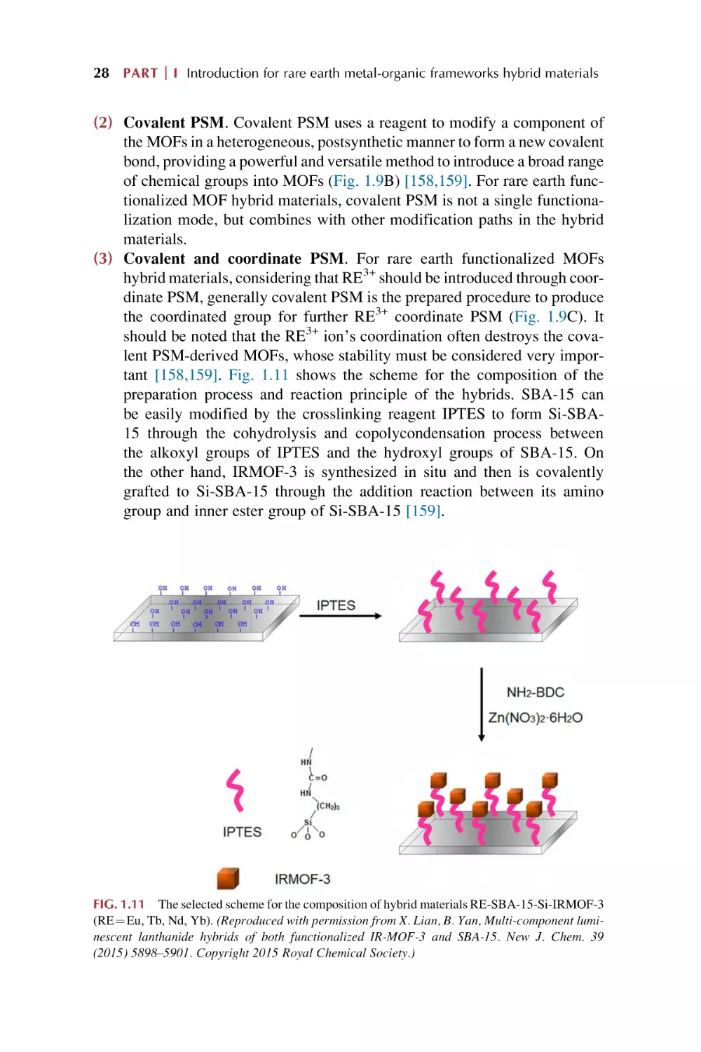
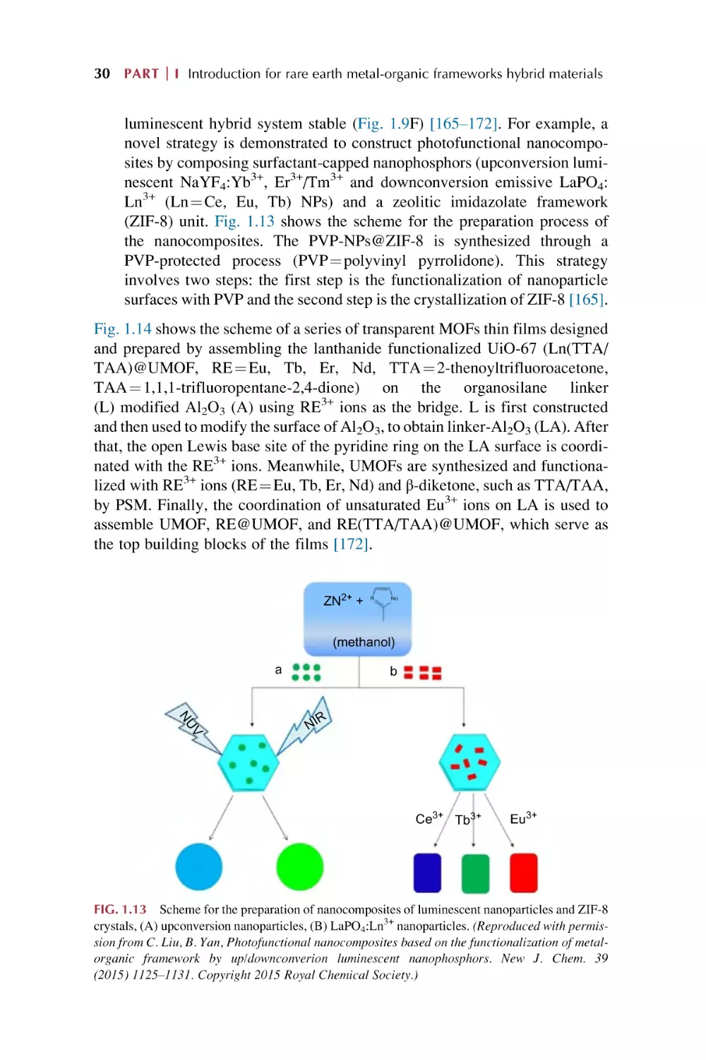
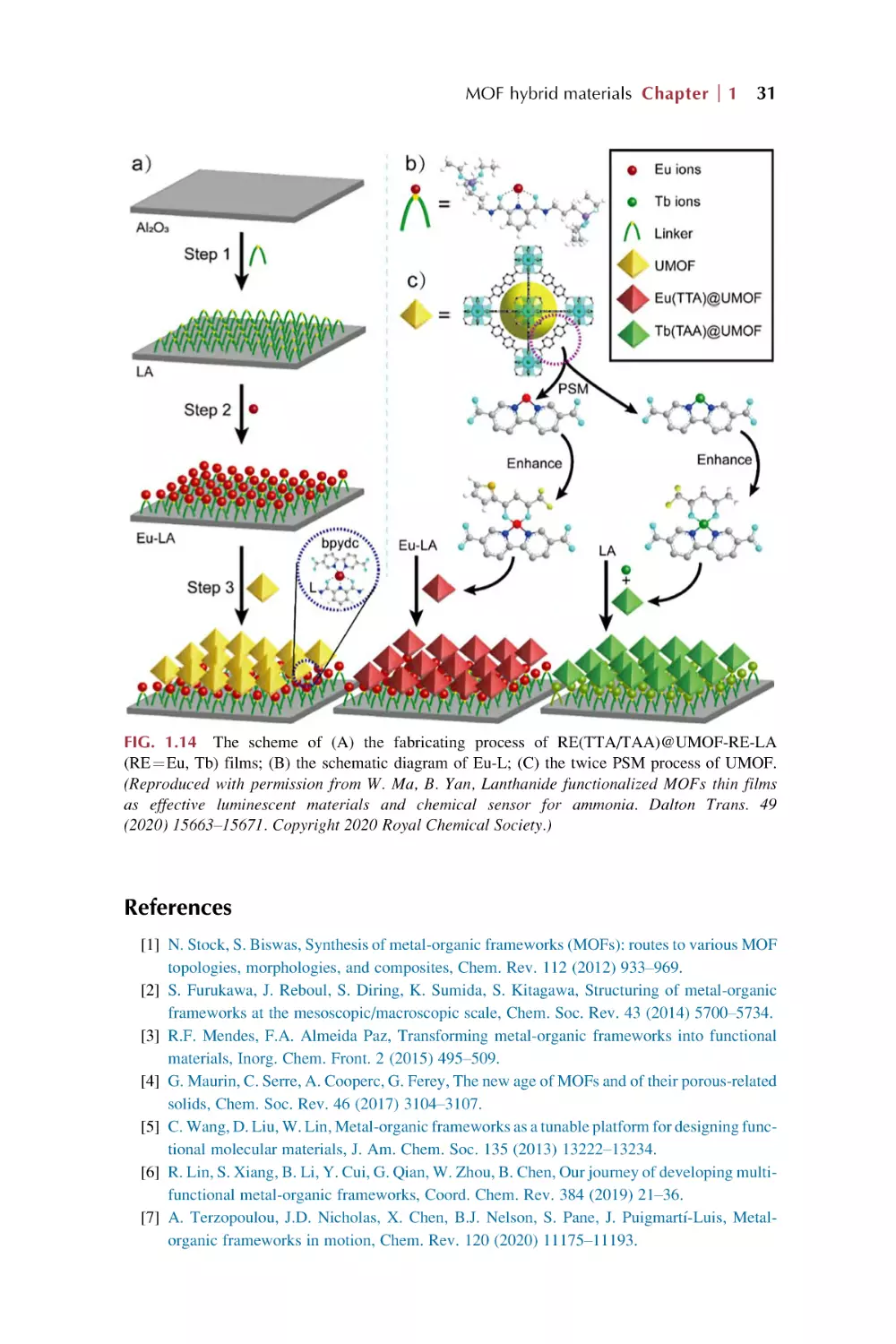
Текст
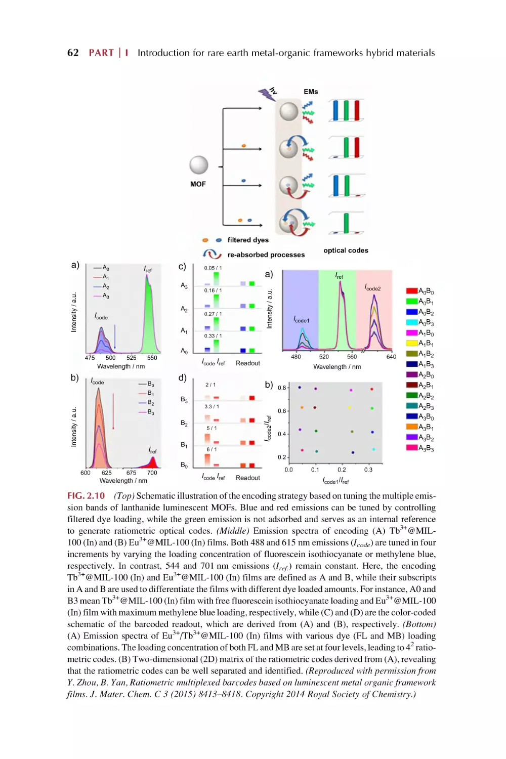
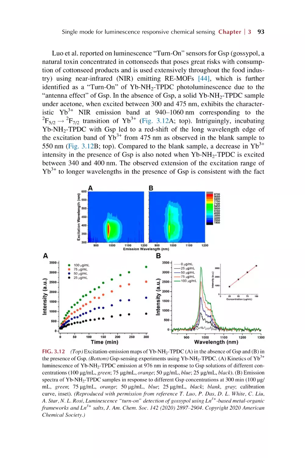
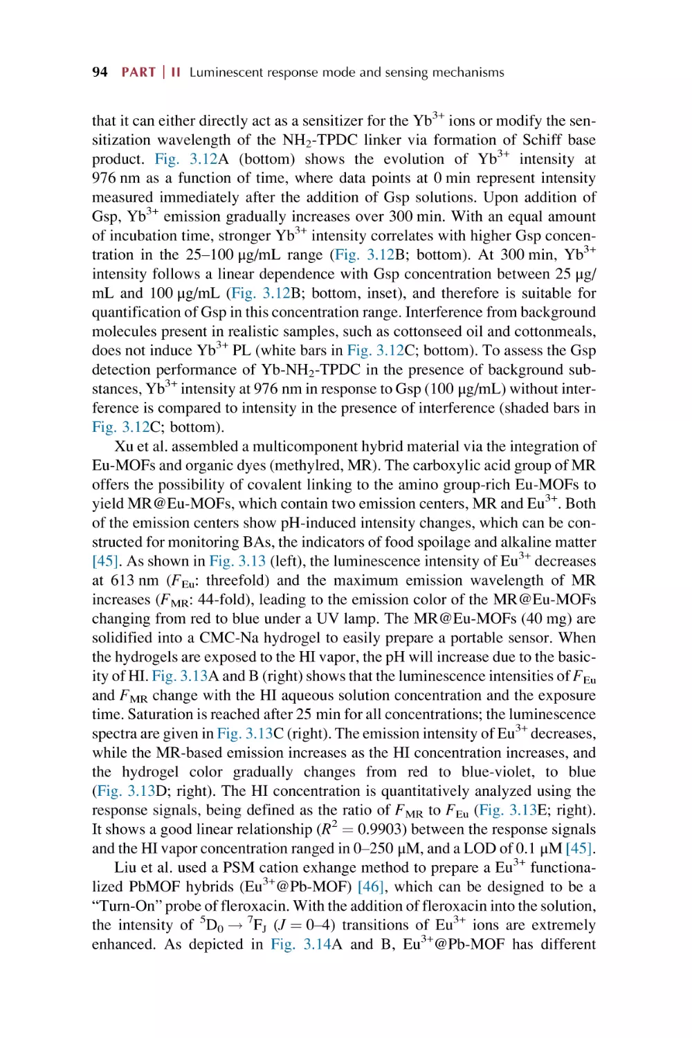
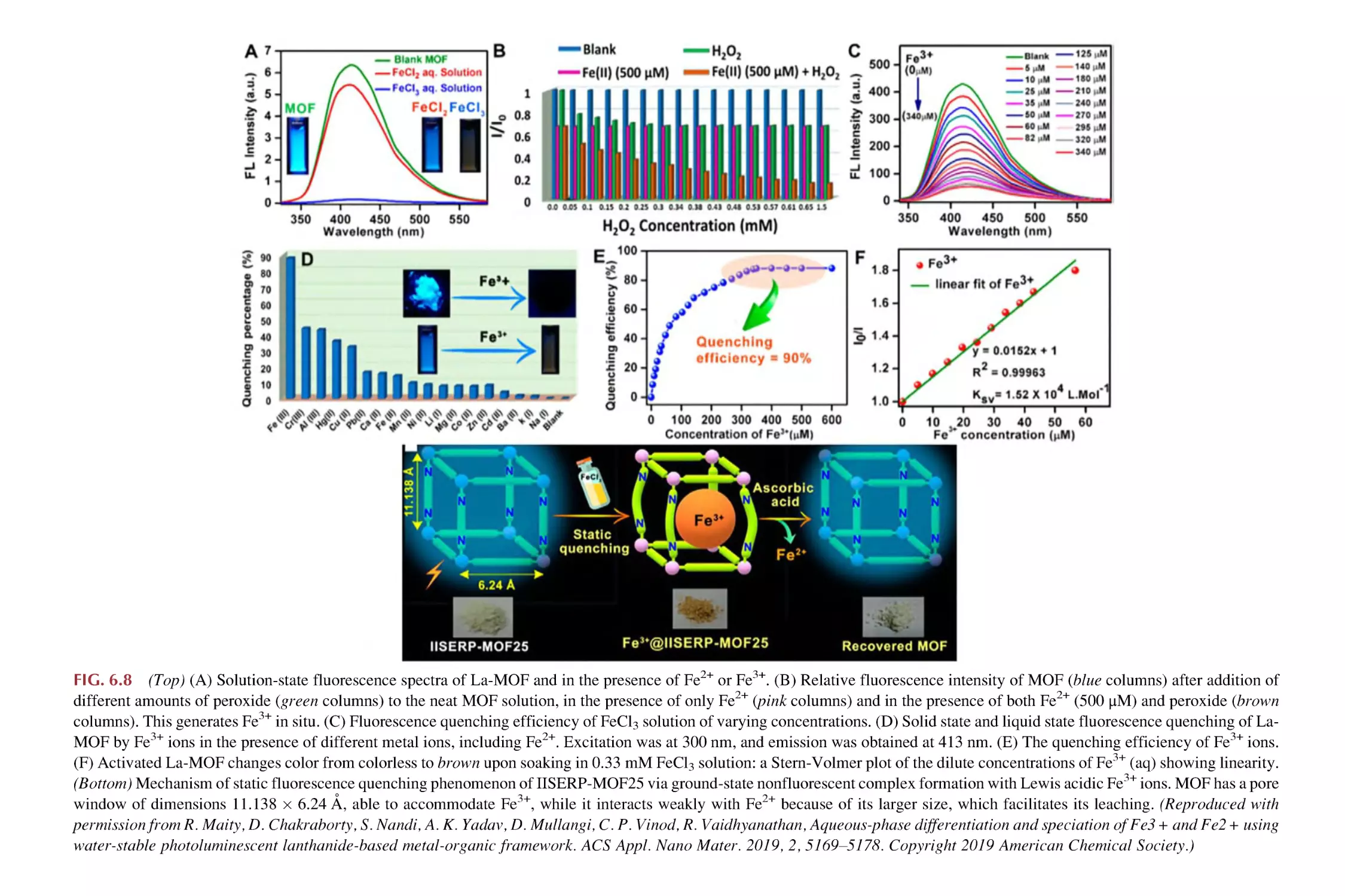
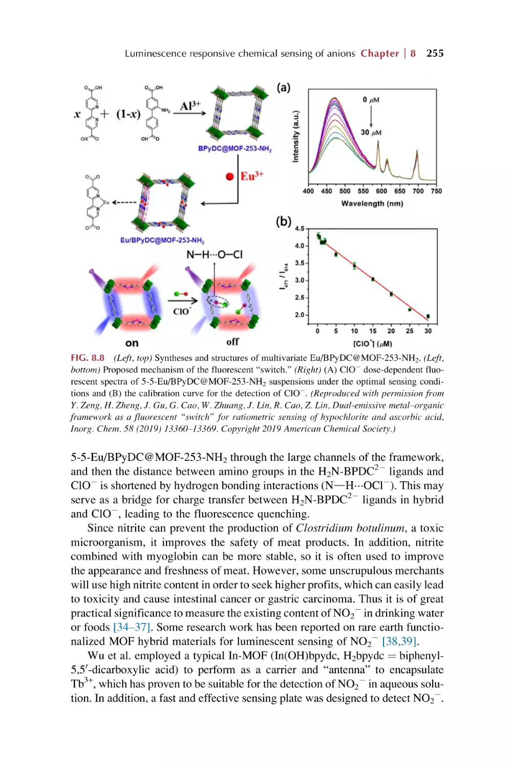
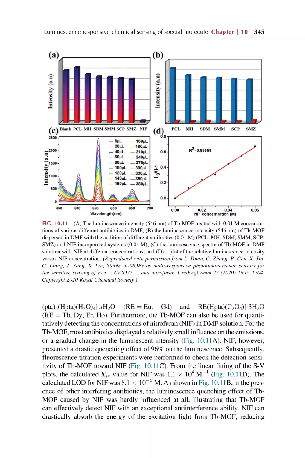
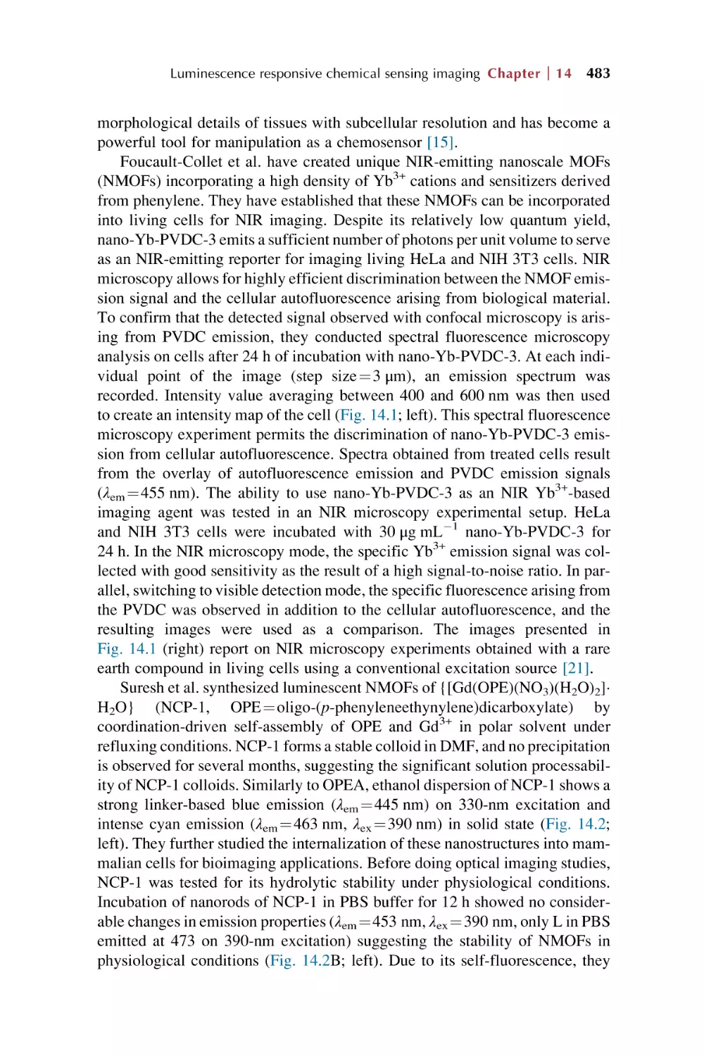
Rare Earth Metal-Organic Framework
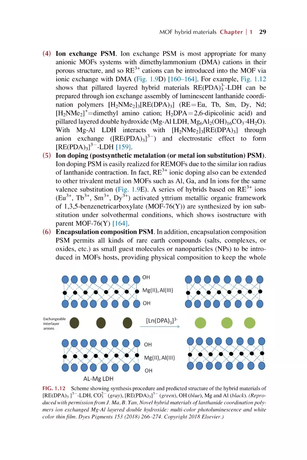
Hybrid Materials for Luminescence
Responsive Chemical Sensors
This page intentionally left blank
Woodhead Publishing Series in Electronic and
Optical Materials
Rare Earth Metal-Organic
Framework Hybrid
Materials for Luminescence
Responsive Chemical
Sensors
Bing Yan
School of Chemical Science and Engineering, Tongji University, Shanghai,
People’s Republic of China
An imprint of Elsevier
Woodhead Publishing is an imprint of Elsevier
50 Hampshire Street, 5th Floor, Cambridge, MA 02139, United States
The Boulevard, Langford Lane, Kidlington, OX5 1GB, United Kingdom
Copyright © 2022 Elsevier Ltd. All rights reserved.
No part of this publication may be reproduced or transmitted in any form or by any means,
electronic or mechanical, including photocopying, recording, or any information storage and
retrieval system, without permission in writing from the publisher. Details on how to seek
permission, further information about the Publisher’s permissions policies and our arrangements
with organizations such as the Copyright Clearance Center and the Copyright Licensing Agency, can
be found at our website: www.elsevier.com/permissions.
This book and the individual contributions contained in it are protected under copyright by the
Publisher (other than as may be noted herein).
Notices
Knowledge and best practice in this field are constantly changing. As new research and experience
broaden our understanding, changes in research methods, professional practices, or medical
treatment may become necessary.
Practitioners and researchers must always rely on their own experience and knowledge in evaluating
and using any information, methods, compounds, or experiments described herein. In using such
information or methods they should be mindful of their own safety and the safety of others,
including parties for whom they have a professional responsibility.
To the fullest extent of the law, neither the Publisher nor the authors, contributors, or editors,
assume any liability for any injury and/or damage to persons or property as a matter of products
liability, negligence or otherwise, or from any use or operation of any methods, products,
instructions, or ideas contained in the material herein.
ISBN: 978-0-323-91236-5 (print)
ISBN: 978-0-323-91430-7 (online)
For information on all Woodhead publications
visit our website at https://www.elsevier.com/books-and-journals
Publisher: Matthew Deans
Acquisitions Editor: Kayla Dos Santos
Editorial Project Manager: Isabella C. Silva
Production Project Manager: Prasanna Kalyanaraman
Cover Designer: Greg Harris
Typeset by STRAIVE, India
Contents
Preface
xiii
Part I
Introduction for rare earth metal-organic
frameworks hybrid materials
1.
Metal-organic frameworks (MOFs), rare earth MOFs,
and rare earth functionalized MOF hybrid materials
1.1
1.2
1.3
2.
Metal-organic frameworks (MOFs)
1.1.1 Synthesis of metal-containing nodes or coordination
bonds and linker design for MOFs
1.1.2 Postsynthetic modification (PSM) of MOFs
1.1.3 MOFs hybrids or composites
1.1.4 Potential applications of MOFs
Rare earth metal-organic frameworks (REMOFs)
1.2.1 REMOF structures
1.2.2 Some applications of REMOFs
Rare earth functionalized metal-organic framework hybrid
materials (REFMOFHs)
References
3
3
5
8
11
22
23
24
26
31
Rare earth luminescence, MOFs luminescence, rare
earth MOFs hybrid materials luminescence,
luminescence response, and chemical sensing
2.1
2.2
2.3
2.4
2.5
Rare earth ion luminescence
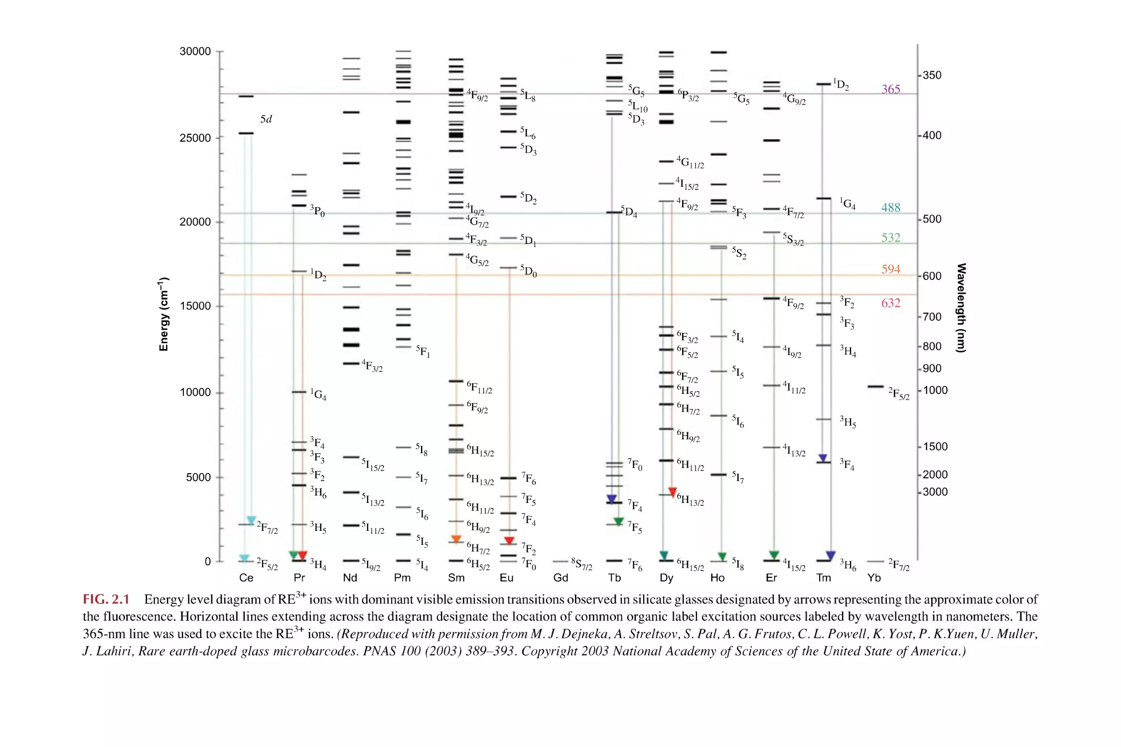
2.1.1 Atomic spectral term (2S+1LJ) and energy level transition
of trivalent rare earth (RE3+) ions
2.1.2 Luminescence and spectroscopy of trivalent rare earth
(RE3+) ions
Rare earth complex molecule luminescence
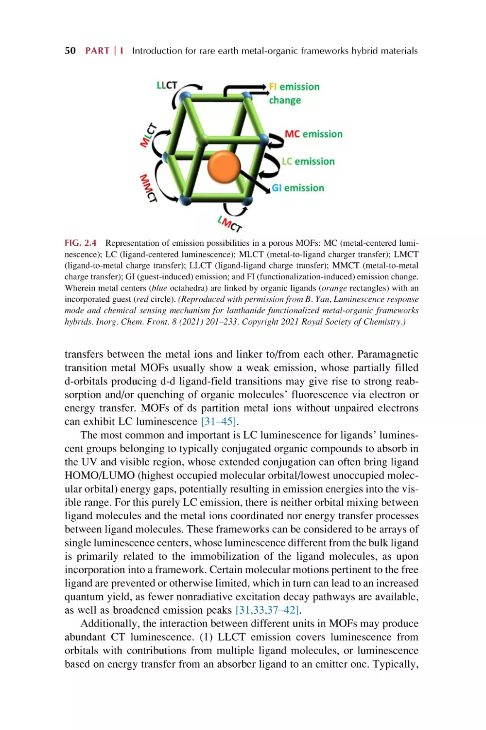
MOFs luminescence
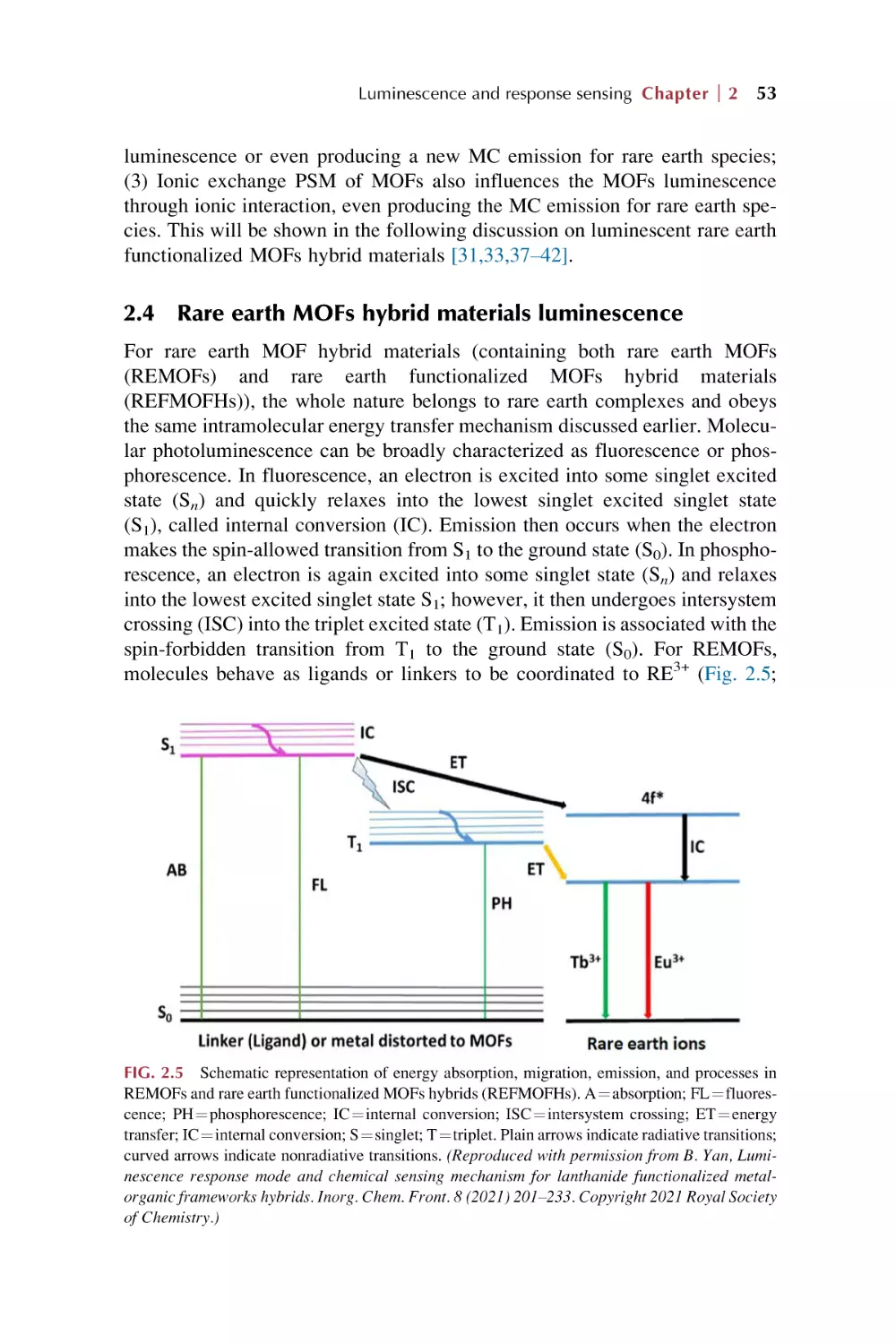
Rare earth MOFs hybrid materials luminescence
Luminescence for rare earth functionalized MOFs hybrid
materials
41
42
42
45
49
53
54
v
vi Contents
2.6
Luminescence response for chemical sensing of rare earth
MOFs hybrid materials
2.6.1 Luminescence response and chemical sensing
in MOFs-based material
2.6.2 MOFs-based materials primarily display special
advantages for chemical sensing
References
63
63
66
67
Part II
Luminescent response mode and sensing
mechanisms in rare earth metal-organic
frameworks hybrid materials
3.
Single mode for luminescence responsive chemical
sensing in rare earth metal-organic framework hybrid
materials
3.1
3.2
3.3
3.4
3.5
4.
Introduction for luminescence response of metal-organic
frameworks
“Turn-off” luminescence response chemical sensing for rare
earth metal-organic framework hybrid materials
“Turn-On” luminescence response chemical sensing for rare
earth metal-organic framework hybrid materials
“Turn-on-off-on” luminescence response chemical sensing
for rare earth metal-organic framework hybrid materials
Both “Turn-On” and “Turn-Off” luminescence response
chemical sensing on different analytes for rare earth MOF
hybrid materials
References
75
79
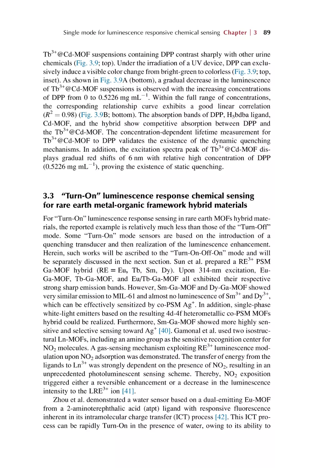
89
97
103
107
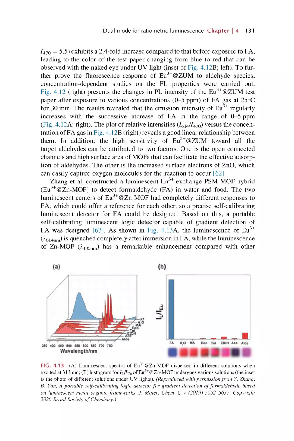
Dual mode for ratiometric luminescence responsive
chemical sensing for rare earth metal-organic
framework hybrid materials
4.1
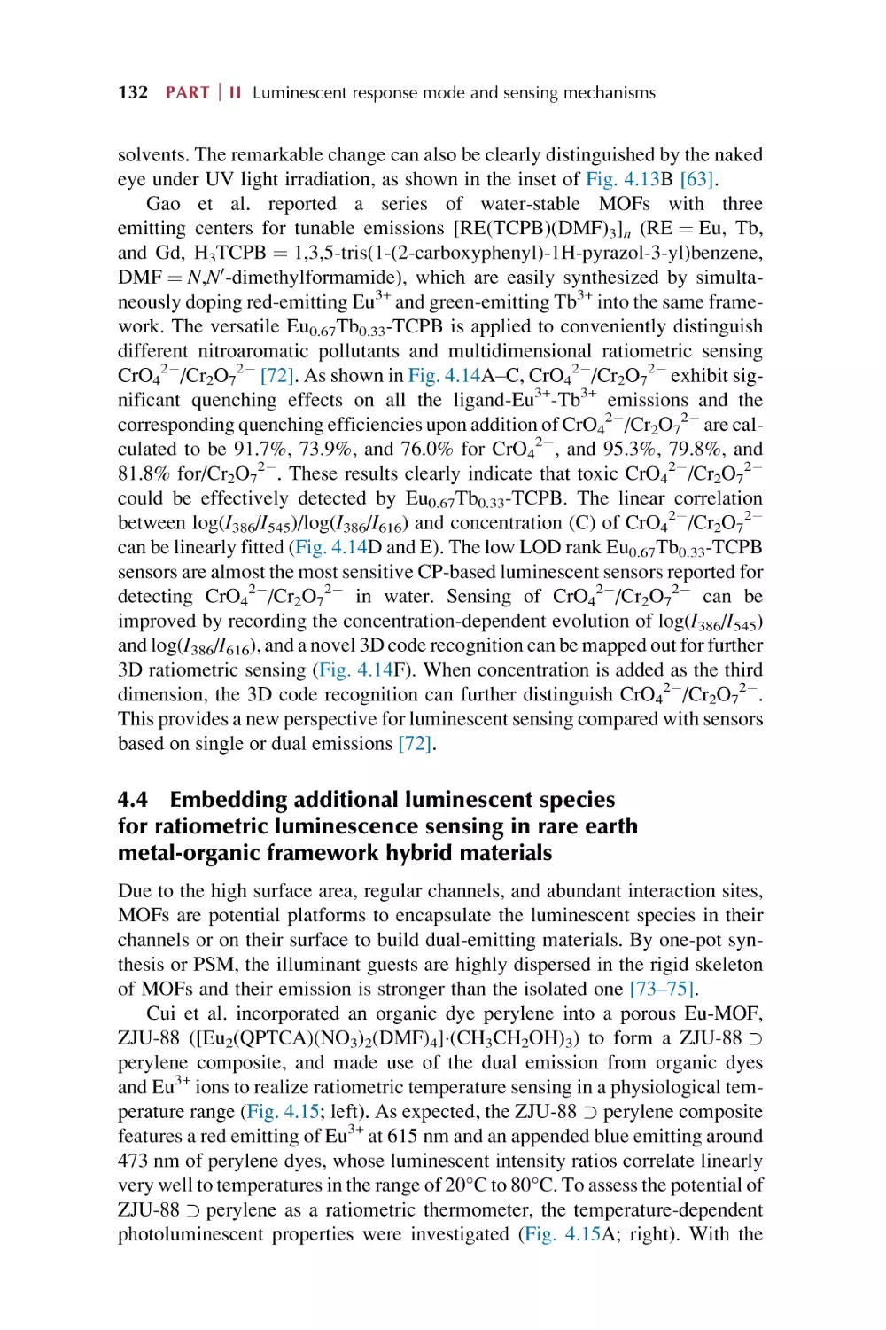
4.2
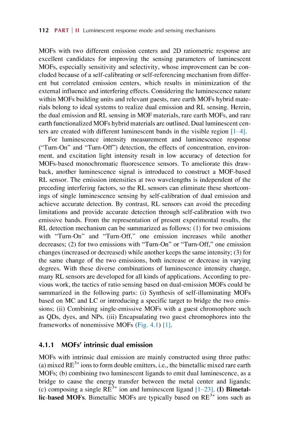
Dual mode for ratiometric luminescence (RL) responsive
chemical sensing of MOFs materials
4.1.1 MOFs’ intrinsic dual emission
4.1.2 Single-emissive MOFs with fluorescent guests
4.1.3 Nonemissive MOFs with encapsulated
chromophores
Dual rare earth ion luminescence for ratiometric
luminescence sensing in rare earth metal-organic framework
hybrid materials
111
112
114
115
116
Contents vii
4.3
4.4
4.5
5.
Ligand (linker) and RE3+ ions through energy transfer
“antenna effect” for ratiometric luminescence sensing in rare
earth metal-organic framework hybrid materials
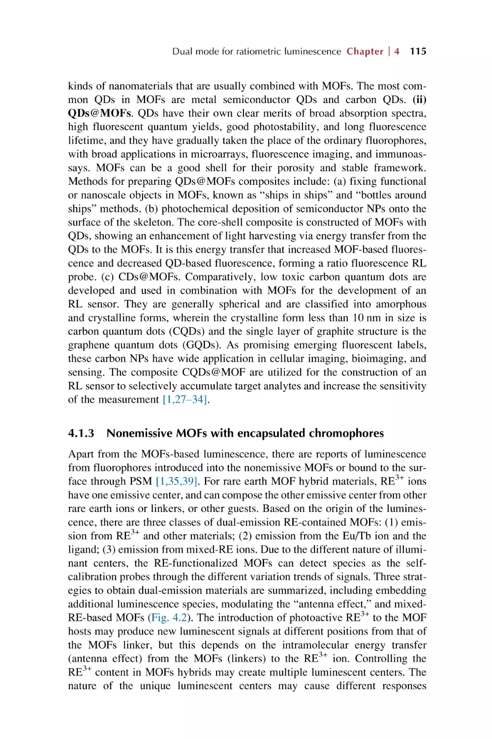
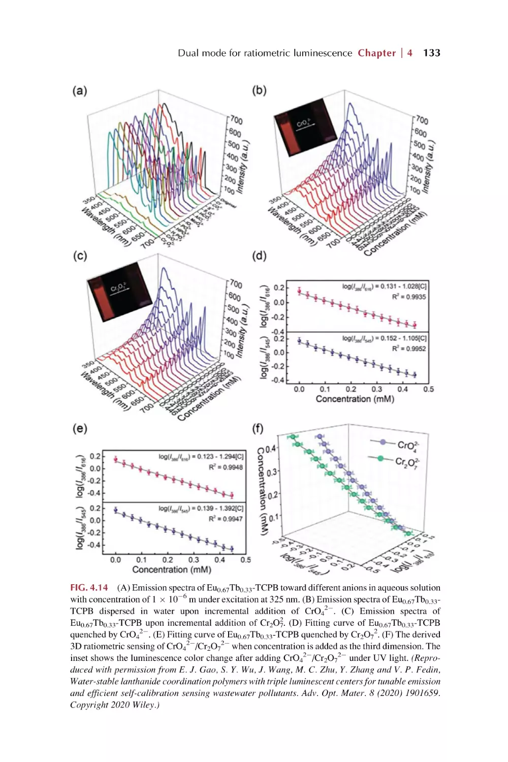
Embedding additional luminescent species for ratiometric
luminescence sensing in rare earth metal-organic framework
hybrid materials
Single rare earth functionalized metal-organic framework
hybrid materials for ratiometric luminescence sensing
References
124
132
136
140
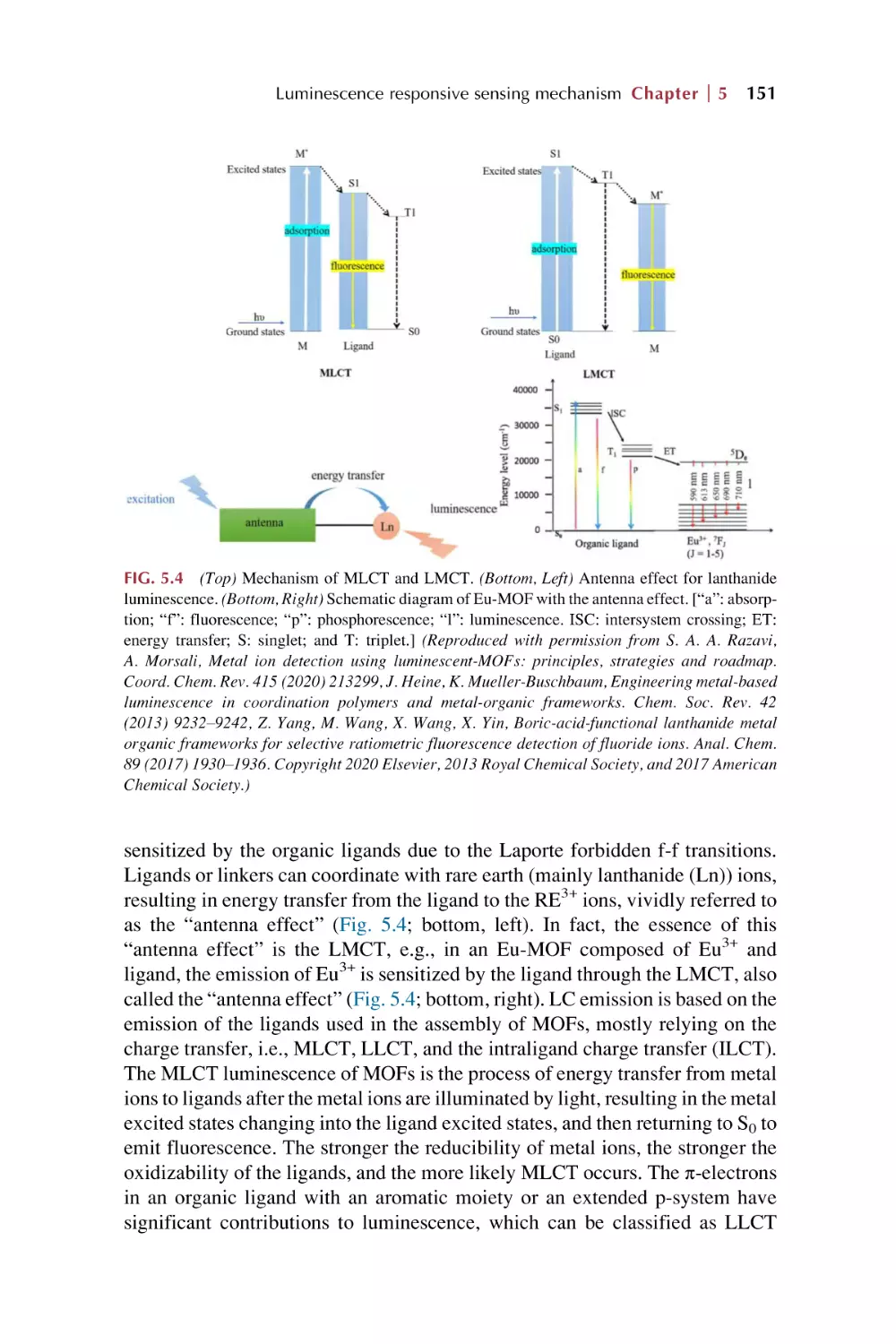
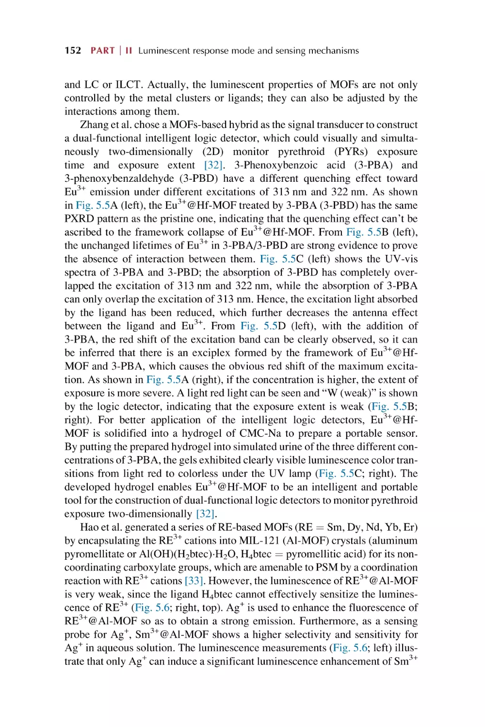
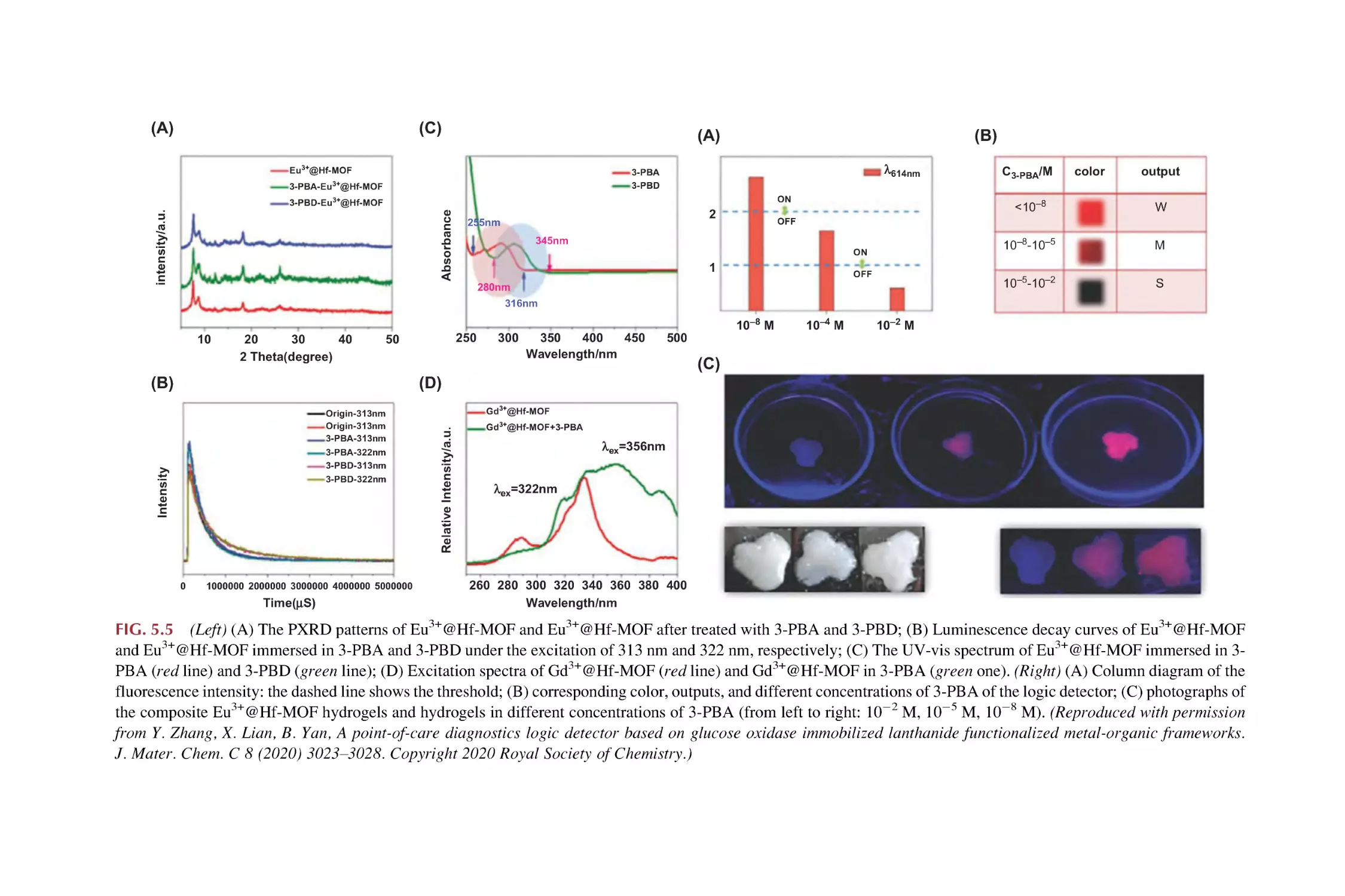
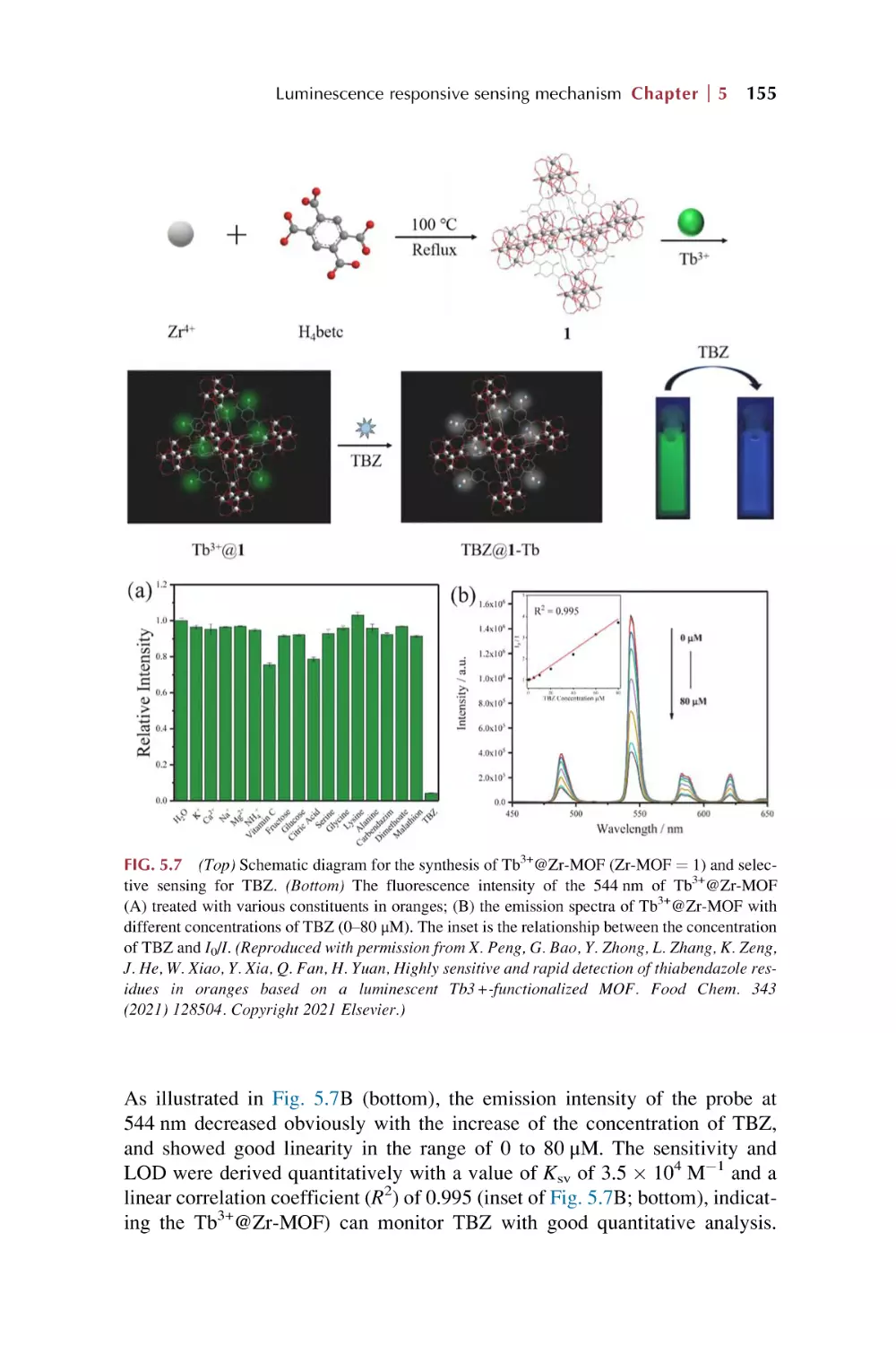
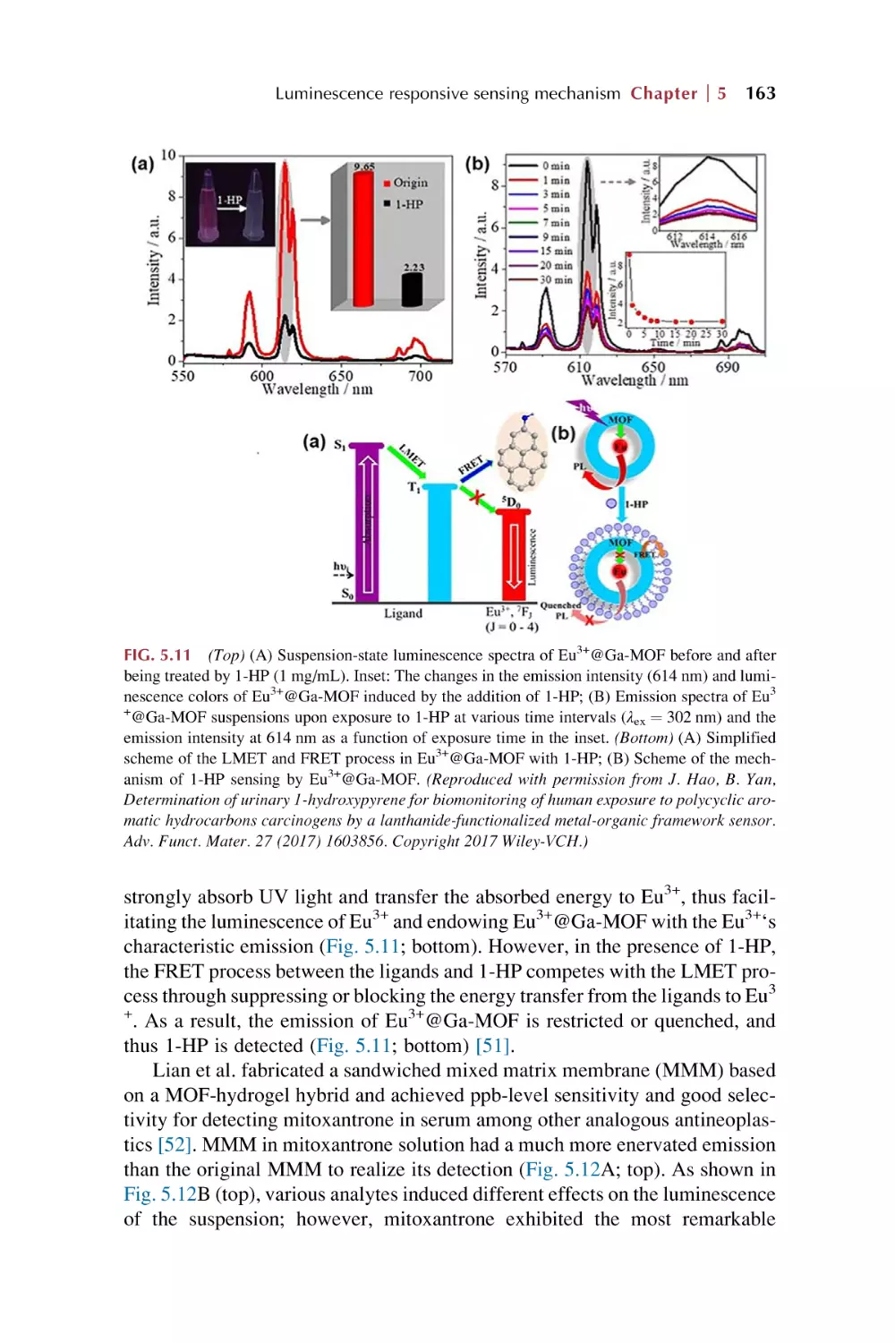
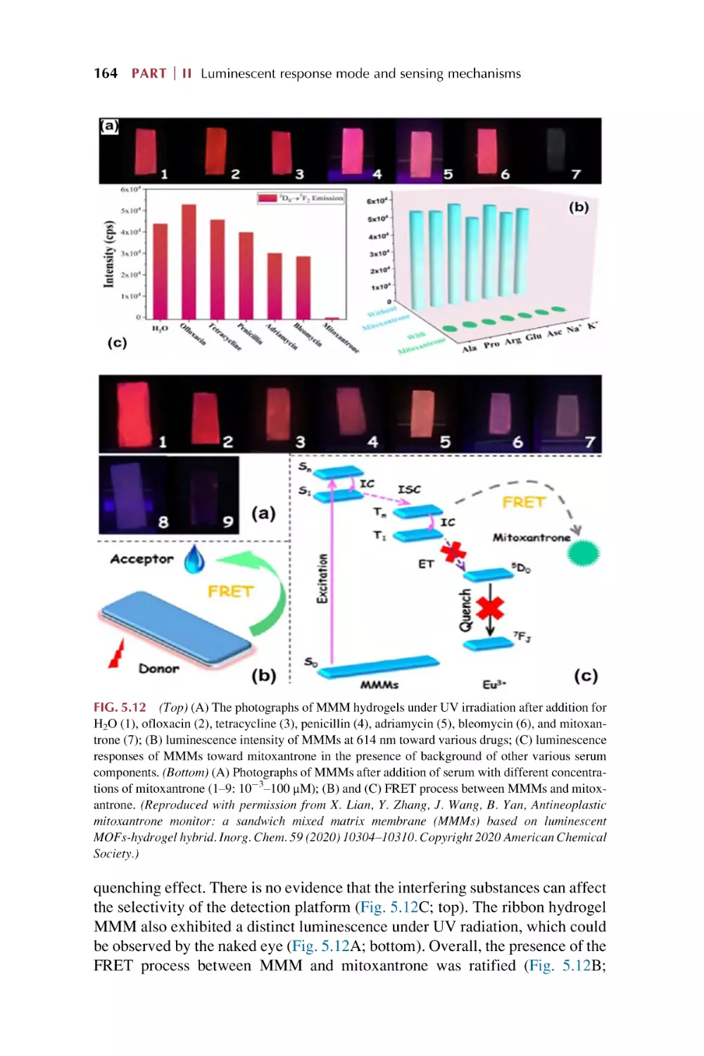
Luminescence responsive sensing mechanism
in rare earth metal-organic framework hybrid
materials
5.1
5.2
5.3
5.4
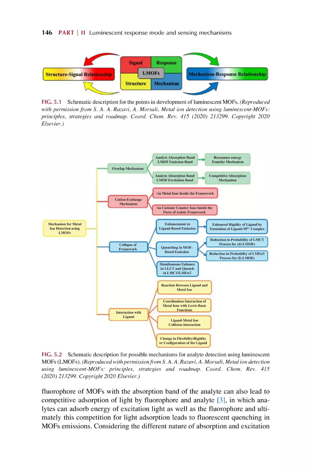
The luminescence responsive sensing mechanism
for metal-organic framework-based materials
5.1.1 Overlap mechanism
5.1.2 Structural collapse mechanism
5.1.3 Ion exchange mechanism
5.1.4 Linker-analytes interaction mechanism
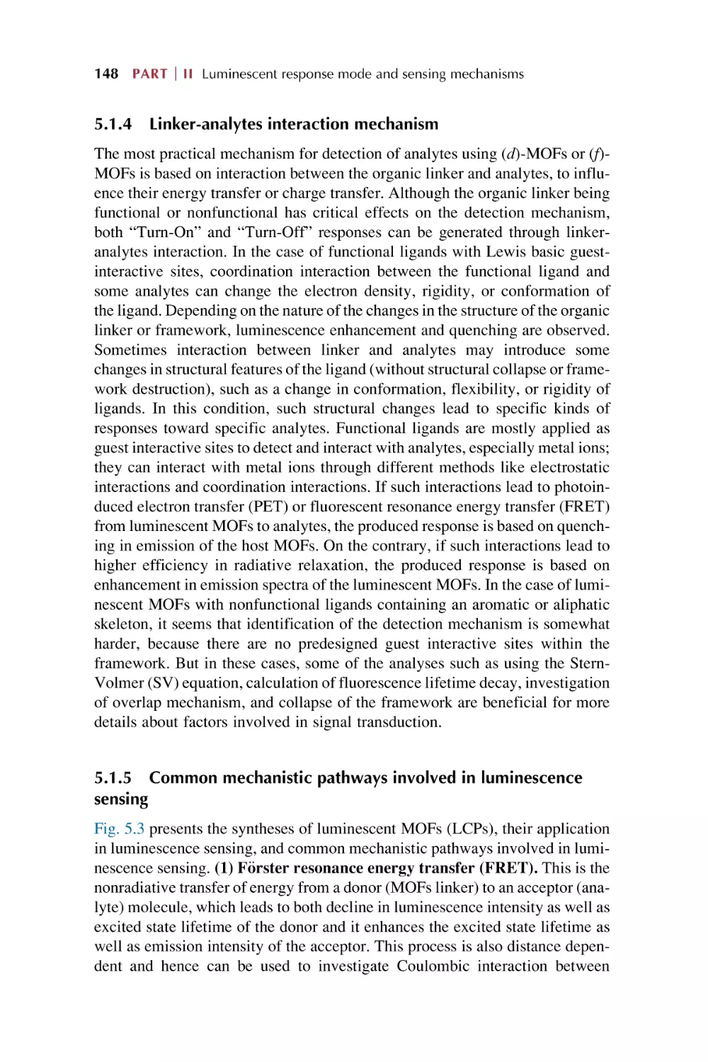
5.1.5 Common mechanistic pathways involved in
luminescence sensing
The LMET for luminescence response on chemical sensing in
rare earth metal-organic framework hybrid materials
The photo-induced energy transfer (PET) and fluorescence
(F€
orster) resonance energy transfer (FRET) for luminescence
response for chemical sensing in rare earth metal-organic
framework hybrid materials
5.3.1 PET for luminescence response in chemical
sensing
5.3.2 FRET for luminescence response in chemical
sensing
Special interactions for luminescence response on
chemical sensing in rare earth metal-organic framework
hybrid materials
5.4.1 Hydrogen bonding for luminescence response on
chemical sensing
5.4.2 Coordination interaction for luminescence response
on chemical sensing
5.4.3 Reduction reaction for luminescence response
for chemical sensing
5.4.4 Precipitation reaction for luminescence response
in chemical sensing
References
145
145
147
147
148
148
150
156
156
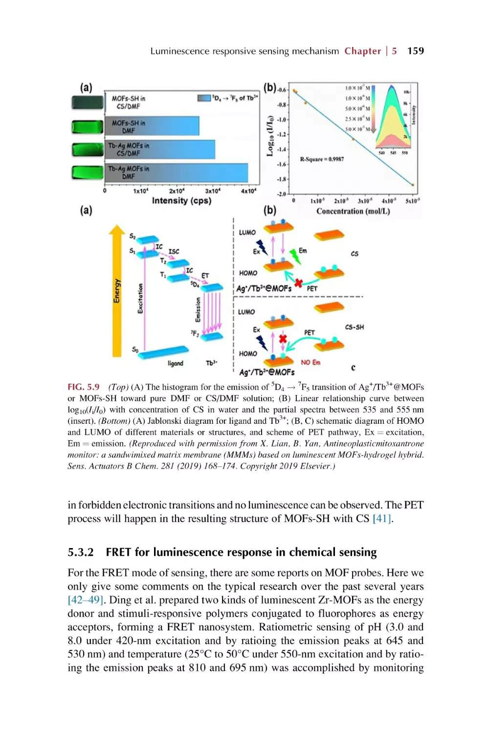
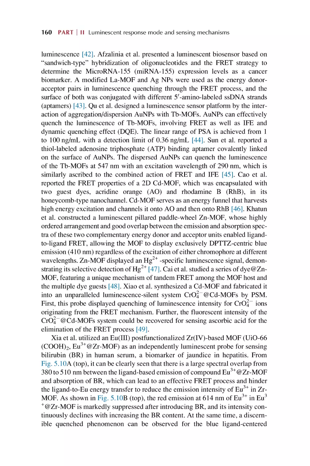
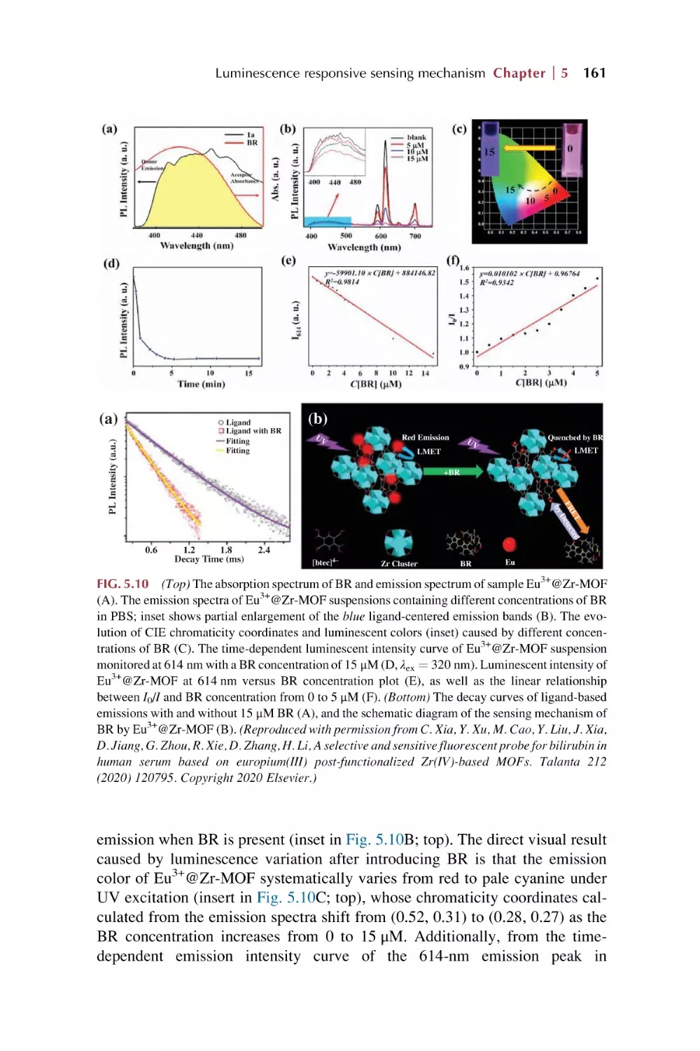
159
165
165
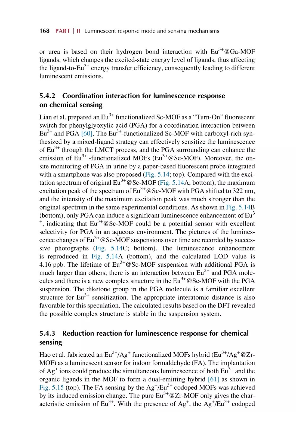
168
168
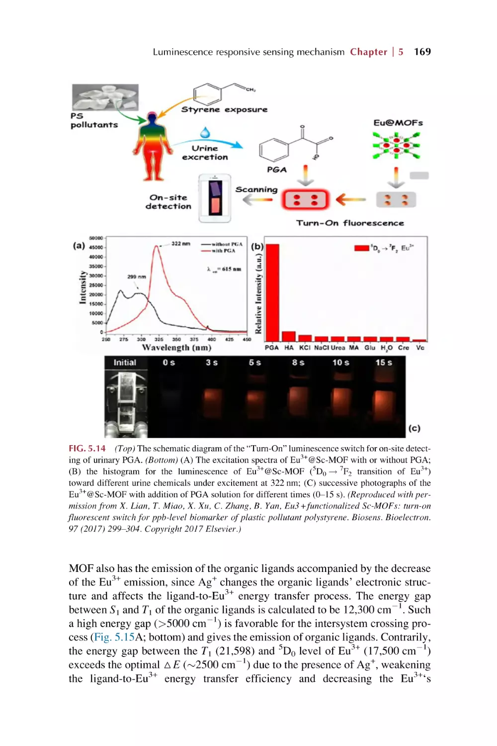
170
172
viii Contents
Part III
Rare earth metal-organic frameworks hybrid
materials as luminescence response chemical
sensors for typical ionic analytes
6.
Rare earth metal-organic framework hybrid materials
for luminescence responsive chemical sensing of
metal ions (I)
6.1
6.2
6.3
6.4
6.5
7.
179
186
190
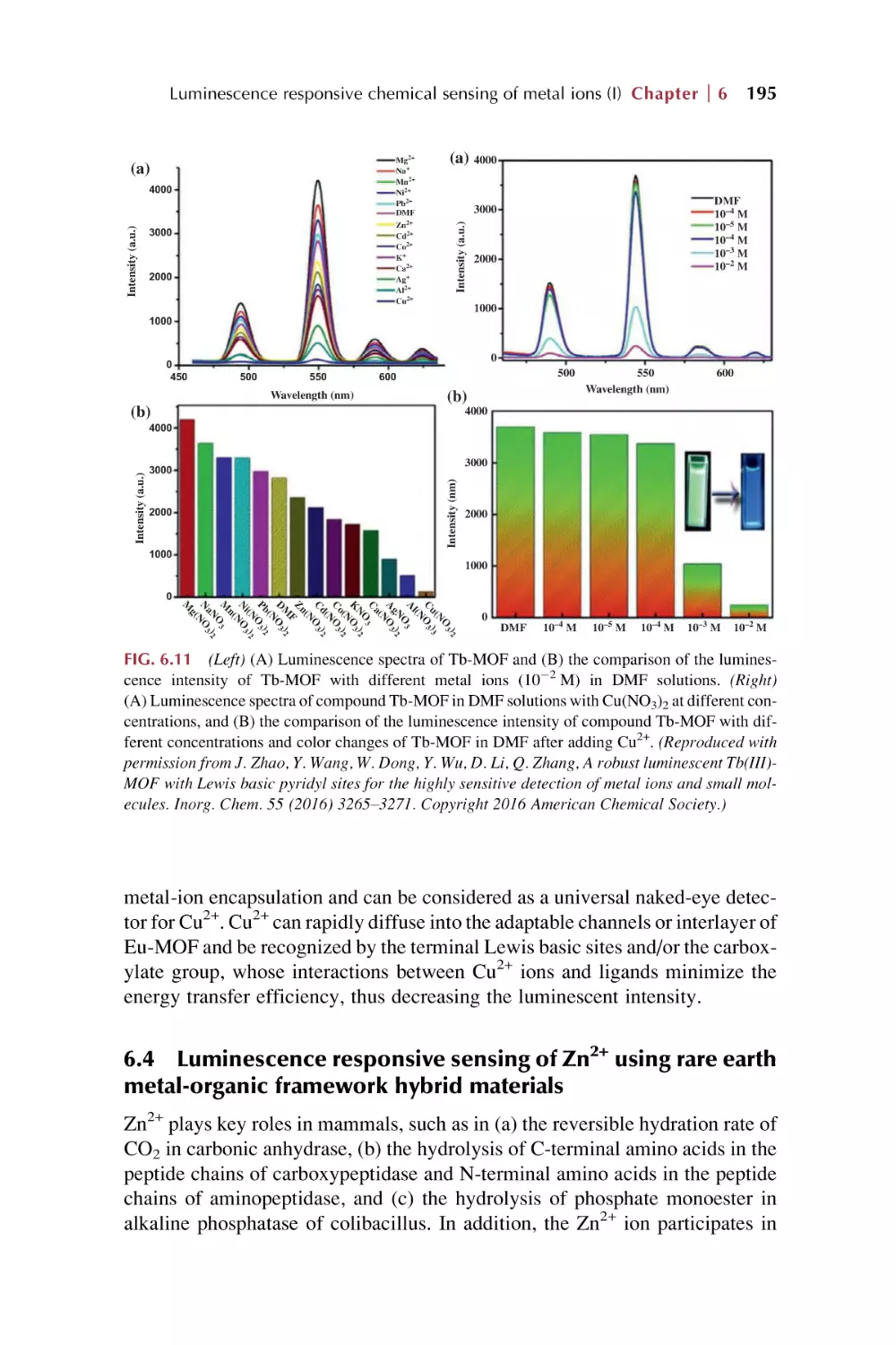
195
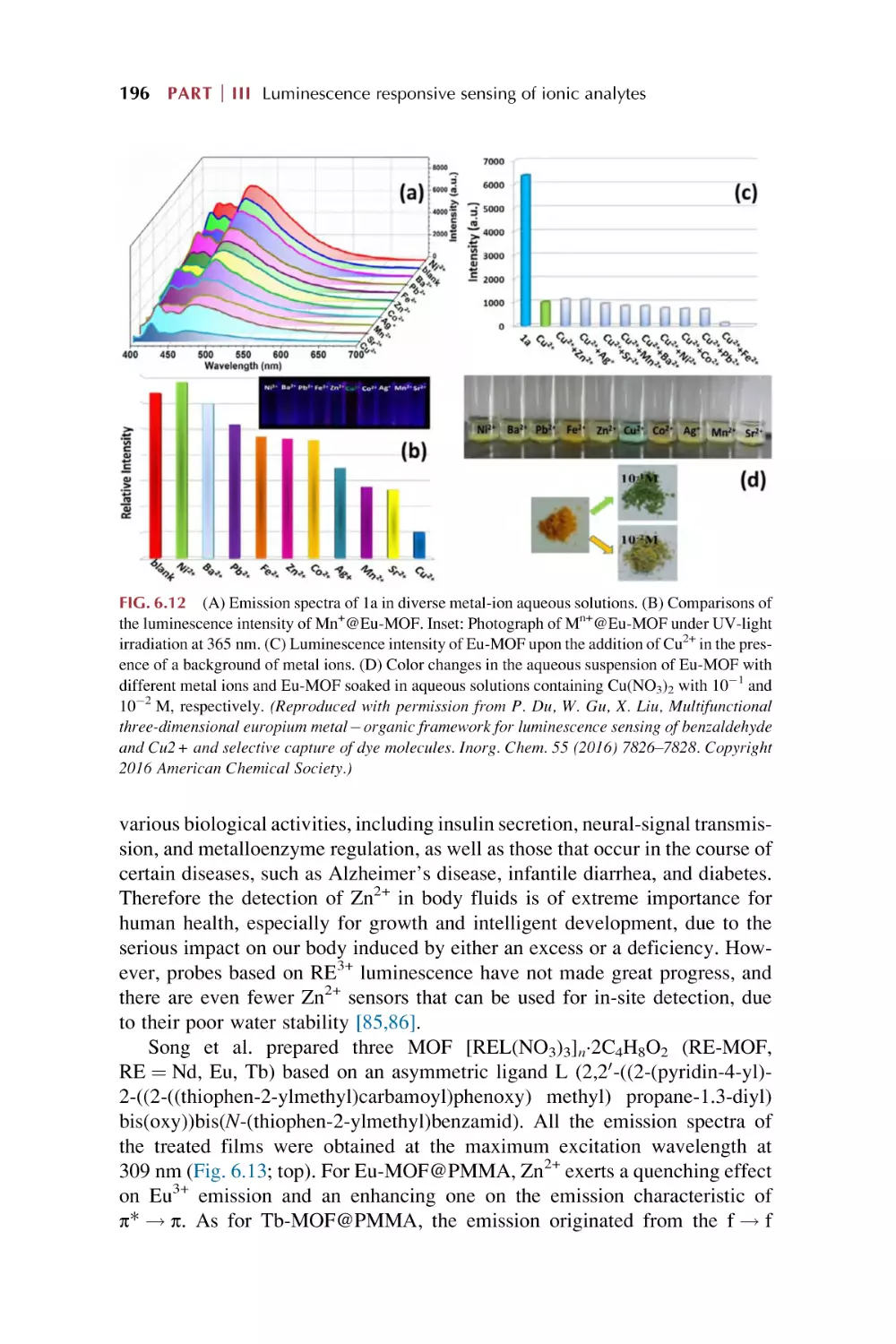
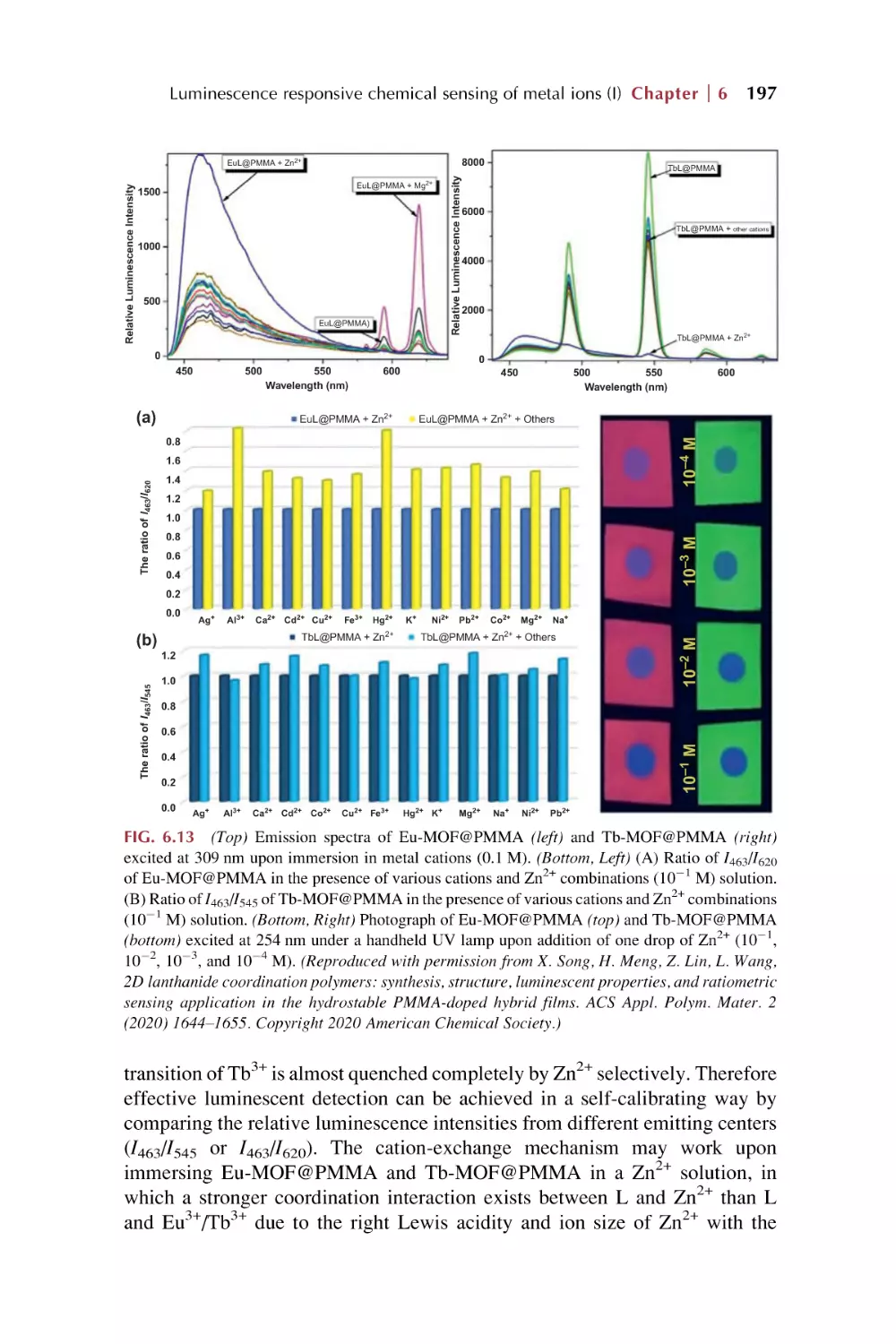
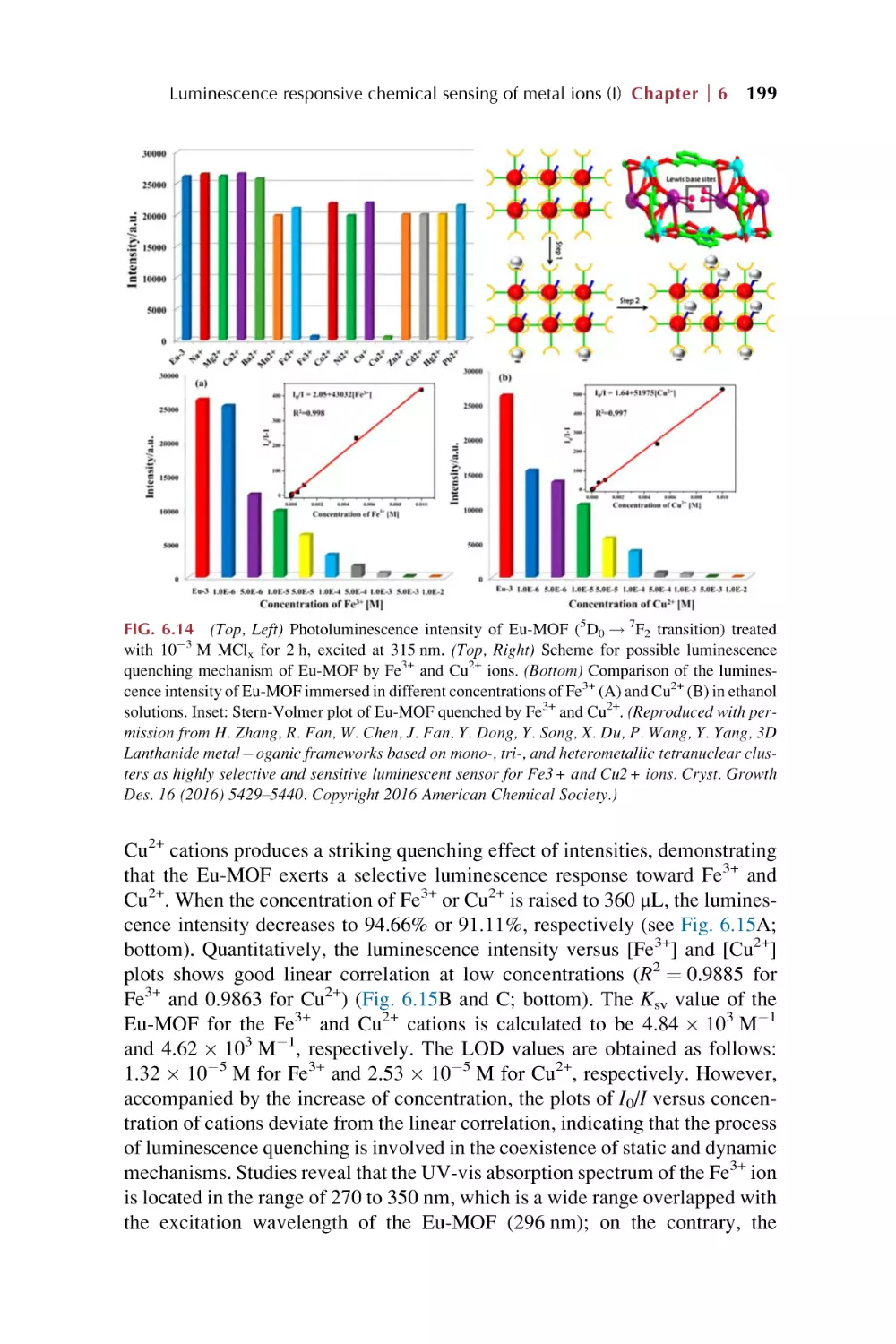
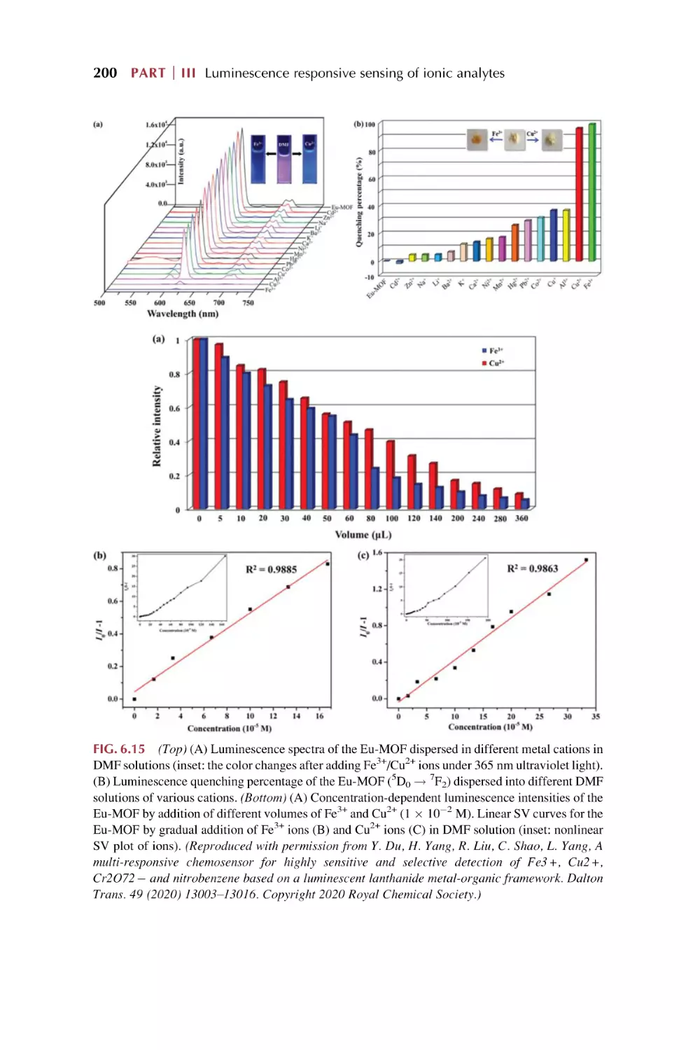
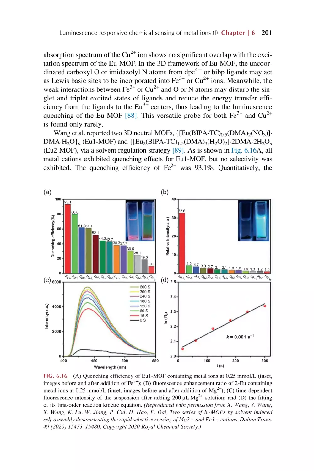
198
202
Rare earth metal-organic framework hybrid
materials for luminescence responsive sensing
of metal ions (II)
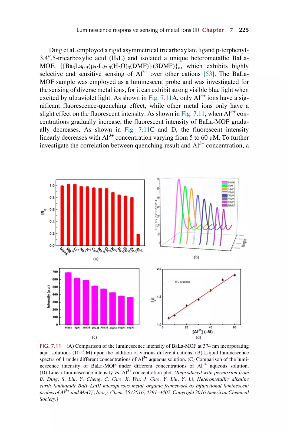
7.1
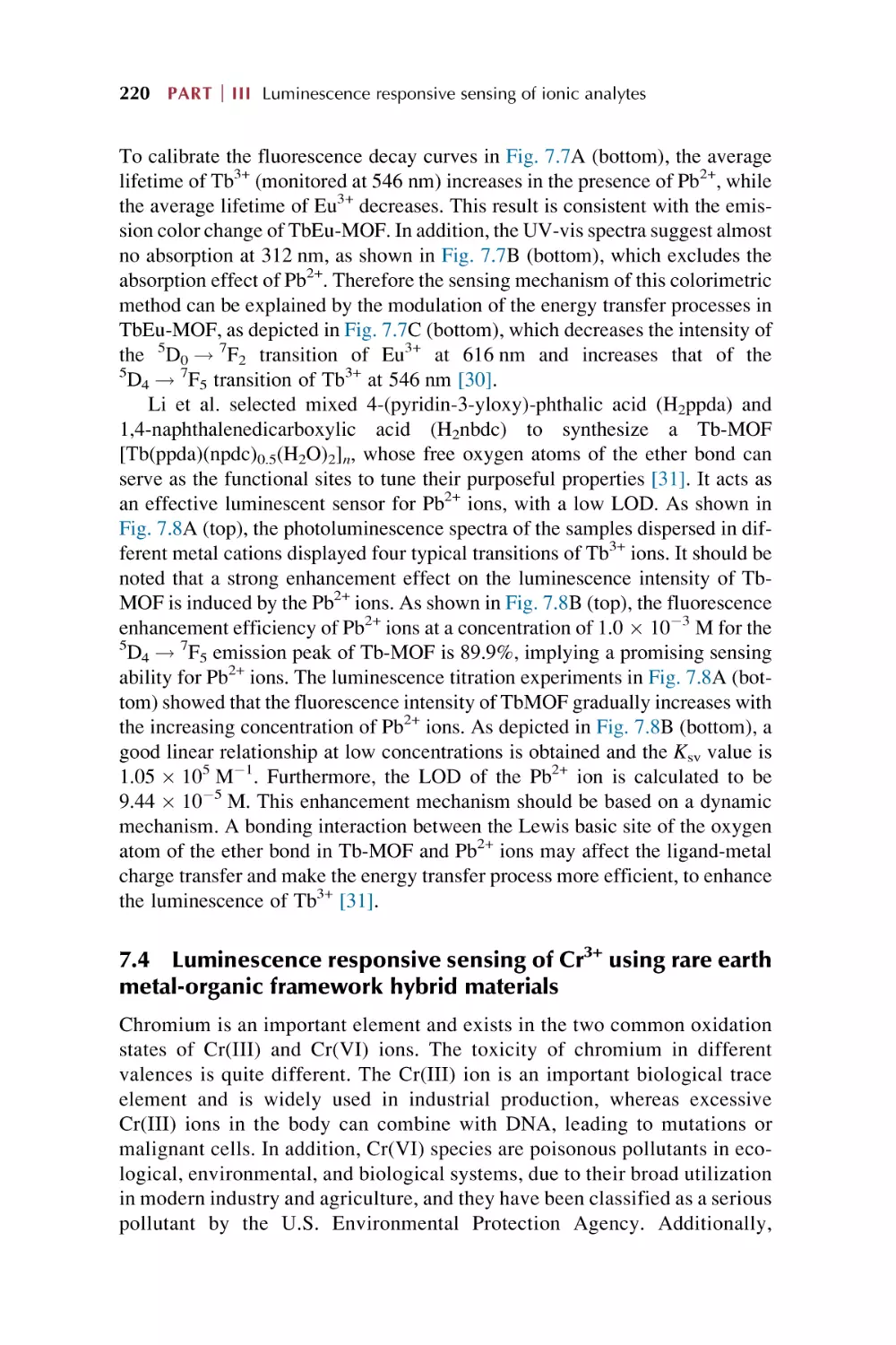
7.2
7.3
7.4
7.5
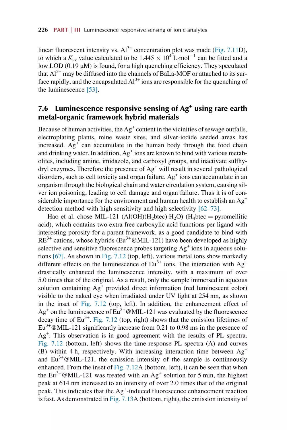
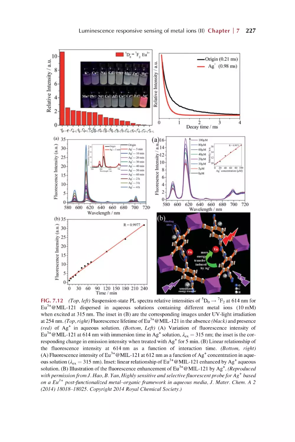
7.6
7.7
7.8
8.
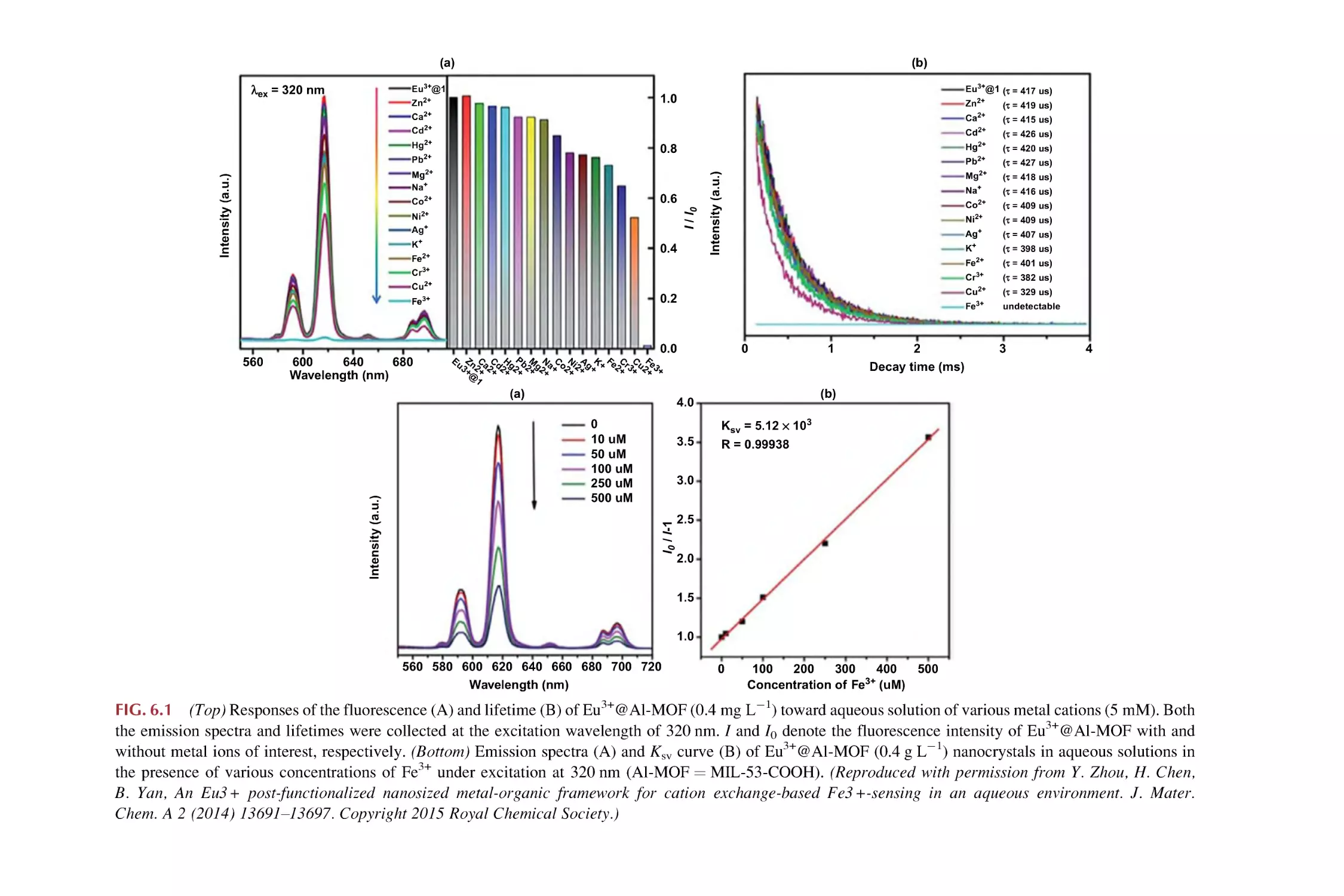
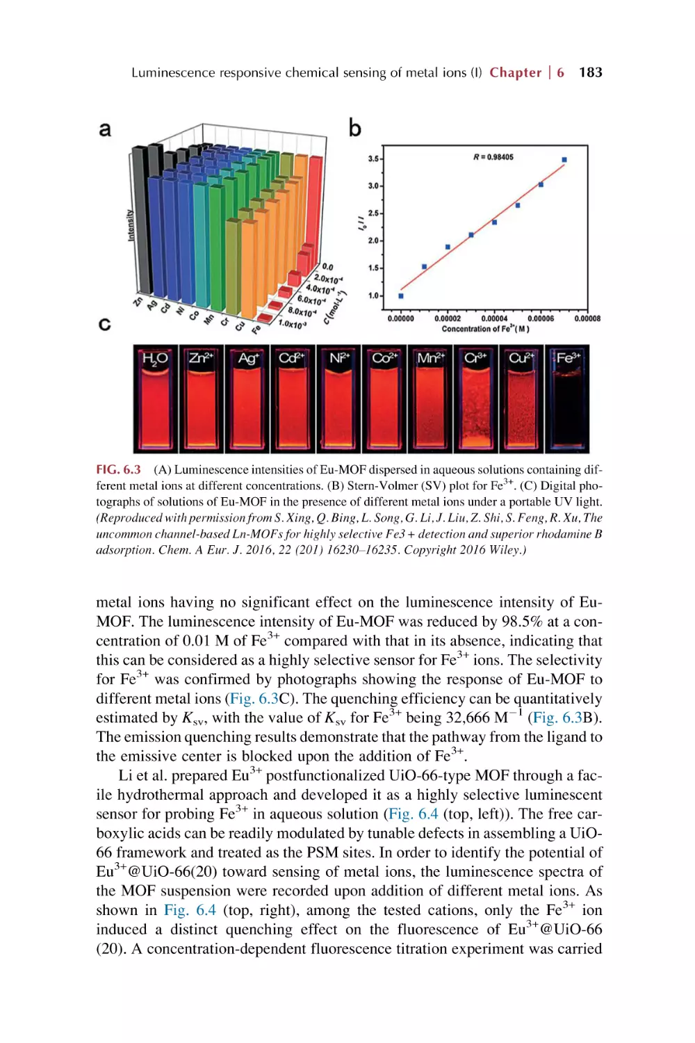
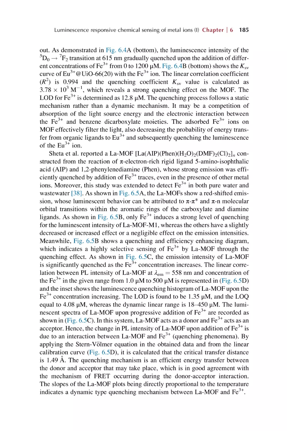
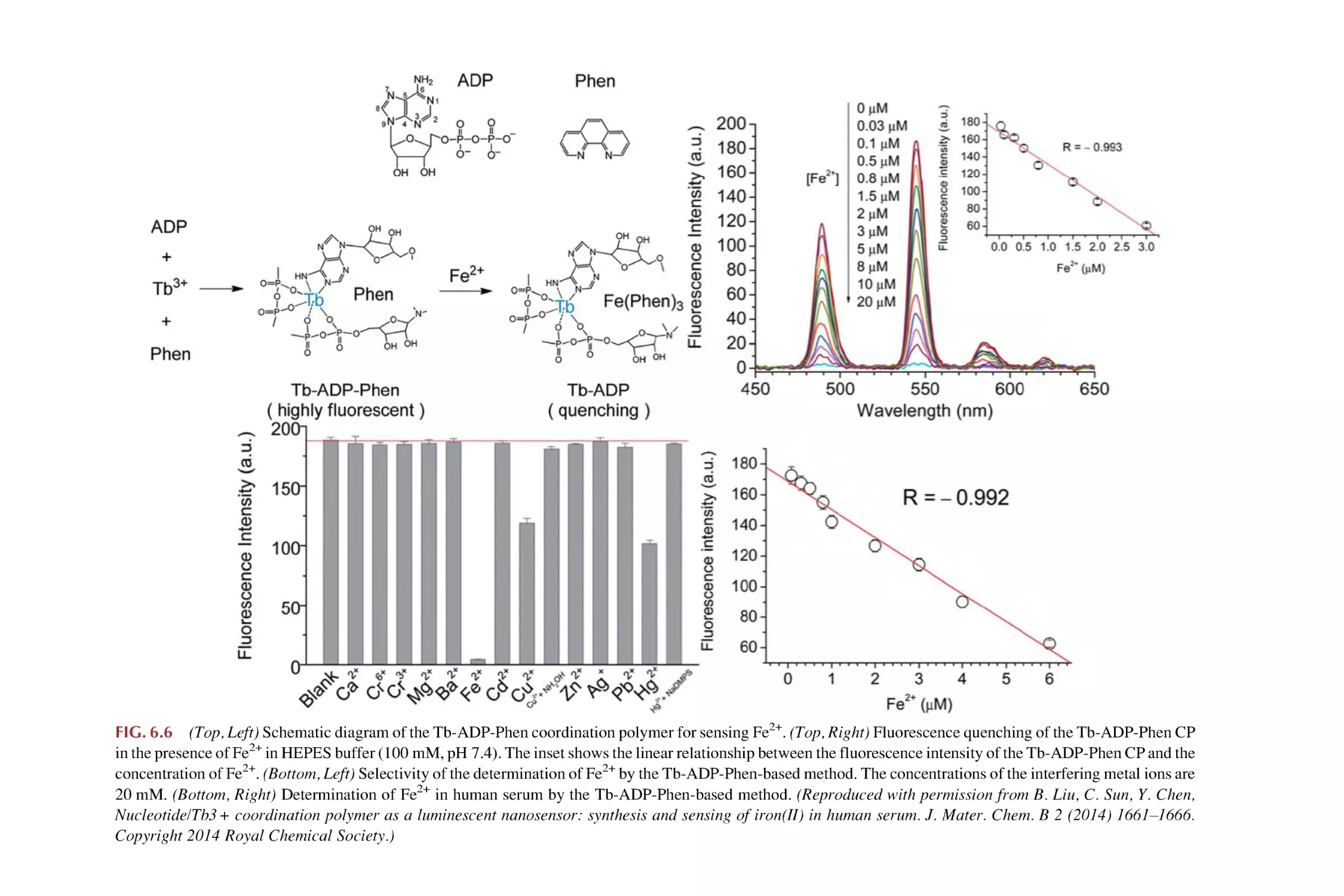
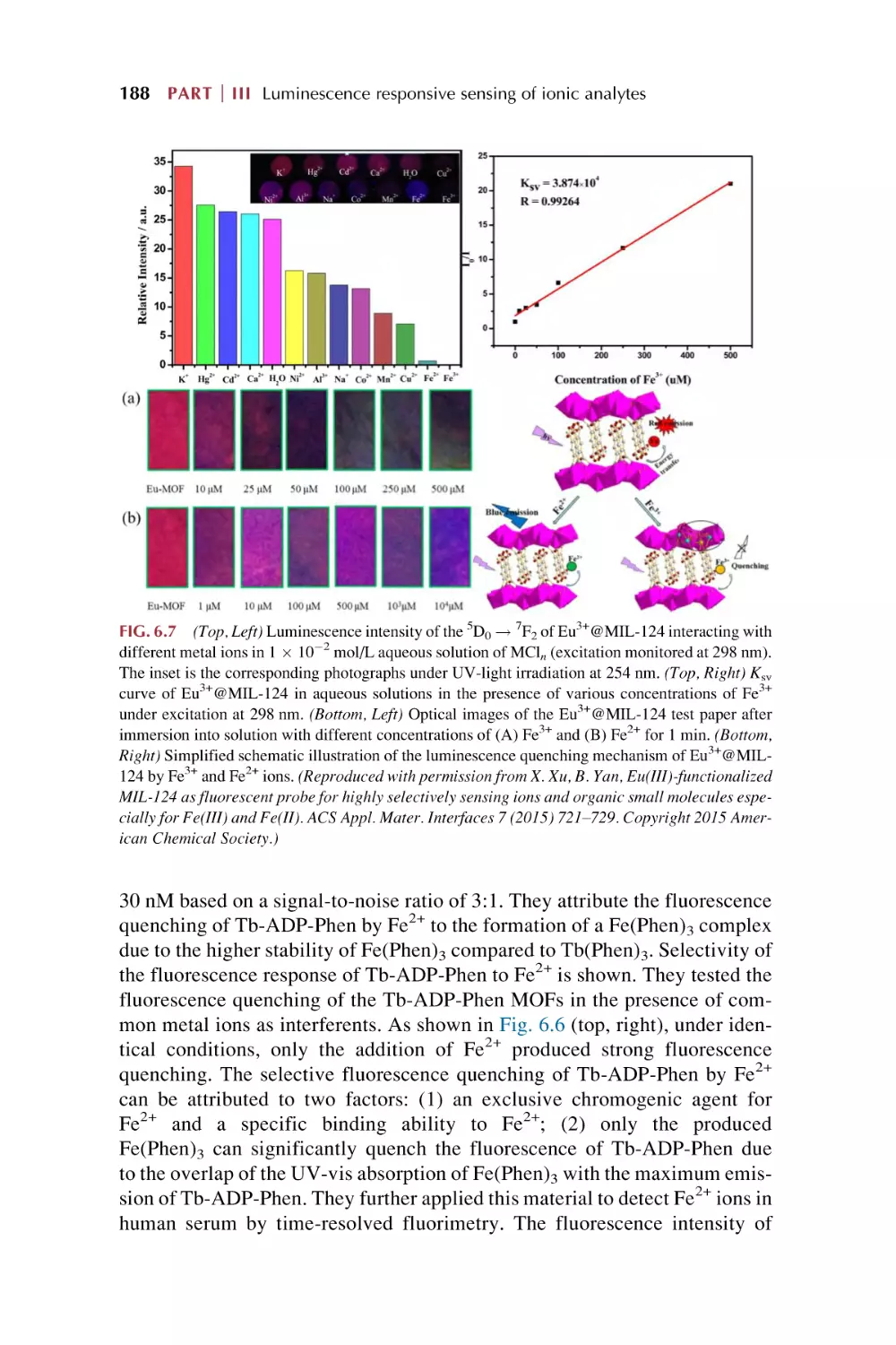
Luminescence responsive sensing of Fe3+ using rare earth
metal-organic framework hybrid materials
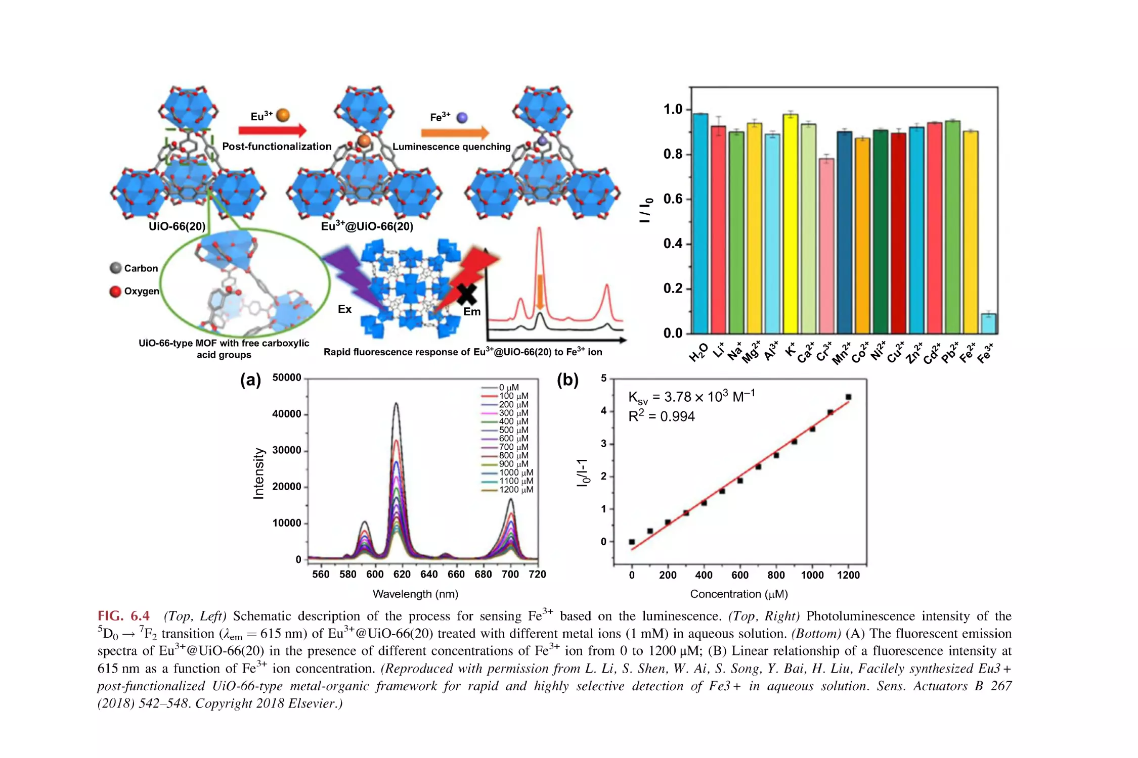
Luminescence responsive sensing of Fe2+ or Fe3+/Fe2+ using
rare earth metal-organic framework hybrid materials
Luminescence responsive sensing of Cu2+ using rare earth
metal-organic framework hybrid materials
Luminescence responsive sensing of Zn2+ using rare earth
metal-organic framework hybrid materials
Luminescence responsive sensing of multimetal cations using
rare earth metal-organic framework hybrid materials
References
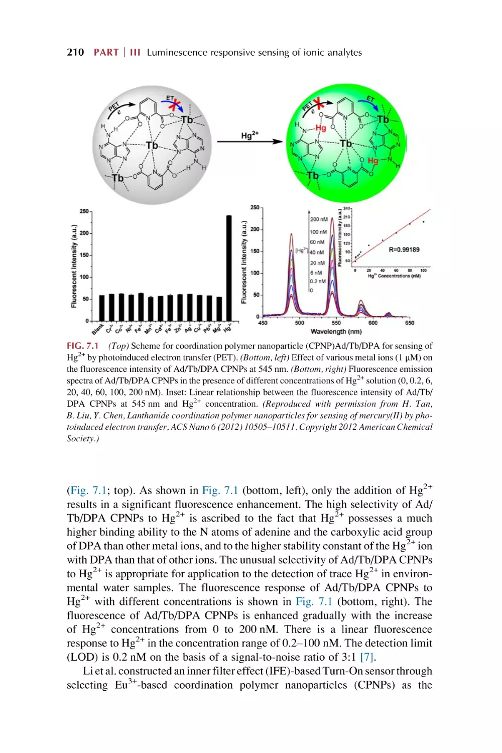
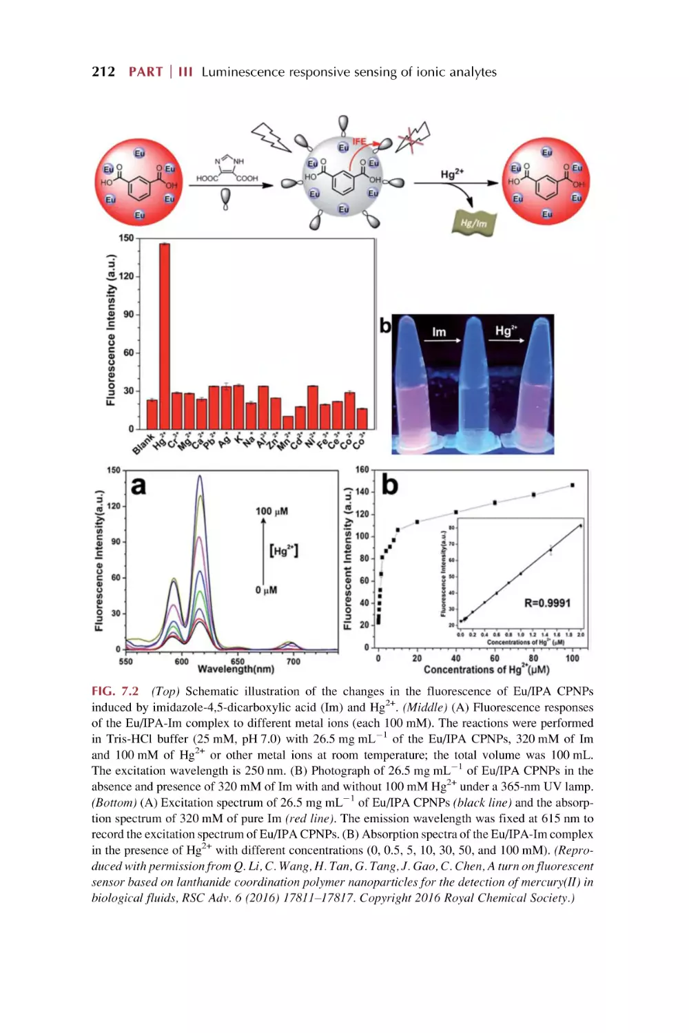
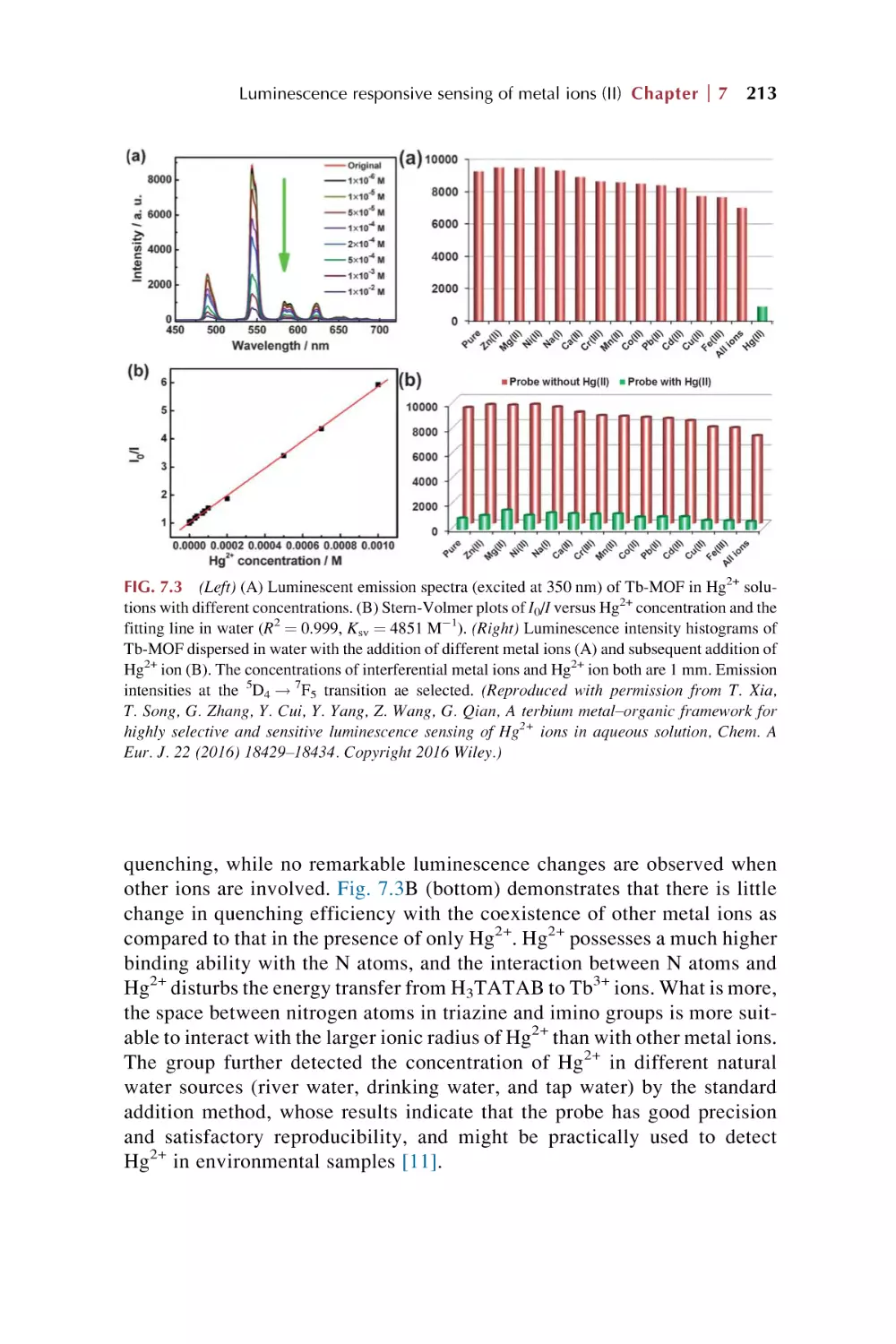
Luminescence responsive sensing of Hg2+ using rare earth
metal-organic framework hybrid materials
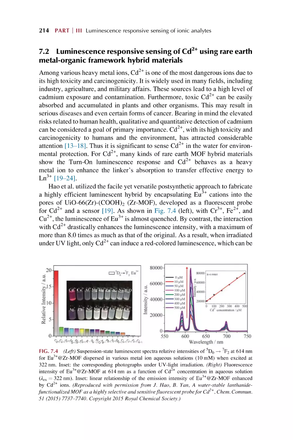
Luminescence responsive sensing of Cd2+ using rare earth
metal-organic framework hybrid materials
Luminescence responsive sensing of Pb2+ using rare earth
metal-organic framework hybrid materials
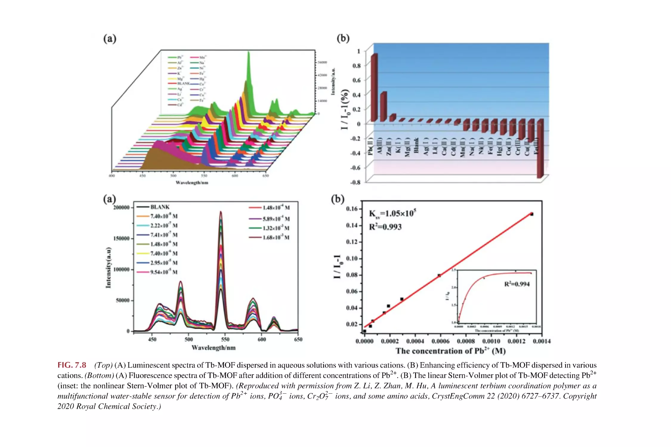
Luminescence responsive sensing of Cr3+ using rare earth
metal-organic framework hybrid materials
Luminescence responsive sensing of Al3+ using rare earth
metal-organic framework hybrid materials
Luminescence responsive sensing of Ag+ using rare earth
metal-organic framework hybrid materials
Luminescence responsive sensing of Co2+/Ni2+ using rare
earth metal-organic framework hybrid materials
Luminescence responsive sensing of f-block metal ions using
rare earth metal-organic framework hybrid materials
References
209
214
218
220
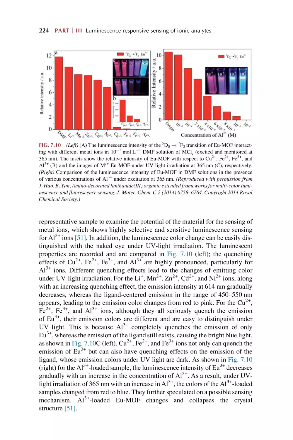
223
226
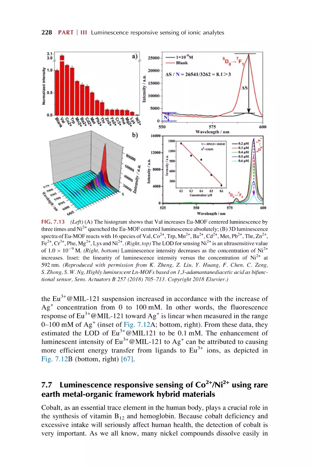
228
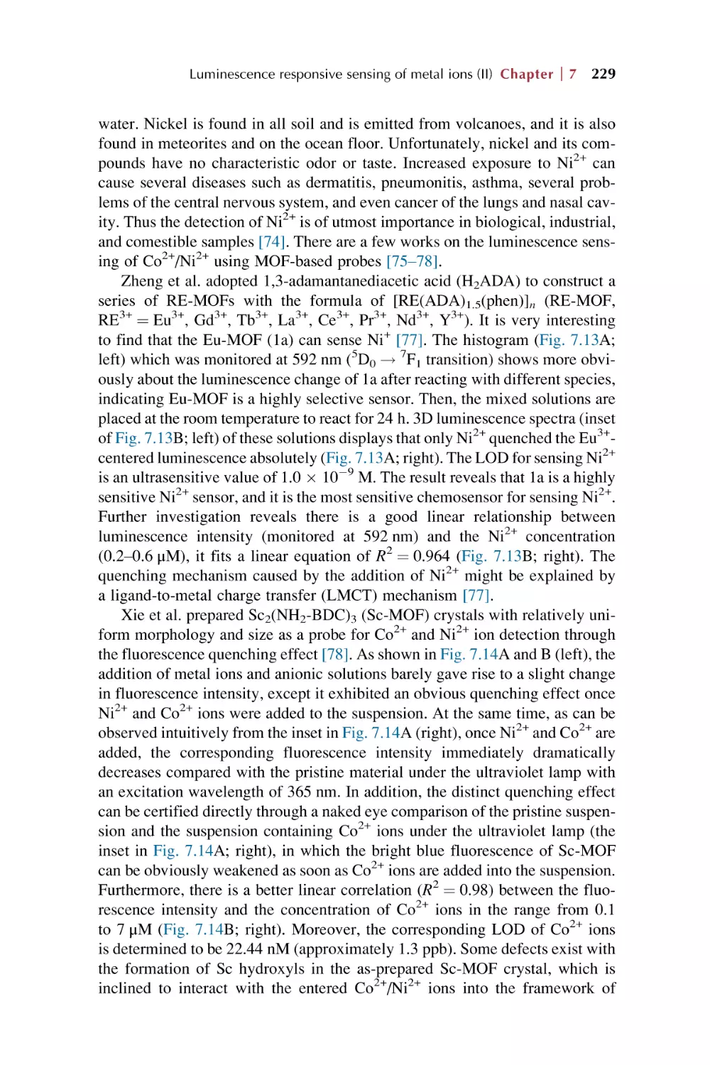
230
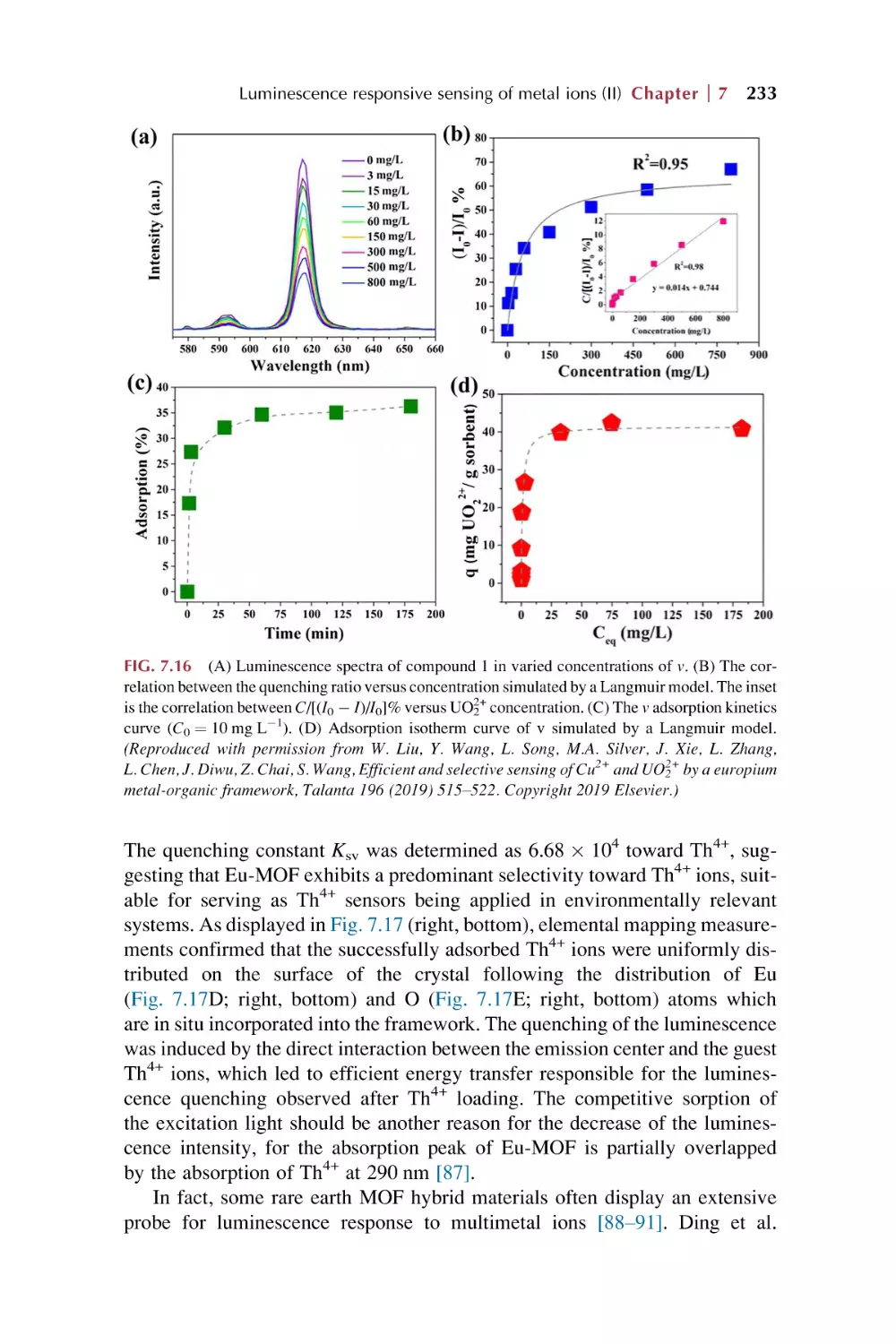
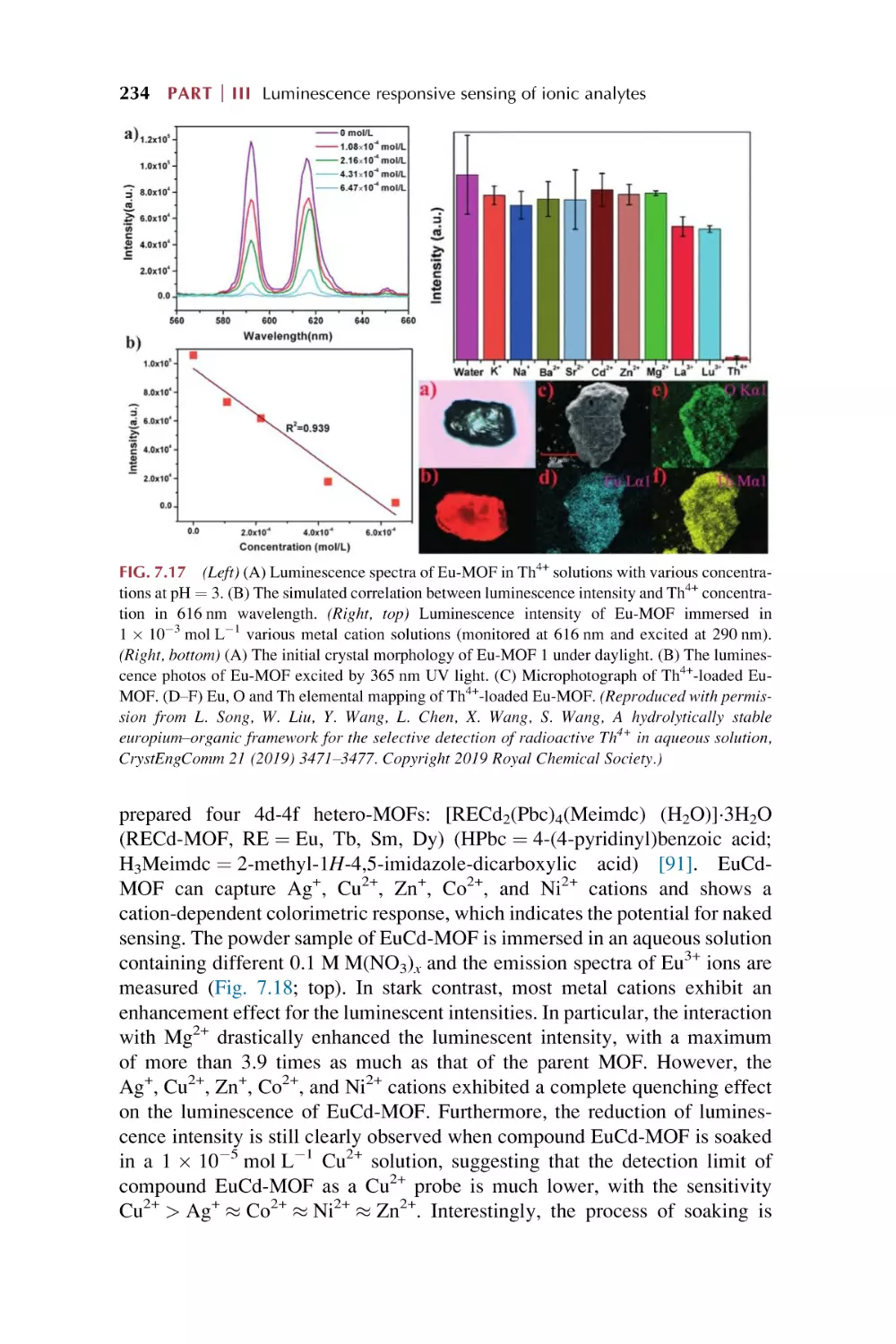
236
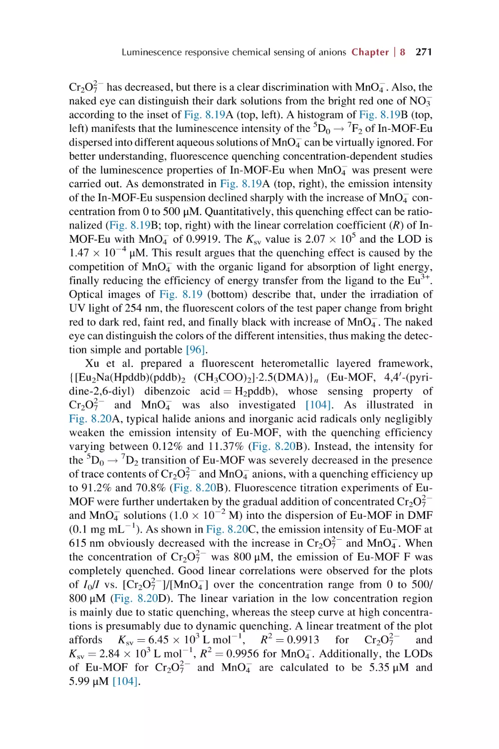
Rare earth metal-organic framework hybrid material for
luminescence responsive chemical sensing of anions
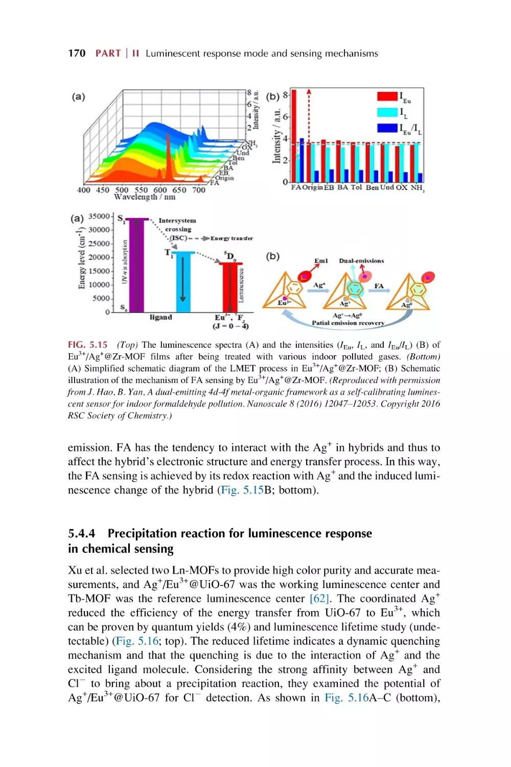
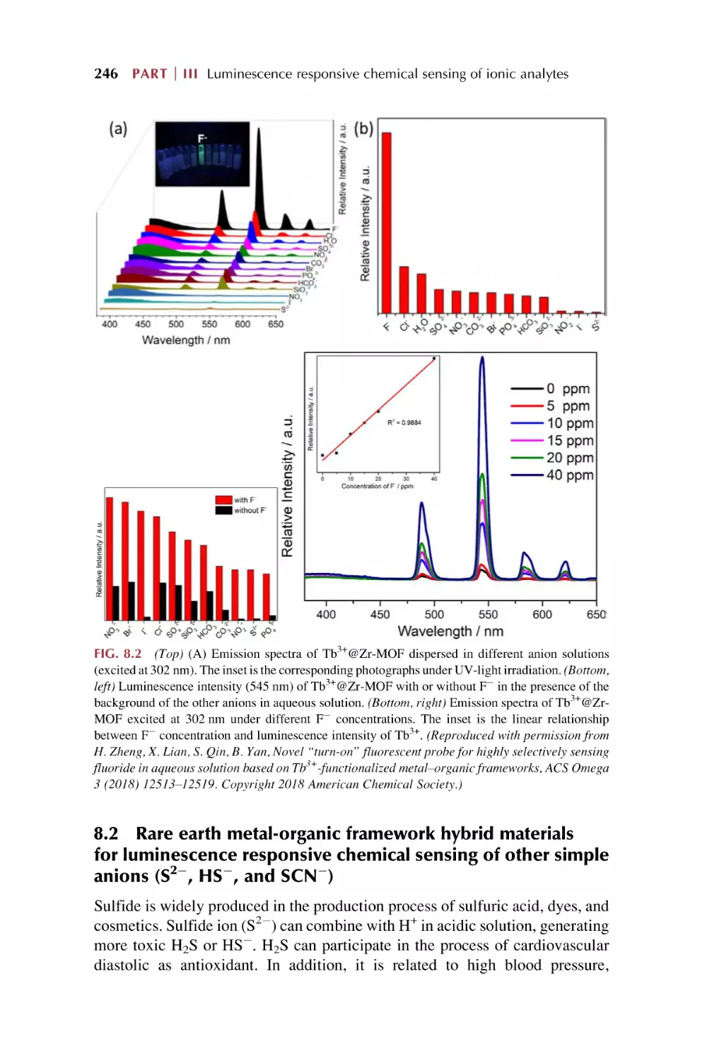
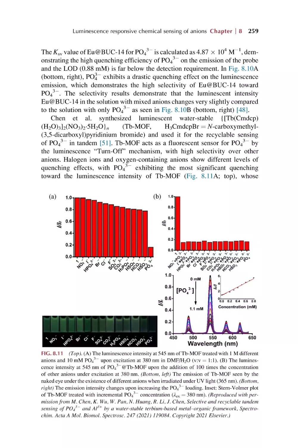
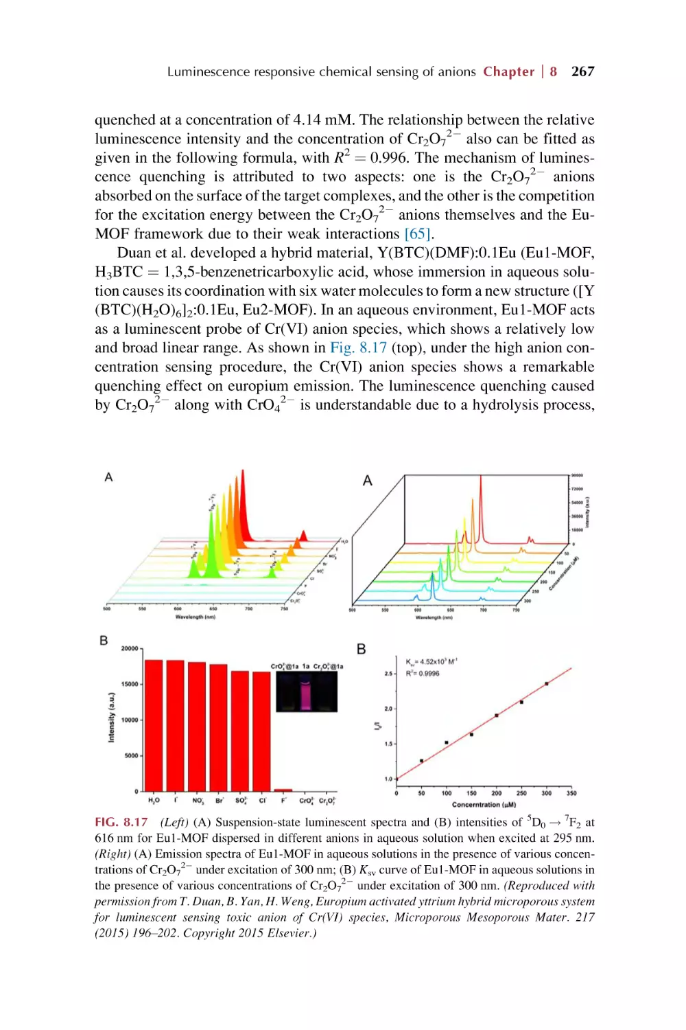
8.1
Rare earth metal-organic framework hybrid materials
for luminescence responsive chemical sensing
of fluoride (F2) ions
243
Contents
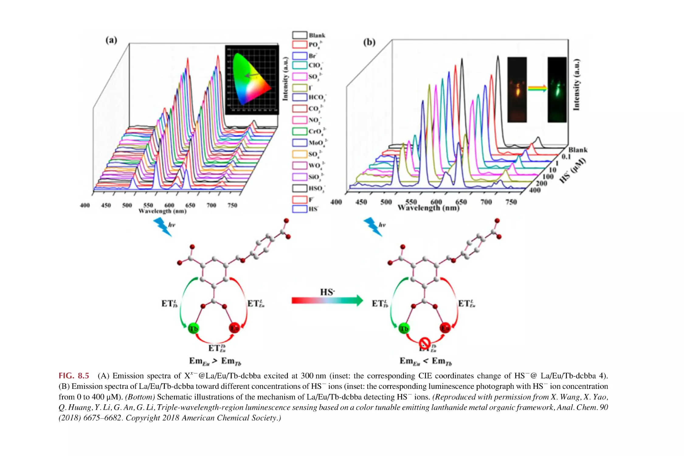
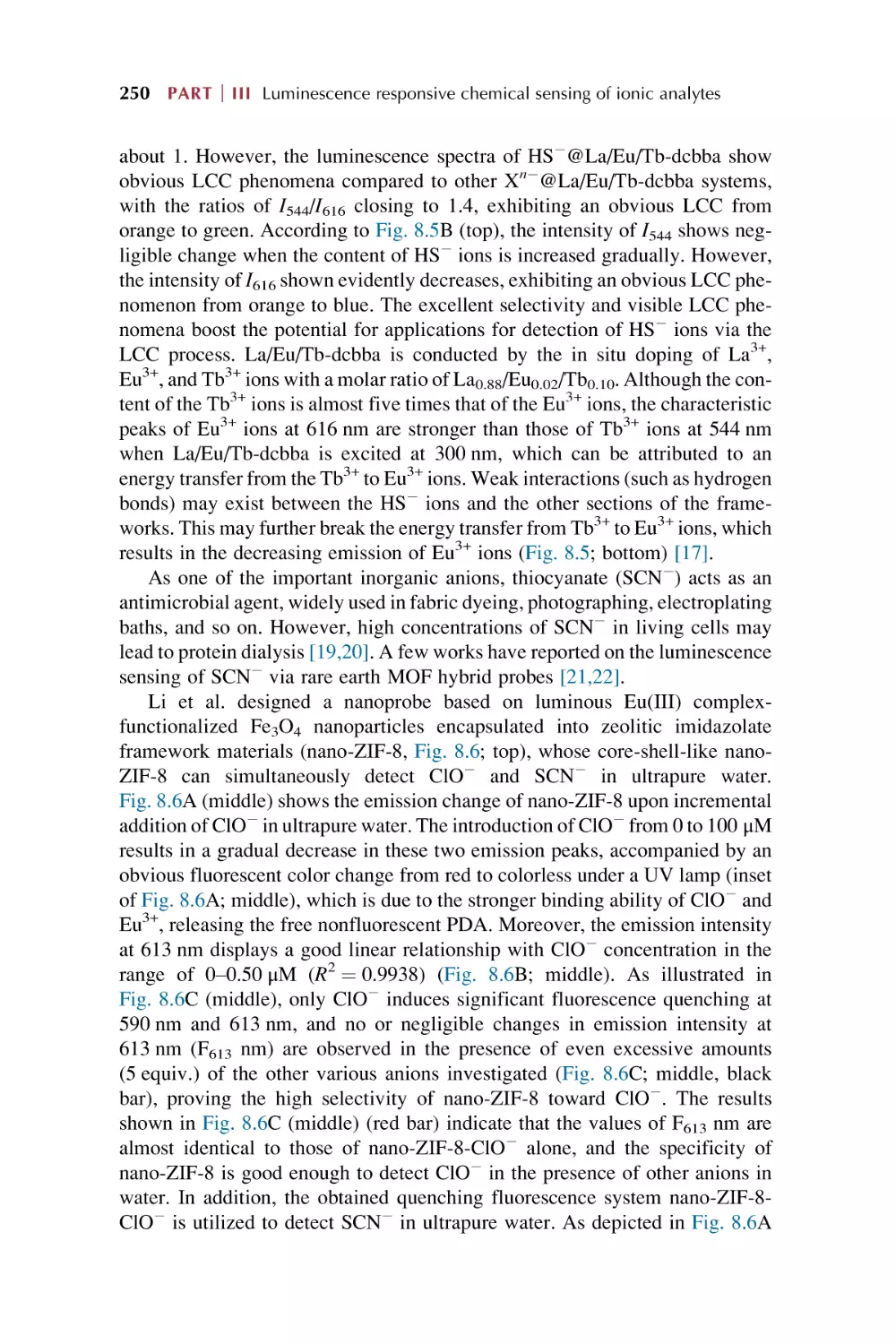
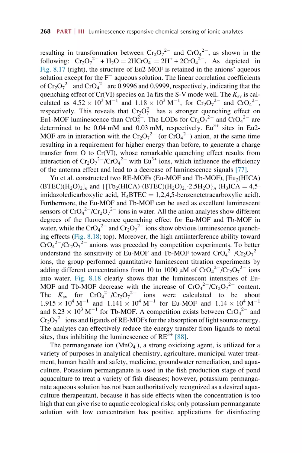
8.2
8.3
8.4
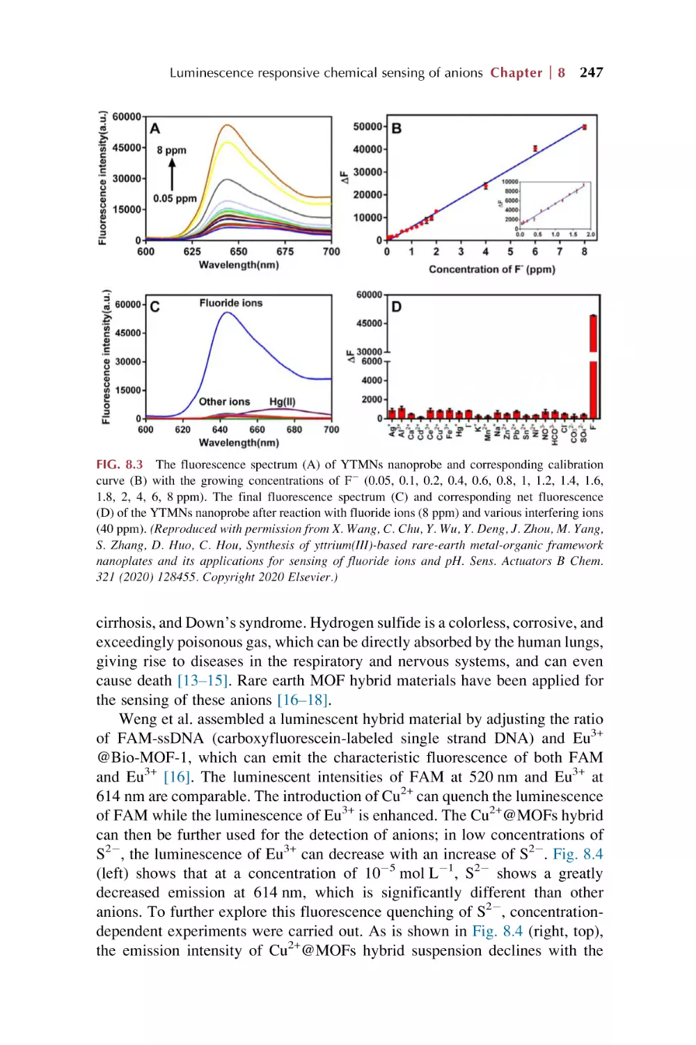
Rare earth metal-organic framework hybrid materials
for luminescence responsive chemical sensing of other
simple anions (S22, HS2, and SCN2)
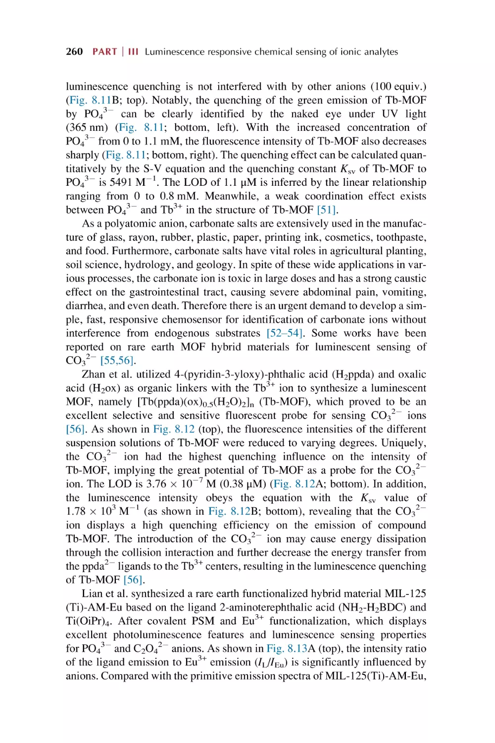
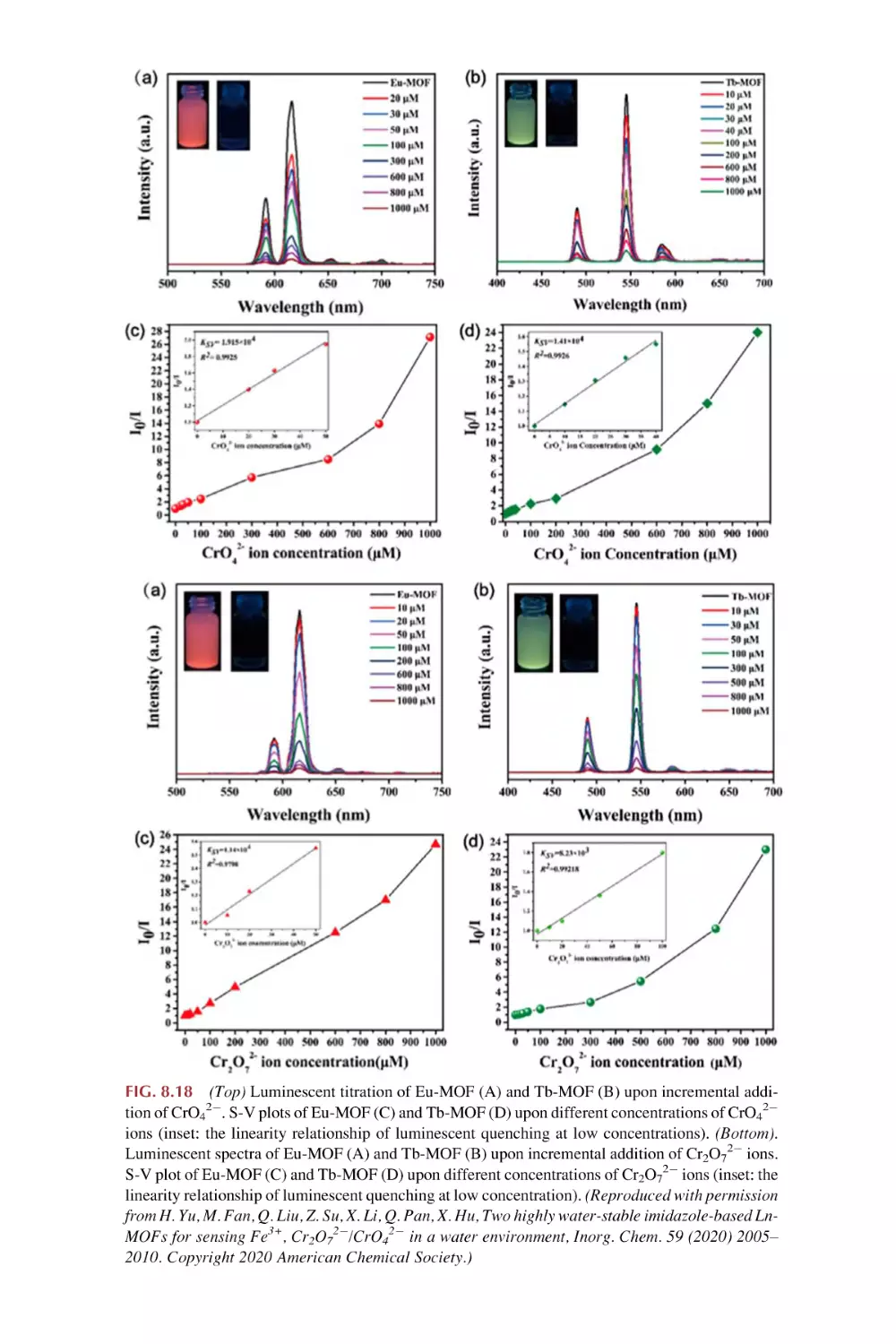
Rare earth metal-organic framework hybrid materials for
luminescence responsive chemical sensing of main group
element oxysalt anions
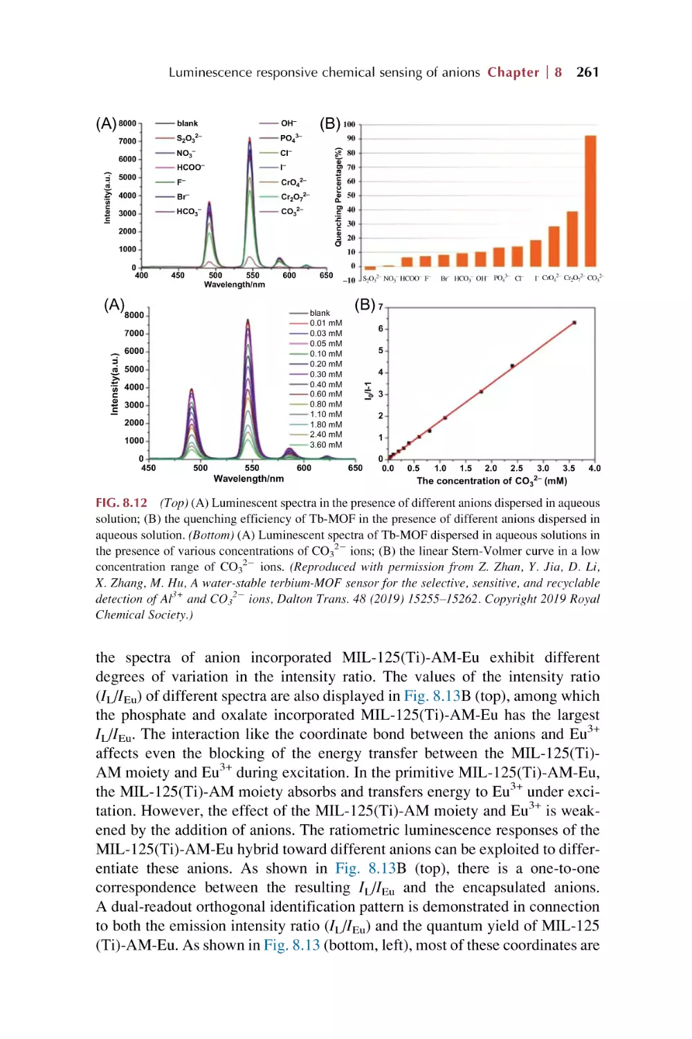
Rare earth metal-organic framework hybrid materials for
luminescence responsive chemical sensing of transition
metal oxysalts anions
References
ix
246
252
262
272
Part IV
Rare earth metal-organic frameworks hybrid
materials as luminescence response chemical
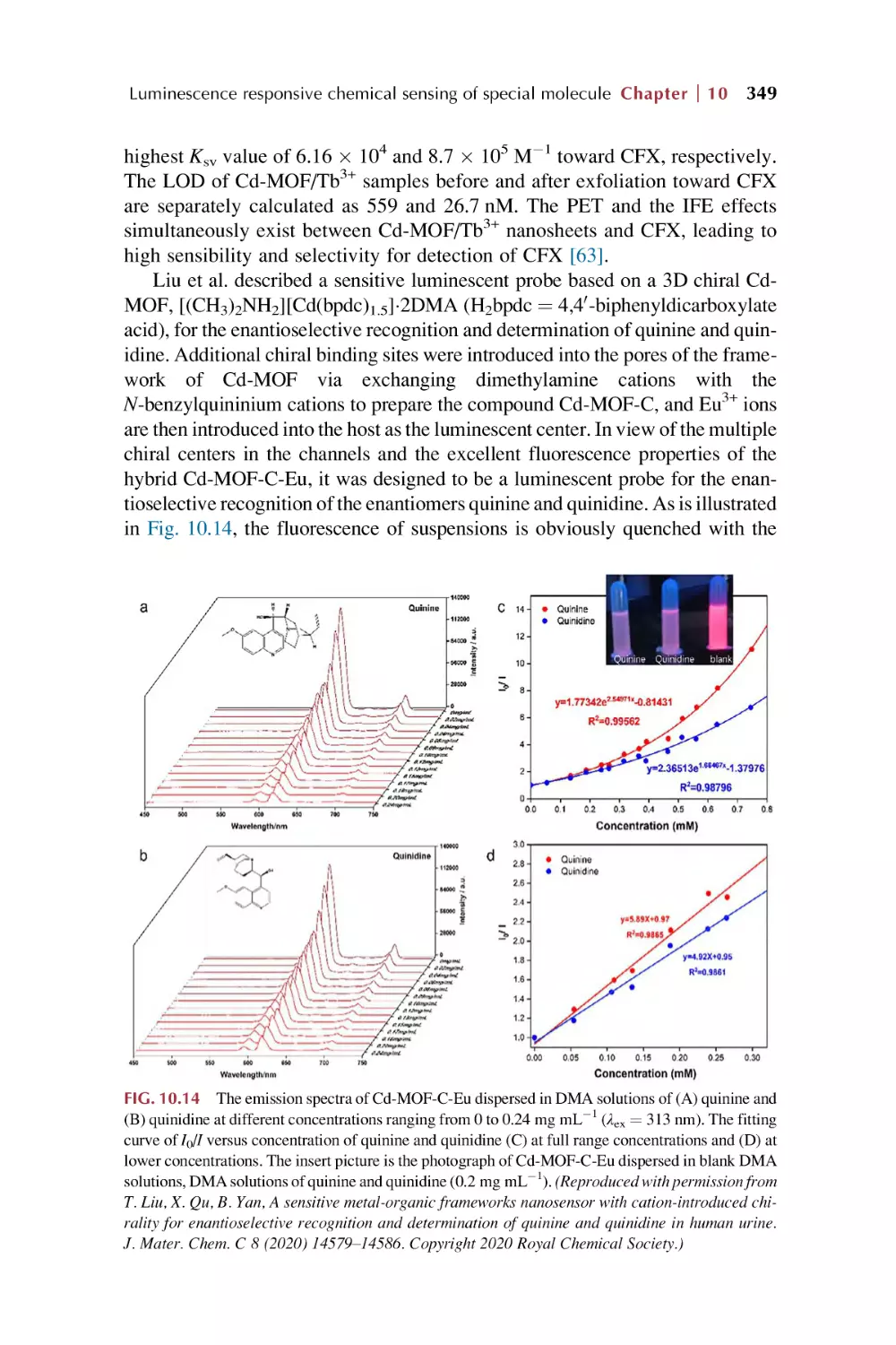
sensors for typical molecular analytes
9.
Rare earth metal-organic framework hybrid materials
for luminescence responsive chemical sensing of
general molecules
9.1
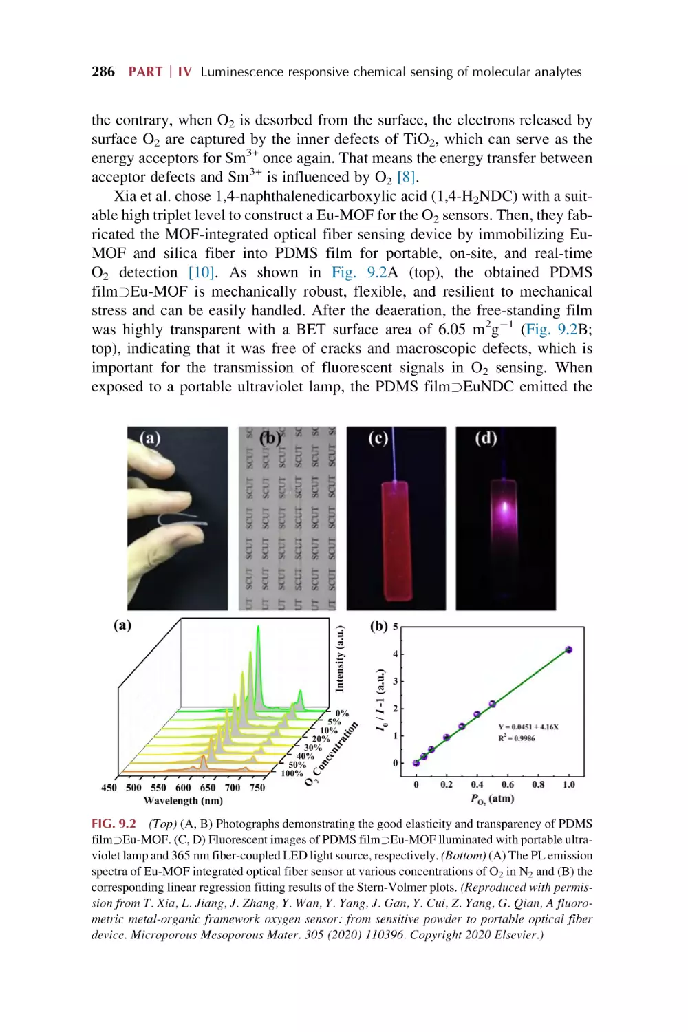
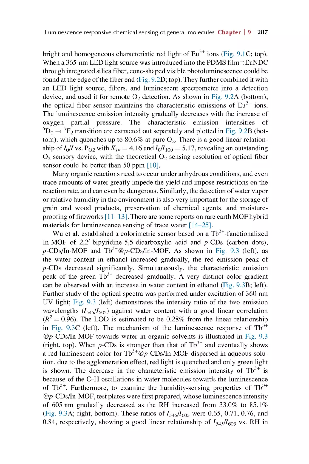
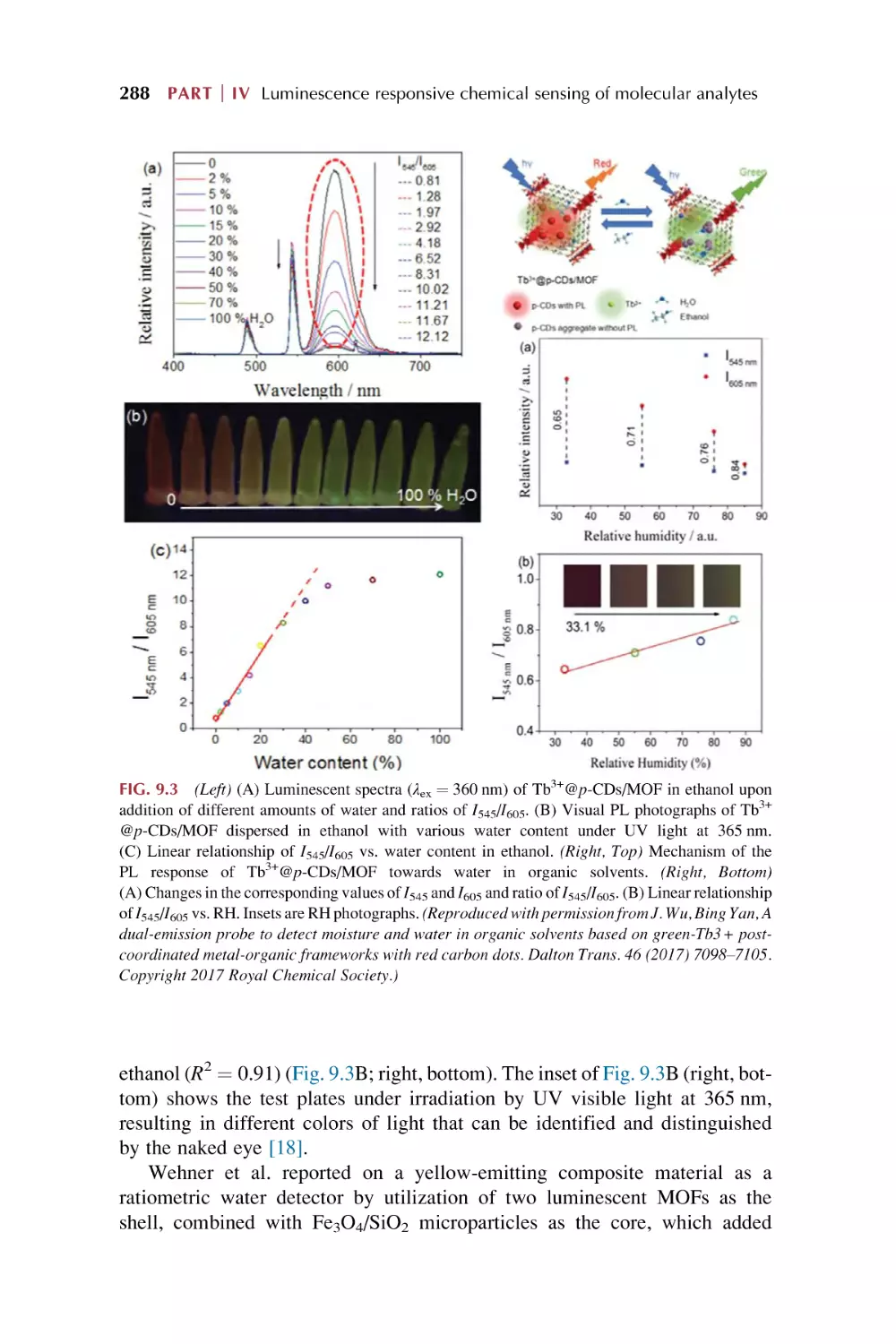
9.2
9.3
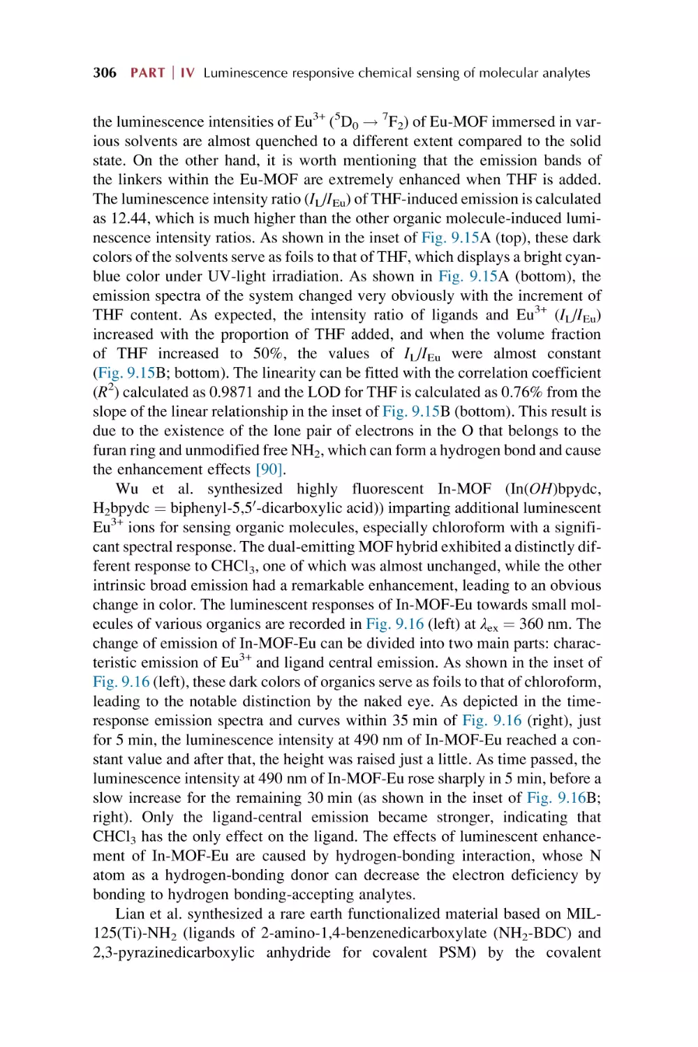
Rare earth metal-organic framework hybrid materials for
luminescence responsive chemical sensing of inorganic
molecules
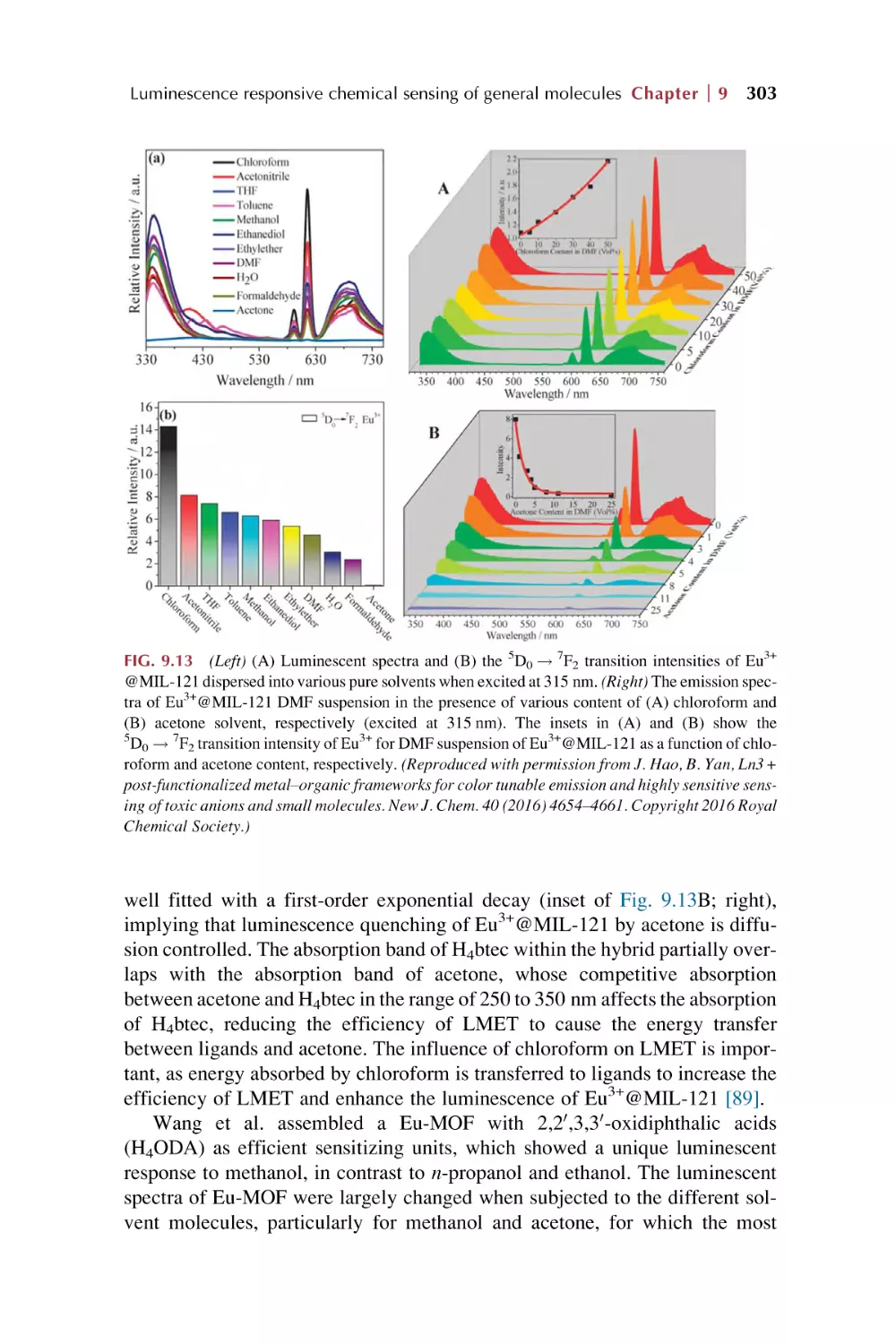
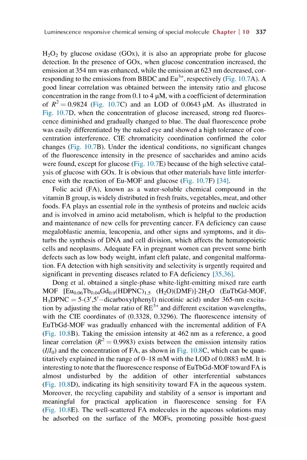
Rare earth metal-organic framework for luminescence
responsive chemical sensing of general organic
molecules
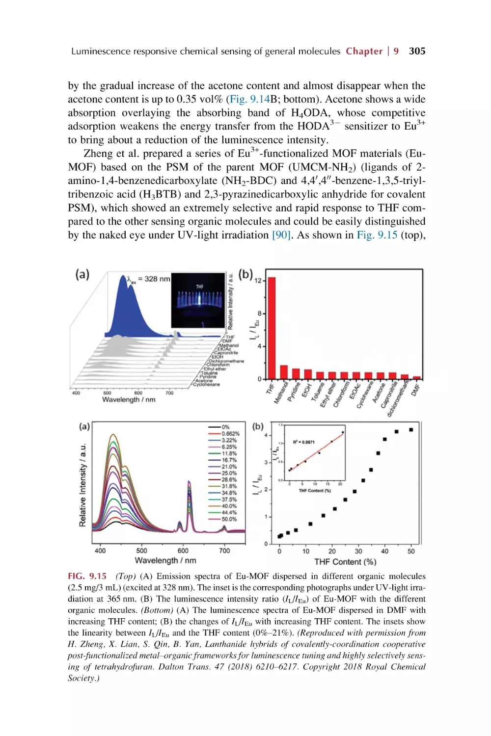
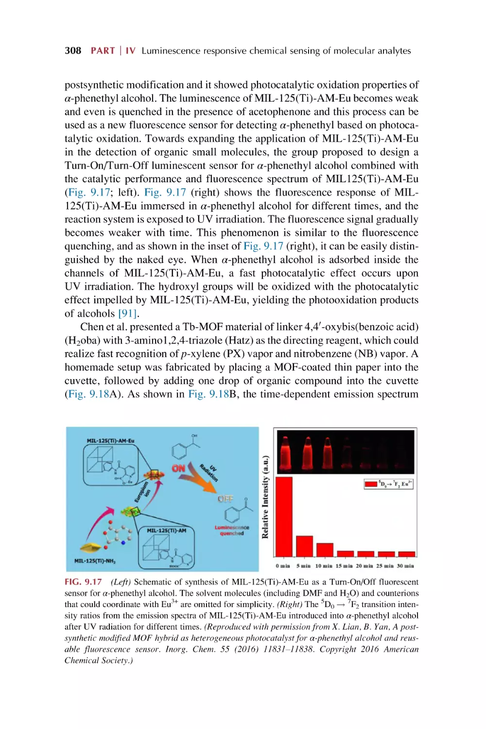
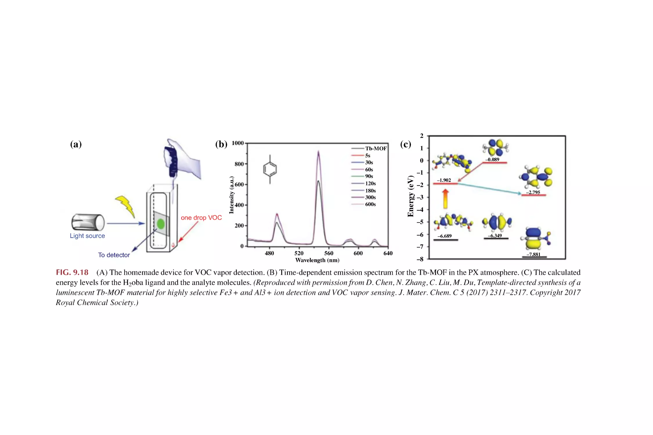
Rare earth metal-organic framework hybrid materials
for luminescence responsive chemical sensing of general
organic pollutant molecules
References
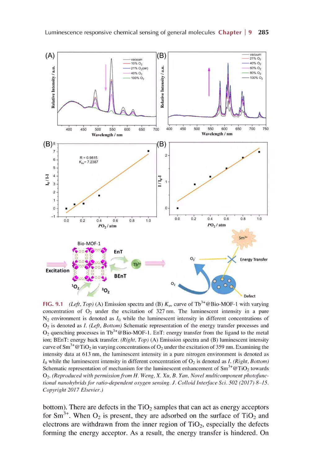
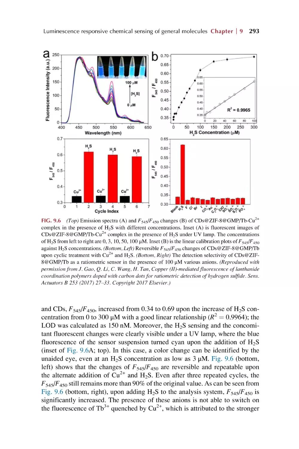
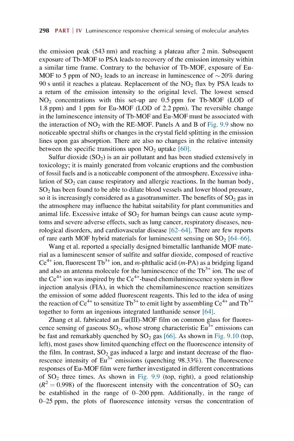
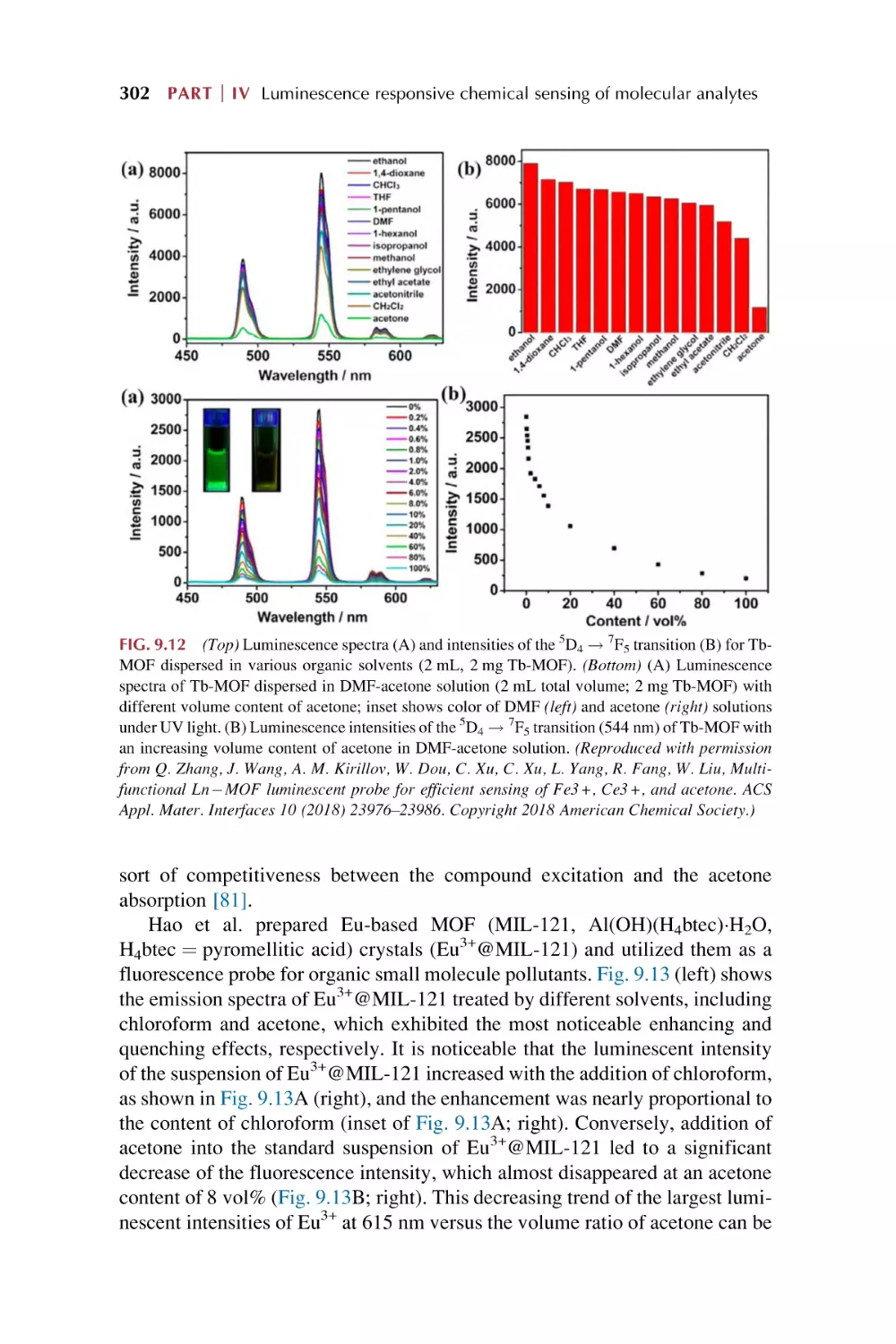
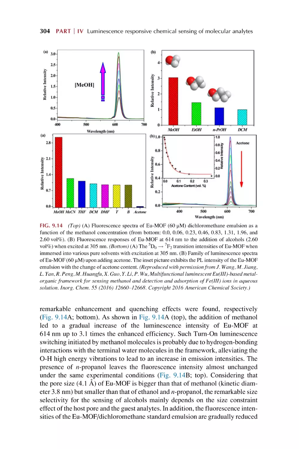
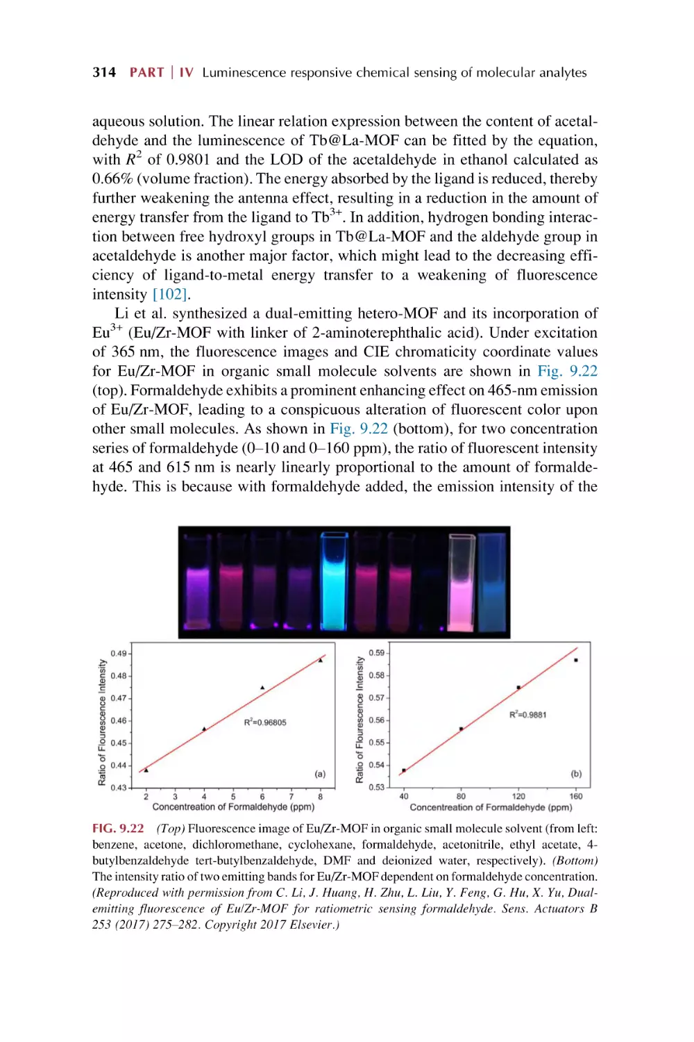
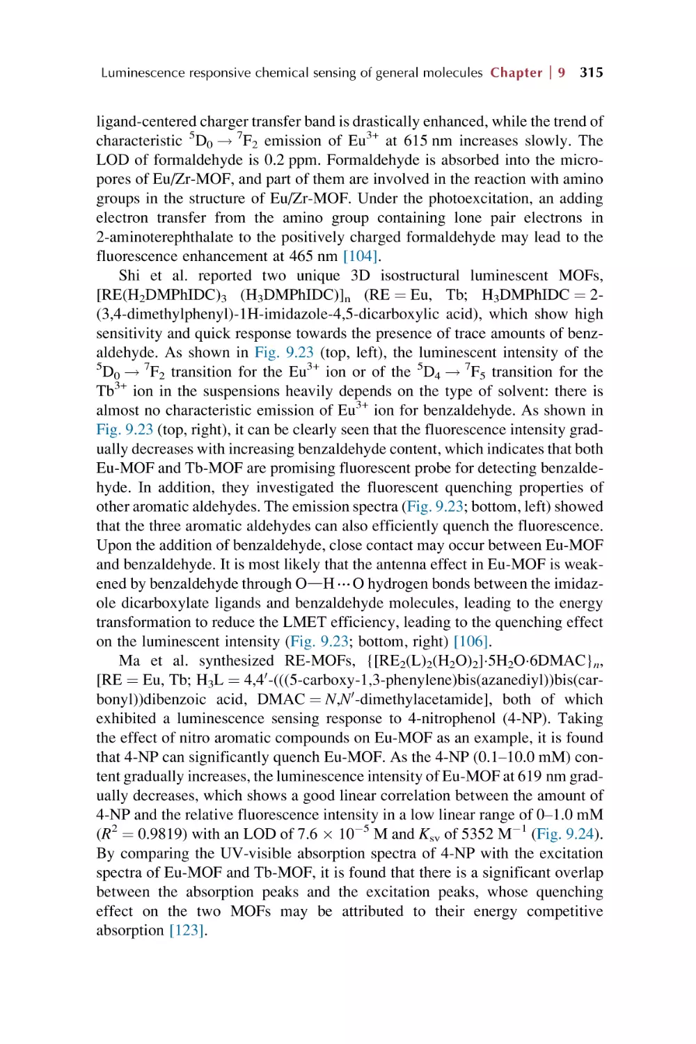
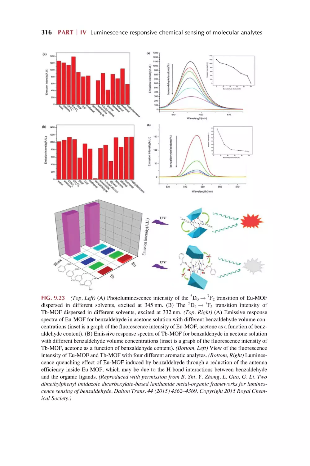
284
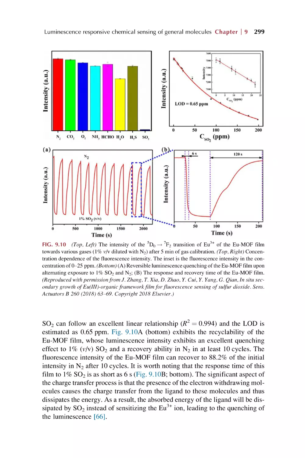
300
312
317
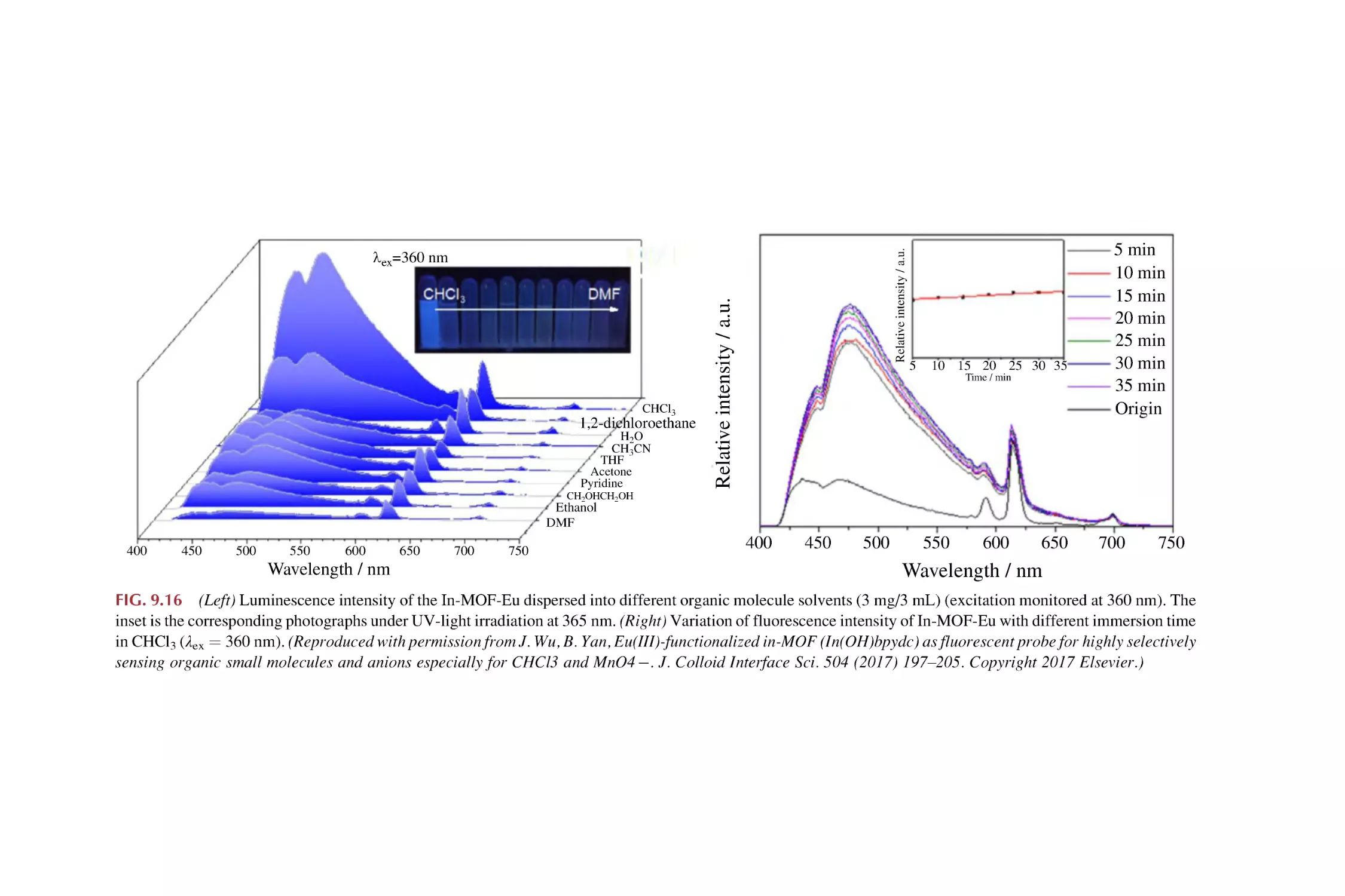
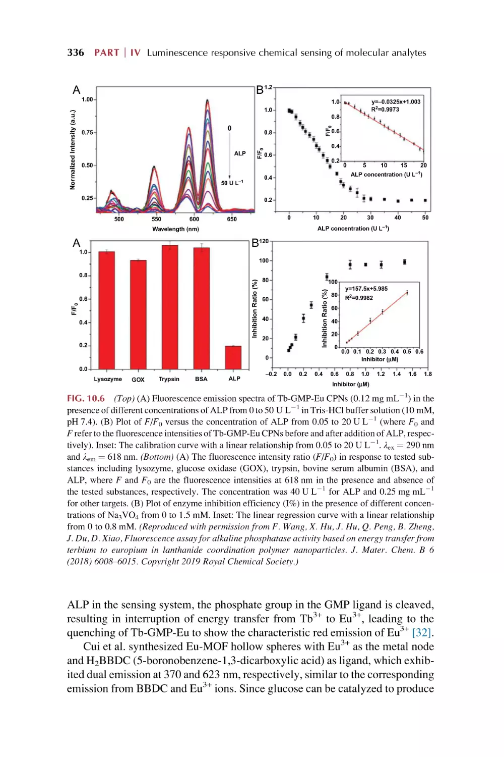
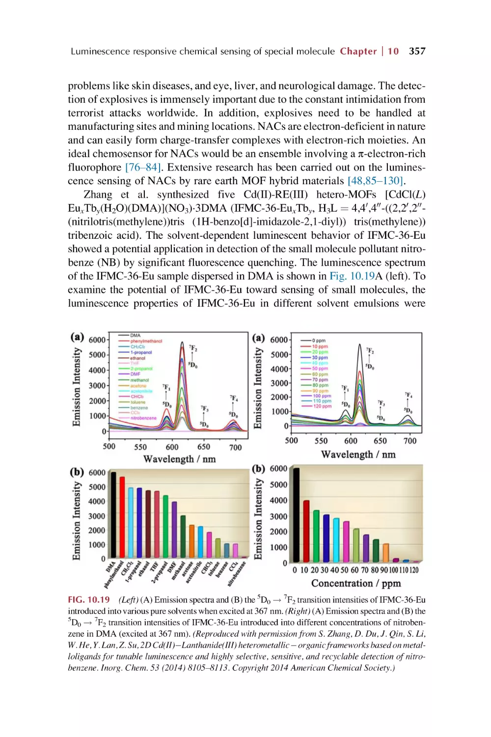
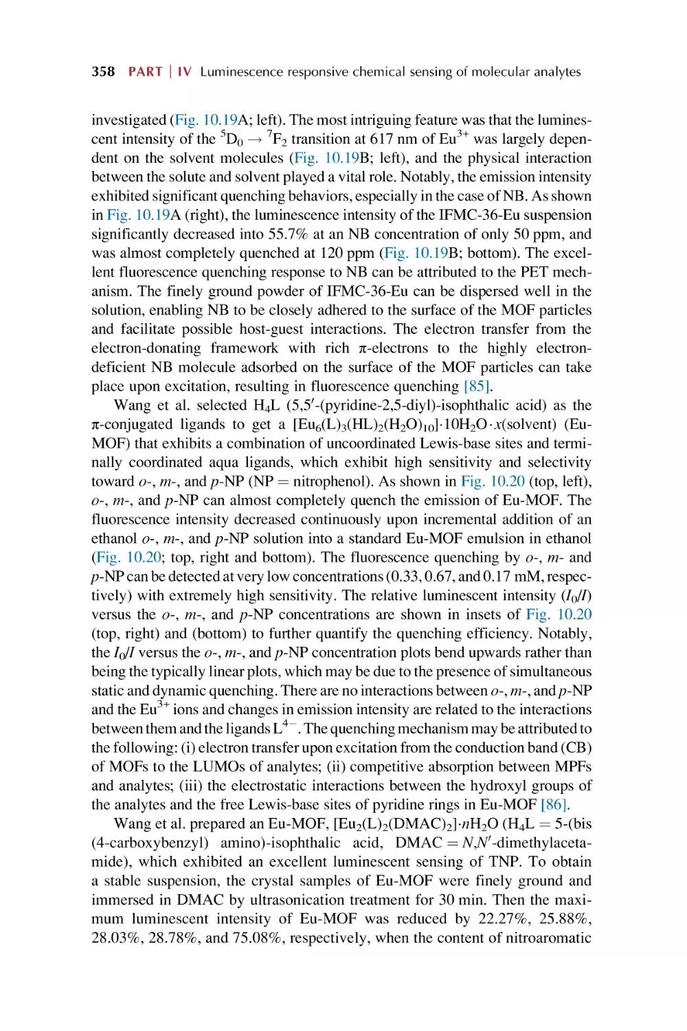
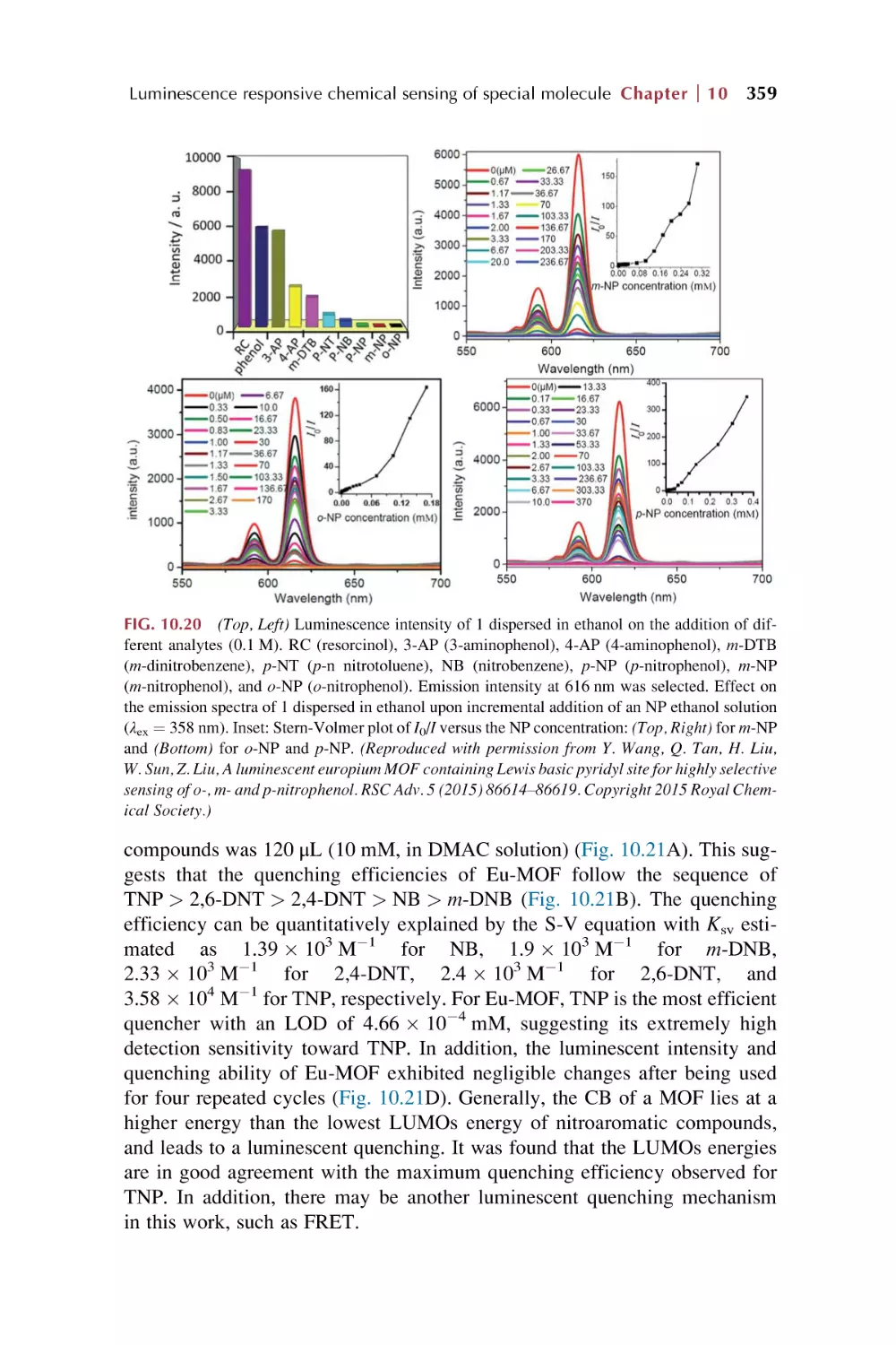
10. Rare earth metal-organic framework hybrid materials
for luminescence responsive chemical sensing of
special molecule species
10.1
10.2
10.3
Rare earth metal-organic framework hybrid materials
for luminescence responsive chemical sensing
of biomolecular species
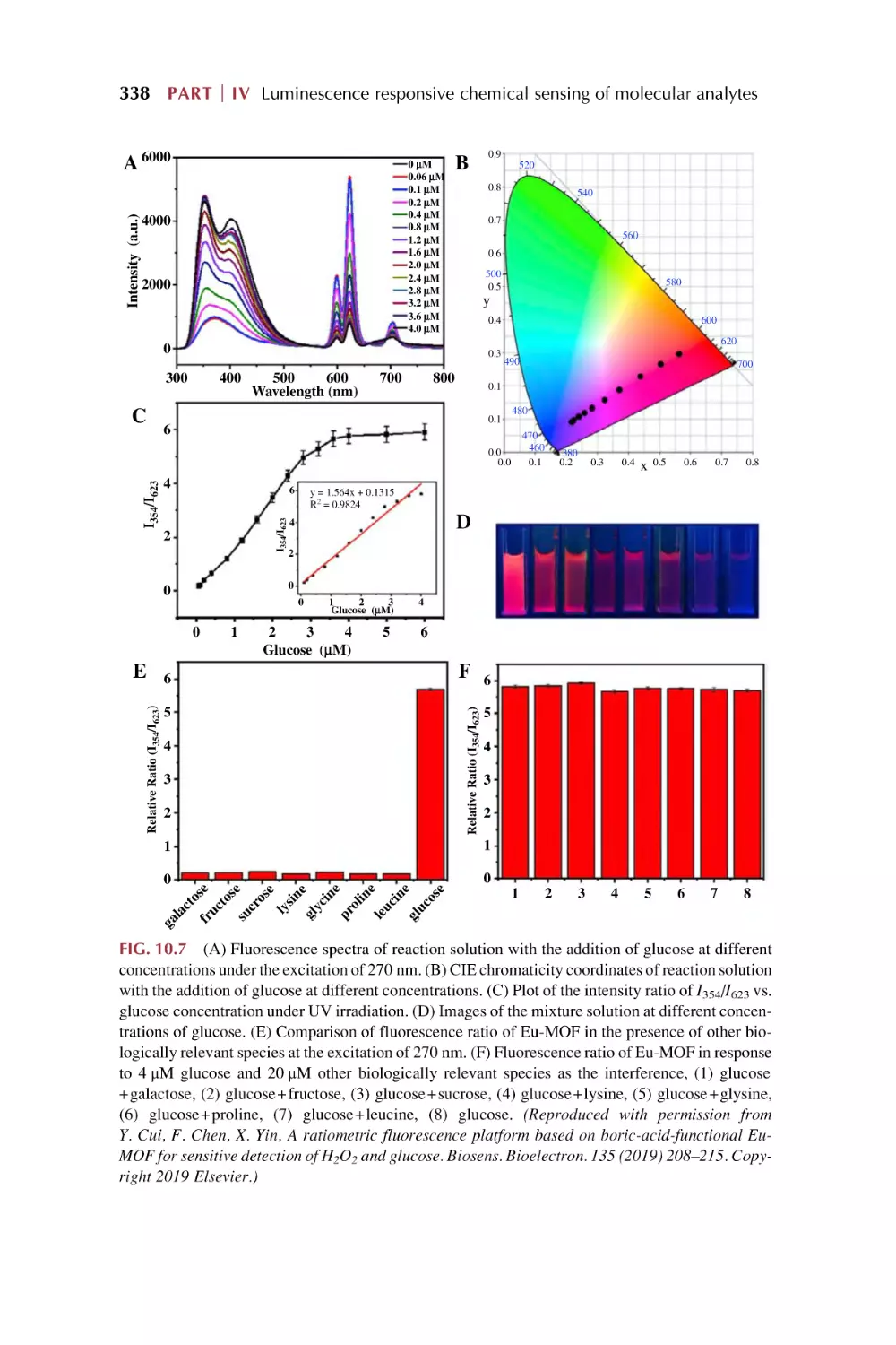
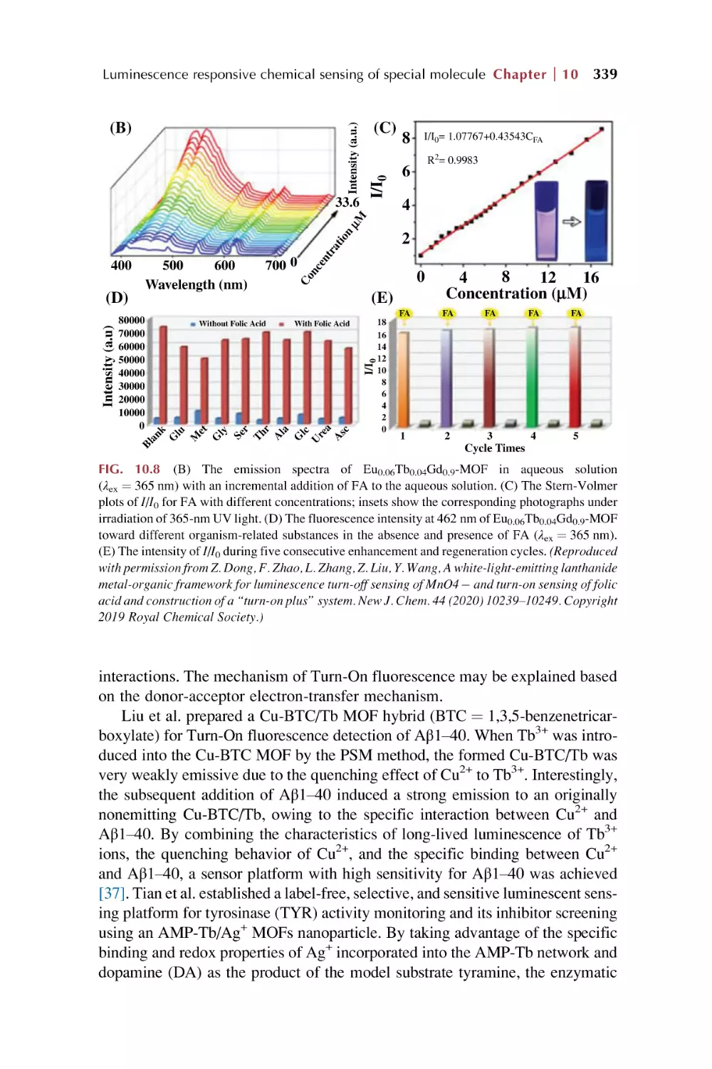
Rare earth metal-organic framework hybrid materials
for luminescence responsive chemical sensing
of antibiotics and drugs
Rare earth metal-organic framework hybrid materials
for luminescence sensing of nitroaromatic explosives (NAE)
and other special dangerous species
References
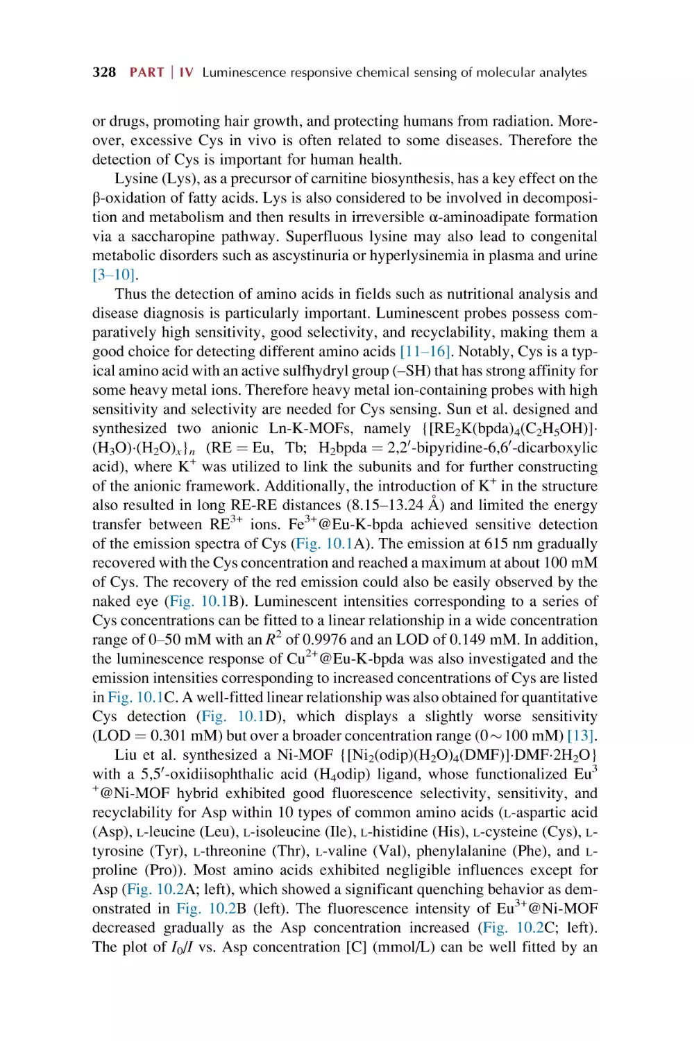
327
340
353
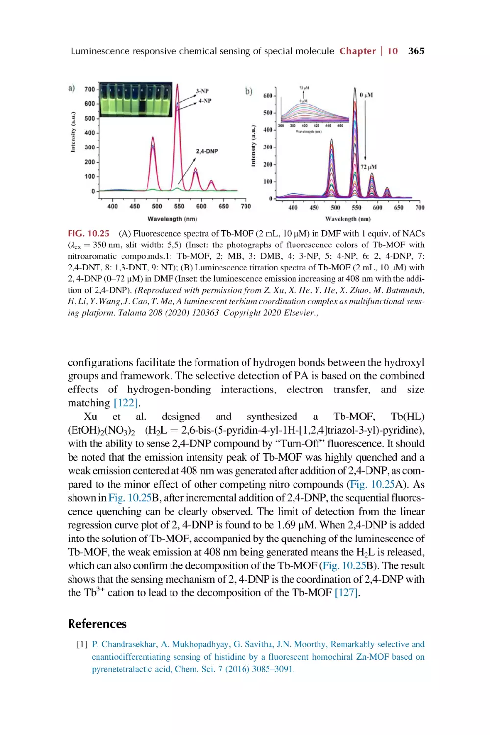
365
x Contents
11. Rare earth metal-organic framework hybrid materials
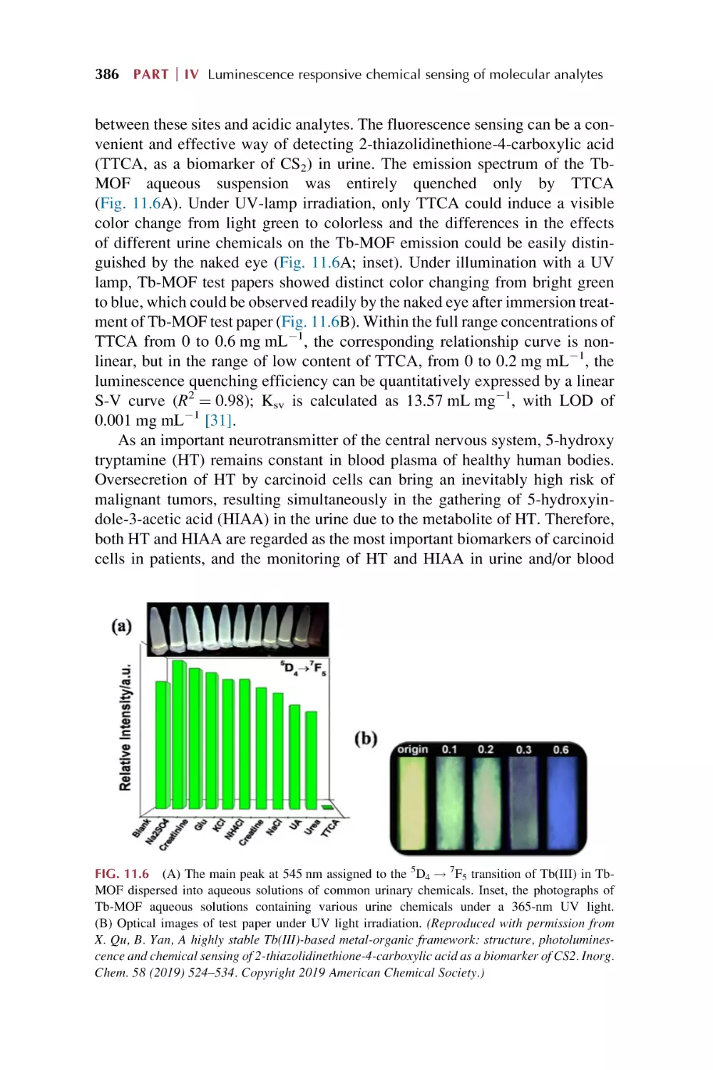
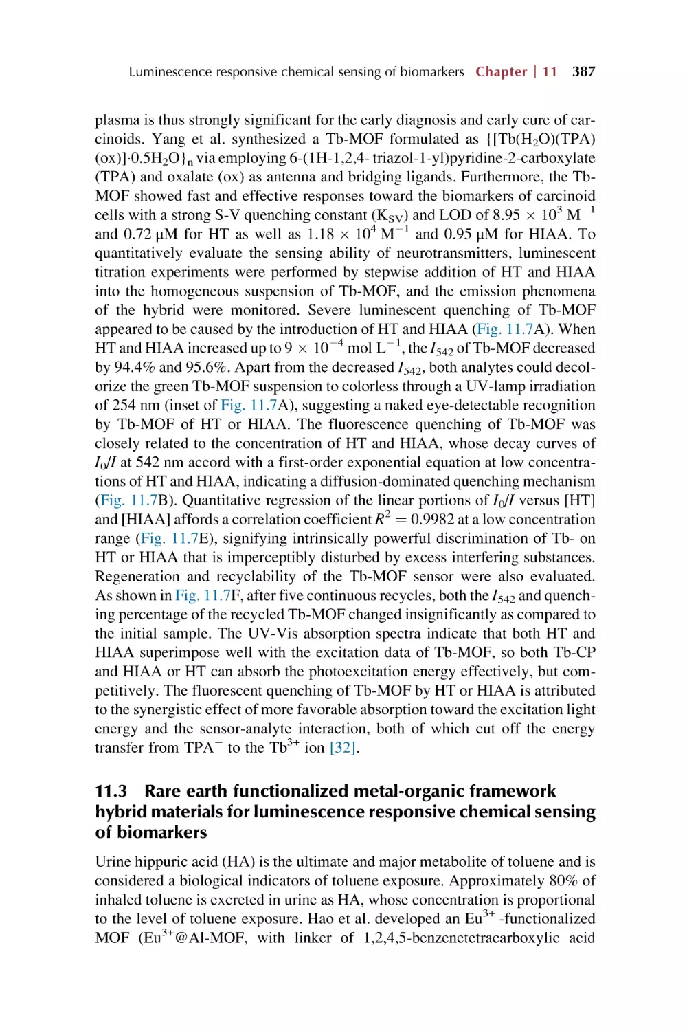
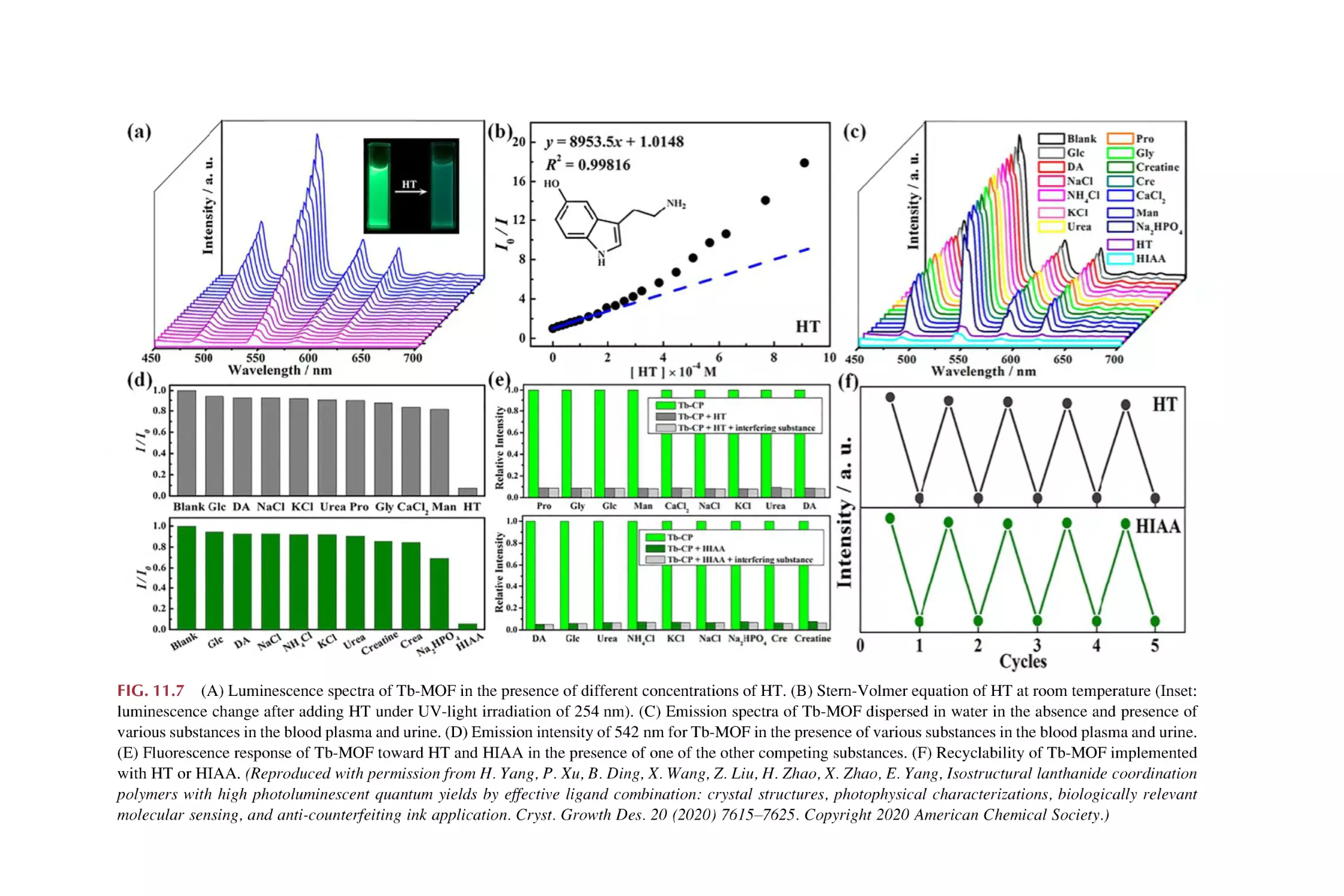
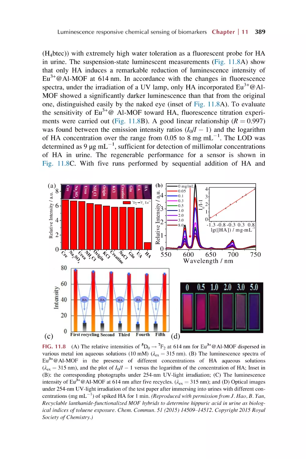
for luminescence responsive chemical sensing of
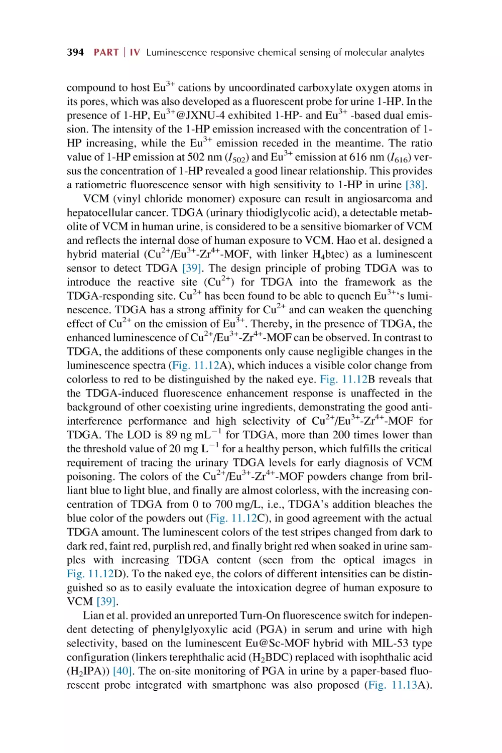
biomarkers
11.1
11.2
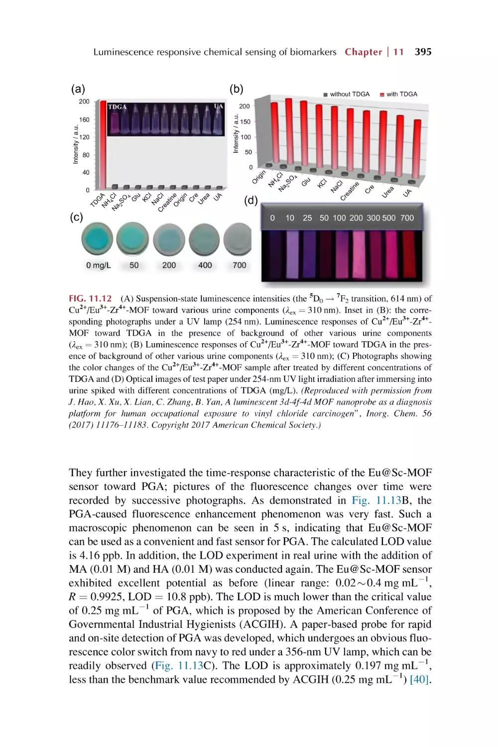
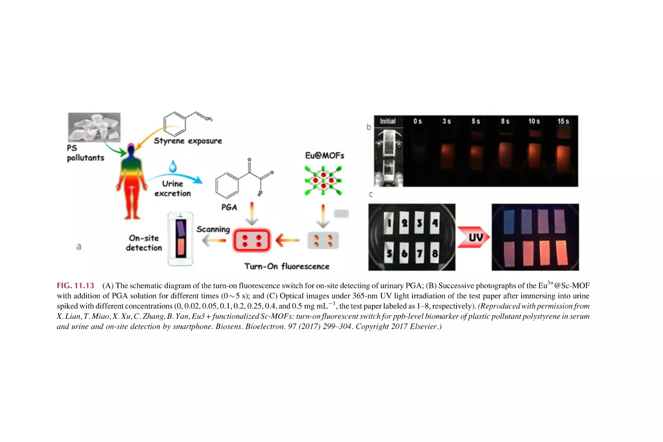
11.3
Biomarkers and their chemical sensing
Rare earth metal-organic framework materials for
luminescence responsive chemical sensing of
biomarkers
Rare earth functionalized metal-organic framework hybrid
materials for luminescence responsive chemical sensing of
biomarkers
References
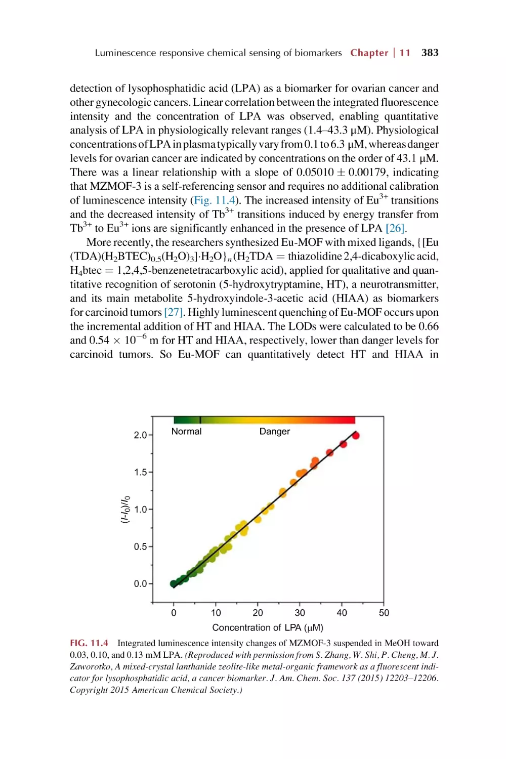
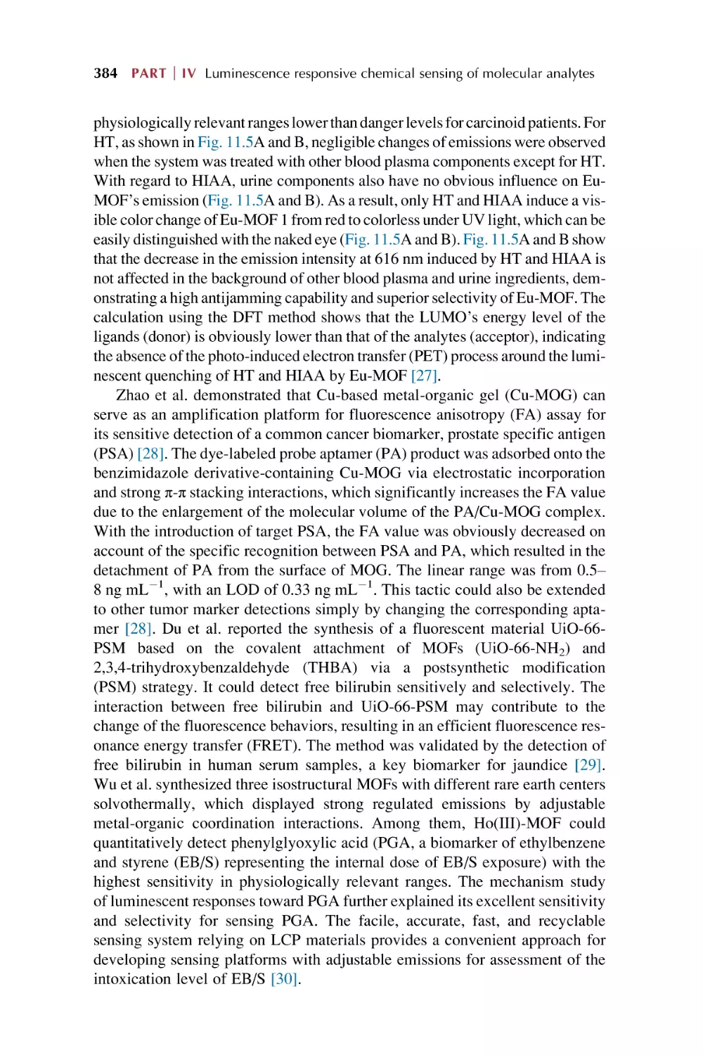
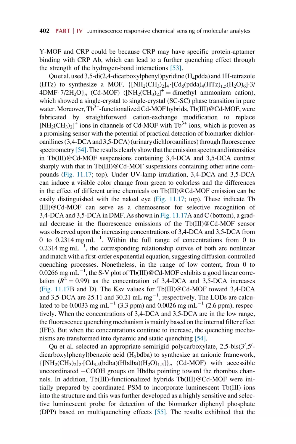
375
378
387
405
Part V
Rare earth metal-organic frameworks hybrid
materials as luminescence response chemical
sensors for others and applications
12. Rare earth metal-organic framework hybrid materials
for luminescence responsive chemical sensing of
temperature and pH value
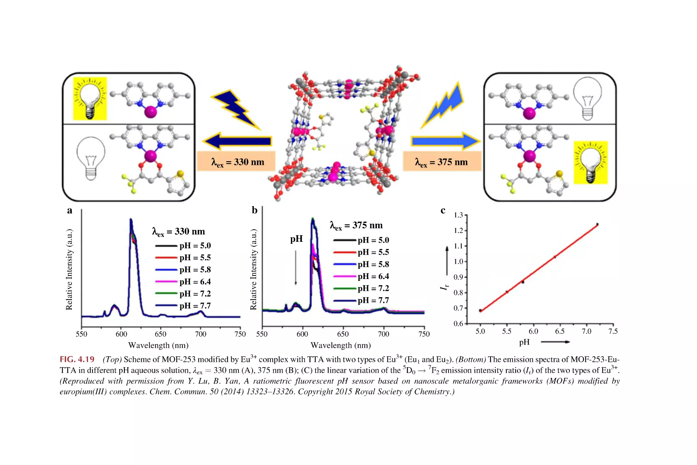
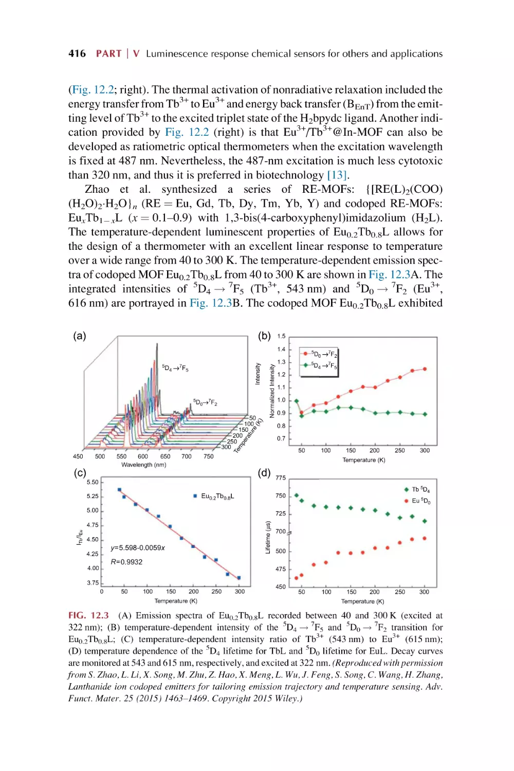
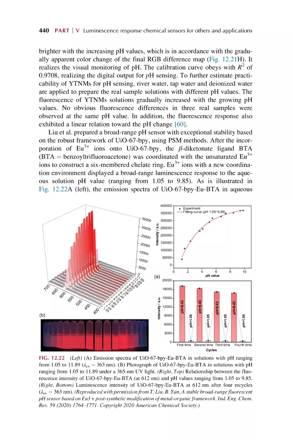
12.1
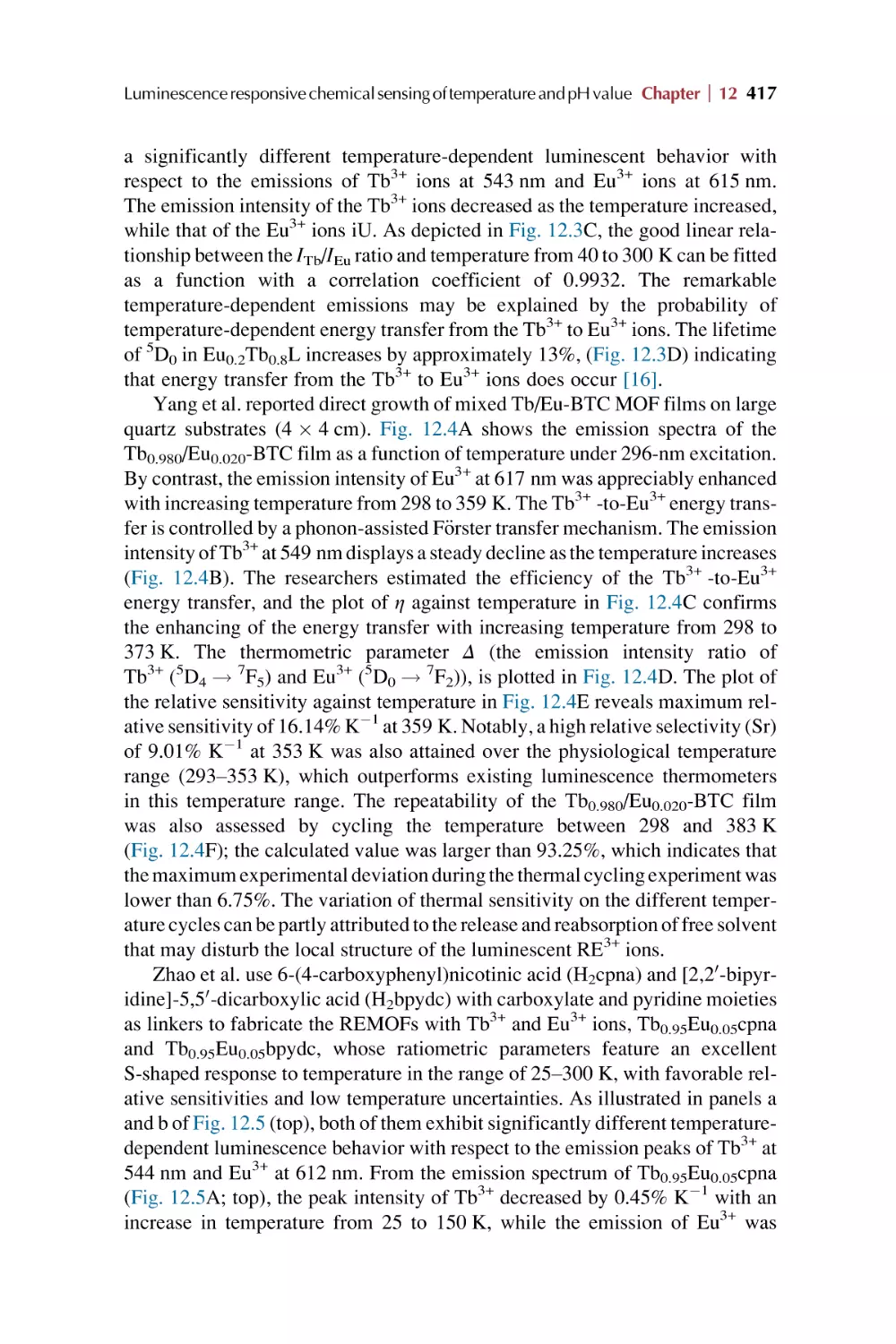
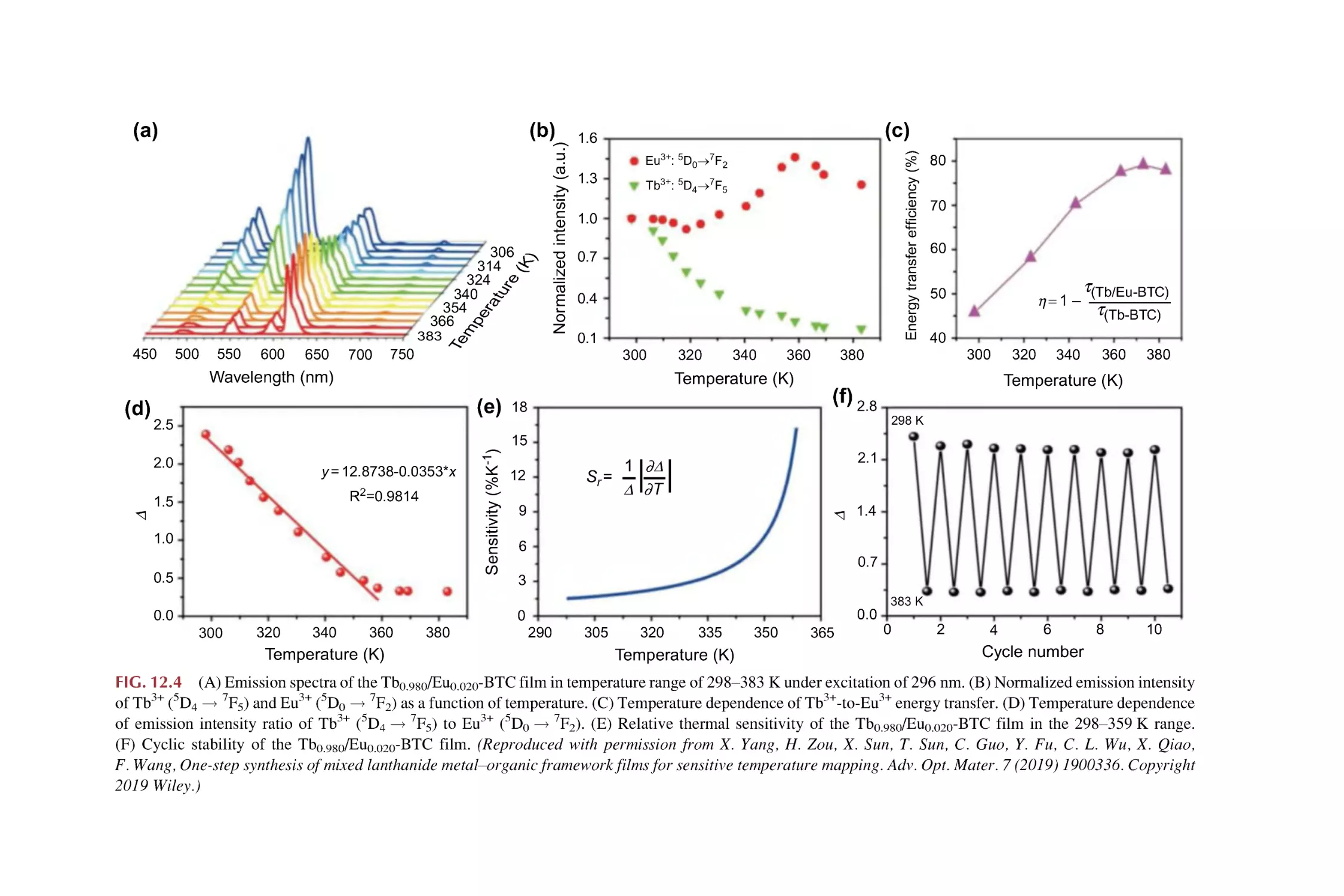
12.2
Rare earth metal-organic framework hybrid materials for
luminescence sensing of temperature
Rare earth metal-organic framework hybrid materials for
luminescence responsive chemical sensing of pH value
References
411
432
441
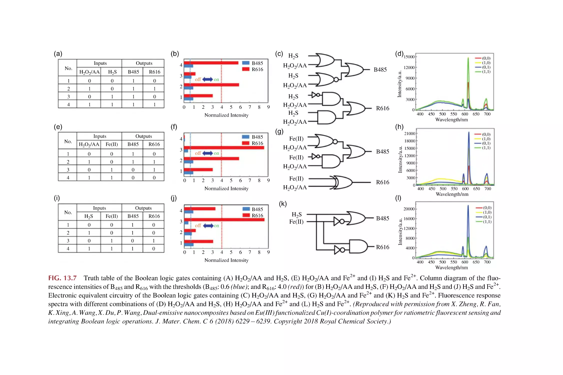
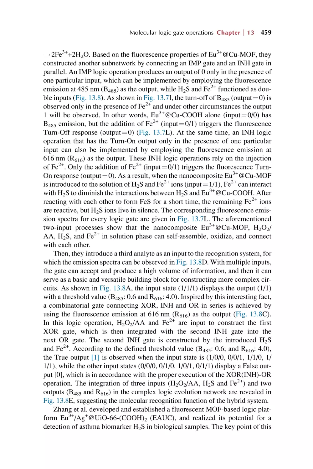
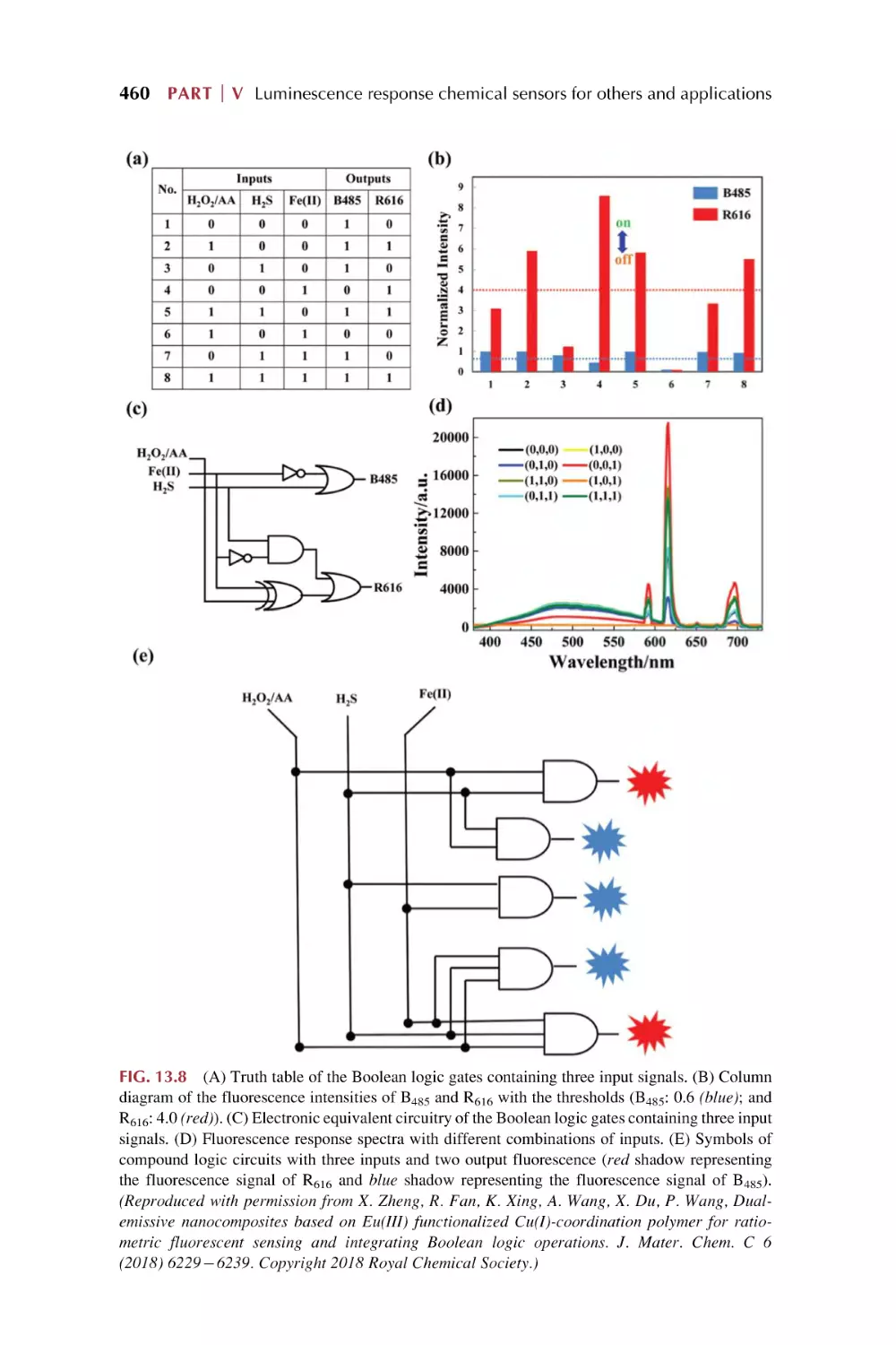
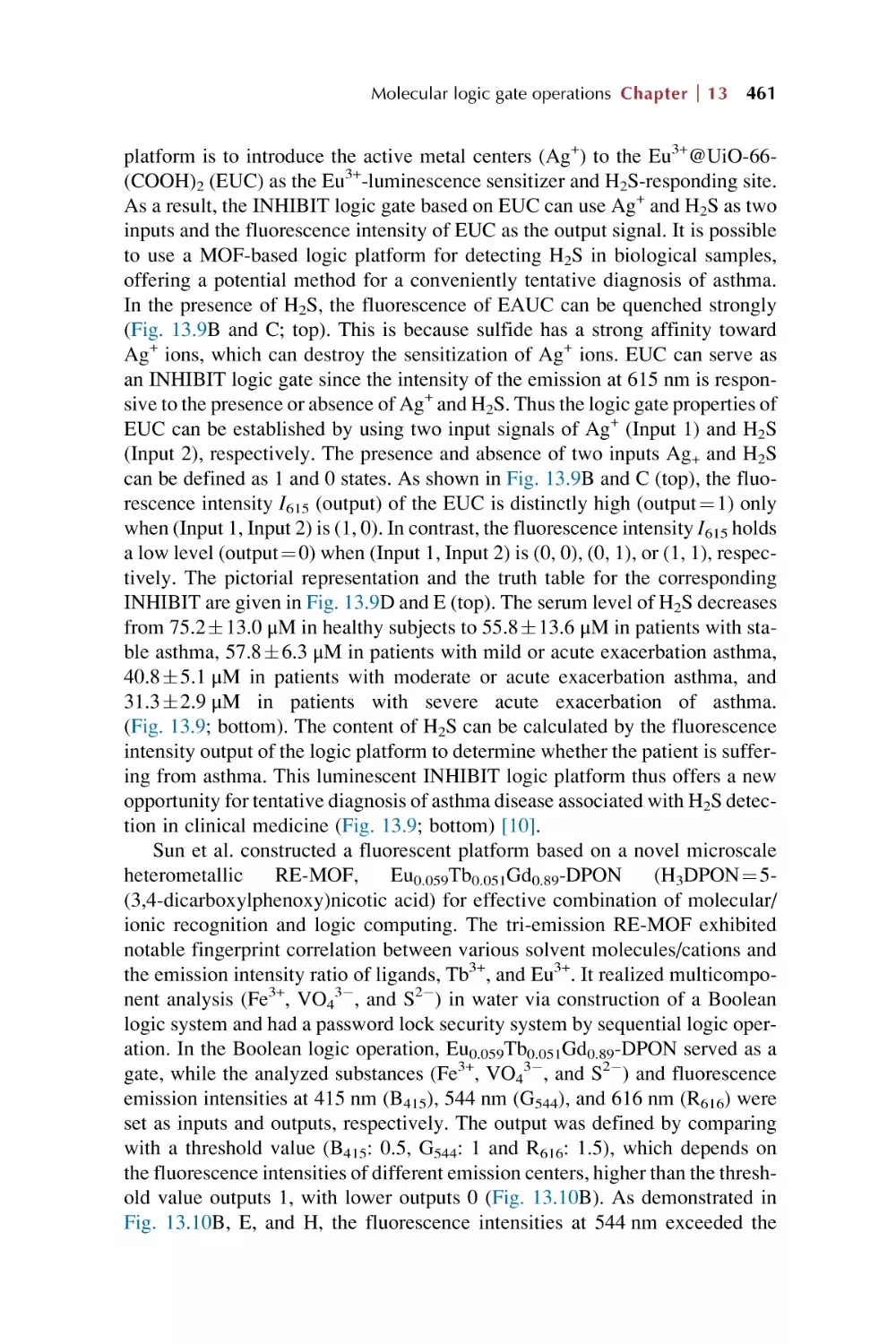
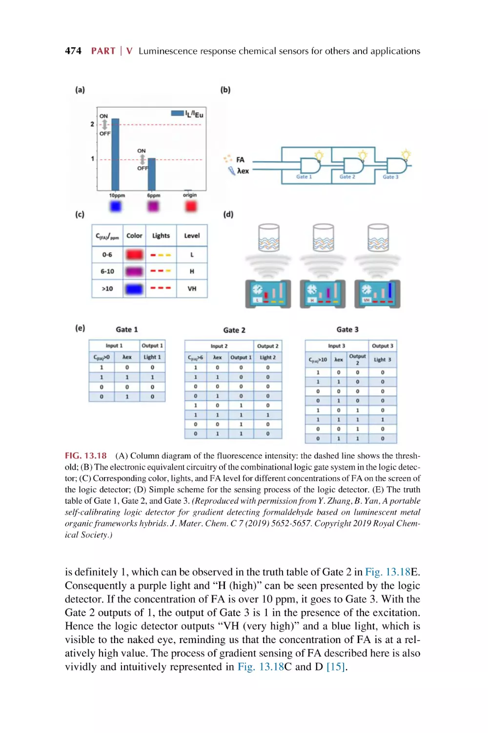
13. Molecular logic gate operations of rare earth
metal-organic framework hybrid materials for
luminescence responsive chemical sensing
13.1
13.2
13.3
Molecular Boolean logic gates
13.1.1 Basic molecular Boolean logic gate operation
13.1.2 Implementation of a two-output combinational
logic gate
13.1.3 Implementation of a cascaded logic gate
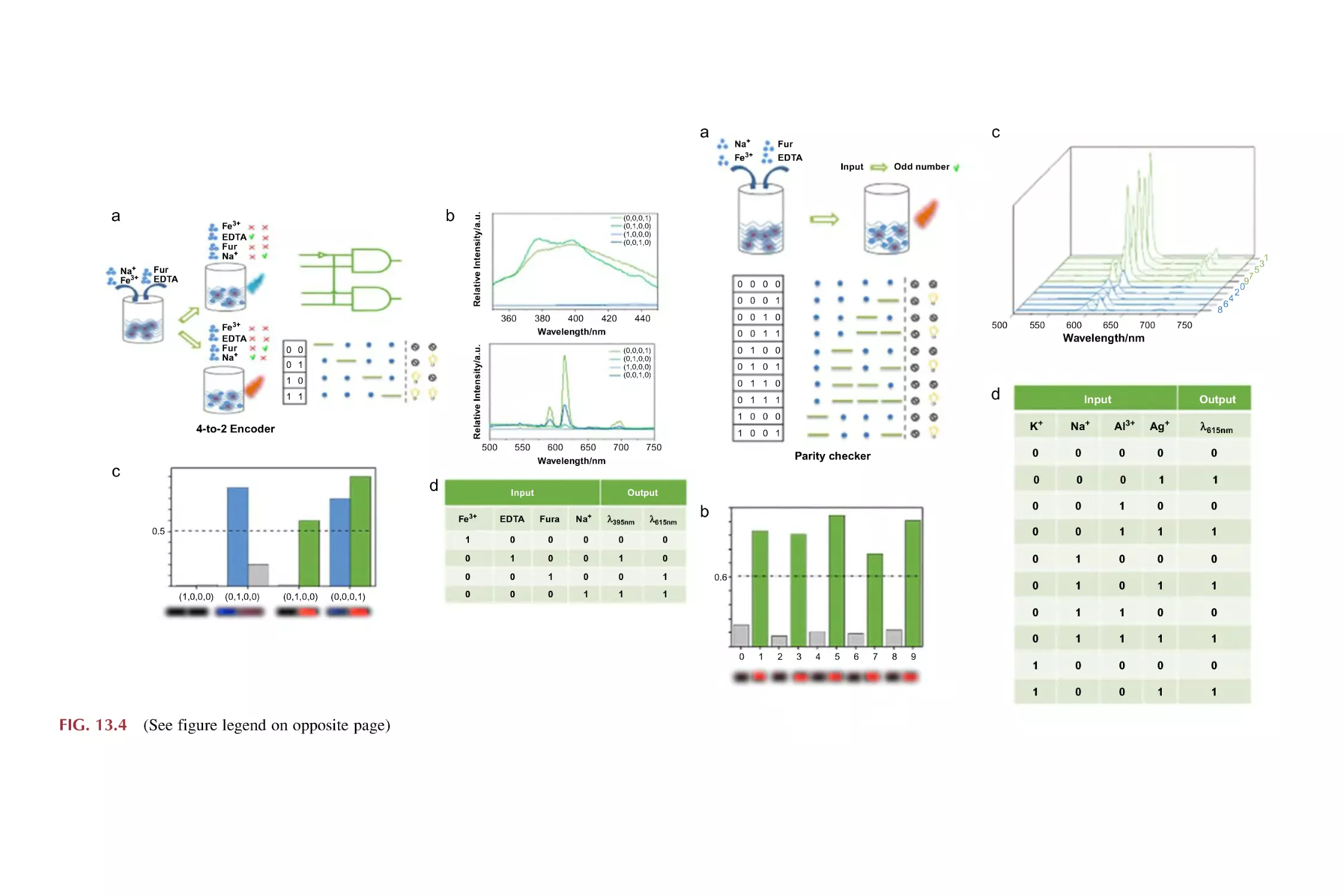
13.1.4 Implementation of logic devices (4-to-2 encoder
and parity checker)
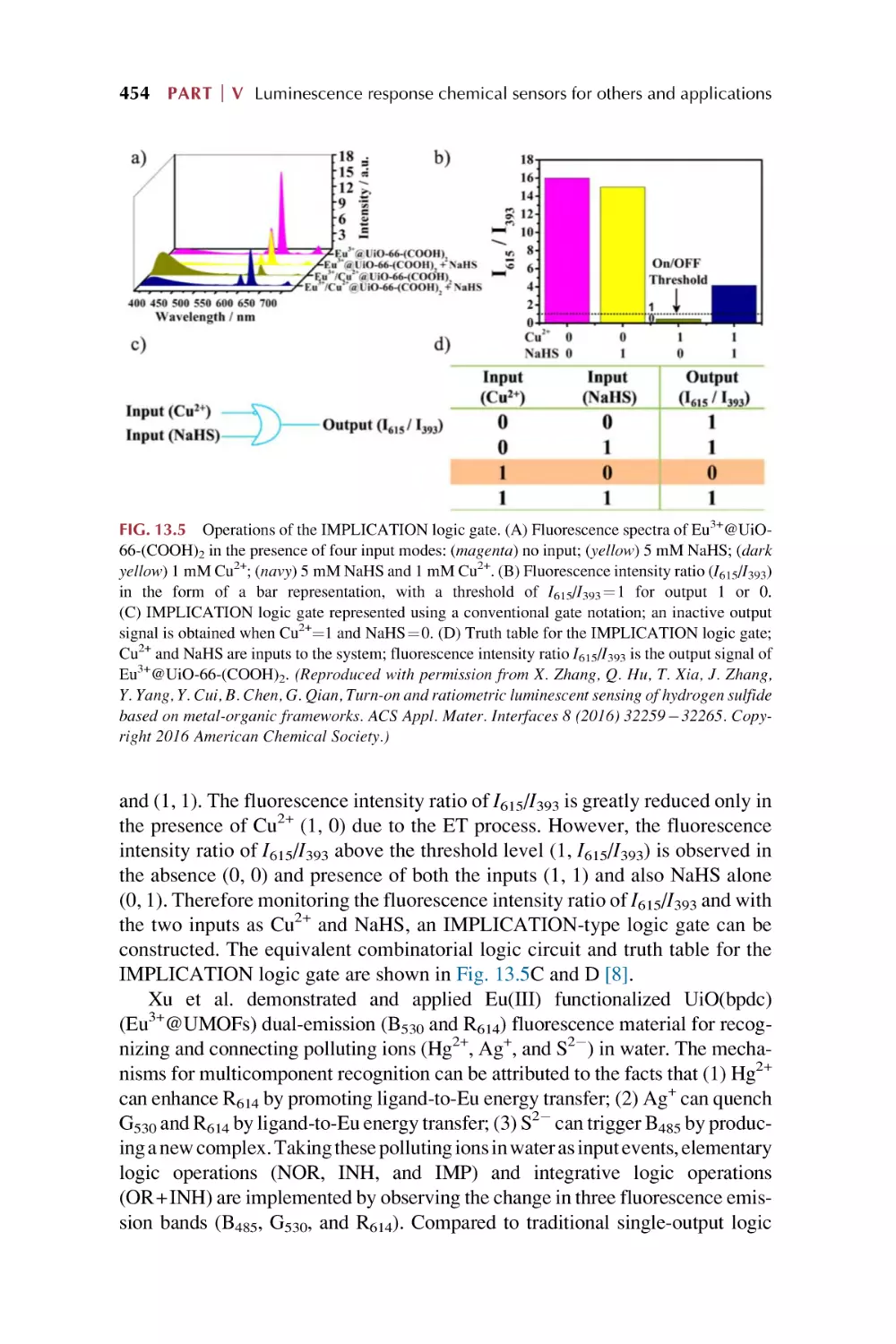
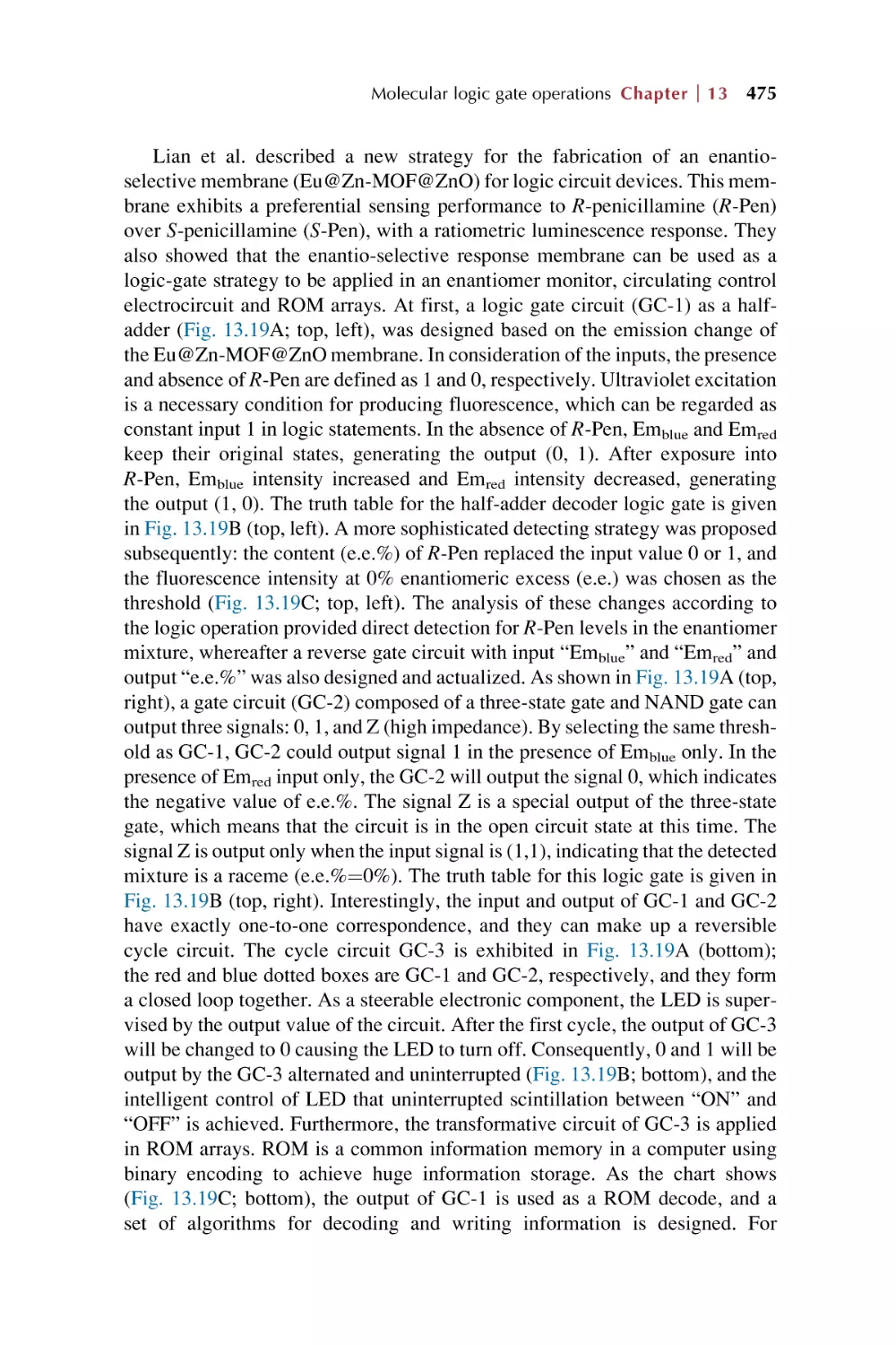
Luminescence responsive sensing of rare earth
metal-organic framework hybrid materials on luminescence
for Boolean logic gates
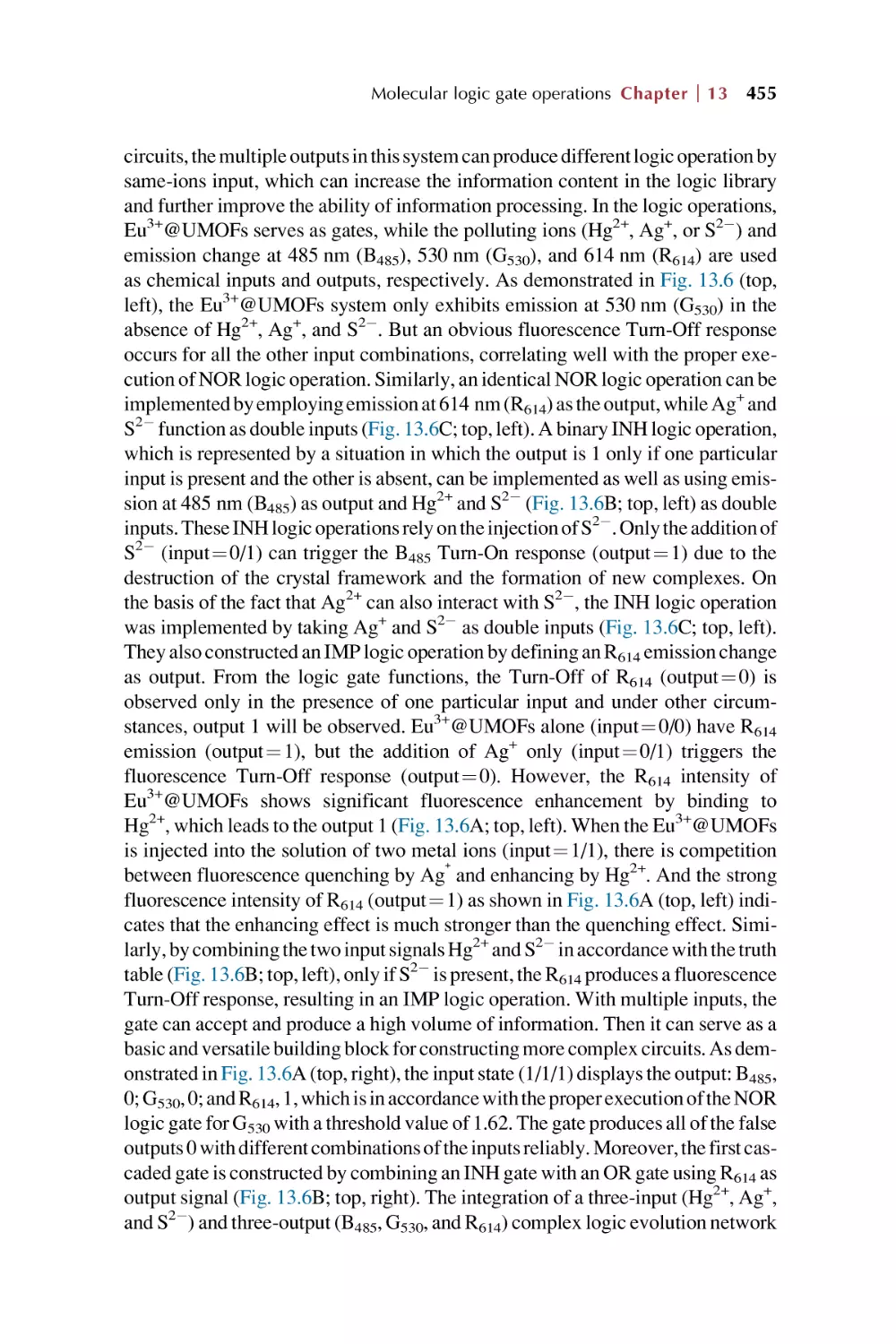
Luminescence responsive chemical sensing of rare earth
metal-organic framework hybrid materials for intelligent
molecular searcher applications
References
445
446
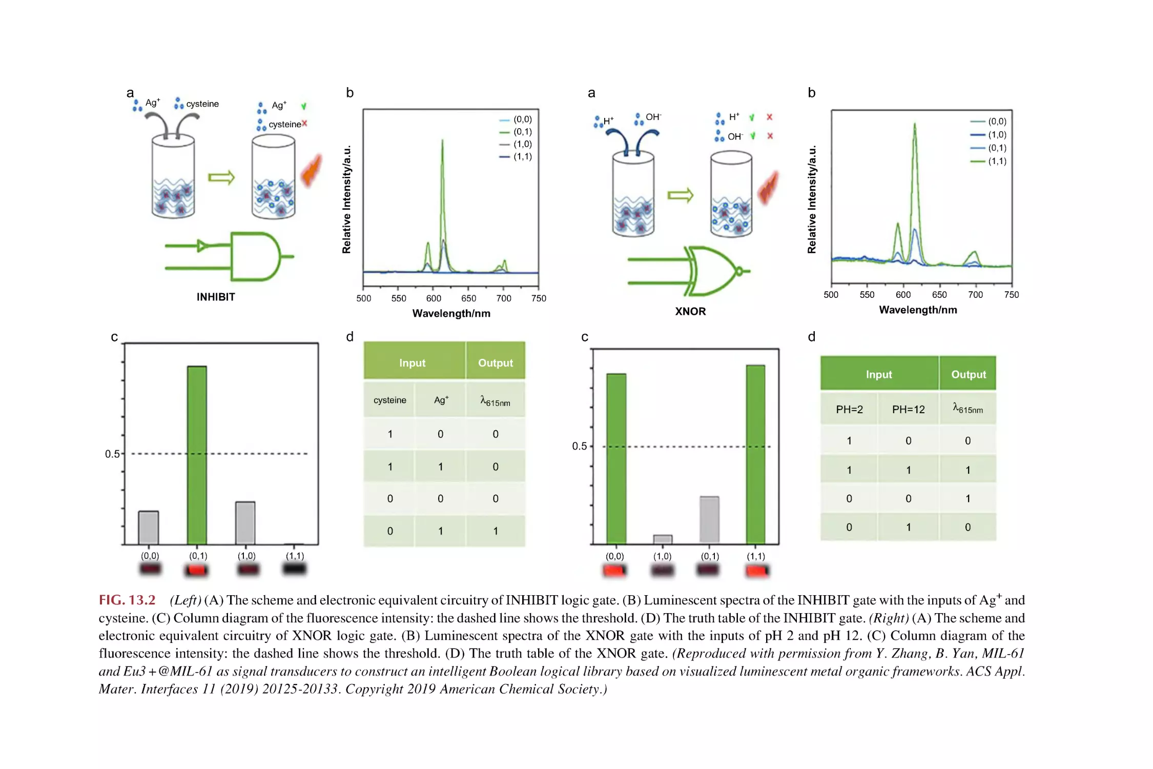
449
451
451
453
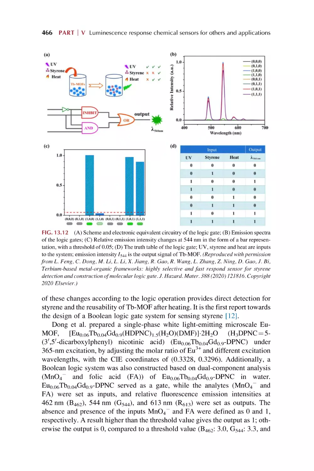
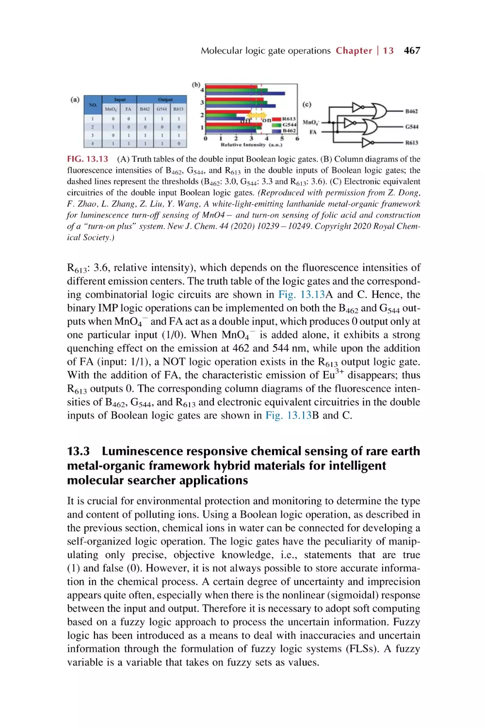
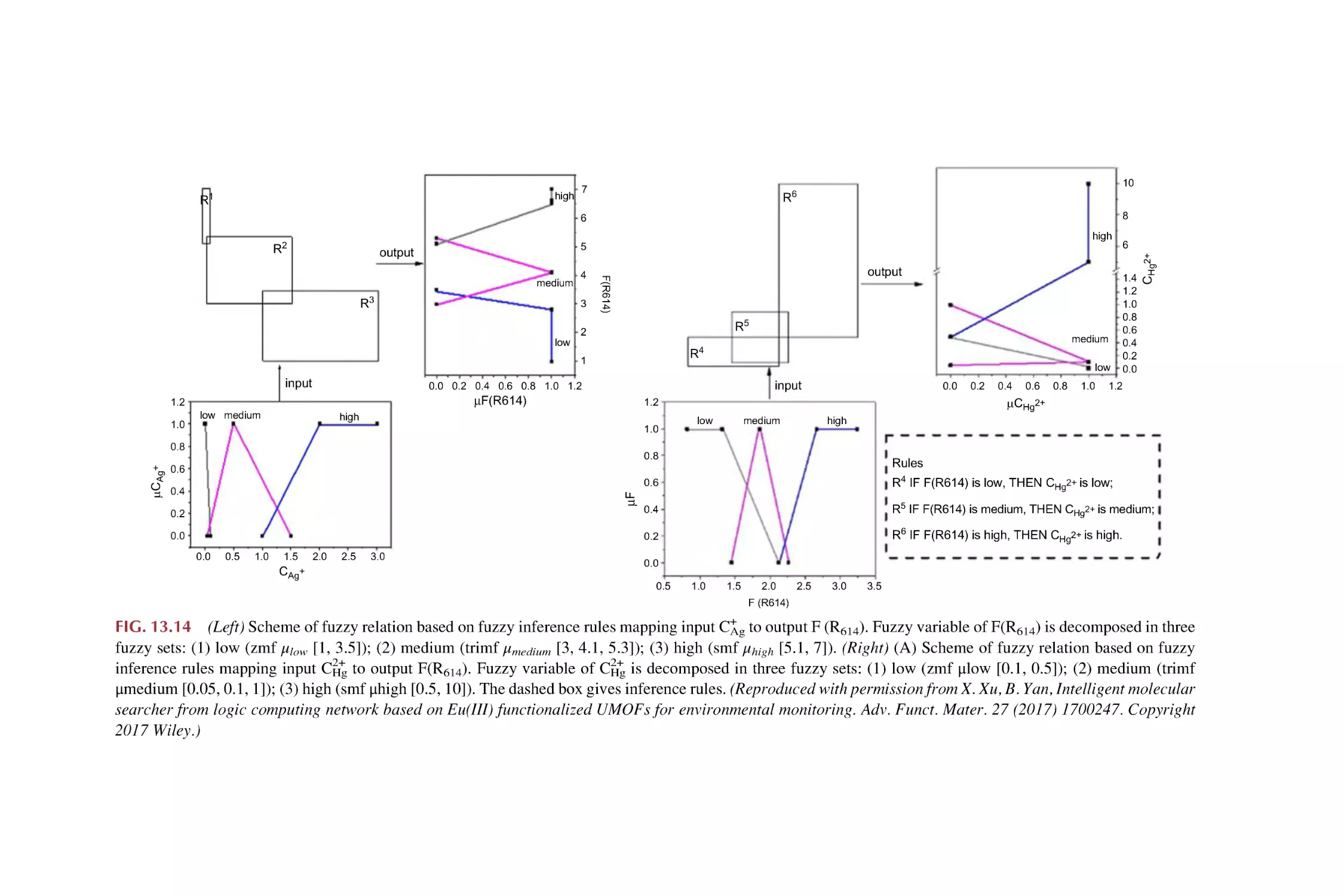
467
479
Contents
xi
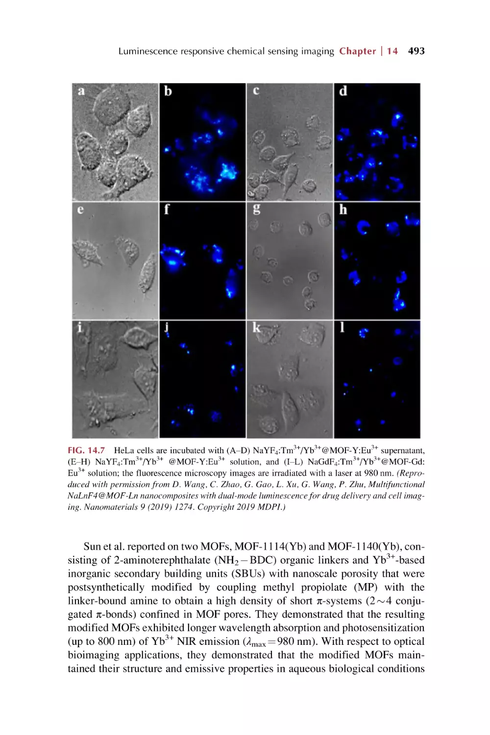
14. Rare earth metal-organic framework hybrid materials
for luminescence responsive chemical sensing
imaging
Part VI
Summary and prospect for rare earth metal-organic
frameworks hybrid materials as luminescence
response chemical sensors
15. Summary and prospects
15.1
15.2
15.3
15.4
Index
Postsynthetic modification to construct rare earth
metal-organic framework hybrid materials
Luminescence responsive mode and chemical sensing
mechanism for rare earth metal-organic framework hybrid
materials
Luminescence responsive chemical sensing of analytes
for rare earth metal-organic framework hybrid
materials
Luminescence responsive chemical sensing application
for rare earth metal-organic framework hybrid
materials
References
503
506
510
513
517
519
This page intentionally left blank
Preface
Rare earths (REs), as strategic resources of the 21st century, have played a great
role in both industry and the economy. Due to the unique electronic structure
and physiochemical properties of rare earth ions, their compounds are important
active candidates in functional materials. In particular, rare earth ions display
excellent optical behaviors, such as sharp emission spectra for high color purity,
broad emission bands covering the ultraviolet (UV)-visible-near-infrared (NIR)
region, a wide range of lifetimes from microseconds to the second level, high
luminescence quantum efficiencies, and so forth, which makes them a huge
treasury of luminescent materials. In recent years, REs have attracted much
attention for their wide variety of applications in the fields of lighting devices
(television and computer displays, optical fibers, optical amplifiers, and lasers)
and biomedical analysis (medical diagnosis and cell imaging).
Metal-organic frameworks (MOFs, also known as porous coordination polymers, or PCPs) are an emerging class of porous molecular materials constructed
from metal-containing nodes (also known as secondary building units (SBUs))
and organic linkers (bridging ligands). Due to their structural and functional
tunability, the area of MOFs has become one of the fastest growing fields in
chemistry. MOFs embody versatile functional applications in gas storage, purification, or separation; heterogeneous catalysis or photocatalysis; optic,
electronic, or magnetic materials or devices; as well as biomedicines or bioimages. Certainly, MOFs are employed as a platform for luminescent materials
based on their intrinsic optical and photonic properties of metal ions and organic
ligands, or guest species collaboratively assembled and/or encapsulated into
their frameworks. The abundant luminescent responsive performance of MOFs
provides their great potential in chemical sensing.
Rare earth metal-organic framework hybrid materials combine the virtues of
both MOF materials and rare earth ions, which can create novel properties as
well as functional and photofunctional applications. In particular, with rare
earth ion functionalized MOF hybrid materials, luminescent RE3+ ions are
incorporated into MOF hosts with little content, and the characteristic emission
of RE3+ is obtained. This is identical to the traditional rare earth ion doped phosphors. Just like pure luminescent rare earth MOF materials, RE3+ can produce
an “antenna effect” and cause a pronounced increase in the luminescence intensity through the intramolecular energy transfer process from linkers to RE3+. In
addition, the relatively limited content of hybrid materials often allows the
xiii
xiv Preface
existence of luminescence of original linkers or MOFs themselves if the functionalized amount of RE3+ is controlled appropriately. This make it possible to
exhibit multiple center luminescence for the same hybrid system and even realize luminescence color tuning or white luminescence integration. With regard
to integrity, the pure rare earth MOF materials are considered in this book. Thus
rare earth MOF hybrid materials encompass two main aspects: one is the pure
rare earth MOFs, and the other is rare earth functionalized MOF hybrid
materials.
This book consists of 6 parts, covered in 15 chapters. The first part (Chapters
1 and 2) is a general introduction to MOFs, rare earth MOFs, and rare earth functionalized MOF hybrid materials (Chapter 1); and rare earth luminescence,
MOF luminescence, rare earth MOF luminescence, as well as luminescence
response and chemical sensing (Chapter 2). The second part (Chapters 3, 4,
and 5) gives an overview of the luminescent response mode and sensing mechanisms in rare earth metal-organic framework hybrid materials: single luminescent mode sensing (1D) (Chapter 3), dual luminescent mode (2D) for
ratiometric sensing (Chapter 4), and luminescent responsive sensing mechanisms (Chapter 5). The third part (Chapters 6, 7, and 8) sheds light on rare earth
metal-organic framework hybrid materials as luminescence response chemical
sensors for typical ionic analytes, metal cations (I) (Chapter 6), (II) (Chapter 7),
and anions (Chapter 8). The fourth part (Chapters 9, 10, and 11) focuses on rare
earth metal-organic framework hybrid materials as luminescence response
chemical sensors for molecules: general molecular chemicals (Chapter 9), special organic molecules (Chapter 10), and biomarkers (Chapter 11). The fifth part
(Chapters 12, 13, and 14) involves rare earth metal-organic framework hybrid
materials as luminescence response chemical sensors for other applications:
including temperature and pH value (Chapter 12), logic gate operations
(Chapter 13), and imaging applications (Chapter 14). The sixth part
(Chapter 15) gives a summary and prospect for rare earth MOF hybrid materials
as luminescence response chemical sensors.
Finally, I want to express my sincere gratitude to my PhD and master’s students, whose research work makes up the main content of this book. I also wish
to show my appreciation to my colleagues, especially to the scholars in the
research fields of rare earth metal-organic frameworks, which is an important
component of this book. Many colleague scholars have provided valuable
reviews of the relevant topics for the instruction and outline of this book. I hope
that this book will provide readers with insights into the recent developments of
rare earth metal-organic framework hybrid materials for luminescent responsive chemical sensing.
Bing Yan
Part I
Introduction for rare earth
metal-organic frameworks
hybrid materials
This page intentionally left blank
Chapter 1
Metal-organic frameworks
(MOFs), rare earth MOFs, and
rare earth functionalized MOF
hybrid materials
1.1
Metal-organic frameworks (MOFs)
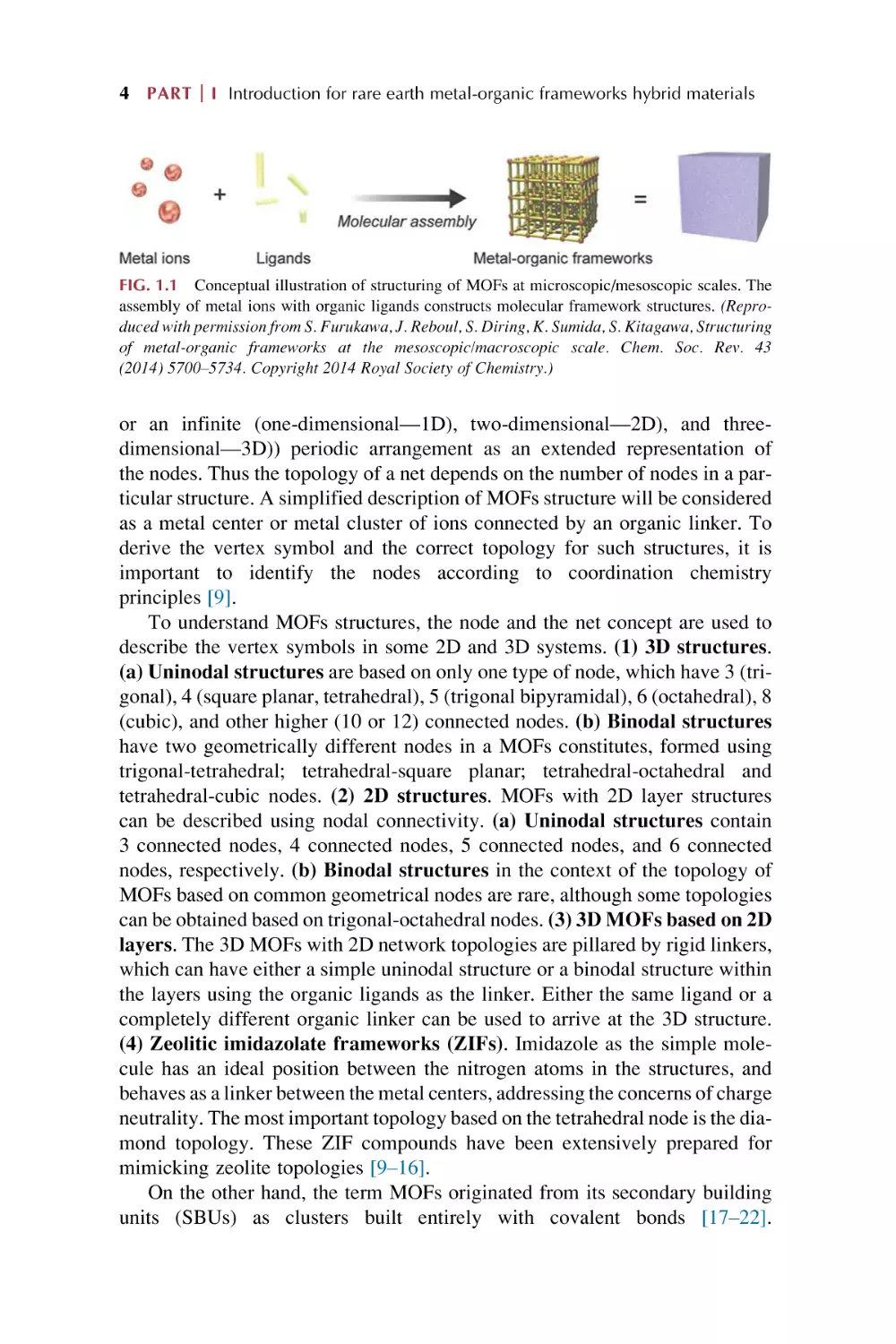
Metal-organic frameworks (abbreviated as MOFs and also known as porous
coordination polymers (PCPs)) are a class of porous polymeric molecular materials, consisting of metal ion nodes connected together by organic bridging
ligands (linkers) (Scheme in Fig. 1.1), which are a new development in the interdisciplinary field of coordination chemistry and functional materials [1–3].
Due to their structural and functional tunability, MOFs have become one of
the fastest growing research fields in inorganic chemistry. The essence of MOFs
chemistry is that the frameworks are assembled by linking molecular units of
well-defined shapes by chemical bonds into periodic frameworks. An important
component of reticular chemistry is the deconstruction of such structures into
their underlying nets to facilitate designed synthesis of materials with targeted
porosity, pore size, and functionality. The organic ligands of MOFs give them
flexibility and diversity in their chemical structures and functions. The synthesis
of MOFs has attracted extensive attention due to the possibility of obtaining a
large variety of interesting structures for a range of applications related to
porous materials [4–8]. The exploration of MOFs mainly involves four categories: (1) synthesis of metal-containing nodes or coordination bonds and linker
design for MOFs; (2) postsynthetic modification (PSM) of MOFs; (3) MOF
hybrids or composites; and (4) potential applications of MOFs.
1.1.1 Synthesis of metal-containing nodes or coordination bonds
and linker design for MOFs
In MOFs structures, a node represents a particular environment (tetrahedra,
octahedra, etc.) connected to a fixed number of related points, which depends
on the geometry (tetrahedral ¼ 4, octahedral ¼ 6, cubic ¼ 8). Their structures can
then be represented mathematically as either a discrete (zero-dimensional—0D)
Rare Earth Metal-Organic Framework Hybrid Materials for Luminescence Responsive Chemical Sensors
https://doi.org/10.1016/B978-0-323-91236-5.00003-7
Copyright © 2022 Elsevier Ltd. All rights reserved.
3
4 PART
I Introduction for rare earth metal-organic frameworks hybrid materials
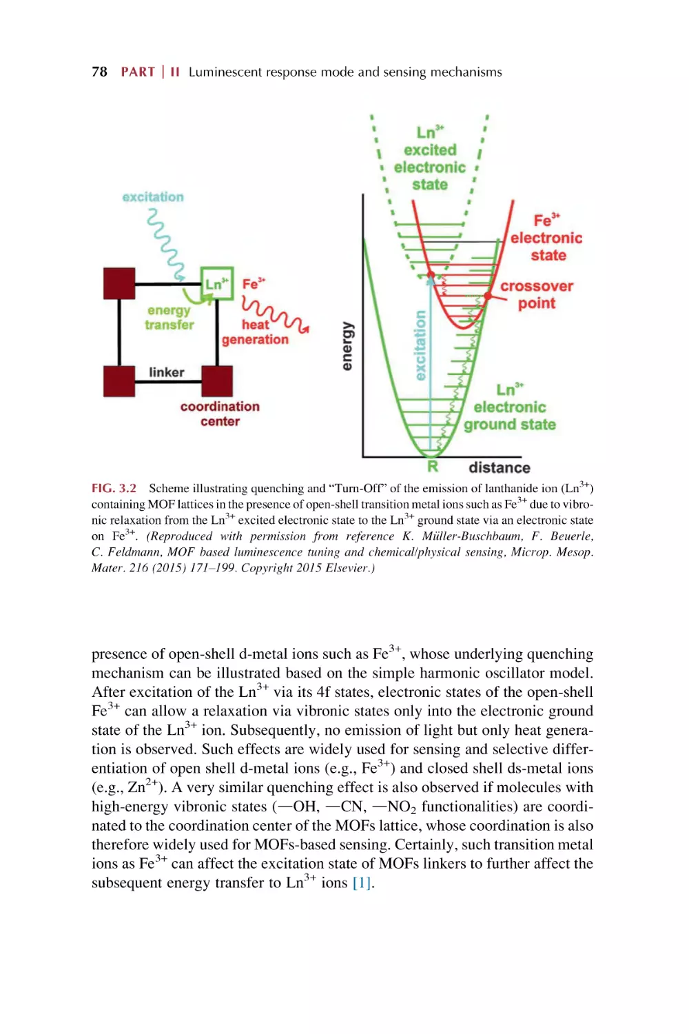
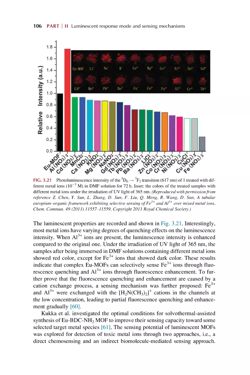
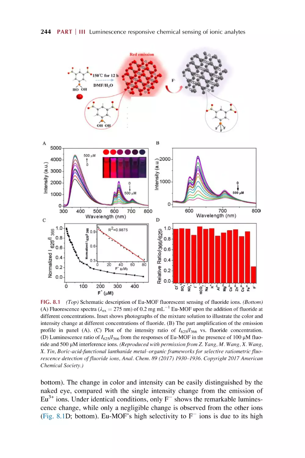
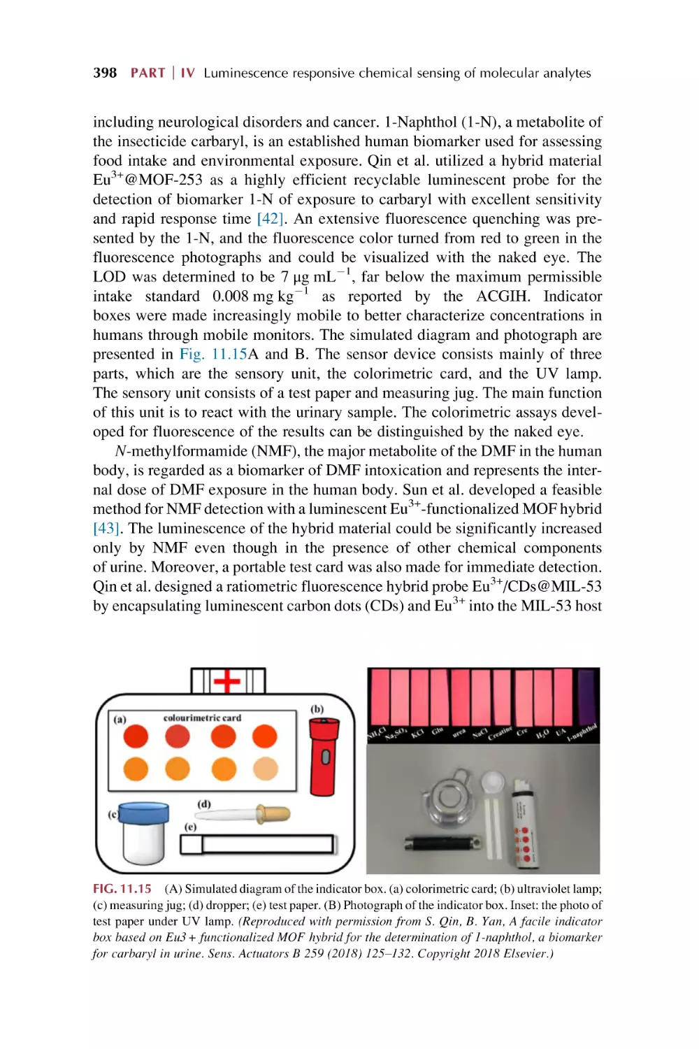
FIG. 1.1 Conceptual illustration of structuring of MOFs at microscopic/mesoscopic scales. The
assembly of metal ions with organic ligands constructs molecular framework structures. (Reproduced with permission from S. Furukawa, J. Reboul, S. Diring, K. Sumida, S. Kitagawa, Structuring
of metal-organic frameworks at the mesoscopic/macroscopic scale. Chem. Soc. Rev. 43
(2014) 5700–5734. Copyright 2014 Royal Society of Chemistry.)
or an infinite (one-dimensional—1D), two-dimensional—2D), and threedimensional—3D)) periodic arrangement as an extended representation of
the nodes. Thus the topology of a net depends on the number of nodes in a particular structure. A simplified description of MOFs structure will be considered
as a metal center or metal cluster of ions connected by an organic linker. To
derive the vertex symbol and the correct topology for such structures, it is
important to identify the nodes according to coordination chemistry
principles [9].
To understand MOFs structures, the node and the net concept are used to
describe the vertex symbols in some 2D and 3D systems. (1) 3D structures.
(a) Uninodal structures are based on only one type of node, which have 3 (trigonal), 4 (square planar, tetrahedral), 5 (trigonal bipyramidal), 6 (octahedral), 8
(cubic), and other higher (10 or 12) connected nodes. (b) Binodal structures
have two geometrically different nodes in a MOFs constitutes, formed using
trigonal-tetrahedral; tetrahedral-square planar; tetrahedral-octahedral and
tetrahedral-cubic nodes. (2) 2D structures. MOFs with 2D layer structures
can be described using nodal connectivity. (a) Uninodal structures contain
3 connected nodes, 4 connected nodes, 5 connected nodes, and 6 connected
nodes, respectively. (b) Binodal structures in the context of the topology of
MOFs based on common geometrical nodes are rare, although some topologies
can be obtained based on trigonal-octahedral nodes. (3) 3D MOFs based on 2D
layers. The 3D MOFs with 2D network topologies are pillared by rigid linkers,
which can have either a simple uninodal structure or a binodal structure within
the layers using the organic ligands as the linker. Either the same ligand or a
completely different organic linker can be used to arrive at the 3D structure.
(4) Zeolitic imidazolate frameworks (ZIFs). Imidazole as the simple molecule has an ideal position between the nitrogen atoms in the structures, and
behaves as a linker between the metal centers, addressing the concerns of charge
neutrality. The most important topology based on the tetrahedral node is the diamond topology. These ZIF compounds have been extensively prepared for
mimicking zeolite topologies [9–16].
On the other hand, the term MOFs originated from its secondary building
units (SBUs) as clusters built entirely with covalent bonds [17–22].
MOF hybrid materials Chapter
1
5
The synthesis of SBUs can be used to direct the assembly of ordered frameworks with rigid organic linkers, which makes it highly possible to predict
the chemistry of the yielded crystalline materials [17–30]. The orientation of
organic linkers will result in the assembly of MOFs with predetermined structural topologies [23–30]. Generally, both SBUs (as connectors) and organic
ligands (as linkers) combine to determine the final framework topology. (1)
Ditopic carboxylate linkers. These linkers possess both ready accessibility
and easily perceivable structures in combination with different SBUs: 4connected paddlewheel clusters, 6-connected octahedral clusters, 6-connected
trigonal-prismatic clusters, 12-connected clusters, and infinite chain clusters,
respectively. (2) Tritopic carboxylate linkers. These linkers are related to different clusters: 4-connected paddlewheel clusters, 6-connected octahedral clusters, 6-connected trigonal-prismatic clusters, and multiple SBUs. (3)
Tetratopic carboxylate linkers. These linkers appear to be very intriguing
building units in MOFs constructions, especially those with tetrahedral geometry. Tetrahedral carboxylate linkers are related to different clusters: 8-connected
cubical clusters, 4-connected square planar clusters, 8-connected hexagonal bipyramidal clusters, and nonregular tetrahedral carboxylate linkers. (4) Hexatopic
carboxylate linkers. These linkers are related to 1,3-atedbenzenedicarboxylate
units, 4,40 -azanediyldibenzoate units and 1,10 :30 ,100 -terphenyl-4,400 -dicarboxylate
units. (5) Octatopic carboxylate linkers. MOFs with these linkers are still rare,
possibly due to the synthetic challenges in the linkers themselves, whose frameworks are based on linkers with long arms that tend to form interpenetrated structures. (6) Mixed linkers. They contain four types: ditopic-ditopic linear linkers,
tritopic carboxylate-ditopic carboxylate linkers, carboxylate-pyridine linkers, and
linkers coordinatively identical but with distinct shapes. (7) Desymmetrized
linkers. (8) Metallo-linkers. These mainly involve four types with different
donors: O and S donors, N and P donors, C donors, and mixed donor groups.
(9) N-heterocyclic linkers. Organic linkers containing N donors, such as pyridine
and azole derivatives, have achieved stable MOFs via N-metal coordination,
including ditopic N-heterocyclic linkers and polytopic N-heterocyclic
linkers [23].
1.1.2
Postsynthetic modification (PSM) of MOFs
Postsynthetic modifications (PSMs) are particularly attractive for use with
MOFs materials for a variety of reasons. (I) The solvothermal reaction conditions to prepare most MOFs greatly limit the types of functional groups that can
be functionalized by PSM. (II) The organic ligands in MOFs open the possibility of employing a wide range of organic transformations. (III) MOFs’ porous
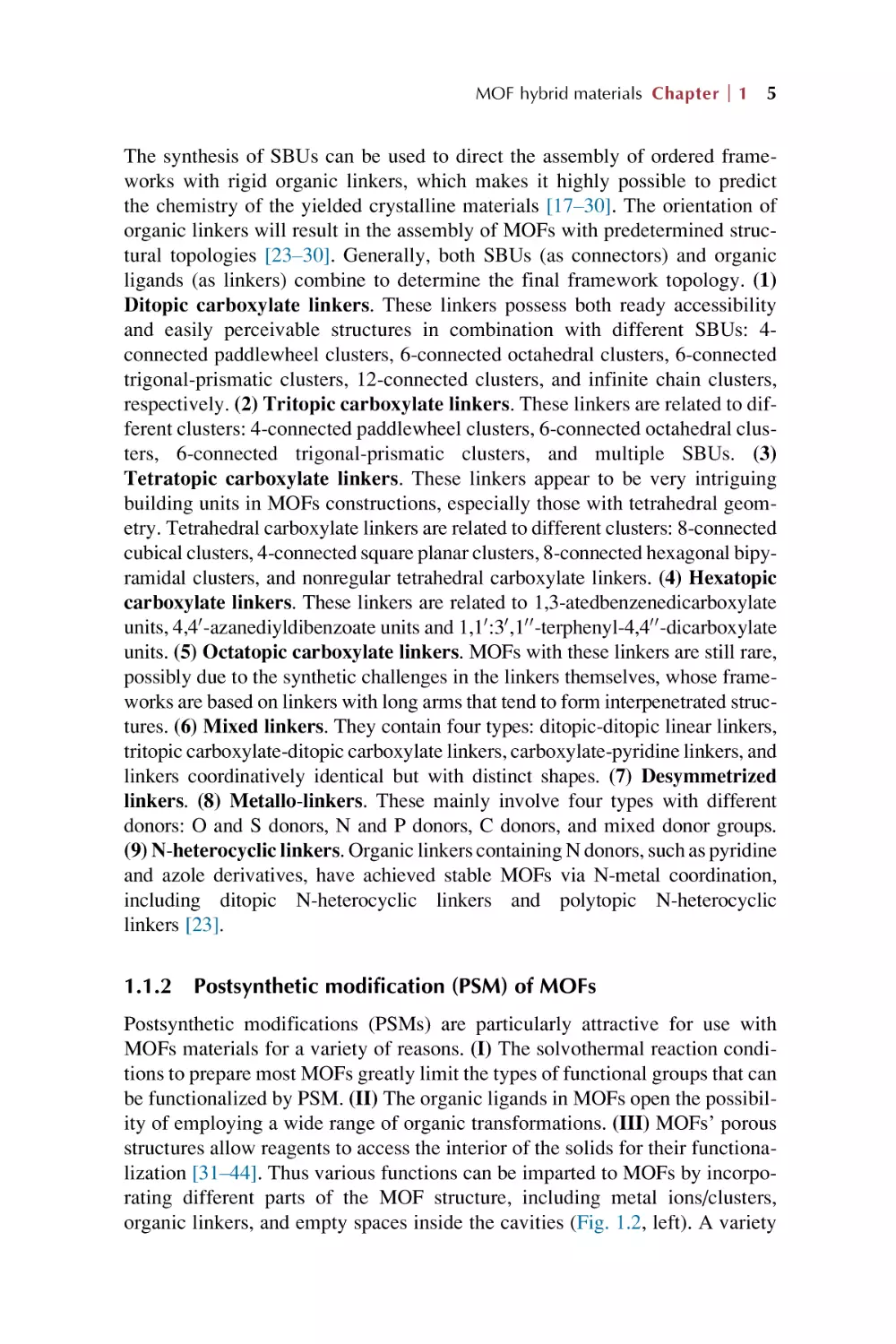
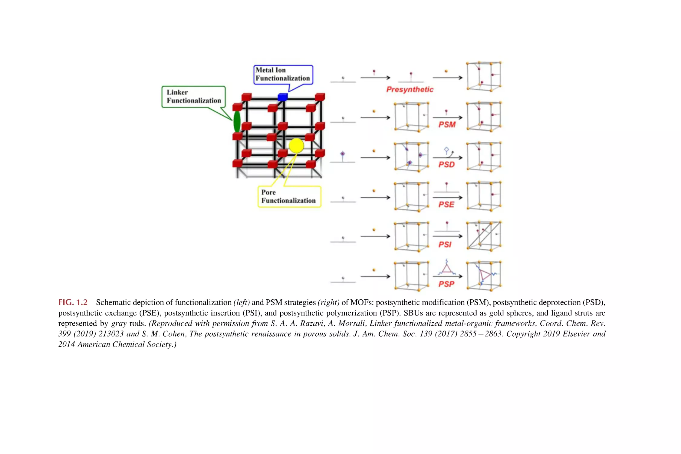
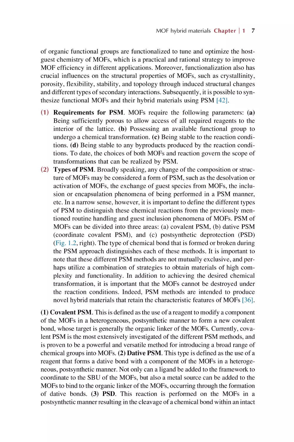
structures allow reagents to access the interior of the solids for their functionalization [31–44]. Thus various functions can be imparted to MOFs by incorporating different parts of the MOF structure, including metal ions/clusters,
organic linkers, and empty spaces inside the cavities (Fig. 1.2, left). A variety
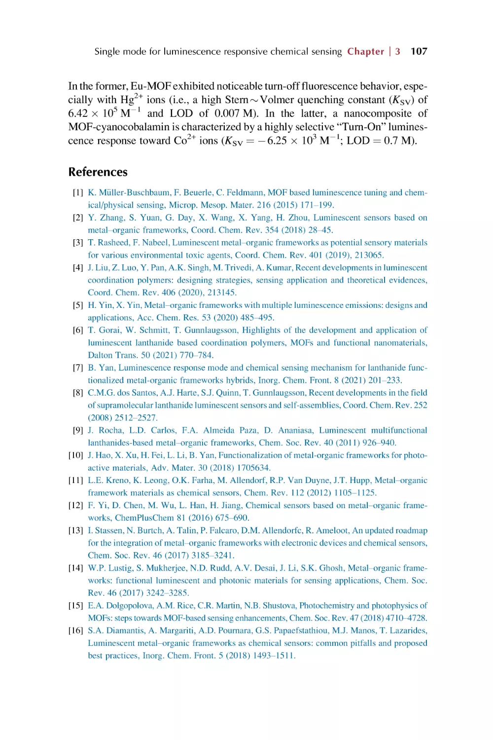
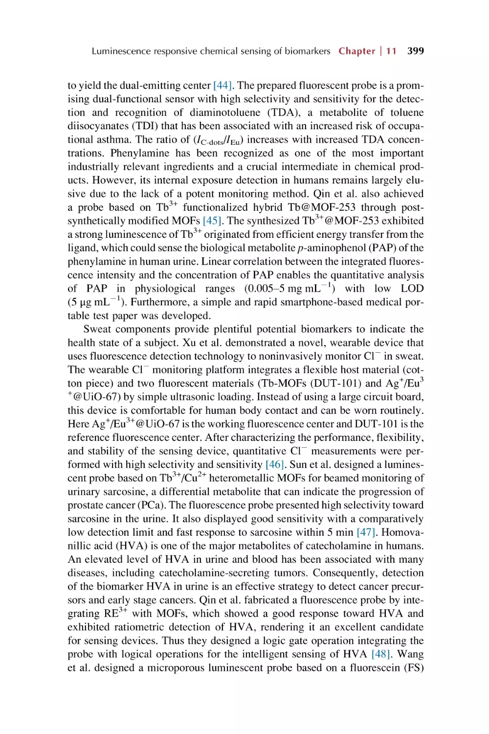
FIG. 1.2 Schematic depiction of functionalization (left) and PSM strategies (right) of MOFs: postsynthetic modification (PSM), postsynthetic deprotection (PSD),
postsynthetic exchange (PSE), postsynthetic insertion (PSI), and postsynthetic polymerization (PSP). SBUs are represented as gold spheres, and ligand struts are
represented by gray rods. (Reproduced with permission from S. A. A. Razavi, A. Morsali, Linker functionalized metal-organic frameworks. Coord. Chem. Rev.
399 (2019) 213023 and S. M. Cohen, The postsynthetic renaissance in porous solids. J. Am. Chem. Soc. 139 (2017) 2855 2863. Copyright 2019 Elsevier and
2014 American Chemical Society.)
MOF hybrid materials Chapter
1
7
of organic functional groups are functionalized to tune and optimize the hostguest chemistry of MOFs, which is a practical and rational strategy to improve
MOF efficiency in different applications. Moreover, functionalization also has
crucial influences on the structural properties of MOFs, such as crystallinity,
porosity, flexibility, stability, and topology through induced structural changes
and different types of secondary interactions. Subsequently, it is possible to synthesize functional MOFs and their hybrid materials using PSM [42].
(1) Requirements for PSM. MOFs require the following parameters: (a)
Being sufficiently porous to allow access of all required reagents to the
interior of the lattice. (b) Possessing an available functional group to
undergo a chemical transformation. (c) Being stable to the reaction conditions. (d) Being stable to any byproducts produced by the reaction conditions. To date, the choices of both MOFs and reaction govern the scope of
transformations that can be realized by PSM.
(2) Types of PSM. Broadly speaking, any change of the composition or structure of MOFs may be considered a form of PSM, such as the desolvation or
activation of MOFs, the exchange of guest species from MOFs, the inclusion or encapsulation phenomena of being performed in a PSM manner,
etc. In a narrow sense, however, it is important to define the different types
of PSM to distinguish these chemical reactions from the previously mentioned routine handling and guest inclusion phenomena of MOFs. PSM of
MOFs can be divided into three areas: (a) covalent PSM, (b) dative PSM
(coordinate covalent PSM), and (c) postsynthetic deprotection (PSD)
(Fig. 1.2, right). The type of chemical bond that is formed or broken during
the PSM approach distinguishes each of these methods. It is important to
note that these different PSM methods are not mutually exclusive, and perhaps utilize a combination of strategies to obtain materials of high complexity and functionality. In addition to achieving the desired chemical
transformation, it is important that the MOFs cannot be destroyed under
the reaction conditions. Indeed, PSM methods are intended to produce
novel hybrid materials that retain the characteristic features of MOFs [36].
(1) Covalent PSM. This is defined as the use of a reagent to modify a component
of the MOFs in a heterogeneous, postsynthetic manner to form a new covalent
bond, whose target is generally the organic linker of the MOFs. Currently, covalent PSM is the most extensively investigated of the different PSM methods, and
is proven to be a powerful and versatile method for introducing a broad range of
chemical groups into MOFs. (2) Dative PSM. This type is defined as the use of a
reagent that forms a dative bond with a component of the MOFs in a heterogeneous, postsynthetic manner. Not only can a ligand be added to the framework to
coordinate to the SBU of the MOFs, but also a metal source can be added to the
MOFs to bind to the organic linker of the MOFs, occurring through the formation
of dative bonds. (3) PSD. This reaction is performed on the MOFs in a
postsynthetic manner resulting in the cleavage of a chemical bond within an intact
8 PART
I Introduction for rare earth metal-organic frameworks hybrid materials
framework. In principle, any kind of chemical bond can be broken during a PSD
reaction to reveal a chemical functionality and produce materials with different
properties. Within some highly stable MOFs, both the elimination and addition of
multitopic linkers or metal ions are possible without destruction of the framework. (4) Postsynthetic metal exchange (PSME). Cation doping is also widely
employed in nanocrystals to tune their properties. (5) Postsynthetic ligand
exchange (PSLE). This represents the exchange of the key extending ligand
of a framework by another similar ligand of different length or functional group,
with the retention of the MOFs topology. (6) Postsynthetic elimination and
installation (PSE&I). Some linkers constituting the framework can be eliminated with coordination changes in the metal cluster but preservation of the infinite framework connection. If the coordination site of the adjacent metal cluster
matches well with an additional linker, PSI may succeed in creating a new MOFs
with higher connection numbers. (7) Tandem PSM. This type is used in producing MOFs with multiple functionalities otherwise difficult or infeasible to acquire
by direct synthetic methods. (i) Engineering porosity and pores by tandem
PSM. Both increased and decreased porosity can be obtained depending on
the size and spatial configuration of the modified groups, whose change in porosity is moderate and limited by the constant topology of the framework. (ii)
Improving structural stability by tandem PSM. The direct introduction of target metal ions may be subjected to low exchange rate, uncompleted conversion,
decomposition of the MOFs, and others. Based on the exchanging mechanism of
different metal ions, tandem PSME is useful for improving structural stability.
(iii) Modifying surface and interior by tandem PSM. Based on the reactivity
and spatial effect within the confined channels, the traditional design of the reaction pathways provides a chance to engineer either the surface or the interior of
MOFs (Fig. 1.2, right) [31–44].
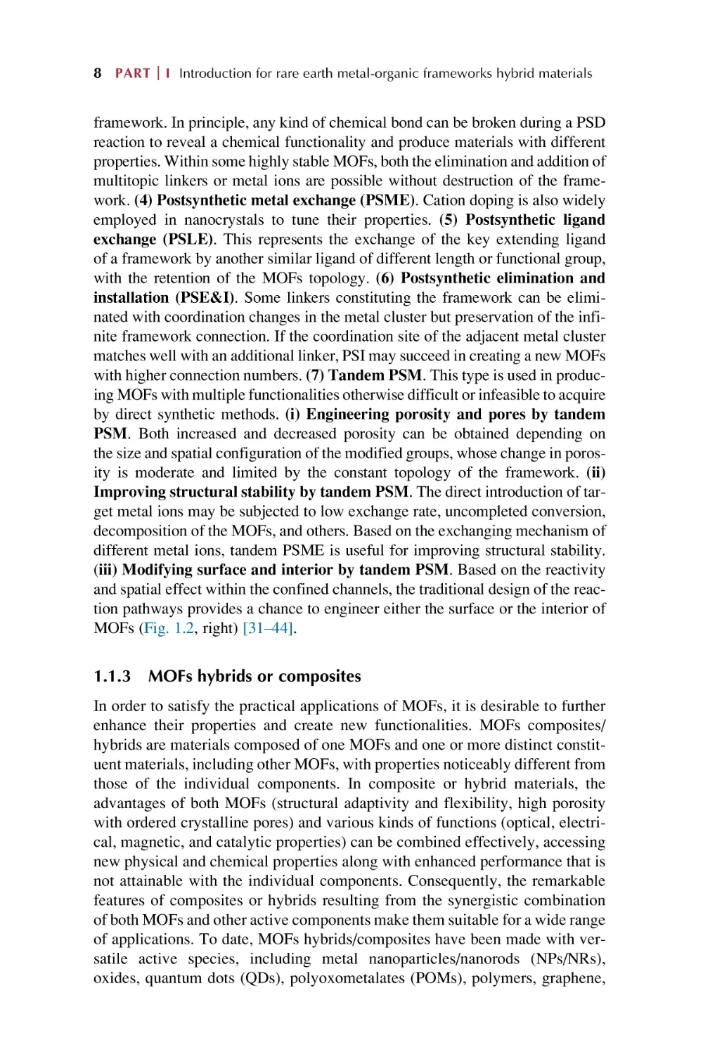
1.1.3 MOFs hybrids or composites
In order to satisfy the practical applications of MOFs, it is desirable to further
enhance their properties and create new functionalities. MOFs composites/
hybrids are materials composed of one MOFs and one or more distinct constituent materials, including other MOFs, with properties noticeably different from
those of the individual components. In composite or hybrid materials, the
advantages of both MOFs (structural adaptivity and flexibility, high porosity
with ordered crystalline pores) and various kinds of functions (optical, electrical, magnetic, and catalytic properties) can be combined effectively, accessing
new physical and chemical properties along with enhanced performance that is
not attainable with the individual components. Consequently, the remarkable
features of composites or hybrids resulting from the synergistic combination
of both MOFs and other active components make them suitable for a wide range
of applications. To date, MOFs hybrids/composites have been made with versatile active species, including metal nanoparticles/nanorods (NPs/NRs),
oxides, quantum dots (QDs), polyoxometalates (POMs), polymers, graphene,
MOF hybrid materials Chapter
1
9
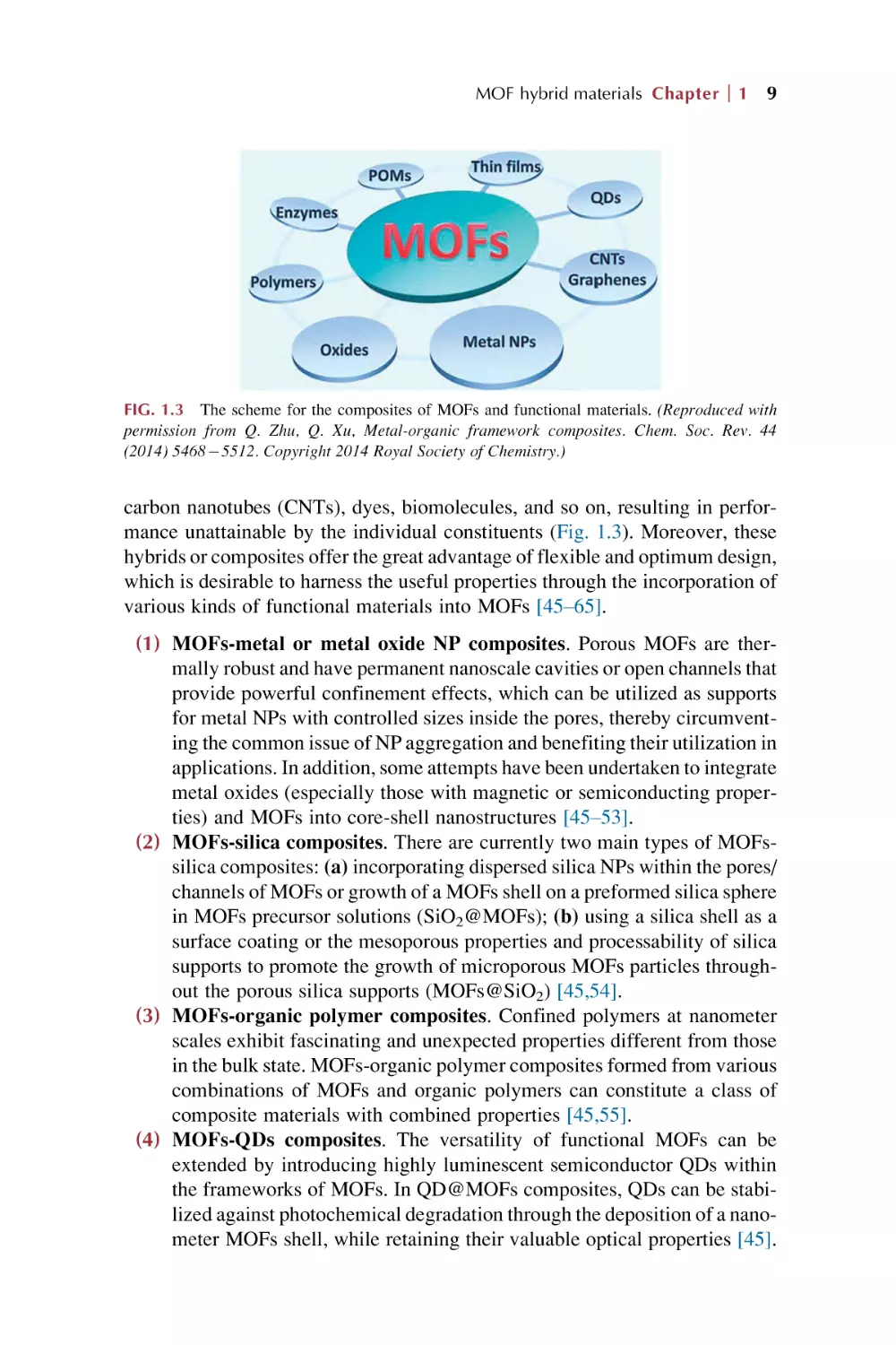
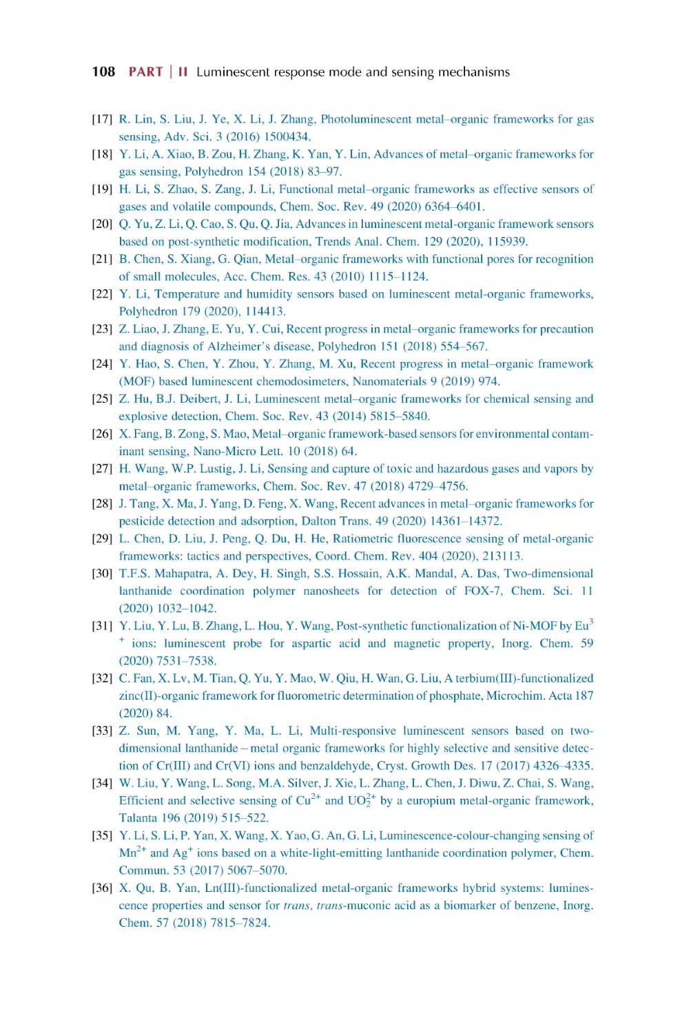
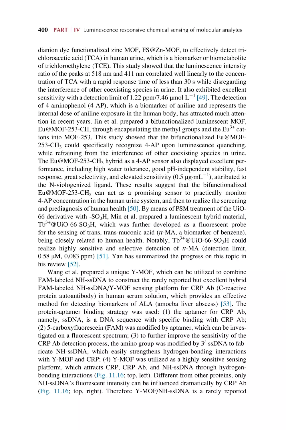
FIG. 1.3 The scheme for the composites of MOFs and functional materials. (Reproduced with
permission from Q. Zhu, Q. Xu, Metal-organic framework composites. Chem. Soc. Rev. 44
(2014) 54685512. Copyright 2014 Royal Society of Chemistry.)
carbon nanotubes (CNTs), dyes, biomolecules, and so on, resulting in performance unattainable by the individual constituents (Fig. 1.3). Moreover, these
hybrids or composites offer the great advantage of flexible and optimum design,
which is desirable to harness the useful properties through the incorporation of
various kinds of functional materials into MOFs [45–65].
(1) MOFs-metal or metal oxide NP composites. Porous MOFs are thermally robust and have permanent nanoscale cavities or open channels that
provide powerful confinement effects, which can be utilized as supports
for metal NPs with controlled sizes inside the pores, thereby circumventing the common issue of NP aggregation and benefiting their utilization in
applications. In addition, some attempts have been undertaken to integrate
metal oxides (especially those with magnetic or semiconducting properties) and MOFs into core-shell nanostructures [45–53].
(2) MOFs-silica composites. There are currently two main types of MOFssilica composites: (a) incorporating dispersed silica NPs within the pores/
channels of MOFs or growth of a MOFs shell on a preformed silica sphere
in MOFs precursor solutions (SiO2@MOFs); (b) using a silica shell as a
surface coating or the mesoporous properties and processability of silica
supports to promote the growth of microporous MOFs particles throughout the porous silica supports (MOFs@SiO2) [45,54].
(3) MOFs-organic polymer composites. Confined polymers at nanometer
scales exhibit fascinating and unexpected properties different from those
in the bulk state. MOFs-organic polymer composites formed from various
combinations of MOFs and organic polymers can constitute a class of
composite materials with combined properties [45,55].
(4) MOFs-QDs composites. The versatility of functional MOFs can be
extended by introducing highly luminescent semiconductor QDs within
the frameworks of MOFs. In QD@MOFs composites, QDs can be stabilized against photochemical degradation through the deposition of a nanometer MOFs shell, while retaining their valuable optical properties [45].
10 PART
I Introduction for rare earth metal-organic frameworks hybrid materials
(5) MOFs-POM composites. The dispersion of POMs within MOFs prevents the POMs from conglomerating and deactivating. In such POMbased MOFs, the organic ligands substitute for the oxo groups of POMs
to covalently link the metallic centers. What’s more, POMs can be encapsulated in the pores of MOFs through host-guest interactions to form
POM@MOFs composites [45].
(6) MOFs-carbon composites. The exceptionally mechanical, electrical,
and thermal properties of carbon materials (graphene and CNTs) commend them as valuable nanostructured fillers in MOF composites. Numerous MOFs-nanocarbon composites have been made with activated
carbons, carbon monoliths, graphene oxide (GO), and CNTs, and have
been intensively explored for diverse applications [45].
(7) MOFs thin films on substrates. The deposition of patterned thin films of
MOFs on a substrate has paved the way for the nanotechnological applications of MOFs-based devices. Generally, two fabrication methods have
been distinguished for the direct growth/deposition of MOFs thin films:
(a) The substrate is added to a MOFs synthesis solution under ambient
or solvothermal conditions, growing on the surface of the substrate and
sometimes in solution at the same time. This growth leads to the formation
of polycrystalline films where crystals are attached to the substrate surface in an intergrown and continuous fashion. (b) The layer-by-layer
(LBL) method was developed for the facile preparation of MOFs thin
films on the substrates and referred to as liquid phase epitaxy. This technique relies on the sequential deposition of monolayers of metal salts and
organic linkers on a functionalized substrate. The LBL method permits
the growth of smooth and homogeneous MOFs ultrathin films with diameters in the nanometer range, which achieve good control over the thickness, crystallographic orientation, and interpenetration of the MOFs
multilayers [45,56–59].
(8) MOFs@MOFs core-shell heterostructures. The construction of multifunctional core-shell heterostructures involves two strategies. (a) Heteroepitaxial growth of a shell MOFs crystal on the external surface of another
seed MOFs crystal could generate a composite crystal, in which the two
coordination components are segregated into different regions of the crystal. This approach is based on a close crystal lattice match between the
underlying MOFs substrate and the deposited MOFs. (b) PSM strategies
include the selective reaction of the reactive residue of an organic linker
and the controlled replacement of the framework metal ions or ligands,
whose modification is selectively constrained to either the external surface or the internal core of the MOFs crystals [45].
(9) MOFs-enzyme composites. The tunable but uniform pore sizes and functionalizable pore walls of porous MOFs may make them appealing to
accommodate enzymes for catalytic applications. Nevertheless, the
micropore size of most MOFs precludes the entry of large-sized enzymes
MOF hybrid materials Chapter
1
11
and can result in only external surface attachment with low enzyme loading via adsorption and/or covalent bonding reaction [60–65].
(10) MOFs-other molecular species composites. Molecular materials such as
organic dyes, organometallic compounds, metalloporphyrins, biomolecules,
and other functional molecules can also be composed with MOFs for various
applications. The use of MOFs as molecular encapsulators takes advantage
of their powerful confinement effect to protect molecules from aggregation,
heterogeneous distribution, and leaching. Impregnation procedures are
mostly used to encapsulate these molecular materials. In addition, the
self-assembly of MOFs in the presence of molecular moieties in the MOFs
precursor solutions can lead to irreversible in-situ encapsulation [45].
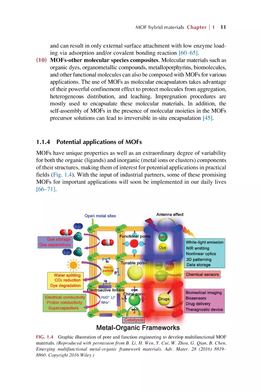
1.1.4
Potential applications of MOFs
MOFs have unique properties as well as an extraordinary degree of variability
for both the organic (ligands) and inorganic (metal ions or clusters) components
of their structures, making them of interest for potential applications in practical
fields (Fig. 1.4). With the input of industrial partners, some of these promising
MOFs for important applications will soon be implemented in our daily lives
[66–71].
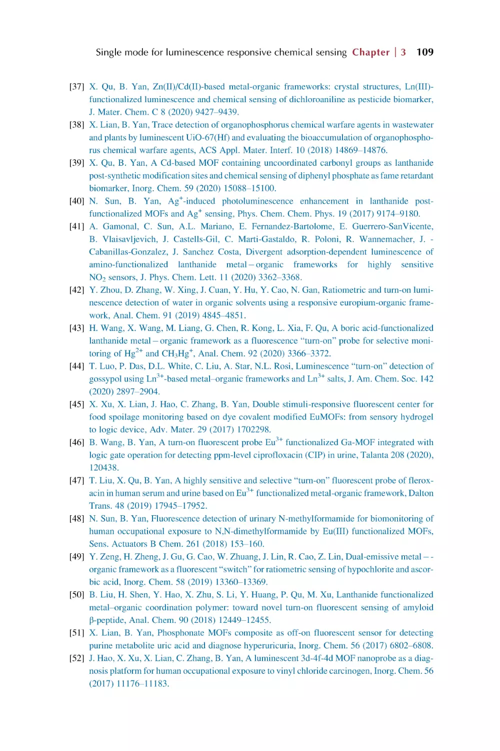
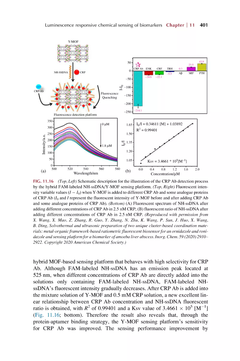
FIG. 1.4 Graphic illustration of pore and function engineering to develop multifunctional MOF
materials. (Reproduced with permission from B. Li, H. Wen, Y. Cui, W. Zhou, G. Qian, B. Chen,
Emerging multifunctional metal-organic framework materials. Adv. Mater. 28 (2016) 8819–
8860. Copyright 2016 Wiley.)
12 PART
I Introduction for rare earth metal-organic frameworks hybrid materials
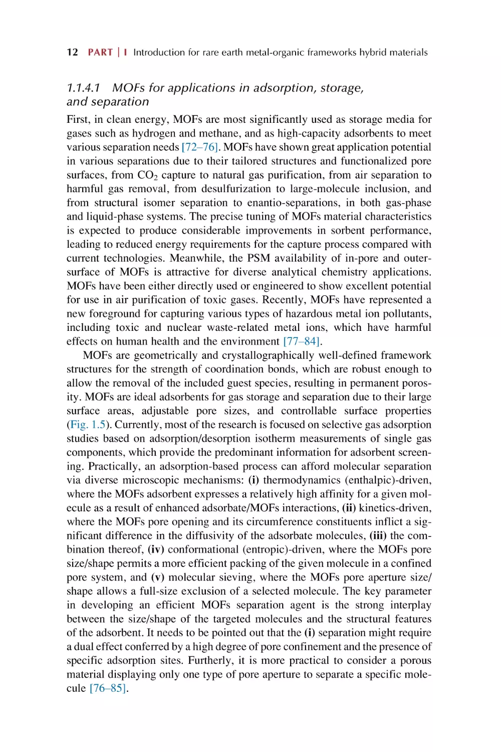
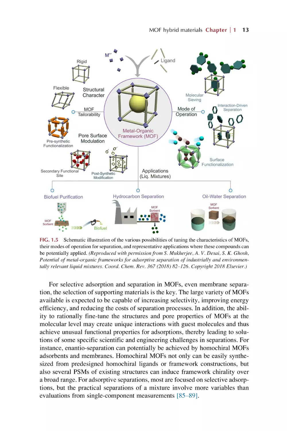
1.1.4.1 MOFs for applications in adsorption, storage,
and separation
First, in clean energy, MOFs are most significantly used as storage media for
gases such as hydrogen and methane, and as high-capacity adsorbents to meet
various separation needs [72–76]. MOFs have shown great application potential
in various separations due to their tailored structures and functionalized pore
surfaces, from CO2 capture to natural gas purification, from air separation to
harmful gas removal, from desulfurization to large-molecule inclusion, and
from structural isomer separation to enantio-separations, in both gas-phase
and liquid-phase systems. The precise tuning of MOFs material characteristics
is expected to produce considerable improvements in sorbent performance,
leading to reduced energy requirements for the capture process compared with
current technologies. Meanwhile, the PSM availability of in-pore and outersurface of MOFs is attractive for diverse analytical chemistry applications.
MOFs have been either directly used or engineered to show excellent potential
for use in air purification of toxic gases. Recently, MOFs have represented a
new foreground for capturing various types of hazardous metal ion pollutants,
including toxic and nuclear waste-related metal ions, which have harmful
effects on human health and the environment [77–84].
MOFs are geometrically and crystallographically well-defined framework
structures for the strength of coordination bonds, which are robust enough to
allow the removal of the included guest species, resulting in permanent porosity. MOFs are ideal adsorbents for gas storage and separation due to their large
surface areas, adjustable pore sizes, and controllable surface properties
(Fig. 1.5). Currently, most of the research is focused on selective gas adsorption
studies based on adsorption/desorption isotherm measurements of single gas
components, which provide the predominant information for adsorbent screening. Practically, an adsorption-based process can afford molecular separation
via diverse microscopic mechanisms: (i) thermodynamics (enthalpic)-driven,
where the MOFs adsorbent expresses a relatively high affinity for a given molecule as a result of enhanced adsorbate/MOFs interactions, (ii) kinetics-driven,
where the MOFs pore opening and its circumference constituents inflict a significant difference in the diffusivity of the adsorbate molecules, (iii) the combination thereof, (iv) conformational (entropic)-driven, where the MOFs pore
size/shape permits a more efficient packing of the given molecule in a confined
pore system, and (v) molecular sieving, where the MOFs pore aperture size/
shape allows a full-size exclusion of a selected molecule. The key parameter
in developing an efficient MOFs separation agent is the strong interplay
between the size/shape of the targeted molecules and the structural features
of the adsorbent. It needs to be pointed out that the (i) separation might require
a dual effect conferred by a high degree of pore confinement and the presence of
specific adsorption sites. Furtherly, it is more practical to consider a porous
material displaying only one type of pore aperture to separate a specific molecule [76–85].
MOF hybrid materials Chapter
1
13
FIG. 1.5 Schematic illustration of the various possibilities of tuning the characteristics of MOFs,
their modes of operation for separation, and representative applications where these compounds can
be potentially applied. (Reproduced with permission from S. Mukherjee, A. V. Desai, S. K. Ghosh,
Potential of metal-organic frameworks for adsorptive separation of industrially and environmentally relevant liquid mixtures. Coord. Chem. Rev. 367 (2018) 82–126. Copyright 2018 Elsevier.)
For selective adsorption and separation in MOFs, even membrane separation, the selection of supporting materials is the key. The large variety of MOFs
available is expected to be capable of increasing selectivity, improving energy
efficiency, and reducing the costs of separation processes. In addition, the ability to rationally fine-tune the structures and pore properties of MOFs at the
molecular level may create unique interactions with guest molecules and thus
achieve unusual functional properties for adsorptions, thereby leading to solutions of some specific scientific and engineering challenges in separations. For
instance, enantio-separation can potentially be achieved by homochiral MOFs
adsorbents and membranes. Homochiral MOFs not only can be easily synthesized from predesigned homochiral ligands or framework constructions, but
also several PSMs of existing structures can induce framework chirality over
a broad range. For adsorptive separations, most are focused on selective adsorptions, but the practical separations of a mixture involve more variables than
evaluations from single-component measurements [85–89].
14 PART
I Introduction for rare earth metal-organic frameworks hybrid materials
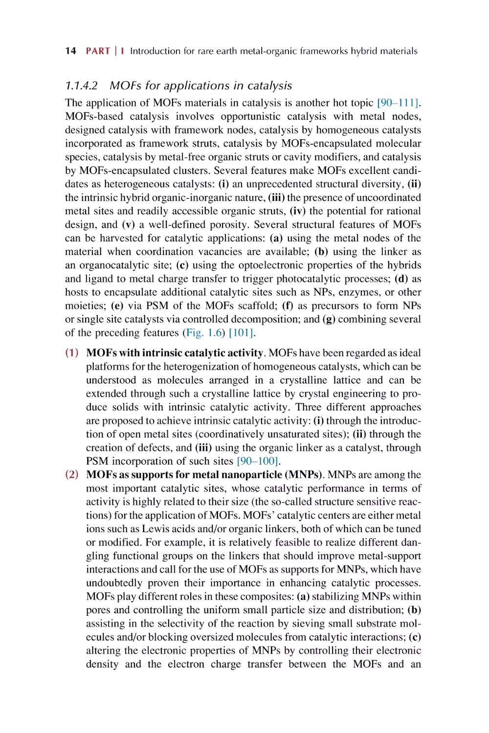
1.1.4.2 MOFs for applications in catalysis
The application of MOFs materials in catalysis is another hot topic [90–111].
MOFs-based catalysis involves opportunistic catalysis with metal nodes,
designed catalysis with framework nodes, catalysis by homogeneous catalysts
incorporated as framework struts, catalysis by MOFs-encapsulated molecular
species, catalysis by metal-free organic struts or cavity modifiers, and catalysis
by MOFs-encapsulated clusters. Several features make MOFs excellent candidates as heterogeneous catalysts: (i) an unprecedented structural diversity, (ii)
the intrinsic hybrid organic-inorganic nature, (iii) the presence of uncoordinated
metal sites and readily accessible organic struts, (iv) the potential for rational
design, and (v) a well-defined porosity. Several structural features of MOFs
can be harvested for catalytic applications: (a) using the metal nodes of the
material when coordination vacancies are available; (b) using the linker as
an organocatalytic site; (c) using the optoelectronic properties of the hybrids
and ligand to metal charge transfer to trigger photocatalytic processes; (d) as
hosts to encapsulate additional catalytic sites such as NPs, enzymes, or other
moieties; (e) via PSM of the MOFs scaffold; (f) as precursors to form NPs
or single site catalysts via controlled decomposition; and (g) combining several
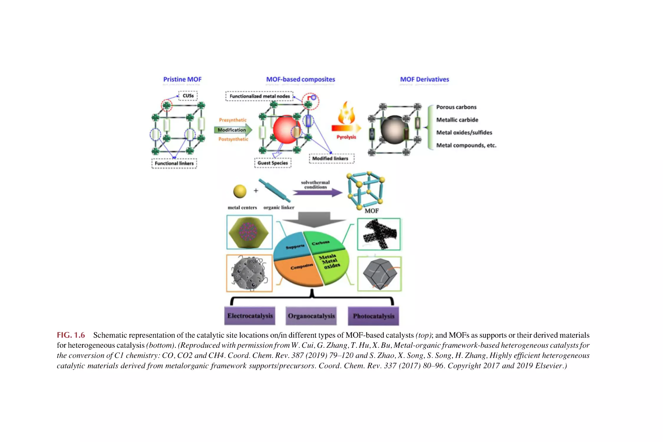
of the preceding features (Fig. 1.6) [101].
(1) MOFs with intrinsic catalytic activity. MOFs have been regarded as ideal
platforms for the heterogenization of homogeneous catalysts, which can be
understood as molecules arranged in a crystalline lattice and can be
extended through such a crystalline lattice by crystal engineering to produce solids with intrinsic catalytic activity. Three different approaches
are proposed to achieve intrinsic catalytic activity: (i) through the introduction of open metal sites (coordinatively unsaturated sites); (ii) through the
creation of defects, and (iii) using the organic linker as a catalyst, through
PSM incorporation of such sites [90–100].
(2) MOFs as supports for metal nanoparticle (MNPs). MNPs are among the
most important catalytic sites, whose catalytic performance in terms of
activity is highly related to their size (the so-called structure sensitive reactions) for the application of MOFs. MOFs’ catalytic centers are either metal
ions such as Lewis acids and/or organic linkers, both of which can be tuned
or modified. For example, it is relatively feasible to realize different dangling functional groups on the linkers that should improve metal-support
interactions and call for the use of MOFs as supports for MNPs, which have
undoubtedly proven their importance in enhancing catalytic processes.
MOFs play different roles in these composites: (a) stabilizing MNPs within
pores and controlling the uniform small particle size and distribution; (b)
assisting in the selectivity of the reaction by sieving small substrate molecules and/or blocking oversized molecules from catalytic interactions; (c)
altering the electronic properties of MNPs by controlling their electronic
density and the electron charge transfer between the MOFs and an
FIG. 1.6 Schematic representation of the catalytic site locations on/in different types of MOF-based catalysts (top); and MOFs as supports or their derived materials
for heterogeneous catalysis (bottom). (Reproduced with permission from W. Cui, G. Zhang, T. Hu, X. Bu, Metal-organic framework-based heterogeneous catalysts for
the conversion of C1 chemistry: CO, CO2 and CH4. Coord. Chem. Rev. 387 (2019) 79–120 and S. Zhao, X. Song, S. Song, H. Zhang, Highly efficient heterogeneous
catalytic materials derived from metalorganic framework supports/precursors. Coord. Chem. Rev. 337 (2017) 80–96. Copyright 2017 and 2019 Elsevier.)
16 PART
(3)
(4)
(5)
(6)
I Introduction for rare earth metal-organic frameworks hybrid materials
MNP; and (d) catalyzing one-pot tandem reactions in which both the
MOFs and MNPs act as separate active sites in their respective reactions
[90–100].
MOFs-mediated materials in heterogeneous catalyst. MOFs can be converted to considerably more stable materials by simple posttreatments
commonly involving pyrolysis. These MOFs-mediated structures retain
most of the unique properties of the parent MOFs, such as the high porosity,
tunable composition, and high metal loading, which is beneficial for heterogeneous catalytic applications, and they frequently outperform their
conventional catalyst counterparts [90–100].
MOFs in photocatalysis. MOFs emerge as a new type of prospective
photocatalytic material, whose modular structure enables them to be facilely immobilized with photoactive sites for photocatalysis. Their metal
nodes can be regarded as isolated semiconductor QDs, which can be
excited directly upon light irradiation or activated by the organic linkers
acting as a light absorption antenna. The availability of a large diversity
of organic linkers and the rich coordination chemistry of metal cations
enables the light absorption properties of the MOFs to be adjusted for efficient utilization of solar light via a judicious selection of these building
units. In addition, photoactive ligands or dyes can be used directly as building blocks to fabricate the MOFs materials or these chromophores can be
grafted on the organic ligands as a photoactive part via a PSM method.
With all these advantages, MOFs have been applied in photocatalytic
hydrogen evolution, CO2 reduction, and pollutant degradation, as well
as promotion of photocatalytic organic transformations [101–105].
Electrocatalysis on MOFs. The potential use of MOFs as electrocatalysts
is receiving increasing attention as these materials circumvent the limitations of homogeneous catalysts. Immobilizing catalysts in the solid state
with various metal nodes can impact the electrochemical driving force necessary for electron transfer between the electrode and the catalyst, as well
as improve the lifetime of the catalytic site and its selectivity. Using MOFs
as an electrocatalyst will require an active surface area, a known propagation of charge, an optimized pore size distribution and good crystallinity.
Three main electrocatalytic conversion processes are involved, which are
CO2 reduction, the hydrogen evolution reaction (HER), and the oxygen
evolution reaction (OER), as well as a selection of other relevant catalytic
processes [106–109].
Biomimetic catalysis. Immobilization of biomimetic molecular catalysts
in MOFs has generated many unique and highly active biomimetic MOFs
hybrid catalysts, which exhibit high efficiency, selectivity, and sustainability. Biomimetic MOFs present several advantages: (a) uniform dispersion
of active sites; (b) tunable hydrophobic and hydrophilic pore nature; (c)
collaborative microenvironment; and (d) confined catalytic reaction
pockets and transmission channels [112,113].
MOF hybrid materials Chapter
1
17
1.1.4.3 .MOFs for applications in magnetic and nonlinear
optical (NLO) materials
The special electric, optic, and magnetic properties of MOFs provide their great
potential for use in functional devices [112–122]. The diversity of magnetic
exchange interactions among MOFs ranges from dimers to oligomers and their
applications in infinite chains, layers, and networks, having a variety of topologies. The different forms of short-range magnetic ordering cause not only
single-molecule magnets (SMMs) and single-chain magnets (SCMs), but also
the long-range ordering of 2D and 3D networks is based on their ion-conducting
behavior [112,113,116–119]. On the other hand, major attention is paid to the
frequency doubling (second-harmonic generation, SHG) and tripling (thirdharmonic generation, THG) effect for nonlinear optical (NLO) materials.
Future efforts should be devoted to their potential technological applications
in electrooptic devices [120–122].
MOFs based on magnetic frameworks
Depending on the origin of the magnetic phenomena, four types of MOFs can be
differentiated: (a) magnetic MOFs with magnetic cooperativity resulting from
magnetic exchange via the ligands; (b) spin-crossover MOFs with nodes of suitable coordination environments for this phenomenon to exist; (c) MOFs with
magnetic relaxation with the nodes of clusters possessing an anisotropic spin
ground state and showing single molecule magnet behavior; and (d) MOFs with
a magnetocaloric effect with nodes of clusters possessing an isotropic spin
ground state. (i) Magnetic MOFs. Magnetic exchange interactions require
short distances between the metal centers, which are most commonly the spin
carriers. The porosity is typically favored with the use of long linkers, which are
often too long for magnetic ordering to exist at temperatures much above absolute zero. (a) Use of short linkers. Among the denser structures with shorter
linkers, long-range magnetic order can emerge, although sorption of gases in
this situation is rather atypical. (b) Metallo-ligand approach. A metal complex
with vacant additional coordination sites is first prepared and isolated, being
used in a second step as a building block towards additional metal ions. Through
a suitable choice of the metallo-ligand, magnetic communication between the
metal nodes can be achieved. (c) Radical-as-ligand approach. An alternative
methodology for facilitating exchange coupling between metal centers of
MOFs is the incorporation of additional spin carriers into the organic linkers
such as radical ligands [112,113,116,117]. (ii) Spin-crossover MOFs. Cooperative effects are still necessary to make this phenomenon useful in order to have
an abrupt crossover, which arises from the elastic forces present in the solid. In
these systems, such a cooperativity is favored by the polymeric nature of the
framework, keeping the spin-crossover centers connected even if they are
not very close. The cooperativity enables this type of MOFs to show high sensitivity to the subtle structural changes occurring in metal coordination
18 PART
I Introduction for rare earth metal-organic frameworks hybrid materials
environments upon inclusion of guest species within the porous framework.
Consequently, the spin-crossover properties are tuned via PSMs of MOFs.
(a) Physisorption of gases. (b) Chemisorption/PSM. (c) MOFs with SMMs
at the inorganic nodes. SMMs are usually formed by polynuclear magnetic
clusters with a large spin value and high magnetic anisotropy. One important
requirement for maximizing the quantum coherence is to minimize the magnetic dipolar interactions between qubits, which can be achieved by separating
these magnetic units in space. MOFs can be ideal platforms to reach this goal
since they provide spatial separation at will. (d) MOFs for magnetic
refrigeration. A magnetocaloric effect (MCE) of high interest for cooling
applications is that of magnetic refrigeration. The use of magnetic MOFs for
magnetic refrigerators is very appealing since they can be designed to consist
of isolated paramagnetic centers that favor a large MCE, combined with higher
thermal and solvent stabilities than their discrete molecular cluster analogs
[112,113,118,119]. (iii) Hybrid MOFs incorporating functional molecules
in the channels. Using this hybrid approach, two-network solids can be prepared through the self-assembly of different molecular fragments used as starting building blocks, or using a PSM method in which a molecular guest is
inserted into a preformed extended network as the host lattice. (a)
SCO@MOFs. (b) SMM@MOFs. (c) Electroactive molecules@MOFs.
POMs form a class of molecular anions that have been incorporated into MOFs,
which are robust species with unique electronic properties due to their ability to
act as electron reservoirs or to accommodate magnetic centers [112].
MOFs for NLO
(i) Producing MOFs for NLO. Noncentrosymmetric organization is a prerequisite for the generation of NLO properties of bulk materials. In conventional
methods of producing MOFs, the creation of such noncentrosymmetric structures is challenging. Not only can the coordination of the organic ligand to a
metal ion yield an increase in charge transfer transitions, but it also allows
organic chromophores to assemble in highly ordered geometries like octahedra
and tetrahedra. This interaction produces charge transfer in several directions.
In addition, this is a type of structure that is relatively easy to obtain by selecting
the right combination of metal ions and organic ligands. (ii) Ligands. (a) Multifunctional ligands. One of the most successful methods for solving the problem
of obtaining a noncentrosymmetric MOFs structure is using a compound as a
ligand that is capable of chelating a metal ion with several functional groups
in different positions. This in turn leads to different degrees of ligand deprotonation, ultimately resulting in different coordination modes upon interaction
with two different metal ions. (b) Chiral ligands. All chiral MOFs are active
for SHG, and there are some basic synthetic approaches to produce a chiral
MOFs. But the presence of a chiral structure does not necessarily guarantee
a high response of SHG. (iii) Diamondoid structures. The most likely candidates for producing crystals with NLO properties are metal ions, which have a
MOF hybrid materials Chapter
1
19
coordination number of 4 or 8 and are connected via a bifunctional ligand. Such
components tend to form the so-called diamond structure, whose crystals inherently crystallize in a centrosymmetric space group so that the inversion center is
in the middle of the CdC bond between two adjacent nodes. (iv) New applications of MOFs in NLO. (a) Third-order NLO. Third-order NLOs for MOFs
have promise for use in all-optical switching in waveguides. (b) Control of
excitons. The crystalline and organic nature of MOFs allows the existence of
Coulomb-bound electron-hole pairs (excitons). The potential offered by manipulating excitons when exposed to light and electric field is the basis for modern
exciton transistors and polarization lasers [120–122].
1.1.4.4 MOFs for applications in electrochemistry
MOFs and their composites show great potential in the electrochemical field for
their unique properties. Creating an effective method for the large-scale production of MOFs and MOFs composites with small size and high conductivity will
facilitate their rapid development in practical electrochemical applications.
Electrochemistry applications are wide ranging, including electrode materials
and batteries, electrocatalysis, electroanalysis, electrochemical sensing, and
even the entire energy and fuel categories (Fig. 1.7) [123–137].
(I) MOFs for batteries. Due to their structural flexibility, low cost, and
redox activity, MOFs are good candidates for electrode materials. However, the practical application of many MOFs is hampered by their poor
conductivity resulting in poor cycle performance of the battery. (i) MOFs
for Li-ion batteries (LIBs). MOFs can be favorable to interfacial charge
transport of the Li+ insertion/extraction strain due to their high surface
area and porosity. MOFs behave as positive, negative, and electrolyte
materials for LIBs. (a) Pure MOFs. (b) MOF composites. The conductivity of pure MOFs materials is not good. Thus it is necessary to seek to
alter the electrochemical performance by combining MOFs with other
conductive materials (metal oxides, conductive polymers, single metal,
and carbon materials). (ii) MOFs for Li-S batteries. MOFs have unparalleled synthetic flexibility and adjustable pore size, and can be used to
capture and immobilize sulfur by weak host-guest interactions. (a) Pure
MOFs. The pore structure and performance of MOFs make them promising cathode materials for Li-S battery systems. (b) MOFs composites.
To improve the capacity and cycle stability, the composites behave as a
substrate for sulfur fixation in Li-S batteries. (c) Others. MOFs are also
more widely used in other batteries, such as Li-O2 batteries, SIBs, and so
on [125–130].
(II) MOFs for supercapacitors (SCs). The direct application of MOFs as SC
electrode materials is mainly faced with poor conductivity and poor
mechanical/chemical stability. In order to improve the conductivity of
MOFs, conjugated guest molecules with redox activity to permeate into
MOFs are used and the permeable MOFs lead to an apparent increase in
20 PART
I Introduction for rare earth metal-organic frameworks hybrid materials
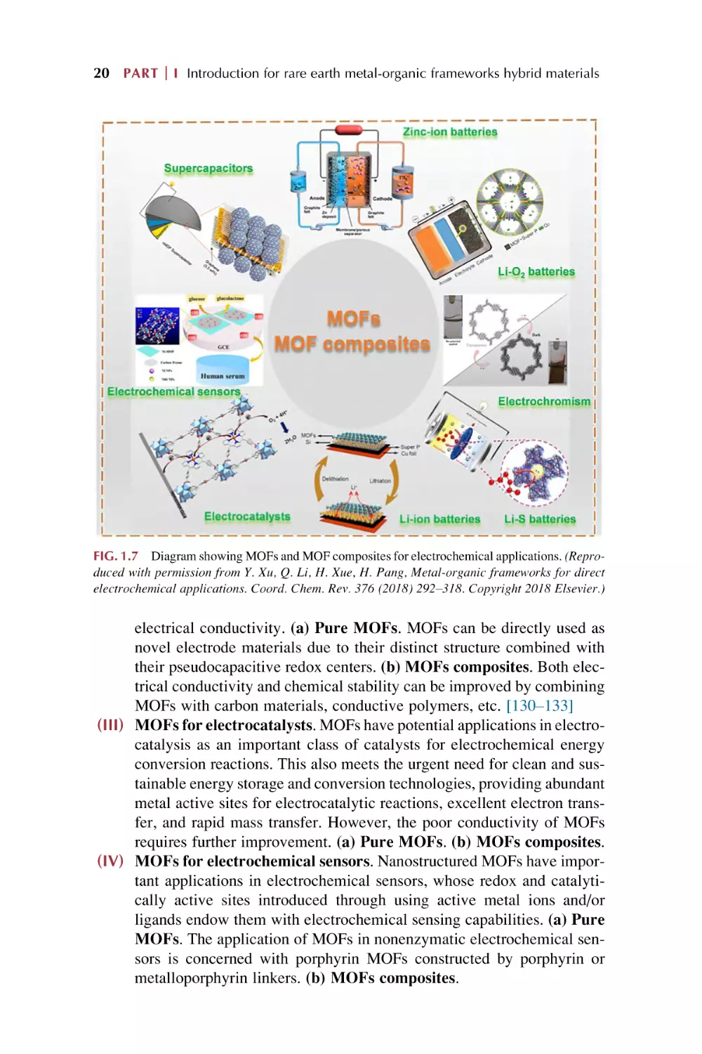
FIG. 1.7 Diagram showing MOFs and MOF composites for electrochemical applications. (Reproduced with permission from Y. Xu, Q. Li, H. Xue, H. Pang, Metal-organic frameworks for direct
electrochemical applications. Coord. Chem. Rev. 376 (2018) 292–318. Copyright 2018 Elsevier.)
electrical conductivity. (a) Pure MOFs. MOFs can be directly used as
novel electrode materials due to their distinct structure combined with
their pseudocapacitive redox centers. (b) MOFs composites. Both electrical conductivity and chemical stability can be improved by combining
MOFs with carbon materials, conductive polymers, etc. [130–133]
(III) MOFs for electrocatalysts. MOFs have potential applications in electrocatalysis as an important class of catalysts for electrochemical energy
conversion reactions. This also meets the urgent need for clean and sustainable energy storage and conversion technologies, providing abundant
metal active sites for electrocatalytic reactions, excellent electron transfer, and rapid mass transfer. However, the poor conductivity of MOFs
requires further improvement. (a) Pure MOFs. (b) MOFs composites.
(IV) MOFs for electrochemical sensors. Nanostructured MOFs have important applications in electrochemical sensors, whose redox and catalytically active sites introduced through using active metal ions and/or
ligands endow them with electrochemical sensing capabilities. (a) Pure
MOFs. The application of MOFs in nonenzymatic electrochemical sensors is concerned with porphyrin MOFs constructed by porphyrin or
metalloporphyrin linkers. (b) MOFs composites.
MOF hybrid materials Chapter
1
21
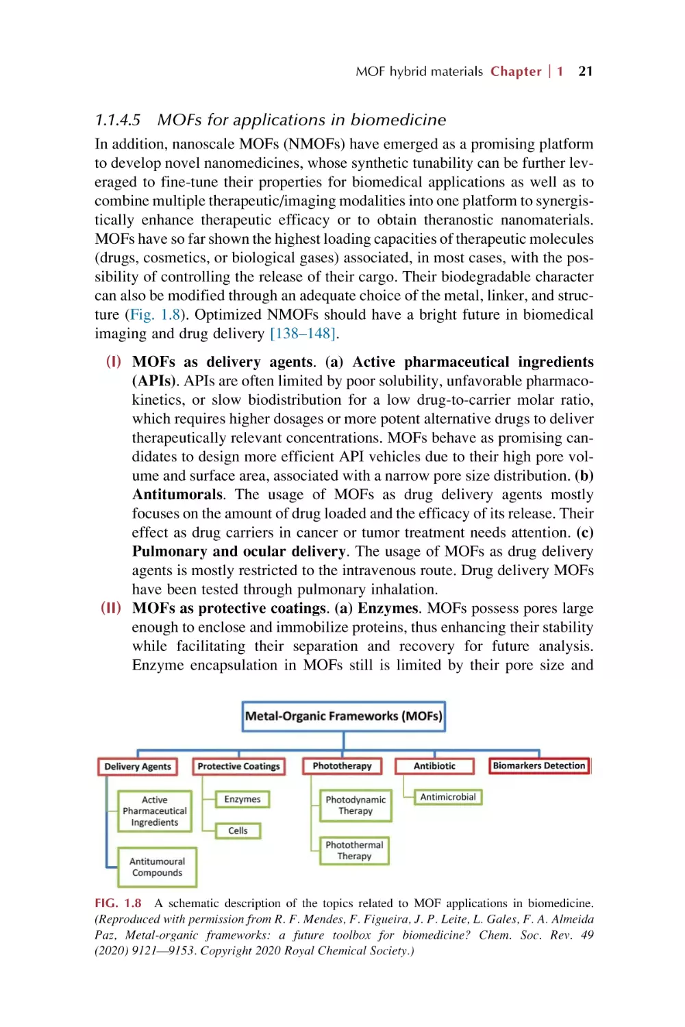
1.1.4.5 MOFs for applications in biomedicine
In addition, nanoscale MOFs (NMOFs) have emerged as a promising platform
to develop novel nanomedicines, whose synthetic tunability can be further leveraged to fine-tune their properties for biomedical applications as well as to
combine multiple therapeutic/imaging modalities into one platform to synergistically enhance therapeutic efficacy or to obtain theranostic nanomaterials.
MOFs have so far shown the highest loading capacities of therapeutic molecules
(drugs, cosmetics, or biological gases) associated, in most cases, with the possibility of controlling the release of their cargo. Their biodegradable character
can also be modified through an adequate choice of the metal, linker, and structure (Fig. 1.8). Optimized NMOFs should have a bright future in biomedical
imaging and drug delivery [138–148].
(I) MOFs as delivery agents. (a) Active pharmaceutical ingredients
(APIs). APIs are often limited by poor solubility, unfavorable pharmacokinetics, or slow biodistribution for a low drug-to-carrier molar ratio,
which requires higher dosages or more potent alternative drugs to deliver
therapeutically relevant concentrations. MOFs behave as promising candidates to design more efficient API vehicles due to their high pore volume and surface area, associated with a narrow pore size distribution. (b)
Antitumorals. The usage of MOFs as drug delivery agents mostly
focuses on the amount of drug loaded and the efficacy of its release. Their
effect as drug carriers in cancer or tumor treatment needs attention. (c)
Pulmonary and ocular delivery. The usage of MOFs as drug delivery
agents is mostly restricted to the intravenous route. Drug delivery MOFs
have been tested through pulmonary inhalation.
(II) MOFs as protective coatings. (a) Enzymes. MOFs possess pores large
enough to enclose and immobilize proteins, thus enhancing their stability
while facilitating their separation and recovery for future analysis.
Enzyme encapsulation in MOFs still is limited by their pore size and
FIG. 1.8 A schematic description of the topics related to MOF applications in biomedicine.
(Reproduced with permission from R. F. Mendes, F. Figueira, J. P. Leite, L. Gales, F. A. Almeida
Paz, Metal-organic frameworks: a future toolbox for biomedicine? Chem. Soc. Rev. 49
(2020) 9121—9153. Copyright 2020 Royal Chemical Society.)
22 PART
(III)
(IV)
(V)
(VI)
I Introduction for rare earth metal-organic frameworks hybrid materials
the incorporation step. Suitable linkers are required to adjust the pore size
to match the enzyme configuration and polarity of the MOFs pores. (b)
Cells. In situ MOFs synthesis can also be applied for the encapsulation
of bigger bioentities, such as viruses and cells.
Phototherapy using MOFs. Phototherapy involves the exposure of tissues to specific wavelengths of light, being in the form of photodynamic
(PDT) or photothermal (PTT) therapies, whose efficiency and selectivity
can be greatly improved by choosing an optimal photosensitizer (PS).
MOFs are very promising candidates for a combination of PDT and
PTT for their organic linker capable of creating superoxide anions, and
the metal center.
Detecting biomarkers with MOFs. If a porous MOFs labeled with a
biorecognition receptor involved in a highly specific interaction, such
as antigen-antibody (immunosensing), protein-ligand, or nucleic acid
base pair complementary, is combined with an efficient detecting technique, it is possible to design ultrasensitive and tunable methods for biomarker detection.
Antimicrobial activity. MOFs can incorporate antibacterial species in
their pores or even be effective themselves in killing different types of
bacteria.
Synergistic nanotherapeutics. MOFs show great potential in biomedicine, especially in terms of cancer treatments, and NMOFs provide many
opportunities for biological and medical applications. Loading of macromolecules such as proteins within NMOFs is also possible for other interesting applications such as nanovaccines. Many organic ligands within
NMOFs may provide therapeutic functions by themselves, such as acting
as chemotherapeutic prodrugs, photosensitizers, and fluorescent dyes. In
addition, certain types of metal ions as the coordination centers may offer
imaging contrast. Importantly, NMOFs in general are biodegradable by
being gradually decomposed into small metal ions and organic molecules
with rapid excretion and little long-term body retention, making them
promising for safe in-vivo applications. In general, NMOF-based nanomedicine systems exhibit the combined benefits of both organic drug
delivery systems and inorganic theranostic nanoplatforms [143–148].
1.2 Rare earth metal-organic frameworks (REMOFs)
Rare earth MOFs (REMOFs here means rare earth ions behaving as the unique
framework metal centers) have garnered much interest due to a wide array of
features from the marriage of rare earth ions (RE3+) with MOFs. MOFs synthesized with rare earth (RE) elements (including scandium (Sc), yttrium (Y) and
the series of 15 lanthanides (Ln)) are an intriguing family of MOFs from the
standpoint of both structure and function. REMOFs not only can possess many
of the same properties common to all MOFs families (i.e., permanent porosity,
MOF hybrid materials Chapter
1
23
tunable pore size/shape, accessible Lewis acidic sites), but also can display
unique structures and properties owing to the high coordination numbers and
distinct optical properties of RE elements. The coordination chemistry of RE
ions is very diverse, with only small energetic differences between different
coordination numbers and geometries, and where geometry is dictated primarily
by ligand steric effects. This can make MOFs structure prediction more complicated, which also opens the door for the discovery of several new structures,
composed of diverse metal nodes, and in some cases giving rise to highly connected nets, and the unique ability to merge multiple net structures using only
one metal ion. Different from the transition metal ions, RE ions have unique
electronic properties dictated by their 4f electron configurations. Given that
the 4f orbitals are shielded from their external environment by 5s and 5p
orbitals, RE ions have distinct electronic and magnetic properties that are not
significantly altered by coordinating ligands. The electrons in the f block of
RE3+ make them capable of having a larger coordination sphere. Based on
the hard-soft acid-base consideration, RE3+ ions have affinity in relatively hard
oxygen-containing linkers over other functional groups. RE3+ can display high
coordination number and connectivity, diverse and coordination modes, which
therefore exhibit great potential in constructing wide variety of MOFs. By carefully tuning the RE3+ node and organic linker components, REMOFs can be
assembled into fascinating structures with diverse and complex topologies.
The highly connective rare earth ions usually lead to the formation of condensed
frameworks in the microporous range. Certainly, just as with other MOFs systems, REMOFs also have practical application value in the fields of separation,
catalysis, and especially as a very promising class of materials for addressing
the challenges in engineering of luminescent centers. RE3+ ion-bearing phosphors find numerous applications in lighting, display (light emitting diode,
LED), optical communications, photonics, and biomedical devices. In addition,
it should be stressed that the abundant luminescence response of REMOFs
embody their versatile probe in chemical sensing [149–153].
1.2.1
REMOF structures
MOFs that are constructed using RE ions have been shown to form intricate
topologies and diverse network structures. Generally, REMOF structures are
classified by the identity of the metal node, being metal ions, chains, or clusters.
(1) Metal ion nodes. Unlike s-, p-, and d-block metal ions, f-block metal ions
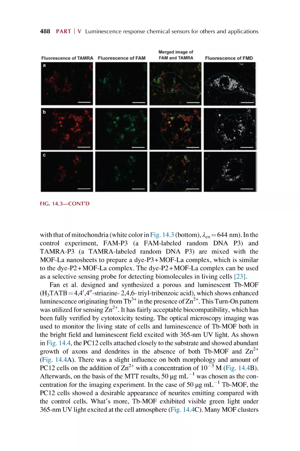
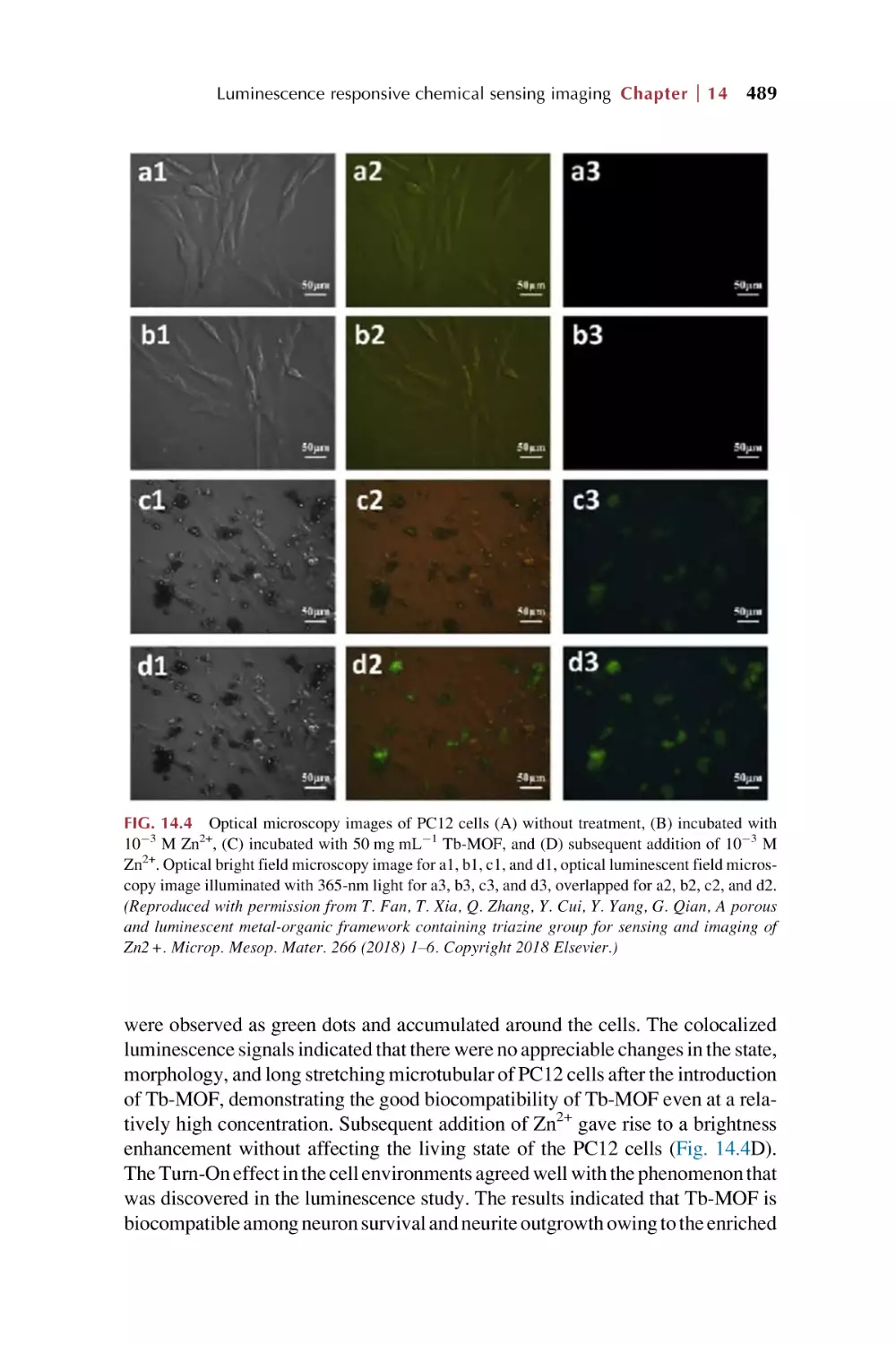
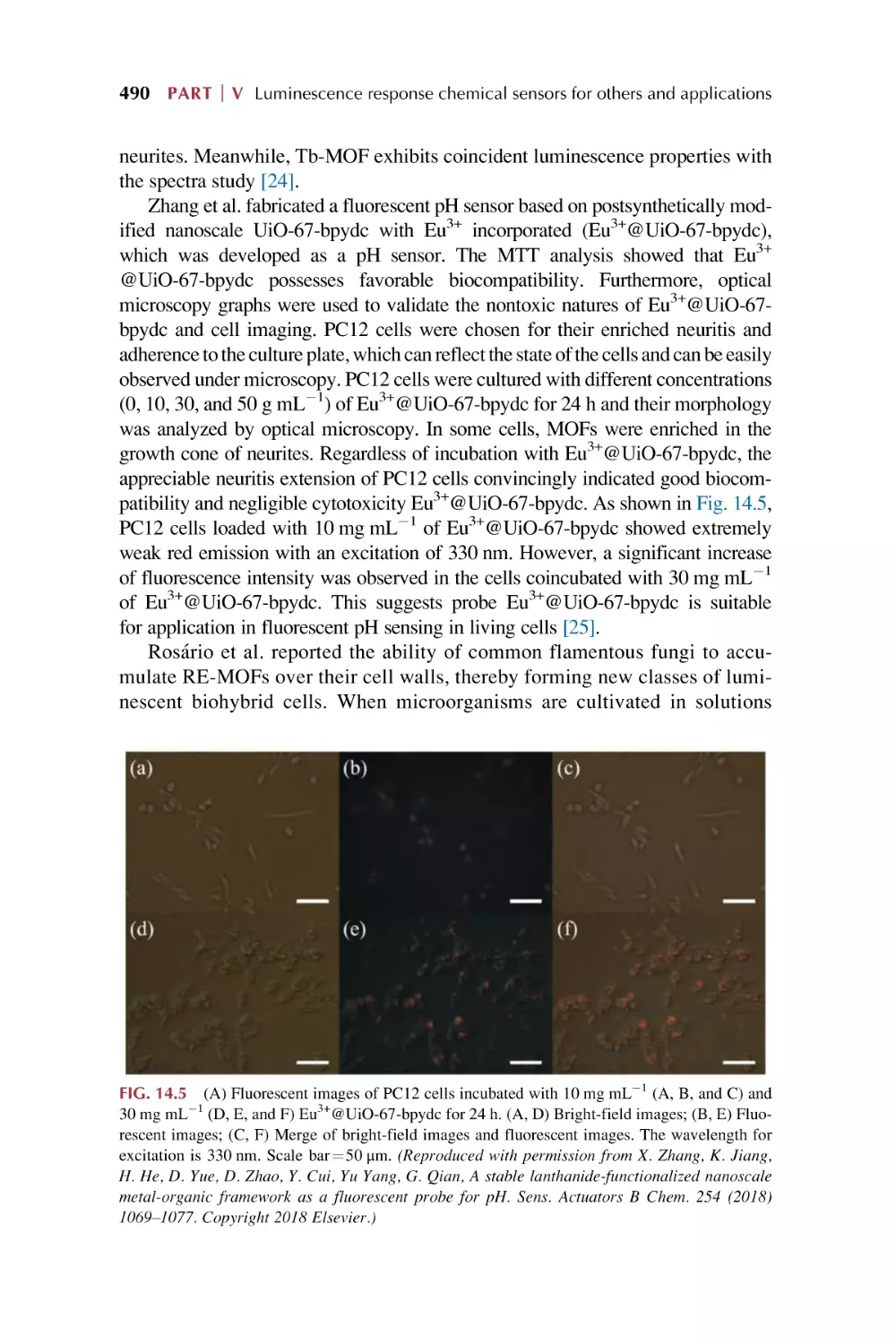
possess high coordination numbers as well as numerous coordination
modes, unlocking a variety of structures that are not accessible through
use of the former. RE3+ ions, especially Ln3+, have a higher coordination
number (in the range of 6–13) and connectivity than transition-metal ions,
which are favorable for stabilizing the frameworks if all these coordination
sites are utilized and bridged by organic linkers. The stable REMOFs not
24 PART
I Introduction for rare earth metal-organic frameworks hybrid materials
only exhibit high thermal stability but also high water and chemical stability in acidic/basic solutions. Variable coordination numbers of RE3+ ions
also make it possible to control structures and modify the pore sizes/sharps
and framework interpenetration of REMOFs.
(2) Metal chain nodes. Rare earth oxo/hydroxo chain nodes, also called rods,
are found for several REMOFs comprising carboxylic acid linkers. The formation of polymeric RE oxo/hydroxo chains tends to be favored in the
presence of bridging carboxylic acid ligands. Many of the underlying
design rules to construct REMOFs with RE3+-chain nodes and open,
porous structures have also been applied to MOFs.
(3) Metal cluster nodes. Controlled hydrolysis of the rare earth precursor is
required to ensure the synthesis of clusters as opposed to RE chains, oxides,
and hydroxides, which can be achieved using ligands as both structuredirecting agents (or modulators) and capping groups to stabilize the cluster.
These same clusters can be generated and stabilized during the synthesis of
REMOFs, functioning as SBUs for a wide range of structures. (a) Dinuclear clusters. These that are 6-connected have since been found for MOFs
comprising ditopic, tritopic, and mixed linkers. In addition, it should be
noted that there are examples of MOFs comprising dinuclear RE cluster
nodes that are 4-connected, 8-connected, and 12-connected. (b) Trinuclear clusters. They are common for MOFs comprising d-block metals,
but are less commonly observed with RE metals. (c) Tetranuclear clusters. They focus primarily on examples with cubane-like, square planar,
and rectangular tetranuclear RE clusters. (d) Hexanuclear clusters.
(e) Heptanuclear metal clusters. (f) Nonanuclear metal clusters
[149–153].
1.2.2 Some applications of REMOFs
(1) Biological applications. MOFs have emerged as promising candidates for
drug delivery and bioimaging, while MOFs comprising RE ions may possess interesting magnetic and optical properties that can be exploited for
these applications. (a) Bioimaging. REMOFs have also been studied for
their potential in optical imaging owing, in part, to the NIR emission signatures of several of the RE3+ ions. In comparison to visible light, NIR light
is advantageous for biological imaging since (i) it interacts less with biological materials, causing less potential damage to the region; (ii) tissue
autofluorescence is low in the NIR region; and (iii) it scatters less, giving
rise to better signals. (b) Drug delivery. The encapsulation of drug molecules into REMOFs-based nanocarriers has been successfully employed
to increase the permeability of biofilms, change the distribution in vivo,
regulate the rate of drug release, and improve stability/bioavailability.
REMOFs have some of the following unique advantages as pharmaceutical
carriers: (I) They can be simulated and designed according to the drug
MOF hybrid materials Chapter
1
25
characteristics, whose compositions and architecture geometries can be
varied. (II) REMOFs have an affinity to effectively capture drug molecules
with high loading doses due to their large pore volume-to-surface area
ratios. (III) Imaging-mediated drug quantification, targeted cell delivery,
and cancer treatments can be achieved by exploiting the unique optical
and magnetic features. (IV) REMOFs can be rationally functionalized with
upconversion luminescence (UCL) to serve as dual-mode drug carriers and
contrast imaging agents with improved accuracy for disease diagnosis and
treatment [149–153].
(2) Catalysis. REMOFs represent one of the emerging areas in MOFs chemistry and show excellent features for catalysis. (I) Heterogeneous catalysis. The metal SBUs of REMOFs are appropriate for the kinds of reactions
that require strongly acidic catalytic sites. In addition, REMOFs can act as
platforms to provide excellent host-guest interactions in the framework
through PSM, which can be used as synergistic materials for photocatalytic
purposes. However, REMOFs as heterogeneous catalysts suffer from significant underlying limitations that restrict their use for organic transformation: (a) Low thermal and chemical stability; (b) Sensitivity to oxygen and
moisture, but this issue can be circumvented by introducing hydrophobic
functionalities into the organic linkers, making REMOFs structures more
water-resistant; (c) Deactivation by the coordination of basic reactants,
products, or coordinating solvents at their free acidic metal sites, but this
can be diminished by minimizing the defects in REMOFs structures; (d)
the lack of a fundamental understanding of the reaction kinetics of
REMOFs; and (e) difficulties in the large-scale synthesis of targeted
REMOFs. (II) Photocatalysts. Designing linkers with high conjugation
capable of light absorption over a wide range of wavelengths in combination with theoretical studies may shed some light on the electron transfer
mechanism in REMOFs. Furthermore, the separation of photoexcited electrons and holes, facilitated by such highly conjugated linkers, combined
with the LMCT process, will take full advantage of unfilled 4f orbitals
of RE ions in photocatalytic transformations [149–153].
(3) Adsorption and separations. The inherent porosity of REMOFs has been
harnessed for gas adsorption, separation, and purification processes. (I)
Adsorption. In addition to N2 or CO2 gas adsorption, REMOFs have also
been studied for the adsorption of liquid phase MeOH and water vapor.
Such tunable pore sizes/sharps can induce highly selective gas storage
and separation via a molecular sieving effect. Furthermore, the open rare
earth sites and other functional sites can be easily incorporated into
REMOFs and widely utilized to enhance the gas storage capacities and
gas separation selectivity. Until now, REMOFs have been developed
and explored for wide applications in H2 storage, selective CO2 capture
and separation, and H2 and CH4 purification. Establishment of permanent
porosity within REMOFs is more difficult and challenging compared with
26 PART
I Introduction for rare earth metal-organic frameworks hybrid materials
transition metal MOFs, which is mainly due to the fact that higher coordination number and the more flexible coordination environment of RE3+
ions allow more small solvent molecules such as DMF and H2O to easily
occupy the coordination sites of RE3+ within REMOFs. (II) Separation.
The chemical functionality, size, and shape of the pores in REMOFs can
be tuned to aid in separating compounds based on size or chemical
properties. A lot of REMOFs have been explored and demonstrated to have
permanent porosities, making them useful for gas storage and separation.
Some strategies such as the open rare earth sites, surface-functionalization,
tunable pore size/sharp, interpenetration, and flexible structures within
REMOFs have been simultaneously explored and widely utilized as a
means to enhance the ability of selective gas storage and separation. Given
the fact that some small solvent molecules can easily coordinate onto the
RE3+, if the coordinated solvent molecules are carefully removed, available open RE3+ sites should be obtained except for being occupied by
the donor atoms from the neighboring organic linkers. The open RE3+ site
formation within the pore surfaces of REMOFs is also considered to be
beneficial for gas adsorption and separation [149–153].
1.3 Rare earth functionalized metal-organic framework
hybrid materials (REFMOFHs)
Rare earth functionalized MOFs hybrid materials (REFMOFHs) mean that
some special photoactive RE3+ ions (such as visible emissive Eu3+, Tb3+,
Sm3+, or Dy3+ and near infrared (NIR) emissive Nd3+, Yb3+, or Er3+) are introduced into the MOF host to realize the functionalization of MOFs units and formation of the hybrid material systems. Currently, the main approach to
assemble REFMOFH materials is PSM of MOFs. There are a variety of different ways to modify a MOFs in a postsynthetic manner, and each of these modification forms has the capacity to alter the physical and chemical properties of
the frameworks. For REFMOFHs materials, the effective PSM strategies can be
divided into six major areas (as shown in Fig. 1.9): coordinate PSM, covalent
PSM, covalent and coordinate PSM, ion exchange PSM, ion doping (postsynthetic metalation or metal ion substitution) PSM, and encapsulation composition PSM. These postsynthetic methods provide the high complexity and
functionality for rare earth functionalized MOFs hybrid materials and the
desired chemical transformation should not destroy the MOFs under the reaction conditions used. Subsequently, some new hybrid materials based on RE3+
and MOFs can be obtained by PSM methods, which still retain the characteristic
features of MOFs such as high crystallinity, high surface areas, and a structure
comprising highly regular coordination bonding. It is worth pointing out that the
content of the functionalized RE3+ in MOFs hosts is limited and small, but it still
displays the effective physical property and functional performance of RE3+ for
practical applications [154–172].
MOF hybrid materials Chapter
1
27
FIG. 1.9 Schematic description of postsynthetic modification (PSM) of MOFs by rare earth
(RE3+) ions for the corresponding hybrid materials (REFMOFHs); red sphere represents the rare
earth species used for functionalized MOFs. (Reproduced with permission from B. Yan, Luminescence response mode and chemical sensing mechanism for lanthanide functionalized metal-organic
frameworks hybrids. Inorg. Chem. Front. 8 (2021) 201–233. Copyright 2021 Royal Chemical
Society.)
(1) Coordinate PSM. Coordinate PSM has the first effective approach for
introducing RE3+ ions to achieve the final hybrid materials, using RE3+ species to coordinate to the free active group in MOFs (Fig. 1.9A) [155–157].
Fig. 1.10 shows an example of coordination PSM of Ga-MOF ((Ga(OH)
(btec)0.5H2O, H4btec¼ pyromellitic acid)) by double cations of both near
infrared-emitting rare earth cations due to its rigid and porous structure
and the presence of noncoordinating carboxylate groups. This allows the
introduction of metal ions into Ga-MOF by PSM. Ag+ can enhance visible
emitting of Ln3+ in Ln3+@Ga-MOF distinctly. Ln3+ (Yb3+, Nd3+, Er3+) and
Ag+ are incorporated into the pores of Ga-MOF and used to analyze the luminescence properties of the resulting host-guest material [156].
FIG. 1.10 Schematic illustration of RE3+ and Ag+ incorporation into Ga-MOF. The RE, Ag, Ga,
C, and O atoms are represented by pansy, pink purple, cobalt blue, black, and red, respectively.
Hydrogen atoms and uncoordinated water molecules are excluded for clarity. (Reproduced with
permission from N. Sun, B. Yan, Near-infrared emission sensitization of lanthanide cation based
on Ag+ functionalized metal-organic frameworks. J. Alloys Compd. 765 (2018) 63–68. Copyright
2018 Elsevier.)
28 PART
I Introduction for rare earth metal-organic frameworks hybrid materials
(2) Covalent PSM. Covalent PSM uses a reagent to modify a component of
the MOFs in a heterogeneous, postsynthetic manner to form a new covalent
bond, providing a powerful and versatile method to introduce a broad range
of chemical groups into MOFs (Fig. 1.9B) [158,159]. For rare earth functionalized MOF hybrid materials, covalent PSM is not a single functionalization mode, but combines with other modification paths in the hybrid
materials.
(3) Covalent and coordinate PSM. For rare earth functionalized MOFs
hybrid materials, considering that RE3+ should be introduced through coordinate PSM, generally covalent PSM is the prepared procedure to produce
the coordinated group for further RE3+ coordinate PSM (Fig. 1.9C). It
should be noted that the RE3+ ion’s coordination often destroys the covalent PSM-derived MOFs, whose stability must be considered very important [158,159]. Fig. 1.11 shows the scheme for the composition of the
preparation process and reaction principle of the hybrids. SBA-15 can
be easily modified by the crosslinking reagent IPTES to form Si-SBA15 through the cohydrolysis and copolycondensation process between
the alkoxyl groups of IPTES and the hydroxyl groups of SBA-15. On
the other hand, IRMOF-3 is synthesized in situ and then is covalently
grafted to Si-SBA-15 through the addition reaction between its amino
group and inner ester group of Si-SBA-15 [159].
FIG. 1.11 The selected scheme for the composition of hybrid materials RE-SBA-15-Si-IRMOF-3
(RE¼Eu, Tb, Nd, Yb). (Reproduced with permission from X. Lian, B. Yan, Multi-component luminescent lanthanide hybrids of both functionalized IR-MOF-3 and SBA-15. New J. Chem. 39
(2015) 5898–5901. Copyright 2015 Royal Chemical Society.)
MOF hybrid materials Chapter
1
29
(4) Ion exchange PSM. Ion exchange PSM is most appropriate for many
anionic MOFs systems with dimethylammonium (DMA) cations in their
porous structure, and so RE3+ cations can be introduced into the MOF via
ionic exchange with DMA (Fig. 1.9D) [160–164]. For example, Fig. 1.12
shows that pillared layered hybrid materials RE(PDA)33 -LDH can be
prepared through ion exchange assembly of luminescent lanthanide coordination polymers [H2NMe2]3[RE(DPA)3] (RE¼ Eu, Tb, Sm, Dy, Nd;
[H2NMe2]+¼dimethyl amino cation; H2DPA¼ 2,6-dipicolinic acid) and
pillared layered double hydroxide (Mg-Al LDH, Mg6Al2(OH)16CO34H2O).
With Mg-Al LDH interacts with [H2NMe2]3[RE(DPA)3] through
anion exchange ([RE(PDA)3]3) and electrostatic effect to form
[RE(PDA)3]3-LDH [159].
(5) Ion doping (postsynthetic metalation (or metal ion substitution) PSM).
Ion doping PSM is easily realized for REMOFs due to the similar ion radius
of lanthanide contraction. In fact, RE3+ ionic doping also can be extended
to other trivalent metal ion MOFs such as Al, Ga, and In ions for the same
valence substitution (Fig. 1.9E). A series of hybrids based on RE3+ ions
(Eu3+, Tb3+, Sm3+, Dy3+) activated yttrium metallic organic framework
of 1,3,5-benzenetricarboxylate (MOF-76(Y)) are synthesized by ion substitution under solvothermal conditions, which shows isostructure with
parent MOF-76(Y) [164].
(6) Encapsulation composition PSM. In addition, encapsulation composition
PSM permits all kinds of rare earth compounds (salts, complexes, or
oxides, etc.) as small guest molecules or nanoparticles (NPs) to be introduced in MOFs hosts, providing physical composition to keep the whole
FIG. 1.12 Scheme showing synthesis procedure and predicted structure of the hybrid materials of
3
[RE(DPA)3 ]3-LDH, CO2
(green), OH (blue), Mg and Al (black). (Repro3 (gray), [RE(PDA)3]
duced with permission from J. Ma, B. Yan, Novel hybrid materials of lanthanide coordination polymers ion exchanged Mg-Al layered double hydroxide: multi-color photoluminescence and white
color thin film. Dyes Pigments 153 (2018) 266–274. Copyright 2018 Elsevier.)
30 PART
I Introduction for rare earth metal-organic frameworks hybrid materials
luminescent hybrid system stable (Fig. 1.9F) [165–172]. For example, a
novel strategy is demonstrated to construct photofunctional nanocomposites by composing surfactant-capped nanophosphors (upconversion luminescent NaYF4:Yb3+, Er3+/Tm3+ and downconversion emissive LaPO4:
Ln3+ (Ln ¼ Ce, Eu, Tb) NPs) and a zeolitic imidazolate framework
(ZIF-8) unit. Fig. 1.13 shows the scheme for the preparation process of
the nanocomposites. The PVP-NPs@ZIF-8 is synthesized through a
PVP-protected process (PVP¼ polyvinyl pyrrolidone). This strategy
involves two steps: the first step is the functionalization of nanoparticle
surfaces with PVP and the second step is the crystallization of ZIF-8 [165].
Fig. 1.14 shows the scheme of a series of transparent MOFs thin films designed
and prepared by assembling the lanthanide functionalized UiO-67 (Ln(TTA/
TAA)@UMOF, RE ¼ Eu, Tb, Er, Nd, TTA ¼ 2-thenoyltrifluoroacetone,
TAA ¼ 1,1,1-trifluoropentane-2,4-dione) on the organosilane linker
(L) modified Al2O3 (A) using RE3+ ions as the bridge. L is first constructed
and then used to modify the surface of Al2O3, to obtain linker-Al2O3 (LA). After
that, the open Lewis base site of the pyridine ring on the LA surface is coordinated with the RE3+ ions. Meanwhile, UMOFs are synthesized and functionalized with RE3+ ions (RE ¼ Eu, Tb, Er, Nd) and β-diketone, such as TTA/TAA,
by PSM. Finally, the coordination of unsaturated Eu3+ ions on LA is used to
assemble UMOF, RE@UMOF, and RE(TTA/TAA)@UMOF, which serve as
the top building blocks of the films [172].
ZN2+ +
N
NH
(methanol)
a
b
V
NU
NI
R
Ce3+ Tb3+
Eu3+
FIG. 1.13 Scheme for the preparation of nanocomposites of luminescent nanoparticles and ZIF-8
crystals, (A) upconversion nanoparticles, (B) LaPO4:Ln3+ nanoparticles. (Reproduced with permission from C. Liu, B. Yan, Photofunctional nanocomposites based on the functionalization of metalorganic framework by up/downconverion luminescent nanophosphors. New J. Chem. 39
(2015) 1125–1131. Copyright 2015 Royal Chemical Society.)
MOF hybrid materials Chapter
1
31
FIG. 1.14 The scheme of (A) the fabricating process of RE(TTA/TAA)@UMOF-RE-LA
(RE ¼Eu, Tb) films; (B) the schematic diagram of Eu-L; (C) the twice PSM process of UMOF.
(Reproduced with permission from W. Ma, B. Yan, Lanthanide functionalized MOFs thin films
as effective luminescent materials and chemical sensor for ammonia. Dalton Trans. 49
(2020) 15663–15671. Copyright 2020 Royal Chemical Society.)
References
[1] N. Stock, S. Biswas, Synthesis of metal-organic frameworks (MOFs): routes to various MOF
topologies, morphologies, and composites, Chem. Rev. 112 (2012) 933–969.
[2] S. Furukawa, J. Reboul, S. Diring, K. Sumida, S. Kitagawa, Structuring of metal-organic
frameworks at the mesoscopic/macroscopic scale, Chem. Soc. Rev. 43 (2014) 5700–5734.
[3] R.F. Mendes, F.A. Almeida Paz, Transforming metal-organic frameworks into functional
materials, Inorg. Chem. Front. 2 (2015) 495–509.
[4] G. Maurin, C. Serre, A. Cooperc, G. Ferey, The new age of MOFs and of their porous-related
solids, Chem. Soc. Rev. 46 (2017) 3104–3107.
[5] C. Wang, D. Liu, W. Lin, Metal-organic frameworks as a tunable platform for designing functional molecular materials, J. Am. Chem. Soc. 135 (2013) 13222–13234.
[6] R. Lin, S. Xiang, B. Li, Y. Cui, G. Qian, W. Zhou, B. Chen, Our journey of developing multifunctional metal-organic frameworks, Coord. Chem. Rev. 384 (2019) 21–36.
[7] A. Terzopoulou, J.D. Nicholas, X. Chen, B.J. Nelson, S. Pane, J. Puigmartı́-Luis, Metalorganic frameworks in motion, Chem. Rev. 120 (2020) 11175–11193.
32 PART
I Introduction for rare earth metal-organic frameworks hybrid materials
[8] X. Zhang, B. Wang, A. Alsalme, S. Xiang, Z. Zhang, B. Chen, Design and applications of
water-stable metal-organic frameworks: status and challenges, Coord. Chem. Rev. 423
(2020), 213507.
[9] S. Natarajan, P. Mahata, Metal-organic framework structures—how closely are they related
to classical inorganic structures? Chem. Soc. Rev. 38 (2009) 2304–2318.
[10] A. Karmakar, A.V. Desai, S.K. Ghosh, Ionic metal-organic frameworks (iMOFs): design
principles and applications, Coord. Chem. Rev. 307 (2016) 313–341.
[11] H. He, L. Hashemi, M. Hu, A. Morsali, The role of the counter-ion in metal-organic frameworks’ chemistry and applications, Coord. Chem. Rev. 376 (2018) 319–347.
[12] S. Zhao, Y. Zhang, S. Song, H. Zhang, Design strategies and applications of charged metal
organic frameworks, Coord. Chem. Rev. 398 (2019), 113007.
[13] S.L. Griffin, N.R. Champness, A periodic table of metal-organic frameworks, Coord. Chem.
Rev. 414 (2020), 213295.
[14] S. Abednatanzi, P.G. Derakhshandeh, H. Depauw, F. Coudert, H. Vrielinck, P. Van Der
Voort, K. Leus, Mixed-metal metal-organic frameworks, Chem. Soc. Rev. 48
(2019) 2535–2565.
[15] R.E. Morris, L. Brammer, Coordination change, lability and hemilability in metal-organic
frameworks, Chem. Soc. Rev. 46 (2017) 5444–5462.
[16] U. Kokçam-Demir, A. Goldman, L. Esrafili, M. Gharib, A. Morsali, O. Weingart, C. Janiak,
Coordinatively unsaturated metal sites (open metal sites) in metal-organic frameworks:
design and applications, Chem. Soc. Rev. 49 (2020) 2751–2798.
[17] D.J. Tranchemontagne, J.L. Mendoza-Cortes, M. O’Keeffe, O.M. Yaghi, Secondary building
units, nets and bonding in the chemistry of metal-organic frameworks, Chem. Soc. Rev. 38
(2009) 1257–1283.
[18] J.J. Perry IV, J.A. Perman, M.J. Zaworotko, Design and synthesis of metal-organic frameworks using metal-organic polyhedra as supermolecular building blocks, Chem. Soc. Rev.
38 (2009) 1400–1417.
[19] G. Yang, L. Hou, X. Luan, B. Wu, Y. Wang, Molecular braids in metal-organic frameworks,
Chem. Soc. Rev. 41 (2012) 6992–7000.
[20] A. Schoedel, M. Li, D. Li, M. O’Keeffe, O.M. Yaghi, Structures of metal-organic frameworks
with rod secondary building units, Chem. Rev. 116 (2016) 12466–12535.
[21] Z. Chen, H. Jiang, M. Li, M. O’Keeffe, M. Eddaoudi, Reticular chemistry 3.2: typical minimal edge-transitive derived and related nets for the design and synthesis of metal-organic
frameworks, Chem. Rev. 120 (2020) 8039–8065.
[22] M. Li, D. Li, M. O’Keeffe, O.M. Yaghi, Topological analysis of metal-organic frameworks
with polytopic linkers and/or multiple building units and the minimal transitivity principle,
Chem. Rev. 114 (2014) 1343–1370.
[23] W. Lu, Z. Wei, Z. Gu, T. Liu, J. Park, J. Park, J. Tian, M. Zhang, Q. Zhang, T. Gentle III,
M. Boscha, H. Zhou, Tuning the structure and function of metal-organic frameworks via
linker design, Chem. Soc. Rev. 43 (2014) 5561–5593.
[24] D. Zhao, D.J. Timmons, D. Yuan, H. Zhou, Tuning the topology and functionality of metalorganic frameworks by ligand design, Acc. Chem. Res. 44 (2011) 123–133.
[25] F.A. Almeida Paz, J. Klinowski, S.M.F. Vilela, J.P.C. Tome, J.A.S. Cavaleiroc, J. Rocha,
Ligand design for functional metal-organic frameworks, Chem. Soc. Rev. 41
(2012) 1088–1110.
[26] Z. Lin, J. Lu, M. Hong, R. Cao, Metal-organic frameworks based on flexible ligands
(FL-MOFs): structures and applications, Chem. Soc. Rev. 43 (2014) 5867–5895.
MOF hybrid materials Chapter
1
33
[27] M. Eddaoudi, D.F. Sava, J.F. Eubank, K. Adila, V. Guillerm, Zeolite-like metal-organic
frameworks (ZMOFs): design, synthesis, and properties, Chem. Soc. Rev. 44 (2015)
228–249.
[28] A.V. Desai, S. Sharma, S. Let, S.K. Ghosh, N-donor linker based metal-organic frameworks
(MOFs): advancement and prospects as functional materials, Coord. Chem. Rev. 395
(2019) 146–192.
[29] Y. He, Y. Tan, J. Zhang, Functional metal-organic frameworks constructed from
triphenylamine-based polycarboxylate ligands, Coord. Chem. Rev. 420 (2020), 213354.
[30] H. Ghasempour, K. Wang, J.A. Powell, F. ZareKarizi, X. Lv, A. Morsali, H. Zhou, Metalorganic frameworks based on multicarboxylate linkers, Coord. Chem. Rev. 426 (2021),
213542.
[31] K.K. Tanabe, S.M. Cohen, Postsynthetic modification of metal-organic frameworks—a progress report, Chem. Soc. Rev. 40 (2011) 498–519.
[32] S.M. Cohen, Postsynthetic methods for the functionalization of metal-organic frameworks,
Chem. Rev. 112 (2012) 970–1000.
[33] V. Valtchev, G. Majano, S. Mintova, J. Perez-Ramırez, Tailored crystalline microporous
materials by post-synthesis modification, Chem. Soc. Rev. 42 (2013) 263–290.
[34] J.D. Evans, C.J. Sumby, C.J. Doonan, Post-synthetic metalation of metal-organic frameworks, Chem. Soc. Rev. 43 (2014) 5933–5951.
[35] P. Deria, J.E. Mondloch, O. Karagiaridi, W. Bury, J.T. Hupp, O.K. Farha, Beyond postsynthesis modification: evolution of metal-organic frameworks via building block replacement, Chem. Soc. Rev. 43 (2014) 5896–5912.
[36] S.M. Cohen, The postsynthetic renaissance in porous solids, J. Am. Chem. Soc. 139
(2017) 2855–2863.
[37] T. Islamoglu, S. Goswami, Z. Li, A.J. Howarth, O.K. Farha, J.T. Hupp, Postsynthetic tuning
of metal-organic frameworks for targeted applications, Acc. Chem. Res. 50 (2017) 805–813.
[38] Z. Yin, S. Wan, J. Yang, M. Kurmoo, M. Zeng, Recent advances in post-synthetic modification of metal-organic frameworks: new types and tandem reactions, Coord. Chem. Rev.
378 (2019) 500–512.
[39] Q. Yu, Z. Li, Q. Cao, S. Qu, Q. Jia, Advances in luminescent metal-organic framework sensors based on post-synthetic modification, Trends Anal. Chem. 129 (2020), 115939.
[40] C.K. Brozek, M. Dinca, Cation exchange at the secondary building units of metal-organic
frameworks, Chem. Soc. Rev. 43 (2014) 5456–5467.
[41] Y. Han, J. Li, Y. Xie, G. Guo, Substitution reactions in metal-organic frameworks and metalorganic polyhedral, Chem. Soc. Rev. 43 (2014) 5952–5981.
[42] S.A.A. Razavi, A. Morsali, Linker functionalized metal-organic frameworks, Coord. Chem.
Rev. 399 (2019), 213023.
[43] P. Li, X. Wang, Y. Zhao, Click chemistry as a versatile reaction for construction and modification of metal-organic frameworks, Coord. Chem. Rev. 380 (2019) 484–518.
[44] M. Xu, Q. Chen, L. Xie, J. Li, Exchange reactions in metal-organic frameworks: new
advances, Coord. Chem. Rev. 421 (2020), 213421.
[45] Q. Zhu, Q. Xu, Metal-organic framework composites, Chem. Soc. Rev. 44 (2014)
5468–5512.
[46] M.L. Foo, R. Matsuda, S. Kitagawa, Functional hybrid porous coordination polymers, Chem.
Mater. 26 (2014) 310–322.
[47] C. Doherty, D. Buso, A.J. Hill, S. Furukawa, S. Kitagawa, P. Falcaro, Using functional nanoand microparticles for the preparation of metal-organic framework composites with novel
properties, Acc. Chem. Res. (2014) 396–405.
34 PART
I Introduction for rare earth metal-organic frameworks hybrid materials
[48] K. Otsubo, T. Haraguchi, H. Kitagawa, Nanoscale crystalline architectures of Hofmann-type
metal-organic frameworks, Coord. Chem. Rev. 346 (2017) 123–138.
[49] J.D. Sosa, T.F. Bennett, K.J. Nelms, B.M. Liu, R.C. Tovar, Y. Liu, Metal-organic framework
hybrid materials and their applications, Crystals 8 (2018) 325.
[50] J. Yu, C. Mu, B. Yan, X. Qin, C. Shen, H. Xue, Huan Pang, Nanoparticle/MOF composites:
preparations and applications, Mater. Horiz. 4 (2017) 557–569.
[51] Y.V. Kaneti, J. Tang, R.R. Salunkhe, X. Jiang, A. Yu, K.C. Wu, Y. Yamauchi, Nanoarchitectured design of porous materials and nanocomposites from metal-organic frameworks,
Adv. Mater. 29 (2017) 1604898.
[52] H.R. Moon, D. Lim, M.P. Suh, Fabrication of metal nanoparticles in metal-organic frameworks, Chem. Soc. Rev. 42 (2013) 1807–1824.
[53] C.R. Kim, T. Uemura, S. Kitagawa, Inorganic nanoparticles in porous coordination polymers,
Chem. Soc. Rev. 45 (2016) 3828–3845.
[54] N. Yuan, X. Zhang, L. Wang, The marriage of metal-organic frameworks and silica materials
for advanced applications, Coord. Chem. Rev. 421 (2020), 213442.
[55] T. Kitao, Y. Zhang, S. Kitagawa, B. Wang, T. Uemura, Hybridization of MOFs and polymers,
Chem. Soc. Rev. 46 (2017) 3108–3133.
[56] O. Shekhah, J. Liu, R.A. Fischer, C. Woll, MOF thin films: existing and future applications,
Chem. Soc. Rev. 40 (2011) 1081–1106.
[57] A. Betard, R. A., Fischer metal-organic framework thin films: from fundamentals to applications, Chem. Rev. 112 (2012) 1055–1083.
[58] Z. Gu, J. Zhang, Epitaxial growth and applications of oriented metal-organic framework thin
films, Coord. Chem. Rev. 378 (2019) 513–532.
[59] D. Bradshaw, A. Garai, J. Huo, Metal-organic framework growth at functional interfaces: thin
films and composites for diverse applications, Chem. Soc. Rev. 41 (2012) 2344–2381.
[60] J. Mehta, N. Bhardwaj, S.K. Bhardwaj, K. Kim, A. Deep, Recent advances in enzyme immobilization techniques: metal-organic frameworks as novel substrates, Coord. Chem. Rev. 322
(2016) 30–40.
[61] S. Liang, X. Wu, J. Xiong, M. Zong, W. Lou, Metal-organic frameworks as novel matrices for
efficient enzyme immobilization: an update review, Coord. Chem. Rev. 406 (2020), 213149.
[62] X. Lian, Y. Fang, E. Joseph, Q. Wang, J. Li, S. Banerjee, C. Lollar, X. Wang, H. Zhou,
Enzyme-MOF (metal-organic framework) composites, Chem. Soc. Rev. 46 (2017)
3386–3401.
[63] R.J. Drout, L. Robison, O.K. Farh, Catalytic applications of enzymes encapsulated in metalorganic frameworks, Coord. Chem. Rev. 381 (2019) 151–160.
[64] H. Cai, Y. Huang, D. Li, Biological metal-organic frameworks: structures, host-guest chemistry and bio-applications, Coord. Chem. Rev. 378 (2019) 207–221.
[65] Q. Qiu, H. Chen, Y. Wang, Y. Ying, Recent advances in the rational synthesis and sensing
applications of metal-organic framework biocomposites, Coord. Chem. Rev. 387 (2019)
60–78.
[66] R.J. Kuppler, D.J. Timmons, Q. Fang, J. Li, T.A. Makal, M.D. Young, D. Yuan, D. Zhao,
W. Zhuang, H. Zhou, Potential applications of metal-organic frameworks, Coord. Chem.
Rev. 253 (2009) 3042–3066.
[67] B. Li, H. Wen, Y. Cui, W. Zhou, G. Qian, B. Chen, Emerging multifunctional metal-organic
framework materials, Adv. Mater. 28 (2016) 8819–8860.
[68] P. Silva, S.M.F. Vilela, J.P.C. Tome, F.A. Almeida Paz, Multifunctional metal-organic
frameworks: from academia to industrial applications, Chem. Soc. Rev. 44 (2015)
6774–6803.
MOF hybrid materials Chapter
1
35
[69] Y. Cui, B. Li, H. He, W. Zhou, B. Chen, G. Qian, Metal-organic frameworks as platforms for
functional materials, Acc. Chem. Res. 49 (2016) 483–493.
[70] X. Cai, Z. Xie, D. Li, M. Kassymova, S. Zang, H. Jiang, Nano-sized metal-organic frameworks: synthesis and applications, Coord. Chem. Rev. 417 (2020), 213366.
[71] J. Li, R.J. Kuppler, H. Zhou, Selective gas adsorption and separation in metal-organic frameworks, Chem. Soc. Rev. 38 (2009) 1477–1504.
[72] L.J. Murray, M. Dinca, J.R. Long, Hydrogen storage in metal-organic frameworks, Chem.
Soc. Rev. 38 (2009) 1294–1314.
[73] M.P. Suh, H.J. Park, T.K. Prasad, D. Lim, Hydrogen Storage in metal-organic frameworks,
Chem. Rev. 112 (2012) 782–835.
[74] Y. He, F. Chen, B. Li, G. Qian, W. Zhou, B. Chen, Porous metal-organic frameworks for fuel
storage, Coord. Chem. Rev. 373 (2018) 167–198.
[75] H. Wang, J. Li, Microporous metal-organic frameworks for adsorptive separation of
C5 C6 alkane isomers, Acc. Chem. Res. 52 (2019) 1968–1978.
[76] K. Sumida, D.L. Rogow, J.A. Mason, T.M. McDonald, E.D. Bloch, Z.R. Herm, T. Bae, J.R.
Long, Carbon dioxide capture in metal-organic frameworks, Chem. Rev. 112 (2012)
724–781.
[77] B. Seoane, J. Coronas, I. Gascon, M.E. Benavides, O. Karvan, J. Caro, F. Kapteijna,
J. Gascon, Metal-organic framework based mixed matrix membranes: a solution for highly
efficient CO2 capture? Chem. Soc. Rev. 44 (2015) 2421–2454.
[78] X. Ma, Y. Chai, P. Li, B. Wang, Metal-organic framework films and their potential applications in environmental pollution control, Acc. Chem. Res. 52 (2019) 1461–1470.
[79] T. Islamoglu, Z. Chen, M.C. Wasson, C.T. Buru, K.O. Kirlikovali, U. Afrin, M.R. Mian, O.
K. Farha, Metal-organic frameworks against toxic chemicals, Chem. Rev. 120
(2020) 8130–8160.
[80] P.A. Kobielska, A.J. Howarth, O.K. Farha, S. Nayak, Metal-organic frameworks for heavy
metal removal from water, Coord. Chem. Rev. 358 (2018) 92–107.
[81] G. Xu, Z. An, K. Xu, Q. Liu, R. Das, H. Zhao, Metal organic framework (MOF)-based micro/
nanoscaled materials for heavy metal ions removal: the cutting-edge study on designs, synthesis, and applications, Coord. Chem. Rev. 427 (2021), 213554.
[82] J. Li, X. Wang, G. Zhao, C. Chen, Z. Chai, A. Alsaedi, T. Hayatf, X. Wang, Metal-organic
framework-based materials: superior adsorbents for the capture of toxic and radioactive metal
ions, Chem. Soc. Rev. 47 (2018) 2322–2356.
[83] S. Rojas, P. Horcajada, Metal-organic frameworks for the removal of emerging organic contaminants in water, Chem. Rev. 120 (2020) 8378–8415.
[84] J. Li, J. Sculley, H. Zhou, Metal-organic frameworks for separations, Chem. Rev. 112
(2012) 869–932.
[85] R. Lin, S. Xiang, H. Xing, W. Zhou, B. Chen, Exploration of porous metal-organic frameworks for gas separation and purification, Coord. Chem. Rev. 378 (2019) 87–103.
[86] T. Wang, E. Lin, Y. Peng, Y. Chen, P. Cheng, Z. Zhang, Rational design and synthesis of
ultramicroporous metal-organic frameworks for gas separation, Coord. Chem. Rev. 423
(2020), 213485.
[87] X. Li, Y. Liu, J. Wang, J. Gascon, J. Li, B. Van der Bruggen, Metal-organic frameworks based
membranes for liquid separation, Chem. Soc. Rev. 46 (2017) 7124–7144.
[88] S. Mukherjee, A.V. Desai, S.K. Ghosh, Potential of metal-organic frameworks for adsorptive
separation of industrially and environmentally relevant liquid mixtures, Coord. Chem. Rev.
367 (2018) 82–126.
[89] J. Lee, O.K. Farha, J. Roberts, K.A. Scheidt, S.T. Nguyen, J.T. Hupp, Metal-organic framework materials as catalysts, Chem. Soc. Rev. 38 (2009) 1450–1459.
36 PART
I Introduction for rare earth metal-organic frameworks hybrid materials
[90] Y. Huang, J. Liang, X. Wang, R. Cao, Multifunctional metal-organic framework catalysts:
synergistic catalysis and tandem reactions, Chem. Soc. Rev. 46 (2017) 126–157.
[91] Y. Kang, Y. Lu, K. Chen, Y. Zhao, P. Wang, W. Sun, Metal-organic frameworks with catalytic centers: from synthesis to catalytic application, Coord. Chem. Rev. 378 (2019)
262–280.
[92] H. Konnerth, B.M. Matsagar, S.S. Chen, M.H.G. Prechtl, F. Shieh, K.C.W. Wu, Metalorganic framework (MOF)-derived catalysts for fine chemical production, Coord. Chem.
Rev. 416 (2020), 213319.
[93] Q. Yang, Q. Xu, H. Jiang, Metal-organic frameworks meet metal nanoparticles: synergistic
effect for enhanced catalysis, Chem. Soc. Rev. 46 (2017) 4774–4808.
[94] A. Dhakshinamoorthy, Z. Li, H. Garcia, Catalysis and photocatalysis by metal organic frameworks, Chem. Soc. Rev. 47 (2018) 8134–8172.
[95] Q. Wang, D. Astruc, State of the art and prospects in metal-organic framework (MOF)-based
and MOF-derived nanocatalysis, Chem. Rev. 120 (2020) 1438–1511.
[96] Y. Wei, M. Zhang, R. Zou, Q. Xu, Metal-organic framework-based catalysts with single
metal sites, Chem. Rev. 120 (2020) 12089–12174.
[97] M. Viciano-Chumillas, M. Mon, J. Ferrando-Soria, A. Corma, A. Leyva-Perez,
D. Armentano, E. Pardo, Metal-organic frameworks as chemical nanoreactors: synthesis
and stabilization of catalytically active metal species in confined spaces, Acc. Chem. Res.
53 (2020) 520–531.
[98] A. Bavykina, N. Kolobov, I. Son Khan, J.A. Bau, A. Ramirez, J. Gascon, Metal-organic
Frameworks in heterogeneous catalysis: recent progress, new trends, and future perspectives,
Chem. Rev. 120 (2020) 8468–8535.
[99] W. Cui, G. Zhang, T. Hu, X. Bu, Metal-organic framework-based heterogeneous catalysts for
the conversion of C1 chemistry: CO, CO2 and CH4, Coord. Chem. Rev. 387 (2019) 79–120.
[100] S. Zhao, X. Song, S. Song, H. Zhang, Highly efficient heterogeneous catalytic materials
derived from metalorganic framework supports/precursors, Coord. Chem. Rev. 337
(2017) 80–96.
[101] T. Zhang, Y. Jin, Y. Shi, M. Li, J. Li, C. Duan, Modulating photoelectronic performance of metalorganic frameworks for premium photocatalysis, Coord. Chem. Rev. 380 (2019) 201–229.
[102] D. Li, M. Kassymova, X. Cai, S. Zang, H. Jiang, Photocatalytic CO2 reduction over metalorganic framework-based materials, Coord. Chem. Rev. 412 (2020), 213262.
[103] S. Liu, C. Zhang, Y. Sun, Q. Chen, L. He, K. Zhang, J. Zhang, B. Liu, L. Chen, Design of
metal-organic framework-based photocatalysts for hydrogen generation Coord, Chem. Rev.
413 (2020), 213266.
[104] D. Jiang, P. Xu, H. Wang, G. Zeng, D. Huang, M. Chen, C. Lai, C. Zhang, J. Wan, W. Xue,
Strategies to improve metal organic frameworks photocatalyst’s performance for degradation
of organic pollutants, Coord. Chem. Rev. 376 (2018) 449–466.
[105] P. Liao, J. Shen, J. Zhang, Metal-organic frameworks for electrocatalysis, Coord. Chem. Rev.
373 (2018) 22–48.
[106] M.B. Solomon, T.L. Church, D.M. D’Alessandro, Metal-organic frameworks and their derivatives as bifunctional electrocatalysts, CrystEngComm 19 (2017) 4049–4065.
[107] J. Du, F. Li, L. Sun, Metal-organic frameworks and their derivatives as electrocatalysts for the
oxygen evolution reaction, Chem. Soc. Rev. (2021), https://doi.org/10.1039/D0CS01191F.
[108] B.D. McCarthy, A.M. Beiler, B.A. Johnson, T. Liseev, A.T. Castner, S. Ott, Analysis of electrocatalytic metal-organic frameworks, Coord. Chem. Rev. 406 (2020), 213137.
[109] M. Zhao, S. Ou, C. Wu, Porous metal-organic frameworks for heterogeneous biomimetic
catalysis, Acc. Chem. Res. 47 (2014) 1199–1207.
MOF hybrid materials Chapter
1
37
[110] K. Chen, C. Wu, Designed fabrication of biomimetic metal-organic frameworks for catalytic
applications, Coord. Chem. Rev. 378 (2019) 445–465.
[111] W. Zhang, R. Xiong, Ferroelectric metal-organic frameworks, Chem. Rev. 112 (2012)
1163–1195.
[112] A.E. Thorarinsdottir, T.D. Harris, Metal-organic framework magnets, Chem. Rev. 120
(2020) 8716–8789.
[113] V. Rubio-Gimenez, S. Tatay, C. Martı-Gastaldo, Electrical conductivity and magnetic bistability in metal-organic frameworks and coordination polymers: charge transport and spin
crossover at the nanoscale, Chem. Soc. Rev. 49 (2020) 5601–5638.
[114] S. Mendiratta, M. Usman, K. Lu, Expanding the dimensions of metal-organic framework
research towards dielectrics, Coord. Chem. Rev. 360 (2018) 77–91.
[115] M. Kurmoo, Magnetic metal-organic frameworks, Chem. Soc. Rev. 38 (2009) 1353–1379.
[116] E. Coronado, G.M. Espallargas, Dynamic magnetic MOFs, Chem. Soc. Rev. 42 (2013)
1525–1539.
[117] G.M. Espallargas, E. Coronado, Magnetic functionalities in MOFs: from the framework to the
pore, Chem. Soc. Rev. 47 (2018) 533–557.
[118] Z. Ni, J. Liu, M.N. Hoque, W. Liu, J. Li, Y. Chen, M. Tong, Recent advances in guest effects
on spin-crossover behavior in Hofmann-type metal-organic frameworks, Coord. Chem. Rev.
335 (2017) 28–43.
[119] C. Wang, T. Zhang, W. Lin, Rational synthesis of noncentrosymmetric metal-organic frameworks for second-order nonlinear optics, Chem. Rev. 112 (2012) 1084–1104.
[120] V.V. Vinogradov, V.A. Milichko, E. Hey-Hawkins, A.V. Vinogradov, Metal-organic
frameworks as competitive materials for non-linear optics, Chem. Soc. Rev. 45
(2016) 5408–5431.
[121] L.R. Mingabudinova, R. Medishetty, J.K. Zare, D. Mayer, M. Samoc, R.A. Fischer, Nonlinear optical properties, upconversion and lasing in metal-organic frameworks, Chem.
Soc. Rev. 46 (2017) 4976–5004.
[122] Y. Xu, Q. Li, H. Xue, H. Pang, Metal-organic frameworks for direct electrochemical applications, Coord. Chem. Rev. 376 (2018) 292–318.
[123] X. Xiao, L. Zou, H. Pang, Q. Xu, Synthesis of micro/nanoscaled metal-organic frameworks
and their direct electrochemical applications, Chem. Soc. Rev. 49 (2020) 301–331.
[124] L. Zhang, H. Liu, W. Shi, P. Cheng, Synthesis strategies and potential applications of metalorganic frameworks for electrode materials for rechargeable lithium ion batteries, Coord.
Chem. Rev. 388 (2019) 293–309.
[125] X. Wen, Q. Zhang, J. Guan, Applications of metal-organic framework-derived materials in
fuel cells and metal-air batteries, Coord. Chem. Rev. 409 (2020), 213214.
[126] R.C. Krishna Reddy, J. Lin, Y. Chen, C. Zeng, X. Lin, Y. Cai, C. Su, Progress of nanostructured metal oxides derived from metal-organic frameworks as anode materials for lithium-ion
batteries, Coord. Chem. Rev. 420 (2020), 213434.
[127] M. Du, Q. Li, Y. Zhao, C. Liu, H. Pang, A review of electrochemical energy storage behaviors
based on pristine metal-organic frameworks and their composites, Coord. Chem. Rev. 416
(2020), 213341.
[128] J.A. Cruz-Navarro, F. Hernandez-Garcia, G.A.A. Romero, Novel applications of metalorganic frameworks (MOFs) as redox-active materials for elaboration of carbon-based electrodes with electroanalytical uses, Coord. Chem. Rev. 412 (2020), 213263.
[129] X. Cao, C. Tan, M. Sindorob, H. Zhang, Hybrid micro-/nano-structures derived from metalorganic frameworks: preparation and applications in energy storage and conversion, Chem.
Soc. Rev. 46 (2017) 2660–2677.
38 PART
I Introduction for rare earth metal-organic frameworks hybrid materials
[130] L. Wang, Y. Han, X. Feng, J. Zhou, P. Qi, B. Wang, Metal-organic frameworks for energy
storage: batteries and supercapacitors, Coord. Chem. Rev. 307 (2016) 361–381.
[131] B. Xu, H. Zhang, H. Mei, D. Sun, Recent progress in metal-organic framework-based supercapacitor electrode materials, Coord. Chem. Rev. 420 (2020), 213438.
[132] R.R. Salunkhe, Y.V. Kaneti, J. Kim, J.H. Kim, Y. Yamauchi, Nanoarchitectures for metalorganic framework-derived nanoporous carbons toward supercapacitor applications, Acc.
Chem. Res. 49 (2016) 2796–2806.
[133] D. Wang, Z. Liang, S. Gao, C. Qu, R. Zou, Metal-organic framework-based materials for
hybrid supercapacitor application, Coord. Chem. Rev. 404 (2020), 213093.
[134] S. Kempahanumakkagari, K. Vellingiri, A. Deep, E.E. Kwon, N. Bolan, K. Kim, Metalorganic framework composites as electrocatalysts for electrochemical sensing applications,
Coord. Chem. Rev. 357 (2018) 105–129.
[135] C. Liu, J. Li, H. Pang, Metal-organic framework-based materials as an emerging platform for
advanced electrochemical sensing, Coord. Chem. Rev. 410 (2020), 213222.
[136] S. Liu, C. Lai, X. Liu, B. Li, C. Zhang, L. Qin, D. Huang, H. Yi, M. Zhang, L. Li, W. Wang,
X. Zhou, L. Chen, Metal-organic frameworks and their derivatives as signal amplification
elements for electrochemical sensing, Coord. Chem. Rev. 424 (2020), 213520.
[137] R. Kaur, K. Kim, A.K. Paula, A. Deep, Recent advances in the photovoltaic applications
of coordination polymers and metal organic frameworks, J. Mater. Chem. A 4
(2016) 3991–4002.
[138] J.D. Rocca, D. Liu, W. Lin, Nanoscale metal-organic frameworks for biomedical imaging and
drug delivery, Acc. Chem. Res. 44 (2011) 957–968.
[139] C. He, D. Liu, W. Lin, Nanomedicine applications of hybrid nanomaterials built from metalligand coordination bonds: nanoscale metal-organic frameworks and nanoscale coordination
polymers, Chem. Rev. 115 (2015) 11079–11108.
[140] M. Gimenez-Marques, T. Hidalgo, C. Serre, P. Horcajada, Nanostructured metal-organic
frameworks and their bio-related applications, Coord. Chem. Rev. 307 (2016) 342–360.
[141] R.F. Mendes, F. Figueira, J.P. Leite, L. Gales, F.A.A. Paz, Metal-organic frameworks: a
future toolbox for biomedicine? Chem. Soc. Rev. 49 (2020) 9121–9153.
[142] D. Wang, D. Jana, Y. Zhao, Metal-organic framework derived nanozymes in biomedicine,
Acc. Chem. Res. 53 (2020) 1389–1400.
[143] S. Rojas, A. Arenas-Vivo, P. Horcajada, Metal-organic frameworks: a novel platform for
combined advanced therapies, Coord. Chem. Rev. 388 (2019) 202–226.
[144] W. Zhu, J. Zhao, Q. Chen, Z. Liu, Nanoscale metal-organic frameworks and coordination
polymers as theranostic platforms for cancer treatment, Coord. Chem. Rev. 398 (2019),
113009.
[145] G. Lan, K. Ni, W. Lin, Nanoscale metal-organic frameworks for phototherapy of cancer,
Coord. Chem. Rev. 379 (2019) 65–81.
[146] K. Ni, T. Luo, G.T. Nash, W. Lin, Nanoscale metal-organic frameworks for cancer immunotherapy published as part of the accounts of chemical research special issue “chemistry in
cancer immunotheranostics”, Acc. Chem. Res. 53 (2020) 1739–1748.
[147] J. Liu, J. Huang, L. Zhang, J. Lei, Multifunctional metal-organic framework heterostructures for enhanced cancer therapy, Chem. Soc. Rev. (2021), https://doi.org/10.1039/
d0cs00178c.
[148] A. Pandey, N. Dhas, P. Deshmukh, C. Caro, P. Patil, M.L. Garcı́a-Martı́n, B., organic frameworks for cancer therapy, imaging, and biosensing: a state-of-the-art review, Coord. Chem.
Rev. 409 (2020), 213212.
[149] P. Cheng, Lanthanide metal-organic frameworks. Structure and Bonding 163 D.M.P. Mingos.
MOF hybrid materials Chapter
1
39
[150] B. Li, H. Wen, Y. Cui, G. Qian, B. Chen, Multifunctional lanthanide coordination polymers,
Prog. Polym. Sci. 48 (2015) 40–84.
[151] F. Saraci, V. Quezada-Novoa, P.R. Donnarumm, A.J. Howarth, Rare-earth metal-organic
frameworks: from structure to applications, Chem. Soc. Rev. 49 (2020) 7949–7977.
[152] A. Alzamly, M. Bakiro, S.H. Ahmed, M.A.A.R. Eaqbi, H.L. Nguyen, Rare-earth metalorganic frameworks as advanced catalytic platforms for organic synthesis, Coord. Chem.
Rev. 425 (2020), 213543.
[153] S.A. Younis, N. Bhardwaj, S.K. Bhardwaj, K. Kim, A. Deep, Rare earth metal-organic frameworks (RE-MOFs): synthesis, properties, and biomedical applications, Coord. Chem. Rev.
429 (2021), 213620.
[154] B. Yan, Luminescence response mode and chemical sensing mechanism for lanthanide functionalized metal-organic frameworks hybrids, Inorg. Chem. Front. 8 (2021) 201–233.
[155] Y. Zhou, B. Yan, Imparting tunable and white-light luminescence to a nanosized metalorganic framework by controlled encapsulation of lanthanide cations, Inorg. Chem. 53
(2014) 3456–3463.
[156] N. Sun, B. Yan, Near-infrared emission sensitization of lanthanide cation based on Ag+ functionalized metal-organic frameworks, J. Alloys Compd. 765 (2018) 63–68.
[157] X. Xu, B. Yan, Intercalation of lanthanide cations to a layer-like metal-organic framework for
color tunable and white light emission, Dalton Trans. 44 (2015) 1178–1185.
[158] X. Xu, B. Yan, Novel photofunctional hybrid materials (alumina and titania) functionalized
with both MOF and lanthanide complexes through coordination bonds, RSC Adv. 4
(2014) 38761–38768.
[159] X. Lian, B. Yan, Multi-component luminescent lanthanide hybrids of both functionalized
IR-MOF-3 and SBA-15, New J. Chem. 39 (2015) 5898–5901.
[160] H. Weng, B. Yan, Cadmium metal-organic frameworks: RE3+ ions functionalized assembly,
fluorescence tuning and polymer film preparation, New J. Chem. 40 (2016) 3732–3737.
[161] G. Jia, B. Yan, Lanthanide functionalized hybrid materials of polyoxometallate based on
metal-organic frameworks for multi-color luminescence, New J. Chem. 41
(2017) 12795–12800.
[162] B. Wang, B. Yan, Polyoxometalate-based metal-organic frameworks NENU-5 hybrid materials for photoluminescence tuning by introducing lanthanide ions and their functionalized
soft ionogels/thin film, CrystEngComm 21 (2019) 1186–1192.
[163] B. Wang, B. Yan, Tunable multi-color luminescence and white emission in lanthanide ion
functionalizedpolyoxometalate-based metal-organic frameworks hybrids and fabricated thin
films, J. Alloys Compd. 777 (2019) 415–442.
[164] J. Ma, B. Yan, Novel hybrid materials of lanthanide coordination polymers ion exchanged
Mg-Al layered double hydroxide: multi-color photoluminescence and white color thin film,
Dyes Pigments 153 (2018) 266–274.
[165] C. Liu, B. Yan, Photofunctional nanocomposites based on the functionalization of metalorganic framework by up/downconverion luminescent nanophosphors, New J. Chem. 39
(2015) 1125–1131.
[166] T. Duan, B. Yan, Hybrids based on lanthanide ions activated yttrium metal organic frameworks: functional assembly, polymer film preparation and luminescence tuning, J. Mater.
Chem. C 2 (2014) 5098–5104.
[167] C. Liu, B. Yan, Photoactive hybrid polymer films incorporated with both lanthanide complex
and ZIF–8 for selectively excited multi-color luminescence, Eur. J. Inorg. Chem.
(2015) 279–287.
40 PART
I Introduction for rare earth metal-organic frameworks hybrid materials
[168] D. Mo, Z. Wang, K. Sun, X. Xie, J. Zhang, K. Cai, Core-shell metal-organic frameworks and
hierarchical host–guest structures toward water-stable luminescence of lanthanide complexes
in encoding beads, J. Mater. Chem. C 8 (2020) 11110–11118.
[169] J. Ma, L. Zhao, C. Jin, B. Yan, Luminescence responsive composites of rare earth metalorganic frameworks covalently linking microsphere resin, Dyes Pigments 173 (2020),
107883.
[170] J. Ma, B. Yan, Multi-component hybrid soft ionogels for photoluminescence tuning and sensing organic solvent vapors, J. Colloid Interface Sci. 513 (2018) 133–140.
[171] J. Ma, B. Yan, Multi-component luminescence responsive Eu3+/Tb3+ hybrids based with
metal-organic frameworks and zeolites A, Spectrochim. Acta A 7 (2019) 5652–5657.
[172] W. Ma, B. Yan, Lanthanide functionalized MOFs thin films as effective luminescent materials and chemical sensor for ammonia, Dalton Trans. 49 (2020) 15663–15671.
Chapter 2
Rare earth luminescence, MOFs
luminescence, rare earth MOFs
hybrid materials luminescence,
luminescence response,
and chemical sensing
2.1
Rare earth ion luminescence
Rare earth (RE) ions and their compounds are a huge treasure of luminescent
materials, which can behave as both luminescent center and host. Almost all
kinds of RE ions can be utilized in phosphors, and they have become the most
useful species for commercial applications due to their typical luminescence
processes and mechanisms. Trivalent rare earth ions (RE3+) have excellent
photophysical properties such as sharp emission spectra for high color purity,
broad emission bands covering the ultraviolet (UV)-visible-near-infrared
(NIR) region, a wide range of lifetimes from the microsecond to the second
level, and high luminescence quantum efficiencies, etc. These properties of rare
earth compounds have attracted much attention for a wide variety of applications in the fields of lighting devices (television and computer displays, optical
fibers, optical amplifiers, lasers) and biomedical analysis (medical diagnosis
and cell imaging). However, it is difficult for RE ions to directly behave as luminescent materials because of their poor light absorption abilities. As is known,
the f-f transitions of RE ions are spin forbidden, so it is hard to generate efficient
emission by their direct excitation. The doping of RE ions into special matrices
such as metallic oxides and oxysalts is a traditionally efficient method to obtain
excellent phosphors, in which the host absorption is the main energy origin for
the energy transfer and sensitization of the luminescence of active RE ions. In
addition, the charge transfer state (CTS) between some RE ions and their coordinated oxygen atoms also appear in some special phosphor systems. These two
main wide energy absorptions can be further excited to activate RE ions, resulting in their effective luminescence. In addition, the f-f transition absorption of
RE ions themselves may also be observed [1–4].
Rare Earth Metal-Organic Framework Hybrid Materials for Luminescence Responsive Chemical Sensors
https://doi.org/10.1016/B978-0-323-91236-5.00011-6
Copyright © 2022 Elsevier Ltd. All rights reserved.
41
42 PART
I Introduction for rare earth metal-organic frameworks hybrid materials
2.1.1 Atomic spectral term (2S+1LJ) and energy level transition
of trivalent rare earth (RE3+) ions
The incompletely filled 4f shell of RE ions possesses single electrons and produces the nonzero spin quantum number (S). So, the spin angular momentum of
RE ions splits and disturbs their orbital angular momentum (L) in the same
space region, resulting in the fine splitting of orbital energy levels, the so-called
spin-orbit coupling (SOC) effect. RE ions possess many single electrons and
have large quantum numbers (L and S), whose SOC effect is very strong. Therefore, the common spectral term 2S+1L based S and L (2S+ 1 here means the spin
multiplicity) cannot show the fine energy levels of RE ions, and so it is necessary to consider the strong SOC effect and introduce the angular quantum number (J) combined with L and S. Subsequently, the electron configurations and
energy levels of RE ions are presented as a spectral subterm 2S+1LJ [4]. Therefore, for RE3+ ions generally, the atomic spectral term is directly represented as
the atomic spectral subterm.
For all the RE ions, the number of single electrons in their 4f orbital shows a
symmetrical distribution centered with the Gd3+ ion, so there is a common rule
for their quantum number (L and S). For L and J, a so-called “gadolinium breaking effect” shows two different formulas: in front of Gd3+, J ¼ L S, L ¼1/
2(n(n 7)) and behind Gd3+, J¼ L + S, L ¼1/2((n 7)(n 14)), where L can
be represented as S, P, D, F, G, H, and I (using spectral symbols), respectively.
So being centered on Gd3+, the light RE ion (in front of Gd3+) and the heavy one
(behind Gd3+) show the same value of L and S symmetrically except for the different J value. Fig. 2.1 presents a substantial part of the energy levels originating
from the 4fn configuration as a function of n for the RE3+ ions (the width of the
bars in the figure gives the order of magnitude of the crystal field splitting). It
shows the great number of possible energy level transitions for these RE3+ ions,
revealing the great potential of RE3+ ions and their compounds as luminescent
materials [4,5].
2.1.2 Luminescence and spectroscopy of trivalent rare earth
(RE3+) ions
RE3+ ions such as Sc3+, Y3+, La3+, and Lu3+ possess a stable, empty, or filled
state, so they are optically inert ions and cannot exhibit photophysical behavior.
Gd3+ has a half-empty 4f shell and produces a very stable 8S7/2 ground state with
excitation energy higher than 32,000 cm1, whose emission is situated in the
ultraviolet region (<318 nm). The 8S7/2 level belongs to a nondegenerate orbital
and cannot be split by the crystal field, so its low-temperature emission spectrum only shows a line peak originated from the transition (6P7/2 !8S7/2).
Despite having the same electron configuration as Eu2+, the 4f65d state of
Gd3+ is situated at a relatively high energy state, and it is impossible to observe
the band emission from the 4f65d! 4f7 transition. The luminescence of Gd3+
30000
4
5
G5
5
L10
5
D3
5
F9/2
L8
5d
5
L6
D3
25000
6
1
P3/2
5
G9/2
365
4
4
P0
20000
D2
4
I9/2
4
G7/2
4
F3/2
3
5
I15/2
4
D4
G11/2
F9/2
5
S2
594
D0
4
F9/2
15000
5
4
10000
F3/2
F1
G4
5
5
3
F2
3
H6
0
2
Ce
F5/2
5
H5
H4
6
I9/2
F6
7
6
H11/2
I6
F4
H9/2
I5
6
5
Sm
H7/2
6
H5/2
F5
7
6
5
I4
7
H13/2
5
Pm
7
6
I7
5
Nd
H15/2
5
I13/2
I11/2
3
Pr
6
6
I8
I15/2
5
3
F7/2
6
5
6
F4
3
F3
2
5
6
F9/2
3
5000
F3/2
6
F5/2
6
F11/2
1
6
6
F0
7
F4
7
F5
7
F6
I4
F7/2
H5/2
H7/2
5
H9/2
6
H13/2
6
H15/2
4
I9/2
3
F2
3
F3
3
H4
632
700
900
I5
4
I11/2
2
F5/2
I6
3
H5
3
F4
1000
1500
I13/2
5
600
800
4
H11/2
500
532
S3/2
Wavelength (nm)
D2
488
5
D1
5
G4
F7/2
F3
5
G5/2
1
4
5
4
1
350
400
5
5
Energy (cm–1)
D2
4
G5
I7
2000
3000
7
Eu
F2
7
F0
8
Gd
S7/2
Tb
Dy
5
Ho
I8
4
Er
I15/2
3
Tm
H6
2
Yb
F7/2
FIG. 2.1 Energy level diagram of RE3+ ions with dominant visible emission transitions observed in silicate glasses designated by arrows representing the approximate color of
the fluorescence. Horizontal lines extending across the diagram designate the location of common organic label excitation sources labeled by wavelength in nanometers. The
365-nm line was used to excite the RE3+ ions. (Reproduced with permission from M. J. Dejneka, A. Streltsov, S. Pal, A. G. Frutos, C. L. Powell, K. Yost, P. K.Yuen, U. Muller,
J. Lahiri, Rare earth-doped glass microbarcodes. PNAS 100 (2003) 389–393. Copyright 2003 National Academy of Sciences of the United State of America.)
44 PART
I Introduction for rare earth metal-organic frameworks hybrid materials
mainly comes from the 6P !8S transition, a line emission at 313 nm. At its high
energy position, the transition emission can be observed in the host lattice with
high energy optical absorption. In addition, the energy transfer is easy from
the 6P energy level of Gd3+ to other RE3+ ions or host lattice groups. Therefore
Gd3+ generally is considered as an inert ion just like Sc3+, Y3+, La3+, and Lu3+
[6–10].
The emission spectral lines of Eu3+ are situated in a red color region, which
corresponds to its transitions (4f6) from the excited state 5D0 to 7FJ (J ¼ 0, 1, 2, 3,
4, 5, 6). Because the 5D0 level Eu3+ cannot be split by the crystal field (J ¼ 0), the
crystal field splitting of its 7FJ levels is produced by the splitting of the emission
transitions. The emission from its higher 5D energy level (5D1, 5D2, 5D3) can
also be observed. 5D0 !7FJ emission is suitable to determine the spectral features of Eu3+. If Eu3+ is located in the lattice with inversion symmetry, the electric dipole transitions between levels of its 4fn configuration are strictly
forbidden by the parity selection rule, and only its weak magnetic dipole transitions or vibronic electric dipole transitions occur. If Eu3+ is situated in the lattice without inversion symmetry, its electric dipole transition is not strictly
forbidden and shows weak spectral lines, the so-called forced electric dipole
transition. These transition emissions need two conditions: (i) the crystallographic lattice of Eu3+ does not have an inversion symmetry center; (ii) the
charge transition state is situated at the low energy level [6–10]. According
to the split number of 7FJ energy level and the transition number of the
5
D0 ! 7FJ transition of Eu3+, it is easy to predict the point group symmetry
of the environment where Eu3+ is situated: (a) When Eu3+ is situated in the lattice with a strict inversion symmetry center, its allowed magnetic-dipole transition 5D0 !7F1 with orange light (about 590 nm) is dominant. (b) When Eu3+ is
situated in the point group symmetry Ci, C2h and D2h, its 5D0 !7F1 transition
shows three spectral lines for the splitting of three states from the completely
removed degeneration of the 7F1 level in the symmetric crystal field.
(c) When Eu3+ is situated in the point group symmetry C4h, D4h, D3d, S6,
C6h, and D6h, its 7F1 level splits into two states to show two
5
D0 !7F1 transition spectral lines. (d) When Eu3+ is situated in the point group
symmetry Th and Oh with a high cubic symmetry, its 7F1 level only has one split
and shows one 5D0 !7F1 transition spectral line. On the other hand, when Eu3+
is situated in the lattice with a deviated inversion symmetry center, its forbidden
electric dipole transition 5D0 !7F0 with red light (about 615 nm) is dominant:
(i) when Eu3+ is situated in the point group symmetry Cs, Cn, and Cnv, its
5
D0 !7F0 transition emission appears at 580 nm because the expanding crystal
field potential energy surface transition needs to involve an odd-order crystal
field term. (ii) The 5D0 !7F0 transition of Eu3+ cannot be split by the crystal
field and shows only one emission peak, and then each peak corresponds to
one crystal lattice. Therefore the number of its 5D0 !7F1 emission peaks can
be utilized to predict the lattice number of point group symmetry where Eu3+
is located (Cs, Cn, and Cnv). (iii) When Eu3+ is situated in point groups with
Luminescence and response sensing Chapter
2
45
low symmetry, such as triclinic crystal system C1 and monoclinic crystal system
Cs, C2, 7F1, and 7F2 levels completely remove the degeneration state and are
split into three and five sublevels, respectively. Subsequently, there are one
5
D0 !7F0 peak, three 5D0 !7F1 peaks, and five 5D0 !7F2 peaks, respectively,
where the 5D0 !7F2 transition shows the dominant red emission [1,6].
The Tb3+’ emission is originated from its 5D4 !7FJ transition, mainly in the
green color region. In addition, the 5D3 !7FJ transition from the high energy
level shows an emission mainly in the blue region, which is easily quenched
by the crossing relaxation energy transfer process Tb(5D3)+Tb(7F6) !
Tb(5D4)+Tb(7F6). The Sm3+ emission is situated in the orange-red color region,
corresponding to 4G5/2 !6HJ (J¼ 5/2, 7/2, 9/2, 11/2, 13/2) [7]. Dy3+ shows two
main emissions in the visible light region, corresponding to 4F9/2 !6H15/2
(470–500 nm, blue region) and 4F9/2 !6H13/2 (570–600 nm, yellow region)
[8]. Among them, the yellow emission 4F9/2 !6H13/2 (570–600 nm) belongs
to a hypersensitive transition. The luminescence color of Dy3+ often is close
to white. When the hypersensitive transition of the host lattice is dominant, it
shows a yellow color emission. The yellow/blue (Y/B) ratio is the important
luminescence parameter. The Pr3+ emission color strongly depends on the host
lattice of doped Pr3+. The red emission derives from its 1D2 level, and the green
color emission comes from its 3P0 level. In addition, it can show a blue emission
from the 1S0 level and a visible emission from the 4f5d state [8]. Nd3+ (4f3), Er3+
(4f11), and Yb3+ (4f13) often show the emission in the near infrared (NIR)
region. For Nd3+, its emission is ascribed to three transitions of 4F3/2 !4IJ
(J ¼ 9/2, 11/2, and 13/2) [11]. For Yb3+, it shows a typical emission coming
from its 2F5/2 !2F7/2 transition. Er3+ exhibits emission in both NIR regions
(4I13/2 !4I15/2) and the visible region (2H11/2 !4I15/2 and 4S3/2 !4I15/2 transition) [8]. Tm3+ shows a weak blue emission originating from the
1
G4 !3H6 transition [8]. Besides the line emission, some RE3+ ions also show
a wideband emission, such as trivalent ions (Ce3+, Pr3+, and Nd3+) and divalent
ions (Eu2+, Sm2+, and Yb2+). Ce3+ has only one 4f electron with excited state
electron configuration 5d1, whose ground state 4f1 contains two energy levels
(2F5/2 and 2F7/2). So Ce3+ exhibits two 5d !4f transition emissions. The Ce3+
emission generally is situated at the ultraviolet or blue region. In a few systems,
the long wavelength emission in the green or red region also can be found, in
which the 5d1 energy level is relatively low and the crystal field strength is high
[9]. The most popular divalent ion is Eu2+ (4f7), whose 5d !4f emission
changes in the range from long wavelength ultraviolet to yellow regions.
Eu2+ mainly shows a wide band emission due to the 4f65d ! 4f7 transition,
which also shows line emissions under low-temperature conditions [10].
2.2
Rare earth complex molecule luminescence
Rare earth coordination compounds or complexes possess high coordination
numbers and a multidimensional structure due to RE ions’ large ion radius; most
46 PART
I Introduction for rare earth metal-organic frameworks hybrid materials
of them use oxygen as the coordinated atom involving ligands of carboxylic
acids, β-diketones, macrocyclic compounds, etc. RE ions only possess weak
light absorption because the molar absorption coefficients of most transitions
in the absorption spectra of RE3+ are smaller than 10 L mol1 cm1; only a very
limited amount of radiation is absorbed by direct excitation in the 4f levels to
obtain weak luminescence. To overcome this disadvantage, Weissman has discovered an “antenna effect” within the RE complexes of organic ligands, which
is based on excitation in an absorption band of the organic ligand and the subsequent intramolecular energy transfer between the organic ligands and RE ions
(as shown in Fig. 2.2) [11–13]. Upon irradiation with ultraviolet radiation,
organic molecules are excited to a vibrational level of the excited singlet state
(Si) and then may undergo a fast internal conversion process (IC) to relax to the
lowest vibrational level of the S1 state, which is a very quick process and easily
occurs in solution due to the interaction with solvent molecules. Commonly, the
excited singlet state of the organic molecule can be deactivated radiatively to
the ground state, resulting in molecular fluorescence with the spin, which allows
transition between two electronic states with the same spin multiplicity from
S1 to S0. On the other hand, the excited singlet state may occur as conversion
with different spin multiplicity from the singlet Si to triplet Ti by microscopic
quantum mechanics perturbation, called intersystem crossing (ISC). The
excited triplet state of the organic molecule can be deactivated radiatively to
the singlet ground state, resulting in molecular phosphorescence with the
spin-forbidden transition from T1 to S0.
When an organic molecule behaves like a ligand in rare earth complexes
through coordination interaction, the complex may undergo a nonradiative transition from the triplet state to an excited state of the RE ion through a so-called
intramolecular energy transfer process. (The detailed scheme is provided in
Fig. 2.3 to discuss the luminescence of rare earth MOF hybrid materials.)
The sensitized RE3+ ion may undergo a radiative transition to a lower 4f state
by characteristic line-like photoluminescence or may be deactivated by nonradiative processes to produce heat. From the emission transition position of
FIG. 2.2 Scheme for luminescence mechanism of rare earth complex molecules. (Reproduced
with permission from T. N. Nguyen, F. M. Ebrahim, K. C. Stylianou, Photoluminescent, upconversion luminescent and nonlinear optical metal-organic frameworks: from fundamental photophysics
to potential applications. Coord. Chem. Rev. 377 (2018) 259–306. Copyright 2018 Elsevier.)
Luminescence and response sensing Chapter
2
47
FIG. 2.3 The scheme for the singlet direct energy transfer pathway diagram (bottom) in complex
Eu(TTA)3L (L ¼ N,N-dialkyl aniline moiety-modified dipyrazolyltriazine derivative). (Reprinted
with permission from C. Yang, L. Fu, Y. Wang, J. Zhang, W. Wong, X. Ai, Y. Qiao, B. Zou,
L. Gui, A highly luminescent europium complex showing visible-light-sensitized red emission: direct
observation of the singlet pathway. Angew. Chem. Int. Ed. 43 (2004) 5010–5013. Copyright 2004
John Wiley & Sons Ltd.)
common RE3+ ions, certain resonance emission levels can be determined: 4G5/2
for Sm3+ (17,800 cm1), 5D0 for Eu3+ (17,250 cm1), 5D4 for Tb3+
(20,500 cm1), and 4F9/2 for Dy3+ (20,960 cm1), respectively. If the RE3+ is
excited to a nonemitting level, the excitation energy is dissipated via nonradiative processes until a resonance level is reached either directly by excitation in
the 4f levels or indirectly by energy transfer. In this case, radiative transitions
will compete with the nonradiative processes to display RE3+-centered emission. So the final line emission of the RE3+ ion depends on the minimization
of nonradiative deactivation or other emissions of molecular fluorescence
and phosphorescence. To populate a resonance level of an RE3+ ion, the prerequisite is the lowest triplet state (Tr) of the ligand, or the complex is higher than
the resonant energy level of the RE3+ ion. Thus the intramolecular energy transfer within the RE complexes may depend on the match between the energy of
the lowest triplet level of the ligands or complexes and the resonance level of
RE3+. Because the position of the triplet level depends on the type of ligand, it is
therefore possible to control the luminescence intensity observed for a given
RE3+ ion by variation of the ligand [14]. The SOC interaction endows the triplet
state with a partially singlet character to relax the spectral selection rules. The
efficiency of the energy transfer is proportional to the overlap between the
phosphorescence spectrum of the ligand and the absorption spectrum of the
48 PART
I Introduction for rare earth metal-organic frameworks hybrid materials
RE3+ ion. The overlap decreases as the triplet state energy increases. Therefore
the intramolecular energy migration efficiency from the organic ligands to the
central RE3+ is the most important factor influencing the luminescence properties
of RE complexes, and it depends mainly on the two energy transfer processes.
One is from the lowest triplet state energy of the organic ligand to the resonant
energy level from Dexter’s resonant exchange interaction theory (Eq. 2.1) [15]:
Z
kET ¼ Pda expð2Rda =LÞ ¼ 2πZ 2 Fd ðEÞEa ðEÞdE expð2Rda =LÞ
(2.1)
where kET is the rate constant of the intermolecular energy transfer and Pda is
the transition probability of the resonant exchange interaction. 2πZ2/R is the
constant relating to the specific mutual distance between the central RE3+
ion and its coordinated atoms. Fd(E) and Ea(E) are the experimental luminescent spectrum of the energy donor (ligands) and the experimental absorption
spectrum of the energy acceptor (RE3+), respectively; Rda is the intermolecular
distance between donor atoms and acceptor atoms, and L is the van der Waals
radius. Both Rda and L may be constant in intramolecular transfer processes, so
kET is proportional to the overlap of Fd(E) and Ea(E). The other is the inverse
energy transition of the thermal deactivation mechanism, shown as follows
(Eq. 2.2) [16,17]:
kðT Þ ¼ A exp ðΔE=RT Þ
(2.2)
Both of the two processes represent the overlap spectrum of RE3+. With the
decrease in the energy difference between the triplet state energy of ligands and
RE3+, the overlap of Fd(E) and Ea(E) is increased. So, it can be concluded that
the larger the overlap between the luminescent spectrum of organic ligands and
the emissive energy of RE3+, the larger the intramolecular energy rate constant
kET. In addition, the activation energy ΔE in Eq. (2.2) is equal to the energy
difference ΔE(Tr-RE3+), while from the formula, the inverse energy transfer
rate constant k(T) increased with decreasing ΔE(Tr-RE3+) [18–21]. Therefore
there should exist an optimal energy difference between the triplet position
of organic ligands and the emissive energy level RE3+; a larger or a smaller
ΔE(Tr-RE3+) will decrease the luminescence of RE complexes. Another possibility for sensitizing RE3+ luminescence is via charge transfer states [22–24].
This is especially the case for RE3+ ions that can easily be reduced to the divalent state (redox-sensitive rare earth ions), like Sm3+, Eu3+, and Yb3+, where
light can be absorbed by an intense ligand-to-metal charge transfer state (LMCT
state) from which the excitation energy can be transferred to the 4f levels of the
RE3+ ion. This process only works well if the LMCT state lies at high-enough
energy. Low-lying LMCT states will partially or totally quench the luminescence. In the case of Eu3+, metal-centered luminescence is totally quenched
if the LMCT energy is less than 25,000 cm1 [25]. Also, strongly absorbing
chromophores containing d-block metals can be used for sensitizing rare earth
luminescence [26]. Because these chromophores absorb in general at longer
Luminescence and response sensing Chapter
2
49
wavelengths than the more-often used organic chromophores (typically in the
visible spectral region), d-block chromophores are especially useful for sensitizing the near-infrared luminescence of RE3+ ions like Nd3+, Er3+, and Yb3+.
In addition, Kleinerman proposes a mechanism of direct transfer of energy
from the excited singlet state S1 to the energy levels of the RE3+ ion [27], which
is now not considered to be of great importance because it is often not efficient,
due to the short lifetime of the excited singlet state. In this way, the energetic
constraints from the T1 state of the ligand can be avoided. However, owing to
the lack of information regarding the emission from the excited states of the
coordinated ligand and the difficulties in the determination of the ligandlocalized triplet-triplet absorption spectra for rare earth chelates, it has been
very difficult to prove for certain which state is responsible for the energy transfer process [28]. All the experimental work conducted on the sensitization of RE
chelate luminescence seems to support the triplet pathway, whereas the singlet
pathway for the sensitization of the Eu complex has not been observed experimentally [29]. Yang et al. report the successful extension of the excitation window to the visible region (460 nm) for an Eu complex, which subsequently
exhibits highly efficient Eu3+-centered luminescence. The sensitization mechanism is shown to take place through the singlet pathway by means of
time-resolved luminescence spectroscopy, which is the first observable case
of excitation energy transfer from the ligand to the luminescent states of the
Eu3+ ion through the singlet pathway in a visible light-sensitized Eu3+ complex
(Fig. 2.3).
2.3
MOFs luminescence
A series of novel MOFs materials have been achieved as platforms for luminescent materials for their intrinsic optical and photonic properties of metal ions
and organic ligands, guest species collaboratively assembled and/or encapsulated into their frameworks. A scheme of luminescence possibilities in porous
MOFs materials is shown in Fig. 2.4. MOFs materials with well-defined spatial
arrangements of lumophores can give rise to several interesting luminescence
activities, mainly involving metal-centered luminescence (MC), ligandcentered luminescence (LC), metal-to-ligand charger transfer (MLCT),
ligand-to-metal charge transfer (LMCT), ligand-ligand charge transfer (LLCT),
and metal-to-metal charge transfer (MMCT). In addition, the guest encapsulated may produce a guest-induced emission and the functionalization of MOFs
may lead to a change in the MOFs’ luminescence. The resulting emission features depend on the nature of MOFs structure, such as the spacing and orientation between linkers, the HOMO-LUMO gap of the linkers versus the accessible
states of the metal units, the electronic configuration of the metal ions, and the
bonding geometry [30]. Luminescence from MOFs containing transition metal
ions in the frameworks is typically centered on the linker, but can also involve
the disturbance of metal ions on linkers or even form all kinds of charge
50 PART
I Introduction for rare earth metal-organic frameworks hybrid materials
FIG. 2.4 Representation of emission possibilities in a porous MOFs: MC (metal-centered luminescence); LC (ligand-centered luminescence); MLCT (metal-to-ligand charger transfer); LMCT
(ligand-to-metal charge transfer); LLCT (ligand-ligand charge transfer); MMCT (metal-to-metal
charge transfer); GI (guest-induced) emission; and FI (functionalization-induced) emission change.
Wherein metal centers (blue octahedra) are linked by organic ligands (orange rectangles) with an
incorporated guest (red circle). (Reproduced with permission from B. Yan, Luminescence response
mode and chemical sensing mechanism for lanthanide functionalized metal-organic frameworks
hybrids. Inorg. Chem. Front. 8 (2021) 201–233. Copyright 2021 Royal Society of Chemistry.)
transfers between the metal ions and linker to/from each other. Paramagnetic
transition metal MOFs usually show a weak emission, whose partially filled
d-orbitals producing d-d ligand-field transitions may give rise to strong reabsorption and/or quenching of organic molecules’ fluorescence via electron or
energy transfer. MOFs of ds partition metal ions without unpaired electrons
can exhibit LC luminescence [31–45].
The most common and important is LC luminescence for ligands’ luminescent groups belonging to typically conjugated organic compounds to absorb in
the UV and visible region, whose extended conjugation can often bring ligand
HOMO/LUMO (highest occupied molecular orbital/lowest unoccupied molecular orbital) energy gaps, potentially resulting in emission energies into the visible range. For this purely LC emission, there is neither orbital mixing between
ligand molecules and the metal ions coordinated nor energy transfer processes
between ligand molecules. These frameworks can be considered to be arrays of
single luminescence centers, whose luminescence different from the bulk ligand
is primarily related to the immobilization of the ligand molecules, as upon
incorporation into a framework. Certain molecular motions pertinent to the free
ligand are prevented or otherwise limited, which in turn can lead to an increased
quantum yield, as fewer nonradiative excitation decay pathways are available,
as well as broadened emission peaks [31,33,37–42].
Additionally, the interaction between different units in MOFs may produce
abundant CT luminescence. (1) LLCT emission covers luminescence from
orbitals with contributions from multiple ligand molecules, or luminescence
based on energy transfer from an absorber ligand to an emitter one. Typically,
Luminescence and response sensing Chapter
2
51
aromatic MOFs ligands make their frameworks, resulting in ligand stacking,
which displays the orbital mixing between ligand molecules for their π-π interactions. If multiple ligands with different oxidation/reduction potentials participate in stacking, this can also lead to charge transfer emission processes. For
ligand molecules with greater separation, F€
orster resonance energy transfer
mechanisms can move excitation from one ligand to another. However, this
would depend on overlap between the emission and absorbance spectra of
the two ligands, and the fact that the ligands are oriented appropriately for
dipole-dipole coupling. LMCT emission can either result from orbital mixing
between metal ions and ligand molecules or from absorbance on ligands followed by energy transfer onto the metal ion, where emission occurs. (2) In
LMCT luminescence through orbital mixing, the LUMO is primarily based
on orbitals from the metal ion, with the HOMO primarily composed of ligand
orbitals. Transitions between mixed metal/ligand orbitals are responsible for
luminescence, with the majority of the HOMO being contributed by metal
orbitals, and the majority of the LUMO being contributed by ligand orbitals.
Transition metal ions with closed d subshells (i.e., d0 and d10 metals) are known
to participate in LMCT luminescence, as the large gap between their HOMO
and LUMO that comes from having a closed d subshell effectively prevents
metal-centered emission processes. However, as metal ions with closed d subshells have high-lying LUMO energy levels; ligand molecules with similarly
high-lying HOMO energy levels are generally required for LMCT in the visible
range. As a result, a combination of closed subshell metals that can be partially
reduced, such as the d10 metals Zn(II) or Cd(II) used in combination with ligand
molecules that can be easily/partially oxidized and have high-lying HOMO
energy levels, can often lead to LMCT luminescence. (3) In metal to ligand
CT (MLCT), transitions between mixed metal/ligand orbitals are responsible
for luminescence, with the majority of the HOMO being contributed by metal
orbitals, and the majority of the LUMO being contributed by ligand orbitals.
Like LMCT, d10 metals can participate in MLCT, as their large HOMO/LUMO
gap prevents metal-centered luminescence in the visible range. However, as the
metal and ligand contributions to the framework HOMO/LUMO are inverted
relative to LMCT, so too are the oxidation/reduction requirements. Metals that
can be partially oxidized, such as Cu(I) and Ag(I), can participate in MLCT
luminescence with ligands that have low-lying LUMO and can be partially
reduced [31,33,37–42].
MC luminescence is most often seen for MOFs materials of framework
metal or doping f partition (actinide and especially lanthanide) ions. RE3+
ion luminescence can produce emissions covering the spectrum from UV to
NIR emissions—in the case of most trivalent lanthanide ions, this emission
comes from an f-f electronic transition. Because only f orbitals participate in
the transition, and because these orbitals are shielded by the closed 5s and
5p orbitals, these transitions are insulated from environmental and ligand
effects; luminescence resulting from these transitions is therefore extremely
52 PART
I Introduction for rare earth metal-organic frameworks hybrid materials
sharp and characteristic for each lanthanide. However, as f-f transitions are
parity-forbidden, most RE3+ ions have extremely poor absorption, resulting
in weak emission under direct excitation. This will be discussed in more detail
later [31,33,37–42].
Other guest species introduced into MOFs host produce luminescence
(so-called GI (guest-induced) emission). The identity of this guest species
can vary widely, from molecular organic phosphors, to fluorescent metal complexes, to metal ions or nanoparticles. These guests can either provide the
entirety of an MOFs’ or PCPs’ or emission, or the guest’s emission energy
can be chosen to complement the emission from the framework, resulting in
broad spectrum emission from the material. This involves three aspects:
(1) “Adsorbed lumophores” through entrapping luminescent molecules or
NPs in another nonemissive MOFs; (2) “Exciplex formation” through π-π interactions between adjacent conjugated linkers or between a linker and a guest
molecule to produce an excited complex with typically exhibits broad, featureless luminescence; and (3) “lumophores bound to the MOFs surface” to create
multifunctional MOFs. Host-guest CT depends primarily on the nature of the
guest species. Dyes with conjugated or aromatic moieties can interact with
the aromatic ligands of the framework through π-π interactions, resulting in
Dexter electronic exchange (DEE) energy transfer mechanisms that can transfer
excitation energy between the host framework and guest species. Dyes with
conjugated or aromatic moieties can interact with the aromatic ligands of the
framework through π-π interactions, resulting in Dexter energy transfer mechanisms that can transfer excitation energy between the host framework and
guest species. Through careful selection of guest and ligand molecules with
appropriate reduction/oxidation potentials, redox-mediated charge transfer
can also play an important role in charge transfer, as can F€orster resonance
energy transfer over longer distances. When the guest species is a metal ion,
often a rare earth ion, it can be chelated by free oxygen- or nitrogen-containing
moieties in the ligand, or it can be located more generally in the pore. As
with the previously described sensitized LMCT luminescence, chelated RE3+
participates in luminescence through a Dexter energy transfer process, while
free RE3+ participates in luminescence through F€orster resonance energy
transfer [31,33,37–42].
Moreover, it is worth pointing out that the functionalization of MOFs
through versatile PSM methods may have an influence on the luminescence
of the MOFs systems, the so-called FI (functionalization-induced) emission
change, which needs to be emphasized. Except for the previously mentioned
guests encapsulation method for MOFs frameworks to produce GI emission,
the other PSM methods to functionalize MOFs may give rise to some interaction
with MOFs themselves: (1) Covalent PSM of MOFs may change the composition and structure of the linker, further changing the LC or CT luminescence.
(2) Coordination PSM of MOFs may form a coordination interaction between
metal ions or complexes and linkers of MOFs, affecting the LC or CT
Luminescence and response sensing Chapter
2
53
luminescence or even producing a new MC emission for rare earth species;
(3) Ionic exchange PSM of MOFs also influences the MOFs luminescence
through ionic interaction, even producing the MC emission for rare earth species. This will be shown in the following discussion on luminescent rare earth
functionalized MOFs hybrid materials [31,33,37–42].
2.4
Rare earth MOFs hybrid materials luminescence
For rare earth MOF hybrid materials (containing both rare earth MOFs
(REMOFs) and rare earth functionalized MOFs hybrid materials
(REFMOFHs)), the whole nature belongs to rare earth complexes and obeys
the same intramolecular energy transfer mechanism discussed earlier. Molecular photoluminescence can be broadly characterized as fluorescence or phosphorescence. In fluorescence, an electron is excited into some singlet excited
state (Sn) and quickly relaxes into the lowest singlet excited singlet state
(S1), called internal conversion (IC). Emission then occurs when the electron
makes the spin-allowed transition from S1 to the ground state (S0). In phosphorescence, an electron is again excited into some singlet state (Sn) and relaxes
into the lowest excited singlet state S1; however, it then undergoes intersystem
crossing (ISC) into the triplet excited state (T1). Emission is associated with the
spin-forbidden transition from T1 to the ground state (S0). For REMOFs,
molecules behave as ligands or linkers to be coordinated to RE3+ (Fig. 2.5;
FIG. 2.5 Schematic representation of energy absorption, migration, emission, and processes in
REMOFs and rare earth functionalized MOFs hybrids (REFMOFHs). A¼ absorption; FL ¼fluorescence; PH¼phosphorescence; IC¼internal conversion; ISC¼intersystem crossing; ET ¼energy
transfer; IC ¼internal conversion; S¼singlet; T¼triplet. Plain arrows indicate radiative transitions;
curved arrows indicate nonradiative transitions. (Reproduced with permission from B. Yan, Luminescence response mode and chemical sensing mechanism for lanthanide functionalized metalorganic frameworks hybrids. Inorg. Chem. Front. 8 (2021) 201–233. Copyright 2021 Royal Society
of Chemistry.)
54 PART
I Introduction for rare earth metal-organic frameworks hybrid materials
right side). Consequently, by coordinating these RE3+ cations with conjugated
organic ligands capable of transferring energy to RE3+ through the “antenna
effect” after absorbing light energy from the environment, RE3+ ions can be sensitized and in turn exhibit intense luminescence. On the basis of the previously
mentioned points, it can be inferred that by selecting appropriate RE3+ as well as
suitable organic ligand/linker desired MOFs that can show excellent luminescent properties can be constructed. Here RE3+ serve as the central framework
metal ions, and the luminescence performance depends on the type of RE3+
ions. For inert RE3+ ions such as La3+, Gd3+, Lu3+, and Y3+, their close shell
electronic configurations make their MOFs materials display the luminescence
of ligands or linkers. While for other RE3+ ions except for above mentioned,
“antenna effect” appears between the T1 of ligands or linkers to the resonant
emissive energy levels of photoactive RE3+ ions (especially Eu3+ and Tb3+).
The effective intramolecular energy transfer process within the REMOFs
may achieve the strong emission of RE3+, whose efficiency is generally discussed by DEE process. This involves quenching of the donor’s fluorescence
by transmission of an excited electron of the donor molecule to the acceptor
molecule via a nonradiative pathway applicable for short-range interactions
operating between donor and acceptor (typically within 10 Å) by their orbital
overlap. Although the luminescence of REMOFs depends on the energy match
between ligands and RE3+, when the energy match is suitable for sensitization
of RE3+, generally the REMOFs will mainly exhibit the luminescence of Ln3+
for it is the framework ion with relatively high content. On the contrary, for the
situation of not having an energy match between them, the linker’s luminescence displays instead of the RE3+ luminescence. Indeed, the high content of
photoactive RE3+ in REMOFs limits the practical applications due to its high
cost [46–52].
2.5 Luminescence for rare earth functionalized MOFs
hybrid materials
For rare earth functionalized MOFs hybrid materials, luminescent RE3+ ions are
incorporated into MOF hosts at little content and the characteristic emission of
RE3+ is obtained. This is identical to the traditional rare earth ion doped phosphors. Just like pure luminescent REMOFs, RE3+ can produce an “antenna
effect” and a pronounced increase in the luminescence intensity is produced
through the intramolecular energy transfer process from linkers to RE3+.
RE3+ ions show weak absorbance and low quantum yields for their forbidden
electronic transitions and so a strongly photo-absorbing linker is introduced
to circumvent this problem. In the majority, the important energy transfer
occurs from the linker T1 to the RE3+ resonant emissive state. So it is significant
to choose a linker for efficient intersystem crossing within the linker. The
energy gap between the linker triplet state and the emitting RE excited state
plays an important role in the transfer of energy from the antenna. In addition,
Luminescence and response sensing Chapter
2
55
it needs to be stated that the relatively limited content of REFMOFHs materials
often allows the existence of luminescence of the original linkers or MOFs
themselves if the functionalized amount of RE3+ is controlled appropriately.
This makes it possible to show multiple center luminescence for the same
hybrid system and even to realize luminescence color tuning or white luminescence integration [53–63]. The interest in the multicolor-emitting materials,
especially white light-emitting materials, stems from their wide applications
in panel displays, room lighting, and other optical devices. The collaborative
properties of the multiple luminescent centers in MOFs are particularly useful
to develop multicolor and white-light-emitting materials. Common approaches
to realize white light integration in MOFs include: (a) monochromatic emitters
covering the entire visible spectrum, (b) dichromatic emitters blending blue and
yellow or orange light, and (c) trichromatic emitters combining three primary
colored (red, green, and blue) components. In addition, the regulation and integration of emission colors of the luminescent MOFs can also be readily implemented by modulating both chemical factors (such as RE3+ types and
concentration) and physical parameters, like excitation wavelength. So far, tremendous efforts have been made toward the synthesis of MOFs with tunable
luminescence or white light. According to the luminescent character of the main
three aspects mentioned previously, these luminescent species show different
spectral bands in the visible bands. For example, linker-based MOFs luminescence often is situated at the short visible region covering visible to blue to
green, and even covering the wide bands of yellow color. The most commonly
used RE3+ system is Eu3+-based MOFs, whose luminescence is mainly in the
red region of long wavelength, so it can be expected to realize luminescent tuning and even white luminescence integration by adjusting the luminescence
center in the MOFs-based hybrid material systems, which is very useful for display and lighting devices or barcoding. Although luminescent MOFs-based
OLED devices have not yet been realized, it is theoretically feasible to incorporate nanoscale electroluminescent MOFs between two conductors for such
useful electronic devices. Nanoscale luminescent REFMOFHs materials are
expected to be fabricated into thin films for straightforward and instant sensing
devices in the future [53–63].
Duan et al. prepared a Y-MOF (MOF-76(Y), Y(1,3,5-BTC)n H2O,
H3BTC ¼ benzene tricarboxylic acid) and then used RE3+ ions (Eu3+, Tb3+,
Sm3+, Dy3+) as activators to obtain a series of luminescent REMOF hybrids.
The color-tunable luminescence of these hybrid MOF systems is realized by
adjusting the doping concentration ratio of RE3+ or varying the excitation wavelength (Fig. 2.6; top, left). Polymer films based on them are also prepared for the
purpose of extending the application fields. Interestingly, the photoluminescent
color of MOF-76(Y):10 mol% Eu/10 mol%Tb can be tuned from yellow,
yellow-green, warm white to orange by changing the excitation wavelength
from 300 to 380 nm. Fig. 2.6 (top, right) shows the corresponding CIE chromaticity diagram of MOF-76(Y):10 mol% Eu/10 mol%Tb under different
56 PART
I Introduction for rare earth metal-organic frameworks hybrid materials
FIG. 2.6 (Top, left) Emission spectra of MOF-76(Y):10 mol% Eu/10 mol% Tb under excitation
ranging from 300–380 nm and CIE x-y chromaticity diagram of MOF-76(Y):10 mol% Eu/10 mol%
Tb under different excitation wavelength. (Top, right) The luminescence of 10 mol% Eu/10 mol%
Tb under the excitation of (A) 300 nm, (B) 330 nm, (C) 360 nm, and (D) 380 nm. (Bottom, left) SEM
image of NMOF-76:Eu polymer film. (Bottom, right) Images of the NMOF-76:RE3+ (RE ¼ Eu, Tb,
Sm, Dy) polymer film under sunlight and in the ultraviolet box (under 254 nm excitation). (Reproduced with permission from T. Duan, B. Yan, Lanthanide ions (Eu3+, Tb3+, Sm3+, Dy3+) activated ZnO embedded zinc 2,5-pridinedicarboxylic metal organic frameworks for luminescent
application. J. Mater. Chem. C 3 (2015) 2823–2830. Copyright 2014 Royal Society of Chemistry.)
excitation wavelengths (300–380 nm). There is a little difference between the
excitation spectra monitored by 616 nm (Eu3+: 5D0 !7F2) and 548 nm (Tb3+:
5
D4 !7F5). The excitation band of Tb3+ ranging from 250-350 nm is much
broader than Eu3+ from 250 to 310 nm; in the meantime, there is an excitation
line centered at 392 nm in the excitation monitored by 616 nm (Eu3+:
5
D0 !7F2). Therefore when the wavelength changes from 300 nm to 380 nm,
the different energy-absorbing trends of Eu3+ and Tb3+ yield various photoluminescence colors. The SEM image of NMOF-76: Eu fabricated polymer film
Luminescence and response sensing Chapter
2
57
is shown in Fig. 2.6 (bottom, left). Both NMOF-76(Y): Eu and NMOF-76(Y):
Tb fabricated polymer films show their characteristic transition emission (red
and green color) under 295 nm excitation. While NMOF-76(Y): Sm and
NMOF-76(Y):Dy fabricated polymer films present relatively weak luminescence, it can be observed that, besides the characteristic emission lines of
Sm3+ and Dy3+, there is a broad band located in 400–500 nm that can be attributed to the luminescence of the polymer matrix. NMOF-76(Y): Ln polymer
films are transparent under sunlight, while showing emission of red, green,
pink, and cold white in the ultraviolet box (254 nm excitation) (Fig. 2.6; bottom,
right) [54].
Lu et al. prepared a MOF-based monolayer luminescent thin film
(Fig. 2.7). A rare earth complex (Eu3+, Tb3+ and Yb3+) was introduced to the
Al-MOF (MOF-253) by PSM first, and then the MOFs modified by PSM could
be assembled on quartz through functional linkers with the ability to coordinate
and sensitize rare earth ions (Eu3+, Tb3+, and Yb3+), as the prepared MOF thin
film is a dense and transparent monolayer with a high degree of coverage. Under
the different excitation wavelengths, the thin film can emit white, red, and green
light and NIR luminescence. In the structure of MOF-253, one-dimensional
(1D) infinite chains of AlO6 corner-sharing octahedral are built by connecting
bpydc (bpydc ¼ 2,20 -bipyridine-5,50 -dicarboxylic) linkers to construct rhombicshaped pores. The functionalized plates, (Eu3+)-quartz, could be further
immersed in a suspension of MOF-253 in toluene to realize step 3, the formation
of Al-MOF(Eu3+)-quartz (Fig. 2.7). The Eu3+ ion is used as a linker to connect
the MOF-253 and the functionalized plates. The transparent Eu-HBAfunctionalized quartz plates (Al-MOF(Eu3+)-quartz) turn opaque upon contact
with the suspension of MOF-253 in dry toluene under vigorous sonication. The
SEM images of the modified quartz plates (Al-MOF(Eu3+)-quartz) reveal that
the coverage degree and packing degree are both highly satisfactory, although
some spots with less surface coverage can be observed in Fig. 2.7A and B (bottom). Then, the thin film is transparent in the photograph (Fig. 2.7C; bottom).
The thickness of the thin film should be less than 100 nm [56].
Lu et al. further fabricated a near-UV white LED based on RE3+ functionalized MOF hybrids (Fig. 2.8). Lanthanide complexes are first encapsulated in
MOFs (MOF-253) by the postsynthetic method (PSM), and then the modified
MOF-253 is introduced into organic polymer by suitable monomer functionalization to achieve polymer hybrid materials based on MOFs. The MOF-based
polymer is assembled on a near-UV GaN chip to realize a near-UV white LED
(Fig. 2.8; top). Functionalization of MOF-253-Eu1-TTA with ethyl methacrylate (EMA) leads to MOF-253-Eu1-TTA-EMA (MOF-EMA). The transparent
hybrid materials (MOF-PEMA) are finally prepared from the radical polymerization of the monomer EMA and MOF-EMA. The amount of EMA is kept the
same and additional MOF-EMA amounts equivalent to 3.5% and 10% (weight
ratio) are denoted as MOF-PEMA-3.5 and MOF-PEMA-10. Under excitation at
395 nm, the emission spectra of MOF-PEMA-3.5 and MOF-PEMA-10 are
FIG. 2.7 (Left) The procedure to prepare the thin film materials based on PSM and functional linker: (1) A1-MOF-quartz, (2) A1-MOF-(Eu3+)quartz, (3) A1-MOF(Eu3+)
quartz and (4) A1-MOF-Eu(TTA)-1(Eu3+)quartz and A1-MOF-Tb(TAA)(Tb3+)quartz. (Right) SEM images of A1-MOF(Eu3+)quartz thin film (A) viewed from surface, the
white light from A1-MOF(Eu3+)quartz under the light of 390 nm (B) and the photograph of A1-MOF(Eu3+)quartz thin film (C). (Reproduced with permission from Y. Lu,
B. Yan, A novel luminescent monolayer thin film based on postsynthetic method and functional linker. J. Mater. Chem. C 2 (2014) 5526–5532. Copyright 2014 Royal Society
of Chemistry.)
Luminescence and response sensing Chapter
2
59
FIG. 2.8 (Left) Procedure for obtaining MOF-PEMA LED; the inset is the LED based on
MOF-PEMA. (Middle) (A) The photograph of MOF-PEMA-3.5 hybrid materials and (B) the bright
white light from MOF-PEMA-3.5 hybrid materials under excitation by 395 nm GaN chip. (Right)
Electroluminescent spectrum of MOF-PEMA-10 LED; the inset is the CIE plot, and the photograph
is the warm white light from the MOF-PEMA-10 LED under 350 mA. (Reproduced with permission
from Y. Lu, B. Yan, Lanthanide organic-inorganic hybrids based on functionalized metal-organic
frameworks (MOFs) for near-UV white LED. Chem. Commun. 50 (2014) 15443–15446. Copyright
2014 Royal Society of Chemistry.)
similar to MOF-253-Eu1-TTA, which is constructed by a broad band from
MOF-253 (550 nm) and a narrow band from the Eu3+ ion (614 nm). Moreover,
the luminescent intensity of MOF-PEMA-10 is obviously stronger than that of
MOF-PEMA-3.5 in THF solution with the same concentration, proving the
integrity of the framework during polymerization. MOF-PEMA-3.5 hybrid
material has high transparency and could emit bright white light under excitation by a 395-nm GaN chip (Fig. 2.8; middle). MOF-PEMA can be further
assembled on a near-UV GaN chip to realize a near-UV white LED. The electroluminescent spectrum of a MOF-PEMA-10 LED is shown in Fig. 2.8 (bottom). The MOF-PEMA-10 white LED operated at 350 mA shows warm white
light with CCT of 3742 K (X¼ 0.39, Y ¼ 0.38) and a promising CRI (Ra) of
87.34. The general CRI is designated by the symbol Ra, which is the average
value of R1 to R8. MOF-PEMA-10 LEDs have higher R values than the commercial YAG: Ce3+ white LED (whose Ra is 78). The white light from the
MOF-PEMA LED can be adjusted by altering the content of MOF-EMA in
the polymer. The LED assembled from MOF-PEMA-3.5 shows natural white
light, the color coordinates being X ¼ 0.33, Y ¼ 0.34, with a CCT of 5603 K.
The white light operated by the MOF-PEMA LED can be changed from cold
white to warm white [58].
60 PART
I Introduction for rare earth metal-organic frameworks hybrid materials
Lu et al. also used a similar MOF (MOF-253) for luminescent barcoding, in which
the barcoding is realized by PSM to introduce RE3+ (Eu3+, Tb3+, and Sm3+).
The emission intensity of each RE3+ is proportional to its amount in the
MOF, resulting in unique luminescent barcodes that depend on the RE3+
ratios and compositions. Luminescence studies demonstrated that the different RE3+ compositions could result in unique and discernible barcoded signals. The emission of MOF-253-TbxEu1 x displays the characteristic
transition of the Eu3+ ion and Tb3+ ion with excitation at 330 nm
(Fig. 2.9A; top). With an increase in the amount of Eu3+ ion and a decrease
in the amount of Tb3+ ion, their characteristic emission intensities increase
and decrease accordingly. That means we can quantitatively control the luminescent intensities of the two emitting RE3+ by controlling the RE3+ composition. Their relative intensities can be reflected as unique and visible color,
which corresponds with three distinct barcodes. The points of emission of
FIG. 2.9 (Top) (A) Eu3+ and Tb3+ emission spectra recorded under excitation at 330 nm, normalized to the Tb3+ signal, (B) Color-code of the barcode readout in the CIE chromaticity diagram and
the photograph of the MOF-253-based barcoded material (dispersed in ethanol) under excitation at
330 nm; i, ii, and iii are MOF-253-Tb0.999Eu0.001, MOF-253-Tb0.995Eu0.005, and MOF-253Tb0.99Eu0.01, respectively. (Bottom) (A) SEM of ITO glass-supported luminescent thin films of
MOF-253-RE-1, the photograph of IL-MOF-RE (B) and under excitation at 330 nm (C), the photograph of thin film constructed by MOF-253-RE-1 (D) and the red light from the thin film under
excitation at 330 nm (E). (Reproduced with permission from Y. Lu, B. Yan, Luminescent lanthanide
barcodes based on postsynthetic modified nanoscale metal-organic frameworks. J. Mater. Chem. C
2 (2014) 7411–7416. Copyright 2014 Royal Society of Chemistry.)
Luminescence and response sensing Chapter
2
61
MOF-253-Tb0.95Eu0.05, MOF-253-Tb0.99Eu0.01, and MOF-253-Tb0.995Eu0.005
in the CIE chromaticity diagram are green (X ¼ 0.2876, Y ¼ 0.4915), yellow
(X ¼ 0.3630, Y¼ 0.4698), and red (X ¼ 0.5276, Y ¼ 0.3980), respectively
(Fig. 2.9B; top). An ionic liquid, 1-(2-hydroxyethyl)-3-methyl imidazolium
bromide (IL), was chosen as the matrix to disperse MOF-253-RE-1, recorded
as IL-MOF-RE, which is clear (about 70°C) and can emit the luminescent
barcoded signal under 330 nm (Fig. 2.9B and C; bottom). ITO glasssupported luminescent thin films of MOF-253-RE-1 are prepared by direct
spin-coating, like the thin films constructed by nanoscale MOF-76. In the
photograph, the thin film is transparent (Fig. 2.9D; bottom) and emits the
luminescent barcoded signal under 330 nm (Fig. 2.9E; bottom). From
SEM images (Fig; 2.9A; bottom), the surface of the thin film is continuous
and defect-free over a large area [61].
Zhou et al. developed a barcoded system based on RE3+ functionalized MOF
films that contained multiple emission bands, which relied on tuning the emission intensities of RE3+ in multiple bands. Varying the filtered dye loading generated distinct ratiometric optical signatures or codes. To achieve the
conceptual barcoding function of RE3+@MIL-100 (In) films, they designed
an encoding strategy based on tuning their emission intensities in multiple
bands through a luminescence reabsorbed process. As shown in Fig. 2.10
(top), two of the emission bands of RE3+@MIL-100 (In) films (488 and
544 nm emissions for Tb3+@MIL-100 (In), and 616 nm and 701 nm for Eu3+
@MIL-100 (In) are employed for the demonstration. For the filtered dyes, fluorescein isothiocyanate (FL) and methylene blue (MB) are selected for respective Tb3+@MIL-100 (In) and Eu3+@MIL-100 (In) films for the reasons of high
extinction coefficients and overlapping absorption spectra with only one of the
two emission bands of RE3+@MIL-100 (In) films. The concentrations of the
dyes are adjusted in four levels. With an increase in the amount of filtered dyes,
the filtered emissions intensities (Icode) fall accordingly, due to the reabsorbed
light processes (Fig. 2.10A (bottom, left) and B (top)). At high dye concentration, the intensity of Icode is reduced up to 15% for Tb3+@MIL-100 (In) and 33%
for Eu3+@MIL-100 (In) film compared the isolated RE3+@MIL-100 (In) films.
In contrast, the intensities of unfiltered emissions (Iref) barely changed. Consequently, four ratiometric intensity ratios were generated for both Tb3+@MIL100 (In) and Eu3+@MIL-100 (In) films, which gave rise to four optical codes,
respectively. To facilitate the interpretation and human qualification, ratiometric optical signals are transferred to color-related codes (Fig. 2.10C
and D; bottom, left). The emission bands (Icode/Iref) are represented by distinctive colors, and their intensities are reflected in the display. As expected, it
exhibits dual emissions of Eu3+ and Tb3+ upon excitation at the same wavelength. Three emission bands (488, 544, and 615 nm) of Eu3+/Tb3+@MIL100 (In) film were chosen as the spectral signals for realizing the barcoding
function (Fig. 2.10A; bottom, right). Of them, 488 (Icode1) and 615 nm
(Icode2) emissions were filtered by FL and MB dyes, respectively. On the contrary, 615 nm emission was unfiltered and served as reference (Iref) for the
62 PART
I Introduction for rare earth metal-organic frameworks hybrid materials
hv
EMs
MOF
filtered dyes
optical codes
re-absorbed processes
Iref
A1
c)
A3
A2
Intensity / a.u.
A3
A2
Icode
A1
475
500
525
550
Icode
B0
B1
Intensity / a.u.
B2
d)
B3
B2
600
625
675
700
Wavelength / nm
0.27 / 1
Iref
Icode2
Icode1
0.33 / 1
Icode Iref
B3
Iref
a)
0.16 / 1
A0
Wavelength / nm
b)
0.05 / 1
Intensity / a.u.
A0
B1
480
Readout
b)
2/1
520
560
640
Wavelength / nm
3.3 / 1
0.8
0.6
Icode2/Iref
a)
5/1
0.4
6/1
A0B0
A0B1
A0B2
A0B3
A1B0
A1B1
A1B2
A1B3
A2B0
A2B1
A2B2
A2B3
A3B0
A3B1
A3B2
A3B3
0.2
B0
Icode Iref
0.0
Readout
0.1
0.2
0.3
Icode1/Iref
FIG. 2.10 (Top) Schematic illustration of the encoding strategy based on tuning the multiple emission bands of lanthanide luminescent MOFs. Blue and red emissions can be tuned by controlling
filtered dye loading, while the green emission is not adsorbed and serves as an internal reference
to generate ratiometric optical codes. (Middle) Emission spectra of encoding (A) Tb3+@MIL100 (In) and (B) Eu3+@MIL-100 (In) films. Both 488 and 615 nm emissions (Icode) are tuned in four
increments by varying the loading concentration of fluorescein isothiocyanate or methylene blue,
respectively. In contrast, 544 and 701 nm emissions (Iref.) remain constant. Here, the encoding
Tb3+@MIL-100 (In) and Eu3+@MIL-100 (In) films are defined as A and B, while their subscripts
in A and B are used to differentiate the films with different dye loaded amounts. For instance, A0 and
B3 mean Tb3+@MIL-100 (In) film with free fluorescein isothiocyanate loading and Eu3+@MIL-100
(In) film with maximum methylene blue loading, respectively, while (C) and (D) are the color-coded
schematic of the barcoded readout, which are derived from (A) and (B), respectively. (Bottom)
(A) Emission spectra of Eu3+/Tb3+@MIL-100 (In) films with various dye (FL and MB) loading
combinations. The loading concentration of both FL and MB are set at four levels, leading to 42 ratiometric codes. (B) Two-dimensional (2D) matrix of the ratiometric codes derived from (A), revealing
that the ratiometric codes can be well separated and identified. (Reproduced with permission from
Y. Zhou, B. Yan, Ratiometric multiplexed barcodes based on luminescent metal organic framework
films. J. Mater. Chem. C 3 (2015) 8413–8418. Copyright 2014 Royal Society of Chemistry.)
Luminescence and response sensing Chapter
2
63
filtered emissions. Similarly, the concentration of FL and MB dyes were both
set at four levels, leading to 16 combinations in total. Since the information of
the combinations can be carried by their emission intensities in multiple bands,
each of the combinations correlates to one 2D ratiometric code (Icode1/Iref, Icode2/
Iref), which is calculated by the ratios of two filtered intensities (Icode1 and Icode2)
to one reference intensity (Iref). The 2D ratiometric codes are arranged in a
matrix (Fig. 2.10B; bottom, right), demonstrating that the optical codes can
be well separated and identified [62].
2.6 Luminescence response for chemical sensing
of rare earth MOFs hybrid materials
The luminescent sensors utilize UV light to produce the glow of the analyte in
the visible region of the spectrum. This excited light produces a signal visible to
the naked eye. On the other hand, the chemodosimeter offers an irreversible recognition of an analyte, providing high selectivity in terms of analytes. This recognition process is less affected by the external factors. Both MOFs themselves
and their rare earth hybrid materials are potential candidates for luminescent
sensing of all kinds of analytes [37,64–84].
2.6.1 Luminescence response and chemical sensing
in MOFs-based material
For MOFs as luminescence-based chemosensors, several pathways have been
examined to elucidate the interaction with foreign species, but luminescence
continues to be the most preferred and relatively easiest mechanism to interpret.
Fig. 2.11 shows a descriptive scheme of the different approaches to construct
MOFs-based luminescence sensors. Actually, the assignment of a particular
effect can be ascribed qualitatively but the concurrent presence of multiple processes has been commonly observed in MOFs, whose exact contributions of all
effects involved in the emission process are not trivial. The key parameters to
develop any chemical sensor include sensitivity, selectivity, response time, stability of the material, and reusability of the probe. Detectability makes luminescence more attractive than light absorption since it is more sensitive and even
small numbers of photons can be detected using photon-counting techniques,
even in the absence of a large background signal. Several potential transduction
mechanisms are possible in luminescent MOFs: (a) solvochromatic shifts;
(b) alteration of the electronic structure via changes in the coordination
sphere; (c) fluorescence quenching by adsorbed species; and (d) exciplex
formation. MOFs with broadly varied fluorometric sensing properties have
been developed by the design strategies of crystal engineering, structureproperty correlations, and functionalization, resulting in large and extensive
interests. The sensing research domains contain a number of crucial sensing
64 PART
I Introduction for rare earth metal-organic frameworks hybrid materials
FIG. 2.11 Schematic outline of the various steps involved in development of luminescent MOFs
(LMOFs) for sensing applications, along with the considerations at every step. (Reproduced with
permission from W. P. Lustig, S. Mukherjee, N. D. Rudd, A. V. Desai, J. Li, S. K. Ghosh, Metalorganic frameworks: functional luminescent and photonic materials for sensing applications. Chem.
Soc. Rev. 46 (2017) 3242–3285. Copyright 2017 Royal Chemical Society.)
species. What’s more, new classes of MOFs sensory materials utilizing
advanced signal transduction by devices based on MOFs photonic crystals
and thin films are being developed. (1) It must exhibit luminescence response
(intensity or wavelength) to an interaction with an analyte (“TurnOn” for
enhancement or “TurnOff” for quenching). Generally, a change in the
luminescence intensity or profile pattern of the MOFs materials can be utilized
as a probe to recognize the presence of interacting species. Typically, intensity
change is the usual mode of detection in the case of MOFs, with enhancement or
quenching being a by-product of the nature of interaction or electronic characteristics of the guest involved. Depending on the extent and the character of
interaction, either or both electron and energy transfer mechanisms are found
to have a role in directing the change of luminescence intensity or peak shifts.
Changes in intensity can also appear when a sensitizer is directly involved in the
Luminescence and response sensing Chapter
2
65
interaction process with the guest. This observation is commonly noticed for
rare earth MOFs hybrid materials, wherein the organic ligands transfer energy
to the HOMO of the RE3+ cations. Once the organic ligands interact with any
incoming guest molecule, the nature of the interaction is directly reflected in the
emission intensities of the RE3+, either as an enhanced signal or depletion of the
emission intensity. Unfortunately, the intensity change may not always be the
precise mode of recognition, which sometimes involves a false response or an
erroneous observation triggered by changes in the local environment of the species. The more reliable and efficient pathway would be the appearance of a new
peak or the disappearance of a peak accompanied by a new emission peak. This
“TurnOn” response usually signifies selective binding to the luminescent
group, which is a sensitive method to detect. Another approach to obtain
“TurnOn” signals is by modulation of the appended secondary functional sites
in a ligand system having an original dark or weakly emitted signal. Such an
approach, commonly referred to as a chemodosimeter, is a much more reliable
pathway for both selective and sensitive response. (2) The response must have
some analyte specificity (selectivity for sensing), which is optimally reversible. Selectivity is probably the basic premise for development of any sensor.
While the molecular sieving effect has been the most reliable approach to attain
selectivity in MOFs, efforts are ongoing to tune the pore surface character to
segregate competing analytes based on affinity to the framework. (3) The
response must be detectable (acceptable limit of detection (LOD)). MOFs,
by virtue of their porous character, are inherently sensitive receptors owing to
the preconcentration effect; the feature of permanent porosity enables them to
be suitable host matrices at the molecular level. The size, shape, and nature of
the porous channels in MOFs are directly related to adsorption kinetics and
strongly influence the rate at which the signal is generated and conveyed. In
the majority of the cases, the guest species are physisorbed in the MOFs pores
and hence can be desorbed with antagonistic stimuli with relatively lesser
energy demand. (4) Stability of the material, especially chemical inertness
and hydrolytic stability, continues to inhibit the promotion to the level of
practical implementation. In addition to stability, another pertinent challenge
for MOFs in seeking real-time applicability as gas/vapor sensors is the ability of
the majority of MOFs to adsorb water, whose presence can substantially hinder
the sensitivity and response time, and in some cases compromise the energy
effectiveness of the detection system on account of stronger binding. The
key to MOFs as sensory materials lies in the features bestowed on them by
the building units and their subsequent assembly, as the ability to tap
structure-property correlation to fabricate target-specific systems is a distinct
advantage over its congeners. It is feasible that MOFs can incorporate and tune
signal transduction centers in the system, which equips them to function as communication devices in addition to the primary role of being host matrices for a
range of analytes. (5) Other considerations such as ionic character, acidity/
basicity, hydrophobicity, polarizability, pore apertures, etc. These
66 PART
I Introduction for rare earth metal-organic frameworks hybrid materials
properties of MOFs are largely affected by the choice of the ligand, metal, or
guest. (i) Ligands. The ligand in MOFs is π-conjugated organic molecules,
which shorten the HOMO-LUMO gap and promote radiative decay. The preferred emission in the visible region of MOFs requires that organic ligands with
green or red emissions be selected for sensing due to their greater suitability. (ii)
Pore surface modulation. It becomes an attractive mode of design strategy to
discriminate between competing guest molecules by preferred binding tendencies. The presence of Lewis acidic/basic sites or π-electron rich/poor surfaces
renders the desired attributes for screening target species. (iii) Metal ions. Paramagnetic transition metals are known to be strong quenchers of luminescence to
promote nonradiative emission by inducing electron or energy transfer from the
excited organic ligand. Metal ions with larger coordination spheres, higher
coordination numbers, or flexible coordination modes can be chosen for modifying the luminescence. (iv) Secondary building units (SBUs). The size of the
SBUs can alter the bandgaps of the MOFs, which is of considerable significance
as a majority of the reported stable MOFs compounds are built around rigid
SBUs. (v) Guest molecules in MOFs. These species are not coordinated
directly to the MOFs and have a role in dictating their emission feature; their
nature, size, and interaction strength determine the extent of their role in regulating the MOF properties. (6) Key parameters for seeking real-time applicability. Among the considerations for a sensor material to seek potential realtime application, stability to air/moisture and in certain cases to varying chemical environments is a primary factor and a core issue, along with selectivity and
sensitivity under operating conditions. In addition to hydrolytic stability, functional MOFs with resistance to degradation over a wide pH range or in biological fluids are the current target of research in this regard [37,40,60–84].
2.6.2 MOFs-based materials primarily display special advantages
for chemical sensing
(1) Sensitivity and selectivity. The high surface area and structural tenability of
MOFs can provide them abundant interaction with guest species, which gives
them the high sensitivity crucial for developing efficient sensors. The versatile
pore or channel structure and functional position of MOFs provide them with
high selectivity. Therefore various analytes can be selectively recognized
through size exclusion of the porosity of MOFs, where the orifices of the MOFs
assist as a sieve to permit the specific moiety to enter the cavity present inside
MOFs. The entering moiety can develop a coordination with the active sites
(acceptor) in the MOFs structure, readily changing their electronic structure
and the corresponding photophysical performance. Various functionalities
can serve as binding sites for MOFs for targeting the specific analytes, which
is expected to construct MOFs with multifunctional sites to collaboratively
induce their preferential and highly sensitive and selective binding with different species. The microporous structure can act as a molecular sieve by blocking
Luminescence and response sensing Chapter
2
67
oversized molecules and hence increasing the sensitivity and selectivity of the
detection system. (2) Sensing mechanism. The crystalline framework structure
and energy transfer process of MOFs materials are beneficial for understanding
the sensing mechanism. The distinct crystalline assemblies of MOF-assisted
detectors permit the exploration of the detecting principle of the frameworks
based on MOFs at the molecular level, whose exceedingly tunable structure
nature assures the optimization and specific design of sensors. (4) Multiple
luminescence response. The multiple luminescent centers (especially for
REFMOFHs materials) easily lend themselves to potential applications for
ratiometric or even intelligent sensing for complicated systems. The different
metal ions or mixed ligands or guests, having diverse luminescent properties,
can be hosted into a MOF structure with the aim of divulging self-calibration
in the detecting systems. On the basis of sensing materials, MOFs can be applied
to develop the engineering of the next generation MOF-based sensing devices,
which rely on the combination of MOFs light-harvesting ability, understanding
energy transfer processes within a framework, and application of MOF-based
functions to sensing enhancement. (5) PSM chemistry. Linkers with carefully
designed functionalities can be used as supports for MOFs, posing molecular
specificity to host-guest interactions. On the other hand, the detection ability
of MOF-based sensors can also be increased by PSM.
References
[1] G. Blasse, B.C. Grabmaier, Luminescent Materials, Springer, Berlin, 1994.
[2] G. Blasse, Vibronic transitions in rare earth spectroscopy, Int. Rev. Phys. Chem. 11 (1992)
71–100.
[3] E.G. Moore, A.P.S. Samuel, K.N. Raymond, From antenna to assay: lessons learned in lanthanide luminescence, Acc. Chem. Res. 42 (2009) 542–552.
[4] M.J. Dejneka, A. Streltsov, S. Pal, A.G. Frutos, C.L. Powell, K. Yost, P.K. Yuen, U. Muller,
J. Lahiri, Rare earth-doped glass microbarcodes, PNAS 100 (2003) 389–393.
[5] T.N. Nguyen, F.M. Ebrahim, K.C. Stylianou, Photoluminescent, upconversion luminescent
and nonlinear optical metal-organic frameworks: from fundamental photophysics to potential
applications, Coord. Chem. Rev. 377 (2018) 259–306.
[6] W.T. Carnall, P.R. Fields, K. Rajnak, Electronic energy levels of the trivalent lanthanide aquo
ions. IV. Eu3+, J. Chem. Phys. 49 (1968) 4450–4455.
[7] W.T. Carnall, P.R. Fields, K. Rajnak, Electronic energy levels of the trivalent lanthanide aquo
ions. III. Tb3+, J. Chem. Phys. 49 (1968) 4447–4449.
[8] W.T. Carnall, P.R. Fields, K. Rajnak, Electronic energy levels in the trivalent lanthanide aquo ions.
I. Pr3+, Nd3+, Pm3+, Sm3+, Dy3+, Ho3+, Er3+, and Tm3+, J. Chem. Phys. 49 (1968) 4424–4442.
[9] B. Di Bartolo, Optical Interaction in Solids, Wiley, New York, 1968.
[10] G. Blasse, G.J. Dirksen, A. Meijerink, The luminescence of ytterbium(II) in strontium tetraborate, Chem. Phys. Lett. 167 (1990) 41–44.
[11] G.A. Crosby, R.E. Whan, R.M. Alire, Intramolecular energy transfer in rare earth
chelates—role of the triplet state, J. Chem. Phys. 34 (1961) 743–748.
[12] S.I. Weissman, Intramolecular energy transfer: the fluorescence of complexes of europium,
J. Chem. Phys. 10 (1942) 214–217.
68 PART
I Introduction for rare earth metal-organic frameworks hybrid materials
[13] G.A. Crosby, R.E. Whan, J. Freeman, Spectroscopic studies of rare earth chelates, J. Phys.
Chem. 66 (1962) 2493–2499.
[14] S. Sato, M. Wada, Relations between intramolecular energy transfer efficiencies and triplet
state energies in rare earth β-diketone chelates, Bull. Chem. Soc. Jpn. 43 (1970) 1955–1962.
[15] D.L. Dexter, A theory of sensitized luminescence in solids, J. Chem. Phys. 21 (1944) 836–850.
[16] T.D. Brown, T.M. Shepherd, Factors affecting the quantum efficiencies of fluorescent
terbium(III) chelates in the solid state, J. Chem. Soc. Dalton Trans. (1973) 336–341.
[17] V. Balzani, L. Moggi, M.F. Manfrin, F. Bolletta, Quenching and sensitization process of coordination compounds, Coord. Chem. Rev. 15 (1975) 321–433.
[18] S. Wu, Y. Wu, Y. Yang, Rare earth(iii) complexes with indole-derived acetylacetones II.
Luminescent intensity for europium(iii) and terbium(iii) complexes, J. Alloys Compd. 180
(1992) 399–402.
[19] Y.S. Song, B. Yan, Z.X. Chen, Different crystal structure and photophysical properties of rare
earth complexes with 5-bromonicotinic acid, J. Solid State Chem. 177 (2004) 3805–3814.
[20] B. Yan, B. Zhou, Photophysical properties of dysprosium complexes with aromatic carboxylic
acids by molecular spectroscopy, J. Photochem. Photobiol. A Chem. 171 (2005) 181–186.
[21] Q. Wang, B. Yan, X. Zhang, Photophysical properties of novel rare earth complexes with long chain
mono-eicosyl cis-butene dicarboxylate, J. Photochem. Photobiol. A Chem. 174 (2005) 119–124.
[22] S. Petoud, J.C.G. Bunzli, T. Glanzman, C. Piguet, Q. Xiang, R.P. Thummel, Influence of
charge-transfer states on the Eu(III) luminescence in mononuclear triple helical complexes
with tridentate aromatic ligands, J. Lumin. 82 (1999) 69–79.
[23] W.M. Faustino, O.L. Malta, G.F. de Sa, Intramolecular energy transfer through charge transfer
state in lanthanide compounds: a theoretical approach, J. Chem. Phys. 122 (2005) 317–325.
[24] A. Daleo, A. Picot, A. Beeby, J.A.G. Williams, B. Le Guennic, C. Andraud, O. Maury, Efficient sensitization of europium, ytterbium, and neodymium functionalized tris-dipicolinate
lanthanide complexes through tunable charge-transfer excited states, Inorg. Chem. 47
(2008) 10258–10268.
[25] W.H. Fonger, C.W. Struck, Eu3+ 5D resonance quenching to the charge-transfer states in
Y2O2S, La2O2S, and LaOCl, J. Chem. Phys. 52 (1970) 6364–6371.
[26] M.D. Ward, Transition-metal sensitized near-infrared luminescence from lanthanides in d-f
heteronuclear arrays, Coord. Chem. Rev. 251 (2007) 1663–1677.
[27] M. Kleinerman, Energy migration in lanthanide chelates, Bull. Am. Phys. Soc. 9
(1964) 265–269.
[28] C. Yang, L. Fu, Y. Wang, J. Zhang, W. Wong, X. Ai, Y. Qiao, B. Zou, L. Gui, A highly luminescent europium complex showing visible-light-sensitized red emission: direct observation of
the singlet pathway, Angew. Chem. Int. Ed. 43 (2004) 5010–5013.
[29] J. Yang, G. Zhu, H. Wang, Application of the co-luminescence effect of rare earths: simultaneous determination of trace amounts of samarium and europium in solution, Analyst 114
(1989) 1417–1419.
[30] J. Hao, X. Xu, H. Fei, L. Li, B. Yan, Functionalization of metal-organic frameworks for
photoactive materials, Adv. Mater. 30 (2018) 1705634.
[31] M.D. Allendorf, C.A. Bauer, R.K. Bhaktaa, R.J.T. Houka, Luminescent metal-organic frameworks, Chem. Soc. Rev. 38 (2009) 1330–1352.
[32] Y. Cui, Y. Yue, G. Qian, B. Chen, Luminescent functional metal-organic frameworks, Chem.
Rev. 112 (2012) 1126–1162.
[33] J. Heine, K. Muller-Buschbaum, Engineering metal-based luminescence in coordination polymers and metal-organic frameworks, Chem. Soc. Rev. 42 (2013) 9232–9242.
Luminescence and response sensing Chapter
2
69
[34] M.L.P. Reddy, S. Sivakumar, Lanthanide benzoates: a versatile building block for the construction of efficient light emitting materials, Dalton Trans. 42 (2013) 2663–2678.
[35] V. Stavila, A.A. Talin, M.D. Allendorf, MOF-based electronic and optoelectronic devices,
Chem. Soc. Rev. 43 (2014) 5994–6010.
[36] Y. Cui, J. Zhang, H. He, G. Qian, Photonic functional metal-organic frameworks, Chem. Soc.
Rev. 47 (2018) 5740–5785.
[37] Y. Zhang, S. Yuan, G. Day, X. Wang, X. Yang, H. Zhou, Luminescent sensors based on metalorganic frameworks, Coord. Chem. Rev. 354 (2018) 28–45.
[38] B. Yan, Photofunctional Rare Earth Hybrid Materials, Springer Publishers, 2017.
[39] W.P. Lustig, J. Li, Luminescent metal-organic frameworks and coordination polymers as alternative phosphors for energy efficient lighting devices, Coord. Chem. Rev. 373 (2018)
116–147.
[40] T. Rasheed, F. Nabeel, Luminescent metal-organic frameworks as potential sensory materials
for various environmental toxic agents, Coord. Chem. Rev. 401 (2019), 213065.
[41] H. Kaur, S. Sundriyal, V. Pachauri, S. Ingebrandt, K. Kim, A.L. Sharma, A. Deep, Luminescent metal-organic frameworks and their composites: potential future materials for organic
light emitting displays, Coord. Chem. Rev. 401 (2019), 213077.
[42] J. Liu, Z. Luo, Y. Pan, A.K. Singh, M. Trivedi, A. Kumar, Recent developments in luminescent
coordination polymers: designing strategies, sensing application and theoretical evidences,
Coord. Chem. Rev. 406 (2020), 213145.
[43] H. Yin, X. Yin, Metalorganic frameworks with multiple luminescence emissions: designs
and applications, Acc. Chem. Res. 53 (2020) 485–495.
[44] A.M. Rice, C.R. Martin, V.A. Galitskiy, A.A. Berseneva, G.A. Leith, N.B. Shustova, Photophysics modulation in photoswitchable metalorganic frameworks, Chem. Rev. 120
(2020) 8790–8813.
[45] T. Gorai, W. Schmitt, T. Gunnlaugsson, Highlights of the development and application of
luminescent lanthanide based coordination polymers, MOFs and functional nanomaterials,
Dalton Trans. 50 (2021) 770–784.
[46] B. Yan, Luminescence response mode and chemical sensing mechanism for lanthanide functionalized metal-organic frameworks hybrids, Inorg. Chem. Front. 8 (2021) 201–233.
[47] C. Wei, L. Ma, H. Wei, Z. Liu, Z. Bian, C. Huang, Advances in luminescent lanthanide complexes and applications, Sci. China Technol. Sci. 61 (2018) 1265–1285.
[48] C.M.G. dos Santos, A.J. Harte, S.J. Quinn, T. Gunnlaugsson, Recent developments in the field
of supramolecular lanthanide luminescent sensors and self-assemblies, Coord. Chem. Rev. 252
(2008) 2512–2527.
[49] J. Rocha, L.D. Carlos, F.A. Almeida Paza, D. Ananiasa, Luminescent multifunctional
lanthanides-based metal-organic frameworks, Chem. Soc. Rev. 40 (2011) 926–944.
[50] Y. Cui, B. Chen, G. Qian, Lanthanide metal-organic frameworks for luminescent sensing and
light-emitting applications, Coord. Chem. Rev. 273–274 (2014) 76–86.
[51] T. Gorai, W. Schmitt, T. Gunnlaugsson, Highlights of the development and application of
luminescent lanthanide based coordination polymers, MOFs Funct. Nanomater.
(2021), https://doi.org/10.1039/d0dt03684f.
[52] D. Hu, Y. Song, L. Wang, Nanoscale luminescent lanthanide-based metal-organic frameworks: properties, synthesis, and applications, J. Nanopart. Res. 17 (2015) 310.
[53] Y. Zhou, B. Yan, Imparting tunable and white-light luminescence to a nanosized metal-organic
framework by controlled encapsulation of lanthanide cations, Inorg. Chem. 53 (2014)
3456–3463.
70 PART
I Introduction for rare earth metal-organic frameworks hybrid materials
[54] T. Duan, B. Yan, Lanthanide ions (Eu3+, Tb3+, Sm3+, Dy3+) activated ZnO embedded zinc
2,5-pridinedicarboxylic metal organic frameworks for luminescent application, J. Mater.
Chem. C 3 (2015) 2823–2830.
[55] T. Duan, B. Yan, Hybrids based on lanthanide ions activated yttrium metal organic frameworks: functional assembly, polymer film preparation and luminescence tuning, J. Mater.
Chem. C 2 (2014) 5098–5104.
[56] Y. Lu, B. Yan, A novel luminescent monolayer thin film based on postsynthetic method and
functional linker, J. Mater. Chem. C 2 (2014) 5526–5532.
[57] X. Xu, B. Yan, Intercalation of lanthanide cations to a layer-like metal-organic framework for
color tunable and white light emission, Dalton Trans. 44 (2015) 1178–1185.
[58] Y. Lu, B. Yan, Lanthanide organic-inorganic hybrids based on functionalized metal-organic
frameworks (MOFs) for near-UV white LED, Chem. Commun. 50 (2014) 15443–15446.
[59] Y. Zhou, B. Yan, Lanthanides post-functionalized nanocrystalline metal-organic frameworks
for tunable white-light emission and orthogonal multi-readout thermometry, Nanoscale 7
(2015) 4063–4069.
[60] J. Wu, B. Yan, Lanthanides post-functionalized Indium metal-organic frameworks (MOFs) for
luminescence tuning, polymer film preparation and near-UV white LED assembly, Dalton
Trans. 45 (2016) 18585–18590.
[61] Y. Lu, B. Yan, Luminescent lanthanide barcodes based on postsynthetic modified nanoscale
metal-organic frameworks, J. Mater. Chem. C 2 (2014) 7411–7416.
[62] Y. Zhou, B. Yan, Ratiometric multiplexed barcodes based on luminescent metal organic
framework films, J. Mater. Chem. C 3 (2015) 8413–8418.
[63] X. Shen, B. Yan, Barcoded materials based on photoluminescent hybrid system of lanthanide
ions-doped metal organic framework and silica via ion exchange, J. Colloid Interface Sci. 468
(2016) 220–226.
[64] L.E. Kreno, K. Leong, O.K. Farha, M. Allendorf, R.P. Van Duyne, J.T. Hupp, Metal-organic
framework materials as chemical sensors, Chem. Rev. 112 (2012) 1105–1125.
[65] F. Yi, D. Chen, M. Wu, L. Han, H. Jiang, Chemical sensors based on metal-organic frameworks, ChemPlusChem 81 (2016) 67–690.
[66] I. Stassen, N. Burtch, A. Talin, P. Falcaro, D.M. Allendorfc, R. Ameloot, An updated roadmap
for the integration of metal-organic frameworks with electronic devices and chemical sensors,
Chem. Soc. Rev. 46 (2017) 3185–3241.
[67] W.P. Lustig, S. Mukherjee, N.D. Rudd, A.V. Desai, J. Li, S.K. Ghosh, Metal-organic frameworks: functional luminescent and photonic materials for sensing applications, Chem. Soc.
Rev. 46 (2017) 3242–3285.
[68] E.A. Dolgopolova, A.M. Rice, C.R. Martin, N.B. Shustova, Photochemistry and photophysics
of MOFs: steps towards MOF-based sensing enhancements, Chem. Soc. Rev. 47 (2018)
4710–4728.
[69] S.A. Diamantis, A. Margariti, A.D. Pournara, G.S. Papaefstathiou, M.J. Manos, T. Lazarides,
Luminescent metal-organic frameworks as chemical sensors: common pitfalls and proposed
best practices, Inorg. Chem. Front. 5 (2018) 1493–1511.
[70] R. Lin, S. Liu, J. Ye, X. Li, J. Zhang, Photoluminescent metal-organic frameworks for gas sensing, Adv. Sci. 3 (2016) 1500434.
[71] Y. Li, A. Xiao, B. Zou, H. Zhang, K. Yan, Y. Lin, Advances of metal-organic frameworks for
gas sensing, Polyhedron 154 (2018) 83–97.
[72] H. Li, S. Zhao, S. Zang, J. Li, Functional metal-organic frameworks as effective sensors of
gases and volatile compounds, Chem. Soc. Rev. 49 (2020) 6364–6401.
Luminescence and response sensing Chapter
2
71
[73] K. M€
uller-Buschbaum, F. Beuerle, C. Feldmann, MOF based luminescence tuning and chemical/physical sensing, Microporous Mesoporous Mater. 216 (2015) 171–199.
[74] Q. Yu, Z. Li, Q. Cao, S. Qu, Q. Jia, Advances in luminescent metal-organic framework sensors
based on post-synthetic modification, Trends Anal. Chem. 129 (2020), 115939.
[75] B. Chen, S. Xiang, G. Qian, Metal-organic frameworks with functional pores for recognition of
small molecules, Acc. Chem. Res. 43 (2010) 1115–1124.
[76] Y. Li, Temperature and humidity sensors based on luminescent metal-organic frameworks,
Polyhedron 179 (2020), 114413.
[77] Z. Liao, J. Zhang, E. Yu, Y. Cui, Recent progress in metal-organic frameworks for precaution
and diagnosis of Alzheimer’s disease, Polyhedron 151 (2018) 554–567.
[78] Y. Hao, S. Chen, Y. Zhou, Y. Zhang, M. Xu, Recent progress in metal-organic framework
(MOF) based luminescent chemodosimeters, Nanomaterials 9 (2019) 974.
[79] J.H. Jung, J.H. Lee, J.R. Silverman, G. John, Coordination polymer gels with important environmental and biological applications, Chem. Soc. Rev. 42 (2013) 924–936.
[80] Z. Hu, B.J. Deibert, J. Li, Luminescent metal-organic frameworks for chemical sensing and
explosive detection, Chem. Soc. Rev. 43 (2014) 5815–5840.
[81] X. Fang, B. Zong, S. Mao, Metal-organic framework-based sensors for environmental contaminant sensing, Nano-Micro Lett. 10 (2018) 64.
[82] H. Wang, W.P. Lustig, J. Li, Sensing and capture of toxic and hazardous gases and vapors by
metal-organic frameworks, Chem. Soc. Rev. 47 (2018) 4729–4756.
[83] J. Tang, X. Ma, J. Yang, D. Feng, X. Wang, Recent advances in metal-organic frameworks for
pesticide detection and adsorption, Dalton Trans. 49 (2020) 14361–14372.
[84] L. Chen, D. Liu, J. Peng, Q. Du, H. He, Ratiometric fluorescence sensing of metal-organic
frameworks: tactics and perspectives, Coord. Chem. Rev. 404 (2020), 213113.
This page intentionally left blank
Part II
Luminescent response
mode and sensing
mechanisms in rare earth
metal-organic frameworks
hybrid materials
This page intentionally left blank
Chapter 3
Single mode for luminescence
responsive chemical sensing
in rare earth metal-organic
framework hybrid materials
3.1 Introduction for luminescence response
of metal-organic frameworks
The permanent porosity of MOFs can be perturbed and further induced by an
encapsulated or interacting guest species, which can alter multiple aspects of the
photophysical properties (light absorption and emission) of the MOF host.
Color change that is visible to the naked eye becomes the most preferred signal
for sensing, and provides a simple and convenient method of detection since it
does not require instrumentation. For example, an exchange of solvent guest
molecules in a crystal lattice of MOFs will shift their emission spectrum to tune
the corresponding color, which is utilized to identify a guest molecule through a
guest-dependent color change. In addition, some ionic species also have a similar colorimetric effect, whose spectroscopic change can potentially be used as a
sensing signal. Depending on the electronic nature of the analytes, either
quenching or enhancing the luminescence of MOFs materials can occur, which
can be attributed to either (both) electron transfer or (and) energy transfer
between the analytes and the MOFs. For luminescence quenching, nitroaromatics are known as strong quenchers due to their high electron affinity, and
paramagnetic metal ions (Mn2+, Co2+, Ni2+, and Cu2+) are capable of quenching
fluorescence due to their inducing of ligand-metal charger transfer (LMCT) to
relax the excitation energy through a nonradiative pathway. On the other hand,
electron-rich species such as benzene derivatives with electron-donating substituents can enhance MOFs’ luminescence due to their ability to donate an
electron from an excited state to the lowest unoccupied molecular orbit
(LUMO) or conduction band (CB) of the MOFs. In addition, the capture of
an analyte results in the shift of an emission peak or a new emission peak in
the visible range different from a dark background, which is called guestinduced emission. This has two main advantages: (i) monitoring the evolution
Rare Earth Metal-Organic Framework Hybrid Materials for Luminescence Responsive Chemical Sensors
https://doi.org/10.1016/B978-0-323-91236-5.00001-3
Copyright © 2022 Elsevier Ltd. All rights reserved.
75
76 PART
II Luminescent response mode and sensing mechanisms
of a new emission peak is more sensitive than comparing the changes in emission intensity of the same peak for higher detection limits; (ii) the intensity
change of an emission is not always specific and molecules of similar electronic
properties for similar intensity response can affect the selectivity. Currently, the
most widely observed sensing mechanism for analyte detection is electron
transfer through host-guest interactions of the analyte, whose LUMOs being
at lower energy level than the MOFs makes the electron transfer possible from
MOFs to guest analytes. Another important factor affecting the sensing behavior is the energy transfer between analyte and MOFs, which greatly depends on
the overlapping region between the absorbance of analyte and emission of the
MOFs. They both can give nonemissive or weakly emissive MOFs owing to the
nonradiative relaxation. The shifting in wavelength can be activated by interaction of the MOFs host and special analytes [1–4].
Certainly, external stimuli can also shift the emission maximum [1–29].
Fig. 3.1 shows a schematic description of relevant luminescence processes to
tune MOFs luminescence and related sensing options for analytes [1]. For rare
FIG. 3.1 Schematic depiction of relevant luminescence processes for the tuning of MOF luminescence and related sensing options for molecules and ions. (Reproduced with permission from reference K. Muller-Buschbaum,
€
F. Beuerle, C. Feldmann, MOF based luminescence tuning and
chemical/physical sensing, Microp. Mesop. Mater. 216 (2015) 171–199. Copyright 2015 Elsevier.)
Single mode for luminescence responsive chemical sensing Chapter
3
77
earth MOF hybrid materials (REMOFHs), especially for rare earth functionalized MOF hybrid materials (REFMOFHs), energy transfer between MOFs
or their linkers and rare earth (RE3+) ions is common for luminescence responsive
sensing of analytes. To date, there are four main luminescence response modes:
luminescence quenching (“Turn-Off”), luminescence enhancement (“TurnOn”), luminescence enhancement-quenching-enhancement (“Turn-On-OffOn”) and ratiometric luminescence (RL) detection. Among these, “Turn-Off” is
most extensive; it is based on luminescence quenching resulting from the interaction between the analyte and the fluorophore, leading to an energy transfer from the
fluorophore to the analyte or even a collapse of the frameworks. On the other hand,
the luminescent sensing of “Turn-On” detection is based on a new emission peak
evolving from a dark background or the luminescence intensity increasing to show
the apparent color change. The “Turn-On-Off-On” mode mainly involves some
transitional metal ions, Cu2+, Ag+ or Fe3+, whose cointroduction in REMOF hybrid
materials sometimes results in the composed effect. Multicenter photo responses
(especially dual emission) have great application potential in ratiometric or selfcalibrating sensing [7].
Signal and response are two important concepts that should clearly be discriminated from each other in sensing fields. Signal is the emission spectrum
generated by luminescent MOFs upon light irradiation without any analyte in
the solution, which greatly depends on the structural features of luminescent
MOFs and existing close relationship with the structure (called the signalstructure approach). Response is defined as the change in luminescent signal
of the MOFs in the presence of an analyte, involving both intensity and wavelength of the luminescent spectrum of the host MOFs. The one-dimensional
(1D) produced response means that the host MOFs change with just intensity
or only wavelength in the presence of analytes, which can be observed when
the generated signal by a luminescent MOFs is only singularly emissive, based
on just ligand or metal-based emissions, or dual emissive based on the same
ligand. In some cases, the 1D responses can be combined with a twodimensional (2D) response. A 1D response is observed for the changes in emission intensity of MOFs materials in the presence of an analyte and can be classified as enhancement (Turn-On) or quenching (Turn-Off). Generally, 1D
response-based “Turn-On” behavior is more suitable than “Turn-Off” because
of increased environmental interference effects in the dark background and
reduction in accuracy and precision of the signal-to-noise ratio. It should be
stated that, for evaluation of 1D (“Turn-On” or “Turn-Off”) behaviors, all sensing parameters (response, selectivity, sensitivity, time, and detection limit) must
be considered and compared comprehensively.
In fact, each single luminescence process can be influenced by chemical and
(or) physical parameters, resulting in an emission intensity change or an emission wavelength shift. Fig. 3.2 gives a typical example: the reduction of the
luminescence intensity by quenching leading to a “Turn-Off” effect of the emission quenching of the lanthanide (Ln3+) ion containing MOFs lattices in the
78 PART
II Luminescent response mode and sensing mechanisms
FIG. 3.2 Scheme illustrating quenching and “Turn-Off” of the emission of lanthanide ion (Ln3+)
containing MOF lattices in the presence of open-shell transition metal ions such as Fe3+ due to vibronic relaxation from the Ln3+ excited electronic state to the Ln3+ ground state via an electronic state
on Fe3+. (Reproduced with permission from reference K. Muller-Buschbaum,
€
F. Beuerle,
C. Feldmann, MOF based luminescence tuning and chemical/physical sensing, Microp. Mesop.
Mater. 216 (2015) 171–199. Copyright 2015 Elsevier.)
presence of open-shell d-metal ions such as Fe3+, whose underlying quenching
mechanism can be illustrated based on the simple harmonic oscillator model.
After excitation of the Ln3+ via its 4f states, electronic states of the open-shell
Fe3+ can allow a relaxation via vibronic states only into the electronic ground
state of the Ln3+ ion. Subsequently, no emission of light but only heat generation is observed. Such effects are widely used for sensing and selective differentiation of open shell d-metal ions (e.g., Fe3+) and closed shell ds-metal ions
(e.g., Zn2+). A very similar quenching effect is also observed if molecules with
high-energy vibronic states (dOH, dCN, dNO2 functionalities) are coordinated to the coordination center of the MOFs lattice, whose coordination is also
therefore widely used for MOFs-based sensing. Certainly, such transition metal
ions as Fe3+ can affect the excitation state of MOFs linkers to further affect the
subsequent energy transfer to Ln3+ ions [1].
Single mode for luminescence responsive chemical sensing Chapter
3
79
3.2 “Turn-off” luminescence response chemical sensing
for rare earth metal-organic framework hybrid materials
The principle of using MOFs-based materials as luminescent probes depends on
the variation in luminescence intensity before and after their interaction with
analytes. However, luminescence quenching is the most typical phenomenon
and may be caused by several factors, such as photoinduced electron transfer
(PET), fluorescence (F€
orster) resonance energy transfer (FRET), intermolecular charge transfer, excited-state intermolecular proton transfer, and excimer/
exciplex. PET can be explained by predicting electron distribution and energy
of the LUMO and the highest occupied molecular orbit (HOMO). As far as we
know, two typical quenching phenomena appear: (i) dynamic quenching, occurring from the collision between the excited-stated fluorophore and the
quencher; (ii) static quenching, occurring because of the formation of nonemissive intermediates [4,30–32]. For both REMOFs and REFMOFHs, the luminescence quenching of the “Turn-Off” effect mainly derives from the interaction
between analytes and RE3+ ions or linkers in the MOFs or their hybrid materials,
which influences the energy transfer from linkers to RE3+ within the MOFs
hybrids. Here some examples are given.
Sun et al. chose 5-((20 -cyano-[1,10 -biphenyl]-4-yl)-methoxy) isophthalic
acid (H2L) as a linking ligand to construct 2D layer structural RE-MOFs,
[RE(L)(HCOO)(H2O)]n (RE ¼ Eu, Gd, Ho, and Tb), whose free N atoms can
interact with some transition metal ions and play a key role in the sensing process through their luminescence intensity changes. Both Eu-MOF and Tb-MOF
exhibit excellent recyclable, selective, and sensitive detection of Cr(III) and
Cr(VI) ions and can be used to detect them. As shown in Fig. 3.3 (left), only
the Cr3+ ion has a significant emission-quenching effect on luminescence of
the two MOFs, implying they both can be regarded as hopeful candidates for
selective recognizing of the Cr3+ ion. Furthermore, the Stern-Volmer (SV)
quenching curve for the Cr3+ ion is nearly linear at low concentrations, which
deviates from the linear correlation with the increase of concentration illustrates
the existence of both static and dynamic quenching processes. The calculated
detection limits (LOD) of the Cr3+ ion are 1.5 and 1.9 μmol L1 for Eu-MOF
and Tb-MOF, respectively. The partial overlap between the Cr3+ absorption
and the excitation bands of the two MOFs may cause Cr3+ ions to absorb the
energy of the excitation, resulting in their luminescence quenching. In addition,
the dCN group of L2 ligands remains uncoordinated for the weak coordination interaction with Cr3+, leading to energy migration and luminescence
quenching. Thus the energy absorption and weak coordination interaction
may have a synergistic effect on the quenching of fluorescence intensity. They
2
also found Cr(VI) (CrO2
4 and Cr2O7 ) anions have a striking quenching effect
(A)
285
190
380
Intensity (a. u.)
380
285
190
95
95
blank
Na+
K+
Mg2+
blank
Cl
Br
I
NO3
2
CO3
Ba2+
Co2+
2+
Ni
Cu2+
Fe3+
SO42
CrO42
Cr2O72
3+
Cr
600
620
640
660
Wavelength (nm)
680
700
720
560
(B)
260
600
620
640
660
Wavelength (nm)
680
700
720
(B)
520
390
580
520
390
260
130
130
blank
Na+
K+
Mg2+
blank
Cl
Br
I
NO3
CO32
Ba2+
Co2+
Ni2+
Cu2+
3+
Fe
SO42
CrO42
2
Cr2O7
3+
Cr
450
500
550
600
Wavelength (nm)
650
700
Intensity (a. u.)
580
Intensity (a. u.)
560
Intensity (a. u.)
(A)
450
500
550
600
Wavelength (nm)
650
700
FIG. 3.3 Emission spectra of Eu-MOF (A) and Tb-MOF (B) dispersed in various cations (left) and anions (right). (Reproduced with permission from reference
Z. Sun, M. Yang, Y. Ma, L. Li, Multi-responsive luminescent sensors based on two-dimensional lanthanide-metal organic frameworks for highly selective and sensitive
detection of Cr(III) and Cr(VI) ions and benzaldehyde, Cryst. Growth Des. 17 (2017) 4326–4335. Copyright 2017 American Chemical Society.)
Single mode for luminescence responsive chemical sensing Chapter
3
81
on the fluorescence of two RE-MOFs, whereas other anions have no or only
minor effects on the fluorescence intensity (Fig. 3.3 (right)). The plots of all
titration experiments deviate from linearity with the concentration increasing,
demonstrating that both static and dynamic quenching happened simulta2
neously. The LOD are calculated as low as 1.2 (CrO2
4 ), 1.0 (Cr2O7 ) for
1
2
2
Eu-MOF, and 1.8 (CrO4 ), and 2.1 (Cr2O7 ) μmol L for Tb-MOF. The extensive overlap of the absorption bands of anions with the excitation bands of the
two MOFs cause the energy of the excitation light to be strongly absorbed by
Cr(VI) anions, reducing the efficiency of the energy transfer from the ligand to
the RE3+ due to luminescence quenching [33].
Liu et al. reported a luminescent Eu-MOF, [Eu2(MTBC)(OH)2
(DMF)3(H2O)4]2DMF7H2O for detecting both Cu2+ and UO2+
2 with high sensitivity, among other selected monovalent, divalent, trivalent metal cations
based on a “Turn-Off” mechanism with LOD to Cu2+ ion of 17.2 μg/L and
to UO2+
2 ions of 309.2 μg/L. As shown in Fig. 3.4A (left), this phenomenon
occurs only in the presence of Cu2+ and UO2+
2 , and the influence from other
metal cations on the luminescence of Eu-MOF is negligible. The ability to perceive this interaction with the naked eye under 365 nm light, as shown in
Fig. 2.4B (left), reveals potential for the use of Eu-MOF as a luminescent probe
for Cu2+ and UO2+
2 through exploitation of this cogent phenomenon. KSV of EuMOF treated with various metal cation solutions is determined as 2251.4 and
3631.5 M1, respectively, indicating a selective quenching capacity for these
cations. The most logical mechanism is an energy transfer process that occurs
from the interaction of Cu2+ and UO2+
2 with Eu-MOF. As shown in the inset of
Fig. 3.4A (right), the excitation of Eu-MOF at 345 nm can be strongly absorbed
by UO2+
2 , creating competition between them for optical excitation. The mantle
area between the UV-vis spectrum of the Cu2+-loaded sample and the emission
spectrum of Eu-MOF clearly demonstrates the reabsorption of the emitted light
from Eu-MOF, which may also contribute to the luminescence quenching phenomenon observed. Moreover, from Fig. 3.4B (right), the 5D0 lifetimes of the
Cu2+- or UO2+
2 -loaded hybrids significantly decrease compared to the pure EuMOF, which establishes that energy transfer occurs throughout the adsorption
process. The energy transfer efficiencies between Eu-MOF and Cu2+ and UO2+
2
are 57% and 16%, respectively, verifying the fact that the LOD toward Cu2+ is
much lower than UO2+
2 [34].
Li et al. synthesized a white-light-emitting material through three isomorphic
1D chain-like REMOFs based on L-O,O0 -dibenzoyl tartaric acid (L-DBTA),
whose luminescent sensing of metal cations was carried out, exhibiting good
responses for both Mn2+ and Ag+ ions via luminescence color change (LCC).
Fig. 3.5A and B shows that the emission intensity of {Et3NH[(Sm0.2Eu0.2Tb0.6)
(L-DBTA)2(CH3OH)2(H2O)2]2H2O}n (SmEuTb-MOF) decreases drastically
with increasing Ag+ and Mn2+ concentration. The intensity of the blue component (400–550 nm) of SmEuTb-MOF is almost entirely quenched as the concentration of Ag+ increases to 5 107 M, which contributes to the LCC from white
to red. For the sensing sensitivity of SmEuTb-MOF to Mn2+ ions, the luminescent
FIG. 3.4 (Left) (A) Luminescence intensity (λEm ¼ 616 nm, λEx ¼ 345 nm) of Eu-MOF immersed in 300 mg/L Mn+ solutions (Mn+ ¼ Na+, Co2+, Mg2+, Ni2+, Pd2+,
2+
Sr2+, Zn2+, Cr3+, Mn2+, UO2+
2 , and Cu ). (B) Corresponding photograph of the luminescence of Eu-MOF immersed in various metal cation solutions. (Right)
(A) Combined UV-vis absorption spectra of Eu-MOF, and Cu2+- and UO2+
2 -loaded species, as well as the photoluminescence spectrum of Eu-MOF. Inset: excitation
spectrum of compound 1 monitored at 616 nm. (B) The photoluminescence decay time of Eu3+ centers in free, Cu2+- and UO2+
2 -loaded Eu-MOF monitored at 616 nm.
(Reproduced with permission from reference W. Liu, Y. Wang, L.Song, M. A. Silver, J. Xie, L. Zhang, L. Chen, J. Diwu, Z. Chai, S. Wang, Efficient and selective
sensing of Cu2+ and UO2+
2 by a europium metal-organic framework, Talanta 196 (2019) 515–522. Copyright 2019 Elsevier.)
add Mn2+
Counts
5u10–8+ M
Ag
0.01 M4
in DMF
add Ag +
400
450
500
550
600
Oem (nm)
650
–2
700 5u10 + M
Ag
For both b) and c)
Blank
5u10–8 M
5u10–7 M
5u10–6 M
5u10–5 M
5u10–4 M
5u10–3 M
5u10–2 M
5u10–1 M
1M
2M
(B)
Counts
(A)
400
5u10–8 M
Mn2+
450
500
550 600
Oem (nm)
650
–2
700 5u10 2+M
Mn
FIG. 3.5 (A) Luminescence spectra of the sensor with distinct concentrations of Ag+ ions, and photographs of Ag+@REMOF with Ag+ ion concentrations of
5 108 M and 5 102 M. (B) Luminescence spectra of the sensor with distinct concentrations of Mn2+ ions, and photographs of Mn2+@REMOF with Mn2+
ion concentrations of 5 108 M and 5 102 M. (Reproduced with permission from reference Y. Li, S. Li, P. Yan, X. Wang, X. Yao, G. An, G. Li,
Luminescence-colour-changing sensing of Mn2+ and Ag+ ions based on a white-light-emitting lanthanide coordination polymer, Chem. Commun. 53
(2017) 5067–5070. Copyright 2017 Royal Chemical Society.)
84 PART
II Luminescent response mode and sensing mechanisms
intensities of the green component (544 nm) and the red component (614 nm)
evidently decrease with increasing Mn2+ ion concentration. Interestingly, the
fluorescent intensity of the blue component (400–550 nm) increases. Therefore
the blue-light emission dominates the emission with continuous addition of Mn2+
ions, which results in an LCC from white to blue. The luminescence lifetime of
Ag+@SmEuTb-MOF in DMF/water solution dramatically decreases, indicating
the existence of interactions between analytes and sensor. Upon introduction of
Ag+ ions, the ligand emission reveals a hypsochromic shift, illustrating an
increase in the energy level of the π* orbits of the ligand, resulting in an increment
in the energy gap between the π* level of the ligand and the resonance energy
level of the RE3+ ions, which facilitates ligand-to-RE3+ ion energy transfer.
As a result, the ligand emission (blue color) is quenched and so the RE3+ ion emission (red and green color) dominates. In contrast, introduced Mn2+ ions induce a
bathochromic shift in the ligand emission, indicating a decrease in its π* energy
level, which blocks the energy transfer pathway from the π* to the RE3+ ions.
Therefore the ligand emission (blue color) increases and the RE3+ luminescence
(red and green color) decreases substantially [35].
Yan’s group has done extensive work on “Turn-Off” sensing based on luminescence quenching [36–39]. Qu et al. applied a straightforward PSM path to
fabricate a hybrid material Tb3+@MOF-SO
3 as a luminescence sensor to biomarker trans, trans-muconic acid (t-MA) of benzene based on luminescence
quenching. The luminescence of Tb3+@MOF-SO
3 suspension is totally
quenched only by tt-MA (Fig. 3.6A; left). Notably, as shown in Fig. 3.6B (left),
the emission intensity of Tb3+ ions at 544 nm in aqueous suspension of
Tb3+@MOF-SO
3 /tt-MA contrasts sharply with that in aqueous suspension of
Tb3+@MOF-SO
3 /other urine chemicals. Fig. 3.6A (right) shows gradual
decreases of ligands-based emission and typical Tb3+ ions appearance as the
tt-MA concentration increases from 0 to 200 μgmL1. When the concentration
of tt-MA is 5 μg mL1, nearly half the intensity of the typical Tb3+ emission at
544 nm is quenched. When the tt-MA concentration is 100 μg mL1, the QE
value of the typical Tb3+ emission is up to 92%, though the blue emission band
of ligands can still be observed. Interestingly, under irradiation with a UV lamp,
Tb3+@MOF-SO
3 test papers show visible luminous variation from light green
to natty blue after soaking in various concentrations of tt-MA aqueous solutions
(from 0 to 100 μg mL1) (Fig. 3.6B; right), demonstrating the good response
sensitivity of Tb3+ ions to tt-MA. In the range of low concentrations of ttMA from 0 to 20 μg mL1, a certain linear relationship (R2 ¼ 0.97) yields a linear curve, with KSV to be calculated as 0.27932 mL μg1 (Fig. 3.6C; right). The
LOD is estimated to be 0.1 μg mL1 (ppm). The strong absorption from 230 to
285 nm is largely overlapped by the absorption band of MOF-SO
3 and
Tb3+@MOF-SO
3 , whose competitive absorption and the inefficient ligandmetal energy transfer can quench the emission of both ligand and Tb3+ ions.
Moreover, the mechanism of dynamic quenching between the excited-stated
Tb3+@MOF-SO
3 and tt-MA can be mainly responsible for the quenching effect
of tt-MA on the luminescence of Tb3+ ions [36].
FIG. 3.6 (Left) Emission spectra of Tb3+@MOF-SO
3 in different aqueous solutions of common ingredients in urine, (A) and the emission intensities (B) of the main
peak at 544 nm assigned to the 5D4 ! 7F5 transition of Tb3+ (λex ¼ 291 nm). (Right) (A) Emission spectra of Tb3+@MOF-SO
3 dispersed in aqueous solutions with
different concentrations of tt-MA (λex ¼ 291 nm); (B) optical images of test paper under 254 nm UV light irradiation after immersing into aqueous solutions with
different concentrations of tt-MA; (C) the S-V curve of I0/I1 versus concentration of tt-MA. (Reproduced with permission from reference X. Qu, B. Yan, Ln(III)functionalized metal-organic frameworks hybrid systems: luminescence properties and sensor for trans, trans-muconic acid as a biomarker of benzene, Inorg. Chem.
57 (2018) 7815–7824. Copyright 2018 American Chemical Society.)
86 PART
II Luminescent response mode and sensing mechanisms
Qu et al. synthesized {[NH2(CH3)2]4[Cd6(pdda)4(HTz)1.5(H2O)6]0.75DMF
3.5H2O}n and fabricated its hybrid Tb3+@Cd-MOF (H2pdda ¼ 3, 5-di(2, 4-dicarboxylphenyl)pyridine, HTz ¼ 1H-tetrazole) by cation exchange, which is
proven for the practical luminescent detection of biomarker dichloroanilines
(3, 4(5)-DCA) (Fig. 3.7; top) [37]. Under UV lamp irradiation, 3, 4(5)-DCA can
FIG. 3.7 (Top) The emission spectra (A) and intensities (B) at 545 nm of Tb3+@Cd-MOF suspensions containing various urine chemicals. Inset for photographs under a 365-nm UV light. (Bottom)
The emission spectra S-V curve of I0/I21 versus concentration of Tb3+@Cd-MOF dispersed in DMF
with different concentrations of 3, 4-DCA (A,B) and 3, 5-DCA (C,D). (Reproduced with permission
from reference X. Qu, B. Yan, Zn(II)/Cd(II)-based metal-organic frameworks: crystal structures, ln
(III)-functionalized luminescence and chemical sensing of dichloroaniline as pesticide biomarker,
J. Mater. Chem. C 8 (2020) 9427–9439. Copyright 2020 Royal Society of Chemistry.)
Single mode for luminescence responsive chemical sensing Chapter
3
87
induce a visible color change from green to colorless and differences in the effect of
different urine chemicals on the Tb3+@Cd-MOF emission (Fig. 3.7; top, inset). As
shown in Fig. 3.7A and C (bottom), a gradual decrease in the emission of
Tb3+@Cd-MOF is observed upon the increasing concentrations of 3, 4(5)-DCA
from 0 to 0.2314 mg mL1, both of which are nonlinear and match with the
first-order exponential equation for the diffusion-controlled quenching processes.
In the range of low content from 0 to 0.0266 mg mL1, it exhibits a good linear
correlation (R2 ¼ 0.99) as the concentration of 3, 4(5)-DCA increases
(Fig. 3.7B and D; bottom). The LODs are calculated to be 3.3 and 2.6 ppm, respectively. In addition, the quenching sensitivity of 3, 5-DCA to hybrids is better than
that of 3, 4-DCA, which may originate from different host-guest effects. The specific interactions between dichloroaniline and the Tb3+ ion are attributed to the
quenching induced by the interfilter effect (IFE). As the concentrations are above
0.0523 mg mL1, the decreases in lifetime values just confirm the existence of
dynamic quenching for the probe toward 3, 4(5)-DCA. The excitation spectra of
Tb3+@Cd-MOF show significant blue shifts with the appearances of new peaks
at about 285 nm and the diminishing of the original emission peaks at 300 nm,
which gives direct proof for the existence of strong static quenching. These results
suggest that more than one quenching mechanism exists in the sensing process: IFE
at low concentrations and dynamic as well as static quenching at high concentrations of 3, 4(5)-DCA [37].
Lian et al. prepared a hybrid Eu3+@Hf-MOF by coordinate postsynthetic
modification (PSM), displaying an MPA (methyl phosphoric acid)-induced
luminescence quenching, which is due to the metal-ligand charge tranfer
(MLCT) from MPA to Hf6+ and generation of exciplex. It can be applied
in wastewater detection, and with finer selectivity, high sensitivity
(LOD ¼ 0.4 ppm), and large linear range (107–103 M) [38]. Fig. 3.8A
(left) for the excitation spectrum shows a blue shift contrast with Hf-MOF
for the energy transfer from the ligand to the Eu3+ ions. The approach for
sensing of organic phosphoric chemical warfare agents (OPCWAs) is further
supported by using luminescence measurement (Fig. 3.8B; left), and there is
no obvious change in the luminescence intensity except MPA. The emission
of Eu3+ to MPA has dropped markedly and quickly; the difference of
5
D0 ! 7F2 emission between blank and testing samples is several times that
of others (Fig. 3.8C; left). In consideration of the strong Br€onsted acidity of
Hf-MOF and deprotonation of phosphoric acid, the strong electron donor
oxygen radical anion (RdO) is generated via the deprotonation of MPA,
and CT occurs from donor to the excited-state Hf12 cluster as an electron
acceptor, subsequently forming exciplex. Finally, the energy transfer from
HfMOF to Eu3+ is forbidden and can result in luminescence quenching
(Fig. 3.8; right) [38].
Qu et al. synthesized an anionic Cd-MOF, {[NH2(CH3)2]2[Cd3.5(bdba)
(Hbdba)(H2O)1.5]}n (H5bdba ¼ 2, 5-bis(30 ,50 -dicarboxylphenyl)-benzoic acid),
further functionalized to a Tb3+@Cd-MOF hybrid for sensing the biomarker
diphenyl phosphate (DPP) [39]. The emission spectra and intensities in
FIG. 3.8 (Left) (A) Luminescent spectra of Eu3+@Hf-MOF in solid state; (B) the histogram of 5D0 ! 7F2 emission of Eu@1 in various solutions of OPCWAs;
(C) photographs of Eu3+@Hf-MOF in suspensions with various OPCWAs. (Right) Mechanism of the MPA-induced quenching. (Reproduced with permission from
reference X. Lian, B. Yan, Trace detection of organophosphorus chemical warfare agents in wastewater and plants by luminescent UiO-67(hf) and evaluating the
bioaccumulation of organophosphorus chemical warfare agents, ACS Appl. Mater. Interf. 10 (2018) 14869–14876. Copyright 2018 American Chemical Society.)
Single mode for luminescence responsive chemical sensing Chapter
3
89
Tb3+@Cd-MOF suspensions containing DPP contrast sharply with other urine
chemicals (Fig. 3.9; top). Under the irradiation of a UV device, DPP can exclusively induce a visible color change from bright-green to colorless (Fig. 3.9; top,
inset). As shown in Fig. 3.9A (bottom), a gradual decrease in the luminescence
of Tb3+@Cd-MOF suspensions is observed with the increasing concentrations
of DPP from 0 to 0.5226 mg mL1. Within the full range of concentrations,
the corresponding relationship curve exhibits a good linear correlation
(R2 ¼ 0.98) (Fig. 3.9B; bottom). The absorption bands of DPP, H5bdba ligand,
Cd-MOF, and the hybrid show competitive absorption between DPP and
the Tb3+@Cd-MOF. The concentration-dependent lifetime measurement for
Tb3+@Cd-MOF to DPP validates the existence of the dynamic quenching
mechanisms. In addition, the excitation spectra peak of Tb3+@Cd-MOF displays gradual red shifts of 6 nm with relative high concentration of DPP
(0.5226 mg mL1), proving the existence of static quenching.
3.3 “Turn-On” luminescence response chemical sensing
for rare earth metal-organic framework hybrid materials
For “Turn-On” luminescence response sensing in rare earth MOFs hybrid materials, the reported example is relatively much less than those of the “Turn-Off”
mode. Some “Turn-On” mode sensors are based on the introduction of a
quenching transducer and then realization of the luminescence enhancement.
Herein, such works will be ascribed to the “Turn-On-Off-On” mode and will
be separately discussed in the next section. Sun et al. prepared a RE3+ PSM
Ga-MOF hybrid (RE 5 Eu, Tb, Sm, Dy). Upon 314-nm excitation, EuGa-MOF, Tb-Ga-MOF, and Eu/Tb-Ga-MOF all exhibited their respective
strong sharp emission bands. However, Sm-Ga-MOF and Dy-Ga-MOF showed
very similar emission to MIL-61 and almost no luminescence of Sm3+ and Dy3+,
which can be effectively sensitized by co-PSM Ag+. In addition, single-phase
white-light emitters based on the resulting 4d-4f heterometallic co-PSM MOFs
hybrid could be realized. Furthermore, Sm-Ga-MOF showed more highly sensitive and selective sensing toward Ag+ [40]. Gamonal et al. used two isostructural Ln-MOFs, including an amino group as the sensitive recognition center for
NO2 molecules. A gas-sensing mechanism exploiting RE3+ luminescence modulation upon NO2 adsorption was demonstrated. The transfer of energy from the
ligands to Ln3+ was strongly dependent on the presence of NO2, resulting in an
unprecedented photoluminescent sensing scheme. Thereby, NO2 exposition
triggered either a reversible enhancement or a decrease in the luminescence
intensity to the LRE3+ ion [41].
Zhou et al. demonstrated a water sensor based on a dual-emitting Eu-MOF
from a 2-aminoterephthalic acid (atpt) ligand with responsive fluorescence
inherent in its intramolecular charge transfer (ICT) process [42]. This ICT process can be rapidly Turn-On in the presence of water, owing to its ability to
90 PART
II Luminescent response mode and sensing mechanisms
FIG. 3.9 (Top) (A) Scheme for Cd-MOF containing uncoordinated carbonyl groups pointing to
the pores acting as PSM site for Tb3+ functionalization; (B) Intensities at 544 nm of Tb3+@CdMOF suspensions containing various urine chemicals. Inset, the corresponding photographs of different suspensions under a 365-nm UV light. (Bottom) (A) The emission spectra of Tb3+@Cd-MOF
dispersed in methanol with different concentrations of DPP, and (B) S-V curve of I0/I1 versus concentration of DPP. (C) Emission spectra of Tb3+@Cd-MOF dispersed in reconstituted urine samples
with different concentrations of DPP, and (D) S-V curve of I0/I1 versus concentration of DPP.
(Reproduced with permission from reference X. Qu, B. Yan, A Cd-based MOF containing uncoordinated carbonyl groups as lanthanide post-synthetic modification sites and chemical sensing of
diphenyl phosphate as fame retardant biomarker, Inorg. Chem. 59 (2020) 15088–15100. Copyright
2020 American Chemical Society.)
Single mode for luminescence responsive chemical sensing Chapter
3
91
boost and stabilize the ICT state. The significant ratiometric luminescence
response induced by water makes Eu-MOF undergo a distinct change of emitting color from red to blue, which is favorable for visual analysis with the naked
eye. The emission color change of Eu-MOF is very sensitive, which is observable by the naked eye from red to magenta in the presence of a low water content
of 1% (v/v) (Fig. 3.10A; left). From the CIE coordinates in Fig. 3.10B (left), the
calculated chromaticity progressively moves from red to blue, and a linear relationship between the CIE coordinate and water content can be established, suggesting the feasibility of quantitative water sensing by monitoring the
luminescence color by the naked eye or with a CCD camera. The emission spectra of atpt in various solvents (acetone, DMF, ethanol, water) are recorded
(Fig. 3.10A; right). With the increase of solvent polarity from cyclohexane
to water, the emission of atpt undergoes a distinct red shift, which supports
FIG. 3.10 (Left) (A) Photographs of Eu-MOF in DMF containing different water contents
(0.05%–10%, v/v) under 365-nm irradiation by a UV lamp. (B) The CIE chromaticity coordinates
calculated from the emission spectra presented in (A, left) showing the linear shift from red to blue
region with increasing water content from 0% to 10% (v/v). (Right) (A) color-coded contour map
showing emission spectra of atpt ligand in DMF with various water contents (0%–15%, v/v).
(B) Emission spectra of atpt (1 107 M) in water and different organic solvents (acetone,
DMF, ethanol). The emission spectra are normalized to the same intensity. (C) Schematic illustration of luminescent-sensing mechanism of Eu-MOF toward water. (Reproduced with permission
from reference Y. Zhou, D. Zhang, W. Xing, J. Cuan, Y. Hu, Y. Cao, N. Gan, Ratiometric and
turn-on luminescence detection of water in organic solvents using a responsive europium-organic
framework, Anal. Chem. 91 (2019) 4845–4851. Copyright 2019 American Chemical Society.)
92 PART
II Luminescent response mode and sensing mechanisms
the charge transfer nature of the emissive state. The ICT state can be further
stabilized by efficient hydrogen bonding with water, owing to its smaller size
and low steric hindrance. The effective stabilization of the ICT state is reflected
by the maximum emission wavelength that occurs in water (Fig. 3.10B; right).
In addition to the stabilization, the hydrogen bonding of water with carboxylic
oxygen increases the electron affinity of the electron acceptor (carboxyl group),
promoting the charge flow from the donor to the acceptor (amino group) and
favoring the ICT process. The ICT emission is subsequently switched on for
water’s boosting formation and effective stabilization (Fig. 3.10C; right) [42].
Wang et al. developed a dual-signal probe based on a boric acid (BA)functionalized REMOF (BA-Eu-MOF) with 5-boronobezene-1, 3-dicarboxylic
acid (5-bop) ligand for the detection of Hg2+ and CH3Hg+ [43], as 5-bop provides reaction sites for Hg2+ and CH3Hg+. Owing to the electron-withdrawing
effect of the BA group, the “antenna” effect of the ligand was passivating and
BA-Eu-MOF showed weak red emission in water. Upon addition of Hg2+ or
CH3Hg+ into the system, a transmetalation reaction takes place, i.e., BA groups
are replaced by Hg2+ or CH3Hg+; therefore the “antenna” effect of the ligand is
triggered, leading to the enhancement of red emission. As Hg2+ or CH3Hg+ concentration increases, the red emission is gradually enhanced, and the color
change is also observed with the naked eye under 365-nm ultraviolet light.
To verify the specific recognition of BA-Eu-MOF toward Hg2+ or CH3Hg+ ions,
a series of fluorescence tests in the presence of various metal ions were performed, and the results are shown in Fig. 3.11. The results showed that, under
the same conditions, only Hg2+ or CH3Hg+ lead to remarkable fluorescence
enhancement [43].
FIG. 3.11 (A) Fluorescence spectra of BA-Eu-MOF toward various metal ions at the same concentrations (120 μM). (B) (F F0)/F0 of different metal ions; the inset shows the detection system
of BA-Eu-MOF (0.3 mg mL1) in response to 120 μM concentrations of different metal ions under
UV light at 365 nm (the white numbers correspond to the ions below from left to right). (Reproduced
with permission from reference H. Wang, X. Wang, M. Liang, G. Chen, R. Kong, L. Xia, F. Qu, A
boric acid-functionalized lanthanide metal-organic framework as a fluorescence “turn-on” probe
for selective monitoring of Hg2+ and CH3Hg+, Anal. Chem. 92 (2020) 3366–3372. Copyright 2020
American Chemical Society.)
Single mode for luminescence responsive chemical sensing Chapter
3
93
Luo et al. reported on luminescence “Turn-On” sensors for Gsp (gossypol, a
natural toxin concentrated in cottonseeds that poses great risks with consumption of cottonseed products and is used extensively throughout the food industry) using near-infrared (NIR) emitting RE-MOFs [44], which is further
identified as a “Turn-On” of Yb-NH2-TPDC photoluminescence due to the
“antenna effect” of Gsp. In the absence of Gsp, a solid Yb-NH2-TPDC sample
under acetone, when excited between 300 and 475 nm, exhibits the characteristic Yb3+ NIR emission band at 940–1060 nm corresponding to the
2
F5/2 ! 2F7/2 transition of Yb3+ (Fig. 3.12A; top). Intriguingly, incubating
Yb-NH2-TPDC with Gsp led to a red-shift of the long wavelength edge of
the excitation band of Yb3+ from 475 nm as observed in the blank sample to
550 nm (Fig. 3.12B; top). Compared to the blank sample, a decrease in Yb3+
intensity in the presence of Gsp is also noted when Yb-NH2-TPDC is excited
between 340 and 400 nm. The observed extension of the excitation range of
Yb3+ to longer wavelengths in the presence of Gsp is consistent with the fact
FIG. 3.12 (Top) Excitation-emission maps of Yb-NH2-TPDC (A) in the absence of Gsp and (B) in
the presence of Gsp. (Bottom) Gsp-sensing experiments using Yb-NH2-TPDC. (A) Kinetics of Yb3+
luminescence of Yb-NH2-TPDC emission at 976 nm in response to Gsp solutions of different concentrations (100 μg/mL, green; 75 μg/mL, orange; 50 μg/mL, blue; 25 μg/mL, black). (B) Emission
spectra of Yb-NH2-TPDC samples in response to different Gsp concentrations at 300 min (100 μg/
mL, green; 75 μg/mL, orange; 50 μg/mL, blue; 25 μg/mL, black; blank, gray; calibration
curve, inset). (Reproduced with permission from reference T. Luo, P. Das, D. L. White, C. Liu,
A. Star, N. L. Rosi, Luminescence “turn-on” detection of gossypol using Ln3+-based metal-organic
frameworks and Ln3+ salts, J. Am. Chem. Soc. 142 (2020) 2897–2904. Copyright 2020 American
Chemical Society.)
94 PART
II Luminescent response mode and sensing mechanisms
that it can either directly act as a sensitizer for the Yb3+ ions or modify the sensitization wavelength of the NH2-TPDC linker via formation of Schiff base
product. Fig. 3.12A (bottom) shows the evolution of Yb3+ intensity at
976 nm as a function of time, where data points at 0 min represent intensity
measured immediately after the addition of Gsp solutions. Upon addition of
Gsp, Yb3+ emission gradually increases over 300 min. With an equal amount
of incubation time, stronger Yb3+ intensity correlates with higher Gsp concentration in the 25–100 μg/mL range (Fig. 3.12B; bottom). At 300 min, Yb3+
intensity follows a linear dependence with Gsp concentration between 25 μg/
mL and 100 μg/mL (Fig. 3.12B; bottom, inset), and therefore is suitable for
quantification of Gsp in this concentration range. Interference from background
molecules present in realistic samples, such as cottonseed oil and cottonmeals,
does not induce Yb3+ PL (white bars in Fig. 3.12C; bottom). To assess the Gsp
detection performance of Yb-NH2-TPDC in the presence of background substances, Yb3+ intensity at 976 nm in response to Gsp (100 μg/mL) without interference is compared to intensity in the presence of interference (shaded bars in
Fig. 3.12C; bottom).
Xu et al. assembled a multicomponent hybrid material via the integration of
Eu-MOFs and organic dyes (methylred, MR). The carboxylic acid group of MR
offers the possibility of covalent linking to the amino group-rich Eu-MOFs to
yield MR@Eu-MOFs, which contain two emission centers, MR and Eu3+. Both
of the emission centers show pH-induced intensity changes, which can be constructed for monitoring BAs, the indicators of food spoilage and alkaline matter
[45]. As shown in Fig. 3.13 (left), the luminescence intensity of Eu3+ decreases
at 613 nm (FEu: threefold) and the maximum emission wavelength of MR
increases (FMR: 44-fold), leading to the emission color of the MR@Eu-MOFs
changing from red to blue under a UV lamp. The MR@Eu-MOFs (40 mg) are
solidified into a CMC-Na hydrogel to easily prepare a portable sensor. When
the hydrogels are exposed to the HI vapor, the pH will increase due to the basicity of HI. Fig. 3.13A and B (right) shows that the luminescence intensities of FEu
and FMR change with the HI aqueous solution concentration and the exposure
time. Saturation is reached after 25 min for all concentrations; the luminescence
spectra are given in Fig. 3.13C (right). The emission intensity of Eu3+ decreases,
while the MR-based emission increases as the HI concentration increases, and
the hydrogel color gradually changes from red to blue-violet, to blue
(Fig. 3.13D; right). The HI concentration is quantitatively analyzed using the
response signals, being defined as the ratio of FMR to FEu (Fig. 3.13E; right).
It shows a good linear relationship (R2 ¼ 0.9903) between the response signals
and the HI vapor concentration ranged in 0–250 μM, and a LOD of 0.1 μM [45].
Liu et al. used a PSM cation exhange method to prepare a Eu3+ functionalized PbMOF hybrids (Eu3+@Pb-MOF) [46], which can be designed to be a
“Turn-On” probe of fleroxacin. With the addition of fleroxacin into the solution,
the intensity of 5D0 ! 7FJ (J ¼ 0–4) transitions of Eu3+ ions are extremely
enhanced. As depicted in Fig. 3.14A and B, Eu3+@Pb-MOF has different
(a)
(b)
0 m
0
5
10
15
20
25
30
200
150
100
80
m
m
m
m
60
40
20
10
0
m
m
m
m
m
35
10 m
20 m
Relative Intensity (a.u.)
Relative Intensity (a.u.))
250 m
40
40
60
80
100
150 m
200 m
250 m
0
5
10
Response time (min)
H2O
(c)
HI
HI
Relative Intensity (a.u.)
44-fold
H2O
HI
FEu
0 m
10 m
20 m
40 m
60 m
80 m
100 m
150 m
200 m
250 m
Relative Intensity (a.u.)
H20
20
25
30
35
40
(e)
Y=0.0122X+0.332
3-fold
400
450
500
550
600
650
0
700
450
(d) 0
500
550
600
650
700
H2O
10
20
40
50
100
150
200
250
Concentration of HI ( m)
Wavelength (nm)
430 nm
400
15
Response time (min)
FMR/FEu
FMR
441 nm
m
m
m
m
60
80
100
150
200
250
HI
Wavelength (nm)
FIG. 3.13 (Left) The emission spectra of MR@Eu-MOFs and comparison of FMR and FEu before and after being treated by HI at an excitation wavelength of
370 nm. Inset for the optical photograph under 365-nm UV-light irradiation. (Right) Emission intensity of FMR (A) and FEu (B) in different times (0–35 min) upon
exposure to HI at concentrations from 0 to 250 μM (λex ¼ 370 nm); concentration-dependent emission spectra (C) and corresponding optical photograph under 365nm UV-light irradiation (D) of MR@Eu-MOFs sensory hydrogel with different concentrations of HI from 0 to 250 μm; (E) the linear fitted curve showing FMR/FEu vs.
HI concentration, λex ¼ 370 nm. (Reproduced with permission from reference X. Xu, X. Lian, J. Hao, C. Zhang, B. Yan, Double stimuli-responsive fluorescent center
for food spoilage monitoring based on dye covalent modified EuMOFs: from sensory hydrogel to logic device, Adv. Mater. 29 (2017) 1702298. Copyright 2017
WILEY-VCH.)
96 PART
II Luminescent response mode and sensing mechanisms
FIG. 3.14 (A) Emission intensities of the main peak at 612 nm assigned to 5D0 ! 7F2 transition of
Eu3+ (λex ¼ 377 nm); (B) the inset shows the photograph of the Eu3+@Pb-MOF aqueous suspensions containing different quinolone drugs under 365-nm UV light; the order is the same as that
in (A); and (C) The fitting curve of the emission intensity (613 nm) of Eu3+@Pb-MOF vs. fleroxacin
concentration. (Reproduced with permission from reference T. Liu, X. Qu, B. Yan, A highly sensitive
and selective “turn-on” fluorescent probe of fleroxacin in human serum and urine based on Eu3+
functionalized metal-organic framework, Dalton Trans. 48 (2019) 17945–17952. Copyright 2019
Royal Society of Chemistry.)
degrees of luminescence response to the quinolone drugs, and the ratios
between the intensity of fleroxacin and that of others at 612 nm are marked
in the figure. The luminescence titration experiment reveals an obvious linear
relationship (R2 ¼ 0.9954) between the emission intensity of solutions at
613 nm and the concentrations of fleroxacin in the experimental range with
LOD of 43.91 ng/m (Fig. 3.14C). The mechanism of luminescence enhancement can be easily attributed to the self-assembly of Eu3+ ions with β-diketonate. The structure of fleroxacin contains a β-diketone moiety and produces
the effective coordination interaction and “antenna effect” on Eu3+ [47].
Sun et al. used a feasible method for N-methylformamide (NMF) detection
with a hybrid (Eu3+@Ga-MOF) [48]. As shown in Fig. 3.15A (left), upon addition of NMF to the suspensions of Eu3+@Ga-MOF, the luminescence intensity
of Eu3+ at 613 nm is enhanced significantly, while the ligand emission is
suppressed. A bright red color can be observed with the naked eye under a
UV lamp. The intensity of the 5D0 ! 7F2 transition increased by five times
in the presence of NMF from the Fig. 3.15B (left). Only NMF induced a distinct
color change of Eu3+@Ga-MOF from dark to bright red under a UV lamp,
which can act as a sign indicating the emergence of DMF in individual performance (inset of Fig. 3.15B; left). The recycling performance of Eu3+@Ga-MOF
as a urinary NMF sensor is shown in Fig. 3.15A (right). After four cycles, every
circle expresses corresponding enhancement and recovery of the luminescence
intensity. As shown in Fig. 3.15B (right), under UV irradiation, the luminescent
Single mode for luminescence responsive chemical sensing Chapter
3
97
FIG. 3.15 (Left) (A) The luminescence spectra of Eu3+@Ga-MOF in the absence and presence of
NMF, respectively. Inset: the changes of luminescence intensity (613 nm) and luminescence color
(under UV-light irradiation of 256 nm) induced by addition of NMF; (B) the corresponding luminescence intensity (5D0 ! 7F2 transition) of Eu3+@Ga-MOF with various urine components. Inset
in (B): the corresponding photograph under irradiation of UV light (254 nm). (Right)
(A) Luminescence intensities of Eu3+@Ga-MOF at 613 nm during three consecutive enhancement
and recovering cycles. (B) The prepared test paper in the absence and in the presence of NMF with
the irradiation of UV lamp. (Reproduced with permission from reference N. Sun, B Yan, Fluorescence detection of urinary N-methylformamide for biomonitoring of human occupational exposure
to N,N-dimethylformamide by Eu(III) functionalized MOFs, Sens. Actuators B Chem. 261
(2018) 153–160. Copyright 2018 Elsevier.)
color of the test card smeared with the suspension of NMF-Eu3+@Ga-MOF
(NMF: 0.01 M) is bright red by the naked eye, which can be used to judge
the degree of DMF intoxication in an individual through the color change.
3.4 “Turn-on-off-on” luminescence response chemical
sensing for rare earth metal-organic framework hybrid
materials
In nature, the “Turn-On-Off-On” luminescent response belongs to a kind of
“Turn-On” mode if we consider the final response to a special analyte. Generally,
here “Turn-On-Off” is the strategy or path to realize the final “Turn-On” effect. In
order to distinguish it from the direct “Turn-On” mode, it will be strengthened
separately. Zeng et al. synthesized a dual-emission multivariate Eu3+@AlMOF by sequential mixed-ligand self-assembly and a PSM method. The hybrid
possessed two emission bands: the strong blue emission from ligands sensitive to
hypochlorite and the red emission from Eu3+ almost invariable. Thus it can realize the ratiometric luminescence “On-Off” sensing of HCl with LOD of
98 PART
II Luminescent response mode and sensing mechanisms
0.094 μM. In addition, the suppressed blue emission is recovered after the addition of ascorbic acid (AA) that consumes ClO via the redox reaction. Therefore
it can be further employed for the RL “On-Off-On” sensing of AA [49].
More research projects are using co-PSM metal ions to quench the luminescence of RE3+ functionalized MOF hybrids, realizing the “Turn-On-Off” effect,
and then responding to the analyte to result in the subsequent “Turn-On”
response. The most commonly used co-PSM metal ion is currently Cu2+
[49–56]. Using Tb3+ and 1,3,5-benzenetricarboxylate (BTC), and Cu2+ as the
signal modulator as well as a recognition unit, Liu et al. proposed a novel
and effective rare earth functionalized MOF fluorescent sensor (Cu-BTC/Tb)
for amyloid β-peptide (Aβ) monomer, a biomarker for Alzheimer’s disease
(AD) [50]. Specifically, Cu-BTC/Tb, created by a PSM strategy, is almost
nonemissive due to the quenching effect of Cu2+ in the MOCP; exhilaratingly,
the presence of Aβ1–40 triggered a significant emission enhancement of CuBTC/Tb assay due to the high binding affinity of Aβ1–40 for Cu2+ and the subsequent suppression of the quenching effect. The Tb-MOF alone emits strong
time-resolve luminescence; however, when Tb3+ is introduced into Cu-BTC by
the PSM method, the formed Cu-BTC/Tb is very weakly emissive due to the
quenching effect of Cu2+ to Tb3+. Interestingly, the subsequent addition of
Aβ1–40 induces a strong emission to an originally nonemitting Cu-BTC/Tb
owing to the specific interaction between Cu2+ and Aβ1–40 (Fig. 3.16; left).
To evaluate the sensing performance of the as-synthesized Cu-BTC/Tb toward
Aβ1–40, the emission variations at 545 nm corresponding to the Tb3+
5
D4 ! 7F5 transition were checked with different concentrations of Aβ1–40
(Fig. 3.16; right). The luminescence intensities of Cu-BTC/Tb suspensions
(mole ratio, Cu:Tb ¼ 1:5) increased gradually upon incremental concentrations
of Aβ1–40 Fin, and an eight times fluorescence enhancement vs. the initial
emission intensity was observed. The intensity at 545 nm correlates well with
the concentration of Aβ1–40 in the range of 1–550 nM with a detection limit of
0.3 nM. They checked the influence of Aβ1–40 on the fluorescence intensity of
Tb-BTC/Tb3+, with no obvious fluorescence sensitization of Tb-BTC/Tb3+ by
Aβ1–40 observed. The increasing luminescence lifetime proved the increase of
signal intensity of Cu-BTC/Tb from the strong interaction between Cu2+ and the
residues H6, H13, H14, and the N-terminus in Aβ1–40 [50].
Lian et al. synthesized a phosphonate MOF and its Eu3+ hybrid Eu3+/
2+
Cu @Al-MOF, whose Eu3+ luminescence rebounds in the presence of uric
acid. This “On-Off-On” pattern is utilized for detecting uric acid, which is
the final metabolite of purine (Fig. 3.17; top, left) [51]. As shown in
Fig. 3.17 (top, right), only uric acid induces a remarkable rebound in the
luminescence intensity of Eu3+ at 612 nm. Uric acid can’t influence
the luminescence of Eu3+@Al-MOF, but a Eu3+/Cu2+@Al-MOF hybrid
shows almost no luminescence emission; the quench effect of Cu2+ for
4f-4f emission of Eu3+ is obvious. However, with the addition of uric acid
into Eu3+/Cu2+@Al-MOF suspensions, the luminescent intensity is
FIG. 3.16 (Left) Schematic diagram of LMOCP (Cu-BTC/Tb) assay for Aβ1–40. (Right) Fluorescence variation of Cu-BTC/Tb upon the addition of Aβ1–40 with
different concentrations 0, 0.3, 10, 100, 180, 370, 490, 580, 630, 670, 700, and 830 nM. The detection was conducted in a mixed solution of DMSO and HEPES buffer
(10 mM, pH 7.4) (volume ratio ¼ 2:1). (Reproduced with permission from reference B. Liu, H. Shen, Y. Hao, X. Zhu, S. Li, Y. Huang, P. Qu, M. Xu Lanthanide
functionalized metal-organic coordination polymer: toward novel turn-on fluorescent sensing of amyloid β-peptide, Anal. Chem. 90 (2018) 12449–12455. Copyright
2018 American Chemical Society.)
FIG. 3.17 (Top, left) The diagrammatic drawing of the whole sensing mechanism and process. (Top, right) The histogram based on the intensity of the
5
D0 ! 7F2 transition of Eu3+ in Eu3+/Cu2+@Al-MOF toward various urine chemicals. (Bottom) (A) The luminescence spectra of Cu2+@Al-MOF in the presence
of different concentrations of uric acid aqueous solution, and the plot of I0/I versus the logarithm of the concentration of uric acid; (D) and (C) are the test papers
under light and UV irradiation. (Reproduced with permission from reference X. Lian, B. Yan, Phosphonate MOFs composite as off-on fluorescent sensor for detecting
purine metabolite uric acid and diagnose hyperuricuria, Inorg. Chem. 56 (2017) 6802–6808. Copyright 2017 American Chemical Society.)
Single mode for luminescence responsive chemical sensing Chapter
3
101
recovered. They found a good linear relationship (R ¼ 0.991) between the
intensity ratios of 5D0 ! 7FJ transition (I0/I) and the logarithm of uric acid
concentration with LOD of 1.6 μmol L21. The test paper is shown in
Fig. 3.17B and C (bottom); under UV irradiation, the luminescent color
of the test paper that was immersed in the urine containing 1000 μmol L21
uric acid is relucent red, and the other test paper treated with lowconcentration uric acid shows almost no luminescence [51].
Hao et al. designed a luminescent nanoprobe based on a Eu3+/Cu2+@ZrMOF hybrid. The implantation of Cu2+ not only can tune the luminescence
of Eu3+ via an energy transfer process but also can serve as reactive sites for
urinary thiodiglycolic acid (TDGA) as a biomarker for carcinogenic vinyl chloride monomer (VCM) [52]. In contrast to TDGA, the addition of these components only causes negligible changes in the luminescence spectra intensities
(Fig. 3.18A). Under a UV lamp irradiation, only TDGA induces a visible color
change from colorless to red, which can be easily distinguished by the naked
eye. As shown in Fig. 3.18B, the emission signals of Eu3+ in the hybrid are
found to gradually increase with the increasing additional amount of TDGA
FIG. 3.18 (A) Luminescence intensities (the 5D0 ! 7F2 transition, 614 nm) of Eu3+/Cu2+@ZrMOF toward various urine components (λex ¼ 310 nm). Inset in (B): the corresponding photographs
under a UV lamp (254 nm). (B) Plot of intensity of Eu3+/Cu2+@Zr-MOF at 614 nm vs. TDGA concentration over the linear range 0–700 mg L1. (C) Photographs showing the color changes of the
Eu3+/Cu2+@Zr-MOF sample treated by different concentrations of TDGA. (D) Optical images of
test paper under 254 nm UV light after immersing into urine spiked with different concentrations
of TDGA. (Reproduced with permission from reference J. Hao, X. Xu, X. Lian, C. Zhang,
B. Yan, A luminescent 3d-4f-4d MOF nanoprobe as a diagnosis platform for human occupational
exposure to vinyl chloride carcinogen, Inorg. Chem. 56 (2017) 11176–11183. Copyright 2018
American Chemical Society.)
102 PART
II Luminescent response mode and sensing mechanisms
and tend to saturate when exceeding 700 mg/L. This linear relationship can be
fitted with an R2 of 0.9878 with LOD of 89 ng mL1 for TDGA. As shown in
Fig. 3.18C, the colors of the Eu3+/Cu2+@Zr-MOF change from brilliant blue to
light blue, and finally are almost colorless with the increasing concentration of
TDGA (0–700 mg/L). The strong complexation ability of TDGA for Cu2+
causes the leakage of Cu2+ from Eu3+@Zr-MOF hybrids and weakens its
quenching effect on Eu3+‘s emission, thus leading to luminescence enhancement with the faded sample color. Moreover, the luminescent colors of the prepared test stripe change from dark to dark red, faint red, purplish red, and finally
bright red with increasing TDGA content (Fig. 3.18D) [52].
Ji et al. designed a strategy for the detection of aspartic acid (Asp) based on a
PSM MOF Cu2+/Tb3+@Zn-MOF. Subsequently, the luminescence-quenched
hybrid was developed as a luminescence “Turn-On” probe for Asp with
LOD of 0.54 ppm. In addition, this “On-Off-On” pattern was further used to
construct an advanced analytical model based on an IMPLICATION-type logic
gate [53]. Zhang et al. utilized PSM Eu3+/Cu2+@In-MOF luminescent hybrid
film, realizing luminescence “Turn-On-Off-On” sensing of gaseous H2S at
room temperature with an LOD of 0.535 ppm. The sensor was designed based
on the strong affinity of H2S with Cu2+. Forming CuS, the “antenna effect”
between the ligand and Eu3+ is recovered, resulting in the “Turn-On” emission
of Eu3+ [54]. Besides Cu2+-Eu3+ co-PSM MOF hybrid systems, a similar sensing mode can also be adopted for Cu2+-Tb3+ PSM MOF hybrids [55]. Zheng
et al. also tried the “Turn-On-Off-On” responsive sensing research based on
Tb3+ PSM Cu-MOFs hybrids [54,55]. They first synthesized a Cu-MOF and
its hybrid Tb3+@Cu-MOF via PSM for sensing H2S. The obtained hybrid emitted a weak typical Tb3+ ion emission and strong LC emission. Interestingly,
H2S, as a strong electron donor, can strongly enhance the luminescence of
Tb3+ through its superior affinity for Cu2+ ions. So, the hybrid belongs to a
“Turn-On-Off-On” ratiometric luminescent probe for H2S detection with
LOD 1.20 μM [56]. Then they designed and developed two novel hybrids based
on a Tb3+/Yb3+-cofunctionalized Cu-MOF that display broad ligand-centered
emission and weak typical Tb3+ ion emission. Similarly, it is used as a ratiometric luminescent sensor (“Turn-On-Off-On” response for Tb3+ luminescence) for the metabolic product NMF of DMF in the human body
(LOD ¼ 0.02 mμM). Sun et al. designed a luminescent probe based on Tb3+/
Cu2+ MOFs for beamed monitoring of urinary sarcosine, a differential metabolite indicating the progression of prostate cancer (PCa). The incorporation of
Cu2+ not only tunes the luminescence of Tb3+ via the LMET process but it also
provides available sites for sarcosine. Cu2+/Tb3+@Ga-MOFs present significantly enhanced luminescence (“Turn-On-Off-On”) toward sarcosine [57].
Lian et al. provide a composite hydrogel Fe3+/Eu3+-MOF@SA for the diagnosis of penicillin anaphylaxis. Penicillamine is the metabolite of penicillin and
can be detected by Fe3+/Eu3+-MOF@SA both in aqueous solution or serum.
More significantly, the determination of β-lactamase by Fe3+/Eu3+-MOF@SA
Single mode for luminescence responsive chemical sensing Chapter
3
103
is achieved through a “Turn-On-Off-On” luminescence trigger pattern [58].
Fe3+ has been found to be able to quench the Fe3+/Eu3+-MOF’s emission,
and penicillamine has a strong affinity for Fe3+ and quenches on the luminescence of Fe3+/Eu3+-MOF. Thereby, luminescence enhancement of Fe3+/Eu3+MOF@SA in the presence of penicillamine is demonstrated in Fig. 3.19A
(top), showing an expeditious response to penicillamine of about a 32-fold
increase. The luminescence data are accounted for and compared in
Fig. 3.19B (top), and only penicillamine can induce this remarkable rebound
in the luminescence of Eu3+. The luminescence color of Fe3+/Eu3+-MOF@SA
changed from colorless to red, after the addition of penicillamine (Fig. 3.19C;
top). As shown in Fig. 3.19A (middle), the luminescence signal of Fe3+/Eu3+MOF@SA is found to gradually increase with the concentration of β-lactamase.
The fitting curve of β-lactamase concentration vs. luminescent intensity (I3) is
plotted in Fig. 3.19B (middle), with LOD of 1.25 U mL1. As shown in
Fig. 3.19C (middle), the luminescent colors of the cordiform hydrogels changed
from dark to bright red with rise of the concentration of β-lactamase. Therefore,
a MOFs-based composite hydrogel Fe3+/Eu3+-MOF@SA is provided for the
diagnosis of penicillin anaphylaxis, which reflects the enzymatic hydrolysis
profiles of penicillin via β-lactamase through an “On-Off-Off-On” fluorescence
trigger pattern [58].
3.5 Both “Turn-On” and “Turn-Off” luminescence response
chemical sensing on different analytes for rare earth MOF
hybrid materials
In fact, in the view of different analytes, the rare earth MOFs hybrid material as a
probe often shows different luminescence responses, even contrary to each other.
They may correspond to different responsive mechanisms and they respond to different effects in spite of a similar mechanism. Therefore rare earth MOF hybrid
materials can be developed as an extensive and universal probe for luminescence
response chemical sensing. Here we give some examples to show this point.
Wang et al. prepared a Gd3+ MOF with 5,50 -(anthracene-9,10-diyl)diisophthalic acid (H4adip), [Gd2(adip)(H2adip)(NMP)2]DMF3H2O (Gd-MOF,
NMP ¼ N-methylpyrrolidone; DMF ¼ N,N-dimethylformamide) with a 3-D
framework and a strong ligand-based blue emission [59]. It can be applied as
3
a multifunctional chemical sensor for UO2+
2 , PO4 with excellent selectivity,
sensitivity, and antiinterference. The fluorescence titration experiment showed
that the emission intensity of a Gd-MOF-H2O suspension progressively
decreases by increasing the UO2+
2 ion concentration from 0 to 1000 μM
(Fig. 3.20A; left). In the 0–20 μM concentration range of UO2+
2 , the KSV is
4.05 104 M1 with an LOD of 1.42 μM. In aqueous solution, the emission
spectrum of the Gd-MOF overlaps the UO2+
2 adsorption spectrum significantly.
Therefore the important reason for fluorescence quenching may be the FRET
mechanism. In addition, the fluorescence quenching response is also affected
FIG. 3.19 (Top) (A) The emission enhancement effect of penicillamine on Fe3+/Eu3+-MOF@SA
hydrogel; the inset is the comparison of the intensities; (B) emission intensities of Fe3+/Eu3+-MOF@SA
to various chemicals under excitation at 325 nm; (C) the corresponding photographs of (B) under UV
radiation. (Middle) (A) Luminescence responses of Fe3+/Eu3+-MOF@SA hydrogels for the β-lactamase
concentration changes; (B) linear curve between intensities of Fe3+/Eu3+-MOF@SA and the βlactamase concentrations with error bars; (C) corresponding photographs of the Fe3+/Eu3+-MOF@SA
hydrogels to the various β-lactamase serum solution. (Bottom) The scheme for a MOFs-based hybrid
hydrogel Fe3+/Eu3+-MOF@SA and the determination of β-lactamase by this hydrogel through a “TurnOn-Off-On” luminescence trigger pattern. (Reproduced with permission from reference X. Lian, B. Yan,
Diagnosis of penicillin allergy: a MOFs-based composite hydrogel for detecting β-lactamase in serum,
Chem. Commun. 55 (2019) 241–244. Copyright 2019 Royal Society of Chemistry.)
Single mode for luminescence responsive chemical sensing Chapter
(A)
0
2
4
6
8
10
12
400
450
500
550
Wavelength (nm)
14
16
18
20
100
500
1000
600
2+
1.8 I0/I = c(UO2
R2 = 0.99685
0
400
(B) 2.5
(B)
).4.05u104 M–1+1.00596
105
PO43– (PM)
Intensity (a.u.)
UO22+ (PM)
0
Intensity (a.u.)
(A)
3
450
0
5
10
15
20
25
30
35
40
45
50
60
70
80
90
100
500
550
Wavelength (nm)
600
I/I0 = c(PO43–).3.70u104 M–1+0.9210
R2 = 0.98715
2.0
I/I0
1.6
I0/I
1.4
1.5
1.2
1.0
1.0
0
4
8
12
16
UO22+ concentration (PM)
20
0
5
10
15
20
25
30
PO43– concentration (PM)
35
FIG. 3.20 (A) Emission spectra of a GdMOF-H2O suspension after the addition of different concen3
2+
trations of UO2+
2 (left) and PO4 (right) under excitation at 372 nm. (B) SV curve for UO2 (left) and
PO3
4 (right) in the concentration range of 0–20 μM. (Reproduced with permission from reference
M. Wang, Z. Liu, X. Zhou, H. Xiao, Y. You, W. Huang, Anthracene-based lanthanide coordination
3
polymer: structure, luminescence, and detections of UO2+
2 , PO4 , and 2-thiazolidinethione-4carboxylic acid in water, Inorg. Chem. 59 (2020) 18027–18034. Copyright 2020 American Chemical
Society.)
by competitive absorption. From the fluorescence titration spectra, on the gradual addition of a very low concentration of the PO3
4 solution to a Gd-MOF-H2O
suspension (Fig. 3.20A; right), fluorescence enhancement is observed when
PO3
4 is added to the Gd-MOF-H2O suspension. The slope of the linear relationship between the PO3
4 concentration in the 0–35 μM concentration range and
I/I0 is 3.70 104 M1 with a detection limit of 1.55 μM. The protons in the
H2adip2 ligand may react with OH ions under a certain alkalinity, destroying
the conjugate system and electron transfer, thereby affecting the luminescence
of the Gd-MOF-H2O suspension. The carboxylic acid groups react with OH
ions to form deprotonated carboxylates, leading to fluorescence “Turn-On”
in the 9.4–11.50 pH range [59].
Chen et al. assembled a luminescent Eu-MOF with tubular channels based
on 5,50 -methylenebis-(2,4,6-trimethyl)isophthalic acid (H4BTMIPA) with
[H2N(CH3)2]+ ions as counter ions located in the channels, whose cation
exchange between [H2N(CH3)2]+ and metal ions can selectively sense Fe3+
and Al3+ ions through fluorescence quenching and enhancement [60].
106 PART
II Luminescent response mode and sensing mechanisms
FIG. 3.21 Photoluminescence intensity of the 5D0 ! 7F2 transition (617 nm) of 1 treated with different metal ions (103 M) in DMF solution for 72 h. Inset: the colors of the treated samples with
different metal ions under the irradiation of UV light of 365 nm. (Reproduced with permission from
reference Z. Chen, Y. Sun, L. Zhang, D. Sun, F. Liu, Q. Meng, R. Wang, D. Sun, A tubular
europium–organic framework exhibiting selective sensing of Fe3+ and Al3+ over mixed metal ions,
Chem. Commun. 49 (2013) 11557–11559. Copyright 2013 Royal Chemical Society.)
The luminescent properties are recorded and shown in Fig. 3.21. Interestingly,
most metal ions have varying degrees of quenching effects on the luminescence
intensity. When Al3+ ions are present, the luminescence intensity is enhanced
compared to the original one. Under the irradiation of UV light of 365 nm, the
samples after being immersed in DMF solutions containing different metal ions
showed red color, except for Fe3+ ions that showed dark color. These results
indicate that complex Eu-MOFs can selectively sense Fe3+ ions through fluorescence quenching and Al3+ ions through fluorescence enhancement. To further prove that the fluorescence quenching and enhancement are caused by a
cation exchange process, a sensing mechanism was further proposed: Fe3+
and Al3+ were exchanged with the [H2N(CH3)2]+ cations in the channels at
the low concentration, leading to partial fluorescence quenching and enhancement gradually [60].
Kukka et al. investigated the optimal conditions for solvothermal-assisted
synthesis of Eu-BDC-NH2 MOF to improve their sensing capacity toward some
selected target metal species [61]. The sensing potential of luminescent MOFs
was explored for detection of toxic metal ions through two approaches, i.e., a
direct chemosensing and an indirect biomolecule-mediated sensing approach.
Single mode for luminescence responsive chemical sensing Chapter
3
107
In the former, Eu-MOF exhibited noticeable turn-off fluorescence behavior, especially with Hg2+ ions (i.e., a high Stern Volmer quenching constant (KSV) of
6.42 105 M1 and LOD of 0.007 M). In the latter, a nanocomposite of
MOF-cyanocobalamin is characterized by a highly selective “Turn-On” luminescence response toward Co2+ ions (KSV ¼ 6.25 103 M1; LOD ¼ 0.7 M).
References
[1] K. M€
uller-Buschbaum, F. Beuerle, C. Feldmann, MOF based luminescence tuning and chemical/physical sensing, Microp. Mesop. Mater. 216 (2015) 171–199.
[2] Y. Zhang, S. Yuan, G. Day, X. Wang, X. Yang, H. Zhou, Luminescent sensors based on
metal–organic frameworks, Coord. Chem. Rev. 354 (2018) 28–45.
[3] T. Rasheed, F. Nabeel, Luminescent metal–organic frameworks as potential sensory materials
for various environmental toxic agents, Coord. Chem. Rev. 401 (2019), 213065.
[4] J. Liu, Z. Luo, Y. Pan, A.K. Singh, M. Trivedi, A. Kumar, Recent developments in luminescent
coordination polymers: designing strategies, sensing application and theoretical evidences,
Coord. Chem. Rev. 406 (2020), 213145.
[5] H. Yin, X. Yin, Metal–organic frameworks with multiple luminescence emissions: designs and
applications, Acc. Chem. Res. 53 (2020) 485–495.
[6] T. Gorai, W. Schmitt, T. Gunnlaugsson, Highlights of the development and application of
luminescent lanthanide based coordination polymers, MOFs and functional nanomaterials,
Dalton Trans. 50 (2021) 770–784.
[7] B. Yan, Luminescence response mode and chemical sensing mechanism for lanthanide functionalized metal-organic frameworks hybrids, Inorg. Chem. Front. 8 (2021) 201–233.
[8] C.M.G. dos Santos, A.J. Harte, S.J. Quinn, T. Gunnlaugsson, Recent developments in the field
of supramolecular lanthanide luminescent sensors and self-assemblies, Coord. Chem. Rev. 252
(2008) 2512–2527.
[9] J. Rocha, L.D. Carlos, F.A. Almeida Paza, D. Ananiasa, Luminescent multifunctional
lanthanides-based metal–organic frameworks, Chem. Soc. Rev. 40 (2011) 926–940.
[10] J. Hao, X. Xu, H. Fei, L. Li, B. Yan, Functionalization of metal-organic frameworks for photoactive materials, Adv. Mater. 30 (2018) 1705634.
[11] L.E. Kreno, K. Leong, O.K. Farha, M. Allendorf, R.P. Van Duyne, J.T. Hupp, Metal–organic
framework materials as chemical sensors, Chem. Rev. 112 (2012) 1105–1125.
[12] F. Yi, D. Chen, M. Wu, L. Han, H. Jiang, Chemical sensors based on metal–organic frameworks, ChemPlusChem 81 (2016) 675–690.
[13] I. Stassen, N. Burtch, A. Talin, P. Falcaro, D.M. Allendorfc, R. Ameloot, An updated roadmap
for the integration of metal–organic frameworks with electronic devices and chemical sensors,
Chem. Soc. Rev. 46 (2017) 3185–3241.
[14] W.P. Lustig, S. Mukherjee, N.D. Rudd, A.V. Desai, J. Li, S.K. Ghosh, Metal–organic frameworks: functional luminescent and photonic materials for sensing applications, Chem. Soc.
Rev. 46 (2017) 3242–3285.
[15] E.A. Dolgopolova, A.M. Rice, C.R. Martin, N.B. Shustova, Photochemistry and photophysics of
MOFs: steps towards MOF-based sensing enhancements, Chem. Soc. Rev. 47 (2018) 4710–4728.
[16] S.A. Diamantis, A. Margariti, A.D. Pournara, G.S. Papaefstathiou, M.J. Manos, T. Lazarides,
Luminescent metal–organic frameworks as chemical sensors: common pitfalls and proposed
best practices, Inorg. Chem. Front. 5 (2018) 1493–1511.
108 PART
II Luminescent response mode and sensing mechanisms
[17] R. Lin, S. Liu, J. Ye, X. Li, J. Zhang, Photoluminescent metal–organic frameworks for gas
sensing, Adv. Sci. 3 (2016) 1500434.
[18] Y. Li, A. Xiao, B. Zou, H. Zhang, K. Yan, Y. Lin, Advances of metal–organic frameworks for
gas sensing, Polyhedron 154 (2018) 83–97.
[19] H. Li, S. Zhao, S. Zang, J. Li, Functional metal–organic frameworks as effective sensors of
gases and volatile compounds, Chem. Soc. Rev. 49 (2020) 6364–6401.
[20] Q. Yu, Z. Li, Q. Cao, S. Qu, Q. Jia, Advances in luminescent metal-organic framework sensors
based on post-synthetic modification, Trends Anal. Chem. 129 (2020), 115939.
[21] B. Chen, S. Xiang, G. Qian, Metal–organic frameworks with functional pores for recognition
of small molecules, Acc. Chem. Res. 43 (2010) 1115–1124.
[22] Y. Li, Temperature and humidity sensors based on luminescent metal-organic frameworks,
Polyhedron 179 (2020), 114413.
[23] Z. Liao, J. Zhang, E. Yu, Y. Cui, Recent progress in metal–organic frameworks for precaution
and diagnosis of Alzheimer’s disease, Polyhedron 151 (2018) 554–567.
[24] Y. Hao, S. Chen, Y. Zhou, Y. Zhang, M. Xu, Recent progress in metal–organic framework
(MOF) based luminescent chemodosimeters, Nanomaterials 9 (2019) 974.
[25] Z. Hu, B.J. Deibert, J. Li, Luminescent metal–organic frameworks for chemical sensing and
explosive detection, Chem. Soc. Rev. 43 (2014) 5815–5840.
[26] X. Fang, B. Zong, S. Mao, Metal–organic framework-based sensors for environmental contaminant sensing, Nano-Micro Lett. 10 (2018) 64.
[27] H. Wang, W.P. Lustig, J. Li, Sensing and capture of toxic and hazardous gases and vapors by
metal–organic frameworks, Chem. Soc. Rev. 47 (2018) 4729–4756.
[28] J. Tang, X. Ma, J. Yang, D. Feng, X. Wang, Recent advances in metal–organic frameworks for
pesticide detection and adsorption, Dalton Trans. 49 (2020) 14361–14372.
[29] L. Chen, D. Liu, J. Peng, Q. Du, H. He, Ratiometric fluorescence sensing of metal-organic
frameworks: tactics and perspectives, Coord. Chem. Rev. 404 (2020), 213113.
[30] T.F.S. Mahapatra, A. Dey, H. Singh, S.S. Hossain, A.K. Mandal, A. Das, Two-dimensional
lanthanide coordination polymer nanosheets for detection of FOX-7, Chem. Sci. 11
(2020) 1032–1042.
[31] Y. Liu, Y. Lu, B. Zhang, L. Hou, Y. Wang, Post-synthetic functionalization of Ni-MOF by Eu3
+
ions: luminescent probe for aspartic acid and magnetic property, Inorg. Chem. 59
(2020) 7531–7538.
[32] C. Fan, X. Lv, M. Tian, Q. Yu, Y. Mao, W. Qiu, H. Wan, G. Liu, A terbium(III)-functionalized
zinc(II)-organic framework for fluorometric determination of phosphate, Microchim. Acta 187
(2020) 84.
[33] Z. Sun, M. Yang, Y. Ma, L. Li, Multi-responsive luminescent sensors based on twodimensional lanthanidemetal organic frameworks for highly selective and sensitive detection of Cr(III) and Cr(VI) ions and benzaldehyde, Cryst. Growth Des. 17 (2017) 4326–4335.
[34] W. Liu, Y. Wang, L. Song, M.A. Silver, J. Xie, L. Zhang, L. Chen, J. Diwu, Z. Chai, S. Wang,
Efficient and selective sensing of Cu2+ and UO2+
2 by a europium metal-organic framework,
Talanta 196 (2019) 515–522.
[35] Y. Li, S. Li, P. Yan, X. Wang, X. Yao, G. An, G. Li, Luminescence-colour-changing sensing of
Mn2+ and Ag+ ions based on a white-light-emitting lanthanide coordination polymer, Chem.
Commun. 53 (2017) 5067–5070.
[36] X. Qu, B. Yan, Ln(III)-functionalized metal-organic frameworks hybrid systems: luminescence properties and sensor for trans, trans-muconic acid as a biomarker of benzene, Inorg.
Chem. 57 (2018) 7815–7824.
Single mode for luminescence responsive chemical sensing Chapter
3
109
[37] X. Qu, B. Yan, Zn(II)/Cd(II)-based metal-organic frameworks: crystal structures, Ln(III)functionalized luminescence and chemical sensing of dichloroaniline as pesticide biomarker,
J. Mater. Chem. C 8 (2020) 9427–9439.
[38] X. Lian, B. Yan, Trace detection of organophosphorus chemical warfare agents in wastewater
and plants by luminescent UiO-67(Hf) and evaluating the bioaccumulation of organophosphorus chemical warfare agents, ACS Appl. Mater. Interf. 10 (2018) 14869–14876.
[39] X. Qu, B. Yan, A Cd-based MOF containing uncoordinated carbonyl groups as lanthanide
post-synthetic modification sites and chemical sensing of diphenyl phosphate as fame retardant
biomarker, Inorg. Chem. 59 (2020) 15088–15100.
[40] N. Sun, B. Yan, Ag+-induced photoluminescence enhancement in lanthanide postfunctionalized MOFs and Ag+ sensing, Phys. Chem. Chem. Phys. 19 (2017) 9174–9180.
[41] A. Gamonal, C. Sun, A.L. Mariano, E. Fernandez-Bartolome, E. Guerrero-SanVicente,
B. Vlaisavljevich, J. Castells-Gil, C. Marti-Gastaldo, R. Poloni, R. Wannemacher, J. Cabanillas-Gonzalez, J. Sanchez Costa, Divergent adsorption-dependent luminescence of
amino-functionalized lanthanide metalorganic frameworks for highly sensitive
NO2 sensors, J. Phys. Chem. Lett. 11 (2020) 3362–3368.
[42] Y. Zhou, D. Zhang, W. Xing, J. Cuan, Y. Hu, Y. Cao, N. Gan, Ratiometric and turn-on luminescence detection of water in organic solvents using a responsive europium-organic framework, Anal. Chem. 91 (2019) 4845–4851.
[43] H. Wang, X. Wang, M. Liang, G. Chen, R. Kong, L. Xia, F. Qu, A boric acid-functionalized
lanthanide metalorganic framework as a fluorescence “turn-on” probe for selective monitoring of Hg2+ and CH3Hg+, Anal. Chem. 92 (2020) 3366–3372.
[44] T. Luo, P. Das, D.L. White, C. Liu, A. Star, N.L. Rosi, Luminescence “turn-on” detection of
gossypol using Ln3+-based metal–organic frameworks and Ln3+ salts, J. Am. Chem. Soc. 142
(2020) 2897–2904.
[45] X. Xu, X. Lian, J. Hao, C. Zhang, B. Yan, Double stimuli-responsive fluorescent center for
food spoilage monitoring based on dye covalent modified EuMOFs: from sensory hydrogel
to logic device, Adv. Mater. 29 (2017) 1702298.
[46] B. Wang, B. Yan, A turn-on fluorescent probe Eu3+ functionalized Ga-MOF integrated with
logic gate operation for detecting ppm-level ciprofloxacin (CIP) in urine, Talanta 208 (2020),
120438.
[47] T. Liu, X. Qu, B. Yan, A highly sensitive and selective “turn-on” fluorescent probe of fleroxacin in human serum and urine based on Eu3+ functionalized metal-organic framework, Dalton
Trans. 48 (2019) 17945–17952.
[48] N. Sun, B. Yan, Fluorescence detection of urinary N-methylformamide for biomonitoring of
human occupational exposure to N,N-dimethylformamide by Eu(III) functionalized MOFs,
Sens. Actuators B Chem. 261 (2018) 153–160.
[49] Y. Zeng, H. Zheng, J. Gu, G. Cao, W. Zhuang, J. Lin, R. Cao, Z. Lin, Dual-emissive metalorganic framework as a fluorescent “switch” for ratiometric sensing of hypochlorite and ascorbic acid, Inorg. Chem. 58 (2019) 13360–13369.
[50] B. Liu, H. Shen, Y. Hao, X. Zhu, S. Li, Y. Huang, P. Qu, M. Xu, Lanthanide functionalized
metal–organic coordination polymer: toward novel turn-on fluorescent sensing of amyloid
β-peptide, Anal. Chem. 90 (2018) 12449–12455.
[51] X. Lian, B. Yan, Phosphonate MOFs composite as off-on fluorescent sensor for detecting
purine metabolite uric acid and diagnose hyperuricuria, Inorg. Chem. 56 (2017) 6802–6808.
[52] J. Hao, X. Xu, X. Lian, C. Zhang, B. Yan, A luminescent 3d-4f-4d MOF nanoprobe as a diagnosis platform for human occupational exposure to vinyl chloride carcinogen, Inorg. Chem. 56
(2017) 11176–11183.
110 PART
II Luminescent response mode and sensing mechanisms
[53] G. Ji, T. Zheng, X. Gao, Z. Liu, A highly selective turn-on luminescent logic gates probe based
on postsynthetic MOF for aspartic acid detection, Sens. Actuators B. Chem. 284 (2019) 91–95.
[54] J. Zhang, F. Liu, J. Gan, Y. Cui, B. Li, Y. Yang, G. Qian, Metal-organic framework film for
fluorescence turn-on H2S gas sensing and anti-counterfeiting patterns, Sci. China-Mater. 62
(2019) 1445–1453.
[55] X. Zheng, R. Fan, Y. Song, A. Wang, K. Xing, X. Du, P. Wang, Y. Yang, A highly sensitive
turn-on ratiometric luminescent probe based on postsynthetic modification of Tb3+@Cu-MOF
for H2S detection, J. Mater. Chem. C 5 (2017) 9943–9951.
[56] X. Zheng, R. Fan, H. Lu, B. Wang, J. Wu, P. Wang, Y. Yang, A dual-emitting Tb(iii)&Yb(iii)functionalized coordination polymer: a "turn-on" sensor for N-methylformamide in urine and a
"turn-off" sensor for methylglyoxal in serum, Dalton Trans. 48 (2019) 14408–14417.
[57] N. Sun, B. Yan, A fluorescence probe based on Tb3+/Cu2+ co-functionalized MOFs to urinary
sarcosine detection, Analyst 143 (2018) 2349–2355.
[58] X. Lian, B. Yan, Diagnosis of penicillin allergy: a MOFs-based composite hydrogel for detecting β-lactamase in serum, Chem. Commun. 55 (2019) 241–244.
[59] M. Wang, Z. Liu, X. Zhou, H. Xiao, Y. You, W. Huang, Anthracene-based lanthanide coor3
dination polymer: structure, luminescence, and detections of UO2+
2 , PO4 , and 2-thiazolidinethione-4-carboxylic acid in water, Inorg. Chem. 59 (2020) 18027–18034.
[60] Z. Chen, Y. Sun, L. Zhang, D. Sun, F. Liu, Q. Meng, R. Wang, D. Sun, A tubular
europium–organic framework exhibiting selective sensing of Fe3+ and Al3+ over mixed metal
ions, Chem. Commun. 49 (2013) 11557–11559.
[61] P. Kukkar, D. Kukkar, H. Sammi, K. Singh, M. Rawat, P. Singh, S. Basud, K. Kim, A facile
means for the improvement of sensing properties of metal-organic frameworks through control
on the key synthesis variables, Sens. Actuators: B. Chem. 271 (2018) 157–163.
Chapter 4
Dual mode for ratiometric
luminescence responsive
chemical sensing for rare earth
metal-organic framework hybrid
materials
4.1 Dual mode for ratiometric luminescence (RL)
responsive chemical sensing of MOFs materials
MOFs possess an important advantage over other candidate classes for
chemosensory materials because of their exceptional structural tunability and
properties. Luminescent sensing using MOFs is a simple, intuitive, and convenient method to recognize species, but the method has limitations, such as insufficient chemical selectivity and signal loss. MOFs contain versatile building
blocks (linkers or ligands) with special chemical reactivity, and postsynthetic
modification (PSM) provides an opportunity to exploit and expand their unique
properties. The linkers in most MOFs contain aromatic subunits that can readily
display luminescence after ultraviolet or visible (typically blue) excitation, and
this is the main luminescent nature of most MOFs, including forming versatile
charge transfer between metal ions and ligand linkers. MOFs’ luminescence
from metal nodes, ligands, and encapsulated guests or PSM functionalization
offers an excellent luminescence response in analysis and detection. However,
MOFs-based monochromatic fluorescence probes are limited by concentration
and the effects of ambient and excitation light intensity, resulting in low detection accuracy. To overcome this defect, MOFs-based RL sensors have been proposed and rapidly developed. Sometimes the emission peak of host luminescent
MOFs is based on two different maximum peaks, called dual emissive MOFs,
which originate from different sources and have different efficacy in the presence of analytes. As a result, they can be applied as ratiometric sensors.
Although singular emissive MOFs with one type of emission center can generate a 1D response and is good enough for quantitative analysis of an analyte,
some sensing parameters like selectivity, sensitivity, detection limit, and linearity range are poor in combination with instrumental methods. Dual emissive
Rare Earth Metal-Organic Framework Hybrid Materials for Luminescence Responsive Chemical Sensors
https://doi.org/10.1016/B978-0-323-91236-5.00002-5
Copyright © 2022 Elsevier Ltd. All rights reserved.
111
112 PART
II Luminescent response mode and sensing mechanisms
MOFs with two different emission centers and 2D ratiometric response are
excellent candidates for improving the sensing parameters of luminescent
MOFs, especially sensitivity and selectivity, whose improvement can be concluded because of a self-calibrating or self-referencing mechanism from different but correlated emission centers, which results in minimization of the
external influence and interfering effects. Considering the luminescence nature
within MOFs building units and relevant guests, rare earth MOFs hybrid materials belong to ideal systems to realize dual emission and RL sensing. Herein,
the dual emission and RL sensing in MOF materials, rare earth MOFs, and rare
earth functionalized MOFs hybrid materials are outlined. Dual luminescent centers are created with different luminescent bands in the visible region [1–4].
For luminescence intensity measurement and luminescence response
(“Turn-On” and “Turn-Off”) detection, the effects of concentration, environment, and excitation light intensity result in low accuracy of detection for
MOFs-based monochromatic fluorescence sensors. To ameliorate this drawback, another luminescence signal is introduced to construct a MOF-based
RL sensor. The emission intensities at two wavelengths is independent of the
preceding interfering factors, so the RL sensors can eliminate these shortcomings of single luminescence sensing by self-calibration of dual emission and
achieve accurate detection. By contrast, RL sensors can avoid the preceding
limitations and provide accurate detection through self-calibration with two
emissive bands. From the representation of present experimental results, the
RL detection mechanism can be summarized as follows: (1) for two emissions
with “Turn-On” and “Turn-Off,” one emission increases while another
decreases; (2) for two emissions with “Turn-On” or “Turn-Off,” one emission
changes (increased or decreased) while another keeps the same intensity; (3) for
the same change of the two emissions, both increase or decrease in varying
degrees. With these diverse combinations of luminescence intensity change,
many RL sensors are developed for all kinds of applications. According to previous work, the tactics of ratio sensing based on dual-emission MOFs could be
summarized in the following parts: (i) Synthesis of self-illuminating MOFs
based on MC and LC or introducing a specific target to bridge the two emissions; (ii) Combining single-emissive MOFs with a guest chromophore such
as QDs, dyes, and NPs. (iii) Encapsulating two guest chromophores into the
frameworks of nonemissive MOFs (Fig. 4.1) [1].
4.1.1 MOFs’ intrinsic dual emission
MOFs with intrinsic dual emission are mainly constructed using three paths:
(a) mixed RE3+ ions to form double emitters, i.e., the bimetallic mixed rare earth
MOFs; (b) combining two luminescent ligands to emit dual luminescence, as a
bridge to cause the energy transfer between the metal center and ligands;
(c) composing a single RE3+ ion and luminescent ligand [1–23]. (I) Bimetallic-based MOFs. Bimetallic MOFs are typically based on RE3+ ions such as
Dual mode for ratiometric luminescence Chapter
4
113
FIG. 4.1 MOFs-based ratiometric fluorescence probes synthesized by different design schemes.
MC: metal-center emission, LC: ligand-centered emission. (Reproduced with permission from
L. Chen, D. Liu, J. Peng, Q. Du, H. He, Ratiometric fluorescence sensing of metal-organic frameworks: tactics and perspectives. Coord. Chem. Rev. 404 (2020) 213113. Copyright 2020 Elsevier.)
RE-MOFs or mixed RE3+-metal REM-MOFs, which can generate strong emissions arising from f-f or f-d energy transfer when exposed to UV light and can be
adjusted by appropriate adjacent chromophores. Eu3+ and Tb3+ are widely
applied among various RE3+ ions because of the large Stokes shift and relatively
long lifetimes. The emission wavelengths of the mixed RE-MOFs can be tuned
by adjusting the Eu3+/Tb3+ ratios. In addition, the emission of Eu3+ in mixed
RE-MOFs can be sensitized by Tb3+. Thus bimetallic RE-MOFs with dual emission upon one excitation can allow construction of an RL sensor with cheap and
easy-to-use materials. REM-MOFs contain mixed metal ions that can link with
each other through the organic ligand, and intervention on one of the metal ions
(transition metal ions) could impact another metal ion (RE3+), so as to adjust
their properties. Unfortunately, different chemical and physical characteristics
of d and f electrons make it harder to synthesize REM-MOFs than homometallic
MOFs. Therefore RE-MOFs are more popular than REM-MOFs in the field of
MOF-based RL detection. RE3+ ions are often used as the center node in both
RE-MOFs and REM-MOFs. In addition, RE3+ ions can also be used for modifying and mixing with other non-RE-MOFs. However, the intrinsic dualemissive MOFs with mixed or functionalized RE3+ ions are rarer than MOFs
that simultaneously incorporate different ligands for Kasha’s rule to allow only
the lowest excited state to emit [1–14]. (II) Ligand-based MOFs. Organic
ligands in MOFs are often used to link metal ions or metal clusters to form stable
and porous frameworks, whose functionalization precisely embodies the designability and tunability and easily regulates the luminescence of MOFs. Common modifications of nonfluorescent ligands to produce fluorescence are
hydroxylation, carboxylation, and amination. Generally, in MOFs constructed
114 PART
II Luminescent response mode and sensing mechanisms
with only one kind of ligand, it is hard to simultaneously excite two emissions
with the antenna effect, except for such single ligands as 2,5-dihydroxyterephthalic acid (H2DHT), whose excited-state intramolecular proton-transfer
(ESIPT) chromophore in protic solvent often shows dual emission from both
the initial excited form (a keto or enol) and the proton-transferred tautomer.
This feature is also preserved in the MOFs and is the simplest way to construct
a MOF-based RL sensor. However, the introduction of mixed ligands in MOFs
provides more possibilities, and the sensor is more designable, having achieved
a new level of rational design and construction, including the synergetic coordination of different ligands with metal ions. The two ligands in MOFs should
have structural similarities (the geometry, functional side groups, acid strength,
and rigidity of the linkers) and often produce close emissions, which makes it
very difficult to demand a special design to achieve ratio detection, so that the
mixed-ligand MOFs sound reasonable, but are not common. Hence, more ingenious methods for achieving RL sensing by guest-induced fluorescence have
been developed, which retain better selectivity while being easier to implement
[1–4,15–21]. (III) Target analyst-induced ratio luminescence. The selective
binding of analytes can activate an intense emission of the originally lowemitting or nonemitting MOFs, namely guest-induced emissions. To further
improve selectivity, an analyte-dependent RL sensing method is developed
in which the analyte can be a switch that turns on dual emission [1–4,22,23].
4.1.2 Single-emissive MOFs with fluorescent guests
As a class of porous materials, MOFs provide a special platform to stabilize
functional species, causing the specific behavior within the defined pores. Actually, through the leading of quantities of fluorophores (e.g., QDs and fluorescent
dyes) into their pores, various sorts of MOF-based composites have been produced as novel functional materials for optical devices [1,24–39]. (I)
Dyes@MOFs. Dyes are diffusely applied as colorants, photographic sensitizers, markers, and fluorescent sensors, but dyes face the problem of
aggregation-caused quenching (ACQ) [1,24,25]. (II) NPs@MOFs Nanomaterial fluorescent sensors, especially those based on MOFs, are attracting special
attention owing to their strong encapsulation capabilities, adjustable pore sizes,
and large specific surface area. Correspondingly, MOFs can be expanded by
encapsulating nanoparticles (NPs) such as noble metal NPs and QDs within
them, forming NPs@MOFs nanocomposites. The NPs@MOFs can also work
as RL probes; sensors of multicolor luminescent species functionalized NPs,
such as the noble metal NPs, QDS, and carbon dots (CDs), have received considerable attention due to their elevated photostability, good water dispersibility, and enriched separation capacity toward analytes. (i) Noble metal
NPs@MOFs. The most commonly adopted nanomaterials in MOFs are the
noble metal NPs, including gold NPs (AuNPs) and silver NPs (AgNCs). In general, the combination of the noble metal NPs and MOFs is a mutually beneficial
method and, apart from this, QDs with highly fluorescent performance are other
Dual mode for ratiometric luminescence Chapter
4
115
kinds of nanomaterials that are usually combined with MOFs. The most common QDs in MOFs are metal semiconductor QDs and carbon QDs. (ii)
QDs@MOFs. QDs have their own clear merits of broad absorption spectra,
high fluorescent quantum yields, good photostability, and long fluorescence
lifetime, and they have gradually taken the place of the ordinary fluorophores,
with broad applications in microarrays, fluorescence imaging, and immunoassays. MOFs can be a good shell for their porosity and stable framework.
Methods for preparing QDs@MOFs composites include: (a) fixing functional
or nanoscale objects in MOFs, known as “ships in ships” and “bottles around
ships” methods. (b) photochemical deposition of semiconductor NPs onto the
surface of the skeleton. The core-shell composite is constructed of MOFs with
QDs, showing an enhancement of light harvesting via energy transfer from the
QDs to the MOFs. It is this energy transfer that increased MOF-based fluorescence and decreased QD-based fluorescence, forming a ratio fluorescence RL
probe. (c) CDs@MOFs. Comparatively, low toxic carbon quantum dots are
developed and used in combination with MOFs for the development of an
RL sensor. They are generally spherical and are classified into amorphous
and crystalline forms, wherein the crystalline form less than 10 nm in size is
carbon quantum dots (CQDs) and the single layer of graphite structure is the
graphene quantum dots (GQDs). As promising emerging fluorescent labels,
these carbon NPs have wide application in cellular imaging, bioimaging, and
sensing. The composite CQDs@MOF are utilized for the construction of an
RL sensor to selectively accumulate target analytes and increase the sensitivity
of the measurement [1,27–34].
4.1.3
Nonemissive MOFs with encapsulated chromophores
Apart from the MOFs-based luminescence, there are reports of luminescence
from fluorophores introduced into the nonemissive MOFs or bound to the surface through PSM [1,35,39]. For rare earth MOF hybrid materials, RE3+ ions
have one emissive center, and can compose the other emissive center from other
rare earth ions or linkers, or other guests. Based on the origin of the luminescence, there are three classes of dual-emission RE-contained MOFs: (1) emission from RE3+ and other materials; (2) emission from the Eu/Tb ion and the
ligand; (3) emission from mixed-RE ions. Due to the different nature of illuminant centers, the RE-functionalized MOFs can detect species as the selfcalibration probes through the different variation trends of signals. Three strategies to obtain dual-emission materials are summarized, including embedding
additional luminescence species, modulating the “antenna effect,” and mixedRE-based MOFs (Fig. 4.2). The introduction of photoactive RE3+ to the MOF
hosts may produce new luminescent signals at different positions from that of
the MOFs linker, but this depends on the intramolecular energy transfer
(antenna effect) from the MOFs (linkers) to the RE3+ ion. Controlling the
RE3+ content in MOFs hybrids may create multiple luminescent centers. The
nature of the unique luminescent centers may cause different responses
116 PART
II Luminescent response mode and sensing mechanisms
FIG. 4.2 Schematic representation of the three strategies to build dual-center emission RE-MOFs
reported so far. (Reproduced with permission from Y. Zhao, D. Li, Lanthanide-functionalized metalorganic frameworks as ratiometric luminescent sensors. J. Mater. Chem. C 8 (2020) 12739–12754.
Copyright 2020 Royal Chemical Society.)
(ratiometric sensing) to sensing species, which may provide a new opportunity
for luminescence research with applications to chemical sensing.
4.2 Dual rare earth ion luminescence for ratiometric
luminescence sensing in rare earth metal-organic framework
hybrid materials
The dual RE3+ ion luminescence for RL sensing mainly focuses on the mixed
rare earth MOFs materials, which contains two photoactive RE3+ centers
through the ion substitution of one RE3+ using another RE3+ [10,40–56].
Due to the similar coordination characteristics and ionic radii, mixed-RE3+
MOFs, including the EuCe-MOF [40], the EuGd-MOF [41], the EuDy-MOF
[42], and especially major EuTb-MOF [10,43–51,53–56], can be easily prepared by one-pot synthesis or ion exchange or coordinative PSM functionalization. By doping two or more RE3+ ions at the same crystallographically
equivalent positions, the MOFs are endowed with two emission sources from
RE3+ ions. Among them, EuTb-MOF or EuTb-functionalized MOFs have been
studied extensively in the detection of temperature [10,43–51].
Li et al. reported a sensor Tb97.11Eu2.89-L1 (H6L1 ¼ hexakis-(4-carboxylatophenoxy)cyclotriph-osphazene) for ratiometric detection of trace water in
Dual mode for ratiometric luminescence Chapter
4
117
CH3CN [54], which displays variable colors at different water content levels in
CH3CN detected with the naked eye from optical photos taken under ultraviolet light (254 nm) illumination. As the water content increases from 0% to
17.6%, the color of Tb97.11Eu2.89-L1 changes from red-orange, yellow, to
green (Fig. 4.3A; top). Upon excitation at 280 nm, the emission intensity of
FIG. 4.3 (Top) (A) Optical images of Tb97.11Eu2.89-L1 under a 254-nm UV lamp after treatment
with various amounts of water in CH3CN. (B) The fluorescence spectra of Tb97.11Eu2.89-L1 at different amounts of water. (C) CIE chromaticity coordinates of Tb97.11Eu2.89-L1 after treatment with
various amounts of water at an excitation wavelength of 280 nm. (Bottom) (A) The linear relationship between the fluorescence intensity I543/I615 and the water content. (B) Optical images showing
fluorescence switch of the as-synthesized sensor Tb97.11Eu2.89-L1 upon heating or Ar blowing and
then cooling in air. (C) Optical images showing the fluorescence changes of the CH3CN-exchanged
Tb97.11Eu2.89-L1 sensor in air. All the solids are dispersed on the glass slides upon 254-nm UV lamp
irradiation. (Reproduced with permission from B. Li, W. Wang, Z. Hong, E. M. El-Sayed, D. Yuan,
Ratiometric fluorescence detection of trace water in an organic solvent based on bimetallic lanthanide metal-organic frameworks. Chem. Commun. 55 (2019) 6926–6929. Copyright 2019 Royal
Society of Chemistry.)
118 PART
II Luminescent response mode and sensing mechanisms
Tb97.11Eu2.89-L1 enhanced upon contacting water in CH3CN, and a further
increase in the water content in CH3CN leads to a decrease in the emission
intensities of Tb3+ and Eu3+ and an increase in that of the ligand. As the water
content increases from 0% to 2.5% (v/v%), the characteristic luminescence
shows an intensity reduction of 75% for the 5D0 ! 7F2 transition of Eu3+,
18% for the 5D4 ! 7F5 transition of Tb3+, and an increase of 345% for the
ligand (Fig. 4.3B; top). The emission spectra of Tb97.11Eu2.89-L1 at different
water contents (0%–2.5%) in CH3CN exhibit a chromaticity shift from
orange, yellow, to white according to the change in the intensity ratio between
the ligand, Tb3+, and Eu3+ peaks. In addition, the corresponding chromaticity
coordinate changes from (0.500, 0.413) to (0.330, 0.348) (Fig. 4.3C; top). As
the water content increases, the fluorescence intensity of Eu3+ quenches more
significantly than that of Tb3+, which eventually leads to an increase in I543/
I615. The linear regression equation of ratiometric fluorescence and water content change from 0% to 2.5% can be written with the correlation coefficients
R2 reaching up to 0.999 (Fig. 4.3A; bottom). To visually demonstrate the color
change of Tb97.11Eu2.89-L1 accompanying water absorption/release processes,
the investigators simply deposited small amounts of the as-synthesized compound Tb97.11Eu2.89-L1 onto glass slides (Fig. 4.3B; bottom). The solid crystals Tb97.11Eu2.89-L1 containing DMA solvent molecules in pores changed the
color slowly from red-orange to green in air upon irradiation under a 254-nm
UV lamp (Fig. 4.3C; bottom) [10].
Zhao et al. designed and applied a 20 -fluoro-[1,10 :40 ,100 -terphenyl]-3,300 ,5,500 tetracarboxylic acid (H4FTPTC) with suitable triplet energy excited state to construct a luminescent RE-MOF for cryogenic temperature sensing [51]. They
developed a new Tb3+/Eu3+ mixed RE-MOF system Tb1xEuxFTPTC
(x ¼ 0.05, 0.1, 0.2), which featured excellent linear responses to temperature with
high relative sensitivity in the cryogenic range of 25–125 K. The mixed
Tb0.95Eu0.05FTPTC has a significantly different temperature dependence of luminescent behavior (Fig. 4.4A and B). Apparently, the luminescence behavior of the
Tb3+ and Eu3+ ions within Tb0.95Eu0.05FTPTC is typical of the Tb3+ -to-Eu3+
energy transfer process, which can be enhanced via a thermally driven
phonon-assisted energy transfer mechanism. The intensity ration of Tb3+ at
543 nm and Eu3+ at 613 nm can serve as a ratiometric parameter for selfcalibrating temperature measurement. As depicted in Fig. 4.4C, the intensity ratio
Δ (¼ ITb/IEu) of the Tb0.95Eu0.05FTPTC as a function of temperature (T) exhibits
an excellent linear relationship in the cryogenic regime from 25 to 125 K with the
correlation coefficient of R2 ¼ 0.97 [51].
Lian et al. realized a near-infrared pumped luminescent MOF thermometer
Nd0.577Yb0.423BDC-F4, with near-infrared fluorescence and excellent sensitivity in the physiological temperature range (293–313 K), which may be potentially applied for biomedical systems [52]. Fig. 4.5 (left) shows the schematic
representation of the energy transfer processes in our RE-MOF. As illustrated in
Fig. 4.5A (right), the intensity of the2F5/2 ! 2F7/2 (Yb3+, 980 nm) transition
FIG. 4.4 (A) Emission spectrum of Tb0.95Eu0.05FTPTC from 25 to 300 K. (B) Temperature-dependent normalized luminescence intensity of Tb3+ (5D4 ! 7F5,
544 nm) and Eu3+ (5D0 ! 7F2, 614 nm) for Tb0.95Eu0.05FTPTC. (C) Intensity ratio ITb/IEu and the fitted curve for Tb0.95Eu0.05FTPTC from 25 to 300 K. (Reproduced
with permission from D. Zhao, D. Yue, L. Zhang, K. Jiang, G. Qian, Cryogenic luminescent Tb/Eu-MOF thermometer based on a fluorine-modified tetracarboxylate
ligand. Inorg. Chem. 57 (2018) 12596–12602. Copyright 2018 American Chemical Society.)
FIG. 4.5 (Left) Schematic representation of energy processes in RE-MOFs. (Right) (A) Emission spectra of Nd0.577Yb0.423BDC-F4 in the range of 293–313 K
excited at 808 nm; inset: temperature dependence of the normalized intensity of the corresponding transitions. (B) Temperature-dependent intensity ratio of Nd3+
(1060 nm) to Yb3+ (980 nm) and the fitted curve for Nd0.577Yb0.423BDC-F4. (Reproduced with permission from X. Lian, D. Zhao, Y. Cui, Y. Yang, G. Qian, A near
infrared luminescent metal-organic framework for temperature sensing in the physiological range. Chem. Commun. 51 (2015) 17676–17679. Copyright 2015
Royal Society of Chemistry.)
Dual mode for ratiometric luminescence Chapter
4
121
strongly increases as the temperature increases, while the intensity of the
4
F3/2 ! 4I11/2 (Nd3+, 1060 nm) transition starts to increase until the temperature
rises to 308 K. The inset of Fig. 4.5A (right) clearly shows the temperature
dependence for the normalized intensity of the corresponding emissions. Furthermore, the increase of the Yb3+ emission is significantly different from
YbBDC-F4, with no emission band excited at 808 nm. This phenomenon is a
typical energy transfer process, which can be attributed to efficient energy
transfer from the Nd3+ to Yb3+ ions and enhances with the increase in temperature based on a thermally driven phonon-assisted transfer mechanism. For
Nd0.577Yb0.423BDC-F4, the intensity ratio of two emissions, the 4F3/2 ! 4I11/2
(Nd3+, 1060 nm) and 2F5/2 ! 2F7/2 (Yb3+, 980 nm), can be applied as the thermometric parameter in ratiometric luminescent thermometers, allowing selfcalibrating measurement of the temperature based on the emission spectra.
As shown in Fig. 4.5B (right), the calibration curve approximately follows a
linear relationship between the INd/IYb ratio and the temperature with a
correlation coefficient R2 ¼ 0.966, which demonstrates that Nd0.577Yb0.423
BDC-F4 is an excellent luminescent thermometer within the temperature range
from 293 to 313 K [52].
Zhou et al. report an orthogonal multireadout nanothermometer in relation
to the emission intensity ratio as well as the decay time and luminescence color
of Eu3+/Tb3+@Zr-MOF nanocrystals, which are prepared via PSM functionalization with RE3+ cations of a robust UiO type Zr-MOF bearing the 2,20 bipyridyl moiety [53]. As shown in Fig. 4.6A (left), Eu3+/Tb3+@Zr-MOF displays an exactly contrary thermal dependence with respect to the emissions of
Tb3+ and Eu3+. As the temperature rises, the Tb3+ emission intensity decreases
sharply, while the Eu3+ emission significantly increases. The decay time of
5
D4 declines dramatically with temperature rising from 100 to 300 K, while
the lifetime of 5D0 increases from 0.666 ms at 100 K to 0.701 ms at 200 K
(Fig. 4.6B; left). These phenomena indicate the occurrence of the Tb3+ to
Eu3+ energy transfer in Eu3+/Tb3+@Zr-MOF. As shown in Fig. 4.6C (left),
the values of ηET are essentially unchanged in the low temperature range of
100–175 K, above which a rapid increase of ηET is observed. It suggests that
the Tb3+ to Eu3+ energy transfer process can be rationalized by the thermally
driven phonon-assisted F€
orster transfer mechanism. The thermal sensitive luminescence properties enable Eu3+/Tb3+@Zr-MOF to serve as a nanoplatform for
temperature sensing (Fig. 4.6D; right). For the intensity ratio-based temperature
readout, the ratiometric parameter is defined as the ratio of the intensities of
5
D4 ! 7F5 of Tb3+ (544 nm) and 5D0 ! 7F2 of Eu3+ (616 nm). Fig. 4.6A (right)
illustrates the thermometric response curve measured for Eu3+/Tb3+@Zr-MOF
nanocrystals, plotting the emission intensity ratio as a function of temperature.
These data show the best thermometric sensitivity of 47.98% K1 at 140 K
(Fig. 4.6B; right), which is the largest one measured so far for the thermometers
using the same intensity ratio technique. When setting a minimum sensitivity
value of 2.5% K1 as the quality limit, an optimal operation range of
a)
450
500
550
600
650
Wavelength (nm)
(c) 4.0
1.2
5
D47F5
D07F2
0.8
0.6
60
p
100
50
20
30
40
Temperature (°C)
60
3.0
2.5
2.0
c)
20
30
40
50
Temperature (°C)
200
250
0.3
0.2
0.1
0.0
300
100
60
d)
1.0
0.8
0.6
0.4
0.2
100
150
200
250
Temperature (K)
150
200
250
300
Temperature (K)
1.2
0.0
10
150
0.4
Temperature (K)
1.0
10
10
m
Te
1.5
0.4
20
0
Y = 4.095 - 0.0497x
R2 = 0.9961
3.5
5
1.0
ITb/IEu (a.u.)
Normalized Intensity
(b)
700
C
(°
Normalized WTb/WEu
50
re
u
at
er
40
30
0.5
Y
30
)
Sensitivity (% K–1)
10
20
ITb/IEu (a.u.)
40
(a)
400
b)
50
300
0.9
0.8
0.7
0.6
0.5
0.4
0.3
0.2
0.1
0.0
100 K
300 K
0.0 0.1 0.2 0.3 0.4 0.5 0.6 0.7 0.8
X
FIG. 4.6 (Left) (A) Temperature-dependent emission spectra (λex ¼ 340 nm) of Eu /Tb @Zr-MOF nanocrystals. Spectra were normalized to the total integrated
intensity. (B) The 5D4 and 5D0 lifetimes of Eu3+/Tb3+@Zr-MOF as a function of temperature. (C) Temperature dependence of the Tb3+ to Eu3+ energy transfer
efficiency (ηET) in Eu3+/Tb3+@Zr-MOF. (Right) Eu3+/Tb3+@Zr-MOF nanocrystals show multireadout orthogonal detection of temperature. (A) Thermometric
response curve plotting ITb(5D4 ! 7F5)/IEu(5D0 ! 7F2) vs. temperature. (B) The relative thermometric sensitivity determined from (A). (C) τTb(5D4 ! 7F5)/τEu(5D0 ! 7F2) as a function of temperature. (D) Thermal dependence of the luminescence color. (Reproduced with permission from Y. Zhou, B. Yan, Lanthanides postfunctionalized nanocrystalline metal-organic frameworks for tunable white-light emission and orthogonal multi-readout thermometry. Nanoscale 7 (2015)
4063–4069. Copyright 2015 Royal Chemical Society.)
3+
3+
Dual mode for ratiometric luminescence Chapter
4
123
100–260 K can be achieved. For the readout based on decay times, similarly,
the normalized luminescence lifetime ratio of 5D4 and 5D0 (τTb/τEu) of Eu3+/
Tb3+@Zr-MOF is defined as a thermometric parameter. Fig. 4.6C (right)
plots the dependence of ITb/IEu on temperature, which reveals a good linear
relationship between normalized τTb/τEu and temperature within the range of
150–300 K with a slope of 0.57% K1. In addition, Eu3+/Tb3+@Zr-MOF
nanocrystals can be developed as a colorimetric thermometer. As depicted
in Fig. 4.6D (right), the chromaticity diagram coordinates transformed from
the temperature-dependent emission spectra shift from green to orange
(Fig. 4.6A; left). This indicates that the variation of ambient temperature
can be estimated by observing the luminescence color of Eu3+/Tb3+@ZrMOF using the naked eye or a CCD camera [53].
Qin et al. developed luminescent, bimetallic MOFs as luminescent sensors
for NMF with a focus on Eu3+ and Tb3+ MOFs as visible emitters with red and
green emission [53]. The concentration dependence of the emission spectra
(0–1) of the mixed MOFs [EuxTb1 x(FDA)3] (x ¼ 0.01, 0.02, 0.2, 0, 2, respectively; H2FDA ¼ 2,5-furandicarboxylic acid) are illustrated in Fig. 4.7A.
FIG. 4.7 (A) Suspension-state luminescent spectra of [Eu0.1Tb1.9(FDA)3(DMF)2]2DMF in volume ratio (ϕ) of NMF/DMF from 0 to 1 excited at 300 nm; (B) the correlation curve for the response
of the sensor toward the NMF in the volume ratio from 0 to 1; and (C) the corresponding luminescence images of [Eu0.1Tb1.9(FDA)3(DMF)2]2DMF under UV light. (Reproduced with permission
from S. Qin, X. Qu, B. Yan, A self-calibrating bimetallic lanthanide metal-organic luminescent sensor integrated with logic gate operation for detecting N-methylformamide. Inorg. Chem. Front.
5 (2018) 2971–2977. Copyright 2018 Royal Society of Chemistry.)
124 PART
II Luminescent response mode and sensing mechanisms
Notably, with the increasing concentration of NMF, the intensity of the Tb3+
luminescence in hybrid substantially increases, while contrary to the Eu3+
emission. A concentration-dependent curve shows a good linear relationship, which is represented as the concentration versus intensity ratio
(5D0 ! 7F2/5D4 ! 7F5 defined the ratiometric parameter I544nm/I614nm) and further confirms that the hybrid is a self-calibrating luminescent sensor (Fig. 4.7B).
As illustrated in Fig. 4.7C, the concentration-dependent luminescence emission
of the hybrid is systematically tuned from red to green at different concentrations of NMF. Furthermore, the luminescence color changes make it particularly easy to use a sensitive luminescent colorimetric ratiometric for
visualizing the NMF concentration over a wide range [54].
4.3 Ligand (linker) and RE3+ ions through energy transfer
“antenna effect” for ratiometric luminescence sensing in rare
earth metal-organic framework hybrid materials
Most of the reported RE-MOFs are monochromatic and exhibit an emission
from an RE3+ ion or an organic ligand. As the “antenna effect” is very sensitive to the coordination environment, the incomplete energy transfer by modulating the “antenna effect” through rational ligand design or regulating the
external conditions will lead to dual-emitting materials with emissions from
both RE3+ and the ligand [57–72].
Zhou et al. described a new strategy for the generation of ratiometric MOF
thermometers, illustrated by imparting an additional Eu3+ luminescence to a
robust MOF with intrinsic ligand-based emission to form a dual-emissive
hybrid for highly sensitive temperature sensing over the physiologic temperature range [57]. The dual emissions of the hybrid have different thermal dependencies, thus enabling their intensity ratio to be highly sensitive to the
temperature. To examine the feasibility of the Eu3+@Zr-MOF for ratiometric
thermometry, the temperature-dependent photoluminescence properties were
studied (Fig. 4.8; left). Eu3+@Zr-MOF exhibits an exactly contrary thermal
dependence with respect to the emissions of linkers and Eu3+. With the temperature increases, the Eu3+ emission intensity declines, while the ligand emission
significantly increases. The significant luminescence enhancement of the linker
as well as the decrease in Eu3+ emission in the Eu3+@Zr-MOF hybrid may be
attributed to the back energy transfer (BEnT) from Eu3+ cations to linker.
Temperature-dependent emission lifetimes (Eu3+) of the MOF hybrid are
recorded for the determination of Kb and Ea (Fig. 4.8A; right). The decay time
of Eu3+ reduced with the increase of temperature, while the decay time of the
ligand emission slightly increased as the temperature elevated. This phenomenon further verifies the BEnT from Eu3+ to MOF. These results indicate the
occurrence of the back energy transfer from Eu3+ to the bpydc linkers embedded
in the framework. The calculated KBEnT enhances with the increase of temperature (Fig. 4.8B; right). The slope of the Arrhenius plots for KBEnT is 1.98,
Dual mode for ratiometric luminescence Chapter
a)
125
500
550
600
650
700
Wavelength / nm
600
550
2.4
450
100
b)
2.0
KBEnT / S–1
1.6
1.2
0.8
500
293 303 313 323 333 343 353
Temperature / K
200
T/K
400
200
0
300
c)
In (KBEnT / S–1)
b)
450
a)
Lifetime / us
Intensity / a.u.
293 K
303 K
313 K
323 K
333 K
343 K
353 K
400
Normalized I530/I614
4
240 280 320 360
T/K
6
5
4
3
3.0 3.5 4.0
T–1 / 10–3 K–1
FIG. 4.8 (Left) (A) Temperature-dependent emission spectra of Eu3+@Zr-MOF; and (B) the
intensity ratio of ligand (530 nm) to Eu3+ (614 nm) as a function of temperature. Ratios are normalized to the value at 293 K. (Right) (A) Temperature-dependence of luminescence lifetime of Eu3+ in
Eu3+@Zr-MOF; (B) Thermometric response curve plotting back energy transfer rate (KBEnT) vs.
temperature; and (C) Arrhenius plots for back KBEnT constants of Eu3+@Zr-MOF. (Reproduced with
permission from Y. Zhou, B. Yan, Ratiometric detection of temperature using responsive dualemissive MOF hybrids. J. Mater. Chem. C 3 (2015) 9353–9358. Copyright 2015 Royal Society
of Chemistry.)
resulting in a value of 44 kJ mol1 for activation energy Ea (Fig. 4.8C; right). It
is interesting, for such an LMET process to take place from Eu3+ to ligand is not
common [57].
Xu et al. encapsulated Eu3+ cations into MIL-124 (Ga2(OH)4(C9O6H4)) and
used it to detect Fe3+/Fe2+ ions via fluorescence quenching of the dual-emission
centers (Eu3+ and MOF) [58]. Although both Fe2+ and Fe3+ ions significantly
quenched the Eu3+ emission, the emission colors of the two ions were different
and easily distinguished under UV light. Therefore, Eu3+@MIL-124 can selectively sense Fe2+ and Fe2+ ions via the different quenching effects on Eu3+ and
the MOF. To detect Fe2+ and Fe3+ in water, a test paper was immersed in aqueous solutions of the metal ions, and the fluorescent colors changed from red to
dark red, faint dark red, and black with an increase in Fe3+ (Fig. 4.9A). When the
test paper was immersed in aqueous solutions with various concentrations of
Fe2+, the color changed from red to blue, as shown in Fig. 4.9B. The quenching
effect of Fe3+ on Eu3+@MIL-124 is ascribed to the partial replacement of Ga3+
by Eu3+, and its substitution is the main reason for the fluorescence quenching
due to Fe2+ [58]. Sun et al. presented a facile, rapid, and selective strategy for
126 PART
II Luminescent response mode and sensing mechanisms
FIG. 4.9 (A) PL spectra of Eu3+@MIL-124 dispersed into different aqueous solution of various
metal ions (102 mol L1) and the inset is Fe2+ (blank line) and Fe3+ (red line). (B) The luminescence intensity of the 4D0-7F2 of Eu3+@MIL-124 interacting with different metal ions in
102 mol L1 aqueous solution of MCln (λex ¼ 298 nm), and inset is the corresponding photographs
under UV-light irradiation at 254 nm. (Reproduced with permission from X. Xu, B. Yan, Eu(III)functionalized MIL-124 as fluorescent probe for highly selectively sensing ions and organic small
molecules especially for Fe(III) and Fe(II). ACS Appl. Mater. Interfaces 7 (2015) 721–729. Copyright 2015 American Chemical Society.)
Dual mode for ratiometric luminescence Chapter
4
127
ratiometric sensing of carbonate ions based on heterobimetallic Eu/Pt-MOFs
with bpydc as linker, which has dual emissions from the ligand and Eu3+.
The interaction with CO32 drastically enhances the luminescence intensity
of Eu3+, and the maximum ratio of the two intensities (IEu(614)/ILigand) increases
upon the addition of carbonate ions. This may be an efficient tool for analytical
monitoring of trace CO32 in real samples because of the high orientation selectivity for CO32 [59].
Zhou et al. examined the capability of Eu3+@bpy-UiO nanohybrids for
volatile organic compound (VOC) sensing [60]. Fig. 4.10A shows the emission spectra of Eu3+@bpy-UiO after exposure to different VOCs, and the
intensity ratio of the ligand-based emission to the Eu3+ emission (IL/IEu) significantly depended on the VOCs. Moreover, the ratiometric luminescence of
Eu3+@bpy-UiO was highly responsive to different concentrations of VOCs
and mixed VOCs. Fig. 4.10B shows the one-to-one correlation between the
resulting IL/IEu and the encapsulated VOCs. The ratiometric emission intensity and luminescence quantum yield (Φ) of the VOC-included Eu3+@bpyUiO are related to a unique, 2D readout (IL/IEu, Φ) arranged in a decoded
map (Fig. 4.10C). These readouts show that one can precisely differentiate
the VOCs using this dual-readout process [60].
Xu et al. prepared a portable luminescent RE-MOF film by in-situ growing
of nanoscale SUMOF-6-Eu on an oxidation-treated nonwoven polypropylene
surface (NMOFs@O-PP) (Fig. 4.11; left), which was then conducted to determine the luminescence response to O2. Upon excitation at 395 nm, emission
intensities at 557 nm and 614 nm were measured after exposure to each PO2.
As shown in Fig. 4.11A (left), the Eu3+ luminescence intensities at 614 nm
decreased gradually with the increase of PO2. However, the ligand-based emission intensities centered at 557 nm have been shown to be invariant under different PO2 values simultaneously, meaning that the emission can potentially
provide a reference signal that allows for the quantification of O2 by the sensor.
The chromaticity coordinates calculated from the emission spectra (Fig. 4.11A;
left) shift from the orange region to the green region. Furthermore, the luminescence decay time (5D0 ! 7F2) of NMOFs@O-PP substantially changed upon
exposure to O2 at different PO2 values, which supports the fact that ratiometric
luminescence responses toward O2 can be rationalized by the guest-dependent
energy transfer from H2bpydc ligands to Eu3+ cations. Good linearity
(R2 ¼ 0.99765) can be seen from the Stern-Volmer plots of coated
NMOFs@O-PP, as shown in Fig. 4.11B (left). The KSV and the R0/
R (PO2 ¼ 1) are estimated to be 6.73 and 7.66, respectively. The LOD is estimated to be 0.45%, which is obtained from the emission intensities of the sensor
under 100% N2 for 20 times. The luminescence intensities of coated
NMOFs@O-PP films were continuously recorded by alternating the atmosphere between 1 atm of O2 and 1 atm of N2 for 7 cycles. As demonstrated
in Fig. 4.11 (right, top), at room temperature, the sensor worked successfully
as a ratiometric luminescent O2 probe with IEu/IL values between 5.91 for
a)
b)
c)
CN
1.6
CN
1.6
1.2
ethylbenzene
chlorobenzene
benzonitrile
450 500 550 600 650 700
Wavelength (nm)
1.2
0.8
Cl
Ratio (IL/IEu)
toluene
o-xylene
m-xylene
p-xylene
Ratio (IL/IEu)
benzene
0.8
0.4
Cl
0.4
0.0
0.0
0.14 0.16 0.18 0.20 0.22 0.24 0.26 0.28
Quantum Yield (M)
FIG. 4.10 The emission spectra (A) and IL/IEu intensity ratios (B) of Eu3+@UiO-bpy after the encapsulation of VOCs and (C) A 2D decoded map of the aromatic
VOCs based on the emission intensity ratio (IL/IEu) and quantum yield (Φ) responses of the Eu3+@bpy-UiO nanocomposite. (Reproduced with permission from
Y. Zhou, B. Yan, A responsive MOF nanocomposite for decoding volatile organic compounds. Chem. Commun. 52 (2016) 2265–2268. Copyright 2016 Royal Society
of Chemistry.)
Dual mode for ratiometric luminescence Chapter
4
129
FIG. 4.11 (Left) (A) Emission spectra of coated NMOFs@O-PP film at different oxygen partial
pressures PO2 and (B) Stern-Volmer plots showing R0/R vs. O2 partial pressure PO2 for the coated
NMOFs@O-PP film, λex ¼ 395 nm. (Right) (Top) Luminescence intensity ratio (IEu/IL) values of
coated NMOFs@O-PP films under repeated cycles of 1 atm O2 and 1 atm N2 conditions,
λex ¼ 395 nm. (Bottom) Photographs of 395-nm LED modified by coated NMOFs@O-PP and emission spectra of coated NMOFs@O-PP under 100% N2 (A) and 100% O2 (B). (Reproduced with permission from X. Xu, B. Yan, Nanoscale LnMOF-functionalized nonwoven fibers protected by a
polydimethylsiloxane coating layer as a highly sensitive ratiometric oxygen sensor. J. Mater.
Chem. C 4 (2016) 8514–8521. Copyright 2016 Royal Society of Chemistry.)
1 atm N2 and 0.77 for 1 atm O2, with reversible O2 quenching and good
N2 recovering properties. The coated NMOF-PP film was also fabricated on
the outer surface of a commercial 395-nm blue-light light-emitting diode
(LED) to construct a simple color-changing ratiometric O2 sensor, which
enables easy identification of O2 concentration by the change in the light color.
As seen in Fig. 4.11A (right, bottom), a bright red emission can be observed by
the naked eye when a modified LED is exposed to 100% N2; under 100% O2, the
obvious red emission of the modified LED turns to green light mainly originating from the emission of the ligand due to a sharp decrease in the Eu3+-based
emission (Fig. 4.11B; right, bottom) [61].
More recently, Xu et al. fabricated ZnO-doped UiO-MOF heterostructures
(ZnO@UiO-MOFs) and tuned them into a dual-emitting material (I614 and I470)
by introducing Eu3+ by coordinated PSM to UiO-MOFs and controlling the
130 PART
II Luminescent response mode and sensing mechanisms
excitation wavelength [62]. Reduced aldehyde molecules could build a bridge
via the charge transfer from ZnO to Eu3+ to increase the intensity of I614 and
realize selective sensing. The open connected channels and high surface area
of UiO-MOFs could facilitate the effective adsorption of aldehydes, leading
to a low detection limit (42 ppb for FA, 58 ppb for AA, and 66 ppb for
ACA). The efficient charge transfer from the MOFs to ZnO can lead to reusable
detectors for aldehyde gases. In addition, the MOFs show stable and reliable
fluorescent responses for aldehydes at different temperatures (25–65°C), which
makes them suitable for practical applications, e.g., vehicle detection. After
exposure to the main volatile gases (10 ppm) at 25°C for 30 min, the photoluminescence responses were recorded and are compared in Fig. 4.12A (left). The
results revealed that various volatile gases displayed markedly different
effects on the PL of Eu3+@ZUM (Fig. 4.12B; left). Most of the volatile
gases do not cause any significant change in the intensity of the sensor
(I614/I470 ¼ 2.3 0.1), which gives a faint blue-white light (inset of
Fig. 4.12B; left). In contrast, the composite shows a significant increased effect
of Eu3+-based PL emission after exposure to FA. The relative ratio (I614/
FIG. 4.12 (Top) The PL intensities (I614 and I470) of Eu3+@ZUM test paper after exposure to various polluting gases in vehicles (λex ¼ 365 nm). (Bottom) (A) PL emission spectra of Eu3+@ZUM
test paper at different concentrations of FA ranging from 0 to 5 ppm and (B) the linear fitted curve
showing I614/I470 vs. FA concentration for the Eu3+@ZUM test paper, λex ¼ 365 nm. (Reproduced
with permission from X. Xu, B. Yan, Eu(III)-functionalized ZnO@MOFs heterostructures: integration of pre-concentration and efficient charge transfer as ppb-level sensing platform for volatile
aldehyde gases in vehicles. J. Mater. Chem. A 5 (2017) 2215–2223. Copyright 2017 Royal Society
of Chemistry.)
Dual mode for ratiometric luminescence Chapter
4
131
I470 ¼ 5.5) exhibits a 2.4-fold increase compared to that before exposure to FA,
leading to the color of the test paper changing from blue to red that can be
observed with the naked eye under UV light (inset of Fig. 4.12B; left). To further prove the fluorescence response of Eu3+@ZUM to aldehyde species,
concentration-dependent studies on the PL properties were carried out.
Fig. 4.12 (right) presents the changes in PL intensity of the Eu3+@ZUM test
paper after exposure to various concentrations (0–5 ppm) of FA gas at 25°C
for 30 min. The results revealed that the emission intensity of Eu3+ regularly
increases with the successive increase of FA in the range of 0–5 ppm
(Fig. 4.12A; right). The plot of relative intensities (I614/I470) versus the concentration of FA gas in Fig. 4.12B (right) reveals a good linear relationship between
them. In addition, the high sensitivity of Eu3+@ZUM toward all the
target aldehydes can be attributed to two factors. One is the open connected
channels and high surface area of MOFs that can facilitate the effective adsorption of aldehydes. The other is the increased surface electrons of ZnO, which
can easily capture oxygen molecules for the reaction to occur [62].
Zhang et al. constructed a luminescent Ln3+ exchange PSM MOF hybrid
(Eu3+@Zn-MOF) to detect formaldehyde (FA) in water and food. The two
luminescent centers of Eu3+@Zn-MOF had completely different responses to
FA, which could offer a reference for each other, so a precise self-calibrating
luminescent detector for FA could be designed. Based on this, a portable
self-calibrating luminescent logic detector capable of gradient detection of
FA was designed [63]. As shown in Fig. 4.13A, the luminescence of Eu3+
(λ614nm) is quenched completely after immersion in FA, while the luminescence
of Zn-MOF (λ405nm) has a remarkable enhancement compared with other
FIG. 4.13 (A) Luminescent spectra of Eu3+@Zn-MOF dispersed in different solutions when
excited at 313 nm; (B) histogram for IL/IEu of Eu3+@Zn-MOF undergoes various solutions (the inset
is the photo of different solutions under UV lights). (Reproduced with permission from Y. Zhang,
B. Yan, A portable self-calibrating logic detector for gradient detection of formaldehyde based
on luminescent metal organic frameworks. J. Mater. Chem. C 7 (2019) 5652–5657. Copyright
2020 Royal Society of Chemistry.)
132 PART
II Luminescent response mode and sensing mechanisms
solvents. The remarkable change can also be clearly distinguished by the naked
eye under UV light irradiation, as shown in the inset of Fig. 4.13B [63].
Gao et al. reported a series of water-stable MOFs with three
emitting centers for tunable emissions [RE(TCPB)(DMF)3]n (RE ¼ Eu, Tb,
and Gd, H3TCPB ¼ 1,3,5-tris(1-(2-carboxyphenyl)-1H-pyrazol-3-yl)benzene,
DMF ¼ N,N0 -dimethylformamide), which are easily synthesized by simultaneously doping red-emitting Eu3+ and green-emitting Tb3+ into the same framework. The versatile Eu0.67Tb0.33-TCPB is applied to conveniently distinguish
different nitroaromatic pollutants and multidimensional ratiometric sensing
CrO42/Cr2O72 [72]. As shown in Fig. 4.14A–C, CrO42/Cr2O72 exhibit significant quenching effects on all the ligand-Eu3+-Tb3+ emissions and the
corresponding quenching efficiencies upon addition of CrO42/Cr2O72 are calculated to be 91.7%, 73.9%, and 76.0% for CrO42, and 95.3%, 79.8%, and
81.8% for/Cr2O72. These results clearly indicate that toxic CrO42/Cr2O72
could be effectively detected by Eu0.67Tb0.33-TCPB. The linear correlation
between log(I386/I545)/log(I386/I616) and concentration (C) of CrO42/Cr2O72
can be linearly fitted (Fig. 4.14D and E). The low LOD rank Eu0.67Tb0.33-TCPB
sensors are almost the most sensitive CP-based luminescent sensors reported for
detecting CrO42/Cr2O72 in water. Sensing of CrO42/Cr2O72 can be
improved by recording the concentration-dependent evolution of log(I386/I545)
and log(I386/I616), and a novel 3D code recognition can be mapped out for further
3D ratiometric sensing (Fig. 4.14F). When concentration is added as the third
dimension, the 3D code recognition can further distinguish CrO42/Cr2O72.
This provides a new perspective for luminescent sensing compared with sensors
based on single or dual emissions [72].
4.4 Embedding additional luminescent species
for ratiometric luminescence sensing in rare earth
metal-organic framework hybrid materials
Due to the high surface area, regular channels, and abundant interaction sites,
MOFs are potential platforms to encapsulate the luminescent species in their
channels or on their surface to build dual-emitting materials. By one-pot synthesis or PSM, the illuminant guests are highly dispersed in the rigid skeleton
of MOFs and their emission is stronger than the isolated one [73–75].
Cui et al. incorporated an organic dye perylene into a porous Eu-MOF,
ZJU-88 ([Eu2(QPTCA)(NO3)2(DMF)4](CH3CH2OH)3) to form a ZJU-88
perylene composite, and made use of the dual emission from organic dyes
and Eu3+ ions to realize ratiometric temperature sensing in a physiological temperature range (Fig. 4.15; left). As expected, the ZJU-88 perylene composite
features a red emitting of Eu3+ at 615 nm and an appended blue emitting around
473 nm of perylene dyes, whose luminescent intensity ratios correlate linearly
very well to temperatures in the range of 20°C to 80°C. To assess the potential of
ZJU-88 perylene as a ratiometric thermometer, the temperature-dependent
photoluminescent properties were investigated (Fig. 4.15A; right). With the
Dual mode for ratiometric luminescence Chapter
4
133
FIG. 4.14 (A) Emission spectra of Eu0.67Tb0.33-TCPB toward different anions in aqueous solution
with concentration of 1 106 m under excitation at 325 nm. (B) Emission spectra of Eu0.67Tb0.33TCPB dispersed in water upon incremental addition of CrO42. (C) Emission spectra of
Eu0.67Tb0.33-TCPB upon incremental addition of Cr2O27. (D) Fitting curve of Eu0.67Tb0.33-TCPB
quenched by CrO42. (E) Fitting curve of Eu0.67Tb0.33-TCPB quenched by Cr2O72. (F) The derived
3D ratiometric sensing of CrO42/Cr2O72 when concentration is added as the third dimension. The
inset shows the luminescence color change after adding CrO42/Cr2O72 under UV light. (Reproduced with permission from E. J. Gao, S. Y. Wu, J. Wang, M. C. Zhu, Y. Zhang and V. P. Fedin,
Water-stable lanthanide coordination polymers with triple luminescent centers for tunable emission
and efficient self-calibration sensing wastewater pollutants. Adv. Opt. Mater. 8 (2020) 1901659.
Copyright 2020 Wiley.)
134 PART
II Luminescent response mode and sensing mechanisms
FIG. 4.15 (Left) Scheme for the design of dual-emitting ZJU-88 perylene composite (EnT:
energy transfer, Em: emission). (Right) (A) Emission spectra of ZJU-88 perylene recorded from
20°C to 80°C, excited at 388 nm. (B) Temperature-dependent intensity ratio of Eu3+ (615 nm) to
perylene (473 nm) and the fitted curve for ZJU-88 perylene. (Reproduced with permission from
Y. Cui, R. Song, J. Yu, M. Liu, Z. Wang, C. Wu, Y. Yang, Z. Wang, B. Chen, G. Qian, Dual-emitting
MOFdye composite for ratiometric temperature sensing. Adv. Mater. 27 (2015) 1420–1425.
Copyright 2015 Wiley.)
increase of temperature from 20°C to 80°C, the luminescence intensity at
473 nm of perylene dye in ZJU-88 perylene substantially decreased due to
the thermal activation of nonradiative-decay pathways, while the intensity of
the 5D0 ! 7F2 transition of Eu3+ at 615 nm increased. The significant luminescence decreases of perylene as well as the enhancement in emission of Eu3+ in
ZJU-88 perylene may be attributed to the energy transfer from perylene molecules to Eu3+ ions. Fig. 4.15B (right) plots the ratio of the two luminescence
intensities at 615 nm (Eu3+) and 473 nm (perylene) versus temperature, normalized to this intensity ratio at 20°C. There is a very good linear relationship
between the intensity ratio and temperature with correlation coefficient
0.998, where I615 and I473 are the luminescence intensity of Eu3+ and perylene
dye, respectively.
Dong et al. encapsulated strongly blue light-emitting nitrogen and sulfurcodoped CDs (N,S-CDs) in the frameworks of a Eu-MOF, whose dual-emission
nanohybrid (Eu-MOFs/N,S-CDs) was developed as a sensitive and visual colorimetric sensor for water contents in organic solvents (Fig. 4.16; top). Because
Dual mode for ratiometric luminescence Chapter
4
135
FIG. 4.16 (A) Scheme of synthesis and detection of water in the CDs@Eu-MOF. (B) PL spectra
and (C) the related photos of the CDs@Eu-MOF in ethanol upon adding different contents of water
under 365-nm excited light. (Reproduced with permission from Y. Dong, J. Cai, Q. Fang, X. You,
Y. Chi, Dual-emission of lanthanide metal-organic frameworks encapsulating carbon-based dots for
ratiometric detection of water in organic solvents. Anal. Chem. 88 (2016) 1748–1752. Copyright
2016 American Chemistry Society.)
the Eu-MOF/N,S-CD composites emit red light in organic solvents and blue
light in water, a colorimetric sensor may be established based on the luminescent properties of the Eu-MOF/N,S-CD composites. As shown in Fig. 4.16A
(bottom), upon gradually increasing the water content in ethanol, the intensity
of the blue emission peak shows continuous enhancement. The luminescent
136 PART
II Luminescent response mode and sensing mechanisms
intensity of the blue emission increases linearly with the water content in the
range of 0%–10% (v/v). Conversely, the red emission peaks decrease contrarily
as the water content is increased (Fig. 4.16B; bottom). It can be seen that the
luminescent intensity of the red emission is quite sensitive to the water content
in the range of 0%–10% (the luminescent intensity is directly proportional to the
water content in the range 0%–4%). As shown in Fig. 4.16C (bottom), the
changes in luminescent intensities of the emission peaks result in continuous
PL color changes from red to blue. The color change is quite sensitive when
the water content is lower than 10%. Even a slight increase of the water content
in ethanol can cause an obvious color change [74].
Xu et al. encapsulated CDs with a strong fluorescence activity into MOF-253,
and the emissions of both the CDs and Eu3+ formed the dual-emissive
Eu3+/CDs@MOF-253. In a solution in the absence of Hg2+, it exhibited both
characteristic emissions of the CDs and Eu3+ (IEu/ICD ¼ 1.5) with a faint blue
light (Fig. 4.17A). In contrast, in the presence of Hg2+, the emission changed from
blue to red under UV light, corresponding to quenching of the CD emission. Hg2+
has less of an influence on the emission of Eu3+ in Eu3+/CDs@MOF-253, which
led to a colorimetric and ratiometric fluorescence Hg2+ sensor. The quenching
effect of Hg2+ on the CDs in Eu3+/CDs@MOF-253 is attributed to the coordination between Hg2+ and functional groups in the CDs. Fig. 4.17B shows the relative photoluminescence (PL) intensities (IEu/ICD) versus the concentration of
Hg2+. The LOD is estimated to be 13 nM [75].
4.5 Single rare earth functionalized metal-organic framework
hybrid materials for ratiometric luminescence sensing
Harbuzaru et al. reported the synthesis, structure, and sensing properties of an
Eu-MOF, ITQMOF-3-Eu of 1,10-phenanthroline-2,9-dicarboxylic acid, which
combines an optical fiber to show a linear photoluminescence response and
achieve the self-calibration of the emitting signal within this pH range. The
excitation spectra of ITQMOF-3-Eu recorded at both room temperature and
12 K (Fig. 4.18A; left) display a series of sharp lines assigned to the
7
F0 ! 5D2–0 and 5L6 Eu3+ intra-4f6 transitions and several intense and broad
bands in the UV region with a maximum at around 350 nm, attributed to ligand
excited states. The sharp lines in the emission spectra (Fig. 4.18B; left) are
assigned to transitions between the first excited nondegenerate 5D0 state and
the 7F0–4 levels of the fundamental Eu3+ septet. There are two different Eu3+
environments, which is indicated by two lines at approximately 579.0 nm
(17,272 cm1) and 580.7 nm (17, 221 cm1) in the 300 K spectrum (ascribed
to Eu2 and Eu1 sites) for the nondegenerate 5D0 ! 7F0 transition (inset in
Fig. 4.18; left), and the local-field splitting of the 7F1 level into six Stark components (Fig. 4.18B; left). Surprisingly, in contrast with the behavior of site Eu1,
the 5D0 ! 7F0 transition intensity of site Eu2 increases with temperature
(Fig. 4.17B; left, inset). The increase of the 5D0 ! 7F0 transition intensity of
Dual mode for ratiometric luminescence Chapter
4
137
(a)
20
0
10
30
20
100
60
IEu/ICD
15
10
5
0
0
25
50
75
100
125
150
175
200
225
Concentration of Hg2+ (µM)
(b)
2+
without Hg and metal ions
2+
with Hg
IEu/ICD
with metal ions
7.5-fold
2+
2+
Zn Hg
2+
2+
2+
3+
2+
2+
3+
Ni H2O Mn Mg2+ K+ Pb2+ Cu2+ Fe Ca2+ Na Cd2+ Fe Al Cr3+ Co
FIG. 4.17 (A) Line relationship between IEu/ICD and CHg2+ (λex ¼ 360 nm). Inset shows photographs of the test plates treated by different concentrations of Hg2+; (B) Comparison of the PL intensity of IEu/ICD for Eu3+/CDs@MOF-253 dispersed in aqueous solutions of metal ions. (Reproduced
with permission from X. Xu, B. Yan, Fabrication and application of ratiometric and colorimetric
fluorescent probe for Hg2+ based on dual-emissive metal-organic framework hybrids with carbon
dots and Eu3 +. J. Mater. Chem. C 4 (2016) 1543–1549. Copyright 2016 Royal Society of
Chemistry.)
138 PART
II Luminescent response mode and sensing mechanisms
a)
a)
7.5
Eu2
7.0
6.5
7
F0®5L6
pH
7
5.5
5.0
5
F0® D2
I
6.0
pH
7
5
F0,1® D0
5
7
12 K F0,1® D1
2.4
I
2.8
Eu1
Ir
3.2
3.6
582
583
300 K
250
b)
300
350
Eu2
400
450
l/nm
5
500
550
600
5
D0®7F3
12 K
5
D0®7F4
6.0
620
640
l/nm
660
680
Ir 3.0
6.5 pH
2.8
300 K
600
5.5
3.2
578 579 580 581 582
580
5.0
3.4
12 K
7
5
D0®7F0 D0® F1
581
580
l/nm
3.6
300 K
5
579
b)
D0®7F2
Eu1
I
578
7.0
2.6
700
0
2
4
6
t/s
8
10
12
14
7.5
FIG. 4.18 (Left) (A) Excitation spectra of ITQMOF-3-Eu crystals recorded at 300 K and 12 K
with the emission monitored at 612.3 nm. (B) Emission spectra of ITQMOF-3-Eu recorded at
300 K and 12 K under 350-nm excitation. The inset shows an expansion of the Eu3+
5
D0 ! 7F0 emission region. (Right) (A) Intensity variation of the Eu2 5D0 ! 7F0 transition from
high (pH 7.5) to low (pH 5) pH values; the inset shows the linear variation of Ir with the pH value.
(B) Time variation of Ir with the pH change from 5 to 7.5 in ramping mode (measured with a glass
electrode). (Reproduced with permission from B. V. Harbuzaru, A. Corma, F. Rey, J. L. Jorda,
D. Ananias, L. D. Carlos, J. Rocha, A miniaturized linear pH sensor based on a highly photoluminescent self-assembled europium(III) metal-organic framework. Angew. Chem. Int. Edi. 48
(2009) 6476–6479. Copyright 2009 Wiley.)
Eu2 with temperature is likely to arise from changes induced in the local site
geometry of the dimers, which is clearly evidenced by the blue shift of the
5
D0 ! 7F0 energy with temperature in the range 12–300 K (Fig. 4.18B; left,
inset). Most interestingly, this sensor requires no calibration because only
one of the two Eu3+ emitting sites is affected by the pH variation in the range
5 to 7.5. By following the intensity ratio (Ir) of the 5D0 ! 7F0 emissions of the
two Eu3+ types (Fig. 4.18A; right), it is possible to determine the pH of the solution to be analyzed. In this pH range, the relation between the Eu2/
Eu1 (579.0 nm/580.5 nm) peak intensities Ir and the pH value present a good
linearity (R ¼ 0.9997) (Fig. 4.18B; right).
Eu3+ can be controlled to produce a dual-emission center. Although the
emission position is the same for Eu3+, its luminescent performance changes
as the surrounding conditions change. Lu et al. reported a ratiometric pH sensor
based on nanoscale PSM MOF-253. Two types of Eu3+ with different excitation
wavelengths are simultaneously present in MOF-253 (Fig. 4.19; top). One of the
lex = 330 nm
b
Relative Intensity (a.u.)
Relative Intensity (a.u.)
lex = 330 nm
pH = 5.0
pH = 5.5
pH = 5.8
pH = 6.4
pH = 7.2
pH = 7.7
c
lex = 375 nm
pH
1.3
1.2
pH = 5.0
pH = 5.5
pH = 5.8
pH = 6.4
pH = 7.2
pH = 7.7
1.1
1.0
0.9
Ir
a
lex = 375 nm
0.8
0.7
0.6
550
600
650
Wavelength (nm)
700
750
550
600
650
Wavelength (nm)
700
750
5.0
5.5
6.0
6.5
7.0
7.5
pH
FIG. 4.19 (Top) Scheme of MOF-253 modified by Eu3+ complex with TTA with two types of Eu3+ (Eu1 and Eu2). (Bottom) The emission spectra of MOF-253-EuTTA in different pH aqueous solution, λex ¼ 330 nm (A), 375 nm (B); (C) the linear variation of the 5D0 ! 7F2 emission intensity ratio (Ir) of the two types of Eu3+.
(Reproduced with permission from Y. Lu, B. Yan, A ratiometric fluorescent pH sensor based on nanoscale metalorganic frameworks (MOFs) modified by
europium(III) complexes. Chem. Commun. 50 (2014) 13323–13326. Copyright 2015 Royal Society of Chemistry.)
140 PART
II Luminescent response mode and sensing mechanisms
ions is insensitive to the pH, and the other one is sensitive to the pH value. In
PSM, the Eu3+ ions have two different coordination environments; Eu1 is only
linked to the bipyridine, and Eu2 is linked to both bipyridine and TTA. The
broad band at 330 nm is assigned to the excitation of Eu1, and the broad band
at 375 nm is mostly from the excitation of Eu2 due to the energy absorption via
TTA. The weakened sensitization causes the excitation wavelength of Eu2 to be
at 375 nm and not 330 nm (Fig. 4.19; bottom). The sensor has a linear response
in the pH range from 5.0 to 7.2, which is required for work with biological
fluids, such as blood and culture cell media [76].
References
[1] L. Chen, D. Liu, J. Peng, Q. Du, H. He, Ratiometric fluorescence sensing of metal-organic
frameworks: tactics and perspectives, Coord. Chem. Rev. 404 (2020), 213113.
[2] B. Yan, Lanthanide functionalized metal-organic frameworks hybrid systems to create multiple luminescent centers for chemical sensing, Acc. Chem. Res. 50 (2017) 2789–2798.
[3] Y. Zhao, D. Li, Lanthanide-functionalized metal-organic frameworks as ratiometric luminescent sensors, J. Mater. Chem. C 8 (2020) 12739–12754.
[4] S.Y. Wu, H. Min, W. Shi, P. Cheng, Multicenter metal-organic framework-based ratiometric
fluorescent sensors, Adv. Mater. 32 (2020) 1805871.
[5] Y. Cue, F. Chen, X. Yin, A ratiometric fluorescence platform based on boric acid-functional
Eu-MOF for sensitive detection of H2O2 and glucose, Biosens. Bioelectron. 135 (2019)
208–215.
[6] L. Li, J. Cheng, Z. Liu, L. Song, Y. You, X. Zhou, W. Huang, Ratiometric luminescent sensor
of picric acid based on the dual-emission mixed lanthanide coordination polymer, ACS Appl.
Mater. Interfaces 10 (2018) 44109–44115.
[7] W. Liu, X. Dai, Y. Wang, L. Song, L. Zhang, D. Zhang, J. Xie, L. Chen, D. Juan, J. Wang,
Z. Chai, S. Wang, Ratiometric monitoring of thorium contamination in natural water using
a dual-emission luminescent europium organic framework, Environ. Sci. Technol.
53 (2019) 332–341.
[8] J. Zhou, H. Li, H. Zhang, H. Li, W. Shi, P. Cheng, A bimetallic lanthanide metalorganic material as a self-calibrating color-gradient luminescent sensor, Adv. Mater. 27 (2015) 7072–7077.
[9] K. Miyata, Y. Konno, T. Nakanishi, A. Kobayashi, M. Kato, K. Fushimi, Y. Hasegawa, Chameleon luminophore for sensing temperatures: control of metal-to-metal and energy back
transfer in lanthanide coordination polymers, Angew. Chem. Int. Ed. 52 (2013) 6413–6416.
[10] B. Li, W. Wang, Z. Hong, E.M. El-Sayed, D. Yuan, Ratiometric fluorescence detection of trace
water in an organic solvent based on bimetallic lanthanide metal-organic frameworks, Chem.
Commun. 55 (2019) 6926–6929.
[11] A. Cadiau, C.D.S. Brites, P.M.F.J. Costa, R.A.S. Ferreira, J. Rocha, L.D. Carlos, Ratiometric
nanothermometer based on an emissive Ln3+-organic framework, ACS Nano 7 (2013)
7213–7218.
[12] D. Dang, B. An, Y. Bai, G. Zheng, J. Niu, Three-dimensional homochiral manganeselanthanide frameworks based on chiral camphorates with multicoordination modes, Chem.
Commun. 49 (2013) 2243–2245.
[13] S. Zhan, M. Li, X. Zhou, J. Wang, J. Yang, D. Li, When Cu4I4 cubane meets Cu3(pyrazolate)3
triangle: dynamic interplay between two classical luminophores functioning in a reversibly
thermochromic coordination polymer, Chem. Commun. 47 (2011) 12441–12443.
Dual mode for ratiometric luminescence Chapter
4
141
[14] S. Liao, W. Gu, L. Yang, T. Li, J. Tian, L. Wang, M. Zhang, X. Liu, Series of novel 3D heterometallic frameworks based on the co(II) coordination chains and ln(III) coordination layers,
Cryst. Growth Des. 12 (2012) 3927–3936.
[15] E.A. Dolgopolova, A.M. Rice, C.R. Martin, N.B. Shustova, Photochemistry and photophysics
of MOFs: steps towards MOF-based sensing enhancements, Chem. Soc. Rev. 47 (2018)
4710–4728.
[16] Q. Du, P. Wu, P. Dramou, R. Chen, H. He, One-step fabrication of a boric acid functionalized
lanthanide metal-organic framework as a ratiometric fluorescence sensor for the selective recognition of dopamine, New J. Chem. 43 (2019) 1291–1298.
[17] J.E. Kwon, S.Y. Park, Advanced organic optoelectronic materials: harnessing excited-state
intramolecular proton transfer (ESIPT) process, Adv. Mater. 23 (2011) 3615–3642.
[18] B. Bhattacharya, A. Halder, L. Paul, S. Chakrabarti, D. Ghoshal, Eye-catching dualfluorescent dynamic metal-organic framework senses traces of water: experimental findings
and theoretical correlation, Chem. Eur. J. 22 (2016) 14998–15005.
[19] W. Zhang, Q. Li, J. Cheng, K. Cheng, X. Yang, Y. Li, X. Zhao, X. Wang, Ratiometric luminescent detection of organic amines due to the induced lactam-lactim tautomerization of
organic linker in a metal-organic framework, ACS Appl. Mater. Interfaces 9 (2017)
31352–31356.
[20] D. Wang, Z. Hu, S. Xu, D. Li, Q. Zhang, W. Ma, H. Zhou, J. Wu, Y. Tian, Fluorescent metalorganic frameworks based on mixed organic ligands: new candidates for highly sensitive
detection of TNP, Dalton Trans. 48 (2019) 1900–1905.
[21] J. Bitzer, W. Kleist, Synthetic strategies and structural arrangements of isoreticular mixedcomponent metal-organic frameworks, Chem. Eur. J. 25 (2019) 1866–1882.
[22] X. Lin, G. Gao, L. Zheng, Y. Chi, G. Chen, Encapsulation of strongly fluorescent carbon quantum dots in metal- organic frameworks for enhancing chemical sensing, Anal. Chem.
86 (2014) 1223–1228.
[23] Z. Dou, J. Yu, Y. Cui, Y. Yang, Z. Wang, D. Yang, G. Qian, Luminescent metal-organic framework films as highly sensitive and fast-response oxygen sensors, J. Am. Chem. Soc. 136 (2014)
5527–5530.
[24] H. Fu, X. Wu, L. Ma, F. Wang, J. Zhang, Dual-emission SG7@MOF sensor via SC-SC transformation: enhancing the formation of excimer emission and the range and sensitivity of detection, ACS Appl. Mater. Interfaces 10 (2018) 18012–18020.
[25] D. Yan, Y. Tang, H. Lin, D. Wang, Tunable two-color luminescence and host-guest energy
transfer of fluorescent chromophores encapsulated in metal-organic frameworks, Sci.
Rep. 4 (2014) 4337.
[26] K. Wang, N. Li, J. Zhang, Z. Zhang, F. Dang, Size-selective QD@MOF core-shell nanocomposites for the highly sensitive monitoring of oxidase activities, Biosens. Bioelectron.
87 (2017) 339–344.
[27] K. Cai, M. Zeng, F. Liu, N. Liu, Z. Huang, Y. Song, L. Wang, BSA-AuNPs@Tb-AMP metalorganic frameworks for ratiometric fluorescence detection of DPA and Hg2+, Luminescence
32 (2017) 1277–1282.
[28] C. Dai, C. Yang, X. Yan, Ratiometric fluorescent detection of phosphate in aqueous solution
based on near infrared fluorescent silver nanoclusters/metal-organic shell composite, Anal.
Chem. 87 (2015) 11455–11459.
[29] C. Fan, X. Lv, F. Liu, L. Feng, M. Liu, Y. Cai, H. Liu, J. Wang, Y. Yang, H. Wang, Silver
nanoclusters encapsulated into metal-organic frameworks with enhanced fluorescence and
specific ion accumulation toward the microdot array-based fluorimetric analysis of copper
in blood, ACS Sens. 3 (2018) 441–450.
142 PART
II Luminescent response mode and sensing mechanisms
[30] J. Aguilera-Sigalat, D. Bradshaw, Synthesis and applications of metal-organic frameworkquantum dot (QD@MOF) composites, Coord. Chem. Rev. 307 (2016) 267–291.
[31] Z. Long, J. Jia, S. Wang, L. Kou, X. Hou, M.J. Sepaniak, Visual enantioselective probe based
on metal organic framework incorporating quantum dots, Microchem. J. 110 (2013) 764–769.
[32] M.L. Desai, S. Jha, H. Basu, R.K. Singhal, P.K. Sharma, S.K. Kailasa, Microwave-assisted
synthesis of water-soluble Eu3+ hybrid carbon dots with enhanced fluorescence for the sensing
of Hg2+ ions and imaging of fungal cells, New J. Chem. 42 (2018) 6125–6133.
[33] J. Hao, F. Liu, N. Liu, M. Zeng, Y. Song, L. Wang, Ratiometric fluorescent detection of Cu2+
with carbon dots chelated Eu-based metal-organic frameworks, Sens. Actuators B Chem. 245
(2017) 641–647.
[34] Y. Dong, J. Cai, Q. Fang, X. You, Y. Chi, Dual-emission of lanthanide metalorganic frameworks encapsulating carbon-based dots for ratiometric detection of water in organic solvents,
Anal. Chem. 88 (2016) 1748–1752.
[35] R. Jalili, A. Khataee, M.R. Rashidi, R. Luque, Dual-colored carbon dot encapsulated metalorganic framework for ratiometric detection of glutathione, Sens. Actuators B Chem.
297 (2019) 8.
[36] Y. Ma, G. Xu, F. Wei, Y. Cen, Y. Ma, Y. Song, X. Xu, M. Shi, S. Muhammad, Q. Hu, A dualemissive fluorescent sensor fabricated by encapsulating quantum dots and carbon dots into
metal-organic frameworks for the ratiometric detection of Cu2+ in tap water, J. Mater.
Chem. C 5 (2017) 8566–8571.
[37] Y. Ma, G. Xu, F. Wei, Y. Cen, X. Xu, M. Shi, X. Cheng, Y. Chai, M. Sohail, Q. Hu, One-pot
synthesis of a magnetic, ratiometric fluorescent nanoprobe by encapsulating Fe3O4 magnetic
nanoparticles and dual-emissive rhodamine B modified carbon dots in metal-organic framework for enhanced HClO sensing, ACS Appl. Mater. Interfaces 10 (2018) 20801–20805.
[38] K. Wang, H. Ren, N. Li, X. Tan, F. Dang, Ratiometric fluorescence sensor based on cholesterol
oxidase-functionalized mesoporous silica nanoparticle@ZIF-8 core-shell nanocomposites for
detection of cholesterol, Talanta 188 (2018) 708–713.
[39] L. Xu, G. Fang, J. Liu, M. Pan, R. Wang, S. Wang, One-pot synthesis of nanoscale carbon dotsembedded metal-organic frameworks at room temperature for enhanced chemical sensing,
J. Mater. Chem. A 4 (2016) 15880–15887.
[40] D. Yue, Y. Huang, L. Zhang, K. Jiang, X. Zhang, Y. Cui, Y. Yu, G. Qian, Ratiometric luminescence sensing based on a mixed Ce/Eu metal-organic framework, J. Mater. Chem. C 6
(2018) 2054–2059.
[41] T. Xia, J. Wang, K. Jiang, Y. Cui, Y. Yang, G. Qian, A Eu/Gd-mixed metal-organic framework
for ultrasensitive physiological temperature sensing, Chin. Chem. Lett. 29 (2018) 861–864.
[42] H. Li, W. Han, R. Lv, A. Zhai, X. Li, W. Gu, X. Liu, Dual-function mixed-lanthanide metal
organic framework for ratiometric water detection in bioethanol and temperature sensing,
Anal. Chem. 91 (2019) 2148–2154.
[43] Y. Cui, F. Zhu, B. Chen, G. Qian, Metal-organic frameworks for luminescence thermometry,
Chem. Commun. 51 (2015) 7420–7431.
[44] Y. Cui, W. Zou, R. Song, J. Yu, W. Zhang, Y. Yang, G. Qian, A ratiometric and colorimetric
luminescent thermometer over a wide temperature range based on a lanthanide coordination
polymer, Chem. Commun. 50 (2014) 719–721.
[45] D. Zhao, X. Rao, J. Yu, Y. Cui, Y. Yang, G. Qian, Design and synthesis of an MOF thermometer with high sensitivity in the physiological temperature range, Inorg. Chem. 54 (2015)
11193–11199.
[46] Y. Cui, H. Xu, Y. Yue, Z. Guo, J. Yu, Z. Chen, J. Gao, Y. Yang, G. Qian, B. Chen, A luminescent mixed-lanthanide metal-organic framework thermometer, J. Am. Chem. Soc. 134
(2012) 3979–3982.
Dual mode for ratiometric luminescence Chapter
4
143
[47] X. Rao, T. Song, J. Gao, Y. Cui, Y. Yang, C. Wu, B. Chen, G. Qian, A highly sensitive mixed
lanthanide metalorganic framework self-calibrated luminescent thermometer, J. Am. Chem.
Soc. 135 (2013) 15559–15564.
[48] D. Zhao, D. Yue, K. Jiang, L. Zhang, C. Li, G. Qian, Isostructural Tb3+/Eu3+ codoped metal
organic framework based on pyridine-containing dicarboxylate ligands for ratiometric luminescence temperature sensing, Inorg. Chem. 58 (2019) 2637–2644.
[49] Y. Wei, R. Sa, Q. Li, K. Wu, Highly stable and sensitive LnMOF ratiometric thermometers
constructed with mixed ligands, Dalton Trans. 44 (2015) 3067–3074.
[50] Y. Zhou, B. Yan, F. Lei, Postsynthetic lanthanides functionalization of nanosized metalorganic frameworks for highly sensitive ratiometric luminescent nanothermometers, Chem.
Commun. 50 (2014) 15235–15238.
[51] D. Zhao, D. Yue, L. Zhang, K. Jiang, G. Qian, Cryogenic luminescent Tb/Eu-MOF thermometer based on a fluorine-modified tetracarboxylate ligand, Inorg. Chem. 57 (2018)
12596–12602.
[52] X. Lian, D. Zhao, Y. Cui, Y. Yang, G. Qian, A near infrared luminescent metal-organic framework for temperature sensing in the physiological range, Chem. Commun. 51 (2015)
17676–17679.
[53] Y. Zhou, B. Yan, Lanthanides post-functionalized nanocrystalline metal-organic frameworks
for tunable white-light emission and orthogonal multi-readout thermometry, Nanoscale
7 (2015) 4063–4069.
[54] S. Qin, X. Qu, B. Yan, A self-calibrating bimetallic lanthanide metal-organic luminescent sensor integrated with logic gate operation for detecting N-methylformamide, Inorg. Chem. Front.
5 (2018) 2971–2977.
[55] J. Ye, J. Lin, Z. Mo, C. He, H. Zhou, J. Zhang, X. Chen, Mixed-lanthanide porous coordination
polymers showing range tunable ratiometric luminescence for O2 sensing, Inorg. Chem.
56 (2017) 4238–4243.
[56] D. Chen, C. Sun, Y. Peng, N. Zhang, H. Si, C. Liu, M. Du, Ratiometric fluorescence sensing
and colorimetric decoding methanol by a bimetallic lanthanide-organic framework, Sens.
Actuators B-Chem. 265 (2018) 104–109.
[57] Y. Zhou, B. Yan, Ratiometric detection of temperature using responsive dual-emissive MOF
hybrids, J. Mater. Chem. C 3 (2015) 9353–9358.
[58] X. Xu, B. Yan, Eu(III)-functionalized MIL-124 as fluorescent probe for highly selectively
sensing ions and organic small molecules especially for Fe(III) and Fe(II), ACS Appl. Mater.
Interfaces 7 (2015) 721–729.
[59] N. Sun, B. Yan, Rapid and facile ratiometric detection of CO2
3 based on heterobimetallic
metal-organic frameworks (Eu/Pt-MOFs), Dyes Pigments 142 (2017) 1–7.
[60] Y. Zhou, B. Yan, A responsive MOF nanocomposite for decoding volatile organic compounds,
Chem. Commun. 52 (2016) 2265–2268.
[61] X. Xu, B. Yan, Nanoscale LnMOF-functionalized nonwoven fibers protected by a polydimethylsiloxane coating layer as a highly sensitive ratiometric oxygen sensor, J. Mater.
Chem. C 4 (2016) 8514–8521.
[62] X. Xu, B. Yan, Eu(III)-functionalized ZnO@MOFs heterostructures: integration of preconcentration and efficient charge transfer as ppb-level sensing platform for volatile aldehyde
gases in vehicles, J. Mater. Chem. A 5 (2017) 2215–2223.
[63] Y. Zhang, B. Yan, A portable self-calibrating logic detector for gradient detection of formaldehyde based on luminescent metal organic frameworks, J. Mater. Chem. C 7 (2019)
5652–5657.
[64] C. Li, J. Huang, H. Zhu, L. Liu, Y. Feng, G. Hu, X. Yu, Dual-emitting fluorescence of Eu/ZrMOF for ratiometric sensing formaldehyde, Sens. Actuator B-Chem. 253 (2017) 275–282.
144 PART
II Luminescent response mode and sensing mechanisms
[65] R. He, Y. Wang, H. Ma, H. Yin, Q. Liu, Eu3+-functionalized metal-organic framework composite as ratiometric fluorescent sensor for highly selective detecting urinary 1-hydroxypyrene,
Dyes Pigments 151 (2018) 342–347.
[66] X. Du, R. Fan, L. Qiang, P. Wang, Y. Song, K. Xing, X. Zheng, Y. Yang, Encapsulation and
sensitization of Ln3+ within indium metal-organic frameworks for ratiometric Eu3+ sensing
and linear dependence of white-light emission, Cryst. Growth Des. 17 (2017) 2746–2756.
[67] K. Luan, R. Meng, C. Shan, J. Cao, J. Jia, W. Liu, Y. Tang, Terbium functionalized micelle
nanoprobe for ratiometric fluorescence detection of anthrax spore biomarker, Anal. Chem.
90 (2018) 3600–3607.
[68] Y. Zhang, B. Yan, A ratiometric fluorescent sensor with dual response of Fe3+/Cu2+ based on
europium post-modified sulfone-metal-organic frameworks and its logical application, Talanta
197 (2019) 291–298.
[69] Y. Cheng, H. Zhang, B. Yang, J. Wu, Y. Wang, B. Ding, J. Huo, Y. Li, Highly efficient fluorescence sensing of phosphate by dual-emissive lanthanide MOFs, Dalton Trans. 47 (2018)
12273–12283.
[70] S. Wu, M. Zhu, Y. Zhang, M. Kosinova, V.P. Fedin, E. Gao, A water-stable lanthanide coordination polymer as multicenter platform for ratiometric luminescent sensing antibiotics,
Chem. Eur. J. 26 (2020) 3137–3144.
[71] L. Yu, Q. Zheng, H. Wang, C. Liu, X. Huang, Y. Xiao, Double-color lanthanide metal-organic
framework based logic device and visual ratiometric fluorescence water microsensor for solid
pharmaceuticals, Anal. Chem. 92 (2020) 1402–1408.
[72] E.J. Gao, S.Y. Wu, J. Wang, M.C. Zhu, Y. Zhang, V.P. Fedin, Water-stable lanthanide coordination polymers with triple luminescent centers for tunable emission and efficient selfcalibration sensing wastewater pollutants, Adv. Opt. Mater. 8 (2020) 1901659.
[73] Y. Cui, R. Song, J. Yu, M. Liu, Z. Wang, C. Wu, Y. Yang, Z. Wang, B. Chen, G. Qian, Dualemitting MOF dye composite for ratiometric temperature sensing, Adv. Mater. 27 (2015)
1420–1425.
[74] Y. Dong, J. Cai, Q. Fang, X. You, Y. Chi, Dual-emission of lanthanide metal-organic frameworks encapsulating carbon-based dots for ratiometric detection of water in organic solvents,
Anal. Chem. 88 (2016) 1748–1752.
[75] X. Xu, B. Yan, Fabrication and application of ratiometric and colorimetric fluorescent probe
for Hg2+ based on dual-emissive metal-organic framework hybrids with carbon dots and Eu3+,
J. Mater. Chem. C 4 (2016) 1543–1549.
[76] Y. Lu, B. Yan, A ratiometric fluorescent pH sensor based on nanoscale metalorganic frameworks (MOFs) modified by europium(III) complexes, Chem. Commun. 50 (2014) 13323–
13326.
Chapter 5
Luminescence responsive
sensing mechanism in rare earth
metal-organic framework hybrid
materials
5.1 The luminescence responsive sensing mechanism
for metal-organic framework-based materials
Razavi et al. have tried to communicate between structural features, signal generation, detection mechanism, and response production in luminescent MOFs to
gain more insight into their design and application as sensors [1]. They have
gathered and classified their findings based on “structure-signal” and “mechanism-response” approaches, which include (i) chemical nature of organic and
inorganic building blocks or structural features of MOFs, (ii) luminescent signal
of host MOFs, (iii) sensing mechanism of analyte detection, and (iv) specific
response (signal change in MOFs in presence of analytes) of MOFs toward
analytes, respectively [1]. Luminescent MOFs with special building blocks,
including organic ligand and metal ion/clusters, can create a specific photoluminescent signal. In addition, unique types of responses can be observed due to
the particular mechanism to detect the analytes using luminescent MOFs materials, whose main close relationships can be further discussed (Fig. 5.1). The
typical sensing mechanisms in luminescent MOFs are introduced in the following sections (Fig. 5.2).
5.1.1
Overlap mechanism
When the absorption band of the nonemissive analyte has an effective overlap
with the emission band of the fluorophore or donor in MOFs, the resonance
energy transfer (RET) process can occur from the fluorophore in MOFs to
the analyte when they are coupled by a dipole-dipole interaction [1,2]. Here,
this mechanism is effective in signal transduction. Moreover, the extent of
the RET process depends on the distance between the fluorophore of MOFs
and analyte. RET can dramatically enhance fluorescence-quenching efficiency
to improve sensitivity. In addition, the overlapping of the excitation band of the
Rare Earth Metal-Organic Framework Hybrid Materials for Luminescence Responsive Chemical Sensors
https://doi.org/10.1016/B978-0-323-91236-5.00006-2
Copyright © 2022 Elsevier Ltd. All rights reserved.
145
146 PART
II Luminescent response mode and sensing mechanisms
FIG. 5.1 Schematic description for the points in development of luminescent MOFs. (Reproduced
with permission from S. A. A. Razavi, A. Morsali, Metal ion detection using luminescent-MOFs:
principles, strategies and roadmap. Coord. Chem. Rev. 415 (2020) 213299. Copyright 2020
Elsevier.)
Analyte Absorption Band
LMOF Emission Band
Resonance energy
Transfer Mechanism
Analyte Absorption Band
LMOF Excitation Band
Competitive Absorption
Mechanism
Overlap Mechanism
via Metal Ions Inside the Framework
Cation-Exchange
Mechanism
via Cationic Counter Ions Inside the
Pores of Anioic Framework
Mechanism for Metal
Ion Detection using
LMOFs
Enhancement in
Ligand-Based Emission
Collapse of
Framework
Enhanced Rigidity of Ligand by
Formation of Ligand-Mn+ Complex
Reduction in Probability of LMCT
Process for (d)-LMOFs
Quenching in MOFBased Emission
Reduction in Probability of LMEnT
Process for (f)-LMOFs
Simultaneous Enhance
in LLCT and Quench
in LMCT/LMEnT
Reaction Between Ligand and
Metal Ion
Interaction with
Ligand
Coordination Interaction of
Metal Ions with Lewis Basic
Functions
Ligand-Metal Ion
Collision Interaction
Change in Flexibility/Rigidity
or Configuration of the Ligand
FIG. 5.2 Schematic description for possible mechanisms for analyte detection using luminescent
MOFs (LMOFs). (Reproduced with permission from S. A. A. Razavi, A. Morsali, Metal ion detection
using luminescent-MOFs: principles, strategies and roadmap. Coord. Chem. Rev. 415
(2020) 213299. Copyright 2020 Elsevier.)
fluorophore of MOFs with the absorption band of the analyte can also lead to
competitive adsorption of light by fluorophore and analyte [3], in which analytes can adsorb energy of excitation light as well as the fluorophore and ultimately this competition for light adsorption leads to fluorescent quenching in
MOFs emissions. Considering the different nature of absorption and excitation
Luminescence responsive sensing mechanism Chapter
5
147
bands (absorption band refers to all absorbed photon energies to excite electrons
from the S0 ground state to any excited state, like Sn (n > 0), while excitation
band refers to specific absorbed photon energies to excite electrons from the
S0 ground state to only the S1 excited state) [4], it is recognizable that the fluorophore excitation band-analyte absorption band overlap is confident to discuss
about possible mechanisms in fluorescence quenching of MOFs. The overlap
mechanism shows a more specific response than other mechanisms, such as
structural collapse or interaction with functional ligands.
5.1.2
Structural collapse mechanism
Another possible mechanism in analyte detection by luminescent MOFs is collapse of the structure (or structural change), which can be easily distinguished
using PXRD and ICP analyses. This mechanism will result in reduction in probability of the ligand-metal charger transfer (LMCT) process in (d)-MOFs and
ligand-metal resonant energy transfer (LMRET) process in (f)-MOFs, whose
proper response is one-dimensional (1D) quenching in the luminescent
MOFs-based emission peak [5–8]. Sometimes ligand-based emission can be
observed due to ligand-metal ion coordination after luminescent MOF structure
destruction [9–11]. The major problem is that sensors developed based on this
mechanism are not stable, and great challenges in stability and recyclability still
exist [1].
5.1.3
Ion exchange mechanism
Ion exchange is another possible mechanism, mainly for both cation and anion
detection using luminescent MOFs, in which cation exchange is very extensive
and common. For a luminescent neutral MOFs, it is based on exchanging the
analyte cation and cation (metal ion) inside the framework (also called ion substitution). For a luminescent anionic MOFs, analyte cations can be exchanged
with both cationic counter ions inside the pores for framework neutralization
(usually NH2Me+2 or NH+4 ) and cations (metal ions) as inorganic building blocks
inside the framework. Generally, the exchange between cations inside the pores
of luminescent anionic MOFs should perform faster than that inside the framework, because the latter exchange needs a stronger driving force than the former
for metal-carboxylate bond cleavage, except for diffusion. If the RE3+ ions are
engaged in the exchange mechanism, it can predict what kind of signal transduction and response can take place to observe RE3+ sensitization efficiently. If
the analyte cation is exchanging with RE3+ ions, the intensity of characteristic
peaks of (f)-MOFs will decrease with the 1D quenching response (RE3+ ions can
be encapsulated in the pores, anchored to the ligand, or applied as inorganic
building blocks in luminescent MOFs structure) [1].
148 PART
II Luminescent response mode and sensing mechanisms
5.1.4 Linker-analytes interaction mechanism
The most practical mechanism for detection of analytes using (d)-MOFs or (f)MOFs is based on interaction between the organic linker and analytes, to influence their energy transfer or charge transfer. Although the organic linker being
functional or nonfunctional has critical effects on the detection mechanism,
both “Turn-On” and “Turn-Off” responses can be generated through linkeranalytes interaction. In the case of functional ligands with Lewis basic guestinteractive sites, coordination interaction between the functional ligand and
some analytes can change the electron density, rigidity, or conformation of
the ligand. Depending on the nature of the changes in the structure of the organic
linker or framework, luminescence enhancement and quenching are observed.
Sometimes interaction between linker and analytes may introduce some
changes in structural features of the ligand (without structural collapse or framework destruction), such as a change in conformation, flexibility, or rigidity of
ligands. In this condition, such structural changes lead to specific kinds of
responses toward specific analytes. Functional ligands are mostly applied as
guest interactive sites to detect and interact with analytes, especially metal ions;
they can interact with metal ions through different methods like electrostatic
interactions and coordination interactions. If such interactions lead to photoinduced electron transfer (PET) or fluorescent resonance energy transfer (FRET)
from luminescent MOFs to analytes, the produced response is based on quenching in emission of the host MOFs. On the contrary, if such interactions lead to
higher efficiency in radiative relaxation, the produced response is based on
enhancement in emission spectra of the luminescent MOFs. In the case of luminescent MOFs with nonfunctional ligands containing an aromatic or aliphatic
skeleton, it seems that identification of the detection mechanism is somewhat
harder, because there are no predesigned guest interactive sites within the
framework. But in these cases, some of the analyses such as using the SternVolmer (SV) equation, calculation of fluorescence lifetime decay, investigation
of overlap mechanism, and collapse of the framework are beneficial for more
details about factors involved in signal transduction.
5.1.5 Common mechanistic pathways involved in luminescence
sensing
Fig. 5.3 presents the syntheses of luminescent MOFs (LCPs), their application
in luminescence sensing, and common mechanistic pathways involved in luminescence sensing. (1) F€
orster resonance energy transfer (FRET). This is the
nonradiative transfer of energy from a donor (MOFs linker) to an acceptor (analyte) molecule, which leads to both decline in luminescence intensity as well as
excited state lifetime of the donor and it enhances the excited state lifetime as
well as emission intensity of the acceptor. This process is also distance dependent and hence can be used to investigate Coulombic interaction between
Luminescence responsive sensing mechanism Chapter
5
149
FIG. 5.3 Schematic presentation for synthesis of LCPs and general mechanisms of sensing by
luminescent MOFs (LCPs). (Reproduced with permission from J. Liu, Z. Luo, Y. Pan, A. K. Singh,
M. Trivedi, A. Kumar, Recent developments in luminescent coordination polymers: designing strategies, sensing application and theoretical evidences. Coord. Chem. Rev. 406 (2020) 213145. Copyright 2020 Elsevier.)
molecules. (2) Dexter electron exchange (DEE). This process involves
quenching of the donor’s fluorescence by transmission of an excited electron
of the donor molecule to the acceptor molecule via a nonradiative pathway
by orbital overlap between the donor and acceptor, which is applicable in the
case of short-range interactions operating between donor and acceptor (typically within 10 Å). Electron transfer can also be possible by concerted or
two-step processes. (3) Photo-induced electron transfer (PET). This is similar to DEE, involving transmission of the excited electron from donor to acceptor via a nonradiative pathway, while it is a redox process and results in the
quenching of luminescence. This phenomenon proceeds through charge recombination followed by regeneration of the ground state donor/acceptor species,
with concomitant release of excess energy in the form of heat. (4) A diverse
set of sensing mechanisms are possible based on complexation between
donor-acceptor (sensing probe-analyte) species. Tuning of HOMO and
LUMO energy levels by complexation of analyte with probe may be reflected
in the luminescence energy as well as intensity of the probe. Sensitization or the
antenna effect is a process whereby the analyte acts as a strong photon absorber,
150 PART
II Luminescent response mode and sensing mechanisms
and then transfers the absorbed excitation energy to the luminescent probe,
thereby resulting in enhancement in the emission intensity. This is especially
relevant to metal center-based luminescence, especially for rare earth-based
sensors having spin-forbidden f-f transitions and hence are difficult to excite
directly.
5.2 The LMET for luminescence response on chemical sensing
in rare earth metal-organic framework hybrid materials
Combinations of different functional or nonfunctional organic ligands with
different inorganic moieties can result in versatile types of luminescent signals when using MOFs as probes for analyte detection. As we known, for
MOFs hosts themselves, all kinds of interaction between metal ions and
ligands are possibly involved, which determine the final photophysical properties of the whole hybrid [12–19]. Therefore, for rare earth MOFs hybrid
materials, they mainly perform the influence on these interactions
[12,20,21]. Therefore, when RE3+ ions behave as the central metal ions for
rare earth MOFs materials or the photoactive ions for rare earth functionalized
MOFs hybrid materials, these hybrids may respond to analytes. The most
common interaction occurs between analytes and metal ion or linkage, or both
of them, in MOFs. Subsequently, analytes may change the interaction and
energy transfer from ligand to RE3+ (LMET). In a broad sense, almost all
the luminescence responses of rare earth MOFs hybrid materials for sensing
analytes are ascribed as the influences on the LMET process in the hybrid system in the final analysis [13,16,17,22–30]. Therefore LMET is too extensive
and common to be excessively emphasized. Herein some recent examples are
given on this point [31].
MOFs allow for sensible choices of metal ions and suitable ligands of various functionalities. It is easy to introduce fluorescent functions through metal
ions, ligands, guest molecules or postsynthetic modification (PSM) functionalization due to the bonding interactions between ligands and metal ions. Simultaneously, ligand-metal charge transfer increases the fluorescence function.
Fig. 5.4 shows the diverse emitting mechanisms for tunable luminescent MOFs
to affect the LMET processes, including metal-centered (MC) emission (e.g.,
ligand-to-metal charge transfer (LMCT)), ligand-centered (LC) emission
(e.g., metal-to-ligand charge transfer (MLCT)), guest-introduced emission,
and so on [1–5,12–19,22–30]. LMCT and MLCT are mainly associated with
relative height of the lowest excited state energy levels in MOFs. If the energy
of the lowest excited state level of the organic ligand is lower than that of the
metal ions, then the charge will transfer from the metal ions to the organic
ligands to emit light, i.e., the process of LMCT. Conversely, the charge will
transfer from the ligands to the metal ions, i.e., the process of MLCT
(Fig. 5.4; top). MC is often found in rare earth MOFs (RE-MOFs) or RE functionalized MOFs hybrids, whose RE3+ ions’ luminescence needs to be
Luminescence responsive sensing mechanism Chapter
5
151
FIG. 5.4 (Top) Mechanism of MLCT and LMCT. (Bottom, Left) Antenna effect for lanthanide
luminescence. (Bottom, Right) Schematic diagram of Eu-MOF with the antenna effect. [“a”: absorption; “f”: fluorescence; “p”: phosphorescence; “l”: luminescence. ISC: intersystem crossing; ET:
energy transfer; S: singlet; and T: triplet.] (Reproduced with permission from S. A. A. Razavi,
A. Morsali, Metal ion detection using luminescent-MOFs: principles, strategies and roadmap.
Coord. Chem. Rev. 415 (2020) 213299, J. Heine, K. Mueller-Buschbaum, Engineering metal-based
luminescence in coordination polymers and metal-organic frameworks. Chem. Soc. Rev. 42
(2013) 9232–9242, Z. Yang, M. Wang, X. Wang, X. Yin, Boric-acid-functional lanthanide metal
organic frameworks for selective ratiometric fluorescence detection of fluoride ions. Anal. Chem.
89 (2017) 1930–1936. Copyright 2020 Elsevier, 2013 Royal Chemical Society, and 2017 American
Chemical Society.)
sensitized by the organic ligands due to the Laporte forbidden f-f transitions.
Ligands or linkers can coordinate with rare earth (mainly lanthanide (Ln)) ions,
resulting in energy transfer from the ligand to the RE3+ ions, vividly referred to
as the “antenna effect” (Fig. 5.4; bottom, left). In fact, the essence of this
“antenna effect” is the LMCT, e.g., in an Eu-MOF composed of Eu3+ and
ligand, the emission of Eu3+ is sensitized by the ligand through the LMCT, also
called the “antenna effect” (Fig. 5.4; bottom, right). LC emission is based on the
emission of the ligands used in the assembly of MOFs, mostly relying on the
charge transfer, i.e., MLCT, LLCT, and the intraligand charge transfer (ILCT).
The MLCT luminescence of MOFs is the process of energy transfer from metal
ions to ligands after the metal ions are illuminated by light, resulting in the metal
excited states changing into the ligand excited states, and then returning to S0 to
emit fluorescence. The stronger the reducibility of metal ions, the stronger the
oxidizability of the ligands, and the more likely MLCT occurs. The π-electrons
in an organic ligand with an aromatic moiety or an extended p-system have
significant contributions to luminescence, which can be classified as LLCT
152 PART
II Luminescent response mode and sensing mechanisms
and LC or ILCT. Actually, the luminescent properties of MOFs are not only
controlled by the metal clusters or ligands; they can also be adjusted by the
interactions among them.
Zhang et al. chose a MOFs-based hybrid as the signal transducer to construct
a dual-functional intelligent logic detector, which could visually and simultaneously two-dimensionally (2D) monitor pyrethroid (PYRs) exposure
time and exposure extent [32]. 3-Phenoxybenzoic acid (3-PBA) and
3-phenoxybenzaldehyde (3-PBD) have a different quenching effect toward
Eu3+ emission under different excitations of 313 nm and 322 nm. As shown
in Fig. 5.5A (left), the Eu3+@Hf-MOF treated by 3-PBA (3-PBD) has the same
PXRD pattern as the pristine one, indicating that the quenching effect can’t be
ascribed to the framework collapse of Eu3+@Hf-MOF. From Fig. 5.5B (left),
the unchanged lifetimes of Eu3+ in 3-PBA/3-PBD are strong evidence to prove
the absence of interaction between them. Fig. 5.5C (left) shows the UV-vis
spectra of 3-PBA and 3-PBD; the absorption of 3-PBD has completely overlapped the excitation of 313 nm and 322 nm, while the absorption of 3-PBA
can only overlap the excitation of 313 nm. Hence, the excitation light absorbed
by the ligand has been reduced, which further decreases the antenna effect
between the ligand and Eu3+. From Fig. 5.5D (left), with the addition of
3-PBA, the red shift of the excitation band can be clearly observed, so it can
be inferred that there is an exciplex formed by the framework of Eu3+@HfMOF and 3-PBA, which causes the obvious red shift of the maximum excitation. As shown in Fig. 5.5A (right), if the concentration is higher, the extent of
exposure is more severe. A light red light can be seen and “W (weak)” is shown
by the logic detector, indicating that the exposure extent is weak (Fig. 5.5B;
right). For better application of the intelligent logic detectors, Eu3+@HfMOF is solidified into a hydrogel of CMC-Na to prepare a portable sensor.
By putting the prepared hydrogel into simulated urine of the three different concentrations of 3-PBA, the gels exhibited clearly visible luminescence color transitions from light red to colorless under the UV lamp (Fig. 5.5C; right). The
developed hydrogel enables Eu3+@Hf-MOF to be an intelligent and portable
tool for the construction of dual-functional logic detectors to monitor pyrethroid
exposure two-dimensionally [32].
Hao et al. generated a series of RE-based MOFs (RE ¼ Sm, Dy, Nd, Yb, Er)
by encapsulating the RE3+ cations into MIL-121 (Al-MOF) crystals (aluminum
pyromellitate or Al(OH)(H2btec)H2O, H4btec ¼ pyromellitic acid) for its noncoordinating carboxylate groups, which are amenable to PSM by a coordination
reaction with RE3+ cations [33]. However, the luminescence of RE3+@Al-MOF
is very weak, since the ligand H4btec cannot effectively sensitize the luminescence of RE3+ (Fig. 5.6; right, top). Ag+ is used to enhance the fluorescence of
RE3+@Al-MOF so as to obtain a strong emission. Furthermore, as a sensing
probe for Ag+, Sm3+@Al-MOF shows a higher selectivity and sensitivity for
Ag+ in aqueous solution. The luminescence measurements (Fig. 5.6; left) illustrate that only Ag+ can induce a significant luminescence enhancement of Sm3+
(A)
(C)
(A)
Eu3+@Hf-MOF
(B)
l614nm
3-PBA
3-PBD
3-PBA-Eu3+@Hf-MOF
ON
Absorbance
intensity/a.u.
3-PBD-Eu3+@Hf-MOF
2
255nm
C3-PBA/M
<10
–8
color
output
W
OFF
345nm
ON
1
OFF
280nm
10–8-10–5
M
10–5-10–2
S
316nm
10–8 M
10
20
30
40
2 Theta(degree)
250
50
350
400
450
Wavelength/nm
10–4 M
10–2 M
500
(C)
(D)
Intensity
Origin-313nm
Origin-313nm
3-PBA-313nm
3-PBA-322nm
3-PBD-313nm
3-PBD-322nm
0
1000000 2000000 3000000 4000000 5000000
Time(mS)
Gd3+@Hf-MOF
Relative Intensity/a.u.
(B)
300
Gd3+@Hf-MOF+3-PBA
lex=356nm
lex=322nm
260 280 300 320 340 360 380 400
Wavelength/nm
FIG. 5.5 (Left) (A) The PXRD patterns of Eu3+@Hf-MOF and Eu3+@Hf-MOF after treated with 3-PBA and 3-PBD; (B) Luminescence decay curves of Eu3+@Hf-MOF
and Eu3+@Hf-MOF immersed in 3-PBA and 3-PBD under the excitation of 313 nm and 322 nm, respectively; (C) The UV-vis spectrum of Eu3+@Hf-MOF immersed in 3PBA (red line) and 3-PBD (green line); (D) Excitation spectra of Gd3+@Hf-MOF (red line) and Gd3+@Hf-MOF in 3-PBA (green one). (Right) (A) Column diagram of the
fluorescence intensity: the dashed line shows the threshold; (B) corresponding color, outputs, and different concentrations of 3-PBA of the logic detector; (C) photographs of
the composite Eu3+@Hf-MOF hydrogels and hydrogels in different concentrations of 3-PBA (from left to right: 102 M, 105 M, 108 M). (Reproduced with permission
from Y. Zhang, X. Lian, B. Yan, A point-of-care diagnostics logic detector based on glucose oxidase immobilized lanthanide functionalized metal-organic frameworks.
J. Mater. Chem. C 8 (2020) 3023–3028. Copyright 2020 Royal Society of Chemistry.)
154 PART
II Luminescent response mode and sensing mechanisms
FIG. 5.6 (Left) (A) Suspension-state PL spectra and (B) the relative intensities of 5G5/2 ! 6H7/2 at
603 nm for Sm3+@Al-MOF dispersed in aqueous solutions containing different metal ions (10 mM)
when excited at 320 nm. (Right, Top) The schematic description for the luminescence enhancement
of RE3+-doped (RE ¼ Sm, Dy, Nd, Yb, Er) MOFs by Ag+. (Right, Bottom) Variation of fluorescence
intensity of Sm3+@MIL-121 at 603 nm with immersion time in Ag+ solution (10 mM),
λex ¼ 320 nm; inset: a fluorescence intensity plot depending on the immersion time. (Reproduced
with permission from J. Hau, B. Yan, Ag+-sensitized lanthanide luminescence in Ln3+ postfunctionalized metal-organic frameworks and Ag+ sensing. J. Mater. Chem. C 3 (2015) 4788–
4792. Copyright 2015 Royal Society of Chemistry.)
and no remarkable luminescence responses are observed upon the addition of
other metal ions. As demonstrated in Fig. 5.6 (right, bottom), the Ag+-induced
luminescence enhancement reaction is very fast. The intensity of the highest
peak at 603 nm increased more than 7.0 times in 1 min and to a constant value
in 30 min. The luminescence intensity of Sm3+@Al-MOF is proportional to
Ag+ concentrations in the range of 0–500 mM and the detection limit (LOD)
of Sm3+@MIL is estimated to be 0.09 mM [33].
Peng et al. constructed a luminescent sensor based on a Tb3+-functionalized
Zr-MOF (Tb3+@UiO-66-(COOH)2, Tb3+@Zr-MOF), which exhibits many
attractive sensing properties toward thiabendazole (TBZ). Moreover, the probe
was employed to determine TBZ in real orange samples, in which good recoveries from 98.41% to 104.48% were obtained. It only took 35 min for the whole
process of detection of TBZ in real orange samples, providing a reliable and
rapid method for monitoring the TBZ in real orange samples (Fig. 5.7; top).
The luminescent properties are given in Fig. 5.7A (bottom); among all the tested
chemicals, only TBZ caused a distinct quenching effect on the fluorescent
intensity, while Vitamin C and citric acid showed a slight decrease, and the
other 14 chemicals showed no obvious changes in luminescence intensity.
Luminescence responsive sensing mechanism Chapter
5
155
FIG. 5.7 (Top) Schematic diagram for the synthesis of Tb3+@Zr-MOF (Zr-MOF ¼ 1) and selective sensing for TBZ. (Bottom) The fluorescence intensity of the 544 nm of Tb3+@Zr-MOF
(A) treated with various constituents in oranges; (B) the emission spectra of Tb3+@Zr-MOF with
different concentrations of TBZ (0–80 μM). The inset is the relationship between the concentration
of TBZ and I0/I. (Reproduced with permission from X. Peng, G. Bao, Y. Zhong, L. Zhang, K. Zeng,
J. He, W. Xiao, Y. Xia, Q. Fan, H. Yuan, Highly sensitive and rapid detection of thiabendazole residues in oranges based on a luminescent Tb3+-functionalized MOF. Food Chem. 343
(2021) 128504. Copyright 2021 Elsevier.)
As illustrated in Fig. 5.7B (bottom), the emission intensity of the probe at
544 nm decreased obviously with the increase of the concentration of TBZ,
and showed good linearity in the range of 0 to 80 μM. The sensitivity and
LOD were derived quantitatively with a value of Ksv of 3.5 104 M1 and a
linear correlation coefficient (R2) of 0.995 (inset of Fig. 5.7B; bottom), indicating the Tb3+@Zr-MOF) can monitor TBZ with good quantitative analysis.
156 PART
II Luminescent response mode and sensing mechanisms
The LOD was calculated to be 0.271 μM. These results indicate that the Tb3
+
@Zr-MOF) can quantitatively detect TBZ in oranges. The possible mechanism
is that, based on the coordination interactions of the Tb3+ ion with Zr-MOF and
TBZ, energy transfers from Zr-MOF to the TBZ molecule, which reduces the
LMET from the linker of Zr-MOF to the Tb3+ ion and further leads to the
quenching effect [34].
5.3 The photo-induced energy transfer (PET) and fluorescence
€ rster) resonance energy transfer (FRET) for luminescence
(Fo
response for chemical sensing in rare earth metal-organic
framework hybrid materials
There are two common mechanistic pathways for MOFs materials in luminescence sensing: photo-induced electron transfer (PET) and fluorescent (F€orster)
resonance energy transfer (FRET), especially for the majority of MOFs with LC
luminescence or all types of CT ones. The energy transfer process mainly
focuses on the probe (often molecule system, linkers of MOFs) and the analytes
(especially molecular species). In this regard, the character of MOFs sensing is
identical to a small molecule system. FRET is the nonradiative transfer of
energy from a donor to an acceptor molecule, which is also distance dependent
and hence can be used to investigate Coulombic interaction between molecules.
On the other hand, PET is similar to DEE, which involves transmission of
excited electrons from donor to acceptor via a nonradiative pathway; however,
this is a redox process and results in the quenching of luminescence. Tuning of
HOMO and LUMO energy levels by complexation of the analyte with a probe
may be reflected in the luminescence energy as well as the intensity of the
probe. This is especially relevant to MC luminescence, especially for
lanthanide-based sensors having spin-forbidden f-f transitions that are hence
difficult to excite directly [35]. But in fact, neither PET nor FRET are common
in MOFs materials and are even rarer in rare earth MOF hybrid materials.
5.3.1 PET for luminescence response in chemical sensing
Herein, several recent works on PET for MOFs-based materials are introduced.
Wang et al. designed and synthesized two Zr-MOFs, which showed intense
luminescence in water and could be solely quenched by trace amounts of Fe3
+
ions with LOD toward Fe3+ ions of 212 and 16 ppb, respectively. The efficient
luminescent quenching effect is attributed to the PET between Fe3+ ions and the
ligands in these MOFs [36]. Bej et al. synthesized Cd2+- and Zn2+-based MOFs
for the selective identification of m-xylene in a pool of other isomers by fluorometric methods. Inhibition of the PET process is the prime reason for luminescence enhancement, owing to the comparable molecular orbital energies for
m-xylene in comparison with o- and p-xylene [37]. Fan et al. designed an
unprecedented self-penetrated Zn-MOF as a multiresponse chemosensor in
Luminescence responsive sensing mechanism Chapter
5
157
detecting dichloronitroaniline pesticide and nitrofuran antibiotics in water with
an LOD (116 ppb) for DCN pesticide, 16 ppb for NFT antibiotic, and 12 ppb for
NTZ antibiotic, respectively. In addition, the mechanisms of luminescence
quenching were revealed from the viewpoint of IFE and PET [38]. Tang
et al. developed a hybrid via PSM of a Zr-MOF through an efficient click reaction with 1-ethynylpyrene for 2,4,6-trinitrophenol (PA) detection. The hybrid
showed an 85% luminescence quenching efficiency with 0.203 mMPA and
an LOD of 4.5 107 M. The hydrogen bond interaction promotes PET
between the MOFs-based hybrid and PA, leading to better selectivity for PA
detection over TNT [39].
Yang et al. synthesized two La-MOFs ({[La4(Cmdcp)6(H2O)9]}n (La-MOF1,
3D) and {[La2(Cbdcp)3(H2O)10]}n (La-MOF2, 2D), H3CmdcpBr (H3CbdcpBr)
¼N-carboxymethyl ((4-carboxybenzyl))-3,5-dicarboxylpyridinium bromide),
which could absorb the carboxyfluorescein (FAM)-tagged probe DNA
(P-DNA) and quench the luminescence of FAM via a PET process. The nonemissive P-DNA@La-MOF hybrids thus formed in turn functioned as sensing platforms to distinguish conservative linear, single-stranded RNA sequences of
Sudan virus [40]. In addition, increasing the concentration of target SUDV
RNA sequences led to a gradual increase in the fluorescence intensity until saturation was observed at a concentration of 50 nM for both P-DNA@La-MOF1 and
P-DNA@La-MOF2 (Fig. 5.8; top), whose fluorescence intensities showed the
anticipated linear relationships with the concentration of the target RNA sequences
(inset of Fig. 5.8; top), giving an LOD of 112 pM with an RSD of 1.2% and 67 pM
with an RSD of 1.1%, respectively. The introduction of completely complementary target Sudan virus RNA T20 resulted in marginal fluorescence enhancement,
with the recovery efficiency (RE) approaching 0.62 0.03 for P-DNA@LaMOF1 and 0.78 0.02 for P-DNA@La-MOF2 (Fig. 5.8; bottom). In addition,
the fluorescence recovery induced by T20 showed much higher concentration
dependence in comparison to that by T1 and T2 (Fig. 5.8; bottom). They extended
the length of target SUDV RNA sequences from 20 (T20) to 30 (T30) and 40 (T40)
nucleobases. As shown in Fig. 5.8 (bottom), introducing T30 and T40 to both systems also resulted in decent fluorescence enhancement, with RE values being 0.49
and 0.37 for P-DNA@La-MOF1 and 0.63 and 0.50 for P-DNA@La-MOF2. Under
these conditions, the LODs were 806 pM for T30 and 1016 pM for T40 of the
P-DNA@La-MOF1 system and 83 pM for T30 and 92 pM for T40 of the
P-DNA@La-MOF2 system. Such results may be rationalized from the structural
traits of MOFs. With the conjugated π-electron system in MOFs and aromatic
nucleotide bases in the probe DNA, the two La-MOFs absorb P-DNA through electrostatic and π-stacking interactions to form hybrids and quench the fluorescence of
the FAM via a PET process [40].
Lian et al. used a hetero bimetallic-organic framework (Ag+/Tb3+@MOFs)
and mercaptan linker to build a versatile platform for sensing and decontaminate
CS lachrymator [41]. MOFs-SH is used as the detector for CS based upon the thiolene reaction. The photos of these suspensions under UV light also display obvious
158 PART
II Luminescent response mode and sensing mechanisms
300
225
330
300
500
270
240
210
0
10 20 30 40 50
Concentration (nM)
150
75
400
525
450
375
300
300
0
10 20 30 40 50
Concentration (nM)
200
100
520
540
560
580
Wavelength (nm)
(A) 360
T20
T30
T40
T1
T2
T40A
330
300
270
0
500
600
520
540
560
580
Wavelength (nm)
600
(B) 600
FL intensity
0
500
FL intensity
600
FL intensity
FL intensity
375
FL intensity
(B) 600
360
FL intensity
(A) 450
T20
T30
T40
T1
T2
T40A
525
450
375
240
300
210
0
20
40
60
80
Concentration (nM)
100
0
20
40
60
80
100
Concentration (nM)
FIG. 5.8 (Top) Fluorescence spectra of the P-DNA@La-MOF system (50 nM/45 μM for
P-DNA@La-MOF1 (A), 50 nM/40 μM for P-DNA@La-MOF2 (B)) incubated with T20 at varying
concentrations. Inset: plot of the fluorescence intensity at 518 nm versus the concentration of T20.
(Bottom) Fluorescence recovery of P-DNA@La-MOF1 (A, 50 nM/45 μM) and P-DNA@LaMOF2 (B, 50 nM/40 μM) systems by target T20, T30, T40, T1, T2, and T40A. A at varying concentrations. (Reproduced with permission from S. Yang, W. Zhao, P. Hu, K. Wu, Z. Jiang, L. Bai, M. Li,
J. Chen, Lanthanum-based metal-organic frameworks for specific detection of Sudan virus RNA conservative sequences down to single-base mismatch. Inorg. Chem. 56 (2017) 14880–14887. Copyright
2017 American Chemical Society.)
luminescence extinction from bright green to dark (Fig. 5.9; top, left), which
exhibits conspicuous significant luminescence quenching effects. However, the
luminescence of Ag+/Tb3+@MOFs has maintained even if it encounters a CS molecule. Concentration-dependent studies on the photoluminescence were carried
out, revealing a good linear relationship (R2 ¼ 0.9987) between the logarithm
of intensities ratios and the molar concentration of CS in aqueous solutions
(Fig. 5.9; top, right) over the range 1.0 107–5.0 105 mol L1 with LOD
as 32.1 ppb. This may be the chemical bond between the CS molecule and
MOFs-SH framework. For Ag+/Tb3+@MOFs and MOFs-SH (Fig. 5.9A; bottom),
such aspects can play an important role, where the organic ligand bpydc has
excited states in a region relevant for an LMET to Tb3+. Hence, the interdiction
of emission is confirmed and a PET pathway is proposed in the following
(Fig. 5.9B and C; bottom). When structures with high HOMO levels appear
around Ag+/Tb3+@MOFs, such as CS-SH, the electrons in the HOMO of
CS-SH will be transferred to the empty HOMO of Ag+/Tb3+@MOF, resulting
Luminescence responsive sensing mechanism Chapter
5
159
FIG. 5.9 (Top) (A) The histogram for the emission of 5D4 ! 7F5 transition of Ag+/Tb3+@MOFs
or MOFs-SH toward pure DMF or CS/DMF solution; (B) Linear relationship curve between
log10(Ii/I0) with concentration of CS in water and the partial spectra between 535 and 555 nm
(insert). (Bottom) (A) Jablonski diagram for ligand and Tb3+; (B, C) schematic diagram of HOMO
and LUMO of different materials or structures, and scheme of PET pathway, Ex ¼ excitation,
Em ¼ emission. (Reproduced with permission from X. Lian, B. Yan, Antineoplasticmitoxantrone
monitor: a sandwimixed matrix membrane (MMMs) based on luminescent MOFs-hydrogel hybrid.
Sens. Actuators B Chem. 281 (2019) 168–174. Copyright 2019 Elsevier.)
in forbidden electronic transitions and no luminescence can be observed. The PET
process will happen in the resulting structure of MOFs-SH with CS [41].
5.3.2
FRET for luminescence response in chemical sensing
For the FRET mode of sensing, there are some reports on MOF probes. Here we
only give some comments on the typical research over the past several years
[42–49]. Ding et al. prepared two kinds of luminescent Zr-MOFs as the energy
donor and stimuli-responsive polymers conjugated to fluorophores as energy
acceptors, forming a FRET nanosystem. Ratiometric sensing of pH (3.0 and
8.0 under 420-nm excitation and by ratioing the emission peaks at 645 and
530 nm) and temperature (25°C to 50°C under 550-nm excitation and by ratioing the emission peaks at 810 and 695 nm) was accomplished by monitoring
160 PART
II Luminescent response mode and sensing mechanisms
luminescence [42]. Afzalinia et al. presented a luminescent biosensor based on
“sandwich-type” hybridization of oligonucleotides and the FRET strategy to
determine the MicroRNA-155 (miRNA-155) expression levels as a cancer
biomarker. A modified La-MOF and Ag NPs were used as the energy donoracceptor pairs in luminescence quenching through the FRET process, and the
surface of both was conjugated with different 50 -amino-labeled ssDNA strands
(aptamers) [43]. Qu et al. designed a luminescence sensor platform by the interaction of aggregation/dispersion AuNPs with Tb-MOFs. AuNPs can effectively
quench the luminescence of Tb-MOFs, involving FRET as well as IFE and
dynamic quenching effect (DQE). The linear range of PSA is achieved from 1
to 100 ng/mL with a detection limit of 0.36 ng/mL [44]. Sun et al. reported a
thiol-labeled adenosine triphosphate (ATP) binding aptamer covalently linked
on the surface of AuNPs. The dispersed AuNPs can quench the luminescence
of the Tb-MOFs at 547 nm with an excitation wavelength of 290 nm, which is
similarly ascribed to the combined action of FRET and IFE [45]. Cao et al.
reported the FRET properties of a 2D Cd-MOF, which was encapsulated with
two guest dyes, acridine orange (AO) and rhodamine B (RhB), in its
honeycomb-type nanochannel. Cd-MOF serves as an energy funnel that harvests
high energy excitation and channels it onto AO and then onto RhB [46]. Khatun
et al. constructed a luminescent pillared paddle-wheel Zn-MOF, whose highly
ordered arrangement and good overlap between the emission and absorption spectra of these two complementary energy donor and acceptor units enabled ligandto-ligand FRET, allowing the MOF to display exclusively DPTTZ-centric blue
emission (410 nm) regardless of the excitation of either chromophore at different
wavelengths. Zn-MOF displayed an Hg2+ -specific luminescence signal, demonstrating its selective detection of Hg2+ [47]. Cai et al. studied a series of dye@ZnMOF, featuring a unique mechanism of tandem FRET among the MOF host and
the multiple dye guests [48]. Xiao et al. synthesized a Cd-MOF and fabricated it
into an unparalleled luminescence-silent system CrO2
4 @Cd-MOFs by PSM.
First, this probe displayed quenching of luminescence intensity for CrO2
4 ions
originating from the FRET mechanism. Further, the fluorescent intensity of the
CrO2
4 @Cd-MOFs system could be recovered for sensing ascorbic acid for the
elimination of the FRET process [49].
Xia et al. utilized an Eu(III) postfunctionalized Zr(IV)-based MOF (UiO-66
(COOH)2, Eu3+@Zr-MOF) as an independently luminescent probe for sensing
bilirubin (BR) in human serum, a biomarker of jaundice in hepatitis. From
Fig. 5.10A (top), it can be clearly seen that there is a large spectral overlap from
380 to 510 nm between the ligand-based emission of compound Eu3+@Zr-MOF
and absorption of BR, which can lead to an effective FRET process and hinder
the ligand-to-Eu energy transfer to reduce the emission intensity of Eu3+ in ZrMOF. As shown in Fig. 5.10B (top), the red emission at 614 nm of Eu3+ in Eu3
+
@Zr-MOF is markedly suppressed after introducing BR, and its intensity continuously declines with increasing the BR content. At the same time, a discernible quenched phenomenon can be observed for the blue ligand-centered
Luminescence responsive sensing mechanism Chapter
(a)
PL Intensity (a.u.)
Ligand
Ligand with BR
Fitting
Fitting
161
5
(b)
UV
Red Emission
Quenched by BR
UV
LMET
LMET
+BR
ET ted
FR etec
D
by
0.6
1.2
1.8
Decay Time (ms)
2.4
[btec]4–
Zr Cluster
BR
Eu
FIG. 5.10 (Top) The absorption spectrum of BR and emission spectrum of sample Eu3+@Zr-MOF
(A). The emission spectra of Eu3+@Zr-MOF suspensions containing different concentrations of BR
in PBS; inset shows partial enlargement of the blue ligand-centered emission bands (B). The evolution of CIE chromaticity coordinates and luminescent colors (inset) caused by different concentrations of BR (C). The time-dependent luminescent intensity curve of Eu3+@Zr-MOF suspension
monitored at 614 nm with a BR concentration of 15 μM (D, λex ¼ 320 nm). Luminescent intensity of
Eu3+@Zr-MOF at 614 nm versus BR concentration plot (E), as well as the linear relationship
between I0/I and BR concentration from 0 to 5 μM (F). (Bottom) The decay curves of ligand-based
emissions with and without 15 μM BR (A), and the schematic diagram of the sensing mechanism of
BR by Eu3+@Zr-MOF (B). (Reproduced with permission from C. Xia, Y. Xu, M. Cao, Y. Liu, J. Xia,
D. Jiang, G. Zhou, R. Xie, D. Zhang, H. Li, A selective and sensitive fluorescent probe for bilirubin in
human serum based on europium(III) post-functionalized Zr(IV)-based MOFs. Talanta 212
(2020) 120795. Copyright 2020 Elsevier.)
emission when BR is present (inset in Fig. 5.10B; top). The direct visual result
caused by luminescence variation after introducing BR is that the emission
color of Eu3+@Zr-MOF systematically varies from red to pale cyanine under
UV excitation (insert in Fig. 5.10C; top), whose chromaticity coordinates calculated from the emission spectra shift from (0.52, 0.31) to (0.28, 0.27) as the
BR concentration increases from 0 to 15 μM. Additionally, from the timedependent emission intensity curve of the 614-nm emission peak in
162 PART
II Luminescent response mode and sensing mechanisms
Fig. 5.10D (top), the fluorescent response rate of Eu3+@Zr-MOF to BR is very
fast. Extending the contact time to 5 min, the luminescence barely decreases
further and remains unchanged. The dependence of intensity for Eu3+@ZrMOF at 614 nm on the BR concentration is plotted in Fig. 5.10E (top). A linear
correlation can be fitted in the low concentration from 0 to 5 μM with KSV of
1.01 105 mol1 (Fig. 5.10F; top), whose LOD and limit of quantification
(LOQ) to BR are estimated to be 0.45 μM and 1.5 μM. The luminescent quenching of the Eu3+@Zr-MOF caused by the addition of BR being attributed to the
FRET process can be further verified by the large spectral overlap between the
ligand-based emission of Eu3+@Zr-MOF and absorption of BR seen in
Fig. 5.10A (top). In addition, the occurrence of the FRET process between
the BR and ligands can also be proven by the variation of the fluorescence lifetime of the ligand-based emission with or without BR (Fig. 5.10A; bottom); the
corresponding high FRET efficiency is calculated as 55.92% between the probe
Zr-MOF and BR. The inherent natures of the molecular structure of BR and ZrMOF allow for several interactions between them, such as interaction between
the free –COOH of BR and the unsaturated Zr (IV) sites, the possible hydrogen
bonding between the hydroxyl groups of Eu3+@Zr-MOF and free –COOH of
BR, and the π-π interaction between Eu3+@Zr-MOF and the aromatic rings
of BR. These preceding intermolecular forces will cause Eu3+@Zr-MOF to
be close to BR as much as possible, to finally effectively promote the FRET
process. As shown in Fig. 5.10B (bottom), a strong red emission originates from
the LMET process: the FRET process between the ligands and BR occurs and
begins to compete with the LMET process between the ligands and Eu3+. The
energy absorbed by the ligand is no longer transmitted to Eu3+, then quenching
the bright red emission from Eu3+ in Zr-MOF [50].
Hao et al. first reported a designed luminescent Eu3+@Ga-MOF sensor,
whose luminescence can be effectively quenched by 1-hydroxypyrene (1HP) via a FRET process, thus achieving recognition of 1-HP [51]. As shown
in Fig. 5.11A (top), upon the addition of 1-HP (1 mg/mL) to the suspension
of, a significant decrease (77%) in the luminescence intensity of Eu3+ at
614 nm can be observed, leading to the emission color of the hybrid changing
from red to colorless. Of particular note is that the fluorescent response rate of
the hybrid probe toward 1-HP is very fast, as illustrated by the time-dependent
emission spectra and intensity curve (Fig. 5.11B; top). The interaction between
1-HP and the MOF linker causes the luminescence quenching. The LUMOs
energy level of the ligands (donor) is obviously lower than that of the 1-HP
(acceptor), indicating the absence of the PET process in the luminescence
quenching by 1-HP. The efficiency of the FRET depends on the extent of spectral overlap between the donor’s emission spectrum and the acceptor’s absorption spectrum. The donor-acceptor (ligand-1-HP) distance (r) and F€orster
distance (R0) in this study were also calculated. This also facilitated the occurrence of the FRET process between the Ga-MOF ligands and 1-HP. The bright
red emissions of Eu3+@Ga-MOF are based on the LMET process: the ligands
Luminescence responsive sensing mechanism Chapter
5
163
FIG. 5.11 (Top) (A) Suspension-state luminescence spectra of Eu3+@Ga-MOF before and after
being treated by 1-HP (1 mg/mL). Inset: The changes in the emission intensity (614 nm) and luminescence colors of Eu3+@Ga-MOF induced by the addition of 1-HP; (B) Emission spectra of Eu3
+
@Ga-MOF suspensions upon exposure to 1-HP at various time intervals (λex ¼ 302 nm) and the
emission intensity at 614 nm as a function of exposure time in the inset. (Bottom) (A) Simplified
scheme of the LMET and FRET process in Eu3+@Ga-MOF with 1-HP; (B) Scheme of the mechanism of 1-HP sensing by Eu3+@Ga-MOF. (Reproduced with permission from J. Hao, B. Yan,
Determination of urinary 1-hydroxypyrene for biomonitoring of human exposure to polycyclic aromatic hydrocarbons carcinogens by a lanthanide-functionalized metal-organic framework sensor.
Adv. Funct. Mater. 27 (2017) 1603856. Copyright 2017 Wiley-VCH.)
strongly absorb UV light and transfer the absorbed energy to Eu3+, thus facilitating the luminescence of Eu3+ and endowing Eu3+@Ga-MOF with the Eu3+‘s
characteristic emission (Fig. 5.11; bottom). However, in the presence of 1-HP,
the FRET process between the ligands and 1-HP competes with the LMET process through suppressing or blocking the energy transfer from the ligands to Eu3
+
. As a result, the emission of Eu3+@Ga-MOF is restricted or quenched, and
thus 1-HP is detected (Fig. 5.11; bottom) [51].
Lian et al. fabricated a sandwiched mixed matrix membrane (MMM) based
on a MOF-hydrogel hybrid and achieved ppb-level sensitivity and good selectivity for detecting mitoxantrone in serum among other analogous antineoplastics [52]. MMM in mitoxantrone solution had a much more enervated emission
than the original MMM to realize its detection (Fig. 5.12A; top). As shown in
Fig. 5.12B (top), various analytes induced different effects on the luminescence
of the suspension; however, mitoxantrone exhibited the most remarkable
164 PART
II Luminescent response mode and sensing mechanisms
FIG. 5.12 (Top) (A) The photographs of MMM hydrogels under UV irradiation after addition for
H2O (1), ofloxacin (2), tetracycline (3), penicillin (4), adriamycin (5), bleomycin (6), and mitoxantrone (7); (B) luminescence intensity of MMMs at 614 nm toward various drugs; (C) luminescence
responses of MMMs toward mitoxantrone in the presence of background of other various serum
components. (Bottom) (A) Photographs of MMMs after addition of serum with different concentrations of mitoxantrone (1–9: 103–100 μM); (B) and (C) FRET process between MMMs and mitoxantrone. (Reproduced with permission from X. Lian, Y. Zhang, J. Wang, B. Yan, Antineoplastic
mitoxantrone monitor: a sandwich mixed matrix membrane (MMMs) based on luminescent
MOFs-hydrogel hybrid. Inorg. Chem. 59 (2020) 10304–10310. Copyright 2020 American Chemical
Society.)
quenching effect. There is no evidence that the interfering substances can affect
the selectivity of the detection platform (Fig. 5.12C; top). The ribbon hydrogel
MMM also exhibited a distinct luminescence under UV radiation, which could
be observed by the naked eye (Fig. 5.12A; bottom). Overall, the presence of the
FRET process between MMM and mitoxantrone was ratified (Fig. 5.12B;
Luminescence responsive sensing mechanism Chapter
5
165
bottom). In this process, the donor is the luminescence center Eu3+ and the
acceptor is the quencher mitoxantrone. The MMMs strongly absorb photons
and the energy is transferred to Eu3+, thus endowing MMMs to display the
Eu3+‘s characteristic emission. However, in the presence of mitoxantrone,
the FRET process restrains the energy transfer from MMMs to Eu3+
(Fig. 5.12C; bottom).
5.4 Special interactions for luminescence response
on chemical sensing in rare earth metal-organic
framework hybrid materials
Besides the general metal ions and linkers or ligands within MOF materials,
some special interactions still exist that affect the luminescence response to
be applied in sensing. They include hydrogen bonding or π ! π interactions
in MOFs hosts; some reactions (coordination, redox, and precipitation, etc.)
even occur between building blocks of MOFs and analytes [52–62]. Although
these interactions do not seem to be very common, they still have useful and
attractive significance for this field.
5.4.1 Hydrogen bonding for luminescence response on chemical
sensing
Comparatively, some research studies have reported on hydrogen bonding for
luminescent responsive sensing. This section offers comments on some works
involving MOFs materials [52–58]. Kahn et al. synthesized a Zn-MOF, whose
luminescence and electron-rich nature allow it to be utilized for selective sensing
of Fe3+ and 2,4,6-trinitrophenol (TNP) in water, with LOD of 3.7 and 1.8 ppm,
respectively. This mechanistic evidence revealed that a combination of strong
hydrogen bonding with FRET and PET processes was synchronously responsible
for the quenching of the luminescence intensity of Zn-MOF [52]. Zhu et al. used
an amino-group functionalized MOF NH2-UiO-66 to obtain a drastic luminescence enhancement at 455 nm induced by F ions with LOD of 0.229 mg L1,
whose luminescent sensing mechanism can be ascribed to the formation
of hydrogen bonds between the guest F anions and the free amino groups in
NH2-UiO-66 to result in incremental electron transfer from ligands to Zr-oxo
clusters [53]. Sharma et al. reported on the synthesis of a hydroxyl-functionalized
MOF for rapid and efficient sensing of NACs and examined in detail
its luminescence-quenching mechanisms. In chloroform, quenching takes place
primarily by exciton migration to the ground-state complex formed between the
MOF and the analytes. A combination of hydrogen-bonding interactions and π-π
stacking interactions are responsible for luminescence quenching, and this
observation is supported by single-crystal structures [54]. Liu et al. revealed
the underlying principles for the sensing mechanism by comprehensively studying the analyte-sensor interactions. They discovered that the hydrogen bond
166 PART
II Luminescent response mode and sensing mechanisms
showed multifunctions during the detecting processes that, on the one hand,
served as the electron transfer bridge and, on the other hand, reinforced the ππ stacking [55]. Tehrani et al. synthesized two urea-containing MOFs (namely
TMU-35 and TMU-36), whose urea functional group can interact favorably
through hydrogen bonding with carbonyl compounds and thus they are potential
candidates for carbonyl compound sensing. When the pillaring linker containing
a benzene core (TMU-35) is replaced by the pillaring linker containing a naphthalene core (TMU-36), luminescence-sensing ability toward carbonyl compounds is dramatically quenched [56]. Pentyala et al. synthesized a Zn-MOF
with specific binding sites for guest molecules, whose sensing interaction with
alcohol gas was studied. It was argued that the sensing mechanism was based
on the exchange of Zn-coordinated water against alcohol molecules. The unfavorable loss of hydrogen bonds when replacing water against alcohol in the structure could be partially balanced by van der Waals interactions between alcohol
alkyl chains and the aromatic rings of the linker molecules [57]. Mallick et al.
reported a luminescent Zr-based MOFs, which exhibited an unprecedentedly
low LOD of 66 nM for amines in aqueous solution. Markedly, this ultralow detection was driven by hydrogen bonding interactions between the linker and the
hosted amines [58].
For rare earth functionalized MOFs hybrid materials sensing via hydrogen
bonding interaction, Hao et al. designed an Eu3+ PSM Ga-MOF hybrid
(Eu3+@Ga-MOF) to detect ammonia gas and its biological metabolite of
ammonia in the human body (urinary urea), which represents a rare example
of a reported luminescent sensor that can realize both pollutant environmental
monitoring and biological indicator detection (scheme in Fig. 5.13; left) [59].
The solid-state luminescent measurements are recorded and compared in
Fig. 5.13 (right, top). Interestingly, most organic vapors have a negligible effect
on the luminescence of hybrid film, except in the case of NH3, which induces a
remarkable reduction (76%) of luminescence intensity of Eu3+ at 614 nm. In
accordance with the change in luminescence spectra, under the irradiation of
a UV lamp, only the hybrid film absorbed NH3 shows a significantly darker
luminescence than that from the original one (inset of Fig. 5.13B; right, top).
More importantly, the hybrid also realizes the determination of the biological
metabolite (urinary urea) of inhaled ammonia gas in the human body. For
the suspension-state fluorescent in Fig. 5.13A (right, bottom), notably only urea
induced a twofold enhancement in the emission intensity at 614 nm. The hydrogen bonds between ammonia or urea and ligands affect the energy transfer efficiency from ligand to Eu3+ ions since the luminescence intensity of RE3+
crucially depends on the efficiency of the ligand to Eu3+ energy transfer. With
the presence of ammonia, the ligands’ emission has a bathochromic shift of
24 nm, indicating a decrease of the π* energy level of the ligands by the hydrogen bonds. On the contrary, urea induces a hypochromatic shift (27 nm) in the
emission of the ligands, demonstrating an increased energy level of the π* orbits
of the ligands by the hydrogen bonds. So, the sensing mechanism for ammonia
FIG. 5.13 (Left) Schematic of the synthesis of nanocrystal Eu3+@Ga-MOF as a luminescent sensor for indoor ammonia and its biological metabolite (urinary urea)
in the human body. (Right, Top) (A) PL spectra of Eu3+@Ga-MOF film after exposure to various indoor gas pollutants for 1 h; (B) the relative emission intensities at
614 nm (λex ¼ 338 nm); the inset shows the corresponding photographs under 365 nm UV-light irradiation. (Right, Bottom) (A) Suspension-state PL spectra and the
relative intensities of 5D0 ! 7F2 at 614 nm (inset of a) for Eu3+@Ga-MOF dispersed in various urine chemicals (102 M, λex ¼ 338 nm); (B) Luminescence responses
of Eu3+@Ga-MOF (1 mg/mL) upon the addition of urea (0.01 M) in the presence of a background of various urine chemicals (0.02 M) in aqueous solution
(λex ¼ 338 nm). (Reproduced with permission from J. Hao, B. Yan, Simultaneous determination of indoor ammonia pollution and its biological metabolite in human
body by use of a recyclable nanocrystalline lanthanide functionalized MOF. Nanoscale 8 (2016) 2881–2886. Copyright 2016 RSC Society of Chemistry.)
168 PART
II Luminescent response mode and sensing mechanisms
or urea is based on their hydrogen bond interaction with Eu3+@Ga-MOF
ligands, which changes the excited-state energy level of ligands, thus affecting
the ligand-to-Eu3+ energy transfer efficiency, consequently leading to different
luminescent emissions.
5.4.2 Coordination interaction for luminescence response
on chemical sensing
Lian et al. prepared an Eu3+ functionalized Sc-MOF as a “Turn-On” fluorescent
switch for phenylglyoxylic acid (PGA) for a coordination interaction between
Eu3+ and PGA [60]. The Eu3+-functionalized Sc-MOF with carboxyl-rich synthesized by a mixed-ligand strategy can effectively sensitize the luminescence
of Eu3+ through the LMCT process, and the PGA surrounding can enhance the
emission of Eu3+ -functionalized MOFs (Eu3+@Sc-MOF). Moreover, the onsite monitoring of PGA in urine by a paper-based fluorescent probe integrated
with a smartphone was also proposed (Fig. 5.14; top). Compared with the excitation spectrum of original Eu3+@Sc-MOF (Fig. 5.14A; bottom), the maximum
excitation peak of the spectrum of Eu3+@Sc-MOF with PGA shifted to 322 nm,
and the intensity of the maximum excitation peak was much stronger than the
original spectrum in the same experimental conditions. As shown in Fig. 5.14B
(bottom), only PGA can induce a significant luminescence enhancement of Eu3
+
, indicating that Eu3+@Sc-MOF could be a potential sensor with excellent
selectivity for PGA in an aqueous environment. The pictures of the luminescence changes of Eu3+@Sc-MOF suspensions over time are recorded by successive photographs (Fig. 5.14C; bottom). The luminescence enhancement
is reproduced in Fig. 5.14A (bottom), and the calculated LOD value is
4.16 ppb. The lifetime of Eu3+@Sc-MOF suspension with additional PGA is
much larger than others; there is an interaction between Eu3+ and PGA molecules and there is a new complex structure in the Eu3+@Sc-MOF with the PGA
suspension. The diketone group in the PGA molecule is a familiar excellent
structure for Eu3+ sensitization. The appropriate interatomic distance is also
favorable for this speculation. The calculated results based on the DFT revealed
the possible complex structure is stable in the suspension system.
5.4.3 Reduction reaction for luminescence response for chemical
sensing
Hao et al. fabricated an Eu3+/Ag+ functionalized MOFs hybrid (Eu3+/Ag+@ZrMOF) as a luminescent sensor for indoor formaldehyde (FA). The implantation
of Ag+ ions could produce the simultaneous luminescence of both Eu3+ and the
organic ligands in the MOF to form a dual-emitting hybrid [61] as shown in
Fig. 5.15 (top). The FA sensing by the Ag+/Eu3+ codoped MOFs was achieved
by its induced emission change. The pure Eu3+@Zr-MOF only gives the characteristic emission of Eu3+. With the presence of Ag+, the Ag+/Eu3+ codoped
Luminescence responsive sensing mechanism Chapter
5
169
FIG. 5.14 (Top) The schematic diagram of the “Turn-On” luminescence switch for on-site detecting of urinary PGA. (Bottom) (A) The excitation spectra of Eu3+@Sc-MOF with or without PGA;
(B) the histogram for the luminescence of Eu3+@Sc-MOF (5D0 ! 7F2 transition of Eu3+)
toward different urine chemicals under excitement at 322 nm; (C) successive photographs of the
Eu3+@Sc-MOF with addition of PGA solution for different times (0–15 s). (Reproduced with permission from X. Lian, T. Miao, X. Xu, C. Zhang, B. Yan, Eu3+functionalized Sc-MOFs: turn-on
fluorescent switch for ppb-level biomarker of plastic pollutant polystyrene. Biosens. Bioelectron.
97 (2017) 299–304. Copyright 2017 Elsevier.)
MOF also has the emission of the organic ligands accompanied by the decrease
of the Eu3+ emission, since Ag+ changes the organic ligands’ electronic structure and affects the ligand-to-Eu3+ energy transfer process. The energy gap
between S1 and T1 of the organic ligands is calculated to be 12,300 cm1. Such
a high energy gap (>5000 cm1) is favorable for the intersystem crossing process (Fig. 5.15A; bottom) and gives the emission of organic ligands. Contrarily,
the energy gap between the T1 (21,598) and 5D0 level of Eu3+ (17,500 cm1)
exceeds the optimal △ E (2500 cm1) due to the presence of Ag+, weakening
the ligand-to-Eu3+ energy transfer efficiency and decreasing the Eu3+‘s
170 PART
II Luminescent response mode and sensing mechanisms
FIG. 5.15 (Top) The luminescence spectra (A) and the intensities (IEu, IL, and IEu/IL) (B) of
Eu3+/Ag+@Zr-MOF films after being treated with various indoor polluted gases. (Bottom)
(A) Simplified schematic diagram of the LMET process in Eu3+/Ag+@Zr-MOF; (B) Schematic
illustration of the mechanism of FA sensing by Eu3+/Ag+@Zr-MOF. (Reproduced with permission
from J. Hao, B. Yan, A dual-emitting 4d-4f metal-organic framework as a self-calibrating luminescent sensor for indoor formaldehyde pollution. Nanoscale 8 (2016) 12047–12053. Copyright 2016
RSC Society of Chemistry.)
emission. FA has the tendency to interact with the Ag+ in hybrids and thus to
affect the hybrid’s electronic structure and energy transfer process. In this way,
the FA sensing is achieved by its redox reaction with Ag+ and the induced luminescence change of the hybrid (Fig. 5.15B; bottom).
5.4.4 Precipitation reaction for luminescence response
in chemical sensing
Xu et al. selected two Ln-MOFs to provide high color purity and accurate measurements, and Ag+/Eu3+@UiO-67 was the working luminescence center and
Tb-MOF was the reference luminescence center [62]. The coordinated Ag+
reduced the efficiency of the energy transfer from UiO-67 to Eu3+, which
can be proven by quantum yields (4%) and luminescence lifetime study (undetectable) (Fig. 5.16; top). The reduced lifetime indicates a dynamic quenching
mechanism and that the quenching is due to the interaction of Ag+ and the
excited ligand molecule. Considering the strong affinity between Ag+ and
Cl to bring about a precipitation reaction, they examined the potential of
Ag+/Eu3+@UiO-67 for Cl detection. As shown in Fig. 5.16A–C (bottom),
Luminescence responsive sensing mechanism Chapter
400
(b)
ClH2O
450
500
550
600
650
700
400
750
Wavelength (nm)
(c)
500
550
600
650
700
750
Wavelength (nm)
(d) 0.40
I614 / I545
0.35
Intensity (a.u.)
Relative Intensity (a.u.)
450
0.45
ClH2O
171
ClH2O
Relative Intensity (a.u.)
Relative Intensity (a.u.)
(a)
5
0.30
0.25
0.20
0.15
0.10
0.05
0.00
400
450
500
550
600
650
700
750
Wavelength (nm)
400
0
20
80
120
180
(f)
10
50
100
150
200
K+
NH4+ LA
UR
3.5
3.0
2.5
Intensity (a.u.)
Relative Intensity (a.u.)
(e)
2+
H20 Ca2+ Mg Na+
0 10 20 50 80 100 120 150 180 200
2.0
1.2%
1.5
1.0
1.4%
0.5
0.0
450
500
550
600
650
Wavelength (nm)
700
750
-0.5
-25
0
25 50
75 100 125 150 175 200 225
Concentration of Cl- (mM)
FIG. 5.16 (Top) Simplified illustration showing the Ag+ induced luminescence quenching of Eu3+
in Ag+/Eu3+@UiO-67. (Bottom) Luminescence response of MCP-1 (A), MCP-2 (B), and MCP-3
(C) toward Cl (1 mL, 100 mM); the insets show the corresponding luminescence photographs
and luminescence intensity of I614/I545; (D) luminescence response of MCP-2 toward various sweat
components in their physiological level; the insets give the corresponding photographs;
(E) concentration-dependent emission spectra of MCP-2 upon adding Cl (0–200 mM); (F) the linear fitted curve showing the luminescence intensity of I614/I545 vs. Cl concentration for MCP-2; the
insets show every concentration photograph; λex ¼ 365 nm and UV-light irradiation is 365 nm.
(Reproduced with permission from X. Xu, B. Yan, Fluorescent wearable platform for sweat Cl
analysis and logic smart-device fabrication based on color adjustable lanthanide MOFs.
J. Mater. Chem. C 7 (2018) 1863–1869. Copyright 2017 RSC Society of Chemistry.)
172 PART
II Luminescent response mode and sensing mechanisms
the presence of Cl triggers an apparent enhancement in the Eu3+ -based emission (24.6-fold for 614 nm). Under a UV lamp, a bright red color can be
observed. By integrating DUT-101 with Ag+/Eu3+@UiO-67, a luminescence
sensor to monitor Cl can be developed using Ag+/Eu3+@UiO-67 for the signal
processing and DUT-101 for the signal calibration. In contrast to Cl, a color
conversion cannot be observed for any of the added components under a 365nm UV lamp (inset of Fig. 5.16D; bottom). The emission spectra are shown in
Fig. 5.16E (bottom) and reflect the Cl sensor response to artificial sweat. Upon
a gradual increase in the concentration of Cl, the Eu3+ emission intensity progressively increases, and the Tb3+-based emission intensity is invariant. The
luminescent colors of the sensing cotton piece change from green to yellow,
and then orange and red as the Cl concentrations increase, under 365-nm
UV light irradiation. As shown in Fig. 5.16F (bottom), a good linear correlation
is observed over the concentration range, and the relationship can be fitted with
a correlation coefficient (R2) of 0.99752 with an LOD of 0.1 mM.
References
[1] S.A.A. Razavi, A. Morsali, Metal ion detection using luminescent-MOFs: principles, strategies
and roadmap, Coord. Chem. Rev. 415 (2020) 213299.
[2] F.S. Wouters, F€orster resonance energy transfer and fluorescence lifetime imaging, in:
U. Kubitscheck (Ed.), Fluorescence Microscopy, 2017, https://doi.org/10.1002/
9783527687732.ch13.
[3] S.Y. Wu, H. Min, W. Shi, P. Cheng, Multicenter metal-organic framework-based ratiometric
fluorescent sensors, Adv. Mater. 32 (2020) 1805871.
[4] Y. Cui, Y. Yue, G. Qian, B. Chen, Luminescent functional metal-organic frameworks, Chem.
Rev. 112 (2012) 1126–1162.
[5] J. Hao, B. Yan, Amino-decorated lanthanide (III)-organic extended frameworks for multicolor luminescence and fluorescence sensing, J. Mater. Chem. C 2 (2014) 6758–6764.
[6] Z. Zhou, Y. Han, X. Xing, S. Du, Microporous lanthanide metal-organic frameworks with multiple 1D channels: tunable colors, white-light emission, and luminescent sensing for iron(II)
and iron(III), ChemPlusChem 81 (2016) 798–803.
[7] S. Dang, T. Wang, F. Yi, Q. Liu, W. Yang, Z. Sun, A nanoscale multiresponsive luminescent
sensor based on a terbium(III) metal-organic framework, Chem. Eur. J. 10 (2015) 1703–1709.
[8] W. Liu, X. Huang, C. Xu, C. Chen, L. Yang, W. Dou, W. Chen, H. Yang, W. Liu, A
multi-responsive regenerable europium-organic framework luminescent sensor for Fe3+, CrVI
anions, and picric acid, Chem. Eur. J. 22 (2016) 18769–18776.
[9] L. Cao, F. Shi, W. Zhang, S. Zang, T. Mak, Selective sensing of Fe3+ and Al3+ ions and detection of 2,4,6-trinitrophenol by a water-stable terbium-based metal-organic framework, Chem.
Eur. J. 21 (2015) 15705–15712.
[10] B. Liu, C. Sun, Y. Chen, Nucleotide/Tb3+ coordination polymer as a luminescent nanosensor:
synthesis and sensing of iron(II) in human serum, J. Mater. Chem. B 2 (2014) 1661–1666.
[11] Y. Kang, X. Zheng, L. Jin, A microscale multi-functional metal-organic framework as a fluorescence chemosensor for Fe(III), Al(III) and 2-hydroxy-1-naphthaldehyde, J. Colloid Interface Sci. 471 (2016) 1–6.
[12] K. M€
uller-Buschbaum, F. Beuerle, C. Feldmann, MOF based luminescence tuning and chemical/physical sensing, Microporous Mesoporous Mater. 216 (2015) 171–199.
Luminescence responsive sensing mechanism Chapter
5
173
[13] Y. Zhang, S. Yuan, G. Day, X. Wang, X. Yang, H. Zhou, Luminescent sensors based on metalorganic frameworks, Coord. Chem. Rev. 354 (2018) 28–45.
[14] T. Rasheed, F. Nabeel, Luminescent metal-organic frameworks as potential sensory materials
for various environmental toxic agents, Coord. Chem. Rev. 401 (2019) 213065.
[15] H. Yin, X. Yin, Metalorganic frameworks with multiple luminescence emissions: designs
and applications, Acc. Chem. Res. 53 (2020) 485–495.
[16] T. Gorai, W. Schmitt, T. Gunnlaugsson, Highlights of the development and application of
luminescent lanthanide based coordination polymers, MOFs and functional nanomaterials,
Dalton Trans. 50 (2021) 770–784.
[17] B. Yan, Luminescence response mode and chemical sensing mechanism for lanthanide functionalized metal-organic frameworks hybrids, Inorg. Chem. Front. 8 (2021) 201–233.
[18] J. Rocha, L.D. Carlos, F.A. Almeida Paza, D. Ananiasa, Luminescent multifunctional
lanthanides-based metal-organic frameworks, Chem. Soc. Rev. 40 (2011) 926–940.
[19] J. Hao, X. Xu, H. Fei, L. Li, B. Yan, Functionalization of metal-organic frameworks for photoactive materials, Adv. Mater. 30 (2018) 1705634.
[20] X. Song, S. Song, H. Zhang, in: P. Cheng (Ed.), Lanthanide Metal–Organic Frameworks,
Springer Publishers, 2015, pp. 109–144.
[21] J. Xiao, L. Song, M. Liu, X. Wang, Z. Liu, Intriguing pH-modulated luminescence chameleon
system based on postsynthetic modified dual-emitting Eu3+@Mn-MOF and its application for
histidine chemosensor, Inorg. Chem. 59 (2020) 6390–6397.
[22] L.E. Kreno, K. Leong, O.K. Farha, M. Allendorf, R.P. Van Duyne, J.T. Hupp, Metal-organic
framework materials as chemical sensors, Chem. Rev. 112 (2012) 1105–1125.
[23] F. Yi, D. Chen, M. Wu, L. Han, H. Jiang, Chemical sensors based on metal-organic frameworks, ChemPlusChem 81 (2016) 67–690.
[24] I. Stassen, N. Burtch, A. Talin, P. Falcaro, D.M. Allendorfc, R. Ameloot, An updated roadmap
for the integration of metal-organic frameworks with electronic devices and chemical sensors,
Chem. Soc. Rev. 46 (2017) 3185–3241.
[25] W.P. Lustig, S. Mukherjee, N.D. Rudd, A.V. Desai, J. Li, S.K. Ghosh, Metal-organic frameworks: functional luminescent and photonic materials for sensing applications, Chem. Soc.
Rev. 46 (2017) 3242–3285.
[26] E.A. Dolgopolova, A.M. Rice, C.R. Martin, N.B. Shustova, Photochemistry and photophysics
of MOFs: steps towards MOF-based sensing enhancements, Chem. Soc. Rev. 47 (2018)
4710–4728.
[27] S.A. Diamantis, A. Margariti, A.D. Pournara, G.S. Papaefstathiou, M.J. Manos, T. Lazarides,
Luminescent metal-organic frameworks as chemical sensors: common pitfalls and proposed
best practices, Inorg. Chem. Front. 5 (2018) 1493–1511.
[28] Q. Yu, Z. Li, Q. Cao, S. Qu, Q. Jia, Advances in luminescent metal-organic framework sensors
based on post-synthetic modification, Trends Anal. Chem. 129 (2020) 115939.
[29] Y. Hao, S. Chen, Y. Zhou, Y. Zhang, M. Xu, Recent progress in metal-organic framework
(MOF) based luminescent chemodosimeters, Nanomaterials 9 (2019) 974.
[30] L. Chen, D. Liu, J. Peng, Q. Du, H. He, Ratiometric fluorescence sensing of metal-organic
frameworks: tactics and perspectives, Coord. Chem. Rev. 404 (2020) 213113.
[31] J. Heine, K. Mueller-Buschbaum, Engineering metal-based luminescence in coordination
polymers and metal-organic frameworks, Chem. Soc. Rev. 42 (2013) 9232–9242.
[32] Y. Zhang, X. Lian, B. Yan, A point-of-care diagnostics logic detector based on glucose oxidase
immobilized lanthanide functionalized metal-organic frameworks, J. Mater. Chem. C 8
(2020) 3023–3028.
[33] J. Hau, B. Yan, Ag+-sensitized lanthanide luminescence in Ln3+ post-functionalized metalorganic frameworks and Ag+ sensing, J. Mater. Chem. C 3 (2015) 4788–4792.
174 PART
II Luminescent response mode and sensing mechanisms
[34] X. Peng, G. Bao, Y. Zhong, L. Zhang, K. Zeng, J. He, W. Xiao, Y. Xia, Q. Fan, H. Yuan,
Highly sensitive and rapid detection of thiabendazole residues in oranges based on a luminescent Tb3+-functionalized MOF, Food Chem. 343 (2021) 128504.
[35] J. Liu, Z. Luo, Y. Pan, A.K. Singh, M. Trivedi, A. Kumar, Recent developments in luminescent
coordination polymers: designing strategies, sensing application and theoretical evidences,
Coord. Chem. Rev. 406 (2020) 213145.
[36] B. Wang, Q. Yang, C. Guo, Y. Sun, L.H. Xie, J.R. Li, Stable Zr(IV)-based metal-organic
frameworks with predesigned functionalized ligands for highly selective detection of
Fe(III) ions in water, ACS Appl. Mater. Interfaces 9 (2017) 10286–10295.
[37] S. Bej, R. Das, N. Murmu, P. Banerjee, Selective identification and encapsulation of biohazardous m-xylene among a pool of its other constitutional C8 alkyl isomers by luminescent d10
MOFs: a combined theoretical and experimental study, Inorg. Chem. 59 (2020) 4366–4376.
[38] L. Fan, F. Wang, D. Zhao, Y. Peng, Y. Deng, Y. Luo, X. Zhang, A self-penetrating and chemically stable zinc (II)-organic framework as multi-responsive chemo-sensor to detect pesticide
and antibiotics in water, Appl. Organomet. Chem. (2020) e5960.
[39] Y. Tang, H. Wu, J. Chen, J. Jia, J. Yu, W. Xu, Y. Fu, Q. He, H. Cao, J. Cheng, A highly fluorescent metal organic framework probe for 2,4,6-trinitrophenol detection via post-synthetic
modification of UiO-66-NH2, Dyes Pigments 167 (2019) 10–15.
[40] S. Yang, W. Zhao, P. Hu, K. Wu, Z. Jiang, L. Bai, M. Li, J. Chen, Lanthanum-based metalorganic frameworks for specific detection of Sudan virus RNA conservative sequences down
to single-base mismatch, Inorg. Chem. 56 (2017) 14880–14887.
[41] X. Lian, B. Yan, Antineoplasticmitoxantrone monitor: a sandwimixed matrix membrane
(MMMs) based on luminescent MOFs-hydrogel hybrid, Sens. Actuators B Chem. 281
(2019) 168–174.
[42] Z. Ding, C. Wang, S. Wang, L. Wu, X. Zhang, Light-harvesting metal-organic framework
nanoprobes for ratiometric fluorescence energy transfer-based determination of pH values
and temperature, Microchim. Acta 186 (2019) 476.
[43] A. Afzalinia, M. Mirzaee, Ultrasensitive fluorescent miRNA biosensor based on a “sandwich”
oligonucleotide hybridization and fluorescence resonance energy transfer process using an Ln
(III)-MOF and Ag nanoparticles for early cancer diagnosis: application of central composite
design, ACS Appl. Mater. Interfaces 12 (2020) 16076–16087.
[44] F. Qu, Y. Ding, X. Lv, L. Xia, J. You, W. Han, Emissions of terbium metal-organic frameworks
modulated by dispersive/agglomerated gold nanoparticles for the construction of prostatespecific antigen biosensor, Anal. Bioanal. Chem. 411 (2019) 3979–3988.
[45] C. Sun, S. Zhao, F. Qu, W. Han, J. You, Determination of adenosine triphosphate based on the
use of fluorescent terbium(III) organic frameworks and aptamer modified gold nanoparticles,
Microchim. Acta 187 (2020) 34.
[46] L. Cao, H. Li, H. Xu, Y. Wei, S. Zang, Diverse dissolution-recrystallization structural transformations and sequential F€orster resonance energy transfer behavior of a luminescent porous
Cd-MOF, Dalton Trans. 46 (2017) 11656–11663.
[47] A. Khatun, D.K. Panda, N. Sayresmith, M.G. Walter, S. Saha, Thiazolothiazole-based luminescent metal-organic frameworks with ligand-to-ligand energy transfer and Hg2+-sensing
capabilities, Inorg. Chem. 58 (2019) 12707–12715.
[48] H. Cai, W. Lu, C. Yang, M. Zhang, M. Li, C. Che, D. Li, Tandem F€oster resonance energy
transfer induced luminescent ratiometric thermometry in dye-encapsulated biological
metal-organic frameworks, Adv. Opt. Mater. 7 (2019) 1801149.
Luminescence responsive sensing mechanism Chapter
5
175
[49] J. Xiao, J. Liu, M. Liu, G. Ji, Z. Liu, Fabrication of a luminescence-silent system based on a
post-synthetic modification Cd-MOFs: a highly selective and sensitive turn-on luminescent
probe for ascorbic acid detection, Inorg. Chem. 58 (2019) 6167–6174.
[50] C. Xia, Y. Xu, M. Cao, Y. Liu, J. Xia, D. Jiang, G. Zhou, R. Xie, D. Zhang, H. Li, A selective
and sensitive fluorescent probe for bilirubin in human serum based on europium(III) postfunctionalized Zr(IV)-based MOFs, Talanta 212 (2020) 120795.
[51] J. Hao, B. Yan, Determination of urinary 1-hydroxypyrene for biomonitoring of human exposure to polycyclic aromatic hydrocarbons carcinogens by a lanthanide-functionalized metalorganic framework sensor, Adv. Funct. Mater. 27 (2017) 1603856.
[52] X. Lian, Y. Zhang, J. Wang, B. Yan, Antineoplastic mitoxantrone monitor: a sandwich mixed
matrix membrane (MMMs) based on luminescent MOFs-hydrogel hybrid, Inorg. Chem. 59
(2020) 10304–10310.
[53] S. Khan, P. Das, S.K. Mandal, Design and construction of a luminescent and highly stable 3D
metal-organic framework with a [Zn4(μ3-OH)2]6+ core, Inorg. Chem. 59 (2020) 4588–4600.
[54] H. Zhu, J. Huang, Q. Zhou, Z. Lv, C. Li, G. Hu, Enhanced luminescence of NH2-UiO-66 for
selectively sensing fluoride anion in water medium, J. Lumin. 208 (2019) 67–74.
[55] A. Sharma, D.W. Kim, J.H. Park, S. Rakshit, J. Seong, G.H. Jeong, O.H. Kwon, M.S. Lah,
Mechanistic insight into the sensing of nitroaromatic compounds by metal-organic frameworks, Commun. Chem. 2 (2019) 39.
[56] L. Liu, X. Chen, J. Qiu, C. Hao, New insights into the nitroaromatics detection mechanism of
the luminescent metal-organic framework sensor, Dalton Trans. 44 (2015) 2897–2906.
[57] A.A. Tehrani, H. Abbasi, L. Esrafili, A. Morsali, Urea-containing metal-organic frameworks
for carbonyl compounds sensing, Sensors Actuators B Chem. 256 (2018) 706–710.
[58] V. Pentyala, P. Davydovskaya, M. Ade, R. Pohle, G. Urban, Metal-organic frameworks for
alcohol gas sensor, Sensors Actuators B Chem. 222 (2016) 904–909.
[59] A. Mallick, A.M. El-Zohry, O. Shekhah, J. Yin, J.T. Jia, H. Aggarwal, A.H. Emwas, O.F.
Mohammed, M. Eddaoudi, Unprecedented ultralow detection limit of amines using a
thiadiazole-functionalized Zr(IV)-based metal-organic framework, J. Am. Chem. Soc. 141
(2019) 7245–7249.
[60] J. Hao, B. Yan, Simultaneous determination of indoor ammonia pollution and its biological
metabolite in human body by use of a recyclable nanocrystalline lanthanide functionalized
MOF, Nanoscale 8 (2016) 2881–2886.
[61] X. Lian, T. Miao, X. Xu, C. Zhang, B. Yan, Eu3+ functionalized Sc-MOFs: turn-on fluorescent
switch for ppb-level biomarker of plastic pollutant polystyrene, Biosens. Bioelectron. 97
(2017) 299–304.
[62] J. Hao, B. Yan, A dual-emitting 4d-4f metal-organic framework as a self-calibrating luminescent sensor for indoor formaldehyde pollution, Nanoscale 8 (2016) 12047–12053.
This page intentionally left blank
Part III
Rare earth metal-organic
frameworks hybrid
materials as luminescence
response chemical sensors
for typical ionic analytes
This page intentionally left blank
Chapter 6
Rare earth metal-organic
framework hybrid materials
for luminescence responsive
chemical sensing of metal ions (I)
6.1 Luminescence responsive sensing of Fe3+ using rare earth
metal-organic framework hybrid materials
Iron species are essential for virtually all organisms due to their functions as a
cofactor in central cellular processes such as respiration, DNA synthesis and
repair, ribosome biogenesis, and metabolism, among others. In this regard,
how to effectively probe the iron ion becomes a challenging issue, particularly
with the emergence of fluorescent imaging technology. Biological iron species
are commonly found in the +2 (ferrous) and +3 (ferric) oxidation states. Designing a sensor with specificity for ferrous over ferric is difficult, due to iron’s propensity for oxidation within aqueous and aerobic conditions. A luminescent
probe to detect Fe3+ in environmental and biological systems mainly depends
on the quenching effect on luminescence of MOFs by metal cations, which originates from three approaches: the interaction between metal cations and organic
ligands, the collapse of the crystal structure by metal cations, and the cation
exchange of central cations in a framework with targeted cations. The sensing
of Fe2+ and even both Fe2+ and Fe3+ or Fe2+ against Fe3+ is expected to be studied deeply. The sensing of Fe3+ is universal and there are many probes based on
rare earth MOF hybrid material systems [1–58].
Zhou et al. demonstrated a strategy to fabricate a luminescent rare earth
functionalized MOF by encapsulating Eu3+ cations in the pores of MIL-53COOH (Al) nanocrystals (Al-MOF) [2], which was developed as a highly selective and sensitive probe for detection of Fe3+ in aqueous solutions. In addition,
the possible sensing mechanism is based on cation exchange between Fe3+ and
the framework Al3+ in Al-MOF. Luminescent responses of Eu3+@Al-MOF
toward aqueous solutions of various metal cations can be seen in Al-MOF. Only
Fe3+ shows a significant quenching effect on the luminescence. The remarkable
quenching effect can be further confirmed by the photograph of the Eu3+@AlMOF suspension under UV-light irradiation (Fig. 6.1A; top). The emitted
Rare Earth Metal-Organic Framework Hybrid Materials for Luminescence Responsive Chemical Sensors
https://doi.org/10.1016/B978-0-323-91236-5.00016-5
Copyright © 2022 Elsevier Ltd. All rights reserved.
179
(a)
lex = 320 nm
(b)
Eu3+@1
Eu3+@1 (t = 417 us)
1.0
Zn2+
Ca2+
Cd2+
Hg2+
0.6
I / I0
Ni2+
Ag+
K+
0.4
Fe2+
Cr3+
Intensity (a.u.)
Intensity (a.u.)
Mg2+
Na+
Co2+
600
640
680
Wavelength (nm)
(t = 418 us)
(t = 416 us)
2+
(t = 409 us)
Ni2+
(t = 409 us)
Ag+
(t = 407 us)
+
(t = 398 us)
K
2+
Fe
(t = 401 us)
Cr3+
(t = 382 us)
Fe
0
1
2
Decay time (ms)
(t = 329 us)
undetectable
3
4
(b)
4.0
3.5
+
Ksv = 5.12 ¥ 103
R = 0.99938
3.0
2.5
I0 / I-1
Intensity (a.u.)
(t = 427 us)
Mg2+
3+
Eu Zn Ca Cd Hg Pb M Na Co Ni Ag K+ Fe Cr Cu Fe
2+ 3+ 2+ 3+
3+ 2+ 2+ 2+ 2+ 2+ g2+ + 2+ 2+ +
@
1
0
10 uM
50 uM
100 uM
250 uM
500 uM
(t = 420 us)
Pb2+
Co
0.2
(a)
(t = 426 us)
2+
Cu2+
0.0
560
(t = 415 us)
Cd2+
Na
Cu2+
Fe3+
(t = 419 us)
Ca2+
Hg
0.8
Pb2+
Zn2+
2.0
1.5
1.0
560 580 600 620 640 660 680 700 720
Wavelength (nm)
0
100
200
300
400
500
Concentration of Fe3+ (uM)
FIG. 6.1 (Top) Responses of the fluorescence (A) and lifetime (B) of Eu3+@Al-MOF (0.4 mg L1) toward aqueous solution of various metal cations (5 mM). Both
the emission spectra and lifetimes were collected at the excitation wavelength of 320 nm. I and I0 denote the fluorescence intensity of Eu3+@Al-MOF with and
without metal ions of interest, respectively. (Bottom) Emission spectra (A) and Ksv curve (B) of Eu3+@Al-MOF (0.4 g L1) nanocrystals in aqueous solutions in
the presence of various concentrations of Fe3+ under excitation at 320 nm (Al-MOF ¼ MIL-53-COOH). (Reproduced with permission from Y. Zhou, H. Chen,
B. Yan, An Eu3+ post-functionalized nanosized metal-organic framework for cation exchange-based Fe3 +-sensing in an aqueous environment. J. Mater.
Chem. A 2 (2014) 13691–13697. Copyright 2015 Royal Chemical Society.)
Luminescence responsive chemical sensing of metal ions (I) Chapter
6
181
visible red light of the Eu3+@Al-MOF suspension is essentially completely
quenched when in contact with the aqueous solution of Fe3+. These results indicate the high selectivity of Eu3+@Al-MOF for the sensing and specific recognition of Fe3+ in an aqueous environment. In addition, the quenching effect of Fe3+
on the luminescence of Eu3+@Al-MOF is evaluated by the fluorescence decay
time of Eu3+ (Fig. 6.1B; top). Cu2+ and Fe3+ exhibit varying degrees of decline
in the fluorescence lifetime. A luminescence titration upon the addition of
Fe(NO3)3 to Eu3+@Al-MOF was further conducted (Fig. 6.1A; bottom), and
the emission intensity of the Eu3+@Al-MOF suspension declined sharply with
the increase of Fe3+ concentration from 0 to 0.5 mM. Fig. 6.1B (bottom) shows
the Ksv curve of Eu3+@Al-MOF nanocrystals with Fe3+. The linear correlation
coefficient (R) is 0.99938. The Ksv value is calculated as 5.12 103, which
reveals a strong quenching effect on the Eu3+@Al-MOF luminescence. It is speculated that a static quenching process is dominant because the absorption spectrum of Eu3+@Al-MOF shows an obvious difference after contact with Fe3+. The
limit of detection (LOD) for Fe3+ was determined as 0.5 mM, whose low value
meets the requirements for intracellular sensing and imaging of Fe3+ [2].
.
Shen et al. prepared the Eu3+ ion modified bio-MOF-1 by ionic exchange
and further introduced some ligands (TTA) by gas diffusion (“ship-in-bottle”)
to improve the luminescence first (Fig. 6.2; top, left), after which they studied
its application in the sensing of metal ions [4]. The relative intensities of
5
D0 ! 7F2 at 616 nm from their suspension-state photoluminescent spectra
are shown in Fig. 6.2 (top, right). Both Cr3+ and Cu2+ produced the marked
quenching of the luminescent intensity of Eu3+@ bio-MOF-1 hybrids. In particular, the introduction of Fe3+ almost quenches the luminescence of Eu3+@
bio-MOF-1 hybrids. These results may be related to the interaction between different metal ions and bio-MOF-1 and the exchange of Eu3+. On checking the
Eu3+ content of Eu3+@bio-MOF-1 after sensing of Fe3+, it decreased from
1.52% to 1.29%, suggesting the exchange reaction between Eu3+ and Fe3+.
In Fig. 6.2A (bottom), the luminescence intensity of Eu3+ decreases gradually
while the concentration of Fe3+ is increasing. When the concentration reaches
1 mmol L1, the emission intensity of Eu3+ is sharply weakened. Fig. 6.2B (bottom) shows the Ksv curve of Eu3+@ bio-MOF-1 with Fe3+. The linear correlation coefficient (R) is 0.994 with the linear range from 0 to 500 μmol/L. The Ksv
value is calculated as 3.38 103, which reveals a strong quenching effect on the
Eu3+@ bio-MOF-1 luminescence.
Xing et al. used an Eu-MOF, [H2N(Me)2][Eu3(OH)(bpt)3(H2O)3]
(DMF)2(H2O)4 with a one-dimensional (1D) channel from the rare trinuclear
[Eu3(OH)(COO)9] clusters and biphenyl-3,4,5-tricarboxylic acid (H3bpt),
which is a promising luminescent probe for sensing Fe3+ in aqueous solution
[15]. The resulting suspensions were analyzed by using a luminescence spectrophotometer, and the highest intensities at 612 nm are presented in
Fig. 6.3A. A considerable quenching effect for Fe3+ was observed, the other
FIG. 6.2 (Top) (Left) The selected scheme for the synthesis procedures of Eu3+ functionalized bio-MOF-1 (Eu3+@bio-MOF-1) via cation exchange and the encapsulation of TTA with the “ship-in-bottle” method; (Right) The relative intensities of 5D0 ! 7F2 at 616 nm for Eu3+@bio-MOF-1 in different metal ion DMF solutions
when excited at 332 nm. (Bottom) Emission spectra (A) and Ksv curve (B) of Eu3+@ bio-MOF-1 in various concentrations of Fe3+ aqueous solutions under excitation
at 317 nm. (Reproduced with permission from X. Shen, B. Yan, Photofunctional hybrids of lanthanide functionalized bio-MOF-1 for fluorescence tuning and sensing.
J. Colloid Interface Sci. 451 (2015) 63–68. Copyright 2015 Elsevier.)
Luminescence responsive chemical sensing of metal ions (I) Chapter
6
183
FIG. 6.3 (A) Luminescence intensities of Eu-MOF dispersed in aqueous solutions containing different metal ions at different concentrations. (B) Stern-Volmer (SV) plot for Fe3+. (C) Digital photographs of solutions of Eu-MOF in the presence of different metal ions under a portable UV light.
(Reproduced with permission from S. Xing, Q. Bing, L. Song, G. Li, J. Liu, Z. Shi, S. Feng, R. Xu, The
uncommon channel-based Ln-MOFs for highly selective Fe3+ detection and superior rhodamine B
adsorption. Chem. A Eur. J. 2016, 22 (201) 16230–16235. Copyright 2016 Wiley.)
metal ions having no significant effect on the luminescence intensity of EuMOF. The luminescence intensity of Eu-MOF was reduced by 98.5% at a concentration of 0.01 M of Fe3+ compared with that in its absence, indicating that
this can be considered as a highly selective sensor for Fe3+ ions. The selectivity
for Fe3+ was confirmed by photographs showing the response of Eu-MOF to
different metal ions (Fig. 6.3C). The quenching efficiency can be quantitatively
estimated by Ksv, with the value of Ksv for Fe3+ being 32,666 M1 (Fig. 6.3B).
The emission quenching results demonstrate that the pathway from the ligand to
the emissive center is blocked upon the addition of Fe3+.
Li et al. prepared Eu3+ postfunctionalized UiO-66-type MOF through a facile hydrothermal approach and developed it as a highly selective luminescent
sensor for probing Fe3+ in aqueous solution (Fig. 6.4 (top, left)). The free carboxylic acids can be readily modulated by tunable defects in assembling a UiO66 framework and treated as the PSM sites. In order to identify the potential of
Eu3+@UiO-66(20) toward sensing of metal ions, the luminescence spectra of
the MOF suspension were recorded upon addition of different metal ions. As
shown in Fig. 6.4 (top, right), among the tested cations, only the Fe3+ ion
induced a distinct quenching effect on the fluorescence of Eu3+@UiO-66
(20). A concentration-dependent fluorescence titration experiment was carried
Eu3+
1.0
Fe3+
Post-functionalization
Luminescence quenching
I / I0
0.8
Eu3+@UiO-66(20)
UiO-66(20)
0.6
0.4
Carbon
0.2
Oxygen
Ex
(a)
50000
0 mM
100 mM
200 mM
300 mM
400 mM
500 mM
600 mM
700 mM
800 mM
900 mM
1000 mM
1100 mM
1200 mM
Intensity
40000
30000
20000
(b)
2+
3+
Fe
2+
Fe
2+
d 2+
Pb
C
2O
H
Li +
N
a+
M
g 2+
A 3
l +
K+
C
a 2+
C
r 3+
M
n 2+
C
o 2+
N 2
i +
C
u 2+
Zn
0.0
Rapid fluorescence response of Eu3+@UiO-66(20) to Fe3+ ion
5
4
Ksv = 3.78 ¥ 103 M–1
R2 = 0.994
3
I0/I-1
UiO-66-type MOF with free carboxylic
acid groups
Em
2
1
10000
0
0
560 580 600 620 640 660 680 700 720
Wavelength (nm)
0
200
400
600
800
1000
1200
Concentration (mM)
FIG. 6.4 (Top, Left) Schematic description of the process for sensing Fe3+ based on the luminescence. (Top, Right) Photoluminescence intensity of the
5
D0 ! 7F2 transition (λem ¼ 615 nm) of Eu3+@UiO-66(20) treated with different metal ions (1 mM) in aqueous solution. (Bottom) (A) The fluorescent emission
spectra of Eu3+@UiO-66(20) in the presence of different concentrations of Fe3+ ion from 0 to 1200 μM; (B) Linear relationship of a fluorescence intensity at
615 nm as a function of Fe3+ ion concentration. (Reproduced with permission from L. Li, S. Shen, W. Ai, S. Song, Y. Bai, H. Liu, Facilely synthesized Eu3+
post-functionalized UiO-66-type metal-organic framework for rapid and highly selective detection of Fe3+ in aqueous solution. Sens. Actuators B 267
(2018) 542–548. Copyright 2018 Elsevier.)
Luminescence responsive chemical sensing of metal ions (I) Chapter
6
185
out. As demonstrated in Fig. 6.4A (bottom), the luminescence intensity of the
5
D0 ! 7F2 transition at 615 nm gradually quenched upon the addition of different concentrations of Fe3+ from 0 to 1200 μM. Fig. 6.4B (bottom) shows the Ksv
curve of Eu3+@UiO-66(20) with the Fe3+ ion. The linear correlation coefficient
(R2) is 0.994 and the quenching coefficient Ksv value is calculated as
3.78 103 M1, which reveals a strong quenching effect on the MOF. The
LOD for Fe3+ is determined as 12.8 μM. The quenching process follows a static
mechanism rather than a dynamic mechanism. It may be a competition of
absorption of the light source energy and the electronic interaction between
the Fe3+ and benzene dicarboxylate moieties. The adsorbed Fe3+ ions on
MOF effectively filter the light, also decreasing the probability of energy transfer from organic ligands to Eu3+ and subsequently quenching the luminescence
of the Eu3+ ion.
Sheta et al. reported a La-MOF [La(AIP)(Phen)(H2O)2(DMF)2(Cl)2]n constructed from the reaction of π-electron-rich rigid ligand 5-amino-isophthalic
acid (AIP) and 1,2-phenylenediamine (Phen), whose strong emission was efficiently quenched by addition of Fe3+ traces, even in the presence of other metal
ions. Moreover, this study was extended to detect Fe3+ in both pure water and
wastewater [38]. As shown in Fig. 6.5A, the La-MOFs show a red-shifted emission, whose luminescent behavior can be attributed to π-π* and π-n molecular
orbital transitions within the aromatic rings of the carboxylate and diamine
ligands. As shown in Fig. 6.5B, only Fe3+ induces a strong level of quenching
for the luminescent intensity of La-MOF-M1, whereas the others have a slightly
decreased or increased effect or a negligible effect on the emission intensities.
Meanwhile, Fig. 6.5B shows a quenching and efficiency enhancing diagram,
which indicates a highly selective sensing of Fe3+ by La-MOF through the
quenching effect. As shown in Fig. 6.5C, the emission intensity of La-MOF
is significantly quenched as the Fe3+ concentration increases. The linear correlation between PL intensity of La-MOF at λem ¼ 558 nm and concentration of
the Fe3+ in the given range from 1.0 μM to 500 μM is represented in (Fig. 6.5D)
and the inset shows the luminescence quenching histogram of La-MOF upon the
Fe3+ concentration increasing. The LOD is found to be 1.35 μM, and the LOQ
equal to 4.08 μM, whereas the dynamic linear range is 18–450 μM. The luminescent spectra of La-MOF upon progressive addition of Fe3+ are recorded as
shown in (Fig. 6.5C). In this system, La-MOF acts as a donor and Fe3+ acts as an
acceptor. Hence, the change in PL intensity of La-MOF upon addition of Fe3+ is
due to an interaction between La-MOF and Fe3+ (quenching phenomena). By
applying the Stern-V€
olmer equation in the obtained data and from the linear
calibration curve (Fig. 6.5D), it is calculated that the critical transfer distance
is 1.49 Å. The quenching mechanism is an efficient energy transfer between
the donor and acceptor that may take place, which is in good agreement with
the mechanism of FRET occurring during the donor-acceptor interaction.
The slopes of the La-MOF plots being directly proportional to the temperature
indicates a dynamic type quenching mechanism between La-MOF and Fe3+.
186 PART
III Luminescence responsive sensing of ionic analytes
FIG. 6.5 (A) The luminescent spectra of La-MOFs (M1 & M2), AIP, and Phen; (B) The luminescent quenching efficiency histogram of La-MOF (inset is the luminescent spectra of La-MOF upon
exposure to 0.4 mM concentration of different metal ions); (C) The PL spectra of La-MOF titration
upon incremental addition of different concentrations of Fe3+; (D) The linear relationship between
emission intensity of La-MOF and different concentrations of Fe3+ (inset is the luminescent quenching histogram of La-MOF). (Reproduced with permission from S. M. Sheta, S. M. El-Sheikh, M. M.
Abd-Elzaher, A. R. Wassel, A novel nano-size lanthanum metal-organic framework based on 5amino-isophthalic acid and phenylenediamine: photoluminescence study and sensing applications.
Appl. Organomet. Chem. 33 (2019) e4777. Copyright 2019 Wiley.)
6.2 Luminescence responsive sensing of Fe2+ or Fe3+/Fe2+
using rare earth metal-organic framework hybrid materials
The sensing of Fe2+ and even both Fe2+ and Fe3+ or Fe2+ against Fe3+ are
expected to be studied deeply [58–62]. Liu et al. constructed a luminescent
nucleotide/Tb3+ MOF with a recognition function for Fe2+ ions by means
of the component flexibility of MOFs (as shown in Fig. 6.6; top, left). The
fluorescent response of Tb-ADP-Phen to Fe2+ in aqueous solution is shown.
In the presence of Fe2+, the fluorescence of Tb-ADP-Phen is strongly
quenched. Approximately 30 nM Fe2+ can produce an obvious reduction of
fluorescence (signal/noise ratio > 3: 1) (Fig. 6.7; top, right). The fluorescence
intensity of Tb-ADP-Phen gradually decreases with an increase in Fe2+ concentration, and there is a good linear correlation in the concentration range of
30 nM3 mM Fe2+ (inset of Fig. 6.6; top, right). The LOD is approximately
FIG. 6.6 (Top, Left) Schematic diagram of the Tb-ADP-Phen coordination polymer for sensing Fe2+. (Top, Right) Fluorescence quenching of the Tb-ADP-Phen CP
in the presence of Fe2+ in HEPES buffer (100 mM, pH 7.4). The inset shows the linear relationship between the fluorescence intensity of the Tb-ADP-Phen CP and the
concentration of Fe2+. (Bottom, Left) Selectivity of the determination of Fe2+ by the Tb-ADP-Phen-based method. The concentrations of the interfering metal ions are
20 mM. (Bottom, Right) Determination of Fe2+ in human serum by the Tb-ADP-Phen-based method. (Reproduced with permission from B. Liu, C. Sun, Y. Chen,
Nucleotide/Tb3+ coordination polymer as a luminescent nanosensor: synthesis and sensing of iron(II) in human serum. J. Mater. Chem. B 2 (2014) 1661–1666.
Copyright 2014 Royal Chemical Society.)
188 PART
III Luminescence responsive sensing of ionic analytes
FIG. 6.7 (Top, Left) Luminescence intensity of the 5D0 ! 7F2 of Eu3+@MIL-124 interacting with
different metal ions in 1 102 mol/L aqueous solution of MCln (excitation monitored at 298 nm).
The inset is the corresponding photographs under UV-light irradiation at 254 nm. (Top, Right) Ksv
curve of Eu3+@MIL-124 in aqueous solutions in the presence of various concentrations of Fe3+
under excitation at 298 nm. (Bottom, Left) Optical images of the Eu3+@MIL-124 test paper after
immersion into solution with different concentrations of (A) Fe3+ and (B) Fe2+ for 1 min. (Bottom,
Right) Simplified schematic illustration of the luminescence quenching mechanism of Eu3+@MIL124 by Fe3+ and Fe2+ ions. (Reproduced with permission from X. Xu, B. Yan, Eu(III)-functionalized
MIL-124 as fluorescent probe for highly selectively sensing ions and organic small molecules especially for Fe(III) and Fe(II). ACS Appl. Mater. Interfaces 7 (2015) 721–729. Copyright 2015 American Chemical Society.)
30 nM based on a signal-to-noise ratio of 3:1. They attribute the fluorescence
quenching of Tb-ADP-Phen by Fe2+ to the formation of a Fe(Phen)3 complex
due to the higher stability of Fe(Phen)3 compared to Tb(Phen)3. Selectivity of
the fluorescence response of Tb-ADP-Phen to Fe2+ is shown. They tested the
fluorescence quenching of the Tb-ADP-Phen MOFs in the presence of common metal ions as interferents. As shown in Fig. 6.6 (top, right), under identical conditions, only the addition of Fe2+ produced strong fluorescence
quenching. The selective fluorescence quenching of Tb-ADP-Phen by Fe2+
can be attributed to two factors: (1) an exclusive chromogenic agent for
Fe2+ and a specific binding ability to Fe2+; (2) only the produced
Fe(Phen)3 can significantly quench the fluorescence of Tb-ADP-Phen due
to the overlap of the UV-vis absorption of Fe(Phen)3 with the maximum emission of Tb-ADP-Phen. They further applied this material to detect Fe2+ ions in
human serum by time-resolved fluorimetry. The fluorescence intensity of
Luminescence responsive chemical sensing of metal ions (I) Chapter
6
189
Tb-ADP-Phen at 545 nm against the concentration of Fe2+ produced a linear
correlation in the range of 80 nM–6 μM (R2 ¼ 0.992) (Fig. 6.6; bottom, right).
The lowest detectable concentration of Fe2+ in serum was approximately
80 nM. Therefore, the current method can satisfy not only the determination
of Fe2+ but can also effectively eliminate the interference of nonspecific
autofluorescence [58].
Xu et al. prepared a layerlike structure, MIL-124 (Ga2(OH)4(C9O6H4)), and
chose for it a parent framework for RE3+-doped luminescent MOFs through
encapsulating Eu3+ cations in MIL-124, which was developed as a highly selective and sensitive fluorescence probe targeting Fe3+ and Fe2+ ions in aqueous
solutions. The different quenching effects lead to changes of emitting color
under UV-light irradiation and the luminescence color changes are completely
consistent with the variation tendency of the emission spectra in the inset of
Fig. 6.7 (top, left). For the Fe2+ and Fe3+ ions, although they both seriously
quench the emission of Eu3+, their emission colors are different and easy to distinguish under UV light (inset of Fig. 6.7; top, left). Fe2+ can completely quench
the emission of Eu3+ but retain the emission of MOF, causing the blue light.
However, Fe3+ ions not only can quench the emission of Eu3+ but also can have
a quenching effect on the emission of MOF, which leads to the dark under UV
light. And upon the introduction of Fe3+ to the mixture of Eu3+@MIL-124 and
other metal cations, the fluorescence is significantly quenched. Fe3+ totally
quenches the emission of pure MOF while Fe2+ ions have no significant quenching effect. The emission intensity of the Eu3+@MIL-124 suspension declines
sharply with the increase of Fe3+ concentration from 0 to 500 μM. As shown
in Fig. 6.7 (top, right), the linear correlation coefficient (R) in the Ksv curve
of Eu3+@MIL-124 with Fe3+ is 0.99264 and the Ksv value is calculated as
3.874 104, which reveals a strong quenching effect on the Eu3+@MIL-124
luminescence. They developed a fluorescence test paper for rapid detection
of metal ions. As shown in Fig. 6.7A (bottom, left), under the irradiation of
UV light of 254 nm, the colors changed from red to dark red, faint dark red,
and finally black with the increase of Fe3+. The least concentration of detection
of Fe3+ by the fluorescence test paper was 50 μM. At the same time, when
immersing the test paper in various concentrations of Fe2+ aqueous solution,
the color changed from red to blue as shown in Fig. 6.7B (bottom, left), indicating obvious differences with Fe3+. Considering these results, it can distinguish the colors of different intensities by the naked eye. They speculate that
the remarkable quenching effect of Eu3+@MIL-124 by Fe3+ and Fe2+ results
from the transformation of the patented framework MIL-124 (Ga) to MIL124 (Fe) via the cation exchange. Therefore the quenching effect on Eu3+ should
be attributed to the interaction between metal ions and organic ligands. The
quenching effect on Eu3+@MIL-124 by Fe3+ can be attributed to the partial
replacement of Ga3+ and the substitution of Eu3+, whereas substitution of
Eu3+ is the main reason for the fluorescence quenching by Fe2+ (Fig. 6.7;
bottom, right).
190 PART
III Luminescence responsive sensing of ionic analytes
Maity et al. designed and developed an La-based blue light-emitting fluorescent MOF (La-MOF) and have utilized it to detect Fe3+ and Fe2+ from aqueous solutions in a very selective and time-effective fashion. No appreciable
fluorescence quenching happens in the case of Fe2+, whereas Fe3+ completely
quenches the fluorescence in less than 1 min (Fig. 6.8A; top). H2O2 can oxidize
Fe2+ to Fe3+ in situ in the detection system, which leads to a gradual decrease in
fluorescence with increasing Fe3+ concentration (Fig. 6.8B and C; top). However, no such changes occur in the absence of peroxide, whose optimum peroxide concentration required for the complete oxidation of the Fe2+ (0.5 mM) is
found to be 1.5 mM (Fig. 6.8B; top). There is no significant quenching of the
MOF fluorescence in neat peroxide solutions (Fig. 6.8C; top). It is observed that
among all the employed metal ions, only Fe3+ can significantly quench the fluorescence (Fig. 6.8D; top). The Ksv value is calculated to be 1.52 104 L mol1
at the low concentration region of 0–55 μL with almost 90% quenching efficiency using a 340 μM Fe3+ solution (Fig. 6.8E and F; top), suggesting a strong
interaction between host and Fe3+ cation. The LOD for Fe3+ is calculated to be
3.6 ppm. The ascorbic acid reduces the trapped Fe3+ to Fe2+ within the pores,
and the latter due to its poor affinity to the MOF pores leaches out upon a gentle
sonication within 5 min (Fig. 6.8; bottom). This is visible from no color change
in the sample (remains brown; see inset photographs of Fig. 6.8; bottom). In
principle, such an exchange of La3+ by Fe3+ in the framework can also give rise
to fluorescence quenching. So, strong Fe3+(guest)-La-MOF(host) interactions
within the pore might be the actual reason for the fluorescence quenching,
and this may form a nonfluorescent ground state complex (Fig. 6.8; bottom).
6.3 Luminescence responsive sensing of Cu2+ using rare earth
metal-organic framework hybrid materials
The detection of Cu2+ in the human body is an essential issue in medicine,
because Cu2+ plays considerable roles in living organisms, particularly in the
brain. The sensing of Cu2+ in the environment and biological systems is
extremely important. For example, Alzheimer’s disease and Wilson’s disease
are related to the homeostasis of Cu2+. There are extensive reports of Cu2+ sensing by rare earth MOF hybrid material probes [63–84].
Liu et al. used H4L to construct four isostructural RE-MOFs, [H2N(Me)2]
[Ln3(L)2(HCOO)2(DMF)2(H2O)] (RE ¼ Eu, Tb, Gd and Dy), showing unusual
nanocage-based 3D frameworks with trinuclear RE3 clusters. Eu-MOF reveals
excellent luminescent sensing for Cu2+ ions [65]. The luminescence spectra of
Eu-MOF dispersed in DMF solution containing the same concentrations of
metal nitrates were studied (Fig. 6.9A). Interestingly, the Cu2+ ion showed a
significant quenching effect on the luminescence intensity of Eu-MOF; in contrast, other metal ions enhanced luminescence intensity to a degree, indicating
highly selective sensing of Eu-MOF for Cu2+ ions. This unique quenching effect
in Eu-MOF possibly results from the stronger affinity of pyridine N atoms in
FIG. 6.8 (Top) (A) Solution-state fluorescence spectra of La-MOF and in the presence of Fe2+ or Fe3+. (B) Relative fluorescence intensity of MOF (blue columns) after addition of
different amounts of peroxide (green columns) to the neat MOF solution, in the presence of only Fe2+ (pink columns) and in the presence of both Fe2+ (500 μM) and peroxide (brown
columns). This generates Fe3+ in situ. (C) Fluorescence quenching efficiency of FeCl3 solution of varying concentrations. (D) Solid state and liquid state fluorescence quenching of LaMOF by Fe3+ ions in the presence of different metal ions, including Fe2+. Excitation was at 300 nm, and emission was obtained at 413 nm. (E) The quenching efficiency of Fe3+ ions.
(F) Activated La-MOF changes color from colorless to brown upon soaking in 0.33 mM FeCl3 solution: a Stern-Volmer plot of the dilute concentrations of Fe3+ (aq) showing linearity.
(Bottom) Mechanism of static fluorescence quenching phenomenon of IISERP-MOF25 via ground-state nonfluorescent complex formation with Lewis acidic Fe3+ ions. MOF has a pore
window of dimensions 11.138 6.24 Å, able to accommodate Fe3+, while it interacts weakly with Fe2+ because of its larger size, which facilitates its leaching. (Reproduced with
permission from R. Maity, D. Chakraborty, S. Nandi, A. K. Yadav, D. Mullangi, C. P. Vinod, R. Vaidhyanathan, Aqueous-phase differentiation and speciation of Fe3+ and Fe2+ using
water-stable photoluminescent lanthanide-based metal-organic framework. ACS Appl. Nano Mater. 2019, 2, 5169–5178. Copyright 2019 American Chemical Society.)
192 PART
III Luminescence responsive sensing of ionic analytes
b)
DMF
Cu2+ 10–6 M
Cu2+ 10–5 M
Intensity (a.u.)
Intensity (a.u.)
a)
Cu2+ 10–4 M
c)
580
d)
1-Eu + mn+
0
0.5
1.0
1.5
2.0
2.5
3.0
3.5
4.0
5.5
8.5
Intensity (a.u.)
Intensity (a.u.)
1-Eu + mn+ + Cu2+
m1
m2
m3
m4
m5
m6
m7
600
580
600
620 640 660 680
Wavelength (nm)
Intensity (a.u.)
D
C MF
a(
N
O
Tb 3 )
(N 2
M O3 )
g( 3
N
O
Zn 3 )
(N 2
O
In 3 )2
(N
O
C 3)
d( 3
N
O
M
n( 3 )2
N
O
N 3)
i(N 2
O
C
o( 3 )2
N
O
3
N )2
aN
O
K 3
N
O
C
u( 3
N
O
3)
2
Cu2+ 10–3 M
Cu2+ 10–2 M
Cu2+ 10–1 M
700
720
R^2 = 0.9971
0.002 0.004 0.006 0.008
Concentration (mM)
620 640 660 680
Wavelength (nm)
700
720
FIG. 6.9 (A) Luminescence intensity at 615 nm of Eu-MOF treated with different metal ions
(102 M) in DMF solutions. (B) Luminescence spectra of Eu-MOF in DMF solutions with
Cu(NO3)2 at different concentrations, and color changes of Eu-MOF in DMF after adding Cu2+
(102 M) under UV light. (C) Luminescence intensity at 615 nm of Eu-MOF dispersed in DMF with
addition of different mixed ions (101 M) (m1: blank; m2: Na+/Mg2+/Ca2+; m3: Mg2+/Ca2+; m4:
Zn2+/Cd2+; m5: Co2+/Ni2+; m6: Zn2+/Cd2+/Ni2+; m7: Zn2+/Cd2+/Co2+) and Cu2+-incorporated systems (102 M). (D) Luminescence spectra of Eu-MOF in DMF in the presence of different amounts
of Cu2+ (inset: the plot of intensity versus Cu2+ concentration). (Reproduced with permission from
B. Liu, W. Wu, L. Hou, Y. Wang, Four uncommon nanocage-based Ln-MOFs: highly selective luminescent sensing for Cu2+ ions and selective CO2 capture. Chem. Commun. 50 (2014) 8731—8734.
Copyright 2014 Royal Chemical Society.)
L4 toward Cu2+, as well as from the smaller ionic radius of Cu2+, which
renders Cu2+ ions more readily able to contact with the pyridine N atom than
other metal ions and, as a result, decreases the efficiency of energy transfer from
L4 linkers to f-f transitions of Eu3+ centers. As illustrated in Fig. 6.9B, the
luminescence intensity of Eu-MOF is almost completely quenched at a
Cu(NO3)2 concentration of 102 M. The quenching coefficient Ksv of
2350 40 M1 is obtained from the luminescent data. It is very striking that
the luminescence intensity of Eu-MOF shows no significant change after adding
the different mixed metal ions (Fig. 6.9C), while the luminescence intensity is
close to complete quenching once the Cu2+ ion (102 M) is incorporated.
Furthermore, the titration experiments of Cu2+ ions in Eu-MOF and mixed
ion systems were also studied (Fig. 6.9D). The luminescence intensity at
Luminescence responsive chemical sensing of metal ions (I) Chapter
6
193
615 nm versus the volume ratio of Cu2+ ions can be well fitted by a single exponential function, establishing a diffusion-controlled process for the Cu2+ ion
luminescent quenching behavior [65].
Lian et al. reported a facile route for the synthesis of core-shell structure
microspheres based on silica and [H2NMe2]3[RE(dpa)3] (RE ¼ Eu, Tb, Sm,
Dy, Nd, and Yb; [H2NMe2]+ ¼ dimethylamino cation; dpa ¼ 2-dipicolinate)
[68]. The core-shell structure materials SiO2@RE-dpa displayed excellent optical properties for applications in the development of white-light devices or sensors for small organic molecules or Cu2+. The luminescence spectra and
histogram are shown in Fig. 6.10 (top), and only Cu2+ reveals a conspicuous
quenching effect on the luminescence originated from the f-f transition of
Eu3+. Well-dispersed suspensions in DMF of the core-shell microspheres at various concentrations of Cu2+ produced a decrease in the luminescence intensity
of SiO2@Eu-dpa microspheres with an increase in the content of Cu2+ from 0 to
500 μM (Fig. 6.10A; bottom). The luminescence Turn-Off effect of Cu2+ on
SiO2@Eu-dpa is also easily observed by the naked eye. In addition, they also
compared the sensitivity of different sensing materials with silica cores or without cores for the detection of Cu2+. As demonstrated in Fig. 6.10B (bottom), the
value of the quenching effect coefficient Ksv (2.93 104 M1) of core-shell
materials exhibits an increase of more than 20% over that of the pure Eu-dpa
complex (2.29 104 M1). The core-shell structure materials enhanced the
selectivity and showed higher sensitivity than a pure MOF system. This selective sensing of Cu2+ is achieved due to the complete destruction of the original
crystal framework, leading to the luminescence of Eu3+ disappearing [68].
Zhao et al. assembled a luminescent terbium MOF with fascinating pyridyl
nitrogen atoms, [Tb3(L)2(HCOO)(H2O)5]DMF4H2O (Tb-MOF, H4L ¼ 4,40 (pyridine-3,5-diyl)diisophthalic acid) [69], which can detect Cu2+ ions with
high selectivity and sensitivity due to its luminescence being nearly entirely
quenched in N,N-dimethylformamide (DMF) solution and a biological system.
Cu2+ exhibits a drastic quenching effect on the luminescence of Tb-MOF, while
other metal ions have no significant effect on the emission except for Cu2+
(Fig. 6.11A; left). The luminescence intensity of the Cu2+ incorporated TbMOF is more than 21 times weaker than that of Tb-MOF with no metal ion
incorporated (Fig. 6.11B; left). As shown in Fig. 6.11 (right), the luminescence
intensity of Tb-MOF gradually decreases with increasing concentration of Cu2+
such that the probe can reach 106 mol/L. The weak bonding between Cu2+ and
the N atom site of the organic ligand might partly quench the singlet and triplet
excited states of the ligand-to-Tb3+ optically active core, which may also
increase the interaction between the pyridyl Lewis basic sites and Cu2+ cations.
Such interactions can decrease the ligands’ antenna efficiency and further magnify the f-f transitions of Tb3+ cations, resulting in the quenching.
Du et al. reported an Eu-MOF {[Eu2(abtc)1.5(H2O)3(DMA)]H2ODMA}n
(DMA ¼ N,N-dimethylacetamide, H4abtc), which is a rare example of a highly
selective and sensitive luminescence probe for benzaldehyde and Cu2+. As
194 PART
III Luminescence responsive sensing of ionic analytes
FIG. 6.10 (Top) Responses of the fluorescence of SiO2@Eu-dpa to DMF solutions of various
metal cations (5 mM). All the emission spectra are recorded at an excitation wavelength of
300 nm. (Bottom) Emission spectra of SiO2@Eu-dpa (A) and linear fitting curve of the luminescence intensity of SiO2@Eu-dpa (black) and pure Eu-dpa complex (red) (B) in DMF suspensions
in the presence of various concentrations of Cu2+ under excitation at 285 nm. (Reproduced with permission from X. Lian, B. Yan, Novel core-shell structure microspheres based on lanthanide complexes for white-light emission and fluorescence sensing. Dalton Trans. 45 (2016) 2666–2673.
Copyright 2016 Royal Chemical Society.)
shown in Fig. 6.12A, the luminescence intensities of M-incorporated Eu-MOF
suspensions are heavily dependent on the species of metal ions. In particular, the
luminescence intensity of Cu2+ -incorporated Eu-MOF is dramatically
quenched compared with that of the initial Eu-MOF in water, which can be further confirmed by a photograph of the Eu-MOF suspension under UV light (as
shown in the inset of Fig. 6.12B). As can be seen from Fig. 6.12C, the interference from conventional metal cations can be neglected, further convincing
proof of the high selectivity of Eu-MOF for Cu2+ detection for practical use.
The addition of Eu-MOF to the solutions of metal ions results in a visible color
change of the solution for Cu2+, while no such visible color change is observed
upon the addition of Eu-MOF to other metal-ion solutions (Fig. 6.12D). In addition, the pictures of Cu2+@Eu-MOF show a color change of Eu-MOF from
orange to green, indicating that Eu-MOF displays unique colors upon
Luminescence responsive chemical sensing of metal ions (I) Chapter
Intensity (a.u.)
4000
3000
2000
(a) 4000
DMF
10–4 M
10–5 M
10–4 M
10–3 M
10–2 M
3000
Intensity (a.u.)
Mg2+
Na+
Mn2+
Ni2+
Pb2+
DMF
Zn2+
Cd2+
Co2+
K+
Ca2+
Ag+
Al2+
Cu2+
(a)
195
6
2000
1000
1000
0
450
0
500
550
Wavelength (nm)
500
600
(b)
(b)
550
Wavelength (nm)
600
4000
3000
3000
2000
Intensity (nm)
Intensity (a.u.)
4000
2000
1000
1000
0
)2
O3
N
u( ) 3
C O3
l(N
A
O3
gN ) 2
A O3
N
a(
C 3
O
N )2
K O3
N
o( ) 2
C O3
N
d( ) 2
C O3
(N
Zn
F
M )2
D O3
(N
Pb O 3) 2
i(N ) 2
N NO 3
n(
M 3
O
aN ) 2
N NO 3
g(
M
0
DMF
10–4 M
10–5 M
10–4 M
10–3 M
10–2 M
FIG. 6.11 (Left) (A) Luminescence spectra of Tb-MOF and (B) the comparison of the luminescence intensity of Tb-MOF with different metal ions (102 M) in DMF solutions. (Right)
(A) Luminescence spectra of compound Tb-MOF in DMF solutions with Cu(NO3)2 at different concentrations, and (B) the comparison of the luminescence intensity of compound Tb-MOF with different concentrations and color changes of Tb-MOF in DMF after adding Cu2+. (Reproduced with
permission from J. Zhao, Y. Wang, W. Dong, Y. Wu, D. Li, Q. Zhang, A robust luminescent Tb(III)MOF with Lewis basic pyridyl sites for the highly sensitive detection of metal ions and small molecules. Inorg. Chem. 55 (2016) 3265–3271. Copyright 2016 American Chemical Society.)
metal-ion encapsulation and can be considered as a universal naked-eye detector for Cu2+. Cu2+ can rapidly diffuse into the adaptable channels or interlayer of
Eu-MOF and be recognized by the terminal Lewis basic sites and/or the carboxylate group, whose interactions between Cu2+ ions and ligands minimize the
energy transfer efficiency, thus decreasing the luminescent intensity.
6.4 Luminescence responsive sensing of Zn2+ using rare earth
metal-organic framework hybrid materials
Zn2+ plays key roles in mammals, such as in (a) the reversible hydration rate of
CO2 in carbonic anhydrase, (b) the hydrolysis of C-terminal amino acids in the
peptide chains of carboxypeptidase and N-terminal amino acids in the peptide
chains of aminopeptidase, and (c) the hydrolysis of phosphate monoester in
alkaline phosphatase of colibacillus. In addition, the Zn2+ ion participates in
196 PART
III Luminescence responsive sensing of ionic analytes
FIG. 6.12 (A) Emission spectra of 1a in diverse metal-ion aqueous solutions. (B) Comparisons of
the luminescence intensity of Mn+@Eu-MOF. Inset: Photograph of Mn+@Eu-MOF under UV-light
irradiation at 365 nm. (C) Luminescence intensity of Eu-MOF upon the addition of Cu2+ in the presence of a background of metal ions. (D) Color changes in the aqueous suspension of Eu-MOF with
different metal ions and Eu-MOF soaked in aqueous solutions containing Cu(NO3)2 with 101 and
102 M, respectively. (Reproduced with permission from P. Du, W. Gu, X. Liu, Multifunctional
three-dimensional europium metalorganic framework for luminescence sensing of benzaldehyde
and Cu2+ and selective capture of dye molecules. Inorg. Chem. 55 (2016) 7826–7828. Copyright
2016 American Chemical Society.)
various biological activities, including insulin secretion, neural-signal transmission, and metalloenzyme regulation, as well as those that occur in the course of
certain diseases, such as Alzheimer’s disease, infantile diarrhea, and diabetes.
Therefore the detection of Zn2+ in body fluids is of extreme importance for
human health, especially for growth and intelligent development, due to the
serious impact on our body induced by either an excess or a deficiency. However, probes based on RE3+ luminescence have not made great progress, and
there are even fewer Zn2+ sensors that can be used for in-site detection, due
to their poor water stability [85,86].
Song et al. prepared three MOF [REL(NO3)3]n2C4H8O2 (RE-MOF,
RE ¼ Nd, Eu, Tb) based on an asymmetric ligand L (2,20 -((2-(pyridin-4-yl)2-((2-((thiophen-2-ylmethyl)carbamoyl)phenoxy) methyl) propane-1.3-diyl)
bis(oxy))bis(N-(thiophen-2-ylmethyl)benzamid). All the emission spectra of
the treated films were obtained at the maximum excitation wavelength at
309 nm (Fig. 6.13; top). For Eu-MOF@PMMA, Zn2+ exerts a quenching effect
on Eu3+ emission and an enhancing one on the emission characteristic of
π* ! π. As for Tb-MOF@PMMA, the emission originated from the f ! f
Luminescence responsive chemical sensing of metal ions (I) Chapter
8000
EuL@PMMA + Mg2+
1500
1000
500
EuL@PMMA)
Relative Luminescence Intensity
Relative Luminescence Intensity
EuL@PMMA + Zn2+
197
TbL@PMMA
6000
TbL@PMMA + other cations
4000
2000
0
TbL@PMMA + Zn2+
0
450
500
(a)
550
Wavelength (nm)
600
450
600
EuL@PMMA + Zn2+ + Others
10–4 M
EuL@PMMA + Zn2+
500
550
Wavelength (nm)
0.8
1.6
1.4
1.2
1.0
0.8
10–3 M
The ratio of I463/I620
6
0.6
0.4
0.2
Ag+
Al3+
Ca2+ Cd2+ Cu2+
Fe3+
Hg2+
TbL@PMMA + Zn2+
K+
Ni2+
Pb2+
Co2+
Mg2+
Na+
TbL@PMMA + Zn2+ + Others
1.0
0.8
0.6
10–1 M
The ratio of I463/I545
1.2
10–2 M
0.0
(b)
0.4
0.2
0.0
Ag+
Al3+
Ca2+ Cd2+ Co2+
Cu2+ Fe3+
Hg2+ K+
Mg2+
Na+
Ni2+
Pb2+
FIG. 6.13 (Top) Emission spectra of Eu-MOF@PMMA (left) and Tb-MOF@PMMA (right)
excited at 309 nm upon immersion in metal cations (0.1 M). (Bottom, Left) (A) Ratio of I463/I620
of Eu-MOF@PMMA in the presence of various cations and Zn2+ combinations (101 M) solution.
(B) Ratio of I463/I545 of Tb-MOF@PMMA in the presence of various cations and Zn2+ combinations
(101 M) solution. (Bottom, Right) Photograph of Eu-MOF@PMMA (top) and Tb-MOF@PMMA
(bottom) excited at 254 nm under a handheld UV lamp upon addition of one drop of Zn2+ (101,
102, 103, and 104 M). (Reproduced with permission from X. Song, H. Meng, Z. Lin, L. Wang,
2D lanthanide coordination polymers: synthesis, structure, luminescent properties, and ratiometric
sensing application in the hydrostable PMMA-doped hybrid films. ACS Appl. Polym. Mater. 2
(2020) 1644–1655. Copyright 2020 American Chemical Society.)
transition of Tb3+ is almost quenched completely by Zn2+ selectively. Therefore
effective luminescent detection can be achieved in a self-calibrating way by
comparing the relative luminescence intensities from different emitting centers
(I463/I545 or I463/I620). The cation-exchange mechanism may work upon
immersing Eu-MOF@PMMA and Tb-MOF@PMMA in a Zn2+ solution, in
which a stronger coordination interaction exists between L and Zn2+ than L
and Eu3+/Tb3+ due to the right Lewis acidity and ion size of Zn2+ with the
198 PART
III Luminescence responsive sensing of ionic analytes
aid of a hydrophobic PMMA matrix. Upon immersing Eu-MOF@PMMA and
Tb-MOF@PMMA into a mixture of Zn2+ and other cations, respectively, their
emission spectra show negligible changes with the ratio of I463/I620 for EuMOF@PMMA and I463/I546 for Tb-MOF@PMMA, as shown in Fig. 6.13 (bottom, left). The preparation process of the as-prepared hybrid films acting as
ratiometric luminescence sensors for Zn2+ is simple and has a high yield. For
practical application, they performed real-time monitoring of Zn2+ by doping
the detection solution on the hybrid films (Fig. 6.13; bottom, right), both of
which displayed a quick luminescence response by the appearance of bright
blue fluorescence under a UV lamp, and the observed minimum LOD can be
104 M [86].
6.5 Luminescence responsive sensing of multimetal cations
using rare earth metal-organic framework hybrid materials
Zhang et al. constructed five novel three-dimensional (3D) RE-MOFs by 4(30 ,50 -dicarboxylphenoxy)
phthalic
acid
(H4dcppa),
and
used
{[Eu2K2(dcppa)2(H2O)6]mH2On (Eu-MOF) and binding of metal ions through
Lewis base interactions for Fe3+ and Cu2+ ions with luminescent quenching.
The highest emission peak at 614 nm of Eu3+ was monitored under the perturbation of various cations, as depicted in Fig. 6.14 (top. left). Most interestingly,
the luminescence intensities of the Mn+-Eu-MOF heavily relied on the identity
of metal ions. The significant luminescent quenching effect of Fe3+ and Cu2+
ions was very clearly observed. Moreover, the pictures of Mn+-Eu-MOF at room
temperature show the incorporation of Fe3+ or Cu2+ and rendered Eu-MOF from
bright to brown or blue, which can be obviously observed with the unaided eye.
The luminescence intensity of Eu-MOF gradually decreases with increasing
concentration of Fe3+ and Cu2+ (Fig. 6.14; bottom). The decrease of the luminescence intensity is still clearly observed when Eu-MOF is immersed in
5 106 Fe3+ solution (106 mol/L Cu2+ solution), indicating that its LOD
as a luminescent probe can reach 106 mol/L. As shown in the inset of
Fig. 6.14 (bottom), the values of Ksv are 4.3 104 L/mol for Fe3+ and
5.2 104 L/mol for Cu2+. The possible luminescence quenching mechanism
of Eu-MOF by Fe3+ and Cu2+ is shown in Fig. 6.14 (top, right), which may follow two processes: one is that the paramagnetic effect of encapsulated Fe3+ and
Cu2+ ions is responsible for the quenching of the luminescence over a short
period. The other is the interaction between Fe3+ or Cu2+ and oxygen atoms
(through Lewis basic sites that the pores bond with), resulting in inefficient
energy transfer between ligand and Eu3+, leading to immediate photoluminescence quenching [87].
Du et al. synthesized a series of RE-MOFs with mixed ligands, namely {[RE
(dpc)(H2O)2](Hbibp)0.5}n. Particularly, Eu-MOF acts as a multiresponsive
luminescent sensor toward Fe3+ and Cu2+ with high sensitivity, selectivity,
and stability [88]. As shown in Fig. 6.15 (top), the introduction of Fe3+ or
Luminescence responsive chemical sensing of metal ions (I) Chapter
6
199
FIG. 6.14 (Top, Left) Photoluminescence intensity of Eu-MOF (5D0 ! 7F2 transition) treated
with 103 M MClx for 2 h, excited at 315 nm. (Top, Right) Scheme for possible luminescence
quenching mechanism of Eu-MOF by Fe3+ and Cu2+ ions. (Bottom) Comparison of the luminescence intensity of Eu-MOF immersed in different concentrations of Fe3+ (A) and Cu2+ (B) in ethanol
solutions. Inset: Stern-Volmer plot of Eu-MOF quenched by Fe3+ and Cu2+. (Reproduced with permission from H. Zhang, R. Fan, W. Chen, J. Fan, Y. Dong, Y. Song, X. Du, P. Wang, Y. Yang, 3D
Lanthanide metaloganic frameworks based on mono-, tri-, and heterometallic tetranuclear clusters as highly selective and sensitive luminescent sensor for Fe3+ and Cu2+ ions. Cryst. Growth
Des. 16 (2016) 5429–5440. Copyright 2016 American Chemical Society.)
Cu2+ cations produces a striking quenching effect of intensities, demonstrating
that the Eu-MOF exerts a selective luminescence response toward Fe3+ and
Cu2+. When the concentration of Fe3+ or Cu2+ is raised to 360 μL, the luminescence intensity decreases to 94.66% or 91.11%, respectively (see Fig. 6.15A;
bottom). Quantitatively, the luminescence intensity versus [Fe3+] and [Cu2+]
plots shows good linear correlation at low concentrations (R2 ¼ 0.9885 for
Fe3+ and 0.9863 for Cu2+) (Fig. 6.15B and C; bottom). The Ksv value of the
Eu-MOF for the Fe3+ and Cu2+ cations is calculated to be 4.84 103 M1
and 4.62 103 M1, respectively. The LOD values are obtained as follows:
1.32 105 M for Fe3+ and 2.53 105 M for Cu2+, respectively. However,
accompanied by the increase of concentration, the plots of I0/I versus concentration of cations deviate from the linear correlation, indicating that the process
of luminescence quenching is involved in the coexistence of static and dynamic
mechanisms. Studies reveal that the UV-vis absorption spectrum of the Fe3+ ion
is located in the range of 270 to 350 nm, which is a wide range overlapped with
the excitation wavelength of the Eu-MOF (296 nm); on the contrary, the
200 PART
III Luminescence responsive sensing of ionic analytes
FIG. 6.15 (Top) (A) Luminescence spectra of the Eu-MOF dispersed in different metal cations in
DMF solutions (inset: the color changes after adding Fe3+/Cu2+ ions under 365 nm ultraviolet light).
(B) Luminescence quenching percentage of the Eu-MOF (5D0 ! 7F2) dispersed into different DMF
solutions of various cations. (Bottom) (A) Concentration-dependent luminescence intensities of the
Eu-MOF by addition of different volumes of Fe3+ and Cu2+ (1 102 M). Linear SV curves for the
Eu-MOF by gradual addition of Fe3+ ions (B) and Cu2+ ions (C) in DMF solution (inset: nonlinear
SV plot of ions). (Reproduced with permission from Y. Du, H. Yang, R. Liu, C. Shao, L. Yang, A
multi-responsive chemosensor for highly sensitive and selective detection of Fe3+, Cu2+,
Cr2O72 and nitrobenzene based on a luminescent lanthanide metal-organic framework. Dalton
Trans. 49 (2020) 13003–13016. Copyright 2020 Royal Chemical Society.)
Luminescence responsive chemical sensing of metal ions (I) Chapter
6
201
absorption spectrum of the Cu2+ ion shows no significant overlap with the excitation spectrum of the Eu-MOF. In the 3D framework of Eu-MOF, the uncoordinated carboxyl O or imidazolyl N atoms from dpc4 or bibp ligands may act
as Lewis basic sites to be incorporated into Fe3+ or Cu2+ ions. Meanwhile, the
weak interactions between Fe3+ or Cu2+ and O or N atoms may disturb the singlet and triplet excited states of ligands and reduce the energy transfer efficiency from the ligands to the Eu3+ centers, thus leading to the luminescence
quenching of the Eu-MOF [88]. This versatile probe for both Fe3+ and Cu2+
is found only rarely.
Wang et al. reported two 3D neutral MOFs, {[Eu(BIPA-TC)0.5(DMA)2(NO3)]
DMAH2O}n (Eu1-MOF) and {[Eu2(BIPA-TC)1.5(DMA)3(H2O)2]2DMA2H2On
(Eu2-MOF), via a solvent regulation strategy [89]. As is shown in Fig. 6.16A, all
metal cations exhibited quenching effects for Eu1-MOF, but no selectivity was
exhibited. The quenching efficiency of Fe3+ was 93.1%. Quantitatively, the
(a)
(b)
40
93.1
32.6
80.0
80
Relative Intensity(a.u.)
Quenching efficiency(%)
100
61.961.1
60
52.1
44.342.7
38.337.7
40
30.5
25.1
19.0
20
30
20
10
4.3 3.7
3.0 2.7 2.1 2.1
1.8 1.8 1.4 1.3 1.2 1.0
10.1
Intensity(a.u.)
(c) 6000
Fe 3 Pd 2 Cd M
Ni 2 Cu Co Zn 2 Cr 3 Al 3 Ca M
2+ n 2
2+
2+
2+ g 2
+
+
+
+
+
+
+
+
Bl
0
an
k
600 S
300 S
240 S
180 S
120 S
60 S
15 S
0S
4000
(d) 2.5
M
B
C
Z
C C
F
C
N C
M P
A
g 2+ n 2+ l 3+ o 2+ r 3+ u 2+ e 3+ d 2+ i 2+ a 2+ n 3+ d 2+ lan
k
2.4
In (I/I0)
0
2.3
2.2
2000
k = 0.001 s–1
2.1
0
400
2.0
450
500
Wavelength (nm)
550
0
100
200
300
t (s)
FIG. 6.16 (A) Quenching efficiency of Eu1-MOF containing metal ions at 0.25 mmol/L (inset,
images before and after addition of Fe3+); (B) fluorescence enhancement ratio of 2-Eu containing
metal ions at 0.25 mmol/L (inset, images before and after addition of Mg2+); (C) time-dependent
fluorescence intensity of the suspension after adding 200 μL Mg2+ solution; and (D) the fitting
of its first-order reaction kinetic equation. (Reproduced with permission from X. Wang, Y. Wang,
X. Wang, K. Lu, W. Jiang, P. Cui, H. Hao, F. Dai, Two series of ln-MOFs by solvent induced
self-assembly demonstrating the rapid selective sensing of Mg2+ and Fe3+ cations. Dalton Trans.
49 (2020) 15473–15480. Copyright 2020 Royal Chemical Society.)
202 PART
III Luminescence responsive sensing of ionic analytes
quenching efficiency was analyzed using the Stern-Volmer equation, whose Ksv
of Eu1-MOF was calculated to be 3.89 103 and 3.19 103 M1 for Fe3+.
However, for Eu2-MOF, Mg2+ had the most obvious effect by fluorescence
enhancement and other metal cations had ignorable influence, revealing that
it has the best sensitivity toward Mg2+ (Fig. 6.16B). The emission intensity
correspondingly enhances with the increase in the Mg2+ concentration, which
reveals the Turn-On fluorescence sensing of Eu2-MOF. In addition, the detection limit calculated is about 1.53 1010 mol/L. The time-reliant fluorescence response was also investigated and, as displayed in Fig. 6.16C, the
fluorescence intensity displayed a clear enhancement after 15 s, and at
600 s, the fluorescence intensity increased by 10.4 times compared to the
blank. Most overlaps between the absorption spectra of the Fe3+ solution
and the excitation spectrum of Eu1-MOF were observed, suggesting the competitive absorption mechanism may be responsible for the quenching effect.
As for Eu2-MOF, because Mg2+ ions have a high charge-to-size ratio, and
conjugated organic ligands with rich π electrons can act as an electron donor.
It is likely that strong electrostatic interactions exist between ligands and
Mg2+ ions. The fluorescence enhancement can be largely enucleated by
the donor-acceptor electron-transfer mechanism [89].
References
[1] S. Cai, S. Zheng, J. Fan, T. Xiao, J. Tan, W. Zhang, A new sensor based on luminescent
terbium-organic framework for detection of Fe3+ in water, Inorg. Chem. Commun. 14
(2011) 937–939.
[2] Y. Zhou, H. Chen, B. Yan, An Eu3+ post-functionalized nanosized metal–organic framework
for cation exchange-based Fe3+-sensing in an aqueous environment, J. Mater. Chem. A 2
(2014) 13691–13697.
[3] W. Sun, J. Wang, G. Zhang, Z. Liu, A luminescent terbium MOF containing uncoordinated
carboxyl groups exhibits highly selective sensing for Fe3+ ions, RSC Adv. 4 (2014) 55252–
55255.
[4] X. Shen, B. Yan, Photofunctional hybrids of lanthanide functionalized bio-MOF-1 for fluorescence tuning and sensing, J. Colloid Interface Sci. 451 (2015) 63–68.
[5] X. Shen, B. Yan, Photoactive rare earth complexes for fluorescence tuning and sensing cations
(Fe3+) and anions (Cr2O2
7 ), RSC Adv. 5 (2015) 6752–6757.
[6] S. Dang, T. Wang, F. Yi, Q. Liu, W. Yang, Z. Sun, A nanoscale multiresponsive luminescent
sensor based on a terbium(III) metal–organic framework, Chem. Asian J. 10 (2015) 1703–
1709.
[7] X. Dong, R. Wang, J. Wang, S. Zang, T.C.W. Mak, Highly selective Fe3+ sensing and proton
conduction in a water-stable sulfonate–carboxylate Tb–organic-framework, J. Mater. Chem. A
3 (2015) 641–647.
[8] S. Zhang, J. Yang, H. Wu, Y. Liu, J. Ma, Systematic investigation of high-sensitivity luminescent sensing for polyoxometalates and iron(III) by MOFs assembled with a new resorcin[4]
arene-functionalized tetracarboxylate, Chem. A Eur. J. 21 (2015) 15806–15819.
[9] K. Zheng, K. Lou, C. Zeng, S. Li, Z. Nie, S. Zhong, Hybrid membrane of agarose and lanthanide coordination polymer: a selective and sensitive Fe3+ sensor, Photochem. Photobiol. 91
(2015) 814–818.
Luminescence responsive chemical sensing of metal ions (I) Chapter
6
203
[10] H. Weng, B. Yan, A flexible Tb(III) functionalized cadmium metal organic framework as fluorescent probe for highly selectively sensing ions and organic small molecules, Sens.
Actuators B 228 (2016) 702–708.
[11] S. Pal, P.K. Bharadwaj, A luminescent terbium MOF containing hydroxyl groups exhibits
selective sensing of nitroaromatic compounds and Fe(III) ions, Cryst. Growth Des. 16
(2016) 5852–5858.
[12] L. Zhou, W. Deng, Y. Wang, G. Xu, S. Yin, Q. Liu, Lanthanide-potassium biphenyl-3,30 -disulfonyl-4,40 -dicarboxylate frameworks: gas sorption, proton conductivity, and luminescent
sensing of metal ions, Inorg. Chem. 55 (2016) 6271–6277.
[13] J. Wang, M. Jiang, L. Yan, R. Peng, M. Huangfu, X. Guo, Y. Li, P. Wu, Multifunctional luminescent Eu(III)-based metal-organic framework for sensing methanol and detection and
adsorption of Fe(III) ions in aqueous solution, Inorg. Chem. 55 (2016) 12660–12668.
[14] Y. Li, D. Wang, Z. Liao, Y. Kang, W. Ding, X. Zheng, L. Jin, Luminescence tuning of the
Dy–Zn metal–organic framework and its application in the detection of Fe(III) ions,
J. Mater. Chem. C 4 (2016) 4211–4217.
[15] S. Xing, Q. Bing, L. Song, G. Li, J. Liu, Z. Shi, S. Feng, R. Xu, The uncommon channel-based
Ln-MOFs for highly selective Fe3+ detection and superior rhodamine B adsorption, Chem. A
Eur. J. 22 (201) (2016) 16230–16235.
[16] R. Li, X. Qu, Y. Zhang, H. Han, X. Li, Lanthanide–organic frameworks constructed from
naphthalenedisulfonates: structure, luminescence and luminescence sensing properties,
CrstEngComm 18 (2016) 5890–5900.
[17] D. Wang, L. Sun, C. Hao, Y. Yan, Z. Liang, Lanthanide metal–organic frameworks based on a
1,2,3-triazole-containing tricarboxylic acid ligand for luminescence sensing of metal ions and
nitroaromatic compounds, RSC Adv. 6 (2016) 57828–57834.
[18] K. Xu, F. Wang, S. Huang, Z. Yu, J. Zhang, J. Yu, H. Gao, Y. Fu, X. Li, Y. Zhao, Selective
fluorescence detection of anilines and Fe3+ ions by two lanthanide metal–organic frameworks,
RSC Adv. 6 (2016) 91741–91747.
[19] Y. Wu, G. Xu, W. Dong, J. Zhao, D. Li, J. Zhang, X. Bu, Anionic lanthanide MOFs as a platform for iron-selective sensing, systematic color tuning, and efficient nanoparticle catalysis,
Inorg. Chem. 56 (2017) 1402–1411.
[20] R.F. Bogale, Y. Chen, J. Ye, Y. Yang, A. Rauf, L. Duan, P. Tian, G. Ning, Highly selective and
sensitive detection of 4-nitrophenol and Fe3+ ion based on a luminescent layered terbium (III)
coordination polymer, Sens. Actuators B 245 (2017) 171–178.
[21] W. Yan, C. Zhang, S. Chen, L. Han, H. Zheng, Two lanthanide metal-organic frameworks as
remarkably selective and sensitive bifunctional luminescence sensor for metal ions and small
organic molecules, ACS Appl. Mater. Interfaces 9 (2017) 1629–1634.
[22] H.N. Abdelhamid, A. Bermejo-Gómez, B. Martı́n-Matute, X. Zou, A water-stable lanthanide
metal-organic framework for fluorimetric detection of ferric ions and tryptophan, Microchim.
Acta 184 (2017) 3363–3371.
[23] R.F. Bogale, Y. Chen, J. Ye, S. Zhang, Y. Li, X. Liu, T. Zheng, A. Raufa, G. Ning, A terbium
(III)-based coordination polymer for selective and sensitive sensing of nitroaromatics and ferric ion: synthesis, crystal structure and photoluminescence properties, New J. Chem. 41
(2017) 12713–12720.
[24] Z. Sun, Y. Li, Y. Ma, L. Li, Dual-functional recyclable luminescent sensors based on 2D
lanthanide-based metal-organic frameworks for highly sensitive detection of Fe3+ and
2,4-dinitrophenol, Dyes Pigm. 146 (2017) 263–271.
[25] Y. Tang, C. Wang, S. Chen, H. Dai, A terbium(III) organic framework as a fluorescent probe
for selectively sensing of organic small molecules and metal ions especially nitrobenzene and
Fe3+, J. Coord. Chem. 70 (2017) 3996–4007.
204 PART
III Luminescence responsive sensing of ionic analytes
[26] F. Zhao, X. Guo, Z. Dong, Z. Liu, Y. Wang, 3D LnIII-MOFs: slow magnetic relaxation and
highly sensitive luminescence detection of Fe3+ and ketones, Dalton Trans. 47 (2018) 8972–
8982.
[27] L. Liu, Y. Wang, R. Lin, Z. Yao, Q. Lin, L. Wang, Z. Zhang, S. Xiang, Two water-stable lanthanide metal–organic frameworks with oxygen-rich channels for fluorescence sensing of
Fe(III) ions in aqueous solution, Dalton Trans. 47 (2018) 16190–16196.
[28] L. Xua, Y. Xu, X. Li, Z. Wang, T. Sun, X. Zhang, Eu3+/Tb3+ functionalized Bi-based
metal–organic frameworks toward tunable white-light emission and fluorescence sensing
applications, Dalton Trans. 47 (2018) 16696–16703.
[29] L. Li, S. Shen, W. Ai, S. Song, Y. Bai, H. Liu, Facilely synthesized Eu3+ post-functionalized
UiO-66-type metal-organic framework for rapid and highly selective detection of Fe3+ in aqueous solution, Sens. Actuators B 267 (2018) 542–548.
[30] K. Shang, J. Sun, D. Hu, X. Yao, L. Zhi, C. Si, J. Liu, Six Ln (III) coordination polymers with a
semirigid tetracarboxylic acid ligand: bifunctional luminescence sensing, NIR-luminescent
emission, and magnetic properties, Cryst. Growth Des. 18 (2018) 2112–2120.
[31] Y. Huang, H. Chen, Y. Wang, Y. Ren, Z. Li, L. Li, Y. Wang, A channel-structured Eu-based
metal–organic framework with a zwitterionic ligand for selectively sensing Fe3+ ions, RSC
Adv. 8 (2018) 21444–21450.
[32] J. Gu, Y. Cai, Y. Liu, X. Liang, A.M. Kirillov, New lanthanide 2D coordination polymers constructed from a flexible ether-bridged tricarboxylate block: synthesis, structures and luminescence sensing, Inorg. Chim. Acta 469 (2018) 98–104.
[33] F. Yang, G. Yang, Y. Wu, Y. Yan, J. Liu, R. Gao, W. Zhang, Y. Wang, Ln(III)-MOFs (Ln ¼
Tb, Eu, Dy, and Sm) based on triazole carboxylic ligand with carboxylate and nitrogen donors
with applications as chemical sensors and magnetic materials, J. Coord. Chem. 71
(2018) 2702–2713.
[34] H. Xu, Y. Dong, Y. Wu, W. Ren, T. Zhao, S. Wang, J. Gao, An -OH group functionalized MOF
for ratiometric Fe3+ sensing, J. Solid State Chem. 258 (2018) 441–446.
[35] Y. Wang, R. Huang, J. Zhang, G. Cheng, H. Yang, Lanthanide (Tb3+, Eu3+)-functionalized a
new one dimensional Zn-MOF composite as luminescent probe for highly selectively sensing
Fe3+, Polyhedron 148 (2018) 178–183.
[36] X. Song, Y. Wang, J. Yan, X. Chen, Y. Meng, Z. Tan, Enhancing the Fe3+ sensing sensitivity
by energy transfer and phase transformation in a bimetallic lanthanide metal-organic framework, ChemistrySelect 3 (2018) 9564–9570.
[37] Y. Su, J. Yu, Y. Li, S.F.Z. Phua, G. Liu, W. Lim, X. Yang, R. Ganguly, C. Dang, C. Yang,
Y. Zhao, Versatile bimetallic lanthanide metal-organic frameworks for tunable emission
and efficient fluorescence sensing, Commun. Chem. 1 (2018) 12.
[38] S.M. Sheta, S.M. El-Sheikh, M.M. Abd-Elzaher, A.R. Wassel, A novel nano-size lanthanum
metal–organic framework based on 5-amino-isophthalic acid and phenylenediamine: photoluminescence study and sensing applications, Appl. Organomet. Chem. 33 (2019), e4777.
[39] S. Hussain, X. Chen, W.T.A. Harrison, M.R.J. Elsegood, S. Ahmad, S. Li, S. Muhammad,
D. Awoyelu, Synthesis, crystal structures and photoluminescent properties of one-dimensional
europium(III)- and terbium(III)-glutarate coordination polymers, and their applications for the
sensing of Fe3+ and nitroaromatics, Front. Chem. 7 (2019) 728.
[40] W. Chen, L. Li, X. Li, L. Lin, G. Wang, Z. Zhang, L. Li, Y. Yu, Layered rare earth-organic
framework as highly efficient luminescent matrix: the crystal structure, optical spectroscopy,
electronic transition, and luminescent sensing properties, Cryst. Growth Des. 19 (2019) 4754–
4764.
Luminescence responsive chemical sensing of metal ions (I) Chapter
6
205
[41] J. Zhang, S.B. Peh, J. Wang, Y. Du, S. Xi, J. Dong, A. Karmakar, Y. Ying, Y. Wang, D. Zhao,
Hybrid MOF-808-Tb nanospheres for highly sensitive and selective detection of acetone vapor
and Fe3+ in aqueous solution, Chem. Commun. 55 (2019) 4727–4730.
[42] W. Yang, J. Li, Z. Xu, J. Yang, Y. Liu, L. Li, A Eu-MOF/EDTA-NiAl-CLDH fluorescent
micromotor for sensing and removal of Fe3+ from water, J. Mater. Chem. C 7
(2019) 10297–10308.
[43] Y. Wu, M. Lin, D. Liu, M. Liu, J. Qian, Two-dimensional Cd(II) coordination polymer
encapsulated by Tb3+ as a reversible luminescent probe for Fe3+, RSC Adv. 9
(2019) 34949–34957.
[44] W. Wang, Q. Gao, X. Li, J. Wang, C. Wang, Y. Zhang, X. Bu, A water-stable lanthanidecoordination polymer with free Lewis site for fluorescent sensing of Fe3+, Chin. Chem. Lett.
30 (2019) 75–78.
[45] X. Zhang, J. Hua, J. Li, T. Liu, J. Wang, X. Ma, A bifunctional luminescent coordination polymer as recyclable sensor for detecting TNP and Fe3+ with high selectivity and sensitivity,
Inorg. Chim. Acta 486 (2019) 556–561.
[46] Y. Qin, J. Feng, S. Yu, J. Zhang, Z. Li, G. Li, Identification performance of two luminescent
lanthanide–organic frameworks, Polyhedron 161 (2019) 40–46.
[47] G.E. Gomez, M. dos Santos Afonso, H.A. Baldoni, F. Roncaroli, G.J.A.A. Soler-Illia, Luminescent lanthanide metal organic frameworks as chemosensing platforms towards agrochemicals and cations, Sensors 19 (2019) 1260.
[48] P. Wu, L. Xia, M. Huangfu, F. Fu, M. Wang, B. Wen, Z. Yang, J. Wang, Lanthanide-based
metal-organic frameworks containing “V-shaped” tetracarboxylate ligands: synthesis, crystal
structures, “naked-eye” luminescent detection, and catalytic properties, Inorg. Chem. 59
(2020) 264–273.
[49] Z. Li, X. Zhu, E. Gao, S. Wu, Y. Zhang, M. Zhu, Bifunctional luminescent Eu metal–organic
framework for sensing nitroaromatic pollutants and Fe3+ ion with high sensitivity and selectivity, Appl. Organomet. Chem. 35 (2020), e6136.
[50] Q. Xu, G. Dong, R. Cui, X. Li, 3D lanthanide-coordination frameworks constructed by a ternary mixed-ligand: crystal structure, luminescence and luminescence sensing, CrstEngComm
22 (2020) 740–750.
[51] X. Liu, L. Du, R. Li, N. Ma, M. You, X. Feng, Different effects in the selective detection of
aniline and Fe3+ by lanthanide-based coordination polymers containing multiple reactive sites,
CrstEngComm 22 (2020) 2837–2844.
[52] G. Liang, S. Wang, M. Xu, H. Chen, G. Liang, L. Gui, X. Wang, 2D lanthanide coordination
polymers constructed from a semi-rigid tricarboxylic acid ligand: crystal structure, luminescence sensing and color tuning, CrstEngComm 22 (2020) 6161–6169.
[53] X. Yin, S. Li, B. Liao, Water-stable ln-exclusive metal-organic framework for highly selective
sensing of Fe3+ ions, Dyes Pigm. 174 (2020), 108035.
[54] X. Cheng, J. Hu, J. Li, M. Zhang, Tunable emission and selective luminescence sensing for
nitro- pollutants and metal ions based on bifunctional lanthanide metal-organic frameworks,
JOL 221 (2020), 117100.
[55] L. Hou, Y. Song, F. Lang, Z. Wang, L. Wang, Fluorometric determination of Fe3+ and polychlorinated benzenes based on Tb3+-pyromellitic acid coordination polymer, J. Ind. Eng.
Chem. 90 (2020) 145–151.
[56] C. Yang, C. Jiang, M. Zhang, X. Chen, P. Zou, R. Yang, H. Rao, G. Wang, A multifunctional
Eu-based coordination polymer luminescent sensor for highly sensitive and selective detection
of Fe3+ and acetone, Polyhedron 175 (2020), 114216.
206 PART
III Luminescence responsive sensing of ionic analytes
[57] P. Jia, Z. Wang, Y. Zhang, D. Zhang, W. Gao, Y. Su, Y. Li, C. Yang, Selective sensing of Fe3+
ions in aqueous solution by a biodegradable platform based lanthanide metal organic framework, Spectrochim. Acta A Mol. Biomol. Spectrosc. 230 (2020), 118084.
[58] B. Liu, C. Sun, Y. Chen, Nucleotide/Tb3+ coordination polymer as a luminescent nanosensor:
synthesis and sensing of iron(II) in human serum, J. Mater. Chem. B 2 (2014) 1661–1666.
[59] X. Xu, B. Yan, Eu(III)-functionalized MIL-124 as fluorescent probe for highly selectively
sensing ions and organic small molecules especially for Fe(III) and Fe(II), ACS Appl. Mater.
Interfaces 7 (2015) 721–729.
[60] Z. Zhou, Y. Han, X. Xing, S. Du, Microporous lanthanide metal–organic frameworks with
multiple 1D channels: tunable colors, white-light emission, and luminescent sensing for
iron(II) and iron(III), ChemPlusChem 81 (2016) 798–803.
[61] R. Maity, D. Chakraborty, S. Nandi, A.K. Yadav, D. Mullangi, C.P. Vinod, R. Vaidhyanathan,
Aqueous-phase differentiation and speciation of Fe3+ and Fe2+ using water-stable photoluminescent lanthanide-based metal-organic framework, ACS Appl. Nano Mater. 2 (2019) 5169–
5178.
[62] E. Gao, M. Zhu, Y. Zhang, M. Kosinova, V.P. Fedin, S. Wu, Logic operation for differentiation
and speciation of Fe3+ and Fe2+ based on two-dimensional metal–organic frameworks with
tunable emissions, Appl. Organomet. Chem. (2020), e6129.
[63] Z. Hao, X. Song, M. Zhu, X. Meng, S. Zhao, S. Su, W. Yang, S. Song, H. Zhang, Onedimensional channel-structured Eu-MOF for sensing small organic molecules and Cu2+ ion,
J. Mater. Chem. A 1 (2013) 11043–11050.
[64] Z. Hao, G. Yang, X. Song, M. Zhu, X. Meng, S. Zhao, S. Song, H. Zhang, A europium(III)
based metal–organic framework: bifunctional properties related to sensing and electronic conductivity, J. Mater. Chem. A 2 (2014) 237–244.
[65] B. Liu, W. Wu, L. Hou, Y. Wang, Four uncommon nanocage-based Ln-MOFs: highly selective
luminescent sensing for Cu2+ ions and selective CO2 capture, Chem. Commun. 50
(2014) 8731–8734.
[66] C. Liu, B. Yan, Highly effective chemosensor of a luminescent silica@lanthanide complex@MOF heterostructured composite for metal ion sensing, RSC Adv. 5 (2015) 101982–101988.
[67] L. Zhang, A. Liu, Y. Liu, J. Shen, C. Du, H. Hou, A luminescent europium metal–organic
framework with free phenanthroline sites for highly selective and sensitive sensing of Cu2+
in aqueous solution, Inorg. Chem. Commun. 56 (2015) 137–141.
[68] X. Lian, B. Yan, Novel core–shell structure microspheres based on lanthanide complexes for
white-light emission and fluorescence sensing, Dalton Trans. 45 (2016) 2666–2673.
[69] J. Zhao, Y. Wang, W. Dong, Y. Wu, D. Li, Q. Zhang, A robust luminescent Tb(III)-MOF with
Lewis basic pyridyl sites for the highly sensitive detection of metal ions and small molecules,
Inorg. Chem. 55 (2016) 3265–3271.
[70] Q. Liu, J. Yang, F. Guo, L. Jin, W. Sun, Facile fabrication of MIL-103(Eu) porous coordination
polymer nanostructures and their sorption and sensing properties, Dalton Trans. 45
(2016) 5841–5847.
[71] S. Wang, T. Cao, H. Yan, Y. Li, J. Lu, R. Ma, D. Li, J. Dou, J. Bai, Functionalization of microporous lanthanide-based metal-organic frameworks by dicarboxylate ligands with methylsubstituted thieno[2,3-b]thiophene groups: sensing activities and magnetic properties, Inorg.
Chem. 55 (2016) 5139–5151.
[72] P. Du, W. Gu, X. Liu, Multifunctional three-dimensional europium metal-organic framework
for luminescence sensing of benzaldehyde and Cu2+ and selective capture of dye molecules,
Inorg. Chem. 55 (2016) 7826–7828.
Luminescence responsive chemical sensing of metal ions (I) Chapter
6
207
[73] S. Wang, R. Ma, Z. Chen, Y. Li, T. Cao, C. Zhou, J. Bai, Solvent- and metal-directed
lanthanide-organic frameworks based on pamoic acid: observation of slow magnetization
relaxation, magnetocaloric effect and luminescent sensing, Sci. China: Chem. 59
(2016) 948–958.
[74] H. Weng, B. Yan, A Eu(III) doped metal-organic framework conjugated with fluoresceinlabeled single-stranded DNA for detection of Cu(II) and sulfide, Anal. Chim. Acta 988
(2017) 89–95.
[75] R. Ma, Z. Chen, S. Wang, Q. Yao, Y. Li, J. Lu, D. Li, J. Dou, Solvent-induced assembly of two
helical Eu(III) metal-organic frameworks and fluorescence sensing activities towards nitrobenzene and Cu2+ ions, J. Solid State Chem. 252 (2017) 142–151.
[76] E. Zhou, C. Qin, D. Tian, X. Wang, B. Yang, L. Huang, K. Shao, Z. Su, A difunctional
metal–organic framework with Lewis basic sites demonstrating turn-off sensing of Cu2+
and sensitization of Ln3+, J. Mater. Chem. C 6 (2018) 7874–7879.
[77] Z. Sun, H. Li, G. Sun, J. Guo, Y. Ma, L. Li, Design and construction of lanthanide metalorganic frameworks through mixed-ligand strategy: sensing property of acetone and Cu2+,
Inorg. Chim. Acta 469 (2018) 51–56.
[78] N. Liu, H. Liu, J. Lu, Z. He, Z. Wang, S. Wang, Y. Li, Y. Huang, Syntheses, structures, fluorescence sensing and magnetic properties of two coordination polymers based on 5-(benzimidazol-2-yl) isophthalic acid ligand, Inorg. Chim. Acta 469 (2018) 515–522.
[79] Y. Liang, L. Luo, Y. Li, B. Ling, B. Chen, X. Wang, T. Luan, A luminescent probe for highly
selective Cu2+ sensing using a lanthanide-doped metal organic framework with large pores,
Eur. J. Inorg. Chem. 2019 (2019) 206–211.
[80] J. Luo, B. Liu, X. Zhang, R. Liu, A Eu3+ post-functionalized metal-organic framework as fluorescent probe for highly selective sensing of Cu2+ in aqueous media, J. Mol. Struct. 1177
(2019) 444–448.
[81] Z. Sun, J. Sun, L. Xi, J. Xie, X. Wang, Y. Ma, L. Li, Two novel lanthanide metal-organic
frameworks: selective luminescent sensing for nitrobenzene, Cu2+, and MnO
4 , Cryst. Growth
Des. 20 (2020) 5225–5234.
[82] X. Peng, G. Bao, Y. Zhong, J. He, L. Zeng, H. Yuan, Highly selective detection of Cu2+ in
aqueous media based on Tb3+-functionalized metal-organic framework, Spectrochim.
Acta A Mol. Biomol. Spectrosc. 240 (2020), 118621.
[83] Z. Ma, J. Shi, M. Wang, L. Tian, Lanthanide-organic complex with uncoordinated Lewis basic
triazolyl sites as multi-responsive sensor for nitrobenzene, Cu2+ and MnO
4 , Dyes Pigm. 185
(2021) 10893.
[84] X. Zhou, J. Peng, C. Wen, Z. Liu, X. Wang, J. Wu, Y. Ou, Tuning the structure and Zn(II)
sensing of lanthanide complexes with two phenylimidazophenanthrolines by acetonitrile
hydrolysis, CrstEngComm 19 (2017) 6533–6539.
[85] X. Song, H. Meng, Z. Lin, L. Wang, 2D lanthanide coordination polymers: synthesis, structure,
luminescent properties, and ratiometric sensing application in the hydrostable PMMA-doped
hybrid films, ACS Appl. Polym. Mater. 2 (2020) 1644–1655.
[86] R. Wang, X. Dong, H. Xu, R. Pei, M. Ma, S. Zang, H. Hou, T.C.W. Mak, A super water-stable
europium–organic framework: guests inducing low-humidity proton conduction and sensing
of metal ions, Chem. Commun. 50 (2014) 9153–9156.
[87] H. Zhang, R. Fan, W. Chen, J. Fan, Y. Dong, Y. Song, X. Du, P. Wang, Y. Yang, 3D Lanthanide metal-organic frameworks based on mono-, tri-, and heterometallic tetranuclear clusters
as highly selective and sensitive luminescent sensor for Fe3+ and Cu2+ ions, Cryst. Growth Des.
16 (2016) 5429–5440.
208 PART
III Luminescence responsive sensing of ionic analytes
[88] Y. Du, H. Yang, R. Liu, C. Shao, L. Yang, A multi-responsive chemosensor for highly sensitive and selective detection of Fe3+, Cu2+, Cr2O2
7 and nitrobenzene based on a luminescent
lanthanide metal-organic framework, Dalton Trans. 49 (2020) 13003–13016.
[89] X. Wang, Y. Wang, X. Wang, K. Lu, W. Jiang, P. Cui, H. Hao, F. Dai, Two series of ln-MOFs
by solvent induced self-assembly demonstrating the rapid selective sensing of Mg2+ and Fe3+
cations, Dalton Trans. 49 (2020) 15473–15480.
Chapter 7
Rare earth metal-organic
framework hybrid materials
for luminescence responsive
sensing of metal ions (II)
7.1 Luminescence responsive sensing of Hg2+ using rare earth
metal-organic framework hybrid materials
Mercury, an extremely toxic heavy metal and a dangerous contaminant, is
widely used in thermometers, barometers, manometers, float valves, and other
devices. Ionic mercury is easily converted into neurotoxic methylmercury by
bacteria when it enters the environment. Bioaccumulated mercury in the food
chain may enter the human body and can cause a variety of diseases.
Mercury(II) ion is one of the most toxic pollutants, and is widely distributed
in the air, water, and soil through natural and human activities. Mercury(II)
can accumulate in the human body, leading to brain damage and other chronic
diseases. Mercury pollutants have become a serious and urgent global problem
due to their potential to do great harm to the environment and to human health.
The water-soluble mercuric ion (Hg2+) is one of the most stable inorganic forms
of mercury, and is nonbiodegradable and bioaccumulable through the food
chain. Even at very low concentrations, it can damage brain, kidneys, and nervous system [1–6]. Therefore the development of highly selective functional
materials to detect and simultaneously remove Hg2+ is urgently needed for environmental protection, as well as to ensure human health [4–12].
Tan et al. attempted to design and prepare rare earth coordination polymer
nanoparticles (CPNPs) with a photoinduced electronic transfer (PET) fluorescence sensing function and to use this nanosensor to detect Hg2+ in aqueous
solution. The nanoparticles were composed of adenine (Ad), Tb3+, and dipicolinic acid (DPA), denoted as Ad/Tb/DPA CPNPs [7]. Under light excitation, the
nitrogen atom of adenine may transfer its electron to DPA and prevent the intramolecular energy transfer from DPA to Tb3+, leading to fluorescence quenching
by the Ad/Tb/DPA CPNPs. Hg2+ ions have a high affinity to N atoms and can be
used to disrupt the PET process. So, the presence of Hg2+ may enhance the fluorescence of Ad/Tb/DPA CPNPs by the coordination of Hg2+ with adenine
Rare Earth Metal-Organic Framework Hybrid Materials for Luminescence Responsive Chemical Sensors
https://doi.org/10.1016/B978-0-323-91236-5.00007-4
Copyright © 2022 Elsevier Ltd. All rights reserved.
209
210 PART
III Luminescence responsive sensing of ionic analytes
FIG. 7.1 (Top) Scheme for coordination polymer nanoparticle (CPNP)Ad/Tb/DPA for sensing of
Hg2+ by photoinduced electron transfer (PET). (Bottom, left) Effect of various metal ions (1 μM) on
the fluorescence intensity of Ad/Tb/DPA CPNPs at 545 nm. (Bottom, right) Fluorescence emission
spectra of Ad/Tb/DPA CPNPs in the presence of different concentrations of Hg2+ solution (0, 0.2, 6,
20, 40, 60, 100, 200 nM). Inset: Linear relationship between the fluorescence intensity of Ad/Tb/
DPA CPNPs at 545 nm and Hg2+ concentration. (Reproduced with permission from H. Tan,
B. Liu, Y. Chen, Lanthanide coordination polymer nanoparticles for sensing of mercury(II) by photoinduced electron transfer, ACS Nano 6 (2012) 10505–10511. Copyright 2012 American Chemical
Society.)
(Fig. 7.1; top). As shown in Fig. 7.1 (bottom, left), only the addition of Hg2+
results in a significant fluorescence enhancement. The high selectivity of Ad/
Tb/DPA CPNPs to Hg2+ is ascribed to the fact that Hg2+ possesses a much
higher binding ability to the N atoms of adenine and the carboxylic acid group
of DPA than other metal ions, and to the higher stability constant of the Hg2+ ion
with DPA than that of other ions. The unusual selectivity of Ad/Tb/DPA CPNPs
to Hg2+ is appropriate for application to the detection of trace Hg2+ in environmental water samples. The fluorescence response of Ad/Tb/DPA CPNPs to
Hg2+ with different concentrations is shown in Fig. 7.1 (bottom, right). The
fluorescence of Ad/Tb/DPA CPNPs is enhanced gradually with the increase
of Hg2+ concentrations from 0 to 200 nM. There is a linear fluorescence
response to Hg2+ in the concentration range of 0.2–100 nM. The detection limit
(LOD) is 0.2 nM on the basis of a signal-to-noise ratio of 3:1 [7].
Li et al. constructed an inner filter effect (IFE)-based Turn-On sensor through
selecting Eu3+-based coordination polymer nanoparticles (CPNPs) as the
Luminescence responsive sensing of metal ions (II) Chapter
7
211
fluorophore and imidazole-4,5-dicarboxylic acid (Im) as the absorber [10]. The
Eu/IPA CPNPs are built from the self-assembly of Eu3+ and isophthalic acid
(IPA), which can emit strong red fluorescence. Upon the addition of Im, a significant decrease in the fluorescence of Eu/IPA CPNPs was observed, due to Im
having a strong absorption band that greatly overlaps with the excitation spectrum of Eu/IPA CPNPs to filter out the excitation energy of Eu/IPA CPNPs
through the internal filter effect (IFE). Im can react with Hg2+ to form a stable
Hg/Im complex due to its high affinity to Hg2+. The introduction of Hg2+ causes
Im to be released from the surface of Eu/IPA CPNPs to form a Hg/Im complex,
leading to the suppression of the IFE of Im. In the presence of Hg2+, therefore, a
recovered fluorescence of Eu/IPA CPNPs through the formation of a Hg/Im
complex would be observed (Fig. 7.2; top). As shown in Fig. 7.2A (middle),
only the addition of Hg2+ results in a significant enhancement in the fluorescence of the Eu/IPA-Im complex, indicating that its highly specific fluorescence response to Hg2+ can be attributed to the higher affinity of Im to Hg2+
than other metal ions. Upon the addition of Im, an obvious decrease in the fluorescence of Eu/IPA CPNPs is observed. Such quenching behavior can even be
observed by the naked eye under a common UV lamp (Fig. 7.2B; middle).
Under optimized conditions, the fluorescence changes of the Eu/IPA-Im complex in the presence of Hg2+ with different concentrations were then investigated. As shown in Fig. 7.2 (bottom), the fluorescence of the Eu/IPA-Im
complex is enhanced with gradually increasing Hg2+ concentration. The fluorescence intensity of the Eu/IPA-Im complex at 615 nm displays a linear correlation to the Hg2+ concentration from 2 nM to 2 mM. The LOD for Hg2+ is
estimated to be about 2 nM. Moreover, the proposed Eu/IPA CPNP-based fluorescent sensor has the advantages of a Turn-On work mode with high sensitivity
and selectivity and a fast response to determine the levels of Hg2+ in urine samples for practical application [10].
Xia et al. chose 4,40 ,400 -s-triazine-1,3,5-triyltri-p-aminobenzoic acid
(H3TATAB) to construct a terbium MOF, Tb(TATAB)(DMF)4(H2O)
(MeOH)0.5 (Tb-MOF) for Hg2+ sensing. The Tb-MOF exhibited good water stability and could exclusively probe Hg2+ even in the presence of other interferential metal ions [11]. The emission intensity of Tb-MOF is gradually quenched
with a Hg2+ concentration increasing from 0 to 1 102 M (Fig. 7.3A; left).
The luminescent response to Hg2+ ions is immediate and reaches a plateau after
only 1 min. Quantitatively, the quenching effect can be analyzed with a Ksv
(Stern-Volmer quenching constant) value of 4851 M1. There is a good linear
relationship between I0/I and [Hg2+] over a wide concentration range (Fig. 7.3B;
left), indicating that either dynamic or static quenching is taking place. The onedimensional (1D) pore channel system may be beneficial for the diffusion and
accumulation of Hg2+ in the framework, which leads to a low LOD, estimated to
be 4.4 nM based on a signal-to-noise ratio of 3:1, which is even lower than the
maximum permitted level of Hg2+ in drinking water (2 ppb, 10 nm). As shown
in Fig. 7.3A (bottom), the addition of Hg2+ causes a significant luminescence
212 PART
III Luminescence responsive sensing of ionic analytes
FIG. 7.2 (Top) Schematic illustration of the changes in the fluorescence of Eu/IPA CPNPs
induced by imidazole-4,5-dicarboxylic acid (Im) and Hg2+. (Middle) (A) Fluorescence responses
of the Eu/IPA-Im complex to different metal ions (each 100 mM). The reactions were performed
in Tris-HCl buffer (25 mM, pH 7.0) with 26.5 mg mL1 of the Eu/IPA CPNPs, 320 mM of Im
and 100 mM of Hg2+ or other metal ions at room temperature; the total volume was 100 mL.
The excitation wavelength is 250 nm. (B) Photograph of 26.5 mg mL1 of Eu/IPA CPNPs in the
absence and presence of 320 mM of Im with and without 100 mM Hg2+ under a 365-nm UV lamp.
(Bottom) (A) Excitation spectrum of 26.5 mg mL1 of Eu/IPA CPNPs (black line) and the absorption spectrum of 320 mM of pure Im (red line). The emission wavelength was fixed at 615 nm to
record the excitation spectrum of Eu/IPA CPNPs. (B) Absorption spectra of the Eu/IPA-Im complex
in the presence of Hg2+ with different concentrations (0, 0.5, 5, 10, 30, 50, and 100 mM). (Reproduced with permission from Q. Li, C. Wang, H. Tan, G. Tang, J. Gao, C. Chen, A turn on fluorescent
sensor based on lanthanide coordination polymer nanoparticles for the detection of mercury(II) in
biological fluids, RSC Adv. 6 (2016) 17811–17817. Copyright 2016 Royal Chemical Society.)
Luminescence responsive sensing of metal ions (II) Chapter
7
213
FIG. 7.3 (Left) (A) Luminescent emission spectra (excited at 350 nm) of Tb-MOF in Hg2+ solutions with different concentrations. (B) Stern-Volmer plots of I0/I versus Hg2+ concentration and the
fitting line in water (R2 ¼ 0.999, Ksv ¼ 4851 M1). (Right) Luminescence intensity histograms of
Tb-MOF dispersed in water with the addition of different metal ions (A) and subsequent addition of
Hg2+ ion (B). The concentrations of interferential metal ions and Hg2+ ion both are 1 mm. Emission
intensities at the 5D4 ! 7F5 transition ae selected. (Reproduced with permission from T. Xia,
T. Song, G. Zhang, Y. Cui, Y. Yang, Z. Wang, G. Qian, A terbium metal–organic framework for
highly selective and sensitive luminescence sensing of Hg2+ ions in aqueous solution, Chem. A
Eur. J. 22 (2016) 18429–18434. Copyright 2016 Wiley.)
quenching, while no remarkable luminescence changes are observed when
other ions are involved. Fig. 7.3B (bottom) demonstrates that there is little
change in quenching efficiency with the coexistence of other metal ions as
compared to that in the presence of only Hg2+. Hg2+ possesses a much higher
binding ability with the N atoms, and the interaction between N atoms and
Hg2+ disturbs the energy transfer from H3TATAB to Tb3+ ions. What is more,
the space between nitrogen atoms in triazine and imino groups is more suitable to interact with the larger ionic radius of Hg2+ than with other metal ions.
The group further detected the concentration of Hg2+ in different natural
water sources (river water, drinking water, and tap water) by the standard
addition method, whose results indicate that the probe has good precision
and satisfactory reproducibility, and might be practically used to detect
Hg2+ in environmental samples [11].
214 PART
III Luminescence responsive sensing of ionic analytes
7.2 Luminescence responsive sensing of Cd2+ using rare earth
metal-organic framework hybrid materials
Among various heavy metal ions, Cd2+ is one of the most dangerous ions due to
its high toxicity and carcinogenicity. It is widely used in many fields, including
industry, agriculture, and military affairs. These sources lead to a high level of
cadmium exposure and contamination. Furthermore, toxic Cd2+ can be easily
absorbed and accumulated in plants and other organisms. This may result in
serious diseases and even certain forms of cancer. Bearing in mind the elevated
risks related to human health, qualitative and quantitative detection of cadmium
can be considered a goal of primary importance. Cd2+, with its high toxicity and
carcinogenicity to humans and the environment, has attracted considerable
attention [13–18]. Thus it is significant to sense Cd2+ in the water for environmental protection. For Cd2+, many kinds of rare earth MOF hybrid materials
show the Turn-On luminescence response and Cd2+ behaves as a heavy
metal ion to enhance the linker’s absorption to transfer effective energy to
Ln3+ [19–24].
Hao et al. utilized the facile yet versatile postsynthetic approach to fabricate
a highly efficient luminescent hybrid by encapsulating Eu3+ cations into the
pores of UiO-66(Zr)-(COOH)2 (Zr-MOF), developed as a fluorescent probe
for Cd2+ and a sensor [19]. As shown in Fig. 7.4 (left), with Cr3+, Fe2+, and
Cu2+, the luminescence of Eu3+ is almost quenched. By contrast, the interaction
with Cd2+ drastically enhances the luminescence intensity, with a maximum of
more than 8.0 times as much as that of the original. As a result, when irradiated
under UV light, only Cd2+ can induce a red-colored luminescence, which can be
FIG. 7.4 (Left) Suspension-state luminescent spectra relative intensities of 5D0 ! 7F2 at 614 nm
for Eu3+@Zr-MOF dispersed in various metal ion aqueous solutions (10 mM) when excited at
322 nm. Inset: the corresponding photographs under UV-light irradiation. (Right) Fluorescence
intensity of Eu3+@Zr-MOF at 614 nm as a function of Cd2+ concentration in aqueous solution
(λex ¼ 322 nm). Inset: linear relationship of the emission intensity of Eu3+@Zr-MOF enhanced
by Cd2+ ions. (Reproduced with permission from J. Hao, B. Yan, A water-stable lanthanidefunctionalized MOF as a highly selective and sensitive fluorescent probe for Cd2+, Chem. Commun.
51 (2015) 7737–7740. Copyright 2015 Royal Chemical Society.)
Luminescence responsive sensing of metal ions (II) Chapter
7
215
clearly seen by the naked eye, while no visible change can be observed upon the
addition of other metal ions (inset of Fig. 7.4). The group measured the fluorescence responses of Eu3+@Zr-MOF in the presence of different concentrations
of Cd2+. As demonstrated in Fig. 7.4 (right), when the Cd2+ concentration
increased from 0 to 500 mM, the emission intensity of Eu3+@Zr-MOF was
gradually enhanced and showed a good linear relationship (R2 ¼ 0.99885)
between the emission intensity of Eu3+ at 614 nm and the concentration of
Cd2+. The LOD of Eu3+@Zr-MOF toward Cd2+ was calculated to be
0.06 mM. The Cd2+-induced fluorescence enhancement reaction is very fast.
The emission intensity of Eu3+ ions at 614 nm has been increased to more than
7.0 times in 1 min and to a constant value in 8 min. The real samples, containing
different concentrations of Cd2+ in tap water and lake water, were regularly analyzed by Eu3+@Zr-MOF, indicating that this probe can be practically used to
quantitatively detect Cd2+ in water samples. A proposed mechanism of luminescence enhancement induced by Cd2+ ions is that Cd2+ ions interact with the
Lewis basic carboxylic oxygen sites within Eu3+@Zr-MOF and facilitate the
efficiency of energy transformation from ligands to Eu3+ ions, which results
in a more efficient energy transition from ligands to Eu3+ for the following reasons: the coordination of Cd2+ to reduce various nonradiative deactivations and
decrease energy loss; Cd2+ ions’ heavy atom effect to promote intersystem
crossing energy transfer (S1–T1); the Cd2+’ d10 closed shell electronic configuration and its ligand-to-metal (Cd2+) charge transfer (LMCT) to change the
energy level of the excited state of the ligands, to make the energy-matching
between the ligands and Eu3+ more suitable [19].
Liu et al. prepared a Eu3+ functionalized MOF CPM-17-Zn-Eu with
appropriate synthesis conditions (Fig. 7.5; top), which possesses the similar
zeolite-type topologies structures but different properties with as-synthesized
CPM-17-Zn [20]. This highly efficient luminescent RE-MOF is fabricated by
encapsulating Eu3+ cations into the channels of CPM-17-Zn, resulting in a
highly selective, strongly sensitive, and rapidly detective fluorescent probe.
CPM-17-Zn-Eu was selected to recognize Cd2+ ions in aqueous solutions
with a Turn-On Eu3+ characteristic luminescence. As shown in Fig. 7.5 (bottom, left), the photoluminescence intensity of CPM-17-Zn-Eu is largely
dependent on the metal ion-water solutions, whose intensity is enhanced
by about 4.0 times with the addition of Cd2+ compared with that of the aqueous solutions. Only Cd2+ could induce a red-colored luminescence that can be
clearly seen under UV light irradiation, and the addition of other metal ions
could not induce visible change. In Fig. 7.5 (bottom, right), the luminescence
intensity of CPM-17-Zn-Eu incorporated with Cd2+ shows a strong dependence on the concentration of the metal ion, whose luminescence intensity
is enhanced proportionally to the concentration of Cd2+. Quantitatively, this
enhancing effect can be rationalized with the linear correlation coefficient
(R) of 0.99883 and Ksv value of 4.95 103 M1. The enhancing effect on
luminescence of CPM-17-Zn-Eu by Cd2+ ions can be ascribed to the fact that
216 PART
III Luminescence responsive sensing of ionic analytes
FIG. 7.5 (Top) Schematic description for the synthetic procedure of CPM-17-Zn-Eu and the fluorescent enhancement of CPM-17-Zn-Eu by Cd2+. (Bottom, left) (A) Suspension-state photoluminescent spectra and (B) the relative intensities of 5D0 ! 7F2 at 613 nm for CPM-17-Zn-Eu dispersed in
various metal ion aqueous solutions upon excitation at 294 nm. The inset: the corresponding
photographs under UV-light irradiation. (Bottom, right) Emission spectra of CPM-17-Zn-Eu in
aqueous solutions in the presence of various concentrations of Cd2+ under excitation at 294 nm
and the inset is the Ksv curve (linear relationship of the emission intensity) of CPM-17-Zn-Eu
enhanced by Cd2+ ions. (Reproduced with permission from C. Liu, B. Yan, Zeolite-type metal
organic frameworks immobilized Eu3+ for cation sensing in aqueous environment, J. Colloid Interface Sci. 459 (2015) 206–211. Copyright 2015 Elsevier.)
Cd2+ ions interact with the third COO– group of H3BTC ligand within CPM17-Zn-Eu and facilitate the efficiency of energy transformation from ligands
to Eu3+ ions. This is because it can induce more efficient intermolecular
energy transfer from ligand to lanthanide ions, when they are added into some
MOFs [20].
Xu et al. assembled an Eu-based MOF (UiO-66-NH2-Eu) [21], whose amino
group, an electron-donating group in UiO-66-NH2-Eu, can be used as a binding
site to bind with metal ions. It behaves as a highly selective and sensitive fluorescence probe targeting Cd2+ ions in aqueous solutions through an impressive
enhancing phenomenon of the typical Eu3+ luminescence (Fig. 7.6; top, left). In
particular, this approach has been successfully applied to determine the
Luminescence responsive sensing of metal ions (II) Chapter
7
217
FIG. 7.6 (Top, left) Schematic representation for and fluorescent enhancement chemosensor of
Cd2+ by UiO-66-NH2-Eu. The inset is a clear photograph under UV-light irradiation at 365 nm
of the sensing effect. (Top, right) Simplified schematic diagrams showing ligand-metal energy transformation (LMET) for luminescence emission (1ππ* and 3ππ* are the singlet state and triplet state of
ligands) and the influence of Cd2+ on LMET. (Bottom, left) PL spectra of UiO-66-NH2-Eu (3 mg)
(A) and the luminescence intensity of the 5D0 ! 7F2 of UiO-66-NH2-Eu (B) dispersed into different
aqueous solutions of various metal ions (102 M) (excitation monitored at 395 nm). The inset is the
corresponding photographs under UV-light irradiation at 365 nm. (Bottom, right) Emission spectra
(A) and Ksv curve (B) of UiO-66-NH2-Eu in aqueous solutions in the presence of various concentrations of Cd2+ under excitation at 395 nm. (Reproduced with permission from X. Xu, B. Yan,
Eu(III) functionalized Zr-based metal-organic framework as excellent fluorescent probe for Cd2+
detection in aqueous environment, Sens. Actuators B 222 (2016) 347–353. Copyright 2016
Elsevier.)
concentration of Cd2+ in environmental samples. A simplified ligand
(1ππ*) ! ligand (3ππ*) ! Eu3+* energy transformation is shown in Fig. 7.6
(top, right). Therefore, they attribute the enhancement of luminescent
intensity of UiO-66-NH2-Eu to the fact that Cd2+ leads a more efficient
energy transfer from ligands to Eu3+. The photoluminescence properties of
Mn+@UiO-66-NH2-Eu are recorded and compared in Fig. 7.6A (bottom, left).
The histogram shows about 13 times as much as that of the original one after
218 PART
III Luminescence responsive sensing of ionic analytes
immersing into a 0.01 mol L1 Cd2+ aqueous solution for a few minutes
(Fig. 7.6B; bottom, left). The remarkable enhancement effect by Cd2+ can
be further confirmed by the photograph of the UiO-66-NH2-Eu suspension
under UV-light irradiation (the inset of Fig. 7.6B; bottom, left). The emitted
visible red light of the UiO-66-NH2-Eu suspension is completely enhanced
when in contact with the aqueous solution of Cd2+. It is obvious that the luminescence intensity at 617 nm of Eu3+ is gradually enhanced as the concentration of the metal cations increases (Fig. 7.6A; bottom, right). Based on the
experimental data in Fig. 7.6B (bottom, right), the linear correlation coefficient (R) in the Ksv curve of UiO-66-NH2-Eu with Cd2+ is 0.99189. The high
sensitivity allows the existence of a small amount of Cd2+ ions to be easily
identified [21].
7.3 Luminescence responsive sensing of Pb2+ using rare earth
metal-organic framework hybrid materials
As is commonly known, Pb2+ ions are one of the most toxic heavy metal ions,
and are widely used in our daily life and usually contained in batteries, gasoline, and pigments. After entering the human body, this ion will damage the
functions of the kidneys, reproductive system, nervous system, and brain cells.
As a common pollutant, Pb2+ ions have extensively been a concern and the
Environmental Protection Agency (EPA) recommends that it should not
exceed 0.015 ppm in drinking water. Among the heavy metals, Pb2+ is often
encountered in the environment and continues to be one of the most
hazardous. A wide variety of diseases including digestive, reproductive, neurological, and developmental disorders have been attributed to excessive Pb2+
concentration in the body. Hence, selective sensing of Pb2+ seems to be very
important [25–27]. There are few examples of luminescence sensing of Pb2+
using MOF-based probes [28–32].
Zeng et al. developed a colorimetric sensing method for Pb2+ based on bimetallic RE-MOFs, Tb1.7Eu0.3(BDC)3(H2O)4 (TbEu-MOF) [30]. As shown in
Fig. 7.7A (top), the emission color of the TbEu-MOF changes from red-orange
to green with a concentration of Pb2+ ranging from 0.25 to 10 mM, and the luminescence intensity of TbEu-MOF decreases with the increased concentration of
Pb2+. They studied the response of TbEu-MOF for 10 mM of Pb2+ after the
MOFs-ion interaction for different periods of time (Fig. 7.7B; top). The color
of the TbEu-MOF changes from red-orange to green as the reaction time
increases. The luminescence color reached steady state after 18 min, which
indicates that the binding of Pb2+ to TbEu-MOF is fast and the method is applicable for rapid detection of Pb2+. In order to evaluate the sensing performance of
TbEu-MOF probes for real analysis, they applied it in the colorimetric sensing
of Pb2+ contained in three Pb2+-polluted environmental water samples. As
shown in Fig. 7.7C (top), TbEu-MOF can be employed as a promising colorimetric sensing probe for Pb2+ monitoring in real time for field screen analysis.
FIG. 7.7 (Top) (A) A photograph of the solutions containing the mixture of Tb1.7Eu0.3(BDC)3(H2O)4 (0.125 mg mL1) with Pb2+ at different concentrations taken
6 h after the mixing. (B) The color changed after different periods of time upon the mixing of Tb1.7Eu0.3(BDC)3(H2O)4 (0.125 mg mL1) and Pb2+ (10 mM). (C) A
photograph taken 30 min after the mixing of Tb1.7Eu0.3(BDC)3(H2O)4 particles (0.125 mg mL1) with three lead-polluted water samples. (Total volume is 100 μL).
(Bottom) (A) Fluorescence decay curves of Tb1.7Eu0.3(BDC)3(H2O)4 (marked as 2) in the absence and presence of Pb2+, monitored at 546 nm (Tb3+) and 616 nm
(Eu3+), respectively. The system is excited at 280 nm; (B) the UV-vis absorption spectra of Pb2+, Tb1.7Eu0.3(BDC)3(H2O)4, and the mixture of
Tb1.7Eu0.3(BDC)3(H2O)4 and Pb2+; and (C) simplified schematic diagram of the ligand-metal energy transfer for the emission (S0 is the ground state of the ligand;
S1 and T1 are the singlet state and triplet state of the ligand, respectively) and the energy transfer between the lowest excited states 5D0 and 5D4 of the Eu3+ and Tb3+ as
metallic centers. (Reproduced with permission from X. Zeng, Y. Zhang, J. Zhang, H. Hu, X. Wu, Z. Long, X. Hou, Facile colorimetric sensing of Pb2+ using bimetallic
lanthanide metal-organic frameworks as luminescent probe for field screen analysis of lead-polluted environmental water, Microchem. J. 134 (2017) 140–145. Copyright 2017 Elsevier.)
220 PART
III Luminescence responsive sensing of ionic analytes
To calibrate the fluorescence decay curves in Fig. 7.7A (bottom), the average
lifetime of Tb3+ (monitored at 546 nm) increases in the presence of Pb2+, while
the average lifetime of Eu3+ decreases. This result is consistent with the emission color change of TbEu-MOF. In addition, the UV-vis spectra suggest almost
no absorption at 312 nm, as shown in Fig. 7.7B (bottom), which excludes the
absorption effect of Pb2+. Therefore the sensing mechanism of this colorimetric
method can be explained by the modulation of the energy transfer processes in
TbEu-MOF, as depicted in Fig. 7.7C (bottom), which decreases the intensity of
the 5D0 ! 7F2 transition of Eu3+ at 616 nm and increases that of the
5
D4 ! 7F5 transition of Tb3+ at 546 nm [30].
Li et al. selected mixed 4-(pyridin-3-yloxy)-phthalic acid (H2ppda) and
1,4-naphthalenedicarboxylic acid (H2nbdc) to synthesize a Tb-MOF
[Tb(ppda)(npdc)0.5(H2O)2]n, whose free oxygen atoms of the ether bond can
serve as the functional sites to tune their purposeful properties [31]. It acts as
an effective luminescent sensor for Pb2+ ions, with a low LOD. As shown in
Fig. 7.8A (top), the photoluminescence spectra of the samples dispersed in different metal cations displayed four typical transitions of Tb3+ ions. It should be
noted that a strong enhancement effect on the luminescence intensity of TbMOF is induced by the Pb2+ ions. As shown in Fig. 7.8B (top), the fluorescence
enhancement efficiency of Pb2+ ions at a concentration of 1.0 103 M for the
5
D4 ! 7F5 emission peak of Tb-MOF is 89.9%, implying a promising sensing
ability for Pb2+ ions. The luminescence titration experiments in Fig. 7.8A (bottom) showed that the fluorescence intensity of TbMOF gradually increases with
the increasing concentration of Pb2+ ions. As depicted in Fig. 7.8B (bottom), a
good linear relationship at low concentrations is obtained and the Ksv value is
1.05 105 M1. Furthermore, the LOD of the Pb2+ ion is calculated to be
9.44 105 M. This enhancement mechanism should be based on a dynamic
mechanism. A bonding interaction between the Lewis basic site of the oxygen
atom of the ether bond in Tb-MOF and Pb2+ ions may affect the ligand-metal
charge transfer and make the energy transfer process more efficient, to enhance
the luminescence of Tb3+ [31].
7.4 Luminescence responsive sensing of Cr3+ using rare earth
metal-organic framework hybrid materials
Chromium is an important element and exists in the two common oxidation
states of Cr(III) and Cr(VI) ions. The toxicity of chromium in different
valences is quite different. The Cr(III) ion is an important biological trace
element and is widely used in industrial production, whereas excessive
Cr(III) ions in the body can combine with DNA, leading to mutations or
malignant cells. In addition, Cr(VI) species are poisonous pollutants in ecological, environmental, and biological systems, due to their broad utilization
in modern industry and agriculture, and they have been classified as a serious
pollutant by the U.S. Environmental Protection Agency. Additionally,
FIG. 7.8 (Top) (A) Luminescent spectra of Tb-MOF dispersed in aqueous solutions with various cations. (B) Enhancing efficiency of Tb-MOF dispersed in various
cations. (Bottom) (A) Fluorescence spectra of Tb-MOF after addition of different concentrations of Pb2+. (B) The linear Stern-Volmer plot of Tb-MOF detecting Pb2+
(inset: the nonlinear Stern-Volmer plot of Tb-MOF). (Reproduced with permission from Z. Li, Z. Zhan, M. Hu, A luminescent terbium coordination polymer as a
2
multifunctional water-stable sensor for detection of Pb2+ ions, PO3
4 ions, Cr2O7 ions, and some amino acids, CrystEngComm 22 (2020) 6727–6737. Copyright
2020 Royal Chemical Society.)
222 PART
III Luminescence responsive sensing of ionic analytes
long-term exposure to Cr(VI) ions, even at low concentrations, can cause several
adverse health effects such as allergic reactions, hereditary genetic defects, and
even lung cancers. Excessive hexavalent chromium (more than 10 ppm) has a
lethal effect on aquatic organisms, and the content of Cr(VI) ions in the body must
be less than 3 ppm. Therefore it is a significant issue to detect chromium ions
sensitively and rapidly in water or edible resources, which is meaningful in environmental protection and food security [33–37]. Cr(VI) ions exist in the environ2
ment as anions (Cr2O2
7 and CrO4 ), which will be touched on in the following
chapter. Here some examples are given of the rare earth MOF hybrid materials for
luminescent responsive sensing of Cr(III) (Cr3+) [38–44].
Liu et al. chose an unexplored ligand 40 -[4,20 ;60 ,400 ]-terpyridin-40 -yl-biphenyl4-carboxylic acid (H4tpbpc) to synthesize an Eu-MOF ([Eu2(tpbpc)4CO34H2O]
DMFsolvent), which behaves as a sensor for detecting Cr3+ ions in aqueous solutions simultaneously [42]. As shown in Fig. 7.9, the bright red fluorescence is
FIG. 7.9 (A) (Top) Relative intensities at 618 nm (5D0 ! 7F2) for Eu complex dispersed in different metal ion aqueous solutions upon excitation at 343 nm. (Bottom) Corresponding photographs
under irradiation of 365-nm UV light. (B) (Top) Relative intensities at 618 nm for Eu complex,
blank (green), addition of other mixed cations (purple), and addition of Cr3+ metal ions (brown).
(Bottom) Corresponding photographs under irradiation of 365-nm UV light. (C and D) Emission
spectra of Eu complex in water solution (excited at 343 nm) at different concentrations and a
Stern-Volmer plot of Cr3+ ion. (Reproduced with permission from J. Liu, G. Ji, J. Xiao, Z. Liu, Ultrastable 1D europium complex for simultaneous and quantitative sensing of Cr(III) and Cr(VI) ions in
aqueous solution with high selectivity and sensitivity, Inorg. Chem. 56 (2017) 4197–4205. Copyright
2017 American Chemical Society.)
Luminescence responsive sensing of metal ions (II) Chapter
7
223
completely quenched when a certain number of Cr3+ ions are added into the system. The inset of Fig. 7.9A shows the strong red emission of the suspension and
addition of other Mn+, which can be easily captured by the naked eye under a 365nm UV lamp, and its brightness disappears with higher concentrations of Cr3+
addition. With increasing concentration of Cr3+ ions, the luminescence intensities
gradually decrease and the emission peak has an obvious red shift. The calculated
LOD of the Cr3+ ion is 3.64 ppm. As shown in Fig. 7.9D, the Stern-Volmer
quenching curve for the Cr3+ ion is nearly linear at low concentrations with a linear fit correlation coefficient of 0.992 and a Ksv value of 5.14 102 M1. The
quenching mechanism of the Eu complex in the presence of Cr3+ can be assigned
to two primary factors. The overlap between the emission spectra of the Eu complex and the UV-vis absorption spectra of the Cr3+ ion solution makes it possible
for the Cr3+ ion to adsorb the emission light of the Eu complex to decrease the
luminescence intensities. Additionally, three N atoms from three pyridyl rings
of each tpbpc ligand are free and provide open Lewis base sites for additional
metal ions, whose further interaction between them causes the π ! π* transition
absorption band to move to a longer wavelength, hence minimizing the energy
transfer from the tpbpc ligands to the Eu(III) ions and attenuating the fluorescence intensity [42].
7.5 Luminescence responsive sensing of Al3+ using rare earth
metal-organic framework hybrid materials
Aluminum is used extensively in our daily lives and is also an important metal
element applied in industrial production. Moreover, on the one hand, with the
massive use of aluminum products, more and more Al3+ ions are exposed to the
air and enter the human body through food and drinking water. Its maximum
intake is estimated to be 7 mg kg1 body weight per week. However, Al3+ is
toxic and environmentally harmful. Due to the frequent use of aluminum
vessels and foil, the risk of Al3+ ion absorption by the human body is increasing.
Al3+ has been implicated as a fatal factor in Alzheimer’s disease, and it also
prevents plant growth on acid grounds. Further study has confirmed that high
aluminum content in food will cause early aging, and also can further cause
brain nerve degeneration, memory loss, intelligence, and even senile dementia.
When the amount of aluminum accumulation in the body exceeds 5–16 times
the normal level, it can inhibit the absorption of phosphorus in the intestine and
interfere with the normal calcium and phosphorus metabolism in the body.
Long-term exposure to a high concentration of Al3+ pollution in the living environment may cause internal environmental changes and result in metabolic dysfunction. Al3+ also affects blood glucose levels in the body. Thus the
development of selective and sensitive Al3+ chemsensors is an urgent need
[45–50]. There are some reports on luminescence sensing of Al3+ using
MOF-based hybrid probes [51–61].
Hao et al. prepared a series of rare earth (RE ¼ Eu, Tb, Sm, and Dy) MOFs
(MOF-LIC-1) with 2-aminoterephthalic acid and selected Eu-MOF as a
224 PART
III Luminescence responsive sensing of ionic analytes
FIG. 7.10 (Left) (A) The luminescence intensity of the 5D0 ! 7F2 transition of Eu-MOF interacting with different metal ions in 102 mol L1 DMF solution of MClz (excited and monitored at
365 nm). The insets show the relative intensity of Eu-MOF with respect to Cu2+, Fe2+, Fe3+, and
Al3+ (B) and the images of Mz+-Eu-MOF under UV-light irradiation at 365 nm (C), respectively.
(Right) Comparison of the luminescence intensity of Eu-MOF in DMF solutions in the presence
of various concentrations of Al3+ under excitation at 365 nm. (Reproduced with permission from
J. Hao, B. Yan, Amino-decorated lanthanide(III) organic extended frameworks for multi-color luminescence and fluorescence sensing, J. Mater. Chem. C 2 (2014) 6758–6764. Copyright 2014 Royal
Chemical Society.)
representative sample to examine the potential of the material for the sensing of
metal ions, which shows highly selective and sensitive luminescence sensing
for Al3+ ions [51]. In addition, the luminescence color change can be easily distinguished with the naked eye under UV-light irradiation. The luminescent
properties are recorded and are compared in Fig. 7.10 (left); the quenching
effects of Cu2+, Fe2+, Fe3+, and Al3+ are highly pronounced, particularly for
Al3+ ions. Different quenching effects lead to the changes of emitting color
under UV-light irradiation. For the Li+, Mn2+, Zn2+, Cd2+, and Ni2+ ions, along
with an increasing quenching effect, the emission intensity at 614 nm gradually
decreases, whereas the ligand-centered emission in the range of 450–550 nm
appears, leading to the emission color changes from red to pink. For the Cu2+,
Fe2+, Fe3+, and Al3+ ions, although they all seriously quench the emission
of Eu3+, their emission colors are different and are easy to distinguish under
UV light. This is because Al3+ completely quenches the emission of only
Eu3+, whereas the emission of the ligand still exists, causing the bright blue light,
as shown in Fig. 7.10C (left). Cu2+, Fe2+, and Fe3+ ions not only can quench the
emission of Eu3+ but can also have quenching effects on the emission of the
ligand, whose emission colors under UV light are dark. As shown in Fig. 7.10
(right) for the Al3+-loaded sample, the luminescence intensity of Eu3+ decreases
gradually with an increase in the concentration of Al3+. As a result, under UVlight irradiation of 365 nm with an increase in Al3+, the colors of the Al3+-loaded
samples changed from red to blue. They further speculated on a possible sensing
mechanism. Al3+-loaded Eu-MOF changes and collapses the crystal
structure [51].
Luminescence responsive sensing of metal ions (II) Chapter
7
225
Ding et al. employed a rigid asymmetrical tricarboxylate ligand p-terphenyl3,400 ,5-tricarboxylic acid (H3L) and isolated a unique heterometallic BaLaMOF, {[Ba3La0.5(μ3-L)2.5(H2O)3(DMF)](3DMF)}n, which exhibits highly
selective and sensitive sensing of Al3+ over other cations [53]. The BaLaMOF sample was employed as a luminescent probe and was investigated for
the sensing of diverse metal ions, for it can exhibit strong visible blue light when
excited by ultraviolet light. As shown in Fig. 7.11A, only Al3+ ions have a significant fluorescence-quenching effect, while other metal ions only have a
slight effect on the fluorescent intensity. As shown in Fig. 7.11, when Al3+ concentrations gradually increase, the fluorescent intensity of BaLa-MOF gradually decreases. As shown in Fig. 7.11C and D, the fluorescent intensity
linearly decreases with Al3+ concentration varying from 5 to 60 μM. To further
investigate the correlation between quenching result and Al3+ concentration, a
FIG. 7.11 (A) Comparison of the luminescence intensity of BaLa-MOF at 374 nm incorporating
aqua solutions (104 M) upon the addition of various different cations. (B) Liquid luminescence
spectra of 1 under different concentrations of Al3+ aqueous solution. (C) Comparison of the luminescence intensity of BaLa-MOF under different concentrations of Al3+ aqueous solution.
(D) Linear luminescence intensity vs. Al3+ concentration plot. (Reproduced with permission from
B. Ding, S. Liu, Y. Cheng, C. Guo, X. Wu, J. Guo, Y. Liu, Y. Li, Heterometallic alkaline
earth–lanthanide BaII–LaIII microporous metal–organic framework as bifunctional luminescent
probes of Al3+ and MnO
4 , Inorg. Chem. 55 (2016) 4391–4402. Copyright 2016 American Chemical
Society.)
226 PART
III Luminescence responsive sensing of ionic analytes
linear fluorescent intensity vs. Al3+ concentration plot was made (Fig. 7.11D),
to which a Ksv value calculated to be 1.445 104 Lmol1 can be fitted and a
low LOD (0.19 μM) is found, for a high quenching efficiency. They speculated
that Al3+ may be diffused into the channels of BaLa-MOF or attached to its surface rapidly, and the encapsulated Al3+ ions are responsible for the quenching of
the luminescence [53].
7.6 Luminescence responsive sensing of Ag+ using rare earth
metal-organic framework hybrid materials
Because of human activities, the Ag+ content in the vicinities of sewage outfalls,
electroplating plants, mine waste sites, and silver-iodide seeded areas has
increased. Ag+ can accumulate in the human body through the food chain
and drinking water. In addition, Ag+ ions are known to bind with various metabolites, including amine, imidazole, and carboxyl groups, and inactivate sulfhydryl enzymes. Therefore the presence of Ag+ will result in several pathological
disorders, such as cell toxicity and organ failure. Ag+ ions can accumulate in an
organism through the biological chain and water circulation system, causing silver ion poisoning, leading to cell damage and organ failure. Thus it is of considerable importance for the environment and human health to establish an Ag+
detection method with high sensitivity and high selectivity [62–73].
Hao et al. chose MIL-121 (Al(OH)(H2btec)H2O) (H4btec ¼ pyromellitic
acid), which contains two extra free carboxylic acid functions per ligand with
interesting porosity for a parent framework, as a good candidate to bind with
RE3+ cations, whose hybrids (Eu3+@MIL-121) have been developed as highly
selective and sensitive fluorescence probes targeting Ag+ ions in aqueous solutions [67]. As shown in Fig. 7.12 (top, left), various metal ions show markedly
different effects on the luminescence of Eu3+ ions. The interaction with Ag+
drastically enhanced the luminescence intensity, with a maximum of over
5.0 times that of the original. As a result, only the sample immersed in aqueous
solution containing Ag+ provided direct information (red luminescent color)
visible to the naked eye when irradiated under UV light at 254 nm, as shown
in the inset of Fig. 7.12 (top, left). In addition, the enhancement effect of
Ag+ on the luminescence of Eu3+@MIL-121 was evaluated by the fluorescence
decay time of Eu3+. Fig. 7.12 (top, right) shows that the emission lifetimes of
Eu3+@MIL-121 significantly increase from 0.21 to 0.98 ms in the presence of
Ag+. This observation is in good agreement with the results of PL spectra.
Fig. 7.12 (bottom, left) shows the time-response PL spectra (A) and curves
(B) within 4 h, respectively. With increasing interaction time between Ag+
and Eu3+@MIL-121, the emission intensity of the sample is continuously
enhanced. From the inset of Fig. 7.12A (bottom, left), it can be seen that when
the Eu3+@MIL-121 was treated with an Ag+ solution for 5 min, the highest
peak at 614 nm increased to an intensity of over 2.0 times that of the original
peak. This indicates that the Ag+-induced fluorescence enhancement reaction
is fast. As demonstrated in Fig. 7.13A (bottom, right), the emission intensity of
Luminescence responsive sensing of metal ions (II) Chapter
7
227
FIG. 7.12 (Top, left) Suspension-state PL spectra relative intensities of 5D0 ! 7F2 at 614 nm for
Eu3+@MIL-121 dispersed in aqueous solutions containing different metal ions (10 mM)
when excited at 315 nm. The inset in (B) are the corresponding images under UV-light irradiation
at 254 nm. (Top, right) Fluorescence lifetime of Eu3+@MIL-121 in the absence (black) and presence
(red) of Ag+ in aqueous solution. (Bottom, Left) (A) Variation of fluorescence intensity of
Eu3+@MIL-121 at 614 nm with immersion time in Ag+ solution, λex ¼ 315 nm; the inset is the corresponding change in emission intensity when treated with Ag+ for 5 min. (B) Linear relationship of
the fluorescence intensity at 614 nm as a function of interaction time. (Bottom, right)
(A) Fluorescence intensity of Eu3+@MIL-121 at 612 nm as a function of Ag+ concentration in aqueous solution (λex ¼ 315 nm). Inset: linear relationship of Eu3+@MIL-121 enhanced by Ag+ aqueous
solution. (B) Illustration of the fluorescence enhancement of Eu3+@MIL-121 by Ag+. (Reproduced
with permission from J. Hao, B. Yan, Highly sensitive and selective fluorescent probe for Ag+ based
on a Eu3+ post-functionalized metal–organic framework in aqueous media, J. Mater. Chem. A 2
(2014) 18018–18025. Copyright 2014 Royal Chemical Society.)
228 PART
III Luminescence responsive sensing of ionic analytes
FIG. 7.13 (Left) (A) The histogram shows that Val increases Eu-MOF centered luminescence by
three times and Ni2+ quenched the Eu-MOF centered luminescence absolutely; (B) 3D luminescence
spectra of Eu-MOF reacts with 16 species of Val, Co2+, Trp, Mn2+, Ba2+, Cd2+, Met, Pb2+, Thr, Zn2+,
Fe2+, Cr3+, Phe, Mg2+, Lys and Ni2+. (Right, top) The LOD for sensing Ni2+ is an ultrasensitive value
of 1.0 109 M. (Right, bottom) Luminescence intensity decreases as the concentration of Ni2+
increases. Inset: the linearity of luminescence intensity versus the concentration of Ni2+ at
592 nm. (Reproduced with permission from K. Zheng, Z. Liu, Y. Huang, F. Chen, C. Zeng,
S. Zhong, S. W. Ng, Highly luminescent Ln-MOFs based on 1,3-adamantanediacetic acid as bifunctional sensor, Sens. Actuators B 257 (2018) 705–713. Copyright 2018 Elsevier.)
the Eu3+@MIL-121 suspension increased in accordance with the increase of
Ag+ concentration from 0 to 100 mM. In other words, the fluorescence
response of Eu3+@MIL-121 toward Ag+ is linear when measured in the range
0–100 mM of Ag+ (inset of Fig. 7.12A; bottom, right). From these data, they
estimated the LOD of Eu3+@MIL121 to be 0.1 mM. The enhancement of
luminescent intensity of Eu3+@MIL-121 to Ag+ can be attributed to causing
more efficient energy transfer from ligands to Eu3+ ions, as depicted in
Fig. 7.12B (bottom, right) [67].
7.7 Luminescence responsive sensing of Co2+/Ni2+ using rare
earth metal-organic framework hybrid materials
Cobalt, as an essential trace element in the human body, plays a crucial role in
the synthesis of vitamin B12 and hemoglobin. Because cobalt deficiency and
excessive intake will seriously affect human health, the detection of cobalt is
very important. As we all know, many nickel compounds dissolve easily in
Luminescence responsive sensing of metal ions (II) Chapter
7
229
water. Nickel is found in all soil and is emitted from volcanoes, and it is also
found in meteorites and on the ocean floor. Unfortunately, nickel and its compounds have no characteristic odor or taste. Increased exposure to Ni2+ can
cause several diseases such as dermatitis, pneumonitis, asthma, several problems of the central nervous system, and even cancer of the lungs and nasal cavity. Thus the detection of Ni2+ is of utmost importance in biological, industrial,
and comestible samples [74]. There are a few works on the luminescence sensing of Co2+/Ni2+ using MOF-based probes [75–78].
Zheng et al. adopted 1,3-adamantanediacetic acid (H2ADA) to construct a
series of RE-MOFs with the formula of [RE(ADA)1.5(phen)]n (RE-MOF,
RE3+ ¼ Eu3+, Gd3+, Tb3+, La3+, Ce3+, Pr3+, Nd3+, Y3+). It is very interesting
to find that the Eu-MOF (1a) can sense Ni+ [77]. The histogram (Fig. 7.13A;
left) which was monitored at 592 nm (5D0 ! 7F1 transition) shows more obviously about the luminescence change of 1a after reacting with different species,
indicating Eu-MOF is a highly selective sensor. Then, the mixed solutions are
placed at the room temperature to react for 24 h. 3D luminescence spectra (inset
of Fig. 7.13B; left) of these solutions displays that only Ni2+ quenched the Eu3+centered luminescence absolutely (Fig. 7.13A; right). The LOD for sensing Ni2+
is an ultrasensitive value of 1.0 109 M. The result reveals that 1a is a highly
sensitive Ni2+ sensor, and it is the most sensitive chemosensor for sensing Ni2+.
Further investigation reveals there is a good linear relationship between
luminescence intensity (monitored at 592 nm) and the Ni2+ concentration
(0.2–0.6 μM), it fits a linear equation of R2 ¼ 0.964 (Fig. 7.13B; right). The
quenching mechanism caused by the addition of Ni2+ might be explained by
a ligand-to-metal charge transfer (LMCT) mechanism [77].
Xie et al. prepared Sc2(NH2-BDC)3 (Sc-MOF) crystals with relatively uniform morphology and size as a probe for Co2+ and Ni2+ ion detection through
the fluorescence quenching effect [78]. As shown in Fig. 7.14A and B (left), the
addition of metal ions and anionic solutions barely gave rise to a slight change
in fluorescence intensity, except it exhibited an obvious quenching effect once
Ni2+ and Co2+ ions were added to the suspension. At the same time, as can be
observed intuitively from the inset in Fig. 7.14A (right), once Ni2+ and Co2+ are
added, the corresponding fluorescence intensity immediately dramatically
decreases compared with the pristine material under the ultraviolet lamp with
an excitation wavelength of 365 nm. In addition, the distinct quenching effect
can be certified directly through a naked eye comparison of the pristine suspension and the suspension containing Co2+ ions under the ultraviolet lamp (the
inset in Fig. 7.14A; right), in which the bright blue fluorescence of Sc-MOF
can be obviously weakened as soon as Co2+ ions are added into the suspension.
Furthermore, there is a better linear correlation (R2 ¼ 0.98) between the fluorescence intensity and the concentration of Co2+ ions in the range from 0.1
to 7 μM (Fig. 7.14B; right). Moreover, the corresponding LOD of Co2+ ions
is determined to be 22.44 nM (approximately 1.3 ppb). Some defects exist with
the formation of Sc hydroxyls in the as-prepared Sc-MOF crystal, which is
inclined to interact with the entered Co2+/Ni2+ ions into the framework of
230 PART
III Luminescence responsive sensing of ionic analytes
FIG. 7.14 (Left) Selectivity of the Sc-MOF toward Ni2+ and Co2+ detection: fluorescence
response of Sc-MOF in the presence of 2 μM various (A) cations and (B) anions in ethanol solution
(λex ¼ 335 nm). (Right) (A) Fluorescence spectra of the Sc-MOF suspension upon the addition of
various concentrations of Co2+ under excitation at 335 nm (Inset: Photographs of fluorescence emission change under a ultraviolet lamp with λex ¼ 365 nm of Sc-MOF in ethanol upon the addition of
Co2+); (B) linear fitting plot between the fluorescence intensity of Sc-MOF and the concentration of
Co2+. (Reproduced with permission from C. Xie, Z. Yu, W. Tong, K. Shehzad, W. Xu, J. Wang, J. Liu,
Insights into the fluorescence sensing mechanism of scandium-based metal-organic frameworks by
solid-state NMR spectroscopy. ChemistrySelect 4 (2019) 5291–5299. Copyright 2019 Wiley.)
Sc-MOF. The electrons are transferred from the framework to the unoccupied
orbitals of Co2+/Ni2+, leading to fluorescent quenching [78].
7.8 Luminescence responsive sensing of f-block metal ions
using rare earth metal-organic framework hybrid materials
On the other hand, Ce3+ species are poisonous pollutants to eco- and biosystems
[79]. Contamination from uranium sources has become a global concern due to
the rapid expansion of nuclear science and technology during the last century.
This element could be readily found in various public accessible industries, such
as uranium mining and processing, glass manufacturing, and nuclear power stations, which all share a significant hazard to human health. Radiotoxicity accompanied by chemotoxicity is what makes the ingestion or inhalation of uranium a
Luminescence responsive sensing of metal ions (II) Chapter
7
231
problem that could lead to serious kidney damage, disruption of bioactive molecules (enzymes), as well as a series of other violent health problems [80–83].
Zhang et al. used H3L (2-(6-carboxypyridin-3-yl)terephthalic acid) to prepare
new RE-MOFs with the formula [RE4(μ6-L)2(μ-HCOO)(μ3-OH)3(μ3-O)(DMF)2(H2O)4]n (RE ¼ Tb, Eu, Gd, Dy, and Er), among which Tb-MOF disclosed
a high detection ability to sense Ce3+ cations through the quenching of luminescence [84]. The luminescent behavior of the obtained dispersions was investigated
(Fig. 7.15; top), revealing a remarkable effect of Ce3+ on quenching the luminescence of Tb-MOF. In contrast, other RE3+ ions showed almost no influence on
luminescence intensity of DMF dispersion of Tb-MOF. These data suggest that
Tb-MOF acts as a remarkable luminescent probe to detect Ce3+ cations.
Fig. 7.15 (bottom) shows the influence of Ce3+ concentration on the emission
quenching of Tb-MOF, exhibiting a linear dependence between the Ce3+ concentration and quenching efficiency (Fig. 7.15B; bottom). This quenching behavior can
be rationalized with a Ksv value of 279.4 M1 for Ce3+ ions. A possible mechanism
is suggested on the basis of the suppression of LRET (luminescence resonance
energy transfer) and the enhancement of intermolecular electron transfer [84].
Liu et al. presented an investigation of a luminescent Eu-MOF, for multifunctional applications as a luminescent sensor of UO2+
2 cations, which directly affects
the luminescence signal with a rapid response. As shown in Fig. 7.16A, with the
increase of the UO2+
2 concentration, the luminescence intensity of Eu-MOF attenuated sharply as well. The quenching process could also be more quantitatively
described by plotting the quenching ratio (denoted as (I0 I)/I0%) against uranium concentration, as exhibited in Fig. 7.16B. Additionally, the quenching process could be well fitted with R2 ¼ 0.95. The UO2+
2 adsorption kinetics and
isotherms were also investigated to confirm the correlation between the luminescence evolution and uranium adsorption process. As shown in Fig. 7.16C, with a
contact time increase, the adsorption of uranium increased as well, taking only
about 60 min to reach equilibrium. As for the sorption isotherm demonstrated
in Fig. 7.16D, it is obvious that a similar evolution process is observed for both
the concentration-dependent luminescence evolution and the adsorption isotherm, which is direct evidence for the inherent correlation between these two
1
processes. The LOD is toward UO2+
2 ion is determined as c. 309.2 μg L , which
is a relative high value and may be induced by the inefficient energy transfer from
the ligand to the guest UO2+
2 ions. Nevertheless, the sensor is still promising for
application in high-level contaminated areas. On the other hand, the method proposed in this contribution could be directly used for probing UO2+
2 contamination
over a wide concentration range (309.2 μg L1 to 800 mg L1) in order to confirm the performance of the material in a real environmental sample for the determination of UO2+
2 contamination. The most logical mechanism to be construed
from the aforementioned data is an energy transfer process that occurs from
the interaction of UO2+
2 with the framework [85]. Gomez et al. also reported
on luminescent Tb-MOFs evaluated and employed as chemical sensors for agrochemical and cationic species, with LOD of 2.9 ppm for [UO2+
2 ] [86].
232 PART
III Luminescence responsive sensing of ionic analytes
FIG. 7.15 (Top) Luminescence spectra (A) and intensities of the 5D4 ! 7F5 transition (B) of TbMOF dispersed in DMF solutions of different RE3+ ions (102 M lanthanide nitrate, 2 mL DMF,
2 mg TbMOF). (Bottom) (A) Luminescence spectra of Tb-MOF dispersed in DMF solutions of
Ce(NO3)3 (101–106 M, 2 mL DMF, 2 mg TbMOF); inset shows the color of solutions under
UV light before (left) and after (right) addition of Ce3+ (102 M). (B) Linear dependence between
the quenching efficiency and the concentration of Ce3+ in the range of 0.1–5 mM. (Reproduced with
permission from Q. Zhang, J. Wang, A. M. Kirillov, W. Dou, C. Xu, C. Xu, L. Yang, R. Fang, W. Liu,
Multifunctional LnMOF luminescent probe for efficient sensing of Fe3+, Ce3+, and acetone, ACS
Appl. Mater. Interfaces 10 (2018) 23976–23986. Copyright 2018 American Chemical Society.)
Song et al. presented a luminescent Eu-MOF, [Eu2(FDC)3(DMA)2]4H2O
(H2FDC ¼ 2,5-furandicarboxylic acid, DMA ¼ N,N-dimethylacetamide),
which showed high hydrolytic stability over a wide range of pH values (from
2 to 12) [87]. As depicted in Fig. 7.17A (left), the luminescence intensity
decreased as the Th4+ concentration increased. Specifically, the intensity
decreased to about 30.94% of the initial value at the Th4+ concentration of
1.08 104 mol L1 by monitoring the luminescence intensity at 616 nm
and was almost completely quenched when the concentration reached
6.47 104 mol L1. Such a distinct quenching characteristic is suitable for
the quantitative determination of Th4+ contamination in aqueous solution. As
shown in Fig. 7.17B (left), the linear correlation in the concentration range
of 0–6.47 104 mol L1 with R2 ¼ 0.939 clearly demonstrates its potential
as a Th4+ probe. In order to investigate the detection capacity of the compound
Eu-MOF, the LOD was determined to be 3.49 105 mol L1, which is close
to the WHO standard of 1.06 106 mol L1. As shown in Fig. 7.17 (right,
top), in the distinct and intriguing phenomenon of the luminescence intensity,
a decrease of Eu-MOF occurs when treated with Th4+ solutions while negligible
luminescence intensity change occurs for other metal ion solutions and anions.
Luminescence responsive sensing of metal ions (II) Chapter
7
233
FIG. 7.16 (A) Luminescence spectra of compound 1 in varied concentrations of v. (B) The correlation between the quenching ratio versus concentration simulated by a Langmuir model. The inset
is the correlation between C/[(I0 I)/I0]% versus UO2+
2 concentration. (C) The v adsorption kinetics
curve (C0 ¼ 10 mg L1). (D) Adsorption isotherm curve of v simulated by a Langmuir model.
(Reproduced with permission from W. Liu, Y. Wang, L. Song, M.A. Silver, J. Xie, L. Zhang,
L. Chen, J. Diwu, Z. Chai, S. Wang, Efficient and selective sensing of Cu2+ and UO2+
2 by a europium
metal-organic framework, Talanta 196 (2019) 515–522. Copyright 2019 Elsevier.)
The quenching constant Ksv was determined as 6.68 104 toward Th4+, suggesting that Eu-MOF exhibits a predominant selectivity toward Th4+ ions, suitable for serving as Th4+ sensors being applied in environmentally relevant
systems. As displayed in Fig. 7.17 (right, bottom), elemental mapping measurements confirmed that the successfully adsorbed Th4+ ions were uniformly distributed on the surface of the crystal following the distribution of Eu
(Fig. 7.17D; right, bottom) and O (Fig. 7.17E; right, bottom) atoms which
are in situ incorporated into the framework. The quenching of the luminescence
was induced by the direct interaction between the emission center and the guest
Th4+ ions, which led to efficient energy transfer responsible for the luminescence quenching observed after Th4+ loading. The competitive sorption of
the excitation light should be another reason for the decrease of the luminescence intensity, for the absorption peak of Eu-MOF is partially overlapped
by the absorption of Th4+ at 290 nm [87].
In fact, some rare earth MOF hybrid materials often display an extensive
probe for luminescence response to multimetal ions [88–91]. Ding et al.
234 PART
III Luminescence responsive sensing of ionic analytes
FIG. 7.17 (Left) (A) Luminescence spectra of Eu-MOF in Th4+ solutions with various concentrations at pH ¼ 3. (B) The simulated correlation between luminescence intensity and Th4+ concentration in 616 nm wavelength. (Right, top) Luminescence intensity of Eu-MOF immersed in
1 103 mol L1 various metal cation solutions (monitored at 616 nm and excited at 290 nm).
(Right, bottom) (A) The initial crystal morphology of Eu-MOF 1 under daylight. (B) The luminescence photos of Eu-MOF excited by 365 nm UV light. (C) Microphotograph of Th4+-loaded EuMOF. (D–F) Eu, O and Th elemental mapping of Th4+-loaded Eu-MOF. (Reproduced with permission from L. Song, W. Liu, Y. Wang, L. Chen, X. Wang, S. Wang, A hydrolytically stable
europium–organic framework for the selective detection of radioactive Th4+ in aqueous solution,
CrystEngComm 21 (2019) 3471–3477. Copyright 2019 Royal Chemical Society.)
prepared four 4d-4f hetero-MOFs: [RECd2(Pbc)4(Meimdc) (H2O)]3H2O
(RECd-MOF, RE ¼ Eu, Tb, Sm, Dy) (HPbc ¼ 4-(4-pyridinyl)benzoic acid;
H3Meimdc ¼ 2-methyl-1H-4,5-imidazole-dicarboxylic acid) [91]. EuCdMOF can capture Ag+, Cu2+, Zn+, Co2+, and Ni2+ cations and shows a
cation-dependent colorimetric response, which indicates the potential for naked
sensing. The powder sample of EuCd-MOF is immersed in an aqueous solution
containing different 0.1 M M(NO3)x and the emission spectra of Eu3+ ions are
measured (Fig. 7.18; top). In stark contrast, most metal cations exhibit an
enhancement effect for the luminescent intensities. In particular, the interaction
with Mg2+ drastically enhanced the luminescent intensity, with a maximum
of more than 3.9 times as much as that of the parent MOF. However, the
Ag+, Cu2+, Zn+, Co2+, and Ni2+ cations exhibited a complete quenching effect
on the luminescence of EuCd-MOF. Furthermore, the reduction of luminescence intensity is still clearly observed when compound EuCd-MOF is soaked
in a 1 105 mol L1 Cu2+ solution, suggesting that the detection limit of
compound EuCd-MOF as a Cu2+ probe is much lower, with the sensitivity
Cu2+ > Ag+ Co2+ Ni2+ Zn2+. Interestingly, the process of soaking is
FIG. 7.18 (Top) (A) Emission spectra of 1a introduced into various 0.1 M aqueous cations solution. (B) Luminescent intensities at 616 nm of 1a treated with various
0.1 M aqueous cation solutions. (Bottom) Crystal photographs of compound 1a dispersed into various 0.1 M aqueous M(NO3)x solutions. (Reproduced with permission from L. Ding, L. Liu, Q. Shi, Y. Sun, Y. Wang, Y. Chen, Luminescent 3D lanthanide–cadmium heterometal–organic frameworks with chemical stability and
selective luminescent sensing, Inorg. Chem. 56 (2017) 14850–14858. Copyright 2017 American Chemical Society.)
236 PART
III Luminescence responsive sensing of ionic analytes
accompanied by visible changes in color. After being immersed in these aqueous solutions of 0.1 mol L1 (M(NO3)x) (M ¼ Ag+, Cu2+, Zn+, Co2+, and Ni2+)
for 3 days, respectively, the surfaces of the crystals of EuCd-MOF are covered
with the powder of metal cations and the crystals turn blue in Cu(NO3)2 solution,
green in Ni(NO3)2 solution, light yellow in Zn(NO3)2 solution, dark brown in
AgNO3 solution, and yellow in Co(NO3)2 solution, respectively, which makes it
easy to distinguish by the naked eye (Fig. 7.18; bottom). The uncoordinated
OCOO ion of Pbc ligands points to the channels and provides binding sites
for metal ions. The distance between the uncoordinated OCOO atoms is
4.2622 Å, so appropriate ionic radii of metal cations, such as Ag+, Cu2+, Zn+,
Co2+, and Ni2+, lead to the easy combination of metal cations and the uncoordinated
OCOO ion. The interaction between uncoordinated OCOO ions and metal cations is expected to perturb the singlet and triplet excited states of ligands, which
will affect antenna efficiency and further decrease the energy transitions from
ligands to Eu3+ ions to lead to the luminescence quenching [91].
References
[1] M.A. Verity, W.J. Brown, Hg2+ induced kidney necrosis. Subcellular localization and
structure-linked lysosomal enzyme changes, Am. J. Pathol. 61 (1970) 57–74.
[2] R.K. Zalups, Molecular interactions with mercury in the kidney, Pharmacol. Rev. 52
(2000) 113–143.
[3] J. Randerson, Even ’safe’ mercury levels harm brain, New Sci. 178 (2003) 7.
[4] X. Cui, L. Zhu, J. Wu, Y. Hou, P. Wang, Z. Wang, M. Yang, A fluorescent biosensor based on
carbon dots-labeled oligodeoxyribonucleotide and graphene oxide for mercury (II) detection,
Biosens. Bioelectron. 63 (2015) 506–512.
[5] S. Wang, B. Lin, L. Chen, N. Li, J. Xu, J. Wang, Y. Yang, Y. Qi, Y. She, X. Shen, X. Xiao,
Branch-migration based fluorescent probe for highly sensitive detection of mercury, Anal.
Chem. 90 (2018) 11764–11769.
[6] N. Chen, Y. Zhang, H. Liu, X. Wu, A. Wu, High-performance colorimetric detection of Hg2+
based on triangular silver nanoprisms, ACS Sens. 1 (2016) 521–527.
[7] H. Tan, B. Liu, Y. Chen, Lanthanide coordination polymer nanoparticles for sensing of
mercury(II) by photoinduced electron transfer, ACS Nano 6 (2012) 10505–10511.
[8] B. Liu, Y. Huang, X. Zhu, Y. Hao, Y. Ding, W. Wei, Q. Wang, P. Qu, M. Xu, Smart lanthanide
coordination polymer fluorescence probe for mercury(II) determination, Anal. Chim. Acta 912
(2016) 139–145.
[9] S. Zhao, L. Wu, J. Feng, S. Song, H. Zhang, An ideal detector composed of a 3D Gd-based
coordination polymer for DNA and Hg2+ ion, Inorg. Chem. Front. 3 (2016) 376–380.
[10] Q. Li, C. Wang, H. Tan, G. Tang, J. Gao, C. Chen, A turn on fluorescent sensor based on lanthanide coordination polymer nanoparticles for the detection of mercury(II) in biological
fluids, RSC Adv. 6 (2016) 17811–17817.
[11] T. Xia, T. Song, G. Zhang, Y. Cui, Y. Yang, Z. Wang, G. Qian, A terbium metal–organic
framework for highly selective and sensitive luminescence sensing of Hg2+ ions in aqueous
solution, Chem. A Eur. J. 22 (2016) 18429–18434.
[12] X. Xu, B. Yan, Fabrication and application of a ratiometric and colorimetric fluorescent probe
for Hg2+ based on dual-emissive metal–organic framework hybrids with carbon dots and Eu3+,
J. Mater. Chem. C 4 (2016) 1543–1549.
Luminescence responsive sensing of metal ions (II) Chapter
7
237
[13] R.R. Lauwerys, A.M. Bernard, H.A. Reels, Cadimium markers as predictors of nephrotoxic
effects, Clin. Chem. 40 (1994) 1391–1394.
[14] L. Xue, C. Liu, H. Jiang, Highly sensitive and selective fluorescent sensor for distinguishing
cadmium from zinc ions in aqueous media, Org. Lett. 11 (2009) 1655–1658.
[15] L. Pari, P. Murugavel, S.L. Sitasawad, K.S. Kumar, Cytoprotective and antioxidant role of diallyl tetrasulfide on cadmium induced renal injury: an in vivo and in vitro study, Life Sci.
80 (2007) 650–658.
[16] G.F. Nordberg, R.F.M. Herber, L. Alessio (Eds.), Cadmium in the Human Environment: Toxicity and Carcinogenicity, Oxford University Press, Oxford, 1992.
[17] C.C. Bridges, R.K. Zalups, Molecular and ionic mimicry and the transport of toxic metals,
Toxicol. Appl. Pharmacol. 204 (2005) 274–308.
[18] H.N. Kim, W.X. Ren, J.S. Kim, J. Yoon, Fluorescent and colorimetric sensors for detection of
lead, cadmium, and mercury ions, Chem. Soc. Rev. 41 (2012) 3210–3244.
[19] J. Hao, B. Yan, A water-stable lanthanide-functionalized MOF as a highly selective and sensitive fluorescent probe for Cd2+, Chem. Commun. 51 (2015) 7737–7740.
[20] C. Liu, B. Yan, Zeolite-type metal organic frameworks immobilized Eu3+ for cation sensing in
aqueous environment, J. Colloid Interface Sci. 459 (2015) 206–211.
[21] X. Xu, B. Yan, Eu(III) functionalized Zr-based metal-organic framework as excellent
fluorescent probe for Cd2+ detection in aqueous environment, Sens. Actuators B
222 (2016) 347–353.
[22] D. Wang, J. Liu, Z. Liu, A chemically stable europium metal-organic framework for bifunctional chemical sensor and recyclable on–off–on vapor response, J. Solid State Chem.
251 (2017) 243–247.
[23] N. Liu, H. Liu, J. Lu, Z. He, Z. Wang, S. Wang, Y. Li, Y. Huang, Syntheses, structures, fluorescence sensing and magnetic properties of two coordination polymers based on 5-(benzimidazol-2-yl) isophthalic acid ligand, Inorg. Chim. Acta 469 (2018) 515–522.
[24] W. Liao, M. Wei, J. Mo, P. Fu, Y. Fan, M. Pan, C. Su, Acidity and Cd2+ fluorescent sensing and
selective CO2 adsorption by a water-stable Eu-MOF, Dalton Trans. 48 (2019) 4489–4494.
[25] S. Lv, J. Liu, C. Li, N. Zhao, Z. Wang, S. Wang, A novel and universal metal-organic frameworks sensing platform for selective detection and efficient removal of heavy metal ions,
Chem. Eng. J. 375 (2019) 122111.
[26] J. Liu, J. Hou, J. Liu, X. Jing, L. Li, J. Du, Pyrazinyl-functionalized Zr(iv)-MOF for ultrasensitive detection of tyrosine/TNP and efficient CO2/N2 separation, J. Mater. Chem. C 7
(2019) 11851–11857.
[27] H.N. Rubin, M.M. Reynolds, Amino-incorporated tricarboxylate metal-organic framework for
the sensitive fluorescence detection of heavy metal ions with insights into the origin of photoluminescence response, Inorg. Chem. 58 (2019) 10671–10679.
[28] L. Li, Q. Chen, Z. Niu, X. Zhou, T. Yang, W. Huang, Lanthanide metal–organic frameworks
assembled from a fluorene-based ligand: selective sensing of Pb2+ and Fe3+ ions, J. Mater.
Chem. C 4 (2016) 1900–1905.
[29] X. Zhang, P. Li, W. Gao, F. Liu, J. Liu, Construction of three lanthanide metal-organic frameworks: synthesis, structure, magnetic properties and highly selective sensing of metal ions,
J. Solid State Chem. 244 (2016) 6–11.
[30] X. Zeng, Y. Zhang, J. Zhang, H. Hu, X. Wu, Z. Long, X. Hou, Facile colorimetric sensing of
Pb2+ using bimetallic lanthanide metal-organic frameworks as luminescent probe for field
screen analysis of lead-polluted environmental water, Microchem. J. 134 (2017) 140–145.
[31] Z. Li, Z. Zhan, M. Hu, A luminescent terbium coordination polymer as a multifunctional
2
water-stable sensor for detection of Pb2+ ions, PO3
4 ions, Cr2O7 ions, and some amino acids,
CrystEngComm 22 (2020) 6727–6737.
238 PART
III Luminescence responsive sensing of ionic analytes
[32] J. Lin, Q. Cheng, J. Zhou, X. Lin, R.C.K. Reddy, T. Yang, G. Zhang, Five 3D lanthanide-based
coordination polymers with 3,3,6T13 topology: structures and luminescent sensor for Hg2+ and
Pb2+ ions, J. Solid State Chem. 270 (2019) 339–345.
[33] A.D. Dayan, A.J. Paine, Mechanisms of chromium toxicity, carcinogenicity and allergenicity:
review of the literature from 1985 to 2000, Hum. Exp. Toxicol. 20 (2001) 439–451.
[34] R. Lv, J. Wang, Y. Zhang, H. Li, L. Yang, S. Liao, W. Gu, X. Liu, An amino-decorated dualfunctional metalorganic framework for highly selective sensing of Cr(iii) and Cr(vi) ions
and detection of nitroaromatic explosives, J. Mater. Chem. A 4 (2016) 15494–15500.
[35] A. Levina, P.A. Lay, Mechanistic studies of relevance to the biological activities of chromium,
Coord. Chem. Rev. 249 (2005) 281–298.
[36] U.S. Department of Health and Human Services, Toxicological Profile for Chromium, Public
Health Service Agency for Toxic Substances and Diseases Registry, Washington, DC, 1991.
[37] K.L. Witt, M.D. Stout, R.A. Herbert, G.S. Travlos, G.E. Kissling, B.J. Collins, M.J.
Hooth, Mechanistic insights from the NTP studies of chromium, Toxicol. Pathol. 41
(2013) 326–342.
[38] D. Cai, H. Guo, L. Wen, C. Liu, Fabrication of hierarchical architectures of Tb-MOF by a
“green coordination modulation method” for the sensing of heavy metal ions, CrystEngComm
15 (2013) 6702–6708.
[39] H. Wang, L. Zhou, Y. Wang, Q. Liu, Terbium-biphenyl-3,30 -disulfonyl-4,40 -dicarboxylate
framework with sulfonate sites for luminescent sensing of Cr3+ ion, Inorg. Chem. Commun.
73 (2016) 94–97.
[40] H. Weng, B. Yan, Multi-color luminescence and sensing of rare earth hybrids by ionic
exchange modification, J. Fluoresc. 26 (2016) 1497–1504.
[41] Y. Wang, H. Yang, G. Cheng, Y. Wu, S. Lin, A new Tb(III)-functionalized layer-like
Cd MOF as luminescent probe for high-selectively sensing of Cr3+, CrystEngComm 19
(2017) 7270–7276.
[42] J. Liu, G. Ji, J. Xiao, Z. Liu, Ultrastable 1D europium complex for simultaneous and quantitative sensing of Cr(III) and Cr(VI) ions in aqueous solution with high selectivity and sensitivity, Inorg. Chem. 56 (2017) 4197–4205.
[43] H. Ma, L. Wang, J. Chen, X. Zhang, L. Wang, N. Xu, G. Yang, P. Cheng, A multi-responsive
luminescent sensor for organic small-molecule pollutants and metal ions based on a 4d–4f
metal–organic framework, Dalton Trans. 46 (2017) 3526–3534.
[44] K. Zheng, Z. Liu, Y. Jiang, P. Guo, H. Li, C. Zeng, S.W. Ng, S. Zhong, Ultrahigh luminescence
quantum yield lanthanide coordination polymer as a multifunctional sensor, Dalton Trans. 47
(2018) 17432–17440.
[45] C.J. Gibbs, Intraneuronal aluminum accumulation in amyotrophic lateral sclerosis and
Parkinsonism-dementia of Guam, Science 217 (1982) 1053–1055.
[46] D.P. Perl, A.R. Brody, Alzheimer’s disease: X-ray spectrometric evidence of aluminum accumulation in neurofibrillary tangle-bearing neurons, Science 208 (1980) 297–299.
[47] G.D. Fasman, Aluminum and Alzheimer’s disease: model studies, Coord. Chem. Rev. 149
(1996) 125–165.
[48] M. Shyamal, P.M. Mazumdar, S. Maity, G.P. Sahoo, G. Salgado-Moran, A. Misra, Pyrene
scaffold as real-time fluorescent turn-on chemosensor for selective detection of trace-level
Al(III) and its aggregation-induced emission enhancement, J. Phys. Chem. A 120
(2016) 210–220.
[49] V.K. Gupta, A.K. Jain, G. Maheshwari, Aluminum(III) selective potentiometric sensor based
on morin in poly(vinyl chloride) matrix, Talanta 72 (2007) 1469–1473.
[50] T.P. Flaten, Aluminium as a risk factor in Alzheimer’s disease, with emphasis on drinking
water, Brain Res. Bull. 55 (2001) 187–196.
Luminescence responsive sensing of metal ions (II) Chapter
7
239
[51] J. Hao, B. Yan, Amino-decorated lanthanide(III) organic extended frameworks for multi-color
luminescence and fluorescence sensing, J. Mater. Chem. C 2 (2014) 6758–6764.
[52] L. Cao, F. Shi, W. Zhang, S. Zang, T.C.W. Mak, Selective sensing of Fe3+ and Al3+ ions and
detection of 2,4,6-trinitrophenol by a water-stable terbium-based metal–organic framework,
Chem. Eur. J. 21 (2015) 15705–15712.
[53] B. Ding, S. Liu, Y. Cheng, C. Guo, X. Wu, J. Guo, Y. Liu, Y. Li, Heterometallic alkaline
earth–lanthanide BaII–LaIII microporous metal–organic framework as bifunctional luminescent probes of Al3+ and MnO
4 , Inorg. Chem. 55 (2016) 4391–4402.
[54] X. Liu, H. Lin, Z. Xiao, W. Fan, A. Huang, R. Wang, L. Zhang, D. Sun, Multifunctional
lanthanide–organic frameworks for fluorescent sensing, gas separation and catalysis, Dalton
Trans. 45 (2016) 3743–3749.
[55] Y. Kang, X. Zheng, L. Jin, A microscale multi-functional metal-organic framework as a fluorescence chemosensor for Fe(III), Al(III) and 2-hydroxy-1-naphthaldehyde, J. Colloid Interface Sci. 471 (2016) 1–6.
[56] X. Zhou, J. Cheng, L. Li, Q. Chen, Y. You, H. Xiao, W. Huang, A europium(III) metal-organic
framework as ratiometric turn-on luminescent sensor for Al3+ ions, Sci. China Mater. 61
(2018) 752–757.
[57] X. Wang, Z. Huang, J. Du, X. Wang, N. Gu, X. Tian, Y. Li, Y. Liu, J. Huo, B. Ding, Hydrothermal preparation of five rare-earth (Re ¼ Dy, Gd, Ho, Pr, and Sm) luminescent clusterbased coordination materials: the first MOFs-based ratiometric fluorescent sensor for lysine
and bifunctional sensing platform for insulin and Al3+, Inorg. Chem. 57 (2018) 12885–12899.
[58] Z. Zhan, Y. Jia, D. Li, X. Zhang, M. Hu, A water-stable terbium-MOF sensor for the
selective, sensitive, and recyclable detection of Al3+ and CO2
ions, Dalton Trans. 48
3
(2019) 15255–15262.
[59] D. Chen, Y. Zheng, S. Fang, A polyhedron-based porous Tb(III)–organic framework with dual
emissions for highly selective detection of Al3+ ion, Inorg. Chem. Commun. 117 (2020) 107967.
[60] H. Guo, N. Wu, R. Xue, H. Liu, L. Li, M. Wang, W. Yao, Q. Li, W. Yang, Multifunctional LnMOF luminescent probe displaying superior capabilities for highly selective sensing of Fe3+
and Al3+ ions and nitrotoluene, Colloids Surf. A 585 (2020) 124094.
[61] M. Chen, K. Wu, W. Pan, N. Huang, R. Li, J. Chen, Selective and recyclable tandem sensing of
PO3
and Al3+ by a water-stable terbium-based metal–organic framework, Spectrochim.
4
Acta A Mol. Biomol. Spectrosc. 247 (2021) 119084.
[62] G. Aragay, J. Pons, A. Merkoci, Recent trends in macro-, micro-, and nanomaterial-based tools
and strategies for heavy-metal detection, Chem. Rev. 111 (2011) 3433–3458.
[63] T.Q. Duong, J.S. Kim, Fluoro- and chromogenic chemodosimeters for heavy metal ion detection in solution and biospecimens, Chem. Rev. 110 (2010) 6280–6301.
[64] C. Hao, L. Xua, C. Xing, H. Kuang, L. Wang, C. Xu, Oligonucleotide-based fluorogenic sensor
for simultaneous detection of heavy metal ions, Biosens. Bioelectron. 36 (2012) 174–178.
[65] Q. Feng, J. Wu, G. Chen, F. Cui, T. Kim, J. Kim, A mechanistic study of the antibacterial effect
of silver ions on Escherichia coli and Staphylococcus aureus, J. Biomed. Mater. Res. 52
(2000) 662–668.
[66] M. Yamanaka, K. Hara, J. Kudo, Bactericidal actions of a silver ion solution on Escherichia
coli, studied by energy-filtering transmission electron microscopy and proteomic analysis,
Appl. Environ. Microbiol. 71 (2005) 7589–7593.
[67] J. Hao, B. Yan, Highly sensitive and selective fluorescent probe for Ag+ based on a Eu3+
post-functionalized metal–organic framework in aqueous media, J. Mater. Chem. A 2
(2014) 18018–18025.
[68] J. Hao, B. Yan, Ag+-sensitized lanthanide luminescence in Ln3+ post-functionalized
metal–organic frameworks and Ag+ sensing, J. Mater. Chem. A 3 (2015) 4788–4792.
240 PART
III Luminescence responsive sensing of ionic analytes
[69] N. Sun, B. Yan, Ag+-induced photoluminescence enhancement in lanthanide postfunctionalized MOFs and Ag+ sensing, Phys. Chem. Chem. Phys. 19 (2017) 9174–9180.
[70] N. Sun, B. Yan, A reliable amplified fluorescence-enhanced chemosensor (Eu-MIL-61) for the
directional detection of Ag+ in an aqueous solution, Dalton Trans. 46 (2017) 875–881.
[71] J. Liu, L. Yu, F. Han, F. Chen, F. Cheng, A thioether-containing luminescent metal-organic
framework for highly selective and sensitive detection of Ag(I) ion, J. Solid State Chem.
270 (2019) 45–50.
[72] D. Liu, G. Dong, X. Wang, F. Nie, X. Li, A luminescent Eu coordination polymer with nearvisible excitation for sensing and its homologues constructed from 1,4-benzenedicarboxylate
and 1H-imidazo[4,5-f][1,10]-phenanthroline, CrystEngComm 22 (2020) 7877–7887.
[73] K. Ge, D. Wang, Z. Xu, R. Chu, A luminescent Eu(III)-MOF for selective sensing of Ag+ in
aqueous solution, J. Mol. Struct. 1208 (2020) 127862.
[74] S. Biswas, S. Acharyya, D. Sarkar, S. Gharami, T.K. Mondal, Novel pyridyl based
azo-derivative for the selective and colorimetric detection of nickel(II), Spectrochim.
Acta A 159 (2016) 157–162.
[75] H. Yang, F. Wang, Y. Tan, Y. Kang, T. Li, J. Zhang, Charge matching on designing neutral
cadmium–lanthanide–organic open frameworks for luminescence sensing, Chem. Asian J. 7
(2012) 1069–1073.
[76] Z. Cui, Zhang, S. Liu, L. Zhou, W. Li, J. Zhang, Anionic lanthanide metal–organic frameworks: selective separation of cationic dyes, solvatochromic behavior, and luminescent sensing of Co(II) ion, Inorg. Chem. 57 (2018) 11463–11473.
[77] K. Zheng, Z. Liu, Y. Huang, F. Chen, C. Zeng, S. Zhong, S.W. Ng, Highly luminescent
Ln-MOFs based on 1,3-adamantanediacetic acid as bifunctional sensor, Sens. Actuators B
257 (2018) 705–713.
[78] C. Xie, Z. Yu, W. Tong, K. Shehzad, W. Xu, J. Wang, J. Liu, Insights into the fluorescence
sensing mechanism of scandium-based metal-organic frameworks by solid-state NMR spectroscopy, ChemistrySelect 4 (2019) 5291–5299.
[79] L. Wang, J. He, Q. Yang, X. Lv, J. Li, D. Chen, X. Ding, X. Huang, Q. Zhou, Abnormal pinocytosis and valence-variable behaviors of cerium suggested a cellular mechanism for plant
yield reduction induced by environmental cerium, Environ. Pollut. 230 (2017) 902–910.
[80] G. Mathews, N. Nagaiah, M.B. Karthik Kumar, M.R. Ambika, Radiological and chemical toxicity due to ingestion of uranium through drinking water in the environment of Bangalore,
India, J. Radiol. Prot. 35 (2015) 447–455.
[81] P.A. Bryant, Chemical toxicity and radiological health detriment associated with the inhalation
of various enrichments of uranium, J. Radiol. Prot. 34 (2014) N1–N6.
[82] M. Yazzie, S.L. Gamble, E.R. Civitello, D.M. Stearns, Uranyl acetate causes DNA single
strand breaks in vitro in the presence of ascorbate (vitamin C), Chem. Res. Toxicol. 16
(2003) 524–530.
[83] K.L. Cooper, E.J. Dashner, R. Tsosie, Y.M. Cho, J. Lewis, L.G. Hudson, Inhibition of
poly(ADP-ribose)polymerase-1 and DNA repair by uranium, Toxicol. Appl. Pharmacol.
291 (2016) 13–20.
[84] Q. Zhang, J. Wang, A.M. Kirillov, W. Dou, C. Xu, C. Xu, L. Yang, R. Fang, W. Liu, Multifunctional LnMOF luminescent probe for efficient sensing of Fe3+, Ce3+, and acetone, ACS
Appl. Mater. Interfaces 10 (2018) 23976–23986.
[85] W. Liu, Y. Wang, L. Song, M.A. Silver, J. Xie, L. Zhang, L. Chen, J. Diwu, Z. Chai, S. Wang,
Efficient and selective sensing of Cu2+ and UO2+
2 by a europium metal-organic framework,
Talanta 196 (2019) 515–522.
Luminescence responsive sensing of metal ions (II) Chapter
7
241
[86] G.E. Gomez, M. dos Santos Afonso, H.A. Baldoni, F. Roncaroli, G.J.A.A. Soler-Illia, Luminescent lanthanide metal organic frameworks as chemosensing platforms towards agrochemicals and cations, Sensors 19 (2019) 1260.
[87] L. Song, W. Liu, Y. Wang, L. Chen, X. Wang, S. Wang, A hydrolytically stable
europium–organic framework for the selective detection of radioactive Th4+ in aqueous solution, CrystEngComm 21 (2019) 3471–3477.
[88] H. Wu, H. Chen, M. Fu, R. Li, P. Ma, J. Wang, J. Niu, A PHBA-functionalized organicinorganic hybrid polyoxometalate as a luminescent probe for selectively sensing chromium
and calcium in aqueous solution, Dyes Pigments 171 (2019) 107696.
[89] Z. Zhan, X. Liang, X. Zhang, Y. Jia, M. Hu, A water-stable europium-MOF as a multifunctional luminescent sensor for some trivalent metal ions (Fe3+, Cr3+, Al3+), PO3
4 ions, and
nitroaromatic explosives, Dalton Trans. 48 (2019) 1786–1794.
[90] Y. Wu, D. Liu, M. Lin, J. Qian, Zinc(II)-based coordination polymer encapsulated Tb3+ as a
2
multi-responsive luminescent sensor for Ru3+, Fe3+, CrO2
4 , Cr2O7 and MnO4 , RSC Adv. 10
(2020) 6022–6029.
[91] L. Ding, L. Liu, Q. Shi, Y. Sun, Y. Wang, Y. Chen, Luminescent 3D lanthanide–cadmium
heterometal–organic frameworks with chemical stability and selective luminescent sensing,
Inorg. Chem. 56 (2017) 14850–14858.
This page intentionally left blank
Chapter 8
Rare earth metal-organic
framework hybrid material
for luminescence responsive
chemical sensing of anions
8.1 Rare earth metal-organic framework hybrid materials
for luminescence responsive chemical sensing of fluoride
(F) ions
Fluoride is an essential trace element in the human body; however, intake of
excessive fluoride can cause serious health problems, including dental and skeletal fluorosis. The fluoride ion (F) is considered one of the most serious pollutants in water because of its high toxicity. The World Health Organization
(WHO) has set a maximum limit of F ions in drinking water of 1.5 ppm
because of the critical threat to human health [1–5]. As a result, it is vital to
monitor the concentrations of F ions in aqueous solution with high selectivity
and sensitivity. There are some reports on luminescent chemical sensing of F
using rare earth MOF hybrid materials [6–12].
Wan et al. used boric acid to tune the optical properties of Eu3+ MOFs (EuMOF) with 5-boronoisophthalic acid (5-bop) for dual luminescence and to
improve their selectivity for the determination of F ions. The ratiometric luminescent detection of F ions was validated with the dual emission at single excitation [9]. They revealed that the rational selection of functional ligands can
improve the sensing efficiency through tuning their optical properties and
enhancing the selectivity toward targets (Fig. 8.1; top). The dual emission of
Eu-MOF was recorded with the F concentration ranging from 0 to 500 μM
(Fig. 8.1A and B; bottom). The intensity ratio at 625 and 366 nm decreased
to 0.26 from the original value of 0.93 when 100 μM F was tested. An excellent linear relationship was observed between the intensity ratio and F concentration ranged between 4 and 80 μM. Thus Eu-MOF has interesting prospects
for the quantitative detection of F ions with the detection limits (LOD) of
2 μM or 0.034 mg L1 (Fig. 8.1C; bottom). When F was added into the EuMOF solution gradually, the bright red luminescence faded away with decreasing intensity, while the color also changed from red to blue (inset of Fig. 8.1A;
Rare Earth Metal-Organic Framework Hybrid Materials for Luminescence Responsive Chemical Sensors
https://doi.org/10.1016/B978-0-323-91236-5.00008-6
Copyright © 2022 Elsevier Ltd. All rights reserved.
243
244 PART
III Luminescence responsive chemical sensing of ionic analytes
FIG. 8.1 (Top) Schematic description of Eu-MOF fluorescent sensing of fluoride ions. (Bottom)
(A) Fluorescence spectra (λex ¼ 275 nm) of 0.2 mg mL1 Eu-MOF upon the addition of fluoride at
different concentrations. Inset shows photographs of the mixture solution to illustrate the color and
intensity change at different concentrations of fluoride. (B) The part amplification of the emission
profile in panel (A). (C) Plot of the intensity ratio of I625/I366 vs. fluoride concentration.
(D) Luminescence ratio of I625/I366 from the responses of Eu-MOF in the presence of 100 μM fluoride and 500 μM interference ions. (Reproduced with permission from Z. Yang, M. Wang, X. Wang,
X. Yin, Boric-acid-functional lanthanide metal–organic frameworks for selective ratiometric fluorescence detection of fluoride ions, Anal. Chem. 89 (2017) 1930–1936. Copyright 2017 American
Chemical Society.)
bottom). The change in color and intensity can be easily distinguished by the
naked eye, compared with the single intensity change from the emission of
Eu3+ ions. Under identical conditions, only F shows the remarkable luminescence change, while only a negligible change is observed from the other ions
(Fig. 8.1D; bottom). Eu-MOF’s high selectivity to F ions is due to its high
Luminescence responsive chemical sensing of anions Chapter
8
245
affinity between boric acid and F, which is generally considered to be a strong
covalent interaction between boron and F and the change from sp2 trigonal
boron B(OH)2 to sp3 hybridized BF3. The disrupted π-π conjugation of 5bop decreases the intersystem crossing efficiency with the addition of F, leading to the increasing emission of 5-bop and the decreasing intensity from Eu3+
ions for the strong electron-withdrawing property of F [9].
Zheng et al. reported a “Turn-On” fluorescent probe for sensing F ions
based on Tb3+-functionalized Zr-MOFs with mixed ligands. Postcoordinated
encapsulation generated the Tb3+-loaded luminescent Tb3+@Zr-MOF, which
was then applied in sensing anions in an aqueous solution [10]. As shown in
Fig. 8.2 (top), F can extremely enhance the emission of Tb3+. The dark colors
of suspensions serve as foils to that of F, which displays a bright green color
(inset of Fig. 8.2; top). As can be seen in Fig. 8.2 (bottom, left), when F is
added to the other anion solution, the luminescence intensity of Tb3+ is apparently increased compared to that in the absence of F. This indicates that sensing of F ions by the Tb3+@Zr-MOF has good antiinterference, vital
for fluorescent probes. A concentration-dependent luminescence analysis of
Tb3+@Zr-MOF was performed and is shown in Fig. 8.2 (bottom, right); the
emission intensity obviously is enhanced with the increasing content of F.
The curve of emission intensity of Tb3+@Zr-MOF versus the concentration
of F is plotted in the inset of Fig. 8.2 (bottom, right), and it can be rationalized
by the linear fit. This linear correlation can be fitted as a function of the
equation with a correlation coefficient of 0.9884 and LOD of 0.35 ppm.
Wang et al. designed and prepared Y-MOF nanoplates of TCCP as the
porphyrin-based planar ligand. Customarily, TCCP (4,4,4,4-(porphine5,10,15,20-tetrayl) tetrakis (benzoic acid)) exhibits a bright red fluorescence
at 645 nm. After the formation of Y-MOF nanoplates (YTMNs), the fluorescence was quenched via the photoinduced electron transfer (PET) effect
[12]. Apart from those used for coordination with ligands, the redundant open
metal sites provide the binding or adsorption positions and high probability of
replacement toward F. Meanwhile, F has the smallest radius in all kinds of
anions, which causes F to exhibit the facile replacement of solvent H2O in the
YTMNs unit cell. A strong Lewis acid-base interaction occurs between YTMNs
and F. The strong electronegativity of F sufficiently changes the original
pathway of PET from TCCP to Y3+ and then recovers the fluorescence of
TCCP. In view of these distinctive host-guest responses, F induced a rapid
fluorescence recovery of YTMNs with high sensitivity and selectivity. As
shown in Fig. 8.3A, the fluorescence intensity of the YTNMs nanoprobe
increases with the growing additions of F, leading to a linear response in
ΔF to F (Fig. 8.3B). The calibration equation is described, with a linear range
from 0.05 to 8 ppm and with R2 of 0.992. The LOD of the proposed method for
F detection is calculated to be 8.5 ppb. As can be seen from Fig. 8.3C and D,
only F ions induce a sufficient fluorescence response, while a negligible
response is observed from other interferents.
246 PART
III Luminescence responsive chemical sensing of ionic analytes
FIG. 8.2 (Top) (A) Emission spectra of Tb3+@Zr-MOF dispersed in different anion solutions
(excited at 302 nm). The inset is the corresponding photographs under UV-light irradiation. (Bottom,
left) Luminescence intensity (545 nm) of Tb3+@Zr-MOF with or without F in the presence of the
background of the other anions in aqueous solution. (Bottom, right) Emission spectra of Tb3+@ZrMOF excited at 302 nm under different F concentrations. The inset is the linear relationship
between F concentration and luminescence intensity of Tb3+. (Reproduced with permission from
H. Zheng, X. Lian, S. Qin, B. Yan, Novel “turn-on” fluorescent probe for highly selectively sensing
fluoride in aqueous solution based on Tb3+-functionalized metal–organic frameworks, ACS Omega
3 (2018) 12513–12519. Copyright 2018 American Chemical Society.)
8.2 Rare earth metal-organic framework hybrid materials
for luminescence responsive chemical sensing of other simple
anions (S2, HS, and SCN)
Sulfide is widely produced in the production process of sulfuric acid, dyes, and
cosmetics. Sulfide ion (S2) can combine with H+ in acidic solution, generating
more toxic H2S or HS. H2S can participate in the process of cardiovascular
diastolic as antioxidant. In addition, it is related to high blood pressure,
Luminescence responsive chemical sensing of anions Chapter
8
247
FIG. 8.3 The fluorescence spectrum (A) of YTMNs nanoprobe and corresponding calibration
curve (B) with the growing concentrations of F (0.05, 0.1, 0.2, 0.4, 0.6, 0.8, 1, 1.2, 1.4, 1.6,
1.8, 2, 4, 6, 8 ppm). The final fluorescence spectrum (C) and corresponding net fluorescence
(D) of the YTMNs nanoprobe after reaction with fluoride ions (8 ppm) and various interfering ions
(40 ppm). (Reproduced with permission from X. Wang, C. Chu, Y. Wu, Y. Deng, J. Zhou, M. Yang,
S. Zhang, D. Huo, C. Hou, Synthesis of yttrium(III)-based rare-earth metal-organic framework
nanoplates and its applications for sensing of fluoride ions and pH. Sens. Actuators B Chem.
321 (2020) 128455. Copyright 2020 Elsevier.)
cirrhosis, and Down’s syndrome. Hydrogen sulfide is a colorless, corrosive, and
exceedingly poisonous gas, which can be directly absorbed by the human lungs,
giving rise to diseases in the respiratory and nervous systems, and can even
cause death [13–15]. Rare earth MOF hybrid materials have been applied for
the sensing of these anions [16–18].
Weng et al. assembled a luminescent hybrid material by adjusting the ratio
of FAM-ssDNA (carboxyfluorescein-labeled single strand DNA) and Eu3+
@Bio-MOF-1, which can emit the characteristic fluorescence of both FAM
and Eu3+ [16]. The luminescent intensities of FAM at 520 nm and Eu3+ at
614 nm are comparable. The introduction of Cu2+ can quench the luminescence
of FAM while the luminescence of Eu3+ is enhanced. The Cu2+@MOFs hybrid
can then be further used for the detection of anions; in low concentrations of
S2, the luminescence of Eu3+ can decrease with an increase of S2. Fig. 8.4
(left) shows that at a concentration of 105 mol L1, S2 shows a greatly
decreased emission at 614 nm, which is significantly different than other
anions. To further explore this fluorescence quenching of S2, concentrationdependent experiments were carried out. As is shown in Fig. 8.4 (right, top),
the emission intensity of Cu2+@MOFs hybrid suspension declines with the
248 PART
III Luminescence responsive chemical sensing of ionic analytes
FIG. 8.4 (Left) (A) Luminescent spectra of Cu2+@Bio-MOF-1 dispersed into aqueous solutions of
various anions with the concentration of 105 mol L1 when excited at 323 nm; (B) Emission intensity of the 5D0 ! 7F2 of Cu2+@Bio-MOF-1 with different anions. (Right, top) Emission spectra of
Cu2+@ Bio-MOF-1 in different concentrations of S2; inset is the plot of (I/I0)1 versus the logarithm of the concentration of S2. (Right, bottom) Emission spectra of Cu2+@ Bio-MOF-1 in S2 at a
concentration of 250 mM. (Reproduced with permission from H. Weng, B. Yan, A Eu(III) doped
metal-organic framework conjugated with fluorescein-labeled single-stranded DNA for detection
of Cu(II) and sulfide, Anal. Chim. Acta 988 (2017) 89–95. Copyright 2017 Elsevier.)
increase of S2 concentration from 0 to 50 mM. As is shown in the inset of
Fig. 8.4 (right, top), there is a good linear relationship (R2 ¼ 0.9951) between
the concentration of S2 and the luminescent intensity at 614 nm, with a Ksv
value of 1.2306 105 and LOD of 1.3 mM. However, as the concentration
of S2 rises to 250 mM, there is an interesting phenomenon: an emission located
at about 520 nm occurs, as is shown in Fig. 8.4 (right, bottom). The emission
spectra are similar to Cu2+@MOFs hybrid, in which Cu2+ is not involved. That
means the existence of S2 can eliminate the influence of Cu2+ toward the Cu2+
@MOFs hybrid step by step.
Wang et al. designed a triple-wavelength-region sensing procedure and utilized it as a trifunctional luminescent color change (LCC) path for the detection
of anions, cations, and organic small molecules. In this system, La3+, Eu3+, and
Tb3+ ions were three independent luminescence centers, which led to a synergism effect on the final luminescence color [17]. As shown in Fig. 8.5 (top), the
luminescence spectra of the blank sample and the majority of anion ions@La/
Eu/Tb-dcbba remain almost the same in terms of the ratios of I544/I616 being
FIG. 8.5 (A) Emission spectra of Xx@La/Eu/Tb-dcbba excited at 300 nm (inset: the corresponding CIE coordinates change of HS@ La/Eu/Tb-dcbba 4).
(B) Emission spectra of La/Eu/Tb-dcbba toward different concentrations of HS ions (inset: the corresponding luminescence photograph with HS ion concentration
from 0 to 400 μM). (Bottom) Schematic illustrations of the mechanism of La/Eu/Tb-dcbba detecting HS ions. (Reproduced with permission from X. Wang, X. Yao,
Q. Huang, Y. Li, G. An, G. Li, Triple-wavelength-region luminescence sensing based on a color tunable emitting lanthanide metal organic framework, Anal. Chem. 90
(2018) 6675–6682. Copyright 2018 American Chemical Society.)
250 PART
III Luminescence responsive chemical sensing of ionic analytes
about 1. However, the luminescence spectra of HS@La/Eu/Tb-dcbba show
obvious LCC phenomena compared to other Xn@La/Eu/Tb-dcbba systems,
with the ratios of I544/I616 closing to 1.4, exhibiting an obvious LCC from
orange to green. According to Fig. 8.5B (top), the intensity of I544 shows negligible change when the content of HS ions is increased gradually. However,
the intensity of I616 shown evidently decreases, exhibiting an obvious LCC phenomenon from orange to blue. The excellent selectivity and visible LCC phenomena boost the potential for applications for detection of HS ions via the
LCC process. La/Eu/Tb-dcbba is conducted by the in situ doping of La3+,
Eu3+, and Tb3+ ions with a molar ratio of La0.88/Eu0.02/Tb0.10. Although the content of the Tb3+ ions is almost five times that of the Eu3+ ions, the characteristic
peaks of Eu3+ ions at 616 nm are stronger than those of Tb3+ ions at 544 nm
when La/Eu/Tb-dcbba is excited at 300 nm, which can be attributed to an
energy transfer from the Tb3+ to Eu3+ ions. Weak interactions (such as hydrogen
bonds) may exist between the HS ions and the other sections of the frameworks. This may further break the energy transfer from Tb3+ to Eu3+ ions, which
results in the decreasing emission of Eu3+ ions (Fig. 8.5; bottom) [17].
As one of the important inorganic anions, thiocyanate (SCN) acts as an
antimicrobial agent, widely used in fabric dyeing, photographing, electroplating
baths, and so on. However, high concentrations of SCN in living cells may
lead to protein dialysis [19,20]. A few works have reported on the luminescence
sensing of SCN via rare earth MOF hybrid probes [21,22].
Li et al. designed a nanoprobe based on luminous Eu(III) complexfunctionalized Fe3O4 nanoparticles encapsulated into zeolitic imidazolate
framework materials (nano-ZIF-8, Fig. 8.6; top), whose core-shell-like nanoZIF-8 can simultaneously detect ClO and SCN in ultrapure water.
Fig. 8.6A (middle) shows the emission change of nano-ZIF-8 upon incremental
addition of ClO in ultrapure water. The introduction of ClO from 0 to 100 μM
results in a gradual decrease in these two emission peaks, accompanied by an
obvious fluorescent color change from red to colorless under a UV lamp (inset
of Fig. 8.6A; middle), which is due to the stronger binding ability of ClO and
Eu3+, releasing the free nonfluorescent PDA. Moreover, the emission intensity
at 613 nm displays a good linear relationship with ClO concentration in the
range of 0–0.50 μM (R2 ¼ 0.9938) (Fig. 8.6B; middle). As illustrated in
Fig. 8.6C (middle), only ClO induces significant fluorescence quenching at
590 nm and 613 nm, and no or negligible changes in emission intensity at
613 nm (F613 nm) are observed in the presence of even excessive amounts
(5 equiv.) of the other various anions investigated (Fig. 8.6C; middle, black
bar), proving the high selectivity of nano-ZIF-8 toward ClO. The results
shown in Fig. 8.6C (middle) (red bar) indicate that the values of F613 nm are
almost identical to those of nano-ZIF-8-ClO alone, and the specificity of
nano-ZIF-8 is good enough to detect ClO in the presence of other anions in
water. In addition, the obtained quenching fluorescence system nano-ZIF-8ClO is utilized to detect SCN in ultrapure water. As depicted in Fig. 8.6A
FIG. 8.6 (Top) Schematization for the encapsulation of Eu(III) complex-functionalized Fe3O4 into
ZIF-8 for detecting ClO and SCN. (Middle) (A) Change in emission spectra of the nano-ZIF-8-ClO
system with the incremental addition of SCN (5, 10, 15, 20, 25, 30, 35, 40, 45, 50, 60, 70, 80, 90,
100 μM) in ultrapure water. Inset: Fluorescent images of the nano-ZIF-8-ClO system solution before
(left) and after (right) addition of SCN under a 284-nm UV lamp. (B) Determination of the LOD of the
nano-ZIF-8-ClO system for sensing SCN in ultrapure water. (C) Fluorescence responses at 613 nm of
the nano-ZIF-8-ClO system to SCN (1 equiv.) and other various interference anions (5 equiv.)
under identical conditions. Black bar: Nano-ZIF-8-ClO + anions (250 μM), red bar: nano-ZIF-8ClO + anions (250 μM) + SCN (50 μM). (λex ¼ 284 nm). (Bottom) (A) Change in emission spectra
of nanocomposite nano-ZIF-8 with the incremental addition of ClO (5, 10, 15, 20, 25, 30, 35, 40, 45, 50,
60, 70, 80, 90, 100 μM) in ultrapure water. Inset: Fluorescent images of nano-ZIF-8 solution before (left)
and after (right) addition of ClO under a 284-nm UV lamp. (B) Determination of the LOD of nano-ZIF8 for sensing ClO in ultrapure water. (C) Fluorescence responses at 613 nm of nano-ZIF-8 to
ClO (1 equiv.) and other various interference anions (5 equiv.) under identical conditions. Black
bar: Nano-ZIF-8 + anions (250 μM), red bar: nano-ZIF-8 + anions (250 μM) + ClO (50 μM).
(λex ¼ 284 nm). (Reproduced with permission from C. Li, J. Hai, S. Li, B. Wang, Z. Yang, Luminescent
magnetic nanoparticles encapsulated in MOFs for highly selective and sensitive detection of ClO/SCN
and anti-counterfeiting, Nanoscale 10 (2018) 8667–8676. Copyright 2018 Royal Chemical Society.)
252 PART
III Luminescence responsive chemical sensing of ionic analytes
(bottom), with excitation at 284 nm, the quenched fluorescence emission by
ClO is gradually enhanced with incremental addition of SCN, and the value
of F613 nm increases approximately 11.06-fold with the addition of 2.0 equiv. of
SCN. Simultaneously, the fluorescent color of the solution changed from colorless to red under a UV lamp (inset of Fig. 8.6A; bottom). The reason for the
fluorescence enhancement by SCN is the oxidation-reduction reaction
between ClO and SCN causing the coordination of Eu3+ with PDA. Furthermore, the fluorescence intensity at 613 nm also shows a good linear relationship
with SCN concentration in the range of 0–0.50 μM (R2 ¼ 0.9929) (Fig. 8.6B;
bottom). The LOD of the nano-ZIF-8-ClO system for sensing SCN is calculated as 0.204 nM. Fig. 8.6C (bottom) (black bar) shows that only SCN results
in remarkable fluorescence enhancement at 590 nm and 613 nm, indicating
high selectivity of the fluorescence quenched system, nano-ZIF-8-ClO,
toward SCN. As shown in the red bar of Fig. 8.6C (bottom), the values of
F613 nm are nearly identical to those of nano-ZIF-8-ClO-SCN, suggesting
that other various interfering anions have negligible effects on the fluorescence
response of the nano-ZIF-8-ClO system to SCN.
There is application potential for nano-ZIF-8 as a solid-state nanoprobe for
sensing ClO and SCN visually. Encouraged by the aforementioned discoveries, the group further tested the usefulness of the nanocomposite, nano-ZIF-8,
as a solid-state nanoprobe for sensing ClO and SCN in coated filter papers.
As can be seen from Fig. 8.7, after dipping increasing amounts of ClO, the red
fluorescence gradually diminished without interference of the background fluorescence under a 284-nm UV light (Fig. 8.7A). Additionally, with further treatment of increasing amounts of SCN by the same method, the nonfluorescent
filter paper changed to red again with more and more obvious fluorescence,
which can be easily detected by the naked eye (Fig. 8.7B). The sensing of ClO
and SCN by nano-ZIF-8 is hardly affected by the presence of other anions, as
their presence exhibits only negligible changes in the fluorescence of nano-ZIF8 and nano-ZIF-8-ClO under a 284-nm UV light (Fig. 8.7C and D), confirming
that our nanocomposite, nano-ZIF-8, can be utilized as a solid-state nanoprobe
for sensing ClO and SCN.
8.3 Rare earth metal-organic framework hybrid materials
for luminescence responsive chemical sensing of main group
element oxysalt anions
The superoxide anion (O2) is very important in medical and biological
research due to its being a critical intermediate in many physiological and toxicological mechanisms of pathogens, responsible for cell signals and some diseases, including cancer, diabetes and neurodegenerative diseases. O2 should
be kept constant under normal conditions as it will lead to the peroxidation of
body lipids and can induce cancer, progressive neurodegenerative diseases,
Luminescence responsive chemical sensing of anions Chapter
8
253
FIG. 8.7 (Top) (A) Photographs of filter papers containing nano-ZIF-8 in the presence of increasing amounts of ClO in ultrapure water under a 284-nm UV light. (B) Photographs of filter papers
containing nano-ZIF-8 in the presence of increasing amounts of SCN in ultrapure water under a
284-nm UV light. (C) Photographs of filter papers containing nano-ZIF-8 in the presence of various
anions in ultrapure water under a 284-nm UV light. (D) Photographs of filter papers containing
nano-ZIF-8 and ClO in the presence of various anions in ultrapure water under a 284-nm
UV light. (Reproduced with permission from C. Li, J. Hai, S. Li, B. Wang, Z. Yang, Luminescent
magnetic nanoparticles encapsulated in MOFs for highly selective and sensitive detection of ClO/
SCN and anti-counterfeiting, Nanoscale 10 (2018) 8667–8676. Copyright 2018 Royal Chemical
Society.)
cardiovascular disease, and hepatitis as the body releases excessive O2 [23–
26]. Thus it is very important to obtain real-time analysis and detection of O2.
Liu et al. designed a ratiometric fluorescent nanosensor for O2 detection
with gold nanoparticles (AuNPs)-bovine serum albumin (AuNPs-BSA)@terbium/guanosine monophosphate disodium (Tb/GMP) nanoscale coordination
polymers (NCPs) (AuNPs-BSA@Tb/GMP NCPs). The abundant hydroxyl
and amino groups of AuNPs-BSA act as binding points for the self-assembly
of Tb3+ and GMP to form core-shell AuNPs-BSA@Tb/GMP NCP nanosensors.
The obtained probe exhibited the characteristic luminescence of both AuNPsBSA and Tb/GMP NCPs. The AuNPs-BSA not only acts as a template to
254 PART
III Luminescence responsive chemical sensing of ionic analytes
accelerate the growth of Tb/GMP NCPs, but also can be used as the reference
luminescence for the detection of O2. The resulting AuNPs-BSA@Tb/GMP
NCP ratiometric luminescence nanosensor for the detection of O2 demonstrated high sensitivity and selectivity with a wide linear response range
(14 nM–10 μM) and a low LOD (4.7 nM) [27].
The hypochlorite anion (ClO), which is regarded as one of the most biologically significant reactive oxygen species (ROSs), originates from the peroxidation process of hydrogen peroxide (H2O2) and the Cl ion, catalyzed
by the heme-containing enzyme myeloperoxidase (MPO) in activated neutrophils to defend against pathogen invasion, playing a vital role in the immune
system against inflammation and microorganisms in the human body, Proper
levels of ClO in biological systems are beneficial to humans, but ClO overproduction can cause damage and is associated with various health problems.
Furthermore, ClO is extensively utilized as a household bleaching agent
and tap water disinfectant; however, an excessive amount of ClO in tap water
can also cause harm to humans [28–31]. Some research has been carried out on
rare earth MOF hybrid materials for sensing ClO [22,32,33].
Zeng et al. adopted multivariate MOFs to introduce a second strong fluorescent center into the MOF-253 framework, where various amounts of
H2N-BPDC2 and BPyDC2 ligands are facilely integrated into multivariate
MOF-253 (denoted as BPyDC@MOF-253-NH2). The Eu3+ encapsulated multivariate Eu/BPyDC@MOF-253-NH2 (Fig. 8.8; left, top) display both the high
blue emission of H2N-BPDC2 ligands and the red emission of Eu3+ cations.
With the ClO content increasing, the blue emission of Eu/BPyDC@MOF253-NH2 is gradually quenched, while the red emission remains almost stable.
Subsequently, a dual-emissive 5-5-Eu/BPyDC@MOF-253-NH2 was employed
as a ratiometric nanosensor for the “On-Off” sensing of ClO, where the blue
emission acts as a detection signal and the red emission serves as a reference
signal. In order to quantitatively determine hypochlorite concentration by the
proposed “On-Off” sensing system, the dose-dependent fluorescence was carried out under optimal sensing conditions (Fig. 8.8; right). With an increase in
hypochlorite concentration from 0.1 to 30 μM, the fluorescent intensities of the
blue emission centered at 471 nm gradually decreased, while those of the red
emission at 614 nm were almost unchanged (Fig. 8.8A; right). So, the blue
emission at 471 nm was employed as a detection signal while the red emission
at 614 nm was used as reference signal. The calibration curve based on the ratio
of fluorescent intensities between the blue and red emissions (I471/I614) versus
hypochlorite concentration was established with a correlation coefficient (R2)
of 0.9930 (Fig. 8.8B; right). The LOD for ClO is 0.094 μM. In that fluorescent “Turn-Off” sensing system, the energy transfer from MIL-53(Al)NH2 to ClO is realized by hydrogen bonding between amino groups and
ClO (NdHOCl) that shortens their distance and thus act as a bridge for
the energy transfer. Based on this discussion, a possible quenching mechanism
is proposed (Fig. 8.8; left, top). First, ClO is swiftly adsorbed into
Luminescence responsive chemical sensing of anions Chapter
8
255
FIG. 8.8 (Left, top) Syntheses and structures of multivariate Eu/BPyDC@MOF-253-NH2. (Left,
bottom) Proposed mechanism of the fluorescent “switch.” (Right) (A) ClO dose-dependent fluorescent spectra of 5-5-Eu/BPyDC@MOF-253-NH2 suspensions under the optimal sensing conditions and (B) the calibration curve for the detection of ClO. (Reproduced with permission from
Y. Zeng, H. Zheng, J. Gu, G. Cao, W. Zhuang, J. Lin, R. Cao, Z. Lin, Dual-emissive metal–organic
framework as a fluorescent “switch” for ratiometric sensing of hypochlorite and ascorbic acid,
Inorg. Chem. 58 (2019) 13360–13369. Copyright 2019 American Chemical Society.)
5-5-Eu/BPyDC@MOF-253-NH2 through the large channels of the framework,
and then the distance between amino groups in the H2N-BPDC2 ligands and
ClO is shortened by hydrogen bonding interactions (NdHOCl). This may
serve as a bridge for charge transfer between H2N-BPDC2 ligands in hybrid
and ClO, leading to the fluorescence quenching.
Since nitrite can prevent the production of Clostridium botulinum, a toxic
microorganism, it improves the safety of meat products. In addition, nitrite
combined with myoglobin can be more stable, so it is often used to improve
the appearance and freshness of meat. However, some unscrupulous merchants
will use high nitrite content in order to seek higher profits, which can easily lead
to toxicity and cause intestinal cancer or gastric carcinoma. Thus it is of great
practical significance to measure the existing content of NO2 in drinking water
or foods [34–37]. Some research work has been reported on rare earth functionalized MOF hybrid materials for luminescent sensing of NO2 [38,39].
Wu et al. employed a typical In-MOF (In(OH)bpydc, H2bpydc ¼ biphenyl5,50 -dicarboxylic acid) to perform as a carrier and “antenna” to encapsulate
Tb3+, which has proven to be suitable for the detection of NO2 in aqueous solution. In addition, a fast and effective sensing plate was designed to detect NO2.
256 PART
III Luminescence responsive chemical sensing of ionic analytes
FIG. 8.9 (Left) (A) The luminescence intensity of the Tb3+@In-MOF dispersed into different food
preservative aqueous solutions (excitement monitored at 320 nm). (B) Corresponding photographs
under UV-light irradiation at 320 nm. (C) Luminescence intensity at 545 nm of Tb3+@In-MOF dispersed into different food preservative aqueous solutions. (Right, top) (A) Emission spectra and
(B) Ksv curve of Tb3+@In-MOF in aqueous solutions in the presence of various concentrations
of NO2 under excitation at 320 nm. (Right, bottom) Scheme of Tb3+@In-MOF coordinates with
NO2, leading to energy transfer and fluorescent quenching. (Reproduced with permission from
J. Wu, B. Yan, Luminescent hybrid Tb3+ functionalized metal–organic frameworks act as food
preservative sensor and water scavenger for NO2, Ind. Eng. Chem. Res. 57 (2018) 7105–7111.
Copyright 2018 American Chemical Society.)
The luminescent properties recorded and compared in Fig. 8.9A (left) indicate
that only NO2 has the distinct quenching effect on the fluorescence and leads
to a dark emission, while the luminescent emissions of others can be observed
clearly. Considering Fig. 8.9B (left), the photograph suggests that only NO2
causes this quenching effect and leads to the dark luminescence. For clear observation and comparison, we can compare the Tb3+ characteristic transitions at
545 nm with each other, as shown in Fig. 8.9C (left). The relative luminescent
intensity of the probe in NO2 aqueous solution can be virtually ignored, whereas
the fluorescent intensity of the larger one (SA) is about 32 times greater than that
Luminescence responsive chemical sensing of anions Chapter
8
257
of NO2. To better understand the luminescence quenching that is caused by
NO2, concentration-dependent research experiments on the luminescence properties of Tb3+@In-MOF in NO2 solutions were carried out, as described in
Fig. 8.9A (right, top). With the existence of NO2 ions, the intensity of Tb3+
@In-MOF decreases gradually with the increase of NO2 content from 0 to
500 μM. The peak intensity at 545 nm declines swiftly and linearly from 0 to
70 μM, followed by a gradual decrease to 150 μM; it tends to be stable eventually.
This quenching effect can be rationalized quantitatively within the range of
NO2 concentration from 0 to 70 μM and a fitting linear equation with a slope
Ksv ¼ 5.3 104 and a linear correlation coefficient R ¼ 0.993. The Tb3+
@In-MOF does not collapse and the structure is not damaged by NO2, so the
luminescent quenching effect by NO2 is due to the interaction of Tb3+ with
NO2 to lead to energy transfer. The NO2 ion is an electron-rich system with
electron-donating ability (a nucleophile), while the Tb3+ has an empty orbit. Both
of them form a coordination bond easily, as shown in Fig. 8.9 (right, bottom). The
existence of the Dexter energy transfer (DEE) between Tb3+ and NO2 causes
NO2 to often serve as a luminescent quencher for Tb3+.
Phosphate ions play a critical role in transduction, energy storage, and gene
construction in biological systems, and are usually used as fertilizers for food
production. Along with the large quantities used in modern agriculture and
industry, phosphate is inevitably released into the aqueous environment. Excessive phosphate in water can cause severe environmental pollution, such as
eutrophication, and can cause aquatic plants to bloom, which can deplete the
dissolved oxygen level, release algal toxins into the water environment, and
lead to the decay and death of vegetation and aquatic life. Thus many countries
and organizations have established regulations for the use of phosphate as a convenient indicator or tracer of organic pollution in water bodies. The detection
requirement of PO43 discharge criteria in a water environment is reported to be
around 0.2–10 mg P L1 (6.4 106 to 3.2 104 mol L1) [40–44]. Rare
earth MOF hybrid materials have been reported for luminescent sensing of
phosphate-based ions [11,45–52].
Zhang et al. developed a luminescent hybrid sensor Eu@BUC-14 ([(4Hap)4(Mo8O26)], 4-ap ¼ 4-aminopyridine) for detecting PO3
4 in aqueous environments [48]. The design rationale and sensing process are illustrated in
Fig. 8.10 (top). After the addition of PO43 anions, the energy transfer process
from ligand to Eu3+ cations can be partially interrupted by the strong interaction
between PO3
anions and Eu3+ cations, resulting in the luminescence of
4
Eu@BUC-14 being quickly quenched, and eventually realizing the detection
process. Obvious fluorescence change is visible to the naked eye in the detection process under a UV lamp, making it more practical. Moreover, Eu@BUC14 exhibits a positive adsorption capacity of 57.9 mg P g1 toward PO43 at
room temperature. The adsorption process can lead to a quick fluorescent
response in the detection process by preconcentrating PO43. As shown in
Fig. 8.10A (bottom, left), the emission intensity at 617 nm gradually decreases
258 PART
III Luminescence responsive chemical sensing of ionic analytes
FIG. 8.10 (Top) A schematic illustration of synthesis of Eu@BUC-14 and the recognition process
of PO43. (Bottom, left) (A) Fluorescent spectra of Eu@BUC-14 in aqueous solutions with different
concentration of PO43 under λex ¼ 312 nm at room temperature. The inset pictures show the color
of aqueous solutions with Eu@BUC-14 under UV illumination at 312 nm without (left) and with
PO43 (right) addition, respectively; (B) the Stern-Volmer plot of Eu@BUC-14 is quenched by
a PO43 aqueous solution. (Bottom, right) (A) Comparison of the luminescence intensity at
617 nm of Eu@BUC-14 toward different anions (100 mM); (B) the effects of different coexistent
anions (100 mM) on the luminescence intensity at 617 nm of Eu@BUC-14 after 100 mM PO43
solution was added (λex ¼ 312 nm). (Reproduced with permission from Y. Zhang, S. Sheng,
S. Mao, X. Wu, Z. Li, W. Tao, I.R. Jenkinson, Highly sensitive and selective fluorescent detection
of phosphate in water environment by a functionalized coordination polymer, Water Res. 163
(2019) 114883. Copyright 2019 Elsevier.)
with the concentration of PO43 rising from 5 to 150 mM, and the fluorescence
quenching reaches nearly 90% of the PO43 concentration of 150 mM, which
can be visually represented as in Fig. 8.10A (bottom, left) (inset). Fig. 8.10B
(bottom, left) shows that the quenching effect can be rationalized by S-V with
a good linear correlation of the concentration in the range of 5–150 mM.
Luminescence responsive chemical sensing of anions Chapter
8
259
The Ksv value of Eu@BUC-14 for PO43 is calculated as 4.87 104 M1, demonstrating the high quenching efficiency of PO43 on the emission of the probe
and the LOD (0.88 mM) is far below the detection requirement. In Fig. 8.10A
(bottom, right), PO3
4 exhibits a drastic quenching effect on the luminescence
emission, which demonstrates the high selectivity of Eu@BUC-14 toward
PO43. The selectivity results demonstrate that the luminescent intensity
Eu@BUC-14 in the solution with mixed anions changes very slightly compared
to the solution with only PO43 as seen in Fig. 8.10B (bottom, right) [48].
Chen et al. synthesized luminescent water-stable {[Tb(Cmdcp)
(H2O)3]2(NO3)25H2O}n
(Tb-MOF,
H3CmdcpBr ¼ N-carboxymethyl(3,5-dicarboxyl)pyridinium bromide) and used it for the recyclable sensing
of PO43 in tandem [51]. Tb-MOF acts as a fluorescent sensor for PO43 by
the luminescence “Turn-Off” mechanism, with high selectivity over other
anions. Halogen ions and oxygen-containing anions show different levels of
quenching effects, with PO43 exhibiting the most significant quenching
toward the luminescence intensity of Tb-MOF (Fig. 8.11A; top), whose
FIG. 8.11 (Top). (A) The luminescence intensity at 545 nm of Tb-MOF treated with 1 M different
anions and 10 mM PO43 upon excitation at 380 nm in DMF/H2O (v:v ¼ 1:1). (B) The luminescence intensity at 545 nm of PO43@Tb-MOF upon the addition of 100 times the concentration
of other anions under excitation at 380 nm. (Bottom, left) The emission of Tb-MOF seen by the
naked eye under the existence of different anions when irradiated under UV light (365 nm). (Bottom,
right) The emission intensity changes upon increasing the PO43 loading. Inset: Stern-Volmer plot
of Tb-MOF treated with incremental PO43 concentration (λex ¼ 380 nm). (Reproduced with permission from M. Chen, K. Wu, W. Pan, N. Huang, R. Li, J. Chen, Selective and recyclable tandem
sensing of PO43 and Al3+ by a water-stable terbium-based metal–organic framework, Spectrochim. Acta A Mol. Biomol. Spectrosc. 247 (2021) 119084. Copyright 2021 Elsevier.)
260 PART
III Luminescence responsive chemical sensing of ionic analytes
luminescence quenching is not interfered with by other anions (100 equiv.)
(Fig. 8.11B; top). Notably, the quenching of the green emission of Tb-MOF
by PO43 can be clearly identified by the naked eye under UV light
(365 nm) (Fig. 8.11; bottom, left). With the increased concentration of
PO43 from 0 to 1.1 mM, the fluorescence intensity of Tb-MOF also decreases
sharply (Fig. 8.11; bottom, right). The quenching effect can be calculated quantitatively by the S-V equation and the quenching constant Ksv of Tb-MOF to
PO43 is 5491 M1. The LOD of 1.1 μM is inferred by the linear relationship
ranging from 0 to 0.8 mM. Meanwhile, a weak coordination effect exists
between PO43 and Tb3+ in the structure of Tb-MOF [51].
As a polyatomic anion, carbonate salts are extensively used in the manufacture of glass, rayon, rubber, plastic, paper, printing ink, cosmetics, toothpaste,
and food. Furthermore, carbonate salts have vital roles in agricultural planting,
soil science, hydrology, and geology. In spite of these wide applications in various processes, the carbonate ion is toxic in large doses and has a strong caustic
effect on the gastrointestinal tract, causing severe abdominal pain, vomiting,
diarrhea, and even death. Therefore there is an urgent demand to develop a simple, fast, responsive chemosensor for identification of carbonate ions without
interference from endogenous substrates [52–54]. Some works have been
reported on rare earth MOF hybrid materials for luminescent sensing of
CO32 [55,56].
Zhan et al. utilized 4-(pyridin-3-yloxy)-phthalic acid (H2ppda) and oxalic
acid (H2ox) as organic linkers with the Tb3+ ion to synthesize a luminescent
MOF, namely [Tb(ppda)(ox)0.5(H2O)2]n (Tb-MOF), which proved to be an
excellent selective and sensitive fluorescent probe for sensing CO32 ions
[56]. As shown in Fig. 8.12 (top), the fluorescence intensities of the different
suspension solutions of Tb-MOF were reduced to varying degrees. Uniquely,
the CO32 ion had the highest quenching influence on the intensity of
Tb-MOF, implying the great potential of Tb-MOF as a probe for the CO32
ion. The LOD is 3.76 107 M (0.38 μM) (Fig. 8.12A; bottom). In addition,
the luminescence intensity obeys the equation with the Ksv value of
1.78 103 M1 (as shown in Fig. 8.12B; bottom), revealing that the CO32
ion displays a high quenching efficiency on the emission of compound
Tb-MOF. The introduction of the CO32 ion may cause energy dissipation
through the collision interaction and further decrease the energy transfer from
the ppda2 ligands to the Tb3+ centers, resulting in the luminescence quenching
of Tb-MOF [56].
Lian et al. synthesized a rare earth functionalized hybrid material MIL-125
(Ti)-AM-Eu based on the ligand 2-aminoterephthalic acid (NH2-H2BDC) and
Ti(OiPr)4. After covalent PSM and Eu3+ functionalization, which displays
excellent photoluminescence features and luminescence sensing properties
for PO43 and C2O42 anions. As shown in Fig. 8.13A (top), the intensity ratio
of the ligand emission to Eu3+ emission (IL/IEu) is significantly influenced by
anions. Compared with the primitive emission spectra of MIL-125(Ti)-AM-Eu,
Luminescence responsive chemical sensing of anions Chapter
blank
OH–
S2O32–
PO43–
NO3–
Cl–
HCOO–
l–
5000
F–
CrO42–
4000
Br–
Cr2O72–
3000
HCO3–
CO32–
7000
90
2000
1000
70
60
50
40
30
20
10
0
400
0
450
550
500
Wavelength/nm
600
(A)
7000
6000
5000
4000
3000
2000
1000
0
450
650
blank
0.01 mM
0.03 mM
0.05 mM
0.10 mM
0.20 mM
0.30 mM
0.40 mM
0.60 mM
0.80 mM
1.10 mM
1.80 mM
2.40 mM
3.60 mM
8000
Intensity(a.u.)
80
500
550
Wavelength/nm
600
2–
–
– –
–10 S2O3 NO3 HCOO F
Br– HCO3– OH– PO43– Cl–
l– CrO42– Cr2O72– CO32–
(B) 7
6
5
4
l0/l-1
Intensity(a.u.)
6000
(B) 100
Quenching Percentage(%)
(A) 8000
261
8
3
2
1
650
0
0.0
0.5
1.0
1.5
2.0
2.5
3.0
3.5
4.0
The concentration of CO32– (mM)
FIG. 8.12 (Top) (A) Luminescent spectra in the presence of different anions dispersed in aqueous
solution; (B) the quenching efficiency of Tb-MOF in the presence of different anions dispersed in
aqueous solution. (Bottom) (A) Luminescent spectra of Tb-MOF dispersed in aqueous solutions in
the presence of various concentrations of CO32 ions; (B) the linear Stern-Volmer curve in a low
concentration range of CO32 ions. (Reproduced with permission from Z. Zhan, Y. Jia, D. Li,
X. Zhang, M. Hu, A water-stable terbium-MOF sensor for the selective, sensitive, and recyclable
detection of Al3+ and CO32 ions, Dalton Trans. 48 (2019) 15255–15262. Copyright 2019 Royal
Chemical Society.)
the spectra of anion incorporated MIL-125(Ti)-AM-Eu exhibit different
degrees of variation in the intensity ratio. The values of the intensity ratio
(IL/IEu) of different spectra are also displayed in Fig. 8.13B (top), among which
the phosphate and oxalate incorporated MIL-125(Ti)-AM-Eu has the largest
IL/IEu. The interaction like the coordinate bond between the anions and Eu3+
affects even the blocking of the energy transfer between the MIL-125(Ti)AM moiety and Eu3+ during excitation. In the primitive MIL-125(Ti)-AM-Eu,
the MIL-125(Ti)-AM moiety absorbs and transfers energy to Eu3+ under excitation. However, the effect of the MIL-125(Ti)-AM moiety and Eu3+ is weakened by the addition of anions. The ratiometric luminescence responses of the
MIL-125(Ti)-AM-Eu hybrid toward different anions can be exploited to differentiate these anions. As shown in Fig. 8.13B (top), there is a one-to-one
correspondence between the resulting IL/IEu and the encapsulated anions.
A dual-readout orthogonal identification pattern is demonstrated in connection
to both the emission intensity ratio (IL/IEu) and the quantum yield of MIL-125
(Ti)-AM-Eu. As shown in Fig. 8.13 (bottom, left), most of these coordinates are
262 PART
III Luminescence responsive chemical sensing of ionic analytes
FIG. 8.13 (Top) The emission spectra (A) and luminescence intensity ratios (IL/IEu) (B) of IL-125
(Ti)-AM-Euu after the encapsulation of different anions. (Bottom, left) The two-dimensional plan of
the anions based on the emission intensity ratio (IL/IEu) and quantum yield of MIL-125(Ti)-AM-Eu
toward various anion accommodation. (Bottom, right) The coordinate (red star) of an unknown
anion is close to that of the bicarbonate ion in the two-dimensional plane, suggesting that the
unknown anion could be recognized as bicarbonate. (Reproduced with permission from X. Lian,
B. Yan, A postsynthetically modified MOF hybrid as a ratiometric fluorescent sensor for anion recognition and detection, Dalton Trans. 45 (2016) 18668–18675. Copyright 2016 Royal Chemical
Society.)
separated and identified in the 2D plane, suggesting that one can accurately differentiate these anions through our method. In Fig. 8.13 (bottom, right), the
emission intensity ratio and quantum yield of nitrate-MIL-125(Ti)-AM-Eu
and sulfate-MIL-125(Ti)-AM-Eu are close to that of the primordial one. Due
to the stable structure and saturated coordination numbers, almost no interaction
exists between these two kinds of anions and MIL-125(Ti)-AM-Eu [57].
8.4 Rare earth metal-organic framework hybrid materials
for luminescence responsive chemical sensing of transition
metal oxysalts anions
The Cr(VI) ion is an internationally recognized ingestion poison and inhalation
extreme poison, and often exists in the waste of metal processing, electroplating, and the leather industry. It can invade the human body through digestion,
Luminescence responsive chemical sensing of anions Chapter
8
263
respiratory tract, skin, and mucous membranes. Contact of Cr(VI) ions with skin
may cause a skin allergy. Inhalation may cause cancer and genetic defects [58–
60]. Compared with other anions, the reports on Cr(VI) ion sensing by rare earth
MOF hybrid materials are extensive [61–91].
CrO42 is the most important form of the Cr(VI) ion and its sensing is very
important in water or other recyclable resources for environmental protection
and ecological security [92]. It is worth noting that rare earth MOF hybrid
materials-based luminescence sensors used to detect CrO42 have hitherto been
relatively scarce [61–63]. Liu et al. presented a highly stable luminescent cationic MOF, [Eu7(mtb)5(H2O)16]NO38DMA18H2O (denoted as Eu-MOF,
H4mtb ¼ 4-[tris(4-carboxyphenyl)methyl]benzoic acid), which can be used
for quantitative and instant detection of CrO42 anions over a wide concentration range (1 ppb to 300 ppm). The detection capability is highly sensitive and
selective, leading to chromate sensing capability in real environmentally relevant systems with a high ionic strength, such as seawater and natural lake water.
On the basis of this speculation, the solid/liquid ratio is reduced from 1.5 to
0.5 mg mL1 to investigate the luminescence behavior at a relatively low concentration (less than 10 ppm) in detail, to determine the LOD (Fig. 8.14; top).
The deionized water system detection was selected first with an LOD to be as
low as 0.56 ppb (Fig. 8.14A; top), whose slope is obtained from the linear fit of
the CrO42 concentration-dependent luminescence intensity (monitored at
616 nm) curve in the low concentration region (Fig. 8.14B; top). As shown
in Fig. 8.14A (bottom), the spectra of samples in the presence of all other anions
exhibit almost identical features, whereas only the emission spectrum of the
chromate ion-soaked sample exhibits a striking quenching effect on the emission intensity of Eu-MOF. These results indicate the superior selective luminescence quenching property of Eu-MOF toward CrO42 ions over that of other
anionic species. Besides these anions, the environmentally abundant cations
are selected to investigate their influence on the luminescence spectra of EuMOF and a negligible luminescence intensity reduction was seen after the addition of these cations (Fig. 8.14B and D; bottom). The quenching constants (Ksv)
are quantified to be as large as 3.3 104. The group further investigated the
performance of Eu-MOF in natural water, fresh lake water, which again confirmed the real application of Eu-MOF as a sensitive and quantitative chromate
sensor in natural water systems [61].
Liu et al. chose a rigid polycarboxylic acid 3,30 ,5,50 azoxybenzenetetracarboxylic acid (H4L) as the ligand and Eu(III) to design a
luminescent sensor, {[Eu2(HL)2(H2O)4]3H2O}n (Eu-MOF), for rapidly sensing CrO42 ions from mixed anions [63]. Furthermore, the repeatability tests
indicated Eu-MOF has good photostability and can be reused in the preceding
two detection systems. As displayed in Fig. 8.15 (top), the luminescence is
significantly quenched, when CrO42 is introduced into the suspension of
Eu-MOF-H2O containing other anions. As shown in Fig. 8.15A (bottom) for
titration experiments, upon increasing the concentration of CrO42, the
264 PART
III Luminescence responsive chemical sensing of ionic analytes
FIG. 8.14 (Top) (A) Luminescence spectra of Eu-MOF in chromate deionized water solution with
a concentration from 0 to 10 ppm. The inset shows the emission intensity evolution of Eu-MOF at
different concentrations, monitored at 616 nm. (B) Plot showing the quenching ratio of emission
intensity (measured at 616 nm) of Eu-MOF as a function of the chromate concentration; the data
points in the low-concentration range from 0 to 4 ppm are fitted in a linear relationship to obtain
the slope. (Bottom) (A, B) Luminescence intensity of 5D0 ! 7F2 of Eu3+ at 616 nm of Eu-MOF dispersed in different aqueous solutions of various anions and cations. (C) Corresponding luminescence
photograph of Eu-MOF immersed in different anion solutions (excited at 365 nm).
(D) Corresponding luminescence photograph of Eu-MOF immersed in different cation solutions
(excited at 365 nm). (Reproduced with permission from W. Liu, Y. Wang, Z. Bai, Y. Li, Y. Wang,
L. Chen, L. Xu, J. Diwu, Z. Chai, S. Wang, Hydrolytically stable luminescent cationic metal organic
framework for highly sensitive and selective sensing of chromate anions in natural water systems,
ACS Appl. Mater. Interfaces 9 (2017) 16448–16457. Copyright 2017 American Chemical Society.)
luminescence of Eu-MOF is gradually quenched. In the meantime, a significant red shift (18 nm) is observed with the increase of CrO42 concentration,
which may be caused by the solvatochromism effect. The luminescence
quenching efficiency can also be quantitatively illustrated with the plot showing CrO42 ions, which is linear at low concentration (Fig. 8.15B; bottom),
FIG. 8.15 (Top) (A) The luminescence intensities of Eu-MOF interacting with different anions in aqueous solution with and without CrO42 ions. (B) The luminescence intensities of Eu-MOF upon the addition of different anions before and after CrO42 addition. (Bottom) (A) The luminescence change of Eu-MOF upon
incremental addition of CrO42 ions. (B) Stern-Volmer plot for the titration experiments. (C) (Imax I)/(Imax Imin) vs. lg [CrO42] plot for the titration experiments.
(Reproduced with permission from Y. Liu, X. Lv, X. Zhang, L. Liu, J. Xie, Z. Che, Eu(III)-organic complex as recyclable dual-functional luminescent sensor for
simultaneous and quantitative sensing of 2,4,6-trinitrophenol and CrO42 in aqueous solution, Spectrochim. Acta A Mol. Biomol. Spectrosc. 239
(2020) 118497. Copyright 2020 Elsevier.)
266 PART
III Luminescence responsive chemical sensing of ionic analytes
with a Ksv value of 7.14 103 M1. From the luminescence titration plot of
2
(Imax I)/(Imax Imin) vs. lg[CrO2
can be calculated
4 ], the LOD for CrO4
5
as 1.60 10 M (Fig. 8.15C; bottom). A fluorescent resonant energy transfer (FRET) mechanism exists in the quenching process of CrO42 to Eu-MOF.
In addition, the O atoms from CrO42 ions may interact with OH groups
from terminal water molecules in Eu-MOF through hydrogen bonding interactions in aqueous solution, resulting in an energy transfer from Eu-MOF to
CrO42 [63].
Gao et al. chose biphenyl-30 -nitro-3,40 ,5-tricarboxylic acid (H3L) as an
organic linker with the Eu(III) to construct [EuL(H2O)3]3H2O0.75DMF
(Eu-MOF), which displays a quenching effect on Cr2O72 [65]. As demonstrated in Fig. 8.16 (top), only the Cr2O72 anion gave a significant fluorescence
quenching effect. As shown in Fig. 8.16 (bottom), a series of titration experiments was performed. The luminescence intensities decreased as the concentration of Cr2O72 increased, and those of Eu-MOF were almost completely
FIG. 8.16 (Top) (A) Luminescence spectra and (B) luminescence intensity at 614-nm intensities
of Eu-MOF in various anions or pure solvents when excited at 395 nm. (Bottom) (A) Luminescence
spectra of Eu-MOF@ Cr2O72@H2O suspensions with Cr2O72 concentration varying from 0 to
4.14 mM (excited at 395 nm). (B) The plot of relative intensity vs. Cr2O72 concentration. (Reproduced with permission from R. Gao, F. Guo, N. Bai, Y. Wu, F. Yang, J. Liang, Z. Li, Y. Wang, Two 3D
isostructural Ln(III)-MOFs: displaying the slow magnetic relaxation and luminescence properties
in detection of nitrobenzene and Cr2O72, Inorg. Chem. 55 (2016) 11323–11330. Copyright 2016
American Chemical Society.)
Luminescence responsive chemical sensing of anions Chapter
8
267
quenched at a concentration of 4.14 mM. The relationship between the relative
luminescence intensity and the concentration of Cr2O72 also can be fitted as
given in the following formula, with R2 ¼ 0.996. The mechanism of luminescence quenching is attributed to two aspects: one is the Cr2O72 anions
absorbed on the surface of the target complexes, and the other is the competition
for the excitation energy between the Cr2O72 anions themselves and the EuMOF framework due to their weak interactions [65].
Duan et al. developed a hybrid material, Y(BTC)(DMF):0.1Eu (Eu1-MOF,
H3BTC ¼ 1,3,5-benzenetricarboxylic acid, whose immersion in aqueous solution causes its coordination with six water molecules to form a new structure ([Y
(BTC)(H2O)6]2:0.1Eu, Eu2-MOF). In an aqueous environment, Eu1-MOF acts
as a luminescent probe of Cr(VI) anion species, which shows a relatively low
and broad linear range. As shown in Fig. 8.17 (top), under the high anion concentration sensing procedure, the Cr(VI) anion species shows a remarkable
quenching effect on europium emission. The luminescence quenching caused
by Cr2O72 along with CrO42 is understandable due to a hydrolysis process,
FIG. 8.17 (Left) (A) Suspension-state luminescent spectra and (B) intensities of 5D0 ! 7F2 at
616 nm for Eu1-MOF dispersed in different anions in aqueous solution when excited at 295 nm.
(Right) (A) Emission spectra of Eu1-MOF in aqueous solutions in the presence of various concentrations of Cr2O72 under excitation of 300 nm; (B) Ksv curve of Eu1-MOF in aqueous solutions in
the presence of various concentrations of Cr2O72 under excitation of 300 nm. (Reproduced with
permission from T. Duan, B. Yan, H. Weng, Europium activated yttrium hybrid microporous system
for luminescent sensing toxic anion of Cr(VI) species, Microporous Mesoporous Mater. 217
(2015) 196–202. Copyright 2015 Elsevier.)
268 PART
III Luminescence responsive chemical sensing of ionic analytes
resulting in transformation between Cr2O72 and CrO42, as shown in the
+
2
following: Cr2O72 + H2O ¼ 2HCrO
4 ¼ 2H + 2CrO4 . As depicted in
Fig. 8.17 (right), the structure of Eu2-MOF is retained in the anions’ aqueous
solution except for the F aqueous solution. The linear correlation coefficients
of Cr2O72 and CrO42 are 0.9996 and 0.9999, respectively, indicating that the
quenching effect of Cr(VI) species on 1a fits the S-V mode well. The Ksv is calculated as 4.52 103 M1 and 1.18 103 M1, for Cr2O72 and CrO42,
respectively. This reveals that Cr2O2
has a stronger quenching effect on
7
2
Eu1-MOF luminescence than CrO2
and CrO42 are
4 . The LODs for Cr2O7
3+
determined to be 0.04 mM and 0.03 mM, respectively. Eu sites in Eu2MOF are in interaction with the Cr2O72 (or CrO42) anion, at the same time
resulting in a requirement for higher energy than before, to generate a charge
transfer from O to Cr(VI), whose remarkable quenching effect results from
interaction of Cr2O72/CrO42 with Eu3+ ions, which influence the efficiency
of the antenna effect and lead to a decrease of luminescence signals [77].
Yu et al. constructed two RE-MOFs (Eu-MOF and Tb-MOF), [Eu2(HICA)
(BTEC)(H2O)2]n and {[Tb2(HICA)-(BTEC)(H2O)2]2.5H2O}n (H3ICA ¼ 4,5imidazoledicarboxylic acid, H4BTEC ¼ 1,2,4,5-benzenetetracarboxylic acid).
Furthermore, the Eu-MOF and Tb-MOF can be used as excellent luminescent
sensors of CrO42/Cr2O72 ions in water. All the anion analytes show different
degrees of the fluorescence quenching effect for Eu-MOF and Tb-MOF in
water, while the CrO42 and Cr2O72 ions show obvious luminescence quenching effects (Fig. 8.18; top). Moreover, the high antiinterference ability toward
CrO42/Cr2O72 anions was preceded by competition experiments. To better
understand the sensitivity of Eu-MOF and Tb-MOF toward CrO42/Cr2O72
ions, the group performed quantitative luminescent titration experiments by
adding different concentrations from 10 to 1000 μM of CrO42/Cr2O72 ions
into water. Fig. 8.18 clearly shows that the luminescent intensities of EuMOF and Tb-MOF decrease with the increase of CrO42/Cr2O72 content.
The Ksv for CrO42/Cr2O72 ions were calculated to be about
1.915 104 M1 and 1.141 104 M1 for Eu-MOF and 1.14 104 M1
and 8.23 103 M1 for Tb-MOF. A competition exists between CrO42 and
Cr2O72 ions and ligands of RE-MOFs for the absorption of light source energy.
The analytes can effectively reduce the energy transfer from ligands to metal
sites, thus inhibiting the luminescence of RE3+ [88].
The permanganate ion (MnO
4 ), a strong oxidizing agent, is utilized for a
variety of purposes in analytical chemistry, agriculture, municipal water treatment, human health and safety, medicine, groundwater remediation, and aquaculture. Potassium permanganate is used in the fish production stage of pond
aquaculture to treat a variety of fish diseases; however, potassium permanganate aqueous solution has not been authoritatively recognized as a desired aquaculture therapeutant, because it has side effects when the concentration is too
high that can give rise to aquatic ecological risks; only potassium permanganate
solution with low concentration has positive applications for disinfecting
FIG. 8.18 (Top) Luminescent titration of Eu-MOF (A) and Tb-MOF (B) upon incremental addition of CrO42. S-V plots of Eu-MOF (C) and Tb-MOF (D) upon different concentrations of CrO42
ions (inset: the linearity relationship of luminescent quenching at low concentrations). (Bottom).
Luminescent spectra of Eu-MOF (A) and Tb-MOF (B) upon incremental addition of Cr2O72 ions.
S-V plot of Eu-MOF (C) and Tb-MOF (D) upon different concentrations of Cr2O72 ions (inset: the
linearity relationship of luminescent quenching at low concentration). (Reproduced with permission
from H. Yu, M. Fan, Q. Liu, Z. Su, X. Li, Q. Pan, X. Hu, Two highly water-stable imidazole-based LnMOFs for sensing Fe3+, Cr2O72/CrO42 in a water environment, Inorg. Chem. 59 (2020) 2005–
2010. Copyright 2020 American Chemical Society.)
270 PART
III Luminescence responsive chemical sensing of ionic analytes
(A)
Relative intensity / a.u.
0Pm
10Pm
50Pm
100Pm
250Pm
500Pm
400
(B)
450
500
550
600
650
700
750
500
600
Wavelength / nm
120
100
Kw = 2.07×10⁵
R = 0.9919
I0 / 1-1
80
60
40
20
0
0
In-MOF-Eu 10 PM
25 PM
50 PM
100 PM
250 PM
300
400
100
200
Concentration of MnO4 /PM
500 PM
FIG. 8.19 (Top, left) (A) Luminescence intensity of the In-MOF-Eu dispersed into different aqueous solutions of various anions (0.5 mM) (3 mg/3 mL) (excitation monitored at 360 nm). The inset is
the corresponding photographs under UV-light irradiation at 365 nm. (B) Luminescence intensity of
the 5D0 ! 7F2 of In-MOF-Eu dispersed into different aqueous solutions of various anions (0.5 mM).
(Top, right) (A) Emission spectra and (B) Ksv curve of In-MOF-Eu in aqueous solutions in the presence of various concentrations of MnO4 under excitation at 360 nm. (Bottom) Optical images of the
In-MOF-Eu test paper after immersion into solution with different concentrations of MnO4 for
1 min. (Reproduced with permission from J. Wu, B. Yan, Eu(III)-functionalized In-MOF (In(OH)
bpydc) as fluorescent probe for highly selectively sensing organic small molecules and anions especially for CHCl3 and MnO4, J. Colloid Interface Sci. 504 (2017) 197–205. Copyright 2017
Elsevier.)
[93,94]. There are some reports on rare earth MOF hybrids for sensing of MnO
4
or both MnO
4 and Cr(VI) oxysalts [95–106].
Wu et al. synthesized a hybrid of In-MOF, In(OH)bpydc (bpydc ¼ biphenyl5,50 -dicarboxylate) imparting additional luminescent Eu3+ ions for sensing
anions, especially MnO
4 [96]. The luminescence intensity of In-MOF-Eu dispersed into different aqueous solutions of various anions (102 M) (3 mg/3 mL)
(excitement monitored at 360 nm) is presented in Fig. 8.19 (top, left); a striking
phenomenon can be noted, which is that MnO
4 has a distinct quenching effect
on the luminescent emission and leads to a dark emission, but the luminescent
emissions of the rest can be observed easily. It should be noted that, based on the
concentration of these anions, the luminescence intensity of the In-MOF-Eu in
Luminescence responsive chemical sensing of anions Chapter
8
271
Cr2O2
7 has decreased, but there is a clear discrimination with MnO4 . Also, the
naked eye can distinguish their dark solutions from the bright red one of NO
3
according to the inset of Fig. 8.19A (top, left). A histogram of Fig. 8.19B (top,
left) manifests that the luminescence intensity of the 5D0 ! 7F2 of In-MOF-Eu
dispersed into different aqueous solutions of MnO
4 can be virtually ignored. For
better understanding, fluorescence quenching concentration-dependent studies
of the luminescence properties of In-MOF-Eu when MnO
4 was present were
carried out. As demonstrated in Fig. 8.19A (top, right), the emission intensity
of the In-MOF-Eu suspension declined sharply with the increase of MnO
4 concentration from 0 to 500 μM. Quantitatively, this quenching effect can be rationalized (Fig. 8.19B; top, right) with the linear correlation coefficient (R) of In5
MOF-Eu with MnO
4 of 0.9919. The Ksv value is 2.07 10 and the LOD is
4
1.47 10 μΜ. This result argues that the quenching effect is caused by the
competition of MnO
4 with the organic ligand for absorption of light energy,
finally reducing the efficiency of energy transfer from the ligand to the Eu3+.
Optical images of Fig. 8.19 (bottom) describe that, under the irradiation of
UV light of 254 nm, the fluorescent colors of the test paper change from bright
red to dark red, faint red, and finally black with increase of MnO
4 . The naked
eye can distinguish the colors of the different intensities, thus making the detection simple and portable [96].
Xu et al. prepared a fluorescent heterometallic layered framework,
{[Eu2Na(Hpddb)(pddb)2 (CH3COO)2]2.5(DMA)}n (Eu-MOF, 4,40 -(pyridine-2,6-diyl) dibenzoic acid ¼ H2pddb), whose sensing property of
Cr2O2
and MnO
was also investigated [104]. As illustrated in
7
4
Fig. 8.20A, typical halide anions and inorganic acid radicals only negligibly
weaken the emission intensity of Eu-MOF, with the quenching efficiency
varying between 0.12% and 11.37% (Fig. 8.20B). Instead, the intensity for
the 5D0 ! 7D2 transition of Eu-MOF was severely decreased in the presence
of trace contents of Cr2O2
7 and MnO4 anions, with a quenching efficiency up
to 91.2% and 70.8% (Fig. 8.20B). Fluorescence titration experiments of EuMOF were further undertaken by the gradual addition of concentrated Cr2O2
7
2
and MnO
M) into the dispersion of Eu-MOF in DMF
4 solutions (1.0 10
(0.1 mg mL1). As shown in Fig. 8.20C, the emission intensity of Eu-MOF at
615 nm obviously decreased with the increase in Cr2O2
7 and MnO4 . When
2
the concentration of Cr2O7 was 800 μM, the emission of Eu-MOF F was
completely quenched. Good linear correlations were observed for the plots
of I0/I vs. [Cr2O2
7 ]/[MnO4 ] over the concentration range from 0 to 500/
800 μM (Fig. 8.20D). The linear variation in the low concentration region
is mainly due to static quenching, whereas the steep curve at high concentrations is presumably due to dynamic quenching. A linear treatment of the plot
affords Ksv ¼ 6.45 103 L mol1, R2 ¼ 0.9913 for Cr2O2
and
7
Ksv ¼ 2.84 103 L mol1, R2 ¼ 0.9956 for MnO
4 . Additionally, the LODs
of Eu-MOF for Cr2O2
and MnO
7
4 are calculated to be 5.35 μM and
5.99 μM [104].
272 PART
III Luminescence responsive chemical sensing of ionic analytes
FIG. 8.20 (A) Emission spectra of Eu-MOF dispersed in DMF in the absence and presence of
selected inorganic anions upon excitation at 394 nm. (B) Quenching efficiency of Eu-MOF by different inorganic anions. (C) Fluorescence titration of Eu-MOF upon incremental addition of concentrated Cr2O72 and MnO4 anions (inset: optical images of Eu-MOF in the presence of
Cr2O72 and MnO4 anions under irradiation of 365 nm UV light). (D) Plot of I0/I vs. [Cr2O72]
/[MnO4] (the blue solid lines represent the linear fit to the Stern-Volmer equation). (Reproduced
with permission from S. Xu, J. Shi, B. Ding, Z. Liu, X. Wang, X. Zhao, E. Yang, A heterometallic
sodium(I)–europium(III)-organic layer exhibiting dual-responsive luminescent sensing for nitrofuran antibiotics, Cr2O72 and MnO4 anions, Dalton Trans. 48 (2019) 1823–1834. Copyright 2019
Royal Chemical Society.)
References
[1] J.J. Clarkson, J. McLoughlin, Role of fluoride in oral health promotion, Int. Dent. J. 50
(2000) 119–128.
[2] A.A. Mohammadi, M. Yousefi, M. Yaseri, M. Jalilzadeh, A.H. Mahvi, Skeletal fluorosis in
relation to drinking water in rural areas of West Azerbaijan, Iran, Sci. Rep. 7 (2017) 17300.
Luminescence responsive chemical sensing of anions Chapter
8
273
[3] A. Rashid, D. Guan, A. Farooqi, S. Khan, S. Zahir, S. Jehan, S.A. Khattak, M.S. Khan,
R. Khan, Fluoride prevalence in groundwater around a fluorite mining area in the flood plain
of the River Swat, Pakistan, Sci. Total Environ. 635 (2018) 203–215.
[4] A. Rasool, A. Farooqi, T. Xiao, W. Ali, S. Noor, O. Abiola, S. Ali, W. Nasim, A review of
global outlook on fluoride contamination in groundwater with prominence on the Pakistan
current situation, Environ. Geochem. Health 40 (2018) 1265–1281.
[5] Q. Zhao, C. Zhang, S. Liu, Y. Liu, K.Y. Zhang, X. Zhou, J. Jiang, W. Xu, T. Yang, W. Huang,
Dual-emissive polymer dots for rapid detection of fluoride in pure water and biological systems with improved reliability and accuracy, Sci. Rep. 5 (2015) 16420.
[6] J. Zhou, W. Shi, N. Xu, P. Cheng, Highly selective luminescent sensing of fluoride and
organic small molecule pollutants based on novel lanthanide metal–organic frameworks,
Inorg. Chem. 52 (2013) 8082–8090.
[7] Y. Wan, W. Sun, J. Liu, Z. Liu, A 3D porous luminescent terbium metal-organic framework for selective sensing of F in aqueous solution, Inorg. Chem. Commun. 80
(2017) 53–57.
[8] D. Wang, J. Liu, Z. Liu, A chemically stable europium metal-organic framework for bifunctional chemical sensor and recyclable on–off–on vapor response, J. Solid State Chem. 251
(2017) 243–247.
[9] Z. Yang, M. Wang, X. Wang, X. Yin, Boric-acid-functional lanthanide metal–organic frameworks for selective ratiometric fluorescence detection of fluoride ions, Anal. Chem. 89
(2017) 1930–1936.
[10] H. Zheng, X. Lian, S. Qin, B. Yan, Novel “turn-on” fluorescent probe for highly selectively
sensing fluoride in aqueous solution based on Tb3+-functionalized metal–organic frameworks, ACS Omega 3 (2018) 12513–12519.
[11] Q. Wang, J. Li, M. Zhang, X. Li, A luminescent Eu(III)-based metal-organic framework as a
highly effective sensor for cation and anion detections, Sens. Actuators B Chem. 258
(2018) 358–364.
[12] X. Wang, C. Chu, Y. Wu, Y. Deng, J. Zhou, M. Yang, S. Zhang, D. Huo, C. Hou, Synthesis of
yttrium(III)-based rare-earth metal-organic framework nanoplates and its applications for
sensing of fluoride ions and pH, Sens. Actuators B Chem. 321 (2020) 128455.
[13] C.A. Wagner, Hydrogen sulfide: a new gaseous signal molecule and blood pressure regulator,
J. Nephrol. 22 (2009) 173–176.
[14] J.W. Calvert, S. Jha, S. Gundewar, J.W. Elrod, A. Ramachandran, C.B. Pattillo, C.G. Kevil,
D.J. Lefer, Hydrogen sulfide mediates cardioprotection through Nrf2 signaling, Circ. Res. 105
(2009) 365–374.
[15] A. Kumar, S. Samanta, A. Singh, M. Roy, S. Singh, S. Basu, M.M. Chehimi, K. Roy,
N. Ramgir, M. Navaneethan, Y. Hayakawa, A.K. Debnath, D.K. Aswal, S.K. Gupta, Fast
response and high sensitivity of ZnO nanowires-cobalt phthalocyanine heterojunction based
H2S sensor, ACS Appl. Mater. Interfaces 7 (2015) 17713–17724.
[16] H. Weng, B. Yan, A Eu(III) doped metal-organic framework conjugated with fluoresceinlabeled single-stranded DNA for detection of Cu(II) and sulfide, Anal. Chim. Acta 988
(2017) 89–95.
[17] X. Wang, X. Yao, Q. Huang, Y. Li, G. An, G. Li, Triple-wavelength-region luminescence
sensing based on a color tunable emitting lanthanide metal organic framework, Anal. Chem.
90 (2018) 6675–6682.
[18] Y. Xu, P. Chen, T. Gao, H. Li, P. Yan, Improved luminescence properties by the selfassembly of lanthanide compounds with a 1-D chain structure for the sensing of CH3COOH
and toxic HS anions, CrystEngComm 21 (2019) 5965–5972.
274 PART
III Luminescence responsive chemical sensing of ionic analytes
[19] R. Ihalin, V. Loimaranta, J. Tenovuo, Origin, structure, and biological activities of peroxidases in human saliva, Arch. Biochem. Biophys. 445 (2006) 261–268.
[20] A.K. Chandra, Goitrogen in food: cyanogenic and flavonoids containing plant foods in the
development of goiter, Bioact. Foods Promot. Health (2010) 691–716.
[21] Y. Zhang, P. Zhang, H. Yin, J. Cheng, Y. Kang, A EuIII-MOF with bis(2-carboxyethyl)
isocyanurate for luminescence sensing of Fe3+ and SCN ions, Z. Anorg. Allg. Chem.
643 (2017) 1126–1130.
[22] C. Li, J. Hai, S. Li, B. Wang, Z. Yang, Luminescent magnetic nanoparticles encapsulated in
MOFs for highly selective and sensitive detection of ClO/SCN and anti-counterfeiting,
Nanoscale 10 (2018) 8667–8676.
[23] Z. Li, T. Liang, S. Lv, Q. Zhuang, Z. Liu, A rationally designed upconversion nanoprobe for
in vivo detection of hydroxyl radical, J. Am. Chem. Soc. 137 (2015) 11179–11185.
[24] P. Li, W. Zhang, K. Li, X. Liu, H. Xiao, W. Zhang, B. Tang, Mitochondria-targeted reactionbased two-photon fluorescent probe for imaging of superoxide anion in live cells and in vivo,
Anal. Chem. 85 (2013) 9877–9881.
[25] X. Shen, Q. Wang, Y. Liu, W. Xue, L. Ma, S. Feng, M. Wan, F. Wang, C. Mao, Manganese
phosphate self-assembled nanoparticle surface and its application for superoxide anion detection, Sci. Rep. 6 (2016) 28989.
[26] Y. Dong, J. Cai, Q. Fang, X. You, Y. Chi, Dual-emission of lanthanide metal-organic frameworks encapsulating carbon-based dots for ratiometric detection of water in organic solvents,
Anal. Chem. 88 (2016) 1748–1752.
[27] N. Liu, J. Hao, K. Cai, M. Zeng, Z. Huang, L. Chen, B. Peng, P. Li, L. Wang, Y. Song, Ratiometric fluorescence detection of superoxide anion based on AuNPs-BSA@Tb/GMP nanoscale coordination polymers, Luminescence 33 (2018) 119–124.
[28] N.M. Domigan, T.S. Charlton, M.W. Duncan, C.C. Winterbourn, A.J. Kettle, Chlorination of
tyrosyl residues in peptides by myeloperoxidase and human neutrophils, J. Biol. Chem. 270
(1995) 16542–16548.
[29] X. Han, Z. Wang, M. Chen, X. Zhang, C. Tang, Z. Wu, Acute responses of microorganisms
from membrane bioreactors in the presence of NaOCl: protective mechanisms of extracellular
polymeric substances, Environ. Sci. Technol. 51 (2017) 3233–3241.
[30] M.D. Rees, C.L. Hawkins, M.J. Davies, Hypochlorite-mediated fragmentation of hyaluronan,
chondroitin sulfates, and related N-acetyl glycosamines: evidence for chloramide intermediates, free radical transfer reactions, and site-specific fragmentation, J. Am. Chem. Soc. 125
(2003) 13719–13733.
[31] L.A. Ramos, K.R. Prieto, E.T.G. Cavalheiro, C.C.S. Cavalheiro, Determination of hypochlorite in bleaching products with flower extracts to demonstrate the principles of flow injection
analysis, J. Chem. Educ. 82 (2005) 1815–1817.
[32] Y. Zeng, H. Zheng, J. Gu, G. Cao, W. Zhuang, J. Lin, R. Cao, Z. Lin, Dual-emissive
metal–organic framework as a fluorescent “switch” for ratiometric sensing of hypochlorite
and ascorbic acid, Inorg. Chem. 58 (2019) 13360–13369.
[33] Y. Zeng, H. Zheng, X. He, G. Cao, B. Wang, K. Wu, Z. Lin, Dual-emissive metal–organic
framework: a novel turn-on and ratiometric fluorescent sensor for highly efficient and specific detection of hypochlorite, Dalton Trans. 49 (2020) 9680–9687.
[34] C.G. Kevil, D.J. Lefer, Review focus on inorganic nitrite and nitrate in cardiovascular health
and disease, Cardiovasc. Res. 89 (2011) 489–491.
[35] Y.M. Ko, J.H. Park, K.S. Yoon, Nitrite formation from vegetable sources and its use as a
preservative in cooked sausage, J. Sci. Food Agric. 97 (2017) 1774–1783.
Luminescence responsive chemical sensing of anions Chapter
8
275
[36] Y. Sun, A. Li, X. Zhang, F. Ma, Regulation of dissolved oxygen from accumulated nitrite
during the heterotrophic nitrification and aerobic denitrification of Pseudomonas stutzeri
T13, Appl. Microbiol. Biotechnol. 99 (2015) 3243–3248.
[37] Q. Liu, X. Yan, J. Guo, D. Wang, L. Li, F. Yan, L. Chen, Spectrofluorimetric determination of
trace nitrite with a novel fluorescent probe, Spectrochim. Acta A Mol. Biomol. Spectrosc. 73
(2009) 789–793.
[38] J. Wu, B. Yan, Luminescent hybrid Tb3+ functionalized metal–organic frameworks act as
food preservative sensor and water scavenger for NO
2 , Ind. Eng. Chem. Res. 57
(2018) 7105–7111.
[39] S. Zhu, L. Zhao, B. Yan, A novel spectroscopic probe for detecting food preservative NO
2:
citric acid functionalized metal-organic framework and luminescence sensing, Microchem.
J. 155 (2020) 104768.
[40] D.F. Caffrey, T. Gunnlaugsson, Displacement assay detection by a dimeric lanthanide luminescent ternary Tb(III)ecyclen complex: high selectivity for phosphate and nitrate anions,
Dalton Trans. 43 (2014) 17964–17970.
[41] S. Banerjee, M. Bhuyan, B. Konig, Tb(III) functionalized vesicles for phosphate sensing:
membrane fluidity controls the sensitivity, Chem. Commun. 49 (2013) 5681–5683.
[42] B. Lin, J. Xu, K. Lin, M. Li, M. Lu, Low-cost automatic sensor for in situ colorimetric
detection of phosphate and nitrite in agricultural water, ACS Sens. 3 (2018) 2541–2549.
[43] W. Cheng, J. Sue, W. Chen, J. Chang, J. Zen, Activated nickel platform for electrochemical
sensing of phosphate, Anal. Chem. 82 (2010) 1157–1161.
[44] J. Yang, Y. Dai, X. Zhu, Z. Wang, Y. Li, Q. Zhuang, J. Shi, J. Gu, Metal–organic
frameworks with inherent recognition sites for selective phosphate sensing through their
coordination-induced fluorescence enhancement effect, J. Mater. Chem. 3 (2015)
7445–7452.
[45] H. Xu, Y. Xiao, X. Rao, Z. Dou, W. Li, Y. Cui, Z. Wang, G. Qian, A metal–organic
framework for selectively sensing of PO3
4 anion in aqueous solution, J. Alloys Compd.
509 (2011) 2552–2554.
[46] Y. Cheng, H. Zhang, B. Yang, J. Wu, Y. Wang, B. Ding, J. Huo, Y. Li, Highly efficient fluorescence sensing of phosphate by dual-emissive lanthanide MOFs, Dalton Trans. 47
(2018) 12273–12283.
[47] Z. Zhan, X. Liang, X. Zhang, Y. Jia, M. Hu, A water-stable europium-MOF as a multifunctional luminescent sensor for some trivalent metal ions (Fe3+, Cr3+, Al3+), PO3
4 ions, and
nitroaromatic explosives, Dalton Trans. 48 (2019) 1786–1794.
[48] Y. Zhang, S. Sheng, S. Mao, X. Wu, Z. Li, W. Tao, I.R. Jenkinson, Highly sensitive and selective fluorescent detection of phosphate in water environment by a functionalized coordination
polymer, Water Res. 163 (2019) 114883.
[49] Z. Li, Z. Zhan, M. Hu, A luminescent terbium coordination polymer as a multifunctional
2
water-stable sensor for detection of Pb2+ ions, PO3
4 ions, Cr2O7 ions, and some amino acids,
CrystEngComm 22 (2020) 6727–6737.
[50] D. Shi, X. Yang, Y. Ma, M. Niu, R.A. Jones, Construction of 14-metal lanthanide nanorings
with NIR luminescence response to ions, Chem. Commun. 56 (2020) 8651–8654.
[51] M. Chen, K. Wu, W. Pan, N. Huang, R. Li, J. Chen, Selective and recyclable tandem sensing
3+
of PO3
by a water-stable terbium-based metal–organic framework, Spectrochim.
4 and Al
Acta A Mol. Biomol. Spectrosc. 247 (2021) 119084.
[52] N. Abramova, S. Levichev, A. Bratov, The influence of CO2 on ISFETs with polymer
membranes and characterization of a carbonate ion sensor, Talanta 81 (2010) 1750–1754.
276 PART
III Luminescence responsive chemical sensing of ionic analytes
[53] S. Tavakoli-Kivi, R.T. Bailey, T.K. Gates, A salinity reactive transport and equilibrium
chemistry model for regional-scale agricultural groundwater systems, J. Hydrol. 572
(2019) 274–293.
[54] C.M. Phillips-Lander, S.R. Parnell, L.E. McGraw, M.E.E. Madden, Carbonate dissolution
rates in high salinity brines: implications for post-Noachian chemical weathering on mars,
Icarus 307 (2018) 281–293.
[55] N. Sun, B. Yan, Rapid and facile ratiometric detection of CO2
3 based on heterobimetallic
metal-organic frameworks (Eu/Pt-MOFs), Dyes Pigm. 142 (2017) 1–7.
[56] Z. Zhan, Y. Jia, D. Li, X. Zhang, M. Hu, A water-stable terbium-MOF sensor for the
selective, sensitive, and recyclable detection of Al3+ and CO2
ions, Dalton Trans. 48
3
(2019) 15255–15262.
[57] X. Lian, B. Yan, A postsynthetically modified MOF hybrid as a ratiometric fluorescent sensor
for anion recognition and detection, Dalton Trans. 45 (2016) 18668–18675.
[58] C.M. Thompson, C.R. Kirman, D.M. Proctor, L.C. Haws, M. Suh, S.M. Hays, J.G. Hixon, M.
A. Harris, A chronic oral reference dose for hexavalent chromium-induced intestinal cancer,
J. Appl. Toxicol. 34 (2014) 525–536.
[59] E.A. Katayev, Y.A. Ustynyuk, J.L. Sessler, Receptors for tetrahedral oxyanions, Coord.
Chem. Rev. 250 (2006) 3004–3037.
[60] A. Zhitkovich, Importance of chromium–DNA adducts in mutagenicity and toxicity of
chromium(VI), Chem. Res. Toxicol. 18 (2005) 3–11.
[61] W. Liu, Y. Wang, Z. Bai, Y. Li, Y. Wang, L. Chen, L. Xu, J. Diwu, Z. Chai, S. Wang, Hydrolytically stable luminescent cationic metal organic framework for highly sensitive and selective sensing of chromate anions in natural water systems, ACS Appl. Mater. Interfaces 9
(2017) 16448–16457.
[62] L. Gao, J. Zhang, L. Zhai, X. Wang, L. Fan, T. Hu, Gas adsorption and fluorescent sensing
properties of two porous lanthanide metal–organic frameworks based on 3,5-bis(2-carboxyphenoxy)-benzoic acid, Polyhedron 165 (2019) 171–176.
[63] Y. Liu, X. Lv, X. Zhang, L. Liu, J. Xie, Z. Che, Eu(III)-organic complex as recyclable dualfunctional luminescent sensor for simultaneous and quantitative sensing of
2,4,6-trinitrophenol and CrO2
4 in aqueous solution, Spectrochim. Acta A Mol. Biomol. Spectrosc. 239 (2020) 118497.
[64] X. Shen, B. Yan, Photoactive rare earth complexes for fluorescence tuning and sensing cations (Fe3+) and anions (Cr2O2
7 ), RSC Adv. 5 (2015) 6752–6757.
[65] R. Gao, F. Guo, N. Bai, Y. Wu, F. Yang, J. Liang, Z. Li, Y. Wang, Two 3D isostructural Ln
(III)-MOFs: displaying the slow magnetic relaxation and luminescence properties in detection of nitrobenzene and Cr2O2
7 , Inorg. Chem. 55 (2016) 11323–11330.
[66] H. Weng, B. Yan, A flexible Tb(III) functionalized cadmium metal organic framework as
fluorescent probe for highly selectively sensing ions and organic small molecules, Sens.
Actuators B 228 (2016) 702–708.
[67] J. Liu, G. Li, W. Liu, Q. Li, B. Li, R.W. Gable, L. Hou, S.R. Batten, Two unusual nanocagebased Ln-MOFs with triazole sites: highly fluorescent sensing for Fe3+ and Cr2O2
7 , and
selective CO2 capture, ChemPlusChem 81 (2016) 1299–1304.
[68] L. Liu, X. Qiu, Y. Wang, Q. Shi, Y. Sun, Y. Chen, NIR emission and luminescent sensing of a
lanthanide–organic framework with Lewis basic imidazole and pyridyl sites, Dalton Trans.
46 (2017) 12106–12113.
[69] H. He, S. Chen, D. Zhang, R. Hao, C. Zhang, E. Yang, X. Zhao, A micrometer-sized
europium(III)–organic framework for selective sensing of the Cr2O2
7 anion and picric acid
in water systems, Dalton Trans. 46 (2017) 13502–13509.
Luminescence responsive chemical sensing of anions Chapter
8
277
[70] C. Yang, J. Zheng, W. Cai, F. Guo, Y. Wang, Two isostructural Ln-MOFs showing luminescent sensing (Eu) and slow magnetic relaxation (Dy) properties, Dalton Trans. 47
(2018) 15656–15660.
[71] Y. Tao, P. Zhang, J. Liu, X. Chen, X. Guo, H. Jin, J. Chai, L. Wang, Y. Fan, Multi-responsive
luminescent sensor based on three-dimensional lanthanide metal–organic framework, New
J. Chem. 42 (2018) 19485–19493.
[72] X. Guo, P. Wang, J. Xu, L. Shen, Y.T. Sun, X. Chen, S. Jing, L. Wang, Y. Fan, A
2D zinc coordination polymer constructed from long and flexible N-containing tricarboxylate ligand for encapsulating Ln3+ ions and luminescent sensing, Inorg. Chim. Acta 479
(2018) 213–220.
[73] X. Shi, X. Qu, J. Chai, C. Tong, Y. Fan, L. Wang, Stable coordination polymers with linear
dependence color tuning and luminescent properties for detection of metal ions and explosives, Dyes Pigm. 170 (2019) 107583.
[74] X. Hou, C. Yan, X. Xu, A. Liang, Z. Song, S. Tang, Two-dimensional layered lanthanide
diphosphonates: synthesis, structures and sensing properties toward Fe3+ and Cr2O2
7 , Dalton
Trans. 49 (2020) 3809–3815.
[75] L. Duan, C. Zhang, P. Cen, X. Jin, C. Liang, J. Yang, X. Liu, Stable Ln-MOFs as multiresponsive photoluminescence sensors for the sensitive sensing of Fe3+, Cr2O2
7 , and nitrofuran, CrystEngComm 22 (2020) 1695–1704.
[76] Y. Wang, Y. Yu, J. Lu, Y. Li, S. Wang, D. Li, J. Dou, A 2D lanthanum coordination polymer
as a multiresponsive luminescent chemosensor with fast response and high sensitivity,
J. Solid State Chem. 283 (2020) 121173.
[77] T. Duan, B. Yan, H. Weng, Europium activated yttrium hybrid microporous system for luminescent sensing toxic anion of Cr(VI) species, Microporous Mesoporous Mater. 217
(2015) 196–202.
[78] X. Zhang, Z. Zhan, X. Liang, C. Chen, X. Liu, Y. Jia, M. Hu, Lanthanide-MOFs constructed
from mixed dicarboxylate ligands as selective multi-responsive luminescent sensors, Dalton
Trans. 47 (2018) 3272–3282.
[79] Y. Liu, J. Ma, C. Xu, Y. Yang, M. Xia, H. Jiang, W. Liu, A water-stable lanthanide coordination polymer as a multiresponsive luminescent sensor for Fe3+, Cr(VI) and 4-nitrophenol,
Dalton Trans. 47 (2018) 13543–13549.
[80] X. Wang, J. Li, C. Jiang, P. Hu, B. Li, T. Zhang, H. Zhou, An efficient strategy for improving
the luminescent sensing performance of a terbium(III) metal–organic framework towards
multiple substances, Chem. Commun. 54 (2018) 13271–13274.
[81] J. Zou, L. Li, S. You, Y. Liu, H. Cui, J. Cui, S. Zhang, Two luminescent lanthanide(III)
metal–organic frameworks as chemosensors for high-efficiency recognition of Cr(VI) anions
in aqueous solution, Dalton Trans. 47 (2018) 15694–15702.
[82] J. Li, R. Lia, X. Li, Construction of metal–organic frameworks (MOFs) and highly luminescent Eu(III)-MOF for the detection of inorganic ions and antibiotics in aqueous medium,
CrystEngComm 20 (2018) 4962–4972.
[83] C. Chen, X. Zhang, P. Gao, M. Hu, A water stable europium coordination polymer as fluo2
rescent sensor for detecting Fe3+, CrO2
ions, J. Solid State Chem. 258
4 , and Cr2O7
(2018) 86–92.
[84] X. Li, J. Tang, H. Liu, K. Gao, X. Meng, J. Wu, H. Hou, A highly sensitive and recyclable LnMOF luminescent sensor for the efficient detection of Fe3+ and CrVI anion, Chem. Asian J. 14
(2019) 3721–3727.
[85] Q. Liu, S. Zhang, J. Yang, K. Yue, A water-stable La-MOF with high fluorescence sensing
and supercapacitive performances, Analyst 144 (2019) 4534–4544.
278 PART
III Luminescence responsive chemical sensing of ionic analytes
[86] D. Sheng, F. Sun, Y. Yu, Y. Wang, J. Lu, Y. Li, S. Wang, J. Dou, D. Li, 1-D multifunctional
Ln-CPs: luminescence probes for Fe3+ and Cr(VI) and uncommon discriminative detection
2
between Cr2O2
7 and CrO4 of Tb-CP in various media, JOL 213 (2019) 140–150.
[87] Z. Li, W. Cai, X. Yang, A. Zhou, Y. Zhu, H. Wang, X. Zhou, K. Xiong, Q. Zhang, Y. Gai,
Cationic metal–organic frameworks based on linear zwitterionic ligands for Cr2O2
7 and
ammonia sensing, Cryst. Growth Des. 20 (2020) 3466–3473.
[88] H. Yu, M. Fan, Q. Liu, Z. Su, X. Li, Q. Pan, X. Hu, Two highly water-stable imidazole-based
2
Ln-MOFs for sensing Fe3+, Cr2O2
in a water environment, Inorg. Chem. 59
7 /CrO4
(2020) 2005–2010.
[89] W. Gao, A. Zhou, H. Wei, C. Wang, J. Liu, X. Zhang, Water-stable LnIII-based coordination
polymers displaying slow magnetic relaxation and luminescence sensing properties, New
J. Chem. 44 (2020) 6747–6759.
[90] D. Liu, G. Dong, X. Wang, F. Nie, X. Li, A luminescent Eu coordination polymer with near
visible excitation for sensing and its homologues constructed from 1,4-benzenedicarboxylate
and 1H-imidazo[4,5-f][1,10]-phenanthroline, CrystEngComm 22 (2020) 7877–7887.
[91] H. Guo, N. Wu, R. Xue, H. Liu, M. Wang, W. Yao, X. Wang, W. Yang, An Eu(III)functionalized Sr-based metal-organic framework for fluorometric determination of Cr(III)
and Cr(VI) ions, Microchim. Acta 187 (2020) 374.
[92] L. Wen, X. Zheng, K. Lv, C. Wang, X. Xu, Two amino-decorated metal-organic frameworks
for highly selective and quantitatively sensing of Hg-II and Cr-VI in aqueous solution, Inorg.
Chem. 54 (2015) 7133–7135.
[93] C.C. Willhite, V.S. Bhat, G.L. Ball, C.J. McLellan, Emergency do not consume/do not use
concentrations for potassium permanganate in drinking water, Hum. Exp. Toxicol. 32
(2013) 275–298.
[94] A.V. Desai, B. Manna, A. Karmakar, A. Sahu, K.A. Ghosh, A water-stable cationic metalorganic framework as a dual adsorbent of oxoanion pollutants, Angew. Chem. Int. Ed. 55
(2016) 7811–7815.
[95] B. Ding, S. Liu, Y. Cheng, C. Guo, X. Wu, J. Guo, Y. Liu, Y. Li, Heterometallic alkaline
earth–lanthanide baII–LaIII microporous metal–organic framework as bifunctional luminescent probes of Al3+ and MnO
4 , Inorg. Chem. 55 (2016) 4391–4402.
[96] J. Wu, B. Yan, Eu(III)-functionalized In-MOF (In(OH)bpydc) as fluorescent probe for highly
selectively sensing organic small molecules and anions especially for CHCl3 and MnO
4,
J. Colloid Interface Sci. 504 (2017) 197–205.
[97] J. Shi, P. Xu, X. Wang, B. Ding, X. Zhao, E. Yang, A dual-responsive luminescent
terbium(III) chain for selective sensing of Fe3+ and MnO
4 ions, Z. Anorg. Allg. Chem.
644 (2018) 1598–1606.
[98] J. Wei, J. Yi, M. Han, B. Li, S. Liu, Y. Wu, L. Ma, D. Li, A water-stable terbium(III)–organic
framework as a chemosensor for inorganic ions, nitro-containing compounds and antibiotics
in aqueous solutions, Chem. Asian J. 14 (2019) 3694–3701.
[99] Z. Dong, F. Zhao, L. Zhang, Z. Liu, Y. Wang, Two novel lanthanide metal–organic frameworks: selective luminescent sensing for nitrobenzene, Cu2+, and MnO
4 , Cryst. Growth Des.
20 (2020) 5225–5234.
[100] Z. Sun, J. Sun, L. Xi, J. Xie, X. Wang, Y. Ma, L. Li, A white-light-emitting
lanthanide metal–organic framework for luminescence turn-off sensing of MnO
4 and
turn-on sensing of folic acid and construction of a “turn-on plus” system, New J. Chem.
44 (2020) 10239–10249.
Luminescence responsive chemical sensing of anions Chapter
8
279
[101] J. Zhao, P. Liu, Z. Dong, Z. Liu, Y. Wang, Eu(III)-organic framework as a multiphotoluminescence sensor for efficient detection of 1-naphthol, Fe3+ and MnO
4 in water,
Inorg. Chim. Acta 511 (2020) 119843.
[102] X. Shia, Y. Fan, J. Xu, H. Qi, J. Chai, J. Sun, H. Jin, X. Chen, P. Zhang, L. Wang, Layerstructured lanthanide coordination polymers constructed from 3,5-bis (3,5-dicarboxylphenyl)pyridine ligand as fluorescent probe for nitroaromatics and metal ions, Inorg. Chim. Acta
483 (2018) 473–479.
[103] J. Jin, G. Yang, Y. Liu, S. Cheng, J. Liu, D. Wu, Y. Wang, Two series of microporous
lanthanide–organic frameworks with different secondary building units and exposed Lewis
base active sites: sensing, dye adsorption, and magnetic properties, Inorg. Chem. 58
(2019) 339–348.
[104] S. Xu, J. Shi, B. Ding, Z. Liu, X. Wang, X. Zhao, E. Yang, A heterometallic sodium(I)–europium(III)-organic layer exhibiting dual-responsive luminescent sensing for nitrofuran antibi
otics, Cr2O2
7 and MnO4 anions, Dalton Trans. 48 (2019) 1823–1834.
[105] J. Ma, W. Liu, Effective luminescence sensing of Fe3+, Cr2O2
7 , MnO4 and 4-nitrophenol by
lanthanide metal–organic frameworks with a new topology type, Dalton Trans. 48
(2019) 12287–12295.
[106] T. Sun, P. Wang, R. Fan, W. Chen, S. Hao, Y. Yang, Functional microscale single phase white
emission lanthanide MOF for tunable fluorescent sensing and water quality monitoring,
J. Mater. Chem. C 7 (2019) 3598–3606.
This page intentionally left blank
Part IV
Rare earth metal-organic
frameworks hybrid
materials as luminescence
response chemical sensors
for typical molecular
analytes
This page intentionally left blank
Chapter 9
Rare earth metal-organic
framework hybrid materials
for luminescence responsive
chemical sensing of general
molecules
Compared to the ion analytes, molecule species are very abundant and complicated, involving both inorganic and organic molecules. The most common molecules are the organic chemicals and pollutants, which are of increasing concern
due to environmental biological hazards. Many of the organic chemicals are
volatile organic solvents (especially organic amines and benzenes). They are
utilized in practical laboratories for both scientific research and industrial production. However, precise sensing of these molecules based on a luminescence
response is very difficult. Nowadays the research mainly focuses on some limited organic solvent molecules, such as acetone, pyridine, tetrahydrofuran, trichloromethane, organic amine, and organic aromatics. Both volatile organic
solvents and their vapors can be detected in solution and the gas state. Among
these, atmospheric benzene homologues (BTEX) are noted for their extensive
use. For volatile organic compounds (VOCs) with similar composition and
structure or properties, a dual-readout identification scheme can be established.
The inorganic and organic pollutants have been studied extensively for their
importance in environmental fields. Among these, the inorganic pollutants such
as amine gas and organic pollutants like formaldehyde are commonly studied
due to their representation in public or confined spaces. In addition, some
important organic molecules, including biomolecules, antibiotics/drugs, and
nitroaromatics explosives are discussed separately in Chapter 10. The special
organic and polymeric chemicals used in industrial applications are also worth
noting, as they are important for the health of workers. However, the direct
detection of these chemical products cannot reflect their actual harmfulness
exactly, and so the trend is to detect their metabolites. This will be discussed
in Chapter 11, which covers biomarkers.
Rare Earth Metal-Organic Framework Hybrid Materials for Luminescence Responsive Chemical Sensors
https://doi.org/10.1016/B978-0-323-91236-5.00013-X
Copyright © 2022 Elsevier Ltd. All rights reserved.
283
284 PART
IV Luminescence responsive chemical sensing of molecular analytes
9.1 Rare earth metal-organic framework hybrid materials
for luminescence responsive chemical sensing of inorganic
molecules
The sensing of molecular oxygen (O2) is of great practical significance in many
fields, including metallurgy, the chemical industry, environmental monitoring,
and life sciences. Optical techniques have become more and more attractive due
to their features of noninvasive operation, rapid response, full reversibility,
extremely low detection limits, inertness to strong electric or magnetic fields,
and ability to enable remote and microarea oxygen monitoring in special situations [1–4]. In fact, nowadays, nearly half of the electrochemical O2 probe
market has been superseded by optical sensors. O2 has been known as a strong
dynamic quencher of luminescence and O2 molecular quenching of RE-MOFs
supposedly results from the deactivation of triplet state organic ligands by the
triplet state of O2 (3O2), which then disrupts the antenna effect. But this process
has little interaction with the singlet state of organic ligands, whose emission
can be retained. Therefore the luminescent response process can be easily realized through rare earth MOF hybrid systems. To date, some reports have concentrated on rare earth MOF hybrid material probes for luminescence
responsive chemical sensing of molecular O2 [5–10].
Weng et al. chose Bio-MOF-1 as a host material to prepare its Tb3+ hybrid
material via a cation exchange method and then investigated the optical properties of Tb3+@Bio-MOF-1 towards O2. Sm3+@TiO2 was assembled with Tb3+
@Bio-MOF-1 and subsequently the oxygen-sensing properties of the hybrid
were explored after spreading the complex on ethanol. This showed that the
luminescent intensity of Tb3+ decreased with increasing O2 content while the
luminescent intensity of Sm3+ was enhanced, suggesting that this method can
be used as a promising ratio-dependent O2 sensing material. As is shown in
Fig. 9.1 (left, top), the emission spectra of Tb3+@Bio-MOF-1 with varying concentration of O2 (0%, 10%, 21%, 40%, 100%) were recorded ranging from 360
to 700 nm when excited at 327 nm. The emission of Tb3+@Bio-MOF-1 with its
maximum at 545 nm was easily quenched by O2. The LOD of the obtained
materials was estimated at 0.49%. The mechanism of luminescent quenching
due to Tb3+ ions towards O2 has been shown in Fig. 9.1 (left, bottom): due
to the energy transfer (EnT) between ligand and RE3+ ions, which is also known
as the antenna effect, the characteristic luminescence of RE3+ ions can be
obtained. However, O2 contact with these MOFs can cause the deactivation
of triplet state organic ligands and further affect the ligand-to-metal energy
transfer (LMET). Meanwhile, an appropriate thermally activated energy back
transfer (BEnT) also exists, to prolong the triplet-state lifetime of ligands,
resulting in a decrease in RE3+ ion emissions. Fig. 9.1 (right, top) shows that
the luminescent intensity of Sm3+@TiO2 increases with varying concentration
of O2 (0%, 21%, 40%, 60%, 80%, 100%) ranging from 395 to 750 nm when
excited at 359 nm; the emission at 613 nm is enhanced by O2. The LOD is calculated as 1.78%. The mechanism is further represented in Fig. 9.1 (right,
Luminescence responsive chemical sensing of general molecules Chapter
(B)
Relative Intensity / a.u.
vacuum
10% O2
21% O2(air)
40% O2
100% O2
400
450
500
550
600
650
700
285
vacuum
21% O2
40% O2
60% O2
80% O2
100% O2
Relative Intensity / a.u.
(A)
9
400
450
500
550
600
650
700
750
Wavelength / nm
Wavelength / nm
(B) 8
(B)
7
2
R = 0.9815
Ksv= 7.2387
6
4
I / I0-I
I0 / I-I
5
3
1
2
1
0
0
–1
0.0
0.2
0.4
0.6
PO2 / atm
0.8
1.0
0.0
0.2
0.4
0.6
0.8
1.0
PO2 / atm
FIG. 9.1 (Left, Top) (A) Emission spectra and (B) Ksv curve of Tb3+@Bio-MOF-1 with varying
concentration of O2 under the excitation of 327 nm. The luminescent intensity in a pure
N2 environment is denoted as I0 while the luminescent intensity in different concentrations of
O2 is denoted as I. (Left, Bottom) Schematic representation of the energy transfer processes and
O2 quenching processes in Tb3+@Bio-MOF-1. EnT: energy transfer from the ligand to the metal
ion; BEnT: energy back transfer. (Right, Top) (A) Emission spectra and (B) luminescent intensity
curve of Sm3+@TiO2 in varying concentrations of O2 under the excitation of 359 nm. Examining the
intensity data at 613 nm, the luminescent intensity in a pure nitrogen environment is denoted as
I0 while the luminescent intensity in different concentration of O2 is denoted as I. (Right, Bottom)
Schematic representation of mechanism for the luminescent enhancement of Sm3+@TiO2 towards
O2. (Reproduced with permission from H. Weng, X. Xu, B. Yan, Novel multicomponent photofunctional nanohybrids for ratio-dependent oxygen sensing. J. Colloid Interface Sci. 502 (2017) 8–15.
Copyright 2017 Elsevier.)
bottom). There are defects in the TiO2 samples that can act as energy acceptors
for Sm3+. When O2 is present, they are adsorbed on the surface of TiO2 and
electrons are withdrawn from the inner region of TiO2, especially the defects
forming the energy acceptor. As a result, the energy transfer is hindered. On
286 PART
IV Luminescence responsive chemical sensing of molecular analytes
the contrary, when O2 is desorbed from the surface, the electrons released by
surface O2 are captured by the inner defects of TiO2, which can serve as the
energy acceptors for Sm3+ once again. That means the energy transfer between
acceptor defects and Sm3+ is influenced by O2 [8].
Xia et al. chose 1,4-naphthalenedicarboxylic acid (1,4-H2NDC) with a suitable high triplet level to construct a Eu-MOF for the O2 sensors. Then, they fabricated the MOF-integrated optical fiber sensing device by immobilizing EuMOF and silica fiber into PDMS film for portable, on-site, and real-time
O2 detection [10]. As shown in Fig. 9.2A (top), the obtained PDMS
filmEu-MOF is mechanically robust, flexible, and resilient to mechanical
stress and can be easily handled. After the deaeration, the free-standing film
was highly transparent with a BET surface area of 6.05 m2g1 (Fig. 9.2B;
top), indicating that it was free of cracks and macroscopic defects, which is
important for the transmission of fluorescent signals in O2 sensing. When
exposed to a portable ultraviolet lamp, the PDMS filmEuNDC emitted the
FIG. 9.2 (Top) (A, B) Photographs demonstrating the good elasticity and transparency of PDMS
filmEu-MOF. (C, D) Fluorescent images of PDMS filmEu-MOF lluminated with portable ultraviolet lamp and 365 nm fiber-coupled LED light source, respectively. (Bottom) (A) The PL emission
spectra of Eu-MOF integrated optical fiber sensor at various concentrations of O2 in N2 and (B) the
corresponding linear regression fitting results of the Stern-Volmer plots. (Reproduced with permission from T. Xia, L. Jiang, J. Zhang, Y. Wan, Y. Yang, J. Gan, Y. Cui, Z. Yang, G. Qian, A fluorometric metal-organic framework oxygen sensor: from sensitive powder to portable optical fiber
device. Microporous Mesoporous Mater. 305 (2020) 110396. Copyright 2020 Elsevier.)
Luminescence responsive chemical sensing of general molecules Chapter
9
287
bright and homogeneous characteristic red light of Eu3+ ions (Fig. 9.1C; top).
When a 365-nm LED light source was introduced into the PDMS filmEuNDC
through integrated silica fiber, cone-shaped visible photoluminescence could be
found at the edge of the fiber end (Fig. 9.2D; top). They further combined it with
an LED light source, filters, and luminescent spectrometer into a detection
device, and used it for remote O2 detection. As shown in Fig. 9.2A (bottom),
the optical fiber sensor maintains the characteristic emissions of Eu3+ ions.
The luminescence emission intensity gradually decreases with the increase of
oxygen partial pressure. The characteristic emission intensities of
5
D0 ! 7F2 transition are extracted out separately and plotted in Fig. 9.2B (bottom), which quenches up to 80.6% at pure O2. There is a good linear relationship of I0/I vs. PO2 with Ksv ¼ 4.16 and I0/I100 ¼ 5.17, revealing an outstanding
O2 sensory device, with the theoretical O2 sensing resolution of optical fiber
sensor could be better than 50 ppm [10].
Many organic reactions need to occur under anhydrous conditions, and even
trace amounts of water greatly impede the yield and impose restrictions on the
reaction rate, and can even be dangerous. Similarly, the detection of water vapor
or relative humidity in the environment is also very important for the storage of
grain and wood products, preservation of chemical agents, and moistureproofing of fireworks [11–13]. There are some reports on rare earth MOF hybrid
materials for luminescence sensing of trace water [14–25].
Wu et al. established a colorimetric sensor based on a Tb3+-functionalized
In-MOF of 2,20 -bipyridine-5,5-dicarboxylic acid and p-CDs (carbon dots),
p-CDs/In-MOF and Tb3+@p-CDs/In-MOF. As shown in Fig. 9.3 (left), as
the water content in ethanol increased gradually, the red emission peak of
p-CDs decreased significantly. Simultaneously, the characteristic emission
peak of the green Tb3+ decreased gradually. A very distinct color gradient
can be observed with an increase in water content in ethanol (Fig. 9.3B; left).
Further study of the optical spectra was performed under excitation of 360-nm
UV light; Fig. 9.3 (left) demonstrates the intensity ratio of the two emission
wavelengths (I545/I605) against water content with a good linear correlation
(R2 ¼ 0.96). The LOD is estimated to be 0.28% from the linear relationship
in Fig. 9.3C (left). The mechanism of the luminescence response of Tb3+
@p-CDs/In-MOF towards water in organic solvents is illustrated in Fig. 9.3
(right, top). When p-CDs is stronger than that of Tb3+ and eventually shows
a red luminescent color for Tb3+@p-CDs/In-MOF dispersed in aqueous solution, due to the agglomeration effect, red light is quenched and only green light
is shown. The decrease in the characteristic emission intensity of Tb3+ is
because of the O-H oscillations in water molecules towards the luminescence
of Tb3+. Furthermore, to examine the humidity-sensing properties of Tb3+
@p-CDs/In-MOF, test plates were first prepared, whose luminescence intensity
of 605 nm gradually decreased as the RH increased from 33.0% to 85.1%
(Fig. 9.3A; right, bottom). These ratios of I545/I605 were 0.65, 0.71, 0.76, and
0.84, respectively, showing a good linear relationship of I545/I605 vs. RH in
288 PART
IV Luminescence responsive chemical sensing of molecular analytes
FIG. 9.3 (Left) (A) Luminescent spectra (λex ¼ 360 nm) of Tb3+@p-CDs/MOF in ethanol upon
addition of different amounts of water and ratios of I545/I605. (B) Visual PL photographs of Tb3+
@p-CDs/MOF dispersed in ethanol with various water content under UV light at 365 nm.
(C) Linear relationship of I545/I605 vs. water content in ethanol. (Right, Top) Mechanism of the
PL response of Tb3+@p-CDs/MOF towards water in organic solvents. (Right, Bottom)
(A) Changes in the corresponding values of I545 and I605 and ratio of I545/I605. (B) Linear relationship
of I545/I605 vs. RH. Insets are RH photographs. (Reproduced with permission from J. Wu, Bing Yan, A
dual-emission probe to detect moisture and water in organic solvents based on green-Tb3+ postcoordinated metal-organic frameworks with red carbon dots. Dalton Trans. 46 (2017) 7098–7105.
Copyright 2017 Royal Chemical Society.)
ethanol (R2 ¼ 0.91) (Fig. 9.3B; right, bottom). The inset of Fig. 9.3B (right, bottom) shows the test plates under irradiation by UV visible light at 365 nm,
resulting in different colors of light that can be identified and distinguished
by the naked eye [18].
Wehner et al. reported on a yellow-emitting composite material as a
ratiometric water detector by utilization of two luminescent MOFs as the
shell, combined with Fe3O4/SiO2 microparticles as the core, which added
Luminescence responsive chemical sensing of general molecules Chapter
9
289
superparamagnetism to the hybrid material’s ratiometric sensing, combined
with signal augmentation upon magnetic harvesting. Both luminescence centers, Tb3+ and Eu3+, showed a rather similar course in luminescence decay. This
resulted in a blue shift of the resulting chromaticity for higher H2O concentrations (Fig. 9.4; top). Fig. 9.4B (top) compares the luminescence decreases for
the 5D4 ! 7F5 transition of Tb3+ and for the 5D0 ! 7F4 transition of Eu3+ and
illustrates a similar decrease rate for both RE3+ with increasing H2O content. A
low LOD can be calculated with 0.03% (20 mg) water content. For ratiometric
sensing applications, the hybrids Eu-BDC, Tb-bipy@Fe3O4/SiO2 were constructed. Both luminescence bands of Eu3+ and Tb3+ showed a decreasing intensity with increasing water content. The increase of water concentration showed
the same trend, e.g., 3% H2O resulted in a luminescence decrease of the
5
D4 ! 7F5 transition of Tb3+ to 1.8% of its original intensity, while the
5
D0 ! 7F4 intensity of Eu3+ was reduced to 11.1% of the original intensity
(Fig. 9.4B; middle). Thus, the ratio of the intensity of the Eu3+ and the Tb3+
transitions differed, thereby indicating different and increasing water content.
This results in a red shift of the chromaticity that is also visible with the
naked eye (Fig. 9.4; top and bottom). Both detector systems are also applicable
to an “on-the-fly” analysis outside a laboratory, as they are visible to the
naked eye. Additional advantages are obtained by the magnetic separation
possibilities [23].
Hydrogen peroxide (H2O2) is one of the reactive oxygen species (ROS) and
an essential participant in food, enzyme catalysis, and environmental detection.
Moreover, H2O2 is an effective biomarker of several cellular processes, including protein folding, growth, signaling, differentiation, and migration in the cells.
Aberrant accumulation of H2O2 results in oxidative stress and the level of
H2O2 is also connected to aging and some serious diseases. Therefore
H2O2 plays an important role as a signaling molecule in monitoring human
health and life, and it is urgent and necessary to develop a rapid, sensitive
H2O2 detection strategy for earlier diagnosis and treatment. H2O2 is a bridge
in the assays of many enzymes and their products; their assays can be achieved
by the assay of H2O2. Therefore, whether from the elucidation of its biological
role as a messenger molecule in living bodies or an important analyte in
enzyme-based assays, the detection of H2O2 is of great significance [26–30].
Some works have been reported on the luminescence sensing of H2O2 using rare
earth MOF hybrid probes [31–34].
Qi et al. designed and synthesized a type of luminescence-indicating PA-TbCu-MOF nanoznzyme for the catalytic reaction of H2O2, whose fluorescence
intensity directly indicated the concentration of H2O2. This MOF nanoenzyme
consisted of the luminescent Tb3+ ion, catalytic Cu2+, and m-phthalic acid (PA)
as a bridging ligand. The catalytic reaction of H2O2 can occur as in Fig. 9.5 (left,
top): the reaction extent (concentration) of H2O2 is luminescently monitored in
real time during catalysis. The catalytic activity of PA-Tb-Cu-MOF for the
reaction of ascorbic acid (AA) and H2O2 is evaluated by means of the
290 PART
IV Luminescence responsive chemical sensing of molecular analytes
FIG. 9.4 See figure legend on opposite page
Luminescence responsive chemical sensing of general molecules Chapter
9
291
fluorescence of the complex of Tb3+ and oxidized AA. As shown in Fig. 9.5 (left,
bottom), very weak fluorescence can be observed from the solution containing
H2O2, AA, and Tb3+(curve a), suggesting that there is a very small amount of
oxidized AA, i.e., the reaction of H2O2 and AA can hardly happen in the absence
of a catalyst. After the addition of Cu2+, the solution becomes weakly fluorescent
(Fig. 9.5; left, bottom, curve B), showing that the Cu2+ ion has a certain catalytic
activity for the reaction of AA and H2O2. In the presence of PA-Tb-Cu-MOF
containing an equal molar quantity of Cu2+ in the solution, however, the fluorescence intensity of the solution of AA and H2O2 is immediately enhanced up to
80-fold (Fig. 9.5; left, bottom, curve C and curve A). Luminescent catalytic
determination of H2O2 by the PA-Tb-Cu-MOF nanoenzyme was assessed. As
shown in Fig. 9.5A (right), the fluorescence intensity of PA-Tb-Cu-MOF
increased steadily with the concentration of H2O2. There was an excellent linear
correlation between the fluorescence intensity of PA-Tb-Cu-MOF and the concentration of H2O2 in the range of 0–500 μM and the LOD was 0.2 μM. As shown
in Fig. 9.5B (left, bottom), only H2O2 could cause a strong fluorescence enhancement. The excellent selectivity is a result of the special catalysis of PA-Tb-Cu
MOF to the H2O2 oxidation reaction [32].
Recent biological studies have indicated that hydrogen sulfide (H2S) is associated with various physiological and pathological processes; for example, H2S
can be produced in the lung and airway tissues via the actions of two H2Sgenerating enzymes, cystathionine β-synthase (CBS) and/or cystathionine
γ-lyase (CSE). And serum H2S levels are significantly reduced in asthma patients,
considered as an early-detection biomarker for asthma. The fluorescence-based
detecting methods are preferred due to their high sensitivity, simplicity, short
response time, noninvasive nature, and real-time monitoring ability [35–38].
FIG. 9.4, CONT’D (Top) Detection of different concentrations of water with the composite material Eu-BDC, Tb-bipy@Fe3O4/SiO2: emission and excitation spectra of the composite after contact
with, the solvent hexane containing defined amounts of H2O (A) relative course of intensity of the
dominant Eu3+ and Tb3+ transitions for increasing water content fitted with an exponential function
(B), and the resulting chromaticity shift for different water content shown in a CIE color diagram
(C). (Middle) Detection of different concentrations of water with the composite material Eu-BDC,
Tb-bipy@Fe3O4/SiO2: emission and excitation spectra of the composite after contact with the
solvent hexane containing defined amounts of H2O (A), relative course of the intensity of the dominant Eu3+ and Tb3+ transitions for increasing water content fitted with an exponential function (B),
resulting chromaticity shift for different water content shown in a CIE color diagram (C) and schematic depiction of the luminescent core/shell system, indicating a chromaticity change for different
water content (D). (Bottom) Ratiometric and colorimetric “on-the-fly” detection of water with
the composite Eu-BDC,Tb-bipy@Fe3O4/SiO2: depiction of the color shift of the composite for different water concentrations (top) and corresponding emission and excitation spectra for a water concentration of 0% (bottom left) and 1.4% (bottom right). (Reproduced with permission from
T. Wehner, M. T. Seuffert, J. R. Sorg, M. Schneider, K. Mandel, G. Sextl, K. Muller-Buschbaum,
Composite materials combining multiple luminescent MOFs and superparamagnetic microparticles
for ratiometric water detection. J. Mater. Chem. C 5 (2017) 10133–10142. Copyright 2017 Royal
Chemical Society.)
292 PART
IV Luminescence responsive chemical sensing of molecular analytes
FIG. 9.5 (Left, Top) Scheme for luminescent catalytic assay of H2O2 using PA-Tb-Cu-MOF
nanoznzyme and ascorbic acid. (Left, Bottom) Excitation and emission spectra of Tb3++AA
+H2O2 (A), Tb3++AA+H2O2 + Cu2+ (B), PA-Tb-Cu MOF + AA +H2O2 (C) and PA-Tb-CuMOF (D). The concentrations of AA, H2O2, Cu2+, Tb3+, and PA-Tb-Cu-MOF are 2 mM,
500 μM, 0.267 mM, 0.267 mM and 0.235 mg mL1, respectively. Inset is the corresponding color
photograph under a common 300-nm UV lamp. (Right) (A) Luminescent catalytic determination of
H2O2 in the presence of PA-Tb-Cu-MOF and 2 mM AA in HAc-NaAc buffer (10 mM, pH 5.05).
Inset: the linear relationship between the concentration of H2O2 and the fluorescence of PA-TbCu-MOF; Color photograph of different concentrations of H2O2 under a common 300-nm UV lamp.
(B) Effect of interfering substances (500 μM) on the catalytic determination of H2O2. (Reproduced
with permission from Z. Qi, L. Wang, Q. You, Y. Chen, PA-Tb-Cu MOF as luminescent nanoenzyme
for catalytic assay of hydrogen peroxide. Biosens. Bioelectron. 96 (2017) 227–232. Copyright 2017
Elsevier.)
Some works have been published on rare earth MOF hybrid materials for luminescence sensing of H2S [39–46].
Gao et al. presented a ratiometric sensor CDs@ZIF-8@GMP/Tb for H2S
based on Cu2+-mediated fluorescence of Tb-MOFs doped with CDs, which
was fabricated by the self-assembly of Tb3+ and guanosine monophosphate
(GMP) on the surface of ZIF-8 loaded with CDs (CDs@ZIF-8) [42]. GMP/
Tb moiety was employed as a response signal due to the On-Off-On behaviors
of Tb3+ fluorescence upon the sequential addition of Cu2+ and H2S, while
CDs@ZIF-8 served as a reference signal because of the good stability of
CDs fluorescence in the presence of Cu2+ and/or H2S. As shown in
Fig. 9.6A (top), with the increase of H2S concentration, the Tb3+ fluorescence
was enhanced gradually, whereas the CDs fluorescence showed negligible
changes. Fig. 9.6B (top) showed the intensity ratio of the emission of Tb3+
Luminescence responsive chemical sensing of general molecules Chapter
9
293
FIG. 9.6 (Top) Emission spectra (A) and F545/F450 changes (B) of CDs@ZIF-8@GMP/Tb-Cu2+
complex in the presence of H2S with different concentrations. Inset (A) is fluorescent images of
CDs@ZIF-8@GMP/Tb-Cu2+ complex in the presence of H2S under UV lamp. The concentrations
of H2S from left to right are 0, 3, 10, 50, 100 μM. Inset (B) is the linear calibration plots of F545/F450
against H2S concentrations. (Bottom, Left) Reversible F545/F450 changes of CDs@ZIF-8@GMP/Tb
upon cyclic treatment with Cu2+ and H2S. (Bottom, Right) The detection selectivity of CDs@ZIF8@GMP/Tb as a ratiometric sensor in the presence of 100 μM various anions. (Reproduced with
permission from J. Gao, Q. Li, C. Wang, H. Tan, Copper (II)-mediated fluorescence of lanthanide
coordination polymers doped with carbon dots for ratiometric detection of hydrogen sulfide. Sens.
Actuators B 253 (2017) 27–33. Copyright 2017 Elsevier.)
and CDs, F545/F450, increased from 0.34 to 0.69 upon the increase of H2S concentration from 0 to 300 μM with a good linear relationship (R2 ¼ 0.9964); the
LOD was calculated as 150 nM. Moreover, the H2S sensing and the concomitant fluorescent changes were clearly visible under a UV lamp, where the blue
fluorescence of the sensor suspension turned cyan upon the addition of H2S
(inset of Fig. 9.6A; top). In this case, a color change can be identified by the
unaided eye, even at an H2S concentration as low as 3 μM. Fig. 9.6 (bottom,
left) shows that the changes of F545/F450 are reversible and repeatable upon
the alternate addition of Cu2+ and H2S. Even after three repeated cycles, the
F545/F450 still remains more than 90% of the original value. As can be seen from
Fig. 9.6 (bottom, right), upon adding H2S to the analysis system, F545/F450 is
significantly increased. The presence of these anions is not able to switch on
the fluorescence of Tb3+ quenched by Cu2+, which is attributed to the stronger
294 PART
IV Luminescence responsive chemical sensing of molecular analytes
binding ability of H2S to Cu2+ than to other anions. The level of H2S in serum
samples was determined with the sensor to test its practicality, and the test
showed that the recoveries of H2S in serum samples were more than 96%
and the relative standard deviations were less than 1.58% [42].
Zhang et al. developed a luminescent MOF film through postfunctionalization of MIL-100(In) with linker 1,3,5-benzenetricarboxylic acid (H3BTC). As
shown in Fig. 9.7A and B (top, left), Eu3+ and Cu2+ ions can be simultaneously
postfunctionalized on the extra carboxylate acid of MIL-100(In) film. Cu2+ ions
have been found to be able to quench the emission of Eu3+ ions. As a result, the
emission of Eu3+ ions in this film is at the off state. However, Cu2+ ions may
drop from the coordination site with the presence of S2 because of the strong
affinity between them, resulting in the fluorescence “Turn-On” of Eu3+ ions
(Fig. 9.7B; top, left). They found that the reaction of Cu2+ and H2S could also
take place in the absence of water and, therefore, with the presence of gaseous
H2S, the characteristic emission of Eu3+ ions in MIL-100(In)@Eu3+/Cu2+
would be enhanced with the recovery of the antenna effect (Fig. 9.7C; top, left).
As shown in Fig. 9.7 (top, right), H2S gas induces a large increase in the fluorescence intensity of Eu3+ emission, which effectively differentiates H2S from
other gases. As shown in Fig. 9.7A (bottom), when the concentration of H2S
increased from 3 to 115 ppm, the fluorescence intensity of MIL-100(In)@
Eu3+/Cu2+ film was gradually enhanced. In Fig. 9.7B (bottom), the enhancing
curve at lower H2S concentrations displays a good linear relationship with
R2 ¼ 0.99945, suggesting that H2S can be detected quantitatively using this
film. The LOD of MIL-100(In)@Eu3+/Cu2+ film to H2S is calculated to be
0.535 ppm [45].
Ammonia (NH3) is an important and extensively used industrial material,
but it is also an extremely toxic chemical even at low concentrations. Ammonia
has a higher over-standard rate and is the most significant indoor air pollutant. It
is a colorless, volatile and corrosive gas with a pungent odor and is harmful to
human health. It irritates the eyes, nose, throat, respiratory tract and skin, which
can lead to vomiting, headaches, pneumonedema, respiratory disease, permanent blindness, and even death [47–50]. Some research has been reported on
the luminescence sensing of NH3 using rare earth MOF hybrid material as
probes [51–54].
Wang et al. selected ({[Eu2(L)3(H2O)2(DMF)2]16H2O}n (Eu-MOF,
H2L ¼ 1,4-bis (5-carboxy-1H- benzimidazole-2-yl) benzene)) to further
explore the function derived from the chemical analyte as a recyclable “OnOff-On” vapo-luminescent sensor for HCl and NH3. As shown in Fig. 9.8 (left,
top), under irradiation of 365-nm UV light, the red luminescence of Eu-MOF
visually disappears when exposed to HCl vapors, and eventually turns to blue
luminescence after 5 min; the HCl-treated sample can also be drifted back to red
luminescence after 2 min with exposure to NH3 vapors. In consideration of
reversibility, the spectral changes at 618 nm were monitored upon alternating
exposure to HCl and NH3 vapors over several HCl-NH3 cycles (Fig. 9.8; right).
b
AT
c
No emission
AT
12
Emission
Normalized intensity (a.u.)
a
hv
hv
H2S gas
CuS
MIL-100(In)@Eu3+/Cu2+ film
MIL-100(In) film
3000
8
6
4
2
0
MIL-100(In)@Eu3+ film
a
10
N2 CO2 O2 CH4 C2H6 C2H4 C2H2 SO2 NH3 NO2 H2S
b
2500
Intensity
Intensity (a.u.)
115 ppm
0 ppm
2000
I = 17.184CH
1500
2S
+ 794.54
R2 = 0.99945
1000
560
580
600
620 640 660
Wavelength (nm)
680
700
720
0
20
40
CH
60
80
(ppm)
100
120
2S
FIG. 9.7 (Top, Left) Schematic diagram of the fluorescence Turn-On sensor for H2S gas of MIL-100(In)@Eu3+/Cu2+ film. (A) The supertetrahedra of MIL-100(In)
film and the coordinated BTC ligands; fluorescence state of MIL-100(In)@ Eu3+/Cu2+ film in (B) absence and (C) presence of H2S. (Top, Right) The enhancement
fold of the emission intensity of 5D0 ! 7F2 transition of Eu3+ of MIL-100(In)@E Eu3+/Cu2+ film towards various gases (1% v/v diluted with N2) at 40°C. (Bottom)
Emission spectra (A) and Ksv curve (B) of MIL-100(In)@ Eu3+/Cu2+ film in the presence of different concentrations of H2S at 40°C. (Reproduced with permission
from J. Zhang, F. Liu, J. Gan, Y. Cui, B. Li, Y. Yang, G. Qian, Metal-organic framework film for fluorescence turn-on H2S gas sensing and anti-counterfeiting
patterns. Sci. China Mater. 62 (2019) 1445–1453. Copyright 2019 Springer.)
296 PART
IV Luminescence responsive chemical sensing of molecular analytes
FIG. 9.8 (Left, Top) Luminescence pictures of UV-light irradiation of 365 nm for a recyclable OnOff-On luminescent response. (Left, Middle) Control experiment of powder sample of free ligand.
(Left, Bottom) Control experiment of mixture sample of the ligand and Eu(NO3)3. (Right) Cycle tests
for recyclable On-Off-On luminescent response. (Reproduced with permission from D. Wang, J. Liu,
Z. Liu, A chemically stable europium metal-organic framework for bifunctional chemical sensor and
recyclable on-off-on vapor response. J. Solid State Chem. 251 (2017) 243–247. Copyright 2017
Elsevier.)
The luminescent colors of the sample changed from chartreuse to blue after
exposure to HCl vapors and further became bright green after exposure to
NH3 vapors (Fig. 9.8; left, middle). Then, the ligand and Eu(NO3)3 were
mechanically mixed and ground evenly; after that the obtained sample was
selected for vapo-luminescent sensing experiments using the same procedure
(Fig. 9.8; left, bottom) The results showed that the luminescent color of the
sample changed from chartreuse to blue after exposure to HCl vapors and further became chartreuse after exposure to NH3 vapors. Eu3+ dissociates from
Eu-MOF in the acid conditions caused by HCl, while the coordination of
Eu3+ with carboxyl is re-formed to its basic condition caused by NH3 [53].
Currently, removal of NOx in industrial settings is handled through an
energy-intensive, catalyst-driven process. Among the most common pollutants,
nitrogen dioxide (NO2) is a toxic gas that is generated by combustion processes
at high temperatures and can cause inflammation of the airways and even death
at high concentrations. It also plays a pivotal role in the formation of smog and
acid rain and is central to the formation of ground level ozone, all being associated with adverse health effects. The standard way to measure NO2 in almost
all air quality studies has been to use surface-conversion techniques to convert
NO2 to NO and to subsequently detect the NO by chemiluminescence. Nevertheless, for low-concentration measurements, this methodology is still affected
by interference and is costly or difficult to implement in the field [55–58]. A few
works have been reported on rare earth MOF hybrid materials as probes for
sensing NOx [59–61].
Gamonal et al. demonstrated a gas-sensing mechanism exploiting RE3+
luminescence modulation upon NO2 adsorption [60]. The transfer of energy
from the organic ligands to RE3+ is strongly dependent on the presence of
Luminescence responsive chemical sensing of general molecules Chapter
9
297
NO2, resulting in an unprecedented photoluminescent sensing scheme.
Thereby, NO2 exposition triggers either a reversible enhancement or a decrease
in the luminescence intensity, depending on the RE3+ (Eu or Tb). The results for
both RE-MOFs of 2-amino-1,4-benzene dicarboxylic acid (H2N-BDC) are plotted in panels a and b of Fig. 9.9. The exposure of Tb-MOF to 5 ppm of
NO2 results in a decrease in the luminescent intensity of 20% monitored at
FIG. 9.9 Evolution of PL spectra in (A) Tb-MOF and (B) Eu-MOF during three NO2/PSA cycles.
Luminescent intensity (C) at 543 nm for Tb-MOF and (D) at 615 nm for Eu-MOF as a function of
time (the NO2 exposition is denoted by the shaded areas). Single-cycle sensing evolution illustrating
the divergent behavior of (E) Tb-MOF and (F) Eu-MOF. (Reproduced with permission from
A. Gamonal, C. Sun, A. L. Mariano, E. Fernandez-Bartolome, E. Guerrero-SanVicente,
B. Vlaisavljevich, J.R Castells-Gil, C. Marti-Gastaldo, R. Poloni, R. Wannemacher,
J. Cabanillas-Gonzalez, J. S. Costa, Divergent adsorption-dependent luminescence of aminofunctionalized lanthanide metalorganic frameworks for highly sensitive NO2 sensors. J. Phys.
Chem. Lett. 11 (2020) 3362–3368. Copyright 2020 American Chemical Society.)
298 PART
IV Luminescence responsive chemical sensing of molecular analytes
the emission peak (543 nm) and reaching a plateau after 2 min. Subsequent
exposure of Tb-MOF to PSA leads to recovery of the emission intensity within
a similar time frame. Contrary to the behavior of Tb-MOF, exposure of EuMOF to 5 ppm of NO2 leads to an increase in luminescence of 20% during
90 s until it reaches a plateau. Replacement of the NO2 flux by PSA leads to
a return of the emission intensity to the original level. The lowest sensed
NO2 concentrations with this set-up are 0.5 ppm for Tb-MOF (LOD of
1.8 ppm) and 1 ppm for Eu-MOF (LOD of 2.2 ppm). The reversible change
in the luminescence intensity of Tb-MOF and Eu-MOF must be associated with
the interaction of NO2 with the RE-MOF. Panels A and B of Fig. 9.9 show no
noticeable spectral shifts or changes in the crystal field splitting in the emission
lines upon gas absorption. There are also no changes in the relative intensity
between the specific transitions upon NO2 uptake [60].
Sulfur dioxide (SO2) is an air pollutant and has been studied extensively in
toxicology; it is mainly generated from volcanic eruptions and the combustion
of fossil fuels and is a noticeable component of the atmosphere. Excessive inhalation of SO2 can cause respiratory and allergic reactions. In the human body,
SO2 has been found to be able to dilate blood vessels and lower blood pressure,
so it is increasingly considered as a gasotransmitter. The benefits of SO2 gas in
the atmosphere may influence the habitat suitability for plant communities and
animal life. Excessive intake of SO2 for human beings can cause acute symptoms and severe adverse effects, such as lung cancer, respiratory diseases, neurological disorders, and cardiovascular disease [62–64]. There are few reports
of rare earth MOF hybrid materials for luminescent sensing on SO2 [64–66].
Wang et al. reported a specially designed bimetallic lanthanide MOF material as a luminescent sensor of sulfite and sulfur dioxide, composed of reactive
Ce4+ ion, fluorescent Tb3+ ion, and m-phthalic acid (m-PA) as a bridging ligand
and also an antenna molecule for the luminescence of the Tb3+ ion. The use of
the Ce4+ ion was inspired by the Ce4+-based chemiluminescence system in flow
injection analysis (FIA), in which the chemiluminescence reaction sensitizes
the emission of some added fluorescent reagents. This led to the idea of using
the reaction of Ce4+ to sensitize Tb3+ to emit light by assembling Ce4+ and Tb3+
together to form an ingenious integrated lanthanide sensor [64].
Zhang et al. fabricated an Eu(III)-MOF film on common glass for fluorescence sensing of gaseous SO2, whose strong characteristic Eu3+ emissions can
be fast and remarkably quenched by SO2 gas [66]. As shown in Fig. 9.10 (top,
left), most gases show limited quenching effect on the fluorescence intensity of
the film. In contrast, SO2 gas induced a large and instant decrease of the fluorescence intensity of Eu3+ emissions (quenching 98.33%). The fluorescence
responses of Eu-MOF film were further investigated in different concentrations
of SO2 three times. As shown in Fig. 9.9 (top, right), a good relationship
(R2 ¼ 0.998) of the fluorescent intensity with the concentration of SO2 can
be established in the range of 0–200 ppm. Additionally, in the range of
0–25 ppm, the plots of fluorescence intensity versus the concentration of
Luminescence responsive chemical sensing of general molecules Chapter
9
299
FIG. 9.10 (Top, Left) The intensity of the 5D0 ! 7F2 transition of Eu3+ of the Eu-MOF film
towards various gases (1% v/v diluted with N2) after 5 min of gas calibration. (Top, Right) Concentration dependence of the fluorescence intensity. The inset is the fluorescence intensity in the concentration of 0–25 ppm. (Bottom) (A) Reversible luminescence quenching of the Eu-MOF film upon
alternating exposure to 1% SO2 and N2; (B) The response and recovery time of the Eu-MOF film.
(Reproduced with permission from J. Zhang, T. Xia, D. Zhao, Y. Cui, Y. Yang, G. Qian, In situ secondary growth of Eu(III)-organic framework film for fluorescence sensing of sulfur dioxide. Sens.
Actuators B 260 (2018) 63–69. Copyright 2018 Elsevier.)
SO2 can follow an excellent linear relationship (R2 ¼ 0.994) and the LOD is
estimated as 0.65 ppm. Fig. 9.10A (bottom) exhibits the recyclability of the
Eu-MOF film, whose luminescence intensity exhibits an excellent quenching
effect to 1% (v/v) SO2 and a recovery ability in N2 in at least 10 cycles. The
fluorescence intensity of the Eu-MOF film can recover to 88.2% of the initial
intensity in N2 after 10 cycles. It is worth noting that the response time of this
film to 1% SO2 is as short as 6 s (Fig. 9.10B; bottom). The significant aspect of
the charge transfer process is that the presence of the electron withdrawing molecules causes the charge transfer from the ligand to these molecules and thus
dissipates the energy. As a result, the absorbed energy of the ligand will be dissipated by SO2 instead of sensitizing the Eu3+ ion, leading to the quenching of
the luminescence [66].
300 PART
IV Luminescence responsive chemical sensing of molecular analytes
9.2 Rare earth metal-organic framework for luminescence
responsive chemical sensing of general organic molecules
As the main sources of atmospheric pollution, general organic molecules, or
known volatile organic compounds (VOCs), have posed various threats to
homeland security as well as human health risks. They are present in indoor/outdoor air, as a consequence of the release from chemical industries, cosmetics,
building blocks, and other sources. Therefore effective detection of VOCs is of
great importance for both environmental and health issues. Although a number
of luminescent MOFs show great promise in sensing VOCs in solution, fluorescence sensing of trace solvent vapors is still challenging due to their low vapor
pressures at room temperature. Thus the luminescence detection in the vapor
phase is much more difficult than in the liquid form [67–69]. Acetone is a highly
volative and flammable VOC, which is easy for humans to absorb, such as
through breath and skin exposure. Absorbed acetone can be distributed over
the body, which can lead to metabolism disorders, diabetes, and other serious
health effects. Several methods have been proposed to detect acetone [70,71].
For rare earth MOF hybrid materials, the reports on luminescence sensing of
acetone are the most extensive [72–88].
Wang et al. prepared a hybrid luminescent coating of Tb3+@MOF-5/ZnO
and developed it as a highly selective luminescent probe targeting acetone.
The luminescence response of the Tb3+@MOF-5/ZnO to the various organic
molecules is displayed in Fig. 9.11 (top, left), and it was found that the luminescence intensity of 5D4 ! 7F5 transition of Tb3+ was largely dependent on
organic molecules. The luminescence intensity of the Tb3+@MOF-5/ZnO significantly quenches only in acetone. It was found that the luminescence intensity gradually decreased as the concentration of acetone increased, as shown in
Fig. 9.11 (top, right) and (bottom, left). The luminescence intensity was almost
undetectable when the acetone content reached 6 vol%. As shown in Fig. 9.11
(top, right) at low concentrations of acetone (up to 0.14 M, 1 vol%), a linear
increase in (I0/I) was observed (Ksv ¼ 6.7 M1). With increasing concentration
of acetone, the S-V plots diverge from linearity and begin to bend upwards in
acetone (Fig. 9.11; top, right and bottom, left). The LOD is about 0.08 M. The
titration experiments indicate high sensitivity of the Tb3+@MOF-5/ZnO
towards acetone, making it an excellent candidate for the detection of acetone.
Tb3+@MOF-5/ZnO was immersed in the solution containing acetone and a
series of other analytes. As shown in Fig. 9.11 (bottom, right), when Tb3+
@MOF-5/ZnO is immersed in the mixed analytes (6 vol% other analyte
+6 vol% acetone), the measurement of the luminescence intensity shows that
the luminescence is quenched completely, indicating that the selectivity for acetone is not interfered with by the existence of other analytes. Acetone absorbs
most of the energy and only a small fraction of energy is transferred through the
ligand to the Tb3+ ions, which illustrates the decreased or fully quenched luminescence intensity of Tb3+@MOF-5/ZnO upon addition of acetone [77].
Luminescence responsive chemical sensing of general molecules Chapter
9
301
FIG. 9.11 (Top, Left) Diagrams of the 5D4 ! 7F5 transition intensities of the Tb3+@MOF-5/ZnO
at 546 nm in various small organic molecules (λex ¼ 299 nm). (Top Right) Plot of I0/I of the Tb3+
@MOF-5/ZnO (at 546 nm) vs. concentration of acetone in lower concentration ranges (0–1 vol%)
of acetone (up to 0.14 M, 1 vol%). (Bottom, Left) Plot of I0/I of the Tb3+@MOF-5/ZnO (at 546 nm)
vs. concentration of acetone in higher concentration ranges of acetone (1–8 vol%). (Bottom, Right)
The 5D4 ! 7F5 transition intensities of the Tb3+@MOF-5/ZnO upon the addition of different analytes (blue) and subsequent addition of acetone (red) (λex ¼ 299 nm). (Reproduced with permission
from Y. Wang, F. Zhang, Z. Fang, M. Yu, Y. Yang, K Wong, Tb(III) postsynthetic functional coordination polymer coatings on ZnO micronanoarrays and their application in small molecule sensing. J. Mater. Chem. C 4 (2016) 8466–8472. Copyright 2016 Royal Chemical Society.)
Zhang et al. obtained five RE-MOFs formulated as [RE4(μ6-L)2(μ-HCOO)
(μ3-OH)3(μ3 O)(DMF)2(H2O)4]n {RE ¼ Tb, Eu, Gd, Dy, and Er} with 2-(6carboxypyridin-3-yl)terephthalic acid (H3L) ligand, which revealed a unique
capability of Tb-MOF for sensing acetone with high efficiency and selectivity.
The emission spectra of Tb-MOF (after being dispersed in organic solvents)
showed typical bands for Tb3+ ions (Fig. 9.12A; left). However, the intensity
of the main transition (5D4 ! 7F5) was significantly affected by solvent, especially for acetone, which provoked a strong luminescence quenching. Furthermore, the effect of the acetone amount on the luminescence quenching of
Tb-MOF was investigated in DMF-acetone suspensions (Fig. 9.12; right).
The emitted visible green light of the suspension of Tb-MOF in DMF
faded upon an addition of acetone, followed by a gradual decrease of the
5
D4 ! 7F5 transition intensity. Analysis of the emission and absorption spectra
suggests that there is an energy transfer between L3 and acetone, indicating a
302 PART
IV Luminescence responsive chemical sensing of molecular analytes
FIG. 9.12 (Top) Luminescence spectra (A) and intensities of the 5D4 ! 7F5 transition (B) for TbMOF dispersed in various organic solvents (2 mL, 2 mg Tb-MOF). (Bottom) (A) Luminescence
spectra of Tb-MOF dispersed in DMF-acetone solution (2 mL total volume; 2 mg Tb-MOF) with
different volume content of acetone; inset shows color of DMF (left) and acetone (right) solutions
under UV light. (B) Luminescence intensities of the 5D4 ! 7F5 transition (544 nm) of Tb-MOF with
an increasing volume content of acetone in DMF-acetone solution. (Reproduced with permission
from Q. Zhang, J. Wang, A. M. Kirillov, W. Dou, C. Xu, C. Xu, L. Yang, R. Fang, W. Liu, Multifunctional LnMOF luminescent probe for efficient sensing of Fe3+, Ce3+, and acetone. ACS
Appl. Mater. Interfaces 10 (2018) 23976–23986. Copyright 2018 American Chemical Society.)
sort of competitiveness between the compound excitation and the acetone
absorption [81].
Hao et al. prepared Eu-based MOF (MIL-121, Al(OH)(H4btec)H2O,
H4btec ¼ pyromellitic acid) crystals (Eu3+@MIL-121) and utilized them as a
fluorescence probe for organic small molecule pollutants. Fig. 9.13 (left) shows
the emission spectra of Eu3+@MIL-121 treated by different solvents, including
chloroform and acetone, which exhibited the most noticeable enhancing and
quenching effects, respectively. It is noticeable that the luminescent intensity
of the suspension of Eu3+@MIL-121 increased with the addition of chloroform,
as shown in Fig. 9.13A (right), and the enhancement was nearly proportional to
the content of chloroform (inset of Fig. 9.13A; right). Conversely, addition of
acetone into the standard suspension of Eu3+@MIL-121 led to a significant
decrease of the fluorescence intensity, which almost disappeared at an acetone
content of 8 vol% (Fig. 9.13B; right). This decreasing trend of the largest luminescent intensities of Eu3+ at 615 nm versus the volume ratio of acetone can be
Luminescence responsive chemical sensing of general molecules Chapter
9
303
FIG. 9.13 (Left) (A) Luminescent spectra and (B) the 5D0 ! 7F2 transition intensities of Eu3+
@MIL-121 dispersed into various pure solvents when excited at 315 nm. (Right) The emission spectra of Eu3+@MIL-121 DMF suspension in the presence of various content of (A) chloroform and
(B) acetone solvent, respectively (excited at 315 nm). The insets in (A) and (B) show the
5
D0 ! 7F2 transition intensity of Eu3+ for DMF suspension of Eu3+@MIL-121 as a function of chloroform and acetone content, respectively. (Reproduced with permission from J. Hao, B. Yan, Ln3+
post-functionalized metal–organic frameworks for color tunable emission and highly sensitive sensing of toxic anions and small molecules. New J. Chem. 40 (2016) 4654–4661. Copyright 2016 Royal
Chemical Society.)
well fitted with a first-order exponential decay (inset of Fig. 9.13B; right),
implying that luminescence quenching of Eu3+@MIL-121 by acetone is diffusion controlled. The absorption band of H4btec within the hybrid partially overlaps with the absorption band of acetone, whose competitive absorption
between acetone and H4btec in the range of 250 to 350 nm affects the absorption
of H4btec, reducing the efficiency of LMET to cause the energy transfer
between ligands and acetone. The influence of chloroform on LMET is important, as energy absorbed by chloroform is transferred to ligands to increase the
efficiency of LMET and enhance the luminescence of Eu3+@MIL-121 [89].
Wang et al. assembled a Eu-MOF with 2,20 ,3,30 -oxidiphthalic acids
(H4ODA) as efficient sensitizing units, which showed a unique luminescent
response to methanol, in contrast to n-propanol and ethanol. The luminescent
spectra of Eu-MOF were largely changed when subjected to the different solvent molecules, particularly for methanol and acetone, for which the most
304 PART
IV Luminescence responsive chemical sensing of molecular analytes
FIG. 9.14 (Top) (A) Fluorescence spectra of Eu-MOF (60 μM) dichloromethane emulsion as a
function of the methanol concentration (from bottom: 0.0, 0.06, 0.23, 0.46, 0.83, 1.31, 1.96, and
2.60 vol%). (B) Fluorescence responses of Eu-MOF at 614 nm to the addition of alcohols (2.60
vol%) when excited at 305 nm. (Bottom) (A) The 5D0 ! 7F2 transition intensities of Eu-MOF when
immersed into various pure solvents with excitation at 305 nm. (B) Family of luminescence spectra
of Eu-MOF (60 μM) upon adding acetone. The inset picture exhibits the PL intensity of the Eu-MOF
emulsion with the change of acetone content. (Reproduced with permission from J. Wang, M. Jiang,
L. Yan, R. Peng, M. Huangfu, X. Guo, Y. Li, P. Wu, Multifunctional luminescent Eu(III)-based metalorganic framework for sensing methanol and detection and adsorption of Fe(III) ions in aqueous
solution. Inorg. Chem. 55 (2016) 12660–12668. Copyright 2016 American Chemical Society.)
remarkable enhancement and quenching effects were found, respectively
(Fig. 9.14A; bottom). As shown in Fig. 9.14A (top), the addition of methanol
led to a gradual increase of the luminescence intensity of Eu-MOF at
614 nm up to 3.1 times the enhanced efficiency. Such Turn-On luminescence
switching initiated by methanol molecules is probably due to hydrogen-bonding
interactions with the terminal water molecules in the framework, alleviating the
O-H high energy vibrations to lead to an increase in emission intensities. The
presence of n-propanol leaves the fluorescence intensity almost unchanged
under the same experimental conditions (Fig. 9.14B; top). Considering that
the pore size (4.1 Å) of Eu-MOF is bigger than that of methanol (kinetic diameter 3.8 nm) but smaller than that of ethanol and n-propanol, the remarkable size
selectivity for the sensing of alcohols mainly depends on the size constraint
effect of the host pore and the guest analytes. In addition, the fluorescence intensities of the Eu-MOF/dichloromethane standard emulsion are gradually reduced
Luminescence responsive chemical sensing of general molecules Chapter
9
305
by the gradual increase of the acetone content and almost disappear when the
acetone content is up to 0.35 vol% (Fig. 9.14B; bottom). Acetone shows a wide
absorption overlaying the absorbing band of H4ODA, whose competitive
adsorption weakens the energy transfer from the HODA3 sensitizer to Eu3+
to bring about a reduction of the luminescence intensity.
Zheng et al. prepared a series of Eu3+-functionalized MOF materials (EuMOF) based on the PSM of the parent MOF (UMCM-NH2) (ligands of 2amino-1,4-benzenedicarboxylate (NH2-BDC) and 4,40 ,400 -benzene-1,3,5-triyltribenzoic acid (H3BTB) and 2,3-pyrazinedicarboxylic anhydride for covalent
PSM), which showed an extremely selective and rapid response to THF compared to the other sensing organic molecules and could be easily distinguished
by the naked eye under UV-light irradiation [90]. As shown in Fig. 9.15 (top),
FIG. 9.15 (Top) (A) Emission spectra of Eu-MOF dispersed in different organic molecules
(2.5 mg/3 mL) (excited at 328 nm). The inset is the corresponding photographs under UV-light irradiation at 365 nm. (B) The luminescence intensity ratio (IL/IEu) of Eu-MOF with the different
organic molecules. (Bottom) (A) The luminescence spectra of Eu-MOF dispersed in DMF with
increasing THF content; (B) the changes of IL/IEu with increasing THF content. The insets show
the linearity between IL/IEu and the THF content (0%–21%). (Reproduced with permission from
H. Zheng, X. Lian, S. Qin, B. Yan, Lanthanide hybrids of covalently-coordination cooperative
post-functionalized metal–organic frameworks for luminescence tuning and highly selectively sensing of tetrahydrofuran. Dalton Trans. 47 (2018) 6210–6217. Copyright 2018 Royal Chemical
Society.)
306 PART
IV Luminescence responsive chemical sensing of molecular analytes
the luminescence intensities of Eu3+ (5D0 ! 7F2) of Eu-MOF immersed in various solvents are almost quenched to a different extent compared to the solid
state. On the other hand, it is worth mentioning that the emission bands of
the linkers within the Eu-MOF are extremely enhanced when THF is added.
The luminescence intensity ratio (IL/IEu) of THF-induced emission is calculated
as 12.44, which is much higher than the other organic molecule-induced luminescence intensity ratios. As shown in the inset of Fig. 9.15A (top), these dark
colors of the solvents serve as foils to that of THF, which displays a bright cyanblue color under UV-light irradiation. As shown in Fig. 9.15A (bottom), the
emission spectra of the system changed very obviously with the increment of
THF content. As expected, the intensity ratio of ligands and Eu3+ (IL/IEu)
increased with the proportion of THF added, and when the volume fraction
of THF increased to 50%, the values of IL/IEu were almost constant
(Fig. 9.15B; bottom). The linearity can be fitted with the correlation coefficient
(R2) calculated as 0.9871 and the LOD for THF is calculated as 0.76% from the
slope of the linear relationship in the inset of Fig. 9.15B (bottom). This result is
due to the existence of the lone pair of electrons in the O that belongs to the
furan ring and unmodified free NH2, which can form a hydrogen bond and cause
the enhancement effects [90].
Wu et al. synthesized highly fluorescent In-MOF (In(OH)bpydc,
H2bpydc ¼ biphenyl-5,50 -dicarboxylic acid)) imparting additional luminescent
Eu3+ ions for sensing organic molecules, especially chloroform with a significant spectral response. The dual-emitting MOF hybrid exhibited a distinctly different response to CHCl3, one of which was almost unchanged, while the other
intrinsic broad emission had a remarkable enhancement, leading to an obvious
change in color. The luminescent responses of In-MOF-Eu towards small molecules of various organics are recorded in Fig. 9.16 (left) at λex ¼ 360 nm. The
change of emission of In-MOF-Eu can be divided into two main parts: characteristic emission of Eu3+ and ligand central emission. As shown in the inset of
Fig. 9.16 (left), these dark colors of organics serve as foils to that of chloroform,
leading to the notable distinction by the naked eye. As depicted in the timeresponse emission spectra and curves within 35 min of Fig. 9.16 (right), just
for 5 min, the luminescence intensity at 490 nm of In-MOF-Eu reached a constant value and after that, the height was raised just a little. As time passed, the
luminescence intensity at 490 nm of In-MOF-Eu rose sharply in 5 min, before a
slow increase for the remaining 30 min (as shown in the inset of Fig. 9.16B;
right). Only the ligand-central emission became stronger, indicating that
CHCl3 has the only effect on the ligand. The effects of luminescent enhancement of In-MOF-Eu are caused by hydrogen-bonding interaction, whose N
atom as a hydrogen-bonding donor can decrease the electron deficiency by
bonding to hydrogen bonding-accepting analytes.
Lian et al. synthesized a rare earth functionalized material based on MIL125(Ti)-NH2 (ligands of 2-amino-1,4-benzenedicarboxylate (NH2-BDC) and
2,3-pyrazinedicarboxylic anhydride for covalent PSM) by the covalent
H2O
CH3CN
THF
Acetone
Pyridine
Relative intensity / a.u.
CHCl3
1,2-dichloroethane
Relative intensity / a.u.
Oex=360 nm
5
10 15 20 25 30 35
Time / min
5 min
10 min
15 min
20 min
25 min
30 min
35 min
Origin
CH2OHCH2OH
Ethanol
DMF
400
450
500
550
600
Wavelength / nm
650
700
750
400
450
500
550
600
650
700
750
Wavelength / nm
FIG. 9.16 (Left) Luminescence intensity of the In-MOF-Eu dispersed into different organic molecule solvents (3 mg/3 mL) (excitation monitored at 360 nm). The
inset is the corresponding photographs under UV-light irradiation at 365 nm. (Right) Variation of fluorescence intensity of In-MOF-Eu with different immersion time
in CHCl3 (λex ¼ 360 nm). (Reproduced with permission from J. Wu, B. Yan, Eu(III)-functionalized in-MOF (In(OH)bpydc) as fluorescent probe for highly selectively
sensing organic small molecules and anions especially for CHCl3 and MnO4. J. Colloid Interface Sci. 504 (2017) 197–205. Copyright 2017 Elsevier.)
308 PART
IV Luminescence responsive chemical sensing of molecular analytes
postsynthetic modification and it showed photocatalytic oxidation properties of
α-phenethyl alcohol. The luminescence of MIL-125(Ti)-AM-Eu becomes weak
and even is quenched in the presence of acetophenone and this process can be
used as a new fluorescence sensor for detecting α-phenethyl based on photocatalytic oxidation. Towards expanding the application of MIL-125(Ti)-AM-Eu
in the detection of organic small molecules, the group proposed to design a
Turn-On/Turn-Off luminescent sensor for α-phenethyl alcohol combined with
the catalytic performance and fluorescence spectrum of MIL125(Ti)-AM-Eu
(Fig. 9.17; left). Fig. 9.17 (right) shows the fluorescence response of MIL125(Ti)-AM-Eu immersed in α-phenethyl alcohol for different times, and the
reaction system is exposed to UV irradiation. The fluorescence signal gradually
becomes weaker with time. This phenomenon is similar to the fluorescence
quenching, and as shown in the inset of Fig. 9.17 (right), it can be easily distinguished by the naked eye. When α-phenethyl alcohol is adsorbed inside the
channels of MIL-125(Ti)-AM-Eu, a fast photocatalytic effect occurs upon
UV irradiation. The hydroxyl groups will be oxidized with the photocatalytic
effect impelled by MIL-125(Ti)-AM-Eu, yielding the photooxidation products
of alcohols [91].
Chen et al. presented a Tb-MOF material of linker 4,40 -oxybis(benzoic acid)
(H2oba) with 3-amino1,2,4-triazole (Hatz) as the directing reagent, which could
realize fast recognition of p-xylene (PX) vapor and nitrobenzene (NB) vapor. A
homemade setup was fabricated by placing a MOF-coated thin paper into the
cuvette, followed by adding one drop of organic compound into the cuvette
(Fig. 9.18A). As shown in Fig. 9.18B, the time-dependent emission spectrum
FIG. 9.17 (Left) Schematic of synthesis of MIL-125(Ti)-AM-Eu as a Turn-On/Off fluorescent
sensor for α-phenethyl alcohol. The solvent molecules (including DMF and H2O) and counterions
that could coordinate with Eu3+ are omitted for simplicity. (Right) The 5D0 ! 7F2 transition intensity ratios from the emission spectra of MIL-125(Ti)-AM-Eu introduced into α-phenethyl alcohol
after UV radiation for different times. (Reproduced with permission from X. Lian, B. Yan, A postsynthetic modified MOF hybrid as heterogeneous photocatalyst for α-phenethyl alcohol and reusable fluorescence sensor. Inorg. Chem. 55 (2016) 11831–11838. Copyright 2016 American
Chemical Society.)
2
(b)
1000
Tb-MOF
5s
30s
60s
90s
120s
180s
300s
600s
one drop VOC
Intensity (a.u.)
800
600
400
1
–0.089
0
–1
–2
–1.902
–2.795
–3
–4
–5
200
Light source
–6
0
To detector
(c)
Energy (eV)
(a)
–6.689
–6.349
–7
480
520
560
Wavelength (nm)
600
640
–8
–7.881
FIG. 9.18 (A) The homemade device for VOC vapor detection. (B) Time-dependent emission spectrum for the Tb-MOF in the PX atmosphere. (C) The calculated
energy levels for the H2oba ligand and the analyte molecules. (Reproduced with permission from D. Chen, N. Zhang, C. Liu, M. Du, Template-directed synthesis of a
luminescent Tb-MOF material for highly selective Fe3+ and Al3+ ion detection and VOC vapor sensing. J. Mater. Chem. C 5 (2017) 2311–2317. Copyright 2017
Royal Chemical Society.)
310 PART
IV Luminescence responsive chemical sensing of molecular analytes
showed that the response rate of the Tb-MOF for PX vapor was 5 s with a 40%
fluorescence enhancement, and the increased fluorescence intensity was stable
within 5 min without an obvious change. This may be attributed to the photoinduced donor-acceptor electron transfer between the antenna ligand and analyte molecules adsorbed on the surface of the MOF particles. The energy levels
of the antenna ligand H2oba were calculated using the DFT theory, and the
results are shown in Fig. 9.18D, which indicates that PX has a higher LUMO
energy level than that of the H2oba ligand. PX has a high-lying π*-type LUMO
orbital that is above the singlet state energy of the ligand, so the excited electrons of PX are easily transferred to the LUMO of the ligand, resulting in fluorescence enhancement [92].
Wu et al. investigated a Eu3+-functionalized In-MOF (In(OH)bpydc,
H2bpydc ¼ 2,20 -bipyridine-5,50 - dicarboxylic acid) hybrid system (InMOF-Eu) with dual emission for fluorescence sensing of benzene homologues
(BTEX) in both vapor and liquid phases. A fluorescence test paper and a 2Ddecoded map were designed to provide the potential for the convenient distinction of BTEX. The images in Fig. 9.19B (left) show the different luminescence
colors produced. The luminescence color of most BTEX compounds is either
blue or green. However, one of them is quite unique: Br-benzene shows dark
red light. The CIE chromaticity diagram and related values are obtained when
the compounds are excited at 360 nm (as shown in Fig. 9.19C and D; left), and
various BTEX compounds locate in two main color regions of green and blue.
Only the emission of the Br-benzene is near the white-yellow area, with the
colors being the same as those in the images (Fig. 9.19B; left). Images of filter
text paper shown in Fig. 9.19 (right, top) indicate that under the irradiation of
UV light of 365 nm, the fluorescence of the test paper of alkyl benzene changes
from dark blue to bright green, whereas the fluorescence of the test paper of
halogenated benzenes changes from dark blue to red. The potential application
of In-MOF-Eu for detecting BTEX volatiles has been investigated. All samples
were placed under a vapor atmosphere of BTEX for around 1 h, and the final
results are shown in Fig. 9.19 (right, bottom) [93].
Zhao et al. assembled a luminescent Tb-MOF ([Tb3(L)2(HCOO)(H2O)5]
DMF4H2O (H4L ¼ 4,40 -(pyridine-3,5-diyl)diisophthalic acid), whose luminescence was nearly entirely quenched in a N,N-dimethylformamide (DMF) solution
and biological system. In addition, Tb-MOF still had high detection for the trace
content of nitromethane with 70 ppm. As shown in Fig. 9.20 (left), the intensities
of the emission peaks largely depend on the solvent molecules, especially nitromethane, which shows a significant quenching effect. A batch of emulsions of
Tb-MOF in DMF with gradually increasing nitromethane concentrations were
prepared to monitor emissive responses. It was found that the luminescent intensity of the suspension of T-MOF decreased with the addition of nitromethane,
which almost disappeared at the content of 70 ppm (Fig. 9.20A and B; right).
The fluorescence decrease was nearly proportional to the nitromethane concentration. The effect of guest organic small molecules on LMET plays a key role for
its emission efficiency determines the luminescent intensity [94].
FIG. 9.19 (Left) (A) Corresponding images obtained under UV-light irradiation at 365 nm for In-MOF-Eu dispersed in various alkyl-benzene solvents
(3 mg/3 mL). (B) CIE chromaticity diagram obtained under excitation at 360 nm. (C) Related values of the CIE chromaticity diagram. (Right, Top) Optical images
of the In-MOF-Eu test paper obtained after immersion into solution with different types of BTEX. (Right, Bottom) Luminescence intensities of the In-MOF-Eu sample
exposed to the vapor atmosphere of different BTEX compounds (excited at λex ¼ 360 nm). (Reproduced with permission from J. Wu, B. Yan, A europium ion postfunctionalized indium metal–organic framework hybrid system for fluorescence detection of aromatics. Analyst 142 (2017) 4633–4637. Copyright 2017 Royal Chemical Society.)
312 PART
IV Luminescence responsive chemical sensing of molecular analytes
FIG. 9.20 (Left) (A) Luminescence spectra and (B) the 5D4 ! 7F5 transition intensities of TbMOF in various organic pure solvents when excited at 327 nm. (Right) (A) Luminescence spectra
of Tb-MOF at different nitromethane concentrations in DMF (excited at 327 nm).
(B) Corresponding emission intensities of the 5D4 ! 7F5 transition with color change. (Reproduced
with permission from J. Zhao, Y. Wang, W. Dong, Y. Wu, D. Li, Q. Zhang, A robust luminescent Tb
(III)-MOF with Lewis basic pyridyl sites for the highly sensitive detection of metal ions and small
molecules. Inorg. Chem. 55 (2016) 3265–3271. Copyright 2016 American Chemical Society.)
9.3 Rare earth metal-organic framework hybrid materials
for luminescence responsive chemical sensing of general
organic pollutant molecules
Besides the general organic solvents or vapor molecules, with industrial development and population increases, the discharge of organic effluents and other
chemical wastes has increased tremendously, becoming one of the greatest concerns for environmental protection and health hazard issues. Many gas phase
hazardous chemicals still exist, such as the aldehydes or amines, among others,
which are widely present both in outdoor and indoor air and are considered to be
an important group of air pollutants due to its short- or long-term adverse health
effects on human beings. Considering this, determining the level of pollutant
gases in vehicles under exposure conditions is crucial for public health
[95–99]. There are extensive reports on the luminescence responsive sensing
of these pollutants using rare earth MOF hybrid materials as probes
[100–128]. Here only a few examples are described.
Luminescence responsive chemical sensing of general molecules Chapter
9
313
Min et al. selected a La-centered MOF ({[La(hisp)(Hhisp)(H2O)2]2H2O}n,
La-MOF, H2hisp ¼ 5-hydroxyisophthalic acid) as the matrix for functionalization by PSM to construct a hybrid Tb@La-MOF for small organic molecule
sensing. It displays particularly high selectivity and sensitivity for acetaldehyde. Fixing the excitation wavelength at 333 nm, the related emission spectra
of Tb@La-MOF in diverse solvents were collected in Fig. 9.21A (top).
Fig. 9.21B (top) was obtained by picking out the emission peak intensity stemming from the representative 5D4 ! 7F5 transition of Tb3+. In Fig. 9.21 (top),
it’s very easy to distinguish the “wall” and “bar” of acetaldehyde from other
molecules. Simply by using a UV lamp, the inset of Fig. 9.21B (top) clearly
shows that acetaldehyde can be sorted out from some small organic molecules
by the naked eye. As shown in Fig. 9.21 (right), the emission of Tb@La-MOF in
ethanol with acetaldehyde differs a bit from the situation in the acetaldehyde
400
450
500
550
Wavelength (nm)
600
2
Relative intensity (a.u.)
R =0.9801
Relative intensity (a.u.)
Relative intensity (a.u.)
0.00%
0.53%
0.78%
1.04%
1.29%
1.90%
2.50%
3.08%
3.64%
4.18%
4.71%
5.22%
5.71%
6.67%
7.57%
8.42%
10.00%
11.43%
12.73%
650
0.0
0.5
1.0
1.5
2.0
2.5
Acetaldehyde content (%)
0
2
4
6
8
10
12
14
Acetaldehyde content (%)
FIG. 9.21 (Top) (A) Emission spectra of Tb@La-MOF (3 mg) suspended in different solvents
(3 mL) at an excitation wavelength of 333 nm; (B) Corresponding emission intensity histogram
selected from the peak arising from 5D4 ! 7F5 transition of Tb3+. (Inset) The photograph of
Tb@La-MOF in a series of different small molecular solvents under 310 nm UV light. (Bottom)
(A) Emission spectra of Tb@La-MOF dispersed in ethanol with different volume fractions of acetaldehyde (λex ¼ 334 nm); (B) Variation of the luminescence intensity of Tb@La-MOF with acetaldehyde content at 545 nm; the inset shows the corresponding linear relationship in the low
concentration range (0%–2.70%). (Reproduced with permission from J. Min, X. Qu, B. Yan, Tb
post-functionalized La (III) metal organic framework hybrid probe for simple and highly sensitive
detection of acetaldehyde. Sens. Actuators B 300 (2019) 126985. Copyright 2019 Elsevier.)
314 PART
IV Luminescence responsive chemical sensing of molecular analytes
aqueous solution. The linear relation expression between the content of acetaldehyde and the luminescence of Tb@La-MOF can be fitted by the equation,
with R2 of 0.9801 and the LOD of the acetaldehyde in ethanol calculated as
0.66% (volume fraction). The energy absorbed by the ligand is reduced, thereby
further weakening the antenna effect, resulting in a reduction in the amount of
energy transfer from the ligand to Tb3+. In addition, hydrogen bonding interaction between free hydroxyl groups in Tb@La-MOF and the aldehyde group in
acetaldehyde is another major factor, which might lead to the decreasing efficiency of ligand-to-metal energy transfer to a weakening of fluorescence
intensity [102].
Li et al. synthesized a dual-emitting hetero-MOF and its incorporation of
Eu3+ (Eu/Zr-MOF with linker of 2-aminoterephthalic acid). Under excitation
of 365 nm, the fluorescence images and CIE chromaticity coordinate values
for Eu/Zr-MOF in organic small molecule solvents are shown in Fig. 9.22
(top). Formaldehyde exhibits a prominent enhancing effect on 465-nm emission
of Eu/Zr-MOF, leading to a conspicuous alteration of fluorescent color upon
other small molecules. As shown in Fig. 9.22 (bottom), for two concentration
series of formaldehyde (0–10 and 0–160 ppm), the ratio of fluorescent intensity
at 465 and 615 nm is nearly linearly proportional to the amount of formaldehyde. This is because with formaldehyde added, the emission intensity of the
FIG. 9.22 (Top) Fluorescence image of Eu/Zr-MOF in organic small molecule solvent (from left:
benzene, acetone, dichloromethane, cyclohexane, formaldehyde, acetonitrile, ethyl acetate, 4butylbenzaldehyde tert-butylbenzaldehyde, DMF and deionized water, respectively). (Bottom)
The intensity ratio of two emitting bands for Eu/Zr-MOF dependent on formaldehyde concentration.
(Reproduced with permission from C. Li, J. Huang, H. Zhu, L. Liu, Y. Feng, G. Hu, X. Yu, Dualemitting fluorescence of Eu/Zr-MOF for ratiometric sensing formaldehyde. Sens. Actuators B
253 (2017) 275–282. Copyright 2017 Elsevier.)
Luminescence responsive chemical sensing of general molecules Chapter
9
315
ligand-centered charger transfer band is drastically enhanced, while the trend of
characteristic 5D0 ! 7F2 emission of Eu3+ at 615 nm increases slowly. The
LOD of formaldehyde is 0.2 ppm. Formaldehyde is absorbed into the micropores of Eu/Zr-MOF, and part of them are involved in the reaction with amino
groups in the structure of Eu/Zr-MOF. Under the photoexcitation, an adding
electron transfer from the amino group containing lone pair electrons in
2-aminoterephthalate to the positively charged formaldehyde may lead to the
fluorescence enhancement at 465 nm [104].
Shi et al. reported two unique 3D isostructural luminescent MOFs,
[RE(H2DMPhIDC)3 (H3DMPhIDC)]n (RE ¼ Eu, Tb; H3DMPhIDC ¼ 2(3,4-dimethylphenyl)-1H-imidazole-4,5-dicarboxylic acid), which show high
sensitivity and quick response towards the presence of trace amounts of benzaldehyde. As shown in Fig. 9.23 (top, left), the luminescent intensity of the
5
D0 ! 7F2 transition for the Eu3+ ion or of the 5D4 ! 7F5 transition for the
Tb3+ ion in the suspensions heavily depends on the type of solvent: there is
almost no characteristic emission of Eu3+ ion for benzaldehyde. As shown in
Fig. 9.23 (top, right), it can be clearly seen that the fluorescence intensity gradually decreases with increasing benzaldehyde content, which indicates that both
Eu-MOF and Tb-MOF are promising fluorescent probe for detecting benzaldehyde. In addition, they investigated the fluorescent quenching properties of
other aromatic aldehydes. The emission spectra (Fig. 9.23; bottom, left) showed
that the three aromatic aldehydes can also efficiently quench the fluorescence.
Upon the addition of benzaldehyde, close contact may occur between Eu-MOF
and benzaldehyde. It is most likely that the antenna effect in Eu-MOF is weakened by benzaldehyde through OdH ⋯ O hydrogen bonds between the imidazole dicarboxylate ligands and benzaldehyde molecules, leading to the energy
transformation to reduce the LMET efficiency, leading to the quenching effect
on the luminescent intensity (Fig. 9.23; bottom, right) [106].
Ma et al. synthesized RE-MOFs, {[RE2(L)2(H2O)2]5H2O6DMAC}n,
[RE ¼ Eu, Tb; H3L ¼ 4,40 -(((5-carboxy-1,3-phenylene)bis(azanediyl))bis(carbonyl))dibenzoic acid, DMAC ¼ N,N0 -dimethylacetamide], both of which
exhibited a luminescence sensing response to 4-nitrophenol (4-NP). Taking
the effect of nitro aromatic compounds on Eu-MOF as an example, it is found
that 4-NP can significantly quench Eu-MOF. As the 4-NP (0.1–10.0 mM) content gradually increases, the luminescence intensity of Eu-MOF at 619 nm gradually decreases, which shows a good linear correlation between the amount of
4-NP and the relative fluorescence intensity in a low linear range of 0–1.0 mM
(R2 ¼ 0.9819) with an LOD of 7.6 105 M and Ksv of 5352 M1 (Fig. 9.24).
By comparing the UV-visible absorption spectra of 4-NP with the excitation
spectra of Eu-MOF and Tb-MOF, it is found that there is a significant overlap
between the absorption peaks and the excitation peaks, whose quenching
effect on the two MOFs may be attributed to their energy competitive
absorption [123].
316 PART
IV Luminescence responsive chemical sensing of molecular analytes
FIG. 9.23 (Top, Left) (A) Photoluminescence intensity of the 5D0 ! 7F2 transition of Eu-MOF
dispersed in different solvents, excited at 345 nm. (B) The 5D4 ! 7F5 transition intensity of
Tb-MOF dispersed in different solvents, excited at 332 nm. (Top, Right) (A) Emissive response
spectra of Eu-MOF for benzaldehyde in acetone solution with different benzaldehyde volume concentrations (inset is a graph of the fluorescence intensity of Eu-MOF, acetone as a function of benzaldehyde content). (B) Emissive response spectra of Tb-MOF for benzaldehyde in acetone solution
with different benzaldehyde volume concentrations (inset is a graph of the fluorescence intensity of
Tb-MOF, acetone as a function of benzaldehyde content). (Bottom, Left) View of the fluorescence
intensity of Eu-MOF and Tb-MOF with four different aromatic analytes. (Bottom, Right) Luminescence quenching effect of Eu-MOF induced by benzaldehyde through a reduction of the antenna
efficiency inside Eu-MOF, which may be due to the H-bond interactions between benzaldehyde
and the organic ligands. (Reproduced with permission from B. Shi, Y. Zhong, L. Guo, G. Li, Two
dimethylphenyl imidazole dicarboxylate-based lanthanide metal-organic frameworks for luminescence sensing of benzaldehyde. Dalton Trans. 44 (2015) 4362–4369. Copyright 2015 Royal Chemical Society.)
Luminescence responsive chemical sensing of general molecules Chapter
9
317
FIG. 9.24 (A) Luminescence intensity at 619 nm of Eu-MOF dispersed in aqueous solutions of
different nitro aromatic compounds, when excited at 394 nm. (B) Nitro aromatic compound selectivity of Eu-MOF (I0/I) in H2O. (C) Luminescence responses of Eu-MOF towards different concentrations of 4-NP (0–1.0 mM) in H2O (λex ¼ 394 nm). (D) Stern-Volmer plot of I0/I versus increasing
concentrations of 4-NP. (Reproduced with permission from J. Ma, W. Liu, Effective luminescence
sensing of Fe3+, Cr2O72, MnO4 and 4-nitrophenol by lanthanide metal–organic frameworks
with a new topology type. Dalton Trans. 48 (2019) 12287–12295. Copyright 2019 Royal Chemical
Society.)
References
[1] X. Wang, O.S. Wolfbeis, Optical methods for sensing and imaging oxygen: materials, spectroscopies and applications, Chem. Soc. Rev. 43 (2014) 3666–3761.
[2] Z. Dou, J. Yu, Y. Cui, Y. Yang, Z. Wang, D. Yang, G. Qian, Luminescent metal-organic
framework films as highly sensitive and fast-response oxygen sensors, J. Am. Chem. Soc.
136 (2014) 5527–5530.
[3] R. Lin, S. Liu, J. Ye, X. Li, J. Zhang, Optical methods for sensing and imaging oxygen: materials, spectroscopies and applications, Adv. Sci. 3 (2016) 1500434.
[4] Y. Xu, S. Liu, J. Liu, L. Zhang, D. Chen, J. Chen, Y. Ma, J. Zhang, Z. Dai, X. Zou, In situ
enzyme immobilization with oxygen-sensitive luminescent metal-organic frameworks to
realize “all-in-one” multifunctions, Chem. A Eur. J. 25 (2019) 5463–5471.
[5] J. Demel, P. Kubat, F. Millange, J. Marrot, I. Cı́sarova, K. Lang, Lanthanide-porphyrin
hybrids: from layered structures to metalorganic frameworks with photophysical properties, Inorg. Chem. 52 (2013) 2779–2786.
[6] X. Xu, B. Yan, Nanoscale LnMOF-functionalized nonwoven fiber protected by polydimethysiloxane coating layer as highly sensitive ratiometric oxygen sensor, J. Mater. Chem. C 4
(2016) 8514–8521.
318 PART
IV Luminescence responsive chemical sensing of molecular analytes
[7] J. Ye, J. Lin, Z. Mo, C. He, H. Zhou, J. Zhang, X. Chen, Mixed-lanthanide porous coordination polymers showing range tunable ratiometric luminescence for O2 sensing, Inorg. Chem.
56 (2017) 4238–4243.
[8] H. Weng, X. Xu, B. Yan, Novel multicomponent photofunctional nanohybrids for ratiodependent oxygen sensing, J. Colloid Interface Sci. 502 (2017) 8–15.
[9] X. Qiao, Z. Ma, L. Si, W. Ding, G. Xu, Doping metal-organic framework with a series of
europium-antenna cations: obviously improved spectral response for O2 gas via long-range
energy rollback procedure, Sens. Actuators B Chem. 299 (2019), 126978.
[10] T. Xia, L. Jiang, J. Zhang, Y. Wan, Y. Yang, J. Gan, Y. Cui, Z. Yang, G. Qian, A fluorometric
metal-organic framework oxygen sensor: from sensitive powder to portable optical fiber
device, Microporous Mesoporous Mater. 305 (2020), 110396.
[11] G.A. Meshram, V.D. Patil, Chemoselective synthesis of 1,1-diacetates from aldehydes
using anhydrous Cobalt(II) bromide under solvent-free conditions, Synth. Commun.
40 (2010) 442–449.
[12] S. Okur, M. Kus, F. Ozel, V. Aybek, M. Yilmaz, Humidity adsorption kinetics of calix[4]
arene derivatives measured using QCM technique, Talanta 81 (2010) 248–251.
[13] A. Pinkert, K.N. Marsh, S. Pang, Reflections on the solubility of cellulose, Ind. Eng. Chem.
Res. 49 (2010) 11121–11130.
[14] L.V. Meyer, F. Sch€onfeld, A. Zurawski, M. Mai, C. Feldmann, K. M€uller-Buschbaum, A blue
luminescent MOF as a rapid turn-off/turn-on detector for H2O, O2 and CH2Cl2, MeCN:
3 ∞[Ce(Im)3ImH]ImH, Dalton Trans. 44 (2015) 4070–4079.
[15] Y. Yu, J. Ma, Y. Dong, Luminescent humidity sensors based on porous Ln3+-MOFs, CrstEngComm 14 (2012) 7157–7160.
[16] S. Mohapatra, B. Rajeswaran, A. Chakraborty, A. Sundaresan, T.K. Maji, Bimodal magnetoluminescent dysprosium (DyIII)-potassium (KI)-oxalate framework: magnetic switchability
with high anisotropic barrier and solvent sensing, Chem. Mater. 25 (2013) 1673–1679.
[17] U. Huizi-Rayo, A. Zabala-Lekuona, A. Terenzi, C.M. Cruz, J.M. Cuerv,
A. Rodrıguz-Dieguez, J. Angel Garcıa, J.M. Seco, E.S. Sebastian, J. Ceped, Influence of thermally induced structural transformations on the magnetic and luminescence properties of
tartrate-based chiral lanthanide organic-frameworks, J. Mater. Chem. C 8 (2020) 8243–8256.
[18] J. Wu, B. Yan, A dual-emission probe to detect moisture and water in organic solvents based
on green-Tb3+ post-coordinated metal-organic frameworks with red carbon dots, Dalton
Trans. 46 (2017) 7098–7105.
[19] J.M. Stangl, D. Dietrich, A.E. Sedykh, C. Janiak, K. Muller-Buschbaum, Luminescent MOF
polymer mixed matrix membranes for humidity sensing in real status analysis, J. Mater.
Chem. C 6 (2018) 9248–9257.
[20] H. Li, W. Han, R. Lv, A. Zhai, X. Li, W. Gu, X. Liu, Dual-function mixed-lanthanide
metalorganic framework for ratiometric water detection in bioethanol and temperature
Sensing, Anal. Chem. 91 (2019) 2148–2154.
[21] Y. Gao, M. Hilbers, H. Zhang, S. Tanase, Designed synthesis of multiluminescent materials
using lanthanide metal-organic frameworks and carbon dots as building-blocks, Eur. J. Inorg.
Chem. (2019) 3925–3932.
[22] Z. Wei, L. Ren, X. Xiao, Q. Zhang, J. Huang, R. Liu, S. Zhao, W. Xu, Biomimetic mineralization of nanoscale lanthanide metal-organic frameworks with thermo-sensitive polymer
as organic ligands for solvent recognition and water detection, J. Solid State Chem. 277
(2019) 594–601.
[23] T. Wehner, M.T. Seuffert, J.R. Sorg, M. Schneider, K. Mandel, G. Sextl, K. MullerBuschbaum, Composite materials combining multiple luminescent MOFs and superparamagnetic microparticles for ratiometric water detection, J. Mater. Chem. C 5 (2017) 10133–10142.
Luminescence responsive chemical sensing of general molecules Chapter
9
319
[24] J. Zhao, X. Qu, B. Yan, Lanthanide coordination polymers of viologen carboxylic acid: crystal structures and luminescence response tuning, J. Photochem. Photobiol. A Chem. 390
(2020), 112296.
[25] P. Majee, P. Daga, D.K. Singha, D. Saha, P. Mahata, S.K. Mondal, A lanthanide doped metalorganic framework demonstrated as naked eye detector of a trace of water in organic solvents
including alcohols by monitoring the turn-on of luminescence, J. Photochem. Photobiol.
A Chem. 402 (2020), 112830.
[26] M. Zhang, D. Wang, Z. Geng, P. Li, Z. Sun, W. Xu, Effect of heat shock protein 90 against
ROS-induced phospholipid oxidation, Food Chem. 240 (2018) 642–647.
[27] K. Chang, Z. Liu, X. Fang, H. Chen, X. Men, Y. Yuan, K. Sun, X. Zhang, Z. Yuan, C. Wu,
Enhanced phototherapy by nanoparticle-enzyme via generation and photolysis of hydrogen
peroxide, Nano Lett. 17 (2017) 4323–4329.
[28] P. Jia, Y. Zhou, X. Zhang, Y. Zhang, H. Dai, Cyanobacterium removal and control of algal
organic matter (AOM) release by UV/H2O2 pre-oxidation enhanced Fe(II) coagulation,
Water Res. 131 (2018) 122–130.
[29] C.E. Paulsen, K.S. Carroll, Cysteine-mediated redox signaling: chemistry, biology, and tools
for discovery, Chem. Rev. 113 (2013) 4633–4679.
[30] Y. Nosaka, A.Y. Nosaka, Generation and detection of reactive oxygen species in photocatalysis, Chem. Rev. 117 (2017) 11302–11336.
[31] H. Tan, C. Ma, Q. Li, L. Wang, F. Xu, S. Chen, Y. Song, Functionalized lanthanide coordination polymer nanoparticles for selective sensing of hydrogen peroxide in biological fluids,
Analyst 139 (2014) 5516–5522.
[32] Z. Qi, L. Wang, Q. You, Y. Chen, PA-Tb-Cu MOF as luminescent nanoenzyme for catalytic
assay of hydrogen peroxide, Biosens. Bioelectron. 96 (2017) 227–232.
[33] X. Zheng, R. Fan, K. Xing, A. Wang, X. Du, P. Wang, Y. Yang, Dual-emissive nanocomposites based on Eu(III) functionalized Cu(I)-coordination polymer for ratiometric fluorescent
sensing and integrating Boolean logic operations, J. Mater. Chem. C 6 (2018) 6229–6239.
[34] Y. Cui, F. Chen, X. Yin, A ratiometric fluorescence platform based on boric-acid-functional
Eu-MOF for sensitive detection of H2O2 and glucose, Biosens. Bioelectron. 135 (2019)
208–215.
[35] H. Kimura, Hydrogen sulfide: its production and functions, Exp. Physiol. 96 (2011) 833–835.
[36] Y. Wen, H. Wang, S. Kho, S. Rinkiko, X. Sheng, H. Shen, Y. Zhu, Hydrogen sulfide protects
HUVECs against hydrogen peroxide induced mitochondrial dysfunction and oxidative stress,
PLoS One 8 (2013), e53147.
[37] P. Wang, G. Zhang, T. Wondimu, B. Ross, R. Wang, Hydrogen sulfide and asthma, Exp. Physiol. 96 (2011) 847–852.
[38] Y. Chen, C. Zhu, Z. Yang, J. Chen, Y. He, Y. Jiao, W. He, L. Qiu, J. Cen, Z. Guo, A ratiometric fluorescent probe for rapid detection of hydrogen sulfide in mitochondria, Angew.
Chem. Int. Ed. 52 (2013) 1688–1691.
[39] X. Zhang, Q. Hu, T. Xia, J. Zhang, Y. Yang, Y. Cui, B. Chen, G. Qian, Turn-on and ratiometric luminescent sensing of hydrogen sulfide based on metalorganic frameworks, ACS
Appl. Mater. Interfaces 8 (2016) 32259–32265.
[40] A. Buragohain, S. Biswas, Cerium-based azide- and nitro-functionalized UiO-66 frameworks
as turn-on fluorescent probes for the sensing of hydrogen sulphide, CrstEngComm 18
(2016) 4374–4381.
[41] X. Zheng, R. Fan, Y. Song, A. Wang, K. Xing, X. Du, P. Wang, Y. Yang, A highly sensitive
turn-on ratiometric luminescent probe based on postsynthetic modification of Tb3+@CuMOF for H2S detection, J. Mater. Chem. C 5 (2017) 9943–9951.
320 PART
IV Luminescence responsive chemical sensing of molecular analytes
[42] J. Gao, Q. Li, C. Wang, H. Tan, Copper (II)-mediated fluorescence of lanthanide coordination
polymers doped with carbon dots for ratiometric detection of hydrogen sulfide, Sens.
Actuators B 253 (2017) 27–33.
[43] L. Chen, H. Tan, F. Xu, L. Wang, Terbium (III) coordination polymer-copper (II) compound
as fluorescent probe for time-resolved fluorescence ‘turn-on’ detection of hydrogen sulfide,
Luminescence 33 (2018) 161–167.
[44] X. Zhang, L. Fang, K. Jiang, H. He, Y. Yang, Y. Cui, B. Li, G. Qian, Nanoscale fluorescent
metal-organic framework composites as a logic platform for potential diagnosis of asthma,
Biosens. Bioelectron. 130 (2019) 65–72.
[45] J. Zhang, F. Liu, J. Gan, Y. Cui, B. Li, Y. Yang, G. Qian, Metal-organic framework film for
fluorescence turn-on H2S gas sensing and anti-counterfeiting patterns, Sci. China Mater.
62 (2019) 1445–1453.
[46] Z. Wang, X. Wang, J. Li, W. Li, G. Li, Eu3+/TFA functionalized MOF as luminescent
enhancement platform: a ratiometric luminescent sensor for hydrogen sulfide in aqueous
solution, J. Inorg. Organomet. Polym. Mater. 29 (2019) 2124–2132.
[47] Y. Wang, Q. Mu, G. Wang, Z. Zhou, Sens. Actuators B Chem. 145 (2010) 847–853.
[48] X. Liu, N. Chen, B. Han, X. Xiao, G. Chen, I. Djerdj, Y. Wang, Nanoscale 7 (2015)
14872–14880.
[49] J. Wang, P. Yang, X. Wei, ACS Appl. Mater. Interfaces 7 (2015) 3816–3824.
[50] C. Chen, L. Cai, B. Tan, Y. Zhang, X. Yang, J. Zhang, Ammonia detection by using flexible
Lewis acidic sites in luminescent porous frameworks constructed from a bipyridinium derivative, Chem. Commun. 51 (2015) 8189–8192.
[51] I.A. Ibarra, T.W. Hesterberg, J. Chang, J.W. Yoon, B.J. Holliday, S.M. Humphrey, Molecular
sensing and discrimination by a luminescent terbium-phosphine oxide coordination material,
Chem. Commun. 49 (2013) 7156–7158.
[52] J. Hao, B. Yan, Simultaneous determination of indoor ammonia pollution and its biological
metabolite in human body by use of a recyclable nanocrystalline lanthanide functionalized
MOF, Nanoscale 8 (2016) 2881–2886.
[53] D. Wang, J. Liu, Z. Liu, A chemically stable europium metal-organic framework for bifunctional chemical sensor and recyclable on-off-on vapor response, J. Solid State Chem. 251
(2017) 243–247.
[54] Z. Li, W. Cai, X. Yang, A. Zhou, Y. Zhu, H. Wang, X. Zhou, K. Xiong, Q. Zhang, Y. Gai,
Cationic metalorganic frameworks based on linear zwitterionic ligands for Cr2O2
7 and
ammonia sensing, Cryst. Growth Des. 20 (2020) 3466–3473.
[55] M.E. Jenkin, S.R. Utembe, R.G. Derwent, Modelling the impact of elevated primary NO2 and
HONO emissions on regional scale oxidant formation in the UK, Atmos. Environ.
42 (2008) 323–336.
[56] T.C. Brueggemann, M.D. Przybylski, S.P. Balaji, F.J. Keil, Theoretical investigation of the
mechanism of the selective catalytic reduction of nitrogen dioxide with ammonia on H-form
zeolites and the role of nitric and nitrous acids as intermediates, J. Phys. Chem. C 114
(2010) 6567–6587.
[57] G.W. Peterson, J.J. Mahle, J.B. DeCoste, W.O. Gordon, J.A. Rossin, Extraordinary
NO2 removal by the metal-organic framework Uio-66-NH2, Angew. Chem. Int. Ed. 55
(2016) 6235–6238.
[58] S. Bhattacharyya, R. Han, J.N. Joshi, G. Zhu, R.P. Lively, K.S. Walton, D.S. Sholl, S. Nair,
Stability of zeolitic imidazolate frameworks in NO2, J. Phys. Chem. C 123 (2019) 2336–2346.
[59] D.F. Sava Gallis, D.J. Vogel, G.A. Vincent, J.M. Rimsza, T.M. Nenof, NOx adsorption and
optical detection in rare earth metalorganic frameworks, ACS Appl. Mater. Interfaces 11
(2019) 43270–43277.
Luminescence responsive chemical sensing of general molecules Chapter
9
321
[60] A. Gamonal, C. Sun, A.L. Mariano, E. Fernandez-Bartolome, E. Guerrero-SanVicente,
B. Vlaisavljevich, J.R. Castells-Gil, C. Marti-Gastaldo, R. Poloni, R. Wannemacher,
J. Cabanillas-Gonzalez, J.S. Costa, Divergent adsorption-dependent luminescence of
amino-functionalized lanthanide metal organic frameworks for highly sensitive NO2 sensors, J. Phys. Chem. Lett. 11 (2020) 3362–3368.
[61] F.G. Moscoso, J. Almeida, A. Sousaraei, T. Lopes-Costa, A.M.G. Silva, J. CabanillasGonzalez, L. Cunha-Silva, J.M. Pedrosa, Luminescent MOF crystals embedded in
PMMA/PDMS transparent films as effective NO2 gas sensors, Mol. Syst. Des. Eng.
5 (2020) 1048–1056.
[62] X. Wang, H. Jin, C. Tang, J. Du, Significance of endogenous sulphur-containing gases in the
cardiovascular system, Clin. Exp. Pharmacol. Physiol. 37 (2010) 745–752.
[63] X. Pan, Sulfur oxides: sources, exposures and health effects, Encycl. Environ. Health
3 (2011) 290–296.
[64] B. Yuliarto, M.F. Ramadhani, N.L.W. Nugraha, K.A. Septiani, Hamam, Enhancement of
SO2 gas sensing performance using ZnO nanorod thin films: the role of deposition time,
J. Mater. Sci. 52 (2016) 4543–4554.
[65] L. Wang, Y. Chen, A reaction-triggered luminescent Ce4+/Tb3+ MOF probe for the detection
of SO2 and its derivatives, Chem. Commun. 56 (2020) 6965–6968.
[66] J. Zhang, T. Xia, D. Zhao, Y. Cui, Y. Yang, G. Qian, In situ secondary growth of Eu(III)organic framework film for fluorescence sensing of sulfur dioxide, Sens. Actuators B 260
(2018) 63–69.
[67] D.T. McQuade, A.E. Pullen, T.M. Swager, Conjugated polymer-based chemical sensors,
Chem. Rev. 100 (2000) 2537–2574.
[68] J. Wang, M. Li, D. Li, A dynamic luminescent and entangled MOF as a qualitative sensor for
volatile organic solvents and a quantitative monitor for acetonitrile vapour, Chem. Sci.
4 (2013) 1793–1801.
[69] F. Yi, Y. Wang, J. Li, D. Wu, Y. Lan, Z. Sun, An ultrastable porous metal-organic framework
luminescent switch towards aromatic compounds, Mater. Horiz. 2 (2015) 245–251.
[70] Y. Li, H. Song, Q. Chen, K. Liu, F. Zhao, W. Ruan, Z. Chang, Two coordination polymers
with enhanced ligand-centered luminescence and assembly imparted sensing ability for acetone, J. Mater. Chem. A 2 (2014) 9469–9473.
[71] M. Righettoni, A. Tricoli, S.E. Pratsinis, Si:WO3 sensors for highly selective detection of acetone for easy diagnosis of diabetes by breath analysis, Anal. Chem. 82 (2010) 3581–3587.
[72] Y. Xiao, L. Wang, Y. Cui, B. Chen, F. Zapata, G. Qian, Molecular sensing with lanthanide
luminescence in a 3D porous metal-organic framework, J. Alloys Compd. 484 (2009)
601–604.
[73] W. Yang, J. Feng, H. Zhang, Facile and rapid fabrication of nanostructured lanthanide coordination polymers as selective luminescent probes in aqueous solution, J. Mater. Chem. 22
(2012) 6819–6823.
[74] S. Dang, X. Min, W. Yang, F. Yi, H. You, Z. Sun, Lanthanide metal-organic frameworks
showing luminescence in the visible and near-infrared regions with potential for acetone sensing, Chem. A Eur. J. 19 (2013) 17172–17179.
[75] Z. Hao, G. Yang, X. Song, M. Zhu, X. Meng, S. Zhao, S. Song, H. Zhang, A europium(III)
based metal-organic framework: bifunctional properties related to sensing and electronic conductivity, J. Mater. Chem. A 2 (2014) 237–244.
[76] S. Wang, T. Cao, H. Yan, Y. Li, J. Lu, R. Ma, D. Li, J. Dou, J. Bai, Functionalization of microporous lanthanide-based metalorganic frameworks by dicarboxylate ligands with methylsubstituted thieno[2,3-b]thiophene groups: sensing activities and magnetic properties, Inorg.
Chem. 55 (2016) 5139–5151.
322 PART
IV Luminescence responsive chemical sensing of molecular analytes
[77] Y. Wang, F. Zhang, Z. Fang, M. Yu, Y. Yang, K. Wong, Tb(III) postsynthetic functional
coordination polymer coatings on ZnO micronanoarrays and their application in small molecule sensing, J. Mater. Chem. C 4 (2016) 8466–8472.
[78] Q. Liu, J. Yang, F. Guo, L. Jin, W. Sun, Facile fabrication of MIL-103(Eu) porous coordination polymer nanostructures and their sorption and sensing properties, Dalton Trans.
45 (2016) 5841–5847.
[79] K. Wang, Y. Ma, H. Tang, Lanthanide coordination polymers as luminescent for the selective
and recyclable detection of acetone, Crystals 7 (2017) 199.
[80] M. Han, S. Wang, Z. Li, Z. Zhou, D. Li, L. Ma, Y. Wang, Significant centre metallic effects
on the sensing properties of two isostructural lanthanide metal-organic frameworks, Inorg.
Chem. Commun. 79 (2017) 12–16.
[81] Q. Zhang, J. Wang, A.M. Kirillov, W. Dou, C. Xu, C. Xu, L. Yang, R. Fang, W. Liu, Multifunctional Ln MOF luminescent probe for efficient sensing of Fe3+, Ce3+, and acetone, ACS
Appl. Mater. Interfaces 10 (2018) 23976–23986.
[82] F. Zhao, X. Guo, Z. Dong, Z. Liu, Y. Wang, 3D LnIII-MOFs: slow magnetic relaxation and
highly sensitive luminescence detection of Fe3+ and ketones, Dalton Trans. 47 (2018)
8972–8982.
[83] Z. Sun, H. Li, G. Sun, J. Guo, Y. Ma, L. Li, Design and construction of lanthanide metalorganic frameworks through mixed-ligand strategy: sensing property of acetone and Cu2+,
Inorg. Chim. Acta 469 (2018) 51–56.
[84] J. Zhang, S. Bo Peh, J. Wang, Y. Du, S. Xi, J. Dong, A. Karmakar, Y. Ying, Y. Wang,
D. Zhao, Hybrid MOF-808-Tb nanospheres for highly sensitive and selective detection of
acetone vapor and Fe3+ in aqueous solution, Chem. Commun. 55 (2019) 4727–4730.
[85] X. Han, J. Yang, Y. Liu, J. Ma, Nine coordination polymers assembled with a novel resorcin
[4]arene tetracarboxylic acid: selective luminescent sensing of acetone and Fe3+ ion, Dyes
Pigments 160 (2019) 492–500.
[86] C. Yang, C. Jiang, M. Zhang, X. Chen, P. Zou, R. Yang, H. Rao, G. Wang, A multifunctional
Eu-based coordination polymer luminescent sensor for highly sensitive and selective detection of Fe3+ and acetone, Polyhedron 175 (2020), 114216.
[87] J. Huang, P. Wu, Controlled assembly of luminescent lanthanide-organic frameworks
via post-treatment of 3D-printed objects, Nano-Micro Lett. 13 (2021) 15.
[88] J. Zhou, W. Shi, H. Li, H. Li, P. Cheng, Experimental studies and mechanism analysis of
high-sensitivity luminescent sensing of pollutional small molecules and ions in Ln4O4 cluster
based microporous metalorganic frameworks, J. Phys. Chem. C 118 (2014) 416–426 (Benzene acetone).
[89] J. Hao, B. Yan, Ln3+ post-functionalized metal-organic frameworks for color tunable emission and highly sensitive sensing of toxic anions and small molecules, New J. Chem.
40 (2016) 4654–4661.
[90] H. Zheng, X. Lian, S. Qin, B. Yan, Lanthanide hybrids of covalently-coordination cooperative post-functionalized metal-organic frameworks for luminescence tuning and highly selectively sensing of tetrahydrofuran, Dalton Trans. 47 (2018) 6210–6217.
[91] X. Lian, B. Yan, A postsynthetic modified MOF hybrid as heterogeneous photocatalyst
for α-phenethyl alcohol and reusable fluorescence sensor, Inorg. Chem. 55 (2016)
11831–11838.
[92] D. Chen, N. Zhang, C. Liu, M. Du, Template-directed synthesis of a luminescent Tb-MOF
material for highly selective Fe3+ and Al3+ ion detection and VOC vapor sensing, J. Mater.
Chem. C 5 (2017) 2311–2317.
Luminescence responsive chemical sensing of general molecules Chapter
9
323
[93] J. Wu, B. Yan, A europium ion post-functionalized indium metal-organic framework hybrid
system for fluorescence detection of aromatics, Analyst 142 (2017) 4633–4637.
[94] J. Zhao, Y. Wang, W. Dong, Y. Wu, D. Li, Q. Zhang, A robust luminescent Tb(III)-MOF with
Lewis basic pyridyl sites for the highly sensitive detection of metal ions and small molecules,
Inorg. Chem. 55 (2016) 3265–3271.
[95] N.A. Rakow, K.S. Suslick, A colorimetric sensor array for odour visualization, Nature
406 (2000) 710–713.
[96] M. Riediker, R. Williams, R. Devlin, T. Griggs, P. Bromberg, Exposure to particulate matter,
volatile organic compounds, and other air pollutants inside patrol cars, Environ. Sci. Technol.
37 (2003) 2084–2093.
[97] M. Mandalakis, E.G. Stephanou, Y. Horii, K. Kannan, Emerging contaminants in car interiors: evaluating the impact of airborne PBDEs and PBDD/Fs, Environ. Sci. Technol.
42 (2008) 6431–6436.
[98] R. Benigni, L. Passerini, Carcinogenicity of the aromatic amines: from structure-activity relationships to mechanisms of action and risk assessment, Mutat. Res. 511 (2002) 191–206.
[99] S.A. Lawrence, Amines: Synthesis, Properties and Applications, Cambridge University
Press, Cambridge, 2004.
[100] P. Wu, J. Wang, Y. Li, C. He, Z. Xie, C. Duan, Luminescent sensing and catalytic performances of a multifunctional lanthanide-organic framework comprising a triphenylamine
moiety, Adv. Funct. Mater. 21 (2011) 2788–2794.
[101] X. Xu, B. Yan, Eu(III)-functionalized ZnO@MOF heterostructures: integration of preconcentration and efficient charge transfer for the fabrication of a ppb-level sensing platform
for volatile aldehyde gases in vehicles, J. Mater. Chem. A 5 (2017) 2215–2223.
[102] J. Min, X. Qu, B. Yan, Tb post-functionalized La (III) metal organic framework hybrid probe
for simple and highly sensitive detection of acetaldehyde, Sens. Actuators B 300 (2019),
126985.
[103] Y. Wang, G. Zhang, F. Zhang, T. Chu, Y. Yang, A novel lanthanide MOF thin film: the highly
performance self-calibrating luminescent sensor for detecting formaldehyde as an illegal preservative in aquatic product, Sens. Actuators B 251 (2017) 667–673.
[104] C. Li, J. Huang, H. Zhu, L. Liu, Y. Feng, G. Hu, X. Yu, Dual-emitting fluorescence of Eu/ZrMOF for ratiometric sensing formaldehyde, Sens. Actuators B 253 (2017) 275–282.
[105] Y. Wang, Y. Wu, C. Zhou, L. Cao, H. Yang, A new bimetallic lanthanide metal-organic
framework as a self-calibrating sensor for formaldehyde, Inorg. Chem. Commun.
89 (2018) 5–9.
[106] B. Shi, Y. Zhong, L. Guo, G. Li, Two dimethylphenyl imidazole dicarboxylate-based lanthanide metal-organic frameworks for luminescence sensing of benzaldehyde, Dalton Trans.
44 (2015) 4362–4369.
[107] P. Du, W. Gu, X. Liu, Multifunctional three-dimensional europium metal organic framework for luminescence sensing of benzaldehyde and Cu2+ and selective capture of dye molecules, Inorg. Chem. 55 (2016) 7826–7828.
[108] Z. Sun, M. Yang, Y. Ma, L. Li, Multi-responsive luminescent sensors based on twodimensional lanthanidemetal organic frameworks for highly selective and sensitive
detection of Cr(III) and Cr(VI) ions and benzaldehyde, Cryst. Growth Des. 17 (2017)
4326–4335.
[109] Y. Kang, X. Zhen, L. Jin, A microscale multi-functional metal-organic framework as a fluorescence chemosensor for Fe(III), Al(III) and 2-hydroxy-1-naphthaldehyde, J. Colloid Interface Sci. 471 (2016) 1–6.
324 PART
IV Luminescence responsive chemical sensing of molecular analytes
[110] H. Huang, W. Gao, X. Zhang, A. Zhou, J. Liu, 3D LnIII-MOFs: displaying slow magnetic
relaxation and highly sensitive luminescence sensing of alkylamines, CrstEngComm 21
(2019) 694–702.
[111] K. Xu, F. Wang, S. Huang, Z. Yu, J. Zhang, J. Yu, H. Gao, Y. Fu, X. Li, Y. Zhao, Selective
fluorescence detection of anilines and Fe3+ ions by two lanthanide metal-organic frameworks,
RSC Adv. 6 (2016) 91741–91747.
[112] G. Chakraborty, P. Das, S.K. Mandal, Strategic construction of highly stable metal-organic
frameworks combining both semi-rigid tetrapodal and rigid ditopic linkers: selective and
ultrafast sensing of 4-nitroaniline in water, ACS Appl. Mater. Interfaces 10 (2018)
42406–42416.
[113] Y. Shi, W. Wang, G. Tang, Y. Zhang, M. Li, Z. Wu, Robust lanthanide metal-organic frameworks with highly sensitive sensing of aniline and slow magnetization relaxation behaviors,
Polyhedron 153 (2018) 122–127.
[114] Y. Pu, Z. Yu, F. Wang, Y. Fu, T. Yan, H. Cheng, Selective fluorescence sensing of
p-nitroaniline and Fe3+ ions by luminescent Eu-based metal-organic framework, Sensor
Rev. 39 (2019) 149–161.
[115] M. Shen, Z. Wei, L. Xu, B. Liu, H. Jiao, A mixed matrix Eu-4,40 -biphenyldicarboxylate coordination polymer film as a fluorescence turn-off sensor to aniline vapor, J. Solid State Chem.
269 (2019) 87–93.
[116] J. Liang, M.Y.B. Zulkifli, S. Choy, Y. Li, M. Gao, B. Kong, J. Yun, K. Liang, Metal organic
frameworkplant nanobiohybrids as living sensors for on-site environmental pollutant
detection, Environ. Sci. Technol. 54 (2020) 11356–11364.
[117] Y. Hidalgo-Rosa, M.A. Treto-Suárez, E. Schott, X. Zarate, D. Páez-Hernández, Sensing
mechanism elucidation of a europium(III) metal-organic framework selective to aniline: a
theoretical insight by means of multiconfigurational calculations, J. Comput. Chem.
41 (2020) 1956–1963.
[118] X. Zhao, Y. Shi, M. Pang, W. Zhang, Y. Li, Facile fabrication of Eu-1,4-NDC-fcu-MOF particles for sensing of benzidine, Main Group Chem. 19 (2020) 117–124.
[119] L. Cao, F. Shi, W. Zhang, S. Zang, T.C.W. Mai, Selective sensing of Fe3+ and Al3+ ions and
detection of 2,4,6-trinitrophenol by a water-stable terbium-based metal-organic framework,
Chem. A Eur. J. 21 (2015) 15705–15712.
[120] Y. Tao, P. Zhang, J. Liu, X. Chen, X. Guo, H. Jin, J. Chai, L. Wang, Y. Fan, Multi-responsive
luminescent sensor based on three dimensional lanthanide metal-organic framework,
New J. Chem. 42 (2018) 19485–19493.
[121] Y. Liu, J. Ma, C. Xu, Y. Yang, M. Xia, H. Jiang, W. Liu, A water-stable lanthanide coordination polymer as a multiresponsive luminescent sensor for Fe3+, Cr(VI) and 4-nitrophenol,
Dalton Trans. 47 (2018) 13543–13549.
[122] X. Xin, M. Zhang, S. Ji, H. Dong, L. Zhang, A luminescent ytterbium(III)-organic framework
for highly selective sensing of 2,4,6-trinitrophenol, J. Solid State Chem. 262 (2018) 186–190.
[123] J. Ma, W. Liu, Effective luminescence sensing of Fe3+, Cr2O2
7 , MnO4 and 4-nitrophenol by
lanthanide metal-organic frameworks with a new topology type, Dalton Trans. 48 (2019)
12287–12295.
[124] M. Jiang, L. Yu, Y. Zhou, J. Jia, X. Si, W. Dong, Z. Tian, J. Zhao, D. Li, A novel d-f heterometallic CdII-EuIII metal-organic framework as a sensitive luminescent sensor for the dual
detection of ronidazole and 4-nitrophenol, Z. Anorg. Allg. Chem. 646 (2020) 268–274.
[125] J. Zhao, P. Liu, Z. Dong, Z. Liu, Y. Wang, Eu(III)-organic framework as a multi-responsive
photoluminescence sensor for efficient detection of 1-naphthol, Fe3+ and MnO
4 in water,
Inorg. Chim. Acta 511 (2020), 119843.
Luminescence responsive chemical sensing of general molecules Chapter
9
325
[126] S. Dang, T. Wang, F. Yi, Q. Liu, W. Yang, Z. Sun, A nanoscale multi-responsive luminescent
sensor based on a terbium(III) metal-organic framework, Chem. Asian J. 10 (2015)
1703–1709.
[127] Y. Zhou, Y. Shi, B. Geng, Q. Bo, Highly water-stable novel lanthanide wheel cluster organic
frameworks featuring coexistence of hydrophilic cage-like chambers and hydrophobic nanosized channels, ACS Appl. Mater. Interfaces 9 (2017) 5337–5347.
[128] Y. Wang, Y. Yu, J. Lu, Y. Li, S. Wang, D. Li, J. Dou, A 2D lanthanum coordination polymer
as a multi-responsive luminescent chemosensor with fast response and high sensitivity,
J. Solid State Chem. 283 (2020), 121173.
This page intentionally left blank
Chapter 10
Rare earth metal-organic
framework hybrid materials
for luminescence responsive
chemical sensing of special
molecule species
10.1 Rare earth metal-organic framework hybrid materials
for luminescence responsive chemical sensing of biomolecular
species
Common biomolecular species include amino acids, proteins, and nucleic acids,
among others. Amino acids, as essential ingredients of proteins needed by living
organisms, are crucially involved in the health of the human body. Many amino
acids are involved in almost all physiological activities in vivo and the proper
balance of various amino acids is a precondition for human health. Lack of any
amino acid will affect the immune system and other normal functions. In recent
years, the content of various amino acids in serum has become an important
indicator for disease screening. Sensing of certain amino acids is important
in nutritional analysis and diagnosis of diseases [1,2].
Aspartic acid (Asp) is widely used in medicine, food, and chemicals. In particular, the L-Asp form becomes a part of proteins used in the human body, and
is also involved in the synthesis of essential amino acids. It can be used as a
carrier to deliver electrolytes to the myocardium, improving the myocardial systolic function. In clinical therapy, it can also be used for treating hypertension,
heart disease, and other disorders.
Both tyrosine (Tyr) and tryptophan (Trp) are nutritional amino acids, which
are closely related to metabolism, growth, and development of humans and animals. Tyr is widely used in the food, feed, medical, and chemical industries.
Excessive or insufficient Tyr may cause depression, uric aciduria, albinism,
Parkinson’s disease, and other diseases. Trp is an important raw material to prepare the neurotransmitter serotonin.
L-Cysteine (Cys) is one of the basic amino acids in vivo with many physiological effects, such as relieving the degree of poisoning from toxic substances
Rare Earth Metal-Organic Framework Hybrid Materials for Luminescence Responsive Chemical Sensors
https://doi.org/10.1016/B978-0-323-91236-5.00014-1
Copyright © 2022 Elsevier Ltd. All rights reserved.
327
328 PART
IV Luminescence responsive chemical sensing of molecular analytes
or drugs, promoting hair growth, and protecting humans from radiation. Moreover, excessive Cys in vivo is often related to some diseases. Therefore the
detection of Cys is important for human health.
Lysine (Lys), as a precursor of carnitine biosynthesis, has a key effect on the
β-oxidation of fatty acids. Lys is also considered to be involved in decomposition and metabolism and then results in irreversible α-aminoadipate formation
via a saccharopine pathway. Superfluous lysine may also lead to congenital
metabolic disorders such as ascystinuria or hyperlysinemia in plasma and urine
[3–10].
Thus the detection of amino acids in fields such as nutritional analysis and
disease diagnosis is particularly important. Luminescent probes possess comparatively high sensitivity, good selectivity, and recyclability, making them a
good choice for detecting different amino acids [11–16]. Notably, Cys is a typical amino acid with an active sulfhydryl group (–SH) that has strong affinity for
some heavy metal ions. Therefore heavy metal ion-containing probes with high
sensitivity and selectivity are needed for Cys sensing. Sun et al. designed and
synthesized two anionic Ln-K-MOFs, namely {[RE2K(bpda)4(C2H5OH)]
(H3O)(H2O)x}n (RE ¼ Eu, Tb; H2bpda ¼ 2,20 -bipyridine-6,60 -dicarboxylic
acid), where K+ was utilized to link the subunits and for further constructing
of the anionic framework. Additionally, the introduction of K+ in the structure
also resulted in long RE-RE distances (8.15–13.24 Å) and limited the energy
transfer between RE3+ ions. Fe3+@Eu-K-bpda achieved sensitive detection
of the emission spectra of Cys (Fig. 10.1A). The emission at 615 nm gradually
recovered with the Cys concentration and reached a maximum at about 100 mM
of Cys. The recovery of the red emission could also be easily observed by the
naked eye (Fig. 10.1B). Luminescent intensities corresponding to a series of
Cys concentrations can be fitted to a linear relationship in a wide concentration
range of 0–50 mM with an R2 of 0.9976 and an LOD of 0.149 mM. In addition,
the luminescence response of Cu2+@Eu-K-bpda was also investigated and the
emission intensities corresponding to increased concentrations of Cys are listed
in Fig. 10.1C. A well-fitted linear relationship was also obtained for quantitative
Cys detection (Fig. 10.1D), which displays a slightly worse sensitivity
(LOD ¼ 0.301 mM) but over a broader concentration range (0 100 mM) [13].
Liu et al. synthesized a Ni-MOF {[Ni2(odip)(H2O)4(DMF)]DMF2H2O}
with a 5,50 -oxidiisophthalic acid (H4odip) ligand, whose functionalized Eu3
+
@Ni-MOF hybrid exhibited good fluorescence selectivity, sensitivity, and
recyclability for Asp within 10 types of common amino acids (L-aspartic acid
(Asp), L-leucine (Leu), L-isoleucine (Ile), L-histidine (His), L-cysteine (Cys), Ltyrosine (Tyr), L-threonine (Thr), L-valine (Val), phenylalanine (Phe), and Lproline (Pro)). Most amino acids exhibited negligible influences except for
Asp (Fig. 10.2A; left), which showed a significant quenching behavior as demonstrated in Fig. 10.2B (left). The fluorescence intensity of Eu3+@Ni-MOF
decreased gradually as the Asp concentration increased (Fig. 10.2C; left).
The plot of I0/I vs. Asp concentration [C] (mmol/L) can be well fitted by an
Luminescence responsive chemical sensing of special molecule Chapter
(a)
329
(b)
800
8000
600
6000
0
400
0
200
0
0
0
0 μM
20 μM
40 μM
60 μM
80 μM
100 μM
120 μM
I = 29.06589+140.90465C
2000
0
20
40
60
80
100
Concentration (μM)
70
0
(d)
8000
800
0
10 μM
30 μM
50 μM
70 μM
90 μM
110 μM
130 μM
600
0
400
0
200
0
0
40
W 500
av
ele
00
ng
th 6
(n
m
)
R2=0.9976
4000
0
0
Intensity (a.u.)
W 500
av
ele
0
ng 60
th
(n
m
)
Intensity (a.u.)
40
Intensity (a.u.)
0
5 μM
15 μM
25 μM
40 μM
60 μM
80 μM
100 μM
Intensity (a.u.)
0 μM
10 μM
20 μM
30 μM
50 μM
70 μM
90 μM
(c)
10
6000 I = 5.79409+71.53298C
R2=0.9989
4000
2000
0
0
20
40
60
80
100 120 140
Concentration (μM)
70
0
FIG. 10.1 (A) Emission spectra of probe Fe3+@Eu-K-bpda with different L-cysteine (Cys) concentrations. (B) The linear relationship between I615 and Cys concentrations in the range of 0–
50 mM; inset shows the corresponding photograph under a UV lamp. (C) Emission spectra of probe
Cu2+@Eu-K-bpda with different Cys concentrations. (D) The linear relationship between I615 and
Cys concentrations in the range of 0–100 mM. (Reproduced with permission from T. Sun, R. Fan,
R. Xiao, T. Xing, M. Qin, Y.Liu, S. Hao, W. Chen, Y. Yang, Anionic Ln-MOF with tunable emission
for heavy metal ion capture and L-cysteine sensing in serum. J. Mater. Chem. A 8 (2020) 5587–5594.
Copyright 2020 Royal Chemical Society.)
exponential function relationship (Fig. 10.2D; left), indicating the coincidence
of dynamic and static quenching processes. At low concentrations, the quenching constant Ksv was calculated to be 1.565 103 L/mol in the range of 0–
1.136 mmol/L. The detection limit (LOD) was calculated to be 2.51 mmol/L.
As shown in Fig. 10.2A (right), when excited at 332 nm, there was an intense
luminescent spectrum at 615 nm, but the luminescence was quenched visibly
once aspartic acid (0.03 mmol) was added. Meanwhile, multiple cycles of
the sensing experiment of Asp were performed (Fig. 10.2B; right). After being
recycled five times, Eu3+@Ni-MOF still mostly maintained its luminescent
intensity after washing. The result illustrates that the luminescence of Eu3
+
@Ni-MOF is almost totally quenched after 150 s (Figs. 10.2C and D; right).
(a)
600
(c)
630 660 690
Wavelength (nm)
Intensity (a.u.)
Intensity (a.u.)
(d)
0
0.205 mM
0.403 mM
0.595 mM
0.781 mM
0.962 mM
1.136 mM
1.532 mM
2.126 mM
2.918 mM
Intensity (a.u.)
l
r
o
p
Va Ty Pr As
r
s le
k is
u
e
an H Ph Th Lc Cy I
720
Bl
18
15
I0/I = 0.619 × exp([C]/0.898) + 0.591
R2 = 0.998
12
I0/I
570
(b)
Blank
His
Phe
Thr
Leu
Cys
Ile
Val
Tyr
Pro
Asp
9
6
3
0
570
600
630
660
Wavelength (nm)
690
0.0
720
1.0
1.5
2.0
Concentration (mM)
2.5
3.0
Intensity (a.u.)
(b)
Intensity (a.u.)
(a)
0.5
His Phe Thr
Leu Cys
Ile
Val
Tyr
Pro
1
(c)
2
3
4
5
(d)
1.0
Ration of intensity
Intensity (a.u.)
20 s
40 s
60 s
90 s
120 s
150 s
195 s
240 s
300 s
360 s
600 s
0.8
20 s
40 s
60 s
90 s
120 s
150 s
0.6
0.4
0.2
0.0
570
600
630
660
Wavelength (nm)
690
720
0
100
200
300
400
Time (s)
500
600
FIG. 10.2 (Top) (A) Luminescent spectrum of Eu3+@Ni-MOF; (B) luminescent intensity at
615 nm; (C) luminescent spectra with different aspartic acid concentrations; (D) and the correlation
between I0/I vs. aspartic acid concentration. (Bottom) (A) Luminescent intensity (I615) of Eu3+@NiMOF vs. different amino acids (colors) and upon the addition of aspartic acid (blue);
(B) luminescent intensity (I615) after five recycles; (C) emission spectra at different times; and
(D) luminescent intensity (I615) at different times (inset: color changes of test papers under
UV light). (Reproduced with permission from Y. Liu, Y. Lu, B. Zhang, L. Hou, Y. Wang, Postsynthetic functionalization of Ni-MOF by Eu3 + ions: luminescent probe for aspartic acid and magnetic property. Inorg. Chem. 59 (2020) 7531–7538. Copyright 2020 American Chemical Society.)
Luminescence responsive chemical sensing of special molecule Chapter
10
331
Under ultraviolet light irradiation of 254 nm, the red luminescence of the test
papers faded gradually. The interactions between Asp and Eu3+@Ni-MOF
caused the energy loss resulting in luminescence quenching [14].
Li et al. synthesized a Tb-MOF based on 4-(pyridin-3-yloxy)-phthalic acid
(H2ppda) and 1,4-naphthalene -dicarboxylic acid (H2nbdc), namely, [Tb(ppda)
(npdc)0.5(H2O)2]n, which acted as a luminescent sensor for amino acids with high
selectivity and sensitivity that discerned tyrosine (Tyr) and tryptophan (Trp) with
the characteristic emission of Tb3+ ions and ligand luminescence in aqueous solution, respectively. With the same concentrations (1.0 103 M), different natural amino acids show different influences on the luminescence intensities of
Tb-MOF (Fig. 10.3; top). Tyr quenched the fluorescence of Tb-MOF to a certain
extent, with a quenching efficiency of 56.76%, while Trp had little variation in the
emission at 545 nm caused by the 5D4 ! 7F5 transition ofTb3+, but the emission
at 465 nm induced by the ligands was significantly enhanced. As shown in
Fig. 10.3A and C (bottom), the luminescence of Tb-MOF differed greatly when
it was immersed in concentrations of Tyr. It was found that the emission intensity
of Tb3+ located at 545 nm decreased regularly. And with the addition of Trp at
different concentrations, the H2npdc emission at 465 nm gradually increased with
the continuous increase of Trp concentration. The relationship between the
quenching effect and the Tyr or Trp concentration was further examined through
the Stern-Volmer equation, linearly fitted with the values of Ksv and the LOD for
Tyr and Trp of 4.01 105 M1 and 1.43 105 M, and 2.60 105 M1 and
6.99 105 M, respectively (Fig. 10.3B and D; bottom). It can be deduced that
the luminescence quenching of compound Tb-MOF induced by Trp or Tyr may
be attributed to the interactions of competitive absorption of energy [16].
DNA is a very important biomacromolecule, containing the biological
instructions that make each species unique. So, sensitive, rapid, and costeffective detection of specific DNA is of great importance to gene expression
analysis, clinical diagnostics, environmental monitoring, and antibioterrorism
[17–19]. Fortunately, MOFs have recently been demonstrated to be a sensing
platform for fluorescent detection of DNA and proteins. Compared with conventional nanomaterial-based fluorescent sensing platforms, MOFs not only
possess simpler preparation procedures, but also exhibit higher quenching efficiency and discriminating ability of single-base mismatch. However, the construction of DNA sensors based on luminescent rare earth MOF hybrid
materials remains to be further exploited [20–22].
Tang et al. explored the application of a luminescent Eu-MOF of
1,3,5-benzenetricarboxylic acid (H3BTC) for sensitive detection of DNA.
Because of the π-rich structure of BTC for providing a conjugated π-electron system and the intrinsic fluorescence quenching ability of the metal cations of
MOCPs, the Eu-MOF is reasonably able to absorb single-stranded DNA (ssDNA)
and quench its labeled fluorescence of 6-carboxyfluorescein (FAM) (Fig. 10.4;
right, top). Upon the introduction of complementary target DNA (tDNA), however, the FAM labeled ssDNA (FAM-DNA) will be released from the Eu-MOF
332 PART
IV Luminescence responsive chemical sensing of molecular analytes
FIG. 10.3 (Top) (A) Fluorescence response of Tb-MOF in various amino acids and (B) quenching
efficiency of Tb-MOF for detecting different amino acids. (Bottom) (A) Luminescent spectra of TbMOF dispersed in the presence of various amounts of Tyr. (B) Nonlinear Stern-Volmer plot of Tyr;
inset: the linear Stern-Volmer plot of Tyr. (C) Luminescent spectra of Tb-MOF dispersed in the
presence of various amounts of Trp. (D) Nonlinear Stern-Volmer plot of Trp, inset: the linear
Stern-Volmer curves of Trp. (Reproduced with permission from Z. Li, Z. Zhan, M. Hu, A luminescent
terbium coordination polymer as a multifunctional water-stable sensor for detection of Pb2+ ions,
PO43 ions, Cr2O72 ions, and some amino acids. CrstEngComm 22 (2020) 6727–6737. Copyright 2020 Royal Chemical Society.)
surface to form a double-stranded DNA (dsDNA), leading to the recovery of the
labeled FAM fluorescence. Nevertheless, the red fluorescence of Eu-MOF itself
remains unchanged during the interaction process (binding and releasing) of the
FAM-DNA probe with the Eu-MOF. As shown in Fig. 10.4A (left), the fluorescence of the Eu-MOF/FAM-DNA hybrid is highly sensitive to tDNA and is
Fluorescent Intensity (a.u.)
a
250
200
[tDNA]
150
100
Fluorescence Intensity (a.u.)
Luminescence responsive chemical sensing of special molecule Chapter
180
+
333
+
FAM-DNA
150
120
tDNA
R = 0.9975
90
60
10
0
10
20
30
+
40
tDNA concentration (nM)
50
b
540
570
600
Wavelength (nm)
630
660
Fluorescence Intensity (a.u.)
200
0
510
150
100
50
No target
T1
T2
T3
T4
FIG. 10.4 (Left) (A) Emission spectra of the Eu-MOF/FAM-DNA complex in the presence of
tDNA with different concentrations (from 0 to 400 nM). Inset is the plot of fluorescent intensity
at 520 nm of FAM-DNA as a function of the tDNA concentration. (B) Visual fluorescence color
changes of Eu-MOF/FAM-DNA complex upon the addition of different concentrations of tDNA
(from left to right: 0, 10, 20, 50, 100, 200, 500 nM). (Right, Top) Schematic illustration of
(A) fabrication process of Eu-MOF and (B) sensing principle and corresponding changes in emission spectra and color for the DNA fluorescent assay. (Right, Bottom) Fluorescent responses of the
Eu-MOF/FAM-DNA complex to complementary target (T1, tDNA), single-base mismatched target
(T2), two-base mismatched target (T3), and noncomplementary target (T4). (Reproduced with permission from G. Tang, J. Gao, C. Wang, H. Tan, Luminescent lanthanide coordination polymer as a
platform for DNA colorimetric detection. Sens. Actuators B 244 (2017) 571–576. Copyright 2017
Elsevier.)
gradually enhanced with the increase of tDNA concentration. The gradually
enhanced fluorescent intensity also suggests that the Eu-MOF/FAM-DNA hybrid
is appropriate for the quantification of tDNA. The inset of Fig. 10.4A (left) outlines the relationship between the fluorescent intensity of the Eu-MOF/
FAM-DNA hybrid complex at 520 nm and the concentration of tDNA. A good
linear relationship is seen (R ¼ 0.9975) between the fluorescent intensity at
520 nm and the concentration of tDNA in the range of 0.5 40 nM. The LOD
of the sensor is calculated to be 0.09 nM. Additionally, due to the unchanged fluorescence of Eu-MOF, the tDNA-induced fluorescence changes can be easily
observed under a UV lamp. From Fig. 10.4B (left), the color of the Eu-MOF/
FAM-DNA hybrid solution gradually turned from red to yellow-green upon
the titration with tDNA, which enabled the tDNA to be detected by the naked
eye. As shown in Fig. 10.4 (right, bottom), compared with the Eu-MOF/
FAM-DNA hybrid alone, the fluorescent intensity of FAM-DNA at 520 nm performed remarkable enhancement for the complementary target T1 (tDNA). In
334 PART
IV Luminescence responsive chemical sensing of molecular analytes
addition, almost no changes in the fluorescence of the Eu-MOF/FAM-DNA
hybrid were observed after the addition of a noncomplementary target T4. These
results indicate that the hybrid-based sensor has a high selectivity for targeting
DNA, and even single-base mismatched nucleotides can be distinguished [21].
Thiamines are also called vitamin B1. It is an important small biomolecule,
and when an organism lacks thiamines, the following responses can appear in
turn: dry beriberi, wet beriberi, Wernicke encephalopathy, Korsakoff syndrome, among others. In view of the close relationship of thiamines with human
health, their effective detection is significant. Wang et al. presented the synthesis and structure of rare earth MOFs and the sensing behavior of the Tb-MOF,
which exhibited highly selective and sensitive detection of thiamines at very
low concentration due to the strong intermolecular force between Tb-MOF
and thiamines [23]. Ascorbic acid (AA, vitamin C) also exhibits multiple functions as an antioxidant and enzyme cofactor. AA as an antioxidant is widely
used in food, animal feed, beverages, and pharmaceutical formulations. However, its abnormal expression also induces several diseases [24–26]. The luminescence sensing of AA by RE-MOF hybrid materials has been lightly explored
[27,28].
Zeng et al. reported a Tb-MOF, {[Tb(Cmdcp)(H2O)3]2(NO3)25H2O}n by a
zwitterionic carboxylate ligand N-carboxymethyl-(3,5-dicarboxyl)pyridinium
bromide (H3CmdcpBr), as a luminescent probe for sensing AA through a luminescence “Off-On” mechanism. The sensing process could be directly observed
with the naked eye, and was transferable to urine and serum samples with a satisfactory near-quantitative recovery, yielding a potent sensor of Tb-MOF for
AA in biological media. The Fe3+@Tb-MOF sensor exhibits a fast response
for AA in aqueous solution even at a lower concentration. With 0.05 mM
AA, the luminescence intensity of Fe3+@Tb-MOF increased to saturation in
2 min, as shown in Fig. 10.5A (top). As demonstrated in Fig. 10.5B (top),
the emission intensity of Fe3+@Tb-MOF increases linearly with the increase
of the AA concentration in the range of 0–1.25 mM (inset of Fig. 10.5B;
top), giving rise to a detection limit of 5.9 μM for AA [28].
Alkaline phosphatase (ALP) is one of the most widely distributed enzymes
in the human body, and it is mainly involved in the regulation of the cellsignaling network by removing phosphate groups from biomolecules such as
nucleotides and proteins. However, the abnormal expression of ALP is often
associated with serious diseases including prostate cancer, bone disease, and
liver diseases. In addition, ALP is crucial to adjusting the level of pyrophosphate ion (PPi) by hydrolyzing PPi to phosphate ion (Pi), which facilitates mineral precipitation and growth and prevents excess vascular calcification
[29–31]. Therefore, it is highly desirable to develop a convenient and sensitive
method for ALP detection in order to accurately evaluate the role of ALP in
physiological and pathological processes [32,33].
Wang et al. developed red-emitting Tb-GMP-Eu nanohybrids to detect the
activity of alkaline phosphatase by utilizing GMP as the bridge ligand to self-
Luminescence responsive chemical sensing of special molecule Chapter
10
335
FIG. 10.5 (Top) (A) The luminescence intensity at 545 nm of Fe3+@Tb-MOF treated with 10 mM
of various interfering substances (H2O2, Gly, GABA, Leu, Ile, Glu, Pro, Thr, AA) upon excitation at
380 nm. Inset: The luminescence of Fe3+@Tb-MOF seen with the naked eye under the existence of
different substances when irradiated under UV light (365 nm). (B) The luminescence intensity at
545 nm of Fe3+@Tb-MOF upon the addition of an equal concentration of other interferential substances and AA under excitation at 380 nm. (Bottom) (A) The emission intensity of Fe3+@Tb-MOF
with AA (0.05 mM) with different immersion times (λex ¼ 380 nm, λem ¼ 545 nm). (B) The fluorescence spectra of Fe3+@Tb-MOF treated with incremental AA concentration (λex ¼ 380 nm).
(Reproduced with permission from K. Wu, L. Qin, C. Fan, S. Cai, T. Zhang, W. Chen, X. Tang,
J. Chen, Sequential and recyclable sensing of Fe3+ and ascorbic acid in water with a terbium
(III)-based metal–organic framework. Dalton Trans. 48 (2019) 8911–8919. Copyright 2019 Royal
Chemical Society.)
assemble with Tb3+ and Eu3+ in Tris-HCl buffer solution. As shown in
Fig. 10.6A (top), the fluorescence intensity of Tb-GMP-Eu gradually decreased
with the increase of ALP concentration, and a good linear relationship between
the fluorescence intensity and ALP concentration was obtained over the range
from 0.05 to 20 U L1 (inset of Fig. 10.6B; top). The fitting regression
equation can be expressed with R2 ¼ 0.9973 and an LOD of 0.004 U L1.
The proposed fluorescence-sensing method was also applied to evaluate the
inhibitory effect of the ALP inhibitor. When the activity of ALP was inhibited
by Na3VO4, GMP could not be dephosphorylated any longer, and the fluorescence quenching effect was hindered accordingly. As shown in Fig. 10.6B (bottom), the fluorescence quenching was limited in proportion to the amount of
Na3VO4 added in the reaction system. The regression equation has
R2 ¼ 0.9982, and an LOD of 0.027 nM for Na3VO4. With the addition of
336 PART
A
IV Luminescence responsive chemical sensing of molecular analytes
B1.2
1.00
1.0
y=–0.0325x+1.003
R2=0.9973
Normalized Intensity (a.u.)
1.0
0
0.6
0.8
ALP
F/F0
0.75
F/F0
0.8
0.4
0.6
0.2
0.50
0
50 U L–1
0.25
5
10
15
20
ALP concentration (U L–1)
0.4
0.2
500
550
600
10
0
650
20
30
40
50
ALP concentration (U L–1)
Wavelength (nm)
A
B120
1.0
100
F/F0
0.6
0.4
80
100
Inhibition Ratio (%)
Inhibition Ratio (%)
0.8
60
40
20
0.2
y=157.5x+5.985
80
60
40
20
0
0
0.0
Lysozyme
GOX
Trypsin
BSA
ALP
–0.2
0.0
0.2
0.4
R2=0.9982
0.6
0.0 0.1 0.2 0.3 0.4 0.5 0.6
Inhibitor (mM)
0.8
1.0
1.2
1.4
1.6
1.8
Inhibitor (mM)
FIG. 10.6 (Top) (A) Fluorescence emission spectra of Tb-GMP-Eu CPNs (0.12 mg mL1) in the
presence of different concentrations of ALP from 0 to 50 U L1 in Tris-HCl buffer solution (10 mM,
pH 7.4). (B) Plot of F/F0 versus the concentration of ALP from 0.05 to 20 U L1 (where F0 and
F refer to the fluorescence intensities of Tb-GMP-Eu CPNs before and after addition of ALP, respectively). Inset: The calibration curve with a linear relationship from 0.05 to 20 U L1. λex ¼ 290 nm
and λem ¼ 618 nm. (Bottom) (A) The fluorescence intensity ratio (F/F0) in response to tested substances including lysozyme, glucose oxidase (GOX), trypsin, bovine serum albumin (BSA), and
ALP, where F and F0 are the fluorescence intensities at 618 nm in the presence and absence of
the tested substances, respectively. The concentration was 40 U L1 for ALP and 0.25 mg mL1
for other targets. (B) Plot of enzyme inhibition efficiency (I%) in the presence of different concentrations of Na3VO4 from 0 to 1.5 mM. Inset: The linear regression curve with a linear relationship
from 0 to 0.8 mM. (Reproduced with permission from F. Wang, X. Hu, J. Hu, Q. Peng, B. Zheng,
J. Du, D. Xiao, Fluorescence assay for alkaline phosphatase activity based on energy transfer from
terbium to europium in lanthanide coordination polymer nanoparticles. J. Mater. Chem. B 6
(2018) 6008–6015. Copyright 2019 Royal Chemical Society.)
ALP in the sensing system, the phosphate group in the GMP ligand is cleaved,
resulting in interruption of energy transfer from Tb3+ to Eu3+, leading to the
quenching of Tb-GMP-Eu to show the characteristic red emission of Eu3+ [32].
Cui et al. synthesized Eu-MOF hollow spheres with Eu3+ as the metal node
and H2BBDC (5-boronobenzene-1,3-dicarboxylic acid) as ligand, which exhibited dual emission at 370 and 623 nm, respectively, similar to the corresponding
emission from BBDC and Eu3+ ions. Since glucose can be catalyzed to produce
Luminescence responsive chemical sensing of special molecule Chapter
10
337
H2O2 by glucose oxidase (GOx), it is also an appropriate probe for glucose
detection. In the presence of GOx, when glucose concentration increased, the
emission at 354 nm was enhanced, while the emission at 623 nm decreased, corresponding to the emissions from BBDC and Eu3+, respectively (Fig. 10.7A). A
good linear correlation was obtained between the intensity ratio and glucose
concentration in the range from 0.1 to 4 μM, with a coefficient of determination
of R2 ¼ 0.9824 (Fig. 10.7C) and an LOD of 0.0643 μM. As illustrated in
Fig. 10.7D, when the concentration of glucose increased, strong red fluorescence diminished and gradually changed to blue. The dual fluorescence probe
was easily differentiated by the naked eye and showed a high tolerance of concentration interference. CIE chromaticity coordination confirmed the color
changes (Fig. 10.7B). Under the identical conditions, no significant changes
of the fluorescence intensity in the presence of saccharides and amino acids
were found, except for glucose (Fig. 10.7E) because of the high selective catalysis of glucose with GOx. It is obvious that other materials have little interference with the reaction of Eu-MOF and glucose (Fig. 10.7F) [34].
Folic acid (FA), known as a water-soluble chemical compound in the
vitamin B group, is widely distributed in fresh fruits, vegetables, meat, and other
foods. FA plays an essential role in the synthesis of proteins and nucleic acids
and is involved in amino acid metabolism, which is helpful to the production
and maintenance of new cells for preventing cancer. FA deficiency can cause
megaloblastic anemia, leucopenia, and other signs and symptoms, and it disturbs the synthesis of DNA and cell division, which affects the hematopoietic
cells and neoplasms. Adequate FA in pregnant women can prevent some birth
defects such as low body weight, infant cleft palate, and congenital malformation. FA detection with high sensitivity and selectivity is urgently required and
significant in preventing diseases related to FA deficiency [35,36].
Dong et al. obtained a single-phase white-light-emitting mixed rare earth
MOF [Eu0.06Tb0.04Gd0.9(HDPNC)1.5 (H2O)(DMF)]2H2O (EuTbGd-MOF,
H3DPNC ¼ 5-(30 ,50 dicarboxylphenyl) nicotinic acid) under 365-nm excitation by adjusting the molar ratio of RE3+ and different excitation wavelengths,
with the CIE coordinates of (0.3328, 0.3296). The fluorescence intensity of
EuTbGd-MOF was gradually enhanced with the incremental addition of FA
(Fig. 10.8B). Taking the emission intensity at 462 nm as a reference, a good
linear correlation (R2 ¼ 0.9983) exists between the emission intensity ratios
(I/I0) and the concentration of FA, as shown in Fig. 10.8C, which can be quantitatively explained in the range of 0–18 mM with the LOD of 0.0883 mM. It is
interesting to note that the fluorescence response of EuTbGd-MOF toward FA is
almost undisturbed by the addition of other interferential substances
(Fig. 10.8D), indicating its high sensitivity toward FA in the aqueous system.
Moreover, the recycling capability and stability of a sensor is important and
meaningful for practical application in fluorescence sensing for FA
(Fig. 10.8E). The well-scattered FA molecules in the aqueous solutions may
be adsorbed on the surface of the MOFs, promoting possible host-guest
338 PART
IV Luminescence responsive chemical sensing of molecular analytes
Intensity (a.u.)
A 6000
0 mM
0.06 mM
0.1 mM
0.2 mM
0.4 mM
0.8 mM
1.2 mM
1.6 mM
2.0 mM
2.4 mM
2.8 mM
3.2 mM
3.6 mM
4.0 mM
4000
2000
B
520
0.8
540
0.7
560
0.6
500
0.5
580
y
0.4
600
620
0
0.3
300
C
0.9
400
500
600
Wavelength (nm)
700
800
490
0.1
6
480
470
460
0.1
6
I354/I623
I354/I623
0.0
0.0
4
2
0.3
0.4
x
0.5
0.6
0.7
0.8
y = 1.564x + 0.1315
R2 = 0.9824
D
4
2
0
0
1
1
2
3
Glucose (mM)
2
3
4
Glucose (mM)
5
4
6
F
6
5
Relative Ratio (I354/I623)
Relative Ratio (I354/I623)
380
0.2
0
0
E
700
0.1
4
3
2
6
5
4
3
2
1
1
l
ga
fr
e
os
t
uc
0
cr
os
e
ly
sin
e
gl
yc
in
e
pr
ol
in
e
le
uc
in
e
gl
uc
os
e
e
os
t
ac
1
2
3
4
5
6
7
8
su
0
FIG. 10.7 (A) Fluorescence spectra of reaction solution with the addition of glucose at different
concentrations under the excitation of 270 nm. (B) CIE chromaticity coordinates of reaction solution
with the addition of glucose at different concentrations. (C) Plot of the intensity ratio of I354/I623 vs.
glucose concentration under UV irradiation. (D) Images of the mixture solution at different concentrations of glucose. (E) Comparison of fluorescence ratio of Eu-MOF in the presence of other biologically relevant species at the excitation of 270 nm. (F) Fluorescence ratio of Eu-MOF in response
to 4 μM glucose and 20 μM other biologically relevant species as the interference, (1) glucose
+galactose, (2) glucose+fructose, (3) glucose+sucrose, (4) glucose+lysine, (5) glucose+glysine,
(6) glucose+proline, (7) glucose+leucine, (8) glucose. (Reproduced with permission from
Y. Cui, F. Chen, X. Yin, A ratiometric fluorescence platform based on boric-acid-functional EuMOF for sensitive detection of H2O2 and glucose. Biosens. Bioelectron. 135 (2019) 208–215. Copyright 2019 Elsevier.)
Luminescence responsive chemical sensing of special molecule Chapter
Intensity (a.u.)
8
339
I/I0= 1.07767+0.43543CFA
R2= 0.9983
6
4
tio
n
mM
33.6
(C)
I/I0
(B)
10
80000
70000
60000
50000
40000
30000
20000
10000
0
Without Folic Acid
tr
a
en
nc
700 0
0
(E)
With Folic Acid
18
16
14
12
10
8
6
4
2
0
8
12
16
4
Concentration (mM)
FA
FA
1
2
FA
FA
FA
3
4
5
I/I0
Intensity (a.u)
(D)
500
600
Wavelength (nm)
Co
400
2
k
an
Bl
lu
G
et
M
ly Ser hr Ala lc rea Asc
G U
T
G
Cycle Times
FIG. 10.8 (B) The emission spectra of Eu0.06Tb0.04Gd0.9-MOF in aqueous solution
(λex ¼ 365 nm) with an incremental addition of FA to the aqueous solution. (C) The Stern-Volmer
plots of I/I0 for FA with different concentrations; insets show the corresponding photographs under
irradiation of 365-nm UV light. (D) The fluorescence intensity at 462 nm of Eu0.06Tb0.04Gd0.9-MOF
toward different organism-related substances in the absence and presence of FA (λex ¼ 365 nm).
(E) The intensity of I/I0 during five consecutive enhancement and regeneration cycles. (Reproduced
with permission from Z. Dong, F. Zhao, L. Zhang, Z. Liu, Y. Wang, A white-light-emitting lanthanide
metal-organic framework for luminescence turn-off sensing of MnO4 and turn-on sensing of folic
acid and construction of a “turn-on plus” system. New J. Chem. 44 (2020) 10239–10249. Copyright
2019 Royal Chemical Society.)
interactions. The mechanism of Turn-On fluorescence may be explained based
on the donor-acceptor electron-transfer mechanism.
Liu et al. prepared a Cu-BTC/Tb MOF hybrid (BTC ¼ 1,3,5-benzenetricarboxylate) for Turn-On fluorescence detection of Aβ1–40. When Tb3+ was introduced into the Cu-BTC MOF by the PSM method, the formed Cu-BTC/Tb was
very weakly emissive due to the quenching effect of Cu2+ to Tb3+. Interestingly,
the subsequent addition of Aβ1–40 induced a strong emission to an originally
nonemitting Cu-BTC/Tb, owing to the specific interaction between Cu2+ and
Aβ1–40. By combining the characteristics of long-lived luminescence of Tb3+
ions, the quenching behavior of Cu2+, and the specific binding between Cu2+
and Aβ1–40, a sensor platform with high sensitivity for Aβ1–40 was achieved
[37]. Tian et al. established a label-free, selective, and sensitive luminescent sensing platform for tyrosinase (TYR) activity monitoring and its inhibitor screening
using an AMP-Tb/Ag+ MOFs nanoparticle. By taking advantage of the specific
binding and redox properties of Ag+ incorporated into the AMP-Tb network and
dopamine (DA) as the product of the model substrate tyramine, the enzymatic
340 PART
IV Luminescence responsive chemical sensing of molecular analytes
reaction and the signal change of the sensing platform were effectively linked.
The cooperative effect of a weakened energy transfer from AMP to Tb3+ by altering the electronic structure of Ag+ and an efficient photoinduced election transfer
(PET) process caused by dopaquinone facilitated the luminescence quenching of
Tb3+. Thus this luminescent sensing platform can be employed for quantitative
evaluation of TYR activity, showing a good linear range for TYR activity from
0.08 to 0.20 U mL1 with a low LOD of 0.004 U mL1. This sensing platform is
very promising in clinical diagnosis and drug screening for TYR-associated diseases [38]. Wu et al. prepared a water-stable MOF of {[Tb(Cmdcp)
(H2O)3]2(NO3)2 5H2O}n
(H3CmdcpBr ¼ N-carboxymethyl-(3,5-dicarboxyl)
pyridinium bromide), which functions as an effective fluorescent sensor for
the label-free detection of dopamine (DA) with an LOD of 0.41 μM. Under
the optimum condition of 80°C, pH 9 for 80 min in Tris-HCl with natural ambient
oxygen, DA polymerizes to give polydopamine (pDA), which adheres to the surface of this MOF and quenches its green luminescence thoroughly. The sensing
process is visible to the naked eye under 365-nm UV light irradiation due to the
partial overlap of its excitation spectrum with the absorption spectrum of pDA.
The sensing process is also adaptable in biological fluids of serum and urine, with
satisfactory recoveries ranging from 96.14% to 104.32% [39].
10.2 Rare earth metal-organic framework hybrid materials
for luminescence responsive chemical sensing of antibiotics
and drugs
Antibiotics are widely applied drugs for treating human and animal diseases as
well as bacterial infections in the aquaculture and agriculture fields. However,
their widespread application has resulted in increased antibiotic residues
appearing in grains, animals, and even in drinking water. Long-term intakes
of the contaminated foods could result in severe disorders, such as immunity
decline, allergic reactions, hereditary genetic defects, and various types of cancers. Overuse of antibiotics has led to serious antibiotic residues in both aquatic
products and domestic water, which are having detrimental impacts on environmental safety and human health. Most antibiotics cannot easily degrade naturally, and thus they pose serious ecological and health risks. High levels of
antibiotic residues resulting from abuse and mismanagement have already been
monitored in the ecological water environment [40–42]. Many efforts have been
made in this area to find a convenient and efficient method using rare earth
MOF hybrid materials for detecting residual antibiotics [43–68].
Yu et al. designed and fabricated an excitation wavelength-independent chemosensor to detect antibiotics in water. An anionic framework of [Me2NH2]
[Tb3(dcpcpt)3(HCOO)]DMF15H2O (Tb-MOF, H3dcpcpt ¼ 3-(3,5-dicarboxylphenyl)-5-(4-carboxylphenl)-1H-1,2,4-triazole, DMF ¼ N,N0 -dimethylformamide) and a cationic dye of rhodamine B (RhB) were selected to prepare
the RhB@Tb-MOF hybrid by the ion-exchange process. According to
Luminescence responsive chemical sensing of special molecule Chapter
10
341
Fig. 10.10A (top), nitrofuran antibiotics (NZF and NFT) presented obvious
luminescent quenching of the sensor, while others exhibited low or even a smaller quenching effect. The alternation of excitation wavelength had little influence on the value of quenching efficiencies by monitoring the luminescent
intensity at 545 nm (Fig. 10.9B; top). In order to assess the detection sensitivity
of RhB@Tb-MOF toward NZF and NFT, quantitative luminescent titration
experiments were carried out. As shown in Fig. 10.9C and D (top), the emission
intensity of the sensor decreased dramatically when the nitrofuran concentration increased from 0 to 0.10 mM, featuring heavy dependence on the concentration of nitrofuran antibiotic. A detailed S-V analysis further revealed that
near-linear correlation exists between the quenching efficiency and the amount
of nitrofuran antibiotic, indicating that either a static or dynamic mechanism
exists in the quenching process (Fig. 10.9C; top). By monitoring at 545 nm,
the LOD toward NZF and NFT is as low as 0.502 μM (99 ppb) and
0.448 μM (106 ppb), respectively. The luminescence intensity of RhB@TbMOF shows slight changes in the presence of excesses of other antibiotics
(Fig. 10.9E and F; top). For the detection of quinolone antibiotics, remarkably,
RhB@Tb-MOF presents an obvious luminescent color-changing process from
yellow to blue after adding quinolones such as CPFX and NFX, while other antibiotics exhibit yellow light emission (Fig. 10.9A; bottom); like those in detecting nitrofurans, quantitative titration experiments of RhB@Tb-MOF toward
CPFX and NFX were performed. According to Fig. 10.9B–D (bottom), the
luminescent color of the samples obviously turned from yellow to white, and
then to blue. With the increase of CPFX and NFX concentrations, the intensities
of the Tb3+ and RhB components decreased rapidly, but the ligand component
remained the same for NFX and even increased for CPFX (Fig. 10.9B and D;
bottom). Therefore, the blue light emission gradually dominated the emission
with continuous addition of CPFX and NFX, resulting in a luminescent
color-changing process from yellow to white to blue. According to
Fig. 10.10F (bottom), the luminescent color of the samples turned to white when
the CPFX or NFX concentration was 4 mM and turned to blue when the concentration was 7 mM. In addition, the plots of relative intensities (I441/I583) versus the concentration of quinolone antibiotics revealed a good linear
relationship (R2 ¼ 0.99878 for CPFX and 0.99306 for NFX). The LODs were
estimated to be 0.21 μM (69 ppb) and 0.17 μM (53 ppb) for CPFX and NFX,
respectively. One possible sensing mechanism toward nitrofuran and quinolone
antibiotics could be the PET from the sensor to the LUMO of the analytes.
Another possible mechanism is the energy allocation and transfer inner filter
effect (IFE), which decides the sensitivity of RhB@Tb-MOF to the two kinds
of antibiotics [45].
Dong et al. designed and synthesized a series of Eu1 xTbx-MOFs using a
flexible ligand H4L (5,50 -(propane-1,3-diylbis(oxy))di-isophthalic acid). A submicrometer bimetallic Eu0.3Tb0.7-MOF with homogeneous morphology was
achieved and the group further fabricated a MOF-based membrane combined
342 PART
IV Luminescence responsive chemical sensing of molecular analytes
FIG. 10.9 (Top) (A) Effect on the emission spectra of 1 g/L RhB@Tb-MOF well dispersed in
water with isovolumetric 0.01 M antibiotic aqueous solution; (B) fluorescence quenching of
RhB@Tb-MOF by different antibiotics under different excitation wavelengths at room temperature;
(Continued)
Luminescence responsive chemical sensing of special molecule Chapter
10
343
with polymer materials, which can act as a self-calibrating luminescent probe
for efficiently sensing different antibiotics within a certain concentration range
through 2D readouts based on the emission intensity ratio. Additionally, they
introduced these bimetallic Eu1 xTbx-MOFs to monitor the concentration of
ornidazole (ODZ) as a prototype system. The luminescent emission spectra
of Eu1 xTbx-MOFs (x ¼ 0, 1.0, 0.35, and 0.60) in various concentrations of
ODZ are shown in Fig. 10.10A–D. The luminescence intensities of Eu3+ and
Tb3+ in Eu1 xTbx-MOFs (x ¼ 0, 1.0, 0.35, and 0.60) gradually decreased along
with increasing concentration of ODZ, and the photograph of liquid samples
shows that a 365-nm UV lamp displays the color change of these Eu1 xTbxMOFs before and after the reaction. The Ksv values for these Eu1 xTbx-MOFs
were calculated to be 0.06, 0.06, 0.04, and 0.03 L/mol, respectively, revealing a
high quenching efficiency of ODZ in the emission of these Eu1 xTbx-MOFs.
Moreover, variable spectra of the Eu0.3Tb0.7-MOF membrane in different concentrations of ODZ are shown in Fig. 10.10E. The LOD of the Eu0.3Tb0.7-MOF10 h membrane as a luminescent probe for detecting ODZ can reach 2 ppm from
the fluorescence quenching curve. The emission color changed from saffron
yellow to colorless with ultraviolet light at 365 nm (the inset in Fig. 10.10E).
As shown in Fig. 10.10F, the results are in keeping with the original ones after
the experiment was repeated six times, indicating that the Eu0.3Tb0.7-MOF
membrane as an antibiotic probe can be reused effectively. The reduced luminescent intensity is caused by the adsorption of ODZ in the UV-Vis region, as
well as the collision interaction between structures and free ODZ energy consumption. They applied Eu0.3Tb0.7-MOF membranes to monitor the ODZ solution using as-prepared lake water, proving the viability of this promising
application in real samples.
Yang et al. employed the difunctional 2-(4-pyridyl)-terephthalic acid (H2pta)
as an organic linker with RE3+ ions to construct two series of RE-MOFs ([RE
FIG. 10.9, CONT’D, (C) effect on the emission spectra of 1 g/L RhB@Tb-MOF well dispersed in
water with different concentrations of NZF at room temperature; and (D) effect on the emission
spectra of 1 g/L RhB@Tb-MOF well dispersed in water with different concentrations of NFT at
room temperature. (E) Luminescence quenching efficiency of RhB@Tb-MOF with 1 mM antibiotic
aqueous solution upon adding different concentrations of NZF. (F) Luminescence quenching efficiency of RhB@Tb-MOF with 1 mM antibiotic aqueous solution upon adding different concentrations of NFT. The inset spectra are SV plots. (Bottom) (A) Effect on the luminescence color of 1 g/L
RhB@Tb-MOF well dispersed in water with isovolumetric 0.01 M antibiotic aqueous solution;
(B) effect on the emission spectra of 1 g/L RhB@Tb-MOF well dispersed in water with different
concentrations of NFX; (C) the luminescence color of RhB@Tb-MOF with different concentrations
of NFX and CPFX; (D) effect on the emission spectra of 1 g/L RhB@Tb-MOF well dispersed in
water with different concentrations of CPFX. The inset spectra are CIE diagrams. The inset photographs are RhB@Tb-MOF under a 365-nm UV lamp after adding quinolone antibiotic at a certain
concentration. (Reproduced with permission from M. Yu, Y. Xie, X. Wang, Y. Li, G. Li, Highly waterstable dye@ln-MOFs for sensitive and selective detection toward antibiotics in water. ACS Appl.
Mater. Interfaces 11 (2019) 21201–21210. Copyright 2019 American Chemical Society.)
700
500
550
600
650
Wavelength (nm)
700
Relative Intensity (a.u.)
pp
m)
20
tio
tra
en
550
600
650
Wavelength (nm)
Co
nc
tio
tra
en
nc
450
500
(F)
700
600
Intensity / a.u.
0
2
pp
5
10
20
50
100
20
30 0
500 0
m) Relative Intensity (a.u.)
450
0
5
n(
n(
tio
en
700
tio
550
600
650
Wavelength (nm)
8
10
20
50
10
20 0
30 0
400 0
500
nc
(E)
550
600
650
Wavelength (nm)
Co
500
tra
500
Co
nc
en
450
20
n(
2
450
Co
5
10
20
5
1000
200
300
500
5
tra
tra
en
nc
Co
700
Relative Intensity (a.u.)
550
600
650
Wavelength (nm)
n(
pp
(D)
500
m)
450
8
10
20
50
10
20 0
300 0
40
5000
pp
m)
pp
m)
20
n(
5
tio
8
10
20
50
10
20 0
300 0
40
5000
750
(C)
Relative Intensity (a.u.)
(B)
Relative Intensity (a.u.)
(A)
500
400
300
200
100
0
1
2
3
4
5
6
Number of recycle
FIG. 10.10 Emission spectra of compounds Eu1 xTbx-MOF (x ¼ 0 (A), 1.0 (B), 0.35 (C) and 0.60 (D)) recorded in different concentrations of ODZ excited at
330 nm. Insert: Luminescent images of 1, 2, 8, and 10 before and after the luminescence reaction under 365-nm UV light. (E) Emission spectra of the bimetallic
Eu0.3Tb0.7-MOF-10 h membrane recorded in different concentrations of ODZ excited at 330 nm. Insert: Luminescent images of the membrane before and after the
luminescence reaction under 365-nm UV light. (F) Luminescent intensity (544 nm) of the membrane containing Eu0.3Tb0.7-MOF-10 h after being recycled five times.
(Reproduced with permission from J. Dong, S. Hou, B. Zhao, Bimetallic lanthanide-organic framework membranes as a self-calibrating luminescent sensor for rapidly detecting antibiotics in water. ACS Appl. Mater. Interfaces 12 (2020) 38124–38131. Copyright 2020 American Chemical Society.)
Luminescence responsive chemical sensing of special molecule Chapter
10
345
FIG. 10.11 (A) The luminescence intensity (546 nm) of Tb-MOF treated with 0.01 M concentrations of various different antibiotics in DMF; (B) the luminescence intensity (546 nm) of Tb-MOF
dispersed in DMF with the addition of different antibiotics (0.01 M) (PCL, MH, SDM, SMM, SCP,
SMZ) and NIF-incorporated systems (0.01 M); (C) the luminescence spectra of Tb-MOF in DMF
solution with NIF at different concentrations; and (D) a plot of the relative luminescence intensity
versus NIF concentration. (Reproduced with permission from L. Duan, C. Zhang, P. Cen, X. Jin,
C. Liang, J. Yang, X. Liu, Stable ln-MOFs as multi-responsive photoluminescence sensors for
the sensitive sensing of Fe3 +, Cr2O72, and nitrofuran. CrstEngComm 22 (2020) 1695–1704.
Copyright 2020 Royal Chemical Society.)
(pta)5(Hpta)(H2O)4]xH2O (RE ¼ Eu, Gd) and RE(Hpta)(C2O4)]3H2O
(RE ¼ Tb, Dy, Er, Ho). Furthermore, the Tb-MOF can also be used for quantitatively detecting the concentrations of nitrofuran (NIF) in DMF solution. For the
Tb-MOF, most antibiotics displayed a relatively small influence on the emissions,
or a gradual change in the luminescent intensity (Fig. 10.11A). NIF, however,
presented a drastic quenching effect of 96% on the luminescence. Subsequently,
fluorescence titration experiments were performed to check the detection sensitivity of Tb-MOF toward NIF (Fig. 10.11C). From the linear fitting of the S-V
plots, the calculated Ksv value for NIF was 1.1 104 M1 (Fig. 10.11D). The
calculated LOD for NIF was 8.1 105 M. As shown in Fig. 10.11B, in the presence of other interfering antibiotics, the luminescence quenching effect of TbMOF caused by NIF was hardly influenced at all, illustrating that Tb-MOF
can effectively detect NIF with an exceptional antiinterference ability. NIF can
drastically absorb the energy of the excitation light from Tb-MOF, reducing
346 PART
IV Luminescence responsive chemical sensing of molecular analytes
the efficiency of the energy transformation from the ligand to the Tb3+ ions,
which definitely induces the luminescence quenching effect. The adsorption of
NIF in the UV-Vis region, as well as the collision interaction between the structures and free NIF, consumes the energy and suppresses the intensity [56].
Tetracycline antibiotics (TCs) are a commonly used type of antibiotic for
treating infections; however, the overuse of TCs has adversely affected human
health and the ecosystem. Thus detection of TCs in water is important, but challenging. Li et al. developed a luminescent [Tb(HL)L(H2O)]n (Tb-MOF,
H2L ¼ salicylic acid) for immediate detection of oxytetracycline (OTC) and tetracycline (TC). As shown in Fig. 10.12 (top), the time-dependent emission spectra show a significant decrease in the luminescence of Tb-MOF after its reaction
with OTC and TC for 1 min. The rapid sensing results imply that Tb-MOF can be
used to monitor OTC and TC in real time and in situ. As shown in Fig. 10.12A
(top), after the reacting of 0.2 mM1 suspension with OTC and TC for 1 min, the
luminescence is quenched significantly; the color changes from green to colorless
under 365-nm UV light (Fig. 10.12B; top) can be distinguished by the naked eye.
Further investigation found that anions (Fig. 10.12C and D; top) and cations
(Fig. 10.12E and F; top) have little effect on the luminescence of Tb-MOF. Thus,
among the 27 tested species of antibiotics, anions, and cations, only OTC and TC
quenched the luminescence of Tb-MOF (Fig. 10.12G; top). As shown in
Fig. 10.12A and C (bottom), the luminescence of Tb-MOF suspension decreased
gradually with the OTC/TC concentration increasing. Meanwhile, the color of the
suspension changed from green to colorless under irradiation of a UV lamp (insets
of Fig. 10.12A and C). The linearity relationship between the luminescence and
OTC/TC concentration is quantitatively illustrated in Fig. 10.12B and D (bottom), which shows the linear correlation coefficients (R2) of OTC and TC are
0.991 and 0.986, respectively. The Ksv values are calculated as
2.18 104 M1 for OTC and 1.39 104 M1 for TC. Meanwhile, the LOD
of Tb-MOF toward OTC and TC are estimated to be 1.95 and 2.77 nM, respectively. The UV light cannot reach the sensor and excite Tb-MOF through the
antenna effect, which can be ascribed to the strong inner filter effect (IFE) [61].
Qin et al. synthesized ultrathin Cd-MOF/Tb3+ fluorescent nanosheets by a
reaction between Cd2+ and 2,3-PDC (2,3-pyridinedicarboxylate) ions, whose
strong fluorescence can be very sensitively quenched by Cefixime antibiotic
in aqueous solution. Fig. 10.13A (top) depicts the emission spectra of the
Cd-MOF/Tb3+ nanosheet suspension after adding various antibiotics with the
same concentration. Only CFX strongly quenches the fluorescence.
Fig. 10.13B (top) exhibits the histograms of the emission intensity at 546 nm
after introducing various antibiotics with the concentration of 10 μM into the
Cd-MOF/Tb3+ nanosheets suspension. Fig. 10.13C (top) investigates the fluorescence changes of the system with interfering antibiotics before and after
introducing CFX antibiotic. Fig. 10.13D (top) displays the correlation between
the relative fluorescence intensity of the system after adding 10 μM CFX and
the duration. Within the initial 20 s, the value of I/I0 dramatically falls; and after
Luminescence responsive chemical sensing of special molecule Chapter
10
347
FIG. 10.12 (Top) 3D luminescence monitoring of 0.2 mM Tb-MOF reacting with various TCs
(A), anions (C), and cations (e) for 1 min; photographs showing the solutions of Tb-MOF reacting
with TCs (B), anions (D), and cations (F) under 365-nm UV light; (G) column diagram of luminescence comparison (at 542 nm) while Tb-MOF reacts with various species. (Bottom) The emission
spectra of Tb-MOF suspension reacting with various concentrations of OTC (A) and TC (C); S-V
plot for the luminescence quenching of Tb-MOF by OTC (B) and TC (D) at room temperature. Insets
show changes of luminescence under UV light (365 nm) after adding OTC and TC. (Reproduced
with permission from C. Li, C. Zeng, Z. Chen, Y. Jiang, H. Yao, Y. Yang, W. Wong, Luminescent
lanthanide metal-organic framework test strip for immediate detection of tetracycline antibiotics
in water. J. Hazard. Mater. 384 (2020) 121498. Copyright 2020 Elsevier.)
348 PART
IV Luminescence responsive chemical sensing of molecular analytes
FIG. 10.13 (Top) (A) Effect on the emission spectra of 50 μg mL1 Cd-MOF/Tb3+ nanosheets
colloidal suspension with isovolumetric 10 μM antibiotics aqueous solution; (B) fluorescence
quenching efficiency of various antibiotics for Cd-MOF/Tb3+ nanosheets at room temperature;
(C) the interfering tests of other antibiotics to CFX; (D) the fluorescence intensity changes of
the present probe with the duration after introducing 10 μM CFX. (Bottom) (A) The emission spectra
of Cd-MOF/Tb3+ nanosheets suspension after adding CFX with various concentrations under UV
excitation of 294 nm; (B) The linear relationships between the I0/I value of Cd-MOF/Tb3+ sample
before (the inset) and after exfoliation and concentrations of CFX in the low concentration range.
(Reproduced with permission from G. Qin, J. Wang, L. Li, F. Yuan, Q. Zha, W. Bai, Y. Ni, A highly
water-stable cd-MOF/Tb3+ ultrathin fluorescence nanosheets for ultrasensitive and selective
detection of Cefixime. Talanta 221 (2021) 121421. Copyright 2021 Elsevier.)
450 s, the value of I/I0 slowly declines. Fig. 10.13A (bottom) depicts the emission spectra of the Cd-MOF/Tb3+ nanosheets suspension after introducing CFX
with various concentrations. With the concentration increase of CFX in the system, the emission intensity of the system gradually decreases. At the low concentration region, a nearly linear correlation is visible (Fig. 10.13B; bottom),
from which the Cd-MOF/Tb3+ samples before and after exfoliation have the
Luminescence responsive chemical sensing of special molecule Chapter
10
349
highest Ksv value of 6.16 104 and 8.7 105 M1 toward CFX, respectively.
The LOD of Cd-MOF/Tb3+ samples before and after exfoliation toward CFX
are separately calculated as 559 and 26.7 nM. The PET and the IFE effects
simultaneously exist between Cd-MOF/Tb3+ nanosheets and CFX, leading to
high sensibility and selectivity for detection of CFX [63].
Liu et al. described a sensitive luminescent probe based on a 3D chiral CdMOF, [(CH3)2NH2][Cd(bpdc)1.5]2DMA (H2bpdc ¼ 4,40 -biphenyldicarboxylate
acid), for the enantioselective recognition and determination of quinine and quinidine. Additional chiral binding sites were introduced into the pores of the framework of Cd-MOF via exchanging dimethylamine cations with the
N-benzylquininium cations to prepare the compound Cd-MOF-C, and Eu3+ ions
are then introduced into the host as the luminescent center. In view of the multiple
chiral centers in the channels and the excellent fluorescence properties of the
hybrid Cd-MOF-C-Eu, it was designed to be a luminescent probe for the enantioselective recognition of the enantiomers quinine and quinidine. As is illustrated
in Fig. 10.14, the fluorescence of suspensions is obviously quenched with the
FIG. 10.14 The emission spectra of Cd-MOF-C-Eu dispersed in DMA solutions of (A) quinine and
(B) quinidine at different concentrations ranging from 0 to 0.24 mg mL1 (λex ¼ 313 nm). The fitting
curve of I0/I versus concentration of quinine and quinidine (C) at full range concentrations and (D) at
lower concentrations. The insert picture is the photograph of Cd-MOF-C-Eu dispersed in blank DMA
solutions, DMA solutions of quinine and quinidine (0.2 mg mL1). (Reproduced with permission from
T. Liu, X. Qu, B. Yan, A sensitive metal-organic frameworks nanosensor with cation-introduced chirality for enantioselective recognition and determination of quinine and quinidine in human urine.
J. Mater. Chem. C 8 (2020) 14579–14586. Copyright 2020 Royal Chemical Society.)
350 PART
IV Luminescence responsive chemical sensing of molecular analytes
gradual addition of quinine and quinidine. In fact, with more enantiomers added,
the difference between fluorescence intensities of the suspensions can even be
detected by the naked eye (Fig. 10.14 inset picture). When the concentrations
of the analytes ranged from 0 to 0.24 mgmL1, the quantitative relationships
between I0/I and the concentrations of the analytes could be fitted to the equations
in Fig. 10.14C, with the Ksv value for quinine being 4522 M1 and for quinidine
3937 M1, respectively. The S-V plots of the probe obtained by the linear fitting
of I0/I and [C] are indicated in Fig. 10.14D with a good correlation (R2 ¼ 0.9865
and 0.9861, respectively). The Ksv values for quinine and quinidine are 5890 and
4920 M1, respectively. It is calculated that the LOD of Cd-MOF-C-Eu for quinine and quinidine are 19.12 and 23.91 μg L1, respectively. That means the sensor Cd-MOF-C-Eu can be used as an effective quantitative fluorescent probe for
the determination of quinine and quinidine at low concentrations ranging from 0
to 0.086 mg mL1. Multiple quenching mechanisms are responsible for the luminescence quenching effects of Cd-MOF-C-Eu toward the analytes [65].
Sha et al. designed a fluorescence resonance energy transfer (FRET) aptasensor for quantitative detection of 17b-estradiol (E2), an important endocrinedisrupting compound [66]. Streptavidin was immobilized on the synthesized
MIL-53-NH2 by covalent bonding, and further linked with the biotin-modified
E2 aptamer (apt) through specific bonding between avidin and biotin to obtain
the FRET donor probe (MIL-53-apt). Meanwhile, complementary DNA
(cDNA) modified Ru(bpy)2+
3 -doped silica nanoparticles (RuSiO2-cDNA) were
prepared through covalent bonding. They act as the FRET acceptor probe, since
its absorption spectrum shows a large overlap with the emission spectrum of
MIL-53-apt. In the presence of E2, aptamer-modified donor probes tend to bind
with E2, owing to their higher selectivity and affinity. The hybridization of
MIL-53-apt and RuSiO2-cDNA is through complementary base pairing, which
shortens the distance between the two probes to lead to FRET. Subsequently,
when different concentrations of E2 are present in the system, the strong specific recognition between E2 aptamer and E2 destroys the energy transfer process to varying extents. This causes the recovery of the fluorescence of MIL-53apt and the fluorescence of RuSiO2-cDNA is quenched, as shown in Fig. 10.15A
(top). The calibration curve with a satisfying linear correlation coefficient
(R2 ¼ 0.99573) was acquired between the fluorescence intensity ratio (I428/
I608) and concentration logarithm of E2, in the range between 0.5 and
1000 nM. Moreover, the corresponding linear regression equation of the sensor
estimates the LOD to be 0.2 nM in Fig. 10.15B (top). The proposed sensing
strategy based on FRET shows good sensitivity for detection of E2. Some properties of the biosensor affecting its application in real samples have been
explored in Fig. 10.15 (bottom). Considering the possible interference of substances in serum, NaCl, CaCl2, MgCl2, L-histidine, urea, glucose, estriol, progesterone, and their mixture (all substances interfering with E2) were selected
to replace the detection target E2 for fluorescence detection in Fig. 10.15A (bottom), suggesting high selectivity of the sensor for 17b-estradiol. Moreover,
Luminescence responsive chemical sensing of special molecule Chapter
10
351
FIG. 10.15 (Top) (A) Fluorescence spectrum of this aptasensor with the presence of different concentrations of E2 (0.5–1000 nM) in pH ¼ 8.0 TE buffer (λex ¼ 329 nm). (B) The corresponding calibration curves. Error bars were calculated based on the results of three experiments. (Bottom)
(A) The selectivity of the constructed FRET aptasensor in the presence of different interfering substances (including: 1-NaCl, 2-CaCl2, 3-MgCl2, 4-L-Histidine, 5-urea, 6-glucose, 7-estriol,
8-progesterone, 9-mixture and 10-E2). Error bars were calculated based on the results of three experiments. (B) The reproducibility of three time tests for different E2 concentrations. (Reproduced with
permission from H. Sha, B. Yan, Design of a ratiometric fluorescence sensor based on metal organic
frameworks and Ru(bpy)32+-doped silica composites for 17β-Estradiol detection. https://www.ru.
nl/science/analyticalchemistry/ J. Colloid Interface Sci. 583 (2021) 50–57. Copyright 2021
Elsevier.)
three times repeated detection of different E2 concentrations, with relative standard deviation (RSD) from 1.5% to 4.1%, as shown in Fig. 10.15B (bottom),
indicates that the sensor had satisfactory reproducibility for the determination
of E2 [66].
Liu et al. selected an ionic MOF, [(Me)4N]2[Pb6K6(m-BDC)9(OH)2]H2O
(PbK-MOF) for further functionalization to a series of hybrids,
RE3+@PbK-MOF (RE ¼ Eu, Tb, Sm and Dy) via cation exchange of RE3+
ion. The hybrid Eu3+@PbK-MOF could be developed as a “Turn-On” fluorescent probe of fleroxacin in human serum and urine with high sensitivity and
selectivity. As depicted in Fig. 10.16 (left), Eu3+@PbK-MOF had different
degrees of fluorescence response to the quinolone drugs, and the ratios between
352 PART
IV Luminescence responsive chemical sensing of molecular analytes
FIG. 10.16 (Left) (A) Emission spectra of the Eu3+@PbK-MOF aqueous suspensions containing
different quinolone drugs; (B) emission intensities of the main peak at 612 nm assigned to
5
D0 ! 7F2 transition of Eu3+ (λex ¼ 377 nm). The insert shows the photograph of the
Eu3+@PbK-MOF aqueous suspensions containing different quinolone drugs under 365-nm UV
light; the order is the same as that in (B). (Right) (A) Emission spectra of the Eu3+@PbK-MOF dispersed in aqueous solutions with different concentrations of fleroxacin, (B) The fitting curve of the
emission intensity (613 nm) of Eu3+@PbK-MOF vs. fleroxacin concentration. (Reproduced with
permission from T. Liu, X. Qu, B. Yan, A highly sensitive and selective “turn-on” fluorescent probe
of fleroxacin in human serum and urine based on Eu3+ functionalized metal-organic framework.
Dalton Trans. 48 (2019) 17945–17952. Copyright 2019 Royal Chemical Society.)
the fluorescence response intensity of fleroxacin and that of others at 612 nm
are marked in the figure. As is observed in Fig. 10.16 (right), the intensities
of the fluorescence responses increased gradually with the concentration of
the fleroxacin solution. Furthermore, it is encouraging that an obvious linear
relationship (correlation coefficient R2 ¼ 0.99541) is present between the emission intensity of solutions at 613 nm and the concentrations of fleroxacin in
the experimental range. The LOD of Eu3+@PbK-MOF toward fleroxacin is calculated to be 43.91 ng/mL. The mechanism of fluorescence enhancement can
be easily attributed to the self-assembly of Eu3+ ions with β-diketonate. As
far as the sensitivity of fleroxacin to the fluorescence of Eu3+@PbK-MOF is
concerned, a high concentration of fleroxacin coordinated to Eu3+ in the framework replaces the coordination H2O molecules, and then enhances the characteristic emission of Eu3+ ions [68].
Luminescence responsive chemical sensing of special molecule Chapter
10
353
10.3 Rare earth metal-organic framework hybrid materials
for luminescence sensing of nitroaromatic explosives (NAE)
and other special dangerous species
Organophosphorus chemical warfare agents (OPCWAs) are organophosphorus
esters or acids with high toxicity applied in warfare, terrorism, and pesticides.
Due to their volatility and lack of color and odor, they can cause contamination
of ground, air, surface, and water and rapid absorption and action through inhalation. OPCWAs are extremely insidious and dangerous to effectively inhibit
acetylcholine esterase, shutting down pulmonary muscle control and causing
death by oxygen deprivation within minutes. Therefore, detection and neutralization of OPCWAs have received anticipated attention given the recent conflict and subsequent ISIS terrorist organization. The fast, sensitive, and
selective detection of OPCWAs is therefore critical to minimize exposure for
both humans and the environment [69–71]. Some works have been reported
on the luminesence sensing of OPCWAs using rare earth MOF hybrid probes
[72–76].
Lian et al. chose dense Hf12 cluster MOFs as a subject to synthesize a luminescent sensor (Eu@Hf-MOF). The reactivity of the Hf12 cluster and the excellent luminescence of Eu3+ encouraged exploration of the sensing performance
of Eu@Hf-MOF for OPCWAs. The methanephosphonic acid (MPA) induced a
prominent fluorescence quenching effect, and an exciplex mechanism was presented after detailed confirmation. This detection method was further verified in
domestic wastewater and a satisfactory outcome was obtained. As shown in
Fig. 10.17B (top), in MPA-containing suspension, the emission of Eu3+ has
dropped markedly and quickly; the difference of 5D0 ! 7F2 emission (ΔI)
between blank and testing samples is several times that of others. This change
can also be observed by the naked eye (Fig. 10.17C; top). Fig. 10.16A (bottom)
shows that when Eu@Hf-MOF was added into the solution of MPA of 104,
103, 102 M with excesses of other chemicals, the intensity change at
614 nm displayed a similar pattern to that with MPA only. To explore the sensitivity of Eu@Hf-MOF to detect a trace quantity of MPA in wastewater, a
batch of Eu@Hf-MOF suspensions in wastewater with different concentrations
of MPA (107–103 M) was prepared and their emission spectra are recorded in
Fig. 10.17B (bottom). As the concentration of MPA gradually increased, the
emission intensity of Eu3+ progressively decreased. The curve of emission
intensity of Eu@Hf-MOF vs. the MPA concentration is plotted in
Fig. 10.17C (bottom), and a good linear relationship between them is observed
within the concentration of MPA from 107 M to 103 M. This linear correlation can be fitted with a correlation coefficient (R2) of 0.9757 and an LOD of
0.4 ppm [73].
Xu et al. described a noninvasive approach that uses a glove sensor for rapid
on-site detection of OPs by a ratiometric fluorescence method, which gives a
clear color change. The new single-use wearable “lab-on-a-glove” device that
(A)
IV Luminescence responsive chemical sensing of molecular analytes
lex = 614 nm
5
7
D0
F2
Intensity (a.u.)
lex = 330 nm
(B)
7
5
D0
25
DI (¥1k cps)
354 PART
F2
D0
5
F0
15
10
5
(C)
300
DIMP
400
500
600
700
Wavelength (nm)
DEMP
EMP
DI
M
DE P
M
EMP
P
H
2
O
DM
M
CE P
E
Et S
O
H
TE
M P
PA
7
D0
5
5
F1
7
7
D0
D0
5
F3
7
F4
20
0
H2O
DMMP CEES
(A)
(B)
s)
Intensity (cp
10–7 M
03
8.0x1
10–6 M
–5
10
03
4.0x1
10
0.0
(C)
0.8
0
I/I0
4
0
1.2x1
MPA
1.0
4
1.6x10
TEP
EtOH
–4
0.6
0.4
M
R = 0.98776
M
0.2
CEES 10–3 M
DIMP
10–4 M
Glu
10–3 M
SO42–
NO–3
10–2 M NH +
4
0.0
550
600
650
700
Wavelength (nm)
750
–7
–6
–5
Log c(MPA)
–4
–3
FIG. 10.17 (Top) (A) Photoluminescent spectra of Eu@Hf-MOF in solid state; (B) the histogram
of 5D0 ! 7F2 emission of Eu@Hf-MOF in various solutions of OPCWAs; (C) photographs of
Eu@Hf-MOF in suspensions with various OPCWAs. (Bottom) (A) Intensity of the luminescence
2
of Eu@Hf-MOF in various appendices solutions (NH+4 , NO
3 , SO4 , Glu, DIMP, and CEES) with
different MPA concentrations (104 M, 103 M, and 102 M); (B) emission spectra of Eu@HfMOF suspensions in wastewater with different concentrations of MPA; (C) fitting curve of
I/I0 vs. concentration of MPA. (Reproduced with permission from X. Lian, B. Yan, Trace detecting
OPCWAs in wastewater and plant by a luminescent Uio-67(Hf) and evaluation bioaccumulation of
OPCWAs. ACS Appl. Mater. Interfaces 10 (2018) 14869–14876. Copyright 2018 American Chemical Society.)
can withstand mechanical deformations includes CMC aerogel containing two
fluorescent materials (Eu-MOFs and CDs) fabricated on a stretchable and flexible nitrile glove. Eu-MOFs with bright red emission act as a working fluorescence center, and their fluorescence can be quenched by target OP (CP) due to
the competitive absorption effect. Blue mission CDs that have no response to
CP serve as a reference fluorescence center. The glove-based fluorescent measurements required swipe sampling (collector) and fluorescent analysis (sensor)
on the same finger, as illustrated in Fig. 10.18A (right, top). Dynamic mechanical deformation and bending studies demonstrated resilience against
FIG. 10.18 (Left) Wearable glove sensor for CP detection on the surfaces of different agricultural products: apple, pear, green pepper, and tomato. (A) The swiping pictures for the
glove sensor on different agricultural product surfaces for sampling; fluorescence emission spectra of the sensing glove on different agricultural product surfaces contaminated by CP (B),
EtOH (C), and DM (D), λex ¼ 365 nm; insets of (B), (C), and (D) are the corresponding photographs under UV-light irradiation of 365 nm. (Right, Top) (A) Simple schematic illustration
of the wearable CP monitoring device containing MOF loading, CMC hydrogel freeze-drying, and swipe process; (B) pictures of dynamic mechanical deformation of the CMC aerogel.
(Right, Bottom) (A) Concentration-dependent emission spectra of the glove sensor upon different concentrations of CP from 5 to 40 μm; the inset is the emission intensity of CDs at
445 nm (FCDs) and Eu-MOFs at 614 nm (FEu) in different concentrations of CP (λex ¼ 365 nm); (B) the linear fitted curve showing FCDs/FEu versus CP contention (λex ¼ 365 nm); insets
are the corresponding photographs of the glove sensor treated by different concentrations of CP under 365-nm UV-light irradiation. (Reproduced with permission from X. Xu, B. Yan,
X. Lian, Wearable glove sensor for non-invasive organophosphorus pesticide detection based on a double-signal fluorescence strategy. Nanoscale 10 (2018) 13722–13729. Copyright
2018 Royal Chemical Society.)
356 PART
IV Luminescence responsive chemical sensing of molecular analytes
mechanical deformations, as expected in real-life applications of the new chemical sensing glove (Fig. 10.18; right, top). Then, they tested the potential of the
glove sensor for CP detection on different agricultural products (fruits and vegetables). Fig. 10.18A (left) shows the photographs of the glove sensor after
swiping the skin surface of apple (A), pear (P), green pepper (G), and tomato
(T) with 100 μM of CP. The optical images of this glove sensor for different
samples under the irradiation of a UV lamp are recorded. As shown in
Fig. 10.18B (left), a blue color is observed for glove-based swiping and sensing
of CP on these agricultural products. In contrast, no color change occurred in the
blank experiments involving the same sample surfaces but without CP
(Fig. 10.18C; left). Similarly, a red color was observed in control experiments
using DM (100 μM) contaminated surface (Fig. 10.18D; left). As expected, the
presence of CP (100 μM) triggered an apparent quench of the Eu3+-based emission of the glove sensor, as shown in Fig. 10.18B (left). The intensity ratios of
FCDs and FEu show 45.5-, 47.8-, 46.6- and 45.7-fold increase for apple, pear,
green pepper and tomato, respectively. However, no clear intensity change in
CDs or Eu3+-based emission is found in the blank (Fig. 10.18C; left) and control
experiments (Fig. 10.18D; left). Considering that our main goal is to obtain
quantitative information for the presence of OP, concentration-dependent studies on the luminescent properties of the sensing glove were carried out. As
exhibited in Fig. 10.18A (right, bottom), the emission intensity of Eu3+
decreases gradually as the contaminated CP concentration increases from 5
to 40 μM, whereas that of CDs is nearly constant. Meanwhile, the relative
PL intensities (FCDs/FEu) also increased as the CP concentration increased.
Fig. 10.18B (right, bottom) presents the relative luminescent intensities
(FCDs/FEu) versus the concentration of CP. A good linear correlation
(R2 ¼ 0.99529) is observed over the 5–40 μM concentration range, and the
LOD is determined to be 89 nM. As shown in the inset of Fig. 10.18B (right,
bottom), the colors of the sensor changed from brilliant orange-red to dark purple and faint blue as the concentration of CP increased from 5 to 40 μM; these
observations can be clearly seen. The concentration-induced color change or
double-readout assay of the glove sensor is reliable for real samples [74].
Nitroaromatic compounds (NACs) are highly volatile in nature, which
makes them an important class of explosives. When a conventional explosive
detonates, relatively unstable chemical bonds convert into more stable bonds,
which produces kinetic energy in the form of thermal energy and blast. The
transformation from less stable bonds to more stable bonds is exothermic.
The NACs in the biosphere are mostly industrial chemicals such as explosives,
dyes, polyurethane foams, herbicides, insecticides, and solvents. Various NAC
explosives, including trinitrotoluene (TNT), tetryl, and picric acid (PA), are
known. TNT is a constituent in many explosive compositions and is quite
heavily used in civilian and military applications. However, the illicit use of
NACs is also prevalent these days. As well as being explosive in nature, NACs
are toxic and are environmental pollutants that can cause serious health
Luminescence responsive chemical sensing of special molecule Chapter
10
357
problems like skin diseases, and eye, liver, and neurological damage. The detection of explosives is immensely important due to the constant intimidation from
terrorist attacks worldwide. In addition, explosives need to be handled at
manufacturing sites and mining locations. NACs are electron-deficient in nature
and can easily form charge-transfer complexes with electron-rich moieties. An
ideal chemosensor for NACs would be an ensemble involving a π-electron-rich
fluorophore [76–84]. Extensive research has been carried out on the luminescence sensing of NACs by rare earth MOF hybrid materials [48,85–130].
Zhang et al. synthesized five Cd(II)-RE(III) hetero-MOFs [CdCl(L)
EuxTby(H2O)(DMA)](NO3)3DMA (IFMC-36-EuxTby, H3L ¼ 4,40 ,400 -((2,20 ,200 (nitrilotris(methylene))tris (1H-benzo[d]-imidazole-2,1-diyl)) tris(methylene))
tribenzoic acid). The solvent-dependent luminescent behavior of IFMC-36-Eu
showed a potential application in detection of the small molecule pollutant nitrobenze (NB) by significant fluorescence quenching. The luminescence spectrum
of the IFMC-36-Eu sample dispersed in DMA is shown in Fig. 10.19A (left). To
examine the potential of IFMC-36-Eu toward sensing of small molecules, the
luminescence properties of IFMC-36-Eu in different solvent emulsions were
FIG. 10.19 (Left) (A) Emission spectra and (B) the 5D0 ! 7F2 transition intensities of IFMC-36-Eu
introduced into various pure solvents when excited at 367 nm. (Right) (A) Emission spectra and (B) the
5
D0 ! 7F2 transition intensities of IFMC-36-Eu introduced into different concentrations of nitrobenzene in DMA (excited at 367 nm). (Reproduced with permission from S. Zhang, D. Du, J. Qin, S. Li,
W. He, Y. Lan, Z. Su, 2D Cd(II)Lanthanide(III) heterometallicorganic frameworks based on metalloligands for tunable luminescence and highly selective, sensitive, and recyclable detection of nitrobenzene. Inorg. Chem. 53 (2014) 8105–8113. Copyright 2014 American Chemical Society.)
358 PART
IV Luminescence responsive chemical sensing of molecular analytes
investigated (Fig. 10.19A; left). The most intriguing feature was that the luminescent intensity of the 5D0 ! 7F2 transition at 617 nm of Eu3+ was largely dependent on the solvent molecules (Fig. 10.19B; left), and the physical interaction
between the solute and solvent played a vital role. Notably, the emission intensity
exhibited significant quenching behaviors, especially in the case of NB. As shown
in Fig. 10.19A (right), the luminescence intensity of the IFMC-36-Eu suspension
significantly decreased into 55.7% at an NB concentration of only 50 ppm, and
was almost completely quenched at 120 ppm (Fig. 10.19B; bottom). The excellent fluorescence quenching response to NB can be attributed to the PET mechanism. The finely ground powder of IFMC-36-Eu can be dispersed well in the
solution, enabling NB to be closely adhered to the surface of the MOF particles
and facilitate possible host-guest interactions. The electron transfer from the
electron-donating framework with rich π-electrons to the highly electrondeficient NB molecule adsorbed on the surface of the MOF particles can take
place upon excitation, resulting in fluorescence quenching [85].
Wang et al. selected H4L (5,50 -(pyridine-2,5-diyl)-isophthalic acid) as the
π-conjugated ligands to get a [Eu6(L)3(HL)2(H2O)10]10H2Ox(solvent) (EuMOF) that exhibits a combination of uncoordinated Lewis-base sites and terminally coordinated aqua ligands, which exhibit high sensitivity and selectivity
toward o-, m-, and p-NP (NP ¼ nitrophenol). As shown in Fig. 10.20 (top, left),
o-, m-, and p-NP can almost completely quench the emission of Eu-MOF. The
fluorescence intensity decreased continuously upon incremental addition of an
ethanol o-, m-, and p-NP solution into a standard Eu-MOF emulsion in ethanol
(Fig. 10.20; top, right and bottom). The fluorescence quenching by o-, m- and
p-NP can be detected at very low concentrations (0.33, 0.67, and 0.17 mM, respectively) with extremely high sensitivity. The relative luminescent intensity (I0/I)
versus the o-, m-, and p-NP concentrations are shown in insets of Fig. 10.20
(top, right) and (bottom) to further quantify the quenching efficiency. Notably,
the I0/I versus the o-, m-, and p-NP concentration plots bend upwards rather than
being the typically linear plots, which may be due to the presence of simultaneous
static and dynamic quenching. There are no interactions between o-, m-, and p-NP
and the Eu3+ ions and changes in emission intensity are related to the interactions
between them and the ligands L4. The quenching mechanism may be attributed to
the following: (i) electron transfer upon excitation from the conduction band (CB)
of MOFs to the LUMOs of analytes; (ii) competitive absorption between MPFs
and analytes; (iii) the electrostatic interactions between the hydroxyl groups of
the analytes and the free Lewis-base sites of pyridine rings in Eu-MOF [86].
Wang et al. prepared an Eu-MOF, [Eu2(L)2(DMAC)2]nH2O (H4L ¼ 5-(bis
(4-carboxybenzyl) amino)-isophthalic acid, DMAC ¼ N,N0 -dimethylacetamide), which exhibited an excellent luminescent sensing of TNP. To obtain
a stable suspension, the crystal samples of Eu-MOF were finely ground and
immersed in DMAC by ultrasonication treatment for 30 min. Then the maximum luminescent intensity of Eu-MOF was reduced by 22.27%, 25.88%,
28.03%, 28.78%, and 75.08%, respectively, when the content of nitroaromatic
Luminescence responsive chemical sensing of special molecule Chapter
10
359
FIG. 10.20 (Top, Left) Luminescence intensity of 1 dispersed in ethanol on the addition of different analytes (0.1 M). RC (resorcinol), 3-AP (3-aminophenol), 4-AP (4-aminophenol), m-DTB
(m-dinitrobenzene), p-NT (p-n nitrotoluene), NB (nitrobenzene), p-NP (p-nitrophenol), m-NP
(m-nitrophenol), and o-NP (o-nitrophenol). Emission intensity at 616 nm was selected. Effect on
the emission spectra of 1 dispersed in ethanol upon incremental addition of an NP ethanol solution
(λex ¼ 358 nm). Inset: Stern-Volmer plot of I0/I versus the NP concentration: (Top, Right) for m-NP
and (Bottom) for o-NP and p-NP. (Reproduced with permission from Y. Wang, Q. Tan, H. Liu,
W. Sun, Z. Liu, A luminescent europium MOF containing Lewis basic pyridyl site for highly selective
sensing of o-, m- and p-nitrophenol. RSC Adv. 5 (2015) 86614–86619. Copyright 2015 Royal Chemical Society.)
compounds was 120 μL (10 mM, in DMAC solution) (Fig. 10.21A). This suggests that the quenching efficiencies of Eu-MOF follow the sequence of
TNP > 2,6-DNT > 2,4-DNT > NB > m-DNB (Fig. 10.21B). The quenching
efficiency can be quantitatively explained by the S-V equation with Ksv estimated as 1.39 103 M1 for NB, 1.9 103 M1 for m-DNB,
2.33 103 M1 for 2,4-DNT, 2.4 103 M1 for 2,6-DNT, and
3.58 104 M1 for TNP, respectively. For Eu-MOF, TNP is the most efficient
quencher with an LOD of 4.66 104 mM, suggesting its extremely high
detection sensitivity toward TNP. In addition, the luminescent intensity and
quenching ability of Eu-MOF exhibited negligible changes after being used
for four repeated cycles (Fig. 10.21D). Generally, the CB of a MOF lies at a
higher energy than the lowest LUMOs energy of nitroaromatic compounds,
and leads to a luminescent quenching. It was found that the LUMOs energies
are in good agreement with the maximum quenching efficiency observed for
TNP. In addition, there may be another luminescent quenching mechanism
in this work, such as FRET.
360 PART
IV Luminescence responsive chemical sensing of molecular analytes
(B)
(A)
TNP
2,6-DNT
2,4-DNT
m-DNB
NB
100
80
Intensity (a.u.)
Quenching efficiency/%
90
70
60
50
40
30
20
10
0
2,6-DNT 2,4-DNT m-DNB
(C)
0 mL
20 mL
40 mL
60 mL
80 mL
100 mL
120 mL
140 mL
1.0
Normalized Intensity
NB
0.8
0.6
0.4
(D)
0
200
400
600
800
1000
1200
1400
1600
1800
NACs content (mL)
Intensity (a.u.)
TNP
0.2
0.0
400
450
500
550
600
650
700
750
RUN 1
RUN 2
RUN 3
RUN 4
Wavelength (nm)
FIG. 10.21 (A) Quenching efficiency of Eu-MOF for various nitroaromatic compounds. (B) The
relationships between emission intensities and nitroaromatic compounds. (C) Emission spectra of
Eu-MOF dispersed in DMAC upon incremental addition of TNP solution in DMAC.
(D) Reproducibility of Eu-MOF dispersed in DMAC with TNP solution of 120 μL. (Reproduced
with permission from X. Wang, P. Yan, Y. Li, G. An, X. Yao, G. Li, Highly efficient white-light emission and UV-visible/NIR luminescence sensing of lanthanide metal organic framework. Cryst.
Growth Des. 17 (2017) 2178–2185. Copyright 2017 American Chemical Society.)
Wang et al. constructed a luminescent Tb-MOF, [Tb4(L)6(H2O)8] (H2L ¼ 4{bis(4-benzoic) amino}-4H-1,2,4-triazole) that exhibited multiresponsive
behavior toward nitromethane, while also being able to detect trace amounts
of NACs with a rapid response in both liquid and vapor phases. The sensing
behavior of Tb-MOF toward other NACs such as nitrobenzene (NB),
1,4-dinitrobenzene (1,4-DNB), 2-nitrotoluene (2-NT), 2,4-dinitrotoluene
(2,4-DNT), 1,3-dinitrotoluene (1,3-DNB), and 2,4,6-trinitrophenol (TNP) in
aqueous solution was also investigated (Fig. 10.22A and B; top). With the addition of 70 ppm of TNP, the emission could be quenched by 98.5%, showing a
high selectivity and sensitivity toward TNP in aqueous solution. The timedependent emission spectra showed the different luminescence responses to
the vaporized analytes, especially for NB and 2-NT (94% and 85%). The
diffusion-controlled process was responsible for explaining the identically fast
and remarkable fluorescence quenching response for NB and 2-NT (Fig. 10.22;
bottom). The energy of the excited light can be strongly absorbed by TNPN
either in the aqueous or vapor phase, t, which reduces the probability of energy
transfer from the H2L ligand to Tb3+, and ultimately results in luminescence
quenching. Once the TNP molecules are captured onto the surface of the
Luminescence responsive chemical sensing of special molecule Chapter
(A)
(B) 1000
1000
500
OH
250
Tb-MOF
1 ppm
5 ppm
10 ppm
20 ppm
30 ppm
40 ppm
50 ppm
60 ppm
70 ppm
NO2
O2N
750
Intensity (a.u.)
750
Intensity (a.u.)
60 ppm
80 ppm
100 ppm
120 ppm
140 ppm
160 ppm
180 ppm
Tb-MOF
1 ppm
5 ppm
10 ppm
20 ppm
30 ppm
40 ppm
50 ppm
NO2
361
10
NO2
500
250
0
0
475
500
525
550
575
625
600
650
475
500
Wavelength (nm)
(A)
550
575
600
625
650
Wavelength (nm)
(B) –1
–2
Energy / eV
2cm
Light source
–2.938
–3.103
–3.442
–3.860
–8.265
–8.155
–5
–6
–6.385
–7.452
1c
–7.758
–8.356
–8
1cm
–3.319
–3.800
–4
–7
m
HOMO
LUMO
–2.246
–3
sample
525
–8.084
–9
H2I
(C)
Tb-MOF
10 s
20 s
30 s
60 s
90 s
120 s
600
400
180 s
240 s
300 s
420 s
480 s
540 s
600 s
2-NT
200
1,3-DNB
1,4-DNB
TNB
2,4-DNT
NO2
800
Intensity (a.u.)
NO2
Intensity (a.u.)
NB
(D)
800
Tb-MOF
10 s
20 s
30 s
60 s
90 s
120 s
180 s
600
400
240 s
300 s
360 s
420 s
480 s
540 s
600 s
200
0
0
475
500
525
550
575
Wavelength (nm)
600
625
650
475
500
525
550
575
600
625
650
Wavelength (nm)
FIG. 10.22 (Top) Fluorescence titration of Tb-MOF dispersed in aqueous solution with the gradual addition of NB (A) and TNP (B). (Bottom) (A) The homemade setup for the NACs vapor detection. (B) The calculated energy levels of the H2L ligand and the selected NACs. Time-dependent
emission spectrum of Tb-MOF in NB vapor (C) and 2-NT vapor (D). (Reproduced with permission
from X. Wang, J. Li, C. Jiang, P. Hu, B. Li, T. Zhang. H. Zhou, An efficient strategy for improving the
luminescent sensing performance of a terbium(III) metal-organic framework towards multiple substances. Chem. Commun. 54 (2018) 13271–13274. Copyright 2018 Royal Chemical Society.)
Tb-MOF, the luminescence quenching can be attributed to the competition of
the absorption of light source energy and PET [105].
Mukherjee et al. synthesized two RE-MOFs, [{Dy(2N3-TPA)2(H2O)
(CH3OH)}]∞(Dy-NMOF1) and [{Dy4(5N3-IPA)6(DMF)3(H2O)4}(DMF)
(H2O)2]∞ (Dy-NMOF2) from two carboxylate ligands (2-azidoterephthalate
(2N3-TPA) and 5-azidoterephthalate (5N3-IPA)), with immobilized azide
functionalities oriented toward the pore surfaces, which were exploited as
fluorescent probes for the detection of various nitro analytes such as nitrotoluene (NT), nitrobenzene (NB), 2,6-dinitrotoluene (DNT), 4-nitrobenzoic
acid (4-NBA), picric acid (PA), etc. The fluorescence titration profile of
PA (Fig. 10.23; top) clearly indicates that complete attenuation of intensity
362 PART
IV Luminescence responsive chemical sensing of molecular analytes
FIG. 10.23 (Top) Effect on the emission spectrum of aqueous suspensions of (A) Dy-NMOF1 and
(B) Dy-NNMOF2 upon incremental addition of PA. The insets show visual color changes of (A) DyNMOF1 and (B) Dy-NNMOF2 before and after titrating with PA under UV radiation. (Middle) SV
plot of PA in water suspensions of (A) Dy-NMOF1 and (B) Dy-NNMOF2. Insets: linear fitting
within the lower concentration region with R2 values of 0.97 for (A) and 0.98 for (B). (Bottom)
(A) Aqueous solution of PA exposed to solid Dy-NMOF11. (B) Optical images of a solid sample
under UV light at various time intervals. (Reproduced with permission from S. Mukherjee,
S. Ganguly, A. Chakraborty, A. Mandal, D. Das, Green synthesis of self assembled nanospherical
dysprosium MOFs: selective and efficient detection of picric acid in aqueous and gas phase. ACS
Sustain. Chem. Eng. 7 (2019) 819–830. Copyright 2018 American Chemical Society.)
Luminescence responsive chemical sensing of special molecule Chapter
10
363
occurs upon addition of 24 and 80 μL of a 0.5 mM solution of PA in DyNMOF1(2). The naked eye blue emission of both NMOFs under UV radiation
becomes completely dark upon addition of the requisite amount of PA in the
respective NMOF suspensions. Evidently, the quenching efficiencies of the
nitro analytes fall in the order PA ≫ DN > NBA > NB > NT, which agrees
well with the order of their π acidity. As depicted in Fig. 10.23 (middle)
(S-V plot for PA), it is quite clear that the nature of the plot remains linear
in the lower concentration region, while at higher concentration it deviates
from linearity considerably, presumably due to the concurrent presence of
static and dynamic quenching processes, self-absorption, and/or certain kinds
of FRET pathways between the PA and the NMOFs. Ksv values for PA detection are 1.0 106 and 2.8 105 M1 using Dy-NMOF1(2). The LODs for PA
sensing are 0.04 μM and 0.125 μM for Dy-NMOF1(2) for the high sensitivity.
These data clearly demonstrate that a FRET process is operative between PA
and NMOFs, whereas for other nitro analytes fluorescence quenching is
merely due to a PET mechanism. So, due to the combined effect of two processes (PET and RRET), the high quenching constants for PA sensing are
encountered. The researchers selected Dy-NMOF1 to study detection phenomena in the vapor phase; a rapid decrease in the fluorescence intensity could
be observed by the naked eye within 4 h of exposure (Fig. 10.23;
bottom) [112].
Wu et al. reported a Tb-MOF based on H4MDIA (5,50 -methylenediisophthalic acid): [Tb(HMDIA)(H2O)3]H2O, which allowed naked-eye
detection of explosives in ethanol. The luminescence responses of Tb-MOF
to various aromatic compounds were investigated by the addition of PA, NB,
DNT, aniline (A), TNT, toluene (T), and benzene (B) at concentrations of
0.33 mM in ethanol [122]. As shown in Fig. 10.24B, the fluorescence intensity
of a Tb-MOF suspension hardly changes with the addition of A, T, and
B. However, a significant quenching of the emission intensity is observed in
the presence of PA, TNT, DNT, and NB, with the quenching efficiency decreasing in the following order: PA > TNT > DNT > NB. In this case, the fluorescence quenching of Tb-MOF originated from PET from its excited state to the
NAC. When up to 0.33 mM PA was added to an ethanol suspension of TbMDIA (0.33 g/L), the quenching efficiency reached >98% (Fig. 10.24A).
The LOD for PA is approximately 5 107 M, indicating that Tb-MDIA
has good sensitivity for PA as a Turn-Off luminescent sensory material. Fitting
the data to the linear S-V equation gives a Ksv ¼ 63,708 M1. The visible green
light from the original Tb-MOF disappears after the addition of PA under UV
radiation. The Tb-MOF framework exhibits different detection performances
for PA and TNT, whose different quenching mechanisms for TNT and PA
may originate from supramolecular interactions (e.g., hydrogen bonding)
between the Tb-MOF framework and hydroxyl groups in the PA molecules.
The quenching effect for p-NP is almost the same as that for PA, different from
m-NP and o-NP. These results indicate that only NPs with suitable
FIG. 10.24 (A) Luminescent emission spectra of Tb-MOF in ethanol (0.33 g/L) with added PA (0.33 mM). The inset shows that the visible green light emitted from
the original Tb-MOF complex disappeared after PA (0.33 mM) incorporation under 365-nm UV radiation. (B) Fluorescence responses of Tb-MOF to different
selected analytes (0.33 mM). The inset shows the molecular structures of the analytes. Excitation is at 305 nm. (Reproduced with permission from P. Wu,
L. Xia, M. Huangfu, F. Fu, M. Wang, B. Wen, Z. Yang, J. Wang, Lanthanide-based metalorganic frameworks containing “V-shaped” tetracarboxylate ligands:
synthesis, crystal structures, “naked-eye” luminescent detection, and catalytic properties. Inorg. Chem. 59 (2020) 264–273. Copyright 2020 American Chemical
Society.)
Luminescence responsive chemical sensing of special molecule Chapter
10
365
FIG. 10.25 (A) Fluorescence spectra of Tb-MOF (2 mL, 10 μM) in DMF with 1 equiv. of NACs
(λex ¼ 350 nm, slit width: 5,5) (Inset: the photographs of fluorescence colors of Tb-MOF with
nitroaromatic compounds.1: Tb-MOF, 2: MB, 3: DMB, 4: 3-NP, 5: 4-NP, 6: 2, 4-DNP, 7:
2,4-DNT, 8: 1,3-DNT, 9: NT); (B) Luminescence titration spectra of Tb-MOF (2 mL, 10 μM) with
2, 4-DNP (0–72 μM) in DMF (Inset: the luminescence emission increasing at 408 nm with the addition of 2,4-DNP). (Reproduced with permission from Z. Xu, X. He, Y. He, X. Zhao, M. Batmunkh,
H. Li, Y. Wang, J. Cao, T. Ma, A luminescent terbium coordination complex as multifunctional sensing platform. Talanta 208 (2020) 120363. Copyright 2020 Elsevier.)
configurations facilitate the formation of hydrogen bonds between the hydroxyl
groups and framework. The selective detection of PA is based on the combined
effects of hydrogen-bonding interactions, electron transfer, and size
matching [122].
Xu et al. designed and synthesized a Tb-MOF, Tb(HL)
(EtOH)2(NO3)2 (H2L ¼ 2,6-bis-(5-pyridin-4-yl-1H-[1,2,4]triazol-3-yl)-pyridine),
with the ability to sense 2,4-DNP compound by “Turn-Off” fluorescence. It should
be noted that the emission intensity peak of Tb-MOF was highly quenched and a
weak emission centered at 408 nm was generated after addition of 2,4-DNP, as compared to the minor effect of other competing nitro compounds (Fig. 10.25A). As
shown in Fig. 10.25B, after incremental addition of 2,4-DNP, the sequential fluorescence quenching can be clearly observed. The limit of detection from the linear
regression curve plot of 2, 4-DNP is found to be 1.69 μM. When 2,4-DNP is added
into the solution of Tb-MOF, accompanied by the quenching of the luminescence of
Tb-MOF, the weak emission at 408 nm being generated means the H2L is released,
which can also confirm the decomposition of the Tb-MOF (Fig. 10.25B). The result
shows that the sensing mechanism of 2, 4-DNP is the coordination of 2,4-DNP with
the Tb3+ cation to lead to the decomposition of the Tb-MOF [127].
References
[1] P. Chandrasekhar, A. Mukhopadhyay, G. Savitha, J.N. Moorthy, Remarkably selective and
enantiodifferentiating sensing of histidine by a fluorescent homochiral Zn-MOF based on
pyrenetetralactic acid, Chem. Sci. 7 (2016) 3085–3091.
366 PART
IV Luminescence responsive chemical sensing of molecular analytes
[2] A. Yang, S. Hou, Y. Shi, G. Yang, D. Qin, B. Zhao, Stable lanthanide-organic framework as a
luminescent probe to detect both histidine and aspartic acid in water, Inorg. Chem. 58
(2019) 6356–6362.
[3] M. Rekharsky, H. Yamamura, M. Kawai, Y. Inoue, Critical difference in chiral recognition of
N-Cbz-D/L-aspartic and glutamic acids by mono- and bis(trimethylammonio)-cyclodextrins,
J. Am. Chem. Soc. 123 (2001) 5360–5361.
[4] J.L. Johnson, Aspartic acid as a precursor for glutamic acid and glycine, Brain Res. 67
(1974) 358–362.
[5] J. Liu, J. Hou, J. Liu, X. Jing, L. Li, J. Du, Pyrazinyl-functionalized Zr(iv)-MOF for ultrasensitive detection of tyrosine/TNP and efficient CO2/N2 separation, J. Mater. Chem. C 7
(2019) 11851–11857.
[6] S.A. Murfitt, P. Zaccone, X. Wang, A. Acharjee, Y. Sawyer, A. Koulman, L.D. Roberts,
A. Cooke, J. Griffin, Metabolomics and lipidomics study of mouse models of type 1 diabetes
highlights divergent metabolism in purine and tryptophan metabolism prior to disease onset,
J. Proteome Res. 17 (2018) 946–960.
[7] M. Kishino, M. Kondoh, T. Hirasawa, Enhanced L-cysteine production by overexpressing
potential L-cysteine exporter genes in an L-cysteine-producing recombinant strain of Corynebacterium glutamicum, Biosci. Biotechnol. Biochem. 83 (2019) 2390–2393.
[8] Y.I. Mahmoud, S.S. Sayed, Effects of L-cysteine on lead acetate induced neurotoxicity in
albino mice, Biotech. Histochem. 91 (2016) 327–332.
[9] H.N. Abdelhamid, A. Bermejo-Gómez, B. Martı́n-Matute, X. Zou, A water-stable lanthanide
metal-organic framework for fluorimetric detection of ferric ions and tryptophan, Microchim.
Acta 184 (2017) 3363–3371.
[10] H. Weng, B. Yan, A sliver ion fabricated lanthanide complex as a luminescent sensor for
aspartic acid, Sens. Actuators B 253 (2017) 1006–1011.
[11] X. Qian, W. Gong, F. Wang, Y. Lin, G. Ning, A pyrylium-based colorimetric and fluorimetric
chemosensor for the selective detection of lysine in aqueous environment and real sample,
Tetrahedron Lett. 56 (2015) 2764–2767.
[12] D. Dousa, J. Brichac, P. Gibala, P. Lehnert, Rapid hydrophilic interaction chromatography
determination of lysine in pharmaceutical preparations with fluorescence detection after postcolumn derivatization with o-phtaldialdehyde, J. Pharm. Biomed. Anal. 54 (2011) 972–978.
[13] X. Wang, Z. Huang, J. Du, X. Wang, N. Gu, X. Tian, Y. Li, Y. Liu, J. Huo, B. Ding, Hydrothermal preparation of five rare-earth (Re ¼ Dy, Gd, Ho, Pr, and Sm) luminescent clusterbased coordination materials: the first MOFs-based ratiometric fluorescent sensor for lysine
and bifunctional sensing platform for insulin and Al3+, Inorg. Chem. 57 (2018) 12885–12899.
[14] X. Wang, X. Wang, Y. Yuan Liu, J. Huo, Y. Li, Q. Wang, K. Liu, B. Ding, Ultrasonic preparation of near-infrared emission cluster-based YbIII and NdIII coordination materials: ratiometric temperature sensing, selective antibiotics detection and “turn-on” discrimination of
L-arginine, Ultrason. Sonochem. 59 (2019), 104734.
[15] T. Sun, R. Fan, R. Xiao, T. Xing, M. Qin, Y. Liu, S. Hao, W. Chen, Y. Yang, Anionic
Ln–MOF with tunable emission for heavy metal ion capture and L-cysteine sensing in serum,
J. Mater. Chem. A 8 (2020) 5587–5594.
[16] Y. Liu, Y. Lu, B. Zhang, L. Hou, Y. Wang, Post-synthetic functionalization of Ni-MOF by
Eu3+ ions: luminescent probe for aspartic acid and magnetic property, Inorg. Chem. 59
(2020) 7531–7538.
[17] D. Yang, L. Lu, S. Feng, M. Zhu, First ln-MOF as a trifunctional luminescent probe for the
efficient sensing of aspartic acid, Fe3+ and DMSO, Dalton Trans. 49 (2020) 7514–7524.
Luminescence responsive chemical sensing of special molecule Chapter
10
367
[18] Z. Li, Z. Zhan, M. Hu, A luminescent terbium coordination polymer as a multifunctional
2
water-stable sensor for detection of Pb2+ ions, PO3
4 ions, Cr2O7 ions, and some amino acids,
CrstEngComm 22 (2020) 6727–6737.
[19] X. Zhao, R. Tapec-Dytioco, W. Tan, Ultrasensitive DNA detection using highly fluorescent
bioconjugated nanoparticles, J. Am. Chem. Soc. 125 (2003) 11474–11475.
[20] H. Zhang, F. Li, B. Dever, X.-F. Li, X.C. Le, DNA-mediated homogeneous binding assays for
nucleic acids and proteins, Chem. Rev. 113 (2012) 2812–2841.
[21] J. Tian, N. Cheng, W. Xing, X. Sun, Cobalt phosphide nanowires: efficient nanostructures for
fluorescence sensing of biomolecules and photocatalytic evolution of dihydrogen from water
under visible light, Angew. Chem. Int. Ed. 54 (2015) 5493–5497.
[22] S. Zhao, L. Wu, J. Feng, S. Song, H. Zhang, An ideal detector composed of a 3D Gd-based
coordination polymer for DNA and Hg2+ ion, Inorg. Chem. Front. 3 (2016) 376–380.
[23] G. Tang, J. Gao, C. Wang, H. Tan, Luminescent lanthanide coordination polymer as a platform for DNA colorimetric detection, Sens. Actuators B 244 (2017) 571–576.
[24] S. Yang, W. Zhao, P. Hu, K. Wu, Z. Jiang, L. Bai, M. Li, J. Chen, Lanthanum-based metalorganic frameworks for specific detection of Sudan virus RNA conservative sequences down
to single-base mismatch, Inorg. Chem. 56 (2017) 14880–14887.
[25] Y. Wang, N. Du, X. Zhang, Y. Wang, Y. Xing, F. Bai, L. Sun, Z. Shi, Functional sensing
materials based on lanthanide N-heterocyclic polycarboxylate crystal frameworks for detecting thiamines, Cryst. Growth Des. 18 (2018) 2259–2269.
[26] Q. Li, Z. Zhang, W. Yao, J. Li, J. Yang, A novel ternary system for the determination of ascorbic
acid concentration based on resonance Rayleigh scattering, Anal. Methods 7 (2015) 9963–9970.
[27] H. Meng, D. Yang, Y. Tu, J. Yan, Turn-on fluorescence detection of ascorbic acid with gold
nanolcusters, Talanta 165 (2017) 346–350.
[28] D. Yue, D. Zhao, J. Zhang, L. Zhang, K. Jiang, X. Zhang, Y. Cui, Y. Yang, B. Chen,
G. Qian, A luminescent cerium metal-organic framework for the turn-on sensing of ascorbic
acid, Chem. Commun. 53 (2017) 11221–11224.
[29] Y. Zeng, H. Zheng, J. Gu, G. Cao, W. Zhuang, J. Lin, R. Cao, Z. Lin, Dual-emissive metalorganic framework as a fluorescent “switch” for ratiometric sensing of hypochlorite and
ascorbic acid, Inorg. Chem. 58 (2019) 13360–13369.
[30] K. Wu, L. Qin, C. Fan, S. Cai, T. Zhang, W. Chen, X. Tang, J. Chen, Sequential and recyclable sensing of Fe3+ and ascorbic acid in water with a terbium(III)-based metal–organic
framework, Dalton Trans. 48 (2019) 8911–8919.
[31] D. Goltzman, D. Miao, Encyclopedia of Endocrine Diseases, Elsevier, New York, 2004,
pp. 164–169.
[32] Y. Liu, K.S. Schanze, Conjugated polyelectrolyte-based real-time fluorescence assay for
alkaline phosphatase with pyrophosphate as substrate, Anal. Chem. 80 (2008) 8605–8612.
[33] S.G. Julien, N. Dube, S. Hardy, M.L. Tremblay, Inside the human cancer tyrosine phosphatome, Nat. Rev. Cancer 11 (2011) 35–49.
[34] C. Wang, G. Tang, H. Tan, Pyrophosphate ion-triggered competitive displacement of ssDNA
from a metal–organic framework and its application in fluorescent sensing of alkaline phosphatase, J. Mater. Chem. B 6 (2018) 7614–7620.
[35] B. Liu, H. Shen, D. Liu, Y. Hao, X. Zhu, Q. Shen, P. Qu, M. Xu, Citrate/Tb lanthanide coordination polymer nanoparticles: preparation and sensing of guanosine-5-monophosphate,
Sens. Actuators B 300 (2019), 126879.
[36] Y. Cui, F. Chen, X. Yin, A ratiometric fluorescence platform based on boric-acid-functional EuMOF for sensitive detection of H2O2 and glucose, Biosens. Bioelectron. 135 (2019) 208–215.
368 PART
IV Luminescence responsive chemical sensing of molecular analytes
[37] D. Chavez-Garacıa, K. Juarez-Moreno, C.H. Campos, E.M. Tejeda, J.B. Alderete, G.A. Hirata, Cytotoxicity, genotoxicity and uptake detection of folic acid-functionalized green upconversion nanoparticles Y2O3/Er3+, Yb3+ as biolabels for cancer cells, J. Mater. Sci. 53
(2018) 6665–6680.
[38] Z. Dong, F. Zhao, L. Zhang, Z. Liu, Y. Wang, A white-light-emitting lanthanide metalorganic framework for luminescence turn-off sensing of MnO
4 and turn-on sensing of folic
acid and construction of a “turn-on plus” system, New J. Chem. 44 (2020) 10239–10249.
[39] B. Liu, H. Shen, Y. Hao, X. Zhu, S. Li, Y. Huang, P. Qu, M. Xu, Lanthanide functionalized
metal-organic coordination polymer: toward novel turn-on fluorescent sensing of amyloid
β-peptide, Anal. Chem. 90 (2018) 12449–12455.
[40] Y. Tian, Z. Zhang, N. Gao, P. Huang, F. Wu, A label-free luminescent assay for tyrosinase
activity monitoring and inhibitor screening with responsive lanthanide coordination polymer
nanoparticles, Spectrochim. Acta A Mol. Biomol. Spectrosc. 228 (2020), 117751.
[41] K. Wu, M. Chen, N. Huang, R. Li, W. Pan, W. Zhang, W. Chen, J. Chen, Facile and recyclable
dopamine sensing by a label-free terbium(III) metal-organic framework, Talanta 221 (2021),
121399.
[42] J.L. Martinez, Environmental pollution by antibiotics and by antibiotic resistance determinants, Environ. Pollut. 157 (2009) 2893–2902.
[43] K. K€
ummerer, Antibiotics in the aquatic environment—a review—part I, Chemosphere 75
(2009) 417–434.
[44] Q. Zhang, G. Ying, C. Pan, Y. Liu, J. Zhao, Comprehensive evaluation of antibiotics emission
and fate in the river basins of China: source analysis, multimedia modeling, and linkage to
bacterial resistance, Environ. Sci. Technol. 49 (2015) 6772–6782.
[45] J. Li, R. Li, X. Li, Construction of metal–organic frameworks (MOFs) and highly luminescent
Eu(III)-MOF for the detection of inorganic ions and antibiotics in aqueous medium, CrstEngComm 20 (2018) 4962–4972.
[46] Z. Wang, L. Zhou, T. Zhao, L. Cai, X. Guo, P. Duan, Q. Sun, Hierarchical self-assembly and
chiroptical studies of luminescent 4d-4f cages, Inorg. Chem. 57 (2018) 7982–7992.
[47] M. Yu, Y. Xie, X. Wang, Y. Li, G. Li, Highly water-stable dye@ln-MOFs for sensitive and selective detection toward antibiotics in water, ACS Appl. Mater. Interfaces 11 (2019) 21201–21210.
[48] J. Wei, J. Yi, M. Han, B. Li, S. Liu, Y. Wu, L. Ma, D. Li, A water-stable terbium(III)–organic
framework as a chemosensor for inorganic ions, nitro-containing compounds and antibiotics
in aqueous solutions, Chem. Asian J. 14 (2019) 3694–3701.
[49] S. Xu, J. Shi, B. Ding, Z. Liu, X. Wang, X. Zhao, E. Yang, A heterometallic sodium(I)-europium(III)-organic layer exhibiting dual-responsive luminescent sensing for nitrofuran antibi
otics, Cr2O2
7 and MnO4 anions, Dalton Trans. 48 (2019) 1823–1834.
[50] J. Zhang, L. Gao, Y. Wang, L. Zhai, X. Niu, T. Hu, A bifunctional 3D Tb-based
metal–organic framework for sensing and removal of antibiotics in aqueous medium,
CrstEngComm 21 (2019) 7286–7292.
[51] C. Li, F. Zhang, X. Li, G. Zhang, Y. Yang, A luminescent Ln-MOF thin film for highly selective detection of nitroimidazoles in aqueous solutions based on inner filter effect, JOL 205
(2019) 23–29.
[52] M. Yu, X. Yao, X. Wang, Y. Li, G. Li, White-light-emitting decoding sensing for eight
frequently-used antibiotics based on a lanthanide metal-organic framework, Polymers 11
(2019) 99.
[53] L. Jia, S. Guo, J. Xu, X. Chen, T. Zhu, T. Zhao, A ratiometric fluorescent nano-probe for rapid
and specific detection of tetracycline residues based on a dye-doped functionalized nanoscaled metal–organic framework, Nanomaterials 9 (2019) 976.
Luminescence responsive chemical sensing of special molecule Chapter
10
369
[54] J. Dong, S. Hou, B. Zhao, Bimetallic lanthanide-organic framework membranes as a selfcalibrating luminescent sensor for rapidly detecting antibiotics in water, ACS Appl. Mater.
Interfaces 12 (2020) 38124–38131.
[55] S. Wu, M. Zhu, Y. Zhang, M. Kosinova, V.P. Fedin, E. Gao, A water-stable lanthanide coordination polymer as multicenter platform for ratiometric luminescent sensing antibiotics,
Chem. A Eur. J. 26 (2020) 3137–3144.
[56] B. Li, Y. Jiang, Y. Sun, Y. Wang, M. Han, Y. Wu, L. Ma, D. Li, The highly selective detecting
of antibiotics and support of noble metal catalysts by a multifunctional Eu-MOF, Dalton
Trans. 49 (2020) 14854–14862.
[57] X. Wang, W. Wei, K. Zhang, S. Du, A stable LnMOF as a highly efficient and selective luminescent sensor for detecting malachite green in water and real samples, RSC Adv. 10
(2020) 6129–6134.
[58] L. Duan, C. Zhang, P. Cen, X. Jin, C. Liang, J. Yang, X. Liu, Stable ln-MOFs as multiresponsive photoluminescence sensors for the sensitive sensing of Fe3+, Cr2O2
7 , and nitrofuran, CrstEngComm 22 (2020) 1695–1704.
[59] S. Sun, X. Sun, Q. Sun, E. Gao, J. Zhang, W. Li, Europium metal-organic framework containing helical metal-carboxylate chains for fluorescence sensing of nitrobenzene and nitrofunans
antibiotics, J. Solid State Chem. 292 (2020), 121701.
[60] H. Yang, P. Xu, X. Wang, X. Zhao, E. Yang, A highly stable (4, 8)-connected Tb-MOF exhibiting efficiently luminescent sensing towards nitroimidazole antibiotics, Z. Anorg. Allg.
Chem. 646 (2020) 23–29.
[61] L. Yu, H. Chen, J. Yue, X. Chen, M. Sun, J. Hou, K.A. Alamry, H.M. Marwani, X. Wang,
S. Wang, Europium metal-organic framework for selective and sensitive detection of doxycycline based on fluorescence enhancement, Talanta 207 (2020), 120297.
[62] Q. Xu, G. Dong, R. Cui, X. Li, 3D lanthanide-coordination frameworks constructed by a ternary mixed-ligand: crystal structure, luminescence and luminescence sensing, CrstEngComm
22 (2020) 740–750.
[63] C. Li, C. Zeng, Z. Chen, Y. Jiang, H. Yao, Y. Yang, W. Wong, Luminescent lanthanide metalorganic framework test strip for immediate detection of tetracycline antibiotics in water,
J. Hazard. Mater. 384 (2020), 121498.
[64] X. Lian, Y. Zhang, J. Wang, B. Yan, Antineoplastic mitoxantrone monitor: a sandwiched
mixed matrix membrane (MMMs) based on luminescent MOFs-hydrogel hybrid, Inorg.
Chem. 59 (2020) 10304–10310.
[65] G. Qin, J. Wang, L. Li, F. Yuan, Q. Zha, W. Bai, Y. Ni, A highly water-stable cd-MOF/Tb3+
ultrathin fluorescence nanosheets for ultrasensitive and selective detection of Cefixime,
Talanta 221 (2021), 121421.
[66] G.E. Gomez, M. dos Santos Afonso, H.A. Baldoni, F. Roncaroli, G.J.A.A. Soler-Illia, Luminescent lanthanide metal organic frameworks as chemosensing platforms towards agrochemicals and cations, Sensors 19 (2019) 1260.
[67] T. Liu, X. Qu, B. Yan, A sensitive metal-organic frameworks nanosensor with cationintroduced chirality for enantioselective recognition and determination of quinine and quinidine in human urine, J. Mater. Chem. C 8 (2020) 14579–14586.
[68] H. Sha, B. Yan, Design of a ratiometric fluorescence sensor based on metal organic frameworks and Ru(bpy)2+
3 -doped silica composites for 17β-Estradiol detection, J. Colloid Interface Sci. 583 (2021) 50–57. https://www.ru.nl/science/analyticalchemistry/.
[69] B. Wang, B. Yan, A turn-on fluorescent probe Eu3+ functionalized Ga-MOF integrated with
logic gate operation for detecting ppm-level Ciprofloxacin (CIP) in urine, Talanta 208 (2020),
120438.
370 PART
IV Luminescence responsive chemical sensing of molecular analytes
[70] T. Liu, X. Qu, B. Yan, A highly sensitive and selective “turn-on” fluorescent probe of fleroxacin in human serum and urine based on Eu3+ functionalized metal-organic framework,
Dalton Trans. 48 (2019) 17945–17952.
[71] G.H. Dennison, M.R. Johnston, Mechanistic insights into the luminescent sensing of organophosphorus chemical warfare agents and simulants using trivalent lanthanide complexes,
Chem. A Eur. J. 21 (2015) 6328–6338.
[72] V. Kumar, E.V. Anslyn, A selective turn-on fluorescent sensor for sulfur mustard simulants,
J. Am. Chem. Soc. 135 (2013) 6338–6344.
[73] G. Yue, S. Su, N. Li, M. Shuai, X. Lai, D. Astruc, P. Zhao, Gold nanoparticles as sensors in the
colorimetric and fluorescence detection of chemical warfare agents, Coord. Chem. Rev. 311
(2016) 75–84.
[74] X. Lian, B. Yan, A dual-functional bimetallic-organic framework nanosensor for detection and
decontamination of lachrymator in drinking water, Sens. Actuators B 281 (2019) 168–174.
[75] X. Lian, B. Yan, Trace detecting OPCWAs in wastewater and plant by a luminescent Uio-67
(Hf) and evaluation bioaccumulation of OPCWAs, ACS Appl. Mater. Interfaces 10
(2018) 14869–14876.
[76] X. Xu, B. Yan, X. Lian, Wearable glove sensor for non-invasive organophosphorus pesticide
detection based on a double-signal fluorescence strategy, Nanoscale 10 (2018) 13722–13729.
[77] B. Wang, X. Lian, B. Yan, Recyclable Eu3+ functionalized Hf-MOF fluorescent probe for
urinary metabolites of some organophosphorus pesticides, Talanta 214 (2020), 120856.
[78] W. Wei, K. Zhang, X. Wang, S. Du, Construction of a highly stable lanthanide metal-organic
framework for effective detection of aryl-organophosphorus flame retardants in simulated
wastewater and fruit juices, Inorg. Chim. Acta 511 (2020), 119840.
[79] P. Kovacic, R. Somanathan, Nitroaromatic compounds: environmental toxicity, carcinogenicity, mutagenicity, therapy and mechanism, J. Appl. Toxicol. 34 (2014) 810–824.
[80] K.S. Ju, R.E. Parales, Nitroaromatic compounds, from synthesis to biodegradation, Microbiol. Mol. Biol. Rev. 74 (2010) 250–272.
[81] Y. Ma, S. Wang, L. Wang, Nanomaterials for luminescence detection of nitroaromatic explosives, Trends Anal. Chem. 65 (2015) 13–21.
[82] K.K. Kartha, A. Sandeep, V.K. Praveen, A. Ajayaghosh, Detection of nitroaromatic explosives with fluorescent molecular assemblies and pi-gels, Chem. Rec. 15 (2015) 252–265.
[83] F. Akhgari, H. Fattahi, Y.M. Oskoei, Recent advances in nanomaterial-based sensors for
detection of trace nitroaromatic explosives, Sens. Actuators B 221 (2015) 867–878.
[84] S. Shanmugaraju, P.S. Mukherjee, π-Electron rich small molecule sensors for the recognition
of nitroaromatics, Chem. Commun. 51 (2015) 16014–16032.
[85] M.E. Germain, M.J. Knapp, Optical explosives detection: from color changes to fluorescence
turn-on, Chem. Soc. Rev. 38 (2009) 2543–2555.
[86] M. Chhatwal, R. Mittal, R.D. Gupt, S.K. Awasthi, Sensing ensembles for nitroaromatics,
J. Mater. Chem. C 6 (2018) 12142–12158.
[87] S. Zhang, D. Du, J. Qin, S. Li, W. He, Y. Lan, Z. Su, 2D Cd(II)-Lanthanide(III) heterometallicorganic frameworks based on metalloligands for tunable luminescence and highly selective, sensitive, and recyclable detection of nitrobenzene, Inorg. Chem. 53 (2014) 8105–8113.
[88] Y. Wang, Q. Tan, H. Liu, W. Sun, Z. Liu, A luminescent europium MOF containing Lewis
basic pyridyl site for highly selective sensing of o-, m- and p-nitrophenol, RSC Adv. 5
(2015) 86614–86619.
[89] D.K. Singha, P. Majee, S.K. Mondal, P. Mahata, A Eu-doped Y-based luminescent
metal–organic framework as a highly efficient sensor for nitroaromatic explosives, Eur.
J. Inorg. Chem. 2015 (2015) 1390–1397.
Luminescence responsive chemical sensing of special molecule Chapter
10
371
[90] X. Liu, H. Lin, Z. Xiao, W. Fan, A. Huang, R. Wang, L. Zhang, D. Sun, Multifunctional
lanthanide–organic frameworks for fluorescent sensing, gas separation and catalysis, Dalton
Trans. 45 (2016) 3743–3749.
[91] S. Pal, P.K. Bharadwaj, A luminescent terbium MOF containing hydroxyl groups exhibits
selective sensing of nitroaromatic compounds and Fe(III) ions, Cryst. Growth Des. 16
(2016) 5852–5858.
[92] S. Wang, T. Cao, H. Yan, Y. Li, J. Lu, R. Ma, D. Li, J. Dou, J. Bai, Functionalization of microporous lanthanide-based metal-organic frameworks by dicarboxylate ligands with methylsubstituted thieno[2,3-b]thiophene groups: sensing activities and magnetic properties, Inorg.
Chem. 55 (2016) 5139–5151.
[93] R. Gao, F. Guo, N. Bai, Y. Wu, F. Yang, J. Liang, Z. Li, Y. Wang, Two 3D isostructural Ln
(III)-MOFs: displaying the slow magnetic relaxation and luminescence properties in detection of nitrobenzene and Cr2O2
7 , Inorg. Chem. 55 (2016) 11323–11330.
[94] D. Wang, L. Sun, C. Hao, Y. Yan, Z. Liang, Lanthanide metal–organic frameworks based on
a 1,2,3-triazole-containing tricarboxylic acid ligand for luminescence sensing of metal ions
and nitroaromatic compounds, RSC Adv. 6 (2016) 57828–57834.
[95] F. Zhang, Y. Wang, T. Chu, Z. Wang, W. Li, Y. Yang, A facile fabrication of electrodeposited
luminescent MOF thin films for selective and recyclable sensing of nitroaromatic explosives,
Analyst 141 (2016) 4502–4510.
[96] R.F. Bogale, Y. Chen, J. Ye, Y. Yang, A. Rauf, L. Duan, P. Tian, G. Ning, Highly selective
and sensitive detection of 4-nitrophenol and Fe3+ ion based on a luminescent layered terbium
(III) coordination polymer, Sens. Actuators B 245 (2017) 171–178.
[97] S. Srivastava, B.K. Gupta, R. Gupta, Lanthanide-based coordination polymers for the sizeselective detection of nitroaromatics, Cryst. Growth Des. 17 (2017) 3907–3916.
[98] X. Wang, P. Yan, Y. Li, G. An, X. Yao, G. Li, Highly efficient white-light emission and UVvisible/NIR luminescence sensing of lanthanide metal-organic framework, Cryst. Growth
Des. 17 (2017) 2178–2185.
[99] R.F. Bogale, Y. Chen, J. Ye, S. Zhang, Y. Li, X. Liu, T. Zheng, A. Raufa, G. Ning, A terbium
(III)-based coordination polymer for selective and sensitive sensing of nitroaromatics and ferric ion: synthesis, crystal structure and photoluminescence properties, New J. Chem. 41
(2017) 12713–12720.
[100] H. He, S. Chen, D. Zhang, R. Hao, C. Zhang, E. Yang, X. Zhao, A micrometer-sized
europium(III)-organic framework for selective sensing of the Cr2O2
7 anion and picric acid
in water systems, Dalton Trans. 46 (2017) 13502–13509.
[101] Z. Sun, Y. Li, Y. Ma, L. Li, Dual-functional recyclable luminescent sensors based on 2D
lanthanide-based metal-organic frameworks for highly sensitive detection of Fe3+ and
2,4-dinitrophenol, Dyes Pigments 146 (2017) 263–271.
[102] X. Zhou, Y. Zhu, L. Li, T. Yang, J. Wang, W. Huang, Lanthanide-organic frameworks based
on terphenyl-tetracarboxylate ligands: syntheses, structures, optical properties and selective
sensing of nitro explosives, Sci. China Chem. 60 (2017) 1130–1135.
[103] T. Xia, F. Zhu, Y. Cui, Y. Yang, Z. Wang, G. Qian, Highly selective luminescent sensing of
picric acid based on a water-stable europium metal-organic framework, J. Solid State Chem.
245 (2017) 127–131.
[104] S. Wang, L. Shan, Y. Fan, J. Jia, J. Xu, L. Wang, Fabrication of Ln-MOFs with color-tunable
photoluminescence and sensing for small molecules, J. Solid State Chem. 245 (2017) 132–137.
[105] R. Ma, Z. Chen, S. Wang, Q. Yao, Y. Li, J. Lu, D. Li, J. Dou, Solvent-induced assembly of
two helical Eu(III) metal-organic frameworks and fluorescence sensing activities towards
nitrobenzene and Cu2+ ions, J. Solid State Chem. 252 (2017) 142–151.
372 PART
IV Luminescence responsive chemical sensing of molecular analytes
[106] M. Han, S. Wang, Z. Li, Z. Zhou, D. Li, L. Ma, Y. Wang, Significant centre metallic effects
on the sensing properties of two isostructural lanthanide metal-organic frameworks, Inorg.
Chem. Commun. 79 (2017) 12–16.
[107] X. Wang, J. Li, C. Jiang, P. Hu, B. Li, T. Zhang, H. Zhou, An efficient strategy for improving
the luminescent sensing performance of a terbium(III) metal–organic framework towards
multiple substances, Chem. Commun. 54 (2018) 13271–13274.
[108] K. Shang, J. Sun, D. Hu, X. Yao, L. Zhi, C. Si, J. Liu, Six Ln (III) coordination polymers with
a semirigid tetracarboxylic acid ligand: bifunctional luminescence sensing, NIR-luminescent
emission, and magnetic properties, Cryst. Growth Des. 18 (2018) 2112–2120.
[109] S. Lu, Y. Liu, Z. Duan, Z. Wang, M. Li, X. He, Improving water-stability and porosity of
lanthanide metal-organic frameworks by stepwise synthesis for sensing and removal of heavy
metal ions, Cryst. Growth Des. 18 (2018) 4602–4610.
[110] Y. Gao, Y. Qi, K. Zhao, Q. Wen, J. Shen, L. Qiu, W. Mou, An optical sensing platform for the
dual channel detection of picric acid: the combination of rhodamine and metal-organic frameworks, Sens. Actuators B 257 (2018) 553–560.
[111] X. Zhang, Z. Zhan, X. Liang, C. Chen, X. Liu, Y. Jia, M. Hu, Lanthanide-MOFs constructed
from mixed dicarboxylate ligands as selective multi-responsive luminescent sensors, Dalton
Trans. 47 (2018) 3272–3282.
[112] X. Shi, Y. Fan, J. Xu, H. Qi, J. Chai, J. Sun, H. Jin, X. Chen, P. Zhang, L. Wang, Layerstructured lanthanide coordination polymers constructed from 3,5-bis (3,5-dicarboxylphenyl)-pyridine ligand as fluorescent probe for nitroaromatics and metal ions, Inorg. Chim. Acta
483 (2018) 473–479.
[113] M. Raizada, F. Sama, M. Ashafaq, M. Shahid, M. Khalid, M. Ahmad, Z.A. Siddiqi, Synthesis,
structure and magnetic studies of lanthanide metal–organic frameworks (Ln–MOFs): aqueous phase highly selective sensors for picric acid as well as the arsenic ion, Polyhedron 139
(2018) 131–141.
[114] S. Mukherjee, S. Ganguly, A. Chakraborty, A. Mandal, D. Das, Green synthesis of self assembled nanospherical dysprosium MOFs: selective and efficient detection of picric acid in aqueous and gas phase, ACS Sustain. Chem. Eng. 7 (2019) 819–830.
[115] Q. Tang, Y. Sun, H. Li, J. Wu, Y. Liang, Z. Zhang, Hexanuclear 3d-4f metal-organic cages
assembled from a carboxylic acid-functionalized tris-triazamacrocycle for highly selective
fluorescent sensing of picric acid, Appl. Organomet. Chem. 33 (2019), e4814.
[116] J. Cheng, Z. Liu, D. Wang, K. Chen, Y. You, X. Zhou, Fluorene-based lanthanide coordination polymer: structure, luminescence and sensing of picric acid, Appl. Organomet. Chem. 33
(2019), e5168.
[117] S. Khullar, S. Singh, P. Das, S.K. Mandal, Luminescent lanthanide-based probes for the
detection of nitroaromatic compounds in water, ACS Omega 4 (2019) 5283–5292.
[118] Q. Chen, J. Cheng, J. Wang, L. Li, Z. Liu, X. Zhou, Y. You, W. Huang, A fluorescent Eu(III)
MOF for highly selective and sensitive sensing of picric acid, Sci. China Chem. 62
(2019) 205–211.
[119] S. Hussain, X. Chen, W.T.A. Harrison, M.R.J. Elsegood, S. Ahmad, S. Li, S. Muhammad,
D. Awoyelu, Synthesis, crystal structures and photoluminescent properties of onedimensional europium(III)- and terbium(III)-glutarate coordination polymers, and their
applications for the sensing of Fe3+ and nitroaromatics, Front. Chem. 7 (2019) 728.
[120] X. Shi, X. Qu, J. Chai, C. Tong, Y. Fan, L. Wang, Stable coordination polymers with linear
dependence color tuning and luminescent properties for detection of metal ions and explosives, Dyes Pigments 170 (2019), 107583.
Luminescence responsive chemical sensing of special molecule Chapter
10
373
[121] L. Ren, Y. Cui, A. Cheng, E. Gao, Water-stable lanthanide-based metal-organic frameworks
for rapid and sensitive detection of nitrobenzene derivatives, J. Solid State Chem. 270
(2019) 463–469.
[122] F. Zhang, S. Chen, S. Nie, J. Luo, S. Lin, Y. Wang, H. Yang, Waste PET as a reactant for
lanthanide MOF synthesis and application in sensing of picric acid, Polymers 11 (2019) 2015.
[123] Z. Sun, J. Sun, L. Xi, J. Xie, X. Wang, Y. Ma, L. Li, Two novel lanthanide metal-organic
frameworks: selective luminescent sensing for nitrobenzene, Cu2+, and MnO
4 , Cryst. Growth
Des. 20 (2020) 5225–5234.
[124] P. Wu, L. Xia, M. Huangfu, F. Fu, M. Wang, B. Wen, Z. Yang, J. Wang, Lanthanide-based
metal-organic frameworks containing “V-shaped” tetracarboxylate ligands: synthesis, crystal
structures, “naked-eye” luminescent detection, and catalytic properties, Inorg. Chem. 59
(2020) 264–273.
[125] Y. Du, H. Yang, R. Liu, C. Shao, L. Yang, A multi-responsive chemosensor for highly sensitive and selective detection of Fe3+, Cu2+, Cr2O2
7 and nitrobenzene based on a luminescent
lanthanide metal-organic framework, Dalton Trans. 49 (2020) 13003–13016.
[126] Z. Li, X. Zhu, E. Gao, S. Wu, Y. Zhang, M. Zhu, Bifunctional luminescent Eu metal–organic
framework for sensing nitroaromatic pollutants and Fe3+ ion with high sensitivity and selectivity, Appl. Organomet. Chem. 35 (2020), e6136.
[127] H. Guo, N. Wu, R. Xue, H. Liu, L. Li, M. Wang, W. Yao, Q. Li, W. Yang, Multifunctional LnMOF luminescent probe displaying superior capabilities for highly selective sensing of Fe3+
and Al3+ ions and nitrotoluene, Colloids Surf. A Physicochem. Eng. Asp. 585 (2020), 124094.
[128] Y. Liu, X. Lv, X. Zhang, L. Liu, J. Xie, Z. Chen, Eu(III)-organic complex as recyclable dualfunctional luminescent sensor for simultaneous and quantitative sensing of
2,4,6-trinitrophenol and CrO2
4 in aqueous solution, Spectrochim. Acta A Mol. Biomol. Spectrosc. 239 (2020), 118497.
[129] Z. Xu, X. He, Y. He, X. Zhao, M. Batmunkh, H. Li, Y. Wang, J. Cao, T. Ma, A luminescent
terbium coordination complex as multifunctional sensing platform, Talanta 208 (2020),
120363.
[130] Z. Ma, J. Shi, M. Wang, L. Tian, Lanthanide-organic complex with uncoordinated Lewis
basic triazolyl sites as multi-responsive sensor for nitrobenzene, Cu2+ and MnO
4 , Dyes Pigments 185 (2021), 108930.
This page intentionally left blank
Chapter 11
Rare earth metal-organic
framework hybrid materials
for luminescence responsive
chemical sensing of biomarkers
11.1 Biomarkers and their chemical sensing
Biomonitoring means monitoring the contents of chemicals and their metabolites from biomaterials (blood, urine, gases, etc.) in the human body, and the
produced nonharmful biological effect level. The comparison between the
determined value and the standard one is utilized to evaluate the extent of bodily
exposure to a chemical substance and its potential influence on health. Therefore it provides an important value for evaluating the dangerous factors in occupational health monitoring and the effect of environmental hygiene in
production. Environmental monitoring alone cannot reflect the total amount
of noxious substances to which different paths (respiratory tract, digestive tract,
and skin, among others) are exposed, as the level of noxious substances in the
environment is not the same as that of such substances touching and being
absorbed into a human body and it cannot effectively reflect the distinction
of individual differences. Biological monitoring can provide the internal dosage
and any harmless effects of the human body touching noxious chemical substances and can reveal the implications of contact with pollutants for bodily
health. This information can be utilized to assess and determine the touching
issue between the individual or crowd and the special substance in order to provide the relationship between external contact and internal dose. Subsequently,
biological monitoring is an effective supplement to environmental monitoring
and has practical value in risk assessment of environmental health, and may not
only aid in carrying out effective health protection but also provide a method to
assess the efficiency of controls and precautions. Proper selection of biomarkers
that represent health and disease states is vital for disease diagnosis and treatment by detecting disease before it manifests [1–5].
Biomarkers are the indicator of biological monitoring. The National
Research Council (NRC) describes a biomarker as an indicator to reflect the
state of a biological system or condition, which can be considered as a tool
Rare Earth Metal-Organic Framework Hybrid Materials for Luminescence Responsive Chemical Sensors
https://doi.org/10.1016/B978-0-323-91236-5.00005-0
Copyright © 2022 Elsevier Ltd. All rights reserved.
375
376 PART
IV Luminescence responsive chemical sensing of molecular analytes
to investigate the relationship between the touching external chemicals and
health lesion. The working group of the National Institutes of Health defines
a biomarker as a characteristic that is objectively measured and evaluated as
an indicator of normal biological processes, pathogenic processes, or pharmacological responses to a therapeutic intervention. The International Programme
on Chemical Safety put forward the generalized definition of biomarker as a
determined index to reflect the interaction between a biological system and
external chemical, physical, and biological factors. As the incipient biological
signals to indicate the toxicological effect of pollutants, a biomarker has been
developed and applied in the research of the metabolic process and pathological
mechanism. A biomarker is designated as a substance or activity that can be
objectively measured and evaluated as an indicator for a normal biological process, pathogenic process, or pharmacological responses to a therapeutic intervention. In medicine, a biomarker is a metric characteristic to reflect the
severity or presence of some disease state. More generally, a biomarker is anything that can be used as an indicator of a particular disease state or some other
physiological state of an organism. Biomarkers include genes, proteins, genetic
variations, and differences in metabolic expression from different sources such
as body fluids or tissues. A biomarker can be a substance that is introduced into
an organism as a means to examine organ function or other aspects of health. It
can be a substance whose detection indicates a particular disease state. More
specifically, a biomarker indicates a change in the expression or state of a protein that correlates with the risk for or progression of a disease or with the susceptibility of the disease to a given treatment [1–5].
Biomarkers can be classified based on different parameters, including their
characteristics, such as imaging biomarkers or molecular biomarkers. Molecular biomarkers can be used to refer to nonimaging biomarkers that have biophysical properties, which allow them to be measured in biological samples
and include nucleic acid-based biomarkers such as gene mutations or polymorphisms and quantitative gene expression analysis, peptides, proteins, lipids
metabolites, and other small molecules. Biomarkers can also be classified based
on their application, such as diagnostic biomarkers, staging of disease biomarkers, disease prognosis biomarkers (cancer biomarkers), and biomarkers
for monitoring the clinical response to an intervention. Another category of biomarkers includes those used in decision making during early drug development.
For instance, pharmacodynamic biomarkers are markers of a certain pharmacological response and are of special interest in dose optimization studies. (a)
Molecular biomarkers. They can be discovered using basic and acceptable
platforms such as genomics and proteomics. (b) Metabolomics approach. This
approach is introduced to refer specifically to the analysis of metabolic
responses to drugs or diseases, which becomes a major area of research as
the study of complex biological systems used to identify biomarkers for various
diseases. (c) Lipidomics approach. (d) Tissue-based biomarkers. A cancer
biomarker usually refers to a substance or process that is indicative of the
Luminescence responsive chemical sensing of biomarkers Chapter
11
377
presence of cancer in the body, whose biomarker may be a molecule secreted by
a tumor or a specific response of the body to the presence of cancer. Ideally,
such biomarkers can be assayed in noninvasively collected biofluids such as
blood or serum. (e) Imaging biomarkers. These are usually noninvasive,
and produce intuitive, multidimensional results. When combined with other
sources of information, they can be very useful to clinicians seeking to make
a diagnosis; they can also help monitor or trace substances in vivo and identify
their metabolites or remains in the body as a pharmacokinetics assay [2,6].
In fact, for chemists, the special molecule species are convenient for consideration as biomarkers to realize the application for diagnosing and monitoring
diseases. Currently, many types of chemical markers (proteins, metabolites,
small molecules, volatile organic compounds (VOCs), pH value, etc.) in all
kinds of biological fluids (blood, urine, sweat, breath, saliva, interstitial fluid,
seminal fluid, nipple aspirate fluid, tears, stool, and cerebrospinal fluid) can be
indicative in disease diagnosis and monitoring. Fig. 11.1 displays the main body
fluids containing disease-related chemical markers. The sources of chemical
markers, categorized into noninvasive and invasive groups, are presented in
the following subsections [1,7–10].
FIG. 11.1 Schematic description for main sources of disease-related chemical markers in body
fluid systems. (Reproduced with permission from Y. Y. Broza, X. Zhou, M. Yuan, D. Qu,
Y. Zheng, R. Vishinkin, M. Khatib, W. Wu, H. Haick, Disease detection with molecular biomarkers:
from chemistry of body fluids to nature-inspired chemical sensors. Chem. Rev. 119 (2019) 11761–
11817. Copyright 2019 American Chemical Society.)
378 PART
IV Luminescence responsive chemical sensing of molecular analytes
11.2 Rare earth metal-organic framework materials
for luminescence responsive chemical sensing of biomarkers
Pure MOF materials utilized for sensing of biomarkers are few and mainly
employ rare earth MOF hybrid materials using photoactive Eu3+ or Tb3+ as central framework metal ions. The early works were focused on the anthrax biomarker DPA (dipicolinic acid) as a convenient biomarker for Bacillus
anthracis [11–20]. Anthrax is one of the acute infectious diseases that infects
both animals and humans. B. anthracis has been reported as a potential bioterrorism agent since the anthrax attack in the United States in 2001, and still
attracts broad attention throughout the world. A number of attempts have been
made to detect bacillus spores using bacteriology, serology/immunoassays, and
polymerase chain reactions (PCR). Rapid, sensitive, and straightforward detection of bacillus spores is crucial for disease and bioterrorism prevention.
Recently, optical techniques for DPA (a unique biomarker and major constituent of bacterial spores) detection have attracted wide attention due to their low
cost, fast response, and easy portability [11–22]. Tan et al. employed a hydrophilic lanthanide/nucleotide coordination polymer constructed by spontaneous
self-assembling of Tb3+ and adenosine monophosphate (AMP) as a receptor
platform for direct detection of DPA in aqueous solution [11]. Due to the coordination of DPA with Tb3+ on the surface of the AMP/Tb, significant enhancement in the fluorescent intensity of AMP/Tb was obtained and the enhanced
fluorescence intensity of AMP/Tb displayed a good linear response to DPA concentrations in the range from 20 nM to 20 μM with a detection of limit (LOD) of
10 nM. The presented receptor platform has the advantage of allowing simple,
direct analysis for DPA without requiring complicated preparation processes or
extra steps of surface modification. They also synthesized another Tb-based
MOF, [Tb(BTC)(H2O)6], in aqueous medium to conveniently and reliably
quantify DPA. The mixing of the Tb-MOF fluorescence probe with DPA led
to a fairly quick reduction in the photoluminescence signal. The PL-quenching
signal maintained a highly linear relationship with increasing DPA concentrations in the range of 1 nM to 5 mM with a very low LOD (0.04 nM) [12]. Gao
et al. reported a facile and novel strategy for detection of the anthrax biomarker
DPA in aqueous solution using heterobinuclear (Tb3+ and Eu3+) rare earth coordination polymer NPs with one type of nucleotide (guanosine 50 monophosphate (GMP)) as the smart ratiometric luminescent probe [13]. This
construction was based on the use of GMP as the bidendate ligand to assemble
with Tb3+/Eu3+ ions to form a GMP-Tb/Eu network, in which the N atoms in the
guanosine subunit and the phosphate groups of GMP could be coordinated with
Tb3+ and Eu3+. Interestingly, GMP can effectively sensitize Tb3+ luminescence
but can hardly switch on Eu3+ luminescence. Fortunately, weak luminescence
from Eu3+ can be improved by energy transfer from Tb3+ to Eu3+, which essentially constitutes a simple and smart dual-emission nanoprobe. Moreover, upon
exposure to the anthrax biomarker DPA, Tb3+ luminescence is largely enhanced
Luminescence responsive chemical sensing of biomarkers Chapter
11
379
while Eu3+ luminescence is gradually decreased, producing a sensitive ratiometric response presenting apparent visual luminescence color changes. This
technique can be applied to visual real-time monitoring of the release of
DPA during the germination of B. subtilis spores, harmless simulants of B.
anthracis. Zhang et al. demonstrated a ratiometric luminescent sensor of
DPA based on a dual-emissive MOF hybrid by encapsulation of Tb3+ cations
into an anionic MOF through a cation exchange process. The Tb3+@MOF
hybrid well inherited the intrinsic ligand emission of the host framework and
the Tb3+ emission. The Tb3+ emission within the MOF hybrid was significantly
enhanced in the presence of DPA molecules owing to the occurrence of antenna
sensitization upon the formation of the Tb-DPA complex, while the ligand
emission was insensitive to DPA [14]. Chen et al. constructed a ratiometric fluorescent composite system for DPA optical sensing, denoted as RB-Eu-BTC,
using a rhodamine-derived molecule as sensing probe (RB) and luminescent
MOF Eu-BTC as supporting substrate, respectively. RB-Eu-BTC was supposed
to depict ratiometric sensing behavior toward DPA along with good selectivity
and precision. Its Eu3+ emission section showed an emission Turn-Off effect
toward DPA, while its rhodamine emission section showed an emission
Turn-On effect toward DPA. Both sensing channels showed linear response
and good selectivity toward DPA [15]. Similarly, Shi et al. reported an optical
sensing hybrid with two sensing channels of both colorimetric sensing and
ratiometric fluorescent sensing for the detection of DPA, which was constructed
using luminescent MOF as the supporting substrate and arhodamine-like dye as
the sensing probe. LOD (limit of detection) value was low, at 4.5 μM, with good
selectivity. Its Eu3+ emission component was quenchable by DPA, contrary to
the rhodamine emission. The sensing mechanism was revealed as the combination of a rhodamine structural transformation initiated by DPA-released protons
and an emission quenching caused by energy transfer from Eu(BTC) to DPA
(pyridine ring) [16].
Zhang et al. presented a direct and rapid strategy for colorimetric detection
of the Bacillus anthracis biomarker DPA using Tb3+/Eu3+ codoped hybrid (Tb/
Eu@bio-MOF-1). Three different Tb3+/Eu3+ molar ratios Tb/Eu@bio-MOF-1
were prepared to study the effect of Tb3+ ion concentration on sensor performance, whose DPA sensing spectra are shown in Fig. 11.2. For the MOF with
Tb3+/Eu3+ molar ratio of 2.55 (Fig. 11.2A), after adding DPA, Tb3+ emission
was gradually increased, while Eu3+ emission was first weakened and then
enhanced at high DPA concentration. For the MOF with Tb3+/Eu3+ molar ratio
of 9.5 (Fig. 11.2B), the Tb3+ emission was almost unchanged; while the Eu3+
emission was weakened with the increase of DPA concentration. In the case
of MOF with Tb3+/Eu3+ molar ratio of 5.89 (Fig. 11.2C), with DPA addition,
Tb3+ emission at 545 nm was gradually enhanced while the Eu3+ emission at
615 nm was significantly weakened. More interestingly, this Tb/Eu@bioMOF-1 with moderate Tb/Eu ratio exhibited visual colorimetric sensing: its
fluorescent color changed from orange-red to green after different
3000
2500
2000
1500
1000
500
3500
3000
2500
2000
1500
1000
500
550
600
650
700
750
m
(n
(n
500
DP
A
450
500
450
Wavelength (nm
5000
4000
3000
2000
1000
DP
A
(n
m
)
0
100
300
500
700
900
450
500
550
600
650
Wavelength (nm
700
750
550
600
650
Wavelength (nm
6
700
750
I545/I615
ExpGrow1Fit of Mean
R2=0.99874
5
4
I545/I615
6000
Fluorescence Intensity (a.u.)
(d)
(c)
0
0
100
200
300
400
500
)
m
)
0
0
100
200
300
400
500
600
(b)
DP
A
(a)
Fluorescence Intensity (a.u.)
IV Luminescence responsive chemical sensing of molecular analytes
Fluorescence Intensity (a.u.)
380 PART
DPA
3
2
1
0
0
200
400
600
DPA Concentration (nM)
800
1000
FIG. 11.2 DPA sensing spectra of the Tb/Eu@bio-MOF-1 with the final molar ratios of Tb3+/Eu3+
as (A) 2.55, (B) 9.5, and (C) 5.89. (D) Plots of the relationship between the integrated intensity (I545/
I615) of Tb/Eu@bio-MOF-1 (Tb3+/Eu3+ ¼ 5.89) and DPA concentrations from 50 nM to 1 μM.
Inset: visible color changes of Tb/Eu@bio-MOF-1 under 254 nm UV light. (Reproduced with permission from Y. Zhang, B. Li, H. Ma, L. Zhang, Y. Zheng, Rapid and facile ratiometric detection of an
anthrax biomarker by regulating energy transfer process in bio-metal-organic framework. Biosens.
Bioelectron. 85 (2016) 287–293. Copyright 2015 Elsevier.)
concentrations of DPA were added, recognizable with the naked eye under UV
light (Fig. 11.2D). When introducing different concentrations of DPA, the
intensity values at 545 nm and 615 nm show opposite changes and the intensity
ratio I545/I615 increases exponentially; the intensity ratio I545/I615 is fitted well
with a single exponential and an R of 99.87%. The LOD for DPA is calculated to
be 34 nM. DPA chelates with Tb3+ ions and forms a ligand-to-metal energy
transfer (LMET). Then the reversible energy transfer tendency between Tb3+
and Eu3+ enhances their response signal [17].
Wu et al. used NH2BTC (2-amino-1,3,5-benzenetricarboxylate) with a dual
functional group to construct a rare earth-based MOF, NH2-MOF-76(Eu) (EuMOF1), which is a rare nonluminescent. Interestingly, the luminescence properties of Eu-MOF1 can be selectively restored by DPA solution with 105-fold
“Turn-On” enhancement, even in the presence of other potentially disturbing
aromatic compounds. Moreover, the interaction between DPA and Eu-MOF
results in the structural transformation (single-crystal to single-crystal phase
transition) from Eu-MOF1 to Eu-MOF2. As shown in Fig. 11.3A (top), the fluorescence intensity of the peak at 614 nm gradually increases in the DPA
Luminescence responsive chemical sensing of biomarkers Chapter
(a)
(b)
1000000
Intensity (a.u.)
800000
600000
400000
200000
Intensity (a.u.)
(I/Imax) 100%
100 mM
80 mM
60 mM
40 mM
20 mM
15 mM
10 mM
7.5 mM
5 mM
0 mM
1.0
0.9
y = 0.121+0.00871 * X
0.8
R2 = 0.9993
0.7
0.6
0.5
0.4
0.3
0.2
0
600
650
Wavelength (nm)
10
700
20
30 40 50 60 70 80
Concentration of DPA (mm)
90
100
(d)
100000
DPA
BA
o-PA
m-PA
p-PA
TA
Asp
Gly
Met
Phe
80000
60000
40000
20000
1.0
0.8
Intensity (a.u.)
(I/IDPA) 100%
(c)
Intensity (a.u.)
381
11
0.6
0.4
0.2
0
e
Ph
ly
et
0.0
G
750
M
700
AS
P
650
-P
A
pPA
TA
500 550 600
Wavelength (nm)
oPA
450
m
400
Bl
an
k
BA
350
FIG. 11.3 (Top) (A) Variations in the luminescence spectra of Eu-MOF1 (0.1 mg mL1) as a
function of DPA concentration varying from 0 to 100 mM (excited at 250 nm). (B) Linear relationships between the fluorescence intensity (I/Imax) and concentration of DPA. (Bottom)
(A) Fluorescence spectra of Eu-MOF1 in EtOH solution under interferents (100 mM). Inset shows
photographs of luminescence test paper toward different interferents. (B) Fluorescence intensity of
Eu-MOF1 in EtOH solution under DPA (60 mM) and interferents (100 mM) existing at the
same time. (Reproduced with permission from D. Wu, Z. Zhang, X. Chen, L. Meng, C. Li, G. Li,
X. Chen, Z. Shi, S. Feng, A non-luminescent Eu-MOF-based “turn-on” sensor towards an anthrax
biomarker through single-crystal to single-crystal phase transition. Chem. Commun. 55
(2019) 14918–14921. Copyright 2019 Royal Chemical Society.)
concentration range of 0 100 mM. The linear response between the fluorescence intensity at 614 nm and the DPA concentration fits well with
R2 ¼ 0.9993 (Fig. 11.3B; top). As can be seen in Fig. 11.3A (bottom), except
for DPA, the fluorescence intensity of Eu-MOF1 shows a negligible luminescent response, indicating the “Turn-On” fluorescence recovery for highly selective detection of DPA. Changes in the luminescence spectra of Eu-MOF1 to
DPA with and without such interfering agents are nearly indistinguishable from
each other (Fig. 11.3B; bottom). Fluorescent paper based with Eu-MOF1 was
prepared for real-scenario tests. The blue-emitting test strip turned red immediately toward DPA under a UV lamp at 254 nm (inset of Fig. 11.3A; bottom).
The “Turn-On” luminescence recovery was ascribed to DPA, as the “antenna”
molecule transferring energy to Eu3+ ions [18].
382 PART
IV Luminescence responsive chemical sensing of molecular analytes
In addition, biomarkers for the polycyclic aromatic hydrocarbon (PAH) 1hydroxypyrene (1-HP) detection have been reported. Sun et al. synthesized a
new sulfonylcalix[4]arene-based dinuclear terbium molecular container and
utilized it as a fluorescence probe for the detection of 1-HP. The sulfonylcalix[4]arene ligand can serve not only as an efficient antenna ligand to promote
the LMET, but also can provide a suitable cavity to accommodate 1-HP. Promising fluorescence-quenching effects are due to the enhancement in the hostguest intermolecular charge transfer and the decrease in the LMET after the
formation of the stable host-guest complex [23]. Zhou et al. demonstrated a
robust microcrystalline MOF (NU-1000) of Zr(IV)ions and tetraethyl
4,40 ,400 ,4000 -(pyrene-1,3,6,8-tetrayl) tetrabenzoic acid (TBAPy) ligands with a
highly fluorescent pyrene core, which exhibited sensitive luminescence detection of urinary 1-HP. The pyrene core within NU-1000 behaves as the signal
converter, whose luminescence is significantly quenched by 1-HP owing to
its efficient π-π charge transfer interactions with pyrene cores. The pore confinement effect of the molecular-sized channel of NU-1000 facilitates the
preconcentration of 1-HP within NU-1000, which makes 1-HP contact with
NU-1000 more sufficient, therefore enhancing the detection efficiency [24].
Asthma is a common chronic disorder, and decreased hydrogen sulfide
(H2S) production in the lungs has been considered as an early detection biomarker for asthma. However, the detection of H2S in biological systems
remains a challenge, because it requires the designed sensors to have the following features: nanoscale size, good biocompatibility, real-time detection, high
selectivity/sensitivity, and good water stability. Zhang et al. suggested the
potential of using the nanoscale fluorescent MOF hybrids Eu3+/Ag+@UiO66-(COOH)2 as a logic platform for tentative diagnosis of asthma by detecting
the biomarker H2S. This INHIBIT logic gate based on Eu3+@UiO-66(COOH)2 can be produced by choosing Ag+ and H2S as inputs and by monitoring the fluorescent signal (I615) as an output. The fluorescent studies indicated
that the Eu3+/Ag+@UiO-66-(COOH)2 exhibited excellent selectivity, extreme
sensitivity (LOD: 23.53 μM), and real-time in-situ detection of H2S. Further,
MTT analysis in PC12 cells showed that the EAUC possesses low cytotoxicity
and favorable biocompatibility that are suitable for the detection of the biomarker H2S in vivo, as demonstrated by the successful detection of spiked
H2S in the diluted serum samples. This work thus presents the possibility of
using a MOF-based logic platform for tentative diagnosis of asthma in clinical
medicine [25].
Cheng and Shi’s group have carried out research using rare earth MOFs for
fluorescence chemical sensing on biomarkers [26,27]. They demonstrated a judicious selection of luminescent RE3+ centers and ligands to form SBUs to construct
a family of RE-ZMOFs with zeolitic topology and formula
[RE48(NO3)48(bpdc)48]Gx (RE ¼ Tb, Eu; G ¼ guest; H2bpdc ¼ 2,20 -bipyridine-6,60 -dicarboxylic acid) together with the mixed-crystal variants EuxTb1 xZMOF [26]. One of these mixed crystals, MZMOF-3, exhibited fluorescence
Luminescence responsive chemical sensing of biomarkers Chapter
11
383
detection of lysophosphatidic acid (LPA) as a biomarker for ovarian cancer and
other gynecologic cancers. Linear correlation between the integrated fluorescence
intensity and the concentration of LPA was observed, enabling quantitative
analysis of LPA in physiologically relevant ranges (1.4–43.3 μM). Physiological
concentrations of LPA in plasma typically vary from 0.1 to 6.3 μM, whereas danger
levels for ovarian cancer are indicated by concentrations on the order of 43.1 μM.
There was a linear relationship with a slope of 0.05010 0.00179, indicating
that MZMOF-3 is a self-referencing sensor and requires no additional calibration
of luminescence intensity (Fig. 11.4). The increased intensity of Eu3+ transitions
and the decreased intensity of Tb3+ transitions induced by energy transfer from
Tb3+ to Eu3+ ions are significantly enhanced in the presence of LPA [26].
More recently, the researchers synthesized Eu-MOF with mixed ligands, {[Eu
(TDA)(H2BTEC)0.5(H2O)3]H2O}n (H2TDA ¼ thiazolidine 2,4-dicaboxylic acid,
H4btec ¼ 1,2,4,5-benzenetetracarboxylic acid), applied for qualitative and quantitative recognition of serotonin (5-hydroxytryptamine, HT), a neurotransmitter,
and its main metabolite 5-hydroxyindole-3-acetic acid (HIAA) as biomarkers
for carcinoid tumors [27]. Highly luminescent quenching of Eu-MOF occurs upon
the incremental addition of HT and HIAA. The LODs were calculated to be 0.66
and 0.54 106 m for HT and HIAA, respectively, lower than danger levels for
carcinoid tumors. So Eu-MOF can quantitatively detect HT and HIAA in
2.0
Normal
Danger
(I-I0)/I0
1.5
1.0
0.5
0.0
0
10
20
30
40
50
Concentration of LPA (mM)
FIG. 11.4 Integrated luminescence intensity changes of MZMOF-3 suspended in MeOH toward
0.03, 0.10, and 0.13 mM LPA. (Reproduced with permission from S. Zhang, W. Shi, P. Cheng, M. J.
Zaworotko, A mixed-crystal lanthanide zeolite-like metal-organic framework as a fluorescent indicator for lysophosphatidic acid, a cancer biomarker. J. Am. Chem. Soc. 137 (2015) 12203–12206.
Copyright 2015 American Chemical Society.)
384 PART
IV Luminescence responsive chemical sensing of molecular analytes
physiologically relevant ranges lower than danger levels for carcinoid patients. For
HT, as shown in Fig. 11.5A and B, negligible changes of emissions were observed
when the system was treated with other blood plasma components except for HT.
With regard to HIAA, urine components also have no obvious influence on EuMOF’s emission (Fig. 11.5A and B). As a result, only HT and HIAA induce a visible color change of Eu-MOF 1 from red to colorless under UV light, which can be
easily distinguished with the naked eye (Fig. 11.5A and B). Fig. 11.5A and B show
that the decrease in the emission intensity at 616 nm induced by HT and HIAA is
not affected in the background of other blood plasma and urine ingredients, demonstrating a high antijamming capability and superior selectivity of Eu-MOF. The
calculation using the DFT method shows that the LUMO’s energy level of the
ligands (donor) is obviously lower than that of the analytes (acceptor), indicating
the absence of the photo-induced electron transfer (PET) process around the luminescent quenching of HT and HIAA by Eu-MOF [27].
Zhao et al. demonstrated that Cu-based metal-organic gel (Cu-MOG) can
serve as an amplification platform for fluorescence anisotropy (FA) assay for
its sensitive detection of a common cancer biomarker, prostate specific antigen
(PSA) [28]. The dye-labeled probe aptamer (PA) product was adsorbed onto the
benzimidazole derivative-containing Cu-MOG via electrostatic incorporation
and strong π-π stacking interactions, which significantly increases the FA value
due to the enlargement of the molecular volume of the PA/Cu-MOG complex.
With the introduction of target PSA, the FA value was obviously decreased on
account of the specific recognition between PSA and PA, which resulted in the
detachment of PA from the surface of MOG. The linear range was from 0.5–
8 ng mL1, with an LOD of 0.33 ng mL1. This tactic could also be extended
to other tumor marker detections simply by changing the corresponding aptamer [28]. Du et al. reported the synthesis of a fluorescent material UiO-66PSM based on the covalent attachment of MOFs (UiO-66-NH2) and
2,3,4-trihydroxybenzaldehyde (THBA) via a postsynthetic modification
(PSM) strategy. It could detect free bilirubin sensitively and selectively. The
interaction between free bilirubin and UiO-66-PSM may contribute to the
change of the fluorescence behaviors, resulting in an efficient fluorescence resonance energy transfer (FRET). The method was validated by the detection of
free bilirubin in human serum samples, a key biomarker for jaundice [29].
Wu et al. synthesized three isostructural MOFs with different rare earth centers
solvothermally, which displayed strong regulated emissions by adjustable
metal-organic coordination interactions. Among them, Ho(III)-MOF could
quantitatively detect phenylglyoxylic acid (PGA, a biomarker of ethylbenzene
and styrene (EB/S) representing the internal dose of EB/S exposure) with the
highest sensitivity in physiologically relevant ranges. The mechanism study
of luminescent responses toward PGA further explained its excellent sensitivity
and selectivity for sensing PGA. The facile, accurate, fast, and recyclable
sensing system relying on LCP materials provides a convenient approach for
developing sensing platforms with adjustable emissions for assessment of the
intoxication level of EB/S [30].
Luminescence responsive chemical sensing of biomarkers Chapter
(b)
550
600
650
Wavelength (nm)
150
100
50
0
700
(c)
without HT
(d)
T
Origin
Urea
L-proline
NaHCO3
NaCI
KCI
Glucose
MgCl2
CaCl2
HT
200
H
80
250
in
U
re
a
Lpr
ol
in
e
N
aH
C
O
3
N
aC
l
KC
l
G
lu
co
se
M
gC
2l
C
aC
2l
160
385
300
O
rig
240
Intensity (a.u.)
320
Intensity (a.u.)
(a)
11
with HT
gC
2l
C
aC
2l
M
l
se
KC
co
lu
G
3
U
re
a
Lpr
ol
in
e
N
aH
C
O
in
rig
O
150
100
N
a
2S
C O4
re
at
in
e
U
re
a
N
H
4C
l
N
aC
l
H
IA
A
se
KC
co
G
lu
e
in
re
700
in
rig
in
0
l
50
C
600
650
Wavelength (nm)
200
O
Origin
Creatinine
KCI
Glucose
Na2SO4
Creatine
Urea
NH4Cl
NaCl
HIAA
250
at
80
(b) 300
Intensity (a.u.)
160
Intensity (a.u.)
240
N
aC
l
T
0
320
550
100
H
2l
C
aC
2l
M
gC
l
KC
co
se
lu
(a)
(c)
200
G
3
N
aC
l
O
rig
Lpr
ol
in
e
N
aH
C
O
U
re
a
in
Intensity (a.u.)
300
(d)
without HIAA
with HIAA
200
100
U
re
a
N
H
4C
l
N
aC
l
in
e
KC
l
G
lu
co
se
N
a
2S
O
4
C
re
at
in
e
in
at
O
rig
in
0
C
re
IA
A
H
N
aC
l
l
4C
N
H
U
re
a
4
C
re
at
in
e
O
se
co
2S
N
a
lu
G
e
in
KC
l
tin
re
a
C
O
rig
in
Intensity (a.u.)
300
FIG. 11.5 (A) Luminescence spectra and (B) intensities of Eu-MOF suspensions with various
urine components, as well as the corresponding photographs under (C) a 254-nm UV light.
(D) Luminescence responses of Eu-MOF toward (top) HT and (right) HIAA in the presence of background of other various urine components. (Reproduced with permission from S. Wu, Y. Lin, J. Liu,
W. Shi, G. Yang, P. Cheng, Rapid detection of the biomarkers for carcinoid tumors by a water stable
luminescent lanthanide metal–organic framework sensor. Adv. Funct. Mater. 28 (2018) 1707169.
Copyright 2018 Wiley.)
Qu et al. prepared a Tb-MOF with a three-dimensional (3D) anhydrous
framework, whose asymmetric unit comprised one Tb(III) ion, one pddb
(H2pddb ¼ 4,40 -(pyridine-2,6-diyl)dibenzoic acid), a half of ox2 anions, and
one phen [31]. The coordinative free double N-sites of pddb ligands directly
into the rectangular pores, suggesting that there can be some interactions
386 PART
IV Luminescence responsive chemical sensing of molecular analytes
between these sites and acidic analytes. The fluorescence sensing can be a convenient and effective way of detecting 2-thiazolidinethione-4-carboxylic acid
(TTCA, as a biomarker of CS2) in urine. The emission spectrum of the TbMOF aqueous suspension was entirely quenched only by TTCA
(Fig. 11.6A). Under UV-lamp irradiation, only TTCA could induce a visible
color change from light green to colorless and the differences in the effects
of different urine chemicals on the Tb-MOF emission could be easily distinguished by the naked eye (Fig. 11.6A; inset). Under illumination with a UV
lamp, Tb-MOF test papers showed distinct color changing from bright green
to blue, which could be observed readily by the naked eye after immersion treatment of Tb-MOF test paper (Fig. 11.6B). Within the full range concentrations of
TTCA from 0 to 0.6 mg mL1, the corresponding relationship curve is nonlinear, but in the range of low content of TTCA, from 0 to 0.2 mg mL1, the
luminescence quenching efficiency can be quantitatively expressed by a linear
S-V curve (R2 ¼ 0.98); Ksv is calculated as 13.57 mL mg1, with LOD of
0.001 mg mL1 [31].
As an important neurotransmitter of the central nervous system, 5-hydroxy
tryptamine (HT) remains constant in blood plasma of healthy human bodies.
Oversecretion of HT by carcinoid cells can bring an inevitably high risk of
malignant tumors, resulting simultaneously in the gathering of 5-hydroxyindole-3-acetic acid (HIAA) in the urine due to the metabolite of HT. Therefore,
both HT and HIAA are regarded as the most important biomarkers of carcinoid
cells in patients, and the monitoring of HT and HIAA in urine and/or blood
FIG. 11.6 (A) The main peak at 545 nm assigned to the 5D4 ! 7F5 transition of Tb(III) in TbMOF dispersed into aqueous solutions of common urinary chemicals. Inset, the photographs of
Tb-MOF aqueous solutions containing various urine chemicals under a 365-nm UV light.
(B) Optical images of test paper under UV light irradiation. (Reproduced with permission from
X. Qu, B. Yan, A highly stable Tb(III)-based metal-organic framework: structure, photoluminescence and chemical sensing of 2-thiazolidinethione-4-carboxylic acid as a biomarker of CS2. Inorg.
Chem. 58 (2019) 524–534. Copyright 2019 American Chemical Society.)
Luminescence responsive chemical sensing of biomarkers Chapter
11
387
plasma is thus strongly significant for the early diagnosis and early cure of carcinoids. Yang et al. synthesized a Tb-MOF formulated as {[Tb(H2O)(TPA)
(ox)]0.5H2O}n via employing 6-(1H-1,2,4- triazol-1-yl)pyridine-2-carboxylate
(TPA) and oxalate (ox) as antenna and bridging ligands. Furthermore, the TbMOF showed fast and effective responses toward the biomarkers of carcinoid
cells with a strong S-V quenching constant (KSV) and LOD of 8.95 103 M1
and 0.72 μM for HT as well as 1.18 104 M1 and 0.95 μM for HIAA. To
quantitatively evaluate the sensing ability of neurotransmitters, luminescent
titration experiments were performed by stepwise addition of HT and HIAA
into the homogeneous suspension of Tb-MOF, and the emission phenomena
of the hybrid were monitored. Severe luminescent quenching of Tb-MOF
appeared to be caused by the introduction of HT and HIAA (Fig. 11.7A). When
HT and HIAA increased up to 9 104 mol L1, the I542 of Tb-MOF decreased
by 94.4% and 95.6%. Apart from the decreased I542, both analytes could decolorize the green Tb-MOF suspension to colorless through a UV-lamp irradiation
of 254 nm (inset of Fig. 11.7A), suggesting a naked eye-detectable recognition
by Tb-MOF of HT or HIAA. The fluorescence quenching of Tb-MOF was
closely related to the concentration of HT and HIAA, whose decay curves of
I0/I at 542 nm accord with a first-order exponential equation at low concentrations of HT and HIAA, indicating a diffusion-dominated quenching mechanism
(Fig. 11.7B). Quantitative regression of the linear portions of I0/I versus [HT]
and [HIAA] affords a correlation coefficient R2 ¼ 0.9982 at a low concentration
range (Fig. 11.7E), signifying intrinsically powerful discrimination of Tb- on
HT or HIAA that is imperceptibly disturbed by excess interfering substances.
Regeneration and recyclability of the Tb-MOF sensor were also evaluated.
As shown in Fig. 11.7F, after five continuous recycles, both the I542 and quenching percentage of the recycled Tb-MOF changed insignificantly as compared to
the initial sample. The UV-Vis absorption spectra indicate that both HT and
HIAA superimpose well with the excitation data of Tb-MOF, so both Tb-CP
and HIAA or HT can absorb the photoexcitation energy effectively, but competitively. The fluorescent quenching of Tb-MOF by HT or HIAA is attributed
to the synergistic effect of more favorable absorption toward the excitation light
energy and the sensor-analyte interaction, both of which cut off the energy
transfer from TPA to the Tb3+ ion [32].
11.3 Rare earth functionalized metal-organic framework
hybrid materials for luminescence responsive chemical sensing
of biomarkers
Urine hippuric acid (HA) is the ultimate and major metabolite of toluene and is
considered a biological indicators of toluene exposure. Approximately 80% of
inhaled toluene is excreted in urine as HA, whose concentration is proportional
to the level of toluene exposure. Hao et al. developed an Eu3+ -functionalized
MOF (Eu3+@Al-MOF, with linker of 1,2,4,5-benzenetetracarboxylic acid
FIG. 11.7 (A) Luminescence spectra of Tb-MOF in the presence of different concentrations of HT. (B) Stern-Volmer equation of HT at room temperature (Inset:
luminescence change after adding HT under UV-light irradiation of 254 nm). (C) Emission spectra of Tb-MOF dispersed in water in the absence and presence of
various substances in the blood plasma and urine. (D) Emission intensity of 542 nm for Tb-MOF in the presence of various substances in the blood plasma and urine.
(E) Fluorescence response of Tb-MOF toward HT and HIAA in the presence of one of the other competing substances. (F) Recyclability of Tb-MOF implemented
with HT or HIAA. (Reproduced with permission from H. Yang, P. Xu, B. Ding, X. Wang, Z. Liu, H. Zhao, X. Zhao, E. Yang, Isostructural lanthanide coordination
polymers with high photoluminescent quantum yields by effective ligand combination: crystal structures, photophysical characterizations, biologically relevant
molecular sensing, and anti-counterfeiting ink application. Cryst. Growth Des. 20 (2020) 7615–7625. Copyright 2020 American Chemical Society.)
Luminescence responsive chemical sensing of biomarkers Chapter
11
389
re
0 mg/mL
0.05
0.1
0.3
0.5
1.0
2.0
3.0
8.0
Relative Intensity / a.u.
Relative Intensity / a.u.
HA
Glu
UA
KCl
4
Creatine
Origin
NH4Cl
Urea
2
Na SO
(b)
4
5
6
D0
7
F 2 Eu
3+
3
4
2
2
4
3
2
1
0
-1.3 -0.8 -0.3 0.3 0.8-1
lg([HA]) / mg. mL
1
A
H
A
U
lu
G
l
aC
N tin e
a
re
C l
C
K n
gi
ri
O Cl
H4
N a
re
U O4
S
a2
re
C
N
0
(c)
Cre
(a)
8
I0/I-1
(H4btec)) with extremely high water toleration as a fluorescent probe for HA
in urine. The suspension-state luminescent measurements (Fig. 11.8A) show
that only HA induces a remarkable reduction of luminescence intensity of
Eu3+@Al-MOF at 614 nm. In accordance with the changes in fluorescence
spectra, under the irradiation of a UV lamp, only HA incorporated Eu3+@AlMOF showed a significantly darker luminescence than that from the original
one, distinguished easily by the naked eye (inset of Fig. 11.8A). To evaluate
the sensitivity of Eu3+@ Al-MOF toward HA, fluorescence titration experiments were carried out (Fig. 11.8B). A good linear relationship (R ¼ 0.997)
was found between the emission intensity ratios (I0/I 1) and the logarithm
of HA concentration over the range from 0.05 to 8 mg mL1. The LOD was
determined as 9 μg mL1, sufficient for detection of millimolar concentrations
of HA in urine. The regenerable performance for a sensor is shown in
Fig. 11.8C. With five runs performed by sequential addition of HA and
0
550
600
650
700
Wavelength / nm
750
(d)
FIG. 11.8 (A) The relative intensities of 5D0 ! 7F2 at 614 nm for Eu3+@Al-MOF dispersed in
various metal ion aqueous solutions (10 mM) (λex ¼ 315 nm). (B) The luminescence spectra of
Eu3+@Al-MOF in the presence of different concentrations of HA aqueous solutions
(λex ¼ 315 nm), and the plot of I0/I 1 versus the logarithm of the concentration of HA; Inset in
(B): the corresponding photographs under 254-nm UV-light irradiation; (C) The luminescence
intensity of Eu3+@Al-MOF at 614 nm after five recycles. (λex ¼ 315 nm); and (D) Optical images
under 254-nm UV-light irradiation of the test paper after immersing into urines with different concentrations (mg mL1) of spiked HA for 1 min. (Reproduced with permission from J. Hao, B. Yan,
Recyclable lanthanide-functionalized MOF hybrids to determine hippuric acid in urine as biological indices of toluene exposure. Chem. Commun. 51 (2015) 14509–14512. Copyright 2015 Royal
Society of Chemistry.)
390 PART
IV Luminescence responsive chemical sensing of molecular analytes
ultrasonic washing, the luminescent intensity of the recycled Eu3+@Al-MOF
was well consistent with that of the original one, suggesting that Eu3+@ AlMOF can be reused to detect HA. Under the irradiation of UV light of 254 nm,
the fluorescent colors of the test paper changed from bright red to dark red, faint
red, and finally black when soaked in 0.5, 1.0, 2.0, 3.0, and 5.0 mg mL1 of HA
spiked urines (Fig. 11.8D). To the naked eye, the colors of different intensities
could be distinguished, thus allowing the degree of toluene intoxication to be
judged [33]. Furthermore, Qin et al. tried a more extensive sensing system with
functionalized MOF (Ga2(OH)4(C9O6H4, Ga-MOF, with linker 1,2,4benzenetetracarboxylic acid (H3btc)) hybrid Ga-MOF@Eu3+ by coordination
postsynthetic modification Eu3+ cations, which exhibited the strong luminescence
of the Eu3+ ion and could extensively sense the biological metabolites of benzene
derivatives (toluene, xylene, and ethylbenzene) in urine [34].
Hao et al. fabricated an Eu3+ PSM-functionalized MOF hybrid Eu3+@GaMOF with nanosize and intense luminescence [35]. It could both detect ammonia gas among various indoor air pollutants and simultaneously detect a
biological metabolite of ammonia in the human body (urinary urea). The joint
determination of indoor environmental ammonia and the urinary urea in the
human body can comprehensively reflect real ammonia exposure and intoxication of humans. As shown in Fig. 11.9A, only urea induced a twofold enhancement in the emission intensity of Eu3+@Ga-MOF at 614 nm, while nearly no
fluorescence intensity change was observed in the presence of others. This indicates Eu3+@Ga-MOF can selectively recognize urea in aqueous solutions. The
emission intensity enhancement induced by urea was not affected by the coexisting components (Fig. 11.9B), further confirming that Eu3+@Ga-MOF is a
highly selective probe for urea. As illustrated in Fig. 11.9C, it was observed that
the fluorescence intensity of Eu3+@Ga-MOF at 614 nm increased linearly with
the concentration of urea in the range of 0.6–50 mg mL1. Urea could be
directly detected at concentrations as low as 0.6 mg mL1, sufficient for detection of millimolar concentrations of urea in urine. They tested the response
rate and regenerated ability of the Eu3+@Ga-MOF sensor for urea. After five
consecutive cycles of enhancing and recovery, the luminescent intensity of
Eu3+@Ga-MOF in water was comparable to the initial state (Fig. 11.9D) [35].
Lian et al. synthesized a phosphonate Al-MOF (MIL-91(Al: Eu)) and modified it to a hybrid material to detect uric acid [36]. The nonblooming hybrid
Cu2+@Al-MOF(Al: Eu) can be used for detecting uric acid due to the rebound
of luminescence in the presence of uric acid [36]. It provides a novel method for
the convenient and nimble diagnosis of hyperuricuria (Fig. 11.10; left). The
luminescence intensity of Cu2+@Al-MOF(Al: Eu) at 612 nm gradually
increased with increasing uric acid concentration. They fit a linear relationship
between the fluorescence intensity (612 nm) of Cu2+@Al-MOF(Al: Eu) vs. the
content of uric acid, and found a good linear relationship (R ¼ 0.991) between
the intensity ratios of 5D0 ! 7FJ transition (I0/I) and the logarithm of uric acid
concentration over the range from 10 to 1200 μmolL1 with LOD of 1.6
Luminescence responsive chemical sensing of biomarkers Chapter
11
391
FIG. 11.9 (A) Suspension-state luminescent spectra and the relative intensities of 5D0 ! 7F2 at
614 nm (inset of A) for Eu3+@Ga-MOF dispersed in various urine chemicals (102 M,
λex ¼ 338 nm); (B) Luminescence responses of Eu3+@Ga-MOF (1 mg mL1) upon the addition
of urea (0.01 M) in the presence of a background of various urine chemicals (0.02 M) in aqueous
solution (λex ¼ 338 nm); (C) Luminescence spectra of Eu3+@Ga-MOF under different concentrations of urea aqueous solution (λex ¼ 338 nm); and (D) the regeneration cycles of Eu3+@Ga-MOF
sensor used in detecting urea. (Reproduced with permission from J. Hao, B. Yan, Simultaneous
determination of indoor ammonia pollution and its biological metabolite in human body by use
of a recyclable nanocrystalline lanthanide functionalized MOF. Nanoscale 8 (2016) 2881–886.
Copyright 2016 RSC Society of Chemistry.)
μmolL1 (Fig. 11.10A; right). Real urine samples from normal people were collected and about 420 μmolL 1of uric acid was added. The concentration of uric
acid using Cu2+@Al-MOF(Al: Eu) suspended solution was determined as 429
μmolL1, very close to the actual value. Moreover, the test paper was infiltrated
in real urine with various concentrations of uric acid (200 and 1000 μmol L1)
and then exposed to air for drying. With the naked eye, the colors of the test
papers could be distinguished, thus allowing a determination as to whether
or not the subject suffered from hyperuricuria (Fig. 11.10B and C; right) [36].
Hao et al. designed a luminescent Eu-MOF hybrid for qualitative and quantitative analysis of urinary 1-HP. [37] As shown in Fig. 11.11A, upon addition of
1-HP (1 mg mL1) to the suspension of Eu3+@Ga-MOF (MIL-61, with linker
1,2,4,5-benzenetetracarboxylic acid (H4btec)), a significant decrease (77%) in
392 PART
IV Luminescence responsive chemical sensing of molecular analytes
FIG. 11.10 (Top) Scheme for the Cu2+@Al-MOF(Al: Eu) hybrid as Off-On fluorescent sensor for
detecting purine metabolite uric acid and diagnose hyperuricuria; (Bottom) The histogram on the
basis of the intensity of 5D0 ! 7F2 transition of Eu3+ in Cu2+@Al-MOF(Al: Eu) toward various
urine chemicals; (A) The luminescence spectra of Cu2+@Al-MOF(Al: Eu) in the presence of different concentrations of uric acid aqueous solution, and the plot of I0/I versus the logarithm of
the concentration of uric acid; (B) and (C) are the test papers under light and UV irradiation. (Reproduced with permission from X. Lian, B. Yan, Phosphonate MOFs composite as off-on fluorescent
sensor for detecting purine metabolite uric acid and diagnose hyperuricuria. Inorg. Chem. 56
(2017) 6802–6808. Copyright 2017 American Chemical Society.)
the luminescence intensity of Eu3+ at 614 nm could be observed, leading to its
emission color changing from red to colorless as seen by the naked eye under a
UV lamp (inset of Fig. 11.11B). Moreover, a 1-HP urine test paper was developed and the optical images under the irradiation of UV light of 254 nm were
recorded. As shown in Fig. 11.11C, the emission colors of the test papers
Luminescence responsive chemical sensing of biomarkers Chapter
11
393
FIG. 11.11 (A) Suspension-state luminescence spectra of Eu3+@Ga-MOF before and after being
treated by 1-HP (1 mg mL1), Inset in (A): the changes in the emission intensity (614 nm) and luminescence colors of Eu3+@Ga-MOF induced by the addition of 1-HP; (B) Intensities (the
5
D0 ! 7F2 transition) of Eu3+@Ga-MOF suspensions with various urine components. Inset in
(B): the corresponding photographs under a UV lamp (254 nm); (C) Optical images of test paper
under 254-nm UV light irradiation after immersing into urine spiked with different amounts of
1-HP. Simplified schematic diagram of LMET and FRET process in Eu3+@Ga-MOF with 1-HP;
and (D) The schematic illustration of the mechanism of 1-HP sensing by Eu3+@Ga-MOF. (Reproduced with permission from J. Hao, B. Yan, Determination of urinary 1-hydroxypyrene for biomonitoring of human exposure to polycyclic aromatic hydrocarbons carcinogens by a lanthanidefunctionalized metal-organic framework sensor. Adv. Funct. Mater. 27 (2017) 1603856. Copyright
2017 Wiley.)
changed from brilliant red to dark red, faint red, and finally colorless with
increasing concentration of 1-HP from 0 to 1 mg mL1. These different colors
could be easily distinguished by the naked eye allowing simple evaluation of the
intoxication degree of PAHs. The luminescence quenching is typically attributed to a FRET mechanism. In the presence of 1-HP, the FRET process between
the ligands and 1-HP competes with the LMET process through suppressing or
blocking the energy transfer from the ligands to Eu3+. So the emission of Eu3
+
@Ga-MOF is restricted or quenched, and thus 1-HP is detected (Fig. 11.11D)
[37]. In addition, He et al. chose an anionic 3D MOF of JXNU-4 as a parent
394 PART
IV Luminescence responsive chemical sensing of molecular analytes
compound to host Eu3+ cations by uncoordinated carboxylate oxygen atoms in
its pores, which was also developed as a fluorescent probe for urine 1-HP. In the
presence of 1-HP, Eu3+@JXNU-4 exhibited 1-HP- and Eu3+ -based dual emission. The intensity of the 1-HP emission increased with the concentration of 1HP increasing, while the Eu3+ emission receded in the meantime. The ratio
value of 1-HP emission at 502 nm (I502) and Eu3+ emission at 616 nm (I616) versus the concentration of 1-HP revealed a good linear relationship. This provides
a ratiometric fluorescence sensor with high sensitivity to 1-HP in urine [38].
VCM (vinyl chloride monomer) exposure can result in angiosarcoma and
hepatocellular cancer. TDGA (urinary thiodiglycolic acid), a detectable metabolite of VCM in human urine, is considered to be a sensitive biomarker of VCM
and reflects the internal dose of human exposure to VCM. Hao et al. designed a
hybrid material (Cu2+/Eu3+-Zr4+-MOF, with linker H4btec) as a luminescent
sensor to detect TDGA [39]. The design principle of probing TDGA was to
introduce the reactive site (Cu2+) for TDGA into the framework as the
TDGA-responding site. Cu2+ has been found to be able to quench Eu3+‘s luminescence. TDGA has a strong affinity for Cu2+ and can weaken the quenching
effect of Cu2+ on the emission of Eu3+. Thereby, in the presence of TDGA, the
enhanced luminescence of Cu2+/Eu3+-Zr4+-MOF can be observed. In contrast to
TDGA, the additions of these components only cause negligible changes in the
luminescence spectra (Fig. 11.12A), which induces a visible color change from
colorless to red to be distinguished by the naked eye. Fig. 11.12B reveals that
the TDGA-induced fluorescence enhancement response is unaffected in the
background of other coexisting urine ingredients, demonstrating the good antiinterference performance and high selectivity of Cu2+/Eu3+-Zr4+-MOF for
TDGA. The LOD is 89 ng mL1 for TDGA, more than 200 times lower than
the threshold value of 20 mg L1 for a healthy person, which fulfills the critical
requirement of tracing the urinary TDGA levels for early diagnosis of VCM
poisoning. The colors of the Cu2+/Eu3+-Zr4+-MOF powders change from brilliant blue to light blue, and finally are almost colorless, with the increasing concentration of TDGA from 0 to 700 mg/L, i.e., TDGA’s addition bleaches the
blue color of the powders out (Fig. 11.12C), in good agreement with the actual
TDGA amount. The luminescent colors of the test stripes changed from dark to
dark red, faint red, purplish red, and finally bright red when soaked in urine samples with increasing TDGA content (seen from the optical images in
Fig. 11.12D). To the naked eye, the colors of different intensities can be distinguished so as to easily evaluate the intoxication degree of human exposure to
VCM [39].
Lian et al. provided an unreported Turn-On fluorescence switch for independent detecting of phenylglyoxylic acid (PGA) in serum and urine with high
selectivity, based on the luminescent Eu@Sc-MOF hybrid with MIL-53 type
configuration (linkers terephthalic acid (H2BDC) replaced with isophthalic acid
(H2IPA)) [40]. The on-site monitoring of PGA in urine by a paper-based fluorescent probe integrated with smartphone was also proposed (Fig. 11.13A).
Luminescence responsive chemical sensing of biomarkers Chapter
(a)
(b)
200
Intensity / a.u.
80
100
50
0
50
200
400
10
UA
Ur
e
a
e
Cr
e
CI
tin
Na
ea
lu
G
KC
I
4
O
4C
(d)
Cr
TD
G
NH A
Na 4 CI
2S
O
4
G
lu
KC
I
Na
Cr CI
ea
tin
O e
rig
in
Cr
e
Ur
ea
UA
Na
2S
in
rig
O
NH
0
I
0
40
0 mg/L
with TDGA
150
120
(c)
395
200
160
Intensity / a.u.
without TDGA
11
25 50 100 200 300 500 700
700
FIG. 11.12 (A) Suspension-state luminescence intensities (the 5D0 ! 7F2 transition, 614 nm) of
Cu2+/Eu3+-Zr4+-MOF toward various urine components (λex ¼ 310 nm). Inset in (B): the corresponding photographs under a UV lamp (254 nm). Luminescence responses of Cu2+/Eu3+-Zr4+MOF toward TDGA in the presence of background of other various urine components
(λex ¼ 310 nm); (B) Luminescence responses of Cu2+/Eu3+-Zr4+-MOF toward TDGA in the presence of background of other various urine components (λex ¼ 310 nm); (C) Photographs showing
the color changes of the Cu2+/Eu3+-Zr4+-MOF sample after treated by different concentrations of
TDGA and (D) Optical images of test paper under 254-nm UV light irradiation after immersing into
urine spiked with different concentrations of TDGA (mg/L). (Reproduced with permission from
J. Hao, X. Xu, X. Lian, C. Zhang, B. Yan, A luminescent 3d-4f-4d MOF nanoprobe as a diagnosis
platform for human occupational exposure to vinyl chloride carcinogen”, Inorg. Chem. 56
(2017) 11176–11183. Copyright 2017 American Chemical Society.)
They further investigated the time-response characteristic of the Eu@Sc-MOF
sensor toward PGA; pictures of the fluorescence changes over time were
recorded by successive photographs. As demonstrated in Fig. 11.13B, the
PGA-caused fluorescence enhancement phenomenon was very fast. Such a
macroscopic phenomenon can be seen in 5 s, indicating that Eu@Sc-MOF
can be used as a convenient and fast sensor for PGA. The calculated LOD value
is 4.16 ppb. In addition, the LOD experiment in real urine with the addition of
MA (0.01 M) and HA (0.01 M) was conducted again. The Eu@Sc-MOF sensor
exhibited excellent potential as before (linear range: 0.02 0.4 mg mL1,
R ¼ 0.9925, LOD ¼ 10.8 ppb). The LOD is much lower than the critical value
of 0.25 mg mL1 of PGA, which is proposed by the American Conference of
Governmental Industrial Hygienists (ACGIH). A paper-based probe for rapid
and on-site detection of PGA was developed, which undergoes an obvious fluorescence color switch from navy to red under a 356-nm UV lamp, which can be
readily observed (Fig. 11.13C). The LOD is approximately 0.197 mg mL1,
less than the benchmark value recommended by ACGIH (0.25 mg mL1) [40].
FIG. 11.13 (A) The schematic diagram of the turn-on fluorescence switch for on-site detecting of urinary PGA; (B) Successive photographs of the Eu3+@Sc-MOF
with addition of PGA solution for different times (05 s); and (C) Optical images under 365-nm UV light irradiation of the test paper after immersing into urine
spiked with different concentrations (0, 0.02, 0.05, 0.1, 0.2, 0.25, 0.4, and 0.5 mg mL1, the test paper labeled as 1–8, respectively). (Reproduced with permission from
X. Lian, T. Miao, X. Xu, C. Zhang, B. Yan, Eu3 + functionalized Sc-MOFs: turn-on fluorescent switch for ppb-level biomarker of plastic pollutant polystyrene in serum
and urine and on-site detection by smartphone. Biosens. Bioelectron. 97 (2017) 299–304. Copyright 2017 Elsevier.)
Luminescence responsive chemical sensing of biomarkers Chapter
(b)
0
35
0
40
0
W
45
av
0
el
50
en
0
gt
55
h/
0
nm
60
0
65
0m
1 5 m g/
1 0 m g/ mL
2 5 g mL
2 0 mg /m
30 5 mgmg/m/mL L
3
4 5 mg /m L
5 0 mg /m L
6 0 mg /m L
70 0 mgmg/m/mL L
1 80 mg /m L
1 00 mg /m L
20 50 m mg/ /mL L
0 m g/ mL
g/m mL
L
Relative
Intensity
/a.u.
250
00
200
00
150
00
100
00
500
0
0
20
40
397
80
100
100
150
tt-MA / mg-mL-1
200
(c)
Experiment
Linear of fit
R2=0.97
400
300
I0/I-1
D4® F3
7
5
5
D4®7F4
5
D4®7F6
5
D4®7F5
(a)
11
200
0
5
10
15
tt-MA / mg-mL-1
20
100
0
0
50
FIG. 11.14 (A) Emission spectra of Tb3+@MOF-SO 3 dispersed in aqueous solutions with different concentrations of tt-MA (λex ¼ 291 nm). (B) Optical images of test paper under 254-nm UV
light irradiation after immersing into aqueous solutions with different concentrations of tt-MA.
(C) The S-V curve of I0/I 1 versus concentration of tt-MA. (Reproduced with permission from
X. Qu, B. Yan, Ln(III)-functionalized metal-organic frameworks hybrid system: luminescence properties and sensor for trans, trans-muconic acid as a biomarker of benzene. Inorg. Chem. 57
(2018) 7815–7824. Copyright 2018 American Chemical Society.)
Urinary trans, trans-muconic acid (tt-MA) is widely used, usually as a benzene metabolite, as a biological biomarker of environmental and occupational
exposure to this xenobiotic. Qu et al. presented an alternative approach to preparing a highly luminescent hybrid Tb(III)@MOF-SO
3 (main linker of 2sulfonylterephthalate (2-H2stp)) [41]. As shown in Fig. 11.14A, gradual
decreases of the fluorescence emissions of ligands and typical Tb3+ ions were
observed in Tb3+@MOF-SO 3 aqueous suspensions as the tt-MA concentration increased from 0 to 200 μgmL1. When the concentration of tt-MA was
5 μg mL1, nearly half the intensity of the typical Tb3+ emission at 544 nm
was quenched, with a quenching efficiency of 49%, while there were almost
no conspicuous emissions of both detected when the tt-MA concentration
was 200 μgmL1. Under irradiation with a UV lamp, Tb3+@MOF-SO 3 test
papers showed visible luminous variation from light green to nattier blue after
soaking in various concentrations of tt-MA aqueous solutions (from 0 to
100 μg mL1) (Fig. 11.14B). However, in the range of low concentrations of
tt-MA from 0 to 20 μg mL1, the S-V equation shows a certain linear relationship (R2 ¼ 0.97), and yield a linear curve by which Ksv can be calculated as
0.27932 mLμg1 (Fig. 11.14C) [41].
Carbaryl is the most frequently used carbamate pesticide in the agriculture
field and is associated with a series of potentially adverse health impacts,
398 PART
IV Luminescence responsive chemical sensing of molecular analytes
including neurological disorders and cancer. 1-Naphthol (1-N), a metabolite of
the insecticide carbaryl, is an established human biomarker used for assessing
food intake and environmental exposure. Qin et al. utilized a hybrid material
Eu3+@MOF-253 as a highly efficient recyclable luminescent probe for the
detection of biomarker 1-N of exposure to carbaryl with excellent sensitivity
and rapid response time [42]. An extensive fluorescence quenching was presented by the 1-N, and the fluorescence color turned from red to green in the
fluorescence photographs and could be visualized with the naked eye. The
LOD was determined to be 7 μg mL1, far below the maximum permissible
intake standard 0.008 mg kg1 as reported by the ACGIH. Indicator
boxes were made increasingly mobile to better characterize concentrations in
humans through mobile monitors. The simulated diagram and photograph are
presented in Fig. 11.15A and B. The sensor device consists mainly of three
parts, which are the sensory unit, the colorimetric card, and the UV lamp.
The sensory unit consists of a test paper and measuring jug. The main function
of this unit is to react with the urinary sample. The colorimetric assays developed for fluorescence of the results can be distinguished by the naked eye.
N-methylformamide (NMF), the major metabolite of the DMF in the human
body, is regarded as a biomarker of DMF intoxication and represents the internal dose of DMF exposure in the human body. Sun et al. developed a feasible
method for NMF detection with a luminescent Eu3+-functionalized MOF hybrid
[43]. The luminescence of the hybrid material could be significantly increased
only by NMF even though in the presence of other chemical components
of urine. Moreover, a portable test card was also made for immediate detection.
Qin et al. designed a ratiometric fluorescence hybrid probe Eu3+/CDs@MIL-53
by encapsulating luminescent carbon dots (CDs) and Eu3+ into the MIL-53 host
FIG. 11.15 (A) Simulated diagram of the indicator box. (a) colorimetric card; (b) ultraviolet lamp;
(c) measuring jug; (d) dropper; (e) test paper. (B) Photograph of the indicator box. Inset: the photo of
test paper under UV lamp. (Reproduced with permission from S. Qin, B. Yan, A facile indicator
box based on Eu3+ functionalized MOF hybrid for the determination of 1-naphthol, a biomarker
for carbaryl in urine. Sens. Actuators B 259 (2018) 125–132. Copyright 2018 Elsevier.)
Luminescence responsive chemical sensing of biomarkers Chapter
11
399
to yield the dual-emitting center [44]. The prepared fluorescent probe is a promising dual-functional sensor with high selectivity and sensitivity for the detection and recognition of diaminotoluene (TDA), a metabolite of toluene
diisocyanates (TDI) that has been associated with an increased risk of occupational asthma. The ratio of (IC-dots/IEu) increases with increased TDA concentrations. Phenylamine has been recognized as one of the most important
industrially relevant ingredients and a crucial intermediate in chemical products. However, its internal exposure detection in humans remains largely elusive due to the lack of a potent monitoring method. Qin et al. also achieved
a probe based on Tb3+ functionalized hybrid Tb@MOF-253 through postsynthetically modified MOFs [45]. The synthesized Tb3+@MOF-253 exhibited
a strong luminescence of Tb3+ originated from efficient energy transfer from the
ligand, which could sense the biological metabolite p-aminophenol (PAP) of the
phenylamine in human urine. Linear correlation between the integrated fluorescence intensity and the concentration of PAP enables the quantitative analysis
of PAP in physiological ranges (0.005–5 mg mL1) with low LOD
(5 μg mL1). Furthermore, a simple and rapid smartphone-based medical portable test paper was developed.
Sweat components provide plentiful potential biomarkers to indicate the
health state of a subject. Xu et al. demonstrated a novel, wearable device that
uses fluorescence detection technology to noninvasively monitor Cl in sweat.
The wearable Cl monitoring platform integrates a flexible host material (cotton piece) and two fluorescent materials (Tb-MOFs (DUT-101) and Ag+/Eu3
+
@UiO-67) by simple ultrasonic loading. Instead of using a large circuit board,
this device is comfortable for human body contact and can be worn routinely.
Here Ag+/Eu3+@UiO-67 is the working fluorescence center and DUT-101 is the
reference fluorescence center. After characterizing the performance, flexibility,
and stability of the sensing device, quantitative Cl measurements were performed with high selectivity and sensitivity [46]. Sun et al. designed a luminescent probe based on Tb3+/Cu2+ heterometallic MOFs for beamed monitoring of
urinary sarcosine, a differential metabolite that can indicate the progression of
prostate cancer (PCa). The fluorescence probe presented high selectivity toward
sarcosine in the urine. It also displayed good sensitivity with a comparatively
low detection limit and fast response to sarcosine within 5 min [47]. Homovanillic acid (HVA) is one of the major metabolites of catecholamine in humans.
An elevated level of HVA in urine and blood has been associated with many
diseases, including catecholamine-secreting tumors. Consequently, detection
of the biomarker HVA in urine is an effective strategy to detect cancer precursors and early stage cancers. Qin et al. fabricated a fluorescence probe by integrating RE3+ with MOFs, which showed a good response toward HVA and
exhibited ratiometric detection of HVA, rendering it an excellent candidate
for sensing devices. Thus they designed a logic gate operation integrating the
probe with logical operations for the intelligent sensing of HVA [48]. Wang
et al. designed a microporous luminescent probe based on a fluorescein (FS)
400 PART
IV Luminescence responsive chemical sensing of molecular analytes
dianion dye functionalized zinc MOF, FS@Zn-MOF, to effectively detect trichloroacetic acid (TCA) in human urine, which is a biomarker or biometabolite
of trichloroethylene (TCE). This study showed that the luminescence intensity
ratio of the peaks at 518 nm and 411 nm correlated well linearly to the concentration of TCA with a rapid response time of less than 30 s while disregarding
the interference of other coexisting species in urine. It also exhibited excellent
sensitivity with a detection limit of 1.22 ppm/7.46 μmol L1 [49]. The detection
of 4-aminophenol (4-AP), which is a biomarker of aniline and represents the
internal dose of aniline exposure in the human body, has attracted much attention in recent years. Jin et al. prepared a bifunctionalized luminescent MOF,
Eu@MOF-253-CH, through encapsulating the methyl groups and the Eu3+ cations into MOF-253. This study showed that the bifunctionalized Eu@MOF253-CH3 could specifically recognize 4-AP upon luminescence quenching,
while refraining from the interference of other coexisting species in urine.
The Eu@MOF-253-CH3 hybrid as a 4-AP sensor also displayed excellent performance, including high water tolerance, good pH-independent stability, fast
response, great selectivity, and elevated sensitivity (0.5 μgmL1), attributed to
the N-viologenized ligand. These results suggest that the bifunctionalized
Eu@MOF-253-CH3 can act as a promising sensor to practically monitor
4-AP concentration in the human urine system, and then to realize the screening
and prediagnosis of human health [50]. By means of PSM treatment of the UiO66 derivative with -SO3H, Min et al. prepared a luminescent hybrid material,
Tb3+@UiO-66-SO3H, which was further developed as a fluorescent probe
for the sensing of trans, trans-muconic acid (tt-MA, a biomarker of benzene),
being closely related to human health. Notably, Tb3+@UiO-66-SO3H could
realize highly sensitive and selective detection of tt-MA (detection limit,
0.58 μM, 0.083 ppm) [51]. Yan has summarized the progress on this topic in
his review [52].
Wang et al. prepared a unique Y-MOF, which can be utilized to combine
FAM-labeled NH-ssDNA to construct the rarely reported but excellent hybrid
FAM-labeled NH-ssDNA/Y-MOF sensing platform for CRP Ab (C-reactive
protein autoantibody) in human serum solution, which provides an effective
method for detecting biomarkers of ALA (amoeba liver abscess) [53]. The
protein-aptamer binding strategy was used: (1) the aptamer for CRP Ab,
namely, ssDNA, is a DNA sequence with specific binding with CRP Ab;
(2) 5-carboxyfluorescein (FAM) was modified by aptamer, which can be investigated on a fluorescent spectrum; (3) to further improve the sensitivity of the
CRP Ab detection process, the amino group was modified by 30 -ssDNA to fabricate NH-ssDNA, which easily strengthens hydrogen-bonding interactions
with Y-MOF and CRP; (4) Y-MOF was utilized as a highly sensitive sensing
platform, which attracts CRP, CRP Ab, and NH-ssDNA through hydrogenbonding interactions (Fig. 11.16; top, left). Different from other proteins, only
NH-ssDNA’s fluorescent intensity can be influenced dramatically by CRP Ab
(Fig. 11.16; top, right). Therefore Y-MOF/NH-ssDNA is a rarely reported
Luminescence responsive chemical sensing of biomarkers Chapter
401
11
Y-MOF
43.9
50
NH-SSDNA
0
CRP
23.5
CRP Ab ENK
CRF
−24.6
−22.1
I-I0
−50
CRP-Ab
Fluorescence
Quenching
TRH
0.3
−5.5
Aβ
MIF
PTH
−100
−150
−200
−250 −214.1
Fluorescence detection platform
350
0 μM
(a)
1.65
250
1.50
200
1.8 μM
150
1.35
100
1.20
50
1.05
0
500
I0 /I = 0.34611 [M] + 1.03892
R2 = 0.99401
I0 /I
Intensity/a.u.
300
520
540
Wavelength/nm
560
580
(b)
Ksv = 3.4661 * 105[M−1]
0.0
0.4
0.8
1.2
1.6
2.0
Concentration/μM
FIG. 11.16 (Top, Left) Schematic description for the illustration of the CRP Ab detection process
by the hybrid FAM-labeled NH-ssDNA/Y-MOF sensing platform. (Top, Right) Fluorescent intensity variable values (I I0) when Y-MOF is added to different CRP Ab and some analogue proteins
of CRP Ab (I0 and I represent the fluorescent intensity of Y-MOF before and after adding CRP Ab
and some analogue proteins of CRP Ab). (Bottom) (A) Fluorescent spectrum of NH-ssDNA after
adding different concentrations of CRP Ab in 2.5 nM CRP; (B) fluorescent ratio of NH-ssDNA after
adding different concentrations of CRP Ab in 2.5 nM CRP. (Reproduced with permission from
X. Wang, X. Mao, Z. Zhang, R. Guo, Y. Zhang, N. Zhu, K. Wang, P. Sun, J. Huo, X. Wang,
B. Ding, Solvothermal and ultrasonic preparation of two unique cluster-based coordination materials: metal-organic framework-based ratiometric fluorescent biosensor for an ornidazole and ronidazole and sensing platform for a biomarker of amoeba liver abscess. Inorg. Chem. 59 (2020) 2910–
2922. Copyright 2020 American Chemical Society.)
hybrid MOF-based sensing platform that behaves with high selectivity for CRP
Ab. Although FAM-labeled NH-ssDNA has an emission peak located at
525 nm, when different concentrations of CRP Ab are directly added into the
solutions only containing FAM-labeled NH-ssDNA, FAM-labeled NHssDNA’s fluorescent intensity gradually decreases. After CRP Ab is added into
the mixture solution of Y-MOF and 0.5 mM CRP solution, a new excellent linear relationship between CRP Ab concentration and NH-ssDNA fluorescent
ratio is obtained, with R2 of 0.99401 and a Ksv value of 3.4661 105 [M1]
(Fig. 11.16; bottom). Therefore the result also reveals that, through the
protein-aptamer binding strategy, the Y-MOF sensing platform’s sensitivity
for CRP Ab was improved. The sensing performance improvement by
402 PART
IV Luminescence responsive chemical sensing of molecular analytes
Y-MOF and CRP could be because CRP may have specific protein-aptamer
binding with CRP Ab, which can lead to a further quenching effect through
the strength of the hydrogen-bond interactions [53].
Qu et al. used 3,5-di(2,4-dicarboxylphenyl)pyridine (H4pdda) and 1H-tetrazole
(HTz) to synthesize a MOF, {[NH2(CH3)2]4 [Cd6(pdda)4(HTz)1.5(H2O)6]3/
4DMF7/2H2O}n (Cd-MOF) ([NH2(CH3)2]+ ¼ dimethyl ammonium cation),
which showed a single-crystal to single-crystal (SC-SC) phase transition in pure
water. Moreover, Tb3+-functionalized Cd-MOF hybrids, Tb(III)@Cd-MOF, were
fabricated by straightforward cation-exchange modification to replace
[NH2(CH3)2]+ ions in channels of Cd-MOF with Tb3+ ions, which is proven as
a promising sensor with the potential of practical detection of biomarker dichloroanilines (3,4-DCA and 3,5-DCA) (urinary dichloroanilines) through fluorescence
spectrometry [54]. The results clearly show that the emission spectra and intensities
in Tb(III)@Cd-MOF suspensions containing 3,4-DCA and 3,5-DCA contrast
sharply with that in Tb(III)@Cd-MOF suspensions containing other urine compounds (Fig. 11.17; top). Under UV-lamp irradiation, 3,4-DCA and 3,5-DCA
can induce a visible color change from green to colorless and the differences
in the effect of different urine chemicals on Tb(III)@Cd-MOF emission can be
easily distinguished with the naked eye (Fig. 11.17; top). These indicate Tb
(III)@Cd-MOF can serve as a chemosensor for selective recognition of
3,4-DCA and 3,5-DCA in DMF. As shown in Fig. 11.17A and C (bottom), a gradual decrease in the fluorescence emissions of the Tb(III)@Cd-MOF sensor
was observed upon the increasing concentrations of 3,4-DCA and 3,5-DCA from
0 to 0.2314 mg mL1. Within the full range of concentrations from 0 to
0.2314 mg mL1, the corresponding relationship curves of both are nonlinear
and match with a first-order exponential equation, suggesting diffusion-controlled
quenching processes. Nonetheless, in the range of low content, from 0 to
0.0266 mg mL1, the S-V plot of Tb(III)@Cd-MOF exhibits a good linear correlation (R2 ¼ 0.99) as the concentration of 3,4-DCA and 3,5-DCA increases
(Fig. 11.17B and D). The Ksv values for Tb(III)@Cd-MOF toward 3,4-DCA
and 3,5-DCA are 25.11 and 30.21 mL mg1, respectively. The LODs are calculated to be 0.0033 mg mL1 (3.3 ppm) and 0.0026 mg mL1 (2.6 ppm), respectively. When the concentrations of 3,4-DCA and 3,5-DCA are in the low range,
the fluorescence quenching mechanism is mainly based on the internal filter effect
(IFE). But when the concentrations continue to increase, the quenching mechanisms are transformed into dynamic and static quenching [54].
Qu et al. selected an appropriate semirigid polycarboxylate, 2,5-bis(30 ,50 dicarboxylphenyl)benzoic acid (H5bdba) to synthesize an anionic framework,
{[NH2(CH3)2]2[Cd3.5(bdba)(Hbdba)(H2O)1.5]}n (Cd-MOF) with accessible
uncoordinated COOH groups on Hbdba pointing toward the rhombus channels. In addition, Tb(III)-functionalized hybrids Tb(III)@Cd-MOF were initially prepared by coordinated PSM to incorporate luminescent Tb(III) ions
into the structure and this was further developed as a highly sensitive and selective luminescent probe for detection of the biomarker diphenyl phosphate
(DPP) based on multiquenching effects [55]. The results exhibited that the
Luminescence responsive chemical sensing of biomarkers Chapter
11
403
FIG. 11.17 (Top) The (A) emission spectra and (B) emission intensities at 545 nm of Tb(III)@CdMOF suspensions containing various urine chemicals: Inset, the photographs of Tb(III)@Cd-MOF
suspensions under a 365-nm UV light. (Bottom) The emission spectra of Tb(III)@Cd-MOF dispersed in DMF with different concentrations of 3,4-DCA (A) and 3,5-DCA (C). S-V curve of I0/
I 1 versus concentration of 3,4-DCA (B) and 3,5-DCA (D). (Reproduced with permission from
X. Qu, B. Yan, Zn(II)/Cd(II)-based metal-organic frameworks: crystal structures, Ln(III)functionalized luminescence and chemical sensing of dichloroaniline as pesticide biomarker.
J. Mater. Chem. C 8 (2020) 9427–9439. Copyright 2020 Royal Chemical Society.)
emission spectra and intensities related to Tb3+@Cd-MOF/methanol suspensions in the presence of DPP contrasted sharply with those of other urine chemicals (Fig. 11.18; top). Under irradiation of a UV device, DPP can exclusively
lead to an obvious color difference from bright-green to colorless, which can be
readily caught by the naked eye (Fig. 11.18; top, inset). To examine the capacity
IV Luminescence responsive chemical sensing of molecular analytes
(b)
D4®7F5
5
Relative Intensity a.u.
D4®7F2
5
5
5
D4®7F3
D4®7F3
5
D4®7F5
(a)
Relative Intensity a.u.
404 PART
Blank
NaCl
Creatinine
Na2SO4
Urea
KCL
Glu
Creatine
NH4CI
Uric acid
Methanol/Urine sample
DPP
600
(b)
D4®7F3
D4®7F5
l
e
lu tine
id
P
4C ac
G
rin DP
a
H
l/U
N
re
ric
C
U
no
ha
t
e
M
Linear of fit
3
0
0.1
0.2
0.3
DPP / mg·mL−1
0.4
0.5
0.4
0.5
Experiment
Linear of fit
2.0
R2=0.96
Blank
0.0266 mg/mL
0.0523 mg/mL
0.0771 mg/mL
1.5
I0/I-1
D4®7F3
l
R2=0.98
(d)
5
KC
Experiment
0.0
5
D4® F6
7
e
4
ea
in
SO Ur
tin
a2
N
4
700
25000
a
re
1
0.2656 mg/mL
0.5226 mg/mL
500 550 600 650
Wavelength (nm)
l
aC
N
2
0.1473 mg/mL
5
Relative Intensity a.u.
I0/I-1
D4®7F4
5
0.1013 mg/mL
0.1246 mg/mL
(c)
1.0
0.1013 mg/mL
20000
0.1246 mg/mL
15000
0.1473 mg/mL
10000
0.2656 mg/mL
5000
0
350
5
Blank
0.0266 mg/mL
0.0523 mg/mL
0.0771 mg/mL
450
7
6
D4®7F4
25000
20000
15000
10000
5000
0
400
k
C
5
Relative Intensity a.u.
5
D4®7F6
(a)
an
Bl
650
5
550
Wavelength/nm
D4®7F5
500
5
450
0.5226 mg/mL
400
450 500 550 600
Wavelength (nm)
650
0.5
0.0
0.0
0.1
0.2
0.3
DPP / mg·mL−1
FIG. 11.18 (Top) (A) Emission spectra and (B) emission intensities related to the
5
D4 ! 7F5 transition of Tb3+@Cd-MOF/methanol suspensions treated by different urine chemicals.
Inset: Corresponding photographs of different suspensions under 365-nm UV light. (Bottom)
(A) Emission spectra of Tb3+@Cd-MOF/methanol suspensions with different concentrations of
DPP and (B) S-V curve of I0/I 1 versus content of DPP. (C) Emission spectra of Tb(III)@CdMOF scattered in methanol-reconstituted urine samples with different concentrations of DPP and
(D) S-V curve of I0/I 1 versus content of DPP. (Reproduced with permission from X. Qu,
B. Yan, A cd-based MOF containing uncoordinated carbonyl groups as lanthanide post-synthetic
modification sites and chemical sensing of diphenyl phosphate as flame retardant biomarker. Inorg.
Chem. 59 (2020) 15088–15100. Copyright 2020 American Chemical Society.)
of Tb3+@Cd-MOF to detect trace amounts of DPP, luminescence titration
experiments were implemented through the drop-by-drop addition of DPP to
the Tb3+@Cd-MOF/methanol suspension. As shown in Fig. 11.18A (bottom),
a gradual depression in the characteristic emissions of Tb3+@Cd-MOF/
Luminescence responsive chemical sensing of biomarkers Chapter
11
405
methanol suspensions was observed as the concentrations of DPP increased
from 0 to 0.5226 mg mL1. Within the whole range of concentrations, the corresponding curve fit exhibited a good linear correlation (R2 ¼ 0.98) when the
content of DPP increased gradually (Fig. 11.18B; bottom). The Ksv values
for Tb3+@Cd-MOF toward DPP and the LOD can be calculated as
11.61 mL mg1 and 0.022 mg mL1 according to the equation 3Sb/k, 86, which
shows that Tb3+@Cd-MOF has good sensitivity to DPP molecules in methanol
for the realization of quantifiable sensing. In addition, dynamic quenching
occurs based on the collision effect between the excited-stated host and the
guest quencher, while static quenching arises upon generation of nonemissive
intermediates. In addition, the static quenching effect usually originates from
the strong interaction between the luminophor and quenching analyte, which
is responsible for the direct change of the excitation energy. These demonstrate
that more than one quenching mechanism is at work in the DPP sensing
process [55].
References
[1] Y.Y. Broza, X. Zhou, M. Yuan, D. Qu, Y. Zheng, R. Vishinkin, M. Khatib, W. Wu, H. Haick,
Disease detection with molecular biomarkers: from chemistry of body fluids to nature-inspired
chemical sensors, Chem. Rev. 119 (2019) 11761–11817.
[2] R. Huss, Chapter 19—Biomarkers, in: Translational Regenerative Medicine, 2015,
pp. 235–241.
[3] K. Strimbu, J.A. Tavel, What are biomarkers? Curr. Opin. HIV AIDS 5 (2010) 463–466.
[4] A. Mishra, M. Verma, Cancer biomarkers: are we ready for the prime time? Cancer 2
(2010) 190–208.
[5] J. Rhea, R.J. Molinaro, Cancer biomarkers: surviving the journey from bench to bedside, Med.
Lab. Obs. 43 (2011) 10–12.
[6] K. W., Biomarkers—predictive, surrogate parameters—a concept definition, in: G. Schmitz,
S. Endres, D. G€otte (Eds.), Biomarker, Schattauer, Germany, 2008.
[7] M. Swierczewska, G. Liu, S. Lee, X. Chen, High-sensitivity nanosensors for biomarker detection, Chem. Soc. Rev. 41 (2012) 2641–2655.
[8] L. Wu, X. Qu, Cancer biomarker detection: recent achievements and challenges, Chem. Soc.
Rev. 44 (2015) 2963–2997.
[9] X. He, X. Hu, T.D. James, J. Yoon, H. Tian, Multiplexed photoluminescent sensors: towards
improved disease diagnostics, Chem. Soc. Rev. 46 (2017) 6687–6696.
[10] R. Gui, H. Jin, X. Bu, Y. Fu, Z. Wang, Q. Liu, Recent advances in dual-emission ratiometric
fluorescence probes for chemo/biosensing and bioimaging of biomarkers, Coord. Chem. Rev.
383 (2019) 82–103.
[11] H. Tan, C. Ma, L. Chen, F. Xu, S. Chen, L. Wang, Nanoscaled lanthanide/nucleotide coordination polymer for detection of an anthrax biomarker, Sens. Actuators B 190 (2014) 621–626.
[12] N. Bhardwaj, S. Bhardwaj, J. Mehta, K. Kim, A. Deep, Highly sensitive detection of dipicolinic acid with a water-dispersible terbium-metal organic framework, Biosens. Bioelectron. 86
(2016) 799–804.
[13] N. Gao, Y. Zhang, P. Huang, Z. Xiang, F. Wu, L. Mao, Perturbing tandem energy transfer in
luminescent heterobinuclear lanthanide coordination polymer nanoparticles enables real-time
406 PART
[14]
[15]
[16]
[17]
[18]
[19]
[20]
[21]
[22]
[23]
[24]
[25]
[26]
[27]
[28]
IV Luminescence responsive chemical sensing of molecular analytes
monitoring of release of the anthrax biomarker from bacterial spores, Anal. Chem. 90
(2018) 7004–7011.
D. Zhang, Y. Zhou, J. Cuan, N. Gan, A lanthanide functionalized MOF hybrid for ratiometric
luminescence detection of an anthrax biomarker, CrstEngComm 20 (2018) 1264–1270.
L. Chen, Z. Fang, Modifying luminescent metal-organic frameworks with rhodamine dye:
aiming at the optical sensing of anthrax biomarker dipicolinic acid, Inorg. Chim. Acta 477
(2018) 51–58.
K. Shi, Z. Yang, L. Dong, B. Yu, Dual channel detection for anthrax biomarker dipicolinic
acid: the combination of an emission turn on probe and luminescent metal-organic frameworks, Sens. Actuators B 266 (2018) 263–269.
Y. Zhang, B. Li, H. Ma, L. Zhang, Y. Zheng, Rapid and facile ratiometric detection of an
anthrax biomarker by regulating energy transfer process in bio-metal-organic framework, Biosens. Bioelectron. 85 (2016) 287–293.
D. Wu, Z. Zhang, X. Chen, L. Meng, C. Li, G. Li, X. Chen, Z. Shi, S. Feng, A non-luminescent
Eu-MOF-based “turn-on” sensor towards an anthrax biomarker through single-crystal to
single-crystal phase transition, Chem. Commun. 55 (2019) 14918–14921.
K. Cai, M. Zeng, F. Liu, N. Liu, Z. Huang, Y. Song, L. Wang, BSA-AuNPs@Tb-AMP metalorganic frameworks for ratiometric fluorescence detection of DPA and Hg2+, Luminescence
32 (2017) 1277–1282.
J. Li, Y. Wu, C. Yang, R. Zhu, K. Zhao, For the optical detection of anthrax biomarker using a
luminescent rare earth-organic framework modified by rhodamine molecules: synthesis, characterization and two sensing channels, Spectrochim. Acta A Mol. Biomol. Spectrosc. 203
(2018) 397–403.
K. Xing, R. Fan, S. Gai, X. Zheng, P. Wang, Y. Yang, Europium-functionalized flexible luminescent zeolite-like supramolecular assembly for ratiometric anthrax biomarker determination, ACS Appl. Mater. Interfaces 11 (2019) 36081–36089.
K. Yi, X. Zhang, L. Zhang, Eu3+@metal-organic frameworks encapsulating carbon dots as
ratiometric fluorescent probes for rapid recognition of anthrax spore biomarker, Sci. Total
Environ. 743 (2020), 140692.
C. Sun, L. Zhang, J. Wang, Z. Chen, F. Dai, Sensitive and selective urinary 1-hydroxypyrene
detection by dinuclear terbium-sulfonylcalixarene complex, Dalton Trans. 47
(2018) 8301–8306.
Y. Zhou, Q. Yang, J. Cuan, Y. Wang, N. Gan, Y. Cao, T. Li, A pyrene-involved luminescent
MOF for monitoring 1-hydroxypyrene, a biomarker for human intoxication of PAH carcinogens, Analyst 143 (2018) 3628–3634.
X. Zhang, L. Fang, K. Jiang, H. He, Y. Yang, Y. Cui, B. Li, G. Qian, Nanoscale fluorescent
metal-organic framework composites as a logic platform for potential diagnosis of asthma,
Biosens. Bioelectron. 130 (2019) 65–72.
S. Zhang, W. Shi, P. Cheng, M.J. Zaworotko, A mixed-crystal lanthanide zeolite-like metalorganic framework as a fluorescent indicator for lysophosphatidic acid, a cancer biomarker,
J. Am. Chem. Soc. 137 (2015) 12203–12206.
S. Wu, Y. Lin, J. Liu, W. Shi, G. Yang, P. Cheng, Rapid detection of the biomarkers for carcinoid tumors by a water stable luminescent lanthanide metal-organic framework sensor, Adv.
Funct. Mater. 28 (2018) 1707169.
T. Zhao, Z. Peng, D. Yuan, S. Zhen, C. Huang, Y. Li, Metal-organic gel enhanced fluorescence
anisotropy for sensitive detection of prostate specific antigen, Spectrochim. Acta A 192
(2018) 328–332.
Luminescence responsive chemical sensing of biomarkers Chapter
11
407
[29] Y. Du, X. Li, X. Lv, Q. Jia, Highly sensitive and selective sensing of free bilirubin using
metal-organic frameworks-based energy transfer process, ACS Appl. Mater. Interfaces 9
(2017) 30925–30932.
[30] S. Wu, M. Zhu, Y. Zhang, M. Kosinova, V.P. Fedin, E. Gao, Luminescent sensors based on
coordination polymers with adjustable emissions for detecting biomarker of pollutant ethylbenzene and styrene, Appl. Organomet. Chem. 35 (2021), e6058.
[31] X. Qu, B. Yan, A highly stable Tb(III)-based metal-organic framework: structure, photoluminescence and chemical sensing of 2-thiazolidinethione-4-carboxylic acid as a biomarker of
CS2, Inorg. Chem. 58 (2019) 524–534.
[32] H. Yang, P. Xu, B. Ding, X. Wang, Z. Liu, H. Zhao, X. Zhao, E. Yang, Isostructural lanthanide
coordination polymers with high photoluminescent quantum yields by effective ligand combination: crystal structures, photophysical characterizations, biologically relevant molecular
sensing, and anti-counterfeiting ink application, Cryst. Growth Des. 20 (2020) 7615–7625.
[33] J. Hao, B. Yan, Recyclable lanthanide-functionalized MOF hybrids to determine hippuric acid
in urine as biological indices of toluene exposure, Chem. Commun. 51 (2015) 14509–14512.
[34] S. Qin, J. Hao, X. Xu, X. Lian, B. Yan, Highly sensing probe for biological metabolite of benzene series pollutants based on recyclable Eu3+ functionalized metal-organic frameworks
hybrids, Sens. Actuators B 253 (2017) 852–859.
[35] J. Hao, B. Yan, Simultaneous determination of indoor ammonia pollution and its biological
metabolite in human body by use of a recyclable nanocrystalline lanthanide functionalized
MOF, Nanoscale 8 (2016) 2881–2886.
[36] X. Lian, B. Yan, Phosphonate MOFs composite as off-on fluorescent sensor for detecting
purine metabolite uric acid and diagnose hyperuricuria, Inorg. Chem. 56 (2017) 6802–6808.
[37] J. Hao, B. Yan, Determination of urinary 1-hydroxypyrene for biomonitoring of human exposure to polycyclic aromatic hydrocarbons carcinogens by a lanthanide-functionalized metalorganic framework sensor, Adv. Funct. Mater. 27 (2017) 1603856.
[38] R. He, Y. Wang, H. Ma, S. Yin, Q. Liu, Eu3+-functionalized metal-organic framework composite as ratiometric fluorescent sensor for highly selective detecting urinary 1-hydroxypyrene,
Dyes Pigm. 151 (2018) 342–347.
[39] J. Hao, X. Xu, X. Lian, C. Zhang, B. Yan, A luminescent 3d-4f-4d MOF nanoprobe as a diagnosis platform for human occupational exposure to vinyl chloride carcinogen, Inorg. Chem. 56
(2017) 11176–11183.
[40] X. Lian, T. Miao, X. Xu, C. Zhang, B. Yan, Eu3+ functionalized Sc-MOFs: turn-on fluorescent
switch for ppb-level biomarker of plastic pollutant polystyrene in serum and urine and on-site
detection by smartphone, Biosens. Bioelectron. 97 (2017) 299–304.
[41] X. Qu, B. Yan, Ln(III)-functionalized metal-organic frameworks hybrid system: luminescence
properties and sensor for trans, trans-muconic acid as a biomarker of benzene, Inorg. Chem. 57
(2018) 7815–7824.
[42] S. Qin, B. Yan, A facile indicator box based on Eu3+ functionalized MOF hybrid for the determination of 1-naphthol, a biomarker for carbaryl in urine, Sens. Actuators B 259 (2018)
125–132.
[43] N. Sun, B. Yan, Fuorescence detection of urinary N-methylformamide for biomonitoring of
human occupational exposure to N,N-dimethylformamide by Eu(III) functionalized MOFs,
Sens. Actuators B 261 (2018) 153–160.
[44] S. Qin, B. Yan, The point-of-care colorimetric detection of the biomarker of phenylamine in
the human urine with smartphone based on Tb3+ functionalized metal-organic framework,
Anal. Chim. Acta 1012 (2018) 82–89.
408 PART
IV Luminescence responsive chemical sensing of molecular analytes
[45] S. Qin, B. Yan, Dual-emissive ratiometric fluorescent probe based on Eu3+/C-dots@MOF
hybrids for the biomarker diaminotoluene sensing, Sens. Actuators B 272 (2018) 510–517.
[46] X. Xu, B. Yan, Fluorescent wearable platform for sweat cl analysis and logic smart-device
fabrication based on color adjustable lanthanide MOFs, J. Mater. Chem. C 6 (2018)
1863–1869.
[47] N. Sun, B. Yan, A fluorescence probe based on Tb3+/Cu2+ co-functionalized MOFs to urinary
sarcosine detection, Analyst 143 (2018) 2349–2355.
[48] S. Qin, B. Yan, An AND logic gate-based fluorescence probe for the detection of homovanillic
acid, an indicator of the tumor, JOL 211 (2019) 431–436.
[49] B. Wang, B. Yan, A dual-emitting dye@MOF crystalline probe serving as a platform for ratiometric sensing the biomarker of trichloroethylene (TCE) carcinogen in the human urine,
CrstEngComm 21 (2019) 4637–4643.
[50] Y. Jin, B. Yan, A bi-functionalized metal-organic framework based on N-methylation and Eu3+
post-synthetic modification for highly sensitive detection of 4-aminophenol (4-AP), a biomarker for aniline in urine, Talanta 227 (2021), 122209.
[51] J. Min, X. Qu, B. Yan, Covalent-coordination tandem functionalization of metal-organic
framework (UiO-66) as a hybrid probe for luminescence detection of trans, trans-muconic acid
as a biomarker of benzene and Fe3+, Analyst 146 (2021) 3052–3061.
[52] B. Yan, Photofunctional MOFs based hybrid materials for chemical sensing application on biomarkers, J. Mater. Chem. 7 (2019) 8155–8175.
[53] X. Wang, X. Mao, Z. Zhang, R. Guo, Y. Zhang, N. Zhu, K. Wang, P. Sun, J. Huo, X. Wang,
B. Ding, Solvothermal and ultrasonic preparation of two unique cluster-based coordination
materials: metalorganic framework-based ratiometric fluorescent biosensor for an ornidazole and ronidazole and sensing platform for a biomarker of amoeba liver abscess, Inorg.
Chem. 59 (2020) 2910–2922.
[54] X. Qu, B. Yan, Zn(II)/Cd(II)-based metal-organic frameworks: crystal structures, Ln(III)functionalized luminescence and chemical sensing of dichloroaniline as pesticide biomarker,
J. Mater. Chem. C 8 (2020) 9427–9439.
[55] X. Qu, B. Yan, A cd-based MOF containing uncoordinated carbonyl groups as lanthanide postsynthetic modification sites and chemical sensing of diphenyl phosphate as flame retardant
biomarker, Inorg. Chem. 59 (2020) 15088–15100.
Part V
Rare earth metal-organic
frameworks hybrid
materials as luminescence
response chemical sensors
for others and applications
This page intentionally left blank
Chapter 12
Rare earth metal-organic
framework hybrid materials
for luminescence responsive
chemical sensing of temperature
and pH value
12.1 Rare earth metal-organic framework hybrid materials
for luminescence sensing of temperature
Temperature is a fundamental physical parameter whose measurement is crucial in both scientific investigations and technological developments [1]. The
luminescence thermometry method exploits the relationship between temperature and luminescence behaviors to realize temperature sensing and mapping
from the spectral and spatial analysis of the luminescence generated from the
object [2]. Temperature-dependent luminescent behaviors include the variation
of spectral positions and shape, the changes in luminescence intensity or decay
lifetime, and the changes in anisotropy. Based on these variable parameters,
luminescence thermometry is commonly classified into intensity-based,
lifetime-based, wavelength-based, and ratiometric methods [1,3]. (1)
Intensity-based luminescence thermometry. Temperature sensing is
achieved through the analysis of luminescence intensity. Temperature-induced
changes in the luminescence intensity are generally caused by the thermal activation of nonradiative deactivation pathways [3–6]. Changes in luminescence
intensity caused by temperature are easy to detect, but the process is often susceptible to errors due to changes in probe concentration, excitation power, or
detection efficiency. Indeed, even if all the experimental conditions, such as
concentration of luminescence centers, excitation wavelength, and light source
power, are kept constant during the measurements, the absorption and scatter
cross-section may vary from sample to sample. These drawbacks reduce the
accuracy and thus make this method inappropriate for precise temperature sensing. (2) Lifetime-based luminescence thermometry. This method is based on
the temperature dependence of the lifetime [3,4,7]. Unlike the luminescence
intensity, the lifetime value is independent of the concentration, size, or
Rare Earth Metal-Organic Framework Hybrid Materials for Luminescence Responsive Chemical Sensors
https://doi.org/10.1016/B978-0-323-91236-5.00010-4
Copyright © 2022 Elsevier Ltd. All rights reserved.
411
412 PART
V Luminescence response chemical sensors for others and applications
geometry of probes. Additionally, it is not affected by light scattering, reflection, or the intensity fluctuation of the excitation source. However, lifetime
determination requires a more sophisticated read-out and a time-consuming
computational treatment. Moreover, this method is less available for large-area
gradient temperature sensing and cannot be used to realize dynamic measurements in which temperature variations occur at time intervals shorter than or
equal to the lifetime of the luminescence. (3) Wavelength-based luminescence
thermometry. The wavelength or frequency shift of luminescence spectra can
be correlated with the temperature values [3,4]. Although this method may be
sufficiently sensitive for practical temperature detection, its broad emission
band introduces some difficulties in accurately identifying the maximum luminescence wavelength. (4) Ratiometric luminescence thermometry. This
method exploits the intensity ratio of two independent transitions from the same
materials whose luminescence spectra consist of several bands to detect temperature [1,3–6]. In ratiometric thermometry, the temperature sensing is independent of the concentration and inhomogeneity of luminescent centers as well as
the optoelectronic drifts of excitation source and detectors, and thus overcomes
the main drawbacks of intensity-based measurements of only one transition [8].
This self-referenced thermometry method can guarantee a precise and absolute
temperature measurement without the need for calibration. Therefore ratiometric luminescence thermometry is a preferred method for temperature
sensing.
Thermographic phosphor thermometry is a noninvasive spectroscopic technique for temperature measurement based on phosphor emission temperature
dependence. In the last decade, many RE3+ -based thermometers have been
reported, covering a wide temperature range, from cryogenic (T < 100 K) to
physiological (298–323 K) values. Among MOF-based materials and their multifunctional heater-thermometer nanoplatforms are employed. The mixed rare earth
(RE, RE0 ) approach is generally based on the dual-emission of Tb3+ and Eu3+
within MOFs to construct ratiometric thermometers. Such materials involve three
energy transfer processes: Tb3+ -to-Eu3+, ligand-to-Eu3+, and ligand-to-Tb3+, and
display the characteristic transition emission of Eu3+ and Tb3+ ions. Although
most research on this MOF-based hybrid thermometry is based on the Tb3+/
Eu3+ emission ratio, a few papers explore the emission of an organic chromophore
(usually involving an excited triplet state) and RE3+. The implementation of these
rare-earth MOF hybrid materials as ratiometric thermometers in diverse applications has been extensively revised in the past decade [9–38].
Cui et al. realized a luminescent mixed rare earth MOF approach to explore
luminescent thermometers, whose targeted self-referencing luminescent thermometer Eu0.0069Tb0.9931-DMBDC (DMBDC ¼ 2,5-dimethoxy-1,4-benzenedicarboxylate) based on two emissions of Tb3+ at 545 nm and Eu3+ at
613 nm with high sensitivity at a wide range from 10 to 300 K. The temperature
dependence of the emission spectra of Tb-DMBDC, Eu-DMBDC, and
Eu0.0069Tb0.9931-DMBDC from 10 to 300 K is illustrated in Fig. 12.1A–C
Luminescence responsive chemical sensing of temperature and pH value Chapter
12 413
FIG. 12.1 (Top) Emission spectra of (A) Tb-DMBDC, (B) Eu-DMBDC, (C) Eu0.0069Tb0.9931DMBDC recorded between 10 and 300 K (excited at 355 nm from the third harmonic of a Nd:
YAG laser), and (D) temperature dependence of integrated intensities of the
5
D4 ! 7F5 (534–562 nm) and 5D0 ! 7F2 (605–633 nm) transitions for Eu0.0069Tb0.9931-DMBDC.
(Inset) Temperature-dependent integrated intensity of the 5D4 ! 7F5 transition of Tb-DMBDC
and 5D0 ! 7F2 transition of Eu-DMBDCF. (Bottom, Left) Temperature dependence of the integrated
intensity ratio of Tb3+ (545 nm) to Eu3+ (613 nm) for Eu0.0069Tb0.9931-DMBDC (black squares), and
temperature dependence of integrated intensity of Tb3+ (545 nm) for Tb-DMBDC (red triangles).
(Inset) Fitted curves of the integrated intensity ratio for Eu0.0069Tb0.9931-DMBDC from 50 to 200 K.
(Bottom, Right) CIE chromaticity diagram showing the luminescence color of Eu0.0069Tb0.9931DMBDC at different temperatures. (Reproduced with permission from Y. Cui, H. Xu, Y. Yue,
Z. Guo, J. Yu, Z. Chen, J. Gao, Y. Yang, G. Qian, B. Chen, A luminescent mixed-lanthanide
metal-organic framework thermometer. J. Am. Chem. Soc. 134 (2012) 3979–3982. Copyright
2012 American Chemical Society.)
414 PART
V Luminescence response chemical sensors for others and applications
(top), and the integrated intensities of the 5D4 ! 7F5 (Tb3+, 545 nm) and
5
D0 ! 7F2 (Eu3+, 613 nm) transitions are shown in Fig. 12.1D (top). The luminescent intensity of both Tb3+ and Eu3+ in Tb-DMBDC and Eu-DMBDC
decreased gradually as the temperature increased, which is normal due to the
thermal activation of nonradiative-decay pathways. The emission intensity of
the Tb3+ ions in Eu0.0069Tb0.9931-DMBDC decreased, while that of the Eu3+
increased with the temperature. Such temperature-dependent emission spectra
are repeatable and reversible. As shown in Fig. 12.1 (bottom, left), the temperature can be readily correlated to the emission intensity ratio of the
5
D4 ! 7F5 (Tb3+, 545 nm) to 5D0 ! 7F2 (Eu3+, 613 nm) transition (ITb/IEu),
which does not require any additional calibration of luminescence intensity.
The fact that the temperature can be linearly related to ITb/IEu indicates that
Eu0.0069Tb0.9931-DMBDC is an excellent luminescent thermometer at this temperature range (Fig. 12.1; bottom, left, inset). Because Tb3+ and Eu3+ within
Eu0.0069Tb0.9931-DMBDC emit different colors of green at 545 nm and red at
613 nm, respectively, direct visualization of the temperature change, instantly
and straightforwardly, is possible. As shown in Fig. 12.1 (bottom, right), the
temperature-dependent luminescence colors of Eu0.0069Tb0.9931-DMBDC are
systematically tuned from the green-yellow to red from 10 to 300 K, which
can be clearly and directly observed even by the naked eye or a camera. This
phenomenon is attributed to the temperature-dependent energy transfer probability from the Tb3+ to Eu3+ ions controlled by the phonon-assisted F€orster
transfer mechanism [9].
Zhou et al. developed a MIL-type MOF, In(OH)(bpydc) (bpydc ¼ 2,20 -bipyridine-5,50 -dicarboxylic acid) for fabricating highly sensitive ratiometric luminescent nanothermometers through straightforward PSM strategy, which
highlights the ability of a broad range of nanosized MOFs to construct
nanothermometers. The temperature-dependent luminescent behavior of Eu3+
and Tb3+ in the Eu3+/Tb3+@In-MOF was significantly different from those in
the single Eu3+@In-MOF and Tb3+@In-MOF (Fig. 12.2A; left). When the temperature increased from 10°C to 60°C, the Tb3+ emission exhibited a much
greater decrease of 60%, as compared to 33% for Tb3+@In-MOF (Fig. 12.2B;
left). Simultaneously, the Eu3+ emission in Eu3+/Tb3+@In-MOF was enhanced,
which is contrary to that in Eu3+@In-MOF. The ratiometric and thermometric
parameters of Eu3+/Tb3+@In-MOF can be defined as the intensity ratio of
5
D4 ! 7F5 (Tb3+, 545 nm) and 5D0 ! 7F2 (Eu3+, 613 nm) transitions (ITb/IEu).
Fig. 12.2C (left) plots the dependence of ITb/IEu on temperature, which reveals
a good linear relationship between it and temperature within the range of 10 to
60°C with a correlation coefficient (R2) of 0.9961. The precision of this thermal
probe is 0.48°C, a value limited by the error (0.024) of ITb/IEu determined from
the data in Fig. 12.2C (left). The temperature-dependent emission spectra of
Eu3+/Tb3+@In-MOF excited at 488 nm of the 7F6 ! 5D4 transition of Tb3+ displayed a gradually enhanced Eu3+ emission, confirming that the Tb3+ to Eu3+
energy transfer occurred and was enhanced with the increase in temperature
(a)
ur
e(
∞C
)
10
20
pe
60
400
450
500
550
650
600
700
Wavelength(nm)
(c) 4.0
1.2
Y= 4.095 - 0.0497X
R2 = 0.9961
3.5
D4Æ7F5
5
1.0
5
D0Æ7F2
0.8
0.6
ITb/IEu (a.u.)
Normalized Intensity
(b)
3.0
Intensity (a.u.)
50
Te
m
40
ra
t
30
Normalized Intensity
1.2
10 ∞C
15 ∞C
20 ∞C
25 ∞C
30 ∞C
35 ∞C
40 ∞C
45 ∞C
50 ∞C
55 ∞C
60 ∞C
1.1
1.0
0.9
0.8
D4Æ7F5
0.7
5
0.6
5
D0Æ7F2
0.5
10
20
30
40
50
60
Temperature (∞C)
2.5
2.0
1.5
0.4
1.0
10
20
30
40
50
Temperature(∞C)
60
10
20
30
40
50
Temperature(∞C)
60
550
600
650
Wavelength(nm)
700
FIG. 12.2 (Left) Eu3+/Tb3+@In-MOF nanocrystals show sensitive spectral changes in response to temperature: (A) emission spectra (λex ¼ 312 nm) recorded in the
temperature range of 1060°C; (B) temperature-dependent intensity of 5D4 ! 7F5 and 5D0 ! 7F2 transitions; and (C) the dependence of ITb/IEu on temperature. The
red line plotted in (C) is the linear fitted curve. (Right) Emission spectra (λex ¼ 487 nm) of Eu3+/Tb3+@In-MOF recorded between 10 and 60°C. The inset is the
temperature-dependent normalized intensity of Eu3+ (5D0 ! 7F2) and Tb3+ (5D4 ! 7F5). (Reproduced with permission from Y. Zhou, B. Yan, F. Lei, Postsynthetic
lanthanide functionalization of nanosized metal–organic frameworks for highly sensitive ratiometric luminescent thermometry. Chem. Commun. 50 (2014) 15235–
15238. Copyright 2014 Royal Chemical Society.)
416 PART
V Luminescence response chemical sensors for others and applications
(Fig. 12.2; right). The thermal activation of nonradiative relaxation included the
energy transfer from Tb3+ to Eu3+ and energy back transfer (BEnT) from the emitting level of Tb3+ to the excited triplet state of the H2bpydc ligand. Another indication provided by Fig. 12.2 (right) is that Eu3+/Tb3+@In-MOF can also be
developed as ratiometric optical thermometers when the excitation wavelength
is fixed at 487 nm. Nevertheless, the 487-nm excitation is much less cytotoxic
than 320 nm, and thus it is preferred in biotechnology [13].
Zhao et al. synthesized a series of RE-MOFs: {[RE(L)2(COO)
(H2O)2H2O}n (RE ¼ Eu, Gd, Tb, Dy, Tm, Yb, Y) and codoped RE-MOFs:
EuxTb1 xL (x ¼ 0.1–0.9) with 1,3-bis(4-carboxyphenyl)imidazolium (H2L).
The temperature-dependent luminescent properties of Eu0.2Tb0.8L allows for
the design of a thermometer with an excellent linear response to temperature
over a wide range from 40 to 300 K. The temperature-dependent emission spectra of codoped MOF Eu0.2Tb0.8L from 40 to 300 K are shown in Fig. 12.3A. The
integrated intensities of 5D4 ! 7F5 (Tb3+, 543 nm) and 5D0 ! 7F2 (Eu3+,
616 nm) are portrayed in Fig. 12.3B. The codoped MOF Eu0.2Tb0.8L exhibited
(b)
Intensity
5
D4 Æ7F5
D0Æ7F2
(K
50
100
150
200
250
300
1.4
5
1.3
5
D0 Æ7F2
D4 Æ7F5
1.2
1.1
1.0
0.9
)
5
1.5
Normalized Intensity
(a)
ra
t
ur
e
0.8
450
(c)
500
550
650
600
Wavelength (nm)
700
750
Te
m
pe
0.7
50
(d)
Eu0.2Tb0.8L
200
250
300
775
Tb 5D4
750
5.00
Eu 5D0
Lifetime (µs)
725
4.75
ITb/IEu
150
Temperature (K)
5.50
5.25
100
4.50
y=5.598-0.0059x
4.25
R=0.9932
4.00
50
500
475
3.75
0
700
100
150
200
Temperature (K)
250
300
450
50
100
150
200
250
300
Temperature (K)
FIG. 12.3 (A) Emission spectra of Eu0.2Tb0.8L recorded between 40 and 300 K (excited at
322 nm); (B) temperature-dependent intensity of the 5D4 ! 7F5 and 5D0 ! 7F2 transition for
Eu0.2Tb0.8L; (C) temperature-dependent intensity ratio of Tb3+ (543 nm) to Eu3+ (615 nm);
(D) temperature dependence of the 5D4 lifetime for TbL and 5D0 lifetime for EuL. Decay curves
are monitored at 543 and 615 nm, respectively, and excited at 322 nm. (Reproduced with permission
from S. Zhao, L. Li, X. Song, M. Zhu, Z. Hao, X. Meng, L. Wu, J. Feng, S. Song, C. Wang, H. Zhang,
Lanthanide ion codoped emitters for tailoring emission trajectory and temperature sensing. Adv.
Funct. Mater. 25 (2015) 1463–1469. Copyright 2015 Wiley.)
Luminescence responsive chemical sensing of temperature and pH value Chapter
12 417
a significantly different temperature-dependent luminescent behavior with
respect to the emissions of Tb3+ ions at 543 nm and Eu3+ ions at 615 nm.
The emission intensity of the Tb3+ ions decreased as the temperature increased,
while that of the Eu3+ ions iU. As depicted in Fig. 12.3C, the good linear relationship between the ITb/IEu ratio and temperature from 40 to 300 K can be fitted
as a function with a correlation coefficient of 0.9932. The remarkable
temperature-dependent emissions may be explained by the probability of
temperature-dependent energy transfer from the Tb3+ to Eu3+ ions. The lifetime
of 5D0 in Eu0.2Tb0.8L increases by approximately 13%, (Fig. 12.3D) indicating
that energy transfer from the Tb3+ to Eu3+ ions does occur [16].
Yang et al. reported direct growth of mixed Tb/Eu-BTC MOF films on large
quartz substrates (4 4 cm). Fig. 12.4A shows the emission spectra of the
Tb0.980/Eu0.020-BTC film as a function of temperature under 296-nm excitation.
By contrast, the emission intensity of Eu3+ at 617 nm was appreciably enhanced
with increasing temperature from 298 to 359 K. The Tb3+ -to-Eu3+ energy transfer is controlled by a phonon-assisted F€
orster transfer mechanism. The emission
intensity of Tb3+ at 549 nm displays a steady decline as the temperature increases
(Fig. 12.4B). The researchers estimated the efficiency of the Tb3+ -to-Eu3+
energy transfer, and the plot of η against temperature in Fig. 12.4C confirms
the enhancing of the energy transfer with increasing temperature from 298 to
373 K. The thermometric parameter Δ (the emission intensity ratio of
Tb3+ (5D4 ! 7F5) and Eu3+ (5D0 ! 7F2)), is plotted in Fig. 12.4D. The plot of
the relative sensitivity against temperature in Fig. 12.4E reveals maximum relative sensitivity of 16.14% K1 at 359 K. Notably, a high relative selectivity (Sr)
of 9.01% K1 at 353 K was also attained over the physiological temperature
range (293–353 K), which outperforms existing luminescence thermometers
in this temperature range. The repeatability of the Tb0.980/Eu0.020-BTC film
was also assessed by cycling the temperature between 298 and 383 K
(Fig. 12.4F); the calculated value was larger than 93.25%, which indicates that
the maximum experimental deviation during the thermal cycling experiment was
lower than 6.75%. The variation of thermal sensitivity on the different temperature cycles can be partly attributed to the release and reabsorption of free solvent
that may disturb the local structure of the luminescent RE3+ ions.
Zhao et al. use 6-(4-carboxyphenyl)nicotinic acid (H2cpna) and [2,20 -bipyridine]-5,50 -dicarboxylic acid (H2bpydc) with carboxylate and pyridine moieties
as linkers to fabricate the REMOFs with Tb3+ and Eu3+ ions, Tb0.95Eu0.05cpna
and Tb0.95Eu0.05bpydc, whose ratiometric parameters feature an excellent
S-shaped response to temperature in the range of 25–300 K, with favorable relative sensitivities and low temperature uncertainties. As illustrated in panels a
and b of Fig. 12.5 (top), both of them exhibit significantly different temperaturedependent luminescence behavior with respect to the emission peaks of Tb3+ at
544 nm and Eu3+ at 612 nm. From the emission spectrum of Tb0.95Eu0.05cpna
(Fig. 12.5A; top), the peak intensity of Tb3+ decreased by 0.45% K1 with an
increase in temperature from 25 to 150 K, while the emission of Eu3+ was
500
550
600
650
700
750
(c)
1.6
1.3
Eu3+: 5D0®7F2
Tb3+: 5D4®7F5
1.0
0.7
0.4
0.1
300
Wavelength (nm)
D
y = 12.8738-0.0353*x
R2=0.9814
1.5
1.0
0.5
0.0
300
320
340
340
360
360
Temperature (K)
380
18
380
(f) 2.8
15
Sr =
12
9
6
70
60
50
40
h=1 –
300
320
t(Tb/Eu-BTC)
t(Tb-BTC)
340
360
380
Temperature (K)
298 K
1.4
0.7
3
0
80
2.1
1 ∂D
D ∂T
D
(e)
2.5
2.0
320
Temperature (K)
Sensitivity (%K-1)
(d)
Energy transfer efficiency (%)
383
450
Te
m
pe
ra
t
ur
e
(K
306
314
324
340
354
366
Normalized intensity (a.u.)
(b)
)
(a)
290
305
320
335
Temperature (K)
350
365
0.0
383 K
0
2
4
6
8
10
Cycle number
FIG. 12.4 (A) Emission spectra of the Tb0.980/Eu0.020-BTC film in temperature range of 298–383 K under excitation of 296 nm. (B) Normalized emission intensity
of Tb3+ (5D4 ! 7F5) and Eu3+ (5D0 ! 7F2) as a function of temperature. (C) Temperature dependence of Tb3+-to-Eu3+ energy transfer. (D) Temperature dependence
of emission intensity ratio of Tb3+ (5D4 ! 7F5) to Eu3+ (5D0 ! 7F2). (E) Relative thermal sensitivity of the Tb0.980/Eu0.020-BTC film in the 298–359 K range.
(F) Cyclic stability of the Tb0.980/Eu0.020-BTC film. (Reproduced with permission from X. Yang, H. Zou, X. Sun, T. Sun, C. Guo, Y. Fu, C. L. Wu, X. Qiao,
F. Wang, One-step synthesis of mixed lanthanide metal–organic framework films for sensitive temperature mapping. Adv. Opt. Mater. 7 (2019) 1900336. Copyright
2019 Wiley.)
FIG. 12.5 (Top) Emission spectra of (A) Tb0.95Eu0.05cpna and (B) Tb0.95Eu0.05bpydc recorded between
25 and 300 K. (Middle) (A) Blue and red symbols depict the values of the experimental thermometric
parameter Δ of Tb0.95Eu0.05cpna and Tb0.95Eu0.05cpna, respectively, and the solid lines present the best
(Continued)
420 PART
V Luminescence response chemical sensors for others and applications
significantly enhanced. Similar behavior was observed in the isostructural
Tb0.95Eu0.05bpydc, but it exhibited different extents of change in the
corresponding emissions (Fig. 12.5B; top). As shown in Fig. 12.5A (middle),
the experimental values of the intensity ratio Δ for Tb0.95Eu0.05cpna and
Tb0.95Eu0.05bpydc are well fitted, with R2 values for the experimental data
points of Tb0.95Eu0.05cpna and Tb0.95Eu0.05bpydc of 0.9997 and 0.9989,
respectively. As shown in Fig. 12.5B (middle), the maximum relative sensitivity of Tb0.95Eu0.05cpna was determined to be 2.55% K1 at 131 K, while that of
Tb0.95Eu0.05bpydc was 2.59% K1 at 179 K. The temperature uncertainty δT is
another index for assessing the thermometer performance and the minimum
temperature uncertainties of Tb0.95Eu0.05cpna and Tb0.95Eu0.05bpydc were estimated to be 0.02 K (at 131 K) and 0.01 K (at 179 K), respectively (Fig. 12.5C;
middle). In addition, they are sensitive luminescent colorimetric probes; the
emission color change of these two MOFs with an increase in temperature is
depicted on the CIE color diagram in Fig. 12.5 (bottom). The emission colors
of both the two probes changed from green (25 K) to red (300K) upon heating,
which can be easily observed by digital cameras and the naked eye.
Yang et al. synthesized ternary codoped REMOFs, EuxTbyGd1 x yL
(H4L ¼ 2,6-di(20 ,40 dicarboxyl phenyl) pyridine) and utilized them as ratiometric temperature sensors. The Gd3+ ion was introduced to achieve ternary
mixed rare earth MOF EuxTbyGd1 x yL due to its biological interest, silence
in f-f emissions, and relatively low price. The ternary mixed rare earth MOFs
presented Gd3+ -to-Eu3+, Gd3+ -to-Tb3+, and Tb3+ -to-Eu3+ energy transfer,
which may provide some new opportunities for designing novel ratiometric
luminescent thermometers. Two ternary mixed RE-MOFs, EuxTbyGd1 x yL
(x ¼ 0.013, y ¼ 0.060; x ¼ 0.033, y ¼ 0.085) were obtained. As shown in
Fig. 12.6, both of them showed similar temperature-dependent luminescent
behaviors. The two materials exhibited good linear relationships between the
temperature and ITb/IEu, which could be fitted with R2 ¼ 0.989 (77–250 K)
and R2 ¼ 0.990 (77–280 K), respectively. The Eu0.013Tb0.060Gd0.927L and
Eu0.033Tb0.085Gd0.882L had a maximum relative sensitivity of 6.11% K1 at
430 K and 3.62% K1 at 400 K, respectively. This is an unprecedented case
of ternary M’RE-MOF ratiometric luminescent thermometers based on energy
transfer of different RE3+ ions. The Gd3+ ions here are considered to play a main
FIG. 12.5—CONT’D, fit of the experimental values. (B) Relative sensitivity of Tb0.95Eu0.05cpna
and Tb0.95Eu0.05bpydc in the range of 25–300 K. (C) Corresponding temperature uncertainty of
Tb0.95Eu0.05cpna and Tb0.95Eu0.05bpydc. (Bottom) CIE chromaticity diagram presenting the
temperature-dependent color of (A) Tb0.95Eu0.05cpna and (B) Tb0.95Eu0.05bpydc changing from
green at 25 K to red at 300 K. (Reproduced with permission from D. Zhao, D. Yue, K. Jiang,
L. Zhang, C. Li, G. Qian, Isostructural Tb3+/Eu3+ co-doped metal-organic framework based
on pyridine-containing dicarboxylate ligands for ratiometric luminescence temperature sensing.
Inorg. Chem. 58 (2019) 2637–2644. Copyright 2019 American Chemical Society.)
Luminescence responsive chemical sensing of temperature and pH value Chapter
12 421
FIG. 12.6 (A) Solid-state emission spectra of Eu0.013Tb0.060Gd0.927L recorded between 77 K and
450 K excited at 335 nm. (B) Temperature-dependent normalized emission intensity of the
5
D4 ! 7F5 and 5D0 ! 7F2 transitions of Eu0.013Tb0.060Gd0.927L. (C) Temperature-dependent intensity ratio of Tb3+ (542 nm) to Eu3+ (612 nm) for ternary mixed Eu0.013Tb0.060Gd0.927L at different
temperatures from 77 K to 450 K. (D) CIE-1931 chromaticity diagram showing the luminescent
colors of the ternary mixed Eu0.013Tb0.060Gd0.927L. (Reproduced with permission from Y. Yang,
L. Chen, F. Jiang, M. Yu, X. Wan, B. Zhang, M. Hong, A family of doped lanthanide metal–organic
frameworks for wide-range temperature sensing and tunable white light emission. J. Mater.
Chem. C 5 (2017) 1981–1989. Copyright 2017 Royal Chemical Society.)
role in acting as a matrix and diluting Eu3+ and Tb3+, thereby reducing the
quenching effect from concentration [38].
Li et al. fabricated a dual-function ratiometric luminescence sensor based on
an Eu0.02Dy0.18-MOF of 5-(bis(4-carboxybenzyl)amino)-isophthalic acid
(H4L), in which the two RE3+ and ligand (H4L) formed three luminescence centers and had different sensitivity to temperature. Using the luminescence intensity ratio of Eu3+ (5D0 ! 7F2, 614 nm) transition to Dy3+ (4F9/2 ! 6H13/2,
573 nm) transition, the MOF could also sense temperature in the range of
293 to 373 K and 150 to 290 K with high accuracy, excellent linear relationship,
reversibility, and recyclability. As shown in Fig. 12.7A (top), the emission
intensity of both Eu3+ and Dy3+ decreased as the temperature increased from
293 to 373 K, and the emission intensity of Dy3+ decreased much more slowly
than that of Eu3+. The relative intensity Δ (Δ ¼ I614/I573) and temperature have a
good linear relationship, which can be fitted with a correction coefficient of
FIG. 12.7 (Top) (A) Temperature-dependent emission spectra of Eu0.02Dy0.18-MOF from 293 to 373 K. (B) Temperature-dependent intensity ratio of Eu3+(614 nm)
to Dy3+(573 nm). (C) Switching cycles of the emission intensities between 298 and 373 K. (Bottom) (A) Emission spectra of Eu0.02Dy0.18-MOF from 150 to 290 K.
(B) Temperature-dependent intensity ratio of Eu3+(614 nm) to Dy3+(573 nm). (C) Switching cycles of the emission intensities between 150 and 298 K. (Reproduced
with permission from H. Li, W. Han, R. Lv, A. Zhai, X. Li, W. Gu, X. Liu, Dual-function mixed-lanthanide metal organic framework for ratiometric water detection in
bioethanol and temperature sensing. Anal. Chem. 91 (2019) 2148–2154. Copyright 2019 American Chemical Society.)
Luminescence responsive chemical sensing of temperature and pH value Chapter
12 423
0.992 (Fig. 12.7B; top). After five cycles, the relative luminescence intensity
remained stable (Fig. 12.7D; top). Moreover, with heating and cooling the
MOF during 150 and 290 K, the emission intensity of both Eu3+ and Dy3+
had the same change tendency (Fig. 12.7; bottom) with that at 293 and
373 K. The relationship between the relative intensity Δ (Δ ¼ I614/I573) and temperature could be fitted with a correction coefficient of 0.97 (Fig. 12.7A and B;
bottom). Good recyclability was also obtained (Fig. 12.7D; bottom). With raising the temperature from 150 to 290 K and 293 to 373 K, both the emission
intensities of Eu3+ and Dy3+ in the Eu0.02Dy0.18-MOF were quenched due to
the thermal activation of nonradiative decay pathways. However, the intensity
of Eu3+ declined faster than that of Dy3+ [39].
Zhao et al. developed a near-infrared (NIR) luminescent mixed rare earth
MOF, Nd0.866Yb0.134BTB (H3BTB ¼ 1,3,5-benzenetrisbenzoic acid), as a
self-calibrated thermometer in the physiological range. The temperature dependence of its emission spectra was investigated and is presented in Fig. 12.8A and
B. At 303 K, the luminescence intensities of Yb3+ (980 nm, 2F5/2 ! 2F7/2) and
Nd3+ (1060 nm, 4F3/2 ! 4I11/2) are comparable; they then increase at different
rates with temperature, whereas at 333 K, the intensity of Yb3+ is more than two
times that of Nd3+. A remarkable intensity enhancement for Yb3+ of
Nd0.866Yb0.134BTB with temperature can be attributed to the efficient energy
transfer from Nd3+ to Yb3+ ions through the thermal driven phonon-assisted
energy transfer mechanism. Fig. 12.8C shows a good linear relationship
between the intensity ratio Δ and temperature T. The calibrated curve follows
a linear-fit function with a correlation coefficient R2 ¼ 0.968, suggesting it is an
excellent luminescent thermometer from 303 to 333 K. As shown in Fig. 12.8D,
the sensitivity range of Nd0.866Yb0.134BTB is from 2.090% to 4.755% K1 [40].
Zhao et al. presented a type of RE-MOF having NIR absorption and emission features based on efficient Nd3+ -to-Yb3+ energy transfer at 808-nm photoexcitation.
The
ratiometric
parameter
of
Nd0.5Yb0.5TPTC
(TPTC ¼ 1,10 :40 ,100 -terphenyl]-3,300 ,5,500 -tetracarboxylic acid) could deliver
good exponential-type luminescence response to temperature in the physiological regime (293–328 K). As shown in Fig. 12.9A, Nd0.5Yb0.5TPTC presents the
different temperature dependence of the luminescence behavior with respect to
the characteristic emissions of Nd3+ and Yb3+ ions. Fig. 12.9B, which presents
the normalized intensity of the 4F3/2 ! 4I11/2 (1062 nm, Nd3+) and the
2
F5/2 ! 2F7/2 (1026 nm, Yb3+) transition in Nd0.5Yb0.5TPTC, displays the temperature dependence. In Fig. 12.9A, one can see that at 293 K, the luminescence
intensity of Nd3+ at 1062 nm is much stronger than that of Yb3+ at 1026 nm.
With the rise in temperature, the emission of Nd3+ decreased by 1.15% K1
in the range of 293 to 313 K, whereas the peak intensity of Yb3+ was significantly increased by a very high rate of 13.14% K1. At 328 K, the emission
intensity of Yb3+ was enhanced by 4.6-fold, which is nearly 2.5 times as compared with the emission of Nd3+. As depicted in Fig. 12.9C, the experimental
data for Nd0.5Yb0.5TPTC were fitted with a correlation coefficient of
Intensity (a.u.)
3.5
3.0
2
F5/2 2I2/2 (980 nm)
4
F3/2 4I11/2 (1060 nm)
2.5
2.0
1.5
ur
e
(K
303
308
313
318
323
328
333
)
Normalized Intensity
(B)
4
2
F5/2 2I2/2
(A)
V Luminescence response chemical sensors for others and applications
F3/2 4I11/2
424 PART
900 1000 1100 1200 1300
Wavelength (nm)
1400
Te
m
pe
ra
t
1.0
(D)
Relative Sensitivity (%.K1)
1.0
0.9
0.8
0.7
0.6
0.5
' = 7.37-0.0209T
0.4
300
305
310 315 320 325
Temperature (K)
305
310
315
320
325
330
335
330
335
Temperature (K)
(C)
Normalized INd/IYb (a.u.)
300
330
335
5.0
4.5
4.0
3.5
3.0
2.5
2.0
300
305
310
315
320
325
Temperature (K)
FIG. 12.8 (A) Emission spectra of Nd0.866Yb0.134BTB recorded between 303 and 333 K.
Temperature-dependent (B) normalized intensity of the corresponding transitions and
(C) intensity ratio of Nd3+ (1060 nm) to Yb3+ (980 nm) and the fitted curve and (D) relative sensitivity for Nd0.866Yb0.134BTB (excited at 808 nm). (Reproduced with permission from D. Zhao,
J. Zhang, D. Yue, X. Lian, Y. Cui, Y. Yang, G. Qian, A highly sensitive near-infrared luminescent
metal–organic framework thermometer in the physiological range. Chem. Commun. 52
(2016) 8259–8262. Copyright 2016 Royal Chemical Society.)
R2 ¼ 0.989. Such an energy difference was matched with the energy gap
between the Nd3+ 4F3/2 and Yb3+ 2F5/2 levels, which can be considered as
the premise of the efficient energy transfer from Nd3+ to Yb3+ ions [41].
Li et al. presented (Me2NH2)3[RE3(FDC)4(NO3)4]4H2O (RE-MOF,
RE ¼ Eu, Gd; H2FDC ¼ 9-fluorenone-2,7-dicarboxylic acid), which is a single
RE-MOF ratiometric luminescent thermometer based on ion-to-ligand back
energy transfer (ΔE ¼ 553 cm1). The temperature-dependent emission spectra of Eu-MOF and Gd-MOF in the 12–320 K range are shown in Fig. 12.10.
The ligand and Eu3+ emission intensities of Eu-MOF (Fig. 12.10C) present a
very distinct temperature dependence. Whereas the FDC emission is essentially
constant over the low temperature range, decreasing only 16% up to 150 K
and 23% up to 230 K, the Eu3+ emission shows a strong temperature quenching,
decreasing 95% up to 150 K and 99% up to 230 K. The intensity decrease of
the FDC emission is much more relevant over the high temperature range,
reaching 65% for Eu-MOF at 320 K (Fig. 12.10C). For 365-nm excitation
(Fig. 12.10A) the IFDC and IEu are determined integrating the emission spectra,
Luminescence responsive chemical sensing of temperature and pH value Chapter
900 1000 1100 1200 1300
Wavelength (nm)
(c) 3.0
Yb (2F5/2 Æ 2F7/2)
6
Nd (4F3/2 Æ 4I11/2)
4
2
0
290
300
310
320
Temperature (K)
330
(d)
12
Nd0.5Yb0.5TPTC
-1
Relative sensitivity (% K )
D
Fitting curve
2.5
Intensity ratio D
1400
298 )
308 e (K
318 atur
328 per
m
Te
Normalized intensity (a.u.)
F3/2 Æ 4I11/2
Intensity (a.u.)
(b)
4
2
F5/2 Æ 2F7/2
(a)
12 425
2.0 D = 87089.7exp(-3359.2/T) - 0.627
R2 = 0.989
1.5
1.0
0.5
10
8
6
4
0.0
290
320
300
310
Temperature (K)
330
290
300
310
320
Temperature (K)
330
FIG. 12.9 (A) Emission spectra of Nd0.5Yb0.5TPTC recorded in the range of 293 to 328 K (excitation wavelength 808 nm). (B) The normalized intensity of Yb3+ (2F5/2 ! 2F7/2, 1026 nm) and
Nd3+ (4F3/2 ! 4I11/2, 1062 nm) for Nd0.5Yb0.5TPTC. (C) Red circular symbols present the values
of the experimental parameter (Δ ¼ IYb/INd) of Nd Nd0.5Yb0.5TPTC, and the solid red line depicts
the best fit of the experimental values. (D) Relative sensitivity (Sr) of Nd0.5Yb0.5TPTC. (Reproduced with permission from D. Zhao, X. Han, S. Wang, J. Liu, Y. Lu, C. Li, 808nm-Light-excited
near-infrared luminescent lanthanide metal-organic frameworks for highly sensitive physiological
temperature sensing. Chem. A Eur. J. 26 (2020) 3145–3151. Copyright 2020 Wiley.)
respectively, between 450 and 575 nm and 605 and 640 nm. A direct energy
transfer occurs from the DSB T1 state to the 6P7/2 level of Gd3+, occupying
the higher energy region. From the 6P7/2 level, further energy transfer takes
place between Gd3+ and Eu3+, resulting in a fairly independent Eu3+
emission [42].
Xia et al. described the first example of a Dy3+-MOF, Dycpia (H3cpia ¼ 5(4-carboxyphenoxy)isophthalic acid), for self-referenced temperature sensing
in the range from 298 to 473 K with high sensitivity. Fig. 12.11A (left) shows
the normalized spectra in the 435–515-nm wavelength range, and a blue shift
in the 4F9/2 ! 6H15/2 transition can be observed. As shown in Fig. 12.11B (left),
the self-calibration thermometric parameter Δ of calcined Dycpia increases
exponentially with temperature (where Δ ¼ I1/I2, which are the integrated
intensities of the 4I15/2 ! 6H15/2 emission in the 441–462 nm range and the
4
F9/2 ! 6H15/2 emission in the 462–500 nm range, respectively). To judge
and compare the performance of different fluorescence thermometers, relative
426 PART
V Luminescence response chemical sensors for others and applications
FIG. 12.10 (A) Emission spectra of Eu-MOF recorded between 12 and 423 K (excited at 365 nm).
The inset shows a magnification of the FDC ligand emission in the 400575 nm region. (B) Digital
photographs of Eu-MOF under 365 nm excitation recorded at different temperatures. (C) Integrated
areas of the 5D0 ! 7F2 (Eu3+, red circles) and of the FDC ligand emission (blue squares). The error
bars result from the uncertainty on the integration of the peaks. (D) Emission spectra of Gd-MOF
recorded between 12 and 423 K (excited at 365 nm). (Reproduced with permission from L. Li,
Y. Zhu, X. Zhou, C. D. S. Brites, D. Ananias, Z. Lin, F. A. A. Paz, J. Rocha, W. Huang, L. D.
Visible-light excited luminescent thermometer based on single lanthanide organic frameworks.
Adv. Funct. Mater. 26 (2016) 8677–8684. Copyright 2016 Wiley.)
sensitivity (Sr) is commonly used and the relative sensitivity Sr of calcined Dycpia steadily increased in the 298–400 K temperature range, and then slowly
increased to a stable value at higher temperature (Fig. 12.11A; right). The
repeatability of the luminescent thermometer was evaluated by heating-cooling
cycles between 298 and 473 K. As shown in Fig. 12.11B (bottom), the calcined
Dycpia exhibited a reversible luminescence response under thermal cycling and
no noticeable hysteresis was induced by the temperature change [43].
Feng et al. prepared two dual-emitting MOFs with integrated RE3+ and
luminescent ligand in a UiO-66-type structure, called RE@UiO-66-hybrid
Luminescence responsive chemical sensing of temperature and pH value Chapter
12 427
FIG. 12.11 (Left) (A) Temperature-dependent normalized spectra of calcined Dycpia excited at
324 nm. (B) Temperature-dependent integrated intensity ratio of the 4I15/2 ! 6H15/2 to 4F9/2 ! 6H15/
2 transition and the fitting curve for calcined Dycpia. (Right) (A) Temperature-dependent relative
sensitivity (Sr) of calcined Dycpia. (B) The reversible emission intensity ratio of the 4I15/2 ! 6H15/2
transition to 4F9/2 ! 6H15/2 transition between 298 and 473 K. (Reproduced with permission from
T. Xia, Y. Cui, Y. Yang, G. Qian, A luminescent ratiometric thermometer based on thermally coupled
levels of a Dy-MOF. J. Mater. Chem. C 5 (2017) 5044–5047. Copyright 2017 Royal Chemical
Society.)
(RE ¼ Eu, Tb), via the combination of postsynthetic modification and postsynthetic exchange methods. Subsequently, the as-synthesized MOFs were deposited onto fluorine tin oxide substrates through electrophoretic deposition by
taking advantage of the charges from the unmodified carboxylic groups of
the MOFs. The as-prepared Tb@UiO-66-hybrid and Eu@UiO-66-hybrid films
were applied to detect temperature changes. As shown in the emission spectra of
the Eu@UiO-66-hybrid film shown in Fig. 12.12 (left), the intensity at 430 nm
gradually decreased with increasing temperature. In addition, the film exhibited
fluorescence with colors ranging from pale pink to red with temperature
increasing from 303 to 403 K, which indicates that the temperature change
could be observed by the naked eye. Moreover, there is a good function relationship between the intensity ratio (Y) of 616–430 nm and T in the temperature
range of 303–403 K, which could be fitted with a correlation coefficient of
0.9954. As shown in Fig. 12.12 (bottom), the intensity at 430 nm decreased
FIG. 12.12 (Left) (A) Emission spectra of the Eu@UiO-66-hybrid film recorded in the temperature range of 273–403 K excited at 330 nm (inset: the corresponding
CIE picture of Eu@UiO-66-hybrid film); (B) emission intensity at 430 and 613 nm in the temperature range of 303–403 K; and (C) emission intensity ratio of the
Eu@UiO-66-hybrid film as a function of temperature with the fitting curve (red line, R2 ¼ 0.9954). (Right) (A) Emission spectra of the Tb@UiO-66-hybrid film
recorded in the temperature range of 303–353 K excited at 330 nm (inset: the corresponding CIE picture of Tb@UiO-66-hybrid film); (B) emission intensity at 430
and 544 nm in the temperature range of 303–353 K; and (C) emission intensity ratio of the Tb@UiO-66-hybrid film as a function of temperature with the fitting curve
(red line, R2 ¼ 0.99) and the relative sensitivity curve. (Reproduced with permission from J. Feng, S. Gao, T. Liu, J. Shi, R. Cao, Preparation of dual-emitting
ln@UiO-66-hybrid films via electrophoretic deposition for ratiometric temperature sensing. ACS Appl. Mater. Interfaces 10 (2018) 6014–6023. Copyright 2018
American Chemical Society.)
Luminescence responsive chemical sensing of temperature and pH value Chapter
12 429
in quantity with increasing temperature. Similarly, there is a good linear relationship between the ratio (Y) of the intensity at 544–430 nm and temperature in
the range of 303–353 K fitted with a correlation coefficient of 0.99. The luminescent lifetimes of the two kinds of hybrid films clearly indicate that energy
transfer occurred from the luminescent ligand to the Eu3+ or Tb3+ ions. The
films have a superior temperature-sensing performance as compared to
powders [44].
Yan et al. designed and synthesized a smart all-in-one thermometer-heater
nanoprobe (THA@Eu-NMOF@Fe/TA), which could achieve precise temperature sensing during PTT under single-beam 808-nm NIR light excitation
(TA ¼ trimesic acid, HTHA ¼ (4,4,4-trifluoro-1-(9-hexylcarbazol-3-yl)1,3-butanedione). HTHA was introduced to sensitize Eu3+, which can absorb
808-nm NIR light excitation to reduce light-induced damage to tissues and
increase the depth of light penetrating tissue. Particularly, the long emission
lifetime and ultrasensitive transition of the Eu3+ endowed the THA@Eu-NMOF
probe with the ability to sense temperature. In order to explore the potential efficiency as a molecular thermometer of THA@Eu-NMOF@Fe/TA, the intensity
ratio I615/I590 was used as the thermometric parameter. The dependence of I615/
I590 on temperature is plotted in Fig. 12.13A, which reveals a good linear relationship between I615/I590 and temperature within the range of 20°C to 60°C
(biological range). The linear relationship can be fitted with a correlation coefficient (R2) of 0.993. As shown in Fig. 12.13B, the maximal sensitivity of the
THA@Eu-NMOF@Fe/TA nanoprobe is 0.59% at 60°C. Fig. 12.13C shows the
influence of temperature on the lifetime of Eu3+ in the nanoprobe THA@EuNMOF@Fe/TA, showing that as the temperature increases from 20°C to
60°C, the average lifetime drops rapidly. Fig. 12.13D displays the maximum
temperature sensitivity as 0.23% at 60°C (from 20°C to 60°C). Therefore it
is more suitable for temperature monitoring in biological systems, which possess the complex biological background interferences [45].
Cui et al. incorporated an organic dye perylene into a porous rare earth MOF
ZJU-88 ([Eu2(QPTCA)(NO3)2(DMF)4](CH3CH2OH)3) of 1,10 :40 ,100 :400 ,1000 quaterphenyl-3,3000 ,5,5000 -tetracarboxylic acid (H4QPTCA) to form the ZJU88 perylene hybrid, and make use of the dual-emitting from organic dyes
and Eu3+ ions within such a hybrid to realize ratiometric temperature sensing
in a physiological temperature range. To assess the potential of ZJU88 perylene as a ratiometric thermometer, the temperature-dependent photoluminescent properties were investigated (Fig. 12.14A). With the increase of
temperature from 20°C to 80°C, the luminescence intensity at 473 nm of perylene dye in ZJU-88 perylene substantially decreased due to the thermal activation of nonradiative-decay pathways, while the intensity of the
5
D0 ! 7F2 transition of Eu3+ at 615 nm increased. The significant luminescence
decreases of perylene as well as the enhancement in emission of Eu3+ in ZJU88 perylene may be attributed to the energy transfer from perylene molecules
to Eu3+ ions. Such an energy transfer behavior has been further confirmed by the
430 PART
(a)
V Luminescence response chemical sensors for others and applications
(b)
4.6
0.58
Sensitivity (%·ºC-1)
4.4
Normalized I615/I590
0.60
4.2
4.0
3.8
0.56
0.54
0.52
0.50
3.6
0.48
20
30
40
50
60
30
20
Temperature (°C)
40
60
50
Temperature (°C)
(c)
(d) 0.235
750
Sensitivity (%·ºC-1)
0.230
Lifetime (µs)
735
720
705
0.225
0.220
0.215
690
675
0.210
20
30
40
50
Temperature (°C)
60
70
20
30
40
50
60
70
Temperature (°C)
FIG. 12.13 (A) Intensity ratio I615/I590 of THA@Eu-NMOF@Fe/TA and the fitted curve. (B) The
relative sensitivity (Sr) for THA@Eu-NMOF@Fe/TA. (C) Temperature-dependent lifetime data of
THA@Eu-NMOF@Fe/TA and the fitted curve. (D) The relative sensitivity (Sr) of temperaturedependent lifetime for THA@Eu-NMOF@Fe/TA. The experimental details: the sample solution
was placed in a fluorescence detector equipped with a temperature-control device (warming procedure: heat for 5 min, keep for 10 min) to collect fluorescence or lifetime signals per 5°C. (Reproduced with permission from H. Yan, H. Ni, J. Jia, C. Shan, T. Zhang, Y. Gong, X. Li, J. Cao, W. Wu,
W. Liu, Y. Tang, Smart all-in-one thermometer-heater nanoprobe based on postsynthetical functionalization of a Eu(III)-metal-organic framework. Anal. Chem. 91 (2019) 5225–5234. Copyright
2019 American Chemical Society.)
FIG. 12.14 (A) Emission spectra of ZJU-88 perylene recorded from 20°C to 80°C, excited at
388 nm. (B) Temperature-dependent intensity ratio of Eu3+ (615 nm) to perylene (473 nm) and the
fitted curve for ZJU-88 perylene. (Reproduced with permission from Y. Cui, R. Song, J. Yu,
M. Liu, Z. Wang, C. Wu, Y. Yang, Z. Wang, B. Chen, G. Qian, Dual-emitting MOFdye composite
for ratiometric temperature sensing. Adv. Mater. 27 (2015) 1420–1425. Copyright 2015 Wiley.)
Luminescence responsive chemical sensing of temperature and pH value Chapter
12 431
lifetime measurements. Fig. 12.14B plots the ratio of the luminescence intensity
at 615 nm (Eu3+) and 473 nm (perylene) versus temperature, normalized to this
intensity ratio at 20°C. There is a very good linear relationship between the
intensity ratio and temperature, which can be fitted with a correlation coefficient of 0.998 [46].
Liu et al. prepared a dual-emissive CsPbBr3 QD encapsulated Eu-MOF
material with expected FIR self-calibrated temperature-sensing activities.
Accordingly, the thermo-response luminescence intensity ratio of
CsPbBr3 QDs to Eu3+ emitters showed an exponential function versus temperature changes, which can effectively serve as a self-calibrating temperature sensor. As shown in Fig. 12.15A, the characteristic green emission of CsPbBr3 QDs
centered at around 528 nm decreased gradually as the temperature increased,
while the CsPbBr3 QDs do not show any change in emission peak position.
In contrast, the emission intensity of Eu3+ ions at 618 nm increased as the temperature increased. Such a significant temperature response of Eu3+ ions in
CsPbBr3@Eu-BTC may be related to the high luminescence efficiency of
the encapsulated guest CsPbBr3 QD phase and the host skeleton limiting effect
of the Eu-BTC matrix. The opposite temperature-sensing behaviors of
CsPbBr3 QDs, and skeleton Eu3+ ions will benefit the application of an optical
nanothermometer based on the FIR of Eu3+ ions (618 nm) to CsPbBr3 (528 nm).
Fig. 12.15A (inset) illustrates the chromaticity diagram of CsPbBr3@Eu-BTC
at different temperatures. It can be found that with an increase in temperature,
the emissive colors evolved from green (0.275, 0.674) to orange (0.536, 0.423).
Fig. 12.15B illustrates the relative luminescent intensity ratio of Eu3+ ions
(618 nm) to CsPbBr3 (528 nm) versus temperature [47].
20°C
30°C
40°C
50°C
60°C
70°C
80°C
90°C
100°C
(B)
100°C
I528 nm
I618 nm
20°C
Intensity(a.u.)
Intensity(a.u.)
(A)
20°C
100°C
450
500
550
600
Wavelength (nm)
650
20
40
60
80
Temperature (°C)
100
FIG. 12.15 (A) Temperature-dependent luminescent spectra of CsPbBr3@Eu-BTC in the temperature range of 20–100°C excited at 339 nm (inset: the CIE (x, y) coordinate diagram of emission
colors at various temperatures). (B) Integrated emission intensities of CsPbBr3 QDs and Eu3+:
5
D0 ! 7F2 versus various temperatures. (Reproduced with permission from J. Liu, Y. Zhao,
X. Li, J. Wu, Y. Han, X. Zhang, Y. Xu, Dual-emissive CsPbBr3@Eu-BTC composite for selfcalibrating temperature sensing application. Cryst. Growth Des. 20 (2020) 454–459. Copyright
2015 American Chemical Society.)
432 PART
V Luminescence response chemical sensors for others and applications
There are some examples of temperature sensing by single luminescence
rare earth ion centers. Li et al. synthesized heterometallic 3D Tb3+-K+ sulfate
and oxalate MOFs via the in-situ generation of sulfate anions, whose tunable
luminescence was observed by changing the temperature. This may have potential applications in temperature sensing [48]. Sun et al. designed an Nd-complex
from an ESIPT ligand; efficient energy transfer occurred from the triplet ketostate T1(K*) of the ligand to the Nd3+ centers, resulting in sensitized NIR emission, and it could be substantially modulated by outer interferences related to
ESIPT processes. Uniquely, anomalous temperature-enhanced NIR emission
was observed in this Nd-complex from 77 to 300 K for the first time, due to
the thermally activated triplet states of the unique four energy level attributes
in ESIPT ligands. The Nd-complex was further coupled with an optical fiber
device to achieve the remote and in-situ detection of temperature over a long
range [49]. Kitagawa et al. proposed a design for an effective molecular luminescent thermometer based on long-range electronic coupling in RE-MOFs.
The zig-zag orientation of the single polymer chains induced the formation
of packed coordination structures containing multiple sites for CH-F intermolecular interactions, resulting in thermal stability above 350°C. The electronic
coupling was controlled by changing the concentration of the Gd3+ ion in the
Eu3+ -Gd3+ polymer. The emission quantum yield and the maximum relative
temperature sensitivity (Sm) of emission lifetimes for the Eu3+-Gd3+-MOF
(Eu: Gd ¼ 1:1, Φtot ¼ 52%, Sm ¼ 3.73% K1) were higher than those for
the pure Eu3+-MOF (Φtot ¼ 36%, Sm ¼2.70% K1), respectively. The
enhanced temperature-sensing properties are caused by control of long-range
electronic coupling based on phosphine oxide with a chrysene framework [50].
12.2 Rare earth metal-organic framework hybrid materials
for luminescence responsive chemical sensing of pH value
Another important sensing application is focused on the pH value of aqueous
media and further utilization. Solution pH is a pivotal target parameter in a
broad range of applications from environmental to industrial to
biomedical. A slight pH fluctuation can devastate the lives of plants and animals. The study of pH changes in living cells is important for revealing the
mechanism of cell metabolism and different physiological and pathological
processes, as well as the intracellular drug delivery process [51–53]. Therefore monitoring the changes and values of pH is highly valuable for exploring and better understanding cellular functions. Also, pH detection is very
important and interesting in luminescence sensing research; however, there
are few reports of MOF materials as fluorescent pH sensors because most
MOFs cannot resist harsh acid and alkali solutions. Among them, MOFs with
pH-dependent fluorescent ligands or pH sensitive fluorescent dyes can be
used as pH sensors. Some rare earth MOFs have been reported for sensing
of pH value, whose luminescence intensity responds to the alternation of pH
Luminescence responsive chemical sensing of temperature and pH value Chapter
12 433
1.1
0.9
(1)
0.8
Intensity (a.u.)
Normalized intensity
1.0
0.7
(2)
0.6
0.5
Normalized intensity
value. These facts are strongly correlated to the protonation level of linkers
and the ionization constant [54–62].
Han et al. constructed [Eu2(D-cam)(Himdc)2(H2O)2] (Eu-MOF) with mixed
ligands of D-H2cam (D-camphoric acid) and 4,5-imidazole dicarboxylic acid
(H3imdc). The imidazole moiety in this Eu-carboxylate system can act as a
buffer solution, which is readily protonated/deprotonated without destroying
the framework structure. Thus this Eu-MOF is highly stable under a wide pH
range and in several boiling solvents and can be used as a pH sensor over a wide
pH range from 2 to 11. More significantly, it also shows a highly sensitive and
linear pH response in the range of 6.8–8.0 for biological fluids. From Fig. 12.16
(left), the emission intensity of Eu-MOF is correlated with pH values in the
whole experimental pH range (2–1), particularly in the range of 5–9. As shown
in Fig. 12.16 (right), the luminescent intensity of Eu-MOF increased with
increasing pH value and had a linear response to the pH values in the range from
6.8 to 8.0, which is required for working with biological fluids such as blood and
culture cell media (Fig. 12.16; right, inset). High sensitivity of up to 1.325% was
observed for the Eu-MOF, which makes it potentially useful for designing new
miniaturized physiological pH sensor prototypes. In high acidity, the abundance
of hydrogen bonds in the Eu-MOF will be destroyed piecemeal, thereby resulting in the decrease of luminescent intensity. However, in alkaline solutions,
deprotonation of the imidazole ring in Himdc2 occurs, leading to the more aromatic imidazole ring that favors the energy transfer from ligand to Eu3+ ions and
hence increases the luminescent intensity [54].
Lu et al. prepared a MOF-253-Eu-TTA hybrid using a PSM, in which Eu3+
ions were introduced into MOF-253 first (recorded as MOF-253-Eu), and
deprotonated TTA (TTA ¼ 2-thenoyltrifluoroacetone) was used to further
6.8
7.0
7.2
7.4
7.6
7.8
8.2
1.0
0.9
0.8
0.7
I = 0.01325pH+0.61876
R2 = 0.986
0.6
6.8
7.0
7.2
7.4
pH
7.6
7.8
8.0
0.4
0.3
0.2
2
3
4
5
6
7
pH
8
9
10
11
610
615
620
625
Wavelength (nm)
630
FIG. 12.16 (Left) The normalized intensity of the emission spectra of Eu-MOF in different pH (2–
11) solutions. (Right) The emission spectra of Eu-MOF after immersion in 1 M HEPES aqueous
solution with various pH values from 6.8 to 8.0. Inset: linear correlation between the fluorescent
response of Eu-MOF and pH. (Reproduced with permission from Y. Han, C. Tian, Q. Li, S. Du,
Highly chemical and thermally stable luminescent EuxTb1-xMOF materials for broad-range pH
and temperature sensors. J. Mater. Chem. C 2 (2014) 8065–8070. Copyright 2014 Royal Chemical
Society.)
434 PART
V Luminescence response chemical sensors for others and applications
sensitize the Eu3+ ions. The deprotonated TTA could construct a six-membered
chelate ring with Eu3+ ions. In the PSM, the amount of Eu3+ ions was four times
that of TTA (according to the feed ratio) for building two different coordination
environments of Eu3+ ions, one of them only linked by bipyridine, denoted as
Eu1, and the other one having two ligands (bipyridine and TTA), denoted as
Eu2. So, MOF-253-Eu-TTA requires no calibration because only one of the
two types of Eu3+ is affected by the pH variation in the range from 5 to 7.2.
The emission intensity of Eu1 (λex ¼ 330 nm) remained almost unchanged in
different pH solutions (Fig. 12.17A; top). Meanwhile, the emission intensity
of Eu2 (λex ¼ 375 nm) weakened with a decrease of the pH value for the protonation of the a-proton of TTA (Fig. 12.17B; top). The change in the emission
intensity corresponds to the results of the excitation spectra, which are obtained
by monitoring the emission wavelength at 614 nm (Fig. 12.17C; top). By following the 5D0 ! 7F2 emission intensity ratio (Ir) of the two Eu3+ types
(Eu1/Eu2), the pH of the solution could be determined. In the pH range of
5.0–7.2, the relationship between the 5D0 ! 7F2 emission intensity ratio (Ir)
and the pH value presented good linearity (R ¼ 0.9991) (Fig. 12.17D; top).
For the single Eu3+ complex with TTA, the free TTA disappeared into the solution after the six-membered chelate ring was broken. However, when the Eu3+
complex is assembled into MOFs, some differences will occur. The free TTA
would still be present in the pore after the six-membered chelate ring is broken,
because MOFs have the adsorption capacity to hold these molecules (TTA). As
a consequence, the sensitization from TTA will still exist, but the effect will be
lessened. More importantly, the weakened sensitization can maintain the excitation wavelength of Eu2 at 375 nm and it does not change to 330 nm, even if the
six-membered chelate ring is broken (Fig. 12.17; bottom). To further prove the
presence of free TTA in the pore, triethylamine was used to recover the luminescence intensity (λex ¼ 375 nm) of MOF-253-Eu-TTA, which was soaked in
an aqueous solution of pH 5 (Fig. 12.17; bottom) [55].
Xia et al. presented mixed-RE-MOFs, EuxTb1 xHdpda (x ¼ 0.097, 0.205,
0.303, H4dpda ¼ 4-(3,5-dicar- boxyphenyl)pyridine-2,6-dicarboxylic acid)
for self-referencing and colorimetric luminescent pH sensing in a biological
pH value range using a single excitation mode. The resultant mixed-rare earth
MOFs showed high stability and contained uncoordinated carboxyl groups to
interact with H+ or OH. The protonation-deprotonation of uncoordinated carboxyl groups enables these mixed-RE-MOFs’ high pH-dependent luminescent
emission and significant pH-dependent color change for self-referencing and
colorimetric luminescent pH sensors in biotechnology and biomedicine. The
emission intensity of Tb3+ increased gradually while that of Eu3+ almost did
not change when increasing the pH from 3.90 to 7.50 (Fig. 12.18; left).
Fig. 12.18B (left) plots the ratios of two emission intensities of
5
D0 ! 7F2 (Eu3+, 616 nm) and 5D4 ! 7F5 (Tb3+, 543 nm) versus pH values,
normalized to the intensity ratio at pH 3.90, which decreases exponentially with
pH increase. The pH-dependent luminescence colors of Eu0.205Tb0.795Hdpda
Luminescence responsive chemical sensing of temperature and pH value Chapter
a
12 435
b
pH= 5.0
pH= 5.5
pH= 5.8
pH= 6.4
pH= 7.2
pH= 7.7
550
600
650
700
lex= 375 nm
Relative Intensity (a.u.)
Relative Intensity (a.u.)
lex= 330 nm
750
pH
550
600
Wavelength (nm)
Relative Intensity (a.u.)
lem= 614 nm
pH= 5.0
pH= 5.5
pH= 5.8
pH= 6.4
pH= 7.2
pH= 7.7
pH
650
700
750
Wavelength (nm)
d
1.3
1.2
1.1
1.0
0.9
Ir
c
pH= 5.0
pH= 5.5
pH= 5.8
pH= 6.4
pH= 7.2
pH= 7.7
0.8
0.7
0.6
200
250
300
350
400
450
500
Wavelength (nm)
5.0
5.5
6.0
6.5
7.0
7.5
8.0
pH
H+
(aqueous)
Triethylamine
(acetonitrile)
FIG. 12.17 (Top) The emission spectra of MOF-253-Eu-TTA in different pH aqueous solutions,
λex ¼ 330 nm for (A) and λex ¼ 375 nm for (B); (C) the excitation spectra of MOF-253-Eu-TTA in
different pH aqueous solutions (λem ¼ 614 nm); (D) the linear variation of the 5D0 ! 7F2 emission
intensity ratio (Ir) of two Eu3+ types with different excitation wavelengths, Eu1 (λex ¼ 330 nm)/Eu2 (λex ¼ 375 nm). (Bottom) Schematic representation of the pH sensor based on MOF-253; H+ protons
cabd impair the sensitization of TTA to Eu3+ by breaking the six-membered chelate ring, and triethylamine can recover the sensitization; the insets show the photographs of MOF-253-Eu-TTA in pH 5
aqueous solution (right) and in pH 7.2 aqueous solution (left) (λex ¼ 375 nm). (Reproduced with
permission from Y. Lu, B. Yan, A ratiometric fluorescent pH sensor based on nanoscale metalorganic frameworks (MOFs) modified by europium(III) complexes. Chem. Commun. 50
(2014) 13323–13326. Copyright 2014 Royal Chemical Society.)
change from orange to green when the pH varies from 3.90 to 7.50, which can be
directly observed by a camera or the naked eye (insert in Fig. 12.18B; right).
The corresponding CIE coordinates change from (0.465, 0.465) at pH 3.90 to
(0.295, 0.597) at pH 7.50 (Fig. 12.18A; right), indicating that
Eu0.205Tb0.795Hdpda can act as a promising colorimetric pH sensor for in-situ
436 PART
V Luminescence response chemical sensors for others and applications
FIG. 12.18 (Left) (A) The pH-dependent emission spectra of Eu0.205Tb0.795Hdpda in PBS with pH
ranging from 3.90 to 7.50. (B) pH-dependent intensity ratios of the 5D0 ! 7F2 and
5
D4 ! 7F5 transition for Eu0.205Tb0.795Hdpda and related fitting curve between IEu/ITb and pH.
(Right) (A) CIE chromaticity diagram of luminescence color of Eu0.205Tb0.795Hdpda at different
pH values. (B) Reversible luminescence response to pH of four consecutive cycles (Insert: luminescence images of Eu0.205Tb0.795Hdpda at pH of 3.90 and 7.50 excited at 365 nm). (Reproduced with
permission from T. Xia, Y. Cui, Y. Yang, G. Qian, Highly stable mixed-lanthanide metal–orgnic
frameworks for self-referencing and colorimetric luminescent pH sensing. ChemNanoMat 3
(2017) 51–57. Copyright 2017 Wiley.)
pH monitoring and imaging. To evaluate the reversibility of
Eu0.205Tb0.795Hdpda, the pH-dependent luminescence spectra are recorded
between pH 3.90 and 7.50 for four consecutive cycles (Fig. 12.18B; right) [56].
Huang et al. designed a MOF-base probe (Tb/GMP/Ag) for pH sensing in
undiluted human serum combining Tb3+, water-soluble biomolecules of guanosine monophosphate (GMP), and Ag+. Tb/GMP/Ag possesses strong luminescence and high quantum yield due to the sensitization by Ag+ and the
fluorescence resonance energy transfer (FRET) from GMP to Tb3+. In alkaline
solutions, the luminescence of Tb/Ag/GMP is quenched distinctively because
of the strong affinity between OH and Ag+, which results in the extraction
of the sensitizer of Ag+ and the formation of Ag2O precipitation. Fig. 12.19
(left) depicts the variation in fluorescence of Tb-GMP/Ag in response to different pH in water solutions. The fluorescence intensity of Tb/GMP/Ag reduces
linearly with an increase in pH from 7.5 to 13.0. Based on the advantage of
Luminescence responsive chemical sensing of temperature and pH value Chapter
12 437
time-resolved fluorescence, the group investigated the achievement of a Tb/
GMP/Ag probe for sensing pH in biosamples of human serum (Fig. 12.19;
right). In addition, only several hundred microliter samples can be easily
detected by the current probe. Therefore, compared with a pH meter, their assay
has more practical application value in trace determination [57].
Zhang et al. selected a robust UiO type framework, Zr6(μ3-O)4(OH)4(bpydc)12 (known as UiO-67-bpydc, bpydc ¼ 2,20 -bipyridine-5,50 -dicarboxylic
acid), which was chosen as a platform to encapsulate Eu3+, and the Eu3+ incorporated sample (Eu3+@UiO-67-bpydc) was developed as a pH sensor.
Fig. 12.20 (top, left) shows the fluorescence intensity and the emission spectra
of Eu3+@UiO-67-bpydc in aqueous solution at various pH values. Significantly, the intensity almost rises along with pH increasing in the whole experimental range, which gives several linear relationships between pH value and
fluorescence intensity (Fig. 12.20; top, left). Quantitatively, this phenomenon
can be rationalized with an excellent correlation (R2 ¼ 0.9989) to the pH from
1.06–10.99. With a pH value of 1.94 for the solution, Eu3+@UiO-67-bpydc
exhibited the same degree of fluorescence quenching after each cycle, and
the fluorescence intensity recovered to a similar level when the pH value of
the solution was changed to 7.60 (Fig. 12.20; top, right). The protonation
and deprotonation of the ligands affects the energy transfer efficiency from
the ligands to the Eu3+ ions, because the luminescence intensity of Eu3+ depends
on the efficiency of the ligand-to-Eu energy transfer (Fig. 12.20A; bottom). The
protonation of nitrogen atoms in the ligands affects the energy transfer efficiency from the ligands to the Eu3+ ions, because the luminescence intensity
of Eu3+ depends on the efficiency of the ligand-to-Eu3+ energy transfer, which
results in significant fluorescence quenching of Eu3+ ions (Fig. 12.20; top, left).
The fluorescence enhancements in the basic solution are assumed to arise from
the efficient energy transfer from the ligand to Eu due to deprotonation of the
imino group of bipyridine involved in the Eu3+@UiO-67-bpydc framework
(Fig. 12.20; bottom). The sensing mechanism of Eu3+@UiO-67-bpydc for
pH is based on protonation and deprotonation of the ligands, which changes
the excited state energy level of the ligands and affects the ligand-to-Eu3+
energy transfer efficiency, leading to different luminescent emissions [59].
Wang et al. designed and prepared RE-MOF nanoplates using yttrium (Y3+)
ions as RE3+ metal centers and TCCP (4,4,4,4-(Porphine-5,10,15,20-tetrayl)tetrakis(benzoic acid)) as the porphyrin-based planar ligand. Customarily, TCCP
exhibits a bright red fluorescence at 645 nm. After the formation of the Y-MOF
nanoplates (YTMNs), the fluorescence was quenched via the PET effect.
YTMNs exhibited fluorescence recovery under an alkaline medium
(pH > 7). These two processes were involved in two different pH ranges. Thus
YTMNs was exploited as a fluorescence nanoprobe for pH sensing. Additionally, YTMNs were further cross-linked on paper strips for visual detection. The
YTNMs nanoprobe exhibited pH-dependent fluorescence in an alkaline
medium, which can be applied for pH sensing. As shown in Fig. 12.21A, no
FIG. 12.19 (Left) Tb/GMP/Ag probe for sensing pH with range from 7.5 to 13.0 in HEPES buffer
solutions. Inset is the linear relationship between the fluorescence intensity of Tb/GMP/Ag CPs at
545 nm and pH. (Right) Tb/GMP/Ag probe for sensing pH with range from 7.5 to 13.0 in HEPES
buffer solutions. Inset is the linear relationship between the fluorescence intensity of Tb/GMP/Ag
CP at 543 nm and pH. (Reproduced with permission from Y. Huang, B. Liu, Q. Shen, X. Zhu, Y. Hao,
Z. Chang, F. Xu, P. Qu, M. Xu, Lanthanide coordination polymer probe for time-gated luminescence
sensing of pH in undiluted human serum. Talanta 164 (2017) 427–431. Copyright 2017 Elsevier.)
FIG. 12.20 (Top, Left) Relationship between the fluorescence intensity of Eu3+@UiO-67-bpydc
(at 615 nm) and pH value. (Top, Right) The luminescence intensity of Eu3+@UiO-67-bpydc at
615 nm after five recycles (λex ¼ 335 nm). (Bottom) (A) Schematic representation of the sensor
based on the protonation of the nitrogen atom in the ligands and energy transfer mechanism of fluorescence emission in Eu3+@UiO-67-bpydc. (B) The influence of protons on the ligand-to-Eu energy
transfer. (Reproduced with permission from X. Zhang, K. Jiang, H. He, D. Yue, D. Zhao, Y. Cui,
Y. Yang, G. Qian, A stable lanthanide-functionalized nanoscale metal-organic framework as a fluorescent probe for pH. Sens. Actuators B 254 (2018) 1069–1077. Copyright 2018 Elsevier.)
12 439
Luminescence responsive chemical sensing of temperature and pH value Chapter
B
75000
60000
45000
45000
12.5
30000
15000
30000
15000
6.5
0
0
600
620
640
660
Wavelength(nm)
Z0 Z0 Z0 Z0 Z0 Z0
D
Z0 Z0 Z0 Z0 Z0 Z0
680
8
9
10
11
12
pH
Z1
Z2
Z3
Z7
Z8
Z9 Z10 Z11 Z12
Standard Image
Acquisition
F
7
700
Z4
Z5
E
Z6
Selection
precinct
Standard Image
Acquisition
G
H
ED analysis
RGB difference map
I
250
Euclid Distance
C
60000
DF
Fluorescence intensity (a.u.)
A
200
150
100
50
0
6
7
8
10
9
pH
11
12
13
FIG. 12.21 The fluorescence spectrum (A) of YTMNs nanoprobe and corresponding calibration
curve (B) with a pH increase from 6 to 12.5. The selection precincts of test zone from YTMNs strip
before (C) and after (D) addition of the detection samples. The photograph (E) of YTMNs strip after
different pH sensing (6, 7, 8, 8.5, 9, 9.5, 10, 10.5, 11, 11.5, 12, 12.5). The standard images acquisition
(F, G) from the selection precincts. The final RGB difference map (H) and corresponding calibration
curve (I) from the European Distance of ΔRGB for pH sensing. (Reproduced with permission from
X. Wang, C. Chu, Y. Wu, Y. Deng, J. Zhou, M. Yang, S. Zhang, D. Huo, C. Hou, Synthesis of yttrium
(III)-based rare-earth metal-organic framework nanoplates and its applications for sensing of fluoride ions and pH. Sens. Actuators B 321 (2020) 128455. Copyright 2018 Elsevier.)
fluorescence signal can be observed at the pH of 5. The protonation process
destroys the π-electron conjugated state, resulting in the fluorescence quenching of TCCP. In contrast, when the pH is higher than 7, the fluorescence intensity of the YTNMs nanoprobe at 645 nm increases gradually, which results from
the imino deprotonation of the TCCP skeleton in YTNMs under an alkaline
medium. In the pH range from 7 to 12.5, the response in ΔF exhibits a linear
relationship toward the pH value (Fig. 12.21B). The calibration curve obeys
with the linear coefficient of 0.994. Customarily, the YTNMs strip had no fluorescence under ultraviolet radiation (Fig. 12.21C). After the addition of different pH samples, the YTNMs strip presented a bright red color (Fig. 12.21D and
E). The standard images (Fig. 12.21F and G) and final ΔRGB map
(Fig. 12.21H) before and after pH response are automatically achieved using
an Intelligent Identification System for Visual Chip. As shown in
Fig. 12.21E, the red fluorescent color of the YTNMs strip gradually gets
440 PART
V Luminescence response chemical sensors for others and applications
brighter with the increasing pH values, which is in accordance with the gradually apparent color change of the final RGB difference map (Fig. 12.21H). It
realizes the visual monitoring of pH. The calibration curve obeys with R2 of
0.9708, realizing the digital output for pH sensing. To further estimate practicability of YTNMs for pH sensing, river water, tap water and deionized water
are applied to prepare the real sample solutions with different pH values. The
fluorescence of YTNMs solutions gradually increased with the growing pH
values. No obvious fluorescence differences in three real samples were
observed at the same pH value. In addition, the fluorescence response also
exhibited a linear relation toward the pH change [60].
Liu et al. prepared a broad-range pH sensor with exceptional stability based
on the robust framework of UiO-67-bpy, using PSM methods. After the incorporation of Eu3+ ions onto UiO-67-bpy, the β-diketonate ligand BTA
(BTA ¼ benzoyltrifluoroacetone) was coordinated with the unsaturated Eu3+
ions to construct a six-membered chelate ring. Eu3+ ions with a new coordination environment displayed a broad-range luminescence response to the aqueous solution pH value (ranging from 1.05 to 9.85). As is illustrated in
Fig. 12.22A (left), the emission spectra of UiO-67-bpy-Eu-BTA in aqueous
400000
Experiment
Fitting curve (pH 1.05~9.85)
350000
200000
150000
100000
0
50000
0
0
5000
0
2
4
6
pH value
8
10
20000
18000
6000
pH=1.05
9000
pH=9.43
12000
pH=9.43
15000
pH=1.05
45
0
40
0
55
0
50
0
(b)
(a)
Intensity / a.u.
0
60
0
65
70
0
0
1
1 1.
1 1. 89
1 1. 49
9 0. 00
9. .85 46
8 4
7 .47 3
6 .3
5 .71 8
5. .48
4 04
3 .10
2 .69
1 .36
1. .54
05
pH=9.43
1500
1000
0
250000
pH=1.05
2000
0
300000
pH=9.43
2500
0
0
pH=1.05
3000
Intensity / a.u.
3500
3000
0
First time Second time Third time
Fourth time
Cycles
FIG. 12.22 (Left) (A) Emission spectra of UiO-67-bpy-Eu-BTA in solutions with pH ranging
from 1.05 to 11.89 (λex ¼ 363 nm). (B) Photograph of UiO-67-bpy-Eu-BTA in solutions with pH
ranging from 1.05 to 11.89 under a 365-nm UV light. (Right, Top) Relationship between the fluorescence intensity of UiO-67-bpy-Eu-BTA (at 612 nm) and pH values ranging from 1.05 to 9.85.
(Right, Bottom) Luminescence intensity of UiO-67-bpy-Eu-BTA at 612 nm after four recycles
(λex ¼ 363 nm). (Reproduced with permission from T. Liu, B. Yan, A stable broad-range fluorescent
pH sensor based on Eu3 + post-synthetic modification of metal-organic framework. Ind. Eng. Chem.
Res. 59 (2020) 1764–1771. Copyright 2020 American Chemical Society.)
Luminescence responsive chemical sensing of temperature and pH value Chapter
12 441
solutions at different pH values indicate that the fluorescence intensity of the
solution (5D0 ! 7FJ, J ¼ 0–4) is positively correlated with pH values ranging
from 1.05 to 9.85. Fig. 12.22B (left) shows that the changing trend of fluorescence intensity is visible to the naked eye. Such a quantitative relationship can
be fitted well, with R2 of 0.9899. The fitting curve is shown in Fig. 12.22 (right,
top). The quenching effect at pH above 9.85 can be ascribed to partial hydrolysis
of zirconium ions, and the structure of the framework is ruined in aqueous solutions with a pH value over 11.49. These results are sufficient to indicate the
potential applications of UiO-67-bpy-Eu-BTA in broad-range pH sensing of
aqueous solutions. As exhibited in Fig. 12.22 (right, bottom), even after the four
sequential cycles, the probe can still emit the characteristic luminescence of Eu3
+
and the fluorescence intensity is basically recoverable [61].
References
[1] X.D. Wang, O.S. Wolfbeis, R.J. Meier, Luminescent probes and sensors for temperature,
Chem. Soc. Rev. 42 (2013) 7834–7869.
[2] Y. Cui, F. Zhu, B. Chen, G. Qian, Metal-organic frameworks for luminescence thermometry,
Chem. Commun. 51 (2015) 7420–7431.
[3] D. Jaque, F. Vetrone, Luminescence nanothermometry, Nanoscale 4 (2012) 4301–4326.
[4] C.D.S. Brites, P.P. Lima, N.J.O. Silva, A. Millan, V.S. Amaral, F. Palacio, L.D. Carlos,
Lanthanide-based luminescent molecular thermometers, New J. Chem. 35 (2011) 1177–1183.
[5] C.D. Brites, P.P. Lima, N.J. Silva, A. Millan, V.S. Amaral, F. Palacio, L.D. Carlos, Thermometry at the nanoscale, Nanoscale 4 (2012) 4799–4829.
[6] E.J. McLaurin, L.R. Bradshaw, D.R. Gamelin, Chem. Mater. 25 (2011) 1283–1292.
[7] K. Okabe, N. Inada, C. Gota, Y. Harada, T. Funatsu, S. Uchiyama, Intracellular temperature
mapping with a fluorescent polymeric thermometer and fluorescence lifetime imaging microscopy, Nat. Commun. 3 (2012) 705.
[8] L.D. Carlos, R.A.S. Ferreira, V. de Zea Bermudez, B. Julian-Lopez, P. Escribano, Progress on
lanthanide-based organic-inorganic hybrid phosphors, Chem. Soc. Rev. 40 (2011) 536–549.
[9] Y. Cui, H. Xu, Y. Yue, Z. Guo, J. Yu, Z. Chen, J. Gao, Y. Yang, G. Qian, B. Chen, A luminescent mixed-lanthanide metalorganic framework thermometer, J. Am. Chem. Soc. 134
(2012) 3979–3982.
[10] X. Rao, T. Song, J. Gao, Y. Cui, Y. Yang, C. Wu, B. Chen, G. Qian, A highly sensitive mixed
lanthanide metalorganic framework self-clibrated luminescent thermometer, J. Am. Chem.
Soc. 135 (2013) 15559–15564.
[11] K. Miyata, Y. Konno, T. Nakanishi, A. Kobayashi, M. Kato, K. Fushimi, Y. Hasegawa, Chameleon luminophore for sensing temperatures: control of metal-to-metal and energy back
transfer in lanthanide coordination polymers, Angew. Chem. Int. Ed. 52 (2013) 6413–6416.
[12] Y. Cui, W. Zou, R. Song, J. Yu, W. Zhang, Y. Yang, G. Qian, A ratiometric and colorimetric
luminescent thermometer over a wide temperature range based on a lanthanide coordination
polymer, Chem. Commun. 50 (2014) 719–721.
[13] Y. Zhou, B. Yan, F. Lei, Postsynthetic lanthanide functionalization of nanosized metal-organic
frameworks for highly sensitive ratiometric luminescent thermometry, Chem. Commun. 50
(2014) 15235–15238.
[14] R. Zhao, X. Rao, J. Yu, Y. Cui, Y. Yang, G. Qian, Design and synthesis of an MOF thermometer
with high sensitivity in the physiological temperature, Inorg. Chem. 54 (2015) 11193–11199.
442 PART
V Luminescence response chemical sensors for others and applications
[15] X. Liu, S. Akerboom, M. de Jong, I. Mutikainen, S. Tanase, A. Meijerink, E. Bouwman,
Mixed-lanthanoid metalorganic framework for ratiometric cryogenic temperature sensing,
Inorg. Chem. 54 (2015) 11323–11329.
[16] S. Zhao, L. Li, X. Song, M. Zhu, Z. Hao, X. Meng, L. Wu, J. Feng, S. Song, C. Wang, H. Zhang,
Lanthanide ion codoped emitters for tailoring emission trajectory and temperature sensing,
Adv. Funct. Mater. 25 (2015) 1463–1469.
[17] X. Shen, B. Yan, Polymer hybrid thin films based on rare earth ion-functionalized MOF:
photoluminescence tuning and sensing as a thermometer, Dalton Trans. 44 (2015) 1875–1881.
[18] X. Shen, Y. Lu, B. Yan, Lanthanide complex hybrid system for fluorescent sensing as thermometer, Eur. J. Inorg. Chem. 2015 (2015) 916–919.
[19] G.E. Gomez, A.M. Kaczmarek, R. Van Deun, E.V. Brusau, G.E. Narda, D. Vega, M. Iglesias,
E. Gutierrez-Puebla, M.A. Monge, Photoluminescence, unconventional-range temperature
sensing, and efficient catalytic activities of lanthanide metal-organic frameworks, Eur.
J. Inorg. Chem. 2016 (2016) 1577–1588.
[20] I. N’Dala-Louika, D. Ananias, C. Latouche, R. Dessapt, L.D. Carlos, H. Serier-Brault, Ratiometric mixed Eu-Tb metal-organic framework as a new cryogenic luminescent thermometer,
J. Mater. Chem. C 5 (2017) 10933–10937.
[21] T. Xia, F. Zhu, K. Jiang, Y. Cui, Y. Yang, G. Qian, A luminescent ratiometric pH sensor based
on a nanoscale and biocompatible Eu/Tb-mixed MOF, Dalton Trans. 46 (2017) 7549–7555.
[22] A.M. Kaczmarek, Y. Liu, C. Wang, B. Laforce, L. Vincze, P. Van Der Voort, R. Van Deun,
Grafting of a Eu3+-tfac complex on to a Tb3+-metal organic framework for use as a ratiometric
thermometer, Dalton Trans. 46 (2017) 12717–12723.
[23] H. Wang, D. Zhao, Y. Cui, Y. Yang, G. Qian, A Eu/Tb-mixed MOF for luminescent hightemperature sensing, J. Solid State Chem. 246 (2017) 341–345.
[24] D. Zhao, D. Yue, L. Zhang, K. Jiang, G. Qian, Cryogenic luminescent Tb/Eu-MOF thermometer based on a fluorine-modified tetracarboxylate ligand, Inorg. Chem. 57
(2018) 12596–12602.
[25] X. Guo, L. Zhou, L. Cai, Q. Sun, Self-assembled bright luminescent lanthanide-organic polyhedra for ratiometric temperature sensing, Chem. A Eur. J. 24 (2018) 6936–6940.
[26] D. Zhao, H. Wang, G. Qian, Synthesis, structure and temperature sensing of a lanthanideorganic framework constructed from a pyridine-containing tetracarboxylic acid ligand,
CrstEngComm 20 (2018) 7395–7400.
[27] Y. Zhu, T. Xia, Q. Zhang, Y. Cui, Y. Yang, G. Qian, A Eu/Tb mixed lanthanide coordination
polymer with rare 2D thick layers: synthesis, characterization and ratiometric temperature
sensing, J. Solid State Chem. 259 (2018) 98–103.
[28] C. Xia, C. Yu, M. Cao, J. Xia, D. Jiang, G. Zhou, D. Zhang, H. Li, A Eu and Tb co-doped MOF5 compound for ratiometric high temperature sensing, Ceram. Int. 44 (2018) 21040–21046.
[29] X. Yang, H. Zou, X. Sun, T. Sun, C. Guo, Y. Fu, C.L. Wu, X. Qiao, F. Wang, One-step synthesis of mixed lanthanide metal-organic framework films for sensitive temperature mapping,
Adv. Opt. Mater. 7 (2019) 1900336.
[30] X. Zhou, H. Wang, S. Jiang, G. Xiang, X. Tang, X. Luo, L. Li, X. Zhou, Multifunctional luminescent material Eu(III) and Tb(III) complexes with pyridine-3,5-dicarboxylic acid linker:
crystal structures, tunable emission, energy transfer, and temperature sensing, Inorg. Chem.
58 (2019) 3780–3788.
[31] D. Zhao, D. Yue, K. Jiang, L. Zhang, C. Li, G. Qian, Isostructural Tb3+/Eu3+ co-doped
metalorganic framework based on pyridine-containing dicarboxylate ligands for ratiometric
luminescence temperature sensing, Inorg. Chem. 58 (2019) 2637–2644.
Luminescence responsive chemical sensing of temperature and pH value Chapter
12 443
[32] Y. Pan, H. Su, E. Zhou, H. Yin, K. Shao, Z. Su, A stable mixed lanthanide metal-organic framework for highly sensitive thermometry, Dalton Trans. 48 (2019) 3723–3729.
[33] F. Zhang, J. Li, Z. Zhao, F. Wang, Y. Pu, H. Cheng, Mixed-LnMOFs with tunable color and
white light emission together with multi-functional fluorescence detection, J. Solid State
Chem. 280 (2019), 120972.
[34] Y. Yang, Y. Wang, Y. Feng, X. Song, C. Cao, G. Zhang, W. Liu, Three isostructural Eu3+/Tb3+
co-doped MOFs for wide-range ratiometric temperature sensing, Talanta 208 (2020), 120354.
[35] S. Wang, J. Jiang, Y. Lu, J. Liu, X. Han, D. Zhao, C. Li, Ratiometric fluorescence temperature
sensing based on single- and dual-lanthanide metal-organic frameworks, JOL 226 (2020), 117418.
[36] H. Wang, S. Jiang, L. Xiang, Y. Yan, G. Xiang, Y. Li, X. Luo, L. Li, X. Tang, X. Zhou, Synthesis and characterization of Tb3+/Eu3+ complexes based on 2,4,6-tris-(4-carboxyphenyl)1,3,5-triazine ligand for ratiometric luminescence temperature sensing, Spectrochim.
Acta A Mol. Biomol. Spectrosc. 244 (2021), 118781.
[37] X. Song, M. Zhang, C. Wang, A.A.A. Shamshooma, H. Meng, W. Xi, Mixed lanthanide coordination polymers for temperature sensing and enhanced NdIII NIR luminescence, JOL 201
(2018) 410–418.
[38] Y. Yang, L. Chen, F. Jiang, M. Yu, X. Wan, B. Zhang, M. Hong, A family of doped lanthanide
metal-organic frameworks for wide-range temperature sensing and tunable white light emission, J. Mater. Chem. C 5 (2017) 1981–1989.
[39] H. Li, W. Han, R. Lv, A. Zhai, X. Li, W. Gu, X. Liu, Dual-function mixed-lanthanide metalorganic framework for ratiometric water detection in bioethanol and temperature sensing,
Anal. Chem. 91 (2019) 2148–2154.
[40] D. Zhao, J. Zhang, D. Yue, X. Lian, Y. Cui, Y. Yang, G. Qian, A highly sensitive near-infrared
luminescent metal-organic framework thermometer in the physiological range, Chem. Commun. 52 (2016) 8259–8262.
[41] D. Zhao, X. Han, S. Wang, J. Liu, Y. Lu, C. Li, 808nm-Light-excited near-infrared luminescent
lanthanide metal-organic frameworks for highly sensitive physiological temperature sensing,
Chem. A Eur. J. 26 (2020) 3145–3151.
[42] L. Li, Y. Zhu, X. Zhou, C.D.S. Brites, D. Ananias, Z. Lin, F.A.A. Paz, J. Rocha, W. Huang,
L. D., Visible-light excited luminescent thermometer based on single lanthanide organic
frameworks, Adv. Funct. Mater. 26 (2016) 8677–8684.
[43] T. Xia, Y. Cui, Y. Yang, G. Qian, A luminescent ratiometric thermometer based on thermally
coupled levels of a Dy-MOF, J. Mater. Chem. C 5 (2017) 5044–5047.
[44] J. Feng, S. Gao, T. Liu, J. Shi, R. Cao, Preparation of dual-emitting ln@UiO-66-hybrid films
via electrophoretic deposition for ratiometric temperature sensing, ACS Appl. Mater. Interfaces 10 (2018) 6014–6023.
[45] H. Yan, H. Ni, J. Jia, C. Shan, T. Zhang, Y. Gong, X. Li, J. Cao, W. Wu, W. Liu, Y. Tang, Smart
all-in-one thermometer-heater nanoprobe based on postsynthetical functionalization of a
Eu(III)-metal organic framework, Anal. Chem. 91 (2019) 5225–5234.
[46] Y. Cui, R. Song, J. Yu, M. Liu, Z. Wang, C. Wu, Y. Yang, Z. Wang, B. Chen, G. Qian, Dualemitting MOFdye composite for ratiometric temperature sensing, Adv. Mater. 27
(2015) 1420–1425.
[47] J. Liu, Y. Zhao, X. Li, J. Wu, Y. Han, X. Zhang, Y. Xu, Dual-emissive CsPbBr3@Eu-BTC
composite for self-calibrating temperature sensing application, Cryst. Growth Des. 20
(2020) 454–459.
[48] Q. Li, Z. Yuan, J. Qian, S. Du, Chemical stability and tunable luminescence of ln(III)-K(I)
coordination polymers featuring a tracery-like architecture, RSC Adv. 5 (2015) 49110–49114.
444 PART
V Luminescence response chemical sensors for others and applications
[49] S. Sun, J. Zhang, Z. Wang, Y. Yu, C. Zhu, M. Pan, C. Su, Anomalous thermally-activated NIR
emission of ESIPT modulated Nd-complexes for optical fiber sensing devices, Chem. Commun. 54 (2018) 6304–6307.
[50] Y. Kitagawa, M. Kumagai, P.P.F. da Rosa, K. Fushimi, Y. Hasegawa, Long-range LMCT coupling in EuIII coordination polymers for an effective molecular luminescent thermometer,
Chem. A Eur. J. 27 (2021) 264–269.
[51] D. Parker, Luminescent lanthanide sensors for pH, pO2 and selected anions, Coord. Chem.
Rev. 205 (2000) 109–130.
[52] D. Wencel, T. Abel, C. McDonagh, Optical chemical pH sensors, Anal. Chem. 86 (2014) 15–
29.
[53] R.D. Vaughan-Jones, K.W. Spitzer, P. Swietach, Intracellular pH regulation in heart, J. Mol.
Cell. Cardiol. 46 (2009) 318–331.
[54] Y. Han, C. Tian, Q. Li, S. Du, Highly chemical and thermally stable luminescent EuxTb1xMOF materials for broad-range pH and temperature sensors, J. Mater. Chem. C 2
(2014) 8065–8070.
[55] Y. Lu, B. Yan, A ratiometric fluorescent pH sensor based on nanoscale metal-organic frameworks (MOFs) modified by europium(III) complexes, Chem. Commun. 50 (2014) 13323–
13326.
[56] T. Xia, Y. Cui, Y. Yang, G. Qian, Highly stable mixed-lanthanide metal-organic frameworks
for self-referencing and colorimetric luminescent pH sensing, ChemNanoMat 3 (2017) 51–57.
[57] Y. Huang, B. Liu, Q. Shen, X. Zhu, Y. Hao, Z. Chang, F. Xu, P. Qu, M. Xu, Lanthanide coordination polymer probe for time-gated luminescence sensing of pH in undiluted human serum,
Talanta 164 (2017) 427–431.
[58] Y. Li, Y. Wang, Q. Liu, The highly connected MOFs constructed from nonanuclear and trinuclear lanthanide-carboxylate clusters: selective gas adsorption and luminescent pH sensing,
Inorg. Chem. 56 (2017) 2159–2164.
[59] X. Zhang, K. Jiang, H. He, D. Yue, D. Zhao, Y. Cui, Y. Yang, G. Qian, A stable lanthanidefunctionalized nanoscale metal-organic framework as a fluorescent probe for pH, Sens.
Actuators B 254 (2018) 1069–1077.
[60] X. Wang, C. Chu, Y. Wu, Y. Deng, J. Zhou, M. Yang, S. Zhang, D. Huo, C. Hou, Synthesis of
yttrium(III)-based rare-earth metal-organic framework nanoplates and its applications for
sensing of fluoride ions and pH, Sens. Actuators B 321 (2020), 128455.
[61] T. Liu, B. Yan, A stable broad-range fluorescent pH sensor based on Eu3+ post-synthetic modification of metal-organic framework, Ind. Eng. Chem. Res. 59 (2020) 1764–1771.
[62] S. Huang, S. Liu, H. Zhang, Z. Han, G. Zhao, X. Dong, S. Zang, Dual-functional protonconducting and pH-sensing polymer membrane benefiting from a Eu-MOF, ACS Appl. Mater.
Interfaces 12 (2020) 28720–28726.
Chapter 13
Molecular logic gate operations
of rare earth metal-organic
framework hybrid materials
for luminescence responsive
chemical sensing
13.1 Molecular Boolean logic gates
Rapid growth in the diversity of luminescent chemosensors has laid the groundwork for the emergence of molecule-based logic gates. The modulation of
different types of luminescent response processes provides clear “Off-On” or
“On-Off” switching characteristics, color changes in absorbance or emission,
which typically accompany internal charge transfer or related processes. This
increases the chances of signal generation dramatically. The first explicit
utilization of a color change in molecular logic design demonstrated that multiple logic behaviors can be extracted from a single system by changing the
wavelength of excitation and/or detection. With the recruitment of new photophysical processes, the hunt for chemical equivalents of all 16 Boolean logic
operators began. More complicated digital designs will soon follow. Simple
molecular Boolean logic gates can be integrated to obtain more complicated
combinational molecular gates, allowing a single molecule to process complex
operations [1–7].
Zhang et al. constructed a Boolean logical library based on MOFs (MIL-61,
Ga-MOF, with linker 1,2,4,5-benzenetetracarboxylic acid (H4btec)). In this
library, MIL-61 and its postsynthesis product (Eu3+@MIL-61) are employed
as a signal transducer that exhibits a luminescence change visible to the naked
eye under UV lamp. The library embodies some basic logic gates (NOT,
NAND, INHIBIT, XNOR), a two-output combinational logic gate, and a cascaded logic gate. Moreover, they also constructed an encoder (to convert data
to code), and a parity checker (to distinguish even numbers and odd numbers),
which can perform nonarithmetic tasks for further application. The new route
for the design of a molecular logic gate library based on RE-MOFs proposed a
new possibility for its application. More importantly, this attempt can further
Rare Earth Metal-Organic Framework Hybrid Materials for Luminescence Responsive Chemical Sensors
https://doi.org/10.1016/B978-0-323-91236-5.00009-8
Copyright © 2022 Elsevier Ltd. All rights reserved.
445
446 PART
V Luminescence response chemical sensors for others and applications
close the gap between RE-MOFs and electrical circuitries and expanded the
application of RE-MOFs in the field of molecular logic gates.
13.1.1 Basic molecular Boolean logic gate operation
In terms of this theoretical analysis, a few basic logic gate (NOT, NAND,
INHIBIT, XNOR) designs are shown. In this Boolean logical library, the fluorescence of MIL-61 (λ375nm) and Eu3+@MIL-61 (λ615nm) was used as the output
with a threshold of 0.5. (1) The realization of NOT logic gate. Of all basic
logic gates, the NOT logic gate is the simplest to implement using only one
input. But it is an important member of the basic logic gate family, and many
complex logic calculations must rely on simple logic gates like it. In NOT logic,
the output is the opposite of the input. Considering that Fe3+ is a fluorescence
quencher of many MOFs in the literature, the researchers used Fe3+ as the input
to construct a NOT gate based on Eu3+@MIL-61 while its fluorescence intensity was used as output. As a result, the fluorescence of Eu3+@MIL-61 is
quenched as shown in Fig. 13.1B (left). Inspired by this, they designed a
NOT logic gate using λ615nm as the output and Fe3+ as input. The presence
and absence of Fe3+ are denoted as 1 and 0, respectively, with a threshold value
of 0.5. When Fe3+ is added to the system, the input can be denoted as 1, the
fluorescence of Eu3+@MIL-61 is quenched, and the output is 0. The switch
effect of Fe3+ on the fluorescence intensity is in line with the principle of the
NOT gate. The electronic equivalent circuitry and truth table of the NOT gate
are also depicted in Fig. 13.1 (left). (2) The realization of NAND logic gate.
Since the digital NAND logic gate is the inverse of an AND gate, the output
signal is 0 only if all inputs are 1. Using this concept, the group constructed
a NAND logic gate based on MIL-61. In this NAND logic gate, H+, Fe2+,
and S2O82 are employed as three inputs and the fluorescence of MIL-61
(λ375nm) is the output with a threshold of 1. As can be ascertained from
Fig. 13.1C (right), only when all these three ions are added in the MIL-61 system, that is the input is (1/1/1), is the fluorescence quenched, resulting in the
output of 0. Upon the addition of S2O82 and H+ in the solution of MIL-61 containing Fe2+, Fe2+ can be oxidized to Fe3+. Fe3+ has a quenching effect on the
fluorescence, as mentioned previously, so the fluorescence is weak in the presence of these three inputs. It can be seen that only when the three of them coexist
does the fluorescence intensity of the MOF change and result in the output 0, so
the effect of these three ions is in line with the design principles of NAND logic
gates. The truth table is shown in Fig. 13.1D (right). (3) The realization of
INHIBIT logic gate. Fig. 13.2A (left) shows the design of an INHIBIT gate
by using Eu3+@MIL-61 as a signal transducer with output of λ615nm. An
INHIBIT gate is a special case of the AND gate, the difference being that in
the INHIBIT gate one of the two input lines has an inversion system (NOT
gate). Ag+ and cysteine were designed as the two inputs to construct the
INHIBIT gate because of the Ag-thiols interaction. As can be seen from the
Fe3+
a
b
S2O82-
(0)
(1)
Fe2+
b
Fe2+
H+
S2O8
H+
2-
(0,0,0)
(0,0,1)
(1,0,0)
(0,1,0)
(1,1,1)
Relative Intensity/a.u.
Fe3+
Relative Intensity/a.u.
a
360
380
400
420
440
Wavelength/nm
500
NOT
550
d
c
Input
0.5
Fe3+
1
(0)
(1)
600
650
700
Wavelength/nm
0
NAND
750
c
d
Output
λ615nm
0.5
0
1
(0,0,0)(0,1,0)(1,0,0)(1,1,0)(0,0,1)(0,1,1)(1,0,1)(1,1,1)
Input
Output
Fe2+
H+
S2O82-
λ395nm
0
0
0
1
0
1
0
1
1
0
0
1
1
1
0
1
0
0
1
1
0
1
1
1
1
0
1
1
1
1
1
0
FIG. 13.1 (Left) (A) The scheme and electronic equivalent circuitry of NOT logic gate. (B) Luminescent spectra of the NOT gate with the input of Fe3+. (C) Column
diagram of the fluorescence intensity: the dashed line shows the threshold. (D) The truth table of the NOT gate. (Right) (A) The scheme and electronic equivalent
circuitry of NAND logic gate. (B) Luminescent spectra of the NAND gate with the inputs of H+, Fe2+, and S2O2
8 . (C) Column diagram of the fluorescence intensity:
the dashed line shows the threshold. (D) The truth table of the NAND gate. (Reproduced with permission from Y. Zhang, B. Yan, MIL-61 and Eu3+@MIL-61 as signal
transducers to construct an intelligent Boolean logical library based on visualized luminescent metal organic frameworks. ACS Appl. Mater. Interfaces 11
(2019) 20125-20133. Copyright 2019 American Chemical Society.)
Ag+
cysteine
Ag+
b
a
(0,0)
(0,1)
(1,0)
(1,1)
Relative Intensity/a.u.
cysteine
INHIBIT
500
550
600
650
700
b
+
H
OH-
500
750
550
600
c
(0,0)
(0,1)
(1,0)
(1,1)
700
750
d
Output
Input
0.5
650
Wavelength/nm
XNOR
d
Input
(0,0)
(1,0)
(0,1)
(1,1)
OH-
Wavelength/nm
c
H+
Relative Intensity/a.u.
a
cysteine
Ag+
λ615nm
1
0
0
1
1
0
0
Output
PH=2
PH=12
λ615nm
1
0
0
0
1
1
1
0
0
0
0
1
1
1
0
1
0
0.5
(0,0)
(1,0)
(0,1)
(1,1)
FIG. 13.2 (Left) (A) The scheme and electronic equivalent circuitry of INHIBIT logic gate. (B) Luminescent spectra of the INHIBIT gate with the inputs of Ag+ and
cysteine. (C) Column diagram of the fluorescence intensity: the dashed line shows the threshold. (D) The truth table of the INHIBIT gate. (Right) (A) The scheme and
electronic equivalent circuitry of XNOR logic gate. (B) Luminescent spectra of the XNOR gate with the inputs of pH 2 and pH 12. (C) Column diagram of the
fluorescence intensity: the dashed line shows the threshold. (D) The truth table of the XNOR gate. (Reproduced with permission from Y. Zhang, B. Yan, MIL-61
and Eu3+@MIL-61 as signal transducers to construct an intelligent Boolean logical library based on visualized luminescent metal organic frameworks. ACS Appl.
Mater. Interfaces 11 (2019) 20125-20133. Copyright 2019 American Chemical Society.)
Molecular logic gate operations Chapter
13
449
histogram in Fig. 13.2C (left), only the presence of Ag+ caused the fluorescence
to increase greatly. This is because the presence of Ag+ (0/1) can induce more
efficient energy transfer from the ligand to the Eu3+ ions, which can result in a
great enhancement of fluorescence. As is known, there is an Ag-thiols interaction between Ag+ and cysteine, so the presence of both Ag+ and cysteine (1/1)
can destroy the induction of Ag+ and quench the fluorescence. As for the inputs
of (0/0) and (1/0), although the fluorescence is not quenched, the intensity of the
fluorescence cannot reach the threshold, so the output remains 0. Therefore the
interaction between Ag+ ions and cysteine, which can affect the fluorescence
intensity of the MOF, is in accordance with the design principles of INHIBIT
logic gates. (4) The realization of XNOR logic gate. XNOR can be seen as an
exclusive NOR gate. In a XNOR gate, the output is “true” if the inputs are the
same and the output is “false” if the inputs are different. This means that the
output is 1 only if all of the inputs are either present or absent. Fig. 13.2A (right)
depicts the construction of the XNOR logic gate. It can be concluded from the
luminescent spectra in Fig. 13.2B (right) that both the lower pH (2) and higher
pH (12) can weaken the fluorescence of Eu3+@MIL-61, so an XNOR logic gate
can be constructed based on this. When the deionized water (0/0) and equimolar
solutions of pH 2 and pH 12 (1/1) are added, the fluorescence of Eu3+@MIL-61
is not affected, because the equal amount of acid and base mix makes the solution neutral. The effect of pH 2 and pH 12 is also in line with the design principles of XNOR logic gates. When Eu3+@MIL-61 is exposed to a strong acid or
a strong base with inputs (1/0) or (0/1), they speculated that strong acidity and
alkalinity can disrupt the energy transfer between Eu3+ and the ligand, so the
fluorescence is quenched.
13.1.2 Implementation of a two-output combinational logic gate
Basic logic gates tend to be incapable of accepting a high volume of information
and thus are unsuitable for constructing more complex circuits, so the implementation of combinational logic gates is urgently needed. Zhang et al. experimentally realized a combinational logic gate composed of a NOT gate and a NAND
gate. In this two-output combinational logic gate, Fe3+ and EDTA were used as
inputs while the fluorescence of MIL-61 (λ375nm) and Eu3+@MIL-61 (λ615nm)
was used as two outputs. From Fig. 13.3B (left), it can be found that Fe3+ can
quench the fluorescence of both MIL-61 (λ375nm) and Eu3+@MIL-61 (λ615nm)
while EDTA can only quench the fluorescence at 615 nm. So if the Fe3+ is present
(the inputs are 1/1 and 0/1), both the two outputs are (0/0). Another situation
occurs if the Fe3+ is not present but EDTA is (the input is 1/0): because of
the chelation of Eu3+ by EDTA, the fluorescence at 615 nm is quenched while
at 395 nm it still exists, and so the output is (1/0). When neither Fe3+ nor EDTA
exists, neither the fluorescence of MIL-61 (λ375nm) nor that of Eu3+@MIL-61
(λ615nm) is affected, so the output is (1/1). The effect of EDTA and Fe3+ on the
FIG. 13.3 (Left) (A) The scheme and electronic equivalent circuitry of the two-output combinational logic gate (NOT+XAND). (B) Luminescent spectra of the
combinational logic gate. (C) Column diagram of the fluorescence intensity: the dashed line shows the threshold (the blue column represents the fluorescence of
395 nm while the green column represents the fluorescence of 615 nm). (D) The truth table of the two-output combinational logic gate. (Right) (A) The
scheme and electronic equivalent circuitry of the cascaded logic gate (INHIBIT +AND). (B) Luminescent spectra of the cascaded logic gate. (C) Column diagram
of the fluorescence intensity: the dashed line shows the threshold. (D) The truth table of the cascaded logic gate. (Reproduced with permission from Y. Zhang, B. Yan,
MIL-61 and Eu3+@MIL-61 as signal transducers to construct an intelligent Boolean logical library based on visualized luminescent metal organic frameworks. ACS
Appl. Mater. Interfaces 11 (2019) 20125-20133. Copyright 2019 American Chemical Society.)
Molecular logic gate operations Chapter
13
451
fluorescence intensity of the MOF is in line with the design principles of the combination of NOT and XAND.
13.1.3 Implementation of a cascaded logic gate
The cascaded logic gate is another complex logic gate that is necessary for multilevel integrated circuits. In order to achieve the integrity of this logic gate
library, a cascaded logic gate composed of an INHIBIT gate and an AND gate
was constructed in this work. In the operation of the combinatorial gate, Ag+,
Fe3+, and Na+ were chosen as input 1, input 2, and input 3, respectively. The
enhancement of fluorescence by Ag+ and the quenching effect of Fe3+ on fluorescence have been mentioned before, and it can be seen from the luminescent
spectra in Fig. 13.3B (right) that sodium ions have little effect on the fluorescence at 615 nm. It can clearly be seen through the truth table (Fig. 13.3D; right)
that only when the inputs are (0/0/1, 0/1/1), the output is 1, whereas when the
inputs are other than that case, the output is 0.
13.1.4 Implementation of logic devices (4-to-2 encoder and parity
checker)
One of the most important applications of molecular logic gates is to perform
nonarithmetic information processing. From this perspective, a 4-to-2 encoder
and a parity checker based on this system were also implemented. An encoder is
a device that processes signals or data into signals that can be used for communication, transmission, and storage. Zhang et al. constructed a 4-to-2 encoder
based on MIL-61 and Eu3+@MIL-61 that could convert four inputs into
two outputs. In this encoder, the four inputs consisted of Fe3+, EDTA, furazolidone, and Na+ while the outputs were the fluorescence of MIL-61 (λ375nm)
or Eu3+@MIL-61 (λ615nm). The effect of Fe3+, EDTA, and Na+ on the fluorescence at 395 nm and 615 nm has been mentioned previously. In addition, it was
found that furazolidone could quench the light at 395 nm and only slightly
weaken the light at 615 nm. It can be concluded from the truth table in
Fig. 13.4D (left) that this encoder can encode four inputs (1/0/0/0, 0/1/0/0, 0/
0/1/0, 0/0/0/1) into four outputs (0/0, 1/0, 0/1, 1/1), respectively.
As for a parity checker, it can be used as a device to detect the presence of
erroneous programs in data processing in addition to the simple application of
identifying even numbers and odd numbers. In this parity checker, 10 decimal
numbers are transformed into corresponding binary numbers, and then K+, Na+,
Al3+, and Ag+ are used as four inputs and act as four bits in the meantime. From
the truth table in Fig. 13.4D (right), it can be seen that the inputs 0/0/0/1, 0/0/1/1,
0/1/0/1, 0/1/1/1, and 1/0/0/1 are encoded into the output 1 with bright fluorescence, while the inputs 0/0/0/0, 0/0/1/1, 0/1/0/0, 0/1/1/0, and 1/0/0/0 are
encoded into the output 0 with weak luminescence. From the photoluminescent
spectra in Fig. 13.4B (right), it can be intuitively seen that all the odd numbers
a
Na+
Fe3+
c
Fur
EDTA
Input
Fur
EDTA
Fe3+
EDTA
Fur
Na+
(0,0,0,1)
(0,1,0,0)
(1,0,0,0)
(0,0,1,0)
0 0 0 0
0 0 0 1
360
0 0
0 1
1 0
1 1
4-to-2 Encoder
c
d
0.5
(0,1,0,0)
(0,1,0,0)
(0,0,0,1)
500
0 0 1 1
0 1 1 0
d
1 0 0 1
Input
650
700
2
750
0 1 0 1
1 0 0 0
600
650
700
Wavelength/nm
600
4
Wavelength/nm
0 1 1 1
550
550
6
0 1 0 0
750
Parity checker
Input
Output
K+
Na+
Al3+
Ag+
l615nm
0
0
0
0
0
0
0
0
1
1
0
0
1
0
0
0
0
1
1
1
0
1
0
0
0
0
1
0
1
1
0
1
1
0
0
0
1
1
1
1
1
0
0
0
0
1
0
0
1
1
Output
Fe3+
EDTA
Fura
Na+
l395nm
l615nm
1
0
0
0
0
0
0
1
0
0
1
0
0
0
1
0
0
1
0
0
0
1
1
1
b
0.6
0
FIG. 13.4 (See figure legend on opposite page)
8
0 0 1 0
440
(0,0,0,1)
(0,1,0,0)
(1,0,0,0)
(0,0,1,0)
500
(1,0,0,0)
380
400
420
Wavelength/nm
Relative Intensity/a.u.
Na+
Fe3+
b
Fe3+
EDTA
Fur
Na+
Relative Intensity/a.u.
a
Odd number
1
2
3
4
5
6
7
8
9
7
9
0
5
1
3
Molecular logic gate operations Chapter
13
453
correspond to strong luminescence intensity, and all even numbers correspond
to weak luminescence intensity, so this parity checker is a logical device that
can distinguish odd and even numbers conveniently and quickly. What’s more,
this logical device can be applied to all natural number ranges because the parity
of a natural number is determined by its single digit number.
13.2 Luminescence responsive sensing of rare earth metalorganic framework hybrid materials on luminescence
for Boolean logic gates
Zhang et al. developed a general strategy for sensing of H2S utilizing postsynthetic modification of a nano MOF UiO-66-(COOH)2 with Eu3+ and Cu2+ ions.
The constitution of Eu3+@UiO-66-(COOH)2 and Eu3+/Cu2+@UiO-66(COOH)2 were also verified by studying the spectra in HEPES buffer (pH
7.4) (Fig. 13.5). In the inclusion of Cu2+, the fluorescence intensity of Eu3+
in Eu3+/Cu2+@UiO-66-(COOH)2 is remarkably suppressed and the ligandcentered (LC) emission is obviously enhanced (Fig. 13.5A). The emission spectrum of the Eu3+/Cu2+@UiO-66-(COOH)2 suspension allows the observation of
two types of luminescence: one is the sharp characteristic emissions of Eu3+ and
the other is the broad LC emission at 393 nm. The presence of the characteristic
emissions of Eu3+ in the fluorescence emission spectra validate an energy transfer (ET) from the H4btec ligands to Eu3+, yet the strong LC emission in the fluorescence emission spectra suggests that the ET efficiency is low (the intensity
ratio I615/I393 of the 5D0 !7F2 line at 615 nm to that of LC emission at 393 nm is
0.45). In comparison, in the presence of sulfide, the enhanced Eu3+ luminescence and the decreased LC emission of Eu3+/Cu2+@UiO-66(COOH)2 demonstrated the more effective ET (I615/I393 ¼ 4.16). Moreover,
with the two inputs Cu2+ (Input 1) and NaHS (Input 2), the emission properties
of Eu3+@UiO-66-(COOH)2 are explained as the IMPLICATION type of logic
gate. In this system, the presence and absence of the two chemical inputs Cu2+
and NaHS can be defined as 1 and 0 states, and the fluorescence intensity ratio
of I615/I393 as the output for the logic gate. Fig. 13.5A and B shows the luminescence features of the Eu3+@UiO-66-(COOH)2 in the presence of the four
possible input combinations (Input 1, Input 2), which are (0, 0), (1, 0), (0, 1),
FIG. 13.4, CONT’D, (Left) (A) The scheme and electronic equivalent circuitry of the 4-to-2
encoder. (B) Luminescent spectra of the 4-to-2 encoder. (C) Column diagram of the fluorescence
intensity: the dashed line shows the threshold (the blue column represents the fluorescence of
395 nm while the green column represents the fluorescence of 615 nm). (D) The truth table of
the 4-to-2 encoder. (Right) (A) The scheme and logic system of the parity checker.
(B) Luminescent spectra of the parity checker. (C) Column diagram of the fluorescence intensity:
the dashed line shows the threshold. (D) The truth table of the parity checker. (Reproduced with
permission from Y. Zhang, B. Yan, MIL-61 and Eu3+@MIL-61 as signal transducers to construct
an intelligent Boolean logical library based on visualized luminescent metal organic frameworks.
ACS Appl. Mater. Interfaces 11 (2019) 20125-20133. Copyright 2019 American Chemical Society.)
454 PART
V Luminescence response chemical sensors for others and applications
FIG. 13.5 Operations of the IMPLICATION logic gate. (A) Fluorescence spectra of Eu3+@UiO66-(COOH)2 in the presence of four input modes: (magenta) no input; (yellow) 5 mM NaHS; (dark
yellow) 1 mM Cu2+; (navy) 5 mM NaHS and 1 mM Cu2+. (B) Fluorescence intensity ratio (I615/I393)
in the form of a bar representation, with a threshold of I615/I393 ¼1 for output 1 or 0.
(C) IMPLICATION logic gate represented using a conventional gate notation; an inactive output
signal is obtained when Cu2+¼1 and NaHS¼0. (D) Truth table for the IMPLICATION logic gate;
Cu2+ and NaHS are inputs to the system; fluorescence intensity ratio I615/I393 is the output signal of
Eu3+@UiO-66-(COOH)2. (Reproduced with permission from X. Zhang, Q. Hu, T. Xia, J. Zhang,
Y. Yang, Y. Cui, B. Chen, G. Qian, Turn-on and ratiometric luminescent sensing of hydrogen sulfide
based on metal-organic frameworks. ACS Appl. Mater. Interfaces 8 (2016) 3225932265. Copyright 2016 American Chemical Society.)
and (1, 1). The fluorescence intensity ratio of I615/I393 is greatly reduced only in
the presence of Cu2+ (1, 0) due to the ET process. However, the fluorescence
intensity ratio of I615/I393 above the threshold level (1, I615/I393) is observed in
the absence (0, 0) and presence of both the inputs (1, 1) and also NaHS alone
(0, 1). Therefore monitoring the fluorescence intensity ratio of I615/I393 and with
the two inputs as Cu2+ and NaHS, an IMPLICATION-type logic gate can be
constructed. The equivalent combinatorial logic circuit and truth table for the
IMPLICATION logic gate are shown in Fig. 13.5C and D [8].
Xu et al. demonstrated and applied Eu(III) functionalized UiO(bpdc)
(Eu3+@UMOFs) dual-emission (B530 and R614) fluorescence material for recognizing and connecting polluting ions (Hg2+, Ag+, and S2) in water. The mechanisms for multicomponent recognition can be attributed to the facts that (1) Hg2+
can enhance R614 by promoting ligand-to-Eu energy transfer; (2) Ag+ can quench
G530 and R614 by ligand-to-Eu energy transfer; (3) S2 can trigger B485 by producing a new complex. Taking these polluting ions in water as input events, elementary
logic operations (NOR, INH, and IMP) and integrative logic operations
(OR+INH) are implemented by observing the change in three fluorescence emission bands (B485, G530, and R614). Compared to traditional single-output logic
Molecular logic gate operations Chapter
13
455
circuits, the multiple outputs in this system can produce different logic operation by
same-ions input, which can increase the information content in the logic library
and further improve the ability of information processing. In the logic operations,
Eu3+@UMOFs serves as gates, while the polluting ions (Hg2+, Ag+, or S2) and
emission change at 485 nm (B485), 530 nm (G530), and 614 nm (R614) are used
as chemical inputs and outputs, respectively. As demonstrated in Fig. 13.6 (top,
left), the Eu3+@UMOFs system only exhibits emission at 530 nm (G530) in the
absence of Hg2+, Ag+, and S2. But an obvious fluorescence Turn-Off response
occurs for all the other input combinations, correlating well with the proper execution of NOR logic operation. Similarly, an identical NOR logic operation can be
implemented by employing emission at 614 nm (R614) as the output, while Ag+ and
S2 function as double inputs (Fig. 13.6C; top, left). A binary INH logic operation,
which is represented by a situation in which the output is 1 only if one particular
input is present and the other is absent, can be implemented as well as using emission at 485 nm (B485) as output and Hg2+ and S2 (Fig. 13.6B; top, left) as double
inputs. These INH logic operations rely on the injection of S2. Only the addition of
S2 (input¼ 0/1) can trigger the B485 Turn-On response (output¼ 1) due to the
destruction of the crystal framework and the formation of new complexes. On
the basis of the fact that Ag2+ can also interact with S2, the INH logic operation
was implemented by taking Ag+ and S2 as double inputs (Fig. 13.6C; top, left).
They also constructed an IMP logic operation by defining an R614 emission change
as output. From the logic gate functions, the Turn-Off of R614 (output¼ 0) is
observed only in the presence of one particular input and under other circumstances, output 1 will be observed. Eu3+@UMOFs alone (input¼ 0/0) have R614
emission (output¼ 1), but the addition of Ag+ only (input¼ 0/1) triggers the
fluorescence Turn-Off response (output¼ 0). However, the R614 intensity of
Eu3+@UMOFs shows significant fluorescence enhancement by binding to
Hg2+, which leads to the output 1 (Fig. 13.6A; top, left). When the Eu3+@UMOFs
is injected into the solution of two metal ions (input¼ 1/1), there is competition
+
between fluorescence quenching by Ag and enhancing by Hg2+. And the strong
fluorescence intensity of R614 (output¼ 1) as shown in Fig. 13.6A (top, left) indicates that the enhancing effect is much stronger than the quenching effect. Similarly, by combining the two input signals Hg2+ and S2 in accordance with the truth
table (Fig. 13.6B; top, left), only if S2 is present, the R614 produces a fluorescence
Turn-Off response, resulting in an IMP logic operation. With multiple inputs, the
gate can accept and produce a high volume of information. Then it can serve as a
basic and versatile building block for constructing more complex circuits. As demonstrated in Fig. 13.6A (top, right), the input state (1/1/1) displays the output: B485,
0; G530, 0; and R614, 1, which is in accordance with the proper execution of the NOR
logic gate for G530 with a threshold value of 1.62. The gate produces all of the false
outputs 0 with different combinations of the inputs reliably. Moreover, the first cascaded gate is constructed by combining an INH gate with an OR gate using R614 as
output signal (Fig. 13.6B; top, right). The integration of a three-input (Hg2+, Ag+,
and S2) and three-output (B485, G530, and R614) complex logic evolution network
456 PART
V Luminescence response chemical sensors for others and applications
FIG. 13.6 (Top, Left) The corresponding electronic equivalent circuitry, truth table, and column
diagram of fluorescence intensities of B485, G530, and R614: the black dashed lines show the thresholds (B485: 2; G530: 2.2, and R614: 2.8) of the Boolean logic gates containing two input signals:
(A) Hg2+ and S2; (B) Hg2+ and S2 and (C) Ag+ and S2. The concentration of every ion is
0.01 M. (Top, Right) (A) Column diagram of fluorescence intensities of B485, G530, and R614: the
black dashed lines show the thresholds (R614: 1.8), truth table, and electronic equivalent circuitry
of the Boolean logic gates containing three input signals; (B) Symbol of compound logic circuits
with three inputs and two output fluorescence (red shadow represents the fluorescence signal of
R614 and blue shadow represents the fluorescence signal of B485). (Bottom) (A) Column diagram
of fluorescence intensities of B485, G530, and R614: the dashed lines show the thresholds (red:
2.25 and blue: 0.5) and (B) the keypad-lock security system with different input sequences. 6, 7
and 8 represent Hg2+, Ag+, and S2, respectively. (Reproduced with permission from X. Xu,
B. Yan, Intelligent molecular searcher from logic computing network based on Eu(III) functionalized UMOFs for environmental monitoring. Adv. Funct. Mater. 27 (2017) 1700247. Copyright
2017 Wiley.)
is revealed in Fig. 13.6B (top, right), which suggests the powerful molecular recognition function of the Eu3+@UMOFs fluorescence system. As shown in
Fig. 13.6A (bottom), the R614 is output 1 only when the input sequence is Hg2+Ag+-S2. They constructed a keypad-lock security system that can be opened
only in the case of knowing the exact password. In addition, multiple outputs of
Eu3+@UMOFs can successfully improve the effectiveness of the security system.
As illustrated in Fig. 13.6A (bottom), the emission of B485 (Th¼ 0.5) can be
Molecular logic gate operations Chapter
13
457
observed only when the input S2 is added first, which can be easily set to alarm
emission. Except for closing the lock, the alarm will be triggered after inputting S2
first. Based on this, unauthorized access defined by the input sequence can
have limited effectiveness. To visualize the sequence-dependent system directly,
Hg2+, Ag+, and S2 ions have been designated as specific numbers 6, 7, and 8,
respectively, and constructed as a password for the security system (Fig. 13.6B;
bottom). The keypad-lock can be opened when the combination of inputs gives
the password “678.” On inverting, the other sequence will be negative to open
the lock and the alarm can be triggered if one inputs the wrong password “786”
or “768.” [9]
Zheng et al. constructed a fluorescence system based on an Eu3+@Cu-MOF
to recognize and connect multiple components (AA/H2O2, H2S, and Fe2+) for
developing dual-emissive (B485 and R616) ratiometric fluorescent sensing and
integrating Boolean logic operations. In the logic operations, Eu3+@Cu-MOF
served as gates, while different analytes (AA/H2O2, H2S, and Fe2+) and fluorescence responses at 485 nm (B485) and 616 nm (R616) were used as chemical
inputs and outputs, respectively. The output value was defined as 1 (higher)
or 0 (lower) by comparing it with the normalized fluorescence signals, with
a set threshold value (B485: 0.6 and R616: 4.0). Subsequently, OR, NOR,
AND, XOR, IMP, and INH logic operations were constructed, driven by the
two analytes inputs. When H2O2/AA and H2S served as the input, the output
of 485 nm was PASS 1 (Fig. 13.7A), which can be seen as a combination of
OR and NOR logic gates (Fig. 13.7C). These results are consistent with the
proper execution of the combined logic gate. On the other hand, the input states
of (1,0) and (1,1) yielded the True output [1] when using the emission at 616 nm
(R616) as the output. This can be regarded as the combination of INH and AND
logic gates (Fig. 13.2C), consistent with the truth table for the circuit. In particular, in the presence of both inputs (input ¼ 1/1), the output value is different
from the output value of H2S alone (input ¼ 0/1), but similar to the output value
of H2O2/AA alone (input ¼ 1/0), which showed the same fluorescence intensity
(Fig. 13.2B and D). Secondly, a multiple logic circuit was constructed to demonstrate the gate networking necessary for the complex multilevel integrated
circuits when H2O2/AA and Fe2+ served as the input. According to the defined
threshold value (B485: 0.6), the True output [1] is observed when the input states
are (0/0) and (1,0), while the other input states displayed a False output [0]
(Fig. 13.7E), which is consistent with the proper execution of the combination
of NOR and INH logic operations (Fig. 13.7G). Meanwhile, the binary XOR
logic operation that has the Turn-On output only in the presence of one input
could also be implemented by using the fluorescence emission at 616 nm
(R616) as the output (Fig. 13.7). Both H2O2/AA and Fe2+ showed a Turn-On
fluorescence effect on the probe Eu3+@Cu-COOH, which did not match well
with the output value (0/0) in the truth table (Fig. 13.7E). It is assumed here that
Fe2+ ions can be oxidized by excess H2O2 to generate Fe3+, which may have a
quenching effect on the nanocomposite Eu3+@Cu-MOF: 2Fe2++2H++2 OH
l
1
(c) H2S
B485
R616
4
H2O2/AA
H2S
B485
R616
0
0
1
0
3
2
1
0
1
1
2
3
0
1
1
0
1
4
1
1
1
1
off
0
1
H2O2/AA
3
4
5
6
7
8
9
Normalized Intensity
(e)
(f)
Outputs
Inputs
No.
1
Fe(II)
B485
R616
0
0
1
0
3
2
1
0
1
1
2
3
0
1
0
1
1
4
1
1
0
0
off
0
1
3
4
5
6
7
8
9
(j)
No.
Inputs
Outputs
Fe(II)
B485
R616
1
0
0
1
0
2
1
0
1
0
2
3
0
1
0
1
1
4
1
1
1
0
3
off
0
1
2
3
4
5
6
7
Normalized Intensity
8
9
6000
0
400
450
500
550
600
650
(h)
Fe(II)
H2O2/AA
(0,0)
(1,0)
(0,1)
(1,1)
18000
B485
15000
12000
9000
6000
3000
R616
0
400
450
500
550
600
650
(0,0)
(1,0)
(0,1)
(1,1)
20000
R616
700
Wavelength/nm
(l)
B485
700
Wavelength/nm
21000
H2S
Fe(II)
on
9000
3000
(k)
B485
R616
4
H2S
(0,0)
(1,0)
(0,1)
(1,1)
12000
R616
Fe(II)
H2O2/AA
Normalized Intensity
(i)
H2S
H2O2/AA
H2S
H2O2/AA
Fe(II)
H2O2/AA
on
2
H2O2/AA
(g)
B485
R616
4
H2O2/AA
B485
H2S
on
2
(d)15000
Intensity/a.u.
No.
Outputs
Intensity/a.u.
(b)
Inputs
Intensity/a.u.
(a)
16000
12000
8000
4000
0
400
450
500
550
600
650
700
Wavelength/nm
FIG. 13.7 Truth table of the Boolean logic gates containing (A) H2O2/AA and H2S, (E) H2O2/AA and Fe2+ and (I) H2S and Fe2+. Column diagram of the fluorescence intensities of B485 and R616 with the thresholds (B485: 0.6 (blue); and R616: 4.0 (red)) for (B) H2O2/AA and H2S, (F) H2O2/AA and H2S and (J) H2S and Fe2+.
Electronic equivalent circuitry of the Boolean logic gates containing (C) H2O2/AA and H2S, (G) H2O2/AA and Fe2+ and (K) H2S and Fe2+. Fluorescence response
spectra with different combinations of (D) H2O2/AA and H2S, (H) H2O2/AA and Fe2+ and (L) H2S and Fe2+. (Reproduced with permission from X. Zheng, R. Fan,
K. Xing, A. Wang, X. Du, P. Wang, Dual-emissive nanocomposites based on Eu(III) functionalized Cu(I)-coordination polymer for ratiometric fluorescent sensing and
integrating Boolean logic operations. J. Mater. Chem. C 6 (2018) 62296239. Copyright 2018 Royal Chemical Society.)
Molecular logic gate operations Chapter
13
459
! 2Fe3++2H2O. Based on the fluorescence properties of Eu3+@Cu-MOF, they
constructed another subnetwork by connecting an IMP gate and an INH gate in
parallel. An IMP logic operation produces an output of 0 only in the presence of
one particular input, which can be implemented by employing the fluorescence
emission at 485 nm (B485) as the output, while H2S and Fe2+ functioned as double inputs (Fig. 13.8). As shown in Fig. 13.7I, the turn-off of B485 (output ¼ 0) is
observed only in the presence of Fe2+ and under other circumstances the output
1 will be observed. In other words, Eu3+@Cu-COOH alone (input ¼ 0/0) has
B485 emission, but the addition of Fe2+ (input ¼ 0/1) triggers the fluorescence
Turn-Off response (output ¼ 0) (Fig. 13.7L). At the same time, an INH logic
operation that has the Turn-On output only in the presence of one particular
input can also be implemented by employing the fluorescence emission at
616 nm (R616) as the output. These INH logic operations rely on the injection
of Fe2+. Only the addition of Fe2+ (input ¼ 0/1) triggers the fluorescence TurnOn response (output ¼ 0). As a result, when the nanocomposite Eu3+@Cu-MOF
is introduced to the solution of H2S and Fe2+ ions (input ¼ 1/1), Fe2+ can interact
with H2S to diminish the interactions between H2S and Eu3+@Cu-COOH. After
reacting with each other to form FeS for a short time, the remaining Fe2+ ions
are reactive, but H2S ions live in silence. The corresponding fluorescence emission spectra for every logic gate are given in Fig. 13.7L. The aforementioned
two-input processes show that the nanocomposite Eu3+@Cu-MOF, H2O2/
AA, H2S, and Fe2+ in solution phase can self-assemble, oxidize, and connect
with each other.
Then, they introduce a third analyte as an input to the recognition system, for
which the emission spectra can be observed in Fig. 13.8D. With multiple inputs,
the gate can accept and produce a high volume of information, and then it can
serve as a basic and versatile building block for constructing more complex circuits. As shown in Fig. 13.8A, the input state (1/1/1) displays the output (1/1)
with a threshold value (B485: 0.6 and R616: 4.0). Inspired by this interesting fact,
a combinatorial gate connecting XOR, INH and OR in series is achieved by
using the fluorescence emission at 616 nm (R616) as the output (Fig. 13.8C).
In this logic operation, H2O2/AA and Fe2+ are input to construct the first
XOR gate, which is then integrated with the second INH gate into the
next OR gate. The second INH gate is constructed by the introduced H2S
and Fe2+. According to the defined threshold value (B485: 0.6; and R616: 4.0),
the True output [1] is observed when the input state is (1/0/0, 0/0/1, 1/1/0, 1/
1/1), while the other input states (0/0/0, 0/1/0, 1/0/1, 0/1/1) display a False output [0], which is in accordance with the proper execution of the XOR(INH)-OR
operation. The integration of three inputs (H2O2/AA, H2S and Fe2+) and two
outputs (B485 and R616) in the complex logic evolution network are revealed in
Fig. 13.8E, suggesting the molecular recognition function of the hybrid system.
Zhang et al. developed and established a fluorescent MOF-based logic platform Eu3+/Ag+@UiO-66-(COOH)2 (EAUC), and realized its potential for a
detection of asthma biomarker H2S in biological samples. The key point of this
460 PART
V Luminescence response chemical sensors for others and applications
FIG. 13.8 (A) Truth table of the Boolean logic gates containing three input signals. (B) Column
diagram of the fluorescence intensities of B485 and R616 with the thresholds (B485: 0.6 (blue); and
R616: 4.0 (red)). (C) Electronic equivalent circuitry of the Boolean logic gates containing three input
signals. (D) Fluorescence response spectra with different combinations of inputs. (E) Symbols of
compound logic circuits with three inputs and two output fluorescence (red shadow representing
the fluorescence signal of R616 and blue shadow representing the fluorescence signal of B485).
(Reproduced with permission from X. Zheng, R. Fan, K. Xing, A. Wang, X. Du, P. Wang, Dualemissive nanocomposites based on Eu(III) functionalized Cu(I)-coordination polymer for ratiometric fluorescent sensing and integrating Boolean logic operations. J. Mater. Chem. C 6
(2018) 62296239. Copyright 2018 Royal Chemical Society.)
Molecular logic gate operations Chapter
13
461
platform is to introduce the active metal centers (Ag+) to the Eu3+@UiO-66(COOH)2 (EUC) as the Eu3+-luminescence sensitizer and H2S-responding site.
As a result, the INHIBIT logic gate based on EUC can use Ag+ and H2S as two
inputs and the fluorescence intensity of EUC as the output signal. It is possible
to use a MOF-based logic platform for detecting H2S in biological samples,
offering a potential method for a conveniently tentative diagnosis of asthma.
In the presence of H2S, the fluorescence of EAUC can be quenched strongly
(Fig. 13.9B and C; top). This is because sulfide has a strong affinity toward
Ag+ ions, which can destroy the sensitization of Ag+ ions. EUC can serve as
an INHIBIT logic gate since the intensity of the emission at 615 nm is responsive to the presence or absence of Ag+ and H2S. Thus the logic gate properties of
EUC can be established by using two input signals of Ag+ (Input 1) and H2S
(Input 2), respectively. The presence and absence of two inputs Ag+ and H2S
can be defined as 1 and 0 states. As shown in Fig. 13.9B and C (top), the fluorescence intensity I615 (output) of the EUC is distinctly high (output ¼ 1) only
when (Input 1, Input 2) is (1, 0). In contrast, the fluorescence intensity I615 holds
a low level (output ¼ 0) when (Input 1, Input 2) is (0, 0), (0, 1), or (1, 1), respectively. The pictorial representation and the truth table for the corresponding
INHIBIT are given in Fig. 13.9D and E (top). The serum level of H2S decreases
from 75.2 13.0 μM in healthy subjects to 55.8 13.6 μM in patients with stable asthma, 57.8 6.3 μM in patients with mild or acute exacerbation asthma,
40.8 5.1 μM in patients with moderate or acute exacerbation asthma, and
31.3 2.9 μM in patients with severe acute exacerbation of asthma.
(Fig. 13.9; bottom). The content of H2S can be calculated by the fluorescence
intensity output of the logic platform to determine whether the patient is suffering from asthma. This luminescent INHIBIT logic platform thus offers a new
opportunity for tentative diagnosis of asthma disease associated with H2S detection in clinical medicine (Fig. 13.9; bottom) [10].
Sun et al. constructed a fluorescent platform based on a novel microscale
heterometallic
RE-MOF,
Eu0.059Tb0.051Gd0.89-DPON
(H3DPON ¼ 5(3,4-dicarboxylphenoxy)nicotic acid) for effective combination of molecular/
ionic recognition and logic computing. The tri-emission RE-MOF exhibited
notable fingerprint correlation between various solvent molecules/cations and
the emission intensity ratio of ligands, Tb3+, and Eu3+. It realized multicomponent analysis (Fe3+, VO43, and S2) in water via construction of a Boolean
logic system and had a password lock security system by sequential logic operation. In the Boolean logic operation, Eu0.059Tb0.051Gd0.89-DPON served as a
gate, while the analyzed substances (Fe3+, VO43, and S2) and fluorescence
emission intensities at 415 nm (B415), 544 nm (G544), and 616 nm (R616) were
set as inputs and outputs, respectively. The output was defined by comparing
with a threshold value (B415: 0.5, G544: 1 and R616: 1.5), which depends on
the fluorescence intensities of different emission centers, higher than the threshold value outputs 1, with lower outputs 0 (Fig. 13.10B). As demonstrated in
Fig. 13.10B, E, and H, the fluorescence intensities at 544 nm exceeded the
FIG. 13.9 ( See figure legend on opposite page)
Molecular logic gate operations Chapter
(a)
No.
(b)
Outputs
Inputs
Fe3+
VO43- B415
(c)
R616
1
0
0
1
1
1
Fe3+
2
1
0
0
0
0
VO43-
3
0
1
0
0
1
4
1
1
0
0
1
R616
G544
B415
4
B415
G544
3
G544
Off
2
No.
1
(e)
Inputs
Outputs
S2-
B415
G544
R616
0
0
1
1
1
2
1
0
0
0
0
3
0
1
1
0
0
4
1
1
0
0
0
(g)
No.
1
Fe3+
2-
VO43-
S2-
B415
G544
R616
0
0
1
1
1
2
1
0
0
0
1
3
0
1
1
0
0
4
1
1
1
0
0
2
R616
G544
B415
Off
0
1
2
3
4
5
Relative Intensity (a.u.)
(i)
B415
G544
R616
6
R616
G544
B415
4
3
S2-
ON
1
R616
VO43-
6
3
S
Outputs
2
3
4
5
Relative Intensity (a.u.)
4
G544
(h)
Inputs
1
(f)
B415
Fe3+
ON
1
R616
0
(d)
463
13
Off
ON
2
1
0
3+
1
2
3
4
5
Relative Intensity (a.u.)
3
3+
6
2
FIG. 13.10 Truth tables of the double input Boolean logic gates (A) Fe +VO4 , (D) Fe +S ,
and (G) VO43+S2. Electronic equivalent circuitries of the double input Boolean logic gates
(B) Fe3++VO43, (E) Fe3++S2 and (H) VO43+S2. Column diagrams of the fluorescence
intensities of B415, G544, and R616 in the double input of Boolean logic gates (C) Fe3++VO43,
(F) Fe3++S2 and (I) VO43+S2; the dashed lines represent the thresholds (B415: 0.5, G544: 1
and R616: 1.5). The concentration of each input is 1 mM. (Reproduced with permission from
T. Sun, P. Wang, R. Fan, W. Chen, S. Hao, Y. Yang, Functional microscale single-phase white emission lanthanide MOF for tunable fluorescent sensing and water quality monitoring. J. Mater.
Chem. C 7 (2019) 35983606. Copyright 2019 Royal Chemical Society.)
threshold (G544: 1) only in the absence of Fe3+, VO43 and S2; otherwise the
output was 0. Similarly, identical NOR logic operation can also be implemented
on the B415 output of Fe3++VO43 double input logic gate and the R616 output of
Fe3++S2 logic gate (Fig. 13.10B and E). The R616 output of Fe3++VO43 double input logic gate belongs to the binary IMP logic operation, which produces 0
FIG. 13.9, CONT’D, (Top) (A) Synthetic scheme and representative crystalline structure: the tetrahedral cage of UC, EAUC, and EUC. (B) Fluorescence spectra of EUC in the presence of four
input modes: (red) no input; (green) 5 mM H2S; (blue) 1 mM Ag+; (pink) 5 mM H2S+1 mM
Ag+. (C) Fluorescence intensity changes at 615 nm in the form of a bar representation, with a threshold of I615 ¼ 0.3 (normalized fluorescence intensity) for output 1 or 0. (D) INHIBIT logic gate represented using a conventional gate notation. (E) Truth table for the INHIBIT logic gate; Ag+ and H2S
are inputs to the system; fluorescence intensity I615 is the output signal of EUC. (Bottom) (A) The
principle of the initial diagnosis of asthma. (B) The relationship between asthma and the concentration of H2S. (C) INHIBIT logic gate represented using a conventional gate notation; an active
output signal is obtained when Ag+¼1 and H2S¼0. (D) Truth table for the INHIBIT logic gate.
(Reproduced with permission from X. Zhang, L. Fang, K. Jiang, H. He, Y. Yang, Y. Cui, B. Li,
G. Qian, Nanoscale fluorescent metal-organic framework composites as a logic platform for potential diagnosis of asthma. Biosens. Bioelectron. 130 (2019) 65–72. Copyright 2019 Elsevier.)
464 PART
V Luminescence response chemical sensors for others and applications
output only at one particular input (1/0). As shown in Fig. 13.10H, the IMP logic
operation can also be implemented on B415 output when VO43+S2 acts as a
double input. This is because of the framework collapse mechanism of S2,
which makes the VO43 live in silence when added together. NOT logic operation also exists in the VO43+S2 logic gate. With the addition of S2, the
characteristic emission of Eu3+ disappears and, thus, R616 outputs 0. The
aforementioned double input logic gate shows that Eu0.059Tb0.051Gd0.89-DPON,
Fe3+, VO43, and S2 can self-dassemble and interact with each other in aqueous solution.
As seen in Fig. 13.11B, when B415 is used as the output signal, the first INH
gate is constructed with VO43 and S2 double input and its output is integrated
with the Fe3+ to form the new input for the second NOR gate. The G544 input
still belongs to the NOR logic operation, since due to the identical quenching
effect of three ions, only the 0/0/0 input can produce a 1 input. As for emission at
616 nm (R616), after the NOT logic operation, the Fe3+ input is integrated into
the next XOR logic operation with S2 input. Moreover, in the presence of all
three inputs (1/1/1), Fe3+ loses its function through excess connection with
VO43 and S2, while the remaining VO43 and S2 have their own effects
on Eu0.059Tb0.051Gd0.89-DPON, resulting in the 1/0/1 output. The integration
of a complex logic evolution network with three inputs (Fe3+, VO43, and
S2) and three outputs (B415, G544, and R616) is revealed in Fig. 13.11D. It
exhibits
the
powerful
molecular
recognition
ability
of
an
Eu0.059Tb0.051Gd0.89-DPON fluorescence system. As shown in Fig. 13.11E,
the emission at 415 nm outputs 1 in A (Fe3+), B (VO43), C (S2) and B,
A, C sequences. Only the A, C, B sequence produces the Turn-On output of
R616 since the connection of Fe3+ and S2 retains the complete framework.
The sequence-dependent output characteristics inspired development of the
logic operation into a simple password lock (Fig. 13.11F). Blue (B415) and
red (R616) emissions are defined as alarm and unlock, respectively, while the
0/0/0 output represents the lock. Obviously, in addition to the correct password
(A, C, B), other passwords cannot open the lock, and some wrong passwords (A,
B, C and B, A, C) will trigger an alarm, which greatly enhances the security of
the password lock [11].
Feng et al. constructed Tb-MOFs with a TMA (trimesic acid) ligand, which
can be used as a highly selective luminescent sensor for styrene solution and
vapor, showing promise for tracing VOCs. They designed the sensor as a logic
gate for further logic application. A logic gate consisting of an INHIBIT gate,
AND gate, and OR gate was constructed in this study. There were three inputs,
consisting of UV, styrene, and heat, respectively, while the output was the emission intensity of Tb3+ at 544 nm (Fig. 13.12A). As shown in Fig. 13.12B, it can
be concluded that the prerequisite for the fluorescence of Tb-MOF is the existence of UV. As can be seen from the bar graph in Fig. 13.12C, the relative
intensity of I544 below and above 0.05 nm can be defined as the threshold value,
which relies on the fluorescence intensities of emission centers. As the intensity
Molecular logic gate operations Chapter
Relative Intensity (a.u.)
(a)
465
(b)
6
B415
G544
R616
5
3
B415
Fe3+
VO43-
4
ON
G544
S2-
2
R616
1
0
13
Off
1
2
3
4
5
6
7
8
(c)
(d)
No.
Fe
3-
3+
Fe3+
Outputs
Inputs
2-
S
VO4
VO43-
S2-
B415 G544 R616
1
0
0
0
1
1
1
2
1
0
0
0
0
0
3
0
1
0
0
0
1
4
0
0
1
1
0
0
5
1
1
0
0
0
0
6
1
0
1
0
0
0
7
0
1
1
1
0
0
8
1
1
1
0
0
1
(e)
2
3
6
1
A
B
C
5
C
A
B
4
B
C
A
(f)
Relative Intensity (a.u.)
2.0
B415
G544
R616
1.5
1.0
0.5
0.0
1
2
3
4
5
6
FIG. 13.11 (A) Column diagram of fluorescence intensities of B415, G544, and R616: the dashed
lines show the thresholds (B415: 0.5, G544: 1 and R616: 2). (B) Electronic equivalent circuitry of the
three input Boolean logic gate. (C) Truth table of the three input Boolean logic gate. (D) Compound
logic circuit with three input and two output fluorescence signal (blue shadow represents the
fluorescence of B415 and red shadow represents the fluorescence of R616). (E) Column diagram
of fluorescence intensities of B415, G544, and R616: the dashed lines show the thresholds (R616: 2
and B415: 0.5). (F) The password lock security system with different input sequences (A, B
2
and C represent Fe3++VO3
4 , and S , respectively). (Reproduced with permission from T. Sun,
P. Wang, R. Fan, W. Chen, S. Hao, Y. Yang, Functional microscale single-phase white emission
lanthanide MOF for tunable fluorescent sensing and water quality monitoring. J. Mater.
Chem. C 7 (2019) 35983606. Copyright 2019 Royal Chemical Society.)
is higher than the threshold value, the outputs are 1, whereas if it is lower than
the threshold value, then the outputs are 0. We can clearly see through the truth
table (Fig. 13.12D) that only when the inputs are (1/0/0, 1/0/1, 1/1/1), the output
is 1, whereas when the inputs are other than this case, the output is 0. If the output is 0, it implies that the luminescence of Tb-MOF is quenched. The analysis
466 PART
V Luminescence response chemical sensors for others and applications
FIG. 13.12 (A) Scheme and electronic equivalent circuitry of the logic gate; (B) Emission spectra
of the logic gates; (C) Relative emission intensity changes at 544 nm in the form of a bar representation, with a threshold of 0.05; (D) The truth table of the logic gate; UV, styrene and heat are inputs
to the system; emission intensity I544 is the output signal of Tb-MOF. (Reproduced with permission
from L. Feng, C. Dong, M. Li, L. Li, X. Jiang, R. Gao, R. Wang, L. Zhang, Z. Ning, D. Gao, J. Bi,
Terbium-based metal-organic frameworks: highly selective and fast respond sensor for styrene
detection and construction of molecular logic gate. J. Hazard. Mater. 388 (2020) 121816. Copyright
2020 Elsevier.)
of these changes according to the logic operation provides direct detection for
styrene and the reusability of Tb-MOF after heating. It is the first report towards
the design of a Boolean logic gate system for sensing styrene [12].
Dong et al. prepared a single-phase white light-emitting microscale EuMOF, [Eu0.06Tb0.04Gd0.9(HDPNC)1.5(H2O)(DMF)]2H2O (H3DPNC ¼ 5(30 ,50 -dicarboxylphenyl) nicotinic acid) (Eu0.06Tb0.04Gd0.9-DPNC) under
365-nm excitation, by adjusting the molar ratio of Eu3+ and different excitation
wavelengths, with the CIE coordinates of (0.3328, 0.3296). Additionally, a
Boolean logic system was also constructed based on dual-component analysis
(MnO4 and folic acid (FA)) of Eu0.06Tb0.04Gd0.9-DPNC in water.
Eu0.06Tb0.04Gd0.9-DPNC served as a gate, while the analytes (MnO4 and
FA) were set as inputs, and relative fluorescence emission intensities at
462 nm (B462), 544 nm (G544), and 613 nm (R613) were set as outputs. The
absence and presence of the inputs MnO4 and FA were defined as 0 and 1,
respectively. A result higher than the threshold value gives the output as 1; otherwise the output is 0, compared to a threshold value (B462: 3.0, G544: 3.3, and
Molecular logic gate operations Chapter
13
467
FIG. 13.13 (A) Truth tables of the double input Boolean logic gates. (B) Column diagrams of the
fluorescence intensities of B462, G544, and R613 in the double inputs of Boolean logic gates; the
dashed lines represent the thresholds (B462: 3.0, G544: 3.3 and R613: 3.6). (C) Electronic equivalent
circuitries of the double input Boolean logic gates. (Reproduced with permission from Z. Dong,
F. Zhao, L. Zhang, Z. Liu, Y. Wang, A white-light-emitting lanthanide metal-organic framework
for luminescence turn-off sensing of MnO4 and turn-on sensing of folic acid and construction
of a “turn-on plus” system. New J. Chem. 44 (2020) 1023910249. Copyright 2020 Royal Chemical Society.)
R613: 3.6, relative intensity), which depends on the fluorescence intensities of
different emission centers. The truth table of the logic gates and the corresponding combinatorial logic circuits are shown in Fig. 13.13A and C. Hence, the
binary IMP logic operations can be implemented on both the B462 and G544 outputs when MnO4 and FA act as a double input, which produces 0 output only at
one particular input (1/0). When MnO4 is added alone, it exhibits a strong
quenching effect on the emission at 462 and 544 nm, while upon the addition
of FA (input: 1/1), a NOT logic operation exists in the R613 output logic gate.
With the addition of FA, the characteristic emission of Eu3+ disappears; thus
R613 outputs 0. The corresponding column diagrams of the fluorescence intensities of B462, G544, and R613 and electronic equivalent circuitries in the double
inputs of Boolean logic gates are shown in Fig. 13.13B and C.
13.3 Luminescence responsive chemical sensing of rare earth
metal-organic framework hybrid materials for intelligent
molecular searcher applications
It is crucial for environmental protection and monitoring to determine the type
and content of polluting ions. Using a Boolean logic operation, as described in
the previous section, chemical ions in water can be connected for developing a
self-organized logic operation. The logic gates have the peculiarity of manipulating only precise, objective knowledge, i.e., statements that are true
(1) and false (0). However, it is not always possible to store accurate information in the chemical process. A certain degree of uncertainty and imprecision
appears quite often, especially when there is the nonlinear (sigmoidal) response
between the input and output. Therefore it is necessary to adopt soft computing
based on a fuzzy logic approach to process the uncertain information. Fuzzy
logic has been introduced as a means to deal with inaccuracies and uncertain
information through the formulation of fuzzy logic systems (FLSs). A fuzzy
variable is a variable that takes on fuzzy sets as values.
468 PART
V Luminescence response chemical sensors for others and applications
Xu et al. took the concentration C of ions and fluorescence F of Eu3+@UMOFs
at a given wavelength as the linguistic variable. As illustrated in Fig. 13.14 (left), the
C+Ag input variable has been decomposed in three fuzzy sets labeled (1) low, with a
μF(x) defined by an asymmetrical polynomial curve (whose symbol is zmf, [0.001,
0.1]); (2) medium, with μF(x) defined by a triangular shaped curve (whose symbol is
trimf, [0.005, 0.5, 1.5]); (3) high, with μF(x) defined by an asymmetrical polynomial
curve (whose symbol is smf).The next step required to implement Mamdani’s FLS
entails formulating fuzzy rules: IF-THEN, wherein the IF is called the antecedent
and the THEN is called the consequence. The inference engine has been running
to map input fuzzy sets into output fuzzy sets in Fig. 13.14 (left), whereas the defuzzifier maps output fuzzy sets produced by the inference engine into crisp numbers. In
our case, the crisp number corresponds to the fluorescence intensity of 614 (R614).
According to the aforementioned logic operation, we can explain the nonlinear correlation between the concentration of ions and the fluorescence signal by FLS. Then
the ion recognition engine can be constructed by a similar FLS function that takes the
fluorescence signal as input variable and concentration as output variable. In the
three-input logic operation, they obtained an INH gate for R614 (Fig. 13.14; right).
The self-organizing compiler can perform a Boolean logic function to determine
whether Hg2+ is present in the solution-based library. If Hg2+ is present, the fluorescence of Eu3+@UMOFs at 614 nm is turned on. Fluorescence variable (R614) has
been decomposed in three fuzzy sets (Fig. 13.14; right): (1) low (zmf μlow [0.82,
2.13]); (2) medium (trimf μmedium [1.45, 1.85, 2.26]); (3) high (smf μhigh [2.13,
3.24]). The inference engine maps F(R614) fuzzy sets into C2+
Hg fuzzy sets
(Fig. 13.14; right). It handles the way in which rules are combined. Finally, the
2+
defuzzifier maps C2+
Hg fuzzy sets into crisp CHg.
On the basis of Boolean logic and fuzzy logic, Xu et al. realized an intelligent molecular searcher by taking chemical events (Hg2+, Ag+, and S2) as programmable words and chemical interactions as syntax (Fig. 13.15). Considering
the particularity of all the input ions, the approach is helpful in developing an
advanced logic program based on Eu3+@UMOFs for application in environmental monitoring. Sensors (MR@Eu-MOF) were created by covalently modifying MR into an -NH2-rich Eu-MOF, which has a high quantum yield
(48%). A double stimuli-responsive fluorescence center was produced via
energy transfer from the ligands to Eu3+ and MR. Portable sensory hydrogels
were obtained by dispersing and solidifying MR@Eu-MOF in water-phase
CMC-Na. The hydrogels exhibited a color transition upon “smelling” HI vapor.
This transition and shift in the MR-based emission peak are closely related to
the HI concentration. Using HI concentration as the input signal and the two
fluorescence emissions as output signals, an advanced analytical device based
on a one-to-two logic gate was constructed. The four output combinations, NOT
(0, 1), YES (1, 0), PASS 1 (1, 1), and PASS 0 (0, 0), allow the direct analysis of
HI levels, which can be used for real-time food freshness evaluations. The novel
strategy suggested here may be a new application for a molecular logic system
in a sensing field.
7
10
R6
8
6
2
R
high
5
output
R
4
3
low
output
F(R614)
medium
3
R5
2
R
1
input
1.2
1.0
μF(R614)
0.0
high
1.0
medium
low
μF
0.4
0.2
0.0
0.0
0.5
1.0
1.5
CAg+
2.0
2.5
3.0
0.4
0.6
0.8
1.0
1.2
high
0.8
0.6
0.2
1.4
1.2
1.0
0.8
0.6
0.4
0.2
0.0
PCHg2+
1.2
0.8
PCAg+
low
input
0.0 0.2 0.4 0.6 0.8 1.0 1.2
low medium
medium
4
6
CHg2+
high
R1
Rules
0.6
R4 IF F(R614) is low, THEN CHg2+ is low;
0.4
R5 IF F(R614) is medium, THEN CHg2+ is medium;
0.2
R6 IF F(R614) is high, THEN CHg2+ is high.
0.0
0.5
1.0
1.5
2.0
2.5
3.0
3.5
F (R614)
FIG. 13.14 (Left) Scheme of fuzzy relation based on fuzzy inference rules mapping input C+Ag to output F (R614). Fuzzy variable of F(R614) is decomposed in three
fuzzy sets: (1) low (zmf μlow [1, 3.5]); (2) medium (trimf μmedium [3, 4.1, 5.3]); (3) high (smf μhigh [5.1, 7]). (Right) (A) Scheme of fuzzy relation based on fuzzy
2+
inference rules mapping input C2+
Hg to output F(R614). Fuzzy variable of CHg is decomposed in three fuzzy sets: (1) low (zmf μlow [0.1, 0.5]); (2) medium (trimf
μmedium [0.05, 0.1, 1]); (3) high (smf μhigh [0.5, 10]). The dashed box gives inference rules. (Reproduced with permission from X. Xu, B. Yan, Intelligent molecular
searcher from logic computing network based on Eu(III) functionalized UMOFs for environmental monitoring. Adv. Funct. Mater. 27 (2017) 1700247. Copyright
2017 Wiley.)
470 PART
V Luminescence response chemical sensors for others and applications
Normalized Intensity (a.u.)
(a) 1.0
FMR
(b)
FEu
0.8
on
0.6
0.4
off
0.2
Input
Output
HI
FMR
FEu
0
0
1
1
1
0
FMR
FEu
HI
0.0
0
Normalized Intensity (a.u.)
1
FMR
(c) 1.0
(d) Input
FEu
0.8
0.6
0.4
on
off
0.2
0.0
0
80
20
40
Concentration of HI (µm)
Output
CHI
FMR
FEu
0
0
1
20
0
1
40
0
0
80
1
0
100
1
0
Color
100
FIG. 13.15 (A) Column diagram of the normalized fluorescence intensity of FMR and FEu towards
HI (250 μM); (B) truth table of the one-to-two decoder logic gate; (C) column diagram of the normalized fluorescence intensity of FMR and FEu towards different concentrations of HI ranging from 0
to 100 μM; (D) truth table of the logic analytical device for HI monitoring. (Reproduced with permission from X. Xu, X. Lian, J. Hao, C. Zhang, B. Yan, Double stimuli-responsive fluorescent center
for food spoilage monitoring based on dye covalent modified EuMOFs: from sensory hydrogel to
logic device. Adv. Mater. 29 (2017) 1702298. Copyright 2017 Wiley.)
Qin et al. developed highly luminescent, bimetallic MOFs as luminescent
sensors for NMF, with a focus on Eu3+ and Tb3+ complexes as visible emitters
with red and green emission. Furthermore, this sensing system was also integrated with AND logic gates using NMF and 300 nm as inputs for intelligent
NMF sensing. The simple chemical species NMF was chosen to be the input
signal. The fluorescence of I544 and I614 was the output signal, which is visible
and easily detected. According to the defined threshold value of 0.5, the normalized intensity values at 544 nm and 614 nm were defined as the OFF (0) and ON
(1) states. The truth table for the logic operation is shown in Fig. 13.16D. As
revealed by Fig. 13.16B, each percentage of NMF from 0 to 1 exhibits a corresponding color, which could enable real-time analysis of NMF levels based on
the decode procedure. According to the direct color visualization with the naked
eye, the percentage of NMF can be readily estimated by comparison with the
concentration-dependent CIE chromaticity diagram, thus providing a useful
tool for determining NMF distribution. Motivated by the results, an “AND”
logic gate was created using similar design strategies (Fig. 13.16E and F).
Molecular logic gate operations Chapter
(a)
13
471
(b)
Color
f/%
0
20
40
Ligand
60
NMF
green emission
80
red emission
100
DMF
(c)
(d)
Input
Relative intensity / a.u.
1.0
l 614 nm
l 544 nm
0.8
614 nm
544 nm
0
1
0
1
0
1
ON
0.6
0.4
544 nm
OFF
Input
614 nm
0.2
0.0
0
1
(e)
(f)
Relative intensity / a.u.
1.0
l 544 nm
0.8
0.6
ON
NMF
hn
0
0
544 nm
0
0
1
0
1
0
0
1
1
1
0.4
OFF
NMF
0.2
544 nm
hn 300 nm
0.0
0
0
0
1
1
0
1
1
FIG. 13.16 (A) Diagram of the operational design of the sensor. (B) Corresponding color for every
NMF volume ratio from 0 to 1. (C) Column diagram of the normalized fluorescence intensities of
I614 and I544 toward NMF. (D) Truth table of the 1-to-2 logic gate. (E) The design of the AND gate,
column diagram of the fluorescence intensities in different states; (F) Truth table of the AND gate.
(Reproduced with permission from S. Qin, X. Qu, B. Yan, A self-calibrating bimetallic lanthanide
metal-organic luminescent sensor integrated with logic gate operation for detecting Nmethylformamide. Inorg. Chem. Front. 5 (2018) 2971-2977. Copyright 2018 Royal Chemical
Society.)
472 PART
V Luminescence response chemical sensors for others and applications
The system performs the “AND” logic function, and the output signal (the green
light at 544 nm) is generated (true value 1) only if the system is subjected to both
inputs simultaneously. In the absence of any inputs (0, 0) or in the presence of
either input (1, 0; 0, 1), no green light at 544 nm would be observed and the
output reading would be 0.21. The visible nature of the response signal allows
observation of “OFF” and “ON” of the system by the naked eye. This Boolean
logic gate can provide an effective systematic pathway and a powerful programming language for monitoring NMF [13].
Zhang et al. proposed an L-MOF-enzyme composite (GOx-Eu3+@UMOF)
that could drive a reaction cascade to generate H2O2 and for further detecting of
Glu. This will open a new opportunity for the design of an intelligent POC diagnostics molecular logic detector based on the L-MOF-enzyme composite.
Based on the superexcellent properties of GOx-Eu3+@UMOF mentioned earlier, a portable, point-of-care (POC) diagnostics logic detector that can realize
the self-diagnosis of glucose in urine was designed, shown in Fig. 13.17. Therefore the concentrations of Glu and λex were used as the two inputs of the POC
diagnostics logic detector and the fluorescent intensity of Eu3+ (λ614nm) as the
output, and the first combinational logic gate named Gate 1 was designed. It can
be seen that only if both CGlu > 0.1 μM and λex are presented, the output1 is 1,
resulting in the Gate 1 open. Considering that they have fixed the excitation into
the logic detector, it will not be described in the future. So the function of the
first gate is to examine if the concentration of Glu is greater than 0.1 μM but less
than 10 μM. If not, the signal can’t go through Gate 1, so there is no warning
light, and the screen of the logic detector displays “UL (ultra low)” and three
green lights. On the other hand, when the first red warning light is on, the screen
of the logic detector displays “L (low)” and a bright red light, which is visible
with the naked eye. To examine if the concentration of Glu exceeds 10 μM and
is less than 10 mM, Gate 2 was designed. In the truth table of Gate 2, it can be
seen that if CGlu > 10 μM and the Output 1 is 1, then the Output 2 is 1, producing
two red warning lights on, a “M (medium)” on the screen, and a dark red light
that is visible to the naked eye. If the concentration of Glu exceeds 10 μM, the
tandem logic gates come to Gate 3 to examine if the concentration is greater
than 10 mM. When the concentration of Glu is greater than 10 mM as well
as the Output 2 is 1, the three red warning lights are all on. Meanwhile, the
screen of the logic detector displays a dark light that is visible with the naked
eye and an “H (high)” on it. The sensing process of this logic detector is simply
and intuitively presented in Fig. 13.17C [14].
Zhang et al. constructed a luminescent MOF (Eu3+@Bio-MOF-1) to detect
FA in water and food. Experiments showed that the two luminescent centers of
Eu3+@Bio-MOF-1 have completely different responses to FA that can offer a
reference for each other, so it can be designed as a precise self-calibrating luminescent detector for FA. Based on this, a portable self-calibrating luminescent
logic detector capable of gradient detection of FA was designed. The three different outputs of this logic detector were L (low), H (high), VH (very high),
Molecular logic gate operations Chapter
(a)
13
473
(b)
Eu3+ emission
1
ON
OFF
CGlu
ON
0.5
OFF
0.1μM
10μM
(c)
Gate 1
Gate 2
Gate 3
10mM
Inputs/CGlu
Outputs
UL
<0.1mM
L
0.1mM-10mM
10mM-10mM
M
L
>10mM
H
FIG. 13.17 (A) Column diagram of the fluorescence intensity: the dashed line shows the threshold
(the inset is the corresponding photographs from Edinburgh FLS920 spectrophotometer with the
excitation of 337 nm); (B) The electronic equivalent circuitry of the combinational logic gate system
in the logic detector; (C) Simple scheme for the sensing process of the logic detector. (Reproduced
with permission from Y. Zhang, B. Yan, A point-of-care diagnostics logic detector based on glucose
oxidase immobilized lanthanide functionalized metal-organic frameworks. Nanoscale 11
(2019) 22946-22953. Copyright 2019 Royal Chemical Society.)
corresponding to three different inputs (<6 ppm, 6 10 ppm, >10 ppm). More
importantly, the color differences can be easily distinguished by the naked eye
under UV light, which makes a significant contribution toward outputting the
concentration of FA more intuitively in daily life. As shown in Fig. 13.18B,
using the concentrations of FA and λex as the double inputs and the intensities
of IL/IEu as the output, the first logic gate, named Gate 1, was designed. In the
truth table of Gate 1 in Fig. 13.18E, only if both FA and λex are present, the
output1 is 1. It can be clearly concluded that the main function of the first Gate
1 is to detect the presence of FA. If there is no FA, the tandem logic gates are
broken. If FA exists, the output1 is 1; as a result the logic detector outputs “L
(low)” and a red light visible to the naked eye. Next, the output of Gate 1, the
concentrations of FA and λex, are used as the three inputs of Gate 2 to detect
whether the concentration of FA exceeds 6 ppm. In the histogram for IL/IEu
in Fig. 13.18A, the threshold of Gate 2 is labeled as 1. In that way the output2
474 PART
V Luminescence response chemical sensors for others and applications
FIG. 13.18 (A) Column diagram of the fluorescence intensity: the dashed line shows the threshold; (B) The electronic equivalent circuitry of the combinational logic gate system in the logic detector; (C) Corresponding color, lights, and FA level for different concentrations of FA on the screen of
the logic detector; (D) Simple scheme for the sensing process of the logic detector. (E) The truth
table of Gate 1, Gate 2, and Gate 3. (Reproduced with permission from Y. Zhang, B. Yan, A portable
self-calibrating logic detector for gradient detecting formaldehyde based on luminescent metal
organic frameworks hybrids. J. Mater. Chem. C 7 (2019) 5652-5657. Copyright 2019 Royal Chemical Society.)
is definitely 1, which can be observed in the truth table of Gate 2 in Fig. 13.18E.
Consequently a purple light and “H (high)” can be seen presented by the logic
detector. If the concentration of FA is over 10 ppm, it goes to Gate 3. With the
Gate 2 outputs of 1, the output of Gate 3 is 1 in the presence of the excitation.
Hence the logic detector outputs “VH (very high)” and a blue light, which is
visible to the naked eye, reminding us that the concentration of FA is at a relatively high value. The process of gradient sensing of FA described here is also
vividly and intuitively represented in Fig. 13.18C and D [15].
Molecular logic gate operations Chapter
13
475
Lian et al. described a new strategy for the fabrication of an enantioselective membrane (Eu@Zn-MOF@ZnO) for logic circuit devices. This membrane exhibits a preferential sensing performance to R-penicillamine (R-Pen)
over S-penicillamine (S-Pen), with a ratiometric luminescence response. They
also showed that the enantio-selective response membrane can be used as a
logic-gate strategy to be applied in an enantiomer monitor, circulating control
electrocircuit and ROM arrays. At first, a logic gate circuit (GC-1) as a halfadder (Fig. 13.19A; top, left), was designed based on the emission change of
the Eu@Zn-MOF@ZnO membrane. In consideration of the inputs, the presence
and absence of R-Pen are defined as 1 and 0, respectively. Ultraviolet excitation
is a necessary condition for producing fluorescence, which can be regarded as
constant input 1 in logic statements. In the absence of R-Pen, Emblue and Emred
keep their original states, generating the output (0, 1). After exposure into
R-Pen, Emblue intensity increased and Emred intensity decreased, generating
the output (1, 0). The truth table for the half-adder decoder logic gate is given
in Fig. 13.19B (top, left). A more sophisticated detecting strategy was proposed
subsequently: the content (e.e.%) of R-Pen replaced the input value 0 or 1, and
the fluorescence intensity at 0% enantiomeric excess (e.e.) was chosen as the
threshold (Fig. 13.19C; top, left). The analysis of these changes according to
the logic operation provided direct detection for R-Pen levels in the enantiomer
mixture, whereafter a reverse gate circuit with input “Emblue” and “Emred” and
output “e.e.%” was also designed and actualized. As shown in Fig. 13.19A (top,
right), a gate circuit (GC-2) composed of a three-state gate and NAND gate can
output three signals: 0, 1, and Z (high impedance). By selecting the same threshold as GC-1, GC-2 could output signal 1 in the presence of Emblue only. In the
presence of Emred input only, the GC-2 will output the signal 0, which indicates
the negative value of e.e.%. The signal Z is a special output of the three-state
gate, which means that the circuit is in the open circuit state at this time. The
signal Z is output only when the input signal is (1,1), indicating that the detected
mixture is a raceme (e.e.%¼0%). The truth table for this logic gate is given in
Fig. 13.19B (top, right). Interestingly, the input and output of GC-1 and GC-2
have exactly one-to-one correspondence, and they can make up a reversible
cycle circuit. The cycle circuit GC-3 is exhibited in Fig. 13.19A (bottom);
the red and blue dotted boxes are GC-1 and GC-2, respectively, and they form
a closed loop together. As a steerable electronic component, the LED is supervised by the output value of the circuit. After the first cycle, the output of GC-3
will be changed to 0 causing the LED to turn off. Consequently, 0 and 1 will be
output by the GC-3 alternated and uninterrupted (Fig. 13.19B; bottom), and the
intelligent control of LED that uninterrupted scintillation between “ON” and
“OFF” is achieved. Furthermore, the transformative circuit of GC-3 is applied
in ROM arrays. ROM is a common information memory in a computer using
binary encoding to achieve huge information storage. As the chart shows
(Fig. 13.19C; bottom), the output of GC-1 is used as a ROM decode, and a
set of algorithms for decoding and writing information is designed. For
FIG. 13.19 (Top, Left) (A) Circuitry for the half-adder GC-1 logic gate; (B) Truth table corresponding to the GC-1 circuit; (C) Column diagram of the normalized
fluorescence intensity of Iblue and Ired toward different enantiomeric excess (e.e.%) of R-Pen ranging from 100% to 100%. (Top, Right) (A) Circuitry for the GC-2
logic gate; (B) Truth table corresponding to the GC-2 circuit. (Bottom) (A) Circuitry for the half-adder GC-3 cycling system; (B) Circuitry for the GC-3 cycling logic
gate; (C) Circuitry for the ROM arrays; (D) The decoding and writing information of ROM arrays based on the output A0 and A1. (Reproduced with permission from
X. Lian, B. Yan, A luminescent hybrid membrane-based logic device: from enantio-selective discrimination to read-only memory for information processing. ACS
Appl. Mater. Interfaces 10 (2018) 29779-29785. Copyright 2018 American Chemical Society.)
Molecular logic gate operations Chapter
13
477
example, when the signal (0, 1) is input into the ROM matrix, the “AND” logic
gate code is calculated as (W0, W1, W2, W3 ¼ 0, 1, 0, 0), and a string (D0, D1, D2,
D3 ¼ 1, 1, 0, 1) is written and stored in the ROM (Fig. 13.19D; bottom). By controlling the e.e.% of R-Pen of the solution, and further controlling the intensity
of the blue and red emission, the function of information storage is realized. By
designing logic circuits and algorithms, the MOFs membrane can integrate
ROM and store binary information [16].
Min et al. prepared a fluorescent and electron-deficient hybrid,
N-methylated [Eu(pddb)phen(ox)0.5]n (Eu-MOF-Me) via PSM, which was
developed as a visualized fluorescent probe. In this system, Eu-MOF-Me served
as the gate; by taking F, I and S2 as the inputs and fluorescence emission
bands (UV380, B437, and R612) as the outputs, some primary logic gates (NOR,
IMP, YES, NOT) and the combination of them were realized. For further application, a molecular computing keypad-lock security system was obtained. The
aforementioned Boolean logic gates are basically employing a combination of
different anions as the inputs, and the outputs haven’t been correlated with the
adding order of the anions utilized. For the purpose of taking full advantage of
the Eu-MOF-Me system in molecular logic gates, its potential in sequential
logic was explored, in which the outputs were linked to the sequence of the input
signals. Therefore Eu-MOF-Me was able to construct a sequential logic with
memory function. Inspired by this important feature, a molecular keypad-lock
system based upon Eu-MOF-Me was fabricated. In this system, as shown in
Fig. 13.20A, UV380 and R612 are defined as the outputs with a threshold value
of 0.2, 1.2, respectively. To simplify and visualize the description of these
sequence-dependent phenomena, as Fig. 13.20C suggests, the input anions
FIG. 13.20 (A) Column diagram of fluorescence intensities at 380 and 614 nm: the dashed lines
show the thresholds (black one for UV380: 0.2 and red one for R612: 1.2); (B) the table showing the
adding sequence of anions (C) the keypad-lock security system with different input sequences. Y, E,
and P represent I, F, and S2, respectively. (Reproduced with permission from J. Min, X. Qu,
B. Yan, Post-modified Eu (III) metal-organic framework as an intelligent molecular logic gate
for visual monitoring important anions. Microchim. Acta 187 (2020) 434. Copyright 2020 Springer.)
478 PART
V Luminescence response chemical sensors for others and applications
I, S2, and F were designated as specific letters “Y”, “E”, “P” separately.
This keypad-lock can be opened only when the correct word “YEP”
is generated, which means the adding order of these three anions must be
I! S2! F. All other sequences are invalid and an alarm will be touched
off if S2 is selected as the first input anion. Additionally, the keypad-lock system is reusable, since fluorescence responses occur without destroying the
structure of the Eu-MOF-Me [17].
Wang et al. selected Ga-MOF (Ga(OH)(btec)0.5H2O), a complex 3D
hybrid framework whose two noncoordinated carboxyl groups make it possible
to introduce RE3+ ions by PSM. With addition of CIP, a distinct luminescence
enhancement can be observed from the compound. A logic gate strategy was
also introduced to the probe to rapidly and conveniently monitor CIP. They
defined the normalized logarithm of the fluorescence intensity of them as the
threshold value to judge OFF (0) or ON (1) states, as shown in Fig. 13.21A.
Fig. 13.21B presents the electronic equivalent circuitry of the Boolean logic
gates, Gate 1 and Gate 2. For Gate 1, they chose the concentration of CIP
(a)
(b)
Log Intensity (a.u.)
ON
OFF
CIP
Gate 2
ON
hn 370 nm
OFF
Gate 1
Gate 2
(c)
Gate 1
25ppm
C(CIP)/ppm
Level(exceed)
25
NORMAL
76
HIGH
Color
76ppm
(d)
(e)
Input 1
Output 1
C(CIP)>25 ppm
hν
614 nm
0
0
0
0
1
0
1
0
0
1
1
1
Input 2
Output 2
C(CIP)>76 ppm
hν
Output 1
614 nm
0
0
0
0
1
0
0
0
0
1
0
0
0
0
1
0
1
1
0
0
0
1
1
0
1
0
1
0
1
1
1
1
FIG. 13.21 (A) Column diagram of the normalized fluorescence intensities and the setting of the
threshold; (B) Specific circuitry of the logic gate operation; (C) Corresponding fluorescence, level
for the concentrations of CIP; (D) Truth table of the 2-to-1 logic gate Gate 1; (E) Truth table of the 3to-1 logic gate Gate 2. (Reproduced with permission from B. Wang, B. Yan, A turn-on fluorescent
probe Eu3 + functionalized Ga-MOF integrated with logic gate operation for detecting ppm-level
Ciprofloxacin (CIP) in urine. Talanta 208 (2020) 120438. Copyright 2020 Elsevier.)
Molecular logic gate operations Chapter
13
479
and 370-nm excitation wavelength as the double input. The logarithm of the
fluorescence intensity of Eu3+ at 614 nm was taken as the single output. Different inputs lead to disparate results. The truth table of the 2-to-1 logic gate Gate 1
suggests that only if the system is subjected to both inputs simultaneously is the
output signal generated (true value 1). For Gate 2, they used a similar design
strategy to create the 3-to-1 logic gate. It is worth mentioning that the output
of Gate 1 was the newly added input of Gate 2. If all of the three inputs are satisfied, the tandem logic gate system continues to operate and produce the output
signal (true value 1). The corresponding possibilities of Gate 2 are displayed in
Fig. 13.21E. While the input of Gate 1 is (0, 1), which means the input of C(cip) is
below 25 ppm at the excitation wavelength of 370 nm, the result of the detection
of CIP is “LOW”. When the input of Gate 2 is (0, 1, 1), the result of the detection
of CIP is “NORMAL”, meaning the concentration of the CIP in urine is between
25 ppm to 76 ppm. If the input of Gate 2 is (1, 1, 1), the result of the detection of
CIP is “HIGH”, meaning the concentration of the CIP in urine exceeds 76 ppm.
Also, the level of the CIP concentration in urine can be readily estimated by
comparison with the concentration-dependent picture, according to the change
of the fluorescence (Fig. 13.21C) [18].
References
[1] A.P. de Silva, Molecular Logic-Based Computation, RSC, 2012.
[2] D.C. Magri, Logical sensing with fluorescent molecular logic gates based on photoinduced
electron transfer, Coord. Chem. Rev. 426 (2021), 213598.
[3] S. Erbas-Cakmak, S. Kolemen, A.C. Sedgwick, T. Gunnlaugsson, T.D. James, J. Yoon, E.U.
Akkaya, Molecular logic gates: the past, present and future, Chem. Soc. Rev. 47 (2018) 2228–
2248.
[4] A.P. de Silva, S. Uchiyama, T.P. Vance, B. Wannalerse, A supramolecular chemistry basis for
molecular logic and computation, Coord. Chem. Rev. 251 (2007) 1623–1632.
[5] A.P. De Silva, H.Q.N. Gunaratne, C.P. Mccoy, A molecular photoionic and gate based on fluorescent signaling, Nature 364 (1993) 42–44.
[6] J. Andreasson, U. Pischel, Molecules with a sense of logic: a progress report, Chem. Soc. Rev.
44 (2015) 1053–1069.
[7] F.M. Raymo, Digital processing and communication with molecular switches, Adv. Mater. 14
(2002) 401–414.
[8] X. Zhang, Q. Hu, T. Xia, J. Zhang, Y. Yang, Y. Cui, B. Chen, G. Qian, Turn-on and ratiometric
luminescent sensing of hydrogen sulfide based on metal-organic frameworks, ACS Appl.
Mater. Interfaces 8 (2016) 32259–32265.
[9] X. Xu, B. Yan, Intelligent molecular searcher from logic computing network based on Eu(III)
functionalized UMOFs for environmental monitoring, Adv. Funct. Mater. 27 (2017) 1700247.
[10] X. Zhang, L. Fang, K. Jiang, H. He, Y. Yang, Y. Cui, B. Li, G. Qian, Nanoscale fluorescent
metal-organic framework composites as a logic platform for potential diagnosis of asthma,
Biosens. Bioelectron. 130 (2019) 65–72.
[11] T. Sun, P. Wang, R. Fan, W. Chen, S. Hao, Y. Yang, Functional microscale single-phase white
emission lanthanide MOF for tunable fluorescent sensing and water quality monitoring,
J. Mater. Chem. C 7 (2019) 3598–3606.
480 PART
V Luminescence response chemical sensors for others and applications
[12] L. Feng, C. Dong, M. Li, L. Li, X. Jiang, R. Gao, R. Wang, L. Zhang, Z. Ning, D. Gao, J. Bi,
Terbium-based metal-organic frameworks: highly selective and fast respond sensor for styrene
detection and construction of molecular logic gate, J. Hazard. Mater. 388 (2020), 121816.
[13] S. Qin, X. Qu, B. Yan, A self-calibrating bimetallic lanthanide metal-organic luminescent sensor integrated with logic gate operation for detecting N-methylformamide, Inorg. Chem. Front.
5 (2018) 2971–2977.
[14] Y. Zhang, B. Yan, A point-of-care diagnostics logic detector based on glucose oxidase immobilized lanthanide functionalized metal-organic frameworks, Nanoscale 11 (2019) 22946–
22953.
[15] Y. Zhang, B. Yan, A portable self-calibrating logic detector for gradient detecting formaldehyde based on luminescent metal organic frameworks hybrids, J. Mater. Chem. C 7
(2019) 5652–5657.
[16] X. Lian, B. Yan, A luminescent hybrid membrane-based logic device: from enantio-selective
discrimination to read-only memory for information processing, ACS Appl. Mater. Interfaces
10 (2018) 29779–29785.
[17] J. Min, X. Qu, B. Yan, Post-modified Eu (III) metal-organic framework as an intelligent molecular logic gate for visual monitoring important anions, Microchim. Acta 187 (2020) 434.
[18] B. Wang, B. Yan, A turn-on fluorescent probe Eu3+ functionalized Ga-MOF integrated with
logic gate operation for detecting ppm-level Ciprofloxacin (CIP) in urine, Talanta 208 (2020),
120438.
Chapter 14
Rare earth metal-organic
framework hybrid materials
for luminescence responsive
chemical sensing imaging
Fluorescence imaging techniques have become powerful tools for noninvasive
visualization of biological processes inside living systems in real time with high
spatial resolution [1–20]. Compared with methods such as nuclear magnetic resonance (NMR) and microelectrodes, fluorimetry provides a better understanding of cellular internalization pathways and pathogenic processes because of its
noninvasive, real-time imaging as well as high spatiotemporal resolution [7].
Fluorescence microscopy is a rapidly developing optical imaging technique
ideal for analyzing biological samples at high resolution such that individual
cells can be imaged. Diffraction limited microscopy has an image resolution
of ca. 200 250 nm, but further developments in hardware and software have
facilitated superresolved fluorescence microscopy at ca. 20 100-nm image
resolution, rendering subcellular components in remarkable detail. For luminescent molecules, the absorption and emission wavelengths, observed lifetime,
and quantum yield are key physical parameters that describe the luminescence.
Typically, the emitted photon possesses a lower energy than the absorbed photon and this difference in wavelengths is defined as Stokes shift. Consideration
of these parameters is critical to the application of luminescent molecules in
biological imaging. However, there are significant photophysical and biological
advantages to the use of f-metal ion coordination compounds in such applications. In particular, the exciting opportunities are afforded by luminescent
RE3+-based systems (large Stokes shifts; long luminescent lifetimes) [5].
Confocal fluorescence microscopy (CFM) is a powerful optical technique
that makes use of luminescence to generate the image. It is possible to obtain
confocal fluorescence microscopes with a range of different light excitation
sources, which, together with suitable optics, can deliver a monochromated
wavelength to the sample. The use of near-infrared (NIR) light allows better
light penetration and improved imaging depth. Multiphoton excitation sources
are now also available, potentially allowing low-energy (e.g., NIR) irradiation
of samples. The magnitudes of two- and three-photon cross sections thus
Rare Earth Metal-Organic Framework Hybrid Materials for Luminescence Responsive Chemical Sensors
https://doi.org/10.1016/B978-0-323-91236-5.00012-8
Copyright © 2022 Elsevier Ltd. All rights reserved.
481
482 PART
V Luminescence response chemical sensors for others and applications
determine the required light intensity for excitation and may constrain bioapplicability of a given probe; typical TPA values for organic chromophores
are 10 100 GM, but higher TPA cross sections facilitate lower irradiating light
intensities. Significant progress has been made in recent years with the utility of
RE3+ complexes and optimized cross sections [5–9]. Fluorescence lifetime
imaging microscopy (FLIM) is a more advanced form of the technique and creates the image based on the emission lifetime rather than the intensity of the
signal. A pulsed light source is required and FLIM commonly employs timecorrelated single-photon counting (TCSPC); modern microscopes can have
picosecond resolution. The principal advantage of FLIM is that it can deliver
quantitative information, since the lifetime of the probe is independent of its
concentration, providing measurement of dynamic events and an ability to monitor cellular compartments with good spatial resolution. The equivalent
approach that makes explicit use of phosphorescent probes, such as relevant
metal complexes, is termed PLIM. In a subtle variation, gated microscopy
allows the luminescence signal to be collected after a prescribed time delay,
which is particularly useful for removing short-lived fluorescence (to 20 ns)
from endogenous (biological) fluorophores [5].
Medical imaging refers to technologies that create visual representations of
the inside of a human body to provide precise positional and morphologic information for clinical diagnosis. It allows identification of abnormalities according
to a database of normal anatomy and physiology. Traditional diagnostic imaging technologies capture images and report qualities such as water content and
density of specific areas. Molecular imaging has emerged as a modern imaging
technology, which focuses on tracking biomarkers, collecting information on
their surroundings, and giving fundamental molecular pathways and functions
in cells, tissues, organs, and organisms. While traditional diagnostic imaging
directly visualizes pathological tissues, molecular imaging allows early diagnosis at the molecular and cellular level before tissue damage occurs or typical
symptoms of a disease are observed. The major modalities of molecular imaging for clinical diagnosis include positron emission tomography, single photon
emission computed tomography (SPECT), magnetic resonance imaging (MRI),
and medical optical imaging, each of which has its own strengths and weaknesses in regard to spatial resolution, temporal resolution, sensitivity, and cost.
For example, positron emission tomography using radioactive isotopes as
tracers has very high sensitivity, but expensive cyclotrons are needed to produce
radionuclides. SPECT is significantly less expensive than positron emission
tomography, but the spatial resolution is reduced because of less radiation event
localization information analyzed. Luminescence imaging has attracted fastgrowing interest owing to its high sensitivity and specificity. The rapid development of luminescence imaging microscopes and availability of a variety
of emissive probes and labels has facilitated visualization of physiological
and pathophysiological processes at cellular, subcellular, and molecular
levels. Luminescence bioimaging offers a unique approach for visualizing
Luminescence responsive chemical sensing imaging Chapter
14
483
morphological details of tissues with subcellular resolution and has become a
powerful tool for manipulation as a chemosensor [15].
Foucault-Collet et al. have created unique NIR-emitting nanoscale MOFs
(NMOFs) incorporating a high density of Yb3+ cations and sensitizers derived
from phenylene. They have established that these NMOFs can be incorporated
into living cells for NIR imaging. Despite its relatively low quantum yield,
nano-Yb-PVDC-3 emits a sufficient number of photons per unit volume to serve
as an NIR-emitting reporter for imaging living HeLa and NIH 3T3 cells. NIR
microscopy allows for highly efficient discrimination between the NMOF emission signal and the cellular autofluorescence arising from biological material.
To confirm that the detected signal observed with confocal microscopy is arising from PVDC emission, they conducted spectral fluorescence microscopy
analysis on cells after 24 h of incubation with nano-Yb-PVDC-3. At each individual point of the image (step size ¼ 3 μm), an emission spectrum was
recorded. Intensity value averaging between 400 and 600 nm was then used
to create an intensity map of the cell (Fig. 14.1; left). This spectral fluorescence
microscopy experiment permits the discrimination of nano-Yb-PVDC-3 emission from cellular autofluorescence. Spectra obtained from treated cells result
from the overlay of autofluorescence emission and PVDC emission signals
(λem ¼ 455 nm). The ability to use nano-Yb-PVDC-3 as an NIR Yb3+-based
imaging agent was tested in an NIR microscopy experimental setup. HeLa
and NIH 3T3 cells were incubated with 30 μg mL1 nano-Yb-PVDC-3 for
24 h. In the NIR microscopy mode, the specific Yb3+ emission signal was collected with good sensitivity as the result of a high signal-to-noise ratio. In parallel, switching to visible detection mode, the specific fluorescence arising from
the PVDC was observed in addition to the cellular autofluorescence, and the
resulting images were used as a comparison. The images presented in
Fig. 14.1 (right) report on NIR microscopy experiments obtained with a rare
earth compound in living cells using a conventional excitation source [21].
Suresh et al. synthesized luminescent NMOFs of {[Gd(OPE)(NO3)(H2O)2]
H2O} (NCP-1, OPE ¼ oligo-(p-phenyleneethynylene)dicarboxylate) by
coordination-driven self-assembly of OPE and Gd3+ in polar solvent under
refluxing conditions. NCP-1 forms a stable colloid in DMF, and no precipitation
is observed for several months, suggesting the significant solution processability of NCP-1 colloids. Similarly to OPEA, ethanol dispersion of NCP-1 shows a
strong linker-based blue emission (λem ¼ 445 nm) on 330-nm excitation and
intense cyan emission (λem ¼ 463 nm, λex ¼ 390 nm) in solid state (Fig. 14.2;
left). They further studied the internalization of these nanostructures into mammalian cells for bioimaging applications. Before doing optical imaging studies,
NCP-1 was tested for its hydrolytic stability under physiological conditions.
Incubation of nanorods of NCP-1 in PBS buffer for 12 h showed no considerable changes in emission properties (λem ¼ 453 nm, λex ¼ 390 nm, only L in PBS
emitted at 473 on 390-nm excitation) suggesting the stability of NMOFs in
physiological conditions (Fig. 14.2B; left). Due to its self-fluorescence, they
FIG. 14.1 (Left) Spectral microscopy of nano-Yb-PVDC-3 in the visible region. (A) Fluorescence intensity map of NIH 3T3 cells after incubation with 30 μg mL1 nano-YbPVDC-3 (λex ¼340 nm, λem ¼390650 nm). (B) Spectra correspond to the mean of intensity of each map. (Right) Visible and NIR microscopy images of nano-Yb-PVDC-3 in
HeLa cells (Upper) and NIH 3T3 cells (Lower) (λex ¼340 nm). Brightfield (A), H2-PVDC emission (λex ¼377/50 nm, λem ¼445/50 nm) (B), and Yb3+ emission
(λex ¼377/50 nm, λem ¼long pass 770 nm) (C) images are shown. (Reproduced with permission from A. Foucault-Collet, K. A. Gogick, K. A. White, S. Villette,
A. Pallier, G. Collet, C. Kieda, T. Li, S. J. Geib, N. L. Rosi, S. Petoud, Lanthanide near infrared imaging in living cells with Yb3+ nano metal organic frameworks. PNAS
110 (2013) 17199–17204. Copyright 2011 National Academy of Sciences of the United State of America.)
FIG. 14.2 Fluorescence microscope images of HEK 293T cells (A) before and (B) after treating with nanoparticles of NCP-1 showing its nontoxicity. The blue color
represents nucleus (DAPI staining), and the cyan color represents NCP-1. (C), (D) confocal microscope images of H1299 cells before and after treating with nanoparticles
of NCP-1, respectively. The green color represents the cell cytoskeleton (tubulin staining), and the cyan color represents NCP-1. Inverted microscopic images of H1299
cells (E) before and (F) after treatment with NCP-1. Arrows indicates the nanoparticles. (Reproduced with permission from V. M. Suresh, S. Chatterjee, R. Modak,
V. Tiwari, A. B. Patel, T. K. Kundu, T. K. Maji, Oligo(p-phenyleneethynylene)-derived porous luminescent nanoscale coordination polymer of GdIII: bimodal imaging
and nitroaromatic sensing. J. Phys. Chem. C 118 (2014) 1224112249. Copyright 2014 American Chemical Society.)
486 PART
V Luminescence response chemical sensors for others and applications
studied the distribution of NCP-1 in the previously mentioned cell lines and
observed appreciable distribution of NCP-1 throughout 293T cells evidenced
through onfocal imaging (Fig. 14.2A and B; right). Immunofluorescence using
antitubulin antibody also suggests the internalization of NCP-1 within the cytoplasm of H1299 cells, further strengthening its potential to cross cell membranes (Fig. 14.2C and D; right). It is thought that the nanorods enter the cell
line longitudinally due to their nanoscale dimension of width (50 100 nm).
Interestingly, no floating or dead cells were observed even after 24 h of treatment, signifying the nontoxic effect of NCP-1 upon cellular internalization
(Fig. 14.2E and F; right). These results suggest that the highly stable, less toxic
nanostructures of NCP-1 can be internalized into cells for bioimaging applications under physiological conditions [22].
Wang et al. designed an adenosine-50 -triphosphate (ATP) -aptamer/MOF-RE
nanosheet complex, to monitor ATP activity in living cells, where ATP aptamers
labeled with FAM and TAMRA are used to assemble multiple dye-aptamer/
MOF-RE nanosheets. Dye-labeled ATP aptamers on MOF-La nanosheets are
used for intracellular two-color fluorescent imaging of ATP. In the presence
of the target ATP, the specific binding of Dye-P2 with ATP induced the formation
of a rigid structure. Similar to dsDNA, the affinity of this rigid ATP aptamer conformation for the surface of MOF-La nanosheets was very weak. This ATP sensing was based on the Turn-On of the fluorescence of TAMRA and the Turn-Off of
the fluorescence of FAM, upon addition of target ATP. Therefore the dye-P2 +
MOF-La biosensor can be used for intracellular adenosine probing in MCF-7
human breast cancer cells using confocal microscopy. After MCF-7 cells were
incubated with dye-P2 + MOF-La, the intracellular adenosine molecules interacted with the dye-P2 (Fig. 14.3A; top). By irradiation with 488-nm light, a distinguishable fluorescence change was observed corresponding to FAM at
500 550 nm (green) and TAMRA at 550 650 nm (red). These findings indicate that the dye-P2 targeted adenosines to form a duplex configuration, resulting
in the detachment of the dye molecules from the surface of the MOF-La
nanosheets and subsequent fluorescence recovery. Adenosine activity in single
cells was also monitored by the dye-P2 + MOF-La biosensor. As shown in
Fig. 14.3B (top), after incubating for 4 h, the fluorescence color in different cells
changed from green to red; this difference in color emission was also monitored
in single cells (Fig. 14.3C–E; top). Adenosine activity was clearly observed in
different cells and different regions of a single cell. By calculating the ratio of
F0 TAMRA/F0 FAM, the adenosine concentration in living cells was semidetermined (data indicated in Fig. 14.3; top). The concentration of adenosines in living
cells, incubated with the dye-P2 + MOF-La biosensor for 2 or 4 h, could be monitored from 0.5 to 15 nM. When the incubation time was more than 6 h, the ratios
of F0 TAMRA/F0 FAM were beyond the linear range of the method using the prepared dye-P2 + MOF-La biosensor. For detecting high concentrations of adenosines (415 nM), more FAM-P2 and TAMRA-P2 should be incubated with the
MOF-La nanosheets during the preparation of the dye-P2 +MOF-La complex.
Luminescence responsive chemical sensing imaging Chapter
14
487
Considering that cellular ATP is predominantly produced in the mitochondria
during aerobic respiration, the distribution of adenosines and mitochondria in living cells are monitored together. As shown in Fig. 14.3A–C (bottom), after the
dye-P2 +MOF-La and FMD were contained in MCF-7 cells, some of the adenosines were monitored around mitochondria. As adenosines generally exist in the
mitochondria and cytosol, the distribution of adenosines does not overlap well
FIG. 14.3 (Top) Intracellular fluorescent imaging of adenosines. (A) Schematic illustration of
intracellular adenosine probing in living cells using dye-P2+MOF-La. (B) Monitoring of adenosine
in living cells. (Bottom) (C–E) Confocal images of intracellular visualization for adenosines
with an incubation time of 2 to 6 h and quantification of adenosines by two-color fluorescence.
Scale bars: B, 25 μm; C, 30 μm; D, E, 10 μm. Fig. 7 Confocal fluorescence images of MCF-7
cells incubated with dye-P2 + MOF-La and FMD. (A–C) Single cells incubated with dye-P2
+ MOF-La and FMD for 4 h. Scale bars: 10 μm. (Reproduced with permission from H. Wang,
J. Li, J. Li, K. Wang, Y. Ding, X. Xia, Lanthanide-based metal-organic framework nanosheets
with unique fluorescence quenching properties for two-color intracellular adenosine imaging in
living cells. NPG Asia Mater. 9 (2017) e354. Copyright 2017 Springer Nature.)
(Continued)
488 PART
V Luminescence response chemical sensors for others and applications
FIG. 14.3—CONT’D
with that of mitochondria (white color in Fig. 14.3 (bottom), λex ¼ 644 nm). In the
control experiment, FAM-P3 (a FAM-labeled random DNA P3) and
TAMRA-P3 (a TAMRA-labeled random DNA P3) are mixed with the
MOF-La nanosheets to prepare a dye-P3 + MOF-La complex, which is similar
to the dye-P2 + MOF-La complex. The dye-P2 + MOF-La complex can be used
as a selective sensing probe for detecting biomolecules in living cells [23].
Fan et al. designed and synthesized a porous and luminescent Tb-MOF
(H3TATB¼ 4,40 ,400 -striazine- 2,4,6- triyl-tribenzoic acid), which shows enhanced
luminescence originating from Tb3+ in the presence of Zn2+. This Turn-On pattern
was utilized for sensing Zn2+. It has fairly acceptable biocompatibility, which has
been fully verified by cytotoxicity testing. The optical microscopy imaging was
used to monitor the living state of cells and luminescence of Tb-MOF both in
the bright field and luminescent field excited with 365-nm UV light. As shown
in Fig. 14.4, the PC12 cells attached closely to the substrate and showed abundant
growth of axons and dendrites in the absence of both Tb-MOF and Zn2+
(Fig. 14.4A). There was a slight influence on both morphology and amount of
PC12 cells on the addition of Zn2+ with a concentration of 103 M (Fig. 14.4B).
Afterwards, on the basis of the MTT results, 50 μg mL1 was chosen as the concentration for the imaging experiment. In the case of 50 μg mL1 Tb-MOF, the
PC12 cells showed a desirable appearance of neurites emitting compared with
the control cells. What’s more, Tb-MOF exhibited visible green light under
365-nm UV light excited at the cell atmosphere (Fig. 14.4C). Many MOF clusters
Luminescence responsive chemical sensing imaging Chapter
14
489
FIG. 14.4 Optical microscopy images of PC12 cells (A) without treatment, (B) incubated with
103 M Zn2+, (C) incubated with 50 mg mL1 Tb-MOF, and (D) subsequent addition of 103 M
Zn2+. Optical bright field microscopy image for a1, b1, c1, and d1, optical luminescent field microscopy image illuminated with 365-nm light for a3, b3, c3, and d3, overlapped for a2, b2, c2, and d2.
(Reproduced with permission from T. Fan, T. Xia, Q. Zhang, Y. Cui, Y. Yang, G. Qian, A porous
and luminescent metal-organic framework containing triazine group for sensing and imaging of
Zn2+. Microp. Mesop. Mater. 266 (2018) 1–6. Copyright 2018 Elsevier.)
were observed as green dots and accumulated around the cells. The colocalized
luminescence signals indicated that there were no appreciable changes in the state,
morphology, and long stretching microtubular of PC12 cells after the introduction
of Tb-MOF, demonstrating the good biocompatibility of Tb-MOF even at a relatively high concentration. Subsequent addition of Zn2+ gave rise to a brightness
enhancement without affecting the living state of the PC12 cells (Fig. 14.4D).
The Turn-On effect in the cell environments agreed well with the phenomenon that
was discovered in the luminescence study. The results indicated that Tb-MOF is
biocompatible among neuron survival and neurite outgrowth owing to the enriched
490 PART
V Luminescence response chemical sensors for others and applications
neurites. Meanwhile, Tb-MOF exhibits coincident luminescence properties with
the spectra study [24].
Zhang et al. fabricated a fluorescent pH sensor based on postsynthetically modified nanoscale UiO-67-bpydc with Eu3+ incorporated (Eu3+@UiO-67-bpydc),
which was developed as a pH sensor. The MTT analysis showed that Eu3+
@UiO-67-bpydc possesses favorable biocompatibility. Furthermore, optical
microscopy graphs were used to validate the nontoxic natures of Eu3+@UiO-67bpydc and cell imaging. PC12 cells were chosen for their enriched neuritis and
adherence to the culture plate, which can reflect the state of the cells and can be easily
observed under microscopy. PC12 cells were cultured with different concentrations
(0, 10, 30, and 50 g mL1) of Eu3+@UiO-67-bpydc for 24 h and their morphology
was analyzed by optical microscopy. In some cells, MOFs were enriched in the
growth cone of neurites. Regardless of incubation with Eu3+@UiO-67-bpydc, the
appreciable neuritis extension of PC12 cells convincingly indicated good biocompatibility and negligible cytotoxicity Eu3+@UiO-67-bpydc. As shown in Fig. 14.5,
PC12 cells loaded with 10 mg mL1 of Eu3+@UiO-67-bpydc showed extremely
weak red emission with an excitation of 330 nm. However, a significant increase
of fluorescence intensity was observed in the cells coincubated with 30 mg mL1
of Eu3+@UiO-67-bpydc. This suggests probe Eu3+@UiO-67-bpydc is suitable
for application in fluorescent pH sensing in living cells [25].
Rosário et al. reported the ability of common flamentous fungi to accumulate RE-MOFs over their cell walls, thereby forming new classes of luminescent biohybrid cells. When microorganisms are cultivated in solutions
FIG. 14.5 (A) Fluorescent images of PC12 cells incubated with 10 mg mL1 (A, B, and C) and
30 mg mL1 (D, E, and F) Eu3+@UiO-67-bpydc for 24 h. (A, D) Bright-field images; (B, E) Fluorescent images; (C, F) Merge of bright-field images and fluorescent images. The wavelength for
excitation is 330 nm. Scale bar ¼50 μm. (Reproduced with permission from X. Zhang, K. Jiang,
H. He, D. Yue, D. Zhao, Y. Cui, Yu Yang, G. Qian, A stable lanthanide-functionalized nanoscale
metal-organic framework as a fluorescent probe for pH. Sens. Actuators B Chem. 254 (2018)
1069–1077. Copyright 2018 Elsevier.)
Luminescence responsive chemical sensing imaging Chapter
14
491
containing a carbon source and discrete particulate RE-MOFs, they deposit
these nonbiological objects over mycelia during the physiological process,
forming a robust layer on the cellular wall of the fungi. RE-MOFs possess
excellent properties that can be useful for in-vivo fungal images, as they
do not require any surface modification to be biologically compatible and
possess intense photoemission that permits easy discrimination between
the target and the background emissions. Biohybrid fungi/RE-MOFs were
examined under a conventional fluorescent microscope to display their luminescent properties. In Fig. 14.6A and B, the luminescence is definitely
observable, due to the thick layer of green emitter attached to the fungal surface. Although both RE-MOFs presented similar emission intensities for
dried biohybrids, a low intensity of the red light can be observed when
FIG. 14.6 Fungal species labeled with RE-MOFs. Fluorescence images of (A,B) P. macrosporus/
Tb-MOF, and (C) Trichoderma sp./Eu-MOF. Vacuolar system of the Trichoderma sp./Tb-MOF biohybrids in (D) bright field, (E) fluorescence, and (F) composite images. The false colors are red and
green for Eu-MOF and Tb-MOF, respectively. Confocal scanning laser images of A. niger/Tb-MOF:
(G) fluorescently labeled organelles and (H) processed image emphasizing the colocalization of the
organelles in the hyphal compartment. 3D cell reconstruction of CLSM images in a (I) longitudinal
view and an (J) axial view, highlighting the inner localization of the luminescent organelles in the
hyphae. (Reproduced with permission from J. Rosário, L. L. da Luz, R. Geris, J. G. S. Ramalho, A. F.
da Silva, S. A. Júnior, M. Malta, Photoluminescent organisms: how to make fungi glow through
biointegration with lanthanide metal-organic frameworks. Sci. Rep. 9 (2019) 7302. Copyright
2019 Springer Nature.)
492 PART
V Luminescence response chemical sensors for others and applications
analyzing living fungi/Eu-MOF investigated under the microscope, which
results from the efficient nonradiative deactivation of luminescence through
coupling between the hydroxyl group of water (O-H strength) and the
f electronic levels of the Eu3+ ions. Therefore, despite the presence of chromophore species deposited on the fungal surface, the photoluminescence signal is quite low for living fungus/Eu-MOFs in aqueous environments. The
group was intrigued by some results of fluorescent microscopy of the
fungi/Eu-MOF, where it was possible to observe bright red spheres located
along the hyphae (see Fig. 14.6C). RE-MOFs can concentrate at organellar
lumen in the inner fungal cells, evidencing luminescence even for EuMOF. By selecting arbitrary regions at the periphery of the fungal biohybrid
mycelia, the uptake of Tb-MOF improved the visualization of spherical and
tubular organelles within cells (Fig. 14.6D and F). In order to confirm that the
fluorescent signal arises from internalized RE-MOFs stored in the fungal
organelles, additional experiments were performed using confocal scanning
laser microscopy (CSLM, Fig. 14.6G–J). Since UV light is known to be prejudicial to the microorganisms, an excitation source was substituted in the
experiments by using a low-energy microscope with a wavelength at
405 nm to study these biosystems. Specifically, Fig. 14.6G emphasizes the
fluorescent vacuoles of the A. niger/Tb-MOF, while Fig. 14.6H shows a representative processed image also considered to be the fungal autofluorescence. Thereby, based on analysis of 3D cell reconstruction (Fig. 14.6H–
J), it is confirmed that the fluorescence emission of RE-MOFs originates
from organelles within the hyphal compartment [26].
Wang et al. developed new multifunctional water-soluble NaREF4@ MOF-RE
nanocomposites with dual-mode luminescence, which is based on Stokes luminescent mesoporous RE-MOFs (MOFs-Y:Eu3+) and anti-Stokes luminescent NaYF4:
Tm3+/Yb3+ nanoparticles. YF4:Tm3+/Yb3+@MOF-Y:Eu3+ nanocomposites’ low
concentration in a shorter incubation time results in high-intensity brightness on
labeled cells. Therefore, as a fluorescent probe, anti-Stokes luminescent nanomaterials are suitable as a new and potential monitoring tool for early-stage tumor
diagnosis. They incubated the supernatant of YF4:Tm3+/Yb3+@MOF-Y:Eu3+
nanocomposites and an aqueous solution with HeLa cells for 4 h for cell imaging.
The use of a fluorescence microscope with a 980-nm source to obtain a fluorescent
labeling image is shown in Fig. 14.7. It can be observed that the light-emitting cell
turnover labeling on the cell and the YF4:Tm3+/Yb3+@MOF-Y:Eu3+ nanocomposite under a bright field coincide. This result indicates that HeLa is efficiently
labeled by YF4:Tm3+/Yb3+@MOF-Y:Eu3+ nanocomposites and it is found mainly
in the cytoplasm and nanoparticles perinuclear region, as shown in Fig. 14.7B, D, F,
and H. Compared to NaGdF4:Tm3+/Yb3+@MOF-Gd:Eu3+ nanocomposites
(Fig. 14.7I–L), YF4:Tm3+/Yb3+@MOF-Y:Eu3+ nanocomposites have higher
brightness and can label cancer cells more completely. This indicates that cellular
uptake of YF4:Tm3+/Yb3+@MOF-Y:Eu3+ nanocomposites may be performed
by energy-dependent endocytosis [27].
Luminescence responsive chemical sensing imaging Chapter
14
493
FIG. 14.7 HeLa cells are incubated with (A–D) NaYF4:Tm3+/Yb3+@MOF-Y:Eu3+ supernatant,
(E–H) NaYF4:Tm3+/Yb3+ @MOF-Y:Eu3+ solution, and (I–L) NaGdF4:Tm3+/Yb3+@MOF-Gd:
Eu3+ solution; the fluorescence microscopy images are irradiated with a laser at 980 nm. (Reproduced with permission from D. Wang, C. Zhao, G. Gao, L. Xu, G. Wang, P. Zhu, Multifunctional
NaLnF4@MOF-Ln nanocomposites with dual-mode luminescence for drug delivery and cell imaging. Nanomaterials 9 (2019) 1274. Copyright 2019 MDPI.)
Sun et al. reported on two MOFs, MOF-1114(Yb) and MOF-1140(Yb), consisting of 2-aminoterephthalate (NH2 BDC) organic linkers and Yb3+-based
inorganic secondary building units (SBUs) with nanoscale porosity that were
postsynthetically modified by coupling methyl propiolate (MP) with the
linker-bound amine to obtain a high density of short π-systems (2 4 conjugated π-bonds) confined in MOF pores. They demonstrated that the resulting
modified MOFs exhibited longer wavelength absorption and photosensitization
(up to 800 nm) of Yb3+ NIR emission (λmax ¼ 980 nm). With respect to optical
bioimaging applications, they demonstrated that the modified MOFs maintained their structure and emissive properties in aqueous biological conditions
494 PART
V Luminescence response chemical sensors for others and applications
of living cells. Corresponding NIR emission and excitation spectra obtained
from these particles showed that they maintained their optical properties with
the observation of the characteristic Yb3+ NIR emission signal located at
980 nm (Fig. 14.8A, B). These well-dispersed and miniaturized MOFs were
incubated with living RAW 264.7 macrophage cells for 18 h. The collected
NIR epifluorescence images (λex ¼ 482 nm) show the Yb3+ emission signal
(Fig. 14.8C E). These results are highly promising, as they reveal that these
miniaturized MOFs can sustain the biological conditions of cell culture through
the incubation process in the presence of living cells by continuing to generate
bright emissions. Such a signal would have been lost had the MOFs dissociated.
In addition, Fig. 14.8C E shows that some of the miniaturized MOFs have
been concentrated on or around the macrophages. Notably, MOF fragments
observed in Fig. 14.8E are not uniformly bright, which can be explained by
the fact that not all organic linkers have been modified by MP during this
Emission
(A)
1000
1050
1100
Wavelength (nm)
(C)
(D)
Intensity (a.u.)
Intensity (a.u.)
lex= 380 nm
lex= 500 nm
950
Excitation
(B)
1150
300
400
500
600
700
800
Wavelength (nm)
(E)
FIG. 14.8 (A) Yb3+ emission spectra of miniaturized MOF-1114(Yb) in H2O with 0.5% Tween20 under λex ¼380-nm and λex ¼ 500-nm excitation; (B) Yb3+ excitation spectra of miniaturized
MOF at λem ¼980 nm in H2O with 0.5% Tween-20; (CE) Microscopy images of RAW 264.7 cells
incubated with miniaturized MOF in water with Tween-20 0.5%. Objective 40, scale
bars¼20 μm: (C) Bright-field imaging; (D) NIR epifluorescence imaging; (E) Merged bright-field
and NIR epifluorescence imaging. (Reproduced with permission from C. Sun, H. Lin, X. Gong,
Z. Yang, Y. Mo, X. Chen, J. Gao, DOTA-branched organic frameworks as giant and potent metal
chelators. J. Am. Chem. Soc. 142 (2020) 198206. Copyright 2020 American Chemical Society.)
Luminescence responsive chemical sensing imaging Chapter
14
495
PSM reaction and the distribution of unmodified linkers throughout an intact
crystal is unknown [28].
Muldoon et al. built a series of DOTA-branched MOFs with multiple chelating holes by organizing DOTA layer by layer with DOTA (1,4,7,10-tetraazacyclododecane-1,4,7,10-tetraacetic acid) as both a functioning unit and a
constructing junction. Their corresponding polynuclear complex containing
Tb3+ showed superior imaging properties, excellent feasibility for peripheral
modification, and unusual kinetic stability. To further demonstrate the versatility of DBOF, Tb-MOF was prepared for the fluorescence imaging application,
whose characterization results are shown in Fig. 14.9A and B. The emission
fluorescence spectra of Tb-MOF feature typical Tb3+-based peaks, which originate from electron transitions between the 5D4 state and the 7FJ (J¼ 3 6)
ground states (Fig. 14.9C). Tb3+ complexes are widely explored as fluorescence
probes for cell imaging. However, most of the reported probes are small TbMOFs that are quickly excreted from cells. Tb-G2 G4 was found to localize
in lysosomes. Among them, Tb-G2 had the highest colocalization degree
(Fig. 14.9D). Upon continuous irradiation, the fluorescence of Lysotracker
Green faded away within 20 min, while the fluorescence of Tb-G2 was sustained even after 50 min (Fig. 14.9E), which indicates that Tb-G2 could stay
in living cells for a relatively long time. Tb-DBOF Tb-MOF complexes are
promising potential fluorescence probes for long-time live cell imaging and
lysosome Tb-MOF tracking [29].
Neufeld et al. described the X-ray luminescent properties of two rare earthNMOFs and their potential as novel platforms for optical molecular imaging
techniques such as X-ray excited radioluminescence (RL) imaging. Upon
X-ray irradiation, the NMOFs displayed sharp tunable emission peaks that
spanned the visible to NIR spectral region (400 700 nm) based on the identity
of the metal (Eu, Tb, or Eu/Tb). Following PEGylation, preliminary toxicity of
the NMOFs was assessed toward murine fibroblast (3T3 NIH) and human liver
carcinoma (HepG2) cells at both 24 and 72 h, under varying concentrations,
with a maximum concentration of 500 μg mL1. Both particles displayed
dose-dependent and time-dependent toxicity with murine epithelial cells displaying increased toxicity compared to human liver cells (Fig. 14.10A and
B; top). As seen in Fig. 14.10B (top), Eu-NOMF-PEG and Tb-NOMF-PEG
demonstrated minimal cytotoxicity toward HepG2 cells at concentrations ranging from 1.9 to 125 μg mL1, with cell viability only dropping below 80% at
500 μg mL1. Murine fibroblasts displayed similar results, except for the significant drop in cell viability at 500 μg mL1 (Fig. 14.10A; top). Furthermore,
biological compatibility was qualitatively assessed by live/dead fluorescence to
visually evaluate the viability and morphology of HepG2 and 3T3 cells following 24 h exposure to the RE-NMOFs at concentrations of 250 μg mL1 and
125 μg mL1 (Fig. 14.10C; top). As expected, comparison to the untreated controls revealed no significant changes to cell proliferation (blue), cell apoptosis
(green), or cell morphology in either cell line when treated with
FIG. 14.9 Tb-DBOF complexes for cell imaging. (A) DLS intensity distributions (top) and GFC (bottom) profiles of Tb-DBOF. (B) Typical TEM images of Tb-G4.
(C) Emission spectra of Tb-DBOF and Tb-DOTA (left) and an optical photograph of the solution containing Tb-G2 with exciting light (right). (D) Confocal microscopy images of HeLa cells after incubation with Tb-G2 (top) and Lysotracker Red (middle); the merged picture (bottom) reveals a significant degree of colocalization
for both probes. (E) Comparison of photostabilities of Tb-G2 and Lysotracker Green in live HeLa cells. (Reproduced with permission from P. F. Muldoon, G. Collet,
S. V. Eliseeva, T. Luo, S. Petoud, N. L. Rosi, Ship-in-a-bottle preparation of long wavelength molecular antennae in lanthanide metalorganic frameworks for
biological imaging. J. Am. Chem. Soc. 142 (2020) 87768781. Copyright 2020 American Chemical Society.)
Luminescence responsive chemical sensing imaging Chapter
14
497
FIG. 14.10 (Top) Cell viability of (A) NIH 3T3 and (B) HepG2 cells incubated at various concentrations of Eu-NOMF-PEG and Tb-NOMF-PEG for 24 h. Dotted lines correspond to 80% cell
viability. (C) Live/dead fluorescence viability images (blue, alive; green, dead) for NIH 3T3 and
HepG2 cells incubated with 250 μg mL1 of Eu-NOMF-PEG and Tb-NOMF-PEG for 24 h. All
scale bars correspond to 400 μm. (D) Ex vivo hemolysis activity of Eu-NOMF-PEG and TbNOMF-PEG at various concentrations. All blood data are compared to the positive (PBS) and negative (Triton-X) control. (Bottom) Luminescence cell imaging of RE-NMOF. (A) Confocal images
of the CT26 cells incubated with Eu-NOMF-PEG and Tb-NOMF-PEG for 3 h. Green and red fluorescence represents the emission from Tb3+ and Eu3+ of the PEGylated NMOFs, respectively. Scale
bars correspond to 50 μm. (B) In vitro RLI of CT26 cell pellets under X-ray excitation of (i) blank
cells and cells incubated with (ii) Eu-NOMF-PEG, (iii) Tb-NOMF-PEG, and (iv) EuxTbxNOMF-PEG for 24 h. (Reproduced with permission from M. J. Neufeld, H. Winter, M. R. Landry,
A. M. Goforth, S. Khan, G. Pratx, C. Sun, Lanthanide metal-organic frameworks for multispectral
radioluminescent imaging. ACS Appl. Mater. Interfaces 12 (2020) 2694326954. Copyright 2020
American Chemical Society.)
Eu-NOMF-PEG or Tb-NOMF-PEG, thus corresponding well with the cell viability experiments. Qualitative fluorescence imaging demonstrated efficient
cell uptake for 24 h incubation, indicating the successful uptake of the PEGylated RE-NMOFs. Following an initial 3 h incubation period at an 18 μg mL1
concentration, NMOF uptake was visualized with green and red fluorescence
498 PART
V Luminescence response chemical sensors for others and applications
emissions from Tb3+ and Eu3+, respectively (Fig. 14.10A; bottom). Taken
together, these data support the utility of these NMOFs as imaging platforms
by demonstrating that emission from these luminescent NMOFs is preserved
and obtainable following incubation at relatively low NMOF concentration.
Further assessment of the efficacy of RE-NMOFs as radioluminescent celllabeling probes was carried out using an in-vitro X-ray excited RL experiment
on CT26 cancer cell pellets, to ensure the retention of X-ray centered luminescent properties. As seen in Fig. 14.10B (bottom), following X-ray irradiation,
cell pellets containing Eu-NOMF-PEG, TbBDCPEG, EuxTbx-NOMF-PEG,
or EuxTbx-NOMF-PEG displayed relatively high X-ray scintillation light output corresponding to the cells incubated with the PEGylated RE-NMOFs. These
discoveries imply that these NMOF materials are promising candidates for
X-ray imaging platforms that merit continued investigation in vivo [30].
References
[1] J. Han, K. Burgess, Fluorescent indicators for iiellular pH, Chem. Rev. 110 (2010) 2709–2728.
[2] X. Chen, T. Pradhan, F. Wang, J.S. Kim, J. Yoon, Fluorescent chemosensors based on
spiroring-opening of xanthenes and related derivatives, Chem. Rev. 112 (2012) 1910–1956.
[3] Y. Yang, Q. Zhao, W. Feng, F. Li, Luminescent chemodosimeters for bioimaging, Chem. Rev.
113 (2013) 192–270.
[4] Z. Guo, S. Park, J. Yoon, I. Shin, Recent progress in the development of near-infrared fluorescent probes for bioimaging applications, Chem. Soc. Rev. 43 (2014) 1–29.
[5] A.J. Amoroso, S.J.A. Pope, Using lanthanide ions in molecular bioimaging, Chem. Soc. Rev.
44 (2015) 4723–4742.
[6] P.T.C. So, C.Y. Dong, B.R. Masters, K.M. Berland, A useful introduction to TPA fluorescence
microscopy, Annu. Rev. Biomed. Eng. 2 (2000) 399–429.
[7] A. D’Aleo, A. Picot, P.L. Baldeck, C. Andraud, O. Maury, Design of dipicolinic acid ligands
for the two-photon sensitized luminescence of europium complexes with optimized crosssections, Inorg. Chem. 47 (2008) 10269–10279.
[8] A. D’Aleo, G. Pompidor, B. Elena, J. Vicat, P.L. Baldeck, L. Toupet, R. Kahn, C. Andraud,
O. Maury, Two-photon microscopy and spectroscopy of lanthanide bioprobes, ChemPhysChem 8 (2007) 2125–2132.
[9] A. Bourdolle, M. Allali, J.-C. Mulatier, B. Le Guennic, J.M. Zwier, P.L. Baldeck, J.C.G. Bunzli, C. Andraud, L. Lamarque, O. Maury, Modulating the photophysical properties of azamacrocyclic europium complexes with charge-transfer antenna chromophors, Inorg. Chem. 50
(2011) 4987–4999.
[10] T.D. Ashton, K.A. Jolliffe, F.M. Pfeffer, Luminescent probes for the bioimaging of small
anionic species in vitro and in vivo, Chem. Soc. Rev. 44 (2015) 4547–4595.
[11] J. Hou, W. Ren, K. Li, J. Seo, A. Sharma, X. Yu, J.S. Kim, Fluorescent bioimaging of pH: from
design to applications, Chem. Soc. Rev. 46 (2017) 2076–2090.
[12] Q. Zhang, X. Tian, H. Zhou, J. Wu, Y. Tian, Lighting the way to see inside two-photon absorption materials: structure–property relationship and biological imaging, Materials 10
(2017) 223.
[13] X. Huang, J. Song, B.C. Yung, X. Huang, Y. Xiong, X. Chen, Ratiometric optical nanoprobes
enable accurate molecular detection and imaging, Chem. Soc. Rev. 47 (2018) 2873–2920.
Luminescence responsive chemical sensing imaging Chapter
14
499
[14] Y. Yang, P. Su, Y. Tang, Stimuli-responsive lanthanide-based smart luminescent materials for
optical encoding and bio-applications, ChemNanoMat 4 (2018) 1097–1120.
[15] K.Y. Zhang, Q. Yu, H. Wei, S. Liu, Q. Zhao, W. Huang, Long-lived emissive probes for timeresolved photoluminescence bioimaging and biosensing, Chem. Rev. 118 (2018) 1770–1839.
[16] Y. Ning, M. Zhu, J. Zhang, Near-infrared (NIR) lanthanide molecular probes for bioimaging
and biosensing, Coord. Chem. Rev. 399 (2019), 213028.
[17] J.H.S.K. Monteiro, Recent advances in luminescence imaging of biological systems using
lanthanide(III) luminescent complexes, Molecules 25 (2020) 2089.
[18] T. Gorai, W. Schmitt, T. Gunnlaugsson, Highlights of the development and application of
luminescent lanthanide based coordination polymers, MOFs and functional nanomaterials,
Dalton Trans. 50 (2021) 770–784.
[19] J.D. Rocca, D. Liu, W. Lin, Nanoscale metal-organic frameworks for biomedical imaging and
drug delivery, Acc. Chem. Res. 44 (2011) 957–968.
[20] Y. Lu, B. Yan, J. Liu, Nanoscale metal–organic frameworks as highly sensitive luminescent
sensors for Fe2+ in aqueous solution and living cells, Chem. Commun. 50 (2014) 9969–9972.
[21] A. Foucault-Collet, K.A. Gogick, K.A. White, S. Villette, A. Pallier, G. Collet, C. Kieda, T. Li,
S.J. Geib, N.L. Rosi, S. Petoud, Lanthanide near infrared imaging in living cells with Yb3+
nano metal organic frameworks, PNAS 110 (2013) 17199–17204.
[22] V.M. Suresh, S. Chatterjee, R. Modak, V. Tiwari, A.B. Patel, T.K. Kundu, T.K. Maji, Oligo(pphenyleneethynylene)-derived porous luminescent nanoscale coordination polymer of GdIII:
bimodal imaging and nitroaromatic sensing, J. Phys. Chem. C 118 (2014) 12241–12249.
[23] H. Wang, J. Li, J. Li, K. Wang, Y. Ding, X. Xia, Lanthanide-based metal-organic framework
nanosheets with unique fluorescence quenching properties for two-color intracellular adenosine imaging in living cells, NPG Asia Mater. 9 (2017), e354.
[24] T. Fan, T. Xia, Q. Zhang, Y. Cui, Y. Yang, G. Qian, A porous and luminescent metal-organic
framework containing triazine group for sensing and imaging of Zn2+, Microporous Mesoporous Mater. 266 (2018) 1–6.
[25] X. Zhang, K. Jiang, H. He, D. Yue, D. Zhao, Y. Cui, Y. Yang, G. Qian, A stable lanthanidefunctionalized nanoscale metal-organic framework as a fluorescent probe for pH, Sensors
Actuators B Chem. 254 (2018) 1069–1077.
[26] J. Rosário, L.L. da Luz, R. Geris, J.G.S. Ramalho, A.F. da Silva, S.A. Júnior, M. Malta, Photoluminescent organisms: how to make fungi glow through biointegration with lanthanide metalorganic frameworks, Sci. Rep. 9 (2019) 7302.
[27] D. Wang, C. Zhao, G. Gao, L. Xu, G. Wang, P. Zhu, Multifunctional NaLnF4@MOF-Ln nanocomposites with dual-mode luminescence for drug delivery and cell imaging, Nanomaterials 9
(2019) 1274.
[28] C. Sun, H. Lin, X. Gong, Z. Yang, Y. Mo, X. Chen, J. Gao, DOTA-branched organic frameworks as giant and potent metal chelators, J. Am. Chem. Soc. 142 (2020) 198–206.
[29] P.F. Muldoon, G. Collet, S.V. Eliseeva, T. Luo, S. Petoud, N.L. Rosi, Ship-in-a-bottle preparation of long wavelength molecular antennae in lanthanide metalorganic frameworks for
biological imaging, J. Am. Chem. Soc. 142 (2020) 8776–8781.
[30] M.J. Neufeld, H. Winter, M.R. Landry, A.M. Goforth, S. Khan, G. Pratx, C. Sun, Lanthanide
metal-organic frameworks for multispectral radioluminescent imaging, ACS Appl. Mater.
Interfaces 12 (2020) 26943–26954.
This page intentionally left blank
Part VI
Summary and prospect for
rare earth metal-organic
frameworks hybrid
materials as luminescence
response chemical sensors
This page intentionally left blank
Chapter 15
Summary and prospects
15.1 Postsynthetic modification to construct rare earth
metal-organic framework hybrid materials
Although there are many new and exciting structures discovered for rare earth
MOFs materials with rare earth ions as metal centers, postsynthetic modification (PSM) will always be an indispensable tool to tune rare earth MOFs properties and pave the way for practical uses, especially for constructing versatile
rare earth MOFs hybrid materials. In addition, through the PSM path, other
MOFs without rare earth ions as centers can also have RE3+ ions introduced
to embody their luminescent features. So PSM provides a universal strategy
to assemble all kinds of rare earth functionalized MOFs hybrid materials, which
may cover different structural properties, for instance the metal species in pore
systems, framework composition, or crystal morphology. PSM is an important
approach for functionalizing MOFs with a wide range of groups that can
enhance the chemical and physical properties of the framework. Through covalent and coordinate covalent PSM, various combinations of metal ions and
organic components can be added to create frameworks of great complexity.
PSM is a highly versatile method and allows for control over the number
and type of functional groups introduced into a single framework, which is difficult to achieve through a direct synthetic route. Moreover, PSM has proven to
be more than just a functionalization strategy, but also a tool to manipulate the
physical properties of the MOFs, providing a clear indication of the opportunities presented from using PSM to functionalize these hybrid materials and
improve upon their usefulness in both fundamental and applied studies. PAM
approaches to the modification and functionalization of MOFs have become
a vibrant area of chemical research and dozens of different chemical reactions
have been reported on MOFs, including both organic transformations and inorganic reactions (ligand or metal addition/exchange). The resulting hybrid materials have been shown to engender MOFs with new properties relevant to
practical applications. Finally, PSM will lead to MOFs of unprecedented complexity and function that are certain to be important in realizing the full potential
of MOFs in a variety of technological applications. PSM has rapidly grown
from an area of exploratory chemistry to a mainstream tool within the MOFs
community [1–14].
Rare Earth Metal-Organic Framework Hybrid Materials for Luminescence Responsive Chemical Sensors
https://doi.org/10.1016/B978-0-323-91236-5.00015-3
Copyright © 2022 Elsevier Ltd. All rights reserved.
503
504 PART
VI Summary and prospect for rare earth MOFs
Nevertheless, challenges remain for the field, particularly in the development of PSM methods that allow for precise spatial control over new functionality within the MOFs. Compared to direct molecular synthesis, PSM methods
are approaching the wide range of chemical functionality accessible in the
realm of small molecules. PSM is first evidenced as a guest exchange process,
with encapsulated solvents, counterions, and the like being transported in and
out of the open space within these frameworks. Currently, it can have the ability
to chemically modify the framework itself, at either the organic struts or the
metal ion nodes, becoming a prominent topic in MOFs chemistry. The role
of PSM in MOFs, particularly those involving chemical modification of the
organic linkers, has recently grown substantially, with the demonstrated ability
to incorporate more than one functional modification onto a single MOFs, via a
tandem modification approach. Ultimately, covalent PSM of MOFs will provide access to an otherwise inaccessible class of MOFs materials, anticipated
to have a prominent and lasting impact on the future of these important compounds. The emergence of PSM brings new inspiration to the limited MOFs
in-situ synthesis, and the combination of PSM and MOFs is a great idea. MOFs
via PSM not only can obtain a large number of new structures, but also produce
some satisfactory property changes of MOFs themselves, such as enhancing the
hydrothermal stability, chemical stability, and luminescence stability of MOFs.
In addition, PSM will show luminescence changes when contacting with targets
so that the purpose of detection can be achieved. Compared with organic luminescent dyes, MOFs can avoid the luminescence quenching caused by aggregation to the greatest extent and can show good photochemical stability. In
addition, MOF sensors exhibit high selectivity and sensitivity toward various
targets such as inorganic ions, which is more conducive to luminescence sensing applications. The modification of MOFs has undergone a transition process
from prefunctionalization to PSM, and from single PSM to tandem PSM. The
modification methods have become diversified, and the properties of the synthesized materials have become more efficient, and are more suitable for sensing applications [1–14].
For rare earth MOFs hybrid materials, in addition to the pure rare earth
MOFs through molecular design and assembly, rare earth functionalized
MOF hybrid materials are constructed using the PSM strategy, which mainly
involves the following four aspects.
(1) Linker covalent PSM and its subsequent coordination PSM. All kinds
of organic ligands behave as linkers in the MOF frameworks, which is the
core determining the structure, activity, and luminescent properties of the
final MOFs. The abundant composition, functional group of these linkers
provide the MOFs the versatile PSM possibility. Considering that the main
path for introduction of RE3+ into MOFs host is coordination interaction,
the coordinated groups for these linkers are important and necessary.
Except for some MOFs with free uncoordinated sites of linkers such as
multicarboxylate or bipydine derivatives, most of the linkers need to be
Summary and prospects Chapter
15
505
further covalently modified to produce the coordinated groups for coordination PSM. The functionalization of linkers has an influence on both reactivity and photophysical properties. In principle, almost all the linkers can
be functionalized with covalent modifications, which provide great potential for PSM of MOFs. If the linker possesses the active groups, it is easy to
realize the covalent modification and generally keep the whole MOFs system stable. Actually, covalent PSM of MOFs is relatively structurally stable, but the further coordination PSM of RE3+ often destroys the MOFs
frameworks, for the common covalent modified groups cannot withstand
the attack of RE3+ as a hard base. Therefore it is still a challenge to solve
the problem of how to construct the stable MOFs hybrid materials by the
dative modification of covalent-coordination PSM.
(2) Exchange or substitution reaction for PSM of MOFs units. For rare
earth MOFs hybrid materials, the exchange or substitution reaction is a
common PSM path for MOF units of both metal ions and linkers. These
exchange or substitution reactions for PSM of MOFs can easily maintain
the stability of the whole MOFs framework structure. Rare earth ion
exchange often appears in the PSM of anionic MOFs with a simple cation
(dimethyl amine cation) to balance the charge of the whole system. Generally, rare earth ion exchange of rare earth MOFs themselves or other trivalent metal cation MOFs are called substitution reactions. So the rare earth
ion exchange or substitution reaction for PSM of MOFs is limited to special
ones, such as anionic or trivalent metal cation MOFs, including RE-MOFs.
Here the linker exchange or substitution reaction for PSM of MOFs should
be emphasized, for it is suitable for all the MOFs. Naturally, the linker
exchange or substitution reaction requires the similarity of functional
groups or coordination performance. The exchanged or substituted linkers
may have a special active group for further coordination PSM of RE3+. It is
obvious that the exchange of metal ions or ligands in MOFs will be a promising strategy for the desired MOFs that are not accessible by a direct onepot self-assembly approach, enriching the family of isoreticular MOFs.
(3) Multiple PSM assembly. As mentioned earlier, the dative reaction of
covalent PSM and coordination PSM belongs to the multiple PSM assembly method for rare earth functionalized MOFs hybrid materials. There are
many additional examples. For a rare earth MOF with special free uncoordinated groups, both substitution and coordination PSM can be employed
for constructing hybrid materials. For anionic rare earth MOFs, both ionic
substitution and ionic exchange can be engaged in the MOFs hybrid materials. On the other hand, the encapsulation or host-guest assembly of special guest molecules can be combined with MOFs together with these PSM
methods already discussed, to realize multicomponent composition of
hybrid materials. At the basis of this PSM of MOFs, the MOFs can be further composed of other units such as polymer, silica, and so on, resulting in
a complicated hybrid material system.
506 PART
VI Summary and prospect for rare earth MOFs
(4) Thin-film devices and other submicro-/nanoconstruction-based MOFs
composites or hybrids. Thin films of MOFs deposited on suitable substrates clearly carry a lot of potential with regard to a realization of highly
complex, multifunctional interfaces with ultimately switchable properties.
The ability to load the MOFs after their synthesis with functional molecules or accomplish their PSM functionalization further extends the number of options offered by these molecular frameworks. Certainly, rare earth
MOFs hybrid thin films offer the promise of further practical device applications. The hybrid thin films deposited on a conducting substrate are well
suited for the application of standard surface science methods. The presence of a homogeneous, flat substrate with defined optical properties opens
up a number of optical applications, which is very important for further
devices based on rare earth MOFs hybrid thin films. Employing the substrate as an electrode opens up a path for electrical and electrochemical
applications, which can be combined and integrated with luminescent
responsive sensors. Using some unique fabricating methods such as
layer-by-layer processing, the flexibility of MOFs is expected to be
extended even further, to produce superlattices consisting of different types
of MOFs layers stacked in an arbitrary sequence.
15.2 Luminescence responsive mode and chemical
sensing mechanism for rare earth metal-organic
framework hybrid materials
For rational fabrication of rare earth MOFs hybrid materials and their application as practical sensors, four major factors are structure, signal, response, and
mechanism, whose communication can be useful for identification of relationships between structural features of rare earth MOFs hybrid materials and
requirements for other applications. These sensing parameters should be discussed deeply, because there are many common misunderstandings about some
topics, including luminescence resonance energy transfer and competitive
absorption mechanisms, response and sensitivity and the relationship between
the Stern-Volmer constant, response and recovery time, and the dynamic or
static nature of the mechanism. With regard to luminescence response chemical
sensing research, the general contents involve the following aspects:
(i) evaluation of structural stability in aquatic media using characterization
techniques; (ii) evaluation of photostability and origination of signal transduction based on charge transfer or energy transfer processes; (iii) examination of
selective and sensitive response toward specific analytes using photophysical
methods along with determination and definition of response and response time,
sensitivity, Stern-Volmer parameters, detection limit, and recovery time; (iv)
investigation of selective response toward target analytes in the presence of
other interferents; (v) examination of structural stability of the framework in
Summary and prospects Chapter
15
507
the presence of target analytes and whether the detection mechanism is based on
structural collapse or not; (vi) investigation of the overlap mechanism through
combination of the absorption band of target analytes and the excitation and
emission band of probes; (vii) investigation of the cation-exchange mechanism
using elemental analyses before and after exposing to target analytes; (viii)
checking the possible interactive functional groups with target analytes; (ix)
assessment of the static or dynamic process in the detection mechanism using
luminescence lifetime decay spectroscopy; and (x) theological calculation and
analysis of the luminescent response sensing mechanism.
Luminescence is the key feature of rare earth MOFs hybrid materials that
facilitates the tuning of chromaticity and efficiency as well as making possible
applications for lighting, displaying, and sensing. These materials not only provide a hybrid character of inorganic and organic components that can be beneficially used both for the luminescence that is MOFs’ natural feature, but also
to provide special advantages for the introduction of rare earth ions to exhibit
the abundant luminescence performance complementary to the MOFs host.
Accordingly, the latter is driven by processes on the ligand side and on the metal
ion side (especially on the rare earth ion side), their electronic interaction or
energy transfer is another source of improved luminescence compared to the
properties of the single components and even non-rare earth MOFs systems.
It is essential that the interaction of the MOFs framework with these analytes
can be accompanied by changes in the luminescence of both rare earth ions
or linkers, whose changes can be either characterized as “Turn-Off” or
“Turn-On” (including the Turn-(On)-Off-On) depending on quenching or on
sensitization of a luminescence process of the MOFs. In addition, reversible
sensing and irreversible detection must be distinguished, both of which can
achieve a luminescence change of potential use for applications, depending
on whether the MOFs hybrid is supposed to be used repeatedly. Even the intrinsic cost of otherwise costly rare earth ions thereby proves a lesser hindrance
compared to other materials, as most sensors will require only a small amount
of material. Probes or chemosensors that exhibit luminescence changes in
response to different analytes are more convenient than other types of sensors,
as the optical signals can be recognized easily either by fluorimeters or by the
naked eye. It has been well established that rare earth MOFs hybrid materials
make excellent sensors due to their inherent advantages, such as crystallinity,
permanent porosity, structural and functional diversity, and tunable luminescence, as well as multiple luminescence [15–24].
Various MOFs-based sensors with different luminescence responses for
sensing and detecting mechanisms have been achieved through rational design.
For rare earth MOF hybrid material sensors, they keep the MOF’s luminescence
response character and protrude the rare earth ions’ luminescence response feature. In the majority of these hybrids, especially pure rare earth MOFs materials,
rare earth ions belong to the main luminescence center and the MOFs or their
linkers behave as the energy sensitizer to RE3+ through the “antenna effect.” In
508 PART
VI Summary and prospect for rare earth MOFs
this condition, the luminescence responses of rare earth ions involve Turn-(On)Off and Turn-(On)-On or even Turn-(On)-Off-On modes. Here “Turn-On”
means the sensitized luminescence of RE3+ by the energy transfer from linkers
of the MOFs host. For Turn-(On)-Off mode, the “Turn-(On)” process is effective and then the further luminescence quenching occurs (Turn-Off). For Turn(On)-On mode, the “Turn-(On)” process is not effective enough and so the
analytes may lead to luminescence enhancement (Turn-On). The latter
“Turn-(On)-On” mode often can be realized by the “Turn-(On)-Off-On” process using a special quencher to decrease the luminescence of the RE3+ ion.
If such a quencher is also an analyte, it can perform a universal probe for practical applications. For rare earth ion functionalized MOFs hybrid materials, the
actual content of luminescent rare earth ions is relatively limited, which presents the possibility of exhibiting the luminescence of both rare earth ions
and linkers of MOFs. The dual emissive center is useful for ratiometric or
self-calibration sensing, whose different emissive centers may be expected to
have abundant luminescence responses. This luminescence responsive performance seems to be complicated but has great potential in practical detection of
analytes. Certainly, other guest species can be encapsulated or functionalized to
rare earth MOFs hybrid materials, which can also integrate multiple luminescence responses. In a minority of rare earth MOFs hybrids with weak luminescence or nonemissivity for inert rare earth ions, they are identical to the non-rare
earth MOFs materials and here the MOFs luminescence response behavior
mainly originates from linkers of their own [15–24].
Turn-Off responsive modes are commonly developed when detecting electron acceptors, where luminescence is quenched through sensor-guest electron
transfer accompanied with energy transfer. To increase the sensitivity and selectivity, the Turn-On detection mode has also been implemented into MOFs
hybrid sensors, which generally leads to luminescent enhancement or wavelength shifts. Moreover, functional sites have been successfully utilized to
detect target compounds. In addition, the embedding of multiluminescent
motifs onto the frameworks can produce more than one signal transition, which
not only can expand the sensing scale but also can impart self-calibration to
MOFs sensors. The majority of rare earth MOFs hybrid sensors operate by
the Turn-Off mechanism of luminescent quenching, and many fewer reports
are based on the Turn-On mechanism, including the luminescent enhancement
or shift of the emission (color change). The Turn-On detection is more sensitive
at lower detection limits, because the naked eye can quickly recognize the
strong emission and color change in the visible range from a previously dark
background or originally low-emitting or nonemitting rare earth MOFs hybrids.
In addition, this mechanism also exhibits better selectivity because the shift of
the emission peak is frequently specific towards target guest molecules based on
guest-host chemistry. More sensors can be fabricated for the Turn-On mechanism by designing a host material with stronger interactions towards a particular
analyte. It needs to be stressed that the “Turn-(On)-Off-On” is an effective
Summary and prospects Chapter
15
509
strategy for “Turn-On” mode despite the fact that it only happens in some special hybrid systems. Electron and energy transfer between the analyte molecule
and related MOFs frameworks is the major contribution for luminescent and
electronic responses. Theoretical calculations can effectively predict host-guest
interactions and improve the design of the structure of rare earth MOFs hybrid
materials. The deep analysis of the sensing mechanism is the most fundamental
thing to achieve controllable syntheses of rare earth MOF hybrid sensors with
high selectivity for target analytes. Some relationships between structure and
luminescent behavior have been revealed through theoretical methods. Based
on theoretical results, the direction of charge/energy transfer between MOFs
and analyte can be confirmed, so it will be feasible to realize controlled luminescent changes through adjusting the electronic structure with specific functional groups and further achieve MOFs sensor materials through rational
design and control of the energy transfer between linkers and RE3+. This is a
very important yet challenging issue because of the difficulty in forming target
products, because these are affected by various factors. In addition, some issues
during practical applications still need to be improved, including the stabilities
of recycling, the cost of materials, and portability.
When analytes meet a rare earth MOFs hybrid materials probe, the MOFs
host’s porous periodic structures consisting of regular arrays of chromophores
display additional advantages such as pores and channels of uniform sizes and
shapes, ease of synthesis and functionalization, and the ability to introduce
luminescence properties in a variety of ways, including the use of luminescent
organic linkers, luminescent rare earth ions, and photoactive guest species. The
production of a sensing signal must necessarily be triggered by a rare earth
MOFs hybrid/analyte association process, which brings the targeted species
within the interaction distance with the chromophores of the material. Consequently, the observed spectral changes that are not initiated by rare earth MOFs
hybrid/analyte interaction, such as radiative energy transfer, also known as the
inner filter effect (IFE), cannot be deemed as true sensing. In addition, care must
be taken to ensure that other trivial phenomena like material decomposition do
not contribute to the observed spectral changes. In addition, it is desirable that
future efforts be directed towards the challenging synthesis of rare earth mesoporous MOFs hybrids to be suitable for the rapid detection of gas phase analytes. One more important future direction is to control the synthesis of the
development of composite nanoscale rare earth MOFs hybrid materials externally functionalized in order to form stable colloidal suspensions in sample
media, to allow the entry of targeted guests within their pores, which is also
adapted to be applied in life science fields. For the rare earth ion luminescence
responsive sensing mechanism, its direct interaction with analytes can be attributed to the following aspects: (i) direct coordination interaction between RE3+
and analytes with coordination sites; (ii) direct substitution of RE3+ by some
metal ion analytes. More extensive is the indirect effect on rare earth ion luminescence, which relays on the intramolecular energy transfer from MOFs hosts
510 PART
VI Summary and prospect for rare earth MOFs
(their linkers) to RE3+. Here the analyte sensing is employed by the luminescence response of MOFs or their linkers and it is similar to the small molecule
sensing mechanism. It mainly includes three extensive aspects: (a) the fluorescent resonant energy transfer (FRET) or photo-induced electron transfer
(PET)), depending on the energy levels between analytes and linkers; (b) the
spectral overlap between the MOFs (linkers) emission spectra and the analytes
absorption one; (c) the other special interaction leading to the change of MOFs
(linkers) from analytes, involving the hydrogen bonding interaction between
molecule analytes and MOFs (linkers), ion substitution interaction between
some ion analytes (especially metal cations), and the framework ion of MOFs.
These interactions strongly affect the MOFs (linkers) and further change their
energy transfer to RE3+. For multiple luminescence responsive centers in rare
earth hybrid materials, the possible three general luminescence characters correspond to RE3+, MOFs (linkers), and guest species, respectively. The analytes
may have different responses to different luminescent centers, but the following
sensing mechanisms are still not beyond the previously mentioned range.
15.3 Luminescence responsive chemical sensing of analytes
for rare earth metal-organic framework hybrid materials
Luminescent probes with simple and clear signal changes are more convenient
than some other sensors. The generated optical signals can be obtained readily
using a fluorimeter, and the color changes can be detected with the naked eye in
the range of visible light. Rare earth MOFs hybrid materials as potential luminescent sensors have some unique advantages, including crystallinity, porosity,
and structural and functional diversity. So far, numerous rare earth MOFs
hybrid materials have been reported as luminescent probes to detect heavy ions
(live or harmful metal ions, and both simple and complicated oxysalt anions),
molecules (general small inorganic and organic molecules; organic solvents and
vapor, environmental pollutants, biological molecules, drugs or antibiotics,
nitro explosives, pesticides, and biomarkers or biometabolites) as well as temperature and pH value. Although these luminescent hybrid MOFs-based materials have shown a lot of promise, more work is still needed to improve the
sensing performance in terms of sensitivity, selectivity, stability, and reusability
for all kinds of analytes. (i) The function of rare earth MOFs hybrid materials in
each sensing platform should be better understood to fully utilize the advantages
of MOFs and to assist with sensor design and performance optimization. In particular, the exact role of the special crystalline and porous structure of MOFs
should be evaluated and demonstrated fully. (ii) It is worthwhile to further
broaden the MOFs host category with new useful properties for the rare earth
MOFs hybrid materials, such as optical/electrical, thermochromic, sensing
catalysis, and sensing absorption properties. This will provide more opportunities in sensor design and integration, even creating some multifunctional platforms. (iii) The new PSM or surface functionalization methods are needed to
Summary and prospects Chapter
15
511
better tune the MOF host structure of rare earth MOFs hybrid materials and
enhance its sensing activity and stability. The stability of MOFs-based hybrid
sensing materials needs to be improved, especially for sensors that work under
aqueous and/or acid environments. (iv) Most of the sensor demonstrations are
conducted using laboratory-prepared samples. Therefore the sensor performance in a complex environmental media, such as real water and special working position, needs to be evaluated and the sensor selectivity and reliability
should be improved, as they are the two major critical requirements for practical
use of the sensors. (v) Most reported rare earth MOFs hybrid-based sensors are
sensitive to a family of guests, whereas selective detection of a specific species
is difficult, especially when the analytes have similar chemical structures. More
effort in developing general strategies to design such hybrid sensors with TurnOn mechanisms is still needed. The structure-luminescence relationships as
well as host-guest interactions have yet to be explored computationally in great
detail [9,25–34].
Rare earth MOFs hybrid materials have been extensively utilized in the
luminescence responsive sensing of ion analytes, but the reports focus on a
few types of ions, such as Fe(III) cation or Cr(VI) oxysalt anion. (1) The rare
earth MOFs hybrid materials for luminescence responsive sensing of cations
(metal ions) are more extensive than for anions. Metal ion analytes have abundant interactions with MOFs, including the coordination reaction to linkers of
MOFs and substitution or exchange with the framework metal ions of MOFs
hosts or functionalized RE3+ within MOFs hybrids. While anion analytes can
only produce a limited interaction with MOFs, which often appears the cationic
MOFs to exchange simple balance anions in the porous frameworks of MOFs.
(2) For common biologically essential metal ions such as Fe3+/Fe2+, Cu2+, and
Zn2+, the reports are mainly focused on Fe3+ ions using rare earth MOFs hybrid
materials as luminescence responsive probes, as they are a universal cation
quencher for luminescence of rare earth MOFs hybrid materials. Additional
efforts must be made for the exploration of these hybrid probes for chemical
sensing of the other three ions, especially Fe2+ and Zn2+. Currently, the reports
on Zn2+ are very few and those on Fe2+ are also rare. (3) For metal ions other
than these four essential cations, including the common harmful heavy metal
ions (Hg2+, Cd2+, Pb2+), the reports are not extensive for rare earth MOFs hybrid
materials as luminescence responsive sensing probes. Thus research in this field
needs to be done to develop some new probe systems. Particularly, luminescent
responsive sensing of the uranyl ion UO2+
2 should be strengthened, to develop an
effective probe from these hybrids, for it is a typical harmful species in the environmental field. (4) For simple anions such as halogen ion or pseudohalogen
ions, the reports mainly focus on the F- ion using rare earth MOFs hybrid materials as luminescence responsive probes, while other anions are rarely reported.
It is worth pointing out that the luminescent responsive sensing of CN- should be
strengthened to develop an effective probe with these hybrids, for it is another
typical harmful species in the environmental field. (5) For complicated oxysalt
512 PART
VI Summary and prospect for rare earth MOFs
2anions, the reports mainly focus on the Cr(IV) anion (CrO24 and Cr2O7 ) using
rare earth MOFs hybrid materials as luminescence responsive probes, while
reports on other anions are relatively few.
Rare earth MOFs hybrid materials have been extensively utilized in luminescence responsive sensing of molecular analytes, much more than in ion species. (1) The number of inorganic molecules is not many, involving O2, H2O,
H2O2, H2S, HCl, NH3, NOx, SO2, etc. Nowadays, the luminescence responsive
sensing of these inorganic molecules has been reported using rare earth MOFs
hybrid materials as probes. Comparatively, the number of relevant reports on
O2, H2O, H2O2, H2S, and NH3 is greater than for other analytes. Therefore
the attempt should be made to discover some rare earth MOF hybrid probe systems for sensing important polluting inorganic gas molecules, such as NOx,
SO2, CO2, and CO, whose detection can be combined with other processing
of removal and conversion. (2) Organic molecule solvents and their vapors
are common in chemical research and industry. In fact, most of these molecules
are pollutants, being harmful to the environment and to human health. Currently, the luminescence responsive sensing of these organic compounds concentrates on acetone or benzene series. The highly selective sensing of
structural derivatives, whose trace amounts in aqueous solutions are important
in daily life, is still very difficult. In addition, much interest centers on organic
amines and aldehydes, which are common pollutant gases in particular places.
(3) Nitroaromatic explosives (NAEs) are harmful compounds whose luminescence responsive chemical sensing is very extensive. The pure rare earth MOFs
have been largely tried for detection of the NAEs, but rare earth functionalized
MOFs hybrid materials are rarely used. On the other hand, pesticides and chemical warfare reagents are both common hazardous materials, which attracts the
interest of researchers to promote their luminescence responsive sensing using
rare earth MOFs hybrids. (4) There are many research works on the luminescence responsive chemical sensing of biomolecules using rare earth MOF
hybrid materials as probes, among which amino acid is relatively more common
than other biomolecules, such as nucleic acid, etc. It is expected that the luminescence responsive chemical sensing of special biomolecules of nucleotides or
special proteins and enzymes will be realized. (5) Drugs and antibiotics enter
the environment and become pollutants, which involves luminescent responsive
detection. In addition, the pharmacological mechanisms of drugs and antibiotics
are very important for practical medical treatment and health, and luminescence
responsive sensing is a useful approach to understand this information. (6) Biomarkers or biometabolites are very useful tools to discover the metabolic pathways in organisms, including the human body. The luminescent response
chemical sensing of rare earth MOFs hybrid material probes provides a powerful method to detect biomarkers or biometabolites, for they respond easily to
special simplified compounds such as biomarkers or biometabolites. The specificity (high selectivity) of sensing of biomarkers is necessary to achieve the
ideal luminescence responsive probes of rare earth MOFs hybrid materials.
Summary and prospects Chapter
15
513
15.4 Luminescence responsive chemical sensing application
for rare earth metal-organic framework hybrid materials
Among the bottlenecks, the ability to process MOFs into workable devices is a
prominent challenge involved in taking the features to the level of practical
implementation. While the parameters governing sensitivity and selectivity
are being explored at the experimental stages, it remains an area that needs significant advancement to achieve the level of performance of real applications.
This includes comprehensive evaluation of the response under real-time conditions (temperature, pressure, and mechanical stress) at varying concentrations
of both the analytes and the interfering species. In addition to addressing the
practicality-focused bottlenecks that have so far limited the widespread adoption of rare earth MOFs hybrid sensor materials, new avenues of research have
begun to open up as the field matures. In vivo sensing and the development of
rare earth MOFs hybrid-based biomedical devices have recently begun to accelerate. Industrial applications calling for combined absorbance and detection of a
specific species, especially when the mechanism of that detection is a colorimetric shift visible to the naked eye, have pushed research in that direction.
Combining rare earth MOF hybrid materials with other advanced materials,
such as quantum dots, allows researchers to leverage the best qualities of both
and create new high-performance devices. As researchers’ ability to rationally
design these sensors for targeted applications continues to improve, these new
avenues of research are expected to yield exciting results. Although excellent
sensing performance has been achieved for some rare earth MOFs hybrid materials, many challenges for commercial applications remain to be overcome.
(1) The employment of other signal transduction methods has provided access
to nonluminescent MOFs, which otherwise display no observable sensing signal. Interferometry, colloidal crystals, and microcantilever devices can be
applied to any MOFs material without concern for the properties or structure
of specific MOFs materials, which holds great promise for the development
of miniaturized, portable sensors. (2) A prominent challenge is to fabricate rare
earth MOF hybrid-based sensing materials into workable devices. The development of hybrid membranes and thin films has come closer to practical applications, especially for quantitative sensing of gas-phase analytes. The recent
proliferation of MOFs thin-film growth techniques has been essential in
enabling the fabrication of these devices. (3) The stability of rare earth MOFs
hybrids in different environments (humidity, temperature, acidic or basic conditions, pressure, etc.) is always a key issue to consider for real-time applications. Continued efforts are necessary to design robust MOFs sensors
capable of retaining high performance over multiple cycles. (4) Although the
sensing performance of rare earth MOFs hybrids has been constantly improved
under laboratory conditions, further development is required to make them suitable for real-world commercial applications; enabling their integration in
microelectronic, microfluidic, and lab-on-a-chip systems will be a crucial step.
514 PART
VI Summary and prospect for rare earth MOFs
(5) The high cost of large-scale production of rare earth MOFs hybrids has substantially hampered their widespread practical applications. In spite of their
excellent performance in laboratory conditions, extensive research and development are still needed in order to see this technology being commercialized.
(6) Wearable devices for personalized and preventive medicine and the development of these devices is driving a renaissance in sensor technology. Usually,
wearable sensors are capable of real-time, on-body monitoring, which can yield
significant, additional insights into the overall health state and performance of
individuals. Wearable devices focus on the measurement of physical characteristics, but sensors have been developed for the capture and biochemical analysis
of body fluids, offering a high clinical value. Rare earth MOFs hybrid materials
can be prepared for use in wearable healthcare devices for luminescence responsive chemical sensing [9,25–34].
The thin-film fabrication of rare earth MOFs hybrid materials for practical
luminescence responsive chemical sensors is a fundamental approach for all
kinds of new devices and real applications. (1) When new techniques are implemented to grow MOFs material structures on substrates, this technique will not
only be able to access different length scales of MOF structures, but also will
create good interfaces between MOFs and support surfaces. In certain cases,
clever design tricks may be used to install sophisticated recognition elements
into MOFs. In principle, each analyte interacts with the series differently to produce a unique fingerprint. In practice, the effectiveness of the approach is somewhat limited, because the “unique fingerprint” changes with analyte
concentration. Additionally, the array or nose addresses only the problem of
identification, and not the problem of signals from interferents. Widespread
implementation of high throughput screening of MOFs for highly specific analyte adsorption, therefore, will likely become practical only when significantly
greater computational power becomes routinely available and/or when reliable
approximate methods for handling more complex molecule/MOF interactions
have been devised and validated. Subsequently, it may be possible to identify
MOFs capable of discriminating between relatively similar molecules. (2) Rare
earth MOFs hybrid materials have the potential to move beyond the pore exclusion approach to develop new sensing devices. Recent advancements in the
photophysics of these hybrid materials foreshadow that light harvesting and
energy transfer are facets that can be implemented towards the rational construction of a novel platform with unrevealed sensing benefits. The natural photosystem can be utilized as a model to integrate the mentioned photophysical
processes inside a single rare earth MOFs hybrid-based material, creating a
“three-in-one” sensing platform. Since light harvesting is the first step in the
development of novel rare earth MOFs hybrid-based sensors of this kind, it
is essential for these scaffolds to possess wide absorption profiles achieved
through efficient coupling of multiple chromophores. In addition to tunability
and high surface area, the main advantage to developing energy transfer-based
Summary and prospects Chapter
15
515
sensors using MOFs hybrid is framework crystallinity, which allows for the precise determination of the distances and angles between self-assembled organic
linkers. (3) MOFs hybrids can be viewed as the next generation of “smart materials,” whose sensing ability is controlled as a function of the external stimuli,
including excitation wavelength, heat, pressure, or pH. Due to porosity and
structural modularity, MOFs can also be employed as tunable nanoreactors,
with the potential to act as a “sensor flask.” Integration of all the discussed
aspects inside one material, along with integration into a sensing device, is still
an inexhaustible challenge [9,15–34].
The multiple luminescence responses of rare earth MOFs hybrid materials,
when used as sensing probes, provide some advantages, such as abundant color
change for visualization and complicated responsive detection. These materials
are thus expected to be suitable for applications in logic gate operation and
device stimulation. The origins and rapid growth of this field can in part be
attributed to pioneering research on chemosensors and logic gates. The progress
of molecular logic gates is already impressive, but the coming era of molecular
logic devices, whether they are based on rare earth MOFs hybrid materials or
not, will have a transformative impact on modern science. It is expected to see a
rise of information processing molecular devices with “disruptive” potential,
such as autonomous therapeutic applications. (1) The wide sensing applications
of rare earth MOFs hybrid materials have caused them to become an important
member in the club of “smart materials.” Making up of DNA-based circuits
effectively, rare earth MOF hybrids have well-defined structures in which a
large number of chromophoric sensitizers and rare earth cations can be incorporated, whose host framework-guest interactions (such as coordination bonds,
π-π interactions and hydrogen bonding) give rise to their high sensing sensitivity. Assembly of rare earth MOFs hybrids is achieved by the facile PSM
method, which is a more cost-effective and less complicated operational process
than DNA modification. So, rare earth MOFs hybrid materials can be considered and proven to be a special molecular system that has promising candidates
for the construction of logical devices. (2) Boolean logical library containing
basic logic gates (NOT, NAND, INHIBIT, XNOR) and combinational gates
have been easily constructed, even can be realized with the same MOFs host
through the different functionalization of RE3+ and other functional species.
This verifies that the rare earth MOFs hybrid materials are candidates for creating logic gate operations. Through the different units, the different luminescence centers, the different luminescence responses, and the different analytes
and their concentrations, all kinds of molecular logic gate operation and stimulators can be designed, resulting in the boolean logical library. (3) For further
application of luminescence responsive chemical sensing based on rare earth
MOFs hybrid materials, some complicated logic devices have been designed
and constructed, including a computing keypad-lock security system using
sequential logic operation, fuzzy logic computing, molecular searcher, circuit
516 PART
VI Summary and prospect for rare earth MOFs
controller, and information memorizer. This provides more opportunities for
the development of artificial intelligence in environmental monitoring.
(4) On the basis of the Boolean logical operation, it can be expected to further
establish some platform using luminescence responsive rare earth MOFs hybrid
material sensors, such as intelligent logic detector point-of-care diagnostics,
food spoilage logic detector, artificial neural networks, and visual molecular
robots. A future effort should be made to develop these stimulated logic gate
operation as the real workable device or platform to engage in practical fields
of commerce and industry.
Rare earth nano-metal-organic framework (NMOF) hybrid materials have
stable pores in their assembly, which is a potential concern in optical imaging
for biomedical applications, involving bioimaging and cellular imaging. They
have become a powerful imaging modality due to the capability of noninvasively distinguishing between healthy tissues and tumors or other contaminated
tissues centered on the differential dye accumulations. (1) Considering the biomedical applications, rare earth nano-metal-organic framework hybrid materials should be synthesized with nanoscale of MOFs host particles.
Nowadays, a particle size of MOFs hybrids can be achieved of 50 100 nm
through the adjusting of the synthesis reaction condition parameters of the
MOFs host. (2) Rare earth MOFs hybrid materials contain heavy metal elements, which usually show certain toxicity to cells. Thus it is necessary to
decrease the rare earth ion content in the hybrid material probes. Certainly,
rare earth ion functionalized MOFs with special kinds of central metal ions
(e.g., Zn2+) are an effective method due to the low content of RE3+. (3) The
focus of probe design will be put on the maximization of the luminescence lifetime response to the analyte, to benefit from the time-resolved photoluminescence measurement and imaging. Multiple luminescence responsive probes
with different luminescence lifetimes can be used simultaneously, which can
be distinguished via time-resolved photoluminescence techniques, even if they
emit with similar intensity at the same wavelength. (4) There are limited examples of long-lived emissive probes used for in-vivo time-resolved photoluminescence imaging due to a lack of long-lived near-infrared (NIR) emissive
probes and imaging techniques. Thus development of NIR emissive probes
based on some rare earth (Yb3+, Nd3+, and Er3+) MOFs hybrid materials with
long lifetime and time-resolved photoluminescence techniques suitable for live
animal models will broaden the scope of time-resolved photoluminescence
applications in vivo. (5) Combination of time-resolved detection with modern
imaging techniques, such as stimulated emission depletion microscopy, terahertz imaging, and fluorescence anisotropy imaging, will open up new directions in the development of imaging techniques. The future development of
new rare earth MOFs hybrid systems for biomedical imaging, together with
the advancement of new imaging techniques, will undoubtedly see rare earth
ions continue to play a pivotal role in the design of next-generation imaging
and therapeutic agents [35–38].
Summary and prospects Chapter
15
517
References
[1] K.K. Tanabe, S.M. Cohen, Postsynthetic modification of metal-organic frameworks—a
progress report, Chem. Soc. Rev. 40 (2011) 498–519.
[2] S.M. Cohen, Postsynthetic methods for the functionalization of metal-organic frameworks,
Chem. Rev. 112 (2012) 970–1000.
[3] V. Valtchev, G. Majano, S. Mintova, J. Perez-Ramırez, Tailored crystalline microporous materials by post-synthesis modification, Chem. Soc. Rev. 42 (2013) 263–290.
[4] J.D. Evans, C.J. Sumby, C.J. Doonan, Post-synthetic metalation of metal-organic frameworks,
Chem. Soc. Rev. 43 (2014) 5933–5951.
[5] P. Deria, J.E. Mondloch, O. Karagiaridi, W. Bury, J.T. Hupp, O.K. Farha, Beyond postsynthesis modification: evolution of metal-organic frameworks via building block replacement, Chem. Soc. Rev. 43 (2014) 5896–5912.
[6] S.M. Cohen, The postsynthetic renaissance in porous solids, J. Am. Chem. Soc. 139 (2017)
2855–2863.
[7] T. Islamoglu, S. Goswami, Z. Li, A.J. Howarth, O.K. Farha, J.T. Hupp, Postsynthetic tuning of
metal organic frameworks for targeted applications, Acc. Chem. Res. 50 (2017) 805–813.
[8] Z. Yin, S. Wan, J. Yang, M. Kurmoo, M. Zeng, Recent advances in post-synthetic modification
of metal-organic frameworks: new types and tandem reactions, Coord. Chem. Rev. 378 (2019)
500–512.
[9] Q. Yu, Z. Li, Q. Cao, S. Qu, Q. Jia, Advances in luminescent metal-organic framework sensors
based on post-synthetic modification, Trends Anal. Chem. 129 (2020), 115939.
[10] C.K. Brozek, M. Dinca, Cation exchange at the secondary building units of metal-organic
frameworks, Chem. Soc. Rev. 43 (2014) 5456–5467.
[11] Y. Han, J. Li, Y. Xie, G. Guo, Substitution reactions in metal-organic frameworks and metalorganic polyhedral, Chem. Soc. Rev. 43 (2014) 5952–5981.
[12] S.A.A. Razavi, A. Morsali, Linker functionalized metal-organic frameworks, Coord. Chem.
Rev. 399 (2019), 213023.
[13] P. Li, X. Wang, Y. Zhao, Click chemistry as a versatile reaction for construction and modification of metal-organic frameworks, Coord. Chem. Rev. 380 (2019) 484–518.
[14] M. Xu, Q. Chen, L. Xie, J. Li, Exchange reactions in metal-organic frameworks: new advances,
Coord. Chem. Rev. 421 (2020), 213421.
[15] Y. Zhang, S. Yuan, G. Day, X. Wang, X. Yang, H. Zhou, Luminescent sensors based on metalorganic frameworks, Coord. Chem. Rev. 354 (2018) 28–45.
[16] T. Rasheed, F. Nabeel, Luminescent metal-organic frameworks as potential sensory materials
for various environmental toxic agents, Coord. Chem. Rev. 401 (2019), 213065.
[17] B. Yan, Luminescence response mode and chemical sensing mechanism for lanthanide functionalized metal-organic frameworks hybrids, Inorg. Chem. Front. 8 (2021) 201–233.
[18] Y. Cui, B. Chen, G. Qian, Lanthanide metal-organic frameworks for luminescent sensing and
light-emitting applications, Coord. Chem. Rev. 273–274 (2014) 76–86.
[19] L.E. Kreno, K. Leong, O.K. Farha, M. Allendorf, R.P. Van Duyne, J.T. Hupp, Metal-organic
framework materials as chemical sensors, Chem. Rev. 112 (2012) 1105–1125.
[20] F. Yi, D. Chen, M. Wu, L. Han, H. Jiang, Chemical sensors based on metal-organic frameworks, ChemPlusChem 81 (2016) 67–690.
[21] I. Stassen, N. Burtch, A. Talin, P. Falcaro, D.M. Allendorfc, R. Ameloot, An updated roadmap
for the integration of metal-organic frameworks with electronic devices and chemical sensors,
Chem. Soc. Rev. 46 (2017) 3185–3241.
518 PART
VI Summary and prospect for rare earth MOFs
[22] W.P. Lustig, S. Mukherjee, N.D. Rudd, A.V. Desai, J. Li, S.K. Ghosh, Metal-organic frameworks: functional luminescent and photonic materials for sensing applications, Chem. Soc.
Rev. 46 (2017) 3242–3285.
[23] E.A. Dolgopolova, A.M. Rice, C.R. Martin, N.B. Shustova, Photochemistry and photophysics
of MOFs: steps towards MOF-based sensing enhancements, Chem. Soc. Rev. 47 (2018)
4710–4728.
[24] S.A. Diamantis, A. Margariti, A.D. Pournara, G.S. Papaefstathiou, M.J. Manos, T. Lazarides,
Luminescent metal-organic frameworks as chemical sensors: common pitfalls and proposed
best practices, Inorg. Chem. Front. 5 (2018) 1493–1511.
[25] R. Lin, S. Liu, J. Ye, X. Li, J. Zhang, Photoluminescent metal-organic frameworks for gas sensing, Adv. Sci. 3 (2016) 1500434.
[26] Y. Li, A. Xiao, B. Zou, H. Zhang, K. Yan, Y. Lin, Advances of metal-organic frameworks for
gas sensing, Polyhedron 154 (2018) 83–97.
[27] H. Li, S. Zhao, S. Zang, J. Li, Functional metal-organic frameworks as effective sensors of
gases and volatile compounds, Chem. Soc. Rev. 49 (2020) 6364–6401.
[28] K. M€
uller-Buschbaum, F. Beuerle, C. Feldmann, MOF based luminescence tuning and chemical/physical sensing, Microporous Mesoporous Mater. 216 (2015) 171–199.
[29] B. Chen, S. Xiang, G. Qian, Metal-organic frameworks with functional pores for recognition of
small molecules, Acc. Chem. Res. 43 (2010) 1115–1124.
[30] Z. Hu, B.J. Deibert, J. Li, Luminescent metal-organic frameworks for chemical sensing and
explosive detection, Chem. Soc. Rev. 43 (2014) 5815–5840.
[31] X. Fang, B. Zong, S. Mao, Metal-organic framework-based sensors for environmental contaminant sensing, Nano-Micro Lett. 10 (2018) 64.
[32] H. Wang, W.P. Lustig, J. Li, Sensing and capture of toxic and hazardous gases and vapors by
metal-organic frameworks, Chem. Soc. Rev. 47 (2018) 4729–4756.
[33] J. Tang, X. Ma, J. Yang, D. Feng, X. Wang, Recent advances in metal-organic frameworks for
pesticide detection and adsorption, Dalton Trans. 49 (2020) 14361–14372.
[34] L. Chen, D. Liu, J. Peng, Q. Du, H. He, Ratiometric fluorescence sensing of metal-organic
frameworks: tactics and perspectives, Coord. Chem. Rev. 404 (2020), 213113.
[35] K.Y. Zhang, Q. Yu, H. Wei, S. Liu, Q. Zhao, W. Huang, Long-lived emissive probes for timeresolved photoluminescence bioimaging and biosensing, Chem. Rev. 118 (2018) 1770–1839.
[36] Y. Ning, M. Zhu, J. Zhang, Near-infrared (NIR) lanthanide molecular probes for bioimaging
and biosensing, Coord. Chem. Rev. 399 (2019), 213028.
[37] Z. Guo, S. Park, J. Yoon, I. Shin, Recent progress in the development of near-infrared fluorescent probes for bioimaging applications, Chem. Soc. Rev. 43 (2014) 1–29.
[38] A.J. Amoroso, S.J.A. Pope, Using lanthanide ions in molecular bioimaging, Chem. Soc. Rev.
44 (2015) 4723–4742.
Index
Note: Page numbers followed by f indicate figures.
A
Adenine (Ad), 209–210
Adenosine monophosphate (AMP), 378–379
Aggregation caused quenching (ACQ),
114–115
Alkaline phosphatase (ALP), 334
Ammonia (NH3), 294
Antenna effect, 284–286
Aspartic acid, 327
Athrax biomarker DPA/dipicolinic acid,
378–379
B
Benzene homologues (BTEX), 310
1,3,5-Benzenetricarboxylic acid (H3BTC),
331–334
(AuNPs)-bovine serum albumin (AuNPs-BSA),
253–254
C
Carbon dots (CDs), 114–115
Carbon nanotubes (CNTs), 8–11
Carbon quantum dots (CQDs), 114–115
6-Carboxyfluorescein, 331–334
Carboxyfluorescein-labeled single strand DNA
(FAM-ssDNA), 247–248
Cation-exchange mechanism, 196–198
Charge transfer state (CTS), 41
Clostridium botulinum, 255
Competitive absorption mechanism, 201–202
Complementary DNA (cDNA), 350–351
Confocal fluorescence microscopy (CFM),
481–482
Coordination polymer nanoparticles (CPNPs),
209–210
Cr (VI) ion, ingestion poison, 262–263
Cystathionine β-synthase (CBS)/
Cystathionine γ-lyase (CSE), 291–292
Donor-acceptor electron transfer mechanism,
201–202
Dopamine (DA), 339–340
Double-stranded DNA (dsDNA), 331–334
Dual mode, ratiometric luminescence (RL),
111–116, 113f
intrinsic dual emission, 112–114
single-emissive MOFs, fluorescent guests,
114–115
nonemissive MOFs, encapsulated
chromophores, 115–116, 116f
Dual rare earth ion luminescence, 116–124,
117f, 119–120f, 122–123f
Dynamic quenching effect (DQE), 159–160
E
Embedding additional luminescence species,
132–136, 134–135f, 137f
Energy back transfer (BEnT), 284–286
Energy transfer (EnT), 145–147, 284–286
Environmental Protection Agency (EPA), 218
Eu-metal-organic framework, 190–193
Excited-state intramolecular proton-transfer
(ESIPT), 112–114
F
Flow injection analysis (FIA), 298
Fluorescence anisotropy (FA), 384
Fluorescence lifetime imaging microscopy
(FLIM), 481–482
Fluorescence (F€orster) resonance energy
transfer (FRET), 79, 148–150, 263–266,
350–351, 384
Folic acid (FA), 337
G
Glucose oxidase (GOx), 336–337
Graphene quantum dots (GQDs), 114–115
D
H
Dexter electron exchange (DEE), 148–150,
255–257
Dipicolinic acid (DPA), 209–210
Highest occupied molecular orbit (HOMO), 79
Hippuric acid (HA), 387–390
Hydrogen peroxide (H2O2), 254
519
520
Index
Hydrogen sulfide (H2S), 291–292
Hypochlorite anion (ClO–), 254
I
Internal/inner filter effect, 86–87, 210–211,
340–341, 509–510
Intersystem crossing (ISC), 45–46
Intraligand charge transfer (ILCT), 150–152
Intramolecular charge transfer (ICT), 89–92
Isophthalic acid (IPA), 210–211
L
La-based blue light-emitting fluorescent MOF
(La-MOF), 190
L-Cysteine (Cys), 327–328
Ligand centered luminescence (LC), 49–50
Ligand-ligand charge transfer (LLCT), 49–50
Ligand-metal charger transfer (LMCT), 49–50,
147, 214–215
Ligand-metal resonant energy transfer
(LMRET), 147
Ligand (linker), RE3+ ions, 124–132, 125–126f,
128–131f, 133f
Ligand-to-metal energy transfer (LMET),
150–156, 151f, 153–155f, 284–286
Li-ion batteries (LIBs), 19
Limit of detection/detection limit (LOD),
179–181, 209–210, 243–245, 328–331
Lowest unoccupied molecular orbit
(LUMO), 75–76
Luminescence, 49–50
anions
fluoride ion (F–), 243–245, 244f,
246–247f
sulfide ion (S2–), hydrogen sulfide
(H2S/HS), thiocyanate (SCN–),
246–252, 248–249f, 251f, 253f
superoxide anion (O2–), 252–262,
255–256f, 258–259f, 261–262f
transition metal oxy salts anions, 262–271,
264–267f, 269–270f, 272f
chemical sensing, MOF-based materials,
63–67, 64f
inorganic molecules, 284–299, 285–286f,
288f, 290–293f, 295–297f, 299f
metal ions (I)
Cu2+, 190–195, 192f, 194–196f
Fe3+, 179–185, 180f, 182–184f, 186f
Fe2+/Fe3+/Fe2+, 186–190, 187–188f, 191f
multimetal cations, 198–202, 199–201f
Zn2+, 195–198, 197f
metal ions (II)
Ag+, 226–228, 227–228f
Al3+, 223–226, 224–225f
Cd2+, 214–218, 214f, 216–217f
Co2+/Ni2+, 228–230, 230f
Cr3+, 220–223, 222f
f-block metal ions, 230–236, 232–235f
Hg2+, 209–213, 210f, 212–213f
Pb2+, 218–220, 219f, 221f
metal-organic frameworks, 75–78, 76f, 78f
MOF hybrid materials, 506–516
organic molecules, 300–311, 301–305f,
307–308f, 311–312f
organic pollutant molecule, 312–316,
313–314f, 316–317f
rare earth functionalized MOFs hybrid
material, 54–67, 56f, 58–60f, 62f
“turn-off” luminescence response chemical
sensing, 79–89, 80f, 82–83f, 85–86f, 88f,
90f
“turn-on” and “turn-off” luminescence
response chemical sensing, 103–107,
105–106f
“turn-on” luminescence response chemical
sensing, 89–97, 91–93f, 95–97f
“turn-on-off-on” luminescence response
chemical sensing, 97–103, 99–101f,
104f
special molecule species
antibiotics, drugs, 340–352, 342–345f,
347–349f, 351–352f
biomolecular species, 327–340, 329–330f,
332–333f, 335–336f, 338–339f
nitroaromatic explosive (NAE), other
dangerous species, 353–365, 354–355f,
357f, 359–362f, 364–365f
Luminescence resonance energy transfer
(LRET), 231
Luminescence responsive sensing mechanism,
145–150, 146f
common mechanistic pathways, 148–150,
149f
ion exchange mechanism, 147
linker-analytes interaction mechanism, 148
overlap mechanism, 145–147
structural collapse mechanism, 147
Luminescent color change (LCC) path,
248–250
Luminescent quenching mechanism, 284–286
Luminescent spectrometer, detection device,
286–287
Lysine (Lys), 328
Index
M
Magnetic resonance imaging (MRI), 482–483
Mercury (II) ion, most toxic pollutant, 209
Metal ions, 181, 209–210
Metal-organic frameworks (MOFs), 3–22, 4f
hybrids, composites, 8–11, 9f
luminescence, 49–53, 50f
magnetic frameworks, 156–159
molecular logic gate operation applications,
445–453
cascaded logic gate, 451
logic devices (4-to-2 encoder and parity
checker), 451–453
molecular logic gate operation, basic,
446–449
two output logic gate, 449–451
post synthetic modification (PSM), 5–8, 6f
potential applications, 11–22, 11f
adsorption, storage, separation, 12–13,
13f
biomedicine, 21–22, 21f
catalysis, 14–16, 15f
electrochemistry, 19–20, 20f
magnetic, nonlinear optical (NLO)
materials, 17–19, 159–165
synthesis, metal-containing nodes, 3–5
Metal-to-ligand charger transfer (MLCT),
49–50
Metal-to-metal charge transfer (MMCT), 49–50
Methane phosphonic acid (MPA), 353
Mixed matrix membrane (MMM), 163–165
Molecular oxygen (O2), dynamic quencher of
luminescence, 284
Myeloperoxidase (MPO), 254
N
Nano-metal-organic framework (NMOF), 516
Nanoparticles (NPs), 114–115
Nanoparticles/nanorods (NPs/NRs), 8–11
Nanoscale coordination polymers (NCPs),
253–254
National Research Council (NRC), 375–376
Nitroaromatic compounds (NACs), 356–357
Nitrofuran antibiotics (NFT), 340–341
Nitrogen dioxide (NO2), 296
N,N-dimethylformamide (DMF) solution, 193
Nuclear magnetic resonance (NMR), 481
O
Organic phosphoric chemical warfare agents
(OPCWAs), 87, 353
521
P
Permanganate ion (MnO–4), 268–270
Phosphate ion (Pi), 334
Photodynamic (PDT) therapies, 22
Photoinduced electronic transfer (PET) effect,
79, 148–150, 209–210, 245, 339–340,
383–384
Photosensitizer (PS), 22
Photothermal (PTT) therapies, 22
Polymerase chain reactions (PCR), 378–379
Polyoxometalates (POMs), 8–11
Porous coordination polymers (PCPs), 3
Possible sensing mechanism, 179–181
Postsynthetic deprotection (PSD), 7
Postsynthetic elimination and installation
(PSE&I), 7–8
Postsynthetic ligand exchange (PSLE), 7–8
Postsynthetic metal exchange (PSME), 7–8
Postsynthetic modification (PSM), 150–152,
384, 503–506
Prognosis biomarkers/cancer biomarkers,
376–377
Prostate specific antigen (PSA), 384
Pyrophosphate ion (PPi), 334
Q
Quantum dots (QDs), 8–11
Quenching mechanism, 185
R
Rare earth complex molecule luminescence,
45–49, 46–47f
Rare earth functionalized metal-organic
framework hybrid materials
(REFMOFHs), 26–30, 27–31f, 53–54
Rare earth ion luminescence, 41–45
atomic spectral term (2S+1LJ), energy level
transition, trivalent rare earth (RE3+)
ions, 42, 43f
luminescence, spectroscopy, trivalent rare
earth (RE3+) ions, 42–45
Rare earth metal-organic framework
(REMOFs), 22–26, 53–54,
375–387
applications, REMOFs, 24–26
imaging biomarkers, 376–377
lipidomics approach, 376–377
luminescence sensing, temperature sensing,
411–432
intensity-based luminescence
thermometry, 411–412
522
Index
Rare earth metal-organic framework
(REMOFs) (Continued)
lifetime-based luminescence thermometry,
411–412
ratiometric luminescence thermometry,
411–412
wavelength-based luminescence
thermometry, 411–412
metabolomics approach, 376–377
molecular biomarkers, 376–377
pH sensing, 432–441
structures, 23–24
tissue-based biomarkers., 376–377
Reactive oxygen species (ROS), 254, 289
Relative standard deviation (RSD), 350–351
Resonance energy transfer (RET), 145–147
S
Secondary building units (SBUs), 4–5
Sensor flask, 514–515
Signal-structure approach, 77
Single photon emission computed tomography
(SPECT), 482–483
Single rare earth functionalized metal-organic
framework hybrid materials, 136–140,
138–139f
Single-stranded DNA (ssDNA), 331–334
Smart materials, 514–515
Spin-orbit coupling (SOC), 42
Sulfur dioxide (SO2), 298
T
Target DNA (tDNA), 331–334
Terbium/guanosine monophosphate disodium
(Tb/GMP), 253–254
Tetracycline antibiotics (TCs), 346
Thiamines/vitamin B1, 334
Thiodiglycolic acid (TDGA), 101–102
Three-dimensional (3D) RE-MOFs, 198
Time-correlated single photon counting
(TCSPC), 481–482
Toxic heavy metal ions, 218
Tryptophan (Trp), 327
Tyrosine (Tyr), 327
V
Volatile organic compounds (VOC), 127, 300,
377
W
World Health Organization (WHO), 243