/


Автор: O'Shea M.
Теги: biology cell biology oxford university press brain surgery neurology scientific articles
ISBN: 0-19-285392-9


Текст
Michael O'Shea
THE BRAIN
A Very Short Introduction
OXFORD
The Brain: A Very Short Introduction
VERY SHORT INTRODUCTIONS are for anyone wanting a stimulating
and accessible way in to a new subject. They are written by experts, and have
been published in more than 25 languages worldwide.
The series began in 1995, and now represents a wide variety of topics
in history, philosophy, religion, science, and the humanities. Over the next
few years it will grow to a library of around 200 volumes - a Very Short
Introduction to everything from ancient Egypt and Indian philosophy to
conceptual art and cosmology.
Very Short Introductions available now:
ANARCHISM Colin Ward
ANCIENTECYPT Ian Shaw
ANCIENT PHILOSOPHY
Julia Annas
ANCIENT WARFARE
Harry Sidebottom
THE ANCLO-SAXON ACE
John Blair
AN IMAL Rl C HTS David DeCrazia
ARCHAEOLOCY Paul Bahn
ARCHITECTURE
Andrew Ballantyne
ARISTOTLE Jonathan Barnes
ART HI STORY Dana Arnold
ARTTHEORY Cynthia Freeland
THE HI STORY OF
ASTRO N OMY Michael Hoskin
ATHEISM Julian Baggini
AUCUSTI NE Henry Chadwick
BARTHES Jonathan Culler
THE BIBLE John Riches
THE BRAIN Michael O'Shea
BRITISH POLITICS
Anthony Wright
BUDDHA Michael Cairithers
BUDDHISM Damien Keown
BUDDHIST ETHICS Damien Keown
CAPITALISM James Fulcher
THE CELTS Barry Cunliffe
CHOICETHEORY
Michael Allingham
CH Rl STI AN ART Beth Williamson
CH Rl STI AN ITY Linda Woodhead
CLASSICS Mary Beard and
John Henderson
CLAUSEWITZ Michael Howard
TH E COLD WAR Robert McMahon
CONSCIOUSNESS Susan Blackmore
CONTINENTAL PHILOSOPHY
Simon Chtchley
COSMOLOCY Peter Coles
THE CRUSADES
Christopher Tyerman
CRYPTOGRAPHY
Fred Piper and Sean Murphy
DADA AND SURREALISM
David Hopkins
DARWIN Jonathan Howard
THE DEADSEA SCROLLS
Timothy Lim
DEMOCRACY Bernard Crick
DESCARTES Tom Sorell
DESICN JohnHeskett
DINOSAURS David Norman
DREAMINC J.Allan Hobson
DRUCS Leslie Iversen
THE EARTH Martin Redfern
EGYPTIAN MYTH Ceraldine Pinch
EIGHTEENTH-CENTURY
BRITAIN Paul Langford
THE ELEMENTS Philip Ball
EMOTION Dylan Evans
EMPIRE Stephen Howe
ENGELS Tenell Carver
ETHICS Simon Blackburn
THE EUROPEAN UNION
John Pinder
EVOLUTION
Bhan and Deborah Charlesworth
FASCISM Kevin Passmore
FEMINISM Margaret Walters
FOSSILS Keith Thomson
FOUCAULT Gary Gutting
THE FRENCH REVOLUTION
William Doyle
FREEWILL Thomas Pink
FREUD Anthony Storr
CALILEO Stillman Drake
CANDHI Bhikhu Parekh
GLOBALIZATION
Manfred Steger
GLOBAL WARMING Mark Maslin
HABERMAS
James Gordon Finlayson
HEGEL Peter Singer
HEIDEGGER Michael Inwood
HIE ROG LYPH S Penelope Wilson
HINDUISM Kim Knott
HISTORY John H.Arnold
HOBBES Richard Tuck
HUMAN EVOLUTION
Bernard Wood
HUME A.J.Ayer
I DEOLOGY Michael Freeden
INDIAN PHILOSOPHY
Sue Hamilton
INTELLIGENCE Ian J.Deary
ISLAM Malise Ruthven
JOURNALISM Ian Hargreaves
J U DAI SM Norman Solomon
JUNG Anthony Stevens
KAFKA Ritchie Robertson
KANT Roger Scruton
KIERKEGAARD Patrick Gardiner
THE KORAN Michael Cook
LI NGUISTICS Peter Matthews
LITERARYTHEORY
Jonathan Culler
LOCKE John Dunn
LOGIC Graham Priest
MACHIAVELLI Quentin Skinner
THE MARQUIS DE SADE
John Phillips
MARX Peter Singer
MATH EMATI CS Timothy Gowers
MEDICAL ETHICS Tony Hope
MEDIEVAL BRITAIN
John Gillingham and Ralph A. Griffiths
MODERN ART David Cottington
MODERN IRELAND Senia Pas'eta
MOLECULES Philip Ball
MUSIC NicholasCook
MYTH Robert A. Segal
NATIONALISM Steven Crosby
NIETZSCHE Michael Tanner
NINETEENTH-CENTURY
BRITAI N Christopher Harvie and
H.C.C. Matthew
NORTHERN IRELAND
Marc Mulholland
PARTICLE PHYSICS Frank Close
PAUL E.P.Sanders
PHILOSOPHY Edward Craig
PHILOSOPHYOFSCIENCE
SamirOkasha
PLATO Julia Annas
POLITICS Kenneth Minogue
POLITICAL PHILOSOPHY
David Miller
POSTCOLONIALISM
Robert Young
POSTMODERNISM
Christopher Butler
POSTSTRUCTURALISM
Catherine Belsey
PREHISTORY Chris Gosden
PRESOCRATIC PHILOSOPHY
Catherine Osborne
PSYCHOLOGY Gillian Butler and
Freda McManus
QUANTUM THEORY
John Polkinghorne
RENAISSANCE ART
Ceraldine A.Johnson
ROMAN BRITAIN PeterSalway
ROUSSEAU Robert Wokler
RUSSELL A. C. Grayling
RUSSIAN LITERATURE
Catriona Kelly
THE RUSSIAN REVOLUTION
S. A. Smith
SCHIZOPHRENIA
Chhs Fhth and Eve Johnstone
SCHOPENHAUER
Ch ri stoph er Ja na way
SHAKESPEARE Cermaine Creer
SIKHISM Eleanor Nesbitt
SOCIAL AND CULTURAL
ANTHROPOLOGY
John Monaghan and
PeterJust
SOCIALISM Michael Newman
SOCIOLOGY Steve Bruce
SOCRATES C. C.W.Taylor
THE SPANISH CIVIL WAR
Helen Graham
SPINOZA Roger Scruton
STUART BRITAIN John Morrill
TE RRO RI SM Charles Townshend
THEOLOGY David F Ford
THE HISTORYOFTIME
Leofranc Holford-Strevens
TRAGEDY Adrian Poole
THE TUDORS John Guy
TWENTIETH-CENTURY
BRITAIN Kenneth 0. Morgan
THE VIKINGS Julian D.Richards
WITTGENSTEIN A. C. Grayling
WORLD MUSIC Philip Bohlman
THE WORLDTRADE
ORGANIZATION
Arnhta Narlikar
Available soon:
AFRICAN HISTORY
John Parker and Richard Rathbone
ANG LICAN ISM Mark Chapman
CHAOS Leonard Smith
CITIZENSHIP Richard Bellamy
CONTEMPORARY ART
Julian Stallabrass
DERRI DA Simon Glendinning
ECONOMICS Partha Dasgupta
GLOBAL CATASTROPHES
Bill McGuire
EXISTENTIALISM Thomas Flynn
THE FIRSTWORLD WAR
Michael Howard
FUNDAMENTALISM
Malise Ruthven
HIV/AIDS Alan Whiteside
INTERNATIONAL RELATIONS
Paul Wilkinson
JAZZ Bhan Morton
MANDELA Tom Lodge
P E RCE PTION Richard Gregory
PHILOSOPHYOF LAW
Raymond Wacks
PHILOSOPHYOF RELIGION
Jack Copeland and Diane Proudfoot
PHOTOGRAPHY Steve Edwards
PSYCHIATRY Tom Burns
RACISM Ali Rattansi
THE RAJ DenisJudd
THE RENAISSANCE Jerry Brotton
ROMAN EMPIRE
Christopher Kelly
ROMANTICISM Duncan Wu
For more information visit our web site
www.oup.co.uk/general/vsi/
lichael O'Shea
THE BRAIN
A Very Short Introduction
OXFORD
UNIVERSITY PRESS
OXFORD
UNIVERSITY PRESS
Great Clarendon Street, Oxford ox 2 6 dp
Oxford University Press is a department of the University of Oxford.
It farthers the University's objective of excellence in research, scholarship,
and education by publishing worldwide in
Oxford New York
Auckland Cape Town Dares Salaam Hong Kong Karachi
Kuala Lumpur Madrid Melbourne Mexico City Nairobi
New Delhi Shanghai Taipei Toronto
With offices in
Argentina Austria Brazil Chile Czech Republic France Greece
Guatemala Hungary Italy Japan Poland Portugal Singapore
South Korea Switzerland Thailand Turkey Ukraine Vietnam
Oxford is a registered trade mark of Oxford University Press
in the UK and in certain other countries
Published in the United States
by Oxford University Press Inc., New York
© Michael O'Shea 2005
The moral rights of the authorhave been asserted
Database right Oxford University Press (maker)
First published as a Very Short Introduction 2005
All rights reserved. No part of this publication may be reproduced,
stored in a retrieval system, or transmitted, in any form or by any means,
without the prior permission in writing of Oxford University Press,
or as expressly permitted by law, or under terms agreed with the appropriate
reprographics rights organizations. Enquiries concerning reproduction
outside the scope of the above should be sent to the Rights Department,
Oxford University Press, at the address above
You must not circulate this book in any other binding or cover
and you must impose this same condition on any acquirer
British Library Cataloguing in Publication Data
Data available
Library of Congress Cataloging in Publication Data
Data available
ISBN 0-19-285392-9
978-0-19-285392-9
13579 10 8642
Typeset by RefineCatch Ltd, Bungay, Suffolk
Printed in Great Britain by
TJ International, Padstow, Cornwall
To my children Annie and Jack. And to my daughter Linda who died
because not enough was known about what to do when stuff goes
seriously wrong in the brain. I hope that some day we shall
know enough.
This page intentionally left blank
Contents
Acknowledgements x
List of illustrations xi
I Thinking about the brain 1
2. From humours to cells: components of mind 12
J Signalling in the brain: getting connected 28
t- From the Big Bang to the big brain 42
D Sensing, perceiving, and acting 64
D Memories are made of this 84
/ Broken brain: invention and intervention 102
8 Epilogue 122
Further reading 125
Index 129
Acknowledgements
I thank Annalie Clark for her intelligent advice, especially on
improving the clarity of the difncult bits. Dr Liz Somerville for her
expert tutorial on the fossilized antecedents of the human cranium.
Also Jenny for her encouragement.
List of illustrations
1 Anatomical drawing by
Leonardo da Vinci,
c.1490 15
© Alinari Archives/Corbis
2 A selection of neurons to
illustrate diversity 19
Drawings by Brigette
Zwickel-Noelle, from Reichert,
H., Introduction to Neurobiology
1992, p. 13
© Thieme New York.
Reprinted by permission
3 Neuron-to-neuron
communication 30
From Hall, Z.W., An Introduction
to Molecular Neurobiology, 1992
© 1992 Sinauer Associates Inc
4 Action potential and ion
channels 34
From Matthews, G.G.,
Neurobiology, 1997
© Blackwell Science Inc
5 Paramecium avoidance
behaviour 45
From Eckert, R.,
Animal Physiology, 1978
© 1978 W.H. Freeman &
Company. Used with permission
6 Hydra, starfish, worm
and insect 48
From Matthews, G.G.,
Neurobiology, 1997
© Blackwell Science Inc
7 Three divisions of the
brain early in
development 50
From Matthews, G.G.,
Neurobiology, 1997
© Blackwell Science Inc
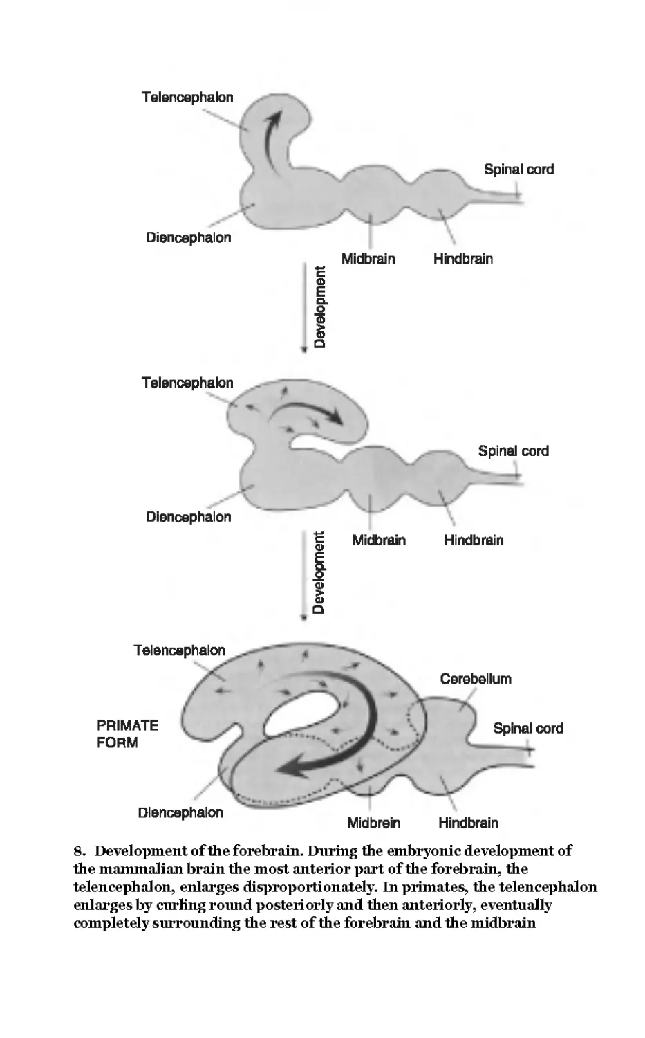
8 Development of the
forebrain 52
From Matthews, G.G.,
Neurobiology, 1997
© Blackwell Science Inc
9 Cephalization 54
From Matthews, G.G.,
Neurobiology, 1997
© Blackwell Science Inc
10 Homunculus 60
© The Natural History Museum,
London
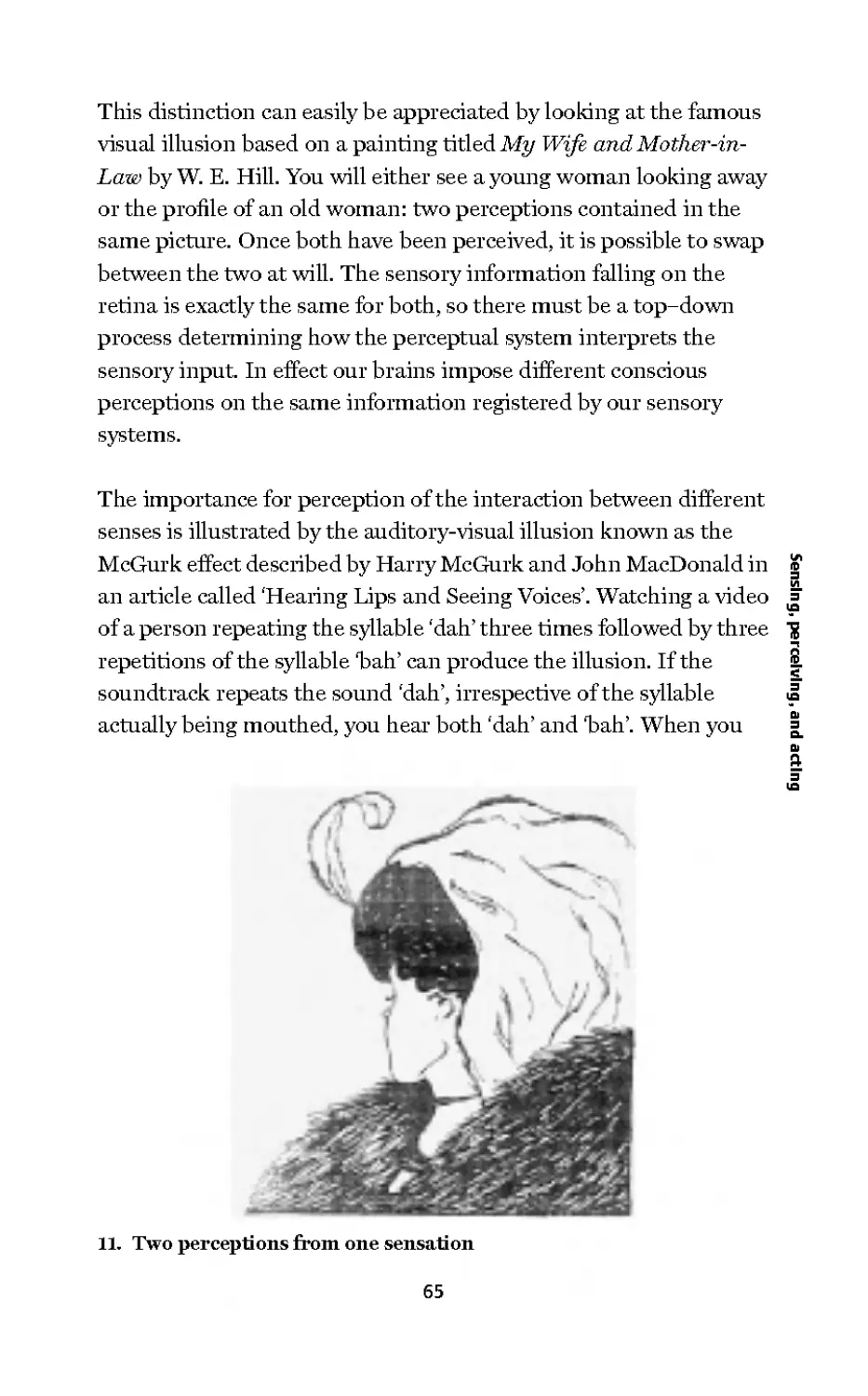
11 Two perceptions from one
sensation 65
12 Eye and retina 69
From Delcomyn, R, Foundations
of Neurobiology, 1997
© 1998 W.H. Freeman &
Company. Used with permission
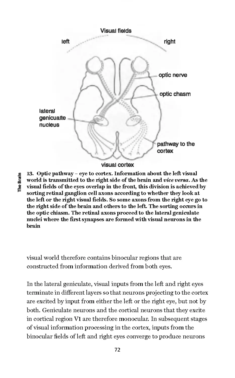
13 Optic pathway - eye to
cortex 72
From Neuroscience: The Science of
the Brain, 2003
© Professor Richard Morris/The
British Neuroscience Association
14 Columnar organization in
primary visual cortex 74
Purvis et al, Neuroscience,
3rd edition
© 2004 Sinauer Associates Inc
15 Sound localization by
coincidence detection 79
From Matthews, G.G.,
Neurobiology, 1997
© 2004 Sinauer Associates Inc
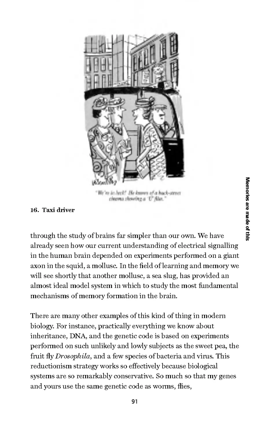
16 Taxi driver 91
Reproduced with permission of
Punch Ltd
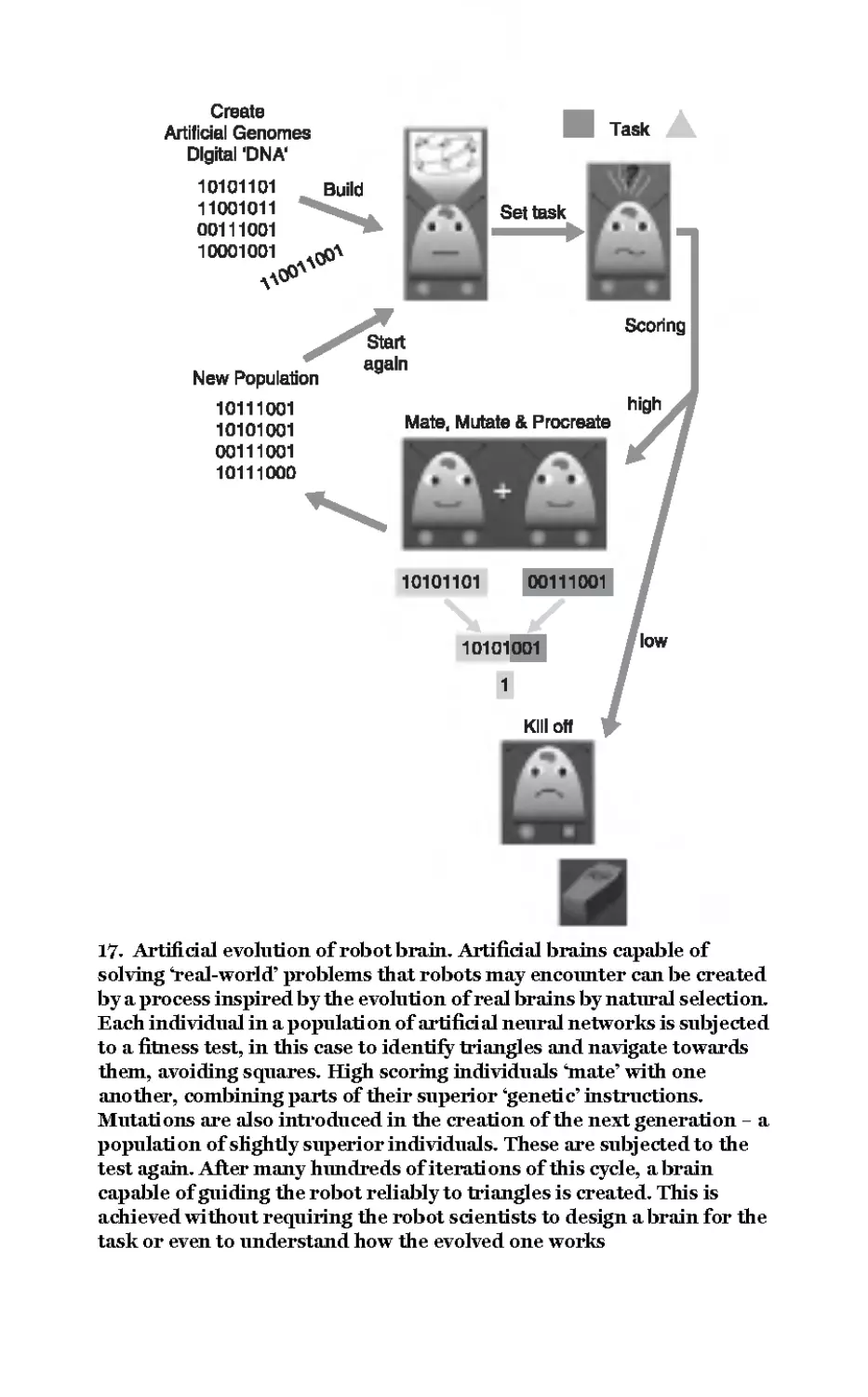
17 Artificial evolution of
robot brain 107
18 Cochlear implant 111
Purvis et al, Neuroscience,
3rd edition
© 2004 Sinauer Associates Inc
19 Monkey thoughts move
robot arm 114
Diiks University Medical Center
Chapter 1
Thinking about the brain
Think for a few moments about a very special machine, your brain -
an organ of just 1.2 kg, containing one hundred billion nerve cells,
none of which alone has any idea who or what you are. In fact the
very idea that a cell can have an idea seems silly. A single cell after
all is far too simple an entity. However, conscious awareness of one's
self comes from just that: nerve cells communicating with one
another by a hundred trillion interconnections. When you think
about it this is a deeply puzzling fact of life. It may not be entirely
unreasonable therefore to suppose that such a machine must be
endowed with miraculous properties. But while the world is full of
mystery, science has no place for miracles and the 21st century's
most challenging scientific problem is nothing short of explaining
how the brain works in purely material terms.
Thinking about your brain is itself something of a conundrum
because you can only think about your brain with your brain.
You'll appreciate the curious circularity of this riddle if you
consider the consequence of concluding, as you might, that your
brain is the most exquisitely complex and extraordinary machine
in the known universe. Clearly this is, and may be nothing more
than, the opinion of your brain about itself: the brain's way of
thinking about the brain. So it seems we are caught in the logical
paradox of a self-referencing, and in this case also a self-obsessed,
system. Perhaps the only reliable conclusion from this thought
1
experiment is that the brain is about as conceited as it is possible
to be!
Notwithstanding the brain's well-developed personal vanity, we
must grant that it provides you with some very distinctive abilities.
It operates in the background of your every action, sensation, and
thought. It allows you to reflect vividly on the past, to make
informed judgements about the present, and to plan rational
courses of action into the future. It endows you with the seemingly
effortless ability to form pictures in your mind, to perceive music in
noise, to dream, to dance, to fall in love, cry, and laugh. Perhaps
most remarkable of all however is the brain's ability to generate
conscious awareness, which convinces you that you aiefree to choose
what you will do next.
We have no idea how consciousness arises from a physical machine
and in trying to understand how the brain does that we may well be
2 up against the most awkward of scientific challenges. That is not to
j= say that the problem cannot in principle be solved, just that the
brain is a finite machine and presumably has a finite capacity for
understanding. But what are the limits of its intellectual capacity
and, at that limit, might we still be asking unanswerable questions
about the brain? Neuroscientists accept that they are faced with an
awesome challenge. The accelerating pace of discovery in
neuroscience however shows that we are a long way from any
theoretical upper limit on our capacity for understanding that
might exist. So rather than despairing of the limitations of the
human intellect, we should be optimistic in our striving for a
complete physical understanding of the brain and of its most
puzzling of properties - consciousness and the sensation of free will.
Although we have barely started this short book we have already
made a fundamental conceptual error in the way we have referred
to 'the brain'. The brain is not an independent agent, residing in
splendid and lofty superiority in our skulls. Rather it is part of an
extended system reaching out to permeate, influence, and be
2
influenced by, every corner and extremity of your body. As the spinal
cord, your brain extends the length of the backbone, periodically
sprouting nerves that convey information to and from eveiy part of
you. Practically nothing is out of its reach. Every breath you take,
every beat of your heart, your every emotion, every movement,
including involuntary ones such as the bristling of the hairs on the
back of your neck and the movement of food through your guts - all
of these are controlled directly or indirectly by the action of the
nervous system, of which the brain is the ultimate part.
From this perspective the brain is not simply a centre for issuing
instructions, it is itself bombarded by a constant barrage of
information flowing in from our bodies and the outside world.
Specialized cells called sensory receptor neurons feed information
via sensory nerves into the nervous system, providing the brain with
real-time data on both the internal state of the body and about the ^
outside world. Furthermore, information flowing into and out of i="
the brain is carried not only by nerve cells. About 20 per cent of the g-
volume of the brain is occupied by blood vessels, which supply the Z
oxygen and glucose for the brain's exceptionally high energy £.
demand. The blood supply provides an alternative communication 5"
channel between the brain and the body and between the body and
brain. Endocrine glands throughout the body release hormones into
the blood stream. These hormones inform the brain about the state
of bodily functions, whilst the brain deposits hormonal instructions
into its blood supply for distribution globally to the rest of the body.
So when we say the brain does x or y, the word 'brain' is a shorthand
for all of the interdependent interactive processes of a complex
dynamical system consisting of the brain, the body, and the outside
world.
The human brain is a highly evolved and stupendously complex
'machine' that is often compared to the most complex of man-made
machines, digital computers. But brains and computers differ
fundamentally. The brain is an evolved biological entity made from
materials such as small organic molecules, proteins, lipids, and
3
carbohydrates, a few trace elements, and quite a lot of salty water. A
modern computer is built with electronic components and switches
made from silicon, metal, and plastic. Does it matter what a
machine is made of? For computers the answer is no - computer
operations are 'medium independent'. That is to say, any
computation can in principle be performed in any medium, using
components made from any suitable material. Thus cogs and levers,
hydraulics or optical devices for that matter could replace the
electronics of a modern computer, without affecting (except in
terms of speed and convenience) the machine's ability to compute.
It seems extraordinarily unlikely either that the brain is simply
performing computational algorithms or that thinking could
equally well be achieved with cogs and levers as with nerve cells. So
perhaps we cannot expect computers to perform like brains unless
we find a way to build them in a biological medium (see Chapter 7)-
| From marks to meaning
CO
j= To gain an insight into questions about the brain that must be
answered, and to set the stage for later chapters, I will now briefly
examine the activity of the brain in the context of a familiar act of
everyday life. Let us consider the behaviour in which you are
currently engaged - namely, reading these words. What exactly is
your brain is doing right now? What kind of behaviour is reading
and what must the brain do in order to achieve it?
Obviously the brain must first learn how to read and equally
obviously reading is a means of learning and engages our
imagination. Reading also demands concentration and attention.
Therefore as you read these words your brain must direct your
attention away from the many potential distractions that are
constantly in the background, all around you. You need not worry
however because, without bothering your conscious awareness,
your brain is keeping a watchful 'eye' on external events. It can at
any moment redirect your attention away from this page and
towards something more important. Your attention can also be
4
distracted by events internal to the brain, the various thoughts that
constantly pass through it and compete for your consciousness.
Reading, when reduced to the rather prosaic level of motor actions,
depends on the brain's ability to orchestrate a series of eye
movements. Now, as you read these words, your brain is
commanding your eyes to make small but very rapid (about 500°
per second) left-to-right movements called saccades (right-to-left or
up-and-down for some other written languages). You are not
consciously aware of it, but these rapid movements are frequently
interrupted by brief periods when the eyes are fixed in position.
Watch someone reading and you will see exactly what I mean. You 11
notice that the eyes do not sweep smoothly along the line of text,
rather they dart from one fixation to another. It is only during the
fixations, when the eyes dwell for about a fifth of a second, that the
brain is able to examine the text in detail. Reading is not possible
during the darting saccadic movements because the eyes are
moving too quickly across the page. You are not aware of the blur
and confusion during a saccade because fortunately there is a brain
mechanism that suppresses vision and protects you from visual
overload.
Reading is only possible between saccades, not only because the
eyes are then stationary but also because gaze is centred on the
retina's fovea. The fovea is the only part of the retina specialized for
high acuity vision (see Chapter 5), but it scrutinizes a very small
area of our visual world. As a literal rule of thumb, fovea! vision is
restricted approximately to the area of your vision covered by your
thumbnail held at arm's length. It is a small window of clear vision
within which you are able to decipher just 7 or 8 letters of normal
print size at a time. The task for the brain is to generate a precise
series of motor commands to the eye muscles which ensure that at
the end of each saccade your high acuity vision is fixed on that part
of the text you need to see most clearly next. As your eyes approach
the end of a line, the brain generates a carriage return. Of course the
return saccade must be to the left, of the correct magnitude and
5
associated with a slight downward shift in gaze in order to bring the
first word on the next line onto the fovea.
I have considered only the simple case of the brain directing eye
movements alone, as if nothing else affects gaze direction. But of
course the relative positions of the eye and page are affected
continuously by head, body, and book motion. Thus the brain must
continually monitor and anticipate factors affecting the future
position of your eyes relative to the text. The fact that you can
effortlessly read on a moving train while eating a sandwich is
evidence that your brain can solve this problem quite easily.
Importantly, it is done automatically and on an unconscious level
without you having to think through every step. If you had to
consciously think about the mechanical process of reading, you
would be illiterate!
Our lack of conscious awareness of underlying brain processes can
2 also be illustrated by reflecting on the subjective experience that the
j= comprehension of written material represents. While reading we
are not conscious of the fragmented nature of comprehension
imposed by underlying move—stop—move—stop activity of the eyes
I've just described or by the fact that only 7 or 8 letters can be
deciphered at each stop. On the contrary, our strong subjective
impression is that comprehension of the text flows uninterrupted
and moreover that we can read several words or even whole
sentences 'at a glance'. That this is not the case can be illustrated by
reading a sentence containing a word that has more than one
meaning and pronunciation. For example, the word tear has two
very different meanings and pronunciations in English - tear the
noun of crying and tear the verb of ripping apart. Clearly such word
ambiguity complicates the brain's task of providing you with an
uninterrupted comprehension. If for instance the word tear
occurred at the beginning of a sentence its meaning might remain
ambiguous until the subject of the sentence appears later. Because
you cannot read the whole sentence at a glance your brain may be
left with no option but to choose one of the alternative meanings
6
(or sounds, if you are reading aloud) of a word and hope for the
best.
While we cannot read whole sentences at a glance, the brain does
recognize each word as a whole. What is quite surprising however is
that the order ofthe letters is not particularly important (good news
for poor spellers). That is whyyou will be able to read the following
passage without consciously having to decode it.
I cdnnolt blveiee talit I clnod anlaclty nesdnatnrd walit I was
rdgnieg. It deosn't mttaer in walit oredr the ltteers in a wrod aer, the
olny iprmoatnt tiling is talit eth frist dan lsat ltteer be in the rghit
pclae. The rset cna be a taotl rases and yno can still raed it wontliit a
porbelra. Tills is bcnseae the huamn ranid deos not raed ervey lteter
by istlef, but the wrod as a wlohe. Arazanig huh?
We will now consider how and in what form textual information
at the gaze point enters the brain. Light-sensitive cells called
photoreceptors capture light focused as two slightly different
images on the left and right retinae. The photoreceptors undertake
a fundamental and remarkable transformation of energy, a
transformation that must occur for all of our senses. This process is
known as sensory transduction and always involves converting the
energy in the sensory stimulus, in this case light energy, into an
electrical signal. This is because the nervous system cannot use light
or sound or touch or smell directly as a currency of information
transmission. In the brain electricity is the critically important
currency of information flow.
The brain interprets or decodes electrical signals according to their
address and destination. We see an electrical signal coming from the
eyes, hear electrical signals from the ears, and feel the electrical
signals coming from touch sensitive cells in the skin. You can
demonstrate the importance of signal origin by pressing very gently
with your little finger into the corner of your closed right eye, next to
your nose. The touch pressure will locally distort the retina and
7
produce an electrical signal that will be transmitted to the brain.
Your brain will 'see' a small spot of light in the visual field caused
by touch. Notice that the light appeal's to be coming from the
peripheral visual field somewhere off to the right; a moment's
thought should tell you why this is so.
The photoreceptor cells of the retina are not connected directly to
the brain. They communicate with a network of retinal neurons
through a mechanism that couples the fluctuating electrical signal
in the photoreceptor to the release of a variety of chemicals known
as neurotransmitters. In their turn, neurotransmitters convey
signals from one neuron to another by generating or suppressing
electrical signals in neighbouring neurons that are specifically
sensitive to particular neurotransmitters. This transformation of an
electrical into a chemical signal occurs mostly at specialized sites
called chemical synapses. Electrical signals can also pass directly
between neurons at sites known as electrical synapses. Thus,
2 through a combination of direct electrical transmission between
CO °
j= neurons and the release of chemical messengers, information about
the visual image captured by the eyes is processed in the retina
before being conveyed by the optic nerve to the brain.
There are about one million output neurons in the retina, known
as retinal ganglion cells, and each one extends a long, slender
fibre or axon in the optic nerve. Axons are specialized for the
high-speed (up to 120m/second), long-distance, and faithful
transmission of brief electrical impulses. Impulses travelling along
the axons of the retinal ganglion cells in the optic nerve reach the
first neurons in the brain about 35 thousandths of a second after the
capture of photons by the photoreceptors. In the brain, the axons
of the retinal ganglion cells terminate and form synapses with a
variety of other neurons which in turn interconnect with many
others, a process which results finally in the conscious awareness
of a vivid picture in your mind of what your eyes are looking at.
Somehow this astonishing electrochemical process that involves no
conscious effort whatsoever produces meaningful pictures in your
8
mind - close your eyes and the picture goes away, open them and it
appears to you apparently instantaneously and effortlessly. Truly
amazing!
Reading does not come naturally; it is a difficult skill that must be
acquired painfully. Once learnt however it is rarely, if ever, forgotten
- thankfully. So we do not have to worry about forgetting how to
read because the skill is robustly established in our long-term
memory banks. Although the enabling skill of reading is retained in
permanent memory, an entirely different type of memory is
required during the active process of reading itself. While reading,
we must retain a short-term 'working memory' for what has just
been read. Some of the information acquired while reading maybe
committed to long-term memory but much is remembered for just
long enough to enable you to understand the text. Memories must
somehow be represented physically in the brain. Brain chemistry ^
and structure is altered by experience and the stability of these i="
physicochemical changes presumably corresponds to the retention g-
duration of memory. So what exactly is a memory? What kind of Z
physical trace is left in the brain after we have learnt some new skill £.
or fact? What is forgetting and why are some memories quickly 5"
forgotten and others never? These are questions to which I shall
return later.
Finally we must consider one of the most elusive of problems. While
accepting that everything that the brain does depends on lawful
processes occurring within and between the brain's cells, how can
we explain how 'meanings' arise in our minds while reading words?
How do marks on paper become images in the mind, how do they
make you think? How can any of this be explained completely by
the responses of individual brain cells and interactions between
them? Consider for example what happens when I recognize the
word banana. I instantly call up an image of a yellow, curved object
about 20 cm in length, 4 cm in girth, that is edible and incidentally
delicious. We might propose that there is a single neuron in my
brain that responds when I read "banana' and triggers all of the
9
remembered associated thoughts. Maybe this is the same neuron
that responds when I see a real banana.
According to this logic, a different neuron fires when I look at an
apple and another recognizes my grandmother. While it is true that
neurons can respond rather specifically to particular stimuli, most
neuroscientists believe that there can be no one-to-one
correspondence between the response of an individual neuron and
a perception. Surely a separate neuron cannot detect and represent
every object and percept? After all, in order to know that that object
is a banana, information about shape, size, texture, and colour must
somehow be bound together with stored knowledge about fruit, my
appetite, and so on. These processes are associated with different
networks of neurons in different parts of the brain and there is no
known way they could all converge on a single neuron which when
activated could trigger 'aha, a banana for lunch'. Another way to
think about the relationship between the activities of neurons and a
2 perception is to consider how assemblies of nerve cells in different
j= parts of the brain cooperate with one another in parallel. Having
said that, we are far from understanding how objects, meanings,
and perceptions are encoded in the brain by the activities of
neurons. This is one of the most intriguing of problems in
neuroscience. While the notion that there is a separate nerve cell in
the brain for each object, meaning, and perception (parodied by the
term 'Grandmother cell') has been roundly rejected, there is a
lasting appeal in this simple idea. Indeed provocative research
published at the time of writing this sheds a fresh perspective on the
way objects are represented in the brain. It suggests that the idea
there may be a neuron in your brain that only recognizes your
grandmother deserves some serious reconsideration - I shall return
to this matter later.
In the following chapters of this book I shall examine in more detail
the questions and issues considered in this introduction. Starting
with a historical perspective on the development of modern brain
science I go on to describe electrical and chemical signalling
10
mechanisms that underlie all mental functions, how the nervous
systems evolved, how the brain responds to sensations and
perceptions, the formation of memories and what can be done when
the brain is damaged. The potential for interfacing the brain with
computers is discussed, as is the contribution of neuroscience to
developments in robotics and artificial nervous systems. Finally, I
discuss the future scientific challenges associated with
understanding how the brain works as a whole.
11
Chapter 2
From humours to cells:
components of mind
The widespread occurrence of the 'surgical' technique of
trepanation, the removal of parts of skull to expose the brain, in
early civilizations suggests that ancient cultures recognized the
brain as a critical organ. This is not to suggest that a link between
the brain and the mind has its roots in prehistory. In fact the long
history of neuroscience prior to the scientific period suggests that it
is not at all self-evident that mental functions must necessarily be
attributed to the brain. The Egyptians for instance clearly did not
hold the brain in particularly high esteem since in the process of
mummification it was scooped out and discarded (a practice that
stopped around the end of the 2nd century ad). To the ancient
Egyptians, it was the heart that was credited with intelligence and
thought - probably for this reason it was carefully preserved when
mummifying the deceased.
Although Hippocrates (460-370 bc) is usually accredited with
being the first in the West to argue that the brain is the most
important organ for sensation, thought, emotion, and cognition,
he was not the first Greek to consider the question of physical
embodiment of mind. Prior to the Hippocratic revolution,
Pythagoras (582-500 bc) believed that matter and mind are
connected somehow and that the mind is attuned to the laws
of mathematics. It was probably of little interest to Pythagoras
whether mind and matter were connected in the brain or, as
12
the Egyptians and the Greeks prior to 500 bc believed, in the
heart.
Alcmaeon of Croton (b. 535 bc), himself a follower of Pythagoras, is
among the first to have realized that the brain is the likely centre of
the intellect. He is also the first known to have conducted human
dissections and in doing so he noticed that the eye is connected to
the brain by what we now know is the optic nerve. It was on the
basis of his direct observations that Alcmaeon astutely speculated, a
century before Hippocrates came to a similar conclusion, that the
brain was the centre of mental activity. Hippocrates went further
than this however and elaborated a theory of four humours that
together were responsible for the temperament. Thus, according to
Hippocrates, the four determinants of temperament were black bile
(melancholy), yellow bile (irascibility), phlegm (equanimity and
sluggishness), and sanguine (passion and cheerfulness). To us the
humoral theory seems implausible, puzzling, and arbitrary. It
seems to have been inspired, not by the evidence of observation,
but by the need to conform with the equally unlikely postulates
of contemporary Greek natural law, namely that there are four
elements: earth, air, water, and fire.
The influence of Hippocrates was to be profound and remarkably
long lasting. Some 400 years after Hippocrates died, Claudius
Galenus of Pergamum (ad 131-201), better known as Galen,
became the most influential physician of his time, in part by
building his own theory on the humoral conjectures of Hippocrates.
Galen was unusually well informed on the internal anatomy of the
human body, an intimate understanding of which he gained while
he was physician at a school for gladiators. However, although we
can be grateful to him for perpetuating the idea that the brain is
the seat of the mind, he continued the Hippocratic tradition of
disregarding the importance of the solid tissues of the brain for
mental functions. Instead Galen associated the presence in the
brain of three fluid filled cavities, or ventricles, with the tripartite
division of mental faculties - the rational soul - into imagination,
13
reason, and memory. According to Galen, the brain's primary
function is to distribute vital fluid from the ventricles through the
nerves to the muscles and organs, thereby somehow controlling
bodily activity. Precisely how the brain's ventricles were supposed
to regulate the three cognitive functions is not explained,
unsurprisingly.
Galen's positive contribution to medical knowledge is undeniable,
but many of his ideas were seriously flawed. This would not have
mattered too much were it not for the fact that, after he died,
Galen's authority dominated and therefore hampered medical
science and practice for some 400 years. A particularly interesting
example of his influence can be seen in the early anatomical
drawings of Leonardo da Vinci (1452-1519). In one drawing of the
head, the brain is depicted crudely consisting of three simple
cavities labelled O, M, and N. Leonardo interpreted the anatomical
division in functional terms in a way that would have been
2 immediately recognizable to Galen in the 1st century.
01
I-
Later Leonardo was to make some of the most important
observations on the brain and its ventricles. He can be credited with
the first recorded use of solidifying wax injection to make castings
to study the internal cavities of the brain and other organs,
including the heart. Using this method, Leonardo accurately
determined the shape and extent of the brain's cavities, but
he clearly continued to place a Galenical interpretation on the
fluid-filled structures. For instance the lateral ventricles carry the
word imprensiva (perceptual) in Leonardo's hand, the third
ventricle is labelled sensus communis, and the fourth ventricle,
memoria. Leonardo's use of wax injections represented a scientific
advance of enormous potential and importance. Unfortunately,
the dominance of Galen's conjectures about the functions of the
ventricles diverted his attention from the solid tissue of the brain,
the true seat of the mind.
Ideas about brain function and mechanisms continued to be
14
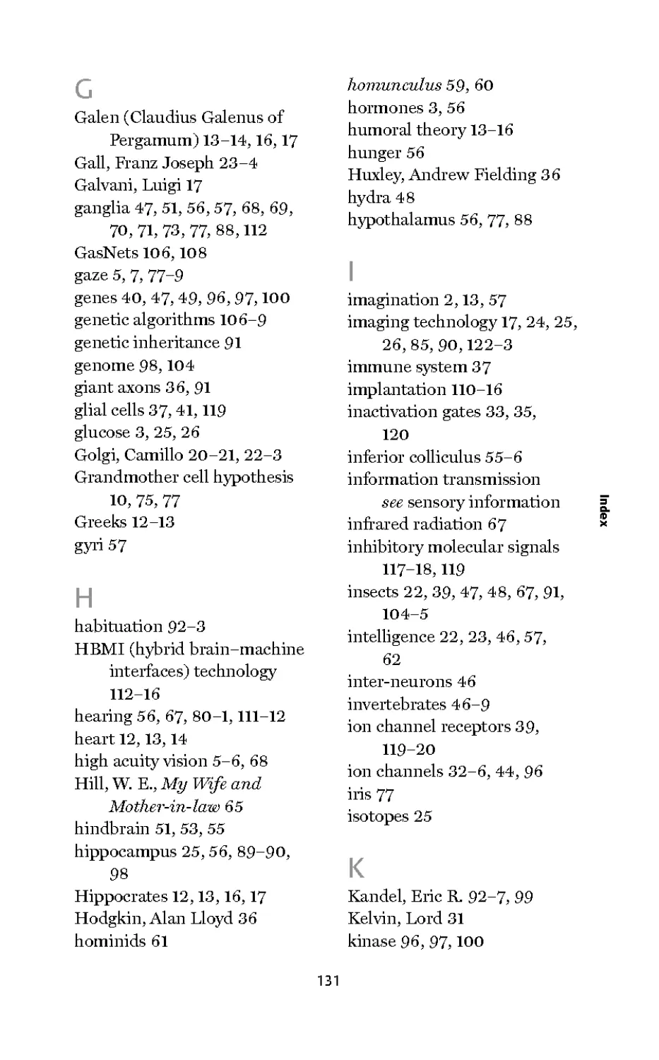
1. Anatomical drawing by Leonardo da Vinci: The human head and its
contents according to Leonardo da Vinci. Probably drawn c.1490,
it represents an attempt to translate a description of the brain given by
medieval philosophers. This drawing shows (wrongly) that the eye is
connected by its optic nerve to three simple cavities labelled O, M andN.
Leonardo ignores the intricate structure of the solid tissues of the brain.
The smaller drawings include a section of an onion (accurate), the eye
and orbit and a horizontal section of the head
strongly influenced by theories involving the flow and distillation of
vital fluids, spirits, and humours well into the 17th century. Indeed
the influence of Hippocrates and Galen can be seen in the hydraulic
model of the brain formulated by the most famous 17th-century
French philosopher, Rene Descartes (1596-1650). Descartes
however reformulated the humour-based description of the brain's
functioning and expressed it in contemporary terms by comparing
the brain to the working of intricate machines of his time, such
as clocks and moving statues, the movements of which were
controlled by hydraulic systems. Importantly he departed from the
classical tradition of locating cognitive processes exclusively in the
brain's fluid-filled ventricles, but he nonetheless still referred to
the flow of spirits through nerves and made no attempt to assign
functions to specific brain structures, with the notable exception of
the pineal gland. The pineal, because it was a unitary and central
structure, was supposed to be the link with the singular soul but was
also given executive control, directing the flow of animal spirits
2 through the brain.
CO °
01
■c
I-
In one important respect Descartes was breaking new ground. By
comparing the workings of the brain with that of complex hydraulic
machines, he was regarding the most technologically advanced
artefacts of his day as templates for understanding the brain. This is
a tradition that persists today; when we refer to computers and
computational operations as models of how the brain acquires,
processes, and stores information, for example. So while Descartes
was hopelessly wrong in detail, he was adopting a modern style of
reasoning.
Perhaps it is not surprising that theories involving the solid tissues
of the brain were difficult to conceive - after all, the brain's solid
substance has no visible moving parts. By the 17th century, however,
the grip of humoral theory was weakening, in part due to the works
of a new generation of anatomists who were describing the internal
structure of the brain with increasing accuracy. Notably, the
Englishman Thomas Willis (1621-75), who coined the term
16
'neurology', argued that solid cerebral tissue has important
functions. He still held that fluid-flow was the key to understanding
brain function, but his focus was on the solid cerebral tissues and he
showed that nervous function depends on the flow of blood to them.
Today's functional brain imaging technique (fMRI) shows that
small local increases in blood flow are associated with the activation
of nerve cells. That there is in effect a local "blushing' of the brain
when the neurons in that region are active is an observation that
Willis might well have expected and enjoyed.
Among the more obvious problems of vital fluid and hydraulic
models of nervous system function, and no doubt known to Willis,
is that nerves are not hollow conduits. And even if they were, the
speed of fluid movement through them could hardly be sufficiently
swift for the rapidity with which sensations and motor commands
seem to be conveyed by nerves. These and other inconsistencies
with fluid models of the nervous system must have worried
physicians of the stature of Willis and caused them pause for
thought. But Willis remained a fluid theorist and the beginning of
the end for the fruitless elaboration of such theories did not come
until the discoveries attributed to Luigi Galvani (1737-98). In the
late 18th century he discovered the importance of electricity to the
operation of the nervous system. As electrical mechanisms were to
provide the necessary speed, attention inevitably turned from fluid
to electrical models. Ironically, the last gasp of the legacy of
Hippocrates and Galen is to be found in the interpretation Galvani
himself placed on his own experiments with 'animal electricity.
Having demonstrated that he could control the contractions of a
frog muscle by applying electrical currents to the muscle's motor
nerve, Galvani claimed to have discovered that animal nerves and
muscle contain an electric 'fluid'. A decisive leap of understanding
however was achieved when Galvani and his contemporary
Alessandro Volta (1745-1827) crucially together linked electricity to
the functions of the nervous systems.
What neither Galvani nor Volta could know however is that the
17
externally applied electrical stimuli were activating biological
processes causing high-speed electrical impulses to travel along
nerves to muscles, resulting in their contraction. It was not until the
middle of the 19th century that the ability of nerves and muscles to
generate rapidly propagating electrical impulses was confirmed by
the German physiologist Du Bois-Reymond (1818-96). This was a
major impetus to the study of the physical workings of the brain and
set the stage for the modern scientific era, which was launched in a
most spectacular way at the dawn of the 20th century by the
recognition of the cellular nature of the brain's tiny functional
units - the neurons.
The true cellular nature of the brain and of its mental functions was
first recognized by the father of modern neuroscience, the Spanish
neuroanatomist Santiago Ramon y Cajal (1852-1934). Although his
proposition that the brain is a cellular machine may today seem
commonplace, in fact it was revolutionary. In the later 19th century,
2 and indeed in the early years of the 20th century, most
j= neuroanatomists believed that the brain was not composed of cells
at all - in spite of a universal recognition that all other organs and
tissues in our bodies were. What was it about the brain that made it
so difficult to see its cellular composition under the microscope?
Part of the answer is that brain cells are quite unlike any other cells.
The very term 'cell' implies uniformity; simple structures defined by
clear boundaries.
In contrast neurons are hugely diverse in morphology. They have
exceedingly fine and profusely branched processes ramifying from
the cell's body and intermingling among the branches of other
neurons. The complexity and diversity of their physical appearance
easily exceeds that of all other cell types found in any other part of
the body. All of this contributed to a rather confusing picture which
anatomists found difficult to reconcile with a simple cellular model
of brain structure. When viewed through a microscope the brain
appeared to consist of a hopelessly tangled morass (a reticulum),
without the distinct cell-defining boundaries that are so evident in
18
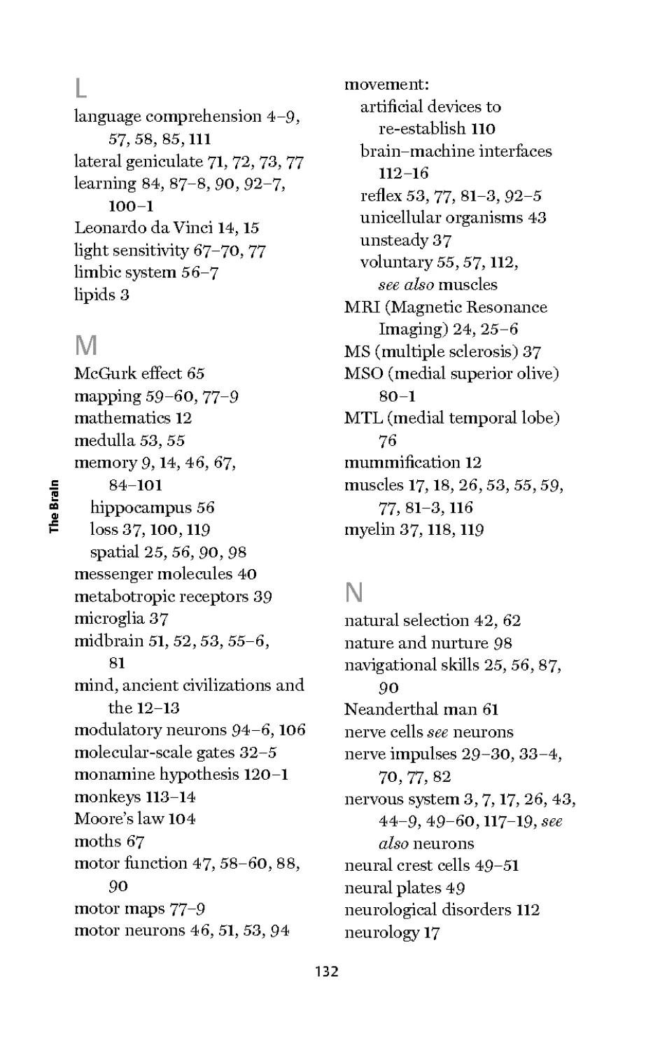
2. A selection of neurons to illustrate diversity: Neurons are more
diverse in their appearance than any other type of cell. Their complex
branched morphologies are a reflection of their need to communicate
with other nearby and more distant neurons. Complexity of shape is
no guide to the overall performance of the brain - a neuron in the
mammalian brain (top left) is hardly more complicated than a neuron
from an insect brain (bottom right)
other tissues. It was therefore not surprising perhaps that cell
theory, the idea that tissues are composed of cells, was thought not
to apply to the brain and a radical alternative model was proposed.
This came to be called the 'reticular theory' of brain anatomy - a
surprisingly resilient interpretation that persisted well into the 20th
century.
The reticular theory was wrong, but that was not the only problem
with it. Scientific theories are allowed to be wrong so long as they
are helpful, but the reticular theory, which held that the brain
contains no discrete components, was actually obstructive to
scientific progress. Progress was hindered by the concept of a
machine without discrete functional components because without
them it is impossible to formulate a plausible mechanism to explain
how the brain might work. Scientists were sure the brain machine
must have components and, given the complexity of what the brain
does, lots of them. But what were they, what did they look like, and
2 what did they do? It was clear that to understand the brain science
j= had to identify the functional components of the brain's
microscopic structure.
Towards the end of the 19th century, the Italian anatomist Camillo
Golgi (1843-1926) developed away of highlighting the morphology
of very few neurons in any particular region of the brain. It was a
staining method that fitted the bill because it allowed individual
neurons to be viewed unobstructed by the tangled mass of branched
processes of neighbouring cells. It incorporated the chemistry of
photographic processing and it revealed individual neurons as dark,
silver-impregnated silhouettes. Paradoxically, the crucial feature of
Golgi's method was that it hardly ever worked! Just one in a
thousand or so neurons were ever revealed and these were scattered
more or less randomly throughout the brain tissue. It was precisely
because of this uncertain aspect of the method that neurons could
for the first time be seen in their entirety disentangled from their
neighbours. Immediately it was apparent that there are discrete
cells in the brain, but they are astonishing cells - unlike any others.
20
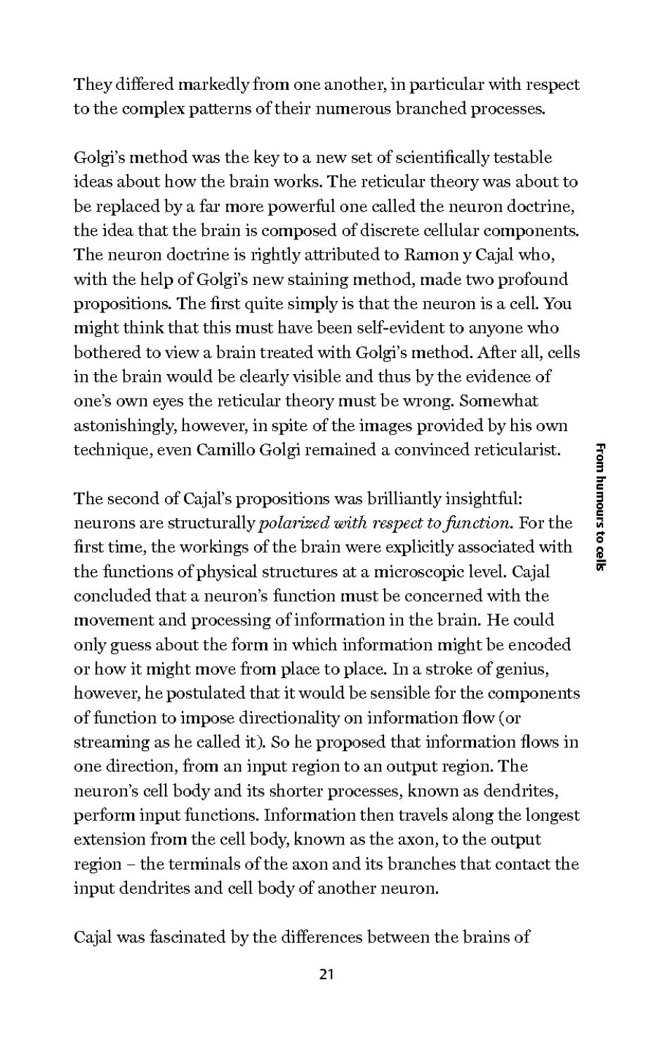
They differed markedly from one another, in particular with respect
to the complex patterns of their numerous branched processes.
Golgi's method was the key to a new set of scientifically testable
ideas about how the brain works. The reticular theory was about to
be replaced by a far more powerful one called the neuron doctrine,
the idea that the brain is composed of discrete cellular components.
The neuron doctrine is rightly attributed to Ramon y Cajal who,
with the help of Golgi's new staining method, made two profound
propositions. The first quite simply is that the neuron is a cell. You
might think that this must have been self-evident to anyone who
bothered to view a brain treated with Golgi's method. After all, cells
in the brain would be clearly visible and thus by the evidence of
one's own eyes the reticular theory must be wrong. Somewhat
astonishingly, however, in spite of the images provided by his own
technique, even Camillo Golgi remained a convinced reticularist.
The second of Cajal's propositions was brilliantly insightful:
neurons are structurally polarized with respect to junction. For the
first time, the workings of the brain were explicitly associated with
the functions of physical structures at a microscopic level. Cajal
concluded that a neuron's function must be concerned with the
movement and processing of information in the brain. He could
only guess about the form in which information might be encoded
or how it might move from place to place. In a stroke of genius,
however, he postulated that it would be sensible for the components
of function to impose directionality on information flow (or
streaming as he called it). So he proposed that information flows in
one direction, from an input region to an output region. The
neuron's cell body and its shorter processes, known as dendrites,
perform input functions. Information then travels along the longest
extension from the cell body, known as the axon, to the output
region - the terminals of the axon and its branches that contact the
input dendrites and cell body of another neuron.
Cajal was fascinated by the differences between the brains of
21
markedly different organisms: human, worms, snails, insects, and
so on. He thought comparisons of their brains might be instructive
precisely because vast differences exist between the behaviour and
intellectual capabilities of different creatures. There is
unquestionably an enormous gulf between human and insect
intelligence, so it would be reasonable to suppose that a comparison
of their brains would expose how structural components reflect
intelligence. Surely, the human brain should contain 'high
performance' components and the insect brain markedly less
sophisticated ones. But the difference between insect and human
neurons does not at all betray the gulf between insect and human
intelligence. Insect neurons are as complex and display as much
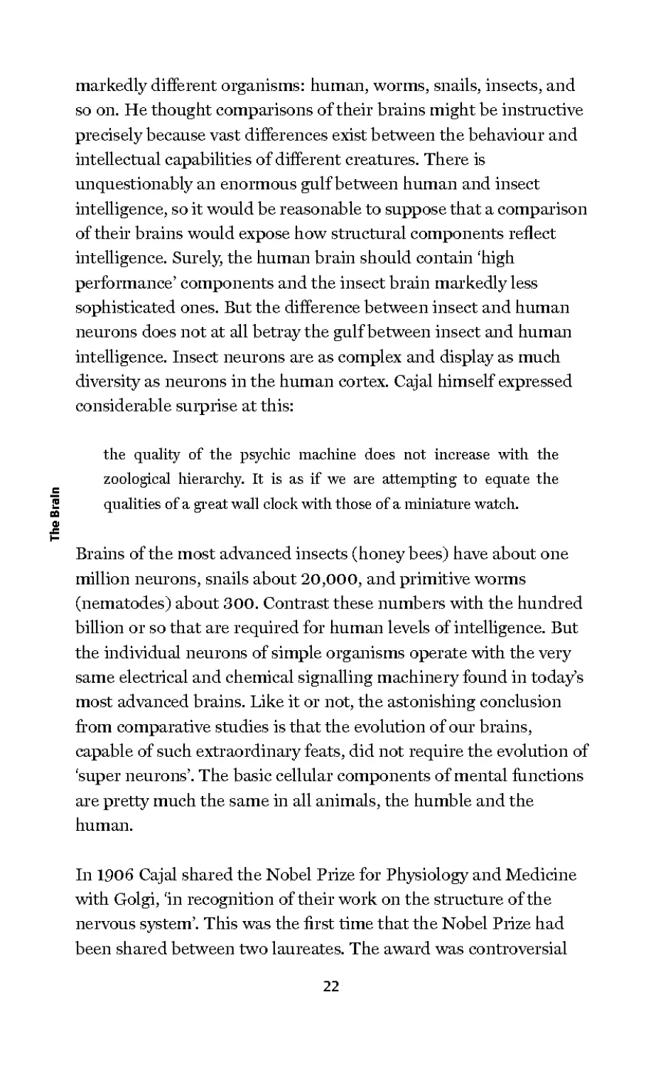
diversity as neurons in the human cortex. Cajal himself expressed
considerable surprise at this:
the quality of the psychic machine does not increase with the
zoological hierarchy. It is as if we are attempting to equate the
qualities of a great wall clock with those of a miniature watch.
Brains of the most advanced insects (honeybees) have about one
million neurons, snails about 20,000, and primitive worms
(nematodes) about 300. Contrast these numbers with the hundred
billion or so that are required for human levels of intelligence. But
the individual neurons of simple organisms operate with the very
same electrical and chemical signalling machinery found in today's
most advanced brains. Like it or not, the astonishing conclusion
from comparative studies is that the evolution of our brains,
capable of such extraordinaiy feats, did not require the evolution of
'super neurons'. The basic cellular components of mental functions
are pretty much the same in all animals, the humble and the
human.
In 1906 Cajal shared the Nobel Prize for Physiology and Medicine
with Golgi, 'in recognition of their work on the structure of the
nervous system'. This was the first time that the Nobel Prize had
been shared between two laureates. The award was controversial
22
because the two disagreed on a crucially important matter - Golgi
remained convinced that Cajal was wrong to reject the reticular
theory. It was of course Golgi who was wrong and fundamentally so.
Other questions over Golgi's interpretations raised serious doubts
in the minds of some of the scientists advising the Nobel Council as
to the appropriateness of his nomination for the prize. But whatever
the merits of the case for a shared prize, 1906 marked the beginning
of the modern era in the neurosciences and it was the first of a series
of Nobel Prizes to be awarded to neuroscientists over subsequent
decades.
Cajal could not have anticipated the extraordinary advances in
brain science that were about to be made. His recognition of the
neurons as polarized units of information transmission was a
defining moment in neuroscience. But at the start of the 20th
century many questions about precisely how and in what form
neurons signal information in the brain remained unanswered. By
the middle of the 20th century, neuroscience had become the fastest
growing discipline in the history of scientific endeavour and by the
end of that century a more or less complete understanding, in
exquisite molecular detail, of how neurons generate electrical and
chemical signals would be achieved.
In this very short histoiy of man's discovery of the workings of the
brain I cannot avoid reference to the discredited pseudo-science of
phrenology, a theory developed by the idiosyncratic Viennese
physician Franz Joseph Gall (1758-1828). Gall believed that the
brain is the organ of the mind but he went much further and
postulated that different distinct faculties of the mind, innate
attributes of personality, and intellectual ability, are located in
different sites in the brain. Gall reasoned that different individuals
will have these innate faculties and that the degree of their
development would be reflected in the size of the surface region of
the brain that housed that particular faculty. These ideas have a very
modern ring to them, but Gall thought that the skull would take the
shape of the brain's relief and therefore that the bumps on the
23
surface of the skull could be 'read' as an index of various
psychological aptitudes.
The practice of phrenology grew and flourished in Europe and then
in America from about 1820, becoming a popular fad in the latter
part of the 19th century before effectively dying out early in the 20th
century (though in fact the British Phrenological Society was not
disbanded until 1967). Its demise in the early 20th century
coincided with the rapid accumulation of real evidence for the
principle that many discrete mental functions are highly and
specifically localized to particular parts of the brain. Much of the
evidence came as a consequence of the First World War in the form
of the many unfortunate victims of gun-shot and shrapnel lesions to
specific regions of the brain that produced reproducible disorders.
More recently, functional brain imaging techniques such as fMRI
have shown beyond doubt that different cognitive functions are
indeed localized to specific parts of the brain. So while the
2 exaggerated claim of phrenologists to be able to read the mind from
j= the bumps on the head was refuted, their premise was vindicated.
Imaging the future of brain research
The first high definition imaging system, called Computed
Axial Tomography (CAT scanning), was developed in the
1970s. It is an X-ray-based technology that was used, and still
is, as a medical diagnostic tool to resolve the position of
brain tumours in the brain for example. In the past 30 years
more powerful imaging technologies have been developed
that have the potential to associate different cognitive
functions with different structures in the brain. These techniques
include most notably Positron Emission Tomography and
Magnetic Resonance Imaging.
24
When PET is used to link function to structure, increases in
local blood flow and glucose consumption associated with
increased neuronal activity are measured. A radioactive
isotope, of glucose for instance, is injected into the blood stream
and the high-energy photons tliat fly off in exactly opposite
directions from the site of an emitting isotope are detected
by an array of detectors tliat encircle the head. The detectors
facing one another on opposite sides of the head will simul-
taneously detect the two photons generated from the same
place within the brain. By the integration of simultaneous
photon detection in the array, the source of the isotope can
be calculated. In this way a computer builds an image of the
structures tliat contain the isotope. In other applications of
PET, the radioactive label is attached to molecides that bind
to paiticiilar receptors, thus revealing the distribution of
neurotransmitter systems receptors in the brain, for
example.
A more sensitive technique, impoitantly tliat does not
involve radioactive tracers, is Magnetic Resonance Imaging
or MRI. Briefly the technique involves the pulsing of a strong
external magnetic field, which evokes transient magnetic
responses within the brain. The evoked magnetic signals are
used to compute 2D and 3D images of the brain's structure.
This technique can be used for purely stru ctural studies, as it
was in the experiments on London taxi drivers tliat showed
they have a larger than expected hippocampus (see Chapter
6). But in its most interesting experimental application it
provides images of the brain in action. When used to reveal
active regions of the brain involved in particular functions,
the technique is known as functional MRI, or fMRI for short.
25
To understand how fMRI works, and to appreciate its
limitations, it is important to realize that it does not image the
electrical activity of neurons directly. It monitors the
indirect consequence of their activity. When a region of the
brain is actively working, more neurons in that region will
require more glucose and oxygen. Tliis is a consequence of
two interesting facts. First, it seems neurons only store
enough energy for the briefest of bouts of activity. If neurons
are active long enough, they need refuelling to enable them
to produce the energy storage molecule ATP required to
recharge their batteries (see Chapter 3). An active brain
region therefore may have a significantly higher metabolic
demand for oxygen and glucose than a quiescent region.
A simple solution to tliis problem would be to pump more
blood into the active brain, much in the same way that a
muscle is supplied with more blood when exercised
vigorously. However unlike a muscle, which becomes engorged
with blood and swells when exercised, the brain is confined
by the skull and cannot be allowed to swell significantly. The
solution to tliis tricky problem is to maintain a constant
overall blood-volume in the brain and to arrange for blood to
be diverted preferentially to active regions. Blood is diverted
by the ability of blood vessels in the brain to dilate in
response to signals coming from nearby active neurons.
Dilation reduces resistance to blood flow, thereby increasing the
supply of blood to the region of elevated neuronal activity.
We are not really sure how the blood vessels 'know5 that
nearby neurons are hyperactive. It is likely however that the
signal for blood vessel dilation is the gas nitric oxide (NO),
26
because NO causes the relaxation of muscle cells in the walls
of blood vessels. It is thought that NO-producing neurons
sense increased activity of nearby neurons and respond by
producing NO in the same region - thus coupling increased
neural activity to increased blood flow in that region.
In detecting regions of increased blood flow, fMRI
recognizes the different magnetic signatures of oxygenated and
deoxygenated haemoglobin. When neurons in a brain region
are sufficiently active for long enough, blood in their vicinity
becomes oxygen depleted. This is followed by an increased
flow of oxygenated blood to that region; quite literally there
is a local blushing of the brain. The fMRI technique is
responsive to the blushing and indirectly assigns increased
neural activity to that region at a spatial resolution of just a
few cubic millimetres. It is in this way that we now have a far
more fine-grained functional map of the brain than was
previously possible. Bold claims are now being made about
complex cognitive functions: where in the brain we recognize
faces and words, where executive functions are carried out,
where false memories are located, and so on.
27
Chapter 3
Signalling in the brain:
getting connected
The problem of connection, the sending of information effectively
around the nervous system, arises because signals must be
communicated undistorted over the length of the body, which
might be a very large distance indeed, in the case of the blue whale
for example. Coupled to this is the fact that, in an unforgiving
world, animals must react quickly to be an effective predator or so
as to avoid being eaten. So the basic requirements of signalling
coded information in the nervous system are that the signals have to
be routed correctly and sent reliably over long distances as rapidly
as possible.
In order to achieve this neurons convey and encode information
electrically. Brief electrical pulses (lasting a few thousandths of a
second), known as action potentials or nerve impulses, travel along
biological cables (axons) that extend from the cell bodies of neurons
to connect their input to their outputs with other neurons.
Compared to the speed of electrical information traffic along the
wires in a computer (close to the speed of light), conduction
velocities of impulses in the brain are slow, about 120 metres per
second in the fastest conducting axons. When they reach the
terminals of axons, impulses trigger the release of chemical signals
that are able to initiate or suppress electrical signals in other
neurons. In this way neurons transmit information from one to
another by an alternating chain of electrical and chemical signals.
28
The chemical signals are released at specialized sites called
synapses, at which the chemical signals (neurotransmitters) pass
across a very narrow gap separating two neurons. Released
neurotransmitter molecules work by binding to and thereby
activating specialized receptor molecules located on the surface
of the receiving neuron on the other side of the synapse.
An activated receptor causes a brief electrical response, called a
synaptic potential, in the receiving neuron. These potentials may be
either inhibitory or excitatory depending on whether the voltage in
the receiving neuron becomes more negative (inhibitory or
hyperpolarizing) or less negative (excitatory or depolarizing).
Inhibitory potentials make the receiving neuron less likely to fire a
nerve impulse. Excitatory potentials increase that probability. A
'decision' to produce nerve impulses is therefore made through the
summation of all of the inhibitory and excitatory potentials £
impinging on a neuron. Once a critical threshold voltage is reached i
by this summation, nerve impulses will be generated. The more the ™
excitation, the higher will be the frequency of the impulse train. An =■
ID
important way that information is coded in the brain is by the ?
impulse frequency (number of impulses or action potentials per 3
second) and by the pattern of impulses. Nerve impulses travel
rapidly along the axon, feeding information to many other neurons
where the process of neurotransmitter release and chemical
communication is repeated.
Neurons may receive chemical signals from hundreds of other
neurons through a thousand or more synapses on their surfaces,
each having some influence on the 'decision' to fire a nerve impulse
and on the firing rate. The complexity of the resulting signalling
network in the brain is almost unimaginable: one hundred billion
neurons each with one thousand synapses, producing a machine
with one hundred trillion interconnections! If you started to count
them at one per second you would still be counting 30 million years
from now!
29
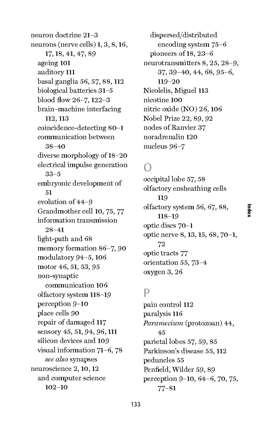
Neurotransmitter Receptor
Presynaptic neuron Postsynaptic neuron
Action Synaptic Action
potential potential potential
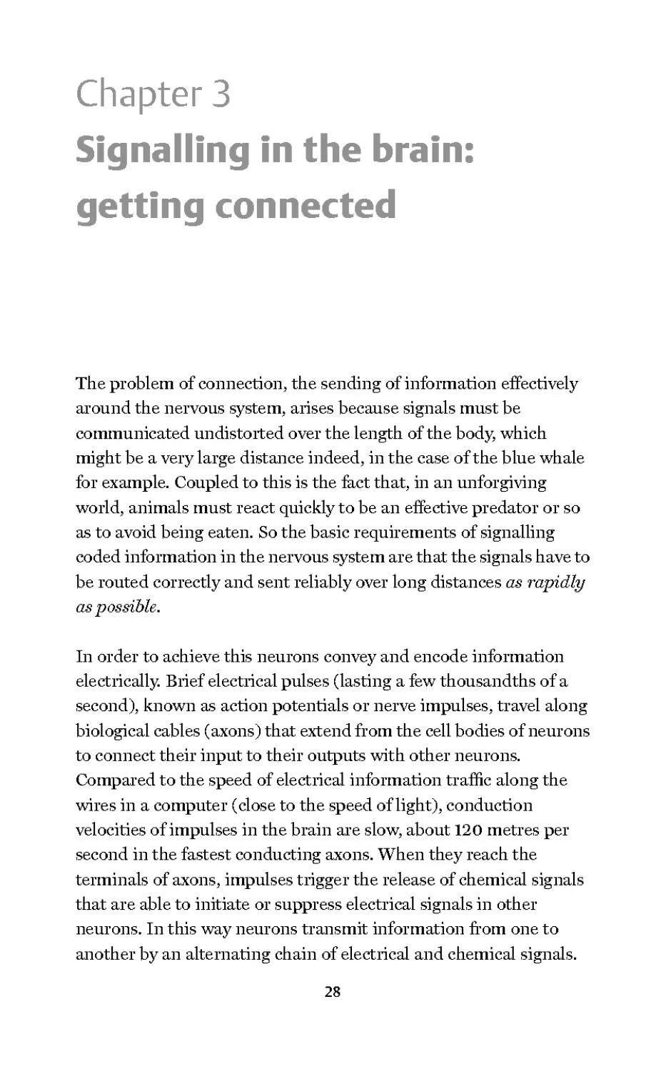
3. Nenron-to-neuron commnnication. An electrical action potential or
nerve impulse travels at speeds up to 120 metres per second along the
axon of the presynaptic neuron. When it reaches the synapse the
impulse causes neurotransmitter molecules to be released. Receptor
molecules react to the neurotransmitter molecules causing the
postsynaptic neuron to be either excited (illustrated) or inhibited. An
inhibitory synaptic potential would dip below the resting potential,
making the postsynaptic neuron less likely to fire an action potential
| Physics and the problem of electrical signalling
CO
j= When a neuron is inactive or at rest there exists a stable negative
voltage across the membrane of about —70mv, known as the resting
potential. When excited by another neuron, or in the case of a
sensory receptor cell by a sensory stimulus, the neuron may
generate a train of action potentials. Nerve impulses attain a
positive voltage of about +50 mv before returning to the resting
potential. So the total voltage excursion of a nerve impulse is about
120mv or 0.12 volts.
We need now to understand something about how these electrical
impulses are generated and propagated along axons in the wet,
salty, and gelatinous medium that is the brain: a very unsuitable
environment for an electrical signalling system. The problem is
made even more difficult by the dreadful electrical properties of
axons. Axons are very poor conductors of electricity, so bad in fact
that over relatively short distances, far less than a typical axon's
length, most of the original signal will leak away into the salty
surroundings. This inescapable problem is a consequence of the
30
way the laws of physics apply to the flow of electricity in electrical
cables immersed in salty water.
These laws were first formulated by the British scientist Lord Kelvin
(1824-1907) who figured out how to send telegraphic information
across the Atlantic Ocean through a submarine cable. Lord Kelvin
defined a parameter called the length constant', which allows us to
compare how good different types of cable are at transmitting
electrical signals over a distance. A length constant is the distance
over which about two-thirds of the electrical signal's amplitude will
be lost and its value can vary enormously. For example, the length
constant of a submarine cable is a few tens of miles. This means it
is not possible simply to lay a cable across the Atlantic and expect
an electrical signal injected at one end to appear at the other end
undiminished, several thousands of kilometres away.
Vl
la
For a submarine cable, the length constant is a small fraction of the i
distance over which information must be sent and the same is true f.
for biological cables, axons. So in a similarly salty environment both =■
ID
submarine cables and axons must detect a failing electrical signal ?
and boost it back to its original strength before sending it on its way 3
again. In submarine cables booster amplifiers placed at regular
intervals achieve this, and axons solve the problem in a rather
similar way. But how, using the unlikely ingredients of a few
proteins, fats, some smaller organic molecules, and plenty of salty
water, can nerve cells make a battery-powered amplifier?
The brain's batteries and impulses
The brain is a major consumer of bodily energy. While it is only 2
per cent of our body weight, it consumes 20 per cent of our energy
and moreover 80 per cent of the brain's energy consumption is
devoted to a single task: producing biological batteries, the power
source of the amplifiers of electrical signals in axons.
Neurons in fact create two batteries. One has a value of about 50mv
31
and faces inwards (positive pole inside) and the other has a value of
about 70 mv and faces outwards (positive pole outside). If the 70mv
battery is turned ON and the 50mv battery OFF, the inside of the
neuron will have a potential of —70mv. On the other hand if we now
turn OFF the 70mvbattery and turn ON the 50mvbattery, then the
inside will be positive by the value of the inward facing battery:
i.e. +50mv. At the peak of an action potential the membrane
voltage reaches about +50 mv before returning within a
thousandth of a second to its resting value of about —70 mv. It is as
if the action potential results from the rapid switching ON and OFF
of the batteries in a well-defined sequence. This sequence of
switching is initiated by a positive shift of the voltage across the
membrane. If the positive change in voltage reaches a critical
threshold value, the +50mv battery is turned on and a nerve
impulse is initiated.
These batteries are 'charged up' by proteins that literally pump two
2 positively charged ions in opposite directions across the membrane
j= of the neuron. The process requires energy to be expended and this
is achieved by the ability of molecular-scale pumps to couple the
expenditure of metabolic energy to the movement of ions. Sodium
ions are pumped out of the neuron whilst potassium ions are
pumped in. These ions are derived from sodium chloride (table salt)
and potassium chloride that are dissolved in the fluid surrounding
all of our cells, providing a salt-water environment for them that is
similar to the composition of the sea water in which cellular life had
its origins. The pumping creates an imbalance between the inside
and outside concentrations of the two ions. Sodium ions are
maintained at about tenfold higher concentration outside than
inside the neuron and approximately the reverse situation exists for
potassium. These concentration gradients, in the absence of
barriers, would result in sodium entering and potassium leaving the
neuron.
Highly specialized protein molecules called ion channels restrict
this passage of sodium and potassium into and out of the neuron by
32
acting as molecular gatekeepers. Mobile parts of the molecule,
'gates', open and close in an orderly sequence. This molecular
machinery enables the membrane to control the switching on and
off of the sodium and potassium batteries. Each potassium channel
has a single gate, known as the activation gate because when
opened the flow of potassium is activated. The sodium channel is
more complicated and has two gates, the activation gate and an
inactivation gate. When the sodium activation gates are open
sodium floods into the neuron due to the concentration gradient.
This is equivalent to turning ON the 50mv sodium battery, making
the inside of the neuron reach its maximum positive potential
of+50mv at the peak of the nerve impulse. When the potassium
gates open, equivalent to turning ON the -70mv battery, potassium
flows out.
Now let's consider how nerve cells generate an electrical impulse
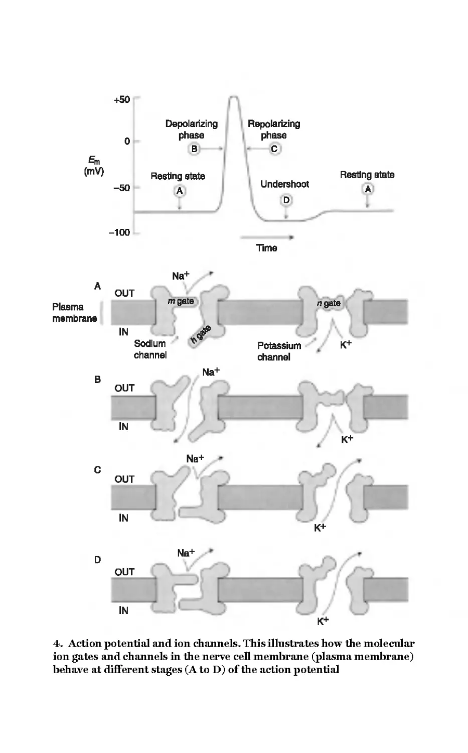
from about -70mv to about +50mv and back in a few milliseconds.
At the resting potential of about -70mv the sodium battery is
switched off, so sodium flow is virtually entirely blocked because,
although the inactivation gates are open, the activation gates are
closed. The potassium battery is partly on because a small
proportion of the potassium channel gates are open and some
potassium is therefore free to flow out of the neuron, leaving the
inside negative. To move the voltage to +50 mv the activation gates
for the inward flow of sodium must be opened. Then, to return to
the resting potential the sodium gates must be closed and the
potassium battery fully switched on, so that potassium flows out.
The sequence of opening and closing during a nerve impulse is
shown in Figure 4.
In order to understand these crucial parts of the signalling story we
need know what causes the molecular-scale gates to open and close.
The answer is that they are sensitive to the voltage across the
membrane, allowing the detection of small changes in voltage and
their amplification into discrete pulses of much greater amplitude.
But how can these channel proteins sense and respond to the
33
450
0
Em
(mV)
-50
Depolarizing
phase
B
Resting state
A
RBpolarizing
phase
C
Undershoot
Resting state
A
-100
Time
Na+
A OUT
Plasma mQate "^
membrane
IN J?
Sodium v Potassium K+
channel channel
B Na+
OUT
IN
K+
Nb+
C
OUT
IN
K+
Na+
OUT
IN
K+
4. Action potential and ion channels. This illustrates how the molecular
ion gates and channels in the nerve cell membrane (plasma membrane)
behave at different stages (A to D) of the action potential
voltage across the membrane? It would seem that the proteins
would have to be very sensitive indeed to changes in voltage - after
all, a change of just a few thousandths of a volt across the
membrane can be sufficient to trigger the opening of a channel gate.
Take a channel's eye view of the fluctuating membrane voltage
however and we get a more realistic impression of the strength of
the electrical forces acting on them. The membrane is exceedingly
thin (about one millionth of a centimetre) and the total voltage
change across this membrane during an action potential is 0.12
volts. If we take the thinness of the membrane into account, the
fluctuation experienced by the protein is an enormous 120,000
volts per centimetre. As the gates of the ion channels are themselves
electrically charged, they will be moved by the change in the
electrical forces across the membrane. It is this movement of the
ion gates that is the key to understanding how a small electrical
excitation is amplified into a full-blown nerve impulse. The first
gates to respond to an excitation are the ones preventing the entry
of sodium. A veiy small excitation will open a few of these gates,
allowing the inflow of sodium. This will cause the voltage to become
even more positive, thus opening more sodium gates. If enough
sodium gates are opened we quickly enter a positive feedback loop,
leading inexorably to the turning on of the +50mv sodium battery
at the peak of the nerve impulse. The voltage returns to its resting
voltage by the delayed closure of the sodium inactivation gates and
by the delayed opening of the potassium gates.
Speed is important
Biological cables are inherently slow conductors of signals because
the nerve impulse depends on the movement of ions across a
membrane rather than the displacement of electrons along a wire.
Higher transmission speed can be achieved by improving the
insulation of the axon membrane and by increasing the electrical
conductivity of the axon's core. The latter can be achieved simply by
increasing the axon's diameter. The fatter the axon the faster the
35
transmission; for speed you need giant axons. Unfortunately this
solution has significant practical drawbacks. The relationship
between axon diameter and conduction speed is unfavourable -
to double the speed you must quadruple the diameter
(conduction velocity is proportional to the square root of the axon
diameter). So to obtain significant gains in speed we would have
to produce axons of gigantic girth. A related drawback is that a
brain would contain fast components, but inevitably there would
be fewer of them. Evolving high performance brains depended in
part on the miniaturization of the brain's components and on
getting as many fast neurons as possible packed into a small
volume. For this, evolutionary selection pressure did not favour
giant neurons.
Nonetheless, giant axons certainly do exist in brains. There are
many examples in the nervous systems of invertebrate and lower
vertebrates, where they tend to be involved in initiating very rapid
2 responses such as escape reactions. An example of particular note is
j= the giant axon of the squid, which can be up to 1 mm in diameter
and transmits nerve impulses from the brain to the body
musculature at several metres per second. This axon activates the
animal's emergency escape behaviour, which requires the rapid
contraction of the mantel cavity causing an explosive expulsion of
water and a 'jet propelled' escape. In the 1940s the enormous
dimensions of this particular axon attracted the attention of two
British physiologists, Alan Lloyd Hodgkin and Andrew Fielding
Huxley. They conducted a series of elegant experiments on the
nerve impulse, using the size of the squid's giant axon to their
advantage. Their experiments led to the discovery of voltage-
sensitive sodium and potassium flow and to the ionic theoiy of the
action potential described above. The Hodgkin and Huxley account
of the action potential in the squid axon was to earn them a share of
the Nobel Prize for Physiology and Medicine in 1963, not least
because the principles and mechanisms uncovered in the squid
were universal - explaining even how our axons transmit electrical
signals. It seems a little unfair on the squid that there is no formal
36
acknowledgement of its contribution to one of the most outstanding
achievements of 20th-century science.
Transmission speeds in excess of 100 metres per second are
possible by improving the axon's insulation with a multilayered
Swiss-roll-like wrapping called myelin. At approximately 1 mm
intervals the myelin wrapping is interrupted by gaps known as
nodes of Ranvier where the axonal membrane is exposed. Voltage
sensitive sodium channels are concentrated at the nodes and the
nerve impulse seems to jump from node to node with negligible
delay. The autoimmune disease known as multiple sclerosis (MS)
cruelly highlights the importance of myelin in normal brain and
bodily function. In MS the body's immune system damages the
myelin and the ability of axons to conduct action potentials is
disrupted. This produces various symptoms including unsteady
movements of the limbs, blurred vision, abnormal eye movements, £
loss of coordination, slow word recall, and forgetfulness. i
ia
5"
Myelin is produced by glia, cells in the nervous system that =■
ID
outnumber neurons at least tenfold. There are many other ?
functions of glial cells; for example, the microglia are able to move 3
around the brain, consuming dead cellular debris as they go.
Other glial cells can alter the way neurons interact with one
another, suggesting that the idea that glia merely provide
supportive roles for neurons is wrong. Indeed recent experiments
have shown that glial cells can detect changes in the voltage across
their membranes and are responsive to chemical signals from
neurons. Others are able physically to cover or uncover regions of
communication between neurons, suggesting they can direct
information traffic between different parts of the brain. If, as now
seems probable, the neurons and glial cells are together essential
for information processing, then by considering only the neurons
we have vastly underestimated the complexity of the brain
machine.
37
From neuron to neuron
Neuron to neuron communication occurs at specialized points of
contact between nerve cells called synapses and there is perhaps in
excess of 100 trillion of them in the human brain. Synaptic
communications are essentially private, in the sense that a single
synapse allows one neuron to speak to just one other. The cell
bodies of communicating neurons need not be close to one another
because neurons can reach out with long connecting processes.
Synapses however are not the sole means of between-neuron
communication and an important distinction can be drawn
between point-to-point information transmission mediated by
them (the brain's wiring diagram) and a more global form of
non-synaptic information transmission (for which wireless
broadcasting is a better analogy).
The point-to-point nature of synaptic communication between the
2 wire-like fibrous extensions of neurons is reminiscent of an
CO
j= electronic circuit. Indeed the synaptic 'wiring diagram' of
connections required for a particular brain function may be
referred to as the neural 'circuit' for that function. Synapses require
two neurons to cooperate in the formation of a small region of
either direct contact (an electrical synapse) or very close apposition
(a chemical synapse). Where there is direct contact, electrical
signals pass with almost no delay from one neuron to another
through protein pores that perforate the membranes of both
neurons at the point of contact. Usually electrical synapses are
bi-directional, electricity being able to pass equally well in both
directions. Electrical synapses can be thought of as soldered joints
in an electronic circuit; they are highly reliable connections and
invariant in their operation, neither adding to nor subtracting from
the signal passing between components. The speed and reliability of
electrical synapses is exploited in neural circuits required for the
activation of a flight response. In an escape behaviour there is no
time for complicated information processing; escape must be
executed as quickly and reliably as possible and this is a job ideally
38
suited to the simple, slavish, and fast properties of electrical
synapses. The next time you unsuccessfully try to swat a fly your
failure will be due to the speed of transmission of visual information
(about you) passing through electrical synapses in the escaping fly's
brain.
At a typical chemical synapse a narrow gap or cleft between two
communicating neurons makes the direct exchange of electrical or
chemical signals impossible. For information to be transmitted
across the gap the electrical activity of a neuron must cause the
release of a chemical message that diffuses across the synaptic cleft
to the receiving neuron. The synaptic machinery allowing electrical
activity in the signalling neuron to be coupled to the release of
neurotransmitter is highly complex and specialized, as are the
mechanisms that capture the chemical message and initiate
responses in the receiving neuron. This means that the two sides of
a chemical synapse are specialized for either sending or for
receiving chemical messengers but not both. Signals therefore pass
in one direction only, from the pre-synaptic to the post-synaptic
neuron. The pre-synaptic side is specialized for the synthesis,
storage, and release of a neurotransmitter. On the post-synaptic
side the chemical message is recognized and converted into an
electrical signal. Usually chemical synapses occur between the axon
terminations of the transmitting neuron and the dendrites or cell
body of the receiving neuron.
The simplest form of synaptic transmission involves an ion channel
receptor (anionotropic receptor) that is opened by the binding of a
neurotransmitter. These mediate a direct and rapid coupling
between neurotransmitter binding and the generation of a brief
electrical signal in the post-synaptic neuron. There is another
important category of 'indirect' neurotransmitter receptors
(metabotropic receptors): the signal they generate is biochemical
rather than electrical. When a neurotransmitter binds to an indirect
receptor it activates a complex cascade of biochemical or metabolic
events in the post-synaptic neuron, mediated by special enzymes
39
that cause the synthesis of signalling molecules called second
messengers.
Primary messengers are the neurotransmitters, which transmit
information from neuron to neuron. Second messengers are the
neuron's internal messenger molecules. It is through their action
that the physiological properties of neurons and their synapses can
be altered, either briefly or for extended periods of time. Second
messengers are even involved in transmitting information from the
synapse to the neuron's nucleus where they initiate long-term
changes in the pattern of gene expression and protein synthesis that
can in turn cause changes in the strength of synapses. It is in the
action of second messengers therefore that we must seek a
mechanism for the changes in the strength of synapses that
accompany the process of memory formation in the brain
(see Chapter 6).
2 The electrical circuit, 'wiring diagram', analogy is a compelling and
j= useful one, but neurons can communicate without synapses. By the
release of freely diffusing messenger molecules, such as the gas
nitric oxide, some neurons broadcast information through volumes
of the brain; communicating with many other neurons within the
affected volume, without the need to be directly connected to all of
them by synapses. Neurons may participate in both modes of
transmission simultaneously. Indeed it may not be possible to
understand how a function is performed without knowing both the
synaptic wiring diagram of its neural circuit and how the circuit is
influenced by signals being broadcast into it from elsewhere.
Putting this all together
In this chapter we have examined in some detail the cellular and
molecular mechanisms of the most basic of brain functions - the
ability of the brain's component cells to communicate with one
another. What emerges is a picture of bewildering complexity in
which it is not easy to see the wood for the trees. So let us stand back
40
and imagine our brain with its hundreds of trillions of synaptic
connections. Each synapse is potentially a unique computational
unit with its own molecular tool kit, history, memory, and function.
The neurons and their synapses are in a constant state of flux - the
connections are dynamic, changing their strength, size, and
location; being formed and unformed. Every second, millions of
electrical impulses course along the fine fibrous extensions of the
neurons, carrying electrical and chemical messages through a
gelatinous interconnected circuitry that is more complex by far than
that of any computer. If the interconnecting fibres in just one cubic
millimetre of cortical grey matter were unravelled and laid end to
end, they would form a strand 5 km (about 3 miles) long! If the
connections in the whole brain were unravelled, the strand would
be long enough to encircle the earth twice - such is the phenomenal
interconnectivity of the brain. And this is only part of the story
because the neurons and their connections make up a very small £
fraction of the brain's cellular machinery. There are as many as 100 i
glial cells for each nerve cell and we are only beginning to ™
understand just how important they are, not simply carrying out =■
ID
housekeeping jobs but participating in the brain's computations, in ?
among other ways, by regulating synaptic transmission. 3
This then - the neurons and their connections and their history,
their companion glial cells, the multitude of chemical messengers
and receptors - is basically all there is to the brain. We are far from
understanding how it works as a whole but there is nothing more,
no magic, no additional components to account for every thought,
each perception and emotion, all of our memories, our personality,
fears, loves, and curiosities.
41
Chapter 4
From the Big Bang to the
big brain
Early evolution of the nervous system
Astronomers tell us that our universe began with a bang, in fact a
Big Bang. Today in our small corner of this universe, some 14 billion
years later, physical entities capable of reflecting on their place in
this universe have somehow come to exist. It is safe to assume that
brains did not simply spring into existence suddenly from nowhere,
but how and by what route were brains created? The answer is that,
in common with all other manifestations of life, the brain is the
result of the haphazard and perilous process of evolution by natural
selection. It did not happen overnight.
Our planet Earth came into existence about 4.5 billion years ago. In
the beginning there was no life but within a billion years or so, the
pre-biotic chemistry of life got started and the first primitive
organisms appeared. Thus began the process of organic evolution
leading to our present-day over-sized human brains. It was a
process that would require three and a half thousand million years
to complete.
As far as we know we are the first self-aware inhabitants of Earth,
and almost certainly the first to question our origins and place in
this strange universe. Today we are on the threshold of manned
exploration and exploitation of other planets in our solar system
42
and perhaps beyond. More than anything else it is our inquisitive
and irrepressibly self-confident brains that have driven this quest
for understanding and allowed all of this to happen.
Insight into the process by which our brains came to exist and to
acquire their remarkable abilities can be gained by studying
animals far simpler than us. Most animals have a nervous system
and some form of brain, which is simply a concentration of nerve
cells -usually sensibly placed at the front end. While we may be the
only species endowed with a brain that bothers to think about itself,
even this must have originated in the task of sensing and
responding to the environment. These are the most basic functions
of the nervous system - providing animals with the ability to detect
salient features of their changing surroundings and to respond
appropriately. While the brain has elaborated the art of sensing and
responding, this ability is by no means exclusive to it. It is of almost
unimaginable antiquity, predating the nervous system by a
considerable margin, and can be traced to the earliest steps in the
origin of life, crucially to the evolution of the very first biological
cell.
Early cells had an aquatic existence and soon acquired the ability to
move. Single cells swim using a variety of means including the
lashing of whip-like flagella and the coordinated waving of many
shorter 'hairs' called cilia. Other unicellular organisms can creep
along by extending and retracting 'feet' or pseudopodia from the
body of the cell. Importantly, movement enabled the early
unicellular creatures to explore and exploit more of their world and
its resources. When the ability to move was coupled with the ability
to sense, movement could be directed to resource-enriched regions
and individuals doing this most effectively reproduced more and
prospered.
So even a single free-living cell can behave adaptively, can orient,
and move 'intelligently' in response to environmental stimuli,
combining all of the sensing and acting functions necessary for
43
survival without a nervous system. This can be seen in modern-day
unicellular organisms such as the ciliated protozoan Paramecium,
which has a rich repertoire of orientation behaviours. They can
sense and then swim towards their major food source - decaying
organic matter. Paramecium also has a primitive touch sense
enabling it to navigate around obstacles after colliding with them.
This involves reversing the direction of swimming and turning away
from the obstacle before proceeding in the forward direction. This
behaviour is mediated by changes in the voltage across the cell
membrane associated with the influx and efflux of calcium and
potassium ions. Thus mechanical stimuli evoke electrical potentials
much like nerve impulses in neurons.
As we have seen, the molecular machinery of sensing and acting
existed in unicellular organisms, long before the arrival of
multicellular animals. But the ability to act on sensations was
transformed when multi-cellular organisms evolved. Now cells
2 could cooperate with one another, dividing between them different
j= and highly specialized roles. Individual cells no longer had to
perform each and every essential role; they could 'differentiate'
functionally one from another. Thus was the stage set for the
evolution of the nervous system - a collection of highly
differentiated cells designed for sensing, analysing, storing, and
transmitting information and for directing adaptive behaviour in
the interests of the survival and reproduction of the whole
organism. The toolkit required for chemical and electrical
signalling on which the functioning of the nervous system depends
was already available. The brain did not have to invent everything
from scratch - it evolved by modifying and incorporating
mechanisms pre-existing in brainless animals.
It is likely that the first animals to have true neurons had a nervous
system consisting of a diffuse nerve net with little or no
concentration of nerve cells in any particular part of the body. This
distributed feature of the nervous system works well for radially
symmetrical animals in which no particular part of the body is more
44
1-10
14
11
12
The avoiding
reaction
13
5. Paramecium avoidance behaviour. An animal consisting of just one
cell can sense and respond to its world - without the need for a brain.
However, the molecular mechanisms that underlie this simple avoidance
behaviour are similar to those that underlie the electrical nerve impulse
in the neurons of our own brains
or less likely than any other to encounter food or danger first. In its
simplest state, the nerve net consists of sensory neurons, directly
acting on contractile cells. Because these neurons must sense the
outside environment they are derived from cells that make up the
outer layer of the body, the ectoderm. Interestingly, our brains are
derived in the embryo from ectodermal tissue that during the
course of development becomes internalized.
45
In more complex arrangements of neurons, sensory and motor
functions are assigned to different types of cells. Specialized sensory
cells activate motor neurons, which innervate the contractile cells.
There are few if any 'inter-neurons' - nerve cells inteiposed between
the sensory and motor neurons. It is in the elaboration of the
central nervous system in higher animals that we see the huge
proliferation of inter-neurons. By mediating between sensing and
motor functions, inter-neurons allow an animal's behaviour to
become less determined by automatic reactions to external stimuli.
In effect inter-neurons introduce the possibility of a cognitive gap
between stimulus and response, allowing for intelligent
consideration of options, 'thinking' in other words, before actions
are executed. Inter-neuron proliferation also provided the numbers
necessary for greater memory capacity and for more sophisticated
processing of information.
The trend towards a true central nervous system received its most
2 irresistible impetus with the evolution of animals with a bilaterally
j= symmetrical body plan. Our ancestors had an elongated body and
creeping lifestyle, feeding on organic matter on the sea floor. Having
the mouth at the front facing downwards made this lifestyle easier
and created a distinction between dorsal (top side), ventral (bottom
side). The most important consequence of bilateral symmetry is
that the front can be differentiated from the back - in effect
inventing the head end. As most bilaterally symmetrical animals
move in the forward direction, it made sense to concentrate the
senses and the central nervous system at the head end, a
phenomenon known as 'cephalization'. After all, the front end will
encounter opportunity, food, and danger first, explaining why the
brain of most animals is at the front.
With the exception of the most primitive of bilateral animals, the
body is divided lengthwise into a series of segments. This is most
evident in annelid worms such as earthworms and leeches where
the body consists of virtually identical segments. The segmentation
of the body is reflected in the segmentation of the central nervous
46
system. In worms and insects each body segment contains a central
'ganglion', a collection of nerve cells concerned with the sensory and
motor functions of that body segment. The ganglia are connected to
one another by paired longitudinal 'connectives' containing the
axons of nerve cells that communicate between ganglia. The ganglia
thus form a 'nerve cord' running the length of the body. This ensures
that the ganglia do not act independently and that the body
segments cooperate with one another in the generation of
coordinated behaviour.
In bilateral invertebrates the nerve cord is ventral to the alimentaiy
canal or gut. This contrasts to the situation in the vertebrates, in
which the spinal cord is dorsal, the gut lying ventral to it. You might
imagine that the plans of the invertebrate and vertebrate nervous
system are so fundamentally different that they must have very
different evolutionary histories. But the vertebrates and
invertebrates share more similarities than differences. Let's deal
directly with the dorsal versus the ventral position of the central
nervous system. During embryonic development, genes are
expressed that determine the developmental programmes leading
to specifically dorsal and ventral tissues. These genes are very
similar in their DNA structure in invertebrate and vertebrate
species, and they must therefore have derived from common
ancestral genes. However, the genes in vertebrates that determine
ventral tissues and organs are most closely related to the genes in
invertebrates that specify dorsal. It seems that the only plausible
evolutionary explanation for this apparent reversal of the functions
of these genes is that something happened to one of our common
ancestors that caused it to roll over, so that what was the ventral
surface now faced upwards - thus becoming, by definition, dorsal.
What might have caused the hapless creature to turn over? One
possibility is that a mutation occurred causing the mouth to move
from its downward facing position to an upward facing position.
Our bottom-grazing creature would be severely handicapped,
unless of course it simply flipped over to bring its mutated mouth
47
A
B
Hydra Starfish
Leech Cockroach
6, Hydra, Starfish, Worm and Insect: Simple nervous systems are
diffuse networks of neurons (nerve nets) such as are found in modern
day jellyfish and their relatives {Hydra, for example). The true central
nervous system arises from the condensation of neurons into centres in
which neurons are concentrated. In bilaterally symmetric animals such
as worms and insects, the anterior end of the central nervous system
becomes enlarged - aphenomenonknownascephalization,
Cephalization resulted in the evolution of a true brain, located in the
head. In animals with radial symmetry, such as the starfish, there is no
head and thus no true brain
back to the preferred downward facing position. Now however the
nervous system lies ventral to the gut just as it does in all of this
creature's descendants - the vast majority of invertebrate animals.
So what was ventral becomes dorsal and vice versa.
Overview of the human nervous system
The human brain has a confusing and complicated structural
organization that certainly would not win awards for design. Unlike
the eye, you cannot tell just by looking at it what the brain's true
purpose is and prior to the scientific era scholars can be excused for
suggesting that the brain functioned to cool the blood. After all, the
folded surface of the cerebral cortex is at least suggestive of a heat
exchanger or radiator.
Much of the brain's structural complexity arises because its
evolution has been less of an 'out with the old, in with the new1
process, and more a case of'on with the old, in with the new'. This
has resulted in new structures being layered upon more primitive
ones, which may retain their original functions albeit in the context
of the new opportunities provided by the newly acquired ones.
Therefore a proper understanding of the brain can only really be
achieved in the context of its evolution. It is also helpful to consider
how the structure of the brain arises during the course of its
embryonic development.
The human brain is basically an elaborated fluid-filled tube, which
is first formed in the embryo. The two major subdivisions of the
central nervous system, the brain and the spinal cord, are derived
from a strip of embryonic skin on the dorsal midline called the
neural plate. About three weeks after conception the neural plate
rolls up to form a groove, which eventually pinches off at the
midline to create the neural tube. The dorsal ectoderm then closes
over the neural tube, which now resides inside the embryo
extending from one end to the other. Flanking the neural plate is a
region of ectoderm called the neural crest. Cells of the neural crest
49
Anterior
Anterior
Telencephalon
Forebrain
Forebrain
Diencephalon
Midbrain
o Mesencephalon
3 (midbrain)
Hi
CD
z
Hindbrain
Rhombencephalon
(hindbrain)
Spinal cord Spinal cord
Posterior Posterior
7. Three divisions of the brain early in development. At its anterior end the neural tube swells in
three places, the forerunners of thefore-mid- and hindbrain (A). Later the swellings become further
elaborated into the major subdivisions of the brain (B)
proliferate to form the peripheral nervous system, the autonomic
system, and the sensory neurons of the dorsal roots of the spinal
cord. The cells of the neural tube also proliferate, becoming the
neurons and glial cells that form the brain and spinal cord. Initially
the neural tube is uniform along its length, but as development
proceeds, the rate of cell proliferation at the anterior end of the tube
far exceeds the rate at the posterior end. Consequently the anterior
end enlarges and later it will become the brain. The posterior end of
the neural tube is destined to become the spinal cord.
At its anterior end the tube expands, forming three vesicles that will
become the major divisions of the brain, the forebrain, midbrain,
and hindbrain. These further subdivide to form the forerunners of
the major component parts of the adult brain. For example, the
anterior end of the forebrain balloons on each side, forming
protrusions that will become the two cerebral hemispheres (the
telencephalon). The protrusions on each side curl in a posterior
direction before growing anteriorly, rather like a pair of ram's horns.
Thus the most anterior part of the forebrain surrounds the
midbrain. At the same time the embryonic mid- and hindbrain
vesicles begin to differentiate and form all of the components of the
brainstem.
From spinal cord to cerebral cortex
The spinal cord has a relatively simple structural organization: a
central region of 'grey matter' containing synapses and the cell
bodies of neurons and a surrounding 'white matter' consisting of the
axons transmitting information up and down the cord. The spinal
cord is segmental and at each segment there are two pairs of'roots',
one dorsal and one ventral, which connect the spinal cord to the
body. The ventral roots contain the outgoing axons of motor
neurons and the dorsal roots contain the incoming axons of sensory
neurons. The cell bodies of the sensory neurons reside in swellings
called dorsal root ganglia close to the spinal cord. Cell bodies of the
motor neurons are found in the ventral grey matter, clustered
51
Telencephalon
Spinal cord
Diencephalon
Midbrain Hindbrain
Telencephalon
Spinal cord
Diencephalon
= Midbrain Hindbrain
E
&
Telencephalon
Cerebellum
PRIMATE Spinal cord
FORM
Diencephalon „.„ . ..._.._.
Midbrain Hindbrain
8. Development of the forebrain. During the embryonic development of
the mammalian brain the most anterior part of the forebrain, the
telencephalon, enlarges disproportionately. In primates, the telencephalon
enlarges by curling round posteriorly and then anteriorly, eventually
completely surrounding the rest of the forebrain and the midbrain
together in functionally related groups. Thus motor neurons that
innervate a given muscle are grouped together and motor neurons
of the extremities are found laterally in the cord, whereas those
innervating trunk muscles are more centrally located. This
grouping by function simplifies the task of activating groups of
muscles sequentially to produce coordinated movements. Inter-
neurons in the spinal cord connect the sensory input to the motor
output and provide the neural circuits that underlie simple reflex
behaviours such as the familiar knee jerk reflex.
The three divisions of the brain can be thought of as a hierarchy in
which the forebrain controls the midbrain, which controls the
hindbrain. The brainstem (mid- and hindbrain) is concerned with
essential but non-cognitive bodily functions such as breathing, the
regulation of blood flow, and the coordination of locomotion. Basic
processing of sensory information is also performed by the
midbrain structures, but more complex processing occurs when this
partly processed information is distributed up the hierarchy to the
forebrain. The forebrain can be regarded as the executive centre,
which considers sensory information of all kinds and formulates
commands, decisions, and judgements based on the sensory
information and on experience. The increased complexity and
flexibility of our behaviour can be attributed to increased
cephalization - the acquisition of new neural functions associated
with the most anterior parts of the brain. This is reflected in the
increase in the relative size of the forebrain, which in fish,
amphibians, and reptiles is a very minor part of the brain, but which
in mammals is much larger. It becomes so large in humans that it
will not fit into the skull without being folded into itself, forming
the characteristic convolutions of the cerebral cortex.
The hindbrain consists of the medulla, pons, and cerebellum. The
pons, medulla, and various divisions of the midbrain, described
below, are referred to as the "brain stem'. A substantial amount of
space in the medulla is occupied by bundles of ascending and
descending axons, carrying information traffic between the brain
53
Forebrain
Fish
Forebrain
Rodent
Forebrain
Human
9. Cephalization. More and more resources are allocated to the most
anterior part of the brain in vertebrate evolution. The relative size of the
forebrain has increased most in evolutionary time. In hominid
evolution the pre-frontal cortex (the most anterior part of the forebrain)
tripled in size in just two million years
and the spinal cord. It is in the medulla that axon tracts descending
from the cerebral cortex pass from the left to the right side,
resulting in each hemisphere controlling movement on the opposite
side of the body. Many visceral automatic functions, including
breathing, heartbeat, and swallowing, are controlled by neural
centres in the medulla. Just anterior to the medulla is the pons, a
structure at the interface between the midbrain and hindbrain. It
also has visceral functions and an important role in the control of
facial expressions. It contains important sensory centres including
the vestibular nuclei, which receive information about the
orientation of the body with respect to gravity and acceleration.
The most complicated part of the hindbrain is the cerebellum, a
structure specialized for the coordination of motor commands. In
size the cerebellum is second only to the cerebral hemispheres.
Placed over the gateway between the brain and spinal cord it is in a
strategic position to oversee the performance of complex
movements. The cerebellum has a veiy distinctive appearance, with
a surface consisting of numerous ripples called folia separated by
deep fissures. It is richly supplied with sensory information about
the position and movements of the body and can encode and
memorize information required for the execution of complex learnt
skills. It is attached to the brain stem and connected to the rest of
the brain by three pairs of axon tracts called peduncles that link it to
the midbrain, the pons, and the medulla. The cerebellum has no
direct connection to the cerebral cortex and while it also has a role
in the control of movement, in contrast to the cortex each side of
the cerebellum controls the same side of the body.
The midbrain consists of the substantia nigra, inferior colliculus,
superior colliculus, and part of the reticular formation, which also
extends into the medulla. The substantia nigra is intimately
involved in the initiation and maintenance of voluntary
movements. Damage to this part of the midbrain is linked with the
symptoms of Parkinson's disease, a condition characterized by
muscle tremor and a difficulty in initiating actions. The inferior
55
colliculus is concerned with hearing and the superior colliculus is a
centre for visual information processing and the generation of eye
movements. The reticular formation has a primary role in the
control and regulation of the arousal state of the brain.
The two major subdivisions of the forebrain are the telencephalon
(cerebral cortex and the basal ganglia) and the evolutionarily older
diencephalon (parts of the limbic system, the thalamus, and
hypothalamus). In the diencephalon, deep in the brain, is a paired
structure called the thalamus. This is a relay station for information
on its way to the cerebral cortex coming from the visual, auditoiy,
and body sensory systems. More ventrally, at the junction of the
thalamus and the midbrain is the hypothalamus, a centre that in
spite of its diminutive size is concerned with a wide range of
important functions such as sex, emotion, the interpretation of
smells, the regulation of body temperature, hunger, and thirst. It is
the brain's link with the body's hormonal system and releases a
2 number of hormones into its blood supply for distribution to the
j= rest of the body.
The hypothalamus is usually included among a series of interrelated
structures collectively called the limbic system. The term limbic
refers to the parts of theforebrain that form a rim (limbusm Latin)
around the bridge between the two cerebral hemispheres, the
coipus callosum. This system includes the amygdala and the
hippocampus that are, in an evolutionary sense, the oldest parts of
the forebrain. The functions of these structures however changed in
the course of evolution. In the lower vertebrates, reptiles for
example, the amygdala is concerned with the sense of smell
(olfaction), whereas in the human brain olfactory function is
minimal and the area is thought to be mainly concerned with the
emotions. Another example of evolutionary change of function is
provided by the hippocampus. This structure in reptiles probably
organizes behavioural responses to olfactory stimuli (such as either
to flee or mate), but in mammals and man the hippocampus has a
major role in the formation of memories. The cinglulate gyrus,
56
another component of the limbic system, provides a connection
between the limbic system and the cerebral cortex.
The anterior and most recently evolved part of the forebrain
consists of the basal ganglia and the enveloping cerebral
hemispheres. The basal ganglia lie hidden under the two
hemispheres and consist of a cluster of three paired groups of
neurons. Their role is to control and regulate voluntary movements
initiated in the cerebral cortex. More than any other brain structure,
it is the cerebral cortex that makes us human. Within the cortex
plans are made, volitional behaviour is initiated, the neural
machinery of language is located, and conscious perceptions are
assembled from sensory information. It is the locus of all of our
creative intelligence and imagination. If indeed we have free will,
then it is in the cortex that its secret will be found.
The cerebral cortex is the convoluted surface of the two cerebral
hemispheres and comprises a single folded sheet, about 2-4 mm
thick. Information enters and leaves the cortex carried by about one
million input-output neurons, but there are more than ten billion
internal connections. The cortex therefore spends most of its time
talking to other bits of itself. Extensive and pronounced infolding of
the cerebral cortex allows a large surface to be accommodated by
the skull. If the left and right cerebral cortices were ironed flat they
would cover a surface of about 1.6 square metres, approximately
four times the area covered by a chimpanzee's. A deep midline
valley, the longitudinal fissure, divides the cortex into its left and
right hemispheres. Both sides are subdivided into four principal
lobes, which derive their names from the bones of the skull that lie
over them. The frontal lobes are the largest, accounting for about
40 per cent of the cortex; the temporal, parietal, and occipital lobes
account for about 20 per cent each. The ridges of the surface
convolutions are called gyri and the valleys that separate them are
called sulci, the deeper of which are called fissures. Superficially the
left and right sides of the cerebral hemispheres are symmetrical in
appearance and largely mirror one another functionally too. There
57
are however important left-right differences concerning some
cognitive tasks such as those required for language.
The deep longitudinal fissure that apparently separates the left
from the right cortex along the midline conceals a major structure
called the coipus callosum, which is of vital importance in
bilaterally coordinated cortical function. It is the communications
bridge between the cerebral hemispheres and consists of a tract of
about one million axons, half of them originating from neurons in
the right and half from neurons in the left cerebral cortex. The
axons of the coipus callosum allow the left cortex to know what the
right cortex is doing, has done, and might do next, and vice versa.
If the coipus callosum is cut through, as it is infrequently in the
treatment of intractable epilepsy, the two hemispheres can
function independently. Following such operations, individuals
referred to as 'split-brain' patients provided the definitive evidence
that the two hemispheres differed in their roles in language.
2 Split-brain patients have been able to name unseen objects held in
j= the right hand. For this naming task the left hemisphere is used
because sensory information from the right side of the body is
processed by the left cerebral cortex. However, using the right
cerebral cortex patients could not provide a verbal account of what
was held in the left hand. These observations confirmed what had
been assumed, namely that while both hemispheres have a
conscious awareness of things, it is the left that expresses its
awareness in words.
The convoluted surface of the human cortex can be divided into a
number of functional regions, but the most basic and simple
division is between areas serving sensory and motor functions.
Sensory cortical areas are defined by the type of information that
they receive, different areas being specialized to receive and process
information coming from particular sensory organs and structures.
For instance information from the eyes projects to the most
posterior part of the occipital lobe known as the primary visual
cortex. Information coming from the body and skin, called
58
somatosensory information, projects to a strip of the parietal lobe
known as the somatosensory cortex.
An important design principle of these sensory cortical areas is
'mapping'. The cortex constructs maps, spatial representations of
the sensory world that is conveyed to it by sensory organs. The
easiest one to understand is the map of the visual world, which is
two-dimensional and formed initially in the eye, which projects an
image of the outside world onto the retina. A neural representation
of this map is preserved as it passes from the retina to the thalamus
and then on to the primary visual cortex. The map is repeated
several times in subdivisions of the cortex specialized for different
aspects of visual perception. Left and right surfaces of the body are
also mapped on the right and left cortical hemispheres respectively.
They are called somatosensory maps and are found in the broad
strips of cortex located just behind the central sulcus.
Movements of the body are controlled by the primary motor
cortex, which is a mirror image of the sensory representation of
the body surface described above. The left motor area controls the
right side of the body and the right the left. The motor areas
occupy the strips of cortical surface just in front of the central
sulcus and there is a motor map, a map of muscles, which
corresponds to the distorted representation of the body in the
somatosensory maps.
None of these cortical maps are drawn to scale; instead they are
variously distorted to reflect the amount of neural processing power
devoted to different regions. This accounts for the grotesque
appearance of the human form (Penfield's famous homunculus)
that can be drawn from a translation of the body's sensory map into
the human form.
Much of the cortex, often referred to as the 'association cortex', is
not directly connected either with the motor control systems or
with the initial stages of sensory information processing. Generally
59
2
CO
01
10. Homunculus: a distorted image of the hitman form reflects the
differenti al allocation of cortical surface to sensory and motor functions
of different parts of the body
the further removed from direct motor or sensory functions the
cortex becomes, the less well can its function be defined.
How the human cortex became so big
If we trace the evolution of the human brain, the greatest and most
rapid growth has occurred in the frontal lobes of the cortex, which
accounts for some 40 per cent of the structure. In our nearest living
relatives, the chimpanzees, the frontal cortex accounts for about
17 per cent. The evolutionary lines leading to modern humans and
60
other living primates, including chimpanzees, diverged about
14 million years ago. The oldest bipedal hominids (3.9-2.5 million
years ago) had a brain size ranging from 400 to 500 cc, only 100 cc
or so larger than that of a chimpanzee. Homo kahilis (2.5-2 million
years ago), probably the first fabricator of stone tools, was slightly
more cerebrally endowed with a brain size up to about 600 cc. So
brain size had been increasing gradually, but relatively slowly, in
primate evolution from 14 million to about 4 million years ago and
in the early phase of hominid evolution up to about 2 million years
ago. But over the course of the next 2 million years, brain size was
almost to triple, with by far the greatest relative change occurring in
the size of the frontal cortical lobes.
Neanderthal man, appearing first in Europe and western Asia, and
modern man Homo sapiens, of African origin, are the most recent
species in the hominid lineage. They appeared at approximately the
same time: around 300,000 years ago for Neanderthal and
250,000 years ago for Homo sapiens and had roughly equivalent
brain size, though in fact the Neanderthal average (l,500cc) is
slightly greater than the human average (1,400 cc). Of course brains
do not fossilize and we do not know what the relative size of the
frontal cortex was in Neanderthal man. We do however know that
in the course of human evolution much of the growth in brain size
has been due to the explosion in the relative size of the frontal
cortex. How did it get so big in such a brief period of evolutionary
time; why has it become so conspicuously large? What is so special
about the frontal cortex?
The frontal cortex is responsible for the processing of information
from diverse and very widely separated regions of the rest of the
cortex. It is in effect a complex integration centre that defines an
individual's personality more than any other region of the brain.
It creates an awareness of'the self in relation to the world and
enables us to plan and execute actions into the future. Deficiencies
and damage in the frontal cortex compromise the ability of an
individual to make sensible predictions about the future
61
consequences of events and actions in the present. Although
intellectual power may be left unaltered, normal restraints on
interpersonal discourse are seriously affected by frontal damage
and one cannot help but conclude that much of what makes us
human, civilized, and creative resides in the part of the brain that
has grown so disproportionately in the course of our evolution.
Nobody really knows why the part of the brain that makes us unique
should have evolved so rapidly; tripling in size in less than two
million years. It certainly looks as though evolution entered a
positive feedback loop in which natural selection favouring creative
intelligence became linked to an ever more extravagant expression
of that intelligence.
Although there is no watertight explanation for the runaway pace of
evolutionary change that human brain development would seem to
require, one of the more imaginative ideas is that our frontal brain
2 is an ornament required for courtship display. According to this
j= idea, the human brain is the product of the mutual preference of
men and women for mating with partners who display unusually
creative intelligence in the rituals of courtship. This can result in a
form of natural selection called sexual selection. It depends on
creativity in courtship and the large brain that it requires being
heritable traits. If the larger brained individuals were genetically
fitter specimens, which they may well have had to be to carry the
burden of an enlarged brain, sexual selection can provide an
explanation for the exaggerated size of the human cortex and
runaway pace of human brain evolution.
A question that does not seem to be answered by the sexual
selection hypothesis, and requiring further examination, is the
reason for the late emergence of physical evidence of creative
intelligence. Human beings, with brains equal in cubic and
intellectual capacity to ours, first appeared in the fossil record more
than 250,000 years ago. If their brains attained cleverness and
creativity in the interests of seduction, why does significant proof of
62
creative intelligence, such as cave paintings, not appeal' until just
35,000 years ago? This apparent discrepancy does not disprove the
role of sexual selection in the brain's evolution, but suggests that it
must be part of a more comprehensive explanation for our
extraordinaiy brains. The truth is that we cannot yet explain why
we enjoy exercising them in the creation and public display of
music, art, poetiy, and humour.
63
Chapter 5
Sensing, perceiving,
and acting
Sensory perception's imperfections
Sensory information flowing into the brain fuels our perceptions,
memories, intentions, and actions. Although we generally refer only
to the five traditional senses - sight, touch, audition, taste, and
smell - there are in fact many more. Other sensations include those
of heat and cold, gravitation, acceleration, pain, etc. Moreover each
of the traditional sensory modalities is a complex mix of distinctly
different sub-senses. In the visual modality for example there is the
ability to sense the motion, colour, form, brightness, texture, and
contrast of objects.
The brain analyses primary sensations, transforming them into
perceptions upon which informed decisions are made about future
actions. Sensations however are just one contributory component to
perception. It is possible to perceive what is not sensed, not to
perceive what is sensed, and to construct more than one perception
from the same sensations. Perceptions are not therefore strictly
determined by sensations, nor are sensory perceptions linked to
single modalities. Perceptions are the brain's educated guesses
about what the combined senses are telling it, and as such they will
almost always depend on interactions between different modalities.
So while making a distinction between sensation and perception
may seem academic, actually it is an important one.
64
This distinction can easily be appreciated by looking at the famous
visual illusion based on a painting titled My Wife and Mother-in-
Law by W. E. Hill. You will either see a young woman looking away
or the profile of an old woman: two perceptions contained in the
same picture. Once both have been perceived, it is possible to swap
between the two at will. The sensory information falling on the
retina is exactly the same for both, so there must be a top-down
process determining how the perceptual system interprets the
sensory input. In effect our brains impose different conscious
perceptions on the same information registered by our sensory
systems.
The importance for perception of the interaction between different
senses is illustrated by the auditory-visual illusion known as the
McGurk effect described by Harry McGurk and John MacDonald in
an article called 'Hearing Lips and Seeing Voices'. Watching a video
of a person repeating the syllable 'dah' three times followed by three
repetitions of the syllable 'bah' can produce the illusion. If the
soundtrack repeats the sound 'dah', irrespective of the syllable
actually being mouthed, you hear both 'dah' and 'bah'. When you
11. Two perceptions from one sensation
65
see the lips mouth 'bah' you hear "bah' even though the sound
entering your ear is in fact 'dah'. Your brain is trying to provide your
consciousness with its best guess about what the senses are telling
it. In this case there is a contradiction between what the eyes and
ears are telling it to perceive. In this instance, the eyes have it.
Perceptual blindness is a striking example of the way the brain is
highly selective in deciding which elements of the sensory
information available to it are consciously perceived. A remarkable
illustration of this is provided by a short video made by Daniel
Simons and Christopher Chabris. The video shows a group of young
people passing a basketball to each other. A person dressed in a
gorilla suit walks into their midst, waves conspicuously into the
camera, and then walks out of the scene. In advance of viewing the
video, the audience is instructed to count the passes and then report
their findings after the clip. Astonishingly, when I saw this
demonstrated recently, about half the audience completely failed to
2 notice the gorilla! But the invisible gorilla was there, its image
j= entered the eyes of each viewer, impressed itself on their retinas,
was sent to the thalamus from where it was relayed to the primary
visual cortex. Where then did the proverbial 900lb gorilla go? It
seems that the brain was so engaged in the counting task that it
decided not to bother itself with generating a conscious perception
of a gorilla, even though a substantial part of the brain's visual
system was fully informed about its presence. So the gorilla was in
effect airbmshed out of the visual perception.
How the brain might achieve such a feat of image manipulation is
beyond our current understanding. The task must involve more
than removing the visual information associated with the gorilla
because the gorilla image is not replaced by a gorilla-shaped hole in
vision. The space occupied by the gorilla is filled in appropriately
with what would have been seen behind it had the gorilla not
actually been there. Equally remarkable, those who witnessed the
invisibility of the gorilla could not recall anything odd about their
perception of the original video. Apparently there was no accessible
66
memory trace left of the strange case of the disappearing gorilla.
This example should make us sceptical about the veracity of
eyewitness accounts where accuracy matters, in the reporting of
crimes, for example.
Our human senses are impoverished when compared to those of
other animals. A male moth when searching for a mate can for
example respond to the 'smell' of individual molecules of the sexual
attractant 'pheromone' which the female exudes into the air to lure
a distant mate. Our eyes are sensitive to wavelengths in the
electromagnetic spectrum between about 400 and 700 nanometres
only. Many insects respond to shorter wavelengths, enabling them
to use information contained in ultraviolet light. Other animals,
such as some snakes, can 'see' the body heat of their prey using the
long wavelengths of infrared radiation. Our hearing is similarly
restricted to a narrow band of frequencies. At best we can hear
sounds up to about 20 kHz, far short of the ultrasonic frequencies
that bats use in echo locating obstacles and prey.
Perceiving the outer world
Open your eyes and you see the world around you apparently
instantaneously, in colour and extraordinary temporal and spatial
detail. Just a glance allows you to identify objects - their colour,
shape, texture, size, location, and spatial relationship to one another
are immediately apparent. If an object is moving with respect to
you, you can instantly tell its direction of motion and estimate its
speed, information that allows you to avoid colliding with it or to
catch it. If you are in motion, you can see the flow of the visual world
around you and extract information from it, enabling you to know
your direction, estimate your speed, and simultaneously adjust
both.
Seeing depends on the retina providing highly processed
information on the quality and quantity of light captured by the
rods and cones, the two systems of photoreceptors. The cones are
67
highly concentrated at the fovea, an area of just one square
millimetre at the back of the retina. This region is responsible for
our high-acuity colour vision and could be rightly regarded as the
most important square millimetre in your body. There are red,
green, and blue sensitive cones and they provide information about
the relative amounts of red, blue, and green light that the brain uses
to 'colour in' an object. So the brain creates all the colours we can
perceive by blending just three, much in the same way that an artist
can mix any colour from just three pigments. As the cones are
rather insensitive to light, colour vision is only possible in relatively
bright light. In the majority of the retina surface, surrounding the
fovea, the highly light-sensitive rod photoreceptors predominate,
outnumbering the cones by some 10 to 1.
Light regulates the amount of neurotransmitter released by the rods
and cones, which in turn regulates the electrical activity of neurons
in the retina. These include the bipolar neurons that transmit
2 information along the light-path and the horizontal and amacrine
j= neurons that transmit information sideways in the retina -
perpendicular to the light-path. The last neurons to be affected are
retinal ganglion cells, the retina's output neurons whose axons form
the optic nerves connecting the eye to the brain.
Your eyes operate over an enormous range of light intensities, from
a starlit night to the brightest sunny day - perhaps a millionfold
difference in luminance. This is achieved by a property of the
photoreceptors called adaptation, which adjusts their sensitivity to
match the average background light intensity. In very dim lighting
the rod sensitivity is increased and in bright light their sensitivity is
reduced. At any given level of sensitivity, the rods reliably report
differences in light intensity within a narrow range above and below
the average background level. The photoreceptors are therefore
very sensitive to small changes in light intensity relative to the
background but not to absolute intensity. If the overall background
level of illumination were to change drastically, as it does when we
enter a cinema on a bright sunny day, we are effectively blind until
68
Light
Horizontal
cell
Bipolar
cell Cones
Ught
Ganglion
08,1 Rods
Amacrlne
cell
Receptor laye
Outer nuclear layer
Outer plewform layer
Inner nuclear layer
Inner plexiform layer
Qangllon cell layer
Tb optic nerve
12. Eye and retina. Light entering the eye passes through two layers of
neurons in the retina before reaching the light-sensitive photoreceptors
- the rods and cones. Information about the quality of the light is then
passed back to the neurons, which integrate and transform it into a
pattern of impulses for transmission to the brain by a million retinal
ganglion cells
our rod photoreceptors have adapted to the reduced intensity by
increasing their sensitivity. Resetting sensitivity depends on the
mobilization of biochemical mechanisms in the photoreceptors and
can take a few minutes to complete for a very large change in light
intensity. As a consequence of light adaptation, the brain is not
informed about the absolute levels of brightness but about rather
more useful information about local differences in brightness and
contrast, regardless of the overall brightness.
69
Information travels to the brain from the eyes as trains of nerve
impulses carried by the axons of the retinal ganglion cells. Each
ganglion cell has a unique view on the visual world, called its
receptive field. This is the small roughly circular region of the world,
light from which can affect the electrical activity of a particular
ganglion cell. There are roughly equal numbers of two different
types of receptive fields of retinal ganglion cells, those that are
excited by an increase in light intensity at the centre of their
receptive field (ON centre ganglion cells) and those that are excited
by a decrease in central intensity (OFF centre). By reporting an
increased rate of firing for both ON centre and OFF centre receptive
fields, a more reliable signal is generated than by reporting an ON
response with an increased rate and an OFF response with a
decreased rate from an already low firing frequency.
Vision is an active information acquisition process - the eyes dart
about frenetically under the direction of the brain. As we have
2 already discovered, clear vision is possible only when the fovea
j= inspects a scene. This provides a very restricted window of clarity
and so to generate the perception of a 'movie in the head' the eyes
must be moved around. The eyes move speedily from one place to
another where they dwell for a while, enabling your brain to take a
high definition snapshot. From a number of these sequential snaps
the brain builds the mind's eye picture of the outside world. The
whole process is an active feedback loop in which the retina
supplies information to the brain, which then makes an educated
guess about what is out there and on this basis instructs the eyes to
move, thereby changing the visual information being supplied.
Multiple brain regions are required to perform all of the
computational tasks that are necessary to convert information
supplied by the eyes into the richness of perception that constitutes
cinematic awareness of the visual world.
The pathway from the eye to the brain starts where the axons of the
retinal ganglion cells exit the retina forming the optic nerve. At this
point, called the optic disc, there are no photoreceptor cells to allow
70
the ganglion cell axons to exit. Each optic disc is therefore
insensitive to light and this produces the so-called "blind spot' in
the visual field of each eye. The optic nerves approach the brain
along intercepting paths and meet at the base of the diencephalon
at a crossing point called the optic chiasm, which directs the
nerve fibres to their targets either on the same or opposite side of
the brain. In humans, about 60 per cent of the ganglion cell axons
in each optic nerve cross over in the chiasm, heading for targets
on the opposite side of the brain, and the remaining 40 per cent
are directed to brain targets on the same side. So, on the brain
side of the chiasm the left and right bundles of ganglion cell
axons, now called the optic tracts, include nerve fibres from
both eyes.
Axons in the optic tracts have a number of targets in the brain, by
far the most important for conscious visual perception being a
region of the thalamus known as the lateral geniculate nucleus.
Axons of retinal ganglion cells terminating in the lateral geniculate
form synapses with other neurons and do not progress further into
the brain. Neurons that have received visual information from the
ganglion cells radiate out of the thalamus and go directly to the
primary visual cortex (also referred to as the striate cortex or region
VI).
It is important to realize that, in the lateral geniculate and in the
cortex, the spatial relationships between neurons in the retina are
maintained. These brain targets of eye-derived information thus
contain map-like representations of visual space. Just as the eyes
are paired, so too are the lateral geniculate nuclei and primary
visual cortical areas. Visual information about the left side is
mapped in the right lateral geniculate and the right visual cortex
and the map of the right visual world is found in the left target
regions. As the visual fields of both eyes overlap extensively, in the
so-called binocular field, extensive regions of the left visual field are
'seen' by both left and right eyes and vice versa. The visual map in
the right-hand side of the brain representing the left side of the
71
Visual fields
left right
optic nerve
optic chasm
lateral
genicualte
nucleus
pathway to the
cortex
visual cortex
c 13. Optic pathway - eye to cortex. Information about the left visual
£ world is transmitted to the right side of the brain and vice versa. As the
]i visual fields of the eyes overlap in the front, this division is achieved by
sorting retinal ganglion cell axons according to whether they look at
the left or the right visual fields. So some axons from the right eye go to
the right side of the brain and others to the left. The sorting occurs in
the optic chiasm. The retinal axons proceed to the lateral geniculate
nuclei where the first synapses are formed with visual neurons in the
brain
visual world therefore contains binocular regions that are
constructed from information derived from both eyes.
In the lateral geniculate, visual inputs from the left and right eyes
terminate in different layers so that neurons projecting to the cortex
are excited by input from either the left or the right eye, but not by
both. Geniculate neurons and the cortical neurons that they excite
in cortical region VI are therefore monocular. In subsequent stages
of visual information processing in the cortex, inputs from the
binocular fields of left and right eyes converge to produce neurons
72
that have two receptive fields, one seen by the right eye and one by
the left. Many such neurons are maximally excited when the
neuronal pathway representing the same part of the visual world in
both left and right eyes is activated. Other binocular neurons are
excited when non-corresponding parts of the left and right visual
fields are visually stimulated. As these binocular neurons report
information about the same or nearby regions of the left and right
visual field, they are likely to be important in generating the
perception of depth when the same object is viewed using both
eyes.
In the primary visual cortex simple details of the features of the
visual field are analysed. The response properties and receptive
fields of lateral geniculate neurons are similar to those of the
retinal ganglion cells. But primary cortical neurons are relatively
unresponsive to a small spot of increasing or decreasing
brightness at the centre of their receptive field - very effective
excitatory stimuli for geniculate and retinal neurons. The
responses of lateral geniculate neurons are integrated in the
cortex to produce high responsiveness to edges or bars with high
light-dark contrast. Importantly, the primary cortical neurons
respond best to bars or edges presented in a particular
orientation angle within the receptive field. Not only do adjacent
regions of the visual world project to adjacent neurons in the
cortex, but the response properties of neurons that share the
same position on the surface of the visual cortex also share
qualitatively similar responses to visual stimuli. There is a
systematic functional organization within and across the cortex.
For example, neurons that are excited by the same optimal
orientation are organized in vertical columns. Between
adjacent orientation columns there is a gradual systematic shift of
orientation preference such that all orientations around 360
degrees are represented eveiy millimetre or so within the map of
the visual world in the primary visual cortex. This ensures that
every part of the cortical visual map contains neurons that are
excited by all orientations.
73
Vertical
electrode
penetration
Oblique
electrode
penetration
14. Columnar organization in primary visual cortex. An electrode
passing through the primary visual cortex vertically (left image) will
encounter neurons with the same preferred orientation. An electrode
moving horizontally or obliquely (right) will encounter a series of
neurons whose preferred orientations change systematically. In layer
IV of the coitex there are no orientation-selective neurons (denoted by
c circles)
2
CO
01
■c
I-
Partly processed visual information is now directed to a number of
other regions of the cortex concerned with vision. In these extra-
striatal visual areas, the visual maps of the external visual world are
repeated several times. It seems the brain prefers to have individual
neurons receiving information about adjacent objects in the outside
world to be close to one another in the inner world of the cortex
also. Here more complex aspects and features are assigned to the
visual information.
As the integration and processing of visual information proceeds in
the coitex, the response properties tend to become more complex.
For example, a cortical neuron in the early stages of processing may
respond only to an edge of a particular orientation presented in a
particular part of the visual field. Many similar neurons therefore
are required to represent in a distributed fashion the many
orientations that make up the complexity of the visual world. At
later stages in processing, neurons may respond to an edge of a
74
given orientation presented anywhere in the visual field. This is
suggestive of a hierarchical organization of neuronal image
processing and object recognition in the cortex, an idea which
when taken to its logical conclusion led to the whimsical
proposition that there is a neuron in your brain that only responds
to your grandmother (the infamous grandmother cell hypothesis).
But it is unlikely that visual perceptions are built by the serial
processing of information from oriented bars, to depth, motion,
adding colour, identifying object shape, object location, direction
and rate of motion, texture, and so on. Each of these components of
vision are processed separately and in different parts of the primaiy
visual and extra-striatal visual cortex. It takes only a fraction of a
second to recognize a familiar face or object, far less time than
would be required if recognition depended on the processing of
information serially, one feature at time. The problem with the
notion of parallel processing, however, is that our most
sophisticated understanding of vision derives from electrical
recordings from single or just a few neurons. Important though
these experiments are, they do not explain how all the disparate bits
of information, each handled separately and in different parts of the
brain, are bound together in visual recognition - of a flying banana
for instance.
How neurons represent different visual perceptions, allowing for
seemingly immediate recognition of familiar objects, remains one
of the most important unanswered questions in neuroscience.
Fascinating recent research on the recognition of famous people
and buildings has however shed fresh perspective on the question.
Currently on this question there exist two opposing hypotheses;
divided by whether the encoding system is thought to be dispersed/
distributed or sparse. Most neuroscientists today believe that
perceptions are dispersed over populations of neurons that require
near simultaneous activation for the generation of visual percepts.
According to this idea individual neurons in the population would
respond to similar features contained in many recognizably
different pictures. In other words, the activity of any individual
75
neuron is not explicitly representative of a particular object. A
minority of neuroscientists however claim that at the level of
individual neurons the encoding system is explicit and highly
selective. According to this idea the activity of visual recognition
neurons is not distributed but 'sparse' - becoming increasingly so
as the deciphering of an object proceeds. Thus for the sparseness
camp, encoding involves the activation of fewer and fewer neurons
as neuronal activity represents more and more selective
combinations of object features. Those that argue for sparseness
coding, however, are perhaps unfairly accused of believing in the
simplistic notion that the brain has a separate neuron for every
recognizable object, including one's grandmother.
Research shedding new light on this issue, and challenging the
dismissive attitude to the sparseness camp, involved recording from
individual neurons in the medial temporal lobe (MTL) of the cortex
in human patients undergoing treatment for intractable epilepsy.
2 Subjects were shown pictures of movie stars and famous buildings
j= while the activity of single neurons in the MTL was recorded. One
of the neurons responded when seven quite different pictures of the
same actress, Jennifer Aniston, were shown. In an extraordinary
display of selectivity and discrimination, however, the same neuron
did not respond to pictures of Jennifer with her then husband Brad
Pitt. These findings show that individual neurons can show a
remarkable consistency of responsiveness across diverse images of
the same object. In some instances a neuron would respond to the
object and to the word representing the object. For example, one
neuron responded selectively to different pictures of the Sydney
Opera House and to the letter string 'Sydney Opera' but not to the
letter string 'Eiffel Tower'. This study reported by Quiroga and
colleagues supports the idea that, at late stages in the
transformation of visual information into the recognition of the
familiar, encoding is remarkably sparse. Also, 'invariance', the
ability of our perceptual system to recognize the same object from
any perspective, in light or shadow, can be detected in the activity of
individual neurons. All of this however does not mean that the
76
brain's visual recognition pathway does not depend on patterns of
activity distributed across populations of neurons. It will
nonetheless invigorate those who are not so dismissive of the
'grandmother cell' sparse coding idea - after all Jennifer Aniston
may in time become a grandmother.
Although for conscious seeing the most important first pre-cortical
destination for information from the eye is the lateral geniculate,
there are other targets receiving information directly from the
retina. Some retinal ganglion cell axons leave the optic tract before
they reach the lateral geniculate nuclei and form a separate pathway
that targets a region in the brain called the pretectum. Here they
activate a reflex response to bright light that causes the constrictor
muscles of the iris to contract, reducing the diameter of the pupil.
The reflex is bilateral so both pupils are constricted at the same
time, even if the bright light enters one eye only. Another important
target for retinal ganglion cell axons is a region of the hypothalamus
that is important in bodily functions that show a day/night or
circadian rhythm.
The hypothalamic and pretectal targets do not require much spatial
detail of the visual world, simply a representation of the overall light
levels. For this reason these targets, in contrast to the lateral
geniculate, do not receive retinal input organized as a spatial map.
In fact some retinal ganglion cells that target them are capable of
responding to light directly. They therefore do not need to be
connected to either rod or cone photoreceptors.
Perceptions that drive actions
One group of retinal axons travel from the eye to a structure in the
brain called the superior colliculus, with an important role in
changing the direction of gaze from one object the brain finds
interesting to another. For this task the superior colliculus needs to
relate visual and motor maps to one another and its layered
structure suggests how this is achieved. The superficial layer has a
77
two-dimensional map of the visual world projected on to it, such
that each point on the surface is excited by a visual stimulus in a
particular part of the world. Deeper layers constitute motor maps
corresponding to the visual sensory map residing directly above
them. The deeper layers are effectively motor maps in the sense that
activity in a particular site in the visual map lies immediately above
the site on the motor layers that can generate a rapid saccadic eye
movement precisely to the point in visual space that activated the
visual map. The motor layers are responsible for initiating the
bursts of nerve impulses in the motor neurons causing the direction
of gaze to move very rapidly from one fixed point to another.
This organization ensures that when a potentially interesting object
appears in the peripheral visual field, it will activate neurons in the
corresponding part of the visual map. These neurons will be
adjacent to (just above) the part of the motor map that can move
the eyes, bringing the new object on to the fovea, allowing the
2 brain to inspect it in greater detail. The fact that the layers are in
j= register ensures that the shortest between-layer connections will
produce the appropriate rotation of the eyes to fixate visually the
new object.
This is a simple and elegant neural mechanism but it is not
'hardwired'. If it were, every new object in our visual world would trigger
an automatic movement of the eyes towards it - this is clearly
neither desirable nor the case. The direct connections between the
superficial and deeper layers of the superior colliculus ensure that
eye movements desired by the brain are generated very rapidly. The
system involves a minimal delay between something new
appearing, the decision of higher brain centres to look at it in more
detail and its fixation by the eyes.
We have already seen how seeing and hearing interact in the brain
to produce perceptions. They are also closely associated with the
initiation of actions that redirect our senses to what should be
attended to next. We have seen that eye movements are strongly
78
15. Sound localisation by coincidence detection. This diagram illustrates how neurons (fromAtoE)in apart
of the brain called the medial superior olive (MSO) compute thelocationof a sound by the difference in the time
of arrival of a sound at the left and right ears. In the example illustrated the sound source is closest to the left ear.
The most sensitive neuron to a sound at this location will be neuron E. This is because the longer path to E from
the left compensates for the delayed activation of the right ear. Action potentials therefore converge on neuron
E at the same time (coincidence) and this double excitation of E will signal to the brain the location of the sound.
Other neurons, when activated by coincident input from the left and right, signal other locations
influenced by novel objects appearing in the peripheral visual field.
They are also affected by sound; we tend to want to look at what we
are listening to. Our ability to shift our gaze towards a sound source
depends on the brain's auditory system accurately localizing sound
by integrating information from the two ears.
An important way in which this is achieved for sound frequencies
below about 1 kHz (roughly two octaves above middle C on a piano
keyboard) requires the brain to detect minute differences in the
arrival time of sound at the two ears. If a sound source is directly
ahead or behind it will be equidistant from each ear and sound
will therefore arrive simultaneously at each ear. If the sound is
directly to one side, say the left, it will arrive at the left ear before
the right and the sound will be perceived as coming from the side
of the leading ear. The interaural time difference for a sound
arising directly from one side is the maximum it can possibly be
since in any other position the time difference will approach zero -
2 as it is when the source is directly ahead or behind. The maximum
j= interaural time difference can be calculated knowing the speed of
sound in air (340 metres per second) and the distance apart of the
ears. This difference will be no more than a few hundred
millionths of a second. Humans can in fact detect a time difference
of just 10 millionths of a second (10 microseconds), a time
associated with a shift in the position of a sound source by just one
degree.
It is astonishing that the brain manages to be aware of such minute
differences because it achieves microsecond discrimination using
neurons that operate with millisecond precision. This feat is
accomplished in a part of the brainstem called the medial superior
olive or MSO. Here signals originating in the left and right ears
converge on individual neurons that only respond when excited
simultaneously by signals from the left and right. In other words
these neurons are coincidence detectors.
The system computes difference in the time of arrival of a sound in
80
the two ears because different coincidence-detecting neurons are
sensitive to different interaural time delays. To do this neurons are
organized anatomically such that signals resulting from a sound
arriving at the left ear say 100 microseconds before arriving at the
right are sent along neural pathways that differ in length. The path
length differences introduce different delays and these are arranged
so that signals initially separated in time converge simultaneously
at a coincidence-detecting neuron. That neuron will be excited by
the coincidence of its excitatory input and its firing will therefore
represent an interaural time of 100 microseconds, the left ear
leading. The brain will therefore interpret the firing of that neuron
as a sound from the left. Many coincidence detectors in the left and
right MSOs compute for the brain the location in space of relatively
low frequency sounds.
For frequencies above about 2 kHz, computing the source of a
sound is rather more straightforward because for higher
frequencies the head casts a significant acoustic shadow. Sound
coming from the left therefore seems louder in the left ear than the
right. So the brain is furnished with information about the location
of a sound source by two different neural computations, one
depending on interaural time difference and the other on interaural
sound intensity. The pathways mediating these two converge in the
midbrain, providing the perceived location of a sound source,
information required by the brain to redirect your attention should
it choose to.
Internal senses also control actions
Our actions are strongly influenced by sensations originating in the
body, of which we are generally not conscious. These senses are
important because they inform the brain about the position of our
limbs, muscle force and length, blood pressure, body temperature,
our hunger and thirst, and so on.
That we are consciously aware of only a small fraction of the many
81
sensations upon which our smoothly and efficiently executed
actions depend can be illustrated by the following. Imagine thirst
initiates a decision to drink a glass of water. You locate an empty
glass, hold it under a tap and turn on the flow of water. We do not
question our ability to hold the glass in a fixed position, in spite of
the obvious fact that it becomes substantially heavier as it fills. How
do we do that? Adopting an inelegant solution, you might generate
lots of tension in antagonistic muscles of the arm and lock the elbow
joint in position. More intelligently, you could watch the water
flowing into the glass and deliberately increase the firing rate of
motor neurons to adjust the muscular force required to compensate
for the increasing weight of the glass. In this way you never have to
exert any more energy than is necessary to hold the glass in place.
This solution to the task would require you consciously to monitor
the water's flow rate visually and then, according to one's experience
of such things, anticipate the rate of increase in muscular force
required to allow for the computed increase in the weight of the
2 glass.
CO °
01
■c
I-
The visual cortex may well have enough computing power to solve
the glass-filling problem easily, but the brain actually delegates the
problem to a much lower level in the nervous system. In this way we
do not need to be consciously aware of the trivial computations
involved in the simple act of obtaining a glass of water. You can
quench your thirst thoughtlessly by relying on a sense that never
enters consciousness, namely the ability to monitor the length of
skeletal muscles. Special sensory structures called muscle spindles
are incorporated into the fabric of our skeletal muscles and monitor
the length of the muscle, information that enables muscles to
compensate automatically for increasing loads.
To see how this works well consider the biceps and triceps,
antagonistic muscles affecting the elbow joint. As the weight of the
glass increases, the biceps will lengthen a little, exciting sensory
neurons in the muscle spindles causing them to fire nerve impulses
at a higher frequency. This information about the change in length
82
is transmitted into the spinal cord by sensory axons, which form
excitatory synapses with motor neurons that innervate the biceps.
Consequently the biceps generate more force, compensating for
the increasing load. The sensitivity of the muscle spindles is so high
that the desired length of the biceps muscle is restored quickly and
the position of the hand is maintained, even in the face of a rapidly
increasing load. In addition to exciting flexor motor neurons,
the spindle sensory neurons of the biceps also excite inhibitory
inter-neurons in the spinal cord. These reduce the activity of the
motor neurons innervating the antagonistic triceps muscle. So at
the same time that the biceps is producing more force, the triceps
muscle generates less.
The reflex that responds to muscle stretch is one of many incredibly
useful automatic control systems operating at the level of the spinal g
cord. Spinal cord neural circuits underlying these reflexes enable i="
the brain to initiate complex intentional movements without having "g
to bother to specify precisely how the motor neurons must act. The &
spinal cord also contains useful non-reflexive neural circuits >p
relieving the brain of the responsibility for co-ordinating =_
movements of the body and limbs during locomotion. Complex a
repeated patterns of motor neuron activity co-ordinating the "
contractions of many muscles are involved, for example, in the
simple act of walking. Once again we see that the brain does not
have a role in orchestrating the detail of the rhythmic pattern of
neural activity required for walking or running. Neural circuits in
the spinal cord, called central pattern generators, produce the
appropriate basic patterns of motor activity for each gait. Sensory
feedback from muscle and joint receptors fine-tunes the basic
pattern, reinforcing it and making adjustments for variations in
terrain.
83
Chapter 6
Memories are made of this
Our ability to modify our behaviour in response to life's experiences
is shared by all animals and is a reflection of the brain's willingness
to learn. Learning results in the formation of memories and in
humans this process reaches its most sophisticated form, allowing
us creatively to associate different reflections on the past, to
generate new ideas, and most importantly to acquire language as a
medium of expression and communication. Memory requires the
brain to be physically altered by experience and it is this remarkable
property that makes thought, consciousness, and language possible.
So what do we know about memory, its nature, precise locations in
the brain, and causes? Exactly what is altered in the brain when we
learn and remember something?
Types of memory
We can all recognize that there are many different kinds of memory.
The most basic distinction is between short-term and long-term
memory. When you think about it, most of our memories must be
fleeting because few of the many experiences we have in the course
of an average day are remembered for very long, nor do they need to
be. This should not be regarded as a failing or impairment of the
brain's memory systems. On the contrary, transient memories are
absolutely essential to the process of understanding the meaning of
events as they occur in the present. This type of very short-term
84
memory for things being experienced now is known as working
memory; it allows you to comprehend what you are reading or to
figure out the meaning of what has just been said to you in a
conversation. Working memory can be thought of as a low capacity
information reservoir that is always full, sensations flowing into it
continuously at about the same rate that they are forgotten. A
memory need only be in the reservoir just long enough to allow you
to comprehend what's going on in the present. From this
perspective you can see that forgetting, far from being a problem, is
for many everyday puiposes an absolutely essential and active
element of the process of interacting effectively with a changing
environment. Working memory is an indispensable form of
transient memory; it a moving window of comprehension that
allows us to understand the present in terms of the very recent
past.
Importantly, working memory has a crucial role in comprehending
spoken and written language. It allows you to keep track of meaning
in the flow of words, as they are being heard or read. This close
association between this form of memory and language is
reinforced by brain imaging studies indicating that language-
associated working memory is located in the frontal and parietal
lobes of the cerebral cortex on the same side of the brain required
for speech (left).
Some of the information held in short-term storage maybe
important enough to be remembered for a long time and must
therefore be transferred to a more stable form of storage.
Neuroscientists and psychologists trying to understand how long-
term memories are formed have found that memories do not
necessarily take a gradual path along which the probability of
forgetting becomes progressively less and less likely. Rather it
appears that learning activates a discontinuous process involving
two distinct phases and physical mechanisms. It seems that
short-term memories, such as memory for a telephone number just
given to you, have an intrinsically unstable physical representation
85
in the brain. Memories that are selected for consolidation become
incorporated into a more-or-less permanent store, which must be
represented by far more robust alterations in the brain's chemical
and physical make-up.
Many factors contribute to determining whether particular
short-term memories are transferred into the long-term store or are
simply forgotten. Surprisingly our experiences do not necessarily
have to be important to be remembered for a long time. For
example, we are all familiar with the phenomenon of the 'flash bulb
memoiy, the vivid memory we have of precisely what we were
doing when for instance we first saw the news of the 9/11 attack on
the World Trade Center. Of course we remember the important
details of that grave event, but we also remember many trivial facts
associated with what we were doing at the time. These are
memories that in normal circumstances would certainly have been
quickly forgotten. Flash bulb memory shows that emotional
2 association is a powerful facilitator of long-term memory
j= formation. There are other facilitators of permanent memory
formation, most of which unfortunately require more deliberate
action on our part, such as dogged practice and rehearsal.
The distinction between short-term and long-term memory
provides a focus for thinking about how the brain is altered by the
formation of a memory. We must assume that both types of memory
require that something physical in the brain, its chemical and/or
electrical properties, must be altered. For short-term memories the
changes are transient whereas the changes associated with long-
term memories must be permanent. Therefore they would seem to
require different mechanisms for their formation and experimental
work on animals supports this conclusion. Animal experimentation
also shows that memory formation involves alteration or
modulation of the properties of synapses in neural circuits. It may
also require the formation of new synapses and changes in the
electrical excitability of neurons. As for the distinction between
short- and long-term memory formation, experiments show that in
86
all cases the most important underlying distinction between the
mechanisms is that the latter requires a dialogue between synapses
and genes and the former does not. I will return to the physical
mechanisms of permanent memory formation, but first it is worth
considering the different kinds of long-term memory from a
psychological perspective.
Life would be difficult without the ability reliably to store and to
recall lots of commonplace facts, the names of our friends and
acquaintances, telephone numbers, our way home after school or
work, etc. These are explicit or semantic memories and they
constitute our declarative knowledge. We know that neurons are
nerve cells, that Edinburgh is the capital city of Scotland, that water
will turn to ice at zero degrees, and that you must first boil water to
make a cup of tea. Declarative knowledge is essential to the
understanding of how things work and thus to an understanding of
the world we live in. It is a body of knowledge that helps us to
regulate our behaviour according to and dependent on reliable
factual memories. Navigational skills, for example, depend on our
ability to deploy a complex store of declarative knowledge,
including detailed spatial memories and representations of the
world. We assume all of the facts that constitute our knowledge of
things must be stored in an organized fashion to be useful. Though
this has not been demonstrated, it seems likely that the brain stores
our semantic memories as modules that have some logical links to
one another; they are grouped by category for instance. When we
are trying to recall some fact, for instance the name of an
acquaintance, our brain knows where to find the memory because it
belongs to a particular category that is stored in a particular
location or has a particular address in the brain.
Our brain's memory banks, however, do much more for us than
store lots of useful facts. One important non-factual category of
memory is procedural knowledge. This is the consequence of
learning how to do something difficult such as riding a bicycle,
knitting, or tying one's shoelaces. These are certainly difficult skills
87
to acquire, but once learnt they are never forgotten, even without
occasional practice. Thus it seems that the knowledge or
information required for the execution of very complex motor
routines or procedures is somehow laid down in a robust
permanent memory store. The parts of the brain involved in the
acquisition of complex motor skills are the basal ganglia and the
cerebellum. Motor skills are an essential part of our memory store,
but it is difficult to describe the 'know-how' in words. In this sense
the memory is said to be implicit; you cannot explain how to ride a
bicycle, whereas you could explain quite effectively your explicit
memory for how to make a cup of tea.
Episodic memory corresponds to our memories of past events or
episodes. Notice that our memory for episodes differs in important
respects from our remembering of facts. First, we can acquire a
memory for a fact gradually - learning a new telephone number for
example may require several attempts. But a remembered episode,
2 a childhood visit to the zoo, is a memory for a unique event that only
j= happened once and there is no opportunity for learning the event by
rehearsal. Secondly, a fact is a fact, our semantic memory for a new
telephone number is therefore either true or false. The memory can
easily be verified and two people's true memory for the same
number will of course be the same. Episodic memories are not so
easily verified. My sister and I may have very different memories of
that visit to the zoo. So episodic memories are personal, highly
selective, idiosyncratic, and possibly false, but they may also be
richly complex and movie-like in character. They constitute the
stories we tell ourselves about our past, they are the things we
would write about in our autobiography. Episodic memories can be
recalled deliberately or are triggered by evocative sensory stimuli. A
particularly powerful stimulus evocative of episodic memory is the
sense of smell. Exactly why this should be so is unclear, as the sense
of smell is not well developed in humans and it links with primitive
brain centres in the hypothalamus (see Chapter 5).
Because you can be surprised by an evoked memory of an episode
88
you were not aware you had, there is uncertainty about exactly how
much of our past is stored but not generally available to us. The
evocation of vivid recollections that you were unaware had been
memorized suggests that not everything is accessible by deliberate
attempts at recall. Evidence for this was first provided in the 1940s
by the American neurosurgeon Wilder Penfield (1891-1976).
Penfield studied medicine at Oxford where in 1914 he was inspired
by the influential British neurophysiologists Charles Sherrrington
(1852-1957), the scientist who coined the terms neuron and
synapse and whose work on many aspects of the mammalian
nervous system won him the Nobel Prize in 1932. In 1934 Penfield
founded the Montreal Institute of Neurology and there performed
many operations on conscious epileptic patients during which he
electrically stimulated small regions of the cerebral cortex. In the
course of the operations patients reported very detailed memories
of long past events. When he stimulated the same small area again §
o
the same memory popped into the patient's mind, memories about £■
things or events that otherwise were not recalled. |
a.
These experiments did show that episodic memories may not be £
readily recallable, but some have wrongly interpreted these z
experiments to mean that every past episode is stored. Most
importantly, however, Penfield had found the first evidence for a
physical basis of memory. The fact that the same memory was
evoked by repeated stimulation in the same place suggested that
specific memories not only have a physical basis but that each also
has a particular physical location in the brain.
The 'where* of memory
As we have seen, various labels are used to describe different types
or categories of memories. The categories are not just a convenient
classification system of academic interest only. Explicit working
memory for example is associated with the pre-frontal cortex. The
hippocampus is likely to be the part of the brain where working
memories are transferred into long-term explicit memories.
89
Damage to the hippocampus prevents the formation of new
declarative memories, but does not affect the brain's ability to learn
new procedural skills. The hippocampus is also implicated in stores
of spatial memories required for effective navigation. In rats and
mice it contains neurons known as place cells that fire bursts of
action potentials when the animal is in a particular place. When
synaptic transmission in the hippocampus is disrupted animals are
unable to learn to navigate around mazes. Structural brain scanning
(MM) in humans indicates that the hippocampus stores detailed
mental maps that help us to navigate. Interestingly, as more spatial
information is stored in the human hippocampus the structure
becomes larger. Thus London taxi drivers, who store huge amounts
of spatial information, have significantly larger hippocampi than
people who do not drive taxis and moreover this is the only brain
region that is affected. For procedural or motor memory it seems
that the hippocampus is not required. Learning complex motor
tasks engages the motor and sensory cortex, the basal ganglia, and
2 the cerebellum. So different types of memories are acquired and
j= stored by different brain regions.
Contributing to this body of understanding of where things happen
are imaging techniques such as fMM and PET that can identify the
parts of the brain that are active while simple tasks are being
performed and learned. While it is of course important to know
where learning and memory happen, a far more important and
fundamental question is how our memories are stored, that is to say,
what is the biological nature of a memory's physical representation
in the brain?
The 'how* of memory
It is difficult if not impossible to directly study the physical
mechanisms of memory formation and storage in the human brain.
Does this mean that we will never understand the physical basis of
human memory? Probably not, because insights into the physical
basis of human memory formation can almost certainly be achieved
90
16. Taxi driver
through the study of brains far simpler than our own. We have
already seen how our current understanding of electrical signalling
in the human brain depended on experiments performed on a giant
axon in the squid, a mollusc. In the field of learning and memory we
will see shortly that another mollusc, a sea slug, has provided an
almost ideal model system in which to study the most fundamental
mechanisms of memory formation in the brain.
There are many other examples of this kind of thing in modern
biology. For instance, practically everything we know about
inheritance, DNA, and the genetic code is based on experiments
performed on such unlikely and lowly subjects as the sweet pea, the
fruit fly Drosophila, and a few species of bacteria and virus. This
reductionism strategy works so effectively because biological
systems are so remarkably conservative. So much so that my genes
and yours use the same genetic code as worms, flies,
91
chrysanthemums, brewers' yeast, and even slime mould for that
matter, and our neurons use the same electrical and chemical
signalling mechanisms as in the most humble of animals. The
reductionist strategy in modern biology has been spectacularly
successful in explaining the complex while investigating the
relatively simple. Perhaps then amongst Nature's more modest
animals there is a simple instance of learning and memory that
would illuminate the secrets of the physical basis of our own
memories?
The challenge of finding an ideal model animal in which a physical
basis of memory formation might be revealed was one taken up in
the 1960s by Eric R. Kandel, who trained as a psychiatrist and had
started his studies of learning and memory in mammals. When
Kandel embarked on his quest for a simple model of memory
formation it was by no means certain that the reductionist
approach would shed any light at all on something as sophisticated
2 as memory in higher animals including man. In the year 2000 Eric
j= was awarded the Nobel Prize in Physiology or Medicine for his
universally important discoveries of how memories are formed in
an animal incalculably simpler than man.
Kandel's search settled on a giant sea slug called Aplysia
californica. The Aplysia brain has about 20,000 neurons, some of
which are large enough to be visible to the naked eye. Aplysia can
learn and most importantly the mechanisms and principles
involved in its formation of short- and long-term memories are
conserved throughout the animal kingdom, including in man. The
behaviour Kandel and his co-workers selected for study is a
protective reflex in which the sea slug withdraws its gill into the
safety of the mantel cavity in response to a mild touch stimulus to
another part of the body called the siphon. If the stimulus to the
siphon is repeated a number of times, the gill withdrawal reflex
becomes weaker until finally the animal ignores the touch stimulus.
The waning of sensitivity to repeated stimulation is known as
habituation and is a very simple form of learning found in all
92
animals, including humans. It is clearly an adaptive behavioural
mechanism as it prevents us from attending to stimuli which
because of their repetition and evident lack of consequence can be
of little or no danger, interest, or importance. The ticking of a
grandfather clock in a small room for example may be quite loud,
but we quickly habituate to it and the sound seems to fade.
Another type of learning, seen in humans and the sea slug, is
sensitization. Sensitization occurs when we are exposed to an
unexpected or strongly unpleasant stimulus. After such a stimulus
our attention is alerted or sensitized and we are likely to become
more responsive to what might previously have been regarded as
innocuous stimuli. We can understand this in human terms if we
again consider the ticking of the grandfather clock, a sound to
which we had become habituated. Imagine that suddenly the clock
chimed loudly, or that you experienced some other startling
stimulus. Now our senses are heightened and we become instantly
aware of the ticking clock again. Sensitization has quite abruptly
reversed the effects of habituation. If nothing else untoward
happens, we will soon become habituated to the clock and again
learn' to ignore its ticking. Generally the sensitizing effect of a
single alarming stimulus is short-lived, lasting perhaps for just a
few minutes. In this case sensitization is a simple form of short-
term memory. But if the alarming stimulus is repeated a number of
times our senses may be heightened for days and now sensitization
is a form of long-term memory.
So by using the simple paradigm of habituation and sensitization,
Kandel has studied the two most basic types of memory, short-term
and long-term. Most importantly he could observe precisely what
was going on in individual neurons and at individual synapses while
the memory was being formed. First Kandel showed that the gill
withdrawal reflex of Aplysia can be sensitized by a single strong
electric shock to the tail. Following the shock the defensive
withdrawal of the gill in response to the weak siphon stimulus is
much stronger. Thus a single shock gives rise to a memory in the
93
form of an enhanced or sensitized responsiveness, lasting just a few
minutes. Next Kandel repeated the tail shock five times in spaced
trials and converted the short-term memory into a long-term
memory that lasted days. These elegant behavioural experiments
dramatically demonstrated that Aplysia displays forms of learning
leading to short- and long-term memory formation that are in
essence strikingly similar to corresponding forms of learning in
humans. What was required now was evidence that the similarity
was deeper than mere resemblance; that there was indeed a true
conservation of the physical or molecular mechanisms underlying
memory formation between Aplysia and mammals.
In mammals it was well known that the biochemical requirements
for short- and long-term memory formation were fundamentally
different, the latter requiring the synthesis of new proteins.
Precisely the same result was found in Aplysia, namely that long-
term sensitization of the gill withdrawal reflex required new protein
2 synthesis but short-term sensitization did not. This was the first
j= unequivocal evidence that the basic molecular machinery
underlying memory formation in Aplysia and mammals might be
the same. In Aplysia, however, because its nervous system is
relatively so simple, there was a realistic hope that the fundamental
cellular and molecular mechanisms of memory could be directly
determined for the first time.
The neuronal network controlling the gill reflex is simplicity itself.
The sensory neurons that innervate the siphon, and which respond
to touch, synapse directly on the motor neurons, excite them, and
cause the gill to be withdrawn. On first inspection this neural circuit
would seem to be too invariant and insufficiently complex to allow
learning to occur. There is however another class of neuron
associated with the circuit that acts precisely where the sensory
neurons synapse with the gill motor neurons. The sensitizing
stimulus to the tail excites these neurons and when they are
inhibited the sensitizing effects of stimulating the tail are blocked.
The activation of these neurons is therefore necessary for the
94
sensitization memory to be formed. These key cells are called
'modulatory neurons' because, while they do not participate directly
in the generation of the behaviour, they modulate (alter) the size
and duration of the behavioural response. In the absence of activity
in the modulatory neurons the sea slug is perfectly capable of
performing the defensive gill withdrawal, but the strength of the
response to touching the siphon is not capable of being enhanced in
the short or long term by shocks to the tail.
Understanding the role of the modulatory neurons therefore
became the crucial task in explaining how the strength of the gill
reflex was modified by experience. This is precisely what Kandel
and his co-workers were able to do. Their experiments showed
that the activation of the modulatory neuron strengthened the
pre-existing synapses between the sensory neurons and the motor
neurons. Touching the siphon now produced an enhanced reflexive §
o
response. Moreover they showed that the neurotransmitter of the £■
modulating neuron is serotonin (also known as 5HT) and that when |
a single puff of serotonin is directed at the sensory to motor neuron e!
synapse, the synapse was strengthened for a few minutes - just as it £
is when the tail is stimulated with a single shock. If four or five puffs z
of serotonin are delivered in succession, the result is a long-term
strengthening of the synapse. Thus serotonin alone can substitute
for tail shock, producing either short or long-term synaptic memory
depending on whether a single puff or five spaced puffs are
delivered to the synapse. So now the questions about memory
formation were reduced to understanding precisely how serotonin,
a neurotransmitter found in all animals including man, strengthens
a synaptic connection in both the short and long term.
Kandel showed that the common denominator explaining both
short- and long-term memory in Aplysia is the ubiquitous second
messenger called cyclic-AMP (see Chapter 3), whose synthesis in
the sensory neurons is triggered by the action of serotonin released
from the modulatory neurons. When cyclic-AMP is injected into
the sensory neurons it mimics the effects of the puffs of serotonin.
95
Now we must ask how cyclic-AMP operates in the sensory neuron
and how it causes short-term memory in response to a single puff of
serotonin and long-term memory after a few puffs. This is quite
complicated but the basic idea is that cyclic-AMP activates an
important type of enzyme called a kinase, which modifies the
properties of particular target proteins by adding a phosphate
molecule to them; the term for this is protein phosphorylation. The
target for this modification in the sensory neuron is a potassium
channel protein. You may recall that a potassium channel is
important in the downward phase of the nerve impulse (see
Chapter 3). Importantly, in the sensory neuron the phosphorylated
form of this channel behaves more slowly and this sluggishness
delays the restoration of the sensory neuron's resting potential. The
net result of this is a prolongation of the action potential in the
sensory neuron and so more neurotransmitter is released by the
sensory neuron. Thus the sensory neuron's synapse with the gill
motor neuron is strengthened.
"e
CO
j= Finally, we have arrived - after a complex chain of events, initiated
by tail-shock, we have strengthened a synapse and modified the
animal's behaviour in the short term. This is only a short-term
memory because special enzymes quickly remove phosphates from
proteins and return them to their original state, restoring the
synaptic strength to its lower pre-sensitized level. Notice that for
the formation of a short-term memory there is a requirement for
protein modification locally at the synapse, but there is no
requirement for new proteins to be synthesized. This was expected
because it had been known for some time that short-term memories
are formed even when all protein synthesis is prevented. Blocking
the synthesis of new proteins, however, prevents long-term memory
formation and this is as true mAplysia as it is in us.
Protein synthesis is initiated following the activation of a gene and
the genes reside in the nucleus of the cell body, which for a neuron
can be a long way from the synapses. Now Kandel had to explain
how a long-term strengthening of a synapse, that requires the
96
involvement of the nucleus, could be achieved by the repetitive
application of serotonin to the synapse and not to the cell body. The
inescapable conclusion was that local events at the synapse must
somehow initiate a dialogue with the remote cell body and its
nucleus. The conversation between synapse and nucleus results in
the activation of the genetic information needed for the synthesis
of the new protein required by the synapse for strengthening it in
the long term. The initial steps in the process must be the same as
were required for short-term memory because serotonin-
stimulated cyclic-AMP synthesis results in both short- and long-
term memory formation, depending on whether the serotonin is
delivered just once or a few times in succession. Following
repeated serotonin delivery, the level of cAMP-activated kinase is
much higher and this allows the crucial step in the formation of
long-term memory to occur. This crucial step is the transport from
the synapse to the cell body of kinase molecules that have been
activated by cyclic-AMP.
Once in the cell body the activated kinases enter the nucleus and
there, in cooperation with other similar molecules, they modify
special proteins that interact directly with DNA and thereby
regulate the expression of particular genes. Through this
mechanism some genes are turned on immediately (called
immediate early genes) and others are turned on later. InAplysia
proteins that result from this process of gene activation are
transported back to the synapse where they are used to maintain
the strength of synapses already affected by local effects of
cyclic-AMP and to grow new synaptic connections. So mAplysia
the conversion of a short-term into a long-term memory involves
the reinforcement of the short-term changes in synaptic strength
and the growth of new synapses, both of which require the synthesis
of new proteins.
97
Memory mechanisms are universal
What lessons have we learnt from the many elegant experiments
performed on the behaviour and relatively simple brain of the
sea slug? For me two fundamental sets of conclusions stand out.
First, we have seen that a crucial target of adaptive change in
the brain of any animal is the synapse. Synaptic change or
plasticity is fundamental to learning and memory formation.
The chemical synapse has built-in molecular machinery whose
only function is to alter the strength of that synapse. It also
embodies the ability to communicate with the cell's genome,
with the express purpose of effecting further change or
consolidating short-term changes made locally at the synapse.
The synapse is capable of initiating a dialogue with the genes that
results in new proteins being transported back to the synapse,
all in the interests of synaptic plasticity. Seen from this
perspective, the synapse is a highly responsive, dynamic, and
2 active participant in the essential process of responding to the
j= changing environment. Neurons are not joined together with
the biological equivalent of solder joints in an electronic circuit.
The joints are not fixed but fluctuate in strength in accordance with
experience. In this way behaviour is adapted continuously
according to the latest experiences in our ever-changing
surroundings.
Secondly, we have seen that built into the very structure of the
genome are molecular mechanisms that allow experiences to
change the pattern of gene expression in the brain. This is an
example of how the distinction between nature and nurture is a
particularly unhelpful way of thinking about how the brain works.
One hears the question asked: are our mental abilities determined
by our genes or our environment - by our nature or our nurture? Yet
within the very nature of the brain is the machinery that allows it to
respond adaptively to nurture. Our ability to learn from experience,
to benefit from nurture, is allowed by the way our genes are
designed to respond to experience.
98
I have described in some detail experiments performed on the lowly
sea slug. Fascinating though these may be, do they throw light on
how memories are formed in a brain as complex as our own? Is a
dialogue between synapses and genes, for example, involved in the
formation of the complicated types of explicit and implicit human
memories already discussed? Indeed is it even possible that the
same types of neurotransmitters, receptors, second messengers, and
even the same genes are as important to human memory formation
as in the sea slug?
The short answer to these questions is yes, and it is unlikely that
Eric Kandel would have been awarded the Nobel Prize for his work
had this not been the case. Of course there are differences in the
details but, at the level of cells, molecules, and genes, the
mechanisms of memory formation in slugs and human beings are
remarkably similar, if not identical. Let us for example return to the §
o
mammalian brain and in particular to the hippocampus, a structure £■
that in people with particularly good spatial memories (such as |
London taxi drivers) grows physically larger. The mammalian e!
hippocampus is involved in explicit spatial memory and in the £
consolidation of short-term memories into a longer term storage. z
Although for spatial memory it is not clear precisely how
information is stored in the hippocampus, it is well established in
the mouse and rat brain that certain synaptic connections in the
hippocampus are strengthened following bursts of stimuli applied
to pre-synaptic neurons. Thus in the hippocampus there are
synapses whose strength can be strengthened by activity. Just as in
Aplysia, a single burst of activity results in short-term synaptic
strengthening which does not require new proteins to be
synthesized whereas repeated trains of stimuli results in a
long-lasting strengthening that does require protein synthesis.
The similarities of the mechanisms of synaptic strengthening in the
snail and the mammal are remarkable. Both have protein synthesis
independent and dependent phases, corresponding to short- and
long-term effects on synaptic strength. Both involve the activation
99
of kinases and the phosphorylation of synaptic proteins. In both
cases the consolidation of the long-term effects on synaptic
transmission requires a similar molecular signal from the synapse
to enter the nucleus, to activate other proteins that regulate the
expression of specific genes. Finally, in both cases the new proteins
synthesized as a consequence of the altered gene expression results
in stabilizing the strength of existing synapses and in the formation
of new synapses.
Our understanding of what memories are made of at the level of
cellular and molecular mechanisms is quite sophisticated. It is
advanced enough that we can reasonably expect it to lead to the
development of 'smart' drugs to improve our ability to learn and
perhaps even to aid in the recall of established memories. Such
drugs might, for example, enhance the effectiveness of
neurotransmitters involved in activation of genes required for long-
term memory formation. But we should be cautious. Of the higher
2 levels of organization of memory we are largely ignorant. Thus we
j= know little about how memories are selected for long-term storage,
how different memories are categorized, or indeed how the brain
makes memories available for easy recall. There are drugs that
marginally enhance memory performance, most of which are
related to nicotine, the addictive ingredient of tobacco. These and
other drugs may provide some level of cognitive enhancement to the
elderly suffering memory loss, but more significant drug-based
benefits will depend on a far better understanding of the
fundamental mechanisms of memory formation, recall, and
forgetting than we currently have.
So the best advice to anyone seeking a better memory and recall
ability is to continue to learn. As we have seen, the brain is an
extraordinarily plastic and responsive machine. When laying down
new memories it makes new proteins and forms new synapses;
some regions of the brain literally grow in response to the
information storage demands placed on them. But just as we grow
older we lose muscle power so we lose brain power, in part because
100
neurons die as we age and cannot be replaced. However, physical
exercise can dramatically improve the physical condition of young
and old alike and today's neuroscience is telling us that mental
exercise can have an equally dramatic effect on the well-being of our
plastic brains. The take-home message would seem to be 'use it or
lose it'.
101
Chapter 7
Broken brain:
invention and intervention
One of the most exciting and potentially beneficial areas of brain
research exists at the interface between neuroscience and the
physical sciences of engineering, information technology, and
robotics. Here biological and physical science converge in a new
creative alliance that aims to exploit similarities and differences
between the ways brains and computers work The potential
benefits of this research are as diverse as they are important. They
include the possibility of creating brain-machine hybrids that will
restore the brain's sensory and motor functions damaged by disease
or accident. These devices may also expand the capabilities of the
normal brain, making the bionic man of science fiction a reality. In
addition, the synergy between neuroscience and computer science
is capable of delivering a new generation of artificially intelligent
agents, autonomous mobile robots, for example, to perform jobs we
would prefer not to do ourselves. In this new interdisciplinary
research area there are scientific and medical opportunities in two
different directions. On the one hand computer science can be used
to help us understand and control the workings of the brain. At the
same time, knowledge of how the brain works might help us design
better computers.
Computer scientists' interest in the neurosciences is quite
understandable. After all, some aspects of what the brain does can
be thought of as 'computational' and the digital computer is a
102
compelling metaphor for the brain. But we must be careful not to
see the brain as being like a computer in the same way that the heart
is like a pump. The heart is not just like a pump, it is a pump; the
brain is not a computer; at least not yet. It is possible to know
everything about how the heart works by understanding how it
functions as a pump. We cannot speak of the brain in a similar way.
If our model of how the brain works is dominated by reference to
the way computers work, we will ultimately fail to understand the
brain because the most interesting thing it does - 'thinking' - is
fundamentally not a computational process.
So, while being in some respects similar to a computer, the brain is
able to perform tasks that computers and robots are more-or-less
completely hopeless at, but which engineers would dearly love to
achieve artificially. Take for example the mundane task of
vacuuming a child's bedroom. Surely the state-of-the-art artificial
intelligence, pattern recognition software, and robotics that enabled
NASA to put a roving explorer on the surface of Mars ought to be
more than sufficient to allow a handy autonomous robotic vacuum
cleaner to be built. But you cannot yet buy a robotic vacuum cleaner
smart enough to be trusted in a child's bedroom. Robots do not
think, and moreover we have no idea how to write a programme for
thinking. It would not be possible to programme a robot to clean a
child's bedroom because you could not write an algorithm for the
mind of the occupant. You would need to formalize what, in a
particular child's world-view, is junk and what is treasure, and this
is simply not possible. To make the task seemingly even less
attainable, the child's treasures are moving targets; today's treasure
may turn into tomorrow's junk. In short, for this commonplace task
you need to have a theory about the mind of the child who occupies
the room. Otherwise youll spend more time retrieving small but
priceless objects from a vacuum bag full of fluff than you spent
cleaning in the first place.
The limited capacity of silicon-based computational devices to
imitate even simple natural intelligent behaviour has led many of us
103
to consider how to make computers more organic. The hope is that
more powerful artificial intelligence, more closely resembling the
natural form, might be possible if artificial agents were to
incorporate non-linear information-processing mechanisms that
are characteristic of real brains. While it is easy to get carried away
with the impressive performance capabilities of today's computers,
even NASA did not trust its robotic Martian explorer to wander
around on its own, guided only by the intelligence of its on-board
computers. The intelligence behind this robot was human, some 64
million miles away, and operating a (very) remote joystick. NASA
couldn't trust the robot to behave autonomously, not because its
computers were too small but because no current computer of any
size could substitute adequately for human intelligence. The world's
most powerful supercomputer weighs a tonne, is the size of an
average room, and consumes energy at more than a million times
the rate of the human brain while at the same time being no match
for it on any measure of creative intelligence.
"e
CO
j= Although we can expect the pace of progress in computational
power to continue, true intelligence will not simply emerge. If
computers continue to follow Moore's law - the density of
components in a computer doubles every 18 months - they are
inevitably set on a path to exceed the packing density of
components in the human brain, possibly by the end of this decade.
But even if Moore's law holds indefinitely, there is no evidence
whatsoever that this alone will lead to the emergence of truly
human-like intelligence from a machine. One reason seems to be
that the relationship between complexity, performance, and the
information required to build computers and brains is not at all
comparable for the two kinds of machine. A fly brain contains about
100,000 neurons, requiring building instructions contained in
about 20,000 genes. A human brain with ten million times more
components can be constructed with instructions contained in just
twice the number of genes (the human genome contains about
40,000 genes). Clearly the human brain is many orders of
magnitude more complex than a fly brain, but more importantly the
104
increased complexity is accompanied by a completely novel set of
properties. The human brain is a creative thinking machine; the
fly's most certainly is not.
No doubt we are building ever more complex and faster computers
at an impressive rate. But even the most complex artificial brains do
not approach the efficiency or capability of a fly's brain. A fly can
after all fly through a bush at high speed using an on-board
computer the size of a full stop. Try replacing its brain with a
supercomputer strapped to the poor fly's back - squashed flies don't
fly! Conventional computers are so inefficient and inherently
unintelligent that it is not surprising that computer scientists are
now turning their attention to neuroscience for inspiration in the
creation of a new generation of efficient and adaptively intelligent
machines.
There are two ways in which neuroscience can be harnessed to
inform future developments in computer science and artificial
intelligence. In the first, biological neural mechanisms are
simulated in silicon devices. In the second, real neurons and real
neuronal networks growing in vitro interact directly with silicon
devices.
Our tendency to consider similarities between biological and man-
made devices, a tradition adopted by Descartes (see Chapter 2), has
already been the source of inspiration for important developments
in computer science. Computer scientists have audaciously
borrowed terms and concepts from neuroscience in the design of
silicon devices called 'artificial neural networks' (ANNs). In ANNs,
neuron-like entities or nodes are connected together in extensive
networks where they integrate inputs until an output firing
threshold is reached, behaving much in the same way as real
neurons do in real neural networks. The artificial networks can be
trained to recognize and respond to complex patterns of input, they
can be configured to function as the artificial nervous systems of
mobile robots, or they can be 'evolved' by the application of genetic
105
algorithms in order to incorporate additional processes borrowed
from brain science.
Clearly these devices represent an indirect attempt to incorporate
an abstraction of the biological medium of the brain into an entirely
artificial system. At best conventional ANNs simulate a highly
simplified version of the brain's neuron-to-neuron communication
system. As we have seen in Chapter 3, there is more to the brain's
signalling system than can be attributed to the synaptic wiring
diagram alone. Conventional ANNs do not however incorporate
artificial versions of modulatory neurotransmission or non-synaptic
communication. Attempts to simulate the brain's chemical
communication system have resulted in a new generation of ANNs
inspired by recent research showing that neurons can communicate
non-synaptically by releasing diffusing gases such as nitric oxide
(NO). NO is produced by special neurons and it diffuses away in all
directions into a volume of the brain that may contain many other
2 neurons and synapses affected by it. The crucial feature here, which
j= is simulated in a 'GasNet' ANN, is that by employing a diffusing
transmitter one neuron can affect another without actually being
'synaptically' connected or wired to it. So GasNets include
connectivity or synaptic wiring like conventional ANNs, but they
also simulate gaseous modulatory communication. By featuring this
additional form of communication, GasNets have proved to be
more adaptive than conventional networks that rely on the
simulation of synaptic connectivity alone.
There are two ways in which the GasNets outperform more
conventional artificial networks. First, for the same computational
power they employ fewer neurons and have a simpler architecture
than conventional artificial networks, making them more efficient.
Secondly, and more importantly, they are significantly easier to
create by the application of a genetic algorithm. To take one
example, a genetic algorithm can be used to evolve artificial nervous
systems for mobile autonomous robots. The advantage of this
artificial evolutionary approach is that we do not have to employ
106
Create
Artificial Genomes
Digital 'DNA'
H Task A.
10101101
11001011 "^^
00111001
10001001 ^>\
Build
Set task
<**
New Population
10111001
10101001
00111001
10111000
17- Artificial evolution of robot brain. Artificial brains capable of
solving 'real-world' problems that robots may encounter can be created
by a process inspired by the evolution of real brains by natural selection.
Each individual in a population of artificial neural networks is subjected
to a fitness test, in this case to identify triangles and navigate towards
them, avoiding squares. High scoring individuals 'mate' with one
another, combining parts of their superior'genetic'instructions.
Mutations are also introduced in the creation of the next generation - a
population of sHghtly superior individuals. These are subjected to the
test again. After many hundreds of iterations of this cycle, a brain
capable of guiding the robot reliably to triangles is created. This is
achieved without requiring the robot scientists to design a brain for the
task or even to understand how the evolved one works
conventional design engineering techniques in the creation of the
robot's brain. All that is required is to specify the behavioural task
you wish the robot to perform. The genetic algorithm generates a
population (the first generation) of brains in which the neural
circuits and mechanisms are specified by a computer code
consisting of long strings of zeros and ones. These sequences can be
regarded as digital DNA, providing the 'genetic' blueprint,
instructions for the structure and function of the artificial brains.
Each robot in the population is tested for its competence at the task
and a fitness score is assigned to each. Initially of course, in the first
generations, the fitness test enables one to say that one individual
performs slightly less abysmally than another. This is followed by an
explicitly elitist mating procedure in which the highest scoring
individuals engage in simulated sex. Pairs of robots exchange parts
of their 'DNA' which are recombined in the production of
instructions for the brains of the next generation. At this stage
mutations are incorporated at random into the instructions
2 (consisting of changing a few zeros to ones and a few ones to
j= zeros). This process results in an offspring population of slightly
better or 'fitter' individuals, which are tested again, with a more
stringent fitness criterion now being applied. At each test of fitness,
poorly performing individuals in the population are killed off - so to
speak.
The more complex the task, the more generations will need to be
tested before the behavioural task is performed satisfactorily. But
the number of generations is at least tenfold fewer if the genetic
algorithm is evolving a GasNet rather than a conventional ANN. It
seems therefore that by simulating diffusion and modulation in
addition to synaptic connectivity, GasNets have the potential to
out-perform more conventional ANNs. This is very encouraging
because it suggests that if we go even further and incorporate even
more realistic neural mechanisms into ANNs, we will improve our
chances of creating more adaptive and perhaps more naturally
intelligent artificial agents. Interestingly, in this evolutionary
approach, computational devices wind up being nothing like those
108
produced using the conventional wisdom of today's digital
computer design.
Is it possible to imagine that thinking machines, perhaps even with
consciousness, could be evolved using genetic algorithms? After all
we have no idea how to design such a machine. So why try to
understand the process when you can evolve it without necessarily
being able to comprehend the relationship between the structure
and its properties - which is reminiscent of our current level of
understanding of how the real brain thinks.
In the second and even more audacious strategy, silicon chip
technology is explicitly coupled with biology by growing a network
of real neurons on the chip. In this way the silicon devices can
communicate with the neurons and vice versa. Ultimately the aim is
to construct, from living nerve cells interacting with an electronic
medium, a super-intelligent computer with a human-like ability to
think. Is it possible to imagine an organic computer of living
neurons, a prosthesis for the brain, deriving its energy efficiently
from oxygen and nutrients? We are a long way from this goal but a
number of labs, including my own, are trying to grow neurons in
such a way that they can be manipulated and interfaced with silicon
devices - hybrids of carbon and silicon-based technologies. In this
way, future computers may be able to harness the non-linear
information-processing dynamics of neurons and thereby become
truly intelligent.
The practical problems of this future technology present an
enormous challenge and both approaches to more organic
computing - the simulation and explicit interfacing of biological
processes - are in their infancy. But the signs are positive. It is clear
that biologically inspired computer technologies and
computational methods offer the real prospect of a quantum leap
in the ability of artificial systems to approach the levels of adaptive
intelligence we take for granted in ourselves. This is not to suggest
that advances in conventional computational technology have been
109
slow-paced or merely incremental in character. Clearly this is not
the case since processing speed, memory capacity, and
miniaturization have advanced spectacularly over the past few
decades. But arguably this pace ought to have brought us
significantly closer to achieving machines that think for themselves
than we currently are.
There seems to be no doubt that the influence of neuroscience on
the next generation of computers will find all sorts of beneficial
applications. Important among them will be medical technologies
that depend on interfacing the brain with artificial electronic
devices. Some of these are already effectively using conventional
computer systems. But because these developments require
computers to interact very directly with natural intelligence, this is
an area that is likely to benefit enormously from the kind of
biologically inspired organic computer technologies that we have
considered and will emerge in the future.
"e
CO
01
"" Machines that fix the broken brain
There are two fundamentally different ways in which brains and
artificial devices can be conjoined therapeutically. In the first, the
implanted device electrically stimulates neurons to replace lost
sensory function. In the second, an electronic device is placed on,
in, or close to the brain and is used to detect electrical output
signals from neurons. These signals are amplified and then
processed in a computer so that they can control useful external
electronic or mechanical devices. By potentially turning thoughts
into action this technology holds the very real promise of restoring
lost brain function, in particular with regard to re-establishing
volitional control of movement. In both input (stimulating) and
output (recording/detecting) types of brain-machine interface, the
computer acts as a surrogate for the damaged brain. I will briefly
consider both approaches and an application in which the
stimulation of the brain is influenced by activity detected in the
brain's output.
110
Microphone
Implantable
cochlea
simulator
Headpiece
Round
wfeidow
Auditory
narvs
Electrode
array
Cable to
speech
18. Cochlear implant. Where the auditory hair cells that respond
directly to sound are damaged or fail to develop, the sensory neurons
they normally communicate with can be stimulated directly by a
filamentous linear array of electrodes connected to a microphone and
sound processor
The most straightforward stimulating device used in humans is the
cochlear implant. In most cases of hearing impairment the sound
receptor hair cells are lost, but the sensory neurons that they excite
and whose axons constitute the auditory nerve remain intact. With
a cochlear implant, a small external microphone captures the sound
waves and transforms them into electrical pulses. These are
delivered, via multi-channel stimulating electrode arrays, to the
sensory neurons in the cochlear. Generally the sound waves are
digitized, so that they can be subjected to processing to enhance
comprehension, especially of speech. The electrical stimulation of
the neurons in the auditory nerve acts to transduce the artificially
generated signal into trains of nerve impulses in the auditory nerve
111
that the brain interprets as sound. Using cochlear implants,
hearing-impaired patients can regain functional hearing. They are
able to perceive speech and, when bilateral implants are used, they
can localize sound (see Chapter 5).
Though a far more challenging proposition, it should be possible to
restore at least partial sight to visually impaired people using a
prosthetic device. As with hearing loss, a common cause of visual
impairment involves the loss of the receptor cells in the retina
rather than the neurons that transmit information to the brain. An
artificial retina might convert images captured by a camera into
patterns of electrical activity in a multiple electrode array. The array
can stimulate neurons along the brain's visual pathway. If the
pathway from the eye to the brain is intact, the implant can be used
to stimulate the retinal ganglion cells. Alternatively a spatially
coherent stimulating array might be placed on the visual cortex,
bypassing the retina and exploiting the fact that the map of the
2 visual world, the spatial coherence of the visual scene as seen by the
j= eyes, is preserved and projected onto the visual cortex.
Brain-machine interfaces that involve the stimulation of neurons
may also ameliorate some neurological disorders, such as
Parkinson's disease and chronic pain. In Parkinson's disease,
following the loss of neurons in the substantia nigra, the motor
control functions of the basal ganglia nuclei are disrupted. These
ganglia are involved in the activation of intentional movements and
in this progressive disorder patients suffer from a frustrating
inability to implement actions corresponding to their intentions.
Stimulating electrodes implanted in the basal ganglia can fulfil
some of the regulatory functions that have been lost, significantly
improving the quality of life for Parkinson's sufferers.
It is in the combination of output and input brain-machine
interface technology that particular promise for treatment of
epilepsy is held. For some types of epileptic seizure, distinctive
signals in the cortex precede the full epileptic attack, sometimes by
112
minutes. A recording device might recognize these signals as the
forerunners of an imminent seizure and that predictive information
could be used to warn the patient or to activate a stimulating
interface device to reduce the severity of the attack. The device
might electrically stimulate neural pathways that reduce cortical
epileptic activity or trigger an embedded drug delivery system. In
this application of interface technology the patient has not one
brain but two and the second, the computer, is designed to control
the biological one, preventing it from doing something harmful to
the patient.
With almost evangelical zeal, Miguel Nicolelis of Duke University in
the USA has pioneered and promoted this type of technology, which
he refers to as 'hybrid brain-machine interfaces' or HBMIs. He is
very optimistic that in the future HBMIs will allow the brain to
control computers and other artificial devices designed to replace
brain functions lost through injury or disease. Nicolelis's initial
experiments with primates, which provided proof of principle, were
performed on a nocturnal owl monkey called Belle. By tapping into
the activity of about 100 neurons in different regions of the
monkey's cortex, he showed that different patterns of activity
predicted and anticipated different specific reaching movements.
The patterns representing intentions to carry out actions preceded
the specific actions by a few fractions of a second.
More recently, using a macaque monkey, whose brain more closely
resembles that of a human, Miguel Nicolelis has used patterns of
neural activity recorded from the surface of the motor cortex to
control the movements of a robot arm. In a remarkable
demonstration of'mind over machine' the macaque can learn, using
visual feedback, to control the movements of a robotic arm, causing
it to reach in particular directions in response to brain activity
associated with similar movements of a joystick grasped by the
monkey.
The monkey is rewarded with a sip of fruit juice for performing
113
c 19- The monkey can be trained to use its thoughts to guide a robotic
2 arm to reach for an object
01
I-
joystick movements that guide a cursor to a target on the computer
screen. The pattern of brain activity associated with that movement
is used to drive a particular reaching movement of the robotic arm.
Eventually the monkey realizes that it can receive the reward simply
by thinking; by generating in its brain an intention to move the
joystick. That intention is then translated computationally into a
specific reaching movement of the robotic arm.
Obviously there are important medical applications of this research
area. But beyond offering a beneficial therapeutic technique for the
patient with a damaged brain, HBMIs could potentially lead to an
unprecedented augmentation in normal brain performance,
significantly extending the brain's power to interact with machines.
This is a prospect both seductive and frightening and one that
certainly demands serious ethical reflection. One area of
application that deserves scrutiny is the military implications of
HBMI technology, implications that have not escaped the attention
114
of the US Defence Advanced Research Projects Agency, which is
funding brain-machine interface research. Seriously provocative
new weapons capabilities could be placed under the direct control
of an individual's thoughts. This offers the edge over an adversary
because time-consuming natural human reticence and ethical
reflection would be neatly bypassed by coupling a thought directly
to a firing trigger.
Notwithstanding these worries, HBMI technology has the potential
to be enormously beneficial to patients in whom the connection
between motor intentions originating in the cortex and the ability
to move a limb has been broken by a spinal injury. In fact today's
medical science can offer very little else because realistically we are
probably decades away from being able to understand how to
persuade the central nervous system to repair itself. Though there
are developments that suggest regeneration therapies offer some
hope, until such therapies are effective in restoring function to
damaged pathways, brain-machine interface technology offers a
realistic alternative and the bionic man is no longer necessarily the
preserve of science fiction.
Of course the experiments on monkeys, showing that mind control
of useful prosthetic devices is possible, involve highly invasive and
expensive surgical procedures. In addition, with current technology
a patient would be required to have his or her brain wired to some
fairly cumbersome computer hardware to decode desired
movement information from the brain to allow the patient to
control the robotic device. We can anticipate that in the not too
distant future wireless technologies and further miniaturization
will enable a prosthetic limb to be controlled effortlessly by
thought-intentions without the need for the patient to be tethered
to a computer by unwieldy cables. But the implanting of
microelectrode arrays on the appropriate regions of the cortex will
still require major surgery.
This unavoidably invasive, and expensive, aspect of brain-machine
115
interface technology has stimulated a renewed interest in
noninvasive techniques using electroencephalography (EEG). Using
electrodes placed on the scalp, EEG records electrical activity
reflecting the summed actions of millions of neurons. There are
doubts however whether such recordings will provide sufficiently
high-resolution information of the brain's intentions.
Notwithstanding the difficulties, advances are being made.
Jonathan Wolpaw of the New York State Department of Health has
shown that people with and without spinal cord injury can learn
how to control their EEG patterns to move a cursor on a computer
monitor. No doubt more needs to be done to make non-invasive
interfacing work more effectively. But the necessarily very high cost
and invasive character of brain-machine interfacing by electrode
implantation will act as the mother of invention, and high-
resolution non-invasive EEG methods coupled to more effective
training and feedback paradigms ultimately will improve
substantially.
"e
CO
j= There are other ways in which brain-machine interfacing might be
made more patient-friendly and more effective in restoring lost
motor function. One could dispense with the robotic prosthetic
limb and arrange for the information about cortical intentions to be
used to control the patients' own motor nerves or muscles. If the
computer can transform cortical activity into an intention-
associated pattern of electrical signals delivered to the actuators of a
robotic limb, then it should also be able to deliver movement-
specific signals to biological actuators - muscles. Direct control of
motor nerves or muscles is obviously attractive, especially because
injuries that result in paralysis generally do not damage the spinal
motor circuits that are capable of controlling limb movements. It
might be possible therefore to stimulate spinal motor circuits to
produce co-ordinated limb movements without the need to target
individual motor neurons. If successful, this technology will allow
the patient to co-ordinate their movements with their own
thoughts, in spite of the disconnection of the biological link
between their brain and their spinal cord.
116
Can the broken brain fix itself?
A number of the applications of brain-machine interface
technology are aimed at restoring motor or sensory function lost as
a consequence of either injury or disease to the central nervous
system. No matter how successful these techniques will become, it is
likely that they will always be limited by the difficulty of artificially
reproducing all of the functions performed by broken or damaged
neuronal pathways. An alternative, and in many ways preferable,
approach to restoring nervous system function will involve
attempting to repair, rather than bypass, the affected neural
pathway or region of the brain.
This represents a prodigious challenge. In contrast to peripheral
sensory neural pathways, which can repair themselves and
regenerate their original connections after injury, central neural
pathways in the mammalian brain cannot. We do not know yet
precisely why this distinction between peripheral and central
nervous system repair capabilities should exist. Far more research is
required to resolve the molecular and cellular mechanisms that
determine whether damaged neurons can repair themselves or not,
or whether dead ones can be replaced by new ones or not. This is an
area of very active research, which we can only hope will shortly
result in therapies to encourage broken neurons to regenerate lost
connections and for dead neurons to be replaced.
Although the repair of spinal cord injury and the full restoration
of function following spinal cord injury remain considerably
beyond the scope of current medical science, fruitful lines of
research have emerged and there is now good reason to be
hopeful that in the not too distant future, spinal cord injury will
be reversible. Much of the research effort is aimed at
understanding why the central nervous system is unable to
repair itself. Some experimental evidence points to the possibility
that this is due to the presence of inhibitory molecular signals and
to the absence of the type of growth-promoting molecules that are
117
present in the periphery. In the adult brain, damaged myelin
produces signals that prevent the regeneration of severed axons.
In the developing brain however myelin supports the growth of
young axons, and in the peripheral nervous system myelin
contributes to the ability of peripheral axons to regrow following
injury. It is as if in the adult central nervous system, but not in
the young brain or in the peripheral nervous system, myelin
produces substances that inhibit the regrowth of axons especially
at the site of a lesion.
If these growth inhibitors could be neutralized, perhaps injured
central axons would then regenerate and find their way to their
targets again. The problem with this approach is that there are
many growth-inhibiting molecules and only a few of them are
known and characterized. Medicines that neutralize some of the
molecules that inhibit growth have been developed, but they have a
very marginal effect on regeneration. Unfortunately it appeal's that
2 the effects of growth-inhibiting molecules are not simply
j= cumulative - one or just a few of them are enough to prevent all
regeneration. In an effective therapy therefore, most, if not all, of
the myelin-derived inhibitors at the lesion site would have to be
neutralized effectively to promote the regeneration of a damaged
neural pathway.
Technical difficulties associated with the delivery of an effective and
comprehensive neutralize! of all growth inhibitors has led some
scientists to search for growth promoters to aid axon regeneration
around, if not through, a site of injury in the brain. This approach,
providing a growth-supportive bridge over the obstacles to
regeneration, depends on exploiting the regeneration environment
in which peripheral neurons live.
Another promising approach exploits an exception to the rule
that neurons in the adult central nervous system do not grow and
are not replaced. This exception is found in the mammalian
olfactory system. Here new neurons continue to be produced
118
throughout adult life. Once born, the new neurons extend an
axon into the central nervous system to connect with the
olfactory centres of the brain. Growing olfactory axons are
sheathed by special myelin formed by the only glial cell type
capable of crossing the boundary between the peripheral nervous
system and the brain. These glial cells, called olfactory
ensheathing cells, are unique in that they enable axons to fend off
the inhibitory signals preventing the growth of other axons in the
brain. In 1994 Ramon-Cueto and Nieto-Sampedro of the Ramon
y Cajal Institute in Madrid showed that implanting these special
cells allowed severed sensory axons to grow back into the spinal
cord where they could reconnect with their targets and restore
sensory function. Now many other scientists have shown that the
regeneration-fostering properties of transplanted olfactory
ensheathing cells may even be able to promote axonal growth
across a severed spinal cord. Olfactory ensheathing cell
transplantation is one of the most promising research strategies
to improve treatment for spinal injury and offers a very real hope
of an effective therapy.
Not broken, but not working properly
In some cases the brain is not physically broken by injury, stroke, or
other insult, but nonetheless is still dysfunctional. With conditions
such as depression, anxiety, amnesia, and schizophrenia it seems
that the chemical communication systems of the brain, not its nerve
pathways, are affected. Most common of all is depression, a
debilitating condition that some 20-40 per cent of adults will
experience at some time in their lives.
You will recall that there are two quite different types of chemical
messengers or neurotransmitters in the brain. There are those that
establish the brain's wiring diagram, ensuring either rapid
excitatory or inhibitory connections between neurons in complex
networks. These fast transmitters work by binding to special
receptor molecules that directly activate the flow of electrically
119
charged ions into or out of the affected neuron. It is thought that
depression does not arise from a disruption to the brain's neural
network. Rather it seems that the problem resides among the
indirect neurotransmitters and it seems depression is a condition
that reflects an abnormality of brain chemistry rather than brain
structure. Strongly implicated in depressive disorders are the slow
monoamine transmitters serotonin, noradrenalin, and dopamine.
In the brain of a depressed person there is an insufficiency of these
transmitters. Precisely how this causes the symptoms of depression,
including loss of interest in life, lowered appetite, sleep disruption,
and suicidal tendencies is not fully understood, though we do know
that drugs that elevate the brain's monoamine levels can be an
effective treatment for this illness.
This important class of drug is known as monoamine selective
re-uptake inhibitors, a name that refers to their mode of action. In
Chapter 3 we saw that, following the release of neurotransmitters,
2 their action must be rapidly terminated so that a following chemical
j= message is not confused with the previous one. The process of
transmitter action termination is known as inactivation.
Inactivation for serotonin and noradrenalin involves their removal
from the region of the synapse by a selective re-uptake mechanism.
By inhibiting a neurotransmitter's re-uptake it tends to accumulate
in the brain, its concentration is increased, and over a period of
weeks the depressive symptoms are reversed or ameliorated.
Antidepressants, the first of which were discovered more than
50 years ago, are now the most widely prescribed drugs. But
depression remains the most prevalent of all psychological diseases
and not all experts are convinced by the efficacy of commonly
prescribed antidepressants, all of which increase the level of
monoamine transmitters in the brain. In fact there is growing
concern that the monoamine hypothesis is wrong and that some
antidepressants may increase the likelihood of suicide. The fact is
we know very little about the fundamental neurobiology of
depression. It is very unlikely to be due simply to an insufficiency of
120
monoamines. If this were the case, it would be difficult to
understand why antidepressant drugs that raise monoamine levels
quickly take several weeks to have a significant effect on depressive
symptoms. This delay suggests that antidepressants work indirectly
and that depression is caused by biochemical dysfunction that is
only distantly linked to monoamine function. Indeed, in their
search for new targets for antidepressant drugs, pharmaceutical
companies are now exploring alternative biochemical pathways,
such as those associated with the regulation of the brain's stress
hormone Cortisol. Growing concerns about the safety and side
effects of the monoamine uptake inhibitors are a driving force
behind the search for more effective new treatments for this baffling
illness of the brain.
In this chapter we have seen how the broken brain might be fixed by
a combination of brain-machine interface technology, the
neutralization of inhibitors of regeneration, or by manipulating the
nervous system's ability to repair itself. Brain dysfunction and
injury is so frightening because it affects who we are, not just what
we are. Effective treatments and cures are not yet available for many
common disorders of brain function and perhaps this is not
surprising: after all the brain is stupendously complicated.
Nevertheless we can be encouraged by the pace of discovery in the
neurosciences and optimistic that ultimately a better understanding
of how the brain works will help us to fix it.
121
Chapter 8
Epilogue
Much can and has been learnt about the brain by determining
where different mental tasks are performed and our ability to do so
has been dramatically enhanced in recent years by the use of
imaging technologies such as fMRI that allow the working brain to
be functionally mapped. We should not however be seduced by
beautiful pictures of the brain in action and there is the need for a
mature evaluation of the contribution that localization of function
by fMRI or other imaging technologies can make to an
understanding of how the brain works as a whole.
It is important to recall that fMRI localizes neuronal activity
indirectly by detecting changes in blood flow, and may therefore
seriously misrepresent it. Certainly there is a link between increased
blood flow and measures of neuronal activity, but the link may not
be obligatory and at best it is likely to be decidedly rough. We know
for instance that the brain can perform all of the functions required
for recognizing a face within about 300 milliseconds, whereas it
takes seconds for blood vessels to dilate. It is possible therefore that
brief bouts of functionally important neural activity do not attract a
blood surge. Also, where increased blood flow is detected in a
region, it might be triggered by a number of quite different distinct
bouts of neural activity involving different neurons performing
different functional operations. The fMRI method is also unlikely to
detect important functional operations that are not highly localized
122
but performed by diffusely distributed networks of neurons. These
may go undetected because the network functions without
requiring more oxygenated blood. In other words there are likely to
be important operations performed by neurons that can be
achieved within the capacity of the normal blood supply to
accommodate them. Actively working neurons may not need to
whistle up more energy resources and so they will not be detected
byfMRL
Even if we grant that fMRI and other imaging technologies can
produce reliable high-resolution maps of the brain's responses to
different cognitive tasks, simply knowing where something happens
is not the same thing as knowing how it happens. Our next
challenge in neuroscience is to explain how the brain works as a
whole, processing massive amounts of information in parallel. This
is a challenge that will require us to leave behind a localizational
way of thinking about the problem. We are left however with a
paradoxical situation in which our most sophisticated
understanding of the brain comes from highly local recordings of
the electrical activity of one or just a few of its countless neurons at
a time.
Will we ever understand completely how the brain works? If here
the word 'understand' is used in the same sense that we can use it to
indicate that we understand how a television works, I doubt it.
Televisions are complicated and remarkable devices, but they were
conceived by the human brain and built by the human hand. In
spite of this, however, few of us would claim to have a complete
understanding of how a TV works - sufficient to fix it when it
doesn't for instance. Nonetheless, we trust that some
knowledgeable individuals do know everything about televisions
and the workings of televisions therefore leave no philosophical
questions unresolved. Our understanding of how the brain works
will probably not reach this level. Some future scientist may
proclaim that he or she has attained a complete understanding of
the brain. But it seems improbable that the rest of us would then
123
simply stop regarding thinking, dreaming, poetiy, and the beauty of
a sunset as somewhat puzzling manifestations of the brain in action
and the cause of some modest philosophical reflection.
124
Further reading
General Books
There are many excellent textbooks on the neurosciences but few that
provide a comprehensive and accessible introduction for the non-
specialist. However, both Fred Delcomyn's Foundations of Neurobiology
(Freeman & Co., 1998) and Essentials of Neural Science and Behaviour
by Eric R. Kandel, James H. Schwartz, and Thomas M. Jessell (Prentice
Hall International, 1995) combine lucid text with unusually helpful
illustrations and can be recommended for readers wishing to take the
subject further. For a guide to the human mind see Oxford Companion
to the Mind, 2nd edn., edited by Richard L Gregory (Oxford University
Press, 2004,).
Chapter 1
For a review of some of the most important literature on the control of
eye movements in reading see Keith Rayner, 'Eye Movements in
Reading and Information Processing: 20 Years of Research',
Psychological Bulletin, 124- (1998), 372-4-22.
Chapter 2
The website http://www.bri.ucla.edu/nha/histneur.htm provides a
number of useful links to authoritative resources on the history of
neuroscience.
125
At http://nobelpHse.org/index.htinl you will find information about the
Nobel Prize awarded to Golgi and Cajal for their pioneering work
discussed in this chapter. Also for a scholarly and comprehensive history
of concepts about the brain in action see Origins ofNeuroscience: A
History of Explorations into Brain Function, by Stanley Finger (Oxford
University Press, 2001).
Chapter 3
Both Fred Delcomyn's book Foundations of Neurobiology (Freeman &
Co., 1998) and Essentials of Neural Science and Behaviour, by Eric R.
Kandel, James H. Schwartz, and Thomas M. Jessell (Prentice Hall
International, 1995) will be helpful in clarifying some ofthe difficult
concepts touched on in this chapter. For a more technical but no less
clear account see An Introduction to Molecular Neurobiology, byZach
W. Hall (Sinauer Associates, 1992).
_e
£ Chapter 4
01
i- For a contemporary view on the animal evolution see Kenneth M.
Halanych, 'The New View of Animal Phytogeny, Annual Reviews of
Ecology, Evolution and Systematics, 35 (2004<), 229-56. For more on
the role of sexual selection in the rapid evolution ofthe human brain see
The Mating Mind: How Sexual Choice Shaped the Evolution of Human
Nature, by Geoffrey F. Miller (Doubleday, 2000).
Chapter 5
The website of Richard L. Gregory (editor of Oxford Companion to the
Mind - see above), http://www.richardgregory.org/index.htm, is
thought provoking on the elusive connection between sensation and
perception, with some fascinating down-loadable illusion movies. For a
contemporary reconsideration ofthe grandmother cell idea see R
Quian Quiroga et al., 'Invariant Visual Representation by Single
Neurons in the Human Brain', Nature, 4-35 (2005), 1102-7; Eric R
Kandel, 'The Molecular Biology of Memory Storage: A Dialogue
between Genes and Synapses', Science, 294< (2001), 1030-8.
126
Chapter 6
For the original research article on the physical consequences of spatial
learning in the brains of taxi drivers see Eleanor A Maguire et al.,
'Navigation-Related Structural Change in the Hippocampi of Taxi
Drivers', Proceedings of the National Academy of Sciences, 97 (2000),
4-398-4-4-03.
Chapter 7
For more on brain-machine interfaces see Miguel A. L. Nicolelis,
'Actions from Thoughts', Nature, 4-09 (2001), 4-03-7, and Aileen
Constans, 'Mind over Machines', The Scientists (14- Feb. 2005), 27-9.
For the latest on overcoming obstacles to regeneration in the adult
mammalian spinal cord see Fouad K. Schnell et al., 'Combining
Schwann Cell Bridges and Olfactory-Ensheathing Glia Grafts with
Chondroitinase Promotes Locomotor Recoveiy after Complete
Transaction of the Spinal Cord', Journal ofNeuroscience, 25 (2005),
1169-78.
127
This page intentionally left blank
Index
A
action potentials 30, 32, 34,
36,37,44,90,96
activation gates 33-5
adaptive behaviour 43, 44,
92-8
Alcmaeon of Croton 13
amphibians 53
amygdala 56
ANNs (artificial neural
networks) 105-8
antidepressants 120-1
anxiety 119
Aplysia californica 92, 93-7,
99
artificial intelligence see
robotics
association cortex 59
auditory system 56, 67, 80-1,
111-12
auditory-visual illusion 65-6
autoimmune diseases 37
autonomic system 51
axons 8, 21, 28, 29, 47, 51, 53,
55,68
coipus callosum 58
electrical conduction 30-1
giant 36, 91
olfactory 119
optic tracts 71
severed 118
transmission speeds 35-7
B
basal ganglia 56, 57, 88,112
bats 67
Big Bang theory 42
bilateral symmetry 46-9
binocular vision 71-3
blood flow 3,17, 26-7, 53,
122-3
brain size 61-3
brain tumours 24
brainstem 80
c
Cajal, Santiago Ramon y 18,
21-3
calcium ions 44
carbohydrates 4
CAT (Computed Axial
Tomography) scanning 24
cells 18, 37, 43-4, 96-7
central nervous system 117-19
central pattern generators 83
cephalization 46, 48, 53, 54
cerebellum 53, 55, 88
cerebral cortex 58-63,112,
113
epileptic signals in 112
language-associated
working memory 85
medial temporal lobe 76
memory 89
visual information 58, 66, 71,
72, 73, 74
chemical messengers see
neurotransmitters
chiasm 71, 72
chimpanzees 60, 61
cinglulate gyrus 56-7
circadian rhythm 77
cochlear implants 111-12
colour vision 68
129
comprehension 4-9, 6-10, 57,
58, 85, 111
computers 16
brain compared with 3-4
brain-machine interfaces
112-16
and neuroscience 102-10
therapeutic technology
110-16
cone photoreceptors 67, 68, 77,
see also fovea
conscious awareness 2, 4, 6, 8,
58
contractile cells 45, 46
coordination 37, 47, 53, 55, 83
coipus callosum 56, 58
Cortisol 121
courtship display 62
B creative intelligence 57, 62
*j cyclic-AMP 95-6, 97
I-
D
dendrites 21, 39
depression 119-21
Descartes, Rene 16,105
diencephalon 50, 52, 56, 71
DNA 47, 91,107-8
dopamine 120
drugs 113
antidepressants 120-1
memory enhancement 100
Du Bois-Reymond, Emil 18
E
ears 80-1,111-12
ectoderm 45, 49
EEG (electroencephalography)
116
Egyptians 12,13
electrical impulses 7~8,18, 28,
29-35, 33-5, 41, 44, 86,
91
electromagnetic spectrum 67
embryonic development 45,
49-51
emotions 56
endocrine glands 3
energy consumption 31-2
enzymes 39-40, 96, 97
epilepsy 58, 76, 89,112-13
episodic memory 88-9
evolution 22, 36, 42-63,
60-3,106-8
eyes 13,15, 58
field of vision 71-3
gaze direction 5, 7, 77~8
movement 5-6, 56, 70, 77
retina 5, 7, 59, 65, 66, 67, 69,
70, 71, 73,112
eyewitness accounts 67
F
facial expressions 55
false memories 27
fish 53, 54
flash bulb memory 86
flight response 36, 38-9
fMRI (functional brain
imaging technique) 17, 24,
25-6,27,90,122-3
folia 55
forebrain 51, 52, 53, 54,
56-7
fovea 5-6, 68, 70, 78
free will 2, 57
frontal lobes 57, 60, 61, 85
130
G
Galen (Claudius Galenus of
Pergamum) 13-14,16,17
Gall, Franz Joseph 23-4
Galvani, Luigi 17
ganglia 47, 51, 56, 57, 68, 69,
70, 71, 73, 77, 88,112
GasNetsl06,108
gaze 5, 7, 77-9
genes 40, 47, 49, 96, 97,100
genetic algorithms 106-9
genetic inheritance 91
genome 98,104
giant axons 36, 91
glial cells 37, 41,119
glucose 3, 25, 26
Golgi, Camillo 20-21, 22-3
Grandmother cell hypothesis
10, 75, 77
Greeks 12-13
gyri57
H
habituation 92-3
HBMI (hybrid brain-machine
interfaces) technology
112-16
hearing 56, 67, 80-1,111-12
heart 12,13,14
high acuity vision 5-6, 68
Hill, W. E., My Wife and
Mother-in-law 65
hindbrain 51, 53, 55
hippocampus 25, 56, 89-90,
98
Hippocrates 12,13,16,17
Hodgkin, Alan Lloyd 36
hominids 61
homunculus 59, 60
hormones 3, 56
humoral theory 13-16
hunger 56
Huxley, Andrew Fielding 36
hydra 48
hypothalamus 56, 77, 88
I
imagination 2,13, 57
imaging technology 17, 24, 25,
26,85,90,122-3
immune system 37
implantation 110-16
inactivation gates 33, 35,
120
inferior colliculus 55-6
information transmission
see sensory information
infrared radiation 67
inhibitory molecular signals
117-18,119
insects 22, 39, 47, 48, 67, 91,
104-5
intelligence 22, 23, 46, 57,
62
inter-neurons 46
invertebrates 46-9
ion channel receptors 39,
119-20
ion channels 32-6, 44, 96
iris 77
isotopes 25
K
Kandel, Eric R. 92-7, 99
Kelvin, Lord 31
kinase 96, 97,100
131
L
language comprehension 4-9,
57, 58, 85, 111
lateral geniculate 71, 72, 73, 77
learning 84, 87-8, 90, 92-7,
100-1
Leonardo da Vinci 14,15
light sensitivity 67-70, 77
limbic system 56-7
lipids 3
M
McGurk effect 65
mapping 59-60, 77-9
mathematics 12
medulla 53, 55
memory 9,14, 46, 67,
= 84-101
2
™ hippocampus 56
k loss 37,100,119
spatial 25, 56, 90, 98
messenger molecules 40
metabotropic receptors 39
microglia 37
midbrain 51, 52, 53, 55-6,
81
mind, ancient civilizations and
the 12-13
modulatory neurons 94-6,106
molecular-scale gates 32-5
monamine hypothesis 120-1
monkeys 113-14
Moore's law 104
moths 67
motor function 47, 58-60, 88,
90
motor maps 77~9
motor neurons 46, 51, 53, 94
movement:
artificial devices to
re-establish 110
brain-machine interfaces
112-16
reflex 53, 77, 81-3, 92-5
unicellular organisms 43
unsteady 37
voluntary 55, 57,112,
see also muscles
MRI (Magnetic Resonance
Imaging) 24, 25-6
MS (multiple sclerosis) 37
MSO (medial superior olive)
80-1
MTL (medial temporal lobe)
76
mummification 12
muscles 17,18, 26, 53, 55, 59,
77, 81-3,116
myelin 37,118,119
N
natural selection 42, 62
nature and nurture 98
navigational skills 25, 56, 87,
90
Neanderthal man 61
nerve cells see neurons
nerve impulses 29-30, 33-4,
70, 77, 82
nervous system 3, 7,17, 26, 43,
44-9, 49-60,117-19, see
also neurons
neural crest cells 49-51
neural plates 49
neurological disorders 112
neurology 17
132
neuron doctrine 21-3
neurons (nerve cells) 1, 3, 8,16,
17,18, 41, 47, 89
ageing 101
auditory 111
basal ganglia 56, 57, 88,112
biological batteries 31-5
blood flow 26-7,122-3
brain-machine interfacing
112,113
coincidence-detecting 80-1
communication between
38-40
diverse morphology of 18-20
electrical impulse generation
33-5
embryonic development of
51
evolution of 44-9
Grandmother cell 10, 75, 77
information transmission
28-41
light-path and 68
memory formation 86-7, 90
modulatory 94-5,106
motor 46, 51, 53, 95
non-synaptic
communication 106
olfactory system 118-19
perception 9-10
place cells 90
repair of damaged 117
sensory 45, 51, 94, 96, 111
silicon devices and 109
visual information 71-6, 78
see also synapses
neuroscience 2,10,12
and computer science
102-10
dispersed/distributed
encoding system 75-6
pioneers of 18, 23-6
neurotransmitters 8, 25, 28-9,
37, 39-40, 44, 68, 95-6,
119-20
Nicolelis, Miguel 113
nicotine 100
nitric oxide (NO) 26,106
Nobel Prize 22, 89, 92
nodes of Ranvier 37
noradrenalin 120
nucleus 96-7
o
occipital lobe 57, 58
olfactory ensheathing cells
119
olfactory system 56, 67, 88,
118-19
optic discs 70-1
optic nerve 8,13,15, 68, 70-1,
72
optic tracts 77
orientation 55, 73-4
oxygen 3, 26
P
pain control 112
paralysis 116
Paramecium (protozoan) 44,
45
parietal lobes 57, 59, 85
Parkinson's disease 55,112
peduncles 55
Penfield, Wilder 59, 89
perception 9-10, 64-6, 70, 75,
77-81
133
perceptual blindness 66-7
personality 23, 61
pheromones 67
phosphate 96
phosphorylation 100
photon detection 25
photoreceptors 7-8, 67, 68-9,
77
phrenology 23-4
physics laws 31
pineal glands 16
place cells 90
pons 53,55
Positron Emission
Tomography 24-5
potassium channels 32-3, 35,
44,96
pretectum 77
B primary motor cortex 59
™ primary visual cortex 58, 66,
£ 71, 72, 73, 74
primates 113-14
procedural knowledge 87, 90
prosthetic limbs 115
protein phosphorylation 96
protein synthesis 40, 94, 96,
97, 99-100
proteins 3, 32-3, 35
psychology, and long-term
memory 87~9
Pythagoras 12
R
radial symmetry 48
Ramon-Cueto, A., and
Nieto-Sampedro, M.
119
reading 4-9, 85
receptors:
iontropic 39,119-20
photo 7-8, 67, 68-9,
77
sensory 3, 30,112
reflex movement 53, 77, 83, 92,
93-5
reptiles 53, 56
respiration 53
resting potential 30, 33
reticular formation 55, 56
reticular theory 20, 23
retina 5, 7, 59, 65, 66, 67, 69,
70, 71, 73,112
retinal ganglion cells 8, 77,
112
robotics 102,103-4,105,
107-8,113-14,116
rod photoreceptors 67, 68, 77,
see also fovea
rodents 54
s
saccades 5-6, 77
schizophrenia 119
sea slugs 91, 92, 93-7, 99
semantic memory (explicit) 87,
88,89
senses/sensations 7, 43-4, 56,
64-6, see also individual
senses
sensitization 93-4
sensory information 46, 47,
53, 55, 57, 58-9, 64-83,
110
sensory neurons 45, 51, 94, 96,
111
serotonin 95-6, 97,120
134
sexual selection hypothesis
62-3
Sherrington, Charles 89
short-term/long-term memory
84-9,93-101
silicon devices 105-6,109
Simons, Daniel and Chabris,
Christopher 66
skin 58-9
skull 23, 57
smell see olfactory system
snails 22
snakes 67
sodium ion channels 32-3, 35,
37
somatosensory cortex
58-60
sound location 80, 81,
112
spatial memory 25, 56, 90,
98
speech 85,111-12
spinal cord 3, 47, 49, 51, 53,
55
injury 115,116,117,119
reflex movements 83
split-brain patients 58
squid 36, 91
streaming 21, 26
submarine cables 31
substantia nigra 55,112
suicide 120
sulci 57, 59
superior colliculus 55, 56, 77>
78,79
synapses 8, 38-41, 51, 71, 89,
106
adaptive change 94-100
memory formation 86-7
selective re-uptake
mechanism 120
T
telencephalon 51, 52, 56
television 123
temperature regulation 56
temporal lobes 57
thalamus 56, 59, 66, 71
thirst 56
touch stimulus 92
trace elements 4
transplants 119
trepanation 12
u
ultrasonic frequencies 67
unconscious thought 6, 81-3
unicellular organisms 43-4
US Defence Advanced
Research Projects Agency
115
V
ventricles 13-14,16
vision 5-6, 39, 56, 59
blurred 37
field of 71-3
impairment 112
and perceptions 64-7, 77-81
primary visual cortex 58
recognition 75-7
Volta, Alessandro 17
voltage 29, 30, 31-2, 33, 35,
37,44
voluntary movements 55, 57,
112
135
w
Willis, Thomas 16-17
Wolpaw, Jonathan 116
working memory 85, 89
worms 22, 46-7, 48, 91
written language 4-9, 85
X
X-rays 24
136