/


Автор: Fields D.R.
Теги: medicine biology neurobiology human brain brainwaves reads be bella books ink
ISBN: 9781946885456
Год: 2020

Текст
PRAISE FOR ELECTRIC BRAIN
“The Electric Brain tells the story behind one of the most basic and important
features of the nervous system—its electrical properties. Forget the hype
you’ve heard about ‘brainwaves.’ Read Doug Fields’s facile account of the
role of brain electrical activity in mind and behavior.”
—Joseph LeDoux, author of The Deep History of Ourselves: The
Four-Billion-Year-Story of How We Got Conscious Brains
“Brainwaves, not heartbeat, determine the threshold between life and death.
They can control drones and in turn be controlled by machines. Doug Fields
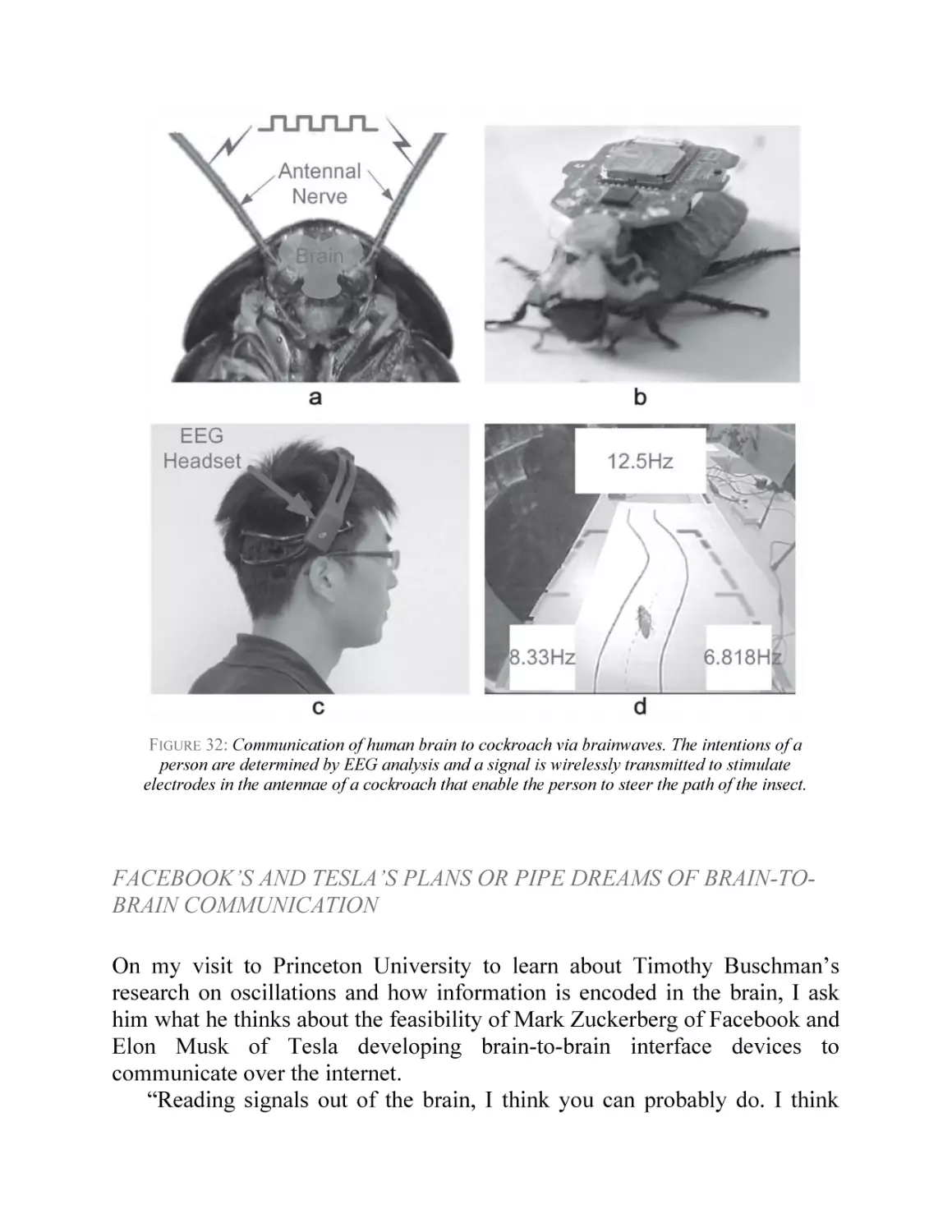
has done a remarkable job of lifting the veil on these mysterious, fascinating
emanations and their potential to change our behavior, influence our
personality, and teach us about the core of consciousness itself.”
—Florence Williams, author of The Nature Fix
“A lively personal account of the wild west of brain wave research, opening
multiple windows to deep mysteries underlying our conscious and
unconscious selves. Lucid discussions of dreams, mental disorders, mind
control, and more provide the reader with a vibrant look into brain science
and its impacts on our lives. Strongly recommended.”
—Paul L. Nunez, PhD, neuroscientist and author of The New
Science of Consciousness: Exploring the Complexity of Brain,
Mind, and Self
“Douglas Fields explains how electricity is exploited in neuronal circuits for
both simple and complex functions. He does it in accessible language,
embedded in beautiful prose, where beyond discussing fascinating scientific
topics of brain works, the scientists come to the fore. A great book to
everyone interested in the most complex matter that exists.”
—György Buzsáki, author of Rhythms of the Brain and The Brain
from Inside Out
Electric Brain copyright © 2020 by R. Douglas Fields
All rights reserved. No part of this book may be used or reproduced in any manner whatsoever without
written permission of the publisher, except in the case of brief quotations embodied in critical articles
or reviews.
BenBella Books, Inc.
10440 N. Central Expressway, Suite 800
Dallas, TX 75231
www.benbellabooks.com
Send feedback to feedback@benbellabooks.com
BenBella is a federally registered trademark.
First E-Book Edition: February 2020
Library of Congress Control Number: 2019952185.
ISBN 9781946885456 (trade cloth)
ISBN 9781948836296 (electronic)
Editing by Sheila Curry Oakes and Alexa Stevenson
Copyediting by Elizabeth Degenhard
Proofreading by Sarah Vostok and Cape Cod Compositors, Inc.
Indexing by WordCo Indexing Services, Inc.
Text design by Publishers’ Design and Production Services, Inc
Cover design by Faceout Studio, Spencer Fuller
Cover image © Shutterstock / Sebestyen Balint; image editing by Faceout Studio, Spencer Fuller
Printed by Lake Book Manufacturing
Distributed to the trade by Two Rivers Distribution, an Ingram brand www.tworiversdistribution.com
Special discounts for bulk sales are available.
Please contact bulkorders@benbellabooks.com.
Dedicated to my father, Richard L. Fields
CONTENTS
PREFACE
PART I
DISCOVERING THE ELECTRIC BRAIN
CHAPTER 1
Broadcasts from the Mind
CHAPTER 2
It’s Alive! The Spark of Life
PART II
UNDERSTANDING BRAINWAVES
CHAPTER 3
Brainwaves Explained
CHAPTER 4
Deciphering the Brain’s Code
CHAPTER 5
Brainwaves, a Window into the Mind
CHAPTER 6
Consciousness, Riding on Brainwaves
CHAPTER 7
A Sea Change in Brainwaves While We Slumber
PART III
HARNESSING BRAINWAVE POWER
CHAPTER 8
Mind Control: Brain-Computer Interface
CHAPTER 9 Brainwaves Reveal Your Thoughts, Strengths, and
Weaknesses
CHATPER 10 Detecting Brain and Mental Disorders
CHAPTER 11 Mastering Your Own Brainwaves
CHAPTER 12 Into the Future
ACKNOWLEDGMENTS
BIBLIOGRAPHY FOR FURTHER READING
REFERENCES AND NOTES
INDEX
ABOUT THE AUTHOR
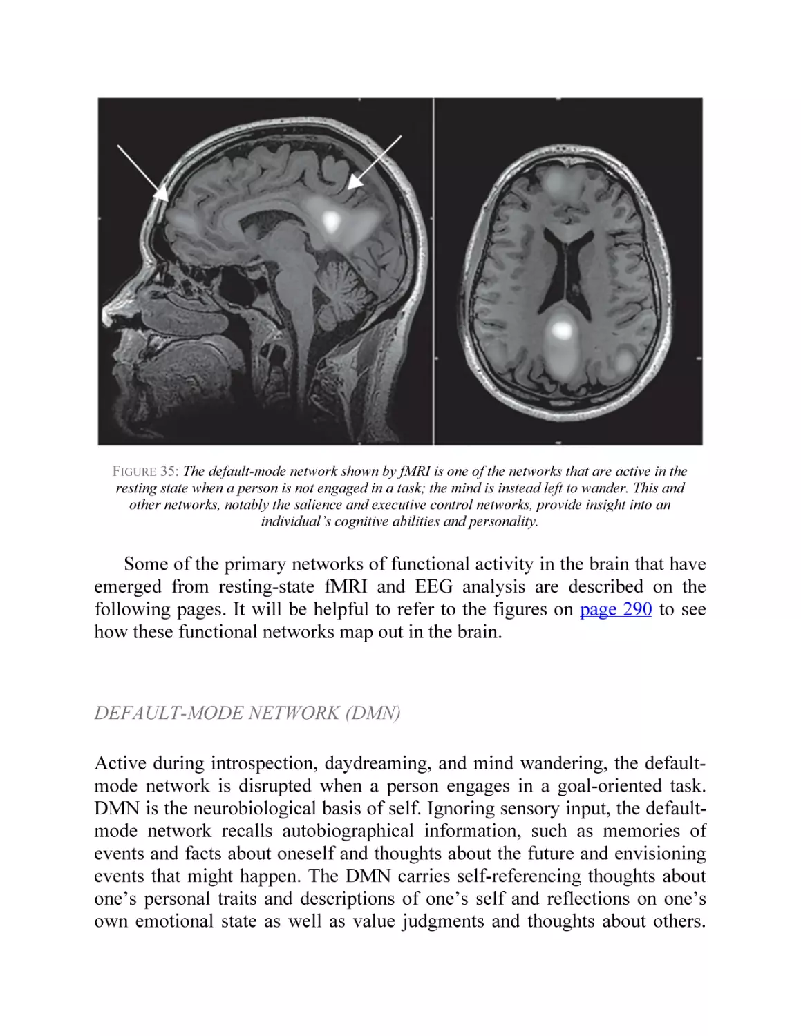
PREFACE
panel lights blink ominously on rows of black computers sealed inside
B lue
a glass room. I walk past the supercomputer nerve center and proceed
down an empty, dark corridor. Arriving at a door, I open it and enter a
cavernous room painted entirely black. Twenty-four digital cameras stationed
around the room pinpoint my every move. Two men stand up from behind a
bank of computer screens. I know that by using their sophisticated
instruments, they can eavesdrop on electrical transmissions flashing through
my brain. The most intimate details of my mind are theirs to see. They can
read my thoughts before I have them. They can watch my brain learn. They
can glean my intelligence, my propensity for adventurism, identify telltale
signatures of mental illness and neurological disorders, and predict my ability
to learn specific types of material. Am I a good reader? Bad at arithmetic?
Prone to depression? Developing early stages of Parkinson’s disease? These
deeply personal insights into my mind are available to them and other brain
scientists who are propelling a revolutionary leap in neuroscience that will
transform our world.
I’ve come to this place to meet these two neuroscientists and experience
for myself how they can watch my brain learn by tracking my brainwaves.
There is no need to open my skull and stick electrodes into my brain to tap
into my neural circuits. Electricity zipping through thousands of neurons deep
inside my cerebral cortex creates electromagnetic waves of energy that
penetrate my scalp. These electrical discharges can be picked up by touching
a wire to my head and feeding the electrical signals from my brain through
the wire into a computer. In Electric Brain I will take you to this and many
other laboratories around the world to see this exciting new brain science
currently unfolding, and to trace back in time to find the roots of this
discovery.
The detection of brainwaves in the early twentieth century is one of the
most important developments in the history of neuroscience, yet brainwaves
and their momentous implications are poorly understood by the general
public. Brainwaves aren’t taught in school. In fact, most college neuroscience
textbooks lack a chapter on brainwaves. This situation has persisted for
nearly a century. Yet, learning about brainwaves isn’t complicated—anyone
who is truly interested can easily understand this new science.
But brainwaves have always been surrounded in controversy. From the
moment a reclusive German doctor in the 1920s discovered waves of
electricity radiating out of the heads of his patients in a mental hospital,
brainwaves sparked astonishment and deep intrigue. His secret experiments
revealed that these cryptic electromagnetic emanations from the innermost
workings of our mind change with our thoughts and mental state. Today we
know that these bioelectric broadcasts expose the most intimate privacies: our
conscious thoughts, our unconscious cognition, and the emotions stirring
inside our brain.
From the moment scientists first glimpsed brainwaves at the turn of the
twentieth century, they were seen as complex and mysterious emanations.
The controversy continues, as brainwaves are hotly debated by
neuroscientists today. Some dismiss brainwaves as the electrical noise of the
brain at work, like the roar of an automobile engine is a by-product of its
mechanism of operation. Others believe brainwaves are how the brain
functions at its most sophisticated level. Brainwaves, these scientists believe,
explain many complex aspects of the human mind that have long mystified
philosophers and scientists alike.
While scientific debate rages over the origin and function of brainwaves,
no one questions the extraordinary capabilities that can be achieved by
monitoring and altering them. Neuroscientists can feed brainwaves into
computers to control software, machines, and prosthetic limbs. This melding
through brainwaves can fuse man and machine into unparalleled levels of
perception, analysis, and problem-solving that far exceed the capability of a
digital computer or the human brain working alone. Conscious and even
preconscious thoughts can control everything from a motorized wheelchair to
a fighter drone. Consciousness and brainwaves are so tightly interwoven that
brainwaves, not the heartbeat, now define the threshold between life and
death. Psychologists analyzing the pattern of brainwaves in a person’s brain
as they sit quietly letting their mind wander can see how that individual’s
brain is wired—normally or abnormally. If the brain’s electromagnetic
oscillations are atypical, the practitioner can program a computer to signal the
patient when their brain’s electrical activity shifts in the appropriate way, and
given this feedback, the brain will correct its brainwave activity—healing
itself without drugs.
Other scientists are using optical and magnetic methods that can read the
brain’s electrical activity through a person’s head, and still others are
implanting electrodes and computer chips into people’s brains. Fusion of
mind and machine means that the flow of information can go both ways.
Human brain function can be controlled by computer-generated signals
stimulating neurons by delivering electric current or electromagnetic pulses
through a person’s skull to manipulate brainwaves. Brainwaves of one person
can be picked up and directly downloaded through a computer into another
person’s brain to transmit information from brain to brain as if by telepathy.
This once obscure area of science has suddenly exploded into the mainstream
as billionaires Elon Musk of Tesla and Mark Zuckerberg of Facebook pour
millions of dollars into developing brain-computer interface technology for
commercial purposes, which Musk claims will provide a third “digital
superintelligence layer” to the human brain. Are such proclamations real or
fantasy-fueled marketing? Here are the facts and open questions:
For the first time in human history, Homo sapiens has developed the
ability to directly interrogate and manipulate the human brain. But with this
new ability come many questions both practical and ethical: What are the
risks and benefits posed by this noninvasive method of knowing what your
particular brain can and cannot do well, and by being able to link your mind
to a computer? How will such profound insights into our innate mental
capabilities influence education and career choices? For some, the new
technology raises Big Brother fears of mind control via radio-controlled
beams or brain implants.
All of these profound developments, transforming our future and our
understanding of the brain, are possible because the brain, unlike most bodily
organs, operates by electricity. How was that remarkable discovery made?
Who were the first people to discover brainwaves? What motivated them to
pursue the bold idea that electromagnetic waves might propagate out of a
person’s head? What did they think they had found? How did other scientists
react? Why was the discovery of human brainwaves kept secret for years?
Why are most people unaware of the name of the person who discovered
them? Why was there no Nobel Prize for this work? The answers are found
by unraveling a fascinating tangle of science and society that surround the
electric brain.
Whenever science crosses the threshold into a new frontier, the strange
phenomena will confuse people and spark controversy, but I did not write this
book to argue my viewpoint. I wrote Electric Brain to share the excitement of
science in action, as seen through the eyes of a neuroscientist working in the
field; to provide you with the latest scientific research (some of it not yet
published) and empower you to form your own opinions. I will, however,
separate fact from hyperbole—there is an enormous amount of hype and
superficiality in the popular press about brainwaves. I will explain at a
scientific level what brainwaves are, what they can do, what we know, and
what we do not know—yet. I will take you into laboratories around the
world, introduce you to my colleagues who are doing this research, allow you
to see the data as I have, and let you learn and draw your own conclusions.
That is, after all, what science is—a process of discovery. But first, we need
to trace back through time to find the roots of the “electric brain.”
CHAPTER 1
Broadcasts from the Mind
fifteen-year-old boy was shot in the head—the circumstances lost to
T hehistory,
but the consequences made history. The bullet, lodged in the
boy’s brain from an accidental gunshot, could not be removed by surgery,
leaving him paralyzed on one side of his body and suffering from vertigo.
After surviving the bullet wound with a piece of his skull missing, the patient,
sent to a mental hospital in Jena, Germany, was just what his doctor, Hans
Berger, was looking for. It was not the now twenty-three-year-old’s mental
condition that interested Dr. Berger but rather the hole through the man’s
skull.
SEEING THROUGH THE SKULL
In eager anticipation, in November 1902 Berger ordered the young patient
brought to the laboratory on the ground floor of the psychiatric clinic where
he conducted his solitary research. Whether the man believed it to be a
treatment of some kind is not known. What is now known is that Dr. Hans
Berger, who would become the director of the hospital in 1919, did not
regard all human life as equally sacred.
The young man sat in the chair as instructed, his paralyzed arm dangling
on one side. Behind him, he felt fingers pinch tufts of his hair and heard the
snipping of scissors. He heard water and the hollow rattle of wood against the
walls of a ceramic cup; there was a pause and then the luxurious warmth of a
frothy shaving brush lathering his head and dispersing the sharp, spicy smell
of soap. With long, slow strokes of a straight razor, the doctor shaved the
young man’s head and toweled it dry, taking extra care with the area around
the head wound, where the missing skull bone had left an irregularly shaped
hole covered only by a thin layer of skin and scar tissue.
FIGURE 1: From Hans Berger’s laboratory notebook from the turn of the twentieth century,
recording his experiments on the connection between psychic energy and physical energy.
Cranial defects, covered only by a thin layer of skin, in his patients who had lost skull bone
enabled Berger to monitor changes in brain volume pulsations in response to sensory
stimulation, drugs, postural changes, and emotions.
Over this hole the doctor placed a strange-looking skullcap made of gutta
percha—a natural latex rubber—and connected the cap to a fluid-filled rubber
tube. The tube resembled a manometer, used for measuring changes in
atmospheric pressure, and Dr. Berger was careful to seal it tightly to the
fitting in the cap so that there were no leaks. The other end of the tube was
connected to a device on the side table that held a sharp metal pen suspended
on a pivot. Once the cap and tube were connected, the pen began to scratch
out a thin white trace on a piece of soot-covered paper attached to a slowly
rotating drum.
Blood, as it pumps through mushy brain tissue, causes the brain to swell
and then retract slightly, like the chest of a panting dog, and these brain
pulsations, accessible to Berger thanks to the window of missing skull, were
what intrigued him. The fluid inside the tube rose and fell with minute
pressure changes in the head wound, and the attached recording device traced
jagged, regularly spaced undulations on the paper of the drum. Berger stared
intensely at the wavy line as it formed in the wake of the shaking pen,
tracking the swelling and shrinking of the young man’s brain as it pulsed in
time with his heartbeat.
Dr. Berger was not providing the young man with medical treatment, and
his purpose was not to understand or alleviate any of his mental or
neurological dysfunctions; Berger’s interest was in testing his theory that
mental states interact with physical processes inside the brain. At a time when
the origins and mechanics of the human psyche, and mental functions in
general, were still mysterious, this was a revolutionary concept.
While the brain’s pulsations were regularly paced along with the young
man’s heart, their amplitude waxed and waned, and Berger believed that
these fluctuations were associated with the mind’s thoughts and emotions
sweeping through brain tissue. He recorded in his notebooks, and later
published in a monograph in 1904,1 that the pulsations were affected by
various drugs and changes in body position—sitting, standing, or tilting the
head to one side—but more interesting by far to Berger was his observation
that the waves also changed in association with conceptual tasks, emotions,
and sensory input. He presented the young man, and other patients he
subjected to similar experiments, with various stimuli and cognitive
challenges: He instructed the patient to stick out his tongue and stimulated his
taste buds with drops of sugar, lemon, or quinine. He passed cotton balls
saturated with various pungent substances under the young man’s nostrils, or
had him sniff small vials containing pepper or other spices. Using a fine
paintbrush, he lightly stroked the back of the man’s hand, arm, and cheek. All
the while, he watched the line as it appeared on the rotating drum, tracking
the swelling and shrinking of the young man’s brain with the tide of its blood
flow. Often, these stimuli perturbed the regular rhythm of the brain’s
throbbing, proof to Berger that the elusive psychic phenomena of the mind—
such as the experiences of taste, smell, and touch—interacted with biological
and physical processes.
FIGURE 2: One of the patients with a cranial defect who Hans Berger studied using a
plethysmograph to monitor fluctuations in brain volume.
Suspecting that human emotions, too, had a basis in the physical, Berger
believed that his brain-volume monitoring device (called a plethysmograph)
could record these manifestations of joy, sadness, or fear. Human emotion
can only be studied in human beings, and Berger was about to use this young
man’s brain to put his theory to a test. He stepped quietly behind the chair
where the man sat . . . and fired a pistol. The deafening report, a sound the
man had not heard since the day he was shot in the head, plunged the young
victim into a state of shock and absolute terror. Berger was thrilled to see the
rapid decrease in brain circulation induced by the mental state of profound
fear. He detailed his findings in a monograph published in two parts in 1904
and 1907, titled Über die Körperlichen Äusserungen Psychischer Zustände
[Physical Manifestations of Mental States].2
Berger would move on from his research on changes in brain pressure to
study the brain’s electrical activity. He performed the first human
electroencephalogram (EEG), becoming the first person to discover and
document waves of electrical energy radiating out from the human skull and
pioneering the technology that would give science its initial glimpse into the
workings of the mind.
Given the significance of what he uncovered, you would think Berger’s
name would be a feature of textbooks. Instead, he remains a shadowy figure.
Why? After all, the unfortunate truth is that the ethically unpalatable practice
of experimenting on patients hardly makes him unique among his
contemporaries in the world of psychiatry. So—who was Hans Berger? And
how did he arrive at his shocking conclusions?
A JOURNEY TO JENA
I traveled to the city of Jena, to see firsthand what might remain of Berger’s
work conducted there a century ago. Rocked by two world wars, subsumed
by the Soviets into communist East Germany, and finally restored to a united
and democratic German state after the fall of the Berlin Wall, Jena has been
tumbled by the most monumental events of the last century. Berger is an
enigma, and history, being a written construct, mutates along with political
transformations, and the physical records and artifacts essential to
reconstructing the true events of the past are all too easily scattered, lost, or
destroyed with the passage of time. By seeing Hans Berger’s world and work
with my own eyes, I hoped to reach a better understanding of the discovery of
a fundamental feature of the human brain: brainwaves.
Behind a medieval church of stone blackened with centuries of soot and
encased like a Mayan temple by dead, ropy vines, I found a graveyard, little
more than a field of green lumps overgrown with ivy. Large trees screened
out the sun. There in a clearing, surrounded by ivy and bracken fern, sat a
roughhewn granite headstone bearing Berger’s name, the two dates
bracketing his life etched into the stone and painted black to contrast with the
gray of the slab. In the silent stillness of the graveyard, Berger was
transformed from a historical abstraction, and was now tangible and real.
Here he was, buried beneath my feet, Hans Berger the human being, along
with his wife and his son, who died in Russia as a soldier in World War II,
just a few months after his father.
FIGURE 3: Hans Berger’s grave in Jena, Germany, where he is buried together with his wife,
Ursula, and son, Klaus, near the psychiatric hospital where he worked and discovered human
brainwaves.
I left the grave and walked to the anatomy building of nearby Friedrich
Schiller University to meet neuroanatomist Christoph Redies and his
colleague, historian Susanne Zimmermann, who had kindly agreed to take me
to the psychiatric hospital where Hans Berger worked. After a short walk
from the university, we came upon the building, now named after Berger,
three stories of yellow and red brick with dormer windows jutting from the
steep roof. The building was vacant and undergoing renovation, one of the
exterior double doors standing open, and out in front was a bust of the doctor
himself. The cold stone suited Berger’s formal, stern-looking countenance.
This is where Berger worked for thirty years, searching for the interface
between the physical (brain) and the mental (psychic energy). His quest led
him from studies of blood flow and brain volume to investigate the stunning
new force of energy that was transforming technology and society in the
early twentieth century: electricity. Berger began his scientific career at a
time of horse-drawn transportation and gas lamps, and ended it in a world of
electric light, automobiles, airplanes, radio, and the splitting of the atom.
Over this same interval, the study of the human brain shifted from its roots in
philosophy and psychology to become the germ of a new science—
neuroscience, as it would come to be known—by applying microscopy for
cellular analysis, biochemistry for analyzing chemical components, and
electronics for investigating the electrical properties of nervous tissue.
In another building at the university, we located Berger’s old notebooks.
They are written in a peculiar stenographic notation, inscribed with precision,
but the code was sometimes impenetrable even to my native German hosts.
As I leafed through one of them, the pages yellow and the spine broken, I
found evidence of a different approach Berger used to test his hypothesis that
mental function interacted with physical and chemical processes. Berger
reasoned from the fact that, according to the laws of thermodynamics, the
work produced by energy is always accompanied by changes in temperature.
From this law of physics, Berger concluded that changes in mental activity
(which he called psychic energy) should be associated with changes in
temperature in the brain as well.3 A scene from another experiment is evoked
by the sketches, graphed data, and notes recorded in fountain pen in Berger’s
tiny script.
His subjects were again patients at the hospital, this time two girls, a
twelve-year-old suffering from epilepsy and an eleven-year-old with
headaches. After a surgeon drilled holes through their skulls, Berger stabbed
a rectal thermometer into the girls’ brains—21 millimeters, according to a
penciled anatomical sketch, or about an inch deep. He then proceeded to
perform experiments similar to those he’d conducted with the
plethysmograph, presenting the children with various sensory stimuli, this
time looking for changes in temperature. He published his results in a
monograph in 1910, titled Untersuchungen über die Temperatur des Gehirns
[Studies on the Temperature of the Brain].4
FIGURE 4: Reasoning from the laws of conservation of energy, Hans Berger believed that
changes in psychic energy, associated with mental function and emotions, should be
accompanied by changes in temperature. To test this theory, he inserted rectal thermometers into
the brains of people and at least one monkey, and noted how the brain’s temperature changed
during mental effort and sensory stimulation.
On the grounds of the old psychiatry clinic, Christoph Redies and I found
the building that had once housed Berger’s laboratory. In the early 1920s,
working in secret in the basement of this outbuilding, Berger began to
assemble the apparatus to record electricity from the human brain. He worked
alone, after hours, using mental patients and his own son as experimental
subjects. That son was Klaus, whose grave I had just left. Klaus would go on
to become a doctor himself before being killed in battle at the age of twentynine in November 1941.
The historic laboratory building is now a small library; although arguably
the most important discovery in electrophysiology of the last 100 years was
made here, it bears no plaque commemorating the place where man first
glimpsed brainwaves. But the librarian knew the building’s history. Berger’s
lab had been in the basement, he told us, but no traces of it remain. The
librarian showed me some faded photographs of the room filled with Berger’s
exotic electronic instruments, showing the first EEG subjects with wires
attached to their heads, but none of Berger’s equipment has been preserved.
In place of the plethysmograph device, which he still used to study brain
pulsations, Berger stuck zinc-plated needles under the skin covering the
missing skull bone in his patients, so that the tips of the needles touched the
surface of the brain. He connected the needles to an instrument in an attempt
to detect electricity, but the signals he extracted were weak and erratic. It was
unclear to him whether he was seeing electricity flowing through the brain or
instead electrical noise caused by the brain’s pulsation, heartbeat, and subtle
movement of muscle. He recorded signals that seemed to waver
independently of the patient’s respiration from several other patients’ brains,
including a forty-year-old man, but Berger was accessing the man’s brain
through the hole in his skull left from surgery to remove a brain tumor five
months earlier. The man died a few weeks after Berger’s experiment, raising
doubts about whether this man’s brain, swelled from disease, could be
considered normal.
FIGURE 5: Dr. Hans Berger, the first person to record the human EEG while working in the
psychiatric hospital where he served as rector of Friedrich Schiller University of Jena, Germany,
from 1927 to 1938. Below his portrait is one of the EEGs from Berger’s first published paper on
human EEGs.
Over time his instruments and techniques improved. Inside this building
one evening in 1924, Berger attached two large foil electrodes to twelve-yearold Klaus, one on the back of his scalp and another on his forehead, and fed
the feeble electrical signals into a device, called a galvanometer, that
recorded fluctuations in voltage by means of a light beam dancing on a strip
of photographic paper scrolled through the device. As Klaus sat with his eyes
closed, the beam plotted waves of electricity as they radiated from his brain,
oscillating slowly at low frequency. When Berger asked his son to open his
eyes, however, the brainwaves suddenly changed, vibrating erratically at high
frequency. Berger’s extensive experiments—on patients, his son, and even
himself—showed that these oscillating waves of electricity changed with
mental activity, arousal, attention, and sensory stimulation, and that they
became perturbed in diseases such as epilepsy. It followed that these waves
might not only give insight into how the brain operates and interfaces with
the mind, but also enable doctors to identify brain disorders, mental aptitudes,
and personality traits. But Hans Berger was hoping that brainwaves, like
broadcasts of radio waves, might do even more.
The philosophy of Dualism regards the mind and body as separate and
distinct from each other. Dualists believe that mental phenomena such as our
thoughts and emotions are nonphysical in nature, but that the psyche, spirit,
or mind interacts with the physical world. For most scientific Dualists of the
time this simply meant that physical stimuli could produce mental events,
while emotions or thoughts arising in the mind could in turn be located in
effects on the brain. Berger, however, describes the mind as a force—a
“psychic energy” that can interact with physical matter. He reasoned that by
the laws of conservation of energy, changes in psychic energy producing
mental work would necessarily require changes in other forms of energy in
the brain, such as temperature and electricity. He believed that the waves of
electricity he detected in the human brain were the rippling energetic
reverberations transforming between psychic and physical energy.
Stunningly, his discovery proved that the human brain’s energy propagated
through the skull and could be detected remotely.
In the building that had once housed Berger’s laboratory, the librarian
retrieved a monograph entitled Psyche, published by Berger in 1940.5 The
title page bears an inscription in his distinctive tiny script, nearly
incomprehensible except for the ornate large capital letters initiating each
sentence, then vanishing into a thin squiggle. In this paper, Berger relates his
belief in mental telepathy and psychic energy. He describes an experience he
had in the spring of 1893 while serving as a volunteer in the German army, in
which he believed himself to have communicated telepathically with his
sister. During a training exercise, Berger’s horse suddenly reared and he was
thrown into the path of a horse-drawn cannon. The driver of the artillery
battery halted it just in time, leaving the young Berger shaken but with no
serious injuries.6 His sister, at home far away, had at that same moment a
sudden strong feeling that Berger was in danger. She insisted that her father
send him a telegram at once. The incident made such an impression on
Berger that it changed the course of his career—after the war, he abandoned
his plan to study astronomy for medical school as a result.7 In this monograph
years later, he wrote, “It was a case of spontaneous telepathy in which at a
time of mortal danger, and as I contemplated certain death, I transmitted my
thoughts, while my sister, who was particularly close to me, acted as the
receiver.”8
Even in his day, Berger’s research at the fringe of paranormal psychology
would have been viewed with skepticism by his scientific contemporaries,
and it is almost certainly one of several reasons his work remained on the
periphery. Nevertheless, his discovery that it is possible to receive the
electrical waves of energy radiating out of the brain by placing electrodes on
a person’s head proved the interaction between the energy of the mind and
the substance of the brain. Working alone in an isolated psychiatric clinic,
Berger shattered the truism that one never really knows what is going on
inside another person’s mind. For the first time in history, it was possible to
directly monitor cognitive and emotional states in the human brain. With this
achievement, the feasibility of transmitting thoughts over a distance via
brainwaves was inescapable.
FIGURE 6: The Edelmann string galvanometer first used by Hans Berger to record brainwaves
from patients with trepanations (holes bored through the skull for medical purposes) and in
people who had suffered injuries to their skull, leaving a portion of the brain covered only by a
thin scar of skin. The oscillating string in the galvanometer was focused through a lens (2) onto a
roll of silver bromide paper (4) that was rolled through the device by the belt drive motor (1) and
developed as a photograph. Timing marks were made by a tuning fork (7) vibrating at 10 Hz and
sustained by a clockwork mechanism driven by a windup spring. Later instruments had a tiny
mirror attached to the string to bounce a beam of light onto the photosensitive paper that
streamed past.
Berger first recorded human brainwaves in his basement laboratory in
Jena in 1924, but he told no one what he was doing; indeed, he shared his
stunning results with none of his colleagues until 1929, when he published
his first report on brainwaves in the Archiv für Psychiatrie, titling it “Über
das Elektrenkephalogramm des Menschen” [“On the Electroencephalogram
of Man”].9 Few scientists took note. Researchers were making great strides in
understanding the cellular and physiological operation of the brain by taking
a reductionist approach—examining brain cells under a microscope, mapping
in detail parts of the brain involved in different functions, determining how
individual nerve cells generated and transmitted electrical impulses; Berger’s
approach of recording activity from the whole head at once made little sense
to leading scientists of the time. His work seemed bizarre; most dismissed the
noisy voltage fluctuations picked up by the scalp electrodes as originating in
electrical disturbances from muscle, not the brain. (When muscles contract
they generate electrical discharges that are much greater voltage than that
generated by neurons; muscles are more massive than nerves and are directly
under the skin where the sensors are attached.)
Berger’s publications established the priority of his discovery, but he
shared his work as narrowly as possible, guarding rather than promoting the
fruits of his research. “During the several semesters I had been his lecture
assistant I never knew him to speak during his course on his own field of
research,” said Rudolf Lemke, who joined the clinic as an assistant in 1931.10
“Only in 1931 did I hear him give a paper at a meeting of the Medical
Society at Jena on his discovery of the EEG,” recalled Lemke. “I can well
remember that the interest amongst his colleagues who listened to him was
not very great.”11
Berger’s sensational findings did not escape the popular press, however,
as indicated in this newspaper article published in the Baltimore Sun on
January 4, 1931:
[Hans Berger has measured] by means of a machine which he has
invented to register the electrical energy set free in the brain in the
course of mental activity. Similar attempts were made some years ago
by an Italian physicist, Dr. Ferdinando Cezzemelli [sic], using a radio
receiver of special design to pick up what were believed to be electric
waves from the brain; a real example of a “brain wave.” Difficulty was
encountered, however, in sorting out from these waves of “brain radio”
other impulses generated by the flow of the blood, the beating of the
heart, the action of muscles and other vital activities. These difficulties
Professor Berger believes that he has overcome by a special apparatus
using electrodes attached to the body to pick up the supposed brain
impulses instead of depending on accompanying radio waves.12
According to an article by Cyril Burtt [sic] in the book Science and ESP
by J. R. Smythies, an Italian neurologist, M. F. Cazzamali [sic], recorded
“electro-magnetic waves” conveying information directly from one brain to
another. He states that Berger believed the brainwaves he had discovered
could be the mode of telepathic communication, and he performed
experiments on brain-to-brain transmission of brainwaves, but he failed to
obtain experimental proof that such transmission by electromagnetic radiation
occurred. He therefore concluded that telepathic communication took place
by the transmission of psychic energy.13 (Note that Cyril Burt’s name is
misspelled in print throughout Smythies’s book as Burtt, and Cazzamalli’s
name is misspelled as Cazzamali.)
Interest in mental telepathy, and other forms of ESP such as clairvoyance
and premonition, peaked in the 1920s and 1930s. Upton Sinclair, renowned
Pulitzer Prize–winning author of The Jungle, was a firm believer. In 1930 he
self-published a book, Mental Radio, detailing his extensive experiments on
mental telepathy, most of them carried out with his wife. Radio was the rage
of the Roaring Twenties, and the emerging science of radio waves and
brainwaves converged in a flash of excitement that crashed as abruptly as the
soaring stock market in 1929.
Ferdinando Cazzamalli was a fellow at the neuropsychiatric clinic at the
University of Rome working in the 1920s and 1930s at the same time Berger
was studying brainwaves in Jena, but Cazzamalli’s studies were focused on
metaphysics and telepathy. Instead of electrodes attached to the scalp,
Cazzamalli positioned a radio antenna in the vicinity of a person’s head to
monitor the feeble brainwave broadcasts.14 Enlisting the assistance of noted
radio engineer Eugenio Gresetta to build the sophisticated electronic
apparatus, Cazzamalli pursued his “mental radio” research for ten years, first
publishing his findings in 1925. The bulk of his research was published in
Italian,15 and one paper in 1935 was published in French,16 so Cazzamalli’s
research remained obscure to mainstream scientists who did not read those
languages or have any interest in the paranormal.
Cazzamalli selected subjects whom he believed would be especially
strong in mental telepathy: artists and musicians. One such subject was a
painter who was also an Alpine mountaineer. The man was escorted into an
unfamiliar room that had walls lined with sheets of lead and was illuminated
only by the eerie dim glow of a red light bulb. The lead walls were necessary
to screen out electromagnetic interference from man-made radio waves and
alternating currents, so that the feeble brainwave broadcasts might be
intercepted.
In a corner of the room was a metal chassis containing an electronic
amplifier, a rectifier, and an oscillator tuned to a frequency of 300,000
kilocycles—all the components of a radio receiver. There was a couch next to
one wall, and the painter was asked to lie down and relax. Cazzamalli
positioned a dipole antenna 70 centimeters (about 2.5 feet) above the man’s
head and fed the signal through a wire into the radio receiver. The output of
the oscillator was recorded on film.
Cazzamalli asked the painter to close his eyes and clear his mind. He
recorded the signals for several minutes as the man remained in a passive
state. The painter’s mountaineering experiences had extended well beyond
Italy and into the Andes where tragedy had struck. Exploiting this,
Cazzamalli asked the man to recall the traumatic experience the mountaineer
had suffered on Mount Tronador in Patagonia searching for the bodies of
fellow climbers who perished on the treacherous mountain. Suddenly, the
beam of light tracing out a steady streak on the streaming photographic film
was abruptly interrupted. After a moment, the trace resumed. Cazzamalli
concluded that all of these emotionally charged thoughts radiated out of the
subject’s brain into the radio receiver and interrupted the signals—clear proof
of a physical basis for mental telepathy.
You will not find a single citation for Cazzamalli’s research in the
PubMed record of scientific publication, but while Cazzamalli is essentially
unknown to neuroscientists, in Italy his reputation is sustained by paranormal
researchers. In 1937, together with Giovanni Schepis and Emilio Servadio,
Ferdinando Cazzamalli founded the Italian Society of Metaphysics to support
experimental research on paranormal phenomena.17 The society continues
today, and it publishes the journal Metapsychic, The Italian Journal of
Parapsychology, which was founded by Cazzamalli in 1946. The publication
is not sold, but it is freely distributed among members of the society.18
Lest one dismiss this esoteric episode as an historical anomaly, it is
important to recognize that science, in exploring the unknown, is always
caught up in a torrent of clear and murky currents, pulled into eddies along
the way, and at times mixing science and pseudoscience. At the Society’s
annual meeting held in Bologna in December 2018, one of the speakers,
William Giroldini, presented research on mental telepathy and EEG.19
Alas, then as it is now, the popularization and sensationalizing of
scientific research can have a corrosive effect on how the work itself is
perceived within the scientific community. In a German newspaper article
published in July 1930, the potential of Berger’s discovery was touted to the
public with still more enthusiastic hyperbole: “Today, the brain still writes
secret signs. Tomorrow, we will probably be able to read neurologic and
psychiatric diseases in it. And the day following tomorrow, we will start to
write our first honest letters in brain script.”20
Such an achievement is a persistent dream. As I write this in 2019,
billionaires Mark Zuckerberg, founder of Facebook, and Elon Musk, founder
of the electric car company Tesla, are reportedly investing in methods of
extracting thoughts and emotions from a person’s brainwaves and
transmitting them over the internet, directly downloading them into other
people’s brains, and using them to operate electronic devices and computers
by thought alone.21 In part III of this book we will visit researchers around
the world pursuing this type of “mind reading” and “telepathic”
communication through brainwaves, to investigate the reality behind the
reports you may have read in the popular press, which is no less prone to
sensationalism now than it was in Berger’s time.
Over nearly a decade, from 1929 until 1938, Berger published one or
more papers a year on his research, all fourteen with the same title as the first,
“Über
das
Elektrenkephalogram
des
Menschen”
[“On
the
Electroencephalogram of Man”], and all in the same journal: Archiv. für
Psychiat. Nervenkr. This peculiar practice served to further shroud the results
of his experiments. The titles scientists give to their publications are typically
very descriptive of the specific new finding being reported, but no clue of
what Berger had discovered in any of his experiments can be gleaned from
the title of this series of publications. Any scientist who might be interested
in what Berger had discovered in a particular experiment would have to dig
through fourteen identically titled papers, as if to find a prize hidden behind
one of not three, but fourteen, identical doors. It is also difficult to reference
any of Berger’s specific findings, as the citations for these fourteen papers
differ only by the year of publication. Cloaking his findings in this way hid
them from the larger scientific community and diminished their impact.
Having made the first recording of a human EEG in 1924, Berger’s work
remained almost entirely unknown until 1934, when Nobel Prize winner
Edgar Douglas Adrian drew attention to the phenomenon of electric brain
activity by repeating Berger’s experiments in a paper coauthored with Bryan
H. C. Matthews and published in a prominent scientific journal, Brain.22 In
that paper, Adrian and Matthews implicitly mocked the significance of what
they called “Berger waves” by including a figure comparing changes in
Adrian’s own brain EEG when he opened and closed his eyes with the EEG
of a water beetle doing the same—evoking identical brainwave responses in
the insect and the Nobel laureate.23 Adrian did not pursue studies of EEG
afterward, and interest in Berger’s EEG recordings within the medical field
remained tepid.
“The lectures which Berger gave each year on his researches [sic] to the
Medical Society of Jena found no greater appreciation as time went on,”
noted Rudolf Lemke. “In 1934, at the meeting of German Neurologists and
Psychiatrists he reported on the EEG at Munster, and there too he did not
arouse the interest he had expected.”24 In the final line of her excellent
scholarly book published in 1961, A History of the Electrical Activity of the
Brain: The First Half-Century, neuroscientist and historian Mary Brazier
wrote, “Berger’s contribution to the first fifty years [of research on electrical
activity in the human brain] was known only to himself: the successful
recording of the electroencephalogram of man.”25
Leaving the place where Berger made his monumental discovery, I
followed Christoph Redies to the basement of another library on campus that
had just received an uncataloged collection of Hans Berger’s letters, photos,
and notes that had been saved by one of his former colleagues. I picked up a
photograph and held the actual recordings of one of the first human EEGs—
the same recording that had been held in the hand of Hans Berger nearly a
century ago.
Berger worked for almost thirty years in nearly complete isolation in this
psychiatric clinic a short walk from the cemetery where his body now rests;
he hardly seems the most likely candidate for such a scientific breakthrough.
Why was no one else at the time apparently pursuing this line of research?26
What led Berger to perform his strange experiments and set him on the path
to discovering the electric brain?
Tracing the source of Berger’s inspiration would take me to the
mountains of Turin, Italy, where in the 1880s, nearly two decades before
Berger’s experiments, Angelo Mosso was the first person to record human
brain activity. But the activity Mosso recorded was not electrical; he
developed the plethysmographic method later used by Berger, using it to
monitor how blood flow in the brain varied in response to exertion, changes
in respiration, and, among other things, adaptation to high altitude in the
mountains surrounding Turin. Berger’s early studies on brain volume
changes were directly adapted from Mosso’s methods and research, and
Berger credits Mosso extensively in his first publication in 1901.27 Today,
functional MRI, imaging of the brain at work, uses changes in blood flow in
localized regions of the brain to pinpoint where specific neural processes are
taking place. It too can trace its origins directly to Angelo Mosso, who
pursued similar investigations using only the primitive methods available to
nineteenth-century scientists.
My route to Mosso’s laboratory in Turin was a circuitous one.
SWELLED BRAINS
Dodging a frigid rain in the coastal village of Alassio, Italy, I escape to the
Caffé Roma, where Ernest Hemingway once scrawled his name on the wall
and started an infectious custom I could not resist. The previous day I’d been
in a hospital in Zaragoza, Spain, watching the ceiling slip away as I slid into
the chamber of an MRI machine, my mother’s scolding whine from weeks
earlier echoing in my head: “Why would you want to damage your brain?
You have such a good brain.”
FIGURE 7: Angelo Mosso, 1846–1910, an Italian scientist and one of the first people to study
brain function quantitatively with instruments. He used a plethysmographic method to measure
fluctuations in brain volume in patients who had cranial defects, which inspired Hans Berger’s
research. Of particular research interest to Mosso was how the brain is affected by high altitude,
which he studied in his laboratory built on Monte Rossa, Turin, Italy.
A good question.
It all started when I read a paper by a pair of Spanish researchers,
radiologist Dr. Nicolás Fayed and neurologist Dr. Pedro Modrego.28 In a
study of thirteen mountain climbers on an expedition to Mount Everest, they
found that all but one returned from the summit with physical brain damage,
as clearly visible on an MRI as a bone fracture on an X-ray. The damage was
permanent.
Mountaineers are well aware that the oxygen-depleted air at high altitude
can cause serious sickness—and in the extreme, brain damage and death—
from what is known as high-altitude cerebral edema (HACE). The body’s
response to the lack of oxygen increases the pressure inside blood capillaries,
causing fluid to seep out of them into surrounding brain tissue, bloating the
brain and crushing the life out of gray matter as it is compressed against the
skull. This phenomenon is precisely what Angelo Mosso was investigating in
the work that inspired Berger. Attaching his manometer device to the heads
of people with cranial defects, Mosso monitored the swelling of their brains
as they climbed the 15,000 feet of Monte Rosa, where he had built a
mountaintop laboratory to study altitude sickness and the physiological
mechanisms of adapting to high-altitude conditions.
The lasting cognitive effects of altitude on some veteran alpinists are well
known among climbers—“spacey” is the usual description—but most highaltitude mountaineers believe that if one acclimatizes oneself properly, severe
altitude sickness and brain injury can be avoided. In fact, the first reports of
altitude illness are from monks accompanying the Spanish conquistadors
advancing high into the Andes in the 1500s. The astute monks noticed that
while the generals were stricken with altitude sickness, the soldiers under
their command were spared. The reason, they soon realized, is that the
generals, who were riding on horseback, ascended the mountains rapidly,
while the soldiers climbed on foot at a much slower pace. (Monks, in addition
to serving God, were both historians and brewmasters; all of their activities
have left lasting impacts.)
The process of acclimatization requires ascending slowly to give the body
sufficient time to adapt to the drop in oxygen at higher altitudes, which
involves a series of fascinating and complicated physiological changes.
Acclimatization takes time, and weekend warriors paying guides to help them
bag a summit often pay a price for their impatience by becoming very sick.
While the subjects of Fayed’s and Modrego’s paper had experienced the
milder reactions to high altitude that virtually all climbers endure—headache,
nausea, fatigue—none had exhibited the more severe symptoms that are the
hallmark of HACE. In short, they weren’t aware that they had injured their
brains at all, much less permanently. The Spanish researchers then studied
climbers scaling other popular mountains of lesser height, including
Aconcagua, Kilimanjaro, and Mont Blanc, and found the same damage
visible on an MRI, although it appeared with less frequency in mountaineers
who had ascended these lesser peaks. Again, none of the climbers had
suffered symptoms of HACE; nevertheless, their brains had swelled enough
at high altitude to cause injury.
Being both a neuroscientist and a climbing enthusiast, I found Fayed’s
and Modrego’s paper especially fascinating, and it occurred to me that the
subject would make an excellent article for the general reader interested in
mountaineering’s effects on the brain. I approached an editor at Outside
magazine and proposed that I climb Mount Rainier, “America’s Mont
Blanc,” taking extra care to acclimatize myself, and have an MRI before and
after to prove that mountaineering could be done safely. They loved the pitch
and commissioned the article.29
That’s when I encountered a roadblock: I could find no one who would
agree to examine my brain by MRI for this project. It didn’t matter whether I
paid cash, or if the venue was a medical clinic or an experimental facility at a
university: Apparently, there is some ethical problem with putting a person in
a position where they might injure themselves just because it would be
interesting to study the damage.
I was stuck—until I thought to contact Dr. Fayed, who had done the
original study on climbers’ brains. “Sure!” he responded agreeably. “Come
on over; we’ll give you an MRI after the clinic closes and all go out for
dinner.”
So that’s what I decided to do. I wouldn’t have a “before” picture, but at
the very least I’d see whether my years of mountaineering had damaged my
brain. And en route to Zaragoza, I would go to Turin, Italy, to see the
laboratory where Angelo Mosso had done the research on brain swelling that
had been the inspiration for Hans Berger. But first, I had a mountain to climb.
CLIMBING MOUNT RAINIER
I set off for a rendezvous with my climbing partner and son, Dylan.
Attempted by hundreds of amateur climbers in a season, Mount Rainier is a
perfect setup for altitude illness. The glacier-shrouded active volcano rises
steeply to an elevation of 14,410 feet from sea level in Seattle, Washington,
where most climbers begin their ascent. Like Mont Blanc, Mount Rainier is
often attempted in a weekend push that is far too short to allow the body to
acclimatize properly to the thin air at the peak. Dylan and I, however, would
take our time.
It was the end of climbing season, and the guided groups and most other
climbers on Mount Rainier had bailed in advance of a ferocious approaching
stormfront. Dylan and I decided to head up into the weather despite whiteout
conditions, hoping to wait out the worst of it in our tent and make for the
summit after the storm had passed. I carried a pulse oximeter to monitor the
oxygen saturation of my blood and my heart rate. On one particularly
strenuous part of the climb, my blood oxygen plummeted to 75 percent, while
my heart rate rocketed to 165 beats per minute. (High heart rate is one of the
mechanisms by which the body tries to make up for reduced oxygen supply.)
I’ve seen my blood oxygen drop lower, while climbing the 19,347-foot
Cotopaxi in Ecuador, and climbers on Everest have registered numbers in the
50s. At normal elevations, people with that low of a level of oxygen
saturation are likely found only in the intensive care unit, and, from a medical
viewpoint, anything below 90 percent would be cause for some concern. But
at high altitude, changes in how tightly the blood’s hemoglobin holds on to
oxygen (among other things) lets us get by on less, demonstrating what a
nimble problem solver the human body can be if given time.
Of course, all the physiological calibration in the world can’t eliminate
some of the risks of climbing. On the way back down, I snagged a crampon
blade on the icy slope and took an invigorating header. When you hear about
mountaineers falling to their deaths, this is usually how it happens: Unable to
stop their screaming slide down the icy slope, the climber is rocketed over a
cliff into space, shot into the abyss of a crevasse (I hate crevasses), or
stopped, bug on a windshield style, by a rock outcropping. Careening down
the peak headfirst, I knew I would only get one chance to sink my ice ax into
the surface and break my fall as my acceleration built exponentially (32 feet
per second squared, as every scientist knows). Once you get going fast
enough, there is nothing you can do—your ax will skitter off the icy surface,
made as impenetrable as cold concrete at 60 miles per hour, and your partner
can only watch you sail by. That is, unless you are roped together, as Dylan
and I were at that moment, in which case your buddy can try to use his own
ax to brace his end of the rope and yank you to a gut-wrenching halt . . . or, if
that fails, join you in tumbling toward death. So, appreciating the situation as
I accelerated rapidly, I jackknifed quickly into the proper position (head
upslope) and swung once, hard, with deliberation and purpose. My ice ax bit.
Grasping the head of the ax with my left hand, I rammed my right shoulder
down onto it, driving the blade into the snow and gouging a furrow down the
mountain, slowing and finally arresting my death slide just before bowling
over Dylan, who was poised like Paul Bunyan with his ax at the ready.
It was a good reminder. Hypoxia is bad, but gravity sucks.
After returning to sea level at the Seattle waterfront, the oxygen-rich air
felt thick as cream, as if I could swim through it frogman-style; supercharged
to operate in the thin air of the mountain, my strength and energy felt
superhuman. This feeling will only last a couple of days, but it is a profound
sensation. It’s easy to see why, living among the mountains of Turin, Mosso
might have been moved to study altitude’s effects.
Climb completed, I was off to Italy to see where Mosso worked . . . and
then to Spain, to get my head examined.
THE CLIMBER’S BRAIN
Dr. Gianni Losano is waiting for me outside the Instituto de Fisiologia
Umana, a complex of nineteenth-century stone buildings at the University of
Turin. Losano, a gentle lion of a man, now works at the same desk Angelo
Mosso once did, a massive oak and leather throne at the center of a spacious
office with twenty-foot-high vaulted ceilings and a parquet wood floor. It’s
Saturday, and the buildings are empty as he takes me to meet his colleague,
Dario Cantino, an expert on Mosso who is engaged in translating his work
from Italian to English and making it available on the internet.
A scientist and mountain climber himself, Cantino is a lean sprite of a
man with an exuberantly cheerful disposition. He is also an eclectic collector
of scientific and mechanical apparatus. He takes me through room after room:
In one, he has collected electron microscopes—from the earliest devices of
the 1950s, which resemble something out of an old Boris Karloff movie, to
recent models—and in another, printing presses of every design are stashed
to the ceiling, with trays of lead letters spilling their contents. Cantino’s
collection of printing presses has grown so large that he has no place to store
them all; some are kept outside covered with tarps to protect them from the
elements.
“Why printing presses?” I ask him. Cantino thinks a minute before
offering that perhaps it is because printing presses translate and record
information and ideas, which is what he does himself, both as a scientist
translating data into insight and in translating Mosso’s work into English.
Cantino walks over to an enormous press and selects a single letter the size of
my hand to give me as a souvenir: a question mark—a most appropriate
selection for one scientist to give another. (Equally appropriate as a
punctuation mark to my present quest, my mom would have thought.)
Next, Losano and Cantino take me to the library, where Angelo Mosso’s
notebooks and instruments are kept neatly organized and cataloged in a room
open only to scholars. Smoked recording drums that turned by brass
clockwork gears and devices to measure changes in blood pressure and brain
volume were displayed on tables or housed in small cubbies. There are
photographs of subjects having their brain swelling monitored and of
Mosso’s various other high-altitude experiments. My fingers become lightly
soiled with soot as I hold in my hands a strip of blackened paper, covered
with the delicate oscillating tracings that were the first recordings of human
brain activity. I am mindful that I have now had the privilege of holding both
of the first two such records ever made—one by monitoring blood flow and
the other electrical signals—each bearing the fingerprints of the scientist who
made the recording a century ago.
FIGURE 8: Apparatus used by Angelo Mosso to study the effects of high altitude on the brain and
body. Fluctuations in brain volume, detected by a plethysmograph, were recorded by a pen
scratching soot off paper on a slowly rotating drum.
From Turin, I drive all day and most of the night to reach Zaragoza,
Spain, after taking a detour through Switzerland (I think) when I make a
wrong turn somewhere in France. Dr. Nicolás Fayed, radiologist at the
Clínica Quirón de Zaragoza, is tall and pewter-haired, with a casual manner
and friendly sense of humor. He asks about my climb as his colleague, Dr.
Jaime Medrano, translates our exchange. “You have to be poco loco [a little
crazy] to be a climber,” Fayed says with a smile, implying that this is not
entirely a bad thing.
We enter Dr. Fayed’s office adjacent to the MRI control room, and he
begins pulling out files; the desk is soon covered with scans of climbers’
brains.
“Atrophy of the frontal lobes,” he says, pointing to the black-and-white
slice of brain on the film. The forebrain is located behind the forehead; in this
scan it is shriveled like a dried fruit. This is the brain region severed in a
prefrontal lobotomy, a surgery that leaves patients intellectually unimpaired
but with deficits in higher-level executive functions like focus and planning
—in other words, “spacey.” I have scrutinized similar images with clinical
interest in published papers, but suddenly this is different. Each of the images
is labeled with a climber’s name.
FIGURE 9: The author holds a recording of physiological responses recorded by Angelo Mosso
using a plethysmograph.
“José suffered the most serious damage,” says Fayed. He hands me a
picture of a robust young climber in red Gore-Tex standing on the snowy
slopes of Aconcagua, with windblown dark hair and a whiskered beard,
bronzed by the high-altitude sun. He looks fit and determined, someone I
would enjoy roping up with. “When José came back, he couldn’t remember
his own phone number. His wife would send him to the store for a loaf of
bread and he would forget why he was there and come home without it.”
“See the lesions in the forebrain?” he asks, pointing at another scan.
Lesions like these are caused by small strokes or hemorrhages. They show up
as bright spots on an MRI throughout the brains of climbers but are especially
common in the white matter regions. White matter is like the core of a
baseball, composed of millions of tightly bundled cables that connect neurons
in the surface layers of the brain together into networks. Damage to white
matter anywhere in the brain can have severe and wide-ranging
consequences, just as when a backhoe digs up power cables in one yard and
causes extensive blackouts throughout the city. White matter seems to be
particularly vulnerable to hypoxia because it is not as richly supplied with
blood capillaries.
Eleven of the climbers scanned had enlarged Virchow-Robin (VR)
spaces, caused by swelling and loss of brain tissue. You don’t need a medical
degree to see these: They look like white shotgun pellets lodged deep in the
brain. VR spaces are the areas that surround blood vessels in the brain; we all
have them, and as our brains age, the spaces widen. However, while multiple
or much-enlarged VR spaces are common in the brains of the elderly and
those with Alzheimer’s disease, they are not normally encountered in healthy
people in their twenties and thirties—the ages of these climbers.
Next I meet Coral Roya, at the controls of the MRI machine. To my
surprise she reveals that she was one of the test subjects on the Kilimanjaro
climb (5,895 meters, or 19,340 feet, above sea level). As is typical, all seven
climbers on that climb experienced mild symptoms of mountain sickness, but
no one suffered HACE or other serious altitude illness. They all had MRIs
before the climb, which showed no abnormalities, but one of them returned
with dilated VR spaces in his brain. On Mont Blanc (4,810 meters, or 15,781
feet, above sea level), one of the seven amateur climbers returned with a
subcortical lesion, and two had multiple dilated VR spaces.
I ask how many of the climbers in the studies, most of them physicians
and engineers, stopped climbing after seeing the damage to their brains.
“They are all still climbing,” Fayed tells me.
“Our purpose is not to stop climbing,” Dr. Modrego emphasizes. “It is to
make people aware of the dangers and the need to acclimatize properly.”
Now it is my turn—the moment of truth. I strip to my underwear and don
green paper booties and a powder-blue hospital robe. The costume makes me
feel instantly infirm.
A smiling assistant, Elisabeth Pérez, hands me a pair of sticky pink wax
plugs to stuff into my ears to blunt the noise of the MRI machine as it takes
pictures of my brain, slice by virtual slice. I lie down rigid as a mummified
pharaoh on the narrow plank sticking out of a monstrous machine that looks
like an industrial clothes dryer on steroids. She adjusts the position of my
head with reference to a luminous grid projected onto my skull. Then she
lowers what looks like a catcher’s mask over my face and activates the
machine, which feeds me headfirst into a tunnel. If this were a James Bond
movie, now would be the time to break out one of Q’s inventions, grab Ms.
Pérez by the waist, and make my escape, but I brave the claustrophobic
confines and try not to move a muscle so as not to blur the photos.
Then the racket starts. Even with the earplugs, it is deafening. It sounds
like a massive construction job is being conducted inside my skull, with
hammering and buzz saws punctuated by bizarre video-game sound effects.
Suddenly it occurs to me that maybe this isn’t such a great idea. What are
they seeing as they virtually slice my brain like thin prosciutto? To hell with
climbing scars—what if they find a freaking brain tumor? My mind taunts me
with the unforgettable image of Homer Simpson’s head X-ray, a walnut-size
brain lost inside his cavernous cranium.
I can see the doctors and technicians framed between my green booties
through a mirror suspended above my eyes. The mirror is intended to relieve
the stress of patients reluctant to bury their head in a machine that from my
current perspective suddenly resembles a guillotine. They are gathered behind
a plate glass window watching the scan on computer screens, but without my
glasses on, I can’t tell if they are disturbed by what they see or just looking
up my skirt and making jokes.
When it is over, I anxiously approach the group standing around
computer screens displaying a part of me far more intimate than my jockeys.
“Perfectly normal,” Dr. Fayed says, a bit too quickly for my comfort. Dr.
Fayed and Dr. Modrego leaf through the slices on the screen, magically
peeling away layers of undulating cerebral cortex, penetrating into the dark
core of my imaged brain and emerging out the other side, as my living brain
tries frantically to take it all in. Dr. Fayed stops the shuffling and points: “A
small VR space.” Flipping through a bit more: “Another one.”
“Perfectly normal,” Dr. Modrego reassures me. “For your age.”
Shit! What does that mean?
Dr. Fayed burns the stack of 3-D images of my brain onto a CD and, with
a smile, hands it to me like a deck of tarot cards.
NAZIS AND BRAINWAVES
Hans Berger committed suicide. Perhaps ironically, for a scientist intimately
familiar with the effects of restricted blood flow on the brain, he chose to do
it by hanging.
Berger served as director of the Psychiatry Clinic at the University of
Jena from 1919 until 1938, when he was made a professor emeritus and
retired. He continued his work under the new director after his retirement
until his death in 1941. Rudolf Lemke, an associate who’d assisted Berger by
taking photographs of the EEG traces, and who was himself a professor at the
University of Jena for the rest of his career, shared his personal
remembrances of Hans Berger in 1956 in an article published in a scientific
journal devoted to research on EEG.30 “Berger was no adherent of Hitler and
so he had to relinquish the service of his University,” wrote Lemke. “Not
having expected this, he was gravely hurt.”
In 1930s Germany, leaders in academia and elsewhere who were not
allied with the Nazis were purged. Berger came to be seen as yet another
tragic victim of the brutal Nazi regime. His suicide inside the hospital in Jena,
using an electrical cord to hang himself (yet another irony), was viewed as
either an act of protest against the Nazis or an act of desperation driven by
Nazi reprisals against him. This belief was echoed and reinforced in the
neurology community for the next fifty years. In a 2001 book, epilepsy and
EEG expert Dr. Eli Goldensohn wrote that “Berger showed his dislike of the
regime and they retaliated. In 1938, he was humiliated by Nazi functionaries
who abruptly removed him.”31 In 2005, Ernst Niedermeyer, another pioneer
in the field, wrote that “his relationship to the Nazi regime was not good and
Berger was most unceremoniously made a professor emeritus at earliest
convenience.”32
It is well known that the horrors of the Nazi “Final Solution” originated in
mental hospitals. The concept of “racial hygiene” involved ridding society of
people perceived to be inferior and therefore detrimental to the ideal of
genetic purity, either by preventing them from reproducing or exterminating
them altogether, and psychiatric institutions were ground zero for the
eugenics craze of the early twentieth century. The forced sterilization of the
mentally or physically handicapped was common practice in mental
institutions long before the rise of Nazism—in fact, the United States was one
of the first countries to implement such practices—and Hitler’s first
extermination programs involved the euthanasia of the mentally ill. The
methods and instruments of killing used by the Nazis, including the gas
chamber, were developed in German mental institutions for use on
psychiatric patients, and later transported and expanded into the death camps
where they were applied to mass genocide. As director of the Psychiatric
Clinic at this time, Berger would have been at the epicenter of the eugenics
movement and the racial hygiene policies that marked the rise of Nazism.
After the war, Jena came under Soviet control, but many of the people
who held positions of authority at the university and hospital during the war
remained in charge afterward, a situation that promoted cover-ups and
dissuaded the Soviets from investigating former Nazis or preserving
documentation. And with many records lost or destroyed, the history written
by those left standing tended to reflect a narrative that meshed with the
sentiments of the day. A narrative that may or may not have been accurate, as
I found out when I visited Jena and met historian Susanne Zimmermann.
Rudolf Lemke, the colleague who placed Berger in opposition to the
Nazis in his postwar remembrances, was in fact a Nazi himself. A member of
the NSDAP (the Nazi party), Lemke worked at the Erbgesundheitsgericht
(Hereditary Health Court) from the time it was established and onward. The
Erbgesundheitsgericht was introduced by the German Reich on January 1,
1934, to carry out the forced sterilization of the mentally and physically unfit,
a broadly defined category that included the disabled, psychiatric patients,
and alcoholics, among others.33 By May 1945, about 350,000 such people
had been subjected to compulsory sterilization via the genetic health
authorities.34 After the war, Lemke stayed on at Jena, and his activities, as
well as his virulently anti-Semitic and anti-homosexual views, were covered
up by authorities. He became director of the Psychiatric Clinic from 1945
until 1948, and his distinguished academic career lasted until his death in
1957—one year after he published his whitewashed recollections of Hans
Berger.
What were Berger’s feelings about racial purity and the atrocities of
euthanasia and forced sterilization taking place around him? Zimmermann
showed me comments in Berger’s notebooks that complained that there were
too many Jews on Hiddensee, a popular island destination in the Baltic,
making it no longer fit as a vacation spot. These were his personal notes, and
thus presumably reflect his true sentiments, but what would provoke him to
make such anti-Semitic comments in his laboratory notebook? Was he
perhaps performing experiments on someone of Jewish faith? We do not
know, but the racism and vitriol are there, written in his own hand.
“Berger was a supporting member of the SS in 1934,” Zimmermann told
me. “Supporting members” were not SS members per se, but they had usually
made a financial contribution in exchange for membership. “Many people
became members to enhance their career,” explained Zimmermann. “NSDAP
membership is not known, which probably means that he was not a member
of this hard-core Nazi organization, but at the time there were many small
Nazi organizations. There was not just one big party.”
Zimmermann pulled out stacks of records she obtained from the Stasi
police files of former communist Berlin, handing me official paperwork from
court proceedings in which Berger participated in the hearings of people
appealing their sterilization orders. There was a mentally disabled eighteenyear-old, a schizophrenic, someone with epilepsy, a sixty-one-year-old
alcoholic, and a woman with lower intelligence and memory deficits
accompanied by her husband, who pleaded with the court not to sterilize his
wife. Berger denied every appeal, condemning them all to forced
sterilization.
I asked, “Was he perhaps coerced to comply with the Nazis out of
necessity to maintain his position?”
“No,” Zimmermann replied. Berger served on the upper court of appeals
for health and hereditary disease (forced sterilizations) from 1938 to 1939.
“This was after his retirement,” she observed. “At this time, he no longer had
a professional motive to cooperate with Nazis or sterilizations.”
Another close associate of Berger’s was Karl Astel, a member of the
NSDAP and notorious for his activities pursuing “racial purity.” Astel
became rector of the University of Jena in 1939 and worked to reshape it into
a model SS institution. He was also the Nazi head of Health Affairs and
director of the Institute for Racial Hygiene. On March 4, 1941, Berger replied
to Astel’s invitation to continue his service overseeing forced sterilizations at
the hospital, writing, “I am willing to serve again as a consultant in Jena and I
thank you for this.” However, he became ill, preventing him from serving,
and he committed suicide only two months later. Berger suffered from
chronic depression, and he committed suicide while in the hospital in search
of a cause for his physical discomfort. “An autopsy found no physical
indications of a medical disorder,” Zimmermann said. On April 3, 1945, Karl
Astel turned a gun on himself, ending his life, as did many who were
complicit in Nazi atrocities and found themselves losers at the end of the war.
After my conversation with Zimmermann, Christoph Redies took me to
the university’s anatomy department, which has a renowned collection. He
unlocked the door and we walked into a room stuffed with neatly arranged
jars of human anatomical specimens and skeletons in glass cabinets. I saw an
amazing assortment of dissected human anatomy: female reproductive
organs, a skinned erect penis, testicles, and human skeletons ranging from
those of babies and children to adults; there were many fetuses in jars, several
with horribly monstrous birth defects. There was a two-headed child. There
were heads sliced in various sections and limbs, hands, arms, and legs, all
stripped of skin to reveal muscle and bone.
These specimens are priceless and essential aids for teaching human
anatomy. Training scientists and doctors in human anatomy is what brings us
medical treatments and relief from suffering, not to mention all the
understanding gleaned about the human body. Still, it is difficult to imagine
the generosity of those who give their bodies to science, knowing that
students of medicine will pick them apart, willingly suffering in death the
indignity of their naked bodies being sliced open, bit by bit, finger by finger,
organ by organ, their face peeled away, their head carved into slices as thin as
baloney to expose their brain, their genitals handled and inspected, then cut
apart to see what is inside.
The vast majority of us choose the conceit of having our dead bodies
embalmed to preserve some likeness of what they were when alive—
pointless for a body entombed in darkness six feet deep in the soil. Yet those
who give their bodies to anatomists and students know that their dead corpse
is otherwise utterly useless, except as food for worms and bugs, and respect it
for what it was—a miraculous vessel for the soul. They give their bodies to
save lives, to train physicians and allow scientists to uncover new insights
into this wondrous human machine. They are the courageous ordinary people
to whom every single one of us owes our lives and our health.
At the time of my visit, Redies and Zimmermann had just discovered and
reported in a scientific paper that approximately 203 bodies in the anatomical
collections at the University of Jena were from prisoners executed by the
Nazis between 1933 and 1945; 200 others were patients in mental hospitals
and retirement homes who were euthanized; and several dozen others had
died at forced labor camps.35
“Some people supported euthanasia in the hospital because anatomists
needed specimens and they welcomed the supply,” said Zimmermann. “This
also solved the Nazi problem of disposing of corpses.”
Having held human brains in my hands and dissected them, I can say that
one always thinks of the person when doing so. It is a sacred, solemn, and
grateful feeling of deep respect for the individual who selflessly gave his or
her body for science. But the anatomical display of a body of a murder victim
is an abomination that goes beyond disrespect to depravity; no scientist or
doctor of any decency would willingly be a party to it. After Redies’s and
Zimmermann’s work, any specimens that could be identified as having come
from those who were euthanized or died in labor camps were removed from
the anatomical display, and a plaque was hung outside the building to honor
them. By nature of the anatomical work, though, the origin of many body
parts cannot be traced. As a result, this valuable collection exists under a pall
that can never quite be dispelled.
Berger’s legacy is colored by a similar uncertainty. Was Hans Berger a
man a century ahead of his time, struggling with primitive instruments to
achieve intellectual greatness, or was he a quack using simple devices—
manometers, thermometers, and galvanometers—in bizarre and sometimes
cruel ways in an attempt to find evidence for the paranormal and an answer to
the eternal mystery of what separates the spirit from the body?
“Was he a genius, a mad genius, or just mad?” I asked a small group of
scholars at the end of my day in Jena. “He was crazy,” one in the group
answered. No one else advanced an opinion.
How could he shove a rectal thermometer into a young girl’s brain?36
How could he condemn his patients and other unfortunates to forced
sterilization, even when they pleaded with him to reconsider? Disregard for
the humanity of those with mental ailments is not unique to Berger or
Germany, but it is an atrocity wherever it occurs. Berger was a Nazi at heart,
whatever the records say. He also made a momentous discovery that will
intrigue and benefit mankind for generations. Both are true.
BERGER AND BECK
The story thus far would give the impression that Berger’s decision to search
for electrical activity in the brain was the result of a brilliant leap of genius,
but that is not the case. It had been known since the eighteenth century that
signals are transmitted in the nervous system by bioelectricity, although how
the nervous system worked, like the nature of electricity itself, were great
mysteries at the time. The idea that mental functions like thoughts might
generate waves of electrical activity that emanated out from the human skull
—much less uniquely identifiable, readable waves—was nowhere on the
horizon. However, Berger was not the first to investigate the possibility of an
electric brain that one might tap into using electrodes attached to electronic
instruments. In fact, just as with Mosso’s methods for monitoring blood flow,
he was simply copying what someone else had done—someone you have
likely never heard of.
HITLER’S BLACK BOOK
In many ways, Polish scientist Adolf Beck is Berger’s mirror opposite, the
two men driven by the same curiosity and passion, engulfed by the same
toxic politics, but their lives polarized—one allied with evil, the other evil’s
victim. The fates of these two scientists are entangled by the atrocities of the
Nazi frenzy that set the world at war, and connected by inspiration: It was
Beck’s work in Poland that inspired Berger’s secretive research in Jena,
exploring the possibility that the human brain functioned by generating
electromagnetic energy.
In the 1880s, brain scientists were preoccupied with attempting to
determine which parts of the brain controlled specific functions. The first
experimental approach, which was extensively used into the early twentieth
century, was to remove bits of brain tissue from animal subjects and observe
the resulting effects. In many cases, this produced useful insights—for
example, in localizing motor function by finding that damage to certain
regions of the cerebral cortex in either hemisphere caused paralysis on
opposite sides of the body—but other times the approach failed to provide
any clues. Beginning in the 1920s, for example, American psychologist Karl
Lashley trained rats to run a maze, then systematically removed portions of
their brains before reintroducing them to the task, in an attempt to pinpoint
where memories are stored. Lashley found that the location in the cortex
where the bits were removed didn’t seem to matter, and after decades of
research he gave up in defeat, concluding in 1950 that it was not possible to
localize a “memory trace” anywhere in the brain.37 Today, we’ve come a
long way toward unraveling the enigma of how mental functions map onto
the physical brain, and we understand that the assumption underlying the
search for a specific locus of a particular mental function is false. Although
specific locations in the brain do indeed correspond to hubs of neurons with a
particular purpose—for example, vision at the back of the brain and speech in
Broca’s area of the cerebral cortex—many functions require sending
information broadly over networks that encompass large territories of the
brain. Still, pinpointing brain regions and brain circuits that contribute to
specific mental, motor, or sensory tasks is essential, not only for
understanding how the brain works, but for practical necessity in treating
traumatic brain injury and performing neurosurgery.
In the 1880s, while experiments to locate the regions of the brain that
controlled motor function by simply damaging parts of the cerebral cortex
were fruitful, the process of determining where sensory information was
processed was considerably more complicated. It had been known for some
time that nerves transmit their messages via electrical impulses, and the
development in the nineteenth century of instruments to record electrical
signals provided a new technique to track down the neural circuits carrying
sensory information: placing a recording electrode on a spot in the brain to
test for a response to sensory stimulation, much as an electrician would probe
an electronic device with a voltmeter to trace an active circuit.
FIGURE 10: Adolf Beck (1863–1942), Polish scientist who recorded brainwaves from electrodes
placed on the exposed brain and spinal cord of experimental animals, beginning with his
doctoral dissertation published in 1891. Beck was imprisoned by the Russians during WWI for
two years. Just shy of his eightieth birthday, Beck swallowed a potassium cyanide capsule to end
his life as the Nazis came to take him to the Janowska extermination camp during WWII.
However, an electrician with a voltmeter has a far easier task than did the
neuroscientists of the late nineteenth century; it is important to appreciate that
all of this advanced study of electrical events in the nervous system was
taking place long before the advent of electronic amplifiers, or in fact any
other electrical device that we now take for granted. These experiments were
conducted in rooms lit by gas lamps and candles, reached by horse and
buggy, and in a time prior to an understanding of the fundamental nature of
electricity. Imagine that you are attempting to record electrical responses in
the brain of a test animal using nothing more than nonelectrical devices and
whatever other materials you might find in your kitchen. How would you do
it?
At the time, the most sensitive instrument available for detecting
electrical phenomena in the brain and nervous system was the string
galvanometer. In a string galvanometer, a magnetized needle is suspended by
a fiber between two coils of wire. When a voltage passes through the circuit,
the electrical field surrounding the needle is disturbed, deflecting the needle
slightly, much like a compass needle when brought close to a metal object.
The deflection of the indicator needle by the weak electrical events of the
nervous system is so miniscule that viewing it required using a magnifying
lens; however—just as a laser pointer greatly magnifies the subtle shaking of
a person’s hand during a PowerPoint presentation—the response of the
needle could be amplified by bouncing a beam of light off a tiny mirror
attached to the fiber. The mirror bounced the beam onto a ruled scale where
the deviations of the needle would be more easily measured, like plotting the
height of a child against a doorframe. Thus, instead of the intensity of the
response being recorded in millivolts, electrical events in the brain and nerves
were measured by millimeters of deflection of the light beam. Mind you, this
beam was not a laser pointer; the light that bounced off the miniature mirror
where it hung inside the elegant polished brass and wood device was
generated by a flame.
FIGURE 11: Adolf Beck used a Wiedemann galvanometer to record electrical brain activity in his
early experiments. Various versions of these instruments were used during this period. Shown
here is a D’Arsonval galvanometer. Note the small circular mirror suspended on the string,
which reflected a beam of light onto a scale in proportion to the electrical signal applied.
When it was developed in 1874, the Wiedemann galvanometer, designed
by Gustav H. Wiedemann, Hermann von Helmholtz, and John Tyndall,
became the most advanced instrument of its kind to measure voltage changes.
It was used by a young Adolf Beck in his dissertation research, after he
entered the University of Jagiellonski in Kraków (now called Jagiellonian
University) in 1886, where he performed his studies under the supervision of
distinguished Polish physiologist Napoleon Cybulski.38 The other equipment
necessary to investigate the electrical activity of the nervous system was
electrodes, and these too presented a challenge in the nineteenth century.
Metal electrodes have the serious problem of creating a battery when placed
in contact with salty (and thus conductive) bodily fluids; the electrochemical
voltages created by these electrodes themselves compromise attempts to
record the subtle electrical events generated by nerve tissue. Indeed,
controversy raged for decades between Italian scientists Alessandro Volta
(1745–1827) and Luigi Galvani (1737–1798) as to whether the body
generated any bioelectricity at all or was instead simply responding to the
artificial battery created when metal probes touched bare nerve, shocking it
like tinfoil on a metal filing. To overcome this problem, nonmetal electrodes
or strips of paper or fabric soaked in salt solution were used to pick up the
weak bioelectric signals from muscle, nerve, and brain. Adolf Beck described
in his dissertation the electrodes he used: “These [electrodes] were of clay
soaked in 1 per cent sodium chloride solution protruding from a glass tube
filled with zinc sulphate [sic], from which zinc wires made the connexion
[sic], to a Wiedemann galvanometer.”39 (Beck’s thesis research clearly met
the challenge posed earlier; sodium chloride is of course table salt, and zinc
sulfate is a dietary supplement—both might be found in the cabinets of a
modern kitchen.)
Adolf Beck began his research with frogs, carefully dissecting them to
expose the brain and spinal cord down to the hind legs. Like a variation of the
recipe pheasant under glass, Beck placed the frog with its filleted legs under a
Bell jar containing wet cotton to moisten the atmosphere and delay
desiccation. His clay electrodes, connected to the string galvanometer, were
carefully placed on the frog’s brain, ready to pick up the response from the
sciatic nerve in the leg, which he would stimulate with a pulse of electricity.
The pulse of electricity was generated by an induction coil—a coil of wire
wrapped around a cylinder, surrounded by a second wire-wrapped cylinder
that has many more wraps of wire than the inner coil. The inner coil was
energized by a homemade battery, and when the connection from the battery
to the coil was broken, the electrical field surrounding it collapsed, and the
force of the collapsing electric field induced a sudden jolt of electricity
through the outer coil that was of much higher voltage than that generated by
the battery but concentrated into a brief flash. The same mechanism is what
generates the spark in a modern automobile engine: The 12 volts from a car’s
battery is transformed into 25,000 volts when the current running between the
battery and the inner winding of an induction coil is repeatedly interrupted in
time with the piston, generating instantaneous short bursts of high-voltage
electricity in the outer winding to provide a powerful spark across the
engine’s spark plugs.
Frog and equipment in place, Beck commenced his experiment, staring
expectantly at the quavering spot of light from the galvanometer. He fired the
induction coil to shock the sciatic nerve—the light beam bounced! The tiny
deflection indicated that the point in the frog’s brain in contact with Beck’s
probing clay electrode was generating electrical energy triggered by impulses
sent from the sciatic nerve when he shocked it. By moving the electrode to
various regions of the brain and spinal cord and stimulating different sensory
nerves, Beck was able to locate where in the brain different senses are
processed.
In the course of this experiment, Beck made an even more fundamental
discovery: He noticed that the beam of light quivered energetically even
when he was not stimulating a nerve. Many people would have dismissed
these subtle vibrations as noise inherent to the delicate measurement system,
but Beck astutely concluded that he was seeing spontaneous oscillations of
electricity surging through the brain. “We are dealing with spontaneous
excitation of the nervous centres,” he wrote in his doctoral thesis, published
in 1891.40 In short—brainwaves.
FIGURE 12: A plot of electrical responses in a dog’s brain responding to light stimulation of the
retina and to stimulation of the foreleg in experiments performed by Adolf Beck for his doctoral
thesis published in 1891. Electrical signals cause small movements of a thin filament inside the
galvanometer measuring device. Reflections of a beam of light off a mirror, suspended on a
filament that twisted when electricity passed through the device, were projected onto a ruled
scale and read through a telescope.
Beck moved from frogs to rabbits and dogs, removing the skull from one
side of the animal’s head to expose the creature’s brain. He found that, when
he activated the animal’s various senses by presenting them with sensory
stimuli, specific parts of the cerebral cortex exhibited abrupt changes in their
electrical activity:
The optic nerve was stimulated with light, the auditory with sound, and
the different sensory nerves of the skin with an induction current. For
the light stimulation of the eye, I used a burning magnesium ribbon,
which was moved by a special clock mechanism so that the flame
reflected by a mirror into the eye remained constant.41
Curiously, stimulation of the senses did not increase the ongoing
electrical activity; it decreased it. In each case, he saw that sensory input
quelled the ongoing electrical fluctuations in the cortex of the animal’s brain.
What we now understand is that, following sensory stimulation, the ongoing
activity of brainwaves within the cerebral cortex becomes desynchronized.
Just as a child rhythmically pushing against the walls of a wading pool will
build large waves that splash exuberantly over the sides, while erratic or
“desynchronized” pushing by two children generates smaller irregular ripples
on the surface, new sensory input somehow disrupted the large-amplitude,
synchronized oscillations of electrical activity seen in the brain before the
stimulus was presented.
With only the most primitive instruments and his brilliant intellect, Beck
had made two fundamental discoveries: In addition to detecting the electrical
events caused by signals entering the brain from the senses, Beck correctly
perceived that there is spontaneous rhythmic electrical activity taking place in
the cerebral cortex all the time, and that this ongoing activity is perturbed by
sensory input. He found it reasonable that “stimulation of some centres
causes a blocking of activity in others . . . hence some sort of active state
must have existed in those centres and it was only the intrinsic activity that
was suppressed.”42 This intriguing brain response later led him to propose
that pain might be blocked by manipulating these waves of electric activity,
something that is at the forefront of research today. Transcranial direct
electric current stimulation and deep brain stimulation (DBS) with implanted
electrodes have both been used to dampen brainwaves in appropriate spots to
regulate, among other things, the sensation of pain. What’s more, as we’ll see
in a later chapter, the sudden desynchronization of brainwaves that Beck
discovered coinciding with sensory and cognitive activity has been used in
developing brain-computer interfaces to enable a person to operate prosthetic
devices through thought alone.
These pioneering experiments performed in Poland by Adolf Beck (and
others we will soon meet), revealing electromagnetic waves coursing through
the brains of animals, are what would inspire Hans Berger to attempt to
record electrical activity through the scalp of the human head. Adolf Beck
extended his work from frogs, rabbits, and dogs to the study of monkeys, all
of them showing the same brainwave responses. Human beings were the
likely next step for Beck, but unfortunately, he was pursuing his research at
the epicenter of international conflict during the outbreak of World War I. On
the cusp of making monumental strides in brain science, Beck’s research was
squashed by political events, war, and bigotry.
In 1895, Beck had accepted the chair of the newly established department
of physiology at the University of Lwów, which was then in the Austrian
section of partitioned Poland. The city of Lwów has changed hands many
times in the upheavals of the past two centuries, and at the turn of the
twentieth it was a diverse center of Polish, Ukrainian, and Jewish culture, the
home of many Polish national societies as well as the first Yiddish daily
newspaper. But in 1914, at the start of the First World War, the city was
taken by the Russians. Poles struggled to retain their cultural identity and
language, making Polish academics de facto resisters of, and threats to,
Russian occupation and domination. The year after the invasion, Beck—
along with nine other prominent citizens—was abducted by the Russians in
their retreat from the combined German and Austrian armies, and he was held
hostage in Kiev.
While a prisoner, Beck wrote to Professor Ivan Pavlov, the prominent
Russian physiologist best known for his studies conditioning dogs to salivate
at the sound of a bell, appealing to Pavlov to plead his case to Russian
authorities. In 1916, after enduring two years of captivity, an exchange of
hostages was arranged through the International Red Cross, and Adolf Beck
returned to Lwów, eternally grateful to Pavlov for securing his release. Lwów
in 1916 was still very much on the battle front, however, now surrounded by
Ukrainian forces. (Ukraine was not at that time part of Russia.) World War I
was followed for Lwów by the Polish-Ukrainian War and the Polish-Soviet
War. In 1919, Poland won its independence for the first time since 1795, but
Lwów continued to be contested territory until 1923, when it was officially
recognized as part of the Polish state.
Beck did not take his research into the electric brain further after his
imprisonment, focusing instead more generally on the science of physiology.
With his mentor, Napoleon Cybulski, he published a textbook on the subject,
and he went on to publish a textbook specifically on neurophysiology as well.
He continued to teach at the university and was a devoted father and a pillar
of the academic and cultural communities. He retired in 1930, at the age of
sixty-seven, but his retirement was not to be a peaceful one. Turmoil erupted
once more when the Germans invaded Poland on September 1, 1939. The
Soviets invaded in response on September 17, and on September 22 the city
of Lwów fell to the Red Army. The USSR annexed the eastern half of the
Polish Republic and renamed the city Lviv. During this period, the university
was renamed after a Ukrainian writer and Ukrainian was made the official
language of instruction. Many Polish academics were dismissed from their
positions, and a dozen were killed by the Soviets over the next year and a
half.
Then on June 22, 1941, Nazi Germany invaded the USSR, and Lviv was
taken by the Germans. What followed was a series of massacres directed at
the city’s Jewish population and orchestrated by the Einsatzgruppen, the Nazi
death squads under the direction of Heinrich Himmler and Reinhard
Heydrich. The first of these, in which some 7,000 Polish Jews were
murdered, was carried out with the help of Ukrainian national groups; the
second targeted a list of some 2,500 citizens believed to be hostile to the Nazi
regime. In addition to orchestrating mass killings of civilians, the
Einsatzgruppen targeted specific undesirables for extermination after
invading a country, guided by a carefully cataloged hit list. The version
drawn up for Britain was known as “Hitler’s Black Book,” and 20,000 copies
of the 144-page book were printed, listing the names and identifying details
of 2,820 people to be tracked down and murdered.43 A similar list was used
by the Einsatzgruppen after the invasion of Poland. The list included not only
Jews, but also members of the clergy, politicians, writers, artists—and
scientists. The intelligentsia, free thinkers, academics, and scientists were as
dangerous to Hitler’s domination as politicians and partisans. “The Polish
lands are to be changed into an intellectual desert,” wrote the German general
Hans Frank, while serving as governor of Poland after the Germans
invaded.44 As an intellectual, a Pole, and a Jew, Beck was very much in
danger.
On July 3, 1941, at the direction of the Einsatzgruppen, Nazi occupation
forces arrested twenty-five Polish academics, along with their families.45 The
detainees were taken to a repurposed dormitory, where they were tortured and
interrogated, and then killed.
At the time of the Nazi-Soviet invasion of Poland at the beginning of
World War II, an estimated 150,000 Jews resided in the city of Lwów,
including 90,000 children.46 Almost none of these 150,000 Jews of Lwów
were alive at the end of the war.47 A former factory on 134 Janowska Street
in the northwestern suburbs of Lwów was converted into a concentration
camp where more than 200,000 people were killed, and among its victims
was Professor Adolf Beck.
Beck remained at his home near the university for a year after the
massacre of Lwów professors, refusing to flee.48 Just shy of his eightieth
birthday, he became ill, and while in the hospital the Germans came to take
him to the Janowska extermination camp. Beck’s son, a physician, had
supplied all members of the family with capsules of potassium cyanide. Beck
swallowed his, choosing to end his own life rather than have it taken by Nazis
in the gas chamber.49
History in some parts of the world is not linear; it is a vortex, repeatedly
sucking the inhabitants back into the same whirlpool of disaster, destruction,
and death. Today, Lviv is a part of Ukraine. After the fall of the USSR in the
1980s, Ukraine became independent, but in 2014 the Russian military took
control of the Ukrainian territory of Crimea, annexing it into the Russian
Federation, and the area is once again awash in conflict. Reviewing these
stories from the dawn of neuroscience reminds us that we are always living
against the backdrop of history, and, one hopes, learning from it. The
distance between Beck’s past and our present is not as great as one might
think: One of the Gestapo officers who orchestrated the massacre of Lwów
professors was found decades later living in Argentina under a false name,
and was only arrested in 1985. He died before he could be extradited.
RICHARD CATON
The world is a small place. A few miles from my home, near Baltimore
Washington International Airport, is the town of Catonsville, Maryland. It
was originally settled by Europeans in 1720, and the Caton family came from
Great Britain to establish their home there in 1787.50 In 1887, Richard Caton,
an MD from Liverpool, England, visited the town founded by his ancestors
while on a trip to present his research at a meeting held at Columbia
University (now Georgetown University) in Washington, DC. Caton’s
presentation was titled “Researches on Electrical Phenomena of Cerebral
Grey Matter.”51
FIGURE 13: Richard Caton (1842–1926), an English physician, was the first person to discover
waves of electrical activity in the brain, in his research on rabbits and a monkey. He is shown
here in his thirties, at about the time he was conducting his research on brainwaves.
“Read my paper on the electrical currents of the brain,” he wrote in his
diary. “It was well received but not understood by most of the audience.”52
A consequence of the relative isolation of scientists in different countries
speaking and writing in different languages is that important discoveries can
go unseen by the larger scientific community. This is still true now, but it was
even more so in the days before the internet, or in fact any fast and reliable
method of international communication. Indeed, when Adolf Beck published
his dissertation in 1891, he had no idea that someone working sixteen years
earlier in the United Kingdom had already discovered brainwaves in
experiments on rabbits and monkeys. Beck had just begun his own study of
electrical responses to sensory stimulation when Richard Caton presented his
research in Washington, DC, in 1887, and, remarkably, Caton had initiated
this research a full twelve years earlier. He had published a brief preliminary
report of his results in 1875, in the proceedings of the meeting of the British
Medical Association, describing that by using a galvanometer he had
succeeded in recording spontaneous electrical currents from the brains of
rabbits and a monkey: “Impressions through the senses were found to
influence the currents of certain areas; e.g., the currents of that part of the
rabbit’s brain which Dr. Ferrier has shown to be related to movements of the
eyelids, were found to be markedly influenced by stimulation of the opposite
retina by light.”53 This brief published account of the electric brain of a rabbit
detailed the discovery of the EEG, although almost no one—including Adolf
Beck—knew of it.
Caton expanded his studies to forty rabbits, several cats, and monkeys,
describing the results in a more extensive report in the British Medical
Journal on May 5, 1877, concluding that “all brains examined have shown
evidence of the existence of electric currents . . . The current is usually in
constant fluctuation; the oscillation of the index being generally small, about
twenty to fifty degrees of the scale. At other times great fluctuations are
observed, which in some instances coincide with some muscular movements
or change in the animal’s mental condition.”54 However, these publications
remained obscure.
In general, biological science in that era was conducted primarily by
medical practitioners, such as Caton, who earned their income from their
medical practice while their scientific work afforded no income and little
notoriety. Physiology (never mind neurophysiology) was not yet recognized
as a scientific discipline and lacked its own academic departments, scientific
societies, and scholarly journals, and Caton’s work was of little practical use
or relevance to medical doctors at the time. After twelve years of research,
the audience at the Medical Congress Caton traveled to in 1887 failed to
grasp the significance of his findings. Although research did not resonate
with the medical community, Caton was nevertheless well respected as a
physician and professor, and he became the dean of the medical faculty at
Liverpool College in 1904.
FIGURE 14: The first report of brainwaves was by London physician Richard Caton. Caton
announced his discovery at the annual meeting of the British Medical Association in August 1875
held in Edinburgh, but the breakthrough went unnoticed. Caton traveled to the United States to
deliver a paper on his research at what is now Georgetown University in Washington, DC, in
1887, but his research was not understood and the discovery was lost to history until half a
century later when Adolf Beck began his own research on brainwaves without knowledge of
Caton’s earlier work.
In 1890, Beck published a summary of his dissertation research detailing
his recordings of brainwaves, which kicked off a flurry of controversy over
who had first discovered brainwaves. Russian physiologist Vasili Danilevsky
claimed to have recorded brainwaves from animals during his dissertation
work in 1877, prior to Beck’s experiments. He had not published his findings,
depositing them instead in a sealed letter in a vault at the Imperial Academy
of Sciences in Vienna. This manner of recording and preserving scientific
discoveries was not unusual in an era before the establishment of scientific
societies and journals to record and disseminate findings in various fields.
But in February 1891, Caton wrote a letter to the journal Centralblatt für
Physiologie, pointing out that he had beat them all with his 1875 paper.55
Quoting from his letter, “In the year 1875 I have a presentation before the
Physiological Section of the British Medical association in which the
electrical currents of the brain in warm-blooded animals was demonstrated
and, in addition, their undoubted relationship with regard to function was
established. May I be permitted to draw your attention to the following
publication: Brit. Med. J. 1875, 2: 278.”56
This settled the matter. Richard Caton, a British MD, was indeed the first
person to observe brainwaves. He found them in the brains of experimental
animals using the unsophisticated equipment available in the 1870s, long
before the world was prepared to recognize the magnitude of this
achievement. In 1891 he resigned his professorship at the age of forty-nine
and abandoned research on the electrical activity of the brain, turning his
attention to medical issues related to fever, poisoning, and acromegaly (a
form of gigantism). Interestingly, in 1897 Caton attended the International
Medical College Congress held in Moscow, where Napoleon Cybulski—
Adolf Beck’s mentor—was also in attendance.57
Richard Caton died in 1926. Unbeknownst to him, Hans Berger in Jena,
Germany, was then two years into his studies on the electroencephalogram of
man, the findings of which he would first publish in 1929—and which, like
Caton’s first animal EEG more than fifty years earlier, would be initially
ignored and dismissed.58
From the perspective of modernity, it is easy to judge those who failed to see
the significance of Caton’s presentation, or to wonder why it took so long for
someone to make the leap from animal brainwaves to those of humans. But
Caton’s and Beck’s pursuit of the idea that electricity flowed through the
brain was particularly bold when you consider that earlier in the 1800s,
electricity flowed through nothing in the man-made world. Except in isolated
laboratories where a few eccentric scientists either captured electricity from
the heavens or made it themselves to ponder this force of nature at a time
before anyone fully understood it, electricity was as rare and mysterious then
as dark energy is today.
CHAPTER 2
It’s Alive! The Spark of Life
The busy morning of half the London population had begun . . . It was
market-morning. The ground was covered, nearly ankle-deep, with
filth and mire; a thick steam, perpetually rising from the reeking
bodies of the cattle, and mingling with the fog, which seemed to rest
upon the chimney-tops, hung heavily above. All the pens in the centre
of the large area, and as many temporary pens as could be crowded
into the vacant space, were filled with sheep; tied up to posts by the
gutter side were long lines of beasts and oxen, three or four deep.
Countrymen, butchers, drovers, hawkers, boys, thieves, idlers, and
vagabonds of every low grade, were mingled together in a mass; the
whistling of drovers, the barking of dogs, the bellowing and plunging
of oxen, the bleating of sheep, the grunting and squeaking of pigs, the
cries of hawkers, the shouts, oaths, and quarrelling on all sides; the
ringing of bells and roar of voices, that issued from every publichouse; the crowding, pushing, driving, beating, whooping, and
yelling; the hideous and discordant din that resounded from every
corner of the market; and the unwashed, unshaven, squalid, and dirty
figures constantly running to and fro, and bursting in and out of the
throng; rendered it a stunning and bewildering scene, which quite
confounded the senses.
—Charles Dickens, Oliver Twist, chapter 21
he center of the universe, London, was the largest city in the world in the
early to mid-1800s. Swollen by a deluge of humanity seeking work, its
1.3 million inhabitants toiled in dirt, mud, manure, and filth. Workers
traveled to punishing jobs at factories and markets on foot or by jolting carts
and buggies that filled the streets with the sound of muddy hooves thumping
over dirt or clopping along cobblestone. Medicine mired in ignorance and
superstition operated amid the slums spanning the gray city—accretions of
decrepit one-room dwellings heated by smoldering coal, lacking running
water and sanitation. Women died miserably in childbirth or shortly
afterward from infection; others were taken abruptly by cholera, tuberculosis,
and diseases of unknown origin.
Yet the upper crust enjoyed lives of leisure and luxury, living in opulent
multiroom homes and estates graced with exquisite furnishings and elaborate
gardens; the house and grounds were kept (and the lavish meals prepared) by
servants. Never having cause to venture into the less-desirable parts of town,
the wealthy met the poor only in their role as master of those in their employ.
Wealthy women were expected to occupy themselves with running their
homes and maintaining intricate social lives, while gentlemen of means
sought comradery, entertainment, and commerce among other men of means.
Many such men satisfied their curiosity by dabbling in natural philosophy,
becoming dilettante experimentalists. This particular hobby was available
only to men who had the means to obtain the equipment and resources
necessary for scientific experimentation—as well as the time to devote to
scholarly but unprofitable pursuits. Essential to the advancement of this
amateur science was membership in the many social clubs and societies
where gentlemen assembled to share and discuss their observations,
launching, for example, the Geological Society in 1807 and the Astronomical
Society in 1820. At the same time, public lectures by professors at
universities were a source of entertainment and enlightenment for interested
and educated citizens. The wealth of these gentlemen patrons helped support
the research of university doctors and scholars, but such public displays also
required that academics be part scientist and part showman.
T
On May 16, 1837, scientific instrument maker Edward Clarke met with
several other London gentlemen, including a chemist and a wealthy wine
merchant, to launch a new society devoted to the scientific exploration of
electricity. Whereas other scientific societies were defined by specialization,
the study of electricity could be applied across diverse swaths of the
biological and physical sciences, “giving us insight into the operations of
nature as connected with the animal, vegetable, and mineral kingdoms . . .”
said H. M. Noad in closing remarks at a London Electrical Society meeting.1
This broad scope of inquiry was in addition to the obvious technological
potential of electricity to revolutionize industry, bring light to the night,
replace steam power, and—although this was beyond imagination at the time
—lead to the development of microelectronics that would usher in the digital
age.
The technological and industrial applications of electricity would be
enduringly transformative, but the electrically inspired medical remedies that
blossomed throughout the nineteenth century largely died in the next.
Applying electricity to a spectrum of maladies ranging from gout to deficient
female orgasm, the majority of “cures” were dubious or outright quackery.
Still, the appeal of electricity in the 1800s as nature’s most powerful and
mysterious force drew many to explore the intersection between electrical
energy and biology, with the suspicion that the enigmatic force of electricity
might also be the spark and essence of the life force itself. Electricity might
explain the operation of the living body, especially the mechanism
controlling muscles and the transmission of sensation to our brain; it might
power the beating heart, and rule the operation of the mind. Understanding
and harnessing this force might even give mankind the God-like power to
apply it to restore life. Mary Shelley captured this shared ambition of the time
in her classic work Frankenstein. Published in 1818, her novel was born of
this age of nineteenth-century gentlemen scientists seeking to harness the
power of lightning and use this electrical power to decode the living and raise
the dead.
LIGHTNING BUG GENESIS
In the Sedgemoor district of Somerset, England, stands Fyne Court, the
country estate of the Crosse family, which was constructed in 1780 and set
amid trim English landscaping surrounded by sixty-five acres of pristine
woodland.2 Following the deaths of his parents, Andrew Crosse assumed
management of the family estate in 1805. Abandoning the practice of law, he
established a laboratory at the isolated Fyne Court and devoted himself
instead to the study of electricity. Crosse suspended wires from poles and
trees to span over one-and-a-quarter miles in length to capture electrical
discharges in the atmosphere, storing the frightful power of lightning in
Leyden jars for use in his experiments.3 Leyden jars were large capacitors
capable of collecting and holding large amounts of electric charge, which
could then be discharged at the experimenter’s will. In addition to the
electricity captured in Leyden jars, Crosse used electricity he created himself,
building enormous batteries to generate powerful voltages. When discharged,
the bolts of electricity reverberated with a noise as if a cannon had been shot,
and Crosse became locally known as “the thunder and lightning man.”4 Mary
Shelley reportedly knew Crosse.5 She recorded in her diary that on
Wednesday evening, December 28, 1814, she attended the lecture “Electricity
and the Elements,” by the “Electrician from Somerset,” Andrew Crosse. This
was the first time Crosse had spoken in public, and Mary, then going by her
maiden name of Godwin, attended together with her paramour, Percy Bysshe
Shelley. In 1816 the former mistress became the poet’s second wife.6 Though
there is no proof that Mary Shelley ever visited Fyne Court, some believe
Crosse’s laboratory inspired the setting of Frankenstein.7 Whether or not
Crosse influenced Shelley’s creation, there is no doubt that some of his later
experiments, conducted almost twenty years after Frankenstein’s publication,
evoke the immortal words: “It’s alive!”
Crosse began studying the effects of passing electricity through various
salt solutions and marveled at the formation of crystals around the electrodes
that “exactly resemble nature.” Crosse was moved to conclude that electricity
was “the secondary cause of every change in the animal, mineral, vegetable,
and gaseous systems.”8
Working in his laboratory in 1836, he concocted a solution of silicate of
potash and muriatic acid that he allowed to drip slowly over a chunk of
porous red oxide stone—volcanic iron from Mount Vesuvius. Two platinum
wires ran from the rock to the poles of a battery Crosse had constructed
called a voltaic pile, that could provide a continuous current. Over the next
month he carefully recorded his observations of the moist electrified rock,
inspecting it every day through a magnifying lens. His hope was that, by
subjecting the fluid to a steady stream of electricity with the aid of the porous
rock, he would cause silica crystals to form. What he saw instead was an
astonishing creation of nature at the hand of man. Reporting in 1837 to the
London Electrical Society, of which he was a member, he wrote:
At the end of fourteen days, two or three very minute white specs or
nipples were visible on the surface of the stone, between the two wires .
. . On the eighteenth day these nipples elongated and were covered with
fine filaments. On the twenty-second day their size and elongation
increased, and on the twenty-sixth day each figure assumed the form of
a perfect insect, standing on a few bristles which formed its tail. On the
twenty-eighth day these insects moved their legs, and in the course of a
few days more, detached themselves from the stone, and moved over its
surface at pleasure, although in general they appeared averse to motion,
more particularly when first borne.9
Crosse repeated the experiment many times with the same result. Swarms
of these minute insects proliferated in his experimental apparatus, which
delivered the power of electricity to inert iron stone cast out from a volcano.
Crosse had no idea what the bugs were, so he took a sample and sent it to an
expert, Monsieur Turpin, at the Académie des Sciences in Paris, who
identified the creature as being from the genus Acarus—a type of mite.
However, Monsieur Turpin reported that it was not a mite he had ever seen
before and suggested that it may be a new species. Crosse then sent
specimens of the strange insects to several eminent authorities in London, all
of whom agreed with Turpin’s assessment. One of these experts was
comparative anatomist Richard Owen at the Royal College of Surgeons in
London, remembered today as the biologist who coined the immortal word
“Dinosauria” (meaning “terrible reptile”). Without endorsing their apparent
miraculous origin, Owen identified the minute creatures as indeed some mite
of the genus Acarus. Crosse christened this form of life that he had seemingly
created with his own name: “Acarus crossii.”
FIGURE 15: “Acarus crossii,” the species of mite Andrew Crosse believed he had created by
passing electricity through a porous stone from the Mount Vesuvius volcano, which he reported
to the London Electrical Society in 1838. This specimen, identified as a new species and a female
containing an egg, is from a description by M. Turpin, published in 1838 in The Annals of
Electricity, Magnetism, and Chemistry and Guardian of Experimental Science 2: 355–360. That
egg raised doubts in Turpin’s mind that the mite had been created by electricity: “. . . there are
distinct sexes; among which there is a coupling and fecundation necessary for the reproduction
of individuals of the species, and consequently we must admit there are genital organs; and,
finally, the females make and lay eggs, whence the young ones are hatched . . .”
The popular press grasped the momentous importance of Crosse, an
amateur electrician, having recreated genesis in his laboratory: “And the Lord
God formed man of the dust of the ground,” Genesis 2:7, and from Psalm
29:7, “The voice of the Lord strikes with flashes of lightning.” Some
condemned Crosse for his blasphemous usurpation of God’s domain, and he
reportedly received death threats. Farmers blamed their failed crops on the
mite Crosse had foolishly created in his home laboratory.10 Crosse was
understandably peeved, and he took pains to note that he had not offered an
opinion about how these mites had come to be, only that they had, and
remarked that he was “sorry that the faith of his neighbours could be
overturned by the claw of a mite.”11 Many, notably Michael Faraday, whose
experiments with electricity were already famous, favored the conclusion
along with most scientists that the moist environment of Crosse’s apparatus
had simply provided suitable conditions for dust mites to flourish.12 Some
scientists believed that electricity had not created the mites from scratch, as it
were, but somehow “revivified” eggs that had been present in the porous
rock.13
After he was invited to present his findings at a meeting of the Electrical
Society in June 1837, the Society commissioned its members to repeat
Crosse’s experiments in a sealed environment to exclude the possibility of
contamination by common dust mites. Electrical Society member W. H.
Weekes, a surgeon, successfully replicated the results of Crosse’s
experiments within a bell jar in 1841,14 and grasping this momentous
achievement, Francis Maceroni championed the findings by writing in
Mechanics’ Magazine that Crosse’s experiments were “clear experimental
proof that . . . the identity of all life, with the electric, galvanic, magnetic, or
solar substance.”15 Others, however, failed to repeat Crosse’s results, thus
sustaining the controversy. A prominent Utopian Socialist, Thomas Simmons
Mackintosh, delivered a series of lectures on the “Electrical Theory of the
Universe,”16 mentioning Crosse’s experiments, and in Mechanics’
Magazine17 he argued against critics protesting that scientists should not play
God, saying, “It is better to view man as an organized machine, and to search
for the seat of those impulses in the functions of his physical nature, where
assuredly they are to be found, than to trace them to sources beyond our
knowledge and above our control.”18
THE ORIGIN OF LIFE?
Jumping through time one century into the future, we find a parallel approach
with the same motivation—to uncover the origin of life and reproduce it in
the laboratory with the aid of electricity—driving a classic twentieth-century
experiment. In the early 1950s, working in the laboratory of Harold C. Urey
at the University of Chicago, Stanley L. Miller sealed purified water inside a
glass globe filled with a mixture of gases that replicated the atmosphere on
Earth when life first arose 3.5 billion years ago. Miller delivered electrical
sparks through platinum electrodes, creating lightning inside a “snow globe”
of the primordial world in miniature.
After two days of electricity arcing within the glass, the water began to
turn pale yellow and a tarry residue collected around the electrodes on the
inside of the flask. Stanley collected the ooze and used paper chromatography
to determine whether any organic molecules had formed from the simple
elements in the atmosphere. In particular, he was looking for amino acids, the
building blocks of proteins and thus of life. His analysis revealed
conclusively that the amino acid glycine had been produced. After prolonging
the electrical stimulation, several other types of amino acids were found.
From these amino acids, themselves formed from inorganic molecules
energized by electricity, large proteins would grow as the amino acids
automatically self-assembled into strings. With these proteins and other
molecules that came together, in time a living cell could form. Simple cells
might then assemble into primitive organisms—all initiated by a spark—and
through eons of evolution the tree of life would expand and ultimately yield
Homo sapiens, “the wise one,” with the cognitive power to recreate life one
day in a Chicago laboratory, inside a globe of glass.
“If God did not do it this way, then he missed a good bet,” said Harold
Urey in reply to a question by Enrico Fermi at a seminar describing Miller’s
results in 1953.19 Many renowned scientists of the Manhattan Project were in
attendance when these results were announced; in fact, Edward Teller (father
of the hydrogen bomb) had worked with Stanley Miller on his early efforts to
understand the genesis of life, but he gave up and moved on to investigate the
God-like power of the atom.
From the nineteenth century to the twentieth—indeed, throughout the
span of history—we see mankind grappling with this great mystery of
science: What is life and how did it arise? What does it mean to be human,
and how do body and spirit derive from one another?
Of course, to apply the scientific experimental method of analysis to the
question of how life infuses and departs the body and whether the force of
electricity is indeed the life force, it is necessary to go beyond experiments on
water and rocks and work instead with the bodies of subjects who were, or at
least had once been, alive.
ARTIFICIAL BRAINS
Today’s news is filled with accounts of scientific progress in artificial
intelligence, in creating artificial cells,20 and in making “brains in a dish”
from stem cells for experimentation,21 but the quest to build an artificial brain
stretches back to the earliest days of neuroscience.
Another supposed source of inspiration for Mary Shelley’s Frankenstein
is the chilling research by Karl August Weinhold,22 which predated Crosse’s
mite-producing experiments by twenty years. Weinhold’s bold goal was
precisely the premise behind Shelley’s book—to bring life to a dead body by
replacing its brain with an artificial one made in his laboratory. Seeing how
electricity from Leyden jars could stimulate the nervous system even after
death (as will be described below), commanding the body to respond under
the control of a scientist, Weinhold sought to make the leap of directly
inserting the source of electrical power directly inside the empty skull cavity.
All that was required to make an artificial brain, he reasoned, was to mix the
proper types of metals together to create a battery, replacing the animal’s
bioelectricity with this man-made electrochemical energy source.23
The first step was to work out the details of his battery-brain. Weinhold
cut the head off a three-week-old kitten and applied electricity to stimulate
the animal’s muscles. The kitten twitched in response to this electrical
probing for about fifteen minutes before it became unresponsive. Then
Weinhold destroyed the headless kitten’s spinal cord, using a sponge attached
to a screw-probe to auger out the spinal canal, which he then filled with an
amalgam of silver and zinc—the components of a battery. The results, as he
described them in his 1817 paper,24 were astonishing. According to
Weinhold, as soon as the metal filled the spinal canal, the kitten’s heart
resumed beating and the muscles of the body began to contract strongly.
“There is no noticeable difference between the natural and the artificial spinal
column,” he wrote. The headless kitten was restored to “life,” and “hopping
around was once again stimulated after the opening in the spinal column was
closed. The animal jumped strongly before it wore down.”25
But the ghoulish spectacle of a blood-soaked headless kitten hopping
around a laboratory bench was only the beginning. Next Weinhold would
attempt to revive a dead kitten by replacing its brain with an electric one,
made from his bimetallic battery. He took another kitten, cut open the back of
its head, and scooped out its brain with a small spoon. The kitten’s cerebrum,
cerebellum, and spinal cord were all removed, and the cranial and spinal
cavities filled with Weinhold’s silver-zinc amalgam. Weinhold wrote:
For almost twenty minutes the animal got into such a life-tension that it
raised its head, opened its eyes, stared for a time, tried to get into a
crawling position, sank down again several times, yet finally got up
with obvious effort, hopped around, and then sank down exhausted.
The heartbeat and the pulse, as well as the circulation, were quite active
during these observations, and continued after I opened the chest and
abdominal cavities fifteen minutes later . . . Body temperature was also
completely restored.26
Weinhold was indeed a “Frankenstein,” a mad scientist on the fringe,
working in isolation—and therein lies the problem. There were no witnesses
to these miraculous results, which were clearly highly exaggerated. Whatever
responses Weinhold did observe were not a result of electrical resurrection
but rather simple reflexes to the battery’s voltage, which induced the muscles
to twitch while the kitten remained quite dead. Even in his own time, other
scientists rejected Weinhold’s fantastic claims. “Time need not be wasted on
criticizing this experiment and the bold conclusions drawn by its
extravagantly gifted creator,” said Austrian physician Max Neuburger.27
Weinhold had been a respected professor and member of the German
scientific community, but after 1820, his reputation began to deteriorate.
Students reported that his lectures were “scandals,” repeating obsolete
information. He was criticized as being “rough and awkward” with patients
in the surgical clinic, “not a surgeon, but a slaughterer.” A formal
investigation by the clinic director concluded that Weinhold was a “fantastic
writer,” with “a great inclination to charlatanism and lies.”28
According to medical historian Hans-Theodor Koch, Weinhold felt
personally persecuted and threatened; Koch suspects the underlying cause of
his professional decay was chronic depression. Weinhold never married, and
autopsy revealed that his genitals were deformed.29 This finding is ironic,
considering Weinhold’s other pet project: In 1829, almost a century before
fellow electric brain researcher Hans Berger presided over forced
sterilizations, Weinhold published a paper advocating that beggars and other
impoverished and unmarried people—along with those unable to work, those
suffering from protracted diseases, and so on—have their genitals yoked
(infibulated), pierced through with a metal ring to prevent procreation:
The operation itself is easy . . . as is the soldering and metallic seal,
which is my invention. The foreskin is advanced, and gently clamped
between a pair of perforated metal plates, so that the piercing of a
hollow needle in which a four to five inch long lead wire is present can
hardly be felt.30
Weinhold’s experiments were appalling and heartless, without question,
but in the fundamental principles behind his research, he was correct. The
brain and nervous system do indeed operate by generating their own
electricity. His ideas were handicapped by both primitive technology and the
neuroscientific naivete of the time; if he were doing his experiments now, it is
interesting to speculate on what his approach might be . . . microelectronic
brain implants and brain-computer interfaces seem likely.
There is no better way to test one’s scientific knowledge of any natural
phenomenon than to recreate it in the lab, whether that be crystallizing a pure
diamond from carbon dust, replacing the heart, lung, or kidney with machines
that replicate the organ’s functioning, or by creating life itself inside a snow
globe version of Earth’s earliest days.
The research of the early nineteenth century led to the conclusion that the
energy powering the nervous system, and indeed the vital energy of the
human body as a whole, was electricity. The only way to prove this, however,
was to experiment on human beings themselves.
WE NEED A HUMAN BODY
Scientific investigation of the body electric was made possible by an ageless
and enduring human activity: crime and punishment. London had no police
force before 1829, and the stark disparity in wealth between the elite class
and the masses fueled rampant crime. Criminals convicted of a range of
offenses, from burglary to murder, were executed by hanging—a public
spectacle attended by thousands of morbid, rowdy onlookers until the
practice was moved indoors in 1868.
Execution by hanging has a long history in England, spanning from the
1700s until capital punishment was abolished there in 1965.31 By the 1960s,
the technique of hanging had been refined to render death quickly, by using a
length of rope calibrated according to the condemned’s body weight so as to
snap the neck from the fall without either decapitating the victim or leaving
him kicking and slowly strangling to death. In the past, however, death by
hanging had been intended to be gruesome and prolonged. In the eighteenth
and nineteenth centuries, a short length of rope was used to leave the victim
struggling above the ground with their hands and feet tied; “mercifully,”
friends and family were frequently permitted to hasten death by pulling down
on the person’s legs as they hung by their neck. Drawing thousands of
onlookers from all walks of life, hangings were carried out in public not only
to dissuade criminal activity, but because public humiliation was considered a
justified part of the punishment.
Sometimes part and parcel of that humiliation was postmortem
anatomization. Doctors and anatomists were frequently permitted to dissect
the bodies of hanging victims, furthering scientific knowledge while having
the added benefit of increasing the severity of the punishment by debasing
the condemned’s body, often before the public.
In 1779, James Brodie, twenty-three, was convicted of murdering an
eight-year-old boy and sentenced to death by hanging. Brodie implored the
court that his body not be anatomized, but the judges rejected the appeal
because Brodie had murdered a child. His body was given to surgeons for
dissection and then displayed in a public hall. “Crowds of men, women and
children indulged their morbid curiosity by thronging to the scene of this
most repulsive spectacle,” wrote E. T. Hurren in his account. He quoted from
the county records:32
The mutilated and frightfully excoriated corpse lay extended on a long
table . . . Words can scarcely give an idea of the coarse and brutal jests,
the obscene remarks, and the horrid blasphemous oaths to which the
display gave voice . . . In the height of the exhibition, when amongst
the crowd were a number of women, a disgruntled fellow treated the
body in a manner which most completely violated every feeling of
decency and which his conduct excited the disapprobation of a few, and
the screams and exclamations of the females, but to the great majority it
served to elicit great peals of laughter.
Explained Hurren, “One man allegedly grabbed a body-part in a sexual
way.”33
It was an ignoble end. Parts of the criminal’s body not kept for study or
display in anatomical museums were disposed of down drains and discarded
as trash. Many of the condemned feared the postmortem disposition of their
dead body as much as death itself.
But these freshly killed bodies did indeed advance scientific
understanding. Not only serving as subjects for general anatomists, they
enabled scientists to test out various theories, including the theory that
bioelectricity (called galvanism at the time) controls the human body. In 1818
in Glasgow, Scotland, Matthew Clydesdale was sentenced to death by
hanging. The same court had also sentenced Simon Ross, James Boyd, and
Margaret Kennedy to the same fate. Ross and Boyd had been convicted of
burglary, and Margaret Kennedy of passing forged banknotes. Clydesdale
was accused of killing a man with a coal pick in a drunken fit of rage. For
such a hideous crime, Clydesdale was denied burial in the prison graveyard
after he was hanged; instead, he was sentenced to being publicly dissected
and anatomized by the professor of anatomy at the University of Glasgow,
James Jeffray. The death sentences of Margaret Kennedy and James Boyd
were commuted—hanging of women was exceedingly rare, with only four
women having been hanged in Glasgow since public executions began in
1765, and Boyd was only fourteen years old—leaving one burglar, Ross, and
the murderer, Clydesdale, to be executed on November 4, 1818.
Inside his cell, Clydesdale could hear crowds beginning to gather on the
Green. Throngs of spectators had traveled from around the city and the
surrounding rural areas to participate in the special occasion of a public
hanging and anatomization. Dressed in a white gown, like a nightshirt with a
hood, Clydesdale was escorted through the crowd by guards, with his hands
bound.
After ascending the stairs of the gallows, built only about shoulder height
to permit better viewing by the crowd, Clydesdale and his fellow condemned
prisoner Simon Ross stood and faced the executioner, Tammas Young, and
the four assistants who had constructed the scaffold the night before. The
prisoners’ legs were tied to prevent them from resisting the drop through the
trapdoor. Now in a state of shock, both men began to pray, and Clydesdale
fell to his knees. The executioners positioned the men and slipped nooses
around their necks. Clydesdale could not see because of the hood over his
head, but he could hear the massive jeering crowd that had come to see him
hanged and then dissected.
The town clock struck three and Tammas Young released the trapdoor
from beneath Clydesdale’s feet. He fell through the air, only to halt when the
rope jerked taut around his neck. Ross and Clydesdale were left to hang in
public for an hour to ensure that they were dead. Then the executioner cut the
men down; Ross’s body was released to his father for burial, while
Clydesdale’s body was taken by horse and cart, with a military escort, from
the gallows to the nearby University of Glasgow School of Anatomy as the
crowd, eager to witness the spectacle, followed in a vast parade. All through
the town along the route people were packed together to watch from rooftops
and other vantage points, cheering as the body passed. At the university they
were met with another crowd; the assembled masses roared as the body was
taken inside the building.
Clydesdale’s body had been given by the court to Professor Jeffray for
dissection, but knowing the unique opportunity that a freshly deceased human
specimen would provide his colleague, Dr. Andrew Ure, Jeffray generously
permitted Ure to perform experiments on the convict. Andrew Ure, who gave
popular lectures on physics and physiology, had a special interest in
electricity. He was keen to perform an experiment using a voltage pile of 270
discs, generating a powerful electric jolt to test his theory that electricity was
the force that directed bodily movement, propelled the beating heart, and
energized mental function in the brain—and that it thus had the potential to
resuscitate the dead. Ure’s intention was not to resurrect this hanged criminal,
but rather to understand the electrical basis of nervous system function to
develop the means to apply electricity to revive drowning victims and others
who died suddenly.
The lecture theater was packed with spectators. Newspapers reported that
Clydesdale’s body, still clothed in the white gown he wore for the hanging,
was placed on the table in front of the professor. The hood covering his
ghastly face and the ropes binding his hands and feet were removed, and the
murderer was lifted into a seated position in a chair directly facing the rapt
audience.
A few minutes before Clydesdale arrived, Ure charged his battery with
nitrosulfuric acid to bring it to full power. Because Ure was not a surgeon,
Jeffray’s assistant, Thomas Marshall, was appointed to conduct the dissection
under Ure’s direction. Ure directed Marshall to begin, making a large incision
into the back of Clydesdale’s neck to expose the cervical vertebrae and the
“spinal marrow” (spinal cord). Next he made a large incision into the left hip
through the buttock to expose the sciatic nerve and another incision into the
heel of the left foot. Grasping the long conducting rods from the battery, Ure
touched one metal probe to Clydesdale’s spinal cord and the other to his
sciatic nerve, which instantly evoked a dramatic reaction that stunned and
appalled the crowd. “Every muscle of the body was immediately agitated
with convulsive movements, resembling a violent shuddering from cold,” Ure
wrote, describing the proceedings in an address to the Glasgow Literary
Society on December 10, 1818.34 Ure’s descriptions, which were objective
and clinical, contrasted with the sensational and alarming accounts that would
appear in the press of Ure raising the dead criminal monster back to life.
Ure’s address continued: “The left side was most powerfully convulsed at
each renewal of the electric contact. On moving the second rod from the hip
to the heel, the knee being previously bent, the leg was thrown out with such
violence, as nearly to overturn one of the assistants, who in vain attempted to
prevent its extension.” The crowd was in an uproar.35
Ure instructed Marshall to make an incision to expose the phrenic nerve,
which controls breathing, and which Ure believed also controlled the heart.
He hoped that electricity would restore breathing and set the stilled heart
pulsing again:
The success of it was truly wonderful. Full, nay, laborious breathing
instantly commenced. The chest heaved and fell; the belly was
protruded, and again collapsed with the relaxation and retiring
diaphragm. This process was continued without interruption, as long as
I continued the electric discharges.36
The experiment took a still more dramatic turn, as far as the audience was
concerned, when a cut was made in Clydesdale’s eyebrow to expose the
supraorbital nerve. When Ure applied electric current to the cut, the dead
man’s face contorted in a series of animated grimaces; Ure manipulated the
expressions by adjusting the stimulus strength. He recounted: “Every muscle
in his countenance was simultaneously thrown into fearful action: rage,
horror, despair, anguish, and ghastly smiles, united their hideous expression
in the murderer’s face surpassing far the wildest representations of Fuseli or a
Kean [famous actors].” Several of those watching fled the room in terror.
Others were overcome with nausea, and at least one spectator fainted.37
But the public display of Ure’s horrible “resurrection” of the dead
murderer was not yet complete; indeed, Ure was working up to the climax.
Ure touched the electrode to the dead man’s ulnar nerve while he placed the
opposite electrode in contact with the spinal cord—Clydesdale’s fingers
instantly pitched into a frantic but nimble pantomime of a violinist. Ure broke
the electric circuit and the fingers ceased their twitching and clenched into a
fist. Then he touched the electrode to the tip of Clydesdale’s forefinger and
the dead man’s arm shot out forcefully, seemingly pointing directly at the
spectators.
Many in the crowd, as the newspapers reported, were convinced that
Clydesdale had come back to life. Ure held no such illusions, but the
experiments furthered his determination to find a means of using electricity to
someday revive victims of asphyxia, stroke, or heart attack: “The experiments
I did on the hanged criminal did not aim at reanimating the cadaver, but only
to acquire a practical knowledge as to whether galvanism can be used as an
auxiliary, and up to which it can override other means of reanimating a man
under such circumstances.”38 He concluded, “It is possible, indeed, that two
small brass knobs, covered with cloth moistened with a solution of sal
ammoniac [ammonium chloride], pressed above and below, on the place of
the [phrenic] nerve and diaphragmatic region, may suffice, without any
surgical operation [to start the heart beating again].”39 Ure had conceived the
modern automated external defibrillator (AED) now common in many public
buildings such as schools, businesses, airports, and where social and athletic
events take place. They are found in every ambulance and hospital.
Most argue that it is chronologically impossible for Ure’s experiment on
Matthew Clydesdale in November 1818 to have inspired Mary Shelley’s
writing of Frankenstein, because this public display took place ten months
after publication of the novel in January of that year. Others argue against
galvanism as the inspiration for her book not on the basis of chronology, but
on the basis of methodology.40 In contrast to Hollywood portrayals of the
monster jolted to life by dramatic bolts of lightning, Shelley is quite vague
about how the monster is brought to life, and chemistry, not electricity, seems
more strongly implicated:
It was already one in the morning; the rain pattered dismally against the
pane, and my candle was nearly burnt out, when, by the glimmer of the
half-extinguished light, I saw the dull yellow eye of the creature open,
it breathed hard, and a convulsive motion agitated its limbs.41
Yet chemistry and electricity were intimately entangled at this time, when
Crosse and other electricians generated electricity with voltaic jars
(homemade batteries). Crosse’s estate was littered with 2,500 such jars, most
of them formed by breaking the necks off empty wine bottles. Moreover,
galvanism and lightning are threaded into Shelley’s tale, which she relates
through a frame narrative from letters and notes written by Robert Walton to
his sister. After Walton, captain of a ship bound for the North Pole, rescues
Victor Frankenstein, exhausted and near death from his grueling search to
chase down the monster he had created, he records the tragic tale told to him
by Frankenstein with his last breaths of life.
Before this I was not unacquainted with the more obvious laws of
electricity. On this occasion a man of great research in natural
philosophy was with us, and excited by this catastrophe, he entered on
the explanation of a theory which he had formed on the subject of
electricity and galvanism, which was at once new and astonishing to
me.42
It is likely that such research and the intention to use electricity to revive
the dead were widely known in the lead-up to Ure’s experiments on the
hanged murderer; indeed, such demonstrations of the power of electricity to
restore movement to a dead body have a longer history. Most famously, this
history includes experiments performed by Italian scientist Giovanni Aldini
on January 17, 1803, at Newgate Prison in London. Furthermore, Mary’s
eventual husband, Percy Shelley, had a long fascination with electricity,
which is why the illicit lovers had attended Crosse’s lecture together in 1814,
two years before Mary began to write her book in 1816. She commenced
writing it in response to a challenge by Lord Byron to his summer guests on
the shores of Lake Geneva, to pen ghost stories.
George Forster was found guilty of drowning his wife and child in a canal
and sentenced to death by hanging, after which his body was delivered to
Aldini for public dissection. Aldini was the nephew of Luigi Galvani, the
discoverer of “galvanism,” or biological electricity. As reported in The Times,
“On the first application of the process to the face, the jaw of the deceased
criminal began to quiver, and the adjoining muscles were horribly contorted,
and one eye actually opened. In the subsequent part of the process the right
hand was raised and clenched and the legs and thighs were set in motion.”43
Aldini then applied electrodes to Forster’s ear and the rectum, exciting
violent contractions of the body. This display of the effects of electricity on a
human subject was the culmination of numerous experiments on animals that
Aldini had performed in Bologna; the Italian scientist Antonio Maria
Vassalli-Eandi (1761–1825) had previously conducted a series of
experiments much like those performed by Aldini on several newly
decapitated criminals in Turin, provoking the same shock and sensationalism
in the public.44
FROM ELECTRIC BRAIN TO ELECTRIC CHAIR
In 1881, a Buffalo, New York, dentist, Alfred P. Southwick, conceived of
applying the life-and-death power of electricity to devise a more humane
method of execution than hanging—the electric chair. Thomas Edison was an
enthusiastic backer of this quest, bankrolling the dentist in a diabolical
maneuver to defeat his rivals, Nikola Tesla and George Westinghouse, with
whom he was locked in a battle for the nation’s electrical grid. Edison
promoted the direct current (DC) model he had developed, while Tesla and
Westinghouse advocated for their alternating current (AC). In a campaign to
convince the public that AC current was dangerous, Edison demonstrated its
lethality by electrocuting dogs, calves, and horses in public demonstrations.
Making sure his rival was firmly associated with the deadly dangers of AC
current, Edison advocated that criminals be “Westinghoused” in an electric
chair.
The first person to be executed by electricity while strapped to the
dentist’s invention was William Kemmler, convicted of murdering his wife
with a hatchet. Kemmler was executed on August 6, 1890, by electric chair in
Auburn Prison in New York, but the process did not go smoothly. Seventeen
seconds of 1,000 volts of AC current rendered him unconscious and
smoldering . . . but still alive. The generator was recharged and boosted to
deliver twice the voltage. The resultant bolt of electricity ruptured blood
vessels and burned the flesh in contact with the electrodes. The horror,
smoke, and stench of burning flesh filling the air caused several in attendance
to be overcome with nausea, and two witnesses fainted. The back of
Kemmler’s coat caught fire; minutes passed before Kemmler finally went
rigid.
“They would have done better using an axe,” said George
Westinghouse.45
Electrocution is still used for executions in the United States in Alabama,
Arkansas, Florida, Kentucky, Oklahoma, South Carolina, Tennessee, and
Virginia; execution by hanging is currently authorized only in Delaware,
New Hampshire, and Washington State.46
This is the bizarre story of the discovery of the electric brain, including
animating the dead, struggling through war and atrocities, and venturing into
the paranormal terrain of mental telepathy. We now know that electrical
brainwaves are generated by all complex brains, even those of octopuses and
other invertebrates, but what are they, really? What can they tell us, and how
can we discover their function? In the next chapters, we will investigate these
questions, in part by exposing my own electric brain.
CHAPTER 3
Brainwaves Explained
tiny room is dark. Inside, the blue glow from a computer monitor,
T hemounted
in a steel tower of electronic instruments bristling with knobs
and switches, illuminates my profile. In my white lab coat, bent over a
microscope suspended on a boom, I peer through the binocular eyepieces.
Beneath the lenses of the microscope, a brilliant white spotlight is focused on
a rat’s furry white head. The top of its skull has been removed to expose its
fleshy brain; wet and glistening, the surface is laced with pulsating blood
vessels. In the center of the spotlight, a thin, four-inch-long needle,
resembling something an acupuncturist might use, sticks up out of the puttycolored flesh. Deeply anesthetized, the rat breathes peacefully, and the room
is quiet except for the hum of electronic equipment, a gently hissing speaker,
and the sound of gurgling fluids—artificial cerebral spinal fluid and solutions
of drugs for the experiments, bubbled with 95 percent oxygen and 5 percent
carbon dioxide.
Neurons communicate by generating electrical impulses of about onetenth of a volt in flashes lasting one one-thousandth of a second, as brief as
the flash of a camera strobe. To detect these weak and fleeting signals it is
necessary to tap into individual neurons by probing them with a sharp
microelectrode. The needle stuck in the rat’s brain is such a microelectrode,
so named because its tip is sharpened to pierce inside a nerve cell. It is
clamped into a device called a micromanipulator, and connected by a pair of
slender wires to a high-gain electronic amplifier that sits within the tower of
instruments next to the stainless-steel table where the unconscious rat rests.
The steel table floats on four air pistons that eliminate any vibration. By
twisting three knobs that control the micromanipulator, I can position the
electrode in 3-D space with minute precision, and advance or retract it by
microscopic distances.
While I slowly turn the control knobs of the micromanipulator, I listen via
a speaker to the signal fed through the amplifier from the microelectrode and
watch a voltage trace as it sweeps across a computer screen. As I penetrate
slowly through brain tissue, sounds like radio static flood the room; when I
begin to hear faint popping sounds emerging from the background static, I
know my probing has encountered a neuron and is pushing up against it. This
popping is the sound of a neuron firing not far from the tip of my
microelectrode. Now I slowly and carefully advance and retract the electrode
like a dowser searching for water, listening to the popping grow fainter or
stronger as I probe.
Suddenly the popping cracks loudly and furiously, like a burst of hail on
the hood of a car in a thunderstorm. My electrode has made decisive contact
—I am directly tapping into the bioelectrical signals of an individual neuron.
With each pop of the neuron firing, I see the voltage trace on my computer
screen spike and fall, looking something like a heartbeat monitor in a
hospital.
I must be very careful not to bump anything that might move the
electrode even microscopically, and I feverishly begin to collect data. I know
that I will only be able to record this neuron’s firing for a brief period of time.
How much time? I never know because at any time the cell can become
damaged or lose contact with the electrode so it can be a few seconds,
minutes, or tens of minutes.
This method of recording the activity of neurons is very instructive. By
studying neuronal firing in response to stimulation, for example, scientists
called electrophysiologists have learned a tremendous amount about the
electrical properties of neurons. Observing the action of individual neurons
has taught us how they function together in neural circuits. As you can see,
however, recording the impulses of a single neuron is a slow, difficult, and
tedious process. It requires delicate brain surgery, carefully maintaining the
respiration and body temperature in the animal under anesthesia, as well as
the ability to extract information and efficiently perform an experiment in the
heat of the moment.
In contrast to the painstaking process of this “single neuron” recording,
the combined activity of large groups of neurons firing together causes
fluctuations in the surrounding electric field that can be detected by
electrodes placed on the scalp. Yes—we have returned to brainwaves. The
activity recorded from outside the skull by an electroencephalogram (EEG)
can be thought of as a bit like the roar of a stadium heard from the parking
lot. You can’t hear the conversations of individual spectators (i.e., the activity
of individual neurons); instead, you hear collective disturbances—the roar
changes quality when a goal is scored, for example. However, anywhere from
a dozen to over a hundred electrodes can be placed on the scalp in precise
locations, each acting something like an applause meter tracking the activity
of everyone cheering in a specific area of the stadium and each one feeds the
signals it picks up through the skull into an amplifier and then into a
computer to be recorded for playback and analysis. Thus, a hundred different
traces of voltage fluctuations at different points on a person’s head can be
collected simultaneously. So rather than spikes of electricity from single
neurons, EEG recordings trace out wobbly lines and waves that look
something like the tremors traced out by pens on a lie detector machine.
But are brainwaves simply the electrical noise of the brain at work or are
they how the brain works? Can changing our brainwaves change our brains?
If we are able to identify brainwave patterns associated with neurological or
psychological dysfunctions, can altering those patterns correct those
dysfunctions? This is precisely what the two women you are about to meet
are attempting to do.
FROM ELECTROSHOCK TO NEUROFEEDBACK
Joey was a beautiful baby, with chubby cheeks and bright blue eyes, but at
six months of age his expression remained doll-like; he wasn’t reacting
joyfully to his mother’s smiles, and while his eyes moved about, he did not
connect with the gazes of others. Joey’s parents had eagerly anticipated
happy, playful time with their new baby, but instead these months were
suffused with worry over their son’s lack of social responses. By the time
Joey was a year old, their concern had swelled into agonizing fear. Joey did
not show any reaction to hearing his own name, something even the family’s
floppy-eared puppy had quickly learned to do. Joey did not engage in the
babble so common to babies—to communicate, he would thrust out his tiny
arms or wave them, but he had no words, and remained seemingly indifferent
to interaction with those caring for him. Something was wrong.
The doctors tested his hearing, finding it normal, and performed a battery
of other medical tests. All of the results pronounced Joey a perfectly healthy
toddler. Then, one unforgettable day, just down the hall from the examination
room, they sat across from Joey’s doctor in his private office. From behind
his polished oak desk before a wall of framed diplomas, the doctor delivered
the news: Joey was autistic.
Autism spectrum disorder (ASD) is a psychological condition that arises
when the brain develops atypically, resulting in differences in the way
information is processed—especially sensory stimuli, which is often
experienced as overwhelming. The neurological causes are being intensely
studied by scientists, and while there are many intriguing clues, there is no
clear understanding of what causes people to be autistic, although there
appears to be a strong genetic component. According to the Centers for
Disease Control and Prevention,1 autism spectrum disorder is alarmingly
prevalent, afflicting one in fifty-nine children in the United States.
Statistically, boys are roughly four times more likely than girls to be
diagnosed as autistic.2
As the name suggests, autistic individuals can experience a wide range of
levels of difficulty as a result of their differences. In Joey’s case, even minor
changes in routine or in his surroundings overwhelmed and upset him. He
repeated words and phrases over and over as he rocked and fidgeted in
isolation. By the time he reached school age, he still had no close friends.
But after a practitioner named Jessica Eure worked with Joey, who was
eight years old at the time, things changed dramatically. Joey’s mother was
so elated, she eagerly agreed to share Joey’s experience. “Normally she can
only complete one or two chores in a given day because if she does more he
decompensates and is overwhelmed,” Eure told me, reading from her notes
on the changes reported by Joey’s mother. “Today they went all over the
place. He got a haircut at a new salon. He tried new foods at a restaurant. She
popped into a new store and bought some clothes. They went to the post
office and the grocery store. Her husband recently took Joey to a movie, and
to the mall for lunch.”
“This has never happened before,” Joey’s mother told Eure. “He’s doing
great and he’s so happy . . . there are more changes that I’m forgetting—there
have been so many firsts.”3
Joey was not treated with a new drug. His transformation did not come
from a novel form of psychotherapy or behavioral modification. Instead,
Jessica Eure and her colleague Robin Bernhard helped Joey with a new and
revolutionary approach. They recorded his brainwaves as he sat quietly in a
chair doing nothing but letting his mind wander for a few minutes. Then they
sent the EEG recordings to their colleague, Jay Gunkelman, in California,
who analyzed Joey’s brainwaves in an effort to pinpoint any patterns of
abnormal electrical activity sweeping through the neural circuits of Joey’s
brain. After receiving Gunkelman’s report, Eure and Bernhard invited Joey
back and used a noninvasive and drug-free method to change his EEG
patterns: neurofeedback.
The neurofeedback method they used involved monitoring Joey’s
brainwaves in real time as a computer gave him a positive indication (a tone)
whenever his EEG patterns were responding to the training protocol based on
previous assessments. There was no conscious effort involved in this process.
The brain can correct itself, automatically and unconsciously, as long as it is
given the guidance needed to do so via neurofeedback.
This approach, although well supported by the latest neuroscience
research, is not currently covered by most health insurance. Some dismiss
neurofeedback as a fad from the 1960s, when an explosion of research in the
area resulted in a glut of practitioners offering biofeedback treatments—with
varying levels of skill and success. But the science has progressed quite a bit
from the lava lamp days. Still, it takes years before new findings from
scientific research are ready to be applied by doctors treating patients. Eure
and Bernhard may be on the cutting edge of a future where psychiatrists
replace their couches with EEG machines, or they may instead be nibbling at
the fringe of alternative medicine that in fact only engages the brain’s
powerful placebo effect. (The placebo effect is when people find benefit from
a treatment that they believe will help them, as when given, unknown to the
person, a sugar pill rather than a real drug.) To find out more for myself, I
contacted Jessica Eure and asked if she would agree to monitor my
brainwaves and permit me to experience the neurofeedback method she has
used to help people with autism and a wide range of psychological
conditions, from addiction to OCD. She enthusiastically agreed.
Jessica Eure’s interest in EEG began in 2001, when she was doing an
undergraduate honor’s thesis on REM sleep. The mystery of sleep and the
changes in the brain as we slumber were unlocked by EEG (as will be
explored in detail in chapter seven), and Eure studied EEG recordings of the
sleeping brain as part of her research. After college, in 2002, Eure began
working in a psychiatric hospital for patients suffering from severe and
persistent mental illnesses, where she soon became discouraged by the
experience of seeing patients suffer while feeling powerless to really help
them. “It was my first job in the mental health field, and I wasn’t sure I
wanted to stay in mental health, seeing what I was seeing there,” she told me.
She was especially disturbed by ECT—electroconvulsive or “shock” therapy
—which many of the patients were receiving routinely.4
ECT THERAPY
The idea of curing mental illness by shooting jolts of electricity through the
brain originated in the 1930s by a Hungarian doctor named Ladislas von
Meduna, working at a mental hospital. He was not a psychiatrist but a
neuropathologist. Meduna examined human brain tissue at autopsy, or taken
by biopsy during brain surgery, and in the course of doing so he made an
interesting observation about a type of brain cell called glia. Unlike neurons,
glia cannot fire electrical impulses, so they were (and still are) largely
ignored by most neuroscientists.5 But Meduna noticed that brain tissue taken
from people who suffered from epileptic seizures had a greater number of
glia, and their glia cells were unusually bloated. On the other hand, brain
tissue from patients with schizophrenia and depression had far fewer glia than
normal.
Meduna put his observations together with another curious set of facts: In
1926, Dr. Robert Gaupp, a psychiatry professor in Tübingen, Germany, had
reported that while schizophrenia and epilepsy are two very common
disorders, very few people suffer both illnesses. In 1929, Drs. Albin
Jablonszky and Julius Nyiro, at the state asylum in Budapest, reported that on
the rare occasions that epileptics developed schizophrenia, their epilepsy was
often cured. The recovery rate from epilepsy was sixteen times greater than
normal after the first psychotic episode. Meduna had also observed from his
time in the hospital that patients with schizophrenia who happened to suffer a
spontaneous seizure often experienced a reduction in psychotic symptoms
afterward.6
Piecing together these clues, Meduna concluded that schizophrenia and
epilepsy were both diseases resulting from an imbalance in glia: too few glia
in schizophrenia, and too many in epilepsy. He theorized that inducing a
seizure in people with schizophrenia would increase the glia in their brain and
cure them. Meduna’s strange ideas about mental illness were viewed by his
colleagues as aberrant, and—like Hans Berger—he carried out his
preliminary research in secret.
On January 23, 1934, Ladislas von Meduna induced a violent seizure in a
schizophrenic patient in an attempt to cure his mental illness. The man,
suffering from schizophrenia, was hopelessly catatonic. He had lain in bed
for four years, immobile and fed through a tube. Meduna injected a chemical,
camphor, into the catatonic patient’s bloodstream. Instantly, the injection
induced a violent seizure that wracked the man’s body for a full minute.
Meduna’s legs gave out in shock upon witnessing what he had done, and he
collapsed. Two nurses had to help the doctor back to his apartment to
recover.
But four days later, Meduna bravely continued his experiment, inducing
another seizure in his patient. On February 10, two days after the patient’s
fifth seizure induced by injection—less than three weeks after the first—the
man awoke, dressed himself, requested breakfast, and cheerfully greeted Dr.
Meduna by name. “I hear them talking that you were going to make some
crazy experiment? Did you do it?” he asked.7
The treatment Meduna had devised seemed barbaric—but it worked. That
man was only the first of several mental patients to become Meduna’s
experimental subjects. Eventually, chemical injections to induce seizure were
replaced by electrical stimulation. In Italy, Ugo Cerletti and Lucio Bini
adapted electrodes that were used to stun pigs at the slaughterhouse for use in
delivering electrical current to the heads of their mental patients. Working
first with dogs, they determined how much voltage they could deliver and
how long it could be applied without killing the animal. Then they set out to
shock the mental illness out of a patient’s brain. Bini asked Meduna if he
thought convulsions induced by the pig–stun gun electrodes would have the
same effect as chemically induced seizures. Meduna predicted that they
would.
On April 11, 1938, electrodes were pressed against the temples of Enrico
X, a schizophrenic patient, as Cerletti, Bini, and several other colleagues
witnessed the first use of an electrically induced seizure to treat mental
illness. After seven more shock treatments, Enrico X was discharged—cured
of schizophrenia.
None of these experiments would have been permitted under the ethical
standards for human experimentation today. Convulsions are frightening for
observer and patient alike, and seizures can be dangerous. In the early years
of ECT, many patients suffered fractures from the violent contractions—
today these are prevented by using muscle relaxants and more controlled
application of electricity. Yet Meduna’s experiments not only led to a new
effective treatment for a life-threatening disorder, they transformed thinking
about mental illness.
Previously, psychotherapy was considered the only treatment for
disorders of the mind, like depression. More severe mental illnesses, such as
schizophrenia, were understood to be genetic defects beyond remedy, and
patients suffering from these conditions were often locked up and condemned
to a life without hope. Meduna’s conclusion that mental illness might have a
biological basis that is treatable through physical intervention was a turning
point. The model of mental illness as a physical dysfunction in the brain is
what ultimately led to the development of the psychoactive drugs that are the
mainstay of modern treatments for depression, schizophrenia, and many other
psychological disorders.
Meduna had no biological understanding of how glia might be involved
in epilepsy or the mechanism by which these same cells might affect mental
illness, but today we know that glia are in a position to control the balance
between mental health and madness. A type of glia cell called astrocytes, for
example, wraps around synapses and removes the neurotransmitter that is
released from neurons in communicating across synapses. Generally,
electrical signals are not transmitted between neurons by direct electrical
connections; instead, a chemical messenger (neurotransmitter) is relayed
across a microscopic gulf of separation between neurons called the synaptic
cleft. When an electrical impulse reaches the tip of a nerve fiber, a
neurotransmitter is released, which stimulates receptors on the recipient
neuron. To terminate the signal, the neurotransmitter must be removed, and
astrocytes perform this important function. An imbalance in
neurotransmitters disrupts normal brain function, affecting our moods and
emotions, and is responsible for the symptoms of many mental illnesses. All
hallucinogenic drugs have their mind-bending effects by disrupting the
normal balance of neurotransmitters in brain circuits involved in emotion and
higher-level cognitive function. All drugs for treating depression and
psychotic disorders work by regulating the level of specific neurotransmitters
in the brain, such as SSRI (selective serotonin reuptake inhibitors) medicines
used to treat depression and other mental illnesses, which regulate the amount
of the neurotransmitter serotonin available in the brain. And regulating
neurotransmitters is precisely what astrocytes do. While our understanding of
how astrocytes influence synaptic transmission has only come about in the
last twenty-five years, Meduna’s hunch does not seem so wild now. Today,
ECT remains the most effective therapy for major depression in cases that
resist treatment by drugs, yet scientists are still unsure how it works.
ECT has been transformative for many, restoring countless men and
women to healthy, productive lives, but it doesn’t always work. Moreover,
sending high-voltage jolts of electricity through a person’s brain to induce a
seizure can have some serious consequences—memory loss is the most
common, but other cognitive impairments or personality changes can occur
as well. Working in the psych ward, Jessica Eure saw many patients with
severe depression being repeatedly treated with multiple rounds of ECT; even
when it was effective, the improvements were often only temporary. Eure
was seeing these discouraging failures firsthand, every day. It bothered her to
see these people suffering, and she became disgusted by the cynicism she saw
in others working in the mental hospital. Too many seemed to view the
mental patients as incurable, and they applied ECT with apparent
indifference, while at the same time regarding the treatment as pointless.
Having studied EEGs in college, Eure could well imagine what was
happening inside the brains of patients as their brainwaves were subjected to
a violent electrical storm via shock therapy. She could visualize the healthy
waves and oscillations traced out on the EEG machine suddenly erupting into
chaos, like the pen on a seismic monitor scribbling madly during an
earthquake. Every circuit in the brain would be blasted, and the explosions of
electrical activity reverberating through the cerebral cortex destroy the
normal flow of information and operation of the brain, causing loss of
consciousness, memory loss, and seizures. In the aftermath of this firestorm
that devastated normal brainwave function, Eure could imagine how the brain
might reset itself differently in recovery, but this approach seemed far too
imprecise to target repair of the particular brain circuits that were not
behaving normally. It was too blunt, too brutal, and too fickle. The
intersection between the ECT treatments she encountered in the hospital and
what she’d learned about EEG in college moved her to explore the possibility
that EEG might be able to detect specific abnormalities or weaknesses in the
brain, and then, by way of neurofeedback, be used to correct them.
MY OWN ELECTRIC BRAIN
I drove to Charlottesville, Virginia, on a sweltering summer afternoon when
the air was thick with humidity. The next morning I would meet Eure and her
colleague Robin Bernhard, who together run the Virginia Center for
Neurofeedback where I’d be having my brainwaves recorded, not far from
the University of Virginia Medical School where I had given a lecture on my
research on brain plasticity only a couple of months earlier.8 Charlottesville,
Virginia, is a college town, grown up around the stately red brick and white
trim of the university founded by Thomas Jefferson, but when I arrived at 8
am I found the Neurofeedback Center isolated from the buzz and urgency of
the hospital and medical school, situated on a shady, quiet side street in a tidy
white-and-green trimmed house with a wide porch spanning the front. I
imagined the patients who are treated here, many of them children, might find
this a relaxing respite from the stress that permeates hospitals and clinics. As
I approached the house, a young woman wearing a bright blue sleeveless
blouse patterned with cheerful blue and white hibiscus flowers rose from a
wicker chair on the porch to greet me. Extending her hand, she introduced
herself as Jessica Eure.
In preparation for my EEG exam, I had completed a survey of several
hundred questions about any problems I might have and asking me to rank
each of a series of statements on a numbered scale from “never” to “virtually
always.” These included “I have a special phrase that I repeat over and over”
and “I have an inability to remember faces,” among many others. Simply
completing the survey gave me new insight into the vast array of difficulties
the patients Eure treats might be experiencing, and made me feel fortunate
that I really have no complaints.
The written instructions I had been given told me to shampoo my hair
twice that morning, dry it completely, and use no styling products. They also
informed me that I must forgo stimulants of any kind prior to the EEG
recording. So I arrived at the Center with unruly, frizzy hair sprouting
untamed from my head and a groggy somnolence from being denied my
morning cup of coffee. I told Eure that she would need to turn the amplifier
gain on her EEG machine up to the maximum, because I wasn’t sure I had
any brainwaves before my morning cup of Peet’s. She said she had that
covered. Her mentor and colleague, Robin Bernhard, would be arriving soon
with coffee, and there would be a fresh cup for me to guzzle right after I
finished my EEG recording. “I don’t want you to suffer a gnarly caffeine
withdrawal headache,” she said with a sympathetic smile.
While we waited, seated comfortably in her office, she answered my
questions and retraced the steps that led her to her work with Bernhard.
Summing up her early experience in the mental hospital, she said, “I was
watching people stay sick.” In addition to the severely psychotic and
homicidal patients in the ward, a lot of the patients were elderly people,
living in nursing homes, who were depressed. “They are depressed and not
taking care of themselves and getting catatonic,” she said. Eventually, they’d
be sent to the psych hospital for treatment. “They give them ECT and it gets
them perked up for a couple of months,” Eure explained. “But then you are
kind of back to where you were, but maybe with some memory loss. It was
just really upsetting to me.”
The despair she felt working there was a pivotal experience in Eure’s life.
Looking back on it now, she said, “I think being fresh out of college, I was
shocked to go into this scenario, and I felt like, this seems pretty dire. I felt
that ECT was so rudimentary in a way, just to create an electrical storm in the
brain and hope that basically the brain forgets the state it was in before, so I
started researching alternatives to ECT.” Eure felt similarly disenchanted
with some of the drug treatments that were being used: “I began to question
what the purpose was—how were we actually healing people?”
“I was really interested in the intersection of mind and body. I decided to
buy the equipment for neurofeedback and slap it on my own head and
experiment. I bought my first EEG machine when I was twenty-two, and I’m
thirty-eight now.”
“What was the experience like when you first tried it on yourself?” I
asked.
“I overtrained myself. I calmed myself down too much and woke up in
the middle of the night in a panic attack [a rebound effect]. That gave me a
healthy respect for its effectiveness. This is something that you can cause
adverse reactions with, even though it is considered safe. I think that
experience was good. I think everyone should practice what they do on their
clients on themselves. I’ve done hundreds of sessions on myself at this
point.”
Contrasting her priorities and path to neurofeedback with those of her
business partner Robin Bernhard, she said, “Robin is a psychotherapist first
and a neurofeedback person second.” In 2003, Eure became Bernhard’s
technician, responsible for hooking patients up to the neurofeedback devices.
“I really liked it. It felt right—the intersection between clinical work and the
science. It felt cutting edge . . . a nonpharmaceutical intervention that didn’t
cause harm and created real change.”
So Eure went back to school to get her master’s degree in counseling,
eventually becoming licensed as a mental health counselor and nationally
certified in neurofeedback. She and Bernhard have run the Center together
since 2007. Coming to neurofeedback therapy from opposite directions has
made them a complementary team.
At this point, Robin Bernhard arrived (bearing the coffee I wasn’t yet
allowed to drink) and joined the conversation. Having heard how Eure found
neurofeedback through her interest in brainwaves and her experience in the
mental hospital, I asked Bernhard how she had gone from standard
psychological practice to using EEG analysis and neurofeedback on her
patients.
“I fell into working with people suffering various types of trauma,” she
told me. “I started with hospice work and working with mothers who were
dying and had young children. I started getting referrals for children who had
witnessed violence, and I got into divorce groups for kids, and then I
expanded into general PTSD.”
She was using established techniques to help these patients, but found
that it wouldn’t take very much for them to regress. This was especially true
for those who had suffered trauma very early in their development. Most
traditional forms of psychotherapy focus on helping the patient consciously
develop and apply insight, but this is difficult for such young patients who
have not developed a cohesive sense of self. “I was interested in
neurofeedback as another way of supporting these clients,” explained
Bernhard, “something where they didn’t have to work so hard.
Neurofeedback hooks up directly to your brain. It bypasses your mind.”
“It is a physiological intervention at the unconscious level,” Eure
explained. “We are interacting with the reward centers in the brain and
reflecting the brain’s activity back to itself through auditory feedback.”
According to Eure, about half their clients find them on their own, and
half are referrals from other clinicians.
“For a lot of our clients, this is their last stop. They’ve been through the
medical mill. They’ve tried every medical therapy; they have sometimes done
electroconvulsive therapy, have been on multiple medications, and the drugs
can have long-standing side effects, even when they are off of that
medication. They feel hopeless. And we can say, ‘Hey, there’s something
else you haven’t tried and this can help you feel better,’ and that’s a really
wonderful thing.”
“It’s a gift,” Bernhard agreed. “When you can see someone come in a
wreck, and go out feeling . . . sometimes remarkably better, and other times
just a little bit better—but better!”
But early on Eure and Bernhard noticed that some people didn’t get better
after neurofeedback, and they couldn’t determine why. At the time, they were
only applying neurofeedback based on the patient’s symptoms and weren’t
using medical-grade EEG to guide their neurofeedback. These devices
worked by monitoring brain activity via EEG in real time and prompting the
user when their EEG activity was changing in a way to increase or decrease
activity of certain frequency components of the EEG, which are associated
with general behavioral states, such as relaxation. They found that by giving
patients a comprehensive nineteen-channel EEG analysis prior to
neurofeedback therapy, they could identify subtle deviations in their patient’s
EEG that were likely to be associated with the cognitive and emotional issues
they were experiencing. Surprisingly, they found that this analysis also
frequently revealed what appeared to be clinically abnormal EEG activity. In
such cases they referred the person to neurologists who could perform
medical EEG analysis and other tests for diagnosis. Many of these disorders
can be treated medically (and with neurofeedback) once they are properly
diagnosed. This “quantitative” EEG analysis data (qEEG) allowed them to
obtain objective measurements of EEG signals and apply more targeted
neurofeedback to strengthen the specific weakness they found in the
individual’s brainwave patterns. The questionnaire I filled out is used to help
localize which cortical regions are the source of the patient’s difficulty.
“Almost always the symptom clusters identified on the questionnaire are
associated with the brain regions we end up training based on the EEG data,”
Eure explained. “So we’ll see a lot of right temporal, limbic regions in the
symptoms, and when we do the EEG analysis it will also come up and say
that the right temporal lobe is unstable, and that’s where we need to train.”
Sophisticated EEG analysis involves a lot of time, expertise, and expensive
equipment, but it greatly increased the success rate of their neurofeedback
therapy.
Now the time has come to record my brain in action. Eagerly anticipating that
cup of coffee, I sit patiently while Eure wires me up, applying conductive gel
and shirt-button-like electrodes to various locations on my scalp. She dabs a
spot of sticky gel onto the bump on my skull that juts out behind my ear, and
then presses an electrode atop it. “Your mastoids are very vertical,” she says,
struggling to keep the electrode from sliding off.
“No one has ever told me that before,” I reply, not sure if her comment is
intended as a compliment or a criticism. The knobby mastoid bone rises
behind the ear from the petrous part of the petromastoid, a piece of the
temporal skull bone that has the distinction of being the densest bone in the
body.9 DNA locked inside this ultradense bone is preserved as if in a vault,
making it possible to recover genetic code from the dry, dead bones of
humans who lived and died thousands of years ago. The petromastoid bone
enables geneticists tracing the evolution of our species to probe the origins of
humankind, but at the moment, mine is thwarting efforts to probe my brain.
Despite this, I soon have nineteen electrodes plastered to my head with
clear goo. My hair is now bizarrely spiked like that of an insane punk rocker,
with wire tendrils cascading down from the electrodes on my head and
attaching to a remarkably small and simple-looking white plastic device
about the size and shape of an elongated cigar box—the EEG amplifier that
connects to the computer. Eure tells me that I can turn and look at the
computer screen; I do and am stunned: Nineteen vigorously wiggling traces
are sweeping across the screen. It is my electric brain!
FIGURE 16: Electrodes on the scalp pick up brainwaves for analysis (electroencephalogram, or
EEG). The standard locations of the electrodes are noted in the figure at the bottom. Prefrontal
(Fp), frontal (F), temporal (T), parietal (P), occipital (O), and central (C). Zero (Z) refers to the
midline position, and the numbers refer to the lateral location of electrodes, with even numbers
on the right and odd numbers on the left of the midline.
The electrical traces respond instantly to whatever I do. When I blink or
stiffen my jaw or neck muscles, the jiggling lines erupt in wild fluctuations.
“Don’t do that,” Eure commands. “Stay still and relaxed.” These disturbances
are the electrical signals from my muscles; the contraction of any muscle
fiber produces a large burst of electricity. To record the weaker electrical
signals permeating my skull from my cerebral cortex, I must remain calm and
stationary.
Looking at the voltage fluctuations being recorded by the nineteen
electrodes, I can see that some are synchronized, tracing out near duplicate
responses from two electrodes, while others appear uncorrelated.
Synchronized signals result from two parts of my cerebral cortex that are
functionally connected and working together, but this connection is dynamic,
like two sections of an orchestra that may play together at one point and then
break off to play individual parts on their own. The appearance of synchrony
between two of the tracings can also arise when two adjacent electrodes are
in fact both sampling the same region of my cerebral cortex. By carefully
analyzing the synchrony of electrodes from precisely placed points on the
skull, EEG researchers can triangulate the signals and locate their origin. This
is called “source detection,” and it allows us to map groups of neurons that
are firing together.
Eure tells me to close my eyes for a moment. The jittery spikes of my
EEG are suddenly quelled and replaced by slow, rolling oscillations—the
low-frequency alpha waves growing in strength as the higher frequency
waves subside. With vision—the main channel of information into the human
brain—shut off, large parts of my cerebral cortex have little to do, and my
brain is left to its own internal business. I see this sea change in my
brainwave activity charted on the screen when I open my eyes.
What we now call alpha waves were originally called “Berger waves,”
and I have the profound sense that I am reliving that very first observation of
human brainwaves in Jena, Germany, when Hans Berger asked his son,
Klaus, to close his eyes, and saw the change in his son’s mental state
reflected in the physical manifestation of electrical energy. But I am also
struck with a strange existential insight. There is no light in the brain. What
we perceive as light is a fabrication of our brain concocted with brainwaves.
FIGURE 17: A brief sampling of the author’s EEG with his eyes opened. Each horizontal line
represents the voltage changes recorded at each of nineteen electrodes placed in different
locations on the scalp.
Eure records two ten-minute sessions of my EEG as I sit doing nothing;
first with my eyes closed and then with my eyes open. Seeing my brainwaves
gives me the peculiar feeling of catching one’s reflection unexpectedly in a
shop window. It’s my brain! It’s me! Me? I’ve never seen it before, but there
it is, alive and changing; responding to my movements and thoughts, the
physical underlying “me” that produces the outward representation of who I
am. It is the man behind the curtain in The Wizard of Oz. It is a fascinating
revelation—and a bit creepy.
They tell me it will take Jay Gunkelman and his colleagues at Brain
Science International, in San Ramon, California, a week to analyze these
recordings of my brainwaves. Once the results are in, I will return to
Charlottesville to hear them, and then attempt to use neurofeedback to
strengthen whatever weaknesses they reveal. As I sit, letting Eure extract my
brain’s secret transmissions, I’m a bit anxious anticipating the outcome. What
will Jay find? With a sense of dread, I imagine I can hear Gunkelman now
—“Perfectly normal for your age . . .” The memory of Dr. Fayed in Zaragoza,
saying those same words after reading my post-mountaineering MRI, pops
into my head. Somewhere in the rippling electrical signals spilling out of my
brain and into Eure’s computer, I know that my recall of this memory is
being reflected. The information is there; the challenge is developing the
understanding and methodology needed to extract it.
We will return to Charlottesville and my brain’s not-so-secret
transmissions in chapter five, but first we need to dive deeper into what
brainwaves tell us and how.
DECODING BRAINWAVES
The electrical signals detected by EEG recordings are complex, fluctuating in
a bewildering dynamic pattern like a chart of stock market prices. The stock
market fluctuates over many different time scales, from the rapid minute-tominute price fluctuations that some investors exploit using computer
algorithms to make automated high-frequency trades at rates of thousands of
transactions per second, to the hourly, daily, quarterly, and long-term changes
that other investors monitor. All of these different scales of price fluctuation
occur simultaneously, and so it is with our brainwaves, which fluctuate
simultaneously at many different rates, from less than one to several hundred
cycles per second (or Hz). Brainwaves are categorized into different types—
which we’ll detail shortly—based on the frequency at which they oscillate.
Just as stock prices fluctuate according to trading activity, brainwave
fluctuations reflect the voltage activity around groups of neurons. But here is
where our comparison with stock prices comes to an end. Brainwaves don’t
only tell us which neurons are firing; we now know that brainwaves
themselves can directly cause neurons to fire. Brainwaves are a means of
communication. Much like transmissions from different radio stations,
brainwaves in different frequency bands radiate through the brain
simultaneously. Each band carries distinct types of neuronal information that
is transmitted and received by different groups of neurons located in different
brain regions. You’ve probably heard the phrase “neurons that fire together,
wire together”? Actually, brainwaves can couple populations of neurons that
are not directly wired together into coordinated functional assemblies that fire
together rhythmically, like soldiers marching in lockstep. Even neurons
spread across distant regions of the brain can be coupled together by
brainwaves, just as soldiers everywhere in a vast parade are synchronized by
a drumbeat. “Neurons that wave together, behave together!”
To understand why this coupling of neurons by brainwaves happens, it’s
important to understand how neurons generate their electrical charge and how
they are triggered to fire it. At rest, neurons are about one-tenth of a volt
more negatively charged than the fluid surrounding them, creating a net
negative voltage across the neuronal membrane. This negative voltage is
created, just as in a battery, by separating positively and negatively charged
ions—the neuron does this by accumulating fewer positively charged sodium
ions inside the cell than outside, with the help of voltage sensors in the cell
membrane that constantly monitor the difference between the voltage inside
and outside the neuron. A neuron “fires” when it rapidly discharges the
negative voltage created by the deficit of positively charged sodium ions
inside the cell; this is the neuronal impulse or “action potential.”
A neuron can be triggered to fire when another neuron releases
neurotransmitters that carry a signal across the synapse, causing channels
through the membrane of the recipient neuron to open briefly, allowing
charged ions to pass through. The rapid flow of charged ions through these
open channels disturbs the resting voltage, slightly reducing the negative
charge in the recipient neuron. When the sensors in the cell membrane detect
that the voltage has dropped to a certain point—say from minus 70 millivolts
to minus 50 millivolts—they open further channels that allow positively
charged sodium ions from the fluid surrounding the neuron to rush into the
cell. The neuron’s remaining negative voltage is completely discharged in a
flash, generating an electrical impulse. A discharge of a tenth of a volt does
not sound like much, but on the microscopic scale of the cell, this is
equivalent to a lightning bolt.10 If you divide one-tenth of a volt across the
microscopic thickness of a cell membrane, you get the same result as dividing
the thousands of volts discharged in a bolt of lightning by the distance the
lightning bolt traverses from cloud to ground.
A neuron’s discharge depletes the “battery” generated by the negative
charge of the cell compared to its surroundings that was present before it
fired the impulse. To fire another impulse, the negative voltage across the
membrane must be re-established, just as a camera flash must recharge to fire
again. To restore the negative charge after firing an impulse, different
channels in the membrane detect that the negative voltage across the cell
membrane has been depleted and open to eject positively charged potassium
ions. These ions are accumulated inside cells by potassium pumps, which
concentrate positively charged potassium ions and hold them in reserve to
help establish and recharge the transmembrane negative voltage. When the
potassium channels open after a neuron fires an impulse, these positive
charges rush out of the cell. Like ballast dumped from a ship carrying too
much weight, the ejected potassium ions restore the normal net negative
charge inside the cell at rest.
To summarize, a neuron fires when positive charges (sodium ions)
flowing into the cell drop the net negative charge across the cell membrane,
triggering a rapid discharge of the neuron’s negative voltage. Positively
charged potassium ions then rush out of the cell, recharging its battery and
readying it to fire another impulse. A neuron is like a banana floating in
seawater—high in potassium on the inside, but surrounded by a sea of
sodium chloride. The key point is that the firing of an electrical impulse is
triggered when the voltage sensor in the neuronal membrane detects that the
voltage across the membrane has reached a specific threshold point. Really,
the concept of firing an action potential when the voltage drops to a specific
point is no more complex than understanding how a toilet flushes. When the
handle is dropped to a specific trigger point, the stored water in the toilet
tank, like the stored negative voltage in a neuron, dumps out rapidly and then
must be refilled.
You may have previously encountered all this information about how
neurons fire in school, but you likely never learned that this textbook
description is only half of the story—brainwaves are the other half. Because
it is a reduction in the net difference in voltage across a neuron’s membrane
that triggers an impulse to fire, two different things can cause the voltage to
reach the trigger point: Either the charge inside the neuron can become more
positive, or the charge outside the neuron can become more negative. Either
change will bring the voltage difference across the neuron’s membrane closer
to the threshold for firing an action potential. To think of it another way, you
could flush a toilet—in a thought experiment at least—by holding the handle
steady and lifting the commode a bit instead.
It should now be obvious how brainwaves, which are fluctuations in
voltage outside neurons, can trigger a neuron to fire. If the strength of the
electrical field surrounding a neuron or group of neurons becomes more
negative, this can reduce the transmembrane voltage difference to the
threshold point to fire an impulse. Conversely, if the voltage in the fluid
surrounding a neuron becomes more positive, this will strengthen the cell’s
“battery,” making the transmembrane voltage difference even larger, which
will inhibit the neuron from firing. In this state, even if a neuron receives
stimulation from another neuron, the recipient neuron may not fire an action
potential because its voltage has moved so far below the threshold for
activation. In other words, neurons are stimulated to fire when brainwaves
make the transmembrane voltage peak, and inhibited from firing at the
troughs.
The textbook description of how neurons communicate—by passing
signals across synapses from one neuron to the next—is easy to understand
and study, but brainwave activity is far more complex. Brainwave activity is
so complicated that the scientists who study it are physicists and
mathematicians using sophisticated methods of analysis. Fundamentally, this
is why the science of brainwaves is outside the experience of most
neuroscientists, and why very few nonscientists (the general public, reporters,
and others) understand brainwaves themselves. Those who analyze
brainwave activity are attempting to pinpoint the source in the brain of a
weak electrical signal picked up by an electrode on the scalp. This is not as
easy as it sounds: Electrical fields radiate in three dimensions; they change
dynamically by constructively and destructively interacting with electrical
fields generated by other sources; they propagate in three-dimensional
vectors according to the combined strength of the fields interacting inside the
brain and according to differences in electrical resistance. Such a complicated
three-dimensional puzzle is exceedingly difficult to solve in a homogenous
medium (like locating the source of a radio broadcast in the atmosphere), but
the brain is not at all a homogenous substance. Brain tissue—and its electrical
resistance—varies with the anatomy and microscopic structural differences of
the brain.
Brainwaves are made up of a torrent of patterns and fluctuations in
voltage, all churning together and changing constantly on millisecond to tensof-seconds time scales, and, as a result, sophisticated mathematical methods
are required to extract the relevant signals from the mix, much like the NSA
(National Security Agency) extracting a single phone call of interest from the
barrage of telephone traffic endlessly screaming around the globe.
Fourier analysis is a mathematical method used to decompose complex
wave behavior into its multiple component frequencies. When the A key is
struck on a piano, for example, most of the power generated comes from the
string vibrating at a frequency of 440 Hz. But the string also vibrates
simultaneously in many different frequencies to generate the distinct sound
that we recognize as that particular note played on a piano and not, for
example, on a guitar. Fourier analysis can determine the power of each of the
different frequencies of sound simultaneously vibrating and plot out the
results on a simple graph. The picture displayed will distinguish an A played
on a piano from the same note played on a guitar.
The same process is used to analyze brainwaves. Like separating pocket
change into stacks of pennies, nickels, dimes, and quarters, Fourier analysis
breaks brainwave activity into its individual components of different
frequency bands—alpha through theta. Combining Fourier analysis and other
methods, brainwave analysis can identify the different frequency bands of
brainwaves being broadcast simultaneously, how the amplitude, phase, and
synchrony of those brainwaves are linked among different parts of the brain,
and how these features change over time and in response to stimulation or
cognitive tasks.
NAMING BRAINWAVES
Brainwaves are categorized, like light waves, in spectral bands by their
frequencies of oscillation. These different frequency bands are designated by
the Greek letters alpha, beta, gamma, delta, and theta, but unfortunately, the
alphabetic sequence of Greek letters does not correspond to the sequential
increase in frequency, because some frequencies of brainwaves were
discovered after certain Greek letters had already been used to name
frequencies discovered earlier. Also, like colors, the cutoff frequencies for
brainwave frequency bands vary somewhat in usage, just as the frequency of
light waves that we call “yellow” is not sharply defined as it transitions into
orange at the lower frequencies and green at the higher frequencies.
The scalp electrodes used in EEG recordings cannot pick up all the
rhythms of brainwave oscillations taking place on the inside of the skull.
Neurons operating deep inside the brain are too far away to be detected by
electrodes on the scalp. The strength of an electric field rapidly decreases
with distance from the source, so the electrodes used in EEG primarily detect
sources of oscillating electric fields in the surface layer of the brain, the
cerebral cortex. Neurons in the cerebral cortex are organized into an intricate
pattern of six interconnected layers, but the cerebral cortex is only the 3millimeter-thick “skin” of the brain. Still, being able to detect activity in the
cerebral cortex is extremely important, because this is where sensory and
motor functions are carried out and consciousness arises, and thus almost
anything that affects the conscious mind will, in some way, be reflected in
activity taking place in cortical neurons. Those 3 millimeters are what makes
us human.
FIGURE 18: Better recording of brainwaves can be obtained by placing electrodes on the surface
of the brain, a technique called electrocorticography (ECoG).
Distance aside, the ability of scalp electrodes to detect various brainwave
frequencies is impeded by the poor electrical conductivity of the skull and
skin. Higher frequencies of brainwaves can be detected by opening the skull
and placing electrodes directly on the surface of the brain, in a technique
called electrocorticography (ECoG). Obviously, this is an invasive and
delicate procedure, which is the main limitation of the method; although I
was quite eager to have Jessica Eure and Robin Bernhard record my
brainwaves, I wasn’t about to open my skull to do it. ECoG requires a
surgical team and a skilled neurosurgeon, but there are situations in which it
is extremely useful, as during surgery for epilepsy, when it is used to assist in
locating the source of a patient’s seizures. Electrocorticography is also used
in brain-computer interfaces. As will be discussed in part III of this book,
these interfaces enable devices, such as artificial limbs, to be directly
controlled by neuronal firing—that is, by thought.
The very best method of recording the electrical fields surrounding
neurons is the use of the kind of sharp microelectrodes I described earlier. To
record brainwaves instead of the firing of an individual neuron, the
microelectrode does not need to impale a neuron or tightly push up against it,
as I described doing in the rat brain at the beginning of this chapter. The
sound of static waxing and waning that I heard as I advanced my electrode
through the rat’s brain tissue was the cacophony of dynamically fluctuating
electric fields flowing around neurons and non-neuronal cells (glia) in a buzz,
while the millions of neurons in the region of my electrode fired in their
individual neural circuits. These extracellular electrical fields are called local
field potentials. Neuronal oscillations of very high frequencies and very low
frequencies, both of which scalp electrodes are deaf to, can be recorded with
microelectrodes implanted in the brain.
Finally, multiple microelectrodes can be etched onto one sharp electrode
thanks to the same printed circuit technology used to fabricate the complex
microcircuits inside computer chips. With these multielectrode arrays, scores
of recordings can be obtained simultaneously from one electrode. Such
electrodes are commonly used by neuroscientists in experimental studies on
animals, and they are also being used in humans for brain-computer
interfaces. What implanted electrodes lack, however—and what EEG
provides—is a global picture of brain activity. With EEG it is possible to
study the brain operating as a system in which information is being processed
locally and passed to other regions. This global view of the brain functioning
as a complex network is transforming our understanding of how our minds
work.
For the sake of completeness, I should mention that a complementary,
and much more technically sophisticated, technique to EEG is
magnetoencephalography (MEG). As the name says, electromagnetic fields
have both an electrical and a magnetic component. The combination of
electric and magnetic forces in electrical currents is the basis for
electromagnets and electric motors. Extremely sensitive superconducting
sensors, which are placed outside a person’s head and cooled with liquid
helium to approximately –269 degrees Celsius, can be used to monitor neural
activity in the brain by detecting the magnetic component of brainwaves.11
Finally, functional brain imaging (abbreviated fMRI) is a variation of MRI
that is used to study the internal anatomy of the brain, as I experienced when
I got my brain imaged after high-altitude mountaineering (see page 33). But
fMRI can discern differences in local blood oxygen content inside brain
tissue that are related to how active neurons are. While fMRI cannot monitor
brainwaves, it can pinpoint areas of the brain that are more active than others
during cognitive tests or even when the person sits quietly in the scanner
doing nothing but letting their mind wander. More about this technology and
the remarkable insights into brain function that can be achieved by fMRI,
especially done in combination with EEG recording, will be in chapter nine.
The following is a list of the primary types of brainwaves as recorded by
EEG, in sequence from the slowest to the fastest rates of oscillation, where
the oscillation frequency is expressed in cycles per second or hertz (Hz).
DELTA WAVES oscillate very slowly, in the range of 0.5 to 3 Hz. These
waves are characteristic of deep sleep, and they also develop in the
unconscious states of coma or when loss of consciousness is induced by
anesthesia.
THETA WAVES oscillate at about 4 to 7 Hz. They are generated during
quiet periods of wakefulness, as in daydreaming and when drifting off
to sleep. This frequency of neural oscillation has been found to be
especially effective in strengthening synapses in the hippocampus, a
part of the temporal lobe important for mapping the spatial layout of
our environment and forming memories.
ALPHA WAVES oscillate at a frequency of 8 to 13 Hz. These brainwaves
predominate when entering a very relaxed state, especially with the
eyes closed, and in meditation. These were the brainwaves that Hans
Berger first recorded in the human brain. They are found to be of larger
amplitude (higher–voltage surges) at the back of the head.
BETA WAVES oscillate at 14 to 30 Hz. They predominate when the mind
is busily engaged in mental tasks.
GAMMA WAVES oscillate very rapidly, at frequencies of 30 to 120 Hz.
As a point of reference, the frequency of AC current in the United
States is 60 Hz. This is so fast that the human eye perceives a tungsten
lightbulb as generating a constant beam of light when in fact it is
blinking at a rate of 60 Hz, which is too fast for our visual system to
follow. Nevertheless, many circuits in the brain operate at these high
frequencies (and higher). Gamma waves are associated with alert
mental states and active information processing in the cerebral cortex.
It is important to understand that, contrary to the simplistic descriptions
that are sometimes given in the popular press, each frequency band of
brainwaves does not “do” a specific cognitive function. Although some
mental states are associated with each of these bands, these are
generalizations. And just as there are a million or more colors, not just the
nine basic colors we name, so too do brainwaves come in nearly infinite
variation and combination. Within brainwave bands, sub-bands are
recognized, and different cognitive states and cognitive functions are
associated with, for example, low-frequency versus high-frequency alpha
waves.
FIGURE 19: A depiction of the primary types of brainwaves, categorized according to frequency
of oscillation.
In addition to these five broad characteristic patterns of brainwave
activity, there are other patterns of brainwaves, which will be discussed later,
among them several types of abnormal brainwave patterns. Many of these
have useful diagnostic value for neurological disorders (and according to the
latest research, perhaps neuropsychiatric disorders as well).
Every frequency band of brainwave oscillation is the result of multiple
complex processes taking place simultaneously in millions of neurons locally
and globally in the brain: Each brainwave frequency band is not generated by
a single oscillator resonating at a specific frequency. Brainwaves are
interdependent. It is fascinating to look out at the ocean as breakers roll in
and crash on the shore, and we can sit and enjoy watching the sea like this for
hours. Watching the surf break is spellbinding not only for its natural beauty,
but because of its intriguing complexity and endless variation. There is a
rhythm to the waves, but the rhythm is dynamic—it changes. A series of
enormous breakers will roll in and then there will be a hush, and in the lull
smaller waves splash and sizzle in the wet sand. Superimposed on every
ocean wave are smaller waves and ripples, all of them the product of
countless forces of nature: the wind puffing in gusts, blowing in forceful
flurries, and raging in gales of prevailing wind patterns globally and locally;
swirling currents; swelling and ebbing tides; the forces of fluids in motion;
the frictional drag of the shallow sandy bottom on the moving masses of
water; the reflection of a violent impact with rock—all of these are recorded
and displayed in each wave as it approaches the shore. So it is with
brainwaves. Wave riding on wave, every one is the collective product of
countless interactions, like a rhythmic seascape vibrant with global forces
leaving traces in salt and surge.
CHAPTER 4
Deciphering the Brain’s Code
we are well on the road to understanding brainwaves, and that
T oday
knowledge is fundamentally changing the way many scientists believe the
brain operates. For me as a neuroscientist, the most fascinating aspect of
this new research on brainwaves is the revolutionary impact this science is
having on our basic understanding of how the brain works. This is science in
action. Whether brainwaves are a breakthrough in understanding how the
brain works, or instead are simply “electrical noise” generated by neurons
communicating with electrical impulses, has deeply split the community of
neuroscientists. As with all pivotal advances in science, many aspects of
brainwaves continue to be mysterious and controversial. Scientists, and
anyone who studies the history of science, know how easily we can go astray,
attracted by an enchanting new idea or exciting discovery.
Any time science pushes the frontier of knowledge, naysayers and
prophets will emerge to confront the strange new aspect of nature. There will
be charlatans building fantasies on valid science that they do not fully
understand or simply wish to exploit for profit. Already we have seen how
research on the electric brain has progressed through countless twists and
turns—from Berger’s dabbling with psychic energy and mental telepathy, to
Weinhold replacing the brain of a kitten with an electrical energy source, to
Crosse recreating life with electrical sparks. Some feel this way about
neurofeedback or reading thoughts by coupling brain and computer together;
indeed, some experts feel this way about brainwaves in general. The truth is
far more complex than the simplistic portrayals of brainwaves available in the
popular media. If you believe the popular accounts that scientists understand
the brain’s code in the way we understand computer code, you are mistaken.
The truth is far more remarkable and fascinating. But this is always the
situation when science progresses from the known into the unknown. In the
fullness of time the scientific method will prevail.
That method is to carefully examine the strange new thing that we have
encountered. To measure it carefully, quantitatively, and objectively. To poke
it (metaphorically speaking) and see what it does. To seek out others around
the world for their insights. In this part of the book I will take you with me as
I explore this intriguing area of brain research, going about it the way a
scientist seeks answers. While I will distill this new information as best I can
for the general reader and offer my synthesis and views, I will strive to be
objective and present alternative opinions with fair consideration, because
right now, the truth is before us, but we can’t be certain what we are seeing.
WATCHING THE BRAIN LEARN
We return to the scene that opened this book: Blue panel lights blink
ominously on rows of black computers sealed inside a glass room. I walk past
the supercomputer nerve center and proceed down an empty, dark corridor.
Arriving at a door, I open it and enter a cavernous room painted entirely
black. Twenty-four digital cameras stationed around the room pinpoint my
every move. Two men stand up from behind a bank of computer screens . . .
I have come to the futuristic supercomputer center at the University of
California, San Diego, to visit Howard Poizner, a neuroscientist. By using
sophisticated instruments to eavesdrop on electrical transmissions flashing
through my cerebral cortex, he claims that he can read my thoughts before I
have them. He can see my brain learn. He can analyze the patterns of
electrical signals flowing through my brain as my mind wanders with my
eyes closed, and know what kind of brain I have. The most intimate details of
my mind are his to see.
Professor Poizner’s goal is to better understand the neurobiological basis
of learning by exploring one of the most fascinating and complex of mental
functions: how our memories are formed. As a scientist investigating the
cellular basis of memory in my own lab, I learned of Poizner’s research
before it was released to the public, but it has now been published in
prominent peer-reviewed journals.1 The United States Navy, which funds
Poizner’s work, hopes to use these advances in neuroscience to help them
train personnel and improve individual performance.
Learning theory rests on the century-old experiments of Ivan Pavlov and
the dogs he trained to salivate at the sound of a bell. This simplistic model of
learning through punishment and reward, called a “conditioned reflex,” is
fine for training pets, but is this really how human learning works? Whether
recognizing a friend in a crowd, negotiating a new environment, picking up
slang, or gaining insight into human nature by reading a novel, our brains are
perpetually engaged in processing, organizing, storing, and recalling
information—in short, learning—without the equivalent of a tasty cheese
reward or punishing electric shock to guide us. Yet, if you ask most
neuroscientists how this works, you’ll get a shrug.
“Most of our learning is unsupervised,” Poizner says. “Pavlov’s learning
experiment was a simple reflex. There was a specific stimulus (a bell) and a
particular response (salivating) that was reinforced by reward (food). [Most
of the time] you are just walking out in the environment and you are learning.
You are learning the spatial layout, how to move in this environment, how to
interact with it.”
“But if this is the kind of learning we do all the time—and probably the
most important kind, for humans—why has it not been studied more?” I ask.
“Well, it’s difficult,” he explains. The challenge of understanding how we
learn to negotiate novel environments, for example, requires developing the
technology to record how someone’s brain responds as they are moving
through new surroundings. Poizner’s idea was to place people in a computergenerated virtual reality and watch their brainwaves as they explore.
This virtual reality is where I am headed. Poizner and his colleague
Joseph Snider fit me with what looks like a swimmer’s cap studded with
seventy electrodes. The wires from the electrodes are bundled together into a
long ponytail, connected to monitoring instruments inside a small black
backpack I am carrying. Normally, to measure brainwaves, people must sit
very still to eliminate the robust electrical impulses from bodily movements
that can obscure the more feeble signals of the brain’s activity, but Poizner’s
group has developed software to eliminate these unwanted muscle-generated
signals and devised hardware that will wirelessly transmit my brainwave data
as I move about freely in a virtual 3-D environment from the instruments in
my backpack to the lab’s computer for real-time analysis.
Poizner’s colleague covers my eyes with large black electronic goggles.
These generate a detailed three-dimensional scene across twelve miniature
video screens on the lenses to provide me with a full-vision, 120-degree view
of the virtual world. The two dozen video cameras arrayed around the room
detect infrared diodes on a jumpsuit I’m wearing to track my movements and
feed them into the computer. In response, the computer will adjust the threedimensional virtual environment so that it moves and changes seamlessly as I
explore. High fidelity speakers around the room create a three-dimensional
sonic space to complete the illusion.
“The way we think of it is, we’re putting you in the video game. It’s not
looking at the video game,” Poizner explains. He’s not kidding: The instant
the goggles are switched on I’m stunned to find myself transported into a
computer-generated reality, like a character in the movie The Matrix. I am no
longer in a laboratory, but inside a storeroom on the US Navy aircraft carrier
Midway. The illusion is totally convincing.
“That’s absolutely critical,” Poizner says. “It is not only critical for
immersion [the virtual reality illusion]; it is critical for uncovering brain maps
that are related to space.” The complex integration of all the rich sensory
information available to us—vision, sound, feedback from the position and
movement of our body—enables us to build spatial maps of our environment
inside neural circuits in our brains. The multidimensional maps our brains
build as we move about the real world depend heavily on our motion, and, as
Poizner notes, “If you are just moving a joystick or hitting a button, you are
not activating the brain circuits that function in constructing spatial maps.”
FIGURE 20: The author experiencing virtual reality in experiments by neuroscientist Howard
Poizner and colleagues at UC San Diego in which they monitor brainwaves to understand how
we learn without being instructed or trained (unsupervised learning). At the controls,
neuroscientist Joseph Snider monitors the virtual reality scene the author is seeing (inside the
aircraft carrier Midway) as he explores the storeroom using virtual reality goggles. The task is
to locate a green bubble on the storage rack (appearing gray in this black-and-white photo). The
device in the author’s hand is used to pop the bubble in the virtual reality environment, revealing
an object hidden inside.
Inside the virtual ship’s storeroom, everywhere I turn I notice new, vivid
details. Looking up, I see the steel triangular trusses reinforcing the ceiling
that supports the flight deck above. Looking down, I see hideous
government-issue blue linoleum. I wander over to an oval hatch and peer out
on to the hangar deck where fighter jets are stationed in rows. I raise my leg
to step over the high threshold. “Don’t go out there,” Poizner says. “You
must stay inside the storage room.” I retract my raised leg from the doorway
—an observer would see me do this while standing in an empty black room,
as if I were performing a pantomime, or sleepwalking.
Turning back to the room, I see beach-ball-size gray bubbles resting on
storage racks. Poizner’s computer screen lets him see what I am seeing and
monitor my reactions to it. “You are looking for a green bubble,” he tells me.
I search the racks and, finding the green bubble on the shelf among the
gray spheres, I reach out and touch it. It pops! An object that was hidden
inside is revealed—a fire extinguisher. I turn and locate another mysterious
green bubble in the opposite corner of the room. When I pop this one, I see
that it contains a wrench.
While his test subjects move about the room and explore, Poizner can see
an increase in a specific frequency of wave activity in the parietal lobe of the
brain—theta waves, oscillating at about 4 to 7 Hz—as the subjects build
spatial maps. The parietal lobe is at the top-back of the brain, roughly beneath
the part of the skull covered by a skullcap. Theta waves are known to be
important in strengthening synapses in forming a memory. Strengthening a
synapse means that the signal it delivers to the neuron it is communicating
with is boosted. In fact, in my own research on the cellular mechanisms of
memory, I stimulate neurons at the theta frequency to strengthen synapses in
slices of rat brain that I keep alive in a dish. (Recall that a synapse signals by
generating a brief voltage in the postsynaptic neuron. A synapse is
strengthened by generating a larger voltage than before, and thus having a
greater influence on whether or not the postsynaptic neuron fires a neural
impulse.)
When analysis of brainwaves is tightly linked to a particular stimulus, it’s
possible to uncover powerful insights into cognitive function. A brainwave
response that is initiated by a stimulus is called an “event-related potential” or
an “evoked potential.” These brainwaves evoked by a stimulus are much like
the ripples caused by casting a stone into a pond. By studying the ripples
produced by a particular stone—the brainwaves produced by a certain
stimulus—we can develop an understanding of the relationship between the
properties of the stone and the ripples produced. For example, doctors use
evoked potentials (neural response to a stimulus) produced by test tones to
determine whether or not an infant has a hearing defect. The newborn infant
cannot verbally communicate, but the doctor will know, by looking for a
brainwave evoked by a tone, if the child can perceive the sound. I used the
same technique in experiments to discover whether or not strange deep-sea
fish called chimaeras, which are distantly related to sharks, could sense
extremely weak bioelectric fields that are generated by all animals in
seawater.2 The evoked potentials I recorded in the fish’s brain as I delivered
different frequencies and intensities of electricity near the strange-looking
nose of the anesthetized fish proved that its brain received and processed this
sixth sense, which humans will never know.3 In my case, Poizner intends to
use evoked brainwave potentials to tap into my brain and find out if I had
learned anything during the virtual reality experience.
Poizner analyzed the evoked brainwave response his test subjects
generated the instant they touched the green bubble and discovered that it
held an object hidden inside. His analysis revealed a characteristic ripple in
my brainwave activity that erupted 160 thousandths of a second after I
popped the green bubble. “This is amazingly fast,” Poizner tells me. “I mean,
it takes 200 milliseconds just to make an eye movement. This is preconscious
perception that the brain is detecting something amiss. ‘I can’t tell you what
it is, but something is amiss.’” In other words, our brain responds to the
surprising fact of a hidden object appearing in a bubble before we’ve
consciously “seen” the object.
Analyzing these stimulus-evoked brainwave changes enabled Poizner’s
team to make some surprising discoveries—specifically, about the kind of
automatic learning and memory formation we humans are engaged in all the
time. When his experimental subjects revisited the virtual ship storeroom the
next day, Poizner and colleagues had cleverly switched some of the objects
that were concealed in the green bubbles. When a person popped a green
bubble in a particular location and found that it contained a wrench where the
day before the same green bubble had concealed a fire extinguisher, the
evoked brainwave response was much larger than when they found an object
hidden in the same location on the previous day. From these responses, it was
clear that without any instruction, forewarning, or effort, the subjects had
memorized the setting in detail while they were simply exploring. Faster than
the blink of an eye, our brain knows when something has changed in our
environment, and our brain knows this before our mind can comprehend it.
The navy is interested in tapping into these rapid preconscious signals for
obvious reasons. Not only would these advances in technology and
brainwave analysis allow them to identify quick thinking and help select the
individuals best suited for the many demanding situations faced by military
personnel, but they might also give the United States an edge in combat by
coupling brainwave monitoring with computerized systems. Reading a pilot’s
brainwaves could enable a computer to take evasive action before the pilot is
consciously aware of a threat. The quickest draw in such a gunfight wouldn’t
even know he had pulled the trigger.
For the military and civilians alike, the hope is that, someday, the
information provided by brainwaves will be as valuable in assessing brain
function as medical tests to measure the body’s physical condition and
performance are today. A United States patent has been granted jointly to
Duke University and the Defense Advanced Research Projects Agency
(DARPA) for “apparatus for acquiring and transmitting neural signals” with
purposes including, but not limited to, “weapons or weapons systems, robots
or robot systems.” For the first time in human history, we are developing the
capability to directly probe a person’s thoughts and alter them. The ability to
manipulate brainwaves has military application in interrogation and could be
used to incapacitate enemy personnel by bombarding them with magnetic
fluxes.
In grappling with this new reality, the Nuffield Council on Bioethics in
England assembled a group of leading neuroscientists to consider the ethical
implications of interfaces between the human brain and computers. In their
report, published in 2013, they conclude that the Geneva Convention is
woefully outdated and ill-equipped to deal with the potential misuse of
neurotechnology in warfare, raising concerns from interrogation to brain
computer interfaces enhancing fighter effectiveness, and noting that “the
existing international conventions outlawing the use of biological and
chemical agents in war do not cover the use of neurodevices.”4 In part III we
will return to the ethical questions we now face by being able to monitor and
change brainwave function, and the potential use of this capability for
neuroweapons, but let’s first return to the scientific question of how
brainwaves could be involved in memory.
Poizner’s research, and the research of others we will soon meet, suggests
that brainwaves are fundamental in forming memories, because a memory
requires knitting together myriad cognitive elements—sight, smell, emotion,
temporal sequence, and preexisting memories—that are processed in different
parts of the brain. How else could our brain create the layered experience that
connects the spicy cinnamon and nutmeg aroma of hot apple pie with the love
we feel for our mother who baked it, with the image of her favorite rolling
pin as she rolled the dough thin and laid it over the top of the pie, and that
Dad always takes his piece with a scoop of vanilla ice cream? The many and
various aspects of a memory draw on millions of neurons from all over the
brain, and to create a coherent scene, they must be edited together into the
proper time sequence and context. By coupling activity in large groups of
neurons across distant brain regions, brainwaves control the processing,
encoding, storage, and retrieval of information. Like all electromagnetic
waves that build or annihilate one another when they interact, these waves of
electrical activity propagating through our brain tissue at various frequencies
constructively and destructively interact in complex ways to filter and sort
signals, carrying the enormous streams of data coming from our senses and
drawing on stored information from many regions of the brain
simultaneously to enable us to recall a memory in a flash or to learn without
effort as we encounter the world around us. It is very difficult to understand
how our brain accomplishes this integration and sorting of information from
the textbook view of how the brain works.
THE BRAIN AT WORK
The human brain has always been an object of wonder, and throughout
history has been viewed through analogy to the most sophisticated
technology of the day. Today the reference is the digital computer, but prior
to computers, the functioning of the human brain was explained variously by
analogy to hydraulics, light, and electric circuits. In the near future, the digital
computer will be surpassed by quantum computers, and that more advanced
subatomic technology will no doubt be tapped as a better analogy for how the
brain works. But analogies are only models—mental crutches to help us
grasp what may be too complicated for us to fully comprehend. The electrical
circuit analogy of the last century that reduced nervous system function to the
simplistic mechanism of an electric doorbell circuit is, of course, woefully
inadequate. The phenomenal capabilities of the human brain cannot be
achieved by simply wiring together more and more electrical “doorbell”
circuits. But neither does the human brain function anything like an
electronic computer. The most advanced information processing system
known, our brain, works in far more sophisticated ways. It has to.
Brainwaves, many but not all neuroscientists argue, are not an analogy; they
are how the brain actually functions, the means by which it creates what we
like to think of as the most complex network in the universe.
From this glimpse into how we learn achieved from the virtual reality and
EEG experiments in San Diego, let’s move on to visit other laboratories
around the world to see how researchers are finding brainwaves to be a
window into thought. Some of these researchers are finding brainwaves to be
the basis of consciousness. Others are using brainwaves to link brains and
computers together and to transmit information from one human brain to
another. There is perhaps no better example of how brainwaves are coupled
to mental activity than sleep; indeed, the stages of sleep are defined by the
systematic changes in our brainwave activity throughout the night.
Brainwaves reveal mental and neurological health, and EEG analysis can
empower us to change our own brain wiring to overcome our individual
cognitive weaknesses. Yet, the nub of the controversy persists: Are
brainwaves the power or the by-product of mental activity?
French neuroscientist Karim Benchenane and his colleagues at ESPCI
(École Supérieure de Physique et de Chimie Industrielles de la Ville de Paris)
are doing fascinating research to try to break through this logical dilemma. In
the course of this pursuit, Benchenane and his colleagues have, among other
things, implanted dreams into a rat’s brain by using a brain-computer
interface and erased a fearful memory from a rat by introducing oscillations
at just the right frequency into the frightened rat’s brain. But still he is queasy
about accepting what he believes in his gut to be true—that oscillations are an
important new dimension of information processing in the brain. “I would not
like that someone in five or ten years should demonstrate that what I did was
an artifact,” Benchenane told me when we met in his office in Paris. (When
scientists speak of an artifact they are referring to an erroneous result that is
unintentionally produced by the experimental procedure.)
BEAUTIFUL ILLUSION OR SECRET CODE?
Founded in 1257, the Sorbonne has endured nearly eight centuries of the
world’s turmoil and transformation; predating Christopher Columbus, the
printing press, and the Renaissance by two centuries, the institution persists,
and indeed flourishes, as a testament to mankind’s greatest strength—
learning. In the same era, in the year 1163, construction of another enduring
testament to the other pillar of humankind’s strength and character,
spirituality and religion, began as the first stones were placed in the
foundation of the Notre-Dame Cathedral, situated on a spindle-shaped island
in the middle of the Seine River that slices through the center of Paris,
flowing in the direction of the sun, east to west. Only a short walk from the
towering cathedral, passing over the river across either of two bridges, brings
one to the Sorbonne, now a cluster of colleges and universities on the south
(left bank) of the river in the Latin Quarter of the city, so named because of
the ancient, universal tongue of scholarship that once permeated the
cobblestone streets surrounding the renowned academic institution.
A small plaque is affixed to the side of a weathered, six-paneled brown
wooden door. Its shiny rod-shaped brass handle stylishly, but ineffectively,
sits at the center of the door, which explains the badly scuffed bottom rail,
chipped and split from countless toe kicks to open the portal through the
massive tan colored stone walls faced with stately quoins. The inscription
commemorates the momentous events that occurred here when a young
woman changed history with her scientific discovery, and this former
governess became the first female to be honored with the Nobel Prize.
“You can’t go into that lab,” a scientist who works at the Sorbonne told
me. “It’s contaminated still. All her notebooks are still highly radioactive.”
The half-life of radium is 1,600 years, which means that the intensity of the
radioactive contamination inside this laboratory has hardly diminished from
the days when Marie Curie worked here and carried around vials of the
highly radioactive element in the pockets of her lab coat. Her discovery has
saved the lives of millions by the application of X-rays in medicine, but her
accomplishments came at the cost of her own life. Unknown to her or anyone
else at the time, the invisible high-energy particles radiating from the curious
elements she studied ransacked DNA in the nucleus of her bone marrow
cells, sterilizing them and terminating their ability to divide normally and
make new blood cells. It is possible that the physical remnants of her work, in
the form of radioactive contamination that is too difficult to expunge, could
outlive both of the stone monuments to knowledge and religion built by men
in the center of Paris.
I wandered through a busy laboratory with students in lab coats scurrying
about with glassware and notebooks in hand. They paid me no mind as I
roamed the hallways looking for Benchenane’s office. I finally came to a
hallway where the doors to every laboratory were not numbered but instead
had names like “amygdala,” “prefrontal cortex,” and other parts of the brain.
I knew I was getting close. I finally met Benchenane, and we sat down for a
conversation. Benchenane sported a seemingly perpetual five-o’clock shadow
and a butch haircut. The small gold ring in his left ear gave him the look of a
jolly marooned pirate.
I got straight to the point, beginning with the accepted facts and outlining
the controversy: Oscillations of electrical activity in the brain (brainwaves)
are a fact, unquestioned by anyone, and obvious to anyone with an electrode
to measure them. These oscillations in voltage can be stationary, like a
standing wave, rising and falling like a child jumping rope, or they can sweep
through the brain, just as all waves can propagate or remain stationary. But
the problem is that almost all of the data we have about the connection
between brainwaves and information processing, cognitive function, and
behavior are correlations. Alpha waves increase when eyes are closed, for
example, but how is this correlation produced? What significance does the
correlation have for neural circuits and behavior; and is the correlation the
cause or the consequence of an underlying process?
“For me I do believe oscillations are important,” Benchenane told me in
French-accented perfect English. He noted that oscillations are everywhere in
nature, and they have many important properties that make them well suited
for information processing in the brain. In particular, he said, oscillations
build and transmit power very effectively. He gave the example of people
clapping hands. Many people clapping in synchrony can rock a stadium with
thunderous sound; however, using the same amount of energy when everyone
is clapping, but not simultaneously, will not have the same effect. We see this
effect dramatically when a skyscraper resonates with the frequency of
vibrations in an earthquake and energy transmits efficiently and compounds,
collapsing the entire structure.
“The problem is demonstrating the importance of oscillations in the brain.
There are not many results that clearly demonstrate that oscillations are
important,” he said. To prove causation, one must break the correlation. The
way to do that in the case of brainwaves is to change the oscillation in some
way and see whether or not the correlated neural event or behavior also
changes in the predicted manner. Benchenane and his colleagues set out to
determine if they could change a rat’s behavior by changing the rate of its
brain oscillations by using optogenetics to drive neural activity in a critical
neural circuit known to mediate a behavior.
Optogenetics is a new method of driving electrical activity in neurons by
using laser light to excite ion channels that are inserted into specific neurons
via genetic engineering. The advantage of optogenetics is that only the
specific neurons that are genetically modified are stimulated by the laser
light, whereas electricity delivered by a microelectrode spreads through
neural tissue, acting on many neurons, including neurons in regions far from
the electrode that have axons passing in the vicinity.
The behavior Benchenane and his colleagues chose to study was fear. If
they could disrupt the fear memory in a rat by driving oscillations at the right
frequency in appropriate neural circuits, this would demonstrate the
importance of oscillations in neural function and open the possibility of new
treatments for anxiety disorders, such as posttraumatic stress disorder
(PTSD).
Learning to fear a specific stimulus, called fear conditioning, requires
communication among several brain regions. The hippocampus encodes
spatial information, the amygdala and limbic system evaluate potential threats
and regulate emotional responses, and the prefrontal cortex integrates the
complex information available to the brain to give the event the proper
context to correctly interpret whether the event is a sudden danger or not. For
example, our prefrontal cortex provides us the context to fear a knife in the
hand of a stranger in a back alley, but not in the hand of a stranger in a
restaurant kitchen.
To study this fear-learning process, rats are first trained to fear the sound
of an alarm by delivering a mild electrical shock to their feet through the wire
floor of their cage immediately following the tone. The rats very quickly
learn to freeze in fear when they hear the tone, instantly freezing when they
hear it even without the foot shock. If electrical activity from the amygdala
and the prefrontal cortex of a rat is recorded during fear conditioning,
oscillations at a frequency of 4 Hz in the prefrontal cortex and amygdala
suddenly become phase locked (synchronized) when the animal freezes in
response to the tone. This resonant locking of theta-wave oscillations in the
prefrontal cortex and amygdala could be how the functional connection and
transfer of information between these brain regions is made to evoke the fear
response. The sound, transmitted to the brain’s threat-detection center, the
amygdala, becomes coupled together with location, coded in the
hippocampus with its connections to the prefrontal cortex, where memories
of past experiences in this test cage are brought together and interpreted by
the prefrontal cortex. The rat will not freeze at the sound if it is presented in
another place or context. The resonant coupling of theta waves between
amygdala and prefrontal cortex package together all of this information
distributed from across the brain and the synchronized cortical-amygdala
feedback of theta waves instantaneously unleashes fear and freezing.
Meanwhile, other brain regions oscillating at different frequencies
independently carry out their distinct neural functions (vision, locomotion,
and so on), like different radio stations broadcasting at different frequencies
are picked up only by radio receivers tuned to the appropriate frequency.
To test this correlation, the researchers generated 4 Hz oscillations in the
prefrontal cortex of rats by using optogenetic stimulation, and the rats froze
in fear, strong evidence that the 4 Hz oscillations cause the behavior. If they
increased the duration of 4 Hz stimulation, the rat freezing behavior was
increased, and if they shortened the duration of 4 Hz stimulation, the animals
froze less frequently.5 By simply generating and manipulating the 4 Hz
oscillations synchronizing these two brain regions, the researchers could
regulate the rat’s fear and behavior in this situation.
Benchenane maintains the disciplined skeptical objectivity of a careful
scientist. No matter how beautiful the theory and elegant the results from an
experimental test of a hypothesis may be, experience shows that conclusions
can be wrong. So scientists try to knock down their beloved deductions by
trying to imagine every possible alternative explanation. This is the
difference between belief and proof. To understand how this seemingly ironclad test of causation between oscillations and behavior in this experiment
might be wrong, it is important to consider the fundamental question of how
information is coded in the nervous system in the first place.
There are two fundamental ways that information is coded by action
potential firing in the brain: 1) rate coding and 2) temporal coding. “What is
important?” Benchenane asked rhetorically. “Is it the precise time of the
spike, or is it the number of spikes?” Clearly, both are important. The
intensity of light, for example, is coded in the frequency of neural impulse
firing—“rate coding.”
My earliest experience seeing rate coding firsthand was as a graduate
student, recording visually evoked responses from horseshoe crabs, squid,
and frogs, as well as studying electric fields that are sensed by special
sensory receptors in sharks, rays, and chimaeras, which was the subject of
both my master’s6 and PhD theses.7
We were patiently schooled in this technique by my adviser, David
Lange, professor of neuroscience at the University of California, San Diego.
Working in a dark room, my partner and I used fine glass needles to fray
apart the frog’s optic nerve under a microscope. We suspended fine wisps of
nerve fibers on a pair of silver chloride wires that we bent into miniature meat
hooks using watchmaker’s forceps. While the anesthetized frog was
positioned in front of a screen, the wires were connected to an electronic
amplifier to pick up action potentials racing from the frog’s retina to its brain.
We could see the action potentials flashing on our oscilloscope and hear
them pop by feeding the signals into a speaker. The brighter the light on the
projection screen, the faster the rate at which neurons in the retina fired action
potentials to the brain, ranging from a rattlesnake-like rattle in dim light to a
screaming buzz, like a deep-sea fishing reel screeching when a monster fish
is hooked, in bright light. We could plot the frequency of action potential
firing against the intensity of light we shined on the retina, and with that
plotting we had an accurate biological light meter. We could determine
precisely how bright the light was, simply by referencing the frequency of
action potential firing.
I’m leaving out a lot of fascinating neurobiology here to stay on point.
But for accuracy, it is important to add that neurons in the frog retina (and
those of most animals) don’t only report the intensity of light; their discharge
rates also depend on the shape of the light spot, the contrast and orientation of
its edges, its movement, and whether the spot of light has flashed on or
flashed off. However, all of these features of visual information are conveyed
to the brain as spike rate codes.
Also, the neurons do not maintain their firing constantly in a single steady
tone; the firing rate gradually slows down, just like the sound a fishing reel
makes when it drops in pitch from the initial force of a fish strike. This
gradual decrease in action potential firing rate while holding a stimulus
constant is called adaptation, a common feature of sensory systems. Walking
out into bright sunshine momentarily blinds us until our visual neurons adapt.
Our retinal neurons fire at maximal rates when the bright light first hits the
eye, but the firing rate subsequently declines as we adjust to the light.
Similarly, we feel the cool cotton cloth of a t-shirt when we pop our head
through it in the morning, but if it stays in place, we don’t feel the t-shirt
throughout the day. Those neurons in our skin responding to touch have
adapted to the constant stimulus. The nervous system is most highly tuned to
changes in our environment, like a sudden movement in the bushes. In fact,
frogs are blind to objects in their visual field that do not move, because those
visual neurons stop firing if they do not detect motion. That may sound
strange, but blindness to stationary objects suits the frog biology well so they
can quickly zero in on buzzing flies and sense the creeping shadows of
predators. An image of a fly projected on the screen that fades away to
nothing in the frog’s brain if the fly does not move is no more a deficiency in
the frog sensory system than being unable to feel our t-shirt throughout the
day is for us.
In other instances, it is not the rate of action potential firing as we saw in
frogs, but instead the precise time that an impulse arrives that conveys the
critical information, which is called “temporal coding.” For example, we
localize the source of a sound in our environment because the brain can
process the minute difference in spike time arrival at a relay point in the brain
that receives input from both ears. If the source of sound is to our right, the
sound waves will arrive at our right ear slightly before they arrive at our left
ear and our brain is able to discern the difference in spike time arrival in a
neural circuit that clocks precisely when an impulse arrives and, by the
difference in spike time arrival, identify the location of the sound.
Oscillations allow for a third possibility of coding information in neural
circuits: “phase coding,” or the point in time when a spike arises with respect
to the phase of oscillation in local field potential or rhythmic firing of a
neuron. A spike occurring at the peak of an oscillation may have a different
effect than a spike occurring at the trough, just as the force of a punch to a
tether ball very much depends on the precise timing of the punch with respect
to the ball’s oscillation around the pole. In the case of Benchenane’s rat
experiments, phase coding is what would encode fear by the coherent 4 Hz
oscillation between prefrontal cortex and amygdala.
It’s possible to interpret the results of the fear conditioning experiments
as a clear demonstration that oscillations are functionally important, but,
cautioned Benchenane, “The problem is that you’ve got more spikes after the
oscillations are imposed, so you can still interpret the result as just basic rate
coding.” Driving 4 Hz oscillations increased spiking, and the longer the
oscillations were induced, the more spikes were generated.
Another sticky problem is that brain oscillations also track with
oscillations in bodily functions, such as heart rate and breathing rate; even
arm movements are oscillatory. We’ve all seen our hands shaking. Do the
body’s systemic, physiological oscillations impose oscillations on the brain?
The pulsing of blood flow and rise and fall of oxygen could have some
influence on neural circuit function, for example. Such bodily oscillations
picked up by electrodes in the brain (or by EEG) could also simply present a
rhythmic background that becomes an artifact in electrophysiological studies.
It may be that the physiological changes that are induced by a behavior also
change respiration or heart rate. From a neural circuit perspective, that would
make the brain oscillations generated by the change in cardiac function an
irrelevant consequence of the behavior. The feedback loop between the
behavior and oscillations in breathing, for example, could simply cause a
change in oscillation picked up by electrodes in the brain or on the scalp in
response to the startling tone. Your respiration is very much affected by a
sudden threat, for example, so an alternative hypothesis could be that the
sudden change in brainwaves could be the consequence of a change in
respiration and not directly regulating the neural circuitry controlling the
behavior.
Input from the senses and rhythmic motor function could have a similar
effect on brainwaves. Many meditation techniques control breathing to alter
cognitive states, which are also accompanied by changes in brainwave
frequencies (discussed in chapter eleven). Strobe lights and rhythmic sounds
ripple waves of activity through the brain that alter and synchronize
brainwave activity, in some cases to the extreme of inducing massive
increases in cortical oscillations that result in an epileptic seizure. Physical
movement can align with wave frequencies; for example, when a rat runs, its
head moves back and forth at the theta frequency. Rats also sniff and whisk
their whiskers back and forth at the theta frequency when they are exploring
their environment. What are the consequences of all of that rhythmic sensory
input to the brain from motor activity accompanying any behavior? The
olfactory system has strong connections to the cortex and hippocampus, thus
breathing introduces theta rhythms in those brain regions. However, it could
be that rhythmically synchronizing information processing in neural circuits
in time with the natural rhythms of walking and breathing could be
beneficial, because it couples neural processing in the brain in sync with the
rhythms of sensation and motion of our body. “And that’s where we are stuck
right now,” Benchenane said, because they can’t prove causation.
Probably the best example of the importance of brain oscillations and
phase coding is in the hippocampus. Cells in the hippocampus, called place
cells, fire when an animal moves to a specific location in its environment.
The spot in its environment where this neuron fires is called the “place field”
for that cell. Cell A will fire when a rat is in the right arm of a Y-shaped
maze; cell B will fire when the rat is at the junction; and cell C fires when the
rat is in the left arm. This is a typical rate code: no spikes in cell A when the
rat is in the left arm, and high-frequency spiking in cell A when the rat enters
the right arm. However, there is also a theta rhythm in the hippocampus, and
if the spike fires at an optimal time with respect to the phase of the
oscillation, spatial coding is improved to more precisely code space. When
the rat moves toward the center of the place field, the phase of the theta
oscillation advances. This is called phase precession. This phase shift in theta
waves provides two types of information about where the animal is in its
environment. The phase shift carries additional information from the rate
coded, because the shifting phase of theta oscillation as the animal moves
signals information about where the animal was, where it is, and where it will
be.
Others argue that this phase coupling and phase precession are simply a
coincidence. If you have prolonged neuronal firing on top of a theta
oscillation in local field potential, you will find apparent phase coupling and
phase precession as a consequence of individual spikes occurring at a slightly
different interval than the period of the theta oscillation, especially when the
theta oscillation changes as the animal moves. More perplexing is that some
animals, such as bats, have very good spatial localization, with place fields in
their hippocampus, but they do not have theta oscillations at all. “If you are
totally honest, you have to be puzzled,” Benchenane said.
There is a bigger technical problem in studying coherence of oscillations,
because electrodes measuring rhythmic changes in local field potential
sample electrical currents flowing through tissue around large populations of
cells that are firing. An electrode recording in one brain region, the prefrontal
cortex, for example, might pick up electrical fields generated by neurons
oscillating in a different brain region like the amygdala as a measurement
artifact. It’s possible this could be responsible for the coherence seen in the
experiments studying 4 Hz oscillations that apparently join these two brain
regions in fear conditioning. The apparent oscillations and coincidence of
oscillations could simply be due to one of the electrodes picking up signals
from the other brain region as well. This inability to weed out the source of
signals is an especially difficult problem in EEG recording. “The reason that
we can do EEG is that electrical fields propagate. That’s good because you
can record brain electrical activity with a remote scalp electrode, but you
have the exact same problem when you put an electrode in the brain,”
Benchenane said. “I think for oscillations, it is so beautiful that sometimes we
go a little bit too far.”
One of the fascinating outcomes of studying place cells is in
understanding how sleep promotes long-term memory, a subject that will be
considered in greater depth in chapter seven. Sleep learning is relevant here,
because Benchenane used it in a clever experiment to determine if these cells
truly code an animal’s location. The hippocampus is necessary for encoding
information, but that information has to be transferred from the hippocampus
to the prefrontal cortex for long-term storage. György Buzsáki, Biggs
Professor of Neuroscience at New York University School of Medicine,
found that as animals solved a Y-maze task, there was strong coherence
between the local field potential oscillations in the hippocampus and in the
prefrontal cortex, indicating a possible transmission of information via phase
coupling between two oscillators. When recordings are taken from place cells
in a rat that is moving down a long hallway, different place cells will become
activated in sequence at different locations along the way as the animal
progresses through the environment. Recordings performed when the rat was
asleep found that the same place cells became reactivated in sequence, just as
they had during the day, indicating that at night the rat was replaying the
experience of moving through that pathway in its mind. In 1989, György
Buzsáki proposed that information is encoded during theta oscillation and
transmitted to the prefrontal cortex, where it is replayed mostly during sleep
and consolidated into long-term memory.8 Recently, Buzsáki’s group has
shown similar reactivation of emotional memories during slow-wave sleep in
rats, by finding reactivation of coordinated activity between the same neurons
in the hippocampus and amygdala as the animal sleeps that were activated
when the animal was trained.9
As compelling as these findings are, they are still correlations. In an effort
to test these lingering alternative explanations for how oscillations encode
and transmit information in the brain during sleep, Benchenane and his
colleagues devised an elegant and imaginative experiment.10 If, by
stimulating neurons while it slept, they could implant a dream into a rat’s
brain about being in a certain place and then see if upon awakening the rat
acted on that dream, this would be a clear demonstration that place cells
encode memory for space and that lasting memory is created during sleep.
This ambitious experiment would eliminate the many confounds, because the
researchers would not record or stimulate any part of the brain after the
animal was awakened. There would be no possible artifact or contamination
of recording and stimulating electrodes—they would simply watch the rat to
see if it acted upon the dream they had implanted while it was asleep.
To achieve this, they combined brain-computer interface with studies of
hippocampal place fields. They recorded from place cells that became
reactivated during sleep, and when a particular place cell fired, this triggered
the computer to deliver an electrical stimulus to make neurons fire in the
animal’s reward circuitry (the medial forebrain bundle, known to give reward
in both animal and human studies). Then they woke up the animal and put it
in the test arena.
“In a sense we asked the rats, did you dream about this location?”
Benchenane explained. The rat answered with its feet. It ran directly to the
spot in the test environment that the scientists had rewarded the animal for
reaching in a dream. “That was the first time we could demonstrate that a
spike in a neuron means a location in the physical world.”
“How did you feel the first time the rat went to that location?” I asked.
“The first time, I didn’t believe it . . . the effect was very strong,” he said.
He immediately began searching for some bias in the experiment that could
have caused the rat to run to that spot. “The second time you say, Oh!” After
that, the researchers were silently and anxiously rooting for the rat to do it
every time, particularly because these were extremely difficult and long-term
experiments. But there was no question: Stimulating the reward pathway in
the rat’s brain when a particular place cell fired spontaneously while the rat
slept had induced goal-directed behavior in the rat to go to that very spot the
next day.
Oscillations are everywhere in nature, and they are beautiful. We see
them in rippling cloud formations, in waving fields of grain, in the blaze of
color on the horizon opening and closing every day. While Benchenane and
others believe neural oscillations are important in the coding and
transmission of information in the brain, the challenge is to provide
unequivocal proof. The oscillations are there. Even if they arise in the brain
as a consequence of how oscillations everywhere in nature arise when
fluctuating forces interact, and not because the brain creates them for
information processing, the brain had to evolve a way to live with these
oscillations. Mechanisms devised to avoid the interference oscillations would
inflict on information processing in the brain are necessary to comprehend, so
brainwaves, through design or accident, are intimately involved in brain
function.
Now, equipped with the necessary background understanding of brainwaves,
we return to Charlottesville to learn the results of my own EEG analysis. The
results will reveal some surprises that would have perplexed Hans Berger—
my brainwaves deviate from normal.
CHAPTER 5
Brainwaves, a Window into the Mind
waited a month to get the results of my brainwave analysis, a time
I had
punctuated by bouts of curiosity and concern; after all, it was my brain
being scrutinized. (The delay was in large part due to my schedule. The
turnaround time is much faster for their clients.) Today, I am about to be
exposed, and there is no turning back. Whatever the EEG recordings reveal, I
will need to live with it. I imagine the worst: They will probably find epileptic
activity or evidence of a brain tumor.
It was a muggy, sweltering summer day when I visited the Virginia
Neurofeedback Center a month ago, but on this visit storm clouds are soaking
the streets of Charlottesville, tires of cars hissing as they pass, and rain
obscures my windshield in defiance of my wipers frantically beating back
and forth. I am driving through the fringes of Hurricane Harvey that
submerged Houston in a devastating flood a few days earlier. Despite the
weather, the Neurofeedback Center is bustling with activity. Several wet
umbrellas sit open and drying on the front porch like an array of satellite
dishes. Inside clients sit in the waiting room, and as I wait, the sounds of
people talking in several rooms upstairs and downstairs penetrate the walls.
Jessica (we are now on a first-name basis) greets me and escorts me upstairs
to her office to meet with Robin. A computer monitor on Jessica’s desk is
cued up for a live video conference with Jay Gunkelman at Brain Science
International in California, who will reveal my brainwave results and explain
what they mean.
Brainwave analysis is a complicated process, requiring a team of several
people with different specialized skills and expertise to distill the wobbly
lines of voltage seeping through my scalp into a comprehensible synthesis.
My raw brainwave recordings had been sent to EEG analysts who combed
through the recordings to edit out artifacts (muscle and eye movement) and
use several techniques to analyze the nineteen channels of EEG recording
from my brain. Their processed data were then sent to neurologist Meyer L.
Proler, MD, in Houston, who studied them clinically for any abnormalities.
Proler’s report and the EEG data were then sent to Jay Gunkelman in
California at Brain Science International for further analysis. This included a
statistical analysis comparing my EEG with a database of 1,000 healthy
controls from the Human Brain Institute (the result of a collaboration
between researchers in Switzerland and St. Petersburg, Russia). Based on
deviations from age-matched controls, Gunkelman outlined recommendations
for a program of specific neurofeedback techniques, which Jessica Eure and
Robin Bernhard would provide.
I am handed a neatly compiled report with my name on the cover page.
Inside I find Dr. Proler’s analysis written in succinct paragraphs, snippets of
my EEG traces, along with the scores and plots of my EEG power spectrum
relative to a large database of normal EEGs. The data are also displayed in
graphs and in a pictorial manner with the relative power of different
frequency bands of my brainwaves (delta, theta, alpha, beta, and gamma).
Color maps of the quantitative EEG analysis are superimposed on a
cartoonlike drawing of my head, viewed from a perspective of looking down
at the top of my skull. These color-coded maps show various aspects of my
brainwaves in different parts of my brain. The cartoon character’s head is
blotched in pseudocolor much like the weather map of Hurricane Harvey on a
map of the United States. Other figures in the report show blue and red lines
connecting different pairs of the nineteen electrodes on my head, each line
representing the degree of coherent activity or coupling between regions of
my brain, and finally the report ends with Gunkelman’s summary and
specific recommendations for neurofeedback training.
The instant I glance at the power spectra I see that my brainwaves are not
typical. They are missing the characteristic peak of alpha power that rises
when the eyes are closed. So as not to bias the report I was about to receive, I
do not mention what I saw.
“This is a normal, clinical EEG,” Gunkelman says, summarizing the
findings of Dr. Proler, “ruling out epilepsy and encephalopathies.” That’s a
relief! I recall Jessica telling me that one third of their clients have clinically
abnormal EEGs. This high rate of abnormality was completely unexpected,
because these people suffered from no obvious seizures, but EEG analysis of
their clients who had been diagnosed with a wide range of psychological
disorders showed that many were in fact having micro-seizure activity in
certain spots inside their brains—a clear indication that their psychological
disorders had a physiological component. This was critical information to
have to prescribe appropriate drugs to treat mental conditions as some
medications could exacerbate the micro-seizures.
Feeling as if I have dodged a bullet, I wait for the next shoe to drop.
“We’ve got a low-voltage EEG,” Gunkelman says. Circling his mouse
pointer around one of the EEG traces, he says, “We do see a little bit of
activity at 8, 9 [Hz], but there’s no persistence to the alpha.”
“The background alpha, most people think of it as the brain twiddling its
thumbs doing nothing; in fact, alpha is enhanced when the brain is resting,
but ready,” Gunkelman explains. “When you don’t see a big alpha, it actually
decreases the efficiency of sensory packeting. When alpha goes
electronegative the thalamo-cortical gate is open and information flows to the
cortex.” (By “sensory packeting,” Gunkelman is referring to how information
is often sent in a series of short bursts, rather than streamed continuously.) In
referring to the thalamo-cortical gate, I understand that he is talking about the
thalamus, a major relay point inside the brain that shunts sensory information
to the cerebral cortex where consciousness arises.
“When it [alpha waves] goes into its electropositive phase, it briefly turns
off,” Gunkelman continues, referring to the peaks as opposed to the troughs
in the alpha-wave oscillations. “Information presented during the positive dip
is actually not perceived. So you packet information with the alpha wave.
Your alpha frequency is your basic sampling rate of the outside world.
Slightly faster alpha, higher resolution; slower alpha, lower resolution.”
Although the terminology might not be familiar, you have experienced
the effects of thalamo-cortical gating from everyday experience. When you
hear that guy yakking into his cell phone in a restaurant oblivious to
everything and everyone around him, you are seeing the thalamo-cortical gate
in action. To enable his mind to focus on the cell phone conversation, sensory
input from the outside world, and his awareness of the outside world, is shut
off from the conscious mind. In this case, the thalamus throttles the flow of
sensory information to our cerebral cortex, making us oblivious to what is
going on around us. Normally, much information in the brain is pulsed
through neural circuits in waves and oscillations, to coordinate and packet
streams of sensory information up to our cerebral cortex where it is analyzed
and pieced back together into a meaningful experience. Like a movie, which
appears to us to be a continuous fluid motion but is really discrete singleimage frames flashed rapidly at twenty-four times a second, much the same
process of quickly shuttering and shuttling bits of information in cortical
circuits normally goes on inside our brain. This packeting of information
splits up the extremely complex processes of decoding sensory information
into meaning and coordinates it with other sensory information and cognitive
functions that would be too difficult to perform in a continuous stream. This
is much like the buffering we see when large amounts of complex video
information being downloaded into our computer is interrupted by abrupt
breaks to process input.
For an example of how packeting information works, studies have shown
that if an audio recording is made of a person talking, and a researcher cuts
the audio track into very short bits that are only a fraction of a second long,
then reverses each bit, stitches them back together, and then plays the entire
audio string again, people do not perceive gibberish of backward speech; they
perceive normal speech.1 (I can verify this phenomenon personally, having
heard this demonstrated in a lecture by David Poeppel, of New York
University, when he presented his experimental results at a scientific
meeting.) If this fragmented auditory information is presented in packets of
optimal frequency, the neural circuits doing the auditory processing and
interpretation are simply doing what they normally do in receiving bursts of
neural activity flowing through the cerebral cortex at appropriate brainwave
frequencies—they make sense of the input and splice it back together
properly into the information stream. The length of the recorded fragments is
critical, however (they must be less than 50 milliseconds), because the
frequency at which our auditory system decodes packets of information is
very precise.2 (Interestingly, much the same thing works in reading, as your
brain automatically unscrambles scrambled words to make sense of
sentences.3)
“Also the sweep of alpha across the cortex helps orchestrate processing,”
Gunkelman says. Recall that waves of any kind can be stationary—that is,
rising and falling in one place like a jump rope spinning between two people
—or waves can propagate by sweeping over a distance, like the ripple of a
fly-fishing line whipping in a curl from rod to tip. “So when alpha is chaotic
[like mine, apparently] there is an inefficient packeting and processing
stream.”
Despite what my EEG analysis shows, I don’t feel as if I have any
difficulty processing information. I don’t think I could have succeeded in my
academic and scientific career if I had such difficulty. This is what makes
EEG analysis so interesting, because a nonstandard EEG does not necessarily
mean that there is any cognitive impairment. This is because individual
brains are different. A brain with a weak or nonexistent peak in the alpha
frequency, for example, likely has offsetting differences in other power bands
that enable the brain to operate efficiently (or even superiorly in certain
respects) to the typical brainwave pattern. The increased power of beta, theta,
and delta waves in my brain, compared to the average population, are likely
compensatory mechanisms, Gunkelman concludes.
In general, there are certain traits that can be associated with EEG
patterns that differ from the norm. These generalities are not necessarily
applicable to a specific individual, in the same way that cigarette smoking is
associated with lung cancer but many smokers never get cancer and live to
old age.
“Low-voltage EEGs with no apparent alpha [like my EEG] are a normal
variant,” Gunkelman says, indicating that it is seen in about 10 percent of the
population. “Meaning that there is nothing medically wrong with that.
However, a person with a low-voltage fast EEG is much more likely to have
overarousal complaints—anxiousness, nervousness, or being tense. It is easy
to get harried or scattered with a low-voltage fast background.”
“Now your alpha is essentially blocked, which means it is essentially
gone with the eyes open. Additionally, we see a peak that is in the slower
frequency range. A little bit more in the spatial than the verbal hemisphere,
but it is ubiquitous through the sensory integration area [of my cerebral
cortex]. It is not in the primary visual field; it is in the sensory integration
area.”
He goes on to point out that there is increased higher-frequency (beta
wave) activity in many regions of my brain. “Usually when we have slow
content and beta (presenting together), the beta is thought of as compensatory
for the slow content—the brain making more fast beta to compensate for
slow (alpha) waves. For example, elite athletes and high-level business
executives have more central beta than is normal [as defined by the large
database of EEG recordings he uses for reference]. To a certain extent it is a
drive mechanism for them. Commonly the elite athletes and high-level
executives can have sleep issues.” Gunkelman explains how this happens due
to the changes in brainwave activity that occur during different stages of
sleep (a subject we will examine in chapter seven).
There is, however, another factor that affects the power of EEG recorded
from electrodes placed on the scalp. “Skull density affects amplitude. If you
have a low skull density with less calcium, it does not attenuate [reduce the
brainwave voltage] as much. When we see something like this across the
board we have to ask, well . . . is this just a thick skull?” Comparing the
relative power of different frequencies of my brainwaves to each other, rather
than the absolute power, can help to correct for the muffling of EEG signal
by thicker skulls. By analogy, two photos might be very different in
brightness of the colors, but if the colors are compared with each other within
the same photo, an accurate adjustment of the image content can be made,
even if one photo is faded. Gunkelman’s analysis suggests that there is
evidence of such signal damping by my skull, but, still, the basic findings are
clear.
During the hour-long conversation, Gunkelman goes through all the data
carefully, explaining in great detail all the information, and answers all of my
questions. In comparing my EEG to the average EEG of large numbers of
people in their database, my EEG is a bit unusual. “Most people don’t have a
theta peak, as is witnessed by the theta/beta ratio in the back of the head,” he
says, pointing to a color-coded map comparing my brain’s EEG to a large
database of comparable individuals. “The age-matched healthy cohort does
not have that theta peak.”
Turning to another analysis, he explains that the red lines drawn between
many pairs of recording electrodes indicate higher than normal coherent
brainwave activity between these two points in my brain. In general, my
brain is hyper connected, especially in the frontal lobe regions and in the
slower brainwaves. (Frontal lobes, behind the forehead, carry out higher-level
executive functions.)
Summarizing, Gunkelman says, “Most of the time we view low-voltage
fast EEG as a marker for overarousal,” and he recommends neurofeedback
training to increase my alpha-wave activity and decrease my delta/theta- and
beta-wave activity.
But given how responsive the EEG is to brain state, I ask him how an
EEG recording of a few minutes could give information about the brain’s
basic wiring or pattern of activity. I was highly aroused and intensely
interested when Jessica and Robin were recording my EEG. No wonder I had
low alpha power—I was certainly not in a meditative alpha state. But
Gunkelman replies that he is confident from the thousands of EEGs that he
has analyzed that the resting-state EEG of a person does reflect intrinsic
features of their brain’s operation and wiring. In fact, in the early days of his
career, needles were stuck into the scalp to record EEGs. “They were a lot
more aroused than you ever would be, because we used to have to stick
twenty-one needles into their head.”
Gunkelman says he performs his EEG analysis without first reading the
patient’s medical history or the initial complaint that motivated them to have
their brainwaves recorded and analyzed. In my case I lost hearing in one ear
several years ago,4 and when I tell him this, Gunkelman says he can see how
my brain’s response to that sensory deprivation has altered connectivity and
brainwave activity in my auditory cortex.
According to Gunkelman, another risk factor for people with low-voltage
fast EEG is developing dependence on alcohol. “It is very common for
someone with a pattern like this to have run into something that winds them
down, and they like it. Now with a low-voltage fast EEG, if you get home in
the evening and you have a couple glasses of wine, you now have alpha,” he
says. This occurs because alcohol acts on inhibitory neurons in the cerebral
cortex, and these neurons are very important in determining the frequency of
brainwave oscillations. Alcohol, activating GABA neurotransmitter
receptors, slows brain oscillations, thus synchronizing and amplifying the
lower-frequency alpha waves. Fortunately, though, I do not have alcohol
dependence issues, and Gunkelman adds that this, too, is reasonable, because
there are many other cognitive and social regulators on drinking alcohol to
excess.
He then outlines a detailed program of neurofeedback training for me.
The first technique will strengthen the sensory motor rhythm, to increase my
brain’s alpha power and decrease beta and delta power. Then they will switch
to alpha/theta training to change the relative power of alpha and theta waves.
These techniques use neurofeedback to strengthen specific frequencies of
brainwaves, relative to others, at specific locations of the brain—that is, at
different electrode recording locations.
It has been a fascinating hour, hearing Gunkelman talk about my brain
and looking at the data on the electrical activity going on inside it. At the end
of our consultation I summarize Gunkelman’s review: “The bottom line here
is that my wife’s right. I have a thick skull, and the doctor recommends that I
drink more.”
Waves of high-amplitude laughter erupt with robust intercoastal
coherence.
NEUROFEEDBACK SESSION: CHANGING MY
MIND
Following the video conference with Gunkelman, Jessica begins attaching the
electrodes to my head for my neurofeedback session. She attaches one wire to
my earlobe and two other electrodes to two points on my skull designated Pz
and C4. These designations represent an electrode over the back of my brain
at the midline (Pz: p for posterior and z designating the midline) and an
electrode over the right parietal/central region of my brain (C4). As she
applies the sticky goo that affixes the electrodes to my scalp and conducts the
electrical signals from my head, she senses my concern that changing my
brain through neurofeedback might not be the best idea. Just because
something differs from the norm, it does not necessarily mean it is bad. It
might even be that the absence of alpha power in my brain is what makes me
—me. It could be that my brain, the way it is, is not only just fine; it might in
some way be the key to my success as a scientist.
“A lot of our clients are exceptional in some ways,” she says, trying to
put me at ease, “but will have complaints in other ways.” She goes on to tell
me that I shouldn’t worry about neurofeedback taking away exceptional
qualities, if I have them. “I will say it’s like a gearshift. If you have a sports
car and it’s always in fifth gear, when you go to park in a parking deck, it’s
going to be really challenging. We all need to be able to park. We all need to
be able to go into reverse. We need that flexibility of all the different gears.”
Neurofeedback training would enable me to do just that.
Robin explains the neurofeedback plan. “What Gunkelman’s plan is that
you are going to train up SMR at C4 [the EEG electrode location on the right
of the midline of my skull, at a point between my ear and the top of my head
referenced to Pz, which is an electrode at the back of my head on the
midline].” SMR refers to sensory motor rhythm, a characteristic wave at 12
to 15 Hz in the sensory motor cortex. “Training the sensory motor rhythm
will help you feel more relaxed and focused.”
I’m ready to go. Jessica tells me to relax and avoid muscle tension. “Also,
take stock of how you are feeling now, because especially initially, changes
are going to be really subtle. So just feeling your body; noticing if you have
tightness or tension anywhere. Just kind of take stock of how you feel in
terms of your arousal level.”
Looking at the computer monitor, I see three green vertical graph bars
bobbing up and down and turning bright red when they rise above a
horizontal line in the middle of the screen. Above these bars there is a single
oscilloscope trace of my live EEG recording dancing across the screen. The
wiggly line sweeps left to right leaving a wobbly track, and when it reaches
the edge, that part of the screen goes blank and the quivering line starts up
again as if drawn by someone with very shaky hands while riding in a car
with no shock absorbers on a rough road. This is the raw electrical signal in
my brain recorded between the two electrodes. The bars reflect the analysis
of how much power is being generated by the different brainwave
frequencies in my brain happening in real time.
“You can watch this [computer display] if you want to,” Robin says, “but
it may take your attention away from your body, and get you into your brain
—your thinking brain. We’re actually trying to slow you down. You could
have your eyes closed and let the auditory (tone indicator) work for you,” she
says. (She is referring to tone signals that will sound when my brainwave
patterns are shifting during neurofeedback in the direction they want to see.)
“But if you are curious and want to peek, that’s fine too.” Jessica adds,
“Some people find this display overstimulating, but usually when I’m first
starting with somebody, I have it on.”
The tones begin. They sound mystical; deep resonant chimes, like a newage jazz musician tuning up or freewheeling on a marimba, searching for
inspiration before launching into a tune.
“What am I supposed to be trying to do?” I ask.
“You don’t need to . . .” Robin halts her reply to my question and turning
to Jessica, she says under her breath, “Did you see what he said to me?”
Rephrasing my question, I ask, “How do I interpret that screen?”
“You see, that’s what I knew would happen,” Robin says in a tone
conveying good-natured, restrained frustration. Jessica giggles knowingly.
“You thinker,” she scolds. “So the cool thing is that your amygdala knows
exactly what is going on already. The conscious mind is really way too stupid
to do anything to affect it.” She and Robin chuckle. They’ve already cracked
into my brain with their machine. They knew I would take a left-brained,
analytical approach to neurofeedback, and that tack would not be of much
help.
“However,” Robin says with emphasis, conceding to the fact that I’m not
going to resist peeking, “in case you are interested . . .”
Bing, bing, chong goes the bell.
“This is the reward frequency,” she says pointing to the bouncing green
bar in the middle of the screen. This [the bar on the left] should be your delta
slow wave that you are trying to suppress, and this [the bouncing bar on the
right] is the high beta that you are suppressing.” She also explains that eye
blink and muscle tension affect the different signals so they must be
distinguished from true electrical activity inside my cerebral cortex.
The mysterious tones continue, and I try to make sense of them.
Interesting, I think to myself. Maybe I’m supposed to make the pitch change
in a certain way? Possibly the tones represent the different EEG frequencies.
I should probably inhibit the base note chimes and the treble ones and try to
emphasize the midrange chiming pitch. But how?
I cross-examine myself: Maybe that’s not it. The tones could be
meaningless, just random variations. Yeah, that’s probably right. Single
tones or simple notes would get boring. No reward in that. Right, but they
could be signaling something? I counterargue with myself: My amygdala will
figure it out. How can my amygdala figure it out? No one told it what to do.
Yeah, but you know and so your amygdala does too. You can’t hide that from
your unconscious brain. Maybe I should try to organize the sounds—play
them like a musical instrument. Can I develop a rhythm here? How about a
melody? Can I play a scale? Quarter notes? Ring the bells in rapid
succession?
I ask aloud, “These tones . . . what are they?”
Jessica explains, “You’ll notice these two bars are changing from red to
green.” I see the left and right green bars bouncing up and down in height
turn red the instant they cross above a horizontal line. “This is the threshold
setting,” she says, pointing to the line that makes the bars turn from green to
red when they cross it. “If you’ll notice these two [bars]; we are trying to
inhibit them, and we are rewarding the middle.” That is, the bar in the middle
represents an increase in the SMR power and the other bars represent a drop
in delta/theta and beta/gamma activity. “When the bar goes over the threshold
it turns red. When it is red you are not meeting criteria, when you are in green
you are. You’ll hear a tone of bells all ringing fast when you see all green on
the screen.”
“So I’m trying to make it green? All channels?” I say to confirm my
understanding.
“Yes, yes,” they both say.
“And I’m trying to do what to the bells?”
Jessica responds, “Well, you are not really trying to do anything.”
“Exactly,” Robin emphasizes.
This is not how I expected neurofeedback would work. I assumed it
would be something like a video game where you are rewarded by racking up
points for doing well. “You wouldn’t tell your clients what they are trying to
do?”
“No,” Robin replies.
My puzzled expression no doubt registers my surprise. “A lot of other
neurofeedback practitioners are coaches for their clients, to engage them in
the process a little more actively,” Jessica says. “Because of the clientele we
tend to see—they tend to be trauma people, and people with ADD and other
issues—they often feel that they have been bootstrapping it, and pushing, and
pushing, and working and working. We take the attitude of, ‘this is a
physiological intervention, and it is going to work whether they do that or
not.’”
I get it, I think. She’s talking about a type of implicit learning that Poizner
was researching in the virtual reality experiments I experienced in the aircraft
storeroom. No one told me I needed to learn which bubble contained the fire
extinguisher—my brain figured that out automatically and unconsciously.
Still, this comes as a surprise to me. I had assumed neurofeedback worked by
trying consciously to do the right thing to my brainwaves, which was
prompted by the tone, like a video game. I’m a bit confused by this, so I press
them.
“So I’m not conscious of anything happening?” I reiterate, puzzled and a
bit slow to accept what I hear them telling me.
“You don’t have to be,” Robin answers. “And really—especially for what
you need—you might want to try the next three minutes trying to loosen all
your muscles, close your eyes, and let go. See if you can track sensation in
your body that will give you information that you are relaxing.”
OK, mindfulness, I think to myself. I’m not much into that. People like it
—I’m sure it’s great, but I don’t have the time. I’ll try.
“What I like to do with my clients is help them experience sensation in
their body while they are being helped to get there with the equipment. And
that might make it easier for you to replicate later.”
The three-minute recording and neurofeedback session ends, and Jessica
clicks her computer keyboard to analyze and display the results on the
computer screen. The bar graphs are replaced by a graph with several lines of
different colors. They are zigzag, jagged lines, as if drawn by a child on an
Etch-a-Sketch. I realize that this is a plot of the voltage of my brain’s EEG
over the three-minute neurofeedback session we just completed. I’m not sure
what the different colored lines represent, but I presume they are traces of the
amplitude of different frequencies of my brainwaves.
“Dark blue and light blue lines are the delta and theta plotted over time,”
Jessica explains. “Dark brown and red are higher frequencies. That beta
activity we want to inhibit,” she says, pointing to the red line soaring above
the others.
Robin points out a prominent spike, like a mountaintop in the traces
midway through the experiment. “You can see when Jessica started talking to
you right there.”
“Yeah, you’ve got a lot more beta and gamma,” Jessica says, pointing to
the peak. “You’ve got this [mental] state change that happened in this area.”
“He might have been interested in what you were saying,” Robin
suggests. The peak occurred at the moment during the neurofeedback session
when they were explaining to me how to interpret the computer display and
the strange chimes.
“Yeah,” Jessica agrees.
“Your eyes might have been blinking a little more while you were
listening,” Robin adds, suggesting that this sudden burst of signal also could
be muscle artifact.
This first taste of neurofeedback was just a trial run to introduce me to the
method and to confirm that the EEG recording equipment, analysis, and
visual/auditory rewards were functioning properly. “In the next session just
gaze down, not looking at the screen and with no talking,” Robin instructs.
“Off again,” Jessica says as she taps the keys on her computer keyboard
to start another session.
The bells, bells, bells start ringing again.
OK, don’t think about it, I think to myself. Ring the bells. Just ring the
bells . . . Bells, bells, bells. That’s Poe. Stop it! Don’t think . . . feel.
Bing, bing . . . chong . . .
There, you did it! I praise myself internally. What did I do? I ask in my
internal dialogue. I don’t know, but do it again? OK . . .
Nothing.
The voices in my head begin again. That’s a really long period of silence.
You are not doing it. You can’t think it. You have to feel it. I can’t stop
thinking, though. No one can. My thoughts ricochet back and forth inside the
walls of my cranium. Well, think about being calm . . . OK, I’m calm. Yeah,
but you can’t be calm when you are doing this as an interview. Yeah, but if
you don’t do it right, the technique will not work and the session will be a
failure. Right. OK, don’t think . . .
Bing, bing . . .
OK, that’s it! . . . but it stopped. You have to do what you did just then
again to ring the bells . . . Poe was crazy. It was his mental illness that gave
him his genius. Tried to commit suicide. Tragic. Neurofeedback might have
helped him.
Stop it! Clear your head!
OK, OK . . .
Bing, bong, chunggg . . .
Now the image in my mind of the jazz musician striking the wooden keys
of a marimba with marshmallow-tipped drumsticks suddenly morphs into a
calypso steel drum player. Yeah, it sounds kind of like a calypso drum. Nice
rich chime tones . . . Those drums are amazing. How do they get such
resonant sounds out of an oil can? It’s amazing. I heard some calypso drums
last week on my trip to the Caribbean. The skin diving was fantastic.
Remember that six-foot shark you came nose to nose with when you swam
into the mouth of that cave? Startling, but I knew it was a nurse shark. And
those remoras were amazing. The image appears of the long black fish
swirling around the big sleepy shark right in front of my mask. They were so
close I could see the ribbed sucker disk on their head that they use to attach to
the shark. The sucker is really a highly modified dorsal fin. I’m really glad I
can hold my breath so long. I don’t really get how those rich resonant sounds
come out of the lid of a dented oil drum. I wonder how they ever discovered
that? Human ingenuity is pretty amazing. I wonder if I could make one? The
calypso drum player is dark-skinned ebony black with dreadlocks down to his
waist. He’s wearing a red headband, and he has a faraway, spacey look in
his eyes. Bob Marley is standing behind him bobbing his head, waiting to join
in.
Chime, bing, gong . . . bong . . . a long pause.
But no tune, no rhythm. He’s just diddling the drum with a stoned look in
his eyes. Too much ganja, probably. No chiming . . . He freezes as if struck by
narcolepsy. Or maybe he’s dumbstruck by a drug-induced stupor. No bells
are ringing . . . He’s catatonic. Probably not marijuana, probably heroin
would do that.
Stop it! You are not supposed to be thinking!
Right.
Dead silence is followed by a couple of chimes.
. . . I don’t think Marley would be there. He’s too good to just stand there
while this guy tries to get it on. Right. Marley’s got rhythm. He’s dead now. I
wonder when he died? Can’t remember. That’s reggae anyway, not calypso.
Came from calypso. Cool sound. This guy is an amateur. He’s stoned . . .
Probably can’t even play the drums.
No one is there! You are not supposed to be thinking! You are supposed
to be making your mind blank. Don’t think about the sounds at all. Yeah, but
you have to make the reward sounds or nothing happens. You have to ring
the bells. But you can’t think about it. Right . . .
A bell rings. Then a series of three more chimes in rapid succession.
That’s it! I wonder how I did that? You have to do that again. You can’t
think it. You have to feel it. But what did I feel just then? Didn’t feel anything
...
Bing, bing, kachunggg . . .
Maybe it is more like a marimba.
The Jamaican calypso player vanishes and the jazz musician is back at the
marimba. One of those glossy varnished wood types. It is amazing how
resonant some hardwoods can be. Like the rhythm sticks I got in Australia.
I’d love to make a guitar out of that hardwood. Australia is nice. Steel drums
are similar to this bell sound, but they have a metallic edge to them. Yeah.
Not quite a steel drum but close. Or what about a wind chime with really
resonant tubes of hardwood. Right, more like that . . . and that would explain
why there’s no rhythm. It is a very pleasant tone. I’m glad it’s not just a sine
wave tone or something. I do like the sound. People like wind chimes. They
find them peaceful. Yeah. Imagine it is a wind chime and relax. Don’t think
about it; just listen . . .
Bing, ba-bing . . .
I made a wind chime once out of copper water pipe cut to different
lengths. Yeah, that was really interesting. It was interesting cutting the pipes
to the right length to make them ring in the proper pitches of the pentatonic
scale. Did I make it with a pentatonic scale? No, I might have made it with a
chromatic scale. Not sure I used that many pipes. That wouldn’t have
sounded like music, if I used a chromatic scale. Probably a pentatonic scale.
You wouldn’t have had to use all twelve notes and they still could have been
in the chromatic scale. I don’t think you had twelve pipes, more like six.
Maybe chord progressions, 1-3-5, 1-4-5, or something. I don’t remember.
Major scale, minor scale, Dorian scale. A minor scale would be perfectly
restful and peaceful. Maybe you did. Yeah. A minor seventh or diminished,
that’s really peaceful. I think my favorite chord is a Cmaj7. I really like all of
those jazz chords. Did I build in any harmonics? Did I span octaves? I can’t
remember. It is amazing how Pythagoras figured out how to make pipes and
strings the proper lengths to make all the notes. Yeah, and he did it all with
geometry. Right, but there is an equation, too, but I can’t remember it. I used
it, though, to calculate the lengths of the pipes on my wind chime. Yeah, but
the geometric method is more fun anyway. It is amazing. All you need is a
protractor. Melanie is waiting outside. Where will we go for lunch? I’m not
that hungry now. You take the length of the string and make a triangle from
the end to the nut and then just swing your protractor down and that’s the
first fret. Then you just keep doing that all the way down. Amazing. I wrote it
up for the child’s science magazine Odyssey. Yeah, but they didn’t publish it.
Odyssey went out of business. Great magazine, though, and Beth was a
wonderful editor. Yeah, but the wind chime didn’t sound very good. I know. It
sounded like junk crashing in the wind. That’s because that’s what it was. But
you made them the proper lengths, according to Pythagoras. Yeah, but it
didn’t work right. Funny how certain frequencies of sounds are pleasant to
the human ear. It’s all ratios. It probably has to do with the physical world,
how the pitch changes when the lengths of things change in meaningful
proportions. Sounds accompany everything. Like breaking a stick or striking
a hollow tree limb, and when the object breaks into exact proportions, halves,
thirds, or whatever, that accompanying sound is rewarding and the human
brain favors it and finds it rewarding. You aren’t going to get rewarded
because you are thinking! The bell’s not ringing! You shouldn’t be thinking.
Stop it!
Bing, chong . . .
. . . Or it could be the physics of the middle ear that makes certain ratios
of sound pleasing. It’s just the way our nervous system is built. The way it
stimulates the cochlea. All of that sensory information that had some reward
aspect to it for our nervous system led to music. Maybe. It could be a bit of
both. My wind chime was fun, but it really didn’t sound very good. Copper
was probably not the best material. Too soft. Needed to be brass or
something. I wonder what happened to it. All the strings got tangled up.
Copper is really expensive now. People steal it. I made that whiskey still out
of copper. Tasted like Lagavulin. It can’t really be rooted entirely in physics,
because it’s cultural. Asian music has only seven notes and Western music
has twelve. Right. Asian music sounds bad. I mean it’s fine to appreciate
because it is exotic, but it’s weird. The twelve-note scale is better. Yeah, but
you are being ethnocentric. Too bad. That’s not nice. At least I get to hear
rock and orchestra music instead of Kabuki string plucking. That’s bad to
say. I got to like Asian music when I was in Japan. Remember you bought all
those CDs of Asian music when you were there? Yeah, but I haven’t played
them in years. I’m not sure where they are. I don’t play CDs anymore
anyway. Pandora now. Pandora is too repetitive. I’m getting bored with it. I
like Japan, everything is different there. The food, art, the people. Symmetry.
They hate symmetry and we love it. It is weird, though. Melanie is waiting
outside. Where will we go for lunch? I’m not that hungry now. Western music
is nice. People listen to it all around the world. Rock, folk, blues, jazz,
country before it went soap opera, bluegrass, opera. Opera makes you cry.
Why does it do that? Rap stinks. The Beatles, amazing. No matter what
country I’m in I hear Western music. It is always playing on the taxi radio.
Yeah, but it is cultural. People all around the world hear it now because of
global communication and the dominance of Western civilization, politics,
and business. The Jihadis hate us. A culture clash caused all the problems
and the 9/11 attack. Their music is strange, all that uvula undulation, belly
dancers, semitones, off key, and what not. Remember the Fado music we
heard in Portugal? It was really nice. Beautiful and sad. Such a memorable
time we had at that café in Lisbon with Morgan. She talked the waiter into
selling her a bottle of Portuguese brandy as a surprise gift for me because I
enjoyed it so much after dinner. She wasn’t even twenty-one. He sold it to her
in secret. It was so good. It’s all gone now. I’ll never get any more. Melanie
is waiting outside. Where will we go for lunch? There are the blue notes in
the blues that are off scale and they sound great. That one note built an entire
genre of music and somehow that particular frequency and sequence of notes
conveys an emotional response that transcends language. It’s visceral. That’s
what you are supposed to be doing now! Feeling! You are not supposed to be
thinking now . . .
Bong . . . bong . . . Time passes.
I’ve got to focus. Make the bells ring. Turn the bars green. I don’t really
feel anything, though . . . Melanie is waiting outside. Where will we go for
lunch? Cultural experience wires the brain, no doubt. Like many of Jessica
and Robin’s clients who have horrible traumatic experiences early in life and
suffer long-term scars because of how that tragic experience altered their
brain wiring. I feel sorry for them. Yeah, but they can help these people by
using neurofeedback to rewire their brain. I’m glad I don’t have any mental
issues. Maybe you do, who knows? I don’t or I would feel bad and want to
change things. I’m happy with my mind. I’m happy with my life. You are
lucky. Yeah. I feel sorry for people with depression and it is such a shame we
can’t cure schizophrenia. That’s such a tragic disorder. Remember that
psychiatrist in Winnipeg, Canada, who treats schizophrenics and his patient
committed suicide the day you were there? They wouldn’t call it suicide. They
said he ‘completed,’ because there is no such thing as a successful suicide.
He was heartbroken and he told me all about the guy. It was so sad. You’re
not supposed to say schizophrenics. You are supposed to say “people with
schizophrenia.” In and out of jail, no friends, horrible hallucinations. I really
admire psychiatrists like him caring for people with such severe psychiatric
disorders when we don’t have the basic understanding of what’s gone wrong
in their brain. “Yes, but I learned a long time ago that being a physician is
often about care not cure. That’s what I try to do,” he said. I’m glad I don’t
have schizophrenia. He was a nice guy. I hope he is doing well now. He told
me he had early signs of Parkinson’s; I hope he’s OK. You could get it. Never
know. One out of 100 people do, but no one talks about it. Anyone could get
schizophrenia. You could get depression or suffer some horrible tragedy in
life. A car wreck or murder or something horrible. So far, you’ve been
blessed. Yes. Everyone in my family is blessed. That’s not the case for
everyone. No. But you should not be thinking about that, you should be
zoning out. Relaxing and listening to the reward tones. Focus. Imagine they
are a wind chime peacefully playing in the wind. OK, I’ll try that . . .
Bonggg, bonggg . . .
Jessica and Robin are really nice. They’ve been so generous with me and
the work they do is so important. They are a great team. Robin’s speech is
calming, slow, deliberate, almost hypnotic, and precise in choosing words
carefully so as not to offend and to be clear. Jessica’s exuberance,
enthusiasm, quick mind, and openness are endearing and both approaches
put you at ease because they are so generous, caring, and friendly. A great
team. I really want this neurofeedback to work. I might not be able to do it
right now, but later and with practice I’ll bet I could. I need to try harder not
to think . . .
I wonder if the bars are green more of the time now. I can’t look. Don’t
peek. Don’t peek. That’s cheating. You’ll spoil Christmas. Just listen to the
bells . . . Wind chimes never sound that peaceful for long. They do at first but
then it gets kind of annoying and repetitious and the chimes always get
wrecked in a windstorm. You have to throw them away. You shouldn’t buy
them anymore. I don’t buy them. I have gotten some nice ones as gifts. Yeah,
from Morgan and Dylan, and I gave Morgan and Lydia wind chimes made
out of white translucent soapstone cut in the shape of butterflies and birds
that I picked up in Mexico last year when I went to Cuba through Mexico.
Hope I can go back to Cuba again, but it will be hard now. Trump changed
everything. A bully stomping on sandcastles. Morgan loves butterflies. She
always has. Yeah, but you’ve probably overloaded her with butterfly stuff.
Really wonderful decorations at her wedding, and they have such special
significance for us. Remember the letter I wrote to her about butterflies when
she left home to go to college. But I’ll bet the butterfly wind chime I gave her
sounds crappy too. They don’t ever really get the scales right. And the strings
get tangled. Bach changed the scale that Pythagoras made. It is true. A
guitar is never in true pitch when you move up the neck and play in different
keys. Right. It is a pity. No way around it, though. Banjos are never in tune.
That’s why the original stringed instruments had movable frets. That’s why
real orchestra instruments like cellos and violins don’t have frets. Kelly is a
wonderful cellist. I love the sound of a cello. I can’t play that thing at all.
Why don’t modern guitars have movable frets? We wouldn’t have to make
them out of catgut now, they could be really high-tech. It would be slick. I
hate putting frets into the guitar necks. I don’t know how other builders do
such a nice job. It is really hard to get them flat and smooth and to have the
neck perfect. Aaahhh, a perfect neck. It’s all about the neck in the end, isn’t
it, with guitars? The shape, feel, and the angle. Yeah, remember the feel of
that guitar neck you picked up at the county fair on San Juan Island back in
1980 when you were at Friday Harbor Labs with Melanie? It had this deep
vee shape. Yeah. The instant I touched it, I felt it. Melanie is waiting outside.
But it is very personal. Different people like different neck shapes. They have
different shaped hands, that’s why, and they play different kinds of music.
The Martin guitar neck is the best. I need to make my necks like that. Most of
my necks are a bit too wide. Yeah, but you used to like it that way. Yeah, but
not so much now. I need to finish the guitar I’m working on for Kelly. Yeah,
that’s taking forever. She wanted all that inlaid pearl on it, that’s why it’s
taking so long. I hate inlaying pearl. Yeah. It’s really tedious and I don’t
think I’m that good at it. Yeah, you kind of botched up the inlay around the
sound hole on her guitar. I think I can cover it up with some careful black
stain. She might not notice. She’ll notice. Other people will notice. It will look
amateurish. Yeah, but there’s not much I can do about it now that it’s there.
I’ll have to fix it. I could start all over. Make an entirely new top. I don’t
think you want to do that. It is taking too long as it is. Stop thinking and
relax. Make the bell ring. Right.
The silence of the room was erratically punctuated by the chime of bells.
How can I make these bells ring? Imagine I’m dozing off to sleep or
something? That’s when alpha gets stronger. I’ll try that . . . Berger recorded
the first alpha waves—on his son and mental patients. He was a Nazi. I’d like
to go back to Jena. Maybe I could play this neurofeedback machine like a
musical instrument and make it play a tune—“Jingle Bells” or something.
No, not “Jingle Bells,” that would sound too childish. Maybe Clapton’s
“Tears in Heaven” or something nice like that. They would love it. Wouldn’t
Robin and Jessica be amazed to hear me playing “Tears in Heaven” with my
brainwaves! You probably can’t do it because you haven’t figured out what
the tones mean and what scale they play, let alone how to make them play the
right sound when you want them to. Yeah. Anyway, the sounds are arbitrary
probably. But I’ll bet I can do it. Wouldn’t they be amazed! It would be like
that Spielberg movie Close Encounters. You can’t do it! I’ll bet I can. Here
goes . . . I wonder if any of their clients can? Maybe that’s what I’m supposed
to do. Figure out how to play these sounds or at least play them in a rhythmic
pattern. No! It’s just a reward and you can’t do it by thinking. I’m not going
to think. I’m going to let my unconscious brain do it. Get a little calypso beat
going here. “Shave and a Haircut,” maybe . . . “Shave and a Haircut . . .
Shave and a Haircut.” Not working. “Shave and a Haircut . . .” You have to
stop thinking! I don’t think anyone can ever stop thinking. But then, you’ve
never been inside anyone else’s head. Jessica and Robin have. Still I think
everyone has these internal dialogues going on constantly. Dialogues with
who? I hate that who/whom business. My prefrontal cortex, I suppose. Yeah,
but talking to what? Your association cortex? That doesn’t make sense.
Default-mode network. Frontal, parietal, temporal, cerebellum. PCC, mPFC.
And who am I talking to? I’m not talking to myself. I’m talking to you. Who
are you? It’s like there are two people inside my head. And you are always
bossing me around, telling me how to behave, what to do. It’s not me talking
to me, it’s you talking to me; not talking really, but telling me what to do.
You should stop it. Stop thinking. Relax. OK, I’ll try. It’s hard. You have
weak alpha and hyper connectivity especially in your prefrontal cortex, that’s
probably it. Yeah, but I seem to be doing just fine without alpha. You need the
alpha to package and coordinate the flow of information. Yeah, but too much
synchrony is bad too. Higher entropy equals higher information. You have
higher bandwidth if your brain can oscillate independently to hold and
process different bits of information and perform computation independently
and simultaneously in different regions and then send on the information to
the next stage in the network. The faster brainwaves probably do it faster
than when constrained by slower alpha waves. Maybe, maybe not. Yeah, and
you have high-power theta. Theta is great for enhancing synaptic plasticity.
You stimulate synapses in rat hippocampal slices with theta in LTP studies.
I’m so glad we got Olea’s LTP paper published. Low power–high frequency
EEG is probably an indicator of dementia. I’m getting Alzheimer’s probably.
Jay won’t tell me. Just like Dr. Fayed in Zaragoza. “Perfectly normal for
your age.” Hate that. Nothing lasts forever. Told them I’d have low EEG
power without my morning coffee. Your brain is going. Don’t think so. You
might not be aware of it. Cognitive reserve may be covering it. Happens with
age. Nothing you can do about it. You are getting old. I don’t feel old. “No
one gets out of this world alive,” Mom says. Melanie is waiting outside. They
say you need to reduce both theta and beta, and maybe you’ll find that mental
state even better. It could make you hyper focused. I don’t have any trouble
focusing. I can sustain concentration for hours on end. It’s that high
dopamine in my prefrontal cortex caused by that gene variant I found I have
when I did 23andMe. I’m 3.8 percent Neanderthal. 23andMe was great but it
is lousy now. The FDA ruined them and now they are exploiting the data. I
want to see what it is like to have higher alpha power. Could be just an
artifact of the EEG recording—low recording sensitivity. I might really have
normal alpha and EEG power. Or just the transient state, not innate wiring.
These EEG data are real. I want to boost alpha and see what it is like. But
you have to stop thinking about it and let the bells ring . . .
Ding, dong, ding . . .
That barred C chord in “Tears in Heaven” is hard. I’m always a bit slow
to get into it. You need to practice more. You don’t practice enough.
You are thinking! Stop and feel your body.
I don’t get who you are. It is not me talking to me, you are telling me
what to do. Maybe it is my two cerebral hemispheres debating with each
other? Is that what happens? Left fighting the Right? Rational versus
analytical? I don’t know. Maybe others don’t have this sort of internal
dialogue. Sure they do. Keeps them up at night. Maybe with themselves but
not with you. How would you know that? Are you my conscience? No, that is
visceral, the unconscious mind. It is your mother. Right. She is never wrong,
but it is easy to miss it sometimes. Your conscience does not talk very loud. It
is a feeling. Have to listen for it. Yeah, but you are not my conscious; you are
demanding. You are telling me what to do. Stop thinking, dammit, and ring
the bell! You are not doing it right.
Bing bong, bing bing.
Hey! I got a string of notes going there. Cool! The steel drummer is back.
Chugged down a swig of Red Stripe and he’s started up again. That’s good.
Just let him play whatever he wants. Still stoned, though. Can’t keep it up.
Bells are not ringing. How do they clean those dreadlocks? Clay had
dreadlocks, but had to cut them off to get his climbing helmet on. Jack
Kerouac wrote his entire book on a scroll of paper using stream of
consciousness. That’s really how the mind works, I guess. His mind anyway. I
thought it was sort of interesting but not really such a big deal. Anyone could
have done that. Somehow that book became a million-copy seller and a
classic. Wish I could figure out how that happens. Books are a lot of work
and I wish mine would be successful. Probably won’t be. But you never know.
No one knows. That’s marketing. No one really understands marketing or
everyone would do it right. It is pretty random. A crapshoot. The bells are not
ringing. Stop thinking and relax.
The three minutes are up.
“So what was that experience like for you?” Jessica asks.
There is a long pause as I try to encapsulate my experience of the last
three minutes and convey it accurately. I can’t do it. “It was interesting” is all
I can choke out with a laugh. It is a laugh of embarrassment, and I instantly
hope that they don’t interpret my laugh as dismissive, so I try again to
communicate something of the experience I just had. “I guess I was
wondering what the different tone pitches meant . . . I guess they mean
nothing.”
They explain to me that the changing pitches of the tones had no
meaning. “The older machines just had one tone and it just drives you
bananas,” Jessica explains.
“Were you able to attune to your body at all?” Robin asks hopefully.
I have to be honest. “No,” I exhale in defeat as my shoulders slumped. If
only they knew what mental frenzy was going on inside my mind during
those three minutes of silence.
“You were thinking,” Robin pronounces, as if she’d caught my hand in
the cookie jar.
“I was trying to play ‘Jingle Bells,’” I confess.
They both burst into uncontrollable laughter.
Looking at the results, Jessica points to a point in time on the graph on
the computer screen and says, “So here’s what happened that time. We still
have a lot of delta . . .”
Clicking the keyboard, she pulls up other brainwave frequencies. “This is
very interesting! You did get a little relaxed.”
“Yeah,” Robin agrees.
“Right around in here. Because the green, your alpha and lower alpha,
light blue and green, peaked up over the top of the amplitudes of your other
frequencies.” I could see those colored lines rising on the chart. “Then it went
back down.”
Robin adds, “The other thing that’s good is that we were trying to
suppress that theta. And the theta went down. That’s the dark blue line on
top.”
I think that this change was at a point in the session when I was most
attending to suppressing thinking and trying to relax. I just wasn’t able to
sustain that and at the end of the session I ended up bickering and going
mano a mano with my internal mind about who was in control of my
thoughts and behavior. That’s when the alpha lines crashed back to baseline.
“What is his SMR doing?” Robin asks, referring to the sensory motor
rhythm. “It’s hard to change SMR,” she continues, seeing that my results
were not positive. Jessica replies, “His SMR is—” Robin interjects, pointing
at the descending line. “It went down.”
So the session was not entirely a failure in their eyes, it seems, but the
changes were very subtle. After some helpful coaching from Robin—she
suggests thinking of a heart-opening thought, “Something you really love to
do, like rock climbing. Or someone you love, or a pet. That tends to increase
SMR”—we start another three-minute session, this time with my eyes closed.
But as I sit there quietly with my eyes closed trying to attend to the
neurofeedback reward bells and to dwell on a pleasant experience, no one
could possibly tell that my internal mental experience was just like the
previous sessions—my usual mode of rapid-fire thoughts and memories
swirling around inside my mind. My mind is always abuzz. It’s a blizzard of
thoughts from which I normally pick one to develop into a coherent train of
thought or to express verbally. Thinking is like catching snowflakes. In
addition to the verbal thoughts, there are images, and probably more
important than either of these is nonverbal and nonvisual thinking—thinking
by knowing. For example, automatically approaching a car from the correct
side to drive it. Unconscious thinking constantly directs your behaviors. You
notice it if you are in England and find yourself opening the driver’s door
when you intended to open the passenger door, which is now on the “wrong”
side of the car.
At the end of the third three-minute neurofeedback session, the EEG
results are much the same as the previous two. My EEG is not changing as
much as desired. I ask if I can try it one more time. Robin suggests that I take
a break and maybe come back another day. We have done enough for one
day and neurofeedback takes time. I insist on trying it again. “I would like,
this time, to see if I feel any different when the bell’s ringing,” I say. “This is
all new and stimulating, and I can’t just veg out [to boost alpha waves], but
I’ll see this time if I feel differently when the bells ring.”
“All right, let’s do it,” Jessica says. Robin whispers to me as if sharing a
secret, “It doesn’t work that way.” Everyone laughs. “It’s fine, though,”
Jessica says. “You can experiment with it.”
So we begin the fourth session, again with my eyes closed.
I try to think of the wonderful sensation of weightlessness I felt while
skin diving in the Caribbean last week. Floating on the surface,
hyperventilating, and then jackknifing, bending at the waist and kicking my
outstretched legs into the air, driving me deep into the water. The image of
the six-foot nurse shark inside the cave comes into my mind. Don’t think
about the shark. That’s too exciting. Think about the sensation of floating
underwater. Drifting with the ebb and flow of the currents; swaying in sync
with the colorful schools of fish; drifting past waving fronds of coral; diving
down deep at the edge of the coral reef where it drops off into deep blue open
water a thousand feet deep, and flowing through the buoyant fluid effortlessly
like a seal rolling and tumbling. I dive deep beneath the surface down, down,
down, below where the stabbing lances of sunbeams penetrate, descending
into the dark silent solitary indigo universe of the deep ocean 60 feet below,
the vertical coral reef wall on one side and infinite open blue water below
and behind me. I’m a visitor in another world. I’m in outer space.
The bells stop ringing for a long time before I notice and realize the
session must have ended. “Is that it?” I ask.
“Um-hum,” Jessica says.
I don’t remember much else that happened during that session.
The results pop up on the computer screen and they are starkly different
from the previous sessions. The graph plummets from a peak at the start of
the recording, decaying exponentially to a low flat line. “The delta had a
learning curve,” I say.
“Yeah, it did!” Jessica replies. “You had a nice decrease in delta, and
that’s great, that’s great.”
Scanning through the other frequency bands of my EEG, she says, “The
different frequency bands are much cleaner and they don’t get tangled up.
They are more stable . . . I like the look of this; it is an indication to me that
your brain was starting to get the idea, to stabilize and calm.”
There before my eyes were the data. My brain had changed itself. My
brain had altered the waves of electrical activity flowing through it exactly as
we sought to do. I don’t know how it did it—the conscious brain can’t do it
alone—but my entire brain figured it out, got together, and changed itself—
mind over matter.
SECOND OPINION
Feeble alpha waves! Who knew? Should I continue neurofeedback, which
began tuning up my electric brain to bring it into normal specs after only
three sessions, or should I leave my brain’s brainwaves to their own unusual
mode of vibrations? The following week I met with a leading authority on
EEG, who runs a large research program at a world-leading institution and
who wishes to remain anonymous, to show him my EEG results and ask
about the implications of having an EEG with low alpha power and higherpower beta, gamma, and delta wave activity. The expert was dismissive of
my EEG report. He rejected the EEG data and discounted the approach of
using EEG in general to identify psychiatric conditions or personality traits,
as well as using neurofeedback to alter brainwaves for therapy. He cited a
researcher from forty years ago who was widely recognized for using EEG
for these purposes, but he stated that this body of work was unsound, and “it
gave EEG a bad name.” (The researcher he was referring to is still active in
EEG research, and well known, so I will not identify the individual by name.)
In his expert opinion, the EEG recordings in these early experiments in the
scientific literature were not sufficiently reliable and the experimental results
were not well replicated.
My expert did not accept that I had low-power alpha waves and he
rejected entirely the coherence data. “You have nothing to worry about,” he
said, trying to reassure me about my apparently abnormal electric brain
function. The EEG data and the analysis were simply not adequate, he said.
Moreover, he did not accept that these recordings of my resting-state EEG
were indicative of fundamental properties of functional connections in my
brain, but rather they most likely reflected the state of my brain activity
during the recording session—not its innate state.
I pressed him on this, considering the large body of studies in the
scientific literature on resting-state EEG correlating with innate brain
properties, functional connectivity, and associations with psychological
disorders and other traits. (We will examine these studies in greater depth in
chapter nine.) He agreed that there was indeed good evidence for this, but
that more research needed to be done before resting-state EEG could be used
clinically to diagnose psychological disorders, or neurofeedback used for
therapy.
This expert is a world-renowned working scientist, not a therapist, and
scientists are skeptics by absolute necessity. As a scientist myself, I can attest
to the fact that we’ve all been burned by accepting our own hypotheses too
soon, only to find with additional experiments that Nature has outwitted our
feeble attempts to figure her out. (Hopefully this revelation happens before
publishing the results in a scientific journal.) Working with that
uncomfortable uncertainty leaves a queasy feeling in one’s stomach, as if
traversing a narrow ledge of a cliff face, but that is where the action is—the
exploration of the unknown. So until a hypothesis is fully supported by
rigorous experimental data using the proper scientific method, and
independently replicated by others, any conclusion remains tentative. In the
view of this expert, there must be strong evidence-based data that the method
works and is reliable before it can be clinically adopted. Clearly, this rigorous
scientific viewpoint is why EEG analysis and neurofeedback treatments are
not currently approved and covered by many health insurance companies.
This cutting-edge research is being actively pursued around the world,
including in this expert’s own laboratory, but this is neuroscience research in
progress. No matter how promising or exciting these new studies are, this
expert feels that it is premature to apply EEG diagnostically or
therapeutically until the conclusive scientific evidence proves that EEG can
be reliably used to diagnose and treat mental illness.
He agreed that the current scientific evidence strongly supports that
resting-state EEG can provide the type of information on the innate state of
functional connectivity in a person’s brain and other features that are
characteristic of certain mental illnesses and individual traits and behaviors
(to be discussed in chapter ten), but that to obtain such information from
EEG, very stringent and sophisticated procedures must be used. The state of
the individual during the recording session must be carefully controlled. The
quality of the recordings must be very high. The methods used to analyze the
data must be appropriate. Coherence measurements in particular, he said, are
very hard to do accurately and they are especially error prone. Conducting
coherence measurements properly requires sophisticated mathematical
methods to reach valid conclusions, he warned. In his research, he uses 128
EEG recording electrodes, in comparison with the nineteen electrodes used to
monitor my brainwaves, and he uses powerful computers running
sophisticated algorithms, supported by a team of expert scientists and
mathematicians, to distill the enormous volume of complex brainwave data
streaming in from those 128 channels and make sense of the information. He
said that two ten-minute-long recording sessions of my EEG were not
adequate, and there were too many artifacts in the records from muscle
contractions and electrode movements in my relatively short recordings.
“You can do it right,” he said, referring to using resting-state EEG to
diagnose cognitive function, but you have to use far more sophisticated
equipment and analysis. He cited studies that accurately assess cognitive
function in people with Alzheimer’s or senile dementia, Parkinson’s disease,
and other brain disorders, and assess the brain’s innate pattern of connectivity
using resting-state EEG or fMRI together with advanced graph theory
methods of analyzing brain networks.
He agreed that neurofeedback can enhance cognitive function, memory,
sleep, attention, and relaxation, as well as have therapeutic benefit in some
studies of psychological disorders, but that more basic research is needed
before it can be applied for diagnosis and treatment.
It is important to appreciate the different perspectives of a therapist using
EEG and neurofeedback to alleviate a person’s debilitating psychological
problems, and of a scientist at one of the premier institutions in the world
doing advanced research on the brain using EEG. The objectives of a
therapist and a scientist are completely different. The equipment and
resources this researcher has in his laboratory for experimental research are
the best available, vastly more sophisticated than the instruments used for my
brain’s EEG analysis. While Jessica and Robin at the Neurofeedback Center
have the best equipment, team of experts, and quantitative EEG analysis that
they can obtain, they are not a research institution, and they do not claim to
be scientists. As long as it helps their clients, that is all they need.
Commenting on the problem of the very large voltages caused by muscle
contractions, which always contaminate EEG recordings and which must be
carefully removed for analysis, Karim Benchenane, when we met in his lab in
Paris, told me that this was a concern for neurofeedback, because relaxation
of the body in an alpha state, for example, will also change signals from
muscles, but that some researchers have used changes in muscle tension to
drive neurofeedback, and it works very well. (Note that biofeedback, in
contrast to neurofeedback, works by monitoring any number of bodily
responses—heart rate, for example—to enable a person to self-regulate
mental state or other bodily functions.)
“How can you be sure that what you’re doing is really controlling
brainwaves and not a by-product [of relaxation or tension in muscles]?” He
cited a specific example: “SMR [the sensory motor rhythm] is used in
neurofeedback to relax, but when you control the experiments rigorously, you
get exactly the same neurofeedback response with EMG (electromyographic
recordings of muscle contraction) that you do with EEG.” (To record EMG,
researchers simply turn down the gain on the amplifiers on their EEG device
so they pick up only the much stronger electrical signals from muscles and do
not detect the weaker electrical activity in neurons.) While this is very
important from a scientific perspective, it does not undermine the benefits of
neurofeedback. “Do you want to cure or do you want to understand?”
Benchenane asked.
We will return to the question of EEG analysis to diagnose an
individual’s cognitive strengths and weaknesses, as well as psychological and
neurological dysfunctions, in part III of this book, plus look in detail at the
latest research into how neurofeedback can be applied to modify brain
function. But in preparation for that, and because we now find ourselves at
the crossroads of what is known and what is unknown, let’s examine at a
deeper level what neuronal oscillations are and separate what oscillations are
known to do from what many neuroscientists believe they may do.
REALITY CHECK
The two-story cottage-style house of white clapboard trimmed with black
shutters is where Albert Einstein lived on the grounds of Princeton University
from 1935 until his death in 1955.5 It is a private residence now, but
according to my cab driver, ogling tourists sometimes still pester the
homeowners. Passing the nineteenth-century home and traversing the quiet
residential fringe into the heart of the university is like journeying through
time as the massive green glass and silver monolithic building of the
Princeton Neuroscience Institute, built in 2013, comes into view. Timothy
Buschman, assistant professor of psychology in the Institute, is investigating
how our brain makes decisions and guides our actions toward a goal—that is,
the behavior of executive control.
“To me, this is thinking,” he says. He goes on to say that we have any
number of thoughts running around in our heads, and we have the ability to
pick one, think about it some more, and act on it. Unlike the simple
conditioned reflex of Pavlov’s dogs responding in a stereotyped way to an
external cue (the dog’s dinner bell), executive control is the result of the
brain’s own internal cogitation or information processing that decides what to
think about, and what to act upon.
This vital task of executive control is carried out by the prefrontal cortex,
located in the forehead region (the lobe severed from the brain in prefrontal
lobotomy), but no brain region can accomplish this complex task alone.
Decision-making requires integrating diverse types of information over large
regions of the brain that extend across a brain-wide network from the
prefrontal cortex (higher-level integration and decision-making) to the
parietal cortex (important for knitting together sensory information of all
kinds) and the basal ganglia deep inside the brain (the hub of the brain’s
reward circuit that is responsible for that puff of elation we feel when we
accomplish a goal). The parietal lobes are in the upper back part of the cortex.
Other brain regions are called upon as well, such as the hippocampus (to
provide memory and spatial mapping) and the amygdala (for threat
detection). Neuronal oscillations and brainwaves are what Buschman and his
colleagues suspect are critical in tying together activity of neurons across
distant regions of the brain into a dynamic, flexible association to make quick
executive decisions.
Buschman, despite his high-profile research demonstrating the
importance of synchrony of neural oscillations in attention and decisionmaking,6 considers this cutting-edge research a strong “working hypothesis”
that requires further proof. “Even within the neuroscience community there is
a debate of whether neural oscillations are playing a functional role,” he says.
Neuroscientists tend to agree that in certain places in the brain neural
oscillations play a fundamental role—for example, in the hippocampus where
interactions between theta and gamma oscillations are clearly important for
the computations and information processing for memory encoding—but this
cannot be expanded into a new information coding mechanism in the brain in
general without much more research.
“The counterargument is that this [neural oscillations] is just reflecting
some process resulting from the true underlying mechanism—the exhaust
fumes of computation,” he says. The reason for this uncertainty is that most
of the current evidence is based on correlation—observations that certain
brain rhythms are in sync with neuronal firing (coherence) and behavior. But
correlation does not necessarily mean causation. (I always awake at sunrise,
but I can’t take credit for producing that spectacular celestial phenomenon.)
Experiments using new methods, such as transcranial magnetic stimulation
(TMS) and optogenetic rhythmic stimulation of neuronal firing, are testing
causation. “TMS has effects,” Buschman says. “If you stimulate people with
TMS at the frequency at which their brain naturally resonates, you get nice
enhancement effects, but this is not something that works 100 percent of the
time.” (In part III we will see how TMS is being used to boost learning and
performance in other mental tasks.)
Better evidence of causation is possible in animal experiments, primarily
with rodents and nonhuman primates. Buschman and others are using TMS
and optogenetics to induce different brain rhythms into the brains of animals
and see how altering rhythmic firing influences neural processing and
behavior. For example, in experiments stimulating the somatosensory cortex
of a mouse to enhance gamma rhythms, the data show that the animal
exhibits better perception of sensory input that is presented at certain phases
in that rhythm, he says.
It is a fact that a very common feature of neuronal firing in neural circuits
is its rhythmicity, which begs the question of why should neuronal firing
oscillate rhythmically if this serves no purpose? “Oscillations are incredibly
ubiquitous,” Buschman says. “You see them across species. You see them
across brain regions, but you also see them being disrupted in a lot of
neuropsychiatric and neurodevelopmental disorders.” Oscillatory phenomena
are everywhere in the natural world and can be very pronounced in many
cases. From the perspective of physics, however, many natural phenomena
exhibit oscillations—a pendulum swinging from the constant pull of gravity
or the vibrations (and sound generated) by striking a ruler against a hard
surface. Oscillations are the bane of many sensitive electronic devices, such
as high-gain amplifiers, where the force of electricity meeting a resistance
sets up a back-and-forth oscillation resulting in “amplifier hum” or the
deafening audio feedback from a PA system in an auditorium when the
speaker’s microphone gets too close to the loudspeaker. In numerous
instances, that oscillatory behavior is vital to the operational mechanism of a
process or device, like a pendulum in a grandfather clock, for example, but
not always. Are neuronal oscillations the clockwork of the brain or the
screeching audio feedback of a PA system?
This is a fundamental question with which neuroscientists around the
world are grappling. Dr. Oscar Herreras Espinosa, at the Cajal Institute in
Madrid, Spain, for example, studies the shape and dynamics of electrical
fields created around neurons when they fire, called local field potentials. As
we have seen, these extracellular potentials are the substrate of brainwaves.
During my visit to his laboratory in Madrid, Herreras Espinosa pulled up on
his computer a record of electric fields that he recorded in the rat
hippocampus. “The oscillation is there,” he said, pointing to the wavy lines
moving up and down across the screen. However, Herreras Espinosa said,
“The important thing to determine is, what is making the neurons oscillate?”
Often oscillations are caused by the push and pull of excitatory and
inhibitory connections collectively stimulating and dampening a neuron’s
firing. Excitatory neurons stimulate neural activity in other neurons, and
inhibitory neurons reduce neural firing—the “gas and brake pedals” that
make signals go at the proper rate in neural circuits. However, Herreras
Espinosa cautioned, just because two populations of neurons are observed
oscillating in synchrony, it does not necessarily mean that the two are united
in the same functional network. “The input causing this oscillation here,” he
said, pointing to a recording from one location in the hippocampus, “may be
totally different from the input to this other region,” which is oscillating in
synchrony with it.
“But of course, there are also many oscillations that we don’t see because
they don’t create field potentials,” he said, indicating another important
confound. Recall that the electric fields surging around neurons like ocean
waves are the result of many factors and interactions among electric fields
created by other neurons. These interactions can cancel out the oscillations in
a field potential detected at a certain point in the brain, although a neuron in
that location might be firing rhythmically if one used an intracellular
electrode to monitor its firing directly. He warned that missing data in
recordings of local field potentials and EEG recordings, like missing pieces
of a puzzle, can corrupt the overall picture formed from piecing together the
data we do have.
Reflecting on Herreras Espinosa’s cautionary words, I now listen as
Buschman says that a primary cause of neuronal oscillations is the necessity
of a neuron to maintain itself in a balanced state of excitation to operate
optimally. If a neuron is too excitable, and firing at maximum rate, it cannot
signal a higher level of activity, and, likewise, if its activity is too depressed,
it cannot signal a decrease. Oscillations help the neuron find and maintain
this optimal state of balance between excitation and inhibition. In this
balanced state, a neuron can encode the maximal amount of information.
The oscillatory activity of a neuron waxing and waning to achieve and
maintain the optimal balance point in excitability could then lead to
oscillating electric field potentials that couple other neurons together in
synchrony. This synchrony may be what binds relevant information across
the brain into a transient functional network to record, integrate, process, and
retrieve information, in complex cognitive functions such as making
executive decisions. But too much synchrony is also a problem. “Imagine if
all the neurons were firing at the same time—there is not much signal there,
right?” Buschman asks. “You might as well have just one giant neuron—if
they are all doing the same thing, then why have a million of them?”
“I think the brain is trying to maintain its excitation/inhibition balance,
but it is a biological system, so you have a lot of noise in biology (unlike if it
were an engineered system). I think what oscillations might be doing is
essentially acting as a way to sample [or test] that excitation/inhibition
balance. So you have this fluctuation of relative excitation and inhibition, and
then in certain periods of the oscillation you are more balanced, which allows
the network to be more dynamic,” and thus process the maximum amount of
information efficiently. “Then you increase the inhibition, otherwise the
network would go crazy—sort of an epileptic state.” Summing up, Buschman
says, “Oscillations enable the network to sample and express different forms
of dynamics.” It’s like balancing on a surfboard—oscillations are like the
adjustments you make to keep the board from becoming overbalanced.
Sharing his current unpublished research, he says that his findings are
supporting a causative role of oscillations in coupling together the prefrontal
cortex with other brain regions in decision-making. “There is synchrony that
is established between prefrontal cortex and basal ganglia at certain times
during learning, but it is unknown exactly how—what specific cell types are
driving these interactions. There are two pathways from basal ganglia [to
cortex] that are antagonistic to one another, that might allow the push-pull
that we were talking about before. Different rhythms allowing you to
positively or negatively select ensembles [of neurons] in the prefrontal
cortex.” By this, Buschman means that sets of neurons in the prefrontal
cortex that come into “resonance” with neurons from the basal ganglia
become coupled functionally, while other sets of neurons that resonate at a
different frequency are excluded. “The strong oscillations that you see in
parietal cortex and prefrontal cortex—these beta oscillations—seem to be
playing a strong role in top-down information processing. [Top-down
processing is behavior that is willful—controlled by higher-level cognitive
function in the cerebral cortex, as opposed to bottom-up processing, the
stream of information into the brain from our senses, which also grabs our
attention and can direct our behaviors reflexively.] Those beta oscillations are
also very strongly represented in the basal ganglia. This is the rhythm that
becomes pathological in Parkinson’s disease.” Parkinson’s disease impairs
control of voluntary movement because of the death of specific neurons that
control the excitation/inhibition balance in the basal ganglia.
In his current research, Buschman is trying to crack the question of
whether oscillations are correlations or causations in binding neurons into
functional networks. “We are putting in different types of rhythms [using
optogenetics to drive neuronal firing] and looking at how they flow through
the brain.” He is also doing these experiments in genetically modified mice
that are animal models of autism to see if this disorder really causes
disruptions in normal brain rhythms. The data thus far support the hypothesis
that these oscillations are instrumental in coupling neurons into functional
ensembles in executive decision-making.
In other pioneering research, which is still under way and not yet
published, Buschman and his colleagues are performing experiments that
attempt to decode what the brain is thinking, and then feed that information
back into an animal’s brain to transplant the specific thought and command a
specific behavioral response, a fascinating subject that we will explore later
in this book (see chapter nine).
But to sum up what we have learned in our quest thus far to understand
the strange discovery that oscillating waves of electromagnetic energy radiate
out of the human brain, easily detected with a wire touching the scalp, using
even the most primitive equipment available when mankind first began to
examine the mysterious force of electricity, let me take you back to San
Diego.
GAMMA WAVES IN THE BRAIN—FUMES OR
FUNDAMENTAL?
I arrived early to get a good seat, but when I turned the corner and peered
down the hall, I was shocked. An overflow crowd of disgruntled scientists
was jammed at the entrance, unable to get into the packed auditorium. Those
in the back of the mob bounced on tiptoes trying to peer over the heads into
the room where muffled sounds of a public address system originated.
I was attending a session at the Society for Neurosciences meeting held in
San Diego in November 2018 on the subject of gamma oscillations. The
presentation, titled “Gamma Waves in the Brain—Fumes or Fundamental?”
featured two experts taking opposite sides in a debate. The program was
inspired by a recent pair of articles in the Journal of Neuroscience examining
the controversy over whether gamma waves are fundamental to how the brain
operates or if they are instead an irrelevant by-product, like the hum of an
electronic amplifier.7 Brainwaves, which had for so long been an esoteric
specialization, had suddenly exploded into a major issue, drawing in
neuroscientists of all kinds and dividing them into two camps. These
neuroscientists from around the world were swarming to the auditorium
because the most fundamental question of how information is coded and
processed in the brain is being re-examined.
Standing among the crowd in the hall, I could hear but could not see the
speakers making their opening statements at the podium. I slowly oozed my
way through the throng and managed to squeeze through the doorway into
the room. Every seat was taken. The aisles were packed with people standing
in the overheated room—a fire marshal’s nightmare scenario. Inching my
way forward, I worked my way through the standing-room-only crowd,
compressing myself between people packed along the walls and in the center
aisles as if in a fully loaded elevator.
The moderator and two speakers laid out the issue: The textbook
explanation that information is digitally coded by the firing rate of neural
impulses (the spike code) is well established, but possibly insufficient. Highfrequency waves of electrical activity are also detected in the brain, the
speaker remarked, oscillating at about the same frequency as AC current in
the United States (60 Hz). Unlike neural impulses, which are digital, with
every impulse being discrete and identical to every other one, there is also an
analog signal in the brain—brainwaves. Brainwaves, like radio waves, can
encode information by the phase, frequency, and amplitude of the oscillation
in the same way radio waves encode and transmit information in AM and FM
(amplitude modulated and frequency modulated signals). Seen as oscillations
in neuronal voltage much weaker than what is required to spark a neural
impulse, and from rhythmically fluctuating electric currents flowing around
them, and also in the rhythmic firing of neural impulses at this frequency,
gamma waves ripple through brain tissue at a frequency of about 60 Hz—but
why?
The argument was put forth that gamma oscillations could be boosting
information processing in sophisticated ways to provide cognitive capabilities
that cannot be achieved by a simple spike code. With spikes in neurons,
information can only be coded by when a spike occurs and by how rapidly
spikes fire in succession. (The spike rate and spike time coding of neural
information is discussed in chapter four.) But because neurons are driven to
fire at the peaks of gamma waves and are inhibited from firing at the troughs,
gamma waves would strongly influence when a spike occurs (as brainwaves
of other frequencies would also do). In theory, the rhythmic oscillations could
sort, filter, and organize the flow of information through the brain with the
proper timing and synchrony required for thinking, perceiving the world
through our senses, focusing attention, and making and recalling memories.
Other frequencies of brainwaves have the potential to influence when a
neuron fires on different time scales, but the frequency of gamma waves is a
perfect match to the time frame over which synapses change their strength
during learning and information processing. In 1949 psychologist Donald
Hebb proposed the fundamental rule of learning at a cellular level. He
hypothesized, and subsequent research verified, that when a synaptic input
onto a neuron fires simultaneously and repeatedly along with the firing of the
postsynaptic neuron, that synapse will become strengthened. Recent research
has precisely defined the time window for what is considered “simultaneous”
firing. If the synapse fires precisely when the postsynaptic neuron fires or up
to 20 milliseconds before the postsynaptic neuron fires in response to other
inputs it receives, biochemical mechanisms are kicked in to make that
synapse stronger. The synapse will generate a larger voltage change in the
postsynaptic neuron when it fires in the future, and thus have a greater
influence on the operation of the neuron it excites. However, if a synapse
fires late, that is, right after the postsynaptic neuron fires a neural impulse
(within 20 milliseconds), it loses out like anyone late to the party, and it will
be weakened. Similarly, a synapse that fires prematurely also gets weakened.
This 20-millisecond time window for strengthening and weakening synapses
matches the time between peaks of oscillations at the gamma frequency.
Oscillations at the gamma frequency could have a commanding influence on
strengthening or weakening synapses during learning. Gamma-wave activity
in the brain increases during heightened attention and cognitive processing.
In contrast, slower oscillations of alpha and delta waves are associated with a
mind at rest.
Secondly, as Benchenane in Paris noted (see page 117), waves transmit
forces very efficiently. This is in part why Tesla’s AC current was destined to
win over Edison’s DC current as the most efficient means of distributing
power through the nation’s electrical grid. The combined force of people
pushing on the bumper of a car may be inadequate to roll it out of a rut, but if
the same people deliver their pushes in carefully timed oscillations, the force
of each shove adds to the rocking car’s momentum, rapidly building up and
propelling the stuck car out of the rut.
Waves of all kinds in nature undergo complex interactions. Just consider
the countless complex interactions and consequences of the simple fact that
our planet spins on its axis in a twenty-four-hour period. The consequences of
the diurnal cycle of the Earth span from weather to biorhythms.
In situations where there are multiple centers of oscillation, complex
interactions result. Two or more waves will interact constructively or
destructively to amplify or quash signals and change their phase
relationships. Higher-order structures are also possible when waves of
different frequencies interact. Waves of high frequency can become nested
within waves of lower frequency, further coordinating and structuring signals
in sophisticated ways, just like quarter notes are organized into measures of a
regular number of beats—three to a measure or four, for example, to create
either a waltz or a march. All of these features of waves that we see around us
in nature would apply to the waves of electrical energy in the brain.
Resonant coupling between two populations of neurons oscillating at
gamma frequencies could unite neurons together into functional ensembles in
the same way the violin section of an orchestra is joined in time and rhythm
with the drum section to create music. Resonant coupling at appropriate
frequencies might combine all the rich elements of a memory—place,
sequence, emotion, and so on—that are processed in different neural circuits
into one coherent, recalled scene.
Wolf Singer, a neuroscientist at the Ernst Strüngmann Institute in
Frankfurt, Germany, and a pioneer in the field of gamma oscillations, rose
after the debate to offer his view: “It seems unavoidable that microcircuits
oscillate, but once you start to couple oscillators then you get very rich
repertoires of behaviors” that greatly expand the capacity and complexity of
information processing in the brain.
The discussion swung back and forth. There is no dispute over the power
of oscillations to contribute a new dimension to information processing in the
brain, but definitive experimental proof is lacking. Critics of brainwaves as
being fundamental to brain function pointed out that oscillations arise
everywhere in nature. The fact that neural circuits can oscillate does not mean
that oscillations are part of the mechanism of neural function.
“Right now, these are a lot of beautiful theories without a lot of
experimental testing,” Jessica Cardin, a neuroscientist at Yale School of
Medicine, countered. She noted that present evidence is largely correlational,
not a rigorous test of causation, and that synchrony will be observed by
chance between any two oscillators. Stronger evidence is necessary to test
whether gamma waves contribute to information processing.
Vikass Sohal, a psychologist at UC San Francisco who took an opposing
view, argued that gamma waves are important for information processing, but
he respects Cardin’s critical perspective and admitted that “science works
because we are all skeptics and that’s essential.”
Regardless of which position one favors (gamma waves are fundamental
or gamma waves are fumes), there was wide agreement among scientists that
gamma waves are often abnormal in neurological and psychological
disorders. “I am a psychiatrist and one of the reasons I am interested in this is
because gamma rhythms are clearly disturbed in a number of psychiatric
conditions, most commonly schizophrenia and autism,” Sohal said to me later
when the speakers, Wolf Singer, and I met privately to continue the
discussion after the session ended. Whether altered gamma waves are a
marker of dysfunction or are responsible for the mental health disorders is
unclear. However, Singer said that his research and others’ have shown that
people can learn to control the power of their gamma waves in specific
regions of their cerebral cortex by using neurofeedback, which might be
therapeutic. Neuroscientist Elizabeth Buffalo of the University of
Washington, Seattle, who moderated the debate, added that “there are a
couple of small studies on autistic kids with biofeedback that have promising
results.”
Other research is beginning to test whether rhythmic electrical brain
stimulation (with electrodes implanted in the brain or by beaming
electromagnetic pulses through the skull) to alter gamma waves (and other
frequencies of neural oscillations) can provide an effective treatment for a
wide range of disorders, including chronic depression, autism, schizophrenia,
and others. Cardin cautioned that this therapeutic approach may not work by
correcting abnormal brainwaves; such stimulation could simply disrupt
neural circuits by overwhelming the brain with excessive stimulation or
inhibition. This is what deep brain stimulation is believed to do in treating
Parkinson’s disease. In the extreme, electroshock or electroconvulsive
therapy (ECT) is effective in treating depression, but this does not necessarily
mean that the high-voltage brain stimulation is treating the disorders by
correcting abnormal oscillations. No one really knows why ECT works.
Apart from the possible therapeutic potential of manipulating brainwaves
by electrical stimulation, this same criticism over the uncertain effects of the
stimulus undermines experimental efforts to test whether oscillations are
fundamental to brain function or just fumes of the cognitive engine at work.
Transcranial stimulation, deep brain stimulation, and optogenetics
(genetically engineering neurons so that they become light sensitive and can
be stimulated by laser) are being used to alter the oscillations, which does
affect behavior, but these methods impact brain activity too severely to
provide a convincing test. “If the whole brain becomes entrained in rhythm,
the spike code is probably going to die,” Cardin said.
The consensus reached at the end of the debate was that rather than using
wholesale perturbations of brain activity produced by current methods, we
need new technology to manipulate the phase of oscillations and the timing
of neural impulses very precisely in specific neurons. In other words, the
proof we seek to settle this dilemma may be far in the future when new
technology develops. Sohal agreed, but he suggested another more likely
possibility. There may be no single “silver bullet” experiment that proves
beyond a shadow of doubt that gamma waves are fundamental to brain
function. Instead, like the theory of evolution, countless experimental
observations over time will accumulate to build an irrefutable case that the
theory must be correct.
After this session ended, I raced to see other scientific presentations on
other subjects, but the controversy and impact of the debate lingered. “I could
not get in, even though this is my field,” Phillip Gander, neuroscientist at the
University of Iowa, told me, referring to the overflow crowd at the debate,
“but they definitely are not fumes.” He shared with me his own new
experiments on gamma oscillations in memory. He performed his
experiments on patients who had electrodes implanted in their brains for
diagnostic purposes prior to undergoing surgery for epilepsy. His
experiments show that when a person remembers a tone, gamma waves are
kicked up in the idling brain during the interval between when a test tone is
sounded until the subject is asked to recall it. The gamma waves surging
through the auditory and frontal cortex—while there is absolutely no
stimulation to generate a code of spikes—are holding that sound in working
memory, Gander said.
That conclusion, he admitted, is a belief, not a proof. But belief—and
disbelief—are what drive science into the unknown.
Whatever belief you may have formed at this point, there is more intriguing
evidence to add to the deliberation. In addition to memory, which we have
already explored, perhaps the two most intriguing mysteries of brain function
are consciousness (our awareness of ourselves and the world around us) and
sleep (the baffling state of altered consciousness in which we spend one third
of our life). Brainwaves are the best measurable indicator we have of mental
function in these two brain states. Let’s closely examine brainwave research
during sleep and consciousness, not only to illuminate these two great
mysteries of brain function, but to collect further evidence to reach a
conclusion in the fundamental debate raging in neuroscience over what
brainwaves do, if anything.
CHAPTER 6
Consciousness, Riding on Brainwaves
has long mystified philosophers and scientists. What is
C onsciousness
conscious awareness? Do animals have it? Consciousness touches on the
most fundamental question in philosophy, psychology, and biology, of
how the brain creates the mind. What we now know with certainty is that
consciousness arises when there is appropriate brainwave activity in the
cerebral cortex. Too much brainwave activity (seizure) or too little
(anesthesia or sleep) results in the loss of consciousness. This insight
illuminates what consciousness is and how it arises from neural networks.
Regions beneath the cerebral cortex are also key to understanding how our
mysterious unconscious and preconscious minds operate, but here we are
concerned with the relation between consciousness and brainwaves, and EEG
detects cortical, not subcortical, electrical activity. In a coma, brainwave
activity is unusually tranquil and it oscillates abnormally. When brainwaves
cease entirely, we are gone, regardless of whether or not our heart is beating.
COMA AND VEGETATIVE STATES
On Friday, July 13, 1984, thirty-nine-year-old Terry Wallis had just
celebrated the birth of his daughter when he lost control of his truck and
plunged through a road barrier into a creek. When he regained consciousness,
his daughter, Amber, was nineteen years old.
People can persist in a coma for years. When patients are in an
unresponsive state, it can be difficult for doctors and family to know if the
person has any awareness of themselves and their surroundings. In such
cases, doctors often cannot predict when or if the person will ever regain
consciousness, leaving loved ones and caretakers in agonizing limbo.
Monitoring electrical activity in the brain with EEG provides doctors with
critical insight, and, for our present purposes, the research on brainwaves and
consciousness adds evidence for those who believe that brainwaves
coordinate neural activity to achieve higher-level cognition.
In another case, Ryan Finley’s wife, Jill, had a heart attack in 2007,
caused by a congenital condition. At the hospital, doctors succeeded in
restarting her heart, but Jill was left in a deep coma from lack of oxygen,
which damaged her brain. After sitting by her bedside for eleven days,
reading the Bible and doing what he could to comfort her, Ryan came to an
agonizing decision. Jill would not want to be kept alive through heroic
measures just to linger indefinitely in a vegetative state with no hope of
recovering. So he solemnly signed the papers authorizing doctors to
disconnect his wife from life support.
When doctors pulled the plug at 6 PM that night, Jill did not die
immediately, but Ryan was determined to stay with her until the end. At
11:45 PM she started to stir. The last gasps before death—it is very common,
the doctors told him. “Get me out of here!” Jill demanded suddenly. Then she
added, “Take me to Ted’s and take me to the Melting Pot.” Ted’s was a
Mexican restaurant the couple enjoyed, so Mexican food and fondue,
apparently, were what Jill felt she needed after two weeks of being fed
through a tube.1 She fully regained consciousness and was not upset with her
husband for pulling the plug. After rehab, she is enjoying time with her
husband.
After a person suffers severe head trauma, as can occur in a car accident
or violent attack, the level of consciousness cannot always be determined
with certainty by a neurological exam. How much can the comatose person
perceive? Do they have any consciousness at all? Are they drifting peacefully
in an alternative dreamlike state, or suffering agonizing pain and despair? A
person may be completely nonresponsive because they are suffering from
trauma-induced Locked-In Syndrome. In this state of absolute horror, the
person can be fully cognizant of their situation and the people around them
but powerless to let anyone know. The scientific literature suggests that 40
percent of patients judged to be unconscious are misclassified and are indeed
conscious but unable to respond.2 Moreover, it is difficult to know if a person
will come out of a coma, and, if so, when? Every brain injury and every
patient is different. If the person awakes from a coma, what will be left of
their brain function, personality, and quality of life as a result of their injury?
The most profound decision about terminating life support of a loved one
hangs in the balance. Compounding the agonizing decision that must be made
based on the patient’s present dire state is the fact that people can sometimes
remain in a deep coma, unresponsive for years, and suddenly recover.
Methods to monitor electrical activity in the brain can be extremely
helpful in assessing what cognitive processing is occurring in patients with
severe traumatic brain injury, but this research is also yielding surprises and
providing insight into how consciousness arises.3 In many cases, EEG
recordings on unresponsive patients in intensive care show electrical
responses in the cerebral cortex in response to the patient hearing speech or
music. In 2017, researchers Brian L. Edlow and colleagues at Massachusetts
General Hospital published their investigation in the journal Brain, using
fMRI and EEG to assess consciousness in patients in ICU suffering from
severe brain trauma.4 Because the patients in this study could not move, talk,
or otherwise respond to questions, the doctors asked their inert and
apparently sensationless patients to imagine performing a specific task. They
were asked to imagine squeezing and relaxing their right hand, for example,
while EEG readings were being taken and also while they were in an fMRI
scanner. Doctors also looked for brain responses to sounds of speech or
music taking place in the higher-order cerebral cortex, where the brain
processes and makes sense of sounds, not in the lower regions of the brain
involved in automatic sound detection. Of eight patients who were classified
as being unable to respond to language in bedside examinations, the brain
activity recordings yielded clear evidence of consciousness in four of them,
including three who were classified in the direst vegetative state.
But 25 percent of healthy control subjects failed to show electrical brain
responses to these stimuli in these studies, as did one comatose patient who
had a remarkable recovery six months later. The reasons for this may be due
to technical failures, but the absence of evidence is not evidence of absence,
even when assessing unconsciousness as measured by the failure to find
electrical responses in the brain. Certainly, if electrical activity is detected in
the cerebral cortex in response to language or other stimuli, those circuits are
processing information and the person may have some cognizance of their
situation and surroundings. But even with this information, it is difficult to
make a prognosis. Overall, the data did not show a reliable connection
between early electrical responses in the brain to these sensory stimuli while
patients were in a comatose state and their long-term outcome. But the
research does indicate that even if a person is in a deep coma and entirely
unresponsive, their brain (the cerebral cortex where consciousness arises) can
in some cases be working and aware.
Better information about cognitive function in comatose patients could be
obtained by more sensitive means of measuring brainwaves than through
electrodes placed on the scalp as in EEG, but sticking electrodes into brain
tissue is riskier. In part III of this book, a new noninvasive approach to
monitoring electrical activity in the brain will be explained. The new method
is experimental, but clinical trials are about to begin. But there is a far more
common comatose state that many of us have or will experience personally—
general anesthesia. New research on electrical activity in the brain as people
are rendered unconscious with anesthesia and awaken from it provide clear
evidence of how consciousness is linked to brainwaves in the cerebral cortex.
ANESTHESIA
Recently, I underwent a routine medical diagnostic procedure, done under
general anesthesia. As I lay in my birthday suit on a gurney, cloaked in a
ridiculous faded blue cotton gown, open in the back with two useless ties, I
was eager to engage in a conversation with my anesthetist about the
miraculous ins and outs of anesthesia, consciousness, unconsciousness, coma,
GABA type A receptors, which I knew the anesthesia would act upon, and
especially brainwaves, but the doc seemed to take my probing inquiries as
questioning her authority. She politely and professionally indulged me as she
mixed up a concoction in a 12-cc plastic syringe, white as milk, without
really responding to my questions. She’s not going to squirt that white crap
into my veins, is she? I thought to myself. Yes, she is, I answered inside my
mind. My thoughts flashed: I don’t like it, it’s white. She stuck the
hypodermic needle into my IV line and pressed the plunger. Instantly a
pleasant sensation of falling into a deep, dreamy sleep overwhelmed me, and
I was out like a boxer tagged on the chin. I admit some envy at her ability to
terminate an unwanted conversation with an annoying person by simply
pressing her thumb on a plastic plunger. What a sense of power that must be!
I awoke immediately. Strangely, she had vaporized in the midst of our
conversation, and now only my doctor and a nurse remained beside my bed,
muttering as they watched my pulse oximeter report my pulse and the oxygen
levels in my blood. I was now fully alert, but I had been out for over half an
hour. I had absolutely no sensation of time having passed. That in itself is
weird, because when we awaken from a deep sleep, we usually know that
time has passed, even if we can’t say exactly how long we’ve been sleeping.
Anesthesia, however, is not sleep, and brainwaves make this clear.
Propofol, the anesthesia I was given, works by inducing delta waves. The
anesthesia does this by upsetting our normal brain rhythms by interfering in
the balance of excitation and inhibition in neural circuits in the cerebral
cortex. The neurotransmitter GABA is used by inhibitory neurons in the
brain, which are the neurons that put the brakes on other neurons firing.
Propofol boosts inhibition of neural activity by activating GABA receptors
on neurons. When that white “milk of amnesia” hits your veins, the effect is
as if every inhibitory neuron in your brain suddenly slams on the brakes. The
strong inhibitory drag slows the normal busy oscillation of electrical activity
in cortical circuits from its delicately balanced inhibition and excitation, and
brainwaves grind to slow, low-frequency delta waves, causing the person to
black out.
Emery N. Brown, MD, PhD, anesthetist at Massachusetts General
Hospital, and professor at MIT, together with his colleagues, is using
anesthesia, not only to render patients unconscious during surgery, but to
explore the very nature of how consciousness arises in the brain. Dr. Brown’s
approach is unique in that he uses EEG to monitor electrical activity inside
the brain as anesthesia takes effect and consciousness slips away. It is
surprising to me that such experiments were not done in earnest until about
2010.5 Brainwave research has been conducted separately from neuroscience
in general, and from medicine as well, except for special fields such as
epilepsy research and treatment, where EEG recordings are used to localize
seizures in the brain. This late-to-the-table application of EEG brain science
and technology is especially concerning because general anesthesia carries
the risk of serious brain injury and death,6 and because anesthetists gauge the
dose and mixture of drugs they apply to keep their patients unconscious by
monitoring consciousness through indirect bodily functions, such as heart and
respiration rate, rather than knowing what is happening inside the person’s
brain in terms of conscious awareness. Too little anesthesia and you can be
subjected to the unimaginable horror of being awake on the operating table
but locked in, paralyzed by drugs, and unable to move or scream while you
are fully conscious of doctors cutting up your body, chatting, and inflicting
excruciating pain. Too much anesthesia, and you may never wake up.
Everyone’s body is slightly different in how we react to and metabolize
different drugs. Redheads, to cite one example, require higher doses of
Novocain, often used by dentists, to have the same pain-relieving effect.7 So
an anesthetist’s skill, as much as pharmacology, determines how well
anesthesia works.
FIGURE 21: Brainwaves and consciousness are intimately related. The effects of the anesthetic
propofol on electroencephalogram activity are shown as the patient loses consciousness:
wakefulness (WA), moderate sedation (MS), deep sedation (DS), and recovery (RS).
Equally fascinating is how neuroscientists, who have long pondered and
pontificated about the nature of consciousness, never thought to study this
connection between mind and brain by using anesthetics—drugs that move
the brain between consciousness and unconsciousness at will—in
combination with electrophysiology, monitoring the electric activity in the
brain. Taking advantage of his skills as an anesthetist, Brown began doing
such experiments on people, by simply applying EEG electrodes to their
heads and watching what happens to their brainwaves while under anesthesia.
I am sitting in a cavernous lecture hall among thousands of other
neuroscientists who have come to hear Dr. Brown give a fascinating
presentation of his research at the Society for Neuroscience meeting in 2016.
As he narrates his PowerPoint presentation, Dr. Brown describes what’s
happening to the brainwaves being recorded from a sixty-year-old woman
undergoing anesthesia for thyroid surgery. Videos of EEG recordings and
pseudocolor maps of brainwave activity superimposed on the brain flash
across the projection screens positioned all throughout the auditorium. The
pseudocolor brainwave maps look just like the ones I obtained of my brain at
the Neurofeedback Center in Charlottesville, with different brainwave
frequencies represented in different colors on the head, but he is showing
video animations, so the audience can see the wash of rainbow colors sweep
over the brain, reflecting how brainwaves are changing with anesthesia and
consciousness. He describes that in studies in which he asked test subjects to
respond to a series of prerecorded words or clicks by pressing a button while
he administered propofol, he and his colleagues were able to determine
exactly when the person lost consciousness as well as when they regained it.
Studying the EEG recordings, they were able to pinpoint a telltale change in
brainwave activity the instant the person loses consciousness or regains it
after anesthesia is stopped.
FIGURE 22: When the anesthesia propofol is used as an anesthetic, loss of consciousness occurs
when alpha waves (8–12 Hz) and slow-wave/delta oscillations (0.1–1.5 Hz) become coupled in
phase such that the peak of the alpha waves occurs at the trough of the slow waves (trough-max),
as illustrated in this drawing. At deeper levels of anesthesia, the coupling reverses such that the
peak of the alpha waves occurs at the peak of the slow waves (peak-max). When anesthesia is
stopped, consciousness returns once the trough-max brainwave pattern develops.
During the state of unconsciousness, very slow, less than one-cyclepersecond oscillations (slow delta waves) undulate everywhere across the
cerebral cortex asynchronously. The recordings show that populations of
neurons in different parts of the cerebral cortex are no longer firing together
rhythmically: They are oscillating, but disconnected and uncoordinated from
one another. Like watching the picture in a beautiful sand painting dissolve
on a table vibrating in an earthquake, the picture our mind paints of the world
disappears as brainwaves in the cerebral cortex interfere with each other and
oscillate destructively.
He explains that when he and his colleagues compared how the alpha
waves and slow-wave/delta oscillations interacted, they noted that during
unconsciousness, the peak in amplitude of alpha waves was in synchrony
with the peaks of low-frequency delta waves, something Brown calls “peakmax.” It is as if someone in a boat at sea, riding slow-rolling waves, waves
his hand at the peaks of the ocean swells. But during transition into and out of
unconsciousness, maximum alpha wave amplitude becomes 180 degrees out
of sync; that is, alpha wave amplitude peaks during the troughs of the lowfrequency waves (which Brown calls “trough-max”). The peak-max
brainwave pattern resembles the slow brainwave oscillations that have been
studied in deep sleep, which we will discuss in chapter seven, but there are
other differences that make brainwave activity during anesthesia very
different from sleep, he says.
Consciousness, then, must arise from the coordinated, precisely timed
activity among many different populations of neurons in our cerebral cortex,
linking together different cognitive processes. If the complex network of
intracerebral communication is disrupted, so that populations of neurons
throughout the cortex fire rhythmically but in an isolated and uncoordinated
fashion, consciousness is lost, as happens during propofol anesthesia. By
analogy, if we were considering sound waves recorded from an orchestra
onstage, rather than electrical waves from the brain, it would be like the
difference between the cacophony of noise and chaos heard as the orchestra
tunes up before the symphony and the sound created when they play music
together. Likewise, if all populations of neurons in the cortex fire absolutely
in synchrony, which is what happens in a seizure, consciousness is lost,
because there is no interchange of information across the cortical expanse of
the brain if all neurons are firing together. No music is possible if every
instrument in the orchestra strikes a note simultaneously with the beat of the
kettle drum.
Although we commonly refer to anesthesia as “putting someone to sleep,”
Dr. Brown says his research monitoring brainwaves during anesthesia shows
that the brain in sleep is nothing like the brain under general anesthesia. Sleep
is an altered state of consciousness, not strictly the absence of consciousness.
Instead, as he promises to demonstrate in a moment, brainwaves under
anesthesia are like those seen during loss of consciousness caused by
traumatic head injury. “General anesthesia is a drug-induced reversible
coma,” Brown says. Patients, understandably, prefer the sleep euphemism to
the truth that the stranger fiddling with their IV line is going to put them into
a coma—even if only temporarily.
Brown explains that typically, a combination of drugs is used during
general anesthesia. Benzodiazepines (such as Valium) or other hypnotic
drugs are used to cause amnesia. Most people do not want to hear or feel any
aspect of their surgery, he notes. Propofol or barbiturates are used to render
the person unconscious, and ether-like gases or nitrous oxide are often given
to maintain the unconscious state. Anticholinergic drugs, like curare, the
paralytic toxin that South American natives paint on the tips of their poison
blowgun darts, are injected to paralyze muscles and prevent the body from
twitching and writhing in response to the surgeon’s knife cuts. You really
don’t want the doctor attempting delicate surgery on a moving target (oops!).
Other drugs are used to maintain blood pressure, kidney function, and
oxygen/CO2 levels within healthy limits, and finally, drugs to reverse the
action of these agents are given to bring the person back out of the
unconscious state. Even for a simple procedure you will receive about ten
drugs during anesthesia, according to Dr. Brown. Any drug can have adverse
side effects, but rendering a person unconscious, pain-free, and
untraumatized by preventing the formation of horrible memories requires
bathing the brain in a pharmacological stew, not just giving one medication.
(What could possibly go wrong?)
“I’m going to show you how her EEG changes over time,” he says,
showing his EEG recordings of the sixty-year-old woman about to have
surgery on her thyroid gland. At first, the woman’s EEG is jiggling actively
in the typical energetic mix of waves of electrical activity that one records
from the brain in the awake state. Flashes of enormous seismic disturbances
periodically erupt, which are caused by her eye blinks. Muscles situated just
beneath the thin skin generate electrical discharges that are huge, compared to
the weak signals from neurons seeping out of the brain and through the skull.
When propofol is injected into her veins, he says, “It’s going to get real,
real noisy,” and it does. All the tracks in her EEG record start to scratch
jagged lines frantically. “That’s because, when you inject propofol into a
small vein, it burns like crazy. She’s tensing up.” (Muscle contractions cause
large-voltage fluctuations in EEG recordings.) I don’t remember feeling any
burning pain when my anesthetist squirted that milkweed-looking stuff into
my veins. But of course, I wouldn’t remember. My memories of anything that
happened while I was under the influence of propofol were erased or, to be
more specific, never formed.
“Now watch, you are going to see when the drugs take over . . . Right
here!” The noise disappears and the EEG traces begin to roll in slow
oscillations like the sea gently rocking in a harbor. “What you have now is a
very regular oscillation, it’s a beta oscillation at about twelve to sixteen
cycles per second. This is the oscillation you would get if you took a sleeping
medication—if you took Ambien, or you took one of the benzodiazepines,
you would get a beta oscillation. You wouldn’t get sleep; you would get this
beta oscillation,” he explains, narrating the EEG recording streaming out
across the display screens. (When we consider brainwaves during sleep in the
next chapter, it will become apparent what Dr. Brown means here. Sleep is a
multistage, complex process. While beta oscillations are characteristic of
some stages of sleep, and Ambien boosts these waves, many more factors and
additional brainwave responses are required to achieve sleep.)
“Now watch what happens next, just a few seconds later—large slow
oscillations.” Extremely low frequency waves of less than one cycle per
second—delta waves—begin to rise in the woman’s brain. Brown explains
why there is a sudden sea change in her EEG. “So the blood is traveling
through the basilar artery now, and going to the brain stem, and it’s hitting
these GABAergic circuits that come out of the preoptic area and it’s
inactivating [this part of] the brain stem and you see these slow oscillations.”
(The brain stem, the stalk at the top of the spinal cord, upon which the rest of
the brain is affixed, controls the general level of arousal throughout the brain.
With the excitatory circuits in the brain stem squelched by the strong
inhibition from the anesthetic drug, the brain is rendered inert in its lowest
possible state of arousal. Nothing but very slow, rhythmic fluctuations in
EEG activity is possible.) “It is like slow-wave sleep, when your brain is
inactivating, and that’s what’s happening here.” Now the woman is
completely flaccid; all muscle tone is gone, her eyes are closed, fixed at the
midline, as if she were dead. But this stage of anesthesia is only a transitory
one resembling the state of deep sleep. Just a few seconds later her EEG
pattern changes abruptly. “She’s very deep,” he says. “She’s in burst
depression . . . There’s the depression right there, and there’s the burst!” He
points. “This is a very deep state like you would see in hypothermia, or
someone who has had traumatic brain injury,” he explains. Essentially the
woman is in a coma. “Between the bursts she’s actually isoelectric [flat
line].”
The spasmodic bursts that erupt in the flat-line EEG trace are like
cannonballs, creating enormous waves in the otherwise tranquil sea. The
normal EEG of a person who is awake is about 5 microvolts in amplitude. A
1.5-volt flashlight battery is 3,000 times stronger. Brown explains that “as
you move under [transition into] anesthesia, the EEG becomes highly
organized and very, very regular, and you have these large oscillations that
occur. So it goes from something that’s about 5 microvolts to something that
is about 10 to 20 microvolts, or even as large as 500 or 1,000 microvolts.”
Such brainwave activity is never seen in an awake, normally functioning
brain.
Next Dr. Brown shares an EEG recording he made of a nineteen-year-old
woman under anesthesia, but this time the raw EEG traces have been
processed into spectrograms, which break down the brainwaves into their
component frequencies and displays them on the brain in pseudocolor,
showing how brainwave activity changes over time and sweeps over the
surface of the entire cerebral cortex. “There is another very important
dynamic that happens every time you anesthetize someone. There is a very
important spatial dynamic,” he says, meaning a change in how the
brainwaves sweep through different regions of the cerebral cortex. Describing
alpha waves, which Hans Berger first discovered, Dr. Brown explains that
when you close your eyes, EEG oscillations at the back of the head in the
alpha frequency (about 10 Hz) increase. “Pretty much everyone has them
[alpha waves], but we tend to lose them as we get older. Maybe 5 to 10
percent of people don’t have them.” (Apparently, I am one of them.) It has
been known for at least forty years from experiments on monkeys that under
ether anesthesia, the alpha oscillations at the back of the head move to the
frontal cortex. They return to the back of the head as the monkey comes out
of anesthesia. In this experiment, Brown and colleagues are about to do the
same thing to a human volunteer, who agreed to receive propofol injections
in different doses to systematically uncover how brainwave activity changes
with the loss of consciousness, and whether the huge sweep of alpha-wave
activity at the back of the brain that moves to the frontal cortex also occurs in
people. If it does, unlike a monkey, a person can describe what is happening
to their mind as they begin to go under.
“This person is sitting there with a very strong eyes-closed alpha
oscillation,” he says, pointing to the slowly oscillating EEG trace. “Now,
drug is injected . . . It is starting to break up at the back.” The alpha waves
become irregular and start to diminish in strength as they begin to build in the
frontal cortex. “It’s starting to appear at the front and now you can see these
really strong alpha oscillations at the front.”
The spectrograms allow one to see what’s happening to the other
brainwaves, which are shown in different colors. Delta-wave activity is
strong and surging at its slow oscillation of less than 1 Hz throughout the
brain. Also, by comparing the timing of the waves recorded at each electrode,
an important effect is seen: The alpha waves at all the electrodes in the frontal
region are oscillating in synchrony. “In other words, it [alpha wave activity]
is moving in phase across the front of the head, whereas the slow oscillation
across the brain is totally incoherent.” When Dr. Brown stops administering
the drug and the patient begins to regain consciousness, the alpha waves in
her frontal lobes subside and build up again in the cerebral cortex at the back
of her head.
“So what’s happening here?” he asks, as he begins to explain the neural
circuitry that is responsible for the changes in alpha waves. Research on
experimental animals, mathematical modeling, and data from epileptic
patients who have electrodes implanted in their brains to help doctors locate
the origin of seizure activity indicate that a feedback loop of activity between
two brain regions generates the large-amplitude, coherent alpha-wave
oscillations in the frontal region. Neurons deep in the brain (found in the
thalamus) that send and receive connections from the frontal cerebral cortex
are oscillating at about 10 Hz, setting up back-and-forth waves of electrical
transmission in lockstep between the frontal cortex and thalamus. If all the
neurons in the brain’s frontal lobes, which are responsible for complex
coordination, processing, and interpretation of all activity received from other
brain regions, are stuck in a phase-locked pendulum swing of synchronous
activity, it is as if the entire frontal lobes had only one neuron. One neuron is
not going to give you consciousness.
Remember that the brainwave oscillations are changes in the electrical
field around neurons. Because the firing of neurons, and thus transmission of
information between neurons, is facilitated when the neurons are depolarized
(that is, at the troughs of the waves), but their firing is inhibited at the
hyperpolarized peaks of brainwaves, then the transmission of information is
greatly impaired when the millions of neurons in the frontal cortex are locked
in phase at 10 Hz. Like a sputtering engine firing on only one cylinder, the
brain cannot function. Only very low-frequency oscillations, which can wax
and wane over a much slower course of time, will be possible.
Consciousness, the recreation of our internal and external state of mind by
receiving and analyzing sensory input, integrated with our memories and
other cognitive functions, is impossible. Brainwaves are key to
consciousness.
But the results thus far are correlations and don’t prove cause and effect.
To determine whether changes in brainwaves during anesthesia are
responsible for consciousness or simply associated with it, Brown begins to
describe experiments he and his colleagues performed on mice in which they
could drive brainwave activity by optogenetic stimulation. Optogenetics uses
genetic engineering to insert ion channels into neurons that will open in
response to specific colors of bright light. Then the researchers can drive
activity in these neurons (stimulate or inhibit it) by beaming laser light into
the brain through a fiber-optic cable. The audience members and I watch the
video of the mouse on the projection screen. As soon as they stimulate
neurons in the circuit between thalamus and cortex in the alpha-wave
frequency, the synchronous alpha waves knock the mouse unconscious,
without anesthetic.
There are many different types of anesthetics, and they do not all produce
the same effects on brainwaves as propofol. In every case, however,
brainwave activity is disrupted in a way that impairs communication among
populations of neurons in the cerebral cortex, leading to loss of
consciousness.
Brown says he finds the current standard of practice of anesthesia
irrational. “We are obliged to monitor heat rate, respiration, temperature,
oxygen, carbon dioxide, but we are not obliged to monitor the brain. The
irony is that the drugs are having the most profound effects on the brain, but
we monitor all the physiological responses besides the brain!” He advocates
that EEG should be standard practice in anesthesia to monitor a patient’s
level of consciousness. By following his patient’s EEG while under
anesthesia, he has been able to give one third the standard dose of propofol,
and still know that the person is unconscious by following their brain
oscillations. Additionally, these patients recover from the anesthesia much
more rapidly and with fewer side effects. Typically, a person receiving
propofol anesthesia is not permitted to drive or engage in other cognitively
demanding activities for twenty-four hours, while the drug is being broken
down by the liver and the brain gets back into its normal swing. I remember
that in my case, my wife drove me home and I took the rest of the day off,
using the opportunity to write. But almost nothing I wrote survived the
editing process when I read it the next day. The sentences were properly
constructed and they were clear and coherent, but the choices I made about
what to write were poor, and I stripped them out of this book the next day.
Brown and his colleagues have taken this line of research a step further to
develop a most remarkable experimental technology, which could eliminate
the need for an anesthesiologist. In place of an anesthesiologist, a braincomputer interface (BCI) monitors brainwaves of the patient on the operating
table, and it regulates the delivery of anesthetic drugs to keep the brain
constantly and safely in a state of unconsciousness as determined by the
brainwave patterns throughout surgery. By automatically dispensing drugs
into his or her own body to keep their own brainwaves in the state of a deep
coma, the patient’s brain, hooked up to a computer, becomes the anesthetist,
and renders itself unconscious by direct action. From Dr. Brown’s experience
observing changes in EEG activity in his patients going in and out of
consciousness during anesthesia, there is no doubt in his mind that
brainwaves are what drive consciousness or obliterate it.
FIGURE 23: Brain-computer interface to automatically maintain anesthesia or a medically
induced coma by computer monitoring EEG activity and delivering anesthetic to maintain burstsuppression EEG activity. Burst-suppression EEG activity are brief bursts of high-voltage
brainwaves that appear against a quiet, flat-line EEG, seen only during deep anesthesia or coma.
A one-minute recording is shown.
General anesthesia is something that not everyone experiences, but all of us
lose consciousness every night. The most mysterious state of mind is the
complex phenomenon that takes place in our brain while we sleep. Sleep is
perhaps the area of human behavior where brainwave analysis has had the
greatest success. In fact, the various stages of sleep are now identified by the
characteristic brainwaves that take place in the five stages of sleep, from
dozing off, to deep slow-wave sleep, to REM sleep and dreaming, as we will
explore in the next chapter.
CHAPTER 7
A Sea Change in Brainwaves While
We Slumber
is sleep and why do we do it? It is fascinating to realize that
W hat
although sleep is an essential requirement for humans and most animals,
and that we spend so much of our time on Earth in this bizarre altered
state of consciousness, we do not fully understand why we sleep. Through
brainwave research, however, we have learned a great deal about how.
Before delving into the brain’s electrical activity in sleep, let’s begin by
examining some fascinating aspects of the peculiar state of consciousness and
unconsciousness we call sleep. Then we will explore how brainwave analysis
reveals what is happening inside our brains as we slumber and dream and
when sleep is abnormal. The most fascinating part of sleep is dreaming. What
are dreams and why do we have them?
DREAMS: OUR NIGHTLY EXCURSION INTO
MADNESS
We spend a third of our life in a completely altered state of consciousness,
which can indeed be considered madness. Dreaming is a descent into what
would otherwise be a severe form of psychosis. These nighttime
hallucinations can be exhilarating, insightful, meaningless, nonsensical, or
terrifying. Nearly everyone has experienced them. What’s in your dreams—
especially dreams that revisit night after night? Recurring dreams are
especially curious and often disturbing. Are you flying high above the heads
of other people in gleeful euphoria? Are you sharing a tender moment with a
loved one in beautiful surroundings? Or are you terror-struck, running for
your life from a monster or murderer? Maybe you are helplessly tumbling
through the air or are badly injured. A recent study revealed the most
common content of recurring dreams and found very different hallucinations
in the dreaming minds of adults versus children.
The study of several hundred boys and girls between the ages of eleven
and fifteen conducted by Aline Gauchat, psychologist at the Université de
Montréal, and colleagues, found that most recurring dreams are not pleasant.1
They are most often terrifying confrontations with deadly threats of some
sort. But the recurring dreams of kids, adolescents, and adults differ in
interesting ways.
Here are the most common themes in recurring dreams according to
Gauchat’s research:
BEING CHASED: The dreamer is chased but not physically attacked.
PHYSICAL AGGRESSION: Suffering a threat or direct attack on one’s
person or character, including sexual aggression, murder, or being
kidnapped or sequestered.
FALLING: The feeling of falling in midair, off cliffs, or from another
elevated object.
CAR ACCIDENTS: The dreamer or another character is involved in a car
accident.
CONTACT WITH STRANGERS
DEATH OF THE DREAMER
DEATH IN THE FAMILY
CONFRONTATION: The dreamer is confronted by monsters, animals,
zombies, or similar creatures.
DREAMER IS INJURED OR ILL
STRANGER ENTERING THE DREAMER’S HOUSE: A stranger is breaking into
the dreamer’s house or trying to enter it.
BEING STUCK OR TRAPPED
FLYING OR BEING ABLE TO CONTROL/WITHSTAND NATURAL FORCES
Consistent with what we all know about children and nightmares, the
study found that only 9 percent of the recurrent dreams of children between
the ages of eleven and fifteen were positive experiences. The most common
recurrent dreams in children involve serious threats, such as confrontations
with monsters, animals, or zombies. The next most common theme in
recurrent dreams of children involved threats of physical aggression, falling,
and being chased. In 87.9 percent of the children’s dreams, the dreamer is the
target of the threat. Themes involving car accidents played out in recurring
dreams of 6.9 percent of boys and girls, but gender differences are found. The
threats to girls in recurring dreams were twice as likely to be related to being
chased (11.3 percent versus 6.5 percent). All the other common dream
themes listed above were reported in only 6 percent of the recurrent dreams
of children.
It is interesting to compare these results on children with a 1996 study on
adults.2 As with children, most of the recurrent dreams of adults were
disturbing (77.3 percent). In contrast to children, where 45.5 percent of the
threats in dreams involved aggression and violence, this harrowing theme
dropped to third place in the top five themes of adult recurrent dreams.
Escapes and pursuits were the most commonly experienced recurrent dreams
of adults (25.9 percent). Interestingly, dreams of physical anomalies were
common in the recurrent dreams of adults, but this terror was absent from the
sampling of children’s dreams.
The most common recurrent dreams of children in this study were:
1.
2.
3.
4.
Aggression and violence (45.5 percent)
Accidents and misfortunes (28.8 percent)
Escapes and pursuits (22.7 percent)
Disasters (4 percent)
Dreams about failures and physical anomalies were not reported by children.
The most common recurrent dreams of adults were:
1.
2.
Escapes and pursuits (25.9 percent)
Accidents and misfortunes (19.7 percent)
3.
4.
5.
Aggression and violence (19 percent)
Physical anomalies (17 percent)
Failures (6.9 percent)
Dreams that were absent from the childhood recurrent dreams were the
common adult dreams of losing one’s teeth, being unable to find a private
toilet, and discovering or exploring new rooms in a house. One striking result
is that friendly interactions were present in almost one third of girls’ recurrent
dreams, but that was more than ten times more frequent than for boys who
have friendly interactions in fewer than 3 percent of their recurrent dreams. In
studies of adults, women’s bad dreams are more frequently centered on
interpersonal conflicts, and women’s dreams are twice as likely to contain
friendly interactions as men’s are.
The researchers speculate that recurrent dreaming is related to life stresses
and that imagining these threats in a dream state simulates threatening events
of real life, thereby enabling our mind to rehearse strategies to avoid and
confront such threats. Because real-world threats to boys and girls, men and
women, are somewhat different, so too are their threatening recurring dreams.
For example, children are much more likely to experience threats from
imaginary creatures and monsters in their dreams than adults are, but adults,
through life experience, have learned that monsters do exist, but they are
usually human beings.
SLEEP PARALYSIS
I was awakened from a dream by a hooded figure standing there in the
shadow of my bedroom doorway. I could see him clearly in his dark
clothes and hood, although his face was obscured. He began to emerge
from the shadows and slither slowly toward my bedside to attack me.
Terror ripped through my body, setting my heart pounding furiously as
a cold wave of panic drenched me. I feared my heart would burst from
the explosive pressure expanding inside my chest. I couldn’t breathe. I
am going to die of a heart attack even before the murderer gets me! I
thought. I struggled to move, but my body was completely frozen. I
could not move a single muscle in my entire body or scream out to alert
my husband sleeping beside me. I tried and tried to scream or at least
moan, but I could not make any sound at all as I lay paralyzed. I could
hear my German Shepherd, Rusty, breathing heavily and fidgeting at
the foot of our bed, but I could not rouse him to drive off the intruder.
Unable to move a muscle, I poured all my strength into trying to roll
my stiff body toward my husband to awaken him, but I could not. Now
another man in dark clothing joined the intruder, and I could hear their
muffled whispers. I was gasping for air, as if bound head to foot and
gagged. Suddenly, my husband rolled over, touching me lightly, and I
awoke instantly. I shot up in bed with my heart racing, drenched in
sweat. But there was no one there—only my husband fast asleep at my
side and Rusty snoring at the foot of my bed.
That scenario is a compilation, created from multiple reports from people
who have suffered this type of terrifying dream. They share in common the
experience of absolute terror at being paralyzed while their bodies are asleep,
fully awake mentally but unable to move a muscle. They feel as if they are
under mortal threat. They are fully conscious and the experience is real, not
dreamlike. Sleep paralysis has been described throughout history, and it is an
unforgettable, terrifying experience for the 7 percent of the population who
have experienced it at least once in their lifetimes.3 While 28 percent of
students and 32 percent of psychiatric patients report experiencing sleep
paralysis (possibly because they are more forthcoming than the general
public), the rate of sleep paralysis is thought to be much higher than reported,
because the experience is so traumatic and, for many people, embarrassing.
Some people may experience only one episode of sleep paralysis in a
lifetime; others may suffer recurring episodes, each one as terrifying as the
last. Some victims of repeated episodes of sleep paralysis can recognize what
is happening to them and become consciously aware that they are not awake
and paralyzed, but instead they are having an attack of sleep paralysis. Even
as they struggle with all their effort to move, knowing that they cannot, they
are fully conscious and understand that they are trapped awake and
immobilized in a dream, but that in time they will be able to move again. The
instant they are able to move a muscle—any muscle—they awaken. High
blood pressure, lack of sleep, narcolepsy, sleep apnea, and alcohol use all
increase the risk of suffering sleep paralysis. Also, people with anxiety
disorders, including PTSD, are more likely to experience sleep paralysis.
LUCID DREAMS
Being conscious and awake during a dream state is not the same thing as
sleep paralysis. As indicated in the list on page 186, being able to fly is a
common dream, but in the following scenario there is something very
peculiar:
I was dreaming that I could fly. I soared above the heads of everyone
with effortless breast strokes. I know that is impossible, to be swimming
through the air. I was dreaming, but I was fully aware that it was a
dream.
(This is again a compilation of several reports of lucid dreaming.)
We experience an endless array of fantastic dreams, often vivid with color
and sounds with complex character and elaborate scenes unfolding, but while
we are conscious of the actions taking place in a dream, they are perceived as
real. No matter how puzzling or enlightening, the experiences we have during
a typical dream are perceived no differently from the experiences we have
while awake. Lucid dreaming is different. This is a dream state in which a
person becomes fully conscious during a dream, but they know that they are
dreaming. Some people who have lucid dreams embrace the hallucination,
enjoy the experience, and consciously direct the dream for maximal
enjoyment. In a lucid dream state, a person can create their own virtual reality
and cross freely over the line separating reality to participate in the fantasy.
NARCOLEPSY
At the opposite extreme of being awake during sleep is instantly falling
asleep. “I would be in the middle of a conversation, walking to the bathroom
or watching TV and just fall over, completely dead weight, and unknown to
the world for about three minutes,” Amanda wrote on the Narcolepsy
Network.4 “One day I passed out and smacked my head on the concrete
floor.” At night, Amanda regularly had trouble sleeping. Often the episode of
narcolepsy is triggered by emotions, such as laughing and joking. There are a
number of experimental animal models, including fifteen breeds of dogs,
with a genetic predisposition for narcolepsy, including Labs, poodles, and
Dachshunds.5
SLEEPLESSNESS
The absence of sleep, often used in torture, will cause madness,
hallucinations, and, eventually, death. But consider Al Herpin (1862–1947).
Herpin lived in Trenton, New Jersey, but his home lacked a bed. He claimed
that he had no need for one because he never slept. Herpin appeared
otherwise normal and he lived to be ninety-four years old. When you
consider that we normally spend one third of our life asleep, this means that
sleepless Herpin had the same amount of life experience as someone else
(who sleeps) would have in a lifetime of one and a quarter centuries. Several
other cases of people who do not sleep have been reported, but some of them
are tragic. People with the rare hereditary condition of Fatal Familial
Insomnia suddenly stop sleeping, and after months of sleepless nights and
extreme exhaustion, they die.6
THE ELECTRIC BRAIN AT NIGHT
In each of these bizarre cases, EEG analysis reveals what is happening inside
the brain to generate these strange instances of sleep disturbances and bizarre
dream states. But to see how, we first need to understand the stages of sleep
and how the brain’s electrical activity changes while we slumber.
Far from simply shutting down the brain and dozing off to sleep, brain
activity transitioning from a conscious wakeful state to the unconscious state
we call sleep is an extremely complex process. It radically alters every
system of the body, and brainwaves are what orchestrate this coordinated
shutdown and progression of the mind and body into and out of sleep on a
regular twenty-four-hour cycle in sync with the rising and setting sun.
FIGURE 24: The stages of sleep, progressing from wakefulness to slow-wave (or non-REM) sleep
and then to rapid eye movement (REM) sleep when dreaming takes place. We go through several
such cycles in a single night’s sleep.
Research in the 1950s using EEG first revealed that the brain and body
progress through several stages of sleep. Pull up the covers. Turn out the
light, and over the next hour or so your brain will go through a progression of
states while you are unconscious. As you close your eyes, your alpha waves
quickly build strength, squashing the higher-frequency waves of electrical
activity buzzing through your cortex just moments ago when you brushed
your teeth and checked your alarm. With your alpha waves surging, you
begin to enter that peaceful state—the alpha state. You feel relaxed and cozy.
Focused, but tranquil, drifting as the curtain falls on the world’s stage and
your thoughts turn inward. As you doze off, your brainwaves begin to slow
down. They become more synchronized and swell to great heights. This
transition into sleep is called the “hypnagogic state.” You are lightly asleep
and entering slow-wave sleep, also called non-rapid eye movement (NREM)
sleep. During slow-wave sleep your muscles relax and become flaccid, your
heart rate slows, your blood pressure drops, and your body temperature cools.
If your neighbor’s dog barks and awakens you from your slow-wave sleep,
you will not recall any active dream. The magic of dreams does not happen in
slow-wave sleep; it occurs at later stages.
Now you drift back to sleep (if your slow-wave sleep was interrupted by
your neighbor’s pooch) and the evolving sleep process in your electric brain
continues. Your brainwaves continue to slow down and synchronize, just as
Dr. Brown described in the early stages of anesthesia. But then over the next
half hour or so your brain begins to generate cortical brainwaves similar to
those that occur in your brain when you are awake! At this stage of sleep,
your brainwaves are active and desynchronized, and the waves are oscillating
rapidly. But there is one profound difference from the awake state. Despite
the awake-state EEG activity in your cerebral cortex during this stage of
sleep, you are essentially a quadriplegic. All the muscles in your body are
paralyzed (atonia), except for those that maintain your breathing and move
your eyes beneath your closed eyelids. You are dreaming now. This busy eye
movement, the telltale outward manifestation of the altered conscious brain
state of vivid hallucination, is what inspired the name for this stage of sleep:
rapid eye movement (REM) sleep. Your eyes dart about, tracking objects in
your hallucinating brain. Your heart rate, blood pressure, and body
temperature vary in reaction to your dream, because to you these
hallucinations are real. The changes in vascular tone often cause men to have
penile erections during the REM stage of sleep, without being sexually
inspired, and women can experience clitoral engorgement. In the awake state,
smooth muscles contract to restrict blood flow to the penis and clitoris, but
during REM sleep these muscles relax and blood flows in, engorging and
swelling these tissues.7 If you are one of those people who say you never
dream, if you are awakened during REM sleep, you will report a vivid dream
unfolding in your mind. If you think you don’t dream, you do, but you just
forget them the next morning. (As we will see, forgetting is an important part
of sleep.)
This first REM period of your nightly slumber usually lasts only ten to
fifteen minutes. Just enough time for a short cerebral video clip. No wonder
some dreams seem fragmentary. Then, a cycle of slow-wave sleep follows
this REM state, and your brain alternates between REM and NREM (slowwave) sleep throughout the night. Toward morning the slow-wave sleep
becomes less deep, and the REM periods become longer. There is plenty of
time now for elaborate dream scenes and plot twists in your predawn REM
states. Soon your alarm will go off, but you habitually awake before it does.
As you become groggily aware of the transition from sleep and dreaming to
becoming partially awake, you enter a transition zone called “hypnopompic
sleep.” This is that fleeting phase of drifting in a suspended state of altered
consciousness, as your unconscious and conscious mind duke it out for
dominance. Your conscious brain, rising in strength, sputters high-frequency
electrical activity throughout your cerebral cortex as it struggles against the
unconscious dreaming mind trying to stay swinging in the hammock of cozy
relaxation and hallucination. As your conscious and unconscious mind
intermix in this fleeting moment, creative insights can often break through
the stranglehold of the conscious mind, now in its weakened state, which
normally commands and constrains our thoughts.
All vertebrates (animals with backbones) have NREM sleep, but REM
sleep is something special seen only in mammals. Human infants spend
sixteen hours a day sleeping, and, unlike in the adult brain, half of this time is
in REM sleep. It is strange to imagine that the brain of an infant learning
about the strange new world spends equal time in a conscious state of reality
as it spends in an altered state of hallucination. (Maybe now I understand that
quizzical expression babies give me.)
EEGS, ELECTRICAL STIMULATION, AND SLEEP
In 1949, Giuseppe Moruzzi and Horace Winchell Magoun found that
electrical stimulation of the midbrain reticular formation instantly
desynchronized the slowly oscillating EEG and aroused sleeping animals.8
Barry Sterman and Carmine Clemente later showed that electrical stimulation
of the basal forebrain could produce slow-wave sleep in animals that were
awake.9 For present purposes it is less important to understand the
neuroanatomy of how stimulating the brain at these spots could affect sleep.
The important point is that together these studies combining electrical brain
stimulation with EEG recording revealed that sleep is actively controlled by
specific circuits in the brain. From this fact it is not difficult to understand
how Al Herpin and others with dysfunctions in neurons that initiate sleep
could have lost the ability to sleep.
FIGURE 25: Depiction of changes in EEG activity through the different stages of sleep.
Brainwaves slow from wakefulness to deep sleep and exhibit distinct characteristics at each stage
of sleep. Note the spindle activity in stages 2 and 3, and the similarity of the high-frequency EEG
activity in REM sleep while dreaming to the high-frequency EEG activity in the awake state.
However, consciousness isn’t easily divided into awake or asleep. There
is a progression, with the transitions in human brainwave activity during
NREM sleep divided into four stages. In stage one sleep, when the eyes
close, the EEG activity slows from the alpha-dominated brainwave state to
the slower theta frequency (4 to 7 Hz).
Stage two sleep is characterized by development of sleep spindles. These
are composed of quickly rising and falling transient oscillations at a
frequency of 12 to 15 Hz (beta wave band). The name “spindle” refers to the
shape of the sudden eruption in the waveform traced out on an EEG chart
recording, which resembles a spindle, rising from sharp points at the leading
and trailing ends. Like the sound of a wheel on an automobile with bad
shocks suddenly jolted into wobbling after hitting a pothole, and then quickly
settling down to its normal rumble, a spindle in an EEG recording is a
transient event, a sudden eruption of an oscillation that is quickly damped
down. We will return to these fascinating and important brainwaves shortly.
Stage three sleep is marked by spindling of very high voltage on a
background of increasing activity of very slow waves (delta waves, 1 to 4
Hz). Your electric brain has slowed its oscillations down to the pace of a
heartbeat.
In stage four sleep, high-voltage slow waves (1 to 3 Hz) dominate the
EEG. You are dead to the world; because your brain and body are in the
deepest stage of delta-wave sleep, it is most difficult to arouse you to a
wakeful state from this stage. In parallel with the decrease in brainwave
frequency and increase in voltage, muscle tone gradually relaxes. Release of
growth and sex hormones from the pituitary increases in slow-wave sleep.
After descending through these four stages of NREM (also called slowwave) sleep, the EEG cycle suddenly reverses itself and progresses backward
in sequence, stepping up from stage four to stages three, two, and one sleep,
increasing the frequency of its oscillations along the way. Then the EEG
switches into its low-voltage, fast brainwave activity as seen in the awake
brain—that is, REM sleep, which is when dreaming takes place. The eyes
make tracking (saccadic) movements as the person hallucinates in a dream;
however, unlike the awake state, muscle tone is strongly inhibited in REM
sleep. If the body was not paralyzed, we would leap up to engage in whatever
hallucinatory activity our brain is experiencing. This bodily shutdown of our
muscles is obviously not working properly for people who sleepwalk or talk
in their sleep. Sleep and brainwave activity are organized in this way in all
mammals, including aquatic and flying species. Most mammals have about
three or more of these cycles between REM and NREM sleep, but in an
average night’s sleep the human brain cycles through four to five REM
periods, each one of about 90 to 100 minutes in duration. In recovery after
sleep deprivation, EEG recordings show an increase in both slow-wave sleep
and REM sleep.10
Cortical activity in sleep is regulated by neurons in the upper pons and
midbrain, which send their outputs to activate the thalamus by releasing the
neurotransmitter acetylcholine, as mentioned previously. The pons and
midbrain control automatic brain functions without conscious thought, and
the thalamus is a master switch box at the brain’s core, relaying information
to and from the cerebral cortex. When the thalamus is activated, it sends
information to the cerebral cortex, activating it, and producing an alert awake
pattern of brainwave activity. Although the acetylcholine synaptic inputs to
the cortex produce a similar alert state of brainwave activity in wakefulness,
in REM sleep, the brain is not responsive to external stimuli. The reason is
that neurons in the brain stem, in the locus coeruleus region, where neurons
use the neurotransmitter norepinephrine, and in the dorsal and medial raphe
region, where neurons use the neurotransmitter serotonin, affect the brain’s
state of arousal. So do neurons in the hypothalamus that use histamine as a
neurotransmitter. Norepinephrine and histamine neurotransmitters are most
active in a wakeful state, but the firing rate of these brain stem neurons slows
down during slow-wave sleep and it stops in REM sleep. (This is why the
antihistamine Benadryl—diphenhydramine—makes you so sleepy. Other
sleep-inducing drugs, like Ambien, which suppress arousal systems in the
brain, can increase sleepwalking and other sleep disturbances.11) These brain
stem neurons receive input from hypothalamic neurons, which control many
basic body cycles, such as respiration, heart rate, reproduction, and circadian
rhythms. An important set of neurons in the lateral hypothalamus use the
neurotransmitter orexin, which excites arousal. Studies in humans with
narcolepsy have detected abnormally low levels of orexin in their brains and
spinal fluid, and genetic disruption of this neurotransmitter in animals is used
as an experimental model of narcolepsy, as mentioned earlier.
The body’s circadian clock, a cluster of neurons in the hypothalamus
called the suprachiasmatic nucleus, contains genes that cycle like clockwork
over a period of twenty-four hours. Inputs from the retina in the eye go to this
brain region to set the clock, which is why we are advised to get some
exposure to sunlight to overcome jet lag, and why people with blindness can
sometimes experience sleep-cycle disruptions, called Non-24, because their
sleep is not well matched to the time of day. The basal forebrain, with
neurons that project directly to the wider cerebral cortex, is also important in
controlling sleep/wake states, as mentioned in the brain stimulation
experiments by Sterman and Clemente.
Shift workers, whose sleep cycles are out of sync with the normal
circadian rhythm, experience a wide range of detrimental effects, including
hormonal imbalance, increased inflammation, impaired glucose metabolism,
obesity, metabolic syndrome, type 2 diabetes, gastrointestinal dysfunction,
compromised immune function, cardiovascular disease, excessive sleepiness,
mood and social disorders, and increased cancer risk.12
Clearly, the simple act of nodding off to sleep is anything but simple. No
wonder sleep disorders are so common, and now the strange sleep disorders
we examined at the opening of this chapter should be less mysterious.
WHY SLEEP DISORDERS HAPPEN
People experiencing sleep paralysis are stuck in a purgatory of the confluence
of REM sleep, with its accompanying paralysis, stage one NREM, and the
partially awake hypnopompic state. EEG recordings show that sleep paralysis
is a type of REM sleep but with a heightened state of arousal. When the mind
is aroused, alpha wave activity is suppressed by increased higher-frequency
beta-wave activity. The brain is stuck in an abnormal state where reality and
hallucination are no longer separated. T. Takeuchi at the Department of
Psychology, Waseda University, in Tokyo, Japan, and colleagues13 observed
that during sleep paralysis, alpha-wave brain patterns are frequently
interrupted by beta waves, especially in subjects who reported visual
hallucinations. Either something has alerted the dreaming mind and grabbed
attention (like the terrifying hallucination of an intruder in their bedroom), or
just the opposite: The shift from the sedated calm mental state of alpha-wave
activity into higher-frequency beta-wave activity could enable brainwaves to
open the thalamic gate to arousal and consciousness while still in a REM
state of paralysis and hallucination. The thalamic gate can be kept closed by
brainwaves in slow-wave sleep because when activity in large populations of
neurons is yoked together in a slow, rhythmic oscillation, the discrete
transmission of information between neurons in many different neural
circuits is impeded. But here again is the chicken-and-the-egg dilemma of
brainwaves—do these rhythmic oscillations of electrical activity in our
cerebral cortex drive our cognitive functions or simply track them?
Sleep paralysis is thought to result from physiological dysfunction in the
orderly transition from REM sleep to wakefulness in the cycles of sleep
throughout the night and in awaking in the morning. Such individuals may
have shorter REM and NREM sleep cycles and fragmented REM sleep.
Specific neurons that use the neurotransmitter acetylcholine (mentioned
earlier) heighten arousal, and they become hyperactivated in sleep paralysis,
but at the same time signals to suppress activity of the sleep-inducing
serotonergic neurons are too weak. The result is that the brain becomes
aroused and consciously aware during REM sleep. In REM sleep, sensory
input from the environment and muscle movement throughout the body are
blocked, and the person is genuinely paralyzed. Becoming conscious during
REM sleep in sleep paralysis is a similar horror of a person becoming fully
conscious while paralyzed on the operating table by drugs that block
contraction of muscles, or a person suffering Locked-In Syndrome from brain
trauma or disease. Sensory input, like Rusty the guard dog snoring and
sensing your husband sleeping beside you, gets through to the consciously
aware mind and mixes with the hallucinations of dreaming.
The typical hallucinations experienced during sleep paralysis are
indicative of the parts of the brain that are not functioning properly—that is,
becoming activated when they should be asleep. The universal experience in
sleep paralysis is the feeling of terror, typically that an intruder is coming for
the person while they are immobilized in bed. This nightmare during sleep
paralysis is thought to result from dysfunction in the multisensory processing
area of the temporoparietal junction of the cerebral cortex. This part of the
cerebral cortex coordinates activity from the thalamus (which regulates
sensory input to the cerebral cortex) and the limbic system (where emotional
and threat-detection functions are carried out), and it integrates information
about the body’s external and internal environment. This is the hub of our
brain’s security alert system. Damage to this brain region or electrical
stimulation produces out-of-body experiences. Impairments in this cortical
center are also associated with anxiety disorders, Alzheimer’s disease,
schizophrenia, and autism spectrum disorder. EEG recordings show that
frontal lobe seizures often accompany sleep terrors and other sleep
abnormalities.14
Turning from sleep paralysis to narcolepsy, brainwave activity also
provides insight into how the brain is malfunctioning in controlling sleep. In
people stricken with narcolepsy, a REM-like sleep EEG pattern (4 to 7.5 Hz)
often accompanies the attack, in contrast to the normal 8 to 13 Hz alpha
activity in the waking state. These people suddenly switch from an awake-
state EEG into a dream-state EEG with no warning and without the usual
sequence of sleep stages taking place in the brain.
At the opposite extreme are people who cannot sleep. Angelo Gemignani,
at the University of Pisa, found that people with Fatal Familial Insomnia
(who lose the ability to sleep and then die of exhaustion) are missing normal
slow-wave EEG activity.15 His recordings showed a marked reduction in
slow-wave sleep oscillations and a reduction in spindle activity, especially in
frontal areas in these individuals. These alterations are likely caused by
neurodegeneration of thalamic neurons relaying information to the cortex. If
these neurons are lost through disease, the thalamus fails to switch off input
to the cerebral cortex to enable the person to sleep. The lights, so to speak,
are always on.
PLAY IT AGAIN, SAM (BACKWARD): TO
LEARN, SLEEP ON IT
One of the reasons that we sleep is to dream, and one of the reasons we
dream is to remember and forget.
First-stage sleep is characterized by theta waves, and stages three and four
by delta waves. As mentioned earlier, peculiar bursts of brainwaves, called
sleep spindles, are sudden eruptions of oscillatory activity that appear during
stage two of light sleep. Also called sigma waves, the fast (13 to 15 Hz) and
slow (11 to 13 Hz) frequency spindle activity increases as we fall deeper into
sleep. Spindles are thought to be correlated with the transfer of information
between the hippocampus (important for spatial memory) and the neocortex
(important for integrating sensations, thoughts, and memories), and thus
spindles are believed to be important in memory consolidation. Memory
consolidation is the process of converting short-term memories into lasting
long-term memories. Before proceeding with the new research on how sleep
and sleep spindles are thought to be involved in memory, it will be helpful to
take a brief aside and explain some key points about how memories are
stored in our brain.
The fundamental difference between short-term memory and long-term
memory is that short-term memory is sustained by temporary increases in the
voltage that a synapse generates (called strengthening or potentiating the
synapse). That synaptic connection, representing a vital link in neural
circuitry that is temporarily holding a new experience in the mind, has its
power boosted. So when we try to recall this recent experience, that circuitry
is prominently represented, and thus easily recalled. Over a short time,
however, the strength of the synaptic connections encoding that recent
experience begins to fade, so the “chalkboard,” so to speak, can be erased and
a new message boldly written over it. Short-term memory is what we use to
recall a person’s name right after being introduced. This sort of memory is
crucial, as is evident when short-term memory fails temporarily as we have a
“senior moment” and forget why we went downstairs to get something, or
when we temporarily misplace our car keys.
Long-term memory has a different purpose, and these more permanent
mental records, like your name, address, what seven times six equals, are
stored in the brain in a different manner. Obviously, these experiences were
first held in short-term memory when we initially encountered them, but
somehow they became permanently engraved in our memory. That transition
is memory “consolidation,” and that conversion requires a growth process
that “hardwires” synaptic connections in the neural circuit holding this
experience, so that these synapses are permanently strengthened. To make
new proteins that cement these synaptic connections together permanently,
appropriate genes in our DNA must be activated to read out the instructions
for neurons to synthesize these needed synaptic proteins. Many labs
conducting research in the last few decades, including mine, have identified
the detailed cellular and molecular mechanisms of memory consolidation.
The key distinction between short-term and long-term memory is that genes
must be turned on and new proteins made for long-term memory but not for
short-term memory.
We can block the conversion of short-term to long-term memory easily
by using drugs or other measures that prevent activation of genes or the
synthesis of proteins. The “neuralyzer” in the movie Men in Black, which
erases the traumatic experience of witnessing an alien invasion, is not science
fiction. We know how to do that. It should not be too shocking that scientists
can create instant amnesia and erase any recent memory. We all know that
after a bump on the head (concussion), anesthesia, or a “blackout” from
alcohol intoxication, memory consolidation is blocked and recent experiences
cannot be recalled because they were not encoded. What’s more of a surprise
is that our brain does this to us every night while we are asleep. These
peculiar beta spindles are an important part of that “nightly neuralyzer”
mechanism.
Interestingly, increased spindle activity is correlated with higher
intelligence, and this correlation is especially strong in women.16 The amount
of spindle activity also increases at night after a person has learned something
new, and tests prove that performance of the newly learned task correlates
with the number of spindles experienced during sleep the night before.
Spindles may represent the brain processing and integrating new information
during light sleep, which is thought to be one reason why memory and
cognitive abilities suffer after sleep deprivation.17
In regard to sleep and learning, unusual brain oscillations occurring in the
hippocampus are sharp wave ripples. This spindle-like activity is seen as
brief bursts of very-high-frequency oscillations of 200 Hz or higher, most
prominent in the hippocampus. This high-speed burst of neuronal firing is
extremely rapid: about ten times faster than the imperceptible frame rate of
movies. During these high-frequency bursts, 50,000 to 100,000 neurons
discharge in synchrony. Why would 100,000 neurons suddenly fire together
simultaneously in response to no sensory input to the brain? Sharp wave
ripples occur during slow-wave sleep as well as during quiet periods of
wakefulness. Research in my laboratory on sharp wave ripples in the rat brain
has revealed some intriguing findings.18 We performed our studies on slices
of rat brain hippocampus, kept alive in a carefully oxygenated and
temperature-regulated chamber, and we used electrodes to stimulate
hippocampal neurons and record the firing of individual neurons, as well as
measure the local field potential oscillations (that is, brainwave activity),
including any sharp wave ripple complexes. What Olena Bukalo, a
postdoctoral fellow in my lab, and I discovered was something that violated
all the standard thinking about how neurons normally operate.19 This led us
to discover a new form of synaptic plasticity that is very important in
memory consolidation during sleep—specifically how our sleeping brain
decides what events of the day to remember and which ones to forget.
The fundamental rule of learning is that “neurons that fire together, wire
together.” The bell and the food must be presented together for Pavlov’s dogs
to learn to associate the sound of the bell with food and begin to salivate in
anticipation. To understand how this rule operates at a cellular level, recall
some basic features of neurons and neural circuits.
A neuron can receive thousands to tens of thousands of synaptic inputs
onto its rootlike dendrites. We refer to the recipient neuron, which receives
synaptic input, as the “postsynaptic neuron” in a neural circuit. A complex
neural network will have many neurons linked together this way, but each
one can be termed either presynaptic or postsynaptic, because electrical
impulses pass only one way through neurons—in the dendrites and out its
axon to pass information on to the next neuron in the network. This
fundamental law of neuroscience, conceived by Nobel Prize–winning
neuroanatomist Ramón y Cajal at the turn of the twentieth century, is called
the neuron doctrine. Each of these synapses onto the postsynaptic neuron
releases a neurotransmitter when they fire, which changes the voltage in the
recipient dendrite slightly. Synapses that excite neuronal firing depolarize
(reduce the negative voltage) across the dendrite of the postsynaptic neuron
slightly, and synapses that inhibit neuronal firing hyperpolarize (increase the
negative voltage) across the dendrite slightly. Remember from an earlier
discussion (see page 95–97) that the postsynaptic neuron will fire an
electrical impulse (action potential) when its transmembrane voltage reaches
a certain threshold voltage.
The thousands of synaptic inputs onto a neuron’s dendrites are
scintillating with activity, all firing at different times, reflecting activity in the
many different neuronal circuits that feed into the recipient neuron, and each
one acts independently to influence the overall transmembrane voltage of the
postsynaptic neuron. The dendrite of the postsynaptic neuron simply takes a
vote, summing the cumulative effect of all the signals it receives from its
synapses, and the postsynaptic neuron makes an all-or-nothing decision—to
fire or not to fire. If the collective input from the population of actively firing
synapses on its dendritic tree move the transmembrane voltage to the trigger
point, the postsynaptic neuron fires. An action potential is sparked at the
junction between the cell body and the neuron’s wire-like axon, and it shoots
at high speed down the axon to fire its synapses onto the dendrite of the next
neuron in the circuit.
At a cellular level, the “neurons that fire together, wire together” rule of
learning means that when a synapse fires at the same time as the postsynaptic
neuron, then a biochemical process is initiated that increases the strength of
that synapse. That is, if this particular synapse depolarized a postsynaptic
neuron by half a millivolt initially, then after it is strengthened, it will
depolarize the postsynaptic neuron by approximately twice as much—a
millivolt. Now that synapse has a more dominant influence on whether the
postsynaptic neuron fires an action potential. This effect increases the power
of that synapse to fire the postsynaptic neuron, relative to weaker synapses
from other circuits that are also connected to that same postsynaptic neuron.
That strengthened synapse, in effect, talks louder, and so the group of
neurons that it represents, with their circuits feeding into it, have more
influence on the actions of the postsynaptic neuron.
I can see this happen in slices of rat hippocampus when I use a
microelectrode to record the transmembrane voltage and firing of a
postsynaptic neuron in response to delivering electrical stimulation to fire
neurons connected to the postsynaptic neuron. I watch a glowing line sweep
across the oscilloscope and on computer screens, plotting how much voltage
the neuron is producing across its cell membrane. When I fire the synaptic
input to this neuron, I see a sudden bump in the trace, looking something like
a heartbeat on a hospital monitor, indicating that the synapse has fired and
momentarily poked the postsynaptic neuron closer to the threshold for it to
fire an action potential. The size of that voltage bump caused by the synapse
firing is the “postsynaptic potential,” and I can easily measure its amplitude
(voltage).
Now, if I fire that synapse simultaneously when the postsynaptic neuron
fires an action potential, the size of that postsynaptic potential is suddenly
much larger when I test-fire it again. Neurons that fire together, wire
together. If my timing is off and I fire the synapse just a few milliseconds
before or after the postsynaptic neuron fires an impulse, the synapse
(postsynaptic potential) is not strengthened. Remarkably, synapses
strengthened in this way maintain their increased voltage kick for as long as I
can keep recording (for weeks when this experiment is performed in freely
behaving animals with electrodes implanted in their brain). This long-term
increase in synaptic strength is called “long-term potentiation,” or LTP. LTP
is induced by the firing of a synapse simultaneously with the postsynaptic
neuron firing an action potential.
When the phenomenon of LTP was first described by Tim Bliss and Terje
Lømo in 1973, something very interesting was discovered. To make the
postsynaptic neuron fire an action potential, intense, high-frequency electrical
stimulation was needed to recruit enough synaptic inputs to fire
simultaneously to depolarize the postsynaptic neuron enough to reach the
threshold to produce an action potential. When that happened, all of those
simultaneously activated synapses became strengthened, but such intense,
high-frequency firing is artificial. Because hippocampal neurons do not fire
in this way, this artificial stimulus tended to diminish the importance of the
finding. However, the researchers found that if they instead delivered milder
stimulation in a pattern of a few action potentials repeated in a way that is
normally seen in the hippocampus, they could induce LTP without the
artificial high-frequency stimulation. The effective stimulus pattern for
inducing LTP turned out to be the theta frequency—short bursts of a few
action potentials, repeated at about 5 Hz.20
This in itself demonstrates the fundamental way in which brainwaves can
regulate the transmission of information through neural circuits. Rhythmic
firing of appropriate frequencies, matched to what might be called the
resonant frequency of a neural circuit, propagate readily, but if the rhythm is
not the appropriate frequency, transmission of information through the circuit
will be impaired or fail. (This is, as discussed previously, one of the features
of all waves in nature—the efficient compounding of energy in synchronous
oscillation, as when rhythmic clapping rocks a stadium to its foundation, but
uncoordinated clapping with the same energy generates only noise.) Different
rhythms of neural firing will be better matched to the resonant properties of
different networks, just as an opera singer can shatter crystal stemware by
singing the proper note (sound frequency) that is matched precisely to the
physical characteristics of that wineglass that let it vibrate robustly at this
frequency. As a result, brainwaves of different frequencies join different
neural populations together into functional circuits. Theta rhythms work
particularly well to depolarize the postsynaptic neurons in the hippocampus
because, as was discussed earlier in the debate about gamma waves (see
pages 162–168), the synchronized rhythmic firing of synapses at the theta
frequency locks more synapses together into synchronized firing at the
optimal rate, thereby very effectively impacting the postsynaptic neuron
voltage.
Years after the discovery of LTP, another form of synaptic plasticity was
identified by Mark Bear, then at Brown University, and his graduate student
Serena Dudek and others, called long-term depression (LTD). It turns out that
the synapses that do not fire at precisely the right time (that is, the ones that
fire out of sync when the postsynaptic neuron fires an action potential)
become weakened. This makes good sense. Selective suppression of synapses
that fire out of step with the postsynaptic neuron will improve the overall
performance of the neuronal circuit in the same way that hushing a clarinet
player does when she is playing out of sync with the rest of the symphony.
The biochemical mechanism that weakens synaptic strength in LTD is also
known in detail, but the big picture significance of LTD is appreciating the
gob-smacking moment for neuroscientists realizing that there was a cellular
mechanism for forgetting—weakening and ultimately breaking functional
synaptic connections. So fixated were scientists in their passionate search to
discover the cellular mechanism of encoding memories by strengthening
synapses in some way, for decades neuroscientists completely overlooked the
possibility that synapses could be actively and selectively weakened.
When Serena Dudek was a postdoctoral fellow in my lab, we explored the
mechanisms that direct the changes in synaptic strength to take place in only
the correct manner (when the synapse fires simultaneously with the majority
of synapses on the dendritic tree).21 This synapse specificity is vital for
learning, because discrete information is encoded in discrete neural circuits
that feed into a postsynaptic neuron through the specific synapse tied to the
appropriate neural circuitry.22 The important concept for both LTP and LTD
is that strengthening or weakening synapses, according to their simultaneous
firing with the postsynaptic neuron firing, is synapse-specific, and that the
change in synaptic strength is dependent on neurotransmitter signaling,
notably the glutamate neurotransmitter receptor called the NMDA receptor.
You can imagine our surprise, then, when Bukalo’s and my experiments
in the slices of rat hippocampus uncovered a mechanism of synaptic plasticity
that completely violated this rule. We were studying how postsynaptic action
potential firing signals to the nucleus of the neuron to alter readout of genes
because proteins that strengthen synaptic connections permanently are made
by genetic instructions. Somehow, the right genes in the nucleus must
become activated to make these proteins and they must be shipped to (or only
act specifically upon) the appropriate synapses among the thousands in that
neuron. However, we found a form of LTD that was not synapse specific, not
dependent on the NMDA receptor, and, in fact, was not driven by synaptic
firing at all. Instead it was driven by the postsynaptic neuron firing action
potentials in a very peculiar way during sharp-wave ripples, with the result
that the strength of all the synapses on the dendrite of the postsynaptic neuron
was altered. We called this new form of plasticity action-potentialinduced
LTD, or AP-LTD. To understand why a form of plasticity that violates the
synapse-specific learning rule, which occurs during sharp-wave ripples,
makes any sense, we need to consider the importance of forgetting, and how
sleep is a time when the brain is actively engaged in resetting and forgetting.
HOW AND WHY WE FORGET . . . AND
REMEMBER
By far most of the experiences we have are quickly forgotten. Who needs to
remember that parking spot they had at Walmart that Sunday afternoon two
years ago? But how does the brain decide which experiences will be
important for the future and need to be retained, and which ones can be
discarded? That decision, like cleaning out the barrage of incoming messages
in your email inbox, requires evaluating each new experience, considering its
relevance compared to other information stored in the brain, and judging the
potential importance this event may have in the future. Active and precise
forgetting is just as important for learning and memory as is active and
precise remembering. As we learned from the research of Benchenane in
Paris (see pages 123–126), EEG recordings in hippocampal neurons in rats
that had learned to run a maze during the day revealed that, as the rat sleeps,
it relives in its brain the experience of running the maze. The same sequence
of neurons in the hippocampus that were activated while the rat ran through
the maze replay their firing activity in the sleeping brain. The stronger the
nocturnal replay, the better the rat’s performance in running the maze the
next day. If the rat’s sleep is disrupted, the poorer the rat’s memory is in
running the maze the next day. Similar studies have shown that much the
same process happens in humans. Together, this body of research has
determined that as we sleep our brain replays the experiences of the day,
associates this new information with existing memories, consolidates those
important synaptic connections (memories) into long-term storage, and lets
the others slip away. Strategic forgetting is vital.
The very rapid oscillatory pattern of sharp-wave ripples in the
hippocampus happens during quiet periods of wakefulness (momentarily
“zoning out”) and during slow-wave sleep. Remember that during slow-wave
sleep we are not engaged in dreaming in the way we are during REM sleep
when our eyes are darting about and our brainwaves are jazzed and rapidly
firing similar to how they do when we are awake. In slow-wave sleep, we are
cut off from the outside world and our brain is idle, with its brainwaves
slowed down to their very slowest rate of oscillation, but interrupted
periodically by curious transient higher-frequency bursts. These are times
when the brain stops taking in information from the outside world and selfgenerates this peculiar pattern of firing that is specific to the hippocampus
and cortical areas associated with it. These very-large-amplitude, short bursts
of extremely rapid oscillations do not report incoming information to the
brain. They must shuttle information internally within the brain, like the
process of autosaving in storing a document during word processing. A body
of research has shown that that’s exactly what they are doing, and that this is
happening to consolidate memories.
EEG recordings of the brain in resting state, and similar data using
implanted electrodes, reveal that much of the neuronal activity self-generated
by the brain is strange. In contrast to brain activity driven by sensory input,
much of the intrinsic activity in the brain is oscillatory, and some of this
neuronal firing operates completely outside the textbook view of how
neurons operate. Intrinsically generated neuronal firing can involve
nonclassical modes of firing that make no sense from the standpoint of
viewing the brain as an input-output electronic device. (That analogy works
fine during the day when our brain is responding to sensory input, but not at
night when we are asleep. Obviously, our brain is still vigorously active
electrically when there is no sensory input, but during these periods, activity
in our neural networks is driven by the brain itself.) The coherent highfrequency oscillations of electrical activity during sharp-wave ripples take
place in thousands of hippocampal neurons simultaneously firing together;
thus, by the rule of “neurons that fire together, wire together,” the oscillations
functionally connect these thousands of neurons into a transient functional
ensemble. This is analogous to certain members of a choir momentarily
singing together, while people on either side of them remain silent.
The mind-boggling finding about these neurons participating in the sharpwave ripple firing is that the synchronized action potential firing is not
caused by synaptic input: These neurons are firing backward! The action
potentials are triggered in the distal region of axons—not in dendrites—and it
zings backward (antidromically) into the cell body and invades the dendrite.
This odd behavior, called “antidromic firing,” violates the neuron doctrine,
the fundamental principle by which neurons transmit and integrate
information—from dendrite to axon. This antidromic firing had been seen
and described by others, but why neurons would do so was a mystery.
Clearly, a neuron firing backward during sharp-wave ripples is not sending
information through the neural circuit like its adjacent neighboring neurons,
which are firing normally and not participating in sharp-wave ripple firing.
What could neurons firing backward together at high frequencies be doing?
Returning again to the fundamental learning rule of “neurons that fire
together, wire together,” an obvious problem becomes evident. Our neurons
are firing together all day. The barrage of input a postsynaptic neuron
receives as we go about our day would strengthen synapses until eventually
all of them would reach the maximal strength they can produce. At this point,
neural circuits would become saturated and could no longer encode or convey
new information. But while considering the learning rule, what would happen
when a neuron fired impulses when there was no synaptic input?
What we found in my lab is that when a neuron fires antidromically
during sharp-wave ripples, all of the synapses on its dendritic tree become
weakened. This happens because these synapses are not firing when they
should be, according to the theory of synaptic plasticity; that is, they are not
firing when the postsynaptic neuron is firing impulses. It is as if the
conductor of an orchestra snaps his baton on the downbeat to launch the
musicians into playing, and all the musicians sit there in silence. Do that a
few times and those musicians sitting in silence will be eliminated. We found
this by using drugs to block synaptic activity in the hippocampal brain slice,
then we positioned a stimulating electrode in the bundle of axons that
normally carry action potentials out from hippocampal neurons (the alveus),
so that we could make the neurons fire backward (antidromically) as they do
in sharp-wave ripples during slow-wave sleep. When we washed out the
drugs to recheck the strength of the synaptic inputs to these neurons through
their dendrites, all of the synapses had been weakened. As a consequence,
synapses that were weakest before antidromic firing would be lost—reduced
from their initially feeble strength to nothing. Stronger synapses became
weakened, too, but they were not completely eliminated—their synaptic
strength had only been adjusted downward. The process that reduces the
strength of all synapses on the dendrite prunes inputs to the postsynaptic
neuron. It trims away the weakest ones to refine the shape of neural circuitry
feeding into it and cuts back the strength of the robust synapses a bit. Like
pruning a bush, this dendrite-wide clipping and pruning of synapses increases
the precision of neural networks.
For example, recall that neurons in your hippocampus called place cells
fire when you move to a particular location in your environment. At first, the
firing of a place cell neuron has a rather broad “tuning curve,” meaning that
its GPS-like activity has less precision until experience improves its
accuracy. Imagine you check into a hotel, take your key card to your assigned
room number 201, and insert it into the door lock. As that green light flashes
on the door lock, a place cell neuron in your hippocampus fires, signaling that
location in your environment. The next day when you walk up to that door,
that same place cell will fire again. However, that neuron also fires when you
are in the general vicinity of your door, because it has not yet been tuned to
recognize the exact spot where your key card works. You might even walk up
to the incorrect door on the opposite side of the hall, for example, and be
surprised when your key card fails. That happens because this cell’s
unrefined GPS behavior is also activated when you are in proximity to the
neighboring door. Eventually, with experience, this place cell neuron
increases its precision and does not fire until you are right at door 201. Now
when you step off the elevator and walk down the hall, you don’t hesitate or
miscalculate; you walk straight to your door and open the lock.
At a cellular level, what this means is that this neuron is stimulated to fire
because a network of neurons feeding into it are activated by your location in
your environment. This is a new location and it is important, so your brain
identifies this experience as something that needs to be recalled for the future.
The first time you checked in, some of the inputs to this neuron were more
highly tuned to adjacent spots in the hallway, but their sensitivity to location
overlapped somewhat with the position of door 201. You need them, for
example, if you need to learn and remember that the fire extinguisher is just
across the hall from door 201. As you gain experience, the strength of
synapses that fire together increases, which builds this memory circuit for the
location of your hotel room door, but when these neurons fire antidromically
during sharp-wave ripples, the strength of all the synaptic neurons to this
functionally connected population of neurons gets weakened. The result is
that the strongest connections prevail, forming a more specific memory
circuit, while the weakest ones get removed to join in firing cooperatively
with another population of neurons that hold slightly different information
(where the fire extinguisher is, for example). In this way, selective forgetting
(pruning away weak synapses) provided by strange brainwaves while we
sleep sharpens memory.
We place so much emphasis on factual, academic learning and memory
that we tend to overlook the amazing and very complex learning that
effortlessly goes on in the brain all the time. It is not until we see someone
struggling with a cognitive dysfunction, such as Alzheimer’s disease, in
which this automatic learning and recall circuitry has been degraded by
disease, do we glimpse how attuned our brains are to learning and using
information on a daily basis. An elderly man with dementia may very well
walk up to the fire extinguisher and try to insert his room key.
It is thought that downscaling synaptic strength during slow-wave sleep is
necessary to prevent saturation of synapses that become potentiated during
wakefulness,23 helping assimilate new memories into a schema (a scene in
memory) by refining and sharpening of previously acquired memories.
Our results show that whether the action potential is generated
antidromically or through synapses (orthodromically) is a critical factor in
synaptic plasticity that had not been previously considered. The fact that
sharp-wave ripples occur during sleep and quiet periods of wakefulness
makes sense too. The reduced sensory input during such periods, when the
brain is not processing external input, would be favorable to replay neuronal
assemblies induced intrinsically and through antidromic firing, thus
contributing to memory consolidation.
The synaptic plasticity induced by antidromic action potentials may have
practical significance for human therapy. Later we will consider how several
modes of electrical brain stimulation can help people with Parkinson’s
disease, obsessive-compulsive disorder (OCD), depression, and addiction.24
Parkinson’s disease (and the others) can alter neural circuit properties, but
how deep brain stimulation helps these patients is unclear. When an electrode
inserted in the brain is stimulated, it will, just as we did in our experiments,
activate both synaptic firing and antidromic firing. Possibly synaptic
depression induced by antidromic firing may weaken synapses from
dysfunctional networks, prune them away, and restore the network to a more
functional state.
With this understanding of the electric brain obtained from the latest research
in leading laboratories around the world, part III will explore how this new
information can be used by you personally, and how the advancements in
brainwave research will change the world. Whether brainwaves are
orchestrating neural information processing in complex ways or they are
simply a by-product of millions of neurons firing bursts of electrical energy is
irrelevant when it comes to the power of using brainwaves to read and control
the brain. The stunning applications include the ability to read your mind,
predict your actions, know your individual intellectual strengths and
weaknesses, foretell your potential for learning specific types of information,
divine your personality, diagnose psychological and neurological disorders,
interface your brain to a computer, transmit thoughts brain to brain, and
more. Brain stimulation and other methods to manipulate your electric brain
can enable you to change your brain. Harnessing brainwave power gives
human beings capabilities beyond anything that our species has achieved
throughout human history: the ability to control behavior by direct
intervention in brain circuitry, and the ability to control minds with machines
and machines with our minds.
CHAPTER 8
Mind Control: Brain-Computer
Interface
research on how brainwaves arise, and how they encode and
B reakthrough
orchestrate information processing in the brain, is leading to
revolutionary technology that couples the brain directly to computers.
This new technology, called brain-computer interface (BCI), enables the
mind to link directly with computers and control an astonishing array of
devices, from wheelchairs to military drones, and enables signals from
computers to control a person’s brain.
RADIO WAVES TO BRAINWAVES
José stood in the blistering-hot bullring as a raging bull, bred for aggression,
charged with its sharp horns lowered to kill. José was not a trained matador.
He carried no sword. Cloaked in dust erupting from its thunderous hooves,
the charging bull screeched to a halt within inches of brutally goring him.
José Manuel Rodriguez Delgado, a Spanish neuroscientist, had stopped the
charging bull by pressing a switch on a small radio transmitter that he
concealed in his hand behind the red cape. The radio signals energized
electrodes he had surgically implanted into the brain of the bull. Vividly
demonstrating that his miniature electrodes could do what swords might not,
Delgado startled the world in the summer of 1964 with the reality of mind
control by directly manipulating brain circuits and thus behavior. With his
electrodes, Delgado squelched the bull’s deadly aggressive behavior, but this
was simply a dramatic presentation. Delgado’s real interest was in controlling
humans.
FIGURE 26: José Manuel Rodriguez Delgado, a Spanish neuroscientist, stops a charging bull in
1964 by pressing a switch on a small radio transmitter that energized electrodes he had
surgically implanted into the bull’s brain. Delgado’s research was motivated to understand and
control normal and abnormal human behavior through brain stimulation.
Delgado attracted a cult following among science-fiction enthusiasts, and
he alarmed some members of the public by the frightening prospect of “mind
control” through radio-activated brain stimulation. Mind control is a lingering
fear among many. It feeds paranoia among some who suffer from mental
illnesses that cause auditory hallucinations or irresistible compulsions, which
they attribute to a microchip implanted in their brain by the CIA or other
nefarious government agencies. But from the time Delgado first began to
experiment with controlling animal (and human) behavior by directly
stimulating neural circuits in the brain through implanted electrodes,
humanity has been forced to confront fundamental questions that are at the
core of what it is to be human, and to trace the critical but tenuous lines of
separation and interdependency between the individual and society. From
Delgado’s perspective, society creates order by controlling individuals, and
now scientists had their hands directly on the levers. Undercutting free will—
the mind, even—and directly manipulating people’s brains meant the last and
most sacred sanctuary had been breached. After the stunning advances of
Delgado and other researchers in the mid-twentieth century in brain
stimulation research to control behavior, this line of research declined for
decades, largely out of public concern that such investigations were opening
a Pandora’s Box that was best left sealed. Slowly, however, research in this
area has become active again, primarily as a result of the discovery that deep
brain stimulation could have profound therapeutic benefits for developing
artificial limbs and other prosthetic devices that must be connected to neural
circuits, as well as for people with neurological disorders such as Parkinson’s
disease. Now the lid is cracked open; and the future—the near future—will
reveal whether we’ve lifted the lid on Pandora’s Box or a treasure chest.
José Manuel Rodriguez Delgado was born in Ronda, Spain, in 1915. He
began his studies for a medical degree at the University of Madrid, but after
the Spanish Civil War (1936–1939) broke out, he joined the Republican side1
—the same side that writers Ernest Hemingway and George Orwell
supported—serving as a medical corpsman. George Orwell fought for seven
months in a rebel militia and published his memoir Homage to Catalonia.
Hemingway’s reporting on the war led to his great novel For Whom the Bell
Tolls.
By nature, civil wars, which rip a society apart, are uniquely tragic and
brutal, but the Spanish Civil War was a boiling cauldron infused with the
world’s most vicious forces, the Nazis and Fascists on the Nationalist side,
and the Soviet Union and Anarchists on the Republican side. Marked by
massive civilian deaths, mass atrocities, systematic killings, and mob
violence committed by both sides,2 the Spanish Civil War took the lives of
nearly one million.3 Ultimately these three men, Delgado, Hemingway, and
Orwell, found themselves on the losing side, defeated by the Nazi- and
Fascist-backed Nationalists, who were led by military dictator Francisco
Franco.
Delgado was held in a concentration camp for five months after the war
ended.4 The details of his internment are sketchy, but he was lucky to have
survived. When the Civil War ended in 1939, about 500,000 Spanish
Republicans escaped to France, one of the few countries accepting Spanish
refugees. The refugees were placed into internment camps, but when the
Germans defeated France in 1940, Nazi authorities condemned the Spanish
Republicans to forced labor camps and deported more than 30,000 to
Germany, where about half of them were imprisoned in concentration camps.
Some 7,000 of them became prisoners in Mauthausen concentration camp in
Austria, where more than half of the Spanish refugees perished.5
Mauthausen was a massive concentration camp, one of the first to be
established by the Nazis and the last to be liberated by the Allies. Noted for
extreme cruelty and sadistic methods of extermination, primarily through
forced labor (Knochenmühle, meaning bone-grinder) and starvation,
Mauthausen was intended to be the toughest of all concentration camps. Its
special purpose was extermination of perceived political enemies and the
intelligentsia, the more highly educated members of society such as
scientists, artists, teachers, and university professors. An engineer who
survived the Mauthausen concentration camp, Simon Wiesenthal became
world renowned for devoting the rest of his life to hunting Nazi war criminals
and bringing them to justice.6 Wiesenthal had been a Jewish resident of
Lwów, (now Lviv, Ukraine), where Professor Adolf Beck had devoted his
life at the university to studying electrical activity in the brain (see pages 40–
50). The Nazis first imprisoned Wiesenthal at the same former factory in the
northwestern suburbs of Lwów that was converted into the Janowska
concentration camp, which Adolf Beck avoided by taking his own life.
After completing his MD degree and then receiving a PhD degree at the
Ramón y Cajal Institute in Madrid, Delgado won a scholarship to Yale
University in 1950 to study under John Fulton. Fulton’s pioneering work on
prefrontal lobotomy in chimpanzees inspired the Portuguese psychiatrist Egas
Moniz to perform the operation on patients with schizophrenia. Moniz
received the Nobel Prize in 1949 for this revolutionary surgical approach to
treating mental illness, but prefrontal lobotomy eventually became
discredited due to its indiscriminate application to manage unruly mental
patients and even children with behavioral problems. Prefrontal lobotomy
was also abandoned not only because of the debilitating side effects, but
because drugs were developed to manage schizophrenia and other mental
illnesses, without the risks that brain surgery entails. It was in this
environment that Delgado began to conceive of a revolutionary new approach
to controlling mental illness—by implanting electrodes into the brain to
activate or inhibit the neural circuits underlying behavior, emotion, and
mental illnesses.
Mired in controversy brewing after his dramatic demonstration in the
Madrid bullring and his other research on mind control by electrical
stimulation, which he conducted as a professor at Yale University in 1974,
Delgado decided to accept an offer to become chair of physiological science
at a new medical school in Madrid, which is now the Ramón y Cajal
Hospital. The Cajal Institute (near the Cajal Hospital he joined in 1974) is
where Delgado had earned his PhD. A red brick building built in the 1980s, it
is sheltered from the busy street by an iron fence painted the color of rust,
which frames a courtyard lined by trees. Inside the building a helical staircase
ascends from the small lobby up three floors of laboratories, each one
concealed behind green doors with a round porthole in them, which gives one
the feeling of being on a ship. Here I met with a scientist who was visiting the
Institute to attend an international symposium of neuroscience where I was
presenting my research on brain plasticity. He is now retired, but I learned
that he was a former colleague of Delgado’s at the Cajal Hospital, having
worked in a laboratory in the same department with him.
“What was he like?” I asked.
“He was crazy,” Delgado’s former colleague instantly replied.
“Crazy? What do you mean, ‘crazy’?” I prodded.
“Read his book. He’s crazy,” the neuroscientist replied.
Delgado published 500 scientific papers and six books. I presume the
colleague was referring to Delgado’s book Physical Control of the Mind:
Towards a Psychocivilized Society, published in 1969.7 The provocative book
expounds on the transformational potential of brain stimulation to control the
human mind and bring human beings into a new era where mental illness and
the horrors of human nature are at last brought under control by this new
technology. Having lived through the horrors of the Spanish Civil War,
Delgado believed that brain stimulation could be humanity’s salvation—his
electrodes would bring, as he wrote in this book, “a future psychocivilized
human being; a less cruel, happier, and better man.”8 In Delgado’s eyes, brain
science could accomplish what religion, regulation, and punishment, which
have been used to keep human nature in check for millennia, have failed to
do.
“By ‘crazy’ do you mean he was ahead of his time, or that he was really
crazy?” I asked.
“He was crazy! Crazy in the way he believed the human brain worked
and that he could control it with his electrodes. He did his brain stimulation
on people too [not just on animals]! Much of his work could not be repeated.
He did not even know where his electrodes were placed in the brain. They
were long and very flexible—he had no idea. His work was sloppy,” the
scientist said, with disdain for Delgado’s motive, his methods, and his
nonscientific pandering to the popular press.
“Where did he place the electrodes in the bull’s brain?” I asked. “If they
had been placed off target, in the striatum, for example, he might have simply
paralyzed the animal with his electric shock.”
“Or in the reticular formation!” he said. (The reticular formation controls
arousal and consciousness. Zapping this part of the brain wouldn’t be any
more elegant than knocking an animal unconscious with a blow to the skull.)
“We don’t know where the electrodes were—he never knew. This was only
one bull. The experiment was not repeated. We don’t have any idea how
many attempts he made before this one.”
“Maybe he was ahead of his time too,” he admitted. “He did accomplish a
great deal bringing brain science—electrophysiology—to Madrid. He built a
very strong department. But he was crazy.”
I was unable to locate Delgado’s notebooks or experimental apparatus
during my short stay in Madrid, but I do end up finding one of his former
PhD students, Dr. Oscar Herreras Espinosa (see page 158), who is now a
faculty member at the Cajal Institute.9 Herreras Espinosa, you will recall,
shared his recordings of neuronal oscillations in the hippocampus with me,
working to understand what causes neuronal oscillations and deliberating
over whether they are critical to how the brain processes information, or are
instead by-products of neural circuits at work. Herreras Espinosa joined
Delgado’s lab in 1981 at the age of twenty-one. His thesis research,
supervised by Delgado, concerned neurotransmitters involved in the theta
rhythm and epilepsy, and Herreras Espinosa used special electrodes to record
neural activity, to stimulate neuronal firing, and to sample the extracellular
fluid around neurons through microscopic cannulas, in search for
neurotransmitters released by synapses when neurons fire. This chemical
signaling between neurons, and the imbalance in neurotransmitters exciting
and inhibiting neuronal firing, is associated with many neurological and
psychological dysfunctions, and thus is a potential avenue for treatment by
drugs manipulating levels of specific neurotransmitters. (SSRIs, for example,
augment signaling by the neurotransmitter serotonin.) Fishing through some
drawers in his office, Herreras Espinosa finds a dusty glass tube the size of a
runner’s baton, stuffed with bunches of these electrodes from his days in
Delgado’s lab. Smiling with delight, as one does when discovering an old
snapshot in a shoebox of faded photographs, he hands them to me. They look
something like a sheaf of clear fiber-optic cables, but the newness has worn
off with the corrosion of time.
“What was Delgado like?” I ask.
“Well, I had really very little contact, because he was running the entire
department. He used to come every now and then and say, ‘Well, I have this
idea, please go ahead with it.’
“He was a very curious person. Some people loved him and others hated
him. I think he had great ideas,” he says.
“I saw him many times talking to the press, the media. It was amazing!
He had such a powerful . . . so convincing—you know, he managed the press
very, very, well. He was a showman” but a very brilliant scientist, he says.
Delgado’s perceived “showmanship” drew criticism and resentment from
some academics, because they felt the scientific method works by taking the
personality out of the picture and replacing it with experimental data. This
pitfall of showmanship can happen anytime a notable scientist tries to
communicate directly with the public. The phenomenon is now referred to
among scientists as the “Carl Sagan” effect, after the acclaimed astronomer
who became a celebrity—it seems you cannot be both.
Herreras Espinosa clearly retains deep respect for Delgado. “When I look
at those days, and I see what we are doing now—we now have animals that
are chronically implanted with electrodes, so we are more or less doing what
he was doing fifty years ago.” (A “chronically implanted” electrode is one
that is implanted in the brain of freely behaving animals for long-term
recording, as opposed to the type of recording I described earlier in which
electrodes are inserted into the brain of an animal under anesthesia with its
head immobilized for stability, and then removed.)
“He was ahead of his time?” I ask.
“I’m not sure,” he says, surprisingly. “I think we are late. That was the
right time to do those things,” he adds, implying that modern scientists have
dropped the ball. “The technique was already good for those experiments,
and it was a very nice approach to modulate behavior. It was kind of stopped,
or slowed down, and it’s only now—since no more than the past ten years,
that behaviorists are coming into electrophysiology—that these things are
coming up again.”
I comment that Delgado’s work was frightening to the public, and he
agrees. “Deep brain stimulation for refractory epilepsy or Parkinson’s in
humans has been out for twenty years, but you talk to people, and they do not
know it. When I talk to someone, common people, they say, ‘Well, I don’t
want anyone to put anything inside of me!’ This is a barrier that we still have
to fight.” He says he works with neurosurgeons on migraine and spreading
depression, but “When I tell them, ‘Why don’t we put an electrode in and do
this or that,’ and they say, ‘No, no, no . . . you cannot put anything inside the
patient.’ Well, they are doing that already for people who are stricken with
epilepsy, but they say, ‘No, this is not socially accepted.’ ” (Spreading
depression is a loss of electrical charge in brain cells that spreads as a wave
across the cerebral cortex, seen in EEG as a cessation of brainwave activity
sweeping over the brain. Spreading depression is associated with pain,
migraine, and other types of brain injury and disease.)10
Science is a luxury that can only be practiced in societies after all the
basic needs of life have been obtained, because scientific research requires
substantial funding and public support. For this reason, science does not
proceed at the pace of scientific innovation; it proceeds at a pace, and in the
specific directions, that is funded by the public or business. Research can be
stalled or halted by regulations. The ban against stem cell research by the
United States government during the George W. Bush administration in 2001
is a good example,11 but one could cite many recent examples under the
Trump administration: Terminating research on climate change is perhaps the
most widely recognized instance of government control of scientific research.
Reflecting on Delgado’s research conducted well over a half century ago,
Herreras Espinosa says, “When I look at those years, I think, we have lost a
lot of time. They had the technique and they could have begun to study
control of behavior or just the neural basis of behavior many, many years
ago.” With his gray hair pulled into a long ponytail, Herreras Espinosa
somewhat resembles an aging hippie, with a radical’s thoughtful, independent
perspective.
When I ask if Delgado was sloppy, because he didn’t even know where
his electrodes were placed, Herreras Espinosa answers, “Well, we don’t know
where the electrodes are placed even now.” The reason, he explains, is that
we have such a superficial knowledge of the brain’s detailed neural circuitry.
He gives an example from his own work mapping circuits in the
hippocampus, which deeply troubles him. If the neuron is silent when his
probing electrode passes by it, “you don’t even know it is there.” Even with
our most sophisticated modern methods, we can’t be too confident that we
have traced out a complex neural circuit properly, because there is no
assurance that we have detected all the connections. “In Delgado’s time, no
one knew precisely where they were recording from.”
I ask about the future—for example, Mark Zuckerberg and Elon Musk
pursuing brain-computer interfaces to communicate over the internet without
typing or to operate machines by brainwaves—and Herreras Espinosa replies,
“I don’t like to think about the future, because I think I am doing the future.”
Regarding such science fiction–like applications connecting mind and
machine through brainwaves or implanted electrodes, he says, “My view is
that this is unstoppable, and we had better get along with it.”
In his book published in 1969, Delgado wrote, “Physical control of many
brain functions is a demonstrated fact, but the possibilities and limits of this
control are still little known.”12
DELGADO’S RESEARCH
Before examining the work of scientists who are “doing the future now” on
brain-computer interface research, let’s reveal what Delgado and his
colleagues were doing and thinking decades ago.
Delgado wrote, “There are basic mechanisms in the brain responsible for
all mental activities, including perceptions, emotions, abstract thought, social
relations, and the most refined artistic creations. These mechanisms may be
detected, analyzed, influenced, and sometimes substituted for by means of
physical and chemical technology . . . Predictable behavioral and mental
responses may be induced by direct manipulation of the brain.”13
As early as 1955 and through the 1960s, Delgado showed that in colonies
of cats and monkeys, aggression, dominance, mating, and other social
interactions could be evoked, modified, or inhibited by radio-controlled
stimulation of electrodes in specific cerebral areas.14 Delgado’s work built on
the pioneering work of Walter Hess conducted in the 1920s who probed the
brains of cats with a stimulating electrode and found a spot deep in the brain,
now called the hypothalamic attack area, which when stimulated would cause
the animal to launch into a violent attack on another animal in its cage.15 In
1954, neurosurgeon Wilder Penfield had found that stimulating specific spots
in the brains of his patients using electrodes that had been implanted during
surgery could block the thinking process, inhibit speech or movement,
provoke talking, and evoke pleasure, laughter, friendliness, hostility, fear,
hallucinations, and vivid memories.16
Summarizing his findings, Delgado reported, “Cats, monkeys, or human
beings can be induced to flex a limb, to reject food, or to feel emotional
excitement under the influence of electrical impulses reaching the depths of
their brains through radio waves purposefully sent by an investigator
[scientist].”17
“The diameter of the pupil can be electrically controlled as if it were a
diaphragm of a photographic camera,” Delgado explained. He found that
electrodes implanted in the left parietal cortex of his patients evoked flexion
of the right hand when stimulated. When his patient was warned of the
oncoming stimulation and was asked to keep his fingers extended, the radiocontrolled signals overruled the man’s own intentions. “I guess, Doctor, that
your electricity is stronger than my will,” the man said, seeing his own hand
operate autonomously.
“Electrical stimulation of the rostral part of the internal capsule [part of
the brain controlling movement] produced head turning and slow
displacement of the body to either side with well-oriented and apparently
normal sequence, as if the patient were looking for something.” When
Delgado asked the patient, whom he had controlled by radio signals, why he
had turned his head, the patient gave clear explanations for his actions: “I am
looking for my slippers.” “I heard a noise.” “I was looking under the bed.”
Not only had Delgado’s electrodes commanded the person’s body, but the
mind had concocted a rational explanation for the movement that was not in
the subject’s control.
This radio-controlled brain stimulation was so simple, a monkey could do
it. Delgado trained a monkey to press a lever inside its cage to trigger radio
stimulation of another monkey’s brain. Pressing the lever induced the
monkey implanted with electrodes to run with excellent coordination around
the cage—better entertainment, no doubt, than a radio-controlled car is to a
child.
Electrodes that were controlled by radio signals were implanted into the
brains of children and adults who were suffering from epileptic seizures and
other behavioral disturbances. Delgado called his device a “stimoceiver” and
showed that brain-to-computer-to-brain feedback (that is, recording EEG
activity in the brain and then stimulating the brain when epileptic activity was
detected) could squelch abnormal seizure activity in chimpanzees and human
patients.
Delgado repeated the early experiments on aggression that Hess had
performed on cats, but he used radio-controlled electrodes implanted in the
brains of monkeys (a much better model of the human brain) and he obtained
the same results. Stimulation of this and related regions provoked rage and a
violent attack on another monkey or inanimate object in the cage. These same
brain regions are also present in humans, and stimulating them in the human
brain evoked similar rage responses. Research on aggression triggered in
animals always draws the criticism that perhaps shocking the brain in this
way simply hurts or irritates the animal, provoking the rage in response.
However, when done with humans, they can describe what is happening. “I
don’t know what came over me,” one patient said. “I felt like an animal.”18
Summarizing this line of research, Delgado observed that “while
individual and collective acts of violence may seem rather distant from the
electrical discharges of neurons, we should remember that personality is not
in the environment but in the nervous tissue.”19 No doubt recalling his
experience during the war and imprisonment in a concentration camp,
Delgado wrote, “Violence, including its extreme manifestation of war, is
determined by a variety of economic and ideological factors; but we must
realize that the elite who make the decisions, and even the individual who
obeys orders and holds a rifle, require for their behavioral performance the
existence of a series of intracerebral electrical signals . . .” These violent acts
could be inhibited by electrical stimulation, he said, but then added, “Human
relations are not going to be governed by electrodes, but they could be better
understood if we considered not only environmental factors, but also the
intracerebral mechanisms responsible for their reception and elaboration.”
Not only are neurological conditions like epilepsy amenable to treatment
by radio-controlled electrical stimulation of electrodes implanted in the brain,
psychological illness can also be studied and addressed therapeutically in this
way. Delgado describes a female patient who was crippled by overwhelming
anxiety attacks that had led her to make several suicide attempts and suffer
from chronic depression. Drugs and psychotherapy had failed her.
“Stimulation of the dorsolateral nucleus of the thalamus evoked precisely the
same type of [anxiety].”20 Moreover, the amount of anxiety the woman
experienced could be controlled by the intensity of the stimulus applied. “It
was possible to find the electrical threshold for a mild anxiety or to increase it
to higher levels simply by turning the dial of the stimulator. One could sit
with one’s hand on the knob and control the level of her anxiety.” Similar
experiments on another patient caused her to swivel her head side to side with
an expression of fear, searching behind her for danger. She reported that she
felt that a threat was approaching and something horrible was going to
happen to her.
Delgado explored the brains of people with schizophrenia as well as the
criminally insane who committed uncontrollable acts of violence. For
example, a “charming and attractive” twenty-year-old girl who “on more than
a dozen occasions [snapping in rage] resulted in an assault on another person,
such as inserting a knife into a stranger’s myocardium or a pair of scissors
into the pleural cavity of a nurse.”21 Studying such patients with normal EEG
recording was practically impossible, because they would not tolerate sitting
still with electrodes on their heads, so for this patient he implanted a
stimoceiver in the young woman’s brain so he could see what was going on
inside her head as she went about her normal activities. He recorded neural
activity from her amygdala and hippocampus, and observed abnormal
brainwaves (spontaneous sharp waves) when she suddenly broke away from
what she was doing and began to walk about aimlessly. During these
episodes, her speech was inhibited for several minutes. “Transitory emotional
excitement was related with an increase in the number and duration of 16cycles-per-second bursts [of brainwave activity].”
When Delgado stimulated one of the electrodes in the woman’s right
amygdala with 1.2 milliamperes of current, she stopped playing the guitar
and singing, threw it away in a fit of rage, and launched an attack against the
wall.22 After which “she gradually quieted down and resumed her usual
cheerful behavior. This effect was repeated on two different days.”23 H. E.
King described a similar result in another patient, a woman suffering from
depression and alienation.24 Suddenly she said in a flat tone of voice with a
blank facial expression, “I feel like I want to get up from this chair! Please
don’t let me do it! Don’t do this to me. I don’t want to be mean . . . I want to
hit something. I want to get something and just tear it up.”25
But the telemetric approach of controlling electrical activity in a person’s
brain could also provoke peaceful responses and vivid memories, and
alleviate chronic pain. “Intractable pain, which cannot be alleviated by the
usual analgesic drugs, and their unbearable suffering could be blocked by
direct intervention in brain structures where sensations reach the perceptual
level of consciousness,” Delgado wrote.26
In animals with electrodes implanted in parts of the brain to provoke
pleasure and a sense of reward, the creatures would persistently and
sometimes frantically press the lever up to as many as 5,000 times per hour.
Pressing the lever stimulated the same reward circuit in the brain activated by
cocaine and heroin, but electrical stimulation drove the brain’s pleasure
circuits to the maximum. Stimulating the septal region of patients’ brains
made them feel sudden sexual pleasure building to an orgasm. “She started
giggling and making funny comments, stating that she enjoyed the sensation
very much. Repetition of these stimulations made the patient more
communicative and flirtatious, and she ended by openly expressing her desire
to marry the therapist.”27 Working his radio-controlled stimoceiver a half
century before Wi-Fi and the computer chip, Delgado was playing the human
brain and controlling behavior like a video gamer commanding an avatar.
Technology of mind control is vastly more sophisticated and powerful today,
but in transcending the border between mind and machine, the ethical and
philosophical questions confronting us have not changed.
CURRENT BRAIN STIMULATION RESEARCH
Much current research is done in mice, but the mouse brain is a feeble
substitute for the human brain in many respects, especially for higher-level
cognition and neuropsychiatric disorders. (Imagine if anyone, through injury
or disease, had brain function reduced to the level of the most intelligent
mouse—there is no comparison.) Our closest biological relatives, nonhuman
primates, have a more complex brain and cognitive function that make it the
best possible model of the human brain. However, research on chimpanzees,
which was the mainstay of this type of research in the 1950s and 1960s, is
now outlawed in the United States and most of Europe,28 and brain
stimulation research on human patients is highly regulated today.
CONNECTING THE BRAIN TO PROSTHETIC
DEVICES
The cochlear implant, which restores hearing to many deaf people, is a great
brain-computer interface success story. A microphone picks up sound and
stimulates an array of electrodes implanted in the cochlea (located in the
inner ear) to stimulate the auditory nerve. While the implant provides
nowhere near the fidelity of normal hearing, cochlear implants are lifechanging because they enable profoundly deaf people to hear sound and
understand speech. Similar devices have been used experimentally to help
those who are blind, by feeding signals from a video camera to an array of
electrodes implanted in the retina or visual cortex. The visual sensation
evoked is extremely crude, but sensation of changes in light can be helpful to
those who are profoundly blind, and the hurdle, although a major one, is in
increasing the number of electrodes that can receive the signals. But this is
only the beginning of what BCI researchers are working to achieve with
neuroprosthetic devices in the future.
UNLOCKED
She slumps in her wheelchair as air is forced in and out of her paralyzed
lungs through a tube in her neck. Neuroscientist Erik Aarnoutse of Utrecht
University and his colleagues gather around the woman, Hanneke, in the
living room of her comfortable home. Observing her intently, Dr. Aarnoutse,
with a Viking physique and shoulder-length blond hair, towers above her
with his arms crossed to avoid influencing her responses through his body
language. “She is very intelligent,” Aarnoutse tells me, “and driven—highly
motivated to communicate.” But she cannot speak. The fifty-nine-year-old
blonde woman is trapped inside her own body, completely paralyzed by Lou
Gehrig’s disease (or ALS) that struck her in 2008 and attacked the neurons
that make her muscles move. She can only operate the tiny muscles that shift
her blue eyes to gaze out from her frozen body on a world she can no longer
interact with, a condition known as Locked-In Syndrome.
FIGURE 27: Brain-computer interface, enabling a person paralyzed by Locked-In Syndrome to
communicate. ECoG electrodes sense changes in brainwaves when the person imagines selecting
the appropriate letter to type a message.
A technological achievement, announced at the Society for Neuroscience
annual meeting in November 2016, and simultaneously published in the New
England Journal of Medicine, has helped break the binds that lock her in, and
has done so by deciphering her brainwaves.29 Using her mind alone she wills
her thoughts to appear on a computer screen, spelling out words one letter
every fifty-six seconds to share her thoughts. Slowly she types out one of her
first thoughts, one letter at a time to the eager scientists gathered around her:
“T . . . R . . . Y . . . I . . . N . . . G . . .” she pecks out with her mind until she
has completed her sentence, “. . . to communicate like this is like tacking a
sailboat,” she tells them, evoking a metaphor from a time in her life when she
must have struggled to beat a sailboat in triumph against a headwind.
Prior to this breakthrough in brainwave communication, the only way the
woman could communicate was through a device that used an infrared
camera to track her eyes, but the device is awkward to put into position and
operate for a person who cannot move, and it does not function well in many
situations, such as outside in the bright sun.
FIGURE 28: A patient with Locked-In Syndrome named Hanneke, in her living room, using the
brain-computer interface (BCI) device to communicate by typing out messages.
Her mind is clear, and the part of her brain that controls her bodily
movements operates perfectly, but the signals from her brain never reach her
muscles because the motor neurons that relay signals from her brain to her
muscles have been attacked by ALS, which also afflicted renowned physicist
Stephen Hawking. But Hawking had a limited ability to move his cheek, and
he used those feeble nudges to operate a computer to translate text into
computer-synthesized speech.
Devices that couple neural activity to computers have been used
experimentally to help patients with a range of neurological disorders,
including Locked-In Syndrome, but have met with limited success. In
pioneering work in 1998, neurologist Philip Kennedy, of Neural Signals Inc.,
implanted an array of electrodes into the brain of a patient who was paralyzed
by a stroke, and, in 2015, a team of researchers led by neuroscientist Leigh
Hochberg of Brown University implanted an array of ninety-six electrodes
into the cerebral cortex of a fifty-eight-year-old woman with Locked-In
Syndrome. In these experiments, patients were able to select words displayed
on a computer screen by mental concentration alone. But implanting
electrodes into the brain carries inherent risks. Frustrated by regulations
restricting brain implant experiments on humans because of the significant
health risks, Kennedy had electrodes implanted into his own brain.30 He
succeeded in obtaining vital experimental data for his research, but he
experienced complications after the surgery that left him unable to speak for a
time, and ultimately he had the electrodes removed because of an infection.
“Anytime there is a wire penetrating the skin there is risk of infection,” says
Andrew Schwartz, an expert on brain-computer interfaces at the University of
Pennsylvania who is developing prosthetic devices controlled by electrodes
implanted in the cerebral cortex. Furthermore, previous attempts at braincomputer interfaces could only be performed in the laboratory because of the
bulky instrumentation required.
To overcome these obstacles, the researchers at Utrecht University
developed a minimally invasive brain implant that communicates wirelessly
to a computer notebook that patients can use to communicate at home and
outside. Surgeons lifted a small flap of scalp and drilled two finger-size holes
through the woman’s skull. Then they slipped a thin plastic strip, which looks
something like cellophane tape with four tiny dots on it, through the holes to
rest on the surface of her brain. The four spots are miniature electrodes that
do not penetrate brain tissue, but because they are beneath the skull, they
make good electrical contact with the brain to record brainwaves in high
fidelity (electrocorticography, or ECoG). Because the surgery does not enter
the brain, it carries less risk. In fact, drilling holes in the skull, called
trepanation, is one of the oldest of all surgeries, dating back to Neolithic
times when the surgical instruments were chipped from stones. The surgeons
then threaded tiny wires from the electrode under the skin to a small
electronic control device that was implanted beneath the woman’s skin in her
chest region, to communicate by a radio transmitter to an ordinary tablet
computer.
Surgeons placed the electrodes over the part of the brain (motor cortex)
that becomes activated when the woman imagines closing her fingers. By
analyzing the brainwaves, the researchers observed a simple but reliable
pattern. Every time she imagined pinching her fingers, the power of certain
frequencies of brainwaves abruptly changed. Like ocean waves stirred by a
sudden gust of wind, the low-frequency brainwaves oscillating at about 20
Hz (beta waves) abruptly ceased as higher-frequency brainwaves oscillating
at about 80 Hz (gamma waves) whipped up. By simply measuring the ratio of
gamma- to beta-wave power in ongoing brainwaves sweeping through her
motor cortex, the computer could detect when the woman was imagining
closing her fingers. The woman quickly learned to operate a cursor on a
video game, mastering that task only two days after the surgery. Next, the
scientists presented her with the alphabet arrayed in rows and columns on a
computer tablet. As the display swept over individual letters in sequence, the
woman imagined selecting the appropriate letter as if she were clicking a
mouse.
Some experts remain concerned that any surgery on fragile ALS patients
to help them communicate is unjustified. “Implantations like the one reported
here may carry an unknown risk for advanced ALS patients,” warns Niels
Birbaumer, an expert in brain-computer interfaces at Tübingen University in
Germany who was not involved in the study. But according to Schwartz,
many people with Locked-In Syndrome choose not to use ventilators to
breathe when their disease reaches an advanced stage because they cannot
communicate, and they feel they are a burden on their loved ones. Studies
have found that, like Hawking, locked-in patients can lead meaningful and
productive lives if they can communicate in some way. “We need to do
anything we can to help these people,” Schwartz says. “We are talking about
life and death.”
More than a year after the device was implanted, Hanneke lives at home
with her husband and one of her children, and she has gotten much faster at
typing out her thoughts. Also, the device works well outdoors in the sunshine
where her eye tracker fails. “She’s happy,” Aarnoutse says. “The ability to
communicate has given her more freedom and made her more independent.
Recently she used the device [to go] on holiday with her husband.”
The pace of this research is accelerating rapidly. In 2019, I heard Krishna
Shenoy,31 a BCI researcher at Stanford University, present his research on
three ALS patients using a point-and-click typing system to communicate.
This system is operated by an array of microelectrodes implanted into the
motor cortex, and these, unlike ECoG, can detect the firing of individual
neurons. Using this BCI device, the paralyzed patients are reportedly able to
type at a rapid rate of 24 to 32 characters per minute without moving a
muscle!
ARTIFICIAL EYES, LIMBS, AND EARS
Research on prosthetic devices interfacing directly with the nervous system
began to accelerate in the 1970s when electrodes implanted in the brains of
laboratory animals (rats and nonhuman primates) were used to determine
how neural circuits in the cerebral cortex operate (for example, when an
animal moves its limbs). Then, by delivering electrical pulses to neural
circuits in the right pattern to mimic the signals that the neurons normally
would have used to energize the limbs, scientists began to command the brain
to make the monkey’s hand grasp a cup. With this information and
technology mastered, it then became possible to use a computer to analyze
the neural spiking occurring in regions of the brain that control a person’s
bodily movements, as when someone with spinal cord damage imagines
trying to grasp a cup. The researchers simply bypassed the injury to deliver
the appropriate electrical stimulation to the muscles directly. In this way the
paralyzed limb can be controlled by the person’s thoughts. Similarly, signals
from a video camera can be used to stimulate the visual cortex to give a sense
of vision to the blind. Cochlear implants work by picking up sound through a
microphone and stimulating auditory nerve fibers in the cochlea of the inner
ear to restore hearing.
Animal studies show the great potential of BCI interfacing with the brain
in this manner. Electrodes detecting cortical activity in the motor cortex of
macaque monkeys can be used to manipulate a robotic arm so they can feed
themselves, for example.32 The drawback to this approach for use in humans
is the need to implant electrodes into a person’s brain, and the slow corrosion
and displacement of electrodes over time that ultimately renders them
ineffective. However, new research on brainwaves that are generated during
cognitive activity has advanced so that implanting electrodes in the brain is
no longer necessary—in some cases. A person can simply wear a cap wired
with electrodes that detects their brainwaves. A computer analyzes the signals
to recognize what the person’s brain would like to do, and in turn operates a
robotic prosthetic arm or other device.
Such research is being conducted in many labs, such as in the laboratory
of Duke University neuroscientist Miguel Nicolelis. I first met Nicolelis at a
neuroscience conference in Germany, where he presented some of his
remarkable and as-yet-unpublished research, which transforms a paralyzed
man into something of an iron man. During the 2014 World Cup Soccer
tournament, Nicolelis, who is a Brazilian-born soccer lover, gave a dramatic
public demonstration of this futuristic technology. He fitted a paraplegic man
with a robotic exoskeleton that was controlled by the man’s own EEG, and,
as thousands of soccer fans watched, the young paralyzed man delivered a
ceremonial first kick to open the games.33
Some have criticized the achievement because the kick was quite feeble,
and because other robotic prosthetic devices that are controlled by muscles
that are still functional in a paraplegic’s body can control a prosthetic device
much better.34 Indeed a paraplegic colleague of mine, between sessions at a
meeting on neural regeneration in California, demonstrated to me how he can
move his legs and walk by pushing buttons on an experimental device he
made that shocks his leg muscles. Sitting in his wheelchair, he pushed one
button that stimulated electrodes in his thigh muscle. His inert lower leg shot
up forcefully. But if the soccer kick was indeed triggered by the paralyzed
man’s EEG (and not, say, by the much larger electrical signals generated by
muscle contractions in the head and face that are the bane of technicians who
do EEG recordings), Nicolelis has made a significant achievement.
Technological improvements in EEG recording and in understanding
what brainwaves mean in detail will bring improvements in the ability to
control prosthetic devices by EEG. Keep in mind that the brainwave activity
that reaches the scalp is the result of millions of neurons firing, generating
electrical signals that interact and propagate over large expanses of the brain.
So the challenge in controlling prosthetic devices and computers by EEG is
the “crowd-in-the-stadium versus the spectator” problem. By listening from
the parking lot to the roar of the crowd in a stadium, some basic information
about what is going on inside can be gleaned, but the individual
conversations of the thousands of spectators cannot be extracted. You cannot
carry on a conversation with a stadium. Fine control of motor and sensory
function requires monitoring and controlling activity in individual neurons
operating in complex circuits—thousands of them—instantly analyzing the
complex traffic of information, understanding it, and selectively delivering
the appropriate firing patterns to hundreds or thousands of individual
neurons.
Although implantable electrodes carry risks and the current devices are
much cruder than required for the desired tasks, there have been great
technical advances in fabricating more biocompatible multielectrode devices
that can be implanted in the brain for long periods of time to interface with
thousands of neurons at a time. Justin Sanchez, director of the Biological
Technologies Office of the Defense Advanced Research Projects Agency
(DARPA), is funding research in many labs to develop brain-computer
interface devices to control prosthetic limbs for people with brain injury from
stroke, trauma, or disease. The technology can allow people with disabilities
to interface with a computer productively in other ways—send an email, for
example—but DARPA is funding research to take this technology a step
further. This research is developing methods of feeding information from
sensors on the prosthetic limb back into the brain to provide sensory
feedback. It is very difficult to grasp a glass of water with a robotic hand
when you don’t have any idea how hard you are grasping it or whether the
glass is slipping. “Closing the circuit to take signals from the arm and send
them back to the brain is really important to carrying out daily activities,”
Sanchez says.35
When President Obama visited Andrew Schwartz’s laboratory at the
University of Pittsburgh in 2016, a paralyzed man in a wheelchair
maneuvered a robotic arm to shake the hand of the president of the United
States.36 By using only his thoughts, which were picked up from his brain
and analyzed by a computer, the paralyzed man achieved his impossible
dream to interact again with the physical world. As the then twenty-eightyear-old Nathan Copeland imagined shaking the president’s hand, he saw his
robotic arm do it. What’s more, as he shook the president’s hand with his
robotic arm, he felt the precious lost sense of touch return. Neurosurgeon
Elizabeth Tyler-Kabara, associate professor in the Department of
Neurological Surgery at the University of Pittsburgh, implanted four tiny
microelectrode arrays, each about half the size of a shirt button, in Nathan’s
motor and sensory cortex. The mechanical hand is equipped with sensory
feedback that activates the electrodes implanted in the sensory cortex of
Nathan’s brain, so he felt the president’s grip on the robotic hand as if it were
in his sensationless, paralyzed limb.37
“I can feel just about every finger—it’s a really weird sensation,”
described Copeland, who suffered a spinal cord injury in a car accident.
“Sometimes it feels electrical and sometimes it’s pressure, but for the most
part, I can tell most of the fingers with definite precision. It feels like my
fingers are getting touched or pushed.”38
PIONEERS INTO THE FUTURE: PEOPLE WITH COMPUTER CHIPS IN
THEIR BRAINS
Nathan Copeland, who shook President Obama’s hand with his BCI-driven
robotic arm, has a wry sense of humor. He loves video games, and, with his
ear piercings and bushy black beard, he strikes me as a techy. “I was eighteen
when this happened to me,” he says. “It sucked a lot. And it will suck for
every person that it happens to.”
But imagining the future, which he was helping to make a reality, Nathan
predicts a time when BCI will transform the lives of people with disabilities
from spinal cord injury and other traumatic injuries. “Someday it will be like
Star Wars. You wake up—you know, like, ‘A terrible thing happened to you.
You were paralyzed. You couldn’t move your hand, but we already fixed it . .
. Your insurance covered it. It’s all good.’”
I had the pleasure of meeting Nathan and several other pioneers who have
experimental devices implanted in their brains to control prosthetic devices.
They were invited by Florian Solzbacher, a BCI researcher at the University
of Utah, to share their experiences at the Society for Neuroscience meeting in
San Diego in November 2018. These remarkable people came from around
the country in their motorized wheelchairs, flanked by medical attendants, to
give working neuroscientists a patient’s perspective on BCI. Clearly their
passion is to bring wider understanding of spinal cord injury and to inspire
scientists to develop better devices to enable people with disabilities to regain
some lost function by connecting computers directly to electrical circuits in
the brain.
“I’m thirty-two,” Nathan says, lying under blankets in a reclined position
in his four-wheeled motorized wheelchair. “When I was eighteen, I was in a
car accident. I broke my neck and now I’m a C5 quadriplegic.”
C5 is the fifth of seven vertebrae in the neck, called cervical vertebrae,
which are numbered in order from C1, the neck bone supporting the skull,
down the neck to the vertebra at the shoulders. Vertebrae in the chest and
lower back are the 12 thoracic, 5 lumbar, and 5 sacral vertebrae, respectively.
The higher up an injury is on the spinal cord, the larger the region of the body
that will suffer paralysis and loss of sensation. A neck broken at the C1 or C2
region, as the American actor Christopher Reeve suffered when he was
thrown from a horse, causes severe life-risking quadriplegia, typically
requiring a respirator to sustain life. An injury in the C6 or C7 vertebrae
results in paraplegia, leaving the patient with some control and sensation in
arms, but paralysis of the trunk and legs.
“I have four arrays implanted in my cortex right now. I’m the first human
to have two implanted in the sensory cortex,” he says with a well-earned
sense of pride.
Nancy Smith, a middle-aged woman with blonde hair, shares her story.
“Ten years ago, I was in a head-on collision with an eighteen-wheeler. I was
on a Girl Scouts trip with my daughter’s Girl Scouts troop.” Nancy was
airlifted to a hospital in grave condition. “I suffered a broken nose,
collarbone, complete fracture of my femur on my left leg, several broken ribs,
internally I was bleeding, and the worst was the spinal cord injury—C3, 4,
and 5 are fused.” (Vertebral fusion is a surgical procedure to stabilize a
broken spine.) “It left me unable to move.”
“I did not fully understand what everything meant—how my life was now
changed. I was having symptoms of PTSD from the trauma.” Nancy shares
with evident embarrassment how she suffered anxiety and depression from
having her life shattered in an instant. “Having been a very active person as a
mother, also a high school teacher, I was not used to not being able to do
anything for myself at all.”
Explaining her situation in the manner of a skilled teacher bringing
understanding to her students, Nancy lays out her story, not as a plea for
sympathy, but as a life lesson. “I lost everything. I had been teaching for
twenty-two years. I really enjoyed being around adolescents, helping them
through their most difficult time in their life, and I miss them horribly. I was
not ready to retire, but there was no way I could go back to work. I can’t use
my hands [or legs]. I certainly wouldn’t be able to man a classroom of young
people.”
The injury was horribly traumatic and excruciatingly painful, but, for
Nancy, disability was crushing. “The trauma of going through an accident
like that and then having to have someone to do every . . . single . . . thing for
you. From scratching your nose to washing your hair—to, uhh, bowel and
bladder. That’s extremely humbling. A lot of tears got shed, because I was
embarrassed.”
She shares the desperation she felt for months after the accident and
rehabilitation, suffering such dark moods that her doctor and husband feared
what she might do. “The depression and anxiety of no longer being in
charge,” she explains. “You could not get me past that. My husband wouldn’t
leave me at home alone with the back door open because we had a swimming
pool . . . It got to that point.”
Suddenly she switches from an emotional tone of suicidal despair to
outraged anger, saying, “Why did I work so hard to get these degrees and get
this knowledge and be successful in what I was doing, just to have it all taken
away? That wasn’t fair.”
The accident took away her abilities, her career, and her independence,
but it also took away something she had not anticipated. “One of the very
difficult things is that you lose a lot of friends.” She recalls how friends
would bring enchilada casserole over at first as a way to help and express
sympathy, but then they stopped coming around. “I don’t know why. They
can’t handle seeing me like this? They’ll come around one or two times and
then they are gone. That hurts. That hurts a lot.”
But Nancy and the others who have volunteered to be experimental
subjects in BCI research are courageous and determined fighters. “We knew
we had to keep going. Our daughter was fourteen when it happened. Good
grief! The worst age ever for something to happen to a daughter’s mom. She
was very active in Girl Scouts and water polo. Academically she was doing
well. We didn’t want to lose that. I was scared to death that I was going to
lose that. I was going to lose her.”
With the fierce determination of a momma bear, Nancy—no matter how
dire her own situation may be—was going to protect her child. “It is difficult
to parent when you are lying flat on your back down the hall. That doesn’t
work very well.” She describes how she insisted on attending her daughter’s
water polo games and Girl Scouts activities. “I had to be there for her. We
had to carry on.”
Ian Burkhart, another of the participants at the meeting, was born in
Columbus, Ohio, in 1991. Athletic and adventurous, Ian was a Boy Scout and
lacrosse goalie, and he had just started college at Ohio University in 2009.
While on summer vacation at the Outer Banks in North Carolina, he dove
into the surf at the beach, something most of us have done numerous times,
but he struck a hidden sandbar and broke his neck, rendering him a
quadriplegic. The doctors told Burkhart that after his injury he would be able
to shrug his shoulders a bit, but he would not be able to move or feel anything
below his neck for the rest of his life.
As he speaks, I spot what looks like a black plastic bottle cap stuck to the
top of his head above his left ear, the connection post bolted to his skull to
connect the electrodes implanted in his cerebral cortex to the computer. The
electrodes in Ian’s brain, like those in Nancy’s and Nathan’s, are not the
voodoo-doll, pincushion-type electrodes of Delgado’s day. The human
cerebral cortex is only 3 millimeters thick, so these electrodes, called the
Utah Array because they originated at the University of Utah, look like a
small patch of Velcro.39 The bristles on the patch are actually 128
microfabricated hairlike microelectrodes, only 1 millimeter long.
Strapped around Ian’s paralyzed forearm is something that looks like a
Roman gladiator arm cuff. This is where electrodes stimulate the nerves and
muscles through his skin to move his hand in response to his mental effort
that is picked up by the brain implant and fed into a computer inside the
laboratory. The computer interprets his intentions—the thought process is no
different from you thinking of grasping a coffee cup, for example—and then
sends the proper electrical commands to the electrodes in his forearm to
clench the correct muscles to carry out the desired hand movements.
However, Ian has to deliberately concentrate on what we all do
subconsciously.
FIGURE 29: Microelectrode “Utah Array” for implantation into the cerebral cortex for BCI and
used to control prosthetic limbs. The array provides 128 channels for recording neural activity
through microfabricated electrodes that are about a millimeter long.
“The first time I was able to move my hand it was a big shock. It was
something that I hadn’t moved in about three and a half years. Now it’s
something so fluid, it is kind of like it was before I had my injury. I just think
about what I want to do and then I can do it. The system is essentially
replacing my spinal cord with an artificial spinal cord,” Ian says.
In a press release accompanying publication of the scientific paper
announcing this achievement in 2016, Chad Bouton, a member of the team of
researchers at the Feinstein Institutes for Medical Research, said, “This is the
first time a completely paralyzed person has regained movement just by using
their own thoughts.”40 This technology could not only help people with
spinal cord injury, it could also be used to enable people with paralysis from
a stroke or traumatic brain injury to regain control of their inert limbs.
Nancy’s brain implant is not connected to a device that stimulates her
own muscles; it is connected to a computer, which she is learning to control
by her thoughts. On the computer screen an animated robotic hand responds
to her mental commands. Nancy describes her elation when she was able to
move the correct fingers on the robotic arm by her mental effort. It was the
first time she could move something to interact with the world since her
spinal cord was severed in her car accident. The task was to touch the correct
dots on a keypad with the correct finger of the animated robotic arm on the
computer screen. “I was able to touch the dots, depending on which finger I
needed. We managed to get this done the very first day that I tried it!” The
animated robotic arm could just as easily be a real robotic arm, like the one
Nathan used to shake President Obama’s hand, or it could be a computer
keyboard on the screen to enable her to type words or commands to operate
other devices that will help her regain independence.
“I played piano before my injury,” Nancy says with a smile. So, to her
delight, the scientists programmed an electronic keyboard on the computer
monitor connected to her brain through the implant, and Nancy began to play
Twinkle, Twinkle, Little Star, a musical performance conducted by her mind.
“I could play the piano by thinking about it, as if my hand were actually
on the keyboard! It is pretty amazing to me.” She laughs as she tells us that
the technician who had programmed the keyboard had no musical training,
and he did not realize where middle C was. When Nancy played what should
have been the middle C key with her imagination, the wrong note sounded.
She laughed, realizing the error, and the technicians quickly reprogrammed
the keys properly so that the middle C key was beneath her imagined right
thumb poised in her mind on the piano keys.
Keven Walgamott of Salt Lake, Utah, gingerly picks up an egg from an
egg crate and delicately transfers it into another empty crate. There is nothing
extraordinary about such a simple task that anyone might do in their kitchen
without a thought, except that in this case, the fingers embracing the egg are
plastic digits on a humanlike robotic hand.
Keven is a double amputee, having lost one hand and one foot in an
electrical accident. He is operating the prosthetic hand as if it were his own,
except that the sensors and motors in the artificial hand connect with his brain
through electrodes in nerves and muscles in Keven’s stump. Connecting into
the peripheral nervous system, rather than through electrodes inserted directly
into the brain, has the advantage of using the powerful computation that the
human brain performs to interact physically with the world, by streaming
information through appropriate neural pathways in our limbs. If you have
ever had your stomach drop watching a child fumble and break a raw egg
while handling it, the high demands of this task with an artificial arm are
obvious. It would be nearly impossible for Keven to grasp the oval shell
without breaking it, if not for the fact that this prosthetic device also has
sensors in it that stimulate the sensory nerves feeding into Keven’s brain,
restoring the remarkable sense of touch. Information in our nerves is
conveyed to the brain by action potentials. Whether those action potentials
are triggered naturally or by electrical stimulation makes no difference to the
brain or to the sensation of touch one feels.
“It almost brought tears to my eyes the first time I felt that,” Keven says
as he watches a video of himself operating the prosthetic limb and, in another
scene, controlling a virtual robotic hand on the computer screen in training
before being fitted with the physical device. “It was the first time I had the
feeling of shaking my wife’s hand since my amputation.”
After fourteen months of experimental research, doctors removed the
experimental device. “They’ve taken all the implants out, unfortunately, so
I’m looking forward to the next phase when they have wireless and I can take
it outside the lab.”
I hope that the “gee whiz” dazzle of this amazing technology does not
overwhelm a deeper appreciation of what is going on here. These are people.
Truly courageous and selfless people, giving of themselves and taking real
risks in the hope of helping others. Ian recalls grappling with the momentous
decision he faced, as did the others, as to whether or not to have a device
implanted in his brain for neuroscientists to use in their experiments. “Do I
want to subject myself to not one, but at least two neurosurgeries?” Ian
recalls, deliberating.
“It is a lot to ask of someone, because it is very invasive—especially
when people are sticking stuff inside of your head,” Nathan adds.
“I had very bad headaches,” Ian says, recalling his recovery after surgery.
“I mean, it’s brain surgery. It is a big risk.”
“You have to be prepared for tough recoveries,” Nathan agrees, but then
adds what many might overlook—the effect the irreversible decision has on
loved ones. “Some of your family members may not agree with your
decision.” Nathan’s mother, deeply concerned for his welfare, was firmly
against her son risking further misery by becoming a human guinea pig in a
bunch of science experiments. “My mother would not talk with me for many
months after I said I’m doing this.” Nathan went through the surgeries on his
brain without his mother at his hospital bedside.
I mentioned that Nathan was the first person to have four implants in his
brain, but I did not explain why. “My recovery was really rough after my first
surgery, which took twelve hours.”
After such a long surgery, recovery can be difficult. “After being under
anesthesia that long you don’t feel that great. I was not having a good time. I
think one of the first things I said to my grandma—she is the one who took
me because my mom was not talking to me at that time—I was, like, maybe
Mom was right, ’cuz it hurt a lot.”
Nathan had great trouble sleeping, and he experienced other serious
complications. “I had aphasia. So right out of the blue, every now and again,
I just couldn’t say the right words. That was super frustrating and gave me
anxiety.”
After undergoing speech therapy, Nathan eventually overcame his
aphasia. “He was a great guy—super good at his job,” Nathan says of the
speech therapist.
In addition to the technical hurdles of brain surgery, a larger issue
neurosurgeons confront is that everyone’s brain is a bit different. The same
anatomical spot in one person’s brain does not necessarily do the same thing
that same spot does in someone else’s brain. This is why neurosurgeons
probe brain functions with electrodes while their patients are awake to map
out functions in the person’s cerebral cortex before they remove brain tissue
to treat epilepsy and other disorders. In Nathan’s case, his implant surgery
was a failure.
“The positioning of the first implant was not right, so it did not work
well. They gave me some options: ‘You can work with it for now. We can
see what that array can get . . .’”
Alternatively, “We can quit.”
And a third option: “Or . . . you know . . . we can redo it.”
Listening to the conversation at his bedside, Nathan’s best friend, who
had brought him to the hospital, told his friend on the way home, “I knew
what you were going to say before you said it. ’Cuz you’ve been my best
friend since high school.”
There was not a moment’s hesitation. “Yeah, we’re redoing it,” Nathan
said, still struggling to speak with his aphasia at the time.
Unable to move a muscle, Nathan, like a courageous marine on a mission,
charged back into the danger, regardless of risk, to succeed. “I said to myself,
somebody needs to. I can. Why not me? Someone has to start somewhere.”
All the others had the same motivation and courage. “Going into the
study I didn’t really think that I, myself, could benefit from it, but after the
study, for younger people it could be life changing,” Keven says.
A former Eagle Scout, Ian echoes the same selflessness. “I really saw it as
an obligation to other people in a similar situation as me, but who may not
have an opportunity to participate in a trial like this, and paving the way
forward for future generations to help them.”
As in all surgeries, patients must give informed consent, and doctors and
scientists must thoroughly explain to the patient or experimental subject all
the potential risks in great detail. “I agreed way faster than I should’ve,”
Nathan says. His commitment was so strong, he says, “I didn’t even read the
packet they sent me. It wouldn’t have mattered what that paper said. It’s not
going to make me better. Hopefully it can make other people better.”
Dismissing the risks with the rodeo cowboy humor of a wild bull rider
strapping his gloved hand to the beast, Nathan says, “The risks . . . I mean,
you can die from Tylenol. Everything has a risk.”
Nathan’s second ride in the ring went famously. “Thankfully the second
surgery was much quicker, because there was already a hole in my head. I
still got aphasia sometimes, but I learned how to deal with it,” he says,
brushing off the side effects like dust slapped off blue jeans with a beat-up
Stetson in the hands of a man standing victorious in the rodeo ring.
As you well appreciate by now, no matter what you may read in
sensational articles, neuroscientists do not yet understand how thoughts,
emotions, and intentions are coded in the pattern of neural impulses zipping
through neural circuits and sweeping through brain tissue as oscillating
brainwaves. The neural code is still a mystery, but computers using advanced
machine learning can begin to recognize patterns of electrical activity in the
brain that are associated with a specific sensory or motor function, and use
that insight as a reliable signal to trigger prosthetic devices to perform useful
functions. This is a complex process, far from being able to decode neural
impulses as one would read computer code.
Even more powerful than the advanced computer computation in machine
learning, the human brain learns to control the artificial device. After all,
machine learning, as impressive as it has become, is only a shadow of the
learning a living human brain can perform. This learning is largely an
unconscious trial-and-error process, just as I experienced when I finally
controlled the power of my own brainwaves using neurofeedback (see page
152). My brain did it, but I can’t say how. Neurofeedback training is how all
BCI patients learn to interface their brain with a computer.
It is a lot of work for the brain to learn to operate an artificial implant
connected to a computer to execute a desired function. “I’m exhausted, and I
have to be put back in bed immediately,” Nancy says about the extreme effort
involved in the neurofeedback sessions in which her brain learns to
communicate with the hardware. “I have to really concentrate on what I’m
doing.”
Keven agrees. “Very exhausting! It was amazing how tiring it was, not
just the muscles in my arm, but I was mentally exhausted.” A lot of eggs
must have cracked in the process of learning to control his robotic hand.
Ian’s experience learning to control the wristband that restored motion to
his paralyzed arm was the same. “Learning how to control it is extremely
straining. It felt like a ten-hour exam—just mentally drained, not wanting to
do anything, as well as physically drained because my muscles had atrophied
quite a bit from not using them. Things have gotten much better. Now it’s
just normal to me,” he says.
“I am just in awe at what you have accomplished,” Keven says, looking
into the eyes of the scientists who conducted the study on him.
Ian is even more awed. He never expected to benefit personally from
having the BCI implanted in his brain, but what astonished him and his
doctors even more than being able to use electronics to bypass his broken
spinal cord and energize his arm muscles directly was that after undergoing
so much neurofeedback training, Ian’s brain learned how to move his
paralyzed limb without the armband!
FIGURE 30: Ian Burkhart using a brain-computer interface to grasp a cup with his paralyzed
hand and arm after he was paralyzed from the neck down from a spinal cord injury. Electrodes
implanted in his motor cortex pick up neural activity and energize electrodes in his armband to
contract the appropriate muscles to make the desired hand and arm movements.
Afterward I met with Ian as he sat in his wheelchair. I introduced myself
and handed him my card. He raised his once paralyzed arm, grasped my card
in his fingers, and read both sides. Although his arm had been paralyzed for
years, apparently latent neural pathways that had been spared or perhaps new
pathways that sprouted were engaged by electrical stimulation in the
neurofeedback training, and now Ian, formerly a quadriplegic, can operate the
joystick of his wheelchair, grasp and swipe his credit card with ease, and
even drive a specially equipped automobile.
Working his atrophied arm muscles by electrical stimulation restored
some strength to them, and likewise to his nerves. Now researchers are
applying electrical stimulation to spinal cord injury patients, coupled together
with physical therapy, and finding great success in what was previously
considered a hopeless situation. Some patients with spinal cord injury have
recovered some ability to walk after combined electrical stimulation and
physical therapy.41 Until very recently, electrical stimulation was not used for
spinal cord injury because it was assumed that with connections severed in
the spinal cord, it would be pointless to strengthen muscles that cannot be
used. However, the purpose of the brain is to control the body and interface
with the world, and if there is any way it can learn to do so, it will.
“I knew going into the study that I may not get anything out of it,” Ian
says. “It was really a drive to help future generations—to push the science
forward. But I have been able to benefit from it. I’ve been able to regain
some coordination in muscles that I do have control over. That has improved
my life quite a bit. I have since moved out of my dad’s home and I live on my
own. I can be mostly independent. For me, that’s what I really wanted after
my injury. The constant needing to have someone do everything in your life
gets old very quick. Doing things for myself really brings me a lot of joy.”
The technological advances currently being developed using wireless
communication will enable BCI implant patients to do even more. “I want to
help bring this study to where I can use this device at home and do things
independently,” Nathan says. Using his BCI device, Nathan enjoys making
art and playing video games.
All of the participants said how being involved in the study had payoffs
beyond what they ever imagined. Beyond the physical benefits they got from
taking the risks of brain surgery and enduring the hard work of training the
interface, they found that being involved in the scientific study returned to
them something precious that their tragic accidents had robbed from them. It
gave each of them a sense of purpose. No longer isolated, they became an
important part of a team. They and the scientists became close friends.
Despite the ordeals, each of them said they would do it again in a minute.
“Looking back at it,” Nathan says, “I would do it as many times as they
would let me. If they say, ‘Hey, we got something new. Do you want to pop
it in?’
“Yeah, go for it. Hey, can’t you just put a plexiglass cover there and a
hinge?”
Ian adds, “It gives hope. Once I saw my hand move for the first time, I
could see the light at the end of the tunnel. Now we just have to keep working
our way there.”
“This is a thing that is real,” Nathan says. “I am honored to be doing this.
Let’s do it better.”
Then something happened that does not happen at a scientific meeting.
Everyone in the packed audience jumped to their feet and gave a thunderous
standing ovation. It was a moving experience. To see these pioneers,
undefeated by their chance accidents and grave disabilities, bravely and
selflessly devoting themselves to advance science and technology for the
benefit of mankind, left everyone awestruck, grateful, and humbled. And all
of those in the room felt united in purpose to move ahead into the future of
BCI to see the day come when:
“Hey, no problem. It’s all good. And your insurance covered it.”
MACHINES WITH MINDS
Imagine a black-and-red checkerboard. Rotate it so that it forms a diamond,
with the corners of the checkerboard at the top, bottom, left, and right. Now
illuminate it with a flashing strobe light, disco style, and you will have the
setup used for the earliest EEG-based BCI system. It was developed by a
computer scientist, Jacques Vidal, at UCLA in the early 1970s.42 Vidal used
five electrodes to record the EEG of people staring at the flashing
checkerboard. The brainwaves evoked by the image each time the light
flashed create a visually evoked response in the brain; that is, a clear ripple in
the EEG response appearing about 100 milliseconds (in this case) after the
image is flashed. Vidal then asked the subjects to fixate on one of the corners
of the checkerboard. Because the checkerboard pattern projects onto the
retina in a different place, depending on which corner of the checkerboard is
centered on the fovea of the retina, the EEG response produced in the visual
cortex also differs. Focus your eye on the left corner and the rest of the
checkerboard falls to one side of the center of your retina. Focus on the right
corner, and now the image of the rest of the checkerboard falls on the
opposite side of your retina. Thus, Vidal could tell from the computer’s
analysis of the person’s EEG response which corner of the checkerboard the
person was looking at. With this information, he was then equipped to enable
the subjects to guide the path of movement through a maze using only their
EEG response—look left, go left, for example. The computer determined
where the subject was looking by analyzing the brainwave that was evoked
and then moved a cursor through a video game–like maze accordingly. At
that time, a large IBM System/360 mainframe computer was required to
handle this real-time EEG detection, analysis, and control, but the BCI
system worked well, with 90 percent accuracy.
Subsequent BCI devices have used other features of EEG; for example, in
the P300 response, a positive (“P”) voltage peak occurs 300 milliseconds
(“300”) after an event is presented to the subject that is novel or of
significance to the person.43 This event can be a sound or an image or even
an unexpected word in a sentence. The P300 response reflects cognitive
processing in the brain relevant to the task, and the P300 EEG signature can
be used to signal a computer to take some action—move a virtual joystick to
navigate a maze or video game, pick out a letter to spell or type using only
brainwaves, or operate a robotic arm.44
Other types of EEG signals that are used for BCI include brainwaves that
change when a movement is executed or imagined, such as the sensory motor
rhythm (SMR)45 and mu or beta rhythms.46 As was discussed in the opening
chapters of this book, this method of detecting a person’s intention exploits
the observation that was made the first time Richard Caton and Adolf Beck
discovered brainwaves in animals a century ago and noticed that brainwave
activity changed and frequently was suppressed upon moving a limb or
receiving stimulation through the senses.
Other methods of noninvasively monitoring neural activity have also been
used, including fMRI47 and MEG (magnetoencephalography).48 MEG is like
EEG, except that it detects the magnetic component of fluctuating electric
fields through the scalp. Because all electromagnetic fields have both an
electrical and a magnetic component, brainwaves also have a magnetic
vector. Near-infrared spectroscopy (NIRS), which detects changes in blood
oxygenation accompanying neural activity, can also be used for BCI using an
array of sensors on the head.49
Better detection of neural activity is possible with multielectrodes
implanted in the brain50 or placed on the surface of the brain (rather than the
scalp) via electrocorticography (ECoG),51 but such invasive procedures are
only appropriate for individuals with serious conditions, such as paralysis, or
those undergoing brain surgery for epilepsy, for example.
EEG-driven BCI has wide application to manipulating the world in many
ways through thought alone. Gert Pfurtscheller, for example, at the Graz
University of Technology in Austria, uses brain-computer interface
technology to enable people to navigate through a virtual reality environment
by picking up their mental activity through electrodes on their scalp.
Panagiotis Artemiadis, director of the Human-Oriented Robotics and Control
Lab at Arizona State University, is conducting BCI research funded by the
US Air Force and DARPA. Artemiadis’s results using BCI to control drones
by a person’s thoughts are a dramatic example of what can be done with
imagination that a pilot could not possibly do with a joystick—control
swarms of drones to attack an enemy. The computer detects electrical brain
activity through 128 electrodes in a skullcap while the person watches
multiple drones flying and thinks about making them move together. In
response, the computer sends commands to the drones to control their flight.
The cognitive loop between brain and drones fuses thought into action. This
capability came as a surprise to Artemiadis. “We have hands and limbs . . .
we don’t [normally] control swarms. I was surprised the brain cares about
that and that it can adapt [to control swarming activity of multiple drones],”
Artemiadis says.52 But it can. Imagination is boundless, and brainwaves
flowing into computers can turn imagination into reality.
BRAIN-TO-BRAIN TRANSMISSION
There is a long history of mind control by radio-controlled signals activating
brain implants, but the ability to record and interpret brainwaves together
with noninvasive methods to alter brainwave activity in a recipient’s brain
has brought us to the reality of achieving brain-to-brain transmission of
information.
During my visit to her lab at the University of Washington, Seattle, in
2015, neuropsychologist Chantel Prat tells me, “We’ve found that you can
use brain signals from one person to communicate with another person. We
can encode information into a human brain.” Prat collaborates with her
husband, Andrea Stocco, a psychologist at the University of Washington
Institute for Learning and Brain Sciences. Using an EEG cap to record
brainwaves of a person playing a “point-and-shoot” video game, Stocco and
colleagues transmitted signals from one player’s brain over the internet to
energize a transcranial magnetic stimulating coil positioned over the head of
a person in another room across campus watching the same video game. The
magnetic stimulation coil was directly positioned over the part of the brain
controlling hand movements in the recipient’s brain. When energized, the
electromagnetic pulse alters ongoing electrical activity of neurons focused in
its beam, causing the person’s hand to respond. Stocco, who participated in
the experiment himself, described the sensation he felt when his distant
partner thought about pushing a button to shoot during the video game. “The
first time I didn’t even realize my hand had moved. I was just waiting for
something to happen,” he said. His partner’s intentions, encoded in
brainwaves, caused Stocco to pull the trigger “telepathically.”53 Using the
same technique to activate the visual cortex, Stocco and colleagues have
since used brain-to-brain transmission to allow two people to cooperate over
distance in visual recognition of objects displayed on a computer screen. The
transcranial stimulation induced by the “sender’s” EEG response to seeing a
certain object causes the recipient to perceive a flash of light. “If I stimulate
your visual cortex and you are seeing [because of it], you are seeing with
your brain, not with your eyes,” Prat tells me.54
FIGURE 31: Brain-to-brain communication using brainwaves. EEG signals are being recorded
from the subject in the top photo, who is watching a computer game located just outside the
frame of this image. The brainwave responses are used to command another person in a remote
location to fire a cannon in the video game. The motor cortex of the recipient is activated by
transcranial magnetic stimulation in response to the other player’s brainwaves, communicating
over the internet. The recipient is visible by Skype on the laptop and shown below, where the
TMS coil is on his head.
Stocco’s work with brain-to-brain transmission of information was a
natural outgrowth of the success of brain-to-computer interface. Brain-tobrain communication uses a computer to stimulate the recipient’s brain in
response to detecting the intention of another animal’s brain through their
neural activity. Intracortical electrical stimulation in monkeys, guided by a
BCI device detecting neural activity in the motor cortex of another monkey,
enabled two monkeys to operate together in a virtual reality task. Two
monkeys, with electrodes implanted in their sensory cortex and motor cortex,
were linked through brain-to-brain interface. In the task, the monkeys
explored objects using a virtual image of an arm as they searched through a
set of virtual objects and selected one with a particular texture to obtain a
reward. The animals’ brains were linked and cooperating in this task,
demonstrating direct bidirectional communication between two primates’
brains to operate an external tool.55
Because brains of many different kinds of animals operate using a similar
neural mechanism, it should be possible to transmit information from brain to
brain, not only between people or between animals of the same species, but
also between the brains of different species. In 2013, researchers in Korea
achieved brain-to-brain interface between humans and animals to make a rat
twitch its tail when the human formed the mental intention for the rat to do
so.56 Moreover, the system used entirely noninvasive methods to detect the
person’s thoughts and to impose them on the rat’s brain. A microelectrode
could have been implanted into the rat’s brain for this purpose, but if brainto-brain transmission between humans is to become practical, people will not
want stimulating electrodes implanted in their brain.
In these experiments, brainwaves from a person were recorded by EEG
electrodes and analyzed by a computer to sense when the person wished to
make the rat flick its tail. Then the computer sent a command to a device to
stimulate neurons in the motor cortex of the rat controlling tail movement.
Transcranial magnetic stimulation (TMS) and transcranial direct current
stimulation (tDCS) can be used to modulate neuronal firing in a person’s
brain by a noninvasive method (as in Stocco’s experiments), but these
techniques influence relatively large areas of the brain, limiting the type of
information and level of detail that can be “encoded” into the recipient’s
brain. Ultrasonic waves, however, can be tightly focused. Indeed, focused
ultrasound is sometimes used as a precision surgical technique to ablate
neurons in specific spots in the brain for neurosurgery. When less intense
ultrasound energy is focused and beamed into the brain through the skull,
neurons at the focal point of the ultrasonic beam fire impulses. The precise
mechanism by which ultrasound causes neurons to fire is an area of active
research, but the evidence suggests that minute motion of the neuronal
membrane triggers stretch-activated ion channels in the membrane to
generate an action potential, but local heating may also excite the neurons.
Regardless, the method is believed to be harmless and highly specific, and it
can be delivered through a simple ultrasonic transducer placed against the
scalp.
The EEG signal that was recorded in these experiments was a brainwave
response triggered by a visual stimulus—a visually evoked potential, or VEP,
much like that used by Vidal in 1977. Interestingly, VEPs are generated
especially well when a visual stimulus is flashed repeatedly at a rapid rate. In
these experiments to unite the brains of human and rat, the image of a circle
was flashed before the person’s eyes on a computer screen at a rate of 15 Hz,
flickering rapidly. Brainwaves are generated in response at the same 15 Hz
frequency, and the amplitude of these waves is very constant. This constancy
makes them highly sensitive to perturbations—for example, by the person
forming the intention to make the rat’s tail twitch. When this occurs, the
amplitude of these brainwaves peaks suddenly, and this triggers the computer
to generate the ultrasonic pulse to stimulate neurons in the rat motor cortex to
make the animal’s tail flick. This human-to-rat brain communication works
with 94 percent accuracy. A video of these experiments can be seen by
accessing this paper published online.57 This same system could be used for
brain-to-brain communication between humans (or other animals, for that
matter), and such signals could be transmitted over long distances over the
internet or by radio transmission—rudimentary computer-assisted mental
telepathy via brainwaves. Similar methods have been used to turn a
cockroach into a cyborg that can be guided by EEG recordings from a human
brain and wirelessly transmitting signals to stimulate the insect’s antennae.58
FIGURE 32: Communication of human brain to cockroach via brainwaves. The intentions of a
person are determined by EEG analysis and a signal is wirelessly transmitted to stimulate
electrodes in the antennae of a cockroach that enable the person to steer the path of the insect.
FACEBOOK’S AND TESLA’S PLANS OR PIPE DREAMS OF BRAIN-TOBRAIN COMMUNICATION
On my visit to Princeton University to learn about Timothy Buschman’s
research on oscillations and how information is encoded in the brain, I ask
him what he thinks about the feasibility of Mark Zuckerberg of Facebook and
Elon Musk of Tesla developing brain-to-brain interface devices to
communicate over the internet.
“Reading signals out of the brain, I think you can probably do. I think
they are going to run into problems because the intersubject reliability is
high,” but still, Buschman says, he thinks detecting signals in the brain will
be possible to a certain extent. But reading signals is not the same thing as
full-level communication. A ripple in your ongoing brainwave activity can be
used to trip a computer to activate a device, but that is a far cry from
decoding thoughts. Buschman says that reading brain activity is pretty easy:
“Even just reading your pupil diameter I can tell you what your cognitive
load is, how many things you are holding in your mind right now. I can tell
you how aroused you are. How much you are paying attention so there is a
lot of potential, but if they want to put stuff back into the brain, that’s where I
think it gets really hard.”
If BCI or brain-to-brain communication is going to be limited to EEG
event detection, it will have very limited practical value, except for special
situations, such as overcoming nervous system disease or damage, or
responding very rapidly by detecting preconscious disturbances in EEG. A
280-character bandwidth on Twitter is enormous by comparison to such EEG
event detection, which is used as a trigger. To get higher fidelity in detecting
a person’s thoughts, it is necessary to sample electrical activity in tens or
thousands of individual neurons—simultaneously. This capability is being
approached in animal experiments, but implanting multielectrodes into the
healthy brains of humans is not possible for ethical and practical reasons.
Turning to the more difficult problem of delivering information, there are
two overwhelming obstacles that prevent encoding information into the brain
in a meaningful way. The first is that neuroscientists do not yet understand
how the brain encodes and processes information anywhere near the level
that would be required to download information into a person’s brain from a
computer interface, as, say, portrayed in the popular dystopian sci-fi
television program Black Mirror. “I will know which brain region to target,
but there is no way I will know which neuron,” Buschman says. “Even if I
could target the same neuron in every individual, what that neuron does will
be so different in different individuals’ brains.”
The technical hurdles to putting information directly back into neural
circuits are daunting. “How would you stimulate 300, 1,000, or 10,000
neurons at the same time?” Buschman asks. Considering this challenge
mathematically, Buschman notes that if you simplify neuronal responses to
two states—firing or not firing, which is an enormous oversimplification—
there are two raised to the 300 states possible in a 300-neuron network.
“Which is more than the number of atoms in the universe,” he calculates. “It
is an impossible number of states.”
Nevertheless, Buschman is actively pursuing research in an effort to
implant ideas into the brains of lab animals, and what he is finding suggests
that it may indeed be possible, not because of our sophisticated electronics,
but because of the brain’s sophisticated capabilities.
“We are trying to get our electrodes to stimulate arbitrary patterns of
neuronal activation [in the visual cortex of animals]. We will stick our
electrodes into V1 [region of visual cortex] and we will define the target: We
want this neuron to be high [firing] and this neuron to be low [silent],
recording thirty-two neurons at the same time. We’ve been able to get the
system to do that quickly.”
Such stimulation produces a visual stimulus that the animal will perceive.
That perception can be used to train the animal to perform a specific
behavioral response. “The next step, which is what we are working on now,
is that the system [of multielectrode neuronal stimulation] will be able to
reproduce behaviorally relevant patterns of activity rather than just some
arbitrary pattern,” Buschman says. In other words, by sampling electrical
activity in a network of neurons that is activated when a mouse or a monkey
sees a blue square, Buschman plans to deliver that same stimulus back into
the network, which should generate the image of a blue square in the
animal’s mind. To know that this has happened, the animal could be trained
to use the blue square as a cue, perhaps to turn right in a maze.
I ask how far he thinks the field is from being able to achieve the
capability of encoding meaningful information in the brain. “I would have
said really far away about a year ago, but we’ve been making surprisingly
good progress,” he replies. “What we’ve been able to do so far is to show that
our stimulation can produce patterns that are similar to what visual
stimulation will do. In the end I think that that is not so amazing, because
when you think about it, the system has so much dynamics built into it.
Basically, what I think is happening is that we are probably putting in some
kind of weird signal that the brain is then making sense of. What we are
doing is just figuring out which one of these weird signals makes the most
sense [to the brain] and enables it to learn from that. I think the brain really
helps out a lot.”
These are ambitious experiments, but Buschman and others are eagerly
performing them. The results of his experiments to encode visual images into
the brains of experimental animals are not yet in, but “I think this is going to
work,” he says with a smile.
CHAPTER 9
Brainwaves Reveal Your Thoughts,
Strengths, and Weaknesses
Berger’s dream was to be able to discern specific mental illnesses,
H ans
different moods, and various cognitive states through brainwave analysis.
The technology and neuroscientific understanding at the time allowed for
only limited success, but today, EEG recording is a standard diagnostic tool
for neurological diagnosis. The diagnostic value of brainwaves goes well
beyond one of the original uses of detecting the focal point of seizure in the
brain to diagnose epilepsy. Many mental illnesses—depression to
schizophrenia—can be assessed by abnormal brainwaves.
These extraordinary achievements will be taken up in chapter ten, but first
I must share with you an even more spectacular achievement taking place
right now. These new capabilities can reveal how your individual brain is
wired. They can not only discern your current mental state, they can
accurately predict your aptitude for learning and your future intentions, and
divine your risk for specific neurological and psychiatric problems—not only
diagnose a disorder you may already have. Using the latest technology,
pioneering scientists are uncovering how the electric brain’s neural circuits
operate as a system to produce the mind. That is, how the brain, a bodily
organ—essentially a biological machine—creates mind: our thoughts,
perceptions, emotions, imagination, memory, personality, the ability to learn
from experience, the capability of thinking, all that makes us who we are as
an individual unique from every other. The interface between mind and brain
has been an eternal mystery, but by monitoring functional activity in the brain
by using EEG analysis of brainwaves and fMRI, scientists can know what
type of brain and cognitive abilities you have. What’s more, they can read
your thoughts.
BRAINWAVES FORETELL YOUR LEARNING
POTENTIAL
Imagine looking out on a classroom of fresh faces and being able to spot
accurately which students will learn a new language rapidly and which ones
will struggle. A researcher at the University of Washington in Seattle, Dr.
Chantel Prat, claims to be able to do just that by simply recording the
brainwaves of a person as they sit quietly at rest. Fascinated, I contacted Dr.
Prat to ask if I could visit her lab and challenged her to tap into my
brainwaves and divine my ability to learn a second language. She eagerly
agreed.
“I think language is the most complex feat the human brain is capable of
performing,” Prat tells me as we discuss her research in her office prior to her
sticking electrodes on my head. By studying the brain’s electrical energy
intercepted by these electrodes, Prat hopes to discover how we learn to speak
and how learning language changes our brain, and, most remarkably, to
determine in an instant if a person has the type of innate brain wiring that will
enable them to learn a second language easily or if it will be a struggle.
We all learn our spoken language as children, simply by experiencing it.
“We know that in utero, fetuses are already starting to learn about the
properties of their language,” Prat says. “Because we are constantly hearing
our native language and practicing it in our thoughts, internal dialogues, and
in conversation every day, we fail to appreciate how complex human
language really is. But when we learn a second language we usually learn in a
classroom, and it becomes immediately obvious how complicated language
is, and how difficult it is to learn.”
Sifting through the brainwave data she collected on students learning a
foreign language, Prat made a remarkable discovery: As a person learns a
second language as an adult, Prat can separate students who were rapidly
mastering the new language from those who are slower to learn, simply by
telltale changes in their brainwaves. No need for a pop quiz—Prat found that
after foreign language training, the power of beta brainwaves increased in a
particular region of the right hemisphere of the cerebral cortex. “The bigger
the change, the better they learned,” she says.
Prat’s brainwave research revealed something even more astonishing. No
method that Prat applied for pretesting students was a useful indicator of a
student’s potential to learn a second language, but, she says, “I definitely
believe that you can have someone close their eyes for five minutes, measure
a pattern of brain activity, and get a much better idea of how they are going to
perform.”
Your brain’s activity when it is idle is what provides this remarkable
insight into its learning potential, because what your brain does at rest
(resting-state EEG) reveals a great deal about how your brain is wired and
how it operates as a system. Prat found that the higher the power of beta
waves in the right temporal and parietal regions of a person’s brain was,
recorded as they sat still doing nothing but letting their mind wander, the
faster the student would be able to learn a second language.
Prat and her graduate student Briana Yamasaki apply electrodes to my
head, moistening each one with a salt solution to improve conduction of the
tiny electrical signals from my brain to the electrodes. As she tests each
electrode, the positions of each one on my skull are displayed on a computer
monitor, changing color from red to green when the signal strength is strong.
Once they are all green, she says, “Close your eyes. It’ll be five minutes—
remain still.” As she dims the lights and slips out the door, she says, “Just
relax. Clear your mind.”
I try, but my mind is racing. I’m recalling my visits to other laboratories
where scientists were able to pick my brain with their electrodes and expose
my mind at work. I am excited and intrigued, and a bit anxious, about what
Prat will find. Five minutes later Prat and Yamasaki return.
“Did you get good data?” I ask.
The two researchers click the keyboard to analyze the power of different
frequencies of my brainwave activity displayed on the computer screen.
“This is a little lower than average,” Prat says, looking at the feeble
power of my alpha waves. (That’s independent confirmation that my
brainwaves are lacking in the alpha band, but whether this is an innate
property of my EEG or just my high level of excitement and engagement in
these experiments is up for debate.) She pulls up a recording of her own
brainwaves at rest showing a sharp peak in the alpha frequency band. It looks
something like a spike in a stock market chart. My brain lacks this peak and
instead is charged with high-frequency gamma-wave activity.
“Am I a good second language learner?” I ask.
“No.”
I’m suddenly feeling exposed and a bit infuriated. I feel as if I’ve been
beamed up to a flying saucer and aliens are dissecting my brain.
“Your slope is about 0.5 and the average is about 0.7,” Prat says
decisively.
It’s true! I took Spanish in high school and German in college, but neither
language really stuck. This is creepier than tarot cards! Recalling all the many
cognitive benefits of being bilingual that I’ve heard about, I am feeling
horribly inadequate.
“There must be something good about it!” I insist.
“Sure . . . plenty of things . . .”
“Tell me one!” I demand.
“You are very entrenched in your first language.”
I utter a groan of disappointment.
“You have less interference in your brain. You are going to be doing
really good in America. You should not move to Europe.”
Her ribbing is small consolation for having my brainpower and weak
second language learning aptitude exposed.
Then she adds mercifully, “The relation of beta power to reading is the
opposite. You are probably an excellent reader. Our brain is optimized, and
when you get better at one thing it comes at a cost to something else.”
Indeed, a week after I returned to my lab a new study by researchers at
the University of Cambridge was published, reporting that monolingual
people are superior to bilingual people at metacognition, which is described
as “thinking about thinking,” and that they excel at correcting their
performance when making errors.1
The article described metacognition as “the need to consider a complex
problem and not just go with the familiar thing.” Thinking about thinking . . .
and learning from failed experiments. That’s exactly what I do in my chosen
career as a neuroscientist! In my free time I enjoy mountain climbing, and
reading and writing—in English. The research I just described has since been
published in the scientific literature.2
Chantel Prat recorded my brainwaves for five minutes as I sat quietly in
the dark letting my mind wander, and she instantly pronounced that I would
not be able to learn a second language easily, because of the low beta-wave
power in a key region of my parietal cortex. I thought it was creepy. Our
minds are our most personal features—our identity—and we need to believe
that our capabilities and ability to reach our goals are, in large part at least,
determined by our individual commitment and effort, rather than being
biologically predetermined. It is disturbing to have someone say that they can
discover your cognitive potential and your future success—especially when it
is based on a five-minute EEG. Was Prat’s ability to predict my future
success or difficulty in learning a specific type of information isolated to the
rather arcane task of learning to speak a foreign tongue? Or was my
experience that day just the door opening to a vast arena in which brain
scientists can know your mind by tapping into your electric brain? A year
after Prat had scrutinized my brain with her EEG machine, I would find
myself north of the Seattle area to discover the answers to my questions.
LEARNING TO READ
On entering the massive auditorium at the Convention Center in Vancouver,
British Columbia, the din of thousands of people fervently conversing in
cocktail party–level volume hit me as if I had been rocked by a roaring gust
of wind. The crowds were abuzz, swarming around banks of bulletin boards
that spanned the entire length of the cavernous space in row upon row, the
scene harshly illuminated from glaring mercury lamps beaming down from
the steel girders above. The rows of bulletin boards were covered with
different printed posters and throngs huddled around them, as if ogling a
spectacle. Like rapt visitors in an art museum in front of a famous painting,
where no one except those in the front row can see, the spectators strained to
hear authorities as they were gesticulating and discoursing on their poster
board displays. It was difficult to thread through the thick flood of people;
most were wearing daypacks or shoulder bags, and all of them were
frantically taking notes. The thicket between the rows of poster boards was
sporadically punctuated by empty spots where no one was gathered. In these
clearings a solitary person stood with their back to their poster, eyeing each
passerby expectantly like a restaurant hostess on the sidewalk, hoping to
beckon in a potential customer for dinner. I walked up to read the title of the
poster as the woman standing there politely eyed me, watching to see if I
found the “menu” she offered interesting enough to engage in a conversation.
The woman, who wishes to remain anonymous until her research is
published in a peer-reviewed journal, is an assistant professor at an East
Coast university. She and the thousands of people gathered in this auditorium
are scientists who had come from around the world to share their work and
learn about the latest research on brain imaging from other scientists.
Much of the research discussed at the Organization for Human Brain
Mapping meeting was still in progress and would not be published in any
scientific journals for at least a year or two. At a meeting of 10,000 to 30,000
neuroscientists, it is impossible to give each participant the podium to deliver
a lecture on their latest research, so most of the exchange at scientific
meetings takes place at “poster sessions.” Here scientists display the results
of their new research on a four- by eight-foot poster, and stand by it
attentively to present the findings to anyone who is interested. Poster sessions
are superior to lectures in many ways, because there is only scant time after a
lecture for a few members of the audience to ask a question. Also, from the
perspective of the speaker, poster sessions can be far more rewarding than
disgorging a preplanned lecture and then walking away from the podium at
the end of your spiel to let the next speaker have the floor. At a poster
session, you can engage one on one in a detailed conversation for as long as
you like.
After quick introductions, she began to go through her poster. Her
interest, she explained, is in the neural circuits that are required to learn to
read. Reading is essential to modern life, but it is not a natural phenomenon.
It must be learned, and learning to read requires years of practice. It is easy to
forget how strange and difficult the process is for decoding shapes of ink on
paper into meaning, emotion, and imagination. Some children have
considerable difficulty learning to read, and this can be a severe impediment
to their general education. What are the circuits in the brain that are engaged
as we drag our eyes robotically from left to right (in English) across the page
and conjure meaning and evoke virtual experiences by automatically,
unconsciously, and effortlessly attaching meaning to strings of letters, words,
and sentences? Are these brain circuits strengthened during the process of
learning to read, and do intrinsic differences in these circuits in different
individuals predispose different people to learn to read efficiently or to
struggle with it?
The technique the scientist used to identify and monitor brain circuits that
are engaged in reading is a type of functional brain imaging called functional
near-infrared spectroscopy (fNIRS). Like functional MRI, this method
measures changes in blood oxygen that are caused by neural activity in
localized regions of the brain, but fNIRS uses the principle of spectroscopy,
not magnetic resonance imaging. Spectroscopy is a technique that breaks
down light into its composite spectrum of different colors, and because
chemical reactions and other physical phenomena are often associated with
changes in color, the technique can be very informative. In astronomy, for
example, spectroscopic analysis of distant starlight can enable scientists to
discern what elements are present on a remote celestial object, light-years
away from Earth and beyond man’s ability to explore directly. Similarly,
fNIRS uses light to determine a subtle change in the color of blood in tissue
at specific spots inside the brain. Blue light, the highest frequency of visible
light, does not penetrate tissue well, but longer-wavelength deep red light
does. If you have ever held your palm over the beam of a flashlight in
darkness and seen your hand turn translucent red to reveal the bones and
knuckles within, you have seen the penetrating power of red light through
your own body. The deepest red, infrared, cannot be seen by the human eye,
but it penetrates tissue even more deeply than visible red light. Infrared
wavelengths, like those used in television remote controls, can only be
detected by electronic sensors.
FIGURE 33: Functional near-infrared spectroscopy (fNIRS) can be used to monitor brain activity
by shining infrared laser light into the brain through the scalp. Actively firing neurons deplete
oxygen from the bloodstream in the vicinity, changing the color of the blood from bright to dull
red. The infrared light reflected back out of the brain is detected by sensors in the fNIRS cap to
monitor neural activity in a manner complementing electrical measurements by EEG and
functional brain imaging (which is much slower) by fMRI.
The beauty of this technique is that instead of sliding a person into the
imposing maw of the massive doughnut-shaped magnet in a room-size MRI
machine, fNIRS uses only a simple headband to record brain activity, or, in
the case of this experiment, a device is used that is similar to a swimming cap
with multiple sensors to cover a broader area of the brain. The headband is
studded with diode lasers that shine invisible infrared beams through the
scalp, penetrating the skull and entering brain tissue. The light is reflected out
of the brain at a specific color and is detected by sensors on the headband.
Oxygenated blood is bright red, but deoxygenated blood is dark red, almost
purple, as we can see in the shallow veins of our legs and arms where blood
depleted of oxygen is returning to the lungs to be refreshed. The sensor
measures the shift in blood color intensity caused by depletion of oxygen in
tissue. In the brain, actively firing neurons draw oxygen out of the blood in
the local vicinity to fuel their high metabolism. By using infrared light to
probe the brain, centers of neural activity that are engaged in any given
cognitive task are revealed as spots in brain tissue where oxygen is depleted
from the blood’s hemoglobin. These changes can be tracked over time, much
like an EEG, to reveal fluctuations and responses in brain activity at discrete
points inside the brain. Also, by correlating patterns of activity in pairs of
brain regions, active circuits can be tracked in the brain. In contrast to an
EEG or an fMRI, both of which require that the subject remain very still to
eliminate movement artifacts, fNIRS is much easier to use because it is less
susceptible to interference by muscle contraction and bodily movements.
Although brain development is one of the most interesting areas of
neuroscience to explore, comparatively little functional brain imaging is done
on children because it is difficult to get a three-year-old to cooperate in
sticking his or her head into a scary MRI machine and for them to stay still
long enough to image activity in their brain during an experiment, which
typically lasts about thirty minutes.
The researcher explained that fNIRS is great for kids and she uses it to
monitor neural activity inside the brains of young children between the ages
of three-and-a-half to four-and-a half years old as they listen to words—both
real and nonsense—being read aloud to them. The brightness of the fNIRS
signal in each region of the brain indicates how active neurons are in each
spot. Comparing the brain responses detected in both conditions, she
subtracts out networks of neural circuits that were activated when she read
the children nonsense words. The result reveals the networks that are active
in the brain when the child is cognitively processing and understanding real
words. She reasoned that making sense of words is fundamental to both
speech and reading, so this approach might show the specific neural
connections in a young child’s brain that are necessary for learning the
meaning of words and being able to read. After all, a child cannot learn to
read and comprehend the word “cat” without already knowing the word we
use for that animal in English. Her analysis showed that the functional
connection between the left inferior frontal gyrus to the right superior
temporal gyrus was increased in the children’s brains in response to hearing
real words.
To explain this anatomy briefly, the human cerebral cortex is deeply
wrinkled like a dried prune. The ridge of each wrinkle is called a gyrus, and
the valley between wrinkles is called a sulcus. Of course, anatomists named
these mounds and valleys in the same way geographers name topographic
features on Earth to identify location and communicate those locales with
others. The inferior frontal gyrus is simply Latin for “below and toward the
front” and refers to a prominent convolution in the frontal lobes of the brain
known to be involved in language and other functions. The famous region
critical for language processing and speech, Broca’s area, is situated along
this ridge, but it is on the left side of your brain if you are right-handed and
the right side if you are left-handed. The French physician Pierre Paul Broca
(1824–1888) observed that his patients with damage to this spot suffered
speech disorders. The superior temporal gyrus is another ridgeline of the
cerebral cortex near the top of the temporal lobe of the brain. The temporal
lobes, like fleshy earmuffs on the sides of the brain, contain the auditory
cortex, which is responsible for processing sounds. This region is also
important for processing speech, especially at one spot called Wernicke’s
area. German physician Carl Wernicke (1848–1905) is credited with
discovering that in addition to damage to Broca’s area, damage to this brain
region also resulted in difficulties in language comprehension. So it is not
surprising that there is increased neural activity detected by fNIRS in these
children’s brains at these spots as they listened to real words and not
nonsense words. But what the research discovered next is truly astonishing.
The researcher then followed these same children on their long path of
learning to read. She found after the children entered school and began to
learn to read, their reading test scores matched up with how well connectivity
in the brain circuits she had monitored in the children’s brains a year earlier
had responded to hearing meaningful words.
Consider what this means. Only ten minutes of sampling brain activity in
these young children was all that was needed to predict approximately how
well that child would be able to accomplish the vital skill of reading in the
future.
Attracted like seagulls to bait, a flock of eager scientists had gathered,
listening intently to her research findings, as she shared her experiments that
are still in progress.
I asked her what she thought about applying her method in schools to
predict which students will excel at reading and which will struggle, and if
that could be used to place schoolchildren into different educational tracks.
“That’s a slippery slope,” she said, betraying her alarm. Apparently, she
had not conceived that her work to understand how children’s brains learn to
read might be used in this way. “We don’t know the brain well enough yet,”
she warned defensively.
I pushed her by drawing an analogy to biomarkers, genetic analysis, and
other medical diagnostic tests, which are routinely used in physical medicine.
For example, we accept that high cholesterol in one’s blood puts one at
increased risk of heart attack and stroke, and we can then act on that
biological information to change behavior: improving one’s diet and exercise,
or taking drugs to correct the abnormality.
She was still uncomfortable with the idea. The prospect of using brain
analysis to predict a child’s learning potential pits biological
predetermination against self-reliance. Acting on this biological information
could restrict individual freedom and replace it with authoritarian planning of
a child’s fate. But she agreed that her brain analysis to predict reading ability
would be similar to a cholesterol test to predict heart attack, only applied to
the brain instead of another bodily organ. Still, she balked.
Then I asked, “If you had a child and he or she was low on this scale of
brain connectivity at age three, what would you do?”
Framing my question in a personal way was unfair, perhaps, and it clearly
unsettled her for a moment. The scenario was troubling, and it caused her to
pause and deliberate. She appeared conflicted, but, after contemplating for a
minute, she said earnestly, “I would give my child every neuroimaging
method available. Then if there were indications [of potential reading
problems], I would increase my reading with her to help build this pathway.”
Like it or not, the genie is out of the bottle, and we had better get to know
her.
READING YOUR MIND
Imagine peeking through the skull and seeing what makes one brain smarter
than another, or identifying the brains of schizophrenics, dyslexics, autistic
children, or people suffering from Alzheimer’s disease—even sorting good
readers from poor, as we have just seen, and natural musicians from the tone
deaf. What if we could watch the brain changing as a new skill is mastered,
or—the ultimate fantasy—pick a person’s brain and read their thoughts?
At a closed meeting of neuroscientists held inside one of the United
States defense department offices, I first heard psychologist Marcel Just
report his astonishing success in reading a person’s mind. Just and his
colleagues at Carnegie Mellon University reported that they could reliably
predict, for example, whether a person is thinking of a chair or a door, what
number from one to seven they have chosen, even what emotion they are
feeling, all by simply looking at the person’s brain’s activity via fMRI.
Mind reading had not been the goal of the scientists’ research; rather, they
were simply looking to test the conclusions of their investigations into brain
function. Just and his colleagues had amassed a large body of data on how
specific types of information flow through the brain’s networks when people
in an fMRI scanner are presented with various images, sounds, and words, or
when they are moved to feel certain emotions or to think specific thoughts.
Using this approach, Just and many other researchers around the world have
traced functional “activity maps” of the human brain. The most rigorous way
to validate their findings about the specific neural circuitry involved in
specific cognitive operations is to attempt to reverse engineer the process.
Having observed that a specific thought gives rise to a specific pattern of
brain activity, it should be possible to analyze the patterns of brain activity
flowing through neural networks of a subject inside an fMRI machine, and
then conclude what that person’s brain was “thinking.”
In practice, a subject in an fMRI scanner would first be shown a blank
screen and then, say, a picture of a house. The researchers would repeat this
sequence over and over, and then use the combined data from all the fMRI
images from the person to form two composite images of the brain’s average
activity in each of the two states. As in visual puzzles where two scenes are
shown and the task is to find what has been subtly changed in one photo, the
computer compares the two brain scan images. “Subtracting” the two images
reveals what has changed in the brain while performing the task—in this
case, seeing a house. The task of viewing a house would naturally boost
neural activity in the visual cortex (in addition to other brain regions), but in
addition, by analyzing the synchrony of activity in different brain regions
with fMRI, researchers can detect populations of neurons that are
functionally interconnected during a given task—viewing an image of a
house—just as EEG researchers analyze the synchrony of EEG signals from
different scalp electrodes to map out networks of interconnections in the
brain.
In studies attempting “mind reading,” after showing the person a house,
researchers would then show the subject a different object—for example, a
hammer—and the activity maps in the brain provoked by perceiving the two
different objects would be compared. While seeing either object excites the
visual cortex, the cognitive representations of a house and a hammer are quite
different. Our home is a refuge, where we enjoy the company of friends and
family, eat and sleep, and so on. A hammer is a tool. It is heavy and must be
gripped in our clenched hand, and it is used to pound nails and other objects.
Thus, perceiving these two objects should activate a somewhat different
constellation of activity across the brain. And it does.
In a series of papers, Marcel Just and his colleagues have shown that they
can reliably identify what object a person is seeing or thinking about, what
number they are thinking of, and what emotion they may be feeling through
exactly the process described above, because different clusters of spots
throughout the brain increase activity with each of these concepts or
emotions. Remarkably, these spots in the brain are consistent from person to
person.
Their latest research goes further still, showing that the phenomenon also
applies to learning new abstract concepts: for example, scientific concepts.
Working with a classroom of students learning about physics, the researchers
were able to identify which of thirty physics concepts the student was
learning and thinking about simply by analyzing fMRI images of their brain
activity throughout the course.3 In a particularly fascinating development, the
data shows that different abstract scientific concepts map onto brain regions
that control what might be considered related or analogous functions.
Learning and thinking about the scientific concept of the periodic motion of
wave propagation, for example, engages the same regions of the brain that
are activated in dancing. It seems that our abstract scientific concepts are
anchored in discrete bodily functions controlled by specific circuits in the
brain. Perhaps this is the neuroscience of metaphor.
Marcel Just has suggested that this insight into brain function could be
exploited to transform teaching methods. Albert Einstein, after all, had his
epiphany that time, space, matter, and energy were interchangeable by
imagining himself riding on a beam of light. This imagination probably
activated his sensory motor cortex and striatum, regions of the brain that
control bodily motion. In Just’s research, all physical concepts of radiation—
light, heat, sound—activated similar brain regions. These subjects are not
typically taught together in physics courses, but perhaps they should be. Why
not present subject matter in terms of how the brain organizes concepts
naturally, rather than according to an artificial outline that collects material in
a way we have deemed “logical”? College physics, typically considered a
difficult course of study, might not be so difficult if the material were taught
according to what our brain was built to do. Clearly, the human brain evolved
to control the body and its interactions with the environment, not to solve
differential equations. Humans have taken our brain’s structural and
functional framework and adapted it to create, compute, and analyze abstract
concepts, but none of those things are in our brain’s organization or function
intrinsically.
Fascinated by the insights generated by his research, I asked Just if I
could visit his laboratory to learn more about his latest work, and he kindly
agreed.
LANGUAGE OF THE MIND
As I drive west to Pittsburgh, Pennsylvania, through a cold morning rain,
frothy fog whips up from green hollows and rises from the glistening wet
pavement threading its asphalt track through the Allegheny Mountains.
Patches of brilliant autumn color tear through the shroud cloaking the hillside
along Interstate Highway 76, exposing large swaths of saffron and mustard,
cinnamon and rust, speckled with splotches of moss green where a few trees
are reluctant to lose their summer color. Towering oaks stipple the summit
ridgeline, standing bare and soaked, and the air smells of wet autumn leaves.
It is a quiet Sunday morning as I drive to Carnegie Mellon University, where
psychologist Marcel Just is engaged in a quest to use the latest brain imaging
technology to understand how the couple of pounds of flesh inside our skull
can think.
The rural countryside is a picturesque landscape of red milk barns with
gleaming steel roofs anchored to towering silos and framed by white fences
like those you see in children’s farmhouse toys. Preachers and country music
stations dominate the FM radio dial until, nearing Pittsburgh, the highway
widens and the flickering sounds of honkytonk piano and Conway Twitty are
crowded out by Fleetwood Mac and Queen. From time to time, strange
broadcasts blast through the familiar—loud, frenzied polkas and a Slovakian
announcer who jabbers enthusiastically before setting a reedy accordion
frenetically pumping. Winding now through the city streets, I find Carnegie
Mellon situated in the hilly Shadyside district, a neighborhood of stately
avenues lined with Victorian mansions dating from Pittsburgh’s days as an
industrial giant. In 1910 Pittsburgh was the eighth largest city in the United
States, producing nearly half the nation’s steel, but with the steel mills long
closed, Pittsburgh rusted into a comparative ghost town, now a distant sixtythird on the list of the country’s largest cities. But the financial legacy of
Andrew Carnegie and other wealthy philanthropists of the industrial era lives
on in Pittsburgh’s outstanding academic institutions. Today, Carnegie Mellon
and the nearby University of Pittsburgh are universally recognized for their
science and engineering research, including flourishing programs in robotics
and neuroscience.
The next morning, I meet Marcel Just and his colleagues Tim Keller,
Robert Varsa, Vladimir Cherkassky, and Rob Mason at their laboratory in
Baker Hall, a spacious three-story yellow brick building with large arched
windows built in 1914 and now designated as a historical landmark. Our
lively discussion begins in the early morning, extends through lunch, and
continues into the afternoon as they share their research in progress and look
into the future to contemplate where the field may be heading. Just’s team
has been attempting to use fMRI to decipher what a person is reading. I
remember at a scientific meeting I’d attended two months earlier, Just
concluded, after describing his most recent work, that “we are getting at the
beginnings of a universal model of the language of the mind.” In research he
published in the journal Science in 2008, his group monitored the brain
activity of volunteers as they read a list of sixty different words. After
training their computers to recognize the patterns of fMRI activity evoked by
each word, the scientists were then able to identify which word a person had
read when they were tested by fMRI later. Not only is this an impressive
accomplishment on its own, this line of research is beginning to uncover how
the brain organizes semantic information. Semantics refers to how meaning
gets conveyed to our mind through language. It is what you are doing now—
painting pictures in your mind and comprehending information from looking
at abstract symbols, be they squiggled codes (written words in the proper
sequence) or, in spoken language, stereotyped mouth sounds (in the proper
sequence).
Interestingly, in analyzing how the brain responds to reading specific
words, they found that the activity patterns evoked by different words were
systematically related according to the similarity of the concepts; for
example, “houses” and “closets” are related conceptually as being enclosures,
and reading these two words evokes rather similar patterns of activity in the
brain. Words for different foods are also similarly represented in the brain’s
activity patterns. This is not surprising, perhaps, but the word “spoon,” too,
was found grouped with the “food” words. It activates similar spots in the
brain because all these words are related to the concept of eating.
This line of research has advanced greatly, and Just has found that, in
fact, these groupings are a consistent form of organization. “The neural
representations of these concepts are common across different people,” he
tells me.
Others are doing similar research. The group headed by Jack Gallant at
UC Berkeley, as described in a 2016 paper published in the journal Nature,
imaged the brains of people listening to the public radio program The Moth
Radio Hour. Using fMRI, they were able to make a map of the human
cerebral cortex in which the meaning of specific words in the English
language were found. The locations were consistent from one person to
another—the brain’s system of organizing the semantics of human speech.4
So as I read the word “hotdog” now, typing it, my brain activity pattern is
identical to the activity pattern that was evoked in your brain as you read it.
Hotdog! There, I did it again! With this mastery of single-word recognition,
would it be possible for scientists studying your brain while you read this
book inside an fMRI machine to decode the sentences you are reading?
The problem that arises, of course, is that the act of reading and
understanding a complete sentence is far more complicated than simply
identifying a specific word. Another daunting challenge is that fMRI is very
slow. How could this method possibly keep pace with the average rate of 200
words a minute at which we read? If the brain read text by simply decoding a
vast collection of words, the way a computer might, the mind reading of what
a person is reading would indeed be impossible. But research suggests that
the brain does not work by translating word by word. The human brain
deciphers the words and stores the information it collects as integrated
concepts.
“If you study the eye movements in reading, as I once did, you see a
pause at the end of the sentence of about 200 milliseconds, so I think if you
image what occurs at the end of a sentence, all of the concepts are sitting
there,” Just tells me. “At the end of a sentence you gather together all the
appropriate concepts [in that sentence] into a structure.” That complete
thought, lingering after a sentence is read and comprehended, should be
sustained by the complex pattern of brain activity that sentence conveys, and
therefore be available for detection by fMRI.
To test this theory, Just and his colleagues took a list of 240 complete
sentences—for example, “The old man threw the stone into the lake”—and
had people read them while inside an fMRI machine. Then they tried to
determine whether they could identify which of the sentences a person was
reading by monitoring their pattern of brain activity. They found that they
could—with a high degree of accuracy. From this remarkable achievement
they moved on to see if they might be able to decode new sentences on the
basis of what they had learned from their fMRI data on how the brain had
decoded the 240 sentences on their list. To accomplish this, they averaged all
the brain imaging data from reading sentences that contained a particular
word—“man,” for example. Doing so would cancel out all the activity from
the other words in the sentences, with the result being the imprint left in the
maps of brain activity by the concept “man.” Recognizing that the word
“man” is more than a noun—it has a discrete thematic meaning in any given
sentence, linked to a complete thought—the researchers also mapped brain
activity according to the specific role played by these various words, for
instance, whether “man” was the subject or the object of the action.
Using this information, Just and his colleagues were able to use fMRI
analysis of brain activity maps to decode completely new sentences being
read by their subjects with an accuracy of 86 percent. Even relatively subtle
nuances of meaning were able to be discerned. Reading “The engineer
walked through the peaceful park,” for example, was easily distinguished
from the task of reading the similar sentence “The old doctor walked through
the hospital.” The team found that sentences containing exactly the same
words, presented in different order, could be parsed by fMRI. A sentence is
not just a bag of words, Marcel Just explains, and the fMRI data shows that
the brain doesn’t decode it that way. His method handily distinguished “The
dog chased the cat” from “The cat chased the dog.” “If you were doing it by
identifying a bag of words, you wouldn’t know which is which,” he says.
Perhaps especially illuminating is the study of cases in which this fMRI
analysis fails and is confused in decoding what a person is reading. “The
flood damaged the hospital,” for example, could not be reliably distinguished
from “The storm destroyed the theater.” This reveals something fundamental
about how the brain works to perceive the world.
“It is not done literally,” Just says. “A hospital doesn’t look like a theater,
but it is a similar concept—a building, destruction, and a force of nature.
That’s the level of coding that occurs that we can capture.” As further proof
of this important insight—the primacy of concepts over words—Marcel Just
and colleagues found that they could use the same brain activity maps that
they had developed from people reading in English to decode sentences in
Portuguese or Mandarin, for example, when read by those for whom these
languages are their native tongues.
“The destruction of a hospital is not an English concept,” explains Just.
“It is a universal human concept.” By directly interrogating the brain in this
way, the particular language used to communicate becomes superfluous. This
process could be automated, Just says. Imagine the potential applications of
this capability. In theory, it could enable a universal translator freely
operating among all languages, dispensing with the scores of human
translators and interpreters frantically at work at the United Nations, in
hospitals, and elsewhere. Of course, while conceptually valid, this would
obviously be technically problematic; however, there are other, more
immediate applications of the science beginning to uncover the universal
language of the mind, as Marcel Just is well aware. “You can see how for a
brain-computer interface, you wouldn’t want an English-only interface. You
would want a universal one.”
For now, of the ability to read a person’s mind with fMRI, the
psychologist says, “It is fun—it is a nice parlor trick.” What Just ultimately
wants to accomplish is the opposite: to predict how the brain’s activity map
would change with any given sentence read, or thought or emotion evoked.
Again, the motivation behind this goal is the scientific verification it would
provide that their brain/mind decoding via fMRI is correct.
“The accuracy is very similar in the two directions,” says Just. With the
data and formulae he’s developed, he can give the computer a sentence and
have it predict the pattern of neural activity that would be evoked in the brain
of a subject reading that sentence in an fMRI machine, then confirm that
prediction with a high degree of accuracy by having a person read the
sentence and matching the evoked neural activity map with the predicted one.
This reverse engineering, incidentally, is exactly what would be required to
plant a thought or sensation into a person’s mind, or to provoke a response. If
the computer could stimulate the appropriate pattern of brain activity in
someone by electrical or magnetic stimulation, information could be directly
fed from a computer into the brain. Or a thought or sensation from one
person’s mind could be read by a computer and then transmitted
“telepathically” via computer, implanted into another person’s mind, as we
have already seen in the research by Andrea Stocco and Chantel Prat earlier
(see page 253). For now, let’s turn to another question—whether there might
be a way to use brain activity detection methods to go beyond reading a word
in someone’s mind to seeing their future actions reflected there.
KNOWING YOUR HIDDEN INTENTIONS AND
PREDICTING YOUR FUTURE
“White male, with long blond hair, white t-shirt, and blue jeans,” the caller
reported, describing a suspicious person to the Georgia Tech campus police.
“Possibly intoxicated, holding a knife, and possibly armed with a gun on his
hip.”
Campus police responded quickly, and confronted a suspect matching the
description, later identified as twenty-one-year-old Georgia Tech student
Scout Schultz.
“Come on, man, let’s drop the knife,” shouts Officer Tyler Beck, his gun
drawn, in the graphic video captured at the scene.5
“Shoot me!” the suspect yells.
The officer backs off. “Nobody wants to hurt you, man.”
Schultz is now surrounded by officers. “Drop it!” one of them shouts.
Schultz pauses, and then takes three slow steps toward another officer,
not in a threatening manner, but deliberating, moving hesitantly like a
tightrope walker, almost zombielike, with both arms held stiffly to his sides.
He takes one more step, and gunshots erupt.
Schultz was pronounced dead shortly afterward at Grady Memorial
Hospital.
Police found that Schultz was not carrying a gun; the knife the caller
reported him brandishing proved to be a multitool. Its blade was not
extended.
“Why did you have to shoot?” pleaded Scout’s grieving father at a news
conference. “Why did you kill my son?”
The police had been set up. Further investigation determined that the
phone call reporting the dangerous man had been made by Scout Schultz
himself; three suicide notes were found in his dormitory room.
Schultz’s anguished parents reported that their child had a long history of
anxiety and depression and had undergone therapy after attempting suicide
by hanging two years earlier. But Schultz seemed to have overcome his
mental health issues. His friends all asserted that Schultz had seemed fine in
recent weeks. In a statement to reporters, Scout’s mother, Lynne Schultz,
said, “We had no clue.”6
This tragic case is complicated, but one thing it illustrates dramatically is
how difficult suicide can be to predict and prevent. Close friends, parents,
and even psychiatrists are often completely unaware of a person’s suicidal
intentions. We like to think we would know if a close loved one was truly
struggling. But the many high-profile suicides—celebrity chef Anthony
Bourdain, actor and comedian Robin Williams, fashion designer Kate Spade,
and others at the top of their fields who were also supported by friends and
family—make it clear that unless the person having suicidal thoughts tells
someone, it is impossible to know that they are intent on killing themselves.
Or maybe not impossible, Marcel Just realized. If he could read a
person’s mind, then he might be able to determine if an individual was
having suicidal thoughts by looking at the patterns of activity generated by
their brain around such thoughts.
In a study involving seventeen people who had clinically documented
suicidal ideation and seventeen controls, Just and colleagues presented the
test subjects with a series of words that might be associated with selfdestructive feelings as well as those that might be associated with the
opposite condition, of personal satisfaction. These words included “death,”
“funeral,” “suicide,” “cruelty,” “trouble,” “carefree,” “good,” and “praise,”
among others. Just tells me that the brain representation for “death” in those
who had suicidal ideation and in the controls is obviously different. From
their functional brain scans alone, he was able to correctly identify every
individual in the study as being in either the suicidal ideation or the control
group, with only two exceptions. Moreover, nine of the seventeen in the
suicidal ideation group had made a previous attempt at suicide, and Just could
spot those people who had actually acted on their suicidal thoughts with 94
percent accuracy. All this because certain words activated a different cluster
of neural responses across the brain from that found in non-suicidal people.
Even more useful than simply identifying those with suicidal ideation by
their neural responses is what Just found when he sought to look at what
emotions might drive these people to contemplate suicide in the first place. In
previous research, his team had been able to identify what emotion a subject
was feeling by the pattern of neural activity seen when people undergoing
fMRI experience different emotions.7 He applied the same method to
analyzing the emotional responses of these two groups to this list of words as
well. From their brains’ signatures of emotions alone, represented in neural
activity patterns in regions of the brain responsible for emotional responses,
Just was able to identify the individuals who were currently contemplating
suicide with 85 percent accuracy. Comparing the control group and group of
people with suicidal thoughts, he notes, “They are different with respect to
the emotions that these various concepts [the words presented] evoke. In
general . . . concepts such as ‘death’ and ‘trouble’ evoke more sadness and
shame but less anger in the group with suicidal ideation. So you see
specifically how the thoughts of the suicidal ideators have been altered. You
see that suicidal ideation can be directly assessed.” This capability, according
to Just, could be used to direct treatments, and to monitor how effective those
treatments—whether medication, counseling, or neurofeedback—have been.
This astonishing accomplishment might give psychiatrists and other
behavioral health professionals something that at the moment they can only
imagine as a fantasy—the ability to truly know what their patients are
thinking.
WHEN YOUR PRIVATE THOUGHTS AREN’T
PRIVATE
It is not difficult to see the ethical questions that arise from these
technologies. What are the implications of being able to get inside a person’s
head, to know not only what they are thinking but also how their thinking
may compare alongside what is considered “normal”? If a person insists he
has no plans to harm himself but his fMRI tells a different story, do we have
him committed? What would this capability have revealed about the hidden
murderous thoughts of the two teens who committed the mass murder in
Columbine High School, or the depraved sniper in Las Vegas who murdered
fifty-eight innocent people and injured 869 others attending an outdoor music
concert before he committed suicide as the police descended upon him?
The new ethical horizons that come with widening our window into
human thought will be examined further (see chapter twelve), but it is
reassuring to remember that we have faced troubling ethical challenges from
new technology and scientific advances in the past. For example, the
advances in knowledge and technology that allowed us to monitor bodily
functions with medical tests and to ascertain genetic risks have already forced
us to confront similar dilemmas about our health: Do you want to know if
you have high cholesterol levels, increasing your risk of stroke, or that you
carry a gene that increases the risk of Alzheimer’s? What’s more, do you
want the government or your insurance company to know? Mind reading, of
course, is thornier territory still. “Our thoughts are our most private and last
refuge,” Just says. “There is nothing more private than a thought.”
As mentioned, while the technical capabilities to rapidly and easily
control thought are likely to exist in the future, as a practical matter, fMRI is
currently a cumbersome, slow, and complicated method. Just notes, “One
thing that lets me sleep better at night is that we need extreme cooperation
from our participants. If they don’t want us to know what they are thinking, it
is extremely easy for them to disable our method.” The participant can do this
because they must concentrate on the thought, object, or concept while the
fMRI is recording their brain activity. They cannot let their mind wander or
think of something else. “All you would have to do is space out in our studies
and we are helpless,” his colleague Vladimir Cherkassky elaborates. “We
need the person to think about an apple six times. So all they have to do is
think about a red apple the first time, a green apple the next time, maybe a
Macintosh computer is an apple the fourth time, and we are done.”
The ability to monitor and decode the language of thought raises the
possibility of altering thoughts by specifically altering neural circuit activity.
Currently this is only possible to a very feeble extent in people, but some
success has been achieved in animal studies. Activity in neural circuits can be
modulated by transcranial stimulation, in which electricity is passed into the
brain from electrodes on the scalp, as well as by inducing electrical activity
inside the brain with transcranial magnetic stimulation (TMS) from
electromagnets positioned outside the head. But to achieve the fine control of
neural circuits necessary to implant or manipulate a thought, arrays of
electrodes would need to be tucked inside the skull and directly attached to
the surface of the brain, or impaled deep into brain tissue. Both methods are
currently used in humans for controlling prosthetic devices and for providing
sensory input to the brain. In animal studies, however, scientists have
succeeded in using multielectrodes to deliver appropriate stimulation to
generate network activity in scores or hundreds of neurons that encode a
perception or cognitive process. Even with current methods, such as TMS,
human thought could be manipulated, Cherkassky suggests, if not yet
outright controlled. “I think you can at least get to a level of modification,” he
tells me. “Say, for example, thoughts of an apple. You could facilitate
thoughts of deliciousness and make the person think more about the
deliciousness of an apple” using TMS—although the method of stimulation
lacks precision, it could activate the general region of the brain that gives us
the sensation of something being delicious or foul.
In working to crack the eternal mystery of how the human brain forms a
thought, it is important to remind ourselves yet again that this organ was not
engineered. It does not work like a computer or indeed like any other humanmade contraption for calculating or performing tasks of any kind. This is an
organ that evolved to orchestrate the myriad bodily functions necessary for
life, and to meet the survival need of animals to sense and interact with their
environment.
“The language of mind emerged from the structure and nature of the
brain,” Just explains when discussion in the lab turns to the more existential
questions raised by his research. Subsystems of the brain that evolved to
perform very specific information processing functions—sensory, motor, and
emotional responses, for example—become coordinated. “Coordination is a
key thing . . . So if I think ‘apple,’ it is all the various things that compose the
concept of an apple and the neural circuits acting together that create the
thought.”
Now that we can see thoughts emerging inside people’s brains and begin
to comprehend how the brain generates the mind, the implications are, well,
thought provoking. “It suggests that the human brain constrains the way we
think,” Just observes. We like to think of imagination as being boundless, but
abstract concepts and thoughts beyond the neural functions our brains
evolved to perform may well be inconceivable to the human mind. In other
words, there may be vast areas of reality that the human mind is incapable of
comprehending. Just hopes that his research is telling us something new
about human thought. “Getting to the essence through this incredibly
complex brain system that generates thoughts, understands thoughts, and lets
us be the human beings that we are.”
THE NETWORKED BRAIN AND ASSESSING
YOUR BRAIN’S INFRASTRUCTURE
Similar to resting-state EEG (recording brainwaves while a person does
nothing but let their mind wander), resting-state fMRI detects the
spontaneous low-frequency fluctuations in functional activity in the brain
while a person is not engaged in any task. This resting-state brain activity
reveals the intrinsic functional organization of a person’s brain. Differences
in resting-state fMRI have been reported that can predict a person’s age8 and
IQ,9 for example, but many other normal and abnormal neurological and
psychological features are reflected in the resting-state fMRI. This capability
has come about only in the last decade from a transformation in the way
neuroscientists think about how the brain operates.
YOUR BRAIN’S ELECTRICAL GRID REVEALS
WHO YOU ARE
At the time Hans Berger was undertaking his studies of EEG, brain science
was engaged in what would become a century-long search to determine
which parts of the brain controlled specific cognitive and biological
functions. Working at the turn of the twentieth century, the anatomist
Korbinian Brodmann, for example, was able to divide the human cerebral
cortex into fifty-two distinct areas, based on subtle differences in cellular
structure of tissue forming a patchwork that covered the entire cerebral
cortex. These anatomical regions continue to be used by scientists, and
decades of research has tied specific cognitive functions to distinct cortical
regions. Speech, as mentioned previously, is dependent on Broca’s area,
spanning Brodmann’s areas 44 and 45 of the left cerebral cortex. But this
reductionist approach, or focusing on the parts of a whole that was the thrust
of scientific research on the brain in Berger’s time, is a primary reason why
his studies of electrical activity emanating from the entire head attracted no
interest.
Reductionism was the path taken by nearly every scientist until recently,
in a search to identify the particular spots in the brain or specific cells
responsible for a particular cognitive function. This approach also stemmed
from the techniques in vogue. Histology (that is, determining the cellular
structure of brain tissue) was the primary approach to brain research in the
late 1800s and early twentieth century because of advances in microscopy
and new techniques to stain brain tissue. Notably, the Spanish neuroanatomist
Santiago Ramón y Cajal, who perceived that neurons were individual cells
separate from all other cells and communicating across an invisible gap of
separation called the synapse, laid the bedrock on which all of cellular
neuroscience is built.10 Likewise, the Italian neuroanatomist Camillo Golgi,
who developed methods to use silver chloride to stain neurons, emphasized
the reductionist approach down to the level of brain cells. Interestingly, in
terms of how our observations and thinking are constrained by our
preconceived assumptions, Golgi rejected Cajal’s “neuron doctrine” in favor
of the view, shared by many at the time, that nerve cells he saw in brain
tissue formed a complicated meshwork of interconnected fibers. This
structure would enable neural signals to be sent broadly through a rich
complex of pathways, much like the postal service delivers discrete messages
over a complicated and interconnected network of roads. Throughout the
nineteenth and twentieth centuries, while anatomy was undertaking
reductionism, so too was physiology. The physiological pursuit to record
electrical signals generated from individual neurons in specific spots in the
brain was driven by the acceptance of Cajal’s neuron doctrine, but it was also
a consequence of new techniques afforded by electronic amplifiers and
microelectrodes to sample electrical responses of individual neurons. (Note
that Golgi and Cajal shared the Nobel Prize in 1906 for work on the structure
of the nervous system.)
FIGURE 34: The human brain showing the major regions of the cerebral cortex, the cerebellum,
and olfactory bulb.
This view of brain functions being localized to specific spots in the brain
has been expanded by a modern concept of neural networks, in which
cognitive functions are regarded as being dependent on integrating
information across large expanses of neural networks, which can span the
entire brain.
Is this a more enlightened view or simply more fashionable? There is
truth in both perspectives, but the transformation in thinking about brain
function in terms of neural networks stems directly from new techniques, just
as the reductionist view of the past stemmed from using histological and
electrophysiological methods. In other words, the modern network view of
the brain, popularized by President Obama’s Brain Initiative in 2013 to map
the human brain, is an outgrowth of modern methods, such as EEG and fMRI
that enable recording functional activity in large populations of neurons. Our
modern view of brain function is now more aligned with the thinking of Hans
Berger, because we are now applying the same approach he used: monitoring
functional activity of the brain as a whole.
This new network view of brain function has been extremely
enlightening. One of the most surprising findings from neural network studies
arose from recording brain-wide activity while the person did no task at all—
they simply sat quietly and let their mind wander as the intrinsic activity in
their brain flowed and was monitored by EEG or fMRI. By monitoring how
signals are coordinated in time from one spot in the brain to other regions of
activity, it is possible to map the functional connectivity among all regions in
the brain.
When the brain is at rest, the EEG continues to fluctuate, as do signals
detected by fMRI. Brain-wide networks traced during these resting-state
recordings are associated with distinct cognitive functions. By simply
monitoring activity in a person’s brain at rest, researchers can determine a
great deal about how that person’s brain is wired. This very personal
information about your brain gives researchers scientific and reliable insight
into your innate cognitive abilities, personality traits, learning capabilities,
and psychological or neurological dysfunctions you may be experiencing, as
well as to predict the future trajectory of your brain function and of your
mind.
FIGURE 35: The default-mode network shown by fMRI is one of the networks that are active in the
resting state when a person is not engaged in a task; the mind is instead left to wander. This and
other networks, notably the salience and executive control networks, provide insight into an
individual’s cognitive abilities and personality.
Some of the primary networks of functional activity in the brain that have
emerged from resting-state fMRI and EEG analysis are described on the
following pages. It will be helpful to refer to the figures on page 290 to see
how these functional networks map out in the brain.
DEFAULT-MODE NETWORK (DMN)
Active during introspection, daydreaming, and mind wandering, the defaultmode network is disrupted when a person engages in a goal-oriented task.
DMN is the neurobiological basis of self. Ignoring sensory input, the defaultmode network recalls autobiographical information, such as memories of
events and facts about oneself and thoughts about the future and envisioning
events that might happen. The DMN carries self-referencing thoughts about
one’s personal traits and descriptions of one’s self and reflections on one’s
own emotional state as well as value judgments and thoughts about others.
This judgment of others is the “theory of mind,” the highly developed
capability of the human brain to understand the emotions and intentions of
other people, and to empathize with other people’s feelings. The DMN also
carries thoughts about morality, right and wrong, and social evaluations about
judgments of other people and social concepts.
If you return to the description of the thoughts swirling through my mind
while I was having my EEG measured for three minutes by Jessica Eure and
Robin Bernhard during neurofeedback training (see pages 139–149), you will
see that the default-mode network was operating in my brain during this
period. Recall my autobiographical thoughts about my recent travels to skin
dive in the Caribbean, experiences with my family, and views about others,
including Jessica, Robin, and even Edgar Allan Poe. I also had thoughts
anticipating events in the future and making personal judgments, such as how
the guitar I was building for my daughter will turn out and how it will be
perceived.
The hubs of the DMN account for why these types of thoughts fly
through our brain at rest. (Hubs or “nodes” are the major intersections of a
neural network, much as Wall Street in Manhattan is a financial hub,
interacting with others around the world.) Thoughts about one’s self engage
the posterior cingulate cortex and precuneus. These cortical regions are where
information from memory and incoming perceptions are combined to focus
attention. The ventral posterior cingulate cortex is activated in all tasks that
relate to thoughts about the self and others, remembering the past, thinking
about the future, processing concepts, and spatial imagery. The temporal
lobes, hippocampus, and parahippocampus are engaged in remembering the
past, imagining the future, and recognizing places and scenes. The dorsal
posterior cingulate cortex is activated by involuntary arousal, such as when a
fire alarm goes off and you drop all other thoughts from your mind and focus
on the potential threat in your environment, or instead turn inward to your
own thoughts. The precuneus is involved in visual and sensorimotor
integration as well as attention. The medial prefrontal cortex is engaged in
making decisions about one’s self, such as personal information,
autobiographical memories, future goals and events, and decisions regarding
people who are personally very close to you, such as family. The ventral part
of the prefrontal cortex is involved in positive emotional information and
internally valued reward.
FIGURE 36: Four of the major long-range networks of activity. Normally shown in pseudo color,
the active regions in the brain are seen here as areas highlighted with a gray cloud. The defaultmode network, active when a person’s mind is wandering, involves interconnections between the
prefrontal cortex, temporal lobes, and hippocampus. The executive control network is active
when you are planning, making decisions, holding thoughts in working memory, reasoning,
problem solving, and thinking abstractly. It regulates impulsive behavior, aggression, and
antisocial behavior. The salience network monitors situational information from external and
internal input.
SALIENCE NETWORK
The salience network activates the anterior insula and dorsal anterior
cingulate cortex, regions engaged in detecting and filtering out salient
stimuli; that is, stimuli that are important and need to be noticed. This
network also connects with substantia nigra, ventral tegmental area, ventral
striatum, amygdala, dorsomedial thalamus, and hypothalamus—brain regions
that detect and integrate emotional and sensory stimuli and modulate the
abrupt switch between internally directed cognition in the default-mode
network to the externally directed cognition about one’s situation and
location, and interactions with the executive control network (described
next). The abrupt thoughts that intruded into my daydreaming, bringing
situational information to my conscious mind (Melanie is waiting outside . . .)
was my resting brain switching transiently from the DMN to the salience
network. This switch can be provoked by something in the environment, but
the brain at rest periodically takes a peek at what’s happening, and then shifts
back to other network functions. An animal’s survival depends on a highfunctioning salience network, and large swaths of the brain are engaged to be
always aware of where we are and what we are doing, on guard for dangers
that may be lurking, all carried out involuntarily and automatically.
EXECUTIVE CONTROL NETWORK
The executive control network, centered on the left and right prefrontal
cortex, is engaged in processing and integrating complex information. It is
active when you are planning, making decisions, holding thoughts in working
memory, reasoning, problem solving, and thinking abstractly. It regulates
impulsive behavior, aggression, and antisocial behavior. Three subregions of
prefrontal cortex are specialized in each of these cognitive functions,
although there is overlap: The dorsolateral prefrontal cortex is engaged in
processing information streams and reasoning, among other functions. The
anterior cingulate cortex is involved in emotional drives, sensory integration,
and making decisions about behavior. The orbitofrontal cortex is concerned
with controlling impulses and aggression. My executive control network
switched on to critique the offerings of my default network (It’s a wind
chime, not a marimba!) and to command my DMN to stop daydreaming and
make the bells ring.
Several other networks are responsible for sensory and motor control,
such as the visual and auditory networks, but the DMN, the salience network,
and the executive control network are the most insightful networks in terms
of higher-level cognitive ability, personality, and behavior (both normal and
abnormal). These are the significant networks, uniquely tailored in each of us
by genetics and our personal experience. They can reveal what kind of brain
and mind you have from only a few minutes of eavesdropping on your
electric brain while you sit quietly and let your mind wander where it will.
Let me give you some examples:
PERSONALITY: Our individual personality is a core feature of our identity, and
it stems from neural circuits in our brain that predispose each of us to interact
somewhat differently with our environment. Some of us are bold and brave,
others are mild and meek, some crave novelty and exploration, and others
cling to routine. Psychologists divide general personality traits in responding
to the environment into three categories: harm avoidance, novelty seeking,
and reward dependency (roughly equal to “I’m scared,” “What’s that!” and “I
want it”). Where you fall on this three-axis scale of motives indicates your
particular psychological temperament. Researchers Rongtao Jiang and
colleagues at the Chinese Academy of Sciences report that they can
determine a person’s personality from the pattern of functional connections in
their brain. This was made possible by the Brainnetome Atlas,11 a large
Chinese database of anatomical and functional connections in the human
brain that includes annotations about the functional and cognitive
characteristics of the individuals in the database.12
In their fMRI study of 360 healthy undergraduates, the researchers were
able to accurately predict temperament scores based on the functional
connectivity determined by resting-state fMRI. Connections between the
amygdala, thalamus, parahippocampus, and orbitofrontal gyrus are known to
be important in the neurobiological basis of personality traits,13 and
dysfunctions in these connections underlie certain psychiatric disorders.14
Their analysis showed that circuit activity among the amygdala, thalamus,
cingulate gyrus, inferior temporal gyrus, and parahippocampus predicted the
tendency for harm avoidance. All of these circuits are core areas of the limbic
system.
Novelty seeking was correlated with functional connectivity in the
orbitofrontal gyrus, parahippocampus, and thalamus. The personality
attribute toward reward behavior corresponded to connectivity between
mesolimbic-dopamine centered networks, including the amygdala,
hippocampus, basal ganglia, prefrontal areas, and inferior temporal gyrus,
which are circuits in the frontal-limbic system.
Knowing the anatomical terminology is not essential to understanding the
important message from these studies: A person’s perception and regulation
of fear, anxiety, reward processing, and attraction to novelty and exploration,
which are fundamental personality traits largely (but not entirely) genetically
determined, can be ascertained by fMRI performed on a person doing nothing
but letting his or her mind wander while the resting-state activity in their
brain is scanned.15
Other studies of EEG power in response to a stimulus have revealed that
asymmetrical responses in alpha power in the left versus right prefrontal
cortex relate to the approach-withdrawal behavior of the individual to that
stimulus. Decreased alpha activity indicates higher alpha desynchronization
and increased activation of cortical circuits. Suppression of alpha rhythm
across the left prefrontal cortex (relative to the right) is associated with a
propensity to approach a stimulus, while the relative power suppression of
the alpha rhythm across the right prefrontal cortex is associated with a
propensity to withdraw from the stimulus.16 This insight has been used to
investigate the effectiveness of different TV commercials to better target the
advertisements to the appropriate age and gender of the viewer.17
BRAIN AGING: In a study of 693 healthy older adults (ages fifty-five to
seventy-five) who were scanned at a single research site and 2,018 healthy
adults (ages eighteen to eighty-one) scanned at multiple sites, researchers in
Germany, in a team led by Deepthi Varikuti, were able to use machine
learning to accurately predict the age of a person by their fMRI.18 (Machine
learning is a type of data analysis performed by a computer using
mathematical methods and artificial intelligence to train the computer to
recognize patterns and make decisions. This is the same analytical approach
used by Marcel Just and his colleagues.)
GENDER: The brains of men and women, just like the rest of their bodies, are
somewhat different. But new research can determine the gender of a person
simply by monitoring their brain at rest using fMRI. A study by Chao Zhang
and Andrew Michael of the Geisinger Health System in Pennsylvania,
composed of 366 males and 454 females between the ages of twenty-two and
twenty-seven, was able to predict the gender of the individuals with 80
percent accuracy from resting-state fMRI.19 The sex-specific functional
connectivity differences were found predominantly in the default-mode
network.
INTELLIGENCE: Intelligence is a thorny subject, fraught with cultural (and
historically racial) bias, disparities in definition, and difficulties in
measurement. IQ is, however, a well-established metric of cognitive ability
that is associated with academic success and a broad range of attributes and
abilities. There are many different types of brain-imaging studies that have
yielded significant differences tightly linked to IQ. For example, resting-state
fMRI from the Human Connectome Project,20 which includes extensive
behavioral data, including IQ on each test subject, can predict fluid
intelligence from the patterns of functional connectivity.21 (Fluid intelligence
is the facility of thinking abstractly and rapidly, accurately identifying
patterns, and quickly reasoning to solve problems. This is in contrast to
crystallized intelligence, which is the ability to use acquired knowledge and
reasoning to recognize patterns and find correct solutions to problems. This
distinction is roughly analogous to “quick thinking” versus “wisdom.”)
In 2000 Norbert Jausovec and Ksenija Jausovec reported that differences
in resting-state EEG taken with eyes closed could predict intelligence and
creativity.22 Weak correlations were found with the power and frequency of
EEG activity, but coherence measures were strongly related to both creativity
and intelligence. With the eyes open, the differences were observed mainly
over the right hemisphere.
In another study by Péter P. Ujma at Semmelweis University in Budapest,
Hungary, differences in EEG spectrum during sleep were found to correlate
with intelligence, but in a gender-specific manner.23 In these studies, the
Raven’s intelligence test, which measures nonverbal reasoning and logical
ability, was used to evaluate fluid intelligence. A clear association between
intelligence and beta power (reflecting increased spindle activity) was found
in the frontal region during REM sleep for females but was not evident in
males. The authors concluded that EEG activity during sleep and wakefulness
is associated with intelligence, but intelligence may have different neural
substrates in males and females.
Slow-wave sleep, and thus the accompanying high EEG delta power, is
associated with neural plasticity. Age-related cognitive decline and
impairment of slow-wave sleep may go hand in hand. Individuals with higher
general intelligence showed less age-related cognitive decline, and a 2017
study found less age-related decrease in slow-wave sleep EEG delta power in
the group of people with higher intelligence (IQs equal to or above 120).24
Another study found that autistic children with normal IQ had fewer sleep
spindles in stage two NREM sleep than children without autism. The duration
of sleep spindles was correlated with verbal IQ in children without autism,
but this correlation was not found in children with autism. The researchers
concluded that differences in organization of neurons in the cerebral cortex
may exist in these two groups.25
In another study of school-age children, spindle power was correlated
with full-scale and fluid IQ, concluding that “sleep EEG can be a marker of
intellectual ability in school age children.”26 These findings are in alignment
with earlier research, published in 2006, showing that children with learning
disabilities had higher-power delta, theta, and alpha power than control
subjects without any learning issues. Viewing these differences against the
changes in EEG power that are seen during average childhood development,
the researchers conclude that these features of EEG that correlate with lower
IQ and learning difficulties may reflect less developed brain maturation
required for complex information processing in the children with learning
disabilities.27
Another possibility is that intelligence correlates with faster thinking.
Faster oscillations indicate faster processing of information, so the speed of
information processing may be gleaned from EEG measurements. The
dominant frequency in the adult human brain in a relaxed state is about 10 Hz
(alpha band), but this differs among individuals. Several studies have found
an association with higher peak frequencies and higher intelligence, but other
studies find no such association, and conclude that “smarter brains do not
seem to run faster.”28 Finally, it is interesting that brilliance in children may
come with some costs. In a longitudinal study of a cohort of children tracked
into adulthood, higher childhood IQ at age eight (compared to others within
the normal range) was associated with manic tendencies when they reached
the age of twenty-two to twenty-three, suggesting that high IQ in childhood is
a risk factor for developing bipolar disorder later in life.29
CREATIVITY: The ability to think creatively is not only valuable for those
drawn to professions such as art, science, writing, or music but it is important
for everyone so they can effectively engage in problem solving. New studies
are tracing the brain circuitry that produces creative thinking. Functional MRI
studies, published in 2018, find distinct features of effective connectivity
between three brain networks in highly creative people: the default-mode,
salience, and executive brain networks, which were described earlier in this
chapter.30
In these studies, participants were engaged in a divergent thinking task
while their brain activity was monitored by fMRI. In this task, the subjects
were presented with the image of a common object, perhaps a hammer, and
then asked to think of an unusual use for it. In a contrasting task, the same
test subjects were asked to think of common uses for the object presented to
them. Their verbal answers in these tests were then evaluated and scored for
the level of creativity, and the functional brain responses during these two
tasks—one creative and the other not creative—were compared. The results,
which will be described in more detail below, enabled the researchers to not
only tell when a person was having a creative or unimaginative thought
simply by the pattern of functional activity in their brain, but also to
accurately predict creative thinkers from people with low creativity simply by
observing the patterns of functional connectivity seen in their brain using
resting-state fMRI. By monitoring your electric brain idling at rest for only
eight minutes, while you are engaged in no specific task at all, the researchers
can determine if your brain is wired in ways that make you highly creative or
lacking in imagination.
The study found that highly creative individuals show brain activation
patterns that can simultaneously engage all three of these brain networks
(default, salience, and executive), presumably facilitating creative thinking.
This is an interesting finding because typically these networks operate in
opposition. For example, the default-mode network is active when one is left
to their internal musings and mind wandering, but activity in this network
stops when a person begins to execute a deliberate act or when the salience
network rapidly surveys the external environment. By measuring the strength
of functional connections among different brain regions, the researchers
found especially dense functional connections between the nodes of these
three networks in highly creative individuals. The centers of these creativity
networks are the posterior cingulate cortex (default-mode network), right
dorsolateral prefrontal cortex (executive network), and left anterior insula
(salience network). The researchers suggest that the default network
generates ideas by calling on memory; the salience network selects candidate
ideas that fit the situation or context; and the executive network evaluates and
elaborates on that concept. This dynamic interaction need not be serial, the
researchers state, but in highly creative minds the interactions can reverberate
simultaneously among all three networks.
From this new information we can see scientifically and objectively that
creativity engages nearly the whole brain. Moreover, the parts of the brain
engaged in creative thinking are where the highest-level cognitive functions
are performed. The connection between creativity and cognitive function is a
concept that should come into play when evaluating education, particularly in
light of schools curtailing art and music programs to save money in favor of
classes that are perceived as being more academically rigorous. The crayon
doodling of a kindergartener is easily dismissed as a charming distraction or
practice to develop motor control—stay between the lines—but actually,
inside that child’s brain the sprouting networks of memory retrieval, mental
stimulation, judgment, and cognitive control are growing and strengthening
along with the brilliant streaks of color and the soothing smell of crayon.
In addition to fMRI, there is a large body of EEG research connecting
brainwaves and creativity.31 A 2006 study by Austrian researchers found that
during a verbal creativity task, individuals with the most original responses
have EEG patterns indicative of lower levels of cortical arousal; that is, sharp
increases in alpha power are seen from just before the creative act, and the
most original responses were accompanied by stronger synchronization of
alpha oscillations in the posterior cortex (centroparietal region).32
Once again, correlation is not causation, and the surge in alpha waves
during a creative flash could be the result, not the cause, of creative thinking.
A study published in 2015 tested causality by using transcranial alternating
current stimulation at the alpha frequency (10 Hz) applied to the frontal
cortex.33 The results showed that boosting alpha activity in this way also
boosted creativity, as measured by the Torrance Test of Creative Thinking,
the most frequently used assay of creative strengths. There was no effect
when 40 Hz stimulation was applied, supporting the conclusion that alpha
waves promote creativity, rather than some other aspect of brain stimulation,
such as novelty or arousal, affecting the results. The researchers believe that
the surge in alpha activity energizes neural networks that activate inhibitory
top-down control (from the prefrontal cortex) and working memory to
critically assess the idea under consideration. (As discussed previously, topdown control refers to behavior that is initiated willfully and with
deliberation, and bottom-up control is a reflexive behavioral response to an
external stimulus.)
MUSIC: A study by researchers at Wesleyan University, published in 2017,
compared EEG responses of nonmusicians, improvisational jazz musicians,
and non-improvising musicians to hearing chord progressions, and the
researchers found that they could spot the more creative music makers by
their brainwaves.34 Baseline testing showed that jazz musicians favored more
unusual musical progressions, rather than the expected sequence of chords.
The EEG response (event-related potential), which was the brainwave
response provoked by hearing an unexpected chord in a progression, was
much larger in the improvisational jazz musicians than in the others in the
study.
Another study by researchers at the University of Western Ontario in
Canada, published in 2017, yields new insight into the creative process by
analyzing EEG activity while musicians listened to, played back, and
improvised jazz melodies.35 The results showed increased frontal alpha-wave
activity during the more creative task of improvisation compared to the less
creative activity of playing back the music the musicians heard. Moreover,
the effect was greatest for musicians with formal jazz improv training,
suggesting a link between creativity and frontal alpha power. Indeed, when
the improvised jazz licks were later ranked for creativity, the power of alphawave activity at the time the piece was being improvised correlated with the
degree of creativity of their performance. This suggests that creativity is a
distinct mental state and that it can be measured by the power of frontal alpha
activity generated during the creative act.
A similar study also found power differences in the alpha and beta bands
in experienced musicians playing learned melodies in contrast to
improvisations of those melodies.36 The researchers pinpointed the effects to
neurons in five regions: the left superior frontal gyrus, supplementary motor
area, left inferior parietal lobe, right dorsolateral prefrontal cortex, and right
superior temporal gyrus. These networks indicate that musical improvisation
requires heightened real-time creativity involving neural networks engaged in
moment-to-moment decision-making, monitoring one’s performance, to
spontaneously create new variations on the melody. Musical training also
boosts EEG responses to hearing a note played slightly out of rhythm, an
ability that musicians excel in. The brainwave evoked over the frontal and
frontal-central areas of the brain right after a delayed note is played is
significantly larger in musicians compared to nonmusicians, indicating the
heightened ability of their brain circuitry to discriminate rhythmic variation in
music.37
VIDEO GAMES: Researchers reported in 2017 that they were able to identify
differences in brain connectivity in males who are addicted to internet
gaming.38 After sampling only ten minutes of resting-state EEG, the team of
researchers at Seoul National University in Korea found that brainwave
activity in the 30 to 40 Hz band (gamma waves) was more highly
synchronous between the left and right brain of gamers than in people who
are not addicted to internet gaming.
Considering internet gaming disorder as an obsessive, compulsive, or
addictive behavior, the researchers hypothesized that people addicted to
alcohol might share similar intercerebral hyperconnectivity in the gamma
band in association with craving alcohol. This is reasonable, considering that
brain circuits involved in addiction, reward, and impulsive behaviors are
involved in both alcohol dependence and in a compulsion to engage in
excessive internet gaming. However, the experiments found something
different. The brains of alcoholics did not exhibit higher intercerebral
interconnectivity in the gamma band, but there was a tendency for somewhat
higher intercerebral interconnectivity in the lower frequency theta band of
brainwaves (4 to 8 Hz) in alcoholics. That difference between brainwaves in
alcoholics compared to healthy controls was not as strong as the finding of
higher intercerebral connectivity in gamma brainwaves in people with
internet gaming disorder. This finding makes sense when one considers that
people dependent on alcohol are not necessarily also compulsive internet
gamers, but this in itself shows how insightful EEG analysis can be in
identifying the neurophysiological basis of psychiatric and other mental
attributes. Interventions to correct alcohol dependence and internet gaming
disorder will change brain connectivity, and this recovery should be reflected
in changes in brainwave oscillations and synchrony.
READING DIFFICULTY: The hereditary component in dyslexia suggests that
underlying neural network properties at a preliterate age might be detected by
resting-state EEG, and several studies support this conclusion. For example,
consistent with the study that opened this chapter showing that measuring
brain activity by using near infrared spectroscopy in young children predicted
their proficiency in reading once they reach school age, a 2014 study used
resting-state EEG to make a similar prediction. Researchers in the
Netherlands and Italy measured resting-state EEG in children three years of
age with their eyes open as they sat on their parent’s lap.39 After the children
reached nine years of age and were learning to read in the third grade, the
researchers found that they could identify the children who had become
fluent readers from those who had difficulty reading on the basis of the
resting-state EEG activity that was measured in the children’s brains when
they were preliterate toddlers. The resting-state EEG recordings of the
nonfluent third graders, measured when they were three years old, showed
significantly lower levels of delta activity and significantly higher alpha
power compared to the toddlers who became effortless readers. Lower alpha
and beta activity have been reported in a number of studies in children with
dyslexia during reading.40 Researchers in Australia reported similar findings
in 2016, in that a dominance of theta activity in eyes-closed resting-state EEG
prior to learning to read suggests that children with dyslexia have delayed
maturation of linguistic networks in the brain.41
Brainwave patterns change during childhood. Delta-wave activity
dominates the human EEG during early development (between birth and age
two) and decreases in power over the course of development. The researchers
studying dyslexia speculate that delta power relative to the power of other
brainwave frequencies can be considered a marker of brain maturation, and,
consistent with this interpretation, correlations between delta-wave activity
and gray matter volume have been shown.42 Theta-wave activity increases
between ages two and six and alpha-wave activity increases between ages
five and eight. Beta waves increase between ages eight and twelve,43 whereas
gamma-wave activity peaks between ages four and five.44 Much of the
speeding up of brainwaves as a child matures is associated with myelination,
the electrical insulation on axons that enables high-speed transmission of
impulses. Different pathways in the brain are myelinated at different times in
fetal and postnatal development, and in many parts of the brain myelination
continues through adolescence and into early adulthood.
A study comparing connectivity networks in the brain determined from
resting-state EEG that those with dyslexia had reduced network integration in
the theta band compared to controls. This analysis supports other compelling
evidence from fMRI data showing differences in brain networks in people
with dyslexia that make it more difficult for these individuals to learn to
read.45 Dyslexics have also been reported to have reduced interhemispheric
coherence of EEG,46 altered evoked potentials to speech sounds (a transient
EEG response that is evoked by a sound),47 slow EEG amplitude oscillations
during NREM sleep,48 and increased spindle activity and power during
NREM sleep.49
Dyslexia is commonly referred to as a dysfunction. While it is certainly a
serious disability in the modern world, I would not consider it a dysfunction,
because reading is not a normal brain function. Reading is something that
human beings never did until very recently in our history. Almost no one
could read a few hundred years ago. Difficulty in learning to read is no more
a brain dysfunction, in my view, than is difficulty in learning to play the
piano a brain disorder. Some can excel at the keyboard, but others will
struggle or fail. From this perspective, dyslexia is an educational dysfunction
that fails to recognize and accommodate the biological fact that individual
brains are different, and they develop different abilities at different rates and
to different extents. Indeed, an interesting article by Matthew Schneps,
published in Scientific American in 2014, finds evidence that people with
dyslexia have a talent for science. Possibly when sentences are not
automatically decoded, and instead each one represents a puzzle, the young
brain becomes extremely proficient at analysis and extracting meaning from
fragmentary information.50 In this respect, the features of resting-state EEG
described earlier may be considered predictors of a three-year-old’s increased
potential to become a scientist.
Like all things in biology, your brain’s electrical grid, which is the backbone
of your unique cognitive abilities, personality, and psychology, is the result
of genes, experience, and chance events. A good example of how experience
affects the brain’s electrical network and EEG is provided by a long-term
study on Romanian orphans. These children suffered severe neglect in
orphanages in Romania under the brutal regime of Communist dictator
Nicolae Ceauşescu in the 1980s and ’90s.51 Research by Charles Nelson,
Boston Children’s Hospital; Nathan Fox, University of Maryland College
Park; and Charles Zeanah of Tulane University found that the orphans’
brainwaves were significantly different from normal.52 The neglected
orphans had low alpha and beta brainwave power and higher theta brainwave
power than normally reared children of the same age. This altered EEG
signature has been associated with attention-deficit/hyperactivity disorder
(ADHD), learning disorders, disruptive behavior disorders, and psychosocial
risk factors. However, orphans who were subsequently placed in foster
homes developed normal EEGs when assessed twelve years later, while those
who remained institutionalized still had these abnormal features in their EEG.
While this tragic example illustrates the detrimental effects of adverse
experience on brain development, which is revealed by resting-state
brainwave activity, it also demonstrates how positive experience can change
the brain’s neural networks and brainwaves. This is a subject that will be
taken up in chapter eleven, but next let’s move from the ability of EEG and
fMRI analysis to discern differences in normal cognitive function and
personality to identifying abnormal function.
CHAPTER 10
Detecting Brain and Mental Disorders
the insights that monitoring electrical activity in the brain by EEG
G iven
and fMRI can yield about personality and cognition, it should be no
surprise that these techniques can also reveal neurological dysfunctions
of all kinds. Research has not reached the point where EEG and fMRI can be
used diagnostically, although in some cases, such as epilepsy, it can. Instead,
the connection between brain activity and neurological and psychological
dysfunction are based on population studies. This is much like how high
blood pressure clearly predicts increased risk of suffering a stroke, but it does
not mean that a person with high blood pressure will have a stroke, although
the biological basis for the correlation is well understood. In time, however,
the number of diagnoses of neurological disease or psychological disorder
made by recording electrical activity in a person’s brain will increase.
NEUROLOGICAL DISORDERS
Neurological disorders are caused by disease or injury in the brain, spinal
cord, sense organs, muscles, and nerves throughout the body. When neuronal
function is altered, so too can the pattern and transmission of electricity in the
brain change. As a consequence, many neurological abnormalities can leave
traces of dysfunction in the EEG, and this direct measure of the brain’s
electrical activity can be a powerful means of diagnosis and treatment.
EPILEPSY
The importance of EEG in the study and treatment of epilepsy is widely
appreciated; indeed, the electrical brain storm during a seizure that sets the
EEG recordings wildly erupting is most likely the image that comes to mind
when one mentions EEG recording, but this was not always so. One of the
pioneers in the field of EEG research as applied to epilepsy was Herbert
Jasper, and his career intersected with those of some of the most well-known
giants in the field of neuroscience. His example illustrates what I hope is
evident from the quest undertaken in this book to understand the electric
brain today, that science is a highly collaborative process that spans the globe
to share ideas and experimental results among a relatively small community
of people who are passionate about discovering nature’s mysteries.
FIGURE 37: Highly synchronized hyperactivity of brainwaves accompany seizure.
Herbert H. Jasper, born in Oregon in 1906, was the first person in
America to publish an EEG recording from the human brain. Jasper’s intrigue
with the technique was originally motivated by its possible application to
diagnosing epilepsy. Hans Berger’s discovery of the human EEG was widely
viewed with skepticism, and it was largely ignored until 1934, when Nobel
Prize winner Edgar Douglas Adrian repeated Berger’s experiments and
published the findings in a paper coauthored with Bryan Matthews in the
scientific journal Brain. The following year, 1935, Jasper traveled to
Germany to meet Hans Berger, and he also visited Adrian and Matthews in
Cambridge, England. Jasper published his EEG recordings of the human
brain in the prestigious journal Science in 1935, recognizing Berger for this
important discovery. In his paper, titled “Electrical Potentials from the Intact
Human Brain,” Jasper wrote:
Dr. Hans Berger, of Jena, has published a series of papers in which he
reports that changes in electrical potential which are correlated with
human brain activity may be magnified and recorded by the use of a
suitable vacuum-tube amplifying system and an oscillograph. These
potential changes are obtained from needle or surface electrodes placed
on different points of the head . . . He holds that the records secured by
the use of this general technique show, among other phenomena, two
characteristic forms of rhythmic electrical oscillations. The waves of
greatest magnitude he calls alpha waves. Smaller oscillations which are
sometimes observed alone and sometimes as superimposed upon the
alpha waves he calls beta waves . . . Dr. Berger has shown in these
experiments that the alpha waves diminish in magnitude under certain
types of anesthesia, during an epileptic seizure and, it may seem at first
sight paradoxically, when the subject is given sensory stimulation or
does a “mental” problem.1
Wilder Penfield, the famed neurosurgeon known for his work on epilepsy
and for using electrical stimulation during brain surgery to guide his surgical
treatment as well as to explore human cognition, gave a seminar in the
Psychology Department at Brown University at the invitation of Jasper in
1935. According to a historical review written by Massimo Avoli, Penfield
described the meeting with Jasper as follows: “He (Jasper) could, he said,
localize the focus of an epileptic seizure by the disturbance of brain rhythms
outside the skull. I doubted that, but hoped it might be true.”
Penfield agreed to operate on two patients that Jasper had examined by
EEG to pinpoint the focus of their seizures, and upon opening the skull,
Penfield found that Jasper’s EEG analysis had accurately located the
epicenter of the seizures. This sparked a collaboration beginning in 1938, in
which Jasper would load a portable EEG machine into his car and drive to the
Montreal Neurological Institute where Penfield worked. Jasper soon
relocated to Montreal, where he pursued a fruitful collaboration with Penfield
that lasted thirty years. Today, EEG analysis is the most powerful tool
guiding physicians and neurosurgeons in treating epilepsy.
ALZHEIMER’S DISEASE
Xiaojing Long and colleagues at the Chinese Academy of Sciences in
Shenzhen, China, have used structural MRI data to detect anatomical
differences in the brain that can be used to predict Alzheimer’s disease and to
distinguish with a high degree of certainty people who will get Alzheimer’s
disease (AD) from others who have the medical condition of mild cognitive
impairment (MCI).2 (Mild cognitive impairment is a more serious
degradation of cognitive ability from the normal decline expected with aging.
MCI can progress to dementia and may be an indicator of developing
Alzheimer’s disease, but not all people with MCI develop Alzheimer’s
disease and some people with MCI can improve.) Currently it is difficult for
doctors to distinguish mild cognitive impairment from early-stage
Alzheimer’s disease; the ability to do so is highly desirable so that
appropriate treatments can be applied early in the course of disease.
In these studies, 227 people in the Alzheimer’s Disease Neuroimaging
Initiative (ADNI) database were analyzed.3 The ADNI is a global database of
brain imaging data on AD patients that includes genetics, cognitive tests, and
biomarkers for the disease for every person in the database. The researchers
compared subjects who had mild cognitive impairment and had not converted
to AD within sixty months to those who suffered progressive MCI and
converted to AD within thirty-six months of their baseline visit. Based on
anatomical differences in the MRIs, the researchers were able to discriminate
people who will only have MCI from those who will suffer progressive MCI
leading to AD. They were able to accomplish this prediction with an accuracy
of 89 percent using an algorithm detecting changes in hippocampus and
amygdala.
The structural differences seen by brain imaging that are predictive of
developing AD should be accompanied by functional differences that might
be detected with EEG, and there are many such studies.
Resting-state EEG analyses show reduced power in the alpha band in
early stages of AD, as well as a slowing of the alpha rhythm and increased
presence of lower-frequency bands.4 The typical EEG features in AD patients
are increased delta or theta rhythm and decreased alpha or beta rhythm
activities. Enhanced gamma-rhythm power in addition to the increased delta
and decreased alpha power has also been reported in AD patients.5 The
pathological increase of ongoing gamma-band power may result from
disruption of the GABAergic interneuron network in AD patients, the
researchers conclude. (Recall that GABAergic interneurons are the neurons in
the cerebral cortex that inhibit other excitatory neurons from firing, and they
are particularly important in generating gamma oscillations.) Furthermore,
the cross-frequency enhanced synchronization in the gamma band seen in AD
patients could indicate reduced complexity of the neuronal network, and to
compensate, AD patients may have to use more neural resources to maintain
resting brain state function, they propose. EEG oscillations are not
independent but instead interact and join across frequencies to regulate
multiple brain networks.6 For example, theta-gamma phase and amplitude
coupling in the hippocampus varies with working memory load,7 and
impairments in theta-gamma-phase coupling is associated with cognitive
deficits in a mouse model of AD.8
A study published in 2018 has linked biomarkers (molecules in cerebral
spinal fluid) indicative of neurodegeneration in AD with global EEG power
and synchronization.9 Decreased amyloid beta42 levels in cerebral spinal
fluid (CSF) was found to correlate with increased theta and delta global EEG
power and synchronization, whereas increased p- and t-tau protein correlated
with decreased alpha and beta power and synchronization. These changes
paralleled cognitive decline in the subjects.
Resting-state MRI studies show disrupted default-mode network (DMN)
function in AD, while, as mentioned above, resting-state EEG studies
consistently report reduced power in the alpha band. A recent study
simultaneously recording resting-state MRI and EEG in AD patients finds a
strong correlation between resting-state EEG power and fMRI signals in
control subjects in the DMN, but the correlation between EEG power and
fMRI is diminished in the DMN and numerous other brain regions in AD.10
In addition to resting-state studies of brain activity, changes in
brainwaves while engaged in a cognitive task can diagnose AD and provide
more detailed insight into the specific neural networks that are impaired. For
example, a prominent feature of AD is a progressive decline in language
ability. A recent study of patients with amnestic MCI compared with elderly
controls in word comprehension tests, while recording EEG activity, found
telltale indicators of dementia and the ability to predict who would progress
from MCI to AD.11 The researchers presented the test subjects with a word
test while recording their EEG responses. For example, the test subjects
would be asked “type of wood,” and they would have to respond by
identifying the appropriate picture associated with that query—for example,
an oak tree. Normally when the test word is presented to healthy controls
there is a rapid increase in theta activity over the posterior brain regions,
which is related to processing words. Immediately thereafter, alpha-wave
activity is suppressed, which is associated with processing the meaning and
context of the word after first perceiving it.
The alpha suppression was observed only for healthy age-matched
controls, and not seen in either MCI patients or those who later developed
AD. The EEG results showed that individuals with MCI who later went on to
develop AD had diminished posterior-parietal theta activity induced by
presenting the target phrase. Moreover, these individuals showed different
oscillatory signatures when processing the test words compared to healthy
controls, in terms of joining different brainwave frequencies. The researchers
did not detect any significant cross-frequency connectivity between theta and
alpha/beta power in either MCI patients or those who would go on to develop
AD, a connectivity that is normally seen in healthy controls during this word
test. The occurrence of weaker alpha suppression in this test provides a useful
indicator for people with MCI and those with MCI who will go on to develop
AD. The differences between the two latter groups may reflect impairments
in networks important for both implicit and explicit memory processes.
Together these studies of signals emitted by the electric brain can predict
the presence of early dementia and pinpoint the specific neural networks that
are becoming dysfunctional. MCI patients who progress to Alzheimer’s
disease, as a group, have subtle deficits in language comprehension that are
revealed by oscillatory changes in their EEG during word processing. These
brainwave differences are thought to indicate degradation of the brain
network providing language comprehension, and by monitoring EEG it may
be possible to foretell who will develop AD and memory failures years
before the disease is evident, permitting early intervention.
PARKINSON’S DISEASE
Any neurodegenerative disorder impairs normal brain network function, and
therefore the electric fields in the brain would be altered in strength,
coherence, and location. Such effects will, in principle, alter brainwaves and,
if these changes are detectable by scalp electrodes, EEG recordings can
provide useful diagnostic information through a completely harmless and
noninvasive method.
Many neurodegenerative disorders begin without symptoms and may
progress for years or decades before becoming fully evident. Parkinson’s
disease (PD) is a good example. Early intervention is desirable in any
degenerative disorder, and if EEG recordings can detect early signs of
disease, the benefits could be life-changing. In addition to impaired
movement, PD is often associated with cognitive deficits. Increased delta and
theta band power, with a decrease in alpha and beta wave power, has been
observed in PD, which is reminiscent of AD.12 These two disorders are very
different, and indeed a recent study using high-density EEG recordings (that
is, using many more EEG electrodes—256 channels as opposed to the 20
electrodes used in the study described earlier) could accurately distinguish
these two groups. Twelve minutes of resting-state EEG recording with eyes
closed was all that was required. Similar differences in EEG are evident
between patients with AD and PD; however, more pronounced slowing of the
EEG rhythm was clearly seen in PD patients as compared to AD patients.13
The alpha/theta ratio in the central left brain region and the theta power in the
temporal left region were the two most important features in distinguishing
PD from AD or MCI by EEG.14
A study published in 2017 on PD patients who had donated their brains to
a PD brain bank, and who had also undergone EEG recordings while
undergoing treatment, found a strong association between delta band power
and pathological protein deposits in PD brain tissue, called Lewy type
synucleinopathy, the same protein deposits found in Lewy body dementia.15
Thus, resting-state EEG may substitute for biopsy and histological analysis of
brain tissue, which is usually only done postmortem. In addition to restingstate EEG, task-specific EEG responses are also indicative of PD. For
example, the time to peak responses to evoked brainwave potentials are
longer in PD than in controls.16
PSYCHOLOGICAL DISORDERS
That neurological disorders alter electrical activity in neural circuits is selfevident—if the hardware of the brain is malfunctioning so too will its activity
exhibit abnormalities that should be detectable with EEG or other methods.
At one time the notion that psychological disorders had a biological basis was
a revolutionary concept, but today this is well understood. Just as monitoring
the electric brain by EEG reveals aspects of personality that are rooted in our
brain’s biology, so too should EEG and fMRI methods illuminate
psychological disorders. Current research is actively exploring, and proving,
this connection between electrical activity in the brain and mental health. It is
not possible to cover this enormous body of research in detail here; but to
illustrate the exciting research in the field of EEG analysis related to
psychological disorders, which promises to greatly advance how
psychological disorders are diagnosed, understood at a biological level, and
treated, let’s consider a few examples.
ATTENTION-DEFICIT/HYPERACTIVITY
DISORDER (ADHD)
In 2013 the FDA approved an EEG marker, the theta/beta power ratio, as a
diagnostic for attention-deficit/hyperactivity disorder (ADHD). This
condition, in which individuals demonstrate a persistent pattern of
inattention, hyperactivity, and impulsivity, was the first EEG method for
diagnosing any psychological condition approved by the FDA. The FDA
approval was based on a body of research associating ADHD with an
increase in theta power (4 to 7 Hz) relative to a decrease in beta power (13 to
30 Hz). This distinctive EEG signature is observed in the frontal brain
regions that are well sampled by an electrode at the Cz position.17 A review
of studies in the scientific literature, published in 2006, found that the method
was 94 percent accurate in diagnosing ADHD,18 but recent studies suggest
that while a large fraction of ADHD patients show this characteristic change
in theta/beta ratio, others do not, making the measure an insufficient
diagnostic method on its own at this time.19
While the scientific underpinnings are sound, practical considerations
make the application of EEG analysis for diagnosing ADHD (and other
mental disorders) problematic. So many factors can influence the power of
different brainwaves, such as fatigue, attention, stimulants, and depressants,
and the technical reliability of the EEG recordings vary greatly with the vast
differences in instrumentation and expertise used in EEG analysis. EEG can
be diagnostic in scientific research studies using the best equipment,
expertise, and procedures available, but it may not yet be reliable enough for
routine use by mental health practitioners in diagnosing their patients, which
is the conclusion of the American Academy of Neurology published in 2016.
Considering the risk of harming people by misdiagnosing them with ADHD,
the Academy recommends that this EEG analysis should not replace standard
clinical evaluation, but that the theta/beta analysis can be useful in
combination with other diagnostic methods for ADHD.20 That is the current
state of affairs, but as the technology and science of EEG research advances
rapidly, this situation is certain to change in the near future.
Another important consideration in ADHD is that this disorder is quite
heterogeneous, and here EEG analysis can be helpful. For example, there is a
subset of adults with ADHD who have high IQ but experience difficulty in
focusing on tasks and organizing, tend to procrastinate, and exhibit emotional
instability. A study published in 2017 of forty adults with ADHD who have
high IQs (above 120) found that these individuals had increased beta and
theta power in eyes-closed resting-state EEG. The researchers conclude that
increased beta-wave power at frontal, central, and parietal brain regions is
associated with decreased impulse control, and that this resting-state EEG
pattern may be a risk factor for delinquent behavior in adults with ADHD.21
AGGRESSION AND PSYCHOPATHOLOGY
In the 1970s a theory for antisocial behavior was put forward by Emeritus
Professor of Psychology Robert Hare of the University of British Columbia,
proposing that low arousal levels in individuals predisposed them to crime
and violence. Early EEG research in the 1950s found correlations with theta
and delta slow-wave brainwaves in adult psychopaths in prison22 and violent
criminals.23 Supporting this concept, slow-wave brain activity is associated
with low states of arousal. Since that time, higher prefrontal EEG activity at
these slow-wave frequencies has been found in several studies on males with
aggressive or violent behavior and in violent psychiatric inpatients. In theory,
a poorly aroused brain may seek higher arousal through risky, impulsive, and
possibly violent activities. Other studies have found that high alpha power in
children is predictive of criminal activity. Most of these studies are on males,
largely because aggression and violence are far more common in males than
in females. Both antisocial behavior and alpha power have a strong genetic
component, suggesting that innate, inherited aspects of brain connectivity,
and thus electrical activity in the brain, could underlie aggression and
antisocial behavior.
A study published in 2015 by psychologist Sharon Niv and colleagues at
the University of Southern California examined this correlation more closely.
Previous studies were derived from cross-comparisons (between controls and
psychopaths, for example), but a longitudinal study design, which performs
repeated tests on the same individual over time, can provide results that are
more reliable and insightful. In this study published in 2015, 900 twins were
given EEGs in late childhood (ages nine and ten) and again in midadolescence (ages fourteen and fifteen), and the level of antisocial behavior in
each person as a child and adolescent was assessed by psychological tests.24
The results showed a strong ability to predict adolescent aggressiveness
in males from their childhood EEG recordings, which showed higher levels
of alpha power in the frontal region of their brains. This association was not
evident in girls. The researchers conclude that “brain patterns are a
potentially useful marker for identifying children who are susceptible to
difficulties [antisocial behavior] later in their lives, and may serve as useful
tools for early detection.”25
Frontal lobe involvement in antisocial behavior makes sense, because this
is the part of the brain that inhibits impulsive behavior and evaluates risk
versus reward. An interesting outcome of this study was that children with
frontal alpha power did not currently exhibit aggressive behavior, but it was
well correlated with aggressive behavior that would emerge when the
children became adolescents. The high frontal alpha power shown at a
younger age somehow predisposes these children to engage in aggressive
behavior years later. Nonaggressive antisocial behavior, such as rule
breaking, was not at all correlated with high alpha power, indicating distinct
neural circuitry involved in these two types of antisocial behavior and
criminality. The reason the relationship between high frontal EEG power and
antisocial behavior holds for males but not females helps explain why
antisocial behavior takes different forms in the two sexes. In contrast to men,
women, having lighter and physically weaker bodies than men on average,
tend to engage in indirect aggression (sabotage, ganging up, gossiping,
poisoning, and so on) rather than get into outright physical fights, which men
(and male vertebrates) generally will do. This finding from EEG analysis fits
with other measures of arousal, such as skin conductance (to measure
perspiration) that correlates with nervousness. In psychopathic individuals,
this response is greatly muted, consistent with them experiencing lower
arousal.
AUTISM SPECTRUM DISORDER (ASD)
A study by Akihiro Yasuhara of 1,014 autistic children in Osaka, Japan,
found micro-seizure activity evident in the EEG recordings in 37 percent of
the study participants.26 Yasuhara concludes that children with ASD have
partial epilepsy and the seizures (evident as discharges in the EEG
recordings) impair function in the brain’s mirror neuron system. These
neurons in the frontal lobe react when an animal moves and when the animal
observes the same action performed by another animal. In humans, the mirror
neuron system is thought to have an important function in higher cognitive
processes such as language and in the ability to interpret intentions from
other people’s actions, as well as in the ability to empathize with the feelings
of others.
SCHIZOPHRENIA
Diagnosing schizophrenia is typically based on self-reports and abnormal
behavior, but patients with schizophrenia can be distinguished from controls
by whole-brain functional network analysis. Researchers Yu Wang and
colleagues at Fudan University, Shanghai, China, and Fengzhu Sun, at the
University of Southern California in Los Angeles, developed computational
methods to identify schizophrenia patients by using fMRI data. In their
studies involving 398 schizophrenia patients and 375 healthy controls, they
were able to accurately identify those people with schizophrenia from
diagnostic features of their fMRI.27
Another study used functional brain imaging to identify people with
schizophrenia based on the “oddball task.”28 The oddball task is a
psychological test of responses to a novel stimulus presented within a string
of repeated stimuli. For example, a series of snare drum strikes might be
heard, but in the midst of that series a piano key is struck. That novel sound
of the piano key is the oddball event. An event-related potential is observed
in the EEG in the parieto-central area of the head that rises about 300
milliseconds after the stimulus is presented, but when an unexpected stimulus
is perceived, this “P300” wave surges to much larger amplitude than the
waves evoked by the repeated stimuli. (“P” refers to positive polarity of the
evoked brainwave response, and 300 refers to the time in milliseconds after
the stimulus when the EEG response peaks.) An fMRI indicates that the
dorsolateral prefrontal cortex, engaged in attention-dependent cognitive
states, is involved. This finding makes sense, because individuals with
schizophrenia have attentional difficulties preventing them from focusing and
staying on task, and they have a poor ability to inhibit responses to
distractions. The EEG oddball response to auditory stimuli is greatly
impaired in people with schizophrenia (meaning they do not react as
strongly), but interestingly, their oddball response to visual stimulation, while
impaired, does not progress with the disease in the same way as deficits in the
auditory oddball task.29 Because visual and auditory information are
processed through different neural circuits, this difference in responding to
novelty in sounds versus images over the course of the disease should be
instructive as to the aberrations in neural networks in this serious mental
illness.
Other research shows that genetic risk for schizophrenia is correlated with
differences in the P300 brainwave response;30 that fast sleep spindle activity
is reduced in schizophrenia;31 and that theta-gamma coupling during working
memory is impaired in schizophrenia32—all lines of research on the electric
brain that are bringing us closer to a better understanding and treatment for
this disorder.
ADDICTION
Recent studies using EEG and fMRI to analyze patterns of brain activity are
being conducted on addiction and substance abuse disorders involving
tobacco, alcohol, and heroin. A 2018 study found increased beta-band
activity in the anterior cingulate cortex during resting state associated with
alcohol craving in people dependent on alcohol, and fMRI showed
hypersynchronization and connectivity between this brain region and others
at the theta frequency band. “The results of our study show a consistent
pattern of alcohol craving elicited by external cues and internal desires,” the
researchers conclude.33
Another study published in 2017 has used resting-state EEG to
successfully screen patients for alcohol use disorder, which is the technical
term for alcoholism.34 EEG was recorded for five minutes with the eyes open
and five minutes with the eyes closed, and researchers could identify people
with alcohol dependence from control subjects with 80.8 percent accuracy by
the differences in interhemispheric coherences and EEG power. “Based on
the results, it is concluded that EEG data (integration of the theta, beta, and
gamma power and interhemispheric coherence) could be utilized as objective
markers to screen the alcohol use dependent patients and healthy controls,”
the researchers concluded.35 Similar studies using resting-state EEG have
been used successfully in heroin addiction.36
Grace Wang and colleagues at the University of Auckland in New
Zealand reported in 2015 that opioid users and addicts being treated with
methadone had higher gamma power in resting-state EEG compared to
controls.37 In reviewing the literature on opioid addiction and resting-state
EEG and fMRI published up to 2017, Hada Fong-ha Ieong and Zhen Yuan, at
the University of Macau, concluded that strengthened brain connectivity in
the reward system, accompanied by enhanced beta and alpha power, is
evident in people addicted to opiates, and that oscillations in the temporal
areas become more normal after methadone treatment.38
A particularly remarkable capability of analyzing resting-state brain
function is in predicting how people will respond to treatments. There seems
to be no way of knowing who can and who cannot break addiction to
cigarettes, but smoking, like all behaviors, and especially addictive ones, is
driven by neural networks. Therefore, you would expect to see differences in
functional neural networks in the brains of people who become hopelessly
addicted to nicotine and people who are able to kick the habit. Xiaoyu Ding
and colleagues at the National Institutes of Health (NIH) in Baltimore and
Duke University report that they can predict who will be able to quit smoking
by examining their brain using fMRI.39 The researchers analyzed the
functional connectivity maps of twelve large-scale brain networks in every
person’s brain in the study, while the subjects did nothing but allow their
minds to wander. (As previously mentioned in explaining the networked
brain, several neural networks have been identified by fMRI in addition to the
default-mode, salience, and executive networks. It is not necessary to explain
all of these networks, but the analysis in this study on smokers included the
anterior and posterior default-mode networks, left and right executive control
network, salience network, striatum network, attention network, auditory
networks, sensorimotor network, and three visual networks.) The smoking
cessation program these people were enrolled in was effective for half of the
participants, but by using only resting-state functional connectivity
measurements, the researchers were able to look inside the smoker’s brain
and predict who would be successful in stopping smoking with an accuracy
of 67.5 percent. What they found was that the functional neural networks of
smokers who relapsed tended to stay within the same state; that is, there were
no changes developing in their brain that were evident in others after
undergoing the smoking cessation program. Functional networks in the brains
of those who were able to quit smoking were inclined to transition between
two particular functional connectivity states the researchers identified.
Another study using EEG analysis, published in 2017, found that increased
coherence in delta-wave and theta-wave activity between the right frontal
region and the left posterior region, observed in resting-state EEG, predicts
the reduction in cigarette craving in smokers undergoing a smoking-cessation
program. This finding suggests that this increased connectivity in this brain
circuit has an important role in reducing cigarette craving, and the researchers
suggest that properly targeted repetitive transcranial magnetic stimulation to
boost this circuit function might help reduce nicotine addiction.40
DEPRESSION
EEG biomarkers for major depressive disorder have been reported. For
example, Sebastian Olbrich and Martijn Arns surveyed the scientific
literature in 2013 and summarized EEG markers for major depressive
disorder in EEG power and event-related potentials. This body of research
indicates that in addition to helping diagnose and understand the underlying
pathological mechanisms of depression, EEG analysis of the P300 response
and frontal theta activity are predictive of a patient’s response to treatments
with SSRIs. However, better optimization and standardization of EEG
recording methods are necessary before psychologists can rely on EEG
analysis for diagnosing depression.41
This brief survey illustrates how EEG analysis is rapidly advancing from its
roots in epilepsy diagnosis to becoming a tool for diagnosing and treating
psychological illnesses. This cutting-edge research illuminates not only how
EEG analysis can help in diagnosing psychological disorders, but also how
monitoring brainwaves can uncover the neurological basis for psychological
impairments and open new avenues of treatments: from using precision
electrical stimulation to neurofeedback training. Learning how to manipulate
brainwaves to alleviate neurological and psychological disorders—and to
enhance normal cognitive function—is the subject of the next chapter.
CHAPTER 11
Mastering Your Own Brainwaves
aspects of personality, mental ability, and neurological and
B ecause
psychological features of a person can be detected by differences in their
brainwaves, then changing brainwaves may provide a way to alter these
aspects of brain function and dysfunction. A large body of evidence supports
this conclusion, providing a relatively safe way to change our brains for the
better, and at the same time providing additional support for a functional
connection between brainwaves and brain function. Meditation, biofeedback,
and several methods that can directly alter the electrical fields in brain tissue
are being used to enable a person to control their own brainwaves and thus
their mental state and cognitive abilities, as well as to treat neurological and
psychological disorders. Improving mood, increasing mental focus,
promoting relaxation and meditation, alleviating chronic pain, and boosting
mental and physical performance are all possible if you learn how to control
your brainwaves.
Some of these techniques are being used by elite athletes. For example,
wrestlers on the United States Olympic team listen to sounds of appropriate
frequencies to control their brainwaves to sharpen mental focus and speed
reaction time, and to relax and promote sleep in preparation for the next
match. These techniques use biofeedback, rhythmic sound, and rhythmic
lights to entrain brainwaves to the desired frequency and synchrony. A
number of companies are beginning to market these techniques.
NEUROFEEDBACK FROM THE FRINGE
It’s been a long strange trip for neurofeedback from its roots in the lava lamp
and flower power days of the 1960s counterculture quest to expand
consciousness, its walk through the new-age mysticism of the 1970s, and
now its jump onto the bandwagon of “brain training” with the zeal of
“neuroplasticity,” the touted miracle that the brain can heal itself. The fringe
science and fleeting fads of EEG-guided meditation as a cure-all for sundry
psychological problems as well as an electromagnetic elixir amplifying
creativity and cognition evolved in parallel with scientific progress in neural
networks and brain oscillations. But the practitioners of both realms operated
in their separate domains, having little common ground to connect them and
operating with considerable fear of interacting and becoming tainted by the
idiosyncratic, antagonistic beliefs and practices of the other. Neurofeedback
was, and still is, regarded by many as science versus quackery.
Today, many reject EEG neurofeedback entirely, citing little proof in the
scientific literature and expressing concerns motivated by the obvious
potential for commercial exploitation of the feckless and vulnerable. Indeed,
the published studies that are available on neurofeedback are often of poor
quality, with inadequate experimental controls, lacking objective outcome
measures, and with results that are open to alternative interpretations.1
The methods and standards of practice for neurofeedback treatments are
not well established. How should the feedback session be run? How many
treatments are required? What changes in brainwaves should be attempted
and for what disorder or desired benefit? What equipment is required and
how are the EEG data to be analyzed? These questions are generally left up to
the individual practitioner to sort out.
A review of the scientific literature between 1994 and 2010 on
neurofeedback for treating pediatric ADHD (most but not all of the studies
used theta/beta neurofeedback with a unipolar-electrode placed at Cz) found
the body of literature lacking in requisite scientific rigor but, nevertheless,
concluded that neurofeedback for pediatric ADHD was “probably
efficacious.”2
Another review article published in 2017 surveying peer-reviewed
publications on neurofeedback draws similar conclusions and states that
“ubiquitous placebo influences” likely drive these treatment outcomes.3 A
study was published in the journal Lancet Psychiatry in 2017, which was
conducted by researchers in Germany who carried out a blinded randomized
controlled trial of neurofeedback for treating adults with ADHD over fifteen
weeks. Of the 118 subjects in the study, 38 received theta/beta neurofeedback
training, 39 received sham neurofeedback, and 41 received meta-cognitive
therapy (talk therapy). Self-reported ADHD symptoms decreased for all three
groups, providing no evidence that neurofeedback provides added benefit to
treating ADHD in adults.4 This study takes the desired experimental
approach that is badly needed, including control subjects, but, with so few
studies, it is unknown if the optimal neurofeedback procedure was used. Even
a drug study with a sample size of only thirty-nine subjects receiving
treatment might well lack the power to show a statistically significant benefit,
especially given the variation in this psychological disorder and its wideranging prognosis in different individuals.
Neurofeedback began in the late 1950s and early 1960s. Dr. Joe Kamiya
at the University of Chicago and Dr. Barry Sterman at UCLA are recognized
as pioneers in the field. Today neurofeedback is offered for an extreme range
of psychological and cognitive purposes, which in itself raises skepticism of
neurofeedback being the latest incarnation of snake oil—a cure for anything
that ails you. Neurofeedback has been used for all of the following:
relaxation; promoting sleep; sharpening awareness and brightening cognition;
improving performance in sports; improving impulse control; enhancing
memory; treating ADHD; increasing IQ; boosting creativity; improving
mental focus and concentration; improving arithmetic skills; lowering blood
pressure; reducing stress; reducing anxiety and treating PTSD; treating
obsessive-compulsive disorder; altering mood and personality; enhancing
musical performance; improving spelling; treating dyslexia; controlling
seizures; treating Asperger’s syndrome and autism; managing pain; treating
headache and migraine; treating drug and alcohol addiction; relieving
depression; reducing tinnitus; treating bipolar disorder; promoting recovery
from traumatic brain injury and stroke; improving balance; treating
incontinence, fibromyalgia, chronic fatigue, and Lyme disease; and
improving performance in golf.
The most compelling case for neurofeedback as a potentially effective
approach to treating neurological and psychiatric disorders is emerging from
basic scientific studies investigating brain oscillations and brain
function/dysfunction, in which neurofeedback is applied as a technique to test
specific scientific hypotheses about brain function. As previously mentioned,
such studies show that neurofeedback can regulate neural network function in
association with changes in cognition, mood, and behavior, but they do not,
in general, aim to devise a therapeutic clinical protocol. Research into
neurofeedback as a therapy does not compete well with other areas of
neuroscience for funding, tainted as it is for being a faddish fringe science,
and there is an extreme mixture of variable practices and applications of the
technique, which is available to anyone. You can purchase neurofeedback
equipment on the internet.
Certification in neurofeedback treatment is earned from the Biofeedback
Certification International Alliance.5 Several organizations, such as the
Association for Applied Psychophysiology and Biofeedback (AAPB) and the
International Society for Neurofeedback and Research (ISNR), host national
meetings and bring scientists and practitioners together to advance the field.
Neurofeedback treatment is not yet covered by most insurance companies.
What is happening now, however, is that the thrust of research in
neuroscience to understand brain oscillations, neural networks, and the neural
circuit dysfunctions underlying brain illnesses is converging with the aims
and territory of neurofeedback practitioners. This confluence will replace
pseudoscience with science, something that always occurs at the cutting edge
of knowledge. Practitioners care only that it works to help their patients; they
don’t care how. Scientists have the opposite view. Given time, research will
uncover the true potential and the mechanisms to use neurofeedback
effectively, if possible, to relieve human suffering.
ENHANCING LEARNING AND COGNITIVE
PERFORMANCE
A study published in 2017 used fMRI-guided neurofeedback to counter
cognitive decline in aging.6 The study included ten patients with early-stage
Alzheimer’s disease, sixteen healthy elderly subjects, and four healthy
subjects who received sham neurofeedback as a control. The subjects were
trained to recall a footpath while fMRI neurofeedback was applied to enhance
activation of the left parahippocampal gyrus, and then the subjects were
given several standardized neuropsychological tests of cognitive ability and
memory. The results showed improvements in visuospatial memory
performance after neurofeedback training in the healthy elderly subjects and
in patients with early-stage AD, but no significant changes were detected in
brain activation over the course of training. Structural MRI following the
neurofeedback training showed increases in gray matter volumes in the
precuneus and frontal cortex. None of these changes were found in the
control group given sham neurofeedback. The researchers concluded that
cognitive decline in healthy aging populations and in early-stage AD can be
counteracted by using fMRI-based neurofeedback. Two other studies using
fMRI-guided neurofeedback report improvement in working memory that
trained the dorsolateral prefrontal cortex.7 Needless to say, you cannot
purchase an MRI machine over the internet, so while this body of research
supports the efficacy of neurofeedback training with objective data, the
practical application of neurofeedback will require a much simpler approach.
A study using EEG-based neurofeedback training to improve attention
also used diffusion tensor imaging (DTI) to determine if the neurofeedback
training altered the structure of white matter tracts in the frontal and parietal
cortical areas. (DTI is an MRI method that can reveal how tightly structured
microscopic features in major connections in the brain—white matter tracts—
are. This method is sensitive to differences in nerve fiber branching,
myelination, and other cellular properties.) The test subjects were university
students who were randomly assigned to the experimental group undergoing
neurofeedback, or to a control group, which received mock neurofeedback.
The EEG-guided neurofeedback training sought to increase the amplitude of
beta waves at electrodes F4 and P4 (in the frontal and parietal regions).
Higher scores on visual and auditory attention were found in the experimental
group after neurofeedback training, and the white matter pathways analyzed
by DTI showed increased organization of the fibers. The volume of gray
matter in the cortical regions connected to these white matter pathways,
known to be involved in attention, was increased, indicating that the
neurofeedback had changed the structure of the subjects’ brains.8
These results are consistent with another study by researchers at the
Université de Montréal published in 2013 using EEG-guided neurofeedback
to increase attention and working memory. The test subjects were randomly
assigned into either a neurofeedback training group, attempting to increase
theta activity in the frontal-midline brain regions, or a group in which
neurofeedback was based on EEG activity in another region unrelated to the
task, as a control.9 Significant improvement in performance on attention and
working memory tests were found relative to the control group, and the
benefits were evident in older individuals, leading the researchers to conclude
that neurofeedback training to increase the power of frontal-midline theta
brainwaves can be an effective intervention for cognitive aging.
As discussed in chapter ten, EEG gamma-wave activity has been
associated with fluid intelligence and memory, but, with aging, fast gammawave activity decreases while the power of the beta wave frequency band
increases. That is, brainwave oscillations, like so many other bodily
functions, tend to slow down in aging. These EEG changes may underlie the
cognitive decline in normal aging. Indeed, loss of gamma-band
synchronization has been found in dementia and mild cognitive impairment.
A study by Sabine Staufenbiel and colleagues published in 2013 used a
double blind experimental design to test whether EEG-guided neurofeedback
could alter these changes in EEG power associated with aging and improve
cognition.10 In one test group, a tone sounded whenever gamma power
detected by one electrode (F2) increased, and in another group the reward
tone sounded when the beta power of this electrode exceeded a specified
limit. Eight thirty-minute neurofeedback sessions were given within a
twenty-one-day period. The twenty elderly test subjects were also given
standard tests of IQ and memory before and after neurofeedback. The results
showed that the neurofeedback training increased the gamma-wave and betawave power in these elderly participants. The changes were evident during
the training sessions, but they did not persist as changes in the resting-state
EEG.
The results show that neurofeedback training can change the power of
beta and gamma waves even in the elderly brain. However, no measurable
improvement in intelligence or recollection was found. The researchers note
that neurofeedback training in young healthy persons has been shown to
improve cognitive abilities, including memory performance, fluid
intelligence, relaxation, and creativity, but they caution that the relatively
small number of subjects in their study make it more difficult to correlate
changes in cognition with the changes in brainwave power that were
produced by neurofeedback.
Neurofeedback training has been shown to increase creativity. For
example, several studies by J. H. Gruzelier and colleagues at the University
of London have shown that alpha/theta and sensory-motor rhythm guided
neurofeedback enhances musical performance by novices and professionals,
as determined by two experienced judges of musical performance.11 The
training raises theta-wave power over alpha power in the eyes-closed state,
which resembles the hypnagogic state in twilight sleep (see page 192).
NEUROFEEDBACK FOR NEUROLOGICAL
DISORDERS
Managing neurological disorders by neurofeedback based on EEG or
functional brain imaging is cutting-edge research, taking place in
experimental laboratories, with results just now coming out at scientific
meetings and scientific journals, but the potential for neurofeedback to
become a valuable and much needed new tool for physicians to help their
patients is clear. Published experimental studies on human subjects have
demonstrated that with practice, reinforcement, and neurofeedback, people
can learn to volitionally control neural activity in their own brain circuits on
the microscale of single neurons,12 on the mesoscale of local field potentials
(LFPs) surrounding small groups of neurons or detected by
electrocorticography (ECoG),13 and on the macroscale across large cortical
networks, as measured with EEG or fMRI.14 That type of control of brain
function is something that physicians strive to achieve with surgery and
drugs, and as technology and understanding improve, neurofeedback seems
likely to become a low-risk and effective method of treating a wide range of
neurological conditions.
PARKINSON’S DISEASE
This neurodegenerative disorder is caused by the death of neurons controlling
voluntary movement. Located in a part of the brain called the substantia
nigra, these neurons use the neurotransmitter dopamine to transmit signals
across their synapses. Any number of factors can cause these neurons to die,
including genetic risks, exposures to toxins, and other forms of disease.
Approximately one million new cases per year are reported in the United
States.15 Symptoms typically begin with tremor in the hands or limbs, but as
the disease progresses, the ability to control movement, speak, walk, write,
and swallow can become severely impaired. There is no cure, but drugs that
increase levels of the neurotransmitter dopamine, brain surgery, and deep
brain stimulation16 (discussed later) are used to treat the symptoms. All of
these treatments can have significant side effects and risks. EEG-based
neurofeedback is being used experimentally as a therapeutic intervention that
carries minimal risk, with the goal of changing the functional connectivity in
motor control circuits to relieve symptoms.
Brainwaves in the motor cortex (M1 cortex) show characteristic changes
associated with voluntary movement, and these EEG signals can be used to
guide neurofeedback or to control BCI devices. In experimental studies,
electrodes implanted into the motor cortex of humans show strong beta-wave
desynchronization during movement tasks, but abnormal beta oscillations in
the motor cortex (13 to 30 Hz) are associated with Parkinson’s disease (PD).
Both deep brain stimulation and drugs that increase dopamine levels to treat
PD reduce beta-band power in these regions, suggesting a causal link
between abnormal beta waves and this movement disorder. Increased
coherence in 10 to 35 Hz oscillations between cortico-cortical regions is also
observed in PD, in addition to the increased power. This abnormal oscillatory
coupling between populations of cortical neurons is thought to impair
computation by neurons in the primary motor cortex. Because this cortical
region is the final output area of movement generation in the brain, one
strategy to overcome the excessive synchronization of beta frequencies in
motor areas may be to reduce the power of beta waves in the M1 region by
neurofeedback. A recent study showed that PD patients can learn to modulate
this abnormal neural rhythmic activity in their cerebral cortex by using a
chronically implanted neurofeedback device.
In a study published in 2017, three PD patients had a therapeutic deep
brain neural stimulator implanted in their brain, but the device could also
record cortical activity (ECoG) and wirelessly stream neural data to a
computer.17 (The approach of stimulating the brain with electricity to relieve
neurological ailments will be discussed later in this chapter.) The highresolution recording of oscillations in the cortex, which ECoG provides, was
used to guide a neurofeedback game that enabled the PD patients to modulate
the beta-band power in their sensorimotor cortical areas. This is the same idea
that I used to reduce the power of delta waves in the targeted region of my
brain in my neurofeedback session described in part II of this book (see page
139). In my case, electrodes on my scalp sensed my EEG and a computer
delivered a reward tone when it detected that the desired changes in EEG
power of a specific region of my brain was occurring. After playing the
neurofeedback game for one to two hours, each of the three patients in this
study significantly increased their performance in controlling their cortical
beta oscillations. This neurofeedback-guided improvement was seen
regardless of what stimulation was applied to their brain through the
therapeutic deep brain stimulation electrodes, which is a standard method for
treating severe Parkinson’s disease. Stop a moment and consider what was
just stated: These people changed their brainwaves by neurofeedback, a
nonsurgical, drug-free intervention. The researchers state that this is the first
study to use a completely implanted neural recording system to guide
neurofeedback, although I would note that Delgado’s “stimoceiver” was a
quite similar device, but Delgado did not study the effects of neurofeedback
(see page 227). The research demonstrates the feasibility of therapeutic
neurofeedback training from a fully implanted, self-contained neurofeedback
device, which could be used at home rather than the cumbersome equipment
presently used in the laboratory setting. Research is underway to determine if
neurofeedback guided by this implant reduces motor impairment in PD
patients. There is good reason to think that it will, based on results from
animal studies.
A placebo-controlled study in nonhuman primates (marmoset monkeys)
with Parkinson’s disease induced by the neurotoxin MPTP18 found that
sensorimotor rhythm (SMR) neurofeedback training19 reduced parkinsonian
symptoms. The SMR is an oscillatory thalamocortical rhythm of
synchronized brain activity in the 12 to 17 Hz frequency band measured
above the sensorimotor cortex. The SMR becomes depressed when moving
limbs on the contralateral side of the body, or even when imagining moving
the limb.20
The study of the marmoset monkeys, published in 2017, found a
significant reduction in PD symptoms after SMR neurofeedback, both when
the animals were on L-DOPA, the standard drug treatment for PD, and when
they were off the drug. Similar neurofeedback training of the SMR has been
previously shown to reduce susceptibility to epilepsy in cats.21
The monkeys in this study were implanted with EEG electrodes to
monitor their brainwaves by telemetry (that is, by wireless communication
between computer and brain electrodes). Neurofeedback training was
accomplished by giving the monkeys food rewards when their EEG showed
increased SMR activity. Control monkeys received the same number of food
rewards, but the treats were delivered without regard for EEG activity. After
neurofeedback training, the SMR activity was greatly increased in the trained
monkeys compared with control animals. The researchers confirmed that in
both groups the neurotoxin had killed over half of the neurons in the
substantia nigra, thereby causing PD, but the PD symptoms were
significantly reduced in the group receiving SMR neurofeedback. If monkeys
can do it, why not humans? Other studies in human PD patients using SMR
neurofeedback combined with respiration-based biofeedback (feedback
guided by breathing rhythm rather than EEG recording) have reported a
reduction in PD symptoms, and that the dosage of L-DOPA drug could be
decreased.22
Two recent studies have used neurofeedback guided by real-time fMRI,
rather than EEG, in Parkinson’s disease. The first study used fMRI-guided
neurofeedback in five PD patients to increase activity in the supplementary
motor areas (SMA) and improve motor speed (finger tapping). Clinical
ratings of symptoms were found in comparison to five PD patients who
received no neurofeedback as a control.23 The second study randomly
assigned PD patients into two groups, fifteen of whom underwent fMRIguided neurofeedback to increase activity in the supplementary motor area
(SMA) in addition to receiving motor training, and the second group received
motor training alone.24 The patients in the neurofeedback group were able to
increase activity in the SMA, but the improvements in motor control and
clinical scores were not different between the two groups.
BRAIN STROKE
A common cause of serious and sudden disability, brain stroke affects 5.2
million Americans a year. Neurofeedback may be helpful in guiding
neuroplasticity during the long healing process following a stroke. A 2017
study on three stroke patients who were fitted with EEG electrodes for
wireless transmission and recording reported that neurofeedback training at
home improved upper limb motor function in these stroke patients, and
improvements were associated with structural changes in brain circuits. After
neurofeedback, MRI revealed increased white matter integrity in the cortical
regions controlling bodily movements.25
In an EEG-based neurofeedback approach, the stroke patients were
presented with visual feedback of their EEG, much the same way I
experienced when watching the colored bar graphs instead of the auditory
tones (“make the bars green”) (see page 135). The real-time EEG signal
informed the stroke patients when the abnormal balance in EEG patterns
between their left and right cortex was improved. Training was given every
other day for four weeks, and the results indicate that neural plasticity in the
motor cortex was improved by neurofeedback-guided alterations in EEG
activity toward a more normal pattern of brainwave activity after the stroke.
Without any intervention, the brain will repair its damaged circuits to the
extent possible after a stroke or other trauma—depending on the extent of the
damage—but neurofeedback can help guide that process of neural plasticity
to speed and improve the repair of neural circuits.
CHRONIC PAIN
Chronic pain is profoundly disabling for millions of people who have
suffered an injury or have a nervous system disease. Unfortunately, the most
powerful drugs to manage chronic pain (narcotics) can lose their
effectiveness over time as tolerance to them increases, reaching a point at
which the high doses cause significant side effects and the drugs lose their
potency to suppress the pain. A recent study, conducted by researchers in
Germany and Switzerland, set out to determine whether neurofeedback can
reduce pain, and to explore why individuals have various levels of pain
tolerance. Twenty-eight healthy subjects were given a harmless painful
stimulus while undergoing fMRI in real time, so that brain imaging could
guide neurofeedback training.26 A thermoelectric probe was placed on each
subject’s right forearm and the temperature was slowly increased. As the
probe became progressively hotter, the test subjects reported the level of pain
they were experiencing on a scale of one to ten. Researchers, mercifully, did
not heat the metal probe beyond the point where the subject’s pain response
reached an intensity of seven. The temperature of the probe was recorded,
providing a quantitative measure of the reported intensity of the painful
stimulus. When the temperature of the probe was compared with the level of
pain that different subjects experienced, the data documented the well-known
fact that different people have quite different tolerances for pain.
When the pain was induced, fMRI showed increased activity in areas of
the brain known to be involved in pain perception (circuits in the primary and
secondary sensory cortex and the posterior insular cortex). The consequences
of pain (for example, arousal and emotion) activate regions of the prefrontal
cortex where higher-level cognition takes place, including the anterior
cingulate cortex, the anterior insula, and prefrontal cortical areas. Subcortical
areas including the basal ganglia and thalamus are also activated by pain in
association with their function in shuttling signals to the cerebral cortex to
grab attention and evaluate whether the stimulus is positive or aversive. Pain
can be shut off from our conscious awareness, as every nurse knows who
distracts a child in order to rip off a bandage painlessly, or when a soldier in
the midst of a life-or-death gun battle can be unaware that he has been shot.
Deeper in the brain still, in the brain stem’s periaqueductal gray and ventral
tegmental areas, transmission of pain signals from the spinal cord to higherlevel brain regions is regulated. During baseline studies, perception of pain
was found to be associated with deactivation of the striatum, the anterior
cingulate cortex, and anterior insular cortex. The data showed that subjects
with higher pain sensitivity had increased activity of neurons in the painrelated areas than people with lower pain sensitivity tolerance. This finding
tells us that their heightened sensitivity to pain was not an exaggeration; their
perception accurately reflected the level of activity in pain circuits in their
brain, and these circuits are more sensitive to pain than in people with a
higher pain tolerance.
The subjects were then asked to reduce the increase in activity of these
brain regions by using neurofeedback, guided in real time by the fMRI
analysis of their brain’s activity in these regions. During neurofeedback,
increased ability to cope with pain was clearly observed, and it was
associated with predicted changes in brain activity in the anterior cingulate
cortex, prefrontal cortex, hippocampus, and visual cortex.
Neurofeedback guided by real-time fMRI is a relatively new approach,
and one that has tremendous potential because it provides the ability to
clearly pinpoint spots of neural activity in the brain activated in cognition,
and because, in contrast to EEG, which records activity from the cerebral
cortex, fMRI can detect neural activity throughout the brain. Application of
fMRI for neurofeedback, however, is currently limited to experimental
studies because of the enormous expense of these instruments and the
sophisticated expertise required to operate them. Neurofeedback guided by
fMRI has been used for studies to reduce tinnitus27 and in previous studies of
chronic pain,28 as well as in treating psychological disorders such as
schizophrenia29 and depression.30 Although MRI-guided neurofeedback is
not practical as a medical therapy, its use in scientific research will increase
our understanding of how to use EEG-guided neurofeedback treatments most
effectively.
HUNTINGTON’S DISEASE
Another recent example of the potential for fMRI-guided neurofeedback to
treat severe neurological disorders is research by Marina Papoutsi and
colleagues from University College London who reported their findings at
the 2017 meeting of the Organization for Human Brain Mapping in
Vancouver, where I presented research from my lab on a new mechanism of
nervous system plasticity. Huntington’s disease (HD), which took the life of
folk guitarist Woody Guthrie, is an inherited genetic disease that impairs
motor and cognitive function, slowly progressing and eventually causing
death ten to thirty years after symptoms first appear. This team of researchers
found that real-time fMRI neurofeedback training in HD patients could
enable them to learn to control activity of neurons in the regions of their brain
affected by the disease, and that the amount of plasticity induced in these
brain circuits by fMRI-guided neurofeedback strongly correlated with the
level of improvement in motor performance.31
NEUROFEEDBACK FOR PSYCHOLOGICAL
DISORDERS
Neurofeedback to increase alpha/theta power have shown promise in
relieving several anxiety disorders. In reviewing the literature, Corydon
Hammond at the University of Utah concludes that depression, anxiety
disorders, and OCD are improved by alpha/theta enhancement by
neurofeedback.32 For example, PTSD is notoriously difficult to treat
effectively, because the incidence of relapsing is so high. A randomized
controlled study by Eugene Peniston and Paul Kulkosky reported in 1991 that
all fourteen Vietnam veterans who received standard treatments for PTSD
relapsed after thirty months, but only three of fifteen who received fifteen
hours of alpha/theta neurofeedback training relapsed.33 The veterans in the
group that received neurofeedback were able to reduce their medication, and
improvements in their anxiety scores were significantly higher than the
control group receiving standard treatment for PTSD.
A recent assessment of the efficacy of EEG-guided neurofeedback in
children by Elizabeth Hurt and colleagues of Wright State University
concludes that EEG-based neurofeedback for ADHD is recommended as a
treatment for families who have tried conventional treatments, based on the
outcome of twelve randomized controlled trials in the scientific literature.34
They found that neurofeedback training in these studies resulted in
measurable improvements in sustained attention, sensory/cognitive
awareness, communication, and sociability in children with autism.
However, a similar analysis of the scientific literature by Samuele Cortese
and colleagues at the University of Southampton, United Kingdom, published
in 2016, comes to the opposite conclusion, because in their analysis, many of
the studies are small and not sufficiently rigorous.35 The quandary here is that
the neurofeedback methods and experimental protocols vary widely.
Researchers on all sides of the debate agree that more studies and
optimization of methodology are required to reach definitive conclusions on
the efficacy of neurofeedback training for ADHD. A poorly controlled small
study can lead us astray, but at the same time, a little bit of bad data can ruin
a lot of good data, so more research is needed.
BRAIN STIMULATION
Several methods of directly stimulating the brain are being used in
experiments to enhance cognitive performance, to treat neurological and
psychological disorders, and as an experimental tool to investigate how
neural circuit function controls behavior.
DEEP BRAIN STIMULATION (DBS)
Neurosurgeon Itzhak Fried of UCLA cares for patients who suffer lifealtering neurological disorders. At a scientific meeting in Israel in 2018, he
showed a video clip of one of his patients, a teenage girl in a wheelchair, who
was slumped in disarray as if she were a marionette tossed into the chair, with
her arms and legs tangled and floppy, her head drooped inertly to one side,
and her long black hair trailing to the floor. She is suffering from a condition
called dystonia, which impairs the brain’s ability to control muscles. In the
next scene the young girl walks with a gleeful smile down the hallway, just
like any other teenager, because of electrodes in her brain placed there by Dr.
Fried and his colleagues that were stimulating the globus palladus region of
her brain. This part of the brain is known to be involved in motor control, but
not much is known yet about the mechanics of deep brain stimulation (DBS)
—where to put the electrodes and what stimulus patterns to use—or even
how it helps. But as Fried told me, “We don’t fully understand how it works,
but we can’t wait for the Drosophila and mouse research; we have to jump to
humans.”
FIGURE 38: Electrodes implanted in a patient with Parkinson’s disease for deep brain stimulation
(DBS) to improve control of bodily movement.
Introduced for treatment of Parkinson’s disease in the 1990s, DBS is now
a well-accepted therapy for patients who fail to respond to medications. Even
though how it works is not clear, DBS can be extremely effective. Susan
Mollohan, who has Parkinson’s disease, described her experience with DBS
in her blog on the Michael J. Fox Foundation website on September 2016:
“I’ve not had one tremor, and my toes no longer curl. In the past I was taking
five Sinemet (levodopa-carbidopa) a day, and I’m now taking only two. I’m
back on my bike preparing for the New England Parkinson’s Ride . . . I can
truly and sincerely say that DBS surgery was worth it.”36
Unfortunately, doctors cannot predict how effective DBS will be for any
given patient, or even if it will work for them at all, because, the truth is, they
are not sure how it works in the first place, although there are several good
theories. Additionally, the procedure itself is hit or miss, which is why the
patient must be awake when the electrodes are implanted into their brain.
Physicians quickly found that if the stimulating electrode that is targeted on a
cluster of neurons that regulates movement (the subthalamic nucleus)
penetrated slightly deeper, the Parkinson’s patient was instantly racked with
severe dysphoria, began crying, and felt profoundly suicidal, but the
psychological meltdown stopped when the stimulation was terminated.
Imagine having surgery on your brain, especially while awake: “I
remained partially awake in a ‘twilight sleep’ to ensure the surgeon
positioned the electrodes in the right place,” Mollohan wrote. “I was
extremely anxious . . . The doctors placed the electrodes in various places in
my brain to see how my body would react. At one point they hit a spot, and
my tremors started going ninety miles an hour. Other spots made me lose my
speech and made my voice sound unfamiliar and slow. The doctors placed
the electrodes in an area of the brain that controls movement, and I became
emotional when I saw that my right hand, where I experience tremor, was
able to open and close almost as fast and freely as my left. It’s been eight
years since I’ve been able to do that.”
After DBS was approved by the FDA for treating Parkinson’s disease, the
technique began to be more widely used medically for treating many other
neurological and psychiatric disorders.37 Some of the extremely varied
conditions that have been and continue to be treated by DBS include
dystonia, Huntington’s disease, multiple system atrophy, epilepsy, pain,
cluster headache, depression, obsessive-compulsive disorder (OCD), and
Tourette’s syndrome, as well as impulsive and violent behavior. In early
research into using DBS for treating the psychiatric illness of OCD, electrical
stimulation was applied to the same brain regions where surgery is used to
produce therapeutic effects38 in the frontal-basal brain networks
(orbitofrontal cortex, anterior cingulate, the caudate, and thalamus). DBS of
the caudate nucleus was attempted to treat depression and anorexia in the
1950s,39 and stimulation of the septal region was first used in 1963 to treat
schizophrenia.40 Today, DBS is used for OCD, major depression,
schizophrenia, dystonia, anorexia, and many other disorders.41
It is remarkable that the approach of directly stimulating brain circuits
with electricity can benefit such a range of neurological and psychiatric
conditions, but in most cases, there is not a good understanding of how the
stimulation works to reduce symptoms of the disorder, precisely where the
electrodes should be placed, and what type of electrical stimulus (the pattern
and strength) would be most beneficial. There is no basis for assuming that
the stimulus parameters used to treat Parkinson’s disease (approved by the
FDA) would be effective in treating obsessive-compulsive disorder, for
example, which involves completely different circuits in the brain.
Moreover, there is little animal research to guide surgeons on how to use
DBS in an attempt to help their desperately ill patients. Psychiatric disorders
in particular are not well modeled by the mouse brain, although certain
aspects of psychiatric conditions can be modeled in the animals. (Is there
such a thing as a mouse with schizophrenia?) Physicians applying DBS to
treat disorders other than Parkinson’s are, in effect, doing human
experimentation without a clear understanding of the mechanism of the
treatment or what neural circuits are being affected and how. But they are
doing it because it is helping, sometimes transforming their patients’ lives for
the better.
As it flows through tissue, electricity will excite some neurons and inhibit
others, so it is not always clear how the stimulation is altering function in the
neural circuits. Another problem is that neurons that are far removed from the
tip of electrodes are also stimulated in addition to the targeted neurons if their
axons pass by the vicinity of the electrode. In addition, electrical currents
from implanted stimulating electrodes excite neural impulses that travel down
the axon in the normal manner to stimulate other neurons connected by
synapses, but impulses triggered at a spot along an axon also travel backward
(antidromically) into the neuronal cell body and dendrite, and this has very
different effects, some of which were discussed in the chapter on brainwaves
during sleep (see page 209).
Finally, not all benefits of DBS may be due to the electrical stimulation
itself, but rather due to a healing response to the electrode implant. Chronic
stimulation and electrode placement cause cellular reactions in non-neuronal
cells (astrocytes and microglia) that produce neuroactive substances, like
neurotransmitters and growth factors, in response to injury. The biological
and electrophysiological responses of the neurons to their prolonged artificial
firing may alter their structure, gene expression, and function, which could
reduce the disease symptoms by indirect means, rather than by DBS directly
driving normal patterns of activity in the circuits to compensate for any
dysfunctional firing. Finally, while there is a good understanding of
Parkinson’s disease at a cellular level, in many other neurological and
especially psychological disorders a clear understanding of the cause for the
disease at the neural circuit level is far from known.
Despite the benefits, DBS is not without risks. In addition to the risks one
accepts with any brain surgery, 10 percent of patients who have undergone
DBS for treatment of Parkinson’s disease experience problems, including
cognitive, language, and memory impairment; alteration in mood and
behavior; and sleep disturbances—in part depending on where the stimulation
electrode is placed.42
Nevertheless, the approach of manipulating brain function by directly
stimulating electrical circuits in the brain is effective in a wide range of
conditions that cannot be treated in other ways, and, while the current
knowledge is weak and current methods are crude, there is enormous
potential in directly and precisely modulating circuits in the electric brain to
improve brain function and relieve human suffering. Surgery to remove
masses of brain tissue and drugs used to treat psychological and neurological
conditions are horribly blunt tools. Surgery doesn’t have the microcircuitlevel precision that is required to treat these issues. Drugs not only affect the
whole brain but impact the body wherever cells that are sensitive to the
compound are located. SSRIs, for example, affect the gut as well as the brain.
With improved understanding of the brain and better technology to
manipulate electrical activity in specific brain circuits, electrical stimulation
will become a much more precise and effective way to correct neurological
disorders, mental illnesses, and even learning disorders.
THE ROOTS OF DBS
The history of DBS as previously outlined (the party line) is, in fact,
revisionist history. Marwan Hariz, a neurosurgeon and deep brain stimulation
expert at University College London, criticizes this oft-repeated historical
scenario that the safe and effective use of DBS in treating Parkinson’s disease
opened its application to psychiatric illness. That account may be true with a
limited historical vision that looks back only to the 1990s, but not if one
looks back further to the roots of DBS. The truth is that the original
motivation for using DBS was a desire to influence psychiatric disorders.
That history is clearly preserved in the scientific literature, but such
experiments on humans came to be regarded by many as unethical, and so the
true origins of DBS get pushed under the rug.43 José Delgado’s life work
with deep brain stimulation (see page 217) clearly shows that his motivation
was to understand and manipulate human behavior, particularly deviant
behavior.
Prefrontal lobotomy and other forms of psychosurgery that were
undertaken in the 1930s and ’40s were, according to Marwan Hariz and
others, the genesis of DBS. Although psychosurgery was a major advance in
that it regarded mental illness as a brain dysfunction that could be treated, the
surgical approach came with serious side effects. More troubling was that
prefrontal lobotomy was increasingly abused to subdue unruly patients,
notoriously by American neurosurgeon Walter Freeman, who devised an ice
pick procedure to simplify the lobotomy to where it could be performed by
almost anyone. Freeman performed the surgery on 2,900 patients, including
18 children! By using his simplified method of hammering an ice pick
through the eye sockets into the forebrain and then slashing it side to side to
amputate the frontal lobes, he did up to twenty prefrontal lobotomies per
day.44 The procedure left many in a vegetative state, and it killed an
estimated 490 others.45 President John F. Kennedy’s sister, Rosemary, who
was born with mild learning difficulties, was given a lobotomy by Walter
Freeman in 1941. She was left incapacitated and institutionalized for the
remainder of her life.
In an attempt to find a better intervention to cure mental illness by the
direct delivery of electricity into the electric brain, electroconvulsive therapy
was developed. In view of the harmful effects of psychosurgery, electroshock
therapy (ECT) became a much more attractive alternative to treat mental
illness. These two approaches (targeted surgery and electrical stimulation)
then converged to treat mental illness. Using stimulation by DBS at specific
spots in the brain would provide the necessary precision to correct psychiatric
illness with fewer side effects.
In addition to Delgado, who worked extensively with DBS in the early
1950s, psychiatrist Robert Heath conducted similar experiments for three
decades at Tulane University in New Orleans, beginning in the early 1950s.
Heath used DBS to stimulate patients with schizophrenia and to search for
the brain’s pleasure center. The latter experiments extended to efforts to
promote heterosexual behavior in a homosexual male.46 In 2000,
psychologist Alan Baumeister published an influential paper reviewing the
history of the electric brain stimulation program at Tulane as an historical
case study in medical ethics.47 In it he writes, “The Tulane electrical brain
stimulation experiments had neither a scientific nor a clinical justification . . .
these experiments were dubious and precarious by yesterday’s standards.”
Heath’s 1972 article on orgasms and his earlier work with electrical selfstimulation of the brain to identify the brain’s pleasure center and to treat
homosexuality are cited as ignoring all standards of ethics.
In their 1972 paper, Charles Moan and Robert Heath described the
homosexual test subject and reported his father’s view that “he considers his
son to be a deeply disappointing young man who was a failure and coward
during childhood and adolescence.”48 The doctors hoped to cure the twentyfour-year-old man of homosexuality by stimulating electrodes in the pleasure
centers of his brain in association with heterosexual experiences and
stimulating the aversive regions of his brain in association with homosexual
experiences.
“[Electrodes] were implanted into the following brain regions: right mid
septal, right hippocampus, left and right amygdalae, right anterior
hypothalamus, right posterior ventral thalamus, left caudate nucleus, and at
two subcortical sites within the left lobe of the cerebellum,”49 Moan and
Heath wrote, describing how they stuck electrodes in regions all over the
man’s brain.
EEG recordings were performed before, during, and after the
experiments. “Conventional scalp EEGs showed an abnormality
characterized by bitemporal slow-wave activity, maximal on the left.
Paroxysmal delta activity also appeared over the right temporal region with
Chlorolose activation.”50 (Chlorolose is a general anesthetic.)
The researchers stimulated the electrodes implanted in various parts of the
man’s brain, increasing the stimulus current from 0.5 to 6 milliamps, until
they zeroed in on one spot, the septal region that evoked an intense sensation
of sexual pleasure in the man when it was stimulated. With this preparation
the researchers were ready to begin their experiment:
At this time, [the patient] viewed a 15-min 8 mm “stag” film featuring
sexual intercourse and related activities between a male and female.
There was continuous EEG recording of the patient, as well as
observation through a two-way mirror. He showed no obvious verbal or
gestural response during the presentation, but was highly resentful,
angry and unwilling to respond at its conclusion. Base rate (pre-film)
EEG indicated the presence of tension and hyperalertness prior to
viewing, reflected by low voltage, low amplitude activity. No
significant changes in EEG were apparent during the time the patient
watched the film; but high amplitude alpha activity was noted at the
end of the session, indicating some relaxation because the movie was
over.51
In the following days, they handed over controls of the three-button
stimulation device to the man so he could deliver electrical stimulation to his
own septal region whenever he wanted.
“[The patient] stimulated himself to a point that, both behaviorally and
introspectively, he was experiencing an almost overwhelming euphoria and
elation and had to be disconnected, despite his vigorous protests. His post
stimulation EEGs were unremarkable,” they noted.
Then they showed the man the same stag film, and this time he became
aroused and was provoked to onanism.
Next, the researchers proceeded to perform their ultimate experiment:
Arrangements were made for a 21-year-old prostitute to spend 2 hours
with him in a laboratory specially prepared to afford complete privacy .
. . On the afternoon of their meeting, the patient’s electrodes were
attached to the encephalograph via an extension cord for increased
mobility and recordings were obtained for 45 min with an interruption
for delivery of passive stimulation of the septal region for 20 sec. [The
patient] was then introduced to the prostitute, and EEGs were obtained
throughout his relationship with her.52
The description of events during this encounter are graphic, but to
summarize, two “successful” intercourses were documented: “Delta waves
appeared at several of these sites as sexual arousal increased and that
immediately prior to orgasm striking changes in recordings from septal leads
occurred resembling epileptiform discharge . . . However, shortly after its
onset the recordings were overwhelmed by the effects of movement; and,
although it was impossible to separate the meaningful from the artifact, septal
seizural activity seemed to endure throughout the orgasmic response.”
Little is known of the man’s eventual fate.
In his 2000 paper, Baumeister wrote that “the ethical responsibility of the
editors who accept reports of this kind for publication should also be
discussed.”53 Editors, peer reviewers, and funding agencies took notice, and
the funding and publication of this line of research was curtailed.
DBS was primarily used for behavioral and thought disorders well into
the 1970s, but ethical criticism brought this line of research to a close.
Treating neurological illness with DBS faced far less daunting ethical issues
than those presented in treating mental illness where even informed consent
can be dubious, so DBS research shifted to treating PD and other
neurological disorders as well as for use in prosthetics, but not for
psychological conditions. Now, mental illness is once again being treated
with DBS, and hopefully we are enlightened by experiences of the past.
RECALLING MEMORIES WITH ELECTRICAL
STIMULATION
It is quite ghoulish to see a young girl’s face as she chats in the rapid fire,
incessant adolescent monologue of teenage girls, except that the top of her
head is open and her glistening bloodshot brain is exposed and pulsating as
doctors insert metal probes into it and take out globs of tissue, leaving holes
like pits dug in wet sand. She would not be undergoing brain surgery if her
life had not become seriously dysfunctional from a neurological illness. Her
conscious cooperation is required to aid the surgeon in placing electrodes and
removing diseased tissue while sparing intermixed healthy tissue and
carrying out vital functions. Unlike a heart surgeon, who is guided by
accurate anatomical maps that are consistent from person to person,
everyone’s brain is a little bit different, and it is those differences that make
us who we are. Any surgeon risks losing his patient, but a neurosurgeon risks
saving his patient’s life but losing the person. The revelation that brain
surgery can be done on someone who is awake because the brain has no pain
sensation has yielded, and continues to provide, the most insightful look into
how the human mind works in terms of neuronal electric circuits.
Famed neurosurgeon Wilder Penfield, mentioned earlier for his work
with Herbert Jasper on epilepsy (see page 307), was the first surgeon to
realize that brain surgery could be performed using local anesthetic while his
patient was fully conscious and able to engage with the medical team, helping
to guide the surgical instruments and the placement of electrodes in the
patient’s own brain.
“The brain has no sensation of its own. Consequently, the patient has no
possible means of knowing when the electrode is applied, unless he is told or
unless he is aware of a positive physiological effect,” Penfield described.54
Stimulation of most brain regions, however, does not evoke a conscious
thought, because most of the brain carries out unconscious brain functions.
However, electrical stimulation is more likely to short-circuit normal neural
network function. Describing the results of his experiments that involved
applying mild electric stimulation to different regions of his patients’ cerebral
cortex during surgery, Penfield wrote:
There are many things that stimulation can never produce. No
constructive thinking is produced, no willed or purposeful behavior. In
general, application of this crude electrical current seems to interfere
with the normal functional employment of the cortex itself in the area
to which the electrode is applied. For example, if it is applied to one of
the speech areas of the dominant hemisphere, such as Broca’s
convolution, the patient is silent. No words can be produced that way.
If the patient is asked to answer while the electrode is still applied, he
discovers, to his surprise, that he is aphasic. That is, he can no longer
find words to express his thought. But they come with a rush when the
electrode is lifted and he says, then, the things he was trying to say
while the electrode was interfering with his employment of the speech
area of cortex.55
Disruption of brain function by electrical stimulation was also the most
common response observed by Delgado pursuing his research in the 1950s
and ’60s, but Penfield was startled by what happened when he stimulated
temporal lobe regions of his patients’ brains: Very vivid memories were
evoked. The memories played out as if in real life, with the proper sequence
and in cinematographic detail of the day that the long-forgotten event
occurred. He described his bewilderment when in 1931 he stimulated the
temporal lobe of a woman’s brain and suddenly the woman experienced a
vivid memory of giving birth to her baby girl. “That had happened years
before,” Penfield wrote, “and meanwhile the girl had grown up. The mother
was now lying on the operating table in my operating room, hoping that I
could cure her attacks of focal epilepsy.”56
This mysterious response was bewildering to Penfield, and, at first, he
attributed the woman’s reaction to an even greater mystery: “This, I thought,
was a strange moment for her to talk of that previous experience, but then, I
reflected, women are unpredictable, and it was never intended that men
should understand them completely.”57
That explanation was sufficient to satisfy Penfield for the next half
decade, until “It was more than five years later when a somewhat similar
psychical state made its appearance during electrical stimulation. This time,
however, it seemed certain that the stimulus had somehow summoned a past
experience.”58 This time he was operating on a fourteen-year-old girl with
severe epilepsy.
He recounted: “Under local anesthesia, I mapped out the somatic sensory
and motor areas for purposes of orientation, and I applied the stimulator to
the temporal cortex. ‘Wait a minute,’ she said, ‘and I will tell you.’ I removed
the electrode from the cortex. After a pause, she said: ‘I saw someone coming
toward me, as though he was going to hit me.’ It was obvious also that she
was suddenly frightened.”59
This type of response was repeated in several other patients.
“I hear music again,” another patient said when Penfield stimulated her
temporal cortex. “It is like radio.” About this patient, Penfield wrote:
Again and again, then, the electrode tip was applied to this point. Each
time, she heard an orchestra playing the same piece of music. It
apparently began at the same point and went on from verse to chorus.
Seeing the electrical stimulator box, from where she lay under the
surgical coverings, she thought it was a gramophone that someone was
turning on from time to time.60
Because the energized electrode evoked a piece of music, Penfield
realized that the woman’s memory of this event was playing out in real time
from her temporal lobe brain circuits: “She was asked to describe the music.
When the electrode was applied again, she began to hum a tune, and all in the
operating room listened in astonished silence. She was obviously humming
along with the orchestra at about the tempo that would be expected.”61
A year later, the woman described the odd experience she had on
Penfield’s operating table: “It was as though it were being played by an
orchestra. Definitely it was not as though I were imagining the tune myself. I
actually heard it. It was not one of my favorite songs, so I don’t know why I
heard that song.”
Penfield concluded that our brain has a permanent record of streams of
consciousness from our experiences locked in the temporal lobes of our
brain.62 Recalling past events may be easy or impossible, but they are there
and released by electrical stimulation, he concluded. “Many a patient has told
me that the experience brought back by the electrode is much more real than
remembering. And yet he is still aware of the present situation. There is a
doubling of consciousness, and yet he knows which is the present.”63
Penfield concluded:
There is a permanent record of the stream of consciousness within the
brain. It is preserved in amazing detail. No man can, by voluntary
effort, call this detail back to memory. But, hidden in the interpretive
areas of the temporal lobes, there is a key to a mechanism that unlocks
the past and seems to scan it for the purpose of automatic interpretation
of the present. It seems probable also, that this mechanism serves us as
we make conscious comparison of present experience with similar past
experiences.64
In recent years the electrodes (and computers) have become much more
sophisticated, enabling researchers to tap into multiple brain circuits
simultaneously. The location of the electrodes can be pinpointed precisely
with MRI, so the approach of experimenting on humans, pioneered by
Penfield, is now yielding startling new insights into how the mind and brain
work.
Using these modern methods, neurosurgeon Itzhak Fried and colleagues
had his patients on the operating table explore a computer-generated virtual
town as he recorded the firing of individual neurons in their brains. All of our
understanding of place cells in the hippocampus and spatial navigation comes
from experiments on rodents and other experimental animals, but does this
understanding also apply to the human brain? We know that there are major
differences in place cell behavior in bats and rats, for example; only by
recording the human brain will we ever know if the same mechanisms
uncovered in animal experiments similarly work in people.
After recording electrical activity in 317 neurons in the temporal and
frontal lobes of his patients, Fried and his colleagues found that there were
specific neurons that only responded to the patient seeing specific landmarks
in scenes presented on a video display. These neurons, like place cells in
rodents, were located primarily in the hippocampus and parahippocampal
brain regions.65 Likewise, the large body of research on rodents implicating
theta rhythms in locomotion and navigation was confirmed in humans in
2017 by Fried’s and colleagues’ studies on their patients who had electrodes
planted in their brain for controlling epilepsy, while they walked around
freely as the firing patterns of their neurons were recorded and analyzed by
computer. As in rodents, the electrodes in the human brain showed neurons
firing at the theta rhythm, and the power of the theta rhythm increased during
bodily movement just like it does in rats.66
Current thinking is that place cells are not unique, but rather they are
doing what other kinds of neurons do in the higher-level cerebral cortex—
responding to very specific features in the environment. For example, Fried
and others have found neurons that respond to very specific faces, such as
those of Bill Clinton, Halle Berry, or Jennifer Aniston.67 But the remarkable
thing is that these neurons are not performing facial recognition—they
respond to the specific face no matter how it appears, in profile, full face,
new photo or dated image, and even to the spoken or written name of the
person. These neurons encode a fully integrated concept of very specific
people. This is in line with what Marcel Just and colleagues are finding in
their “mind reading” by fMRI experiments. This coding of information in the
brain as integrated concepts occurs because the neuron that is firing the action
potentials that the electrode is sampling is connected to a vast network of
other neurons that encode the full concept of Jennifer Aniston, for example.
At a scientific meeting in Tel Aviv, Fried showed the results of
experiments in which his patients watched brief video clips of different TV
programs. One of the neurons that his electrodes tapped into fired when his
patients viewed a scene from the TV show featuring the comedian Jerry
Seinfeld. The neuron started firing as soon as the Seinfeld clip began to play,
and it continued to fire throughout the entire twelve-second video clip until
the researchers cut to a different TV program. When they cut back to
Seinfeld, the neuron started firing again.68 Other neurons responded
specifically to video clips of the cartoon show The Simpsons.
It would appear that this neuron encodes the concept of Seinfeld, in all of
the rich features that we attribute to him, but how can we know for sure? In
an animal experiment, it is very difficult to answer this question, but in
human experiments all you have to do is ask. After the experiment, the
researchers asked the patient what she remembered seeing during the tests.
She accurately recalled all the snippets of different TV shows that she had
been shown, and when she said “Seinfeld,” that same neuron that researchers
had seen firing when they played the Seinfeld clip started firing as the woman
recalled having seen it.
In the experiments in which patients negotiated a computer-generated
environment, Fried and colleagues found that if they stimulated neurons in
the hippocampus with a short burst of impulses at the theta frequency, just as
researchers use to increase synaptic strength in brain slices of rats, called LTP
(see page 205), they could enhance memory. When they stimulated neurons
at the theta frequency in association with specific locations in the virtual
environment, or if they presented a series of faces and delivered a theta burst
stimulus while the patients viewed some faces but not others, the objects that
were seen when the hippocampal neurons were simultaneously stimulated by
their electrodes were remembered much better in memory tests afterward.69
The key, Fried told me, is to stimulate the white matter tracts connecting
with the hippocampus, rather than to stimulate the gray matter itself. (Gray
matter is the dense network of interconnected neurons, but white matter,
lying beneath gray matter, is formed from bundles of axons that connect
neurons in gray matter into circuits.) Firing of neural circuits can be better
controlled by specifically boosting activity in connections to specific neurons
that encode the concept being stored in memory and recalled, rather than
stimulating gray matter tissue where there is a complex jumble of highly
interconnected neural circuits. Stimulating white matter works for the same
reason that plugging in a television works, but zapping the TV set directly
with a bolt of electricity would not.
This body of research shows that electrical stimulation of the
hippocampus can improve recall, and this method of brain stimulation may
one day help Alzheimer’s patients or others with cognitive and memory
problems. Electrical stimulation of the proper neural circuits involved in
memory could do for the weakened brain what a pacemaker does for the
weakened heart. Such stimulation, depending on when and how it is applied,
could improve encoding memory, consolidating short-term memory into
long-term memory, or retrieving memories. A 2019 study using TMS to
synchronize theta and gamma waves in the frontotemporal cortex reports
significantly improved working memory in older adults.70
Penfield must have glimpsed this potential of brain stimulation when he
randomly applied his electrodes to different parts of his patient’s temporal
lobes and the electrical stimulus evoked random, but vivid, long-forgotten
events. Today the technology is much improved, and there is much better
technology on the way. The checkered past of brain stimulation experiments
from the 1950s to 1970s is an enormous impediment to accepting brain
stimulation as a therapeutic approach to cognitive dysfunctions, but seeing
the benefits to his patients, Fried believes that electrical stimulation of the
electric brain is a potentially powerful therapeutic approach that needs to be
developed.
TRANSCRANIAL BRAIN STIMULATION
The work of António Egas Moniz, who launched psychosurgery in 1935 with
his Nobel Prize–winning idea of severing connections to the prefrontal cortex
(lobotomy) of psychiatric patients, and Delgado’s deep brain stimulation that
began in the 1950s were motivated to find an effective intervention to control
deviant and especially violent behavior in psychiatric patients. Although
harshly criticized and eventually stifled in applying their methods, both
scientists were boldly breaking new ground, blazing a trail in the right
direction, but using primitive technology and a primitive understanding of
brain function that were simply inadequate to achieve their goal. New
methods to modulate electrical activity in brain circuits, by passing a weak
DC current into the brain through the skull, together with functional brain
imaging (fMRI), are bringing about new understanding of the biological basis
of anger and aggression, and, in experimental studies at least, providing a
new approach to controlling anger and aggression by directly manipulating
electrical activity in these circuits.
Devonte Washington, fifteen, heading to a barber shop to get a haircut for
Easter Sunday church service, was waiting on the platform of a Washington,
DC, Metro subway station with his mother and two sisters. The young man
glanced up at a stranger, seventeen-year-old Maurice Bellamy, who instantly
took offense, pulled out a .38 caliber pistol, and shot him to death. Three
weeks earlier, Bellamy had murdered an off-duty Secret Service agent in a
robbery. The teenage gunman had a long history of extreme impulsive anger
and violence, which his family tried to control through appropriate
medication and counseling, but to no avail. In January 2019, Bellamy was
sentenced to sixty-five years in prison for the two murders.71 The judge
reduced the sentence from the eighty years the prosecution requested because
Bellamy was a minor at the time of his crimes.
This tragic example, like many others that fill the daily news, highlights
the obvious fact that most violent crimes are committed by individuals,
primarily males, who are prone to bursts of pathological anger and violence.
Seemingly the only option in response to their behavior is to remove such
people from society permanently. Science may have an answer for better
options.
It is known that the limbic system is a central hub of threat detection and
anger, but the prefrontal cortex can suppress anger and aggression through
descending connections that modulate the limbic system. Temper tantrums
are common in children because these connections from the prefrontal cortex
are not fully developed until after adolescence. This is the biological
underpinning of why the Supreme Court holds that juveniles are not held
criminally responsible to the same extent as adults—they do not have the
circuitry an adult has to control impulsive anger. These circuits can be
disrupted through genetics, life experience, drugs, or disease, and fail to
perform their normal function to squelch violent impulses, leading to bursts
of anger, assault, and violent crime.72
By comparison, while the prefrontal cortex suppresses impulsive
aggression, severing it entirely by prefrontal lobotomy pacifies patients by
destroying motivation. Severing all connections from the brain’s higher-level
cognitive control center, which spans throughout the brain, incapacitates
patients.
New research is identifying the neural circuits of anger and aggression,
and the findings offer hope of understanding how violent impulses are
triggered as well as suppressed in normal individuals, and how abnormalities
in these circuits can result in pathology.73 A study recently published in the
journal Cortex by Gadi Gilam and colleagues at Tel Aviv University reports
that anger and aggression can be suppressed by targeting a specific region of
the brain by using weak direct current stimulation through the skull.74 Using
the technique of transcranial direct current stimulation (tDCS), an electrode is
placed on the forehead (in this case) and a weak DC current (1.5 mA) is
delivered for twenty-two minutes. Unlike electroshock therapy, which drives
powerful electric currents through the brain to induce a massive seizure, there
are no obvious effects of applying this weak voltage to the forehead, except
that neurons in the brain beneath the positive pole of an electrode will
become more excitable. Conversely, electrical activity in neural circuits
beneath a negative electrode becomes suppressed. In this way, activity in
neural circuits of the human brain can be stimulated or inhibited by this
noninvasive technique. This new study used tDCS to modulate a neural
circuit that has been implicated in impulse control while test subjects were in
an fMRI machine so that changes in neural activity in these brain circuits
could be monitored while they were provoked to anger.
A particular region of the prefrontal cortex, the ventromedial prefrontal
cortex (vmPFC) becomes engaged during anger and aggression, as can be
seen by using fMRI. In this study by Gilam and colleagues, the researchers
were able to show by using functional brain imaging that tDCS effectively
increased neural activity in the vmPFC, and when this circuitry was activated
by the applied voltage, anger was suppressed.
The subjects in the study participated in a game in which a monetary
payout was distributed fairly or unfairly. Naturally, being “cheated” provoked
anger in the test subjects, which was rated by the participants on a scale of
one to ten. When participants in the game felt cheated, their level of anger
was reduced when tDCS was applied to activate the circuits from the vmPFC
that inhibit the amygdala in the limbic system. Fake stimulation designed to
produce similar sensations on the skin but that did not manipulate activity in
neural circuits served as a control.
The purpose of the study is to identify and understand the neurobiological
circuitry of anger and aggression, but the authors state that the findings have
potential clinical use. Anger and aggression provoked by interpersonal
challenges are common in numerous psychiatric disorders and medical
conditions, and there is a need for better treatments than the currently
available drugs, counseling, and the last resort of incarceration or
institutionalization. The authors state that their findings suggest that tDCS
may provide “a noninvasive adjuvant to improve anger coping capabilities in
individuals with pathological manifestations of anger, and perhaps also for
therapeutic inoculation for populations at risk of developing such
pathology.”75 In other words, acting like a pacemaker for the brain, tDCS
may hold the promise of an intervention to prevent the type of ticking time
bomb and the senseless tragedy that everyone involved with Bellamy (and
others like him) saw coming and tried, but failed, to prevent.
AMPING UP BRAIN FUNCTION WITH DC
CURRENT
Methods to deliver electrical stimulation through the skull to manipulate
electrical circuits in the brain eliminate the risk and expense of brain surgery,
and thus open the technique to wider applications. The simplest approach is
to apply electrodes to the scalp and deliver either a direct current of the
appropriate polarity or apply alternating currents to stimulate or inhibit
neuronal excitability (depending on the polarity of the voltage) in local brain
regions. This is a technique that has been used to enhance learning.
One of the most difficult tasks to teach air force pilots who guide
unmanned attack drones is how to pick out targets in complex radar or video
images. Pilot training is currently one of the biggest bottlenecks in deploying
these new, deadly weapons. Air force researchers found that they were able to
cut training time in half by delivering a mild electrical current (two
milliamperes of direct current for thirty minutes) to pilots’ brains during
training sessions on video simulators. The current is delivered through EEG
electrodes placed on the scalp. I first heard about this research at the annual
Society for Neuroscience meeting in 2011.76 Biomedical engineer Andy
McKinley and colleagues at the Air Force Research Laboratory at WrightPatterson Air Force Base contrasted the cognitive boost of tDCS with
caffeine and other stimulants that have been tested as enhancements to
learning. “I don’t know of anything that would be comparable,” he told me.
The application of tDCS not only accelerated learning, it enhanced
performance. Pilot accuracy was sustained in trials lasting up to forty minutes
whereas accuracy in identifying threats normally declines steadily after
twenty minutes. This method of boosting brain performance could also have
many medical applications in accelerating retraining and recovery after brain
injury or loss of function due to disease.
FIGURE 39: The US military is using EEG and brain-computer interface methods to apply
neuroscience to increase combat capabilities, as shown here where an army researcher’s
brainwaves are being monitored in a virtual reality computer simulation.
Subjects who have experienced tDCS perceive the stimulation but state
that it is not unpleasant. “It feels like a mild tickling or slight burning,”
undergraduate student Lauren Bullard told me, describing how the
stimulation she applied to the test subjects felt to her. “Afterward I feel more
alert,” she says. It is not clear why. It could be a placebo effect (a positive
effect that results from expecting a result) or the peculiar sensation on the
scalp might heighten arousal. Increased arousal and attention are well known
to enhance learning. However, control experiments were done using a sham
electrical stimulus that tickled the scalp but would not be effective in
stimulating the brain.
Bullard and her coauthors sought to determine if they could measure any
tangible changes in the brain after tDCS, which could explain how the
treatment accelerates learning. The researchers looked for both functional
changes in the brain (altered brain-wave activity) and physical changes (by
examining MRI brain scans) after tDCS.
McKinley, Bullard, and colleagues used magnetoencephalography
(MEG) to record brainwaves that are evoked by sensory stimulation (sound,
touch, and light, for example) while test subjects received tDCS. They found
that tDCS gave a six-times baseline boost to the amplitude of a brainwave
generated in response to stimulating a sensory nerve in the arm. This effect
was not seen when mock tDCS was used, which produced a similar sensation
on the scalp but did not excite brain tissue.
The effects persisted long after tDCS was stopped. Fifty minutes after
tDCS, the sensory-evoked brainwave was two and a half times greater than
normal. These results suggest that tDCS may increase cerebral cortex
excitability, thereby heightening arousal, increasing responses to sensory
input, and accelerating information processing in cortical circuits.
Remarkably, MRI brain scans revealed clear structural changes in the
brain that could be detected five days after tDCS. The fiber bundles in
subcortical white matter were found to be more robust and more highly
organized after tDCS. This was detected by a type of MRI called diffusion
tensor imaging, which showed increased anisotropy of water diffusion in the
tracts (that is, water diffusion through brain tissue that was more highly
restricted in one direction along the fiber tracts). Like a paintbrush wicking
up paint between bristles, more highly organized axons in white matter tracts
restrict water diffusion along the axons. This more ordered structure can be
caused by less branching and crossing of axons and by increased myelination,
which is the electrical insulation on axons that increases the speed that
impulses travel from neuron to neuron. No changes were seen on the opposite
side of the brain that was not stimulated by the scalp electrodes.
Discussing the results of the air force study with other scientists at the
Society for Neuroscience meeting, an expert on brain imaging, Robert Turner
of the Department of Neurophysics at the Max Planck Institute for Human
Cognitive and Brain Sciences in Leipzig, Germany, favored the possibility
that increased myelin was responsible for the structural changes detected
following tDCS to accelerate learning. “In my present view . . . the
previously unmyelinated axonal fibers within white matter become rapidly
myelinated when they start to carry frequent action potentials,” he says. By
this he means that bare axons in these fiber tracts in the brain became
myelinated afterward, which could contribute to improved performance in the
learning task.
I also favor this view, because this has been a focus of research in my
own lab. Over the last thirty years, my colleagues and I have shown that
neural impulses are sensed by the cells that wrap myelin insulation around
axons (a type of cell called oligodendrocytes), and we have identified many
details about how action potentials are sensed by oligodendrocytes and many
ways that this stimulates the formation of myelin.77 The most recent research
from my lab takes this one step further by finding that fully formed mature
myelin can change its thickness to adjust the velocity at which neural
impulses are transmitted. As will be explained later, the proper speed of
impulse transmission can improve learning and neural circuit performance by
ensuring that neural impulses arrive at critical relay points in neural networks
at precise times.78
The air force researchers who performed this study were confident that
transcranial stimulation could accelerate many kinds of learning, and this has
been shown by a wide range of experiments in many different laboratories.
Even at this scientific meeting in 2011, another researcher, Matthias
Witkowski, at the Institute for Medicine, Psychology, and Behavioral
Neurobiology at the University of Tübingen in Germany, demonstrated the
potential of tDCS to accelerate learning in a very different situation.
Witkowski conducts research on brain-computer interfaces to control
prosthetic limbs. As was seen for the patients using BCI devices described
earlier in this book (see chapter eight), learning to generate the proper
brainwaves to control an artificial hand through thought alone requires
considerable training. Witkowski found that if his patients received twenty
minutes of tDCS stimulation once a day during five days of training, they
learned to control the prosthetic hand with their thoughts much more rapidly.
TDCS influences neuronal firing by driving electrical current into neurons,
which will either hyperpolarize or depolarize them, and decrease or increase
their ability to fire impulses, depending on the polarity of the current. When
delivered to the correct brain region, tDCS is a simple method to directly
manipulate electrical activity in the brain to provide a harmless and drug-free
method to double the speed of learning.
Despite these findings concerning tDCS, a paper published in 2018 by
György Buzsáki’s laboratory at New York University and colleagues at the
University of Szeged in Hungary questions whether this technique, as it is
usually applied, can alter brainwave oscillations or neuronal firing at all.79
Their studies measuring electric current flow into human cadaver heads and
in rodents found that three quarters of the current applied to the scalp fails to
penetrate into brain tissue because the path of the current flow is shunted
through the skin and skull before reaching the brain. They find that much
stronger currents than are usually used in tDCS must be delivered to
influence electric fields inside brain tissue enough to alter neuronal firing.
Alternating currents (AC), in contrast to direct currents (DC), are also
used for transcranial electrical stimulation, and often applied to test whether
altering the frequency of brain oscillations modifies information processing
and behavior. When sufficiently strong stimulation is applied through
transcranial electrodes, brain oscillations and neuronal firing are clearly
affected. Buzsáki’s studies showed that when 6 milliamp currents are applied
to humans in a slowly oscillating 1 Hz sine wave pattern while alpha-wave
activity was monitored by EEG, alpha waves were clearly modified. The
amplitude of the alpha waves increased and the transcranial stimulation
coupled them to surge in phase with the transcranial stimulus. Alpha-wave
power peaked at the crest of the wave of oscillating electricity delivered by
transcranial stimulation, and was suppressed at the troughs. However,
stimulation at 4.5 milliamp and higher causes a tingling and burning feeling,
and the test subjects reported feeling dizzy and seeing a horizontally
oscillating light in sync with the stimulation frequency, even though their
eyes were closed and they were in a darkened room. The subjects also
reported hearing a noise moving left to right at the same 1 Hz stimulus
frequency accompanied by a metallic taste in their mouth. All of these effects
stopped when the stimulus was stopped.
Using this new information, the researchers devised a new and more
effective way to modulate brainwaves in humans using transcranial
stimulation, by delivering higher frequency pulses from multiple electrodes
arranged on the scalp so that they would intersect and generate sufficiently
large voltages in localized regions of the brain, called intersectional short
pulse (ISP) stimulation, and their experiments demonstrated that this
stimulation had a robust effect on neuronal firing and brainwave oscillations.
Buzsáki and colleagues agree that the large body of research on tDCS
shows that it is effective in modulating neuronal function and several
behaviors, but many of these results may be due to indirect effects of the
stimulus or possibly placebo effects. At the same time, however, they
acknowledge that a neuron with a transmembrane voltage close to the
threshold of firing an electrical impulse will be triggered to fire by a very
small voltage, so that even weak tDCS may trigger firing of a subset of such
neurons poised on the threshold of firing.
It is important to appreciate that our ability to detect electrophysiological
changes in the brain, including by EEG, is limited. The brain and behavior
are typically much more sensitive to stimulation than we can show with
electrode recordings. In other words, the effects of tDCS in controlling
psychiatric conditions and in boosting learning, alertness, and performance,
as reported in the study on drone pilots and others, likely demonstrate true
biological effects. At the same time, however, the method of transcranial
stimulation used is very important; if ineffective methods are applied and
experimental controls are not adequate, placebo effects are likely.
TRANSCRANIAL MAGNETIC STIMULATION
Transcranial magnetic stimulation (TMS) has been discussed as a technique
to increase or decrease excitability of neural circuits by beaming high-energy
pulses of magnetic fields into the brain from coils placed above the head.
This technique is also being used therapeutically for a number of
psychological conditions. The FDA approved TMS for treatment of
obsessive-compulsive disorder (OCD) in August 2018.80 The FDA reached
this decision after reviewing data from a randomized, multicenter study of
100 patients, half of which received TMS and the other half received sham
TMS. The results showed that 38 percent of the patients responded positively
to the treatment as assessed by psychological testing. Interestingly, 11
percent of patients responded positively to the sham device. This approval for
treating OCD follows FDA approval of TMS for treating depression in adults
for whom antidepressants are ineffective, granted in October 2008. Because
TMS is considered safe and is approved for treatment, the method can be
applied in an effort to help patients with many other neurological and
psychological conditions. TMS is used in poststroke treatment; for severe
depression, PTSD, and postconcussion syndrome; to improve working
memory; to relieve pain; and to treat ADHD, Parkinson’s disease,
Alzheimer’s disease, headache, addiction to methamphetamine, anorexia
nervosa, schizophrenia, multiple sclerosis, and more.
TMS is considered safe, but it can induce seizures, and it frequently
causes a painful or unpleasant sensation by stimulating facial nerves that are
also activated by the magnetic field beamed into the brain.
The method modifies excitability in relatively large regions of brain tissue
in the focus of the magnetic pulses, so it is believed that, depending on the
frequency of stimulation, neurons in the cerebral cortex are either excited or
inhibited from firing. The most effective area of brain to be targeted is
unclear in many disorders, but the prefrontal cortex is most frequently
targeted (orbitofrontal and dorsal lateral prefrontal cortex). Some, but not all,
insurance companies will cover the cost of TMS treatments.
People can respond differently to medical treatments of any kind, and this
is true for TMS treatment of PTSD. A recent study found that EEG analysis
could predict whether or not TMS would be effective in treating this
condition. Patients at the Veterans Affairs Medical Center in Providence,
Rhode Island, were given a resting-state EEG before and after receiving TMS
at 5 Hz to the dorsolateral prefrontal cortex for forty daily sessions to treat
PTSD and major depressive disorder. The treatment was effective for about
half of the twenty-nine patients, but when researchers examined the EEGs of
these patients before receiving TMS, they observed that patients with higher
alpha-wave coherence in the frontal brain regions predicted the amount of
clinical improvement in depression and PTSD.81
FIGURE 40: Neural activity can be stimulated or inhibited in neural circuits by transcranial
magnetic stimulation (TMS), in which strong magnetic fields from an electromagnet placed
above the scalp are pulsed into the brain. TMS is used in experimental research and for treating
several neurological and psychological disorders.
This area of research is moving from lab to clinic. In 2019, I visited the
National Intrepid Center of Excellence at Walter Reed National Military
Medical Center to speak about my research on brain plasticity, and I saw how
brain stimulation by TMS is being used there to help veterans with PTSD.82
SIMPLE WAYS TO REGULATE BRAINWAVES
Because brainwaves are so responsive to sensory input and to mental state, it
is clear that rhythmically modulating sensory experience, with such
techniques as meditation, which are based on controlled breathing rhythms
and thoughts, will alter brainwaves. These methods have long been used to
alter cognitive function, mood, and brainwaves. This effect is most clearly
experienced in rhythmic music, where everyone in a crowd will begin to
sway and tap their feet rhythmically to the beat of music, which alters both
brainwave activity and mood. In the extreme, rhythmic lights can
synchronize and boost the activity of brainwaves in some people to the point
that they will experience an epileptic seizure.
MEDITATION
Written records document the practice of meditation to as far back as 1500
BCE in India, but archeological artifacts showing people seated in meditative
postures date the practice to as early as 5000 BCE. Naturally, over this long
time span, many variations of meditation have evolved. These different
meditative techniques are accompanied by differences in brainwave activity.
So you don’t need any more sophisticated equipment to alter your brainwaves
than prehistoric people had. Meditation or drumbeat will do it.
Meditation techniques span a continuum, from methods that tightly focus
attention to those that encourage achieving an open mindful awareness state.
Himalayan Yoga tradition relies on focused attention, for example, while, at
the opposite extreme, the Isha Yoga tradition uses open awareness. Vipassana
meditation falls midway between these two approaches to achieve a
meditative state. Focused meditation employs repetition of a mantra, whereas
open meditation concentrates on awareness of body sensations. The approach
used in Vipassana meditation is to mentally scan each body part one by one
and feel the sensations of each part. The Isha Yoga tradition focuses on
breathing techniques and awareness of body sensations.
There have been a large number of EEG studies on meditation to better
understand what is happening in the brain in these altered states of
consciousness and mood. Most EEG studies find an increase or decrease in
power of lower-frequency theta and alpha waves,83 but more recent research
has also found differences in the high-frequency gamma waves.84 A 2017
study by Claire Braboszcz, at the Université de Toulouse, and colleagues at
universities in the United States conducted an EEG analysis of practitioners
using each of these three approaches to meditation, and the data showed that
all of them altered brainwave activity compared to a control group of
participants instructed to simply let their minds wander. Regardless of which
technique the meditators used, they all had higher gamma-wave power (60 to
110 Hz) in the parietal and occipital regions than control subjects. In
addition, higher alpha activity was found in the Vipassana group compared to
all the other groups.
The researchers suggest that the increase in gamma power reflects a
heightened level of attention and may represent top-down control of mental
state (top-down control is the conscious, willful control of behavior
dependent on the prefrontal cortex). Higher alpha-wave power is associated
with redirecting attention to internal mental states and shutting out sensory
input. Several resting-state EEG studies link the mind-wandering state to
activity in the default-mode network, but meditation is associated with
deactivation of this network. This is reasonable because during meditation,
subjects are striving to free their thoughts from memories and
autobiographical experiences and focus their awareness in some way; thus,
the DMN becomes inactivated.
SENSORY STIMULATION
The most dramatic example of how rhythmic stimulation can alter
brainwaves is seen in a certain form of epilepsy. When someone has an
epileptic seizure, the busily fluctuating brainwave oscillations associated with
neurons processing information in the cerebral cortex simultaneously in many
different circuits is suddenly disrupted by large-amplitude waves of electrical
activity that are highly synchronized sweeping through the cortex. (These are
called sharp waves or spikes because of the abrupt, jagged squiggling
produced in the EEG record.) A seizure is excessive abnormal neural activity
—not necessarily accompanied by a convulsion. Partial seizures localized to
specific spots in the brain can occur, and the person may not be consciously
aware of having one. The person having a partial seizure may “space out”
temporarily, abruptly change behavior in various ways, or show no outward
signs at all. However, disruptions in normal EEG activity are associated with
disruption of normal function in the neural circuits. If all the neurons in a
circuit are firing in synchrony—doing the same thing—they cannot process
complex information, any more than you could carry on a conversation in a
crowd while you were all chanting in unison. In a generalized seizure, the
entire brain becomes involved in the highly synchronized abnormal
brainwaves, globally disrupting brain function and causing unconsciousness
and often convulsion. (At the opposite extreme, a temporary silencing of
EEG, called spreading depression, is associated with migraine.)
About 3 percent of people with epilepsy will have seizures that are
triggered by certain patterns of flashing lights, a condition known as
photosensitive epilepsy. Flickering TV screens and computer monitors or
rolling images will do it. Video games or TV broadcasts with rapid flashes of
light or alternating patterns of different colors and intense strobe lights
flashing at a frequency of five to thirty per second will trigger these seizures.
What’s happening in the brain is that the rhythmic sensory input is causing
ongoing brainwave activity to become synchronized with the stimulus and, in
people prone to seizure, the heightened amplitude and synchrony of
brainwaves causes a seizure. A Maryland man was charged with aggravated
assault with a deadly weapon by tweeting an animated strobe image to a
journalist who has epilepsy along with the message, “You deserve a seizure
for your post.”85
Seizures induced by rhythmic input to the visual system dramatically
demonstrate how sensory input can organize ongoing brainwaves into highly
synchronized patterns, but synchronization of brainwaves by rhythmic
stimulation happens in all of us (without inducing seizures). Rhythmic sound
and lights can be used to modulate the power of specific brainwaves; there
are many consumer devices marketed to use this approach to induce desired
cognitive states to improve mental and even physical performance. US
Olympic swimmer Michael Phelps famously wears headphones to listen to
music until the last possible moment before diving into the pool. “It helps me
to relax and get into my own little world,” he says.86 Of course, we all listen
to music to alter our moods. It is the rhythmic aspects of music that lull us to
sleep or jazz our bodies into frenzied dance.
You walk into a bar and all heads are bobbing and feet tapping in
synchrony with the throbbing rhythm of music. That’s because the
brainwaves (alpha and beta waves) of everyone in the room are synchronized
to the musical rhythm. The moment your alpha wave peaks as it oscillates
happens precisely at the same time as everyone else in the room (likewise for
the faster-oscillating beta waves). Moreover, the rhythmic pattern of
brainwave activity gates the input of information into the brain (letting the
information in) in sync with the rhythm, such that an event or information
presented to a person on the beat will evoke a greater brainwave response and
heightened awareness of the event than if it is presented out of rhythm.87 So
too does language, which is highly rhythmic, synchronize brain activity of
speaker and listener as detected by fMRI. The synchronization between
brains of people in conversation is thought to promote communication.88
Returning to brainwave synchronization and athletic competition,
consider that whether or not a competitor stands on the podium wearing an
Olympic metal can depend on a thousandth of a second difference in
finishing times. Brainwaves can be what separates winners from losers when
the margin is that close.
Springing from the starting blocks too late can mean failure to medal, but
jumping the gun results in disqualification. What happens inside the brain in
the roughly one to two tenths of a second reaction to hearing the starting
pistol is an extremely complex process. Rapid sensory perception to detect
the starting signal, massive information processing to launch a widespread
response throughout the brain, and rapid execution of precise motor
commands are all carried out unconsciously—faster than the speed of
thought.
Oscillations in the electric field of brain tissue help coordinate the firing
of neurons in phase with each other, just as boats are rocked in synchrony by
waves in a harbor. That coordinated firing is believed to be critical for
organizing and coupling information transmission across distant regions of
the brain. One of the most important brainwave frequency bands for
controlling movements is mu waves. These waves oscillate in the motor
cortex at 7.5 to 12.5 Hz. The motor cortex is a strip of cerebral cortex that
runs roughly ear to ear like a plastic headband. As discussed in association
with BCI devices that operate by detecting changes in mu waves, just before
a movement is initiated, mu waves are quashed (or desynchronized). This
sudden calming of mu waves is believed to reflect different circuits of motor
control neurons breaking pace with the larger group and starting to fire
independently to execute specific motor commands to appropriate muscles.
But mu waves are themselves riding on top of slower brainwave
oscillations, in the same way that windblown ripples oscillate on top of
slower ocean swells. These very slow oscillations in brain activity were once
dismissed as artifacts caused by fluctuations in blood pressure as the heart
beats; however, blood flow affects brain function. Thus, there is an
interactive mind-body loop between brain and heart, and both slowly oscillate
together. Although an athlete’s heart rate may be sixty-five beats per minute,
when followed over several tens of minutes the heart rate will vary slightly,
oscillating at about 0.1 Hz (one cycle every 10 seconds). These very slow
oscillations in blood flow affect the firing of neurons in the brain.
So as Olympic swimmers are poised motionless and ready to leap into the
pool, inside their brain neuronal excitability in their cerebral cortex is slowly
oscillating on a 0.1 Hz cycle. By chance, some competitors will be at the
peak of the neuronal excitability wave when the starting pistol fires, and
others will be in the trough. “This excitability cycle modulates the central mu
rhythms,” says Gert Pfurtscheller, a neuroscientist who specializes in
brainwaves in motor control working at the Graz University of Technology in
Austria. “It can be expected, therefore, that there exists a relationship
between the 0.1 Hz excitability cycle, the starting gun, and the race
outcome,” he says.89
The connection between neural oscillations and brain function provides a
link that can be used to enhance cognitive and athletic performance, mood,
and learning by directly influencing brainwaves through many different
means, including electrical stimulation, drugs, neurofeedback, meditation,
and sensory stimulation.
CHAPTER 12
Into the Future
cognitive enhancement to control of prosthetics, from self-driving
F rom
cars to surveillance, the electric brain is on a new frontier. The technology
to use brainwaves to interface mind with machine is beginning to leave
the scientific laboratory and become available in the marketplace. There are a
number of new companies offering headsets of various designs to consumers
that claim to enable users to dispense with keyboard and mouse to control
their laptops and other electronic devices through a simple EEG recording
device.
I interviewed high school students in Washington, DC, who connected
one such consumer device to a motorized wheelchair to drive it around
campus with “no hands,” using only their brainwaves. I also tested an EEG
device to control a video game, to make an object move on the screen by
mental exertion. In both cases, however, I am not convinced that the
operations were actually responding to my EEG, because the companies
making the brain interface devices guard the proprietary information about
how the device works and what it actually detects. The devices might be
sensing muscle tension (EMG) as I furrowed my brow, willing the interface
to follow my commands, rather than reading brainwaves.
Transcranial direct current stimulation (tDCS) to modulate brainwaves by
applying DC voltage to the scalp is a simple method that can directly
influence brain function. As described in chapter eleven, tDCS is being used
by the United States Air Force to accelerate training of drone pilots and to
improve their performance on the job by heightening and sustaining mental
focus while scanning the stream of video feeds from the battlefield. No need
to join the air force if you are interested in amping up your brainpower this
way; you can pick up what you need on the internet or at a local hardware
store.
BRAINWAVE GADGETS
In contrast to TMS, which requires sophisticated instrumentation to deliver
high-energy magnetic pulses to the brain from electrical coils positioned over
the head, tDCS is a perfect device for the DIY hobbyist. All it takes to make
a DIY model is four 9-volt batteries, some adhesive electrodes, and wire.
Tech-savvy college students are already using homemade tDCS devices to
improve mood and concentration for study and to boost their brainpower
before exams. College student Matt Herich, for example, uses a DYI brain
stimulator made by another college student at Northern Arizona University to
put himself into an intense state of mental focus to help him learn math much
faster in online college courses.1 For those who are less handy with a
soldering iron, there is a flurry of new companies selling low-cost tDCS
headsets online for home use that sell for about $300. Of course, there are
risks of using DYI medical devices that are not approved by the FDA and not
manufactured to exacting standards, and detrimental neurological
consequences of delivering electrical current into the brain, such as seizure,
are a possibility.
Just before Christmas 2016, Professor Christopher James, director of
Engineering in Biomedicine at the University of Warwick in England,
announced that his team developed improved headsets that enable children to
operate toy robots and remote-controlled cars and helicopters by mental
concentration alone. “The exciting bit is what comes next—how long before
we start unlocking the front door or answering the phone through braincomputer interfaces?” he asks.2
New brainwave devices are under development that could transform daily
life. Devices using brainwaves as a biometric ID can replace passwords.
Going beyond lie detector tests, which are based on reading emotions
influencing heart rate, respiration, and perspiration, brainwaves can reveal
true or false thoughts. Reading a witness’s brainwaves that are evoked by a
photograph of a crime scene or other evidence could reveal whether or not
the person has seen the material before. The P300 “oddball” visually evoked
brainwave potential (see page 317) that erupts when we see or hear
something novel will not appear if the object or information presented to the
witness is not new to them.
Japanese automaker Nissan Motor Co. announced at the Consumer
Electronics Show (CES) in Las Vegas in January 2018 that they are
developing a self-driving car that uses the driver’s brainwaves to steer the
vehicle. Drivers of the future, Nissan says, will don a fashionable lightweight
helmet, resembling an open mesh bike helmet, that picks up brainwaves. The
brainwaves will be fed into an onboard computer and the car will be
autonomously guided without any conscious effort on the part of the driver.
Researchers at Nissan claim that they can read the driver’s preconscious
intentions of whether to turn left or right or slam on the brakes. Senior
Research Innovator Dr. Lucian Gheorghe at Nissan in Japan says, “Let’s say
you are changing lanes. Before you turn the steering wheel to change the
direction of your car, you should have made a decision in your mind, and
before that, your brain must have sensed some circumstance that made you
think about changing lanes.”3 The ability to drive an automobile by tapping
into a person’s brainwaves would greatly enhance the pleasure of driving, in
Gheorghe’s view, by relieving the driver from being consciously aware of the
harrying and niggling details that make driving tedious. Imagine riding in a
car motoring among other vehicles on a crowded Los Angeles freeway,
swerving in and out of traffic or inching spasmodically in stop-and-go traffic
jams while you freely carry on conversations, text or talk on the phone, or
turn your conscious mind to more pressing thoughts. No worries about being
crushed in a multicar pileup; preconscious brainwaves would transport you
and those around you effortlessly through traffic as if being chauffeured in a
limo.
It must be acknowledged that much of the appeal of the CES and auto
shows is in showcasing futuristic electronic devices and concept vehicles,
many of which will look ridiculously fanciful in retrospect.4 For example, the
development of rockets and the jet engine in the 1940s launched a squadron
of concept vehicles through the late 1950s and early 1960s that incorporated
design features of jets and rockets. Bizarrely innovative vehicles hinged at the
midpoint to pivot around corners; amphibious and even flying vehicles have
dazzled the public in past shows. However, some of the incredible
innovations displayed at auto shows in the past eventually evolved into
features in cars that are on the road today: for example, automatic brake
systems, turbine-powered engines, and airbags automatically deployed in the
blink of an eye to protect passengers in a crash in the milliseconds before
their body would impact the dashboard or fly through the windshield. In
theory, the idea of a vehicle guided by brainwaves may not be as absurd as it
sounds, because your brain normally drives your car largely without your
conscious awareness. The hurdles faced for this technology are primarily
technological, but they are also biological. The neuroscience of brainwave
research does not currently provide the necessary capability or level of
understanding to do much more than demonstrate rudimentary feasibility of
the concept at present, but the science of brainwave analysis and
development of technology to record them are rapidly advancing.
If, as it appears, cars and highways of the future will evolve into a system
of transportation that uses self-driving vehicles, there is still the need for a
“kill switch” to wrest control away from the computer in the car and allow
the driver to act, as Neil Armstrong did in wresting control from the
computer-guided landing of Apollo 11 seconds before reaching the surface of
the moon and narrowly avoiding a potentially tragic impact.
“Our [onboard] systems will be able to tell an autonomous vehicle, ‘the
driver will be steering in the next 300 milliseconds,’” Gheorghe says, by
detecting the driver’s preconscious intentions reflected in brainwaves. “Then
we can use this window in time to enhance the execution synchronizing the
support of the AV without your own actions.”5
NEW APPROACHES TO EAVESDROPPING ON
BRAIN ACTIVITY
Simpler and far less invasive methods of monitoring brain activity are
necessary if brainwaves are to be used for consumer devices. A novel
approach for a brain-computer interface is being developed at a start-up
company, CTRL-Labs, launched by the inventor of Microsoft Internet
Explorer Thomas Reardon and his partners.6 To get around the problem of
implanting electrodes into the brain or wearing an unsightly EEG cap for
brain-computer interface, Reardon and colleagues are developing a BCI
device that taps into your brain through your arm.
Nine stories up in an office building in midtown Manhattan, the elevator
doors open to the CTRL-Labs nerve center of lab benches and computer
screens, with energetic people fiddling with electronic components and
manipulating robotic arms. Others are conferring inside a sleek glassenclosed conference room. Off-road bikes lean against the wall, and on the
table directly in front of the elevator is a copy of the book by Eric Kandel,
James Schwartz, and Thomas Jessell, Principles of Neuroscience.
Thomas Reardon, wearing a navy-blue hooded sweatshirt with Columbia
(the university) stenciled in white letters across its chest, glances up as I walk
in. Chief Strategy Officer Josh Duyan greets me and makes introductions.
With Reardon, Patrick Kaifosh cofounded the company, which grew out of
their work together as graduate students at Columbia University, where they
were pursuing PhD degrees in neuroscience in the laboratories of Thomas
Jessell and Attila Losonczy, two premier researchers in cellular neuroscience.
Duyan slips something onto his wrist that resembles a spiked choker that
a goth punker might favor, but the golden square studs are on the inside of
the band, pressed against his skin. A ribbon cable drapes from the wristband
to a laptop, where on the screen a white mannequin-like hand spreads her
fingers, waves, bends her wrist, and makes a fist, all mimicking Duyan’s own
hand movements, picked up electronically through this wristband.
The wristband is not sensing neural impulses from the brain; it is sensing
voltage bursts produced when a muscle cell contracts. The computer analyzes
the electrical discharges from arm muscles and calculates what hand motions
would be created by that electronic signature, as well as the speed of motion
and force of grip. Then the computer drives the same motions in the virtual
hand, but the limb on the screen could just as easily be one of the real blackand-chrome robotic arms I see lying about the office like pieces of Iron
Man’s suit. In a video demonstration, Reardon taps his fingers on a blank
tabletop, and letters string out on a computer screen at twenty words a
minute. The system is recognizing specific muscle contractions encoded to a
particular letter—no keyboard needed!
This method of typing by encoding letters to individual muscles twitching
is a major accomplishment because the electrical signals picked up on the
skin are a cacophony of thousands of individual muscle fibers contracting in
his forearm. Streaking across the computer screen, the raw recording of
voltage fluctuations picked up by the armband looks like the erratic trace of a
seismometer when an earthquake hits. In recent years, neurophysiologists
have developed mathematical methods to isolate the firing of individual
muscle fibers among the thousands that are simultaneously active when a
muscle contracts. This process can be likened to sophisticated acoustical
analysis and filtering to isolate the sound of one person talking in a noisy
room. With this wristband intercepting neuromuscular signals, you can dance
your fingers in the air or twitch them in your pockets and type away (in
principle). Forget about pecking at a cell phone touch screen or keyboard;
specific muscle twitches can be assigned to specific letters. The applications
go beyond typing. Imagine, a robotic arm that could go places and do things
that your own arm could not—snake through the aorta and do surgery on a
faulty heart valve beating inside a patient’s chest, perhaps. But there is far
greater potential.
The human hand is a marvel, and with it our brain interacts with the
world to transmit our thoughts to others. Consider the richness of American
Sign Language, for example, but clay tablets, pencils, and QWERTY
keyboards were the primitive technologies that Homo sapiens first developed
to transmit information via motor control. In the twenty-first century, we
have much more powerful capabilities than these simple tools.
“Your kids will not type,” Reardon says, meaning that they will have no
use for typing when commands can be transmitted to computers through
muscle twitches in our forearms. Not only will this electronic interface
between mind, muscle, and machine be faster and more capable than the old
tools, Reardon claims the new technology will enable the human brain to
transcend the anatomical limits imposed by evolution. “The input of the
human brain is enormous,” Reardon says, considering the phenomenal
capacity of our five senses to take in streams of information about the world,
“but its output is poor.” (The brain’s only means of output is through muscles
—written or spoken language, and both of these are very slow relative to the
speed and capacity of information processing in the brain.) The goal at
CTRL-Labs is to overcome this keyhole effect limiting the human brain’s
output.
A pianist communicates the music in his mind to a listener’s mind by
tapping keys on a piano. Duyan could do the same with only a picture of a
piano keyboard, imagining himself playing Chopsticks or Chopin. It is not
necessary for finger muscle to move bone and forcefully depress a wooden
lever (piano key); imperceptible twitching of a microscopic muscle fiber is
enough to create an electrical discharge that is easily detected by this braincomputer interface. Rather than being limited to the five digits on one hand
that evolution of mammals has given us, what if Duyan imagines having
twelve fingers, one for each note on the chromatic scale (all the white and
black keys in an octave)? Now the frenzied chords and blizzard of arpeggios
in Chopin’s Polonaise are a piece of cake. Of course, the pianist would have
to learn to activate the muscle fibers in his hand in a different way than he is
accustomed to when moving only the five digits we have, but why not?
Learning to control twelve digits is not that different from learning to control
five. Spiders control eight legs with ease, and you don’t see millipedes
tripping over themselves. Those are bug brains. Imagine what a human brain
could do.
Neurofeedback is used to train the brain and computer to interface with
each other. The process is as effortless as playing a video game. For example,
Duyan simply imagines trying to point the on-screen mannequin’s index
finger and it responds. What is happening inside his motor cortex is beyond
his perception, but when the mannequin points her finger, his brain learned.
“You are training the computer too,” Reardon says. Eventually skill in
moving the hand on the screen develops unconsciously and automatically.
But what is the limit? The goal these researchers are working toward is to
tap into electrical activity from every single motor neuron operating your
arm. For example, we have 774 motor axons that control all the muscle fibers
in our biceps. Each motor axon typically forms connections to several
individual microscopic muscle fibers, called a motor unit, that contract
together when action potentials transmitted through the axon stimulate them.
The simultaneous contraction of several motor units is required to make the
entire muscle contract. Reardon claims that CTRL-Labs’ hardware and
software advances have enabled them to achieve “single unit detection.” This
capability would mean that rather than the 774 motor units in our biceps
doing only one interaction with the world—pulling back the forearm—they
could in principle do 774 different types of interactions with a computer. For
that to work, the human brain would have to have the ability to control its
single motor units individually. The brain’s capability to do so was in
question, but their research has shown that it is able to have that level of
control. When you think about it, single-unit control of motor units is what
our brain is doing all the time to give us that fine, fluid control of our limbs.
Training to achieve single-unit control of a computer interface is simply
doing what the brain is already doing but taking it to a brain-computer
interface.
I was not permitted to give the wristband a test drive, which suggests to
me that the device is still at an early stage of development. Instead, Patrick
Kaifosh, who is a skilled expert, sits down, straps on the device, and begins
playing a game of Asteroids on his cell phone wired up to the wristband,
flying his spaceship around the screen, shooting down invaders with his
lasers, and dodging incoming missiles from aliens, all while his palm rests
motionless on the table, with only the slightest twitching of skin here and
there reflecting individual motor units being activated, as he carries on a
conversation with me.
“What does it feel like?” I ask.
Perplexed by the question, he asks, “What does it feel like for you to
think about moving your fingers?” he asks.
Indeed, I am effortlessly interfacing now with my computer through my
fingertips, automatically pressing twenty-six keys of the alphabet as I
transmit my thoughts to you. What if instead of only two dozen keys, I had
hundreds of them, all automatically controlled by my brain?
FANTASTIC VOYAGE
Another futuristic method of tapping into the electric brain evokes, in my
mind at least, scenes of Raquel Welch, the bombshell American beauty and
actress of the 1960s and ’70s, clad in her glistening wet, skintight white
rubber wet suit in the 1966 sci-fi movie Fantastic Voyage. In the movie,
based on Isaac Asimov’s tale, rescuers in a miniature submarine are injected
into a man’s carotid artery and swept through the vascular system on a wild
adventure to destroy a blood clot.
Neurosurgery hasn’t quite reached that point yet, but when the
neuroscientist conducting groundbreaking research emailed me about the
technique he and his colleagues were developing to monitor brainwave
activity, I wanted to see it in action. I caught the train to New York City, rode
to the Bronx on the New York subway, where water drips from the tunnel
walls in dim light. Fluorescent light fixtures flicker or hang from the ceiling,
cold and broken. Paint peels in thick flakes like bark from eucalyptus trees,
and the rusty steel girders shot through with holes are crumbling. The racket
is deafening, and the stench of rot invades the nostrils. After emerging from
the subway station, a short walk through a cold light drizzle takes me to the
bustling Mount Sinai Hospital where this research is being conducted.
FIGURE 41: Noninvasive methods of monitoring electrical activity in the brain and stimulating
neurons are being developed, such as the Stentrode device, which is inserted into the brain
through the vascular system. The patient here is having a vascular malformation in his brain
treated by neurosurgeons who are inserting a slender cannula into the femoral artery of the leg
and the jugular vein in the neck to meet at the point where blood vessels in the brain are
constricted, with the potential of a life-threatening rupture.
Dressed in a sterile white jumpsuit in a futuristic operating room
resembling a scene from Fantastic Voyage, I watch in amazement as a team
of doctors snake a slender wire through a man’s jugular vein, threading it
through a maze of blood vessels into his brain. At my side in the operating
room is Mount Sinai Medical Center neurologist Dr. Thomas Oxley, who
explains how his new invention, called a Stentrode, uses the bloodstream as a
pathway into the brain to implant microelectrodes. His goal is to couple mind
and machine, but in probing the brain with their new electrodes, the
researchers are also probing the limits of knowledge about how the brain
operates.
Only the eyes of the seven doctors, nurses, and technicians are visible as,
cloaked in blue surgical gowns and masks, they fixate like NASA engineers
on eight flat-screen monitors, tracking the advance of the probe wriggling
like a parasite inside the patient’s brain. The man, buried beneath surgical
drapes, is anesthetized with his head at the hub of an enormous gyroscopic
array of steel arches that suspend mobile X-ray machines beaming to the
monitors’ real-time 3-D displays of the probe’s progress through the brain’s
blood vessels. The surgeons push one probe (a cannula) through the man’s
femoral artery in his groin and thread another through his jugular vein, to
bring them together at a dangerous spot where blood vessels in his brain have
ballooned under pressure, threatening to burst and kill him.
The patient is being treated for a life-threatening vascular malformation in
his brain, but Oxley conceived that if microelectrodes were engineered into a
vascular shunt, a device neurosurgeons routinely insert into the brain’s blood
vessels to treat strokes, he could also record the brain’s electrical activity
with high fidelity from within the blood vessel without the need to drill holes
through the skull to pierce the brain with electrodes.
“How do they navigate the cannula through the tangled network of
vessels?” I ask. Oxley, who is from Melbourne and speaks with a “Good on
You!” Aussie accent, offers to show me. We strip off our sterile garb and
dash to a nearby room, where Holly Oemke, manager of the Neurosurgery
Simulation Core, and Carter Chatillon, program lead at Surgical Theater, are
operating computers displaying computer-generated colorful 3-D renderings
of the patient’s brain, compiled from MRI imaging conducted before the
patient underwent the endovascular shunt procedure. Everyone’s brain and
vascular anatomy are somewhat different, so before operating on a living
person’s brain the neurosurgeon rehearses his or her planned medical mission
like a jet fighter pilot in a flight simulator. Dragging a mouse, the technicians
rotate, slice, and zoom in on the 3-D image of the man’s brain to reveal
anatomical details from any angle.
Then, it’s my turn. They hand me pistol-grip controls for each of my
hands and fit virtual-reality goggles over my eyes. Now in 3-D stereo vision,
I am suspended in space above the patient’s head. Flailing my hands, which
guide my movement, I whorl above the scalp like a dizzy out-of-control
helicopter pilot looking for an emergency landing spot. Squeezing the
triggers, I flinch as a green laser beam shoots from my ray gun, pinpointing
my trajectory and reporting the distance on my heads-up digital display.
Following Oemke’s instructions, I begin to get the hang of it. I work my
hands back and forth as if crawling, drawing myself toward the scalp. Soon
the scalp fills my view and the scene resembles the Apollo 11 space capsule
touching down on the moon. Suddenly I pop through the patient’s skull to
find myself inside his brain. Amazing! Everywhere I look I see the insides of
the patient’s brain in vivid detail! I look down and see the opening of his
spinal cord. I look back and see the occipital lobes and their vasculature. I
hover over the crimson blood vessels and fly like a drone along them, tracing
their tortuous course to where the stent needs to be placed.
FIGURE 42: Top: Surgeons watch the progress of the cannula as they thread it into the brain’s
vascular system on a live X-ray. Bottom: The Stentrode used for BCI is developed from a
standard vascular shunt but it is equipped with electrodes to record and stimulate neurons inside
the brain.
Oxley demonstrated the remarkably simple way they guide a cannula
through the twists and turns of arteries and veins. He threaded a slender wire
through a fine plastic cannula, a flexible tube that can range in diameter from
that of a cooked spaghetti noodle to only half a millimeter. When the tip of
the wire was thrust out the end of the cannula, he pinched a bend in it. By
twisting the wire inside the cannula, the bent tip protruding from the opening
swivels in any direction. If he wants to take the right fork out of the jugular,
he simply twists the wire so the bend flips to the right and then progresses
into that branch. He slips the cannula encasing the wire forward to advance it
bit by bit, negotiating the tangle of blood vessels through hairpin turns and
loops with ease. As I watched the surgeons doing this on the monitors inside
the operating room, the tip of the wire writhed inside the patient’s brain like a
plumber’s snake auguring out a clogged drain.
A typical vascular stent, which is shoved through the cannula, is a tiny
tubular mesh that will expand against the walls of a blood vessel, but Oxley’s
modified stent—the “Stentrode”—has sixteen electrodes in it. About six days
after it is inserted, the stent becomes implanted into the vessel wall
permanently by the body’s healing response, making good electrical contact
to intercept electrical signals radiating from brain tissue through the thin
blood vessel walls. Oxley and his partners are developing methods to use the
Stentrode to operate prosthetic devices and to interface with computers for
other purposes by using the brain’s own electrical signals, as well as to
activate neural circuits by stimulating electrodes in the shunt.
Many sensational articles about brain-computer interfaces (BCI) can give
the impression that scientists have the ability to extract and translate electrical
activity in neural networks as if the neural code was as clear to
neuroscientists as computer code is to programmers. “We don’t have it—not
even close,” says Dr. Joshua Bederson, chairman, Department of
Neurosurgery at Mount Sinai, when asked if this scientific knowledge and
technical capability exists.
Oxley agrees, but the key, he says, is to keep things simple. It is not
currently possible to decipher the neural code of electrical impulses zipping
through complex neural circuits, but the brain’s overall buzz of electrical
activity fluctuates and surges in waves that are associated with particular
mental states and functions. By detecting critical transitions in brainwave
activity that are known to change with certain motor and cognitive functions,
neuroscientists can program computers to activate a motor in a prosthetic
limb or to click a mouse to interface with a computer, much like how one
learns to depress the clutch in an automobile when the sound of the engine
reaches a specific pitch.
THE STENTRODE
From the day brainwaves were first discovered by Richard Caton, the
physician in Liverpool, England, who performed experiments on rabbits in
1875 (see pages 50–54), a surprising property was evident. The rhythmic
oscillations of brainwaves are instantly disrupted at the moment a sensory or
motor action commences. It is as if an orchestra suddenly stops their
rhythmic coordinated playing to tune up and then resumes playing.
Neuroscientists analyze the shifts in synchrony and the mixture of different
frequencies of ongoing brainwaves to detect the abrupt desynchronization of
brainwaves that occurs the instant a person visualizes performing a motor
action.
“The brain does the work for us,” Bederson says. How the human brain
works is still a deep mystery, and we may never fully comprehend it, but BCI
works because the brain itself will exploit the new channel of information
provided to it from a computer and learn to control a prosthetic device. It
learns from trial-and-error feedback to click a mouse or move a motorized
limb by modulating brainwaves in the appropriate brain region. How the
brain does that is obscure.
The waves of electrical activity in the cerebral cortex detected by
electrodes on the scalp (EEG) are like muffled conversations inside a room
heard by pressing an ear to the door to eavesdrop. Electrical signals in the
brain are diminished and muffled by the loss of high frequencies dampened
by the bony skull. As has been previously discussed, when the skull is opened
under surgery, electrodes can be placed directly on the surface of the brain to
receive crisp, strong signals. This approach, termed electrocorticography
(ECoG), is like having a microphone planted inside a room for surveillance.
Both of these methods (EEG and ECoG) can be used for BCI, but they
only access electrical activity in the superficial layer of brain tissue (cerebral
cortex). Oxley’s Stentrode can be inserted at the surface of the brain or deep
inside, anywhere that can be reached through the vascular system. The
Stentrode wirelessly transmits the brainwave signals it detects through a
broadcast device implanted near the collarbone, similar to pacemakers used
for cardiac patients.
A major practical impediment of using EEG for BCI is that it requires a
person to wear a garish swimming cap of EEG electrodes sprouting from the
head that feed signals into bulky electronic instruments. EEG is fine for the
laboratory, but it is difficult to have normal social interactions looking like a
space alien.
Better signals can be obtained by drilling holes in the skull and poking
sharp electrodes or arrays of microelectrodes into the brain, like the brain
implants that are helping Nathan Copeland and others with spinal cord injury
overcome their disabilities (see pages 238–250). In fact, individual neural
impulses can be detected with implanted electrodes rather than the waves of
electrical activity generated from populations of neurons detected by EEG or
ECoG, but implanting them is an invasive procedure. Over time the
electrodes can move and lose contact with neurons due to scar tissue forming
around them. Moreover, the prospect of brain surgery is frightening for
anyone, but inserting a shunt into a blood vessel is a common procedure and
much more acceptable to most people. Stentrodes provide the recording
sensitivity of ECoG without the risks of open skull surgery.
Dr. Chris Kellner, a vascular neurosurgeon at Mount Sinai who routinely
places shunts in patients’ brains, says that the risks of inserting shunts into
the brain’s blood vessels is extremely low. Complications arise in “less than
one in a thousand [cases],” he says. Placing shunts in arteries is riskier than
placing them in veins, because if a blood clot forms in an artery it could
travel to a constriction, block blood supply to part of the brain, and cause a
stroke; but if there is a blockage in veins, collateral vessels will drain the
oxygen-depleted blood back to the heart.
Building on successful experiments on sheep that had Stentrodes
implanted in their brains for up to six months,7 Oxley and his colleagues have
applied for FDA approval to begin clinical trials in humans. In the meantime,
they launched a start-up company, Synchron, to perfect the device and
develop better methods to decode the neural signals the Stentrode detects to
control prosthetic devices. Because the Stentrode can be used to both detect
and stimulate neural signals, it has the potential to provide sensory feedback
from a prosthetic limb by activating the brain’s sensory cortex to give a
patient the sense of touch, which is essential to handle delicate objects. In
addition to prosthetic limbs, they are looking to develop computer interfaces
to enable people who suffer strokes or motor neuron diseases, like ALS, to
communicate by a computer interface that would type or speak words the
person selects by controlling a mouse.
This new technology of tapping into the brain through its blood vessels
provides fresh capabilities for basic research and medicine. Dr. Bederson
believes that the diagnostic applications of the Stentrode are even more
important than the BCI applications. “I would use it now, if I had it,” he says.
Some of his patients with traumatic brain injury are unconscious or in a
coma. It is difficult to gauge the level of consciousness in these situations. “A
large number of patients can exhibit profoundly altered states of
consciousness but have a normal EEG,” he says. Neurologists could use a
Stentrode in comatose patients to easily explore the state of electrical activity
throughout the brain, examining regions that are not detectable by EEG and
using this information to guide appropriate therapies.
The combined force of basic science and medical research on braincomputer interface technology is propelling what was once an obscure and
specialized area of neuroscience into the mainstream, but now BCI
technology is being further boosted by the industrial power of visionary
businessmen like Elon Musk, Mark Zuckerberg, and many other biotech
start-up companies working to develop noninvasive ways to tap into
electrical activity in the brain and to manipulate it. This exciting race to
develop new technology to interface with the brain electronically is certain to
result in more powerful and much less invasive techniques to monitor and
manipulate neural activity, but unification of electronics with neurons will
also bring new technological risks.
INTERFERENCE WITH THE ELECTRIC BRAIN IN
THE ELECTRONIC AGE
While monitoring and harnessing the electrical impulses in the brain can lead
to innovation in therapies such as BCI, there is the possibility that the
ubiquitous electronics we encounter in daily life may be having an effect on
our electronic brain.
Every electronic device comes with a UL sticker of approval assuring that
the device will not emit an electrical disturbance that will interfere with other
electronic devices in our home, or, if it will, there is a warning of the
potential disruption that an internet router, for example, may cause a cell
phone or stereo receiver. Prior to the recent breakthroughs in understanding
the importance of brainwaves in cognition, there was no reason to suspect
that electronic devices could disrupt our brain function, but there is concern
that silent and imperceptible interactions between electronics in our modern
world and the waves of bioelectrical activity in our brain can occur. As more
wireless electronic devices, such as cell phones, radiate electromagnetic
waves through our environment in the “electronic age,” the potential for their
electromagnetic emissions to perturb brainwaves is becoming clear. For
example, research shows that cell phone broadcasts can disrupt brainwaves in
a way that prevents sleep.
According to a 2008 study, cell phone signals can interfere with a
person’s brainwaves.8 Neuroscientist Rodney Croft and his colleagues at
Swinburne University of Technology in Australia found that a Nokia 6110
cell phone, operating in transmission mode in contact with the heads of 120
men and women, altered the power of their alpha waves. They compared the
EEG recordings when the phone was transmitting and not transmitting, under
the control of the experimenters, without subjects’ knowledge. The EEG
recordings showed a sudden power boost in the volunteers’ alpha brainwaves,
only on the side of the brain in contact with the cell phone when it was
transmitting. Croft speculates that the heightened alpha waves reflect the
mind concentrating to overcome the electrical interference in brain circuits
caused by the pulsed electromagnetic radiation from cell phones.
This alpha boost may be consistent with findings of sleep researchers at
Loughborough University, United Kingdom, who found that when people
tried to sleep right after a cell phone is switched off, they take nearly twice as
long compared to when the phone had previously been off or in “standby”
mode. James Horne, one of the study’s authors, reassures that the effects are
harmless and less disruptive to sleep than half a cup of coffee. Still, Horne
wonders, “If with different doses, durations, or other devices, would there be
greater effects?”9
This research also raises questions of whether electromagnetic radiation
from other sources that flood our modern-day environment might interfere
with electromagnetic activity in the brain. High-voltage electromagnetic
fields (EMF), such as those radiating from high-voltage power lines, can have
a wide range of biological effects, but studies of the possible interactions with
brainwaves are few. Substantial harmful effects can be ruled out by the very
common usage of these devices without obvious deleterious impacts, which
diminishes incentive for further study. However, an article published in 2017
argues that more research is needed into the possible effects of cell phones
and other radio frequency, electromagnetic field–emitting electronic devices
on human cognition, because a review of the literature supports the
conclusion that there is an increase in cortical excitability during EMF
exposure, and that such hyperexcitability may disrupt sleep or have other
subtle effects, but there is not a general consensus on this.10
For people with brain implants, however, there is no question that
electromagnetic energy generated by electronics in our environment poses a
safety concern.11 Radio and TV signals, EM radiation from AC circuits,
overhead power lines, security systems, citizen band and ham radio, and
appliances like vacuum cleaners, microwaves, and so on are all sources of
EM interferences that have a real potential to cause harm by interfering with
DBS or other brain implants used in BCI, similar to risks posed for people
with pacemakers.
DON’T FORGET GLIA: THE NONELECTRIC
BRAIN
From the information presented so far, one might assume that all the cells in
the brain are neurons. In fact, although you rarely hear about them, by far
most of the cells in brain tissue (up to 85 percent) are not neurons. These
non-neuronal cells are called glia, and the name derives from the word
“glue.” Until very recently, glia have been regarded simply as support cells
for neurons. Glia were considered to be a type of connective tissue in the
brain, which explains why there is no singular noun to describe them. Like
“neuron,” the singular noun for neurons, the singular for glia should have
been “glion,” perhaps, but that word does not exist because neuroscientists
regarded these non-neuronal cells as “stuff” packed between neurons. That
narrow-minded perspective, betrayed by the language we use in speaking
about them, was a subtle but powerful constraint on scientists’ thinking about
what glia are and what they might do. The majority of neuroscientists ignored
them. (Most still do.) It was understood that the “stuff between neurons”
(glia) provided physical and nutritional support for neurons and responded to
injury and disease. But the dazzle of electric neurons attracted all the
attention and constrained thinking about how learning and memory, and all
the other functions our brain performs, are accomplished. The results of glial
research did not get published in high-profile journals, grant funding was
meager for research on these nonelectric support cells, and the scientists who
dared to study them suffered by devoting their career to pursuing a backwater
area of neuroscience. But as my neuroscience colleague, Alfonso Araque, at
the University of Minnesota, observed: From the viewpoint of a neuron
looking out, what it sees surrounding it is glia.
Surprising research has revealed that while glia do not generate electrical
impulses like neurons do, they communicate without using electricity. To the
shock of neuroscientists, glia were found to communicate by using
neurotransmitters, and could be triggered to do so by neurons firing.
Neuroscientists were gobsmacked. Glia were found to release, take up, and
detect the same neurotransmitters that neurons use to communicate at
synapses, and glia use neurotransmitters to relay signals from cell to cell
through glial networks. Moreover, because they exchange information using
the common currency of neurotransmitters that were assumed (incorrectly) to
be the exclusive coin of the realm of neurons, glia can control communication
between neurons. By regulating the levels of neurotransmitters at synapses
and by releasing neuroactive substances, glia can amplify or quash synaptic
transmission between neurons. We now know that glia can sense action
potentials and synaptic activity, communicate that information through a glial
network by using nonelectrical means of communication (neurotransmitters),
and then influence synaptic communication between neurons at a different
place in the brain. All of this communication and information processing by
glia is outside the neuron doctrine, which is the bedrock belief on which all
neuroscience is built. The neuron doctrine, conceived by Nobel laureate
Ramón y Cajal, states that all communication in the nervous system takes
place by electrical signals passing in one direction across synapses between
neurons. That fundamental theory is not entirely correct.
The implicit assumption that all activity in the brain was carried out by
electrical signals transmitted between neurons meant that the tools scientists
used to study the brain were deaf to glial communications. Electrodes were
useless for tapping into glial communications that were being transmitted
without electricity. A new technique for studying neural activity revealed
glial communication, communication between non-neuronal cells that had
been taking place undetected in nearly all previous studies of the cellular
mechanisms of nervous system function.
In the late 1980s and early 1990s, neuroscientists developed new
fluorescent dyes that glowed more brightly or shifted the color of their
fluorescence when they bound specific types of ions inside neurons. Most
important were dyes developed to study the flow of calcium ions in cells,
because calcium is called the cellular “second messenger.” Cells have a
plethora of sensors on their cell membrane that constantly monitor the
environment, looking for specific chemicals, temperature changes, ion fluxes,
and transmembrane voltage shifts. These receptors are the cellular “first
messengers.” When the signals are detected, calcium ions are most
commonly how these receptors alert the biochemistry inside the cell to the
event. Calcium is ideal as a second messenger to relay information from
events detected outside the cell to the interior of the cell, because cells live in
a virtual sea of calcium ions. Inside the cell, however, calcium ions are
scarce, because they are actively extruded or sequestered inside special
intracellular compartments. Just as a dim flashlight beam can brightly signal
in a dark night, so too can a small spurt of calcium ions in a cell’s cytoplasm
signal events that the cell detects. The intracellular calcium signals drive
biochemical reactions that marshal an appropriate response to the event
detected, and they even relay the message deep into the nucleus of cells to
activate or suppress appropriate genes to synthesize appropriate proteins for
an adaptive response. For example, calcium signaling is what strengthens
synapses, according to Hebb’s postulate that neurons that fire together wire
together, and the calcium signals that reach the nucleus switch on genes to
convert short-term memory into long-term memory.12
When scientists first used calcium-sensitive dyes to soak neurons in cell
culture or in slices of brain tissue kept alive in a dish and looked at the living
cells through their microscopes, they were delighted to see neurons flash
when they spontaneously fired action potentials or when scientists stimulated
them with electrodes.
The first time I saw this phenomenon was in cell cultures I made from
dorsal root ganglion neurons, the cells that give us the sense of touch. Cheers
of joy erupted in the middle of the night, echoing through the dark empty
halls of the laboratory, when we stimulated the neurons to fire. At the time,
live cell calcium imaging was something of a black art. The dyes were
finicky and the specially designed computers and microscopes to study
calcium signaling in live cells were pushing the edges of technology and
were prone to failure. I traveled to Colorado State University in Fort Collins
to learn firsthand from the masters of this new technique, Stan Kater and
Peter Guthrie. After our success in watching neurons light up when they fired
electrical impulses, I brought the technology back to my lab at the NIH and
began to study how calcium signaling controlled the growth of axons from
neurons and turned on and off specific genes according to the pattern of
neural impulses they were generating. Ultimately I began to investigate how
calcium signaling in neurons of the hippocampus converts short-term
memory into long-term memory.13 But what I soon saw, together with others
doing calcium imaging at the time, was that calcium signaling was going on
right next to the neurons—in glia. Still more surprising was that when
neurons were stimulated to fire action potentials, the glia responded by
flashing back, indicating that they too had generated calcium signals in
response to the neuron firing.
I have described the discovery and our current understanding of glia in
my previous book The Other Brain,14 so there is no need to do so here, but it
is critical in a book about the electric brain to recognize that electricity is only
half the story. With an understanding of what brainwaves are and how they
are generated, it should be obvious that cells that control neurotransmitters
and ions and thus influence the voltage across a neuron’s membrane must be
involved in neural oscillations and brainwaves. Indeed, they are. Glial cells
called astrocytes are involved in electrical storms of brain seizure, and they
have recently been found to influence gamma waves and to intervene in the
cellular mechanisms of learning that are dependent on gamma wave
activity.15
My recent research on a different type of glial cell, called an
oligodendrocyte, shows glial involvement in brainwave activity through a
different mechanism—controlling the speed of impulse transmission through
nerve fibers. These cells form the wrapping of electrical insulation on nerve
fibers, called myelin, which is essential for rapid transmission of neural
impulses.16 Our research shows that oligodendrocytes sense and respond to
neural impulses to regulate the formation of myelin,17 which has been
confirmed and extended by research in many other labs. Most recently our
research has found that myelin that has already formed can change its
structure and this transformation alters the speed of impulse transmission
through axons. By affecting the spike time arrival at critical relay points in
the brain and altering the function of these neural circuits, the behaviors they
enable animals to perform are altered by these type of glia; specifically, in
our experiments vision was affected.18
I view this form of plasticity as a companion to synaptic plasticity. What
Hebb and others overlooked in formulating the cellular learning rule of
“neurons that fire together, wire together” is the time it takes for impulses to
travel between synaptic relay points in complex neural networks. If the
distance between two neurons connected to a common target neuron are
different, the impulse will arrive first from the neuron with the shortest
distance to travel. Spike arrival time is critical in synaptic plasticity, just as
arrival time is critical in any transportation or information network. Yet this
concept applied to what we like to think of as the most complicated network
in the universe, the human brain, eluded neuroscientists who were focused on
mechanisms that alter synaptic transmission in learning and memory.19
These glial cells influence the speed of impulse transmission through the
myelin sheath on axons. When one considers coupling neural oscillations and
brainwaves of specific frequencies across large distances in the brain,
obviously the speed of transmission is critical. Our mathematical modeling
shows that the frequency of brainwave transmission and the phase and
amplitude coupling of neuronal oscillators will be very sensitive to the speed
of impulse transmission and thus to myelin.20
ELECTRIC BRAIN 2.0
We are only seeing the proverbial tip of the iceberg. Our current
understanding of brainwaves is superficial. Current technology to interface
with neural circuits is horribly primitive and utterly inadequate to achieve the
potential we imagine is possible if we could really interface seamlessly with
the electric brain, and if we understood in detail how the brain codes,
processes, and retrieves information. But we are at a turning point in human
history. For the first time, we are beginning to analyze and manipulate the
human brain through electricity—the electricity that infuses this bodily organ
with life and the electricity that we deliver into its cellular circuitry. This is
significant, because in using this force of nature we are dealing directly with
the fundamental mechanism by which this astonishing organ operates.
In the past we diagnosed brain and mental function and treated
dysfunctions by talking, drugs, and surgery. All of these approaches are
lifesaving, and in many cases miraculous, but they are also feeble, indirect
approaches. We are crossing a threshold, much as we did by monitoring the
heart’s electrical activity with an EKG and controlling its rhythms with
pacemakers to diagnose and treat cardiac function. We are moving toward a
future where electricity will be how we interact with the supreme bodily
organ inside our skull. It is impossible to predict when we will fully achieve
this capability. It may be decades or a century away before we achieve
anything like the current capability that we have in using electricity in
cardiology to being able to use electricity to equal effect in brain biology, but,
if we look at past scientific progress, that day will come.
EEG, ECoG, and fMRI are now enabling scientists to glean a person’s
cognitive strengths and weaknesses, to diagnose neurological and
psychological abnormalities, and to access a person’s thoughts. These
methods will continue to develop into the future, and more sophisticated
analytical methods to decode the meaning of electrical activity in neural
circuits will be devised to increase capabilities to probe and diagnose brain
function. This capability will leave the experimental laboratory and enter into
the medical community and beyond. Astute technological diagnosis and
“mind reading” will rely on deeper understanding of fundamental brain
biology learned from animal experiments, together with advancements in
artificial intelligence, to enable computers to analyze and comprehend the
complex signals swirling through the brain that elude the primitive analytical
power we have today. By analyzing electricity in a person’s brain, scientists
of the future will be able to know as much about brain function as we can
now discern about body function from medical tests.
Brain-computer interface technology will continue to advance and build
on such transformative successes as the cochlear implant that brings the
delight of sound to the deaf, to achieving the dream of compensating for
other sensory and motor dysfunctions and for alleviating cognitive
dysfunctions such as memory loss and dementia. However, scalp electrodes
for recording EEG are not practical outside the laboratory, and they do not
have sufficient capability to intercept the origin and detailed behavior of
electrical activity that makes our brain function. Dry electrodes, which do not
require an electrical paste to increase conductivity through skin, or some
other method to access electrical activity in the brain through the skull, must
be developed. Even current electrodes being implanted into the brain cannot
sample neural circuitry adequately. The electrode materials corrode, induce
tissue scarring, break physically and fail electrically, and must be replaced
after only a few years. Electrodes made of flexible and biocompatible
materials that can record and stimulate thousands of neurons with precision
must be developed. Although in looking at current technology we don’t know
how this essential capability can be achieved, it is certain that in time it will.
Currently a laboratory of bulky, costly, sophisticated electronic
equipment, computers, and highly skilled technicians and scientists are
required to interface between the brain and prosthetic devices. In the future,
electronic chips implanted in the brain will communicate wirelessly with
compact electronic devices that are as simple to use and as portable as a tablet
computer. From the perspective of today’s tablet computer and cell phone,
ENIAC, the first electronic computer that filled an entire room in 1945,
dazzled the world by performing what is considered trivial by today’s
standards.21 We can be assured that the same trends in engineering more
powerful and miniaturized BCI devices in the future is a certainty. The only
question is when.
The surgery required to implant computer chips in the brain is highly
invasive. Brain surgery is major surgery—frightening to consider,
challenging to perform, and risky. Noninvasive means of recording and
stimulating neurons are being developed that use ultrasound, for example, but
the future could bring nanoparticle-type detectors infused into the brain that
wirelessly interface with computers. Stimulating electrodes, such as those
used in deep brain stimulation, will always have the problem of being
incapable of exclusively stimulating the appropriate neurons. Electricity
follows the path of least resistance, stimulating any neurons in the path of an
electric current, and nerve fibers passing by the electrode from neurons
located long distances away will be inadvertently stimulated. Delivering
electrical stimulation through the scalp by tDCS and TMS is far too crude at
present to control activity in a precise manner in selected neural circuits.
These methods alter the general level of excitation in a mass of brain tissue
beneath the electrodes, not the precision firing in specific neural circuits.
The current technology of optogenetics—using light to control electrical
firing in cells—has solved this problem in research with animals. Foreign
genes can be precisely introduced into the specific neurons of interest, which
enable stimulating or inhibiting electrical activity in individual neurons by
flashes of light beamed from lasers into the brain through a slender fiberoptic cable. To treat neurological disorders like Parkinson’s and psychiatric
disorders like schizophrenia or depression, delivering appropriate excitatory
or inhibitory stimulation in the right pattern to the right neurons is essential.
When this technology is perfected for use in people, our current approaches
of shock therapy, TMS, DBS, and tDCS will look barbaric, much as
prefrontal lobotomy is viewed today. However, there are serious ethical
obstacles to applying optogenetics to humans. Unlike a drug, which can be
discontinued if side effects develop, a foreign gene or a virus introduced to
infect neurons or glia with specific genes cannot be removed. Many experts,
however, firmly believe that we will have no choice: In the foreseeable future
this approach must be used to relieve the considerable human suffering
caused by neurological and psychiatric diseases. Developing treatments for
serious psychological illnesses is especially urgent. Psychosurgery is too
primitive and dangerous. Electrical stimulation is too blunt. Drug therapies
can be effective, but they don’t always work. They are often applied in a trialand-error manner, and they affect the entire brain and body. As a result,
people with severe mental illness are often condemned to institutionalization.
In time, as optogenetic technology and neuroscience advance, ethical
considerations will demand the use of this approach.
Other developments on the near horizon are devices that regulate brain
stimulation automatically by closed-loop systems that sense aberrant
electrical activity in the brain and instantly deliver stimulation to control it.
Through appropriate electrical feedback, the brain will command devices to
control itself. Electrical stimulation will be increasingly used to speed
recovery from stroke and injury. Already, electrical stimulation of the spinal
cord has been used to restore motor function to people who are paralyzed,
giving them some ability to operate their once paralyzed limbs and to control
blood pressure and bladder and bowel function. Electrical stimulation in
combination with physical therapy is accelerating recovery from traumatic
brain injury and disease.
Another effective route into the brain is via the vagus nerve, which has
widespread connections between the brain and body. Vagus nerve stimulation
is being used effectively to improve rehabilitation after stroke by driving
plasticity in the spared neurons, as well as to treat many other conditions.
Advances in science and technology on the electric brain are being made
at an astonishing pace. When I began writing this book a little over three
years ago, the prospect of being able to decode language without speech from
the brainwaves of people simply thinking seemed a distant dream or fantasy,
but this capability was achieved as this book was in the final stages of
production. A few months ago, I heard, with my own ears, speech generated
by a computer interpreting brainwaves from people who had electrodes
implanted in their brains to guide surgery on their brains for epilepsy. In a
noisy poster session at a scientific meeting, the scientist who was part of a
team that had accomplished this feat stretched headphones over my ears for
me to listen. It sounded as clear as Alexa, the humanoid computer in the
cloud that can comprehend and produce human speech, but this speech was
coming directly from a person’s thoughts rippling in waves of electrical
activity through the skull. With such advances, new ethical challenges
confront us.
ETHICS OF BRAIN HACKING
Most people are well aware of the profound ethical dilemmas personal
genomics raises. Do you want to know if you have a gene for Alzheimer’s?
Do you want your employer or insurance company to know? Few people
realize the greater ethical dilemma we now face as a result of the profound
insights into the cognitive ability of a person that can be obtained by
monitoring their brainwaves for five minutes at rest. Resting-state brainwaves
reveal a great deal about how an individual’s brain is wired. A person’s
gender, IQ, propensity to learn specific types of information (foreign
languages, for example), creativity, and personality characteristics are all
identifiable from brainwave recordings. How will this ability to assess a
child’s (or adult’s) cognitive and mental aptitude influence his or her
educational and professional opportunities?
In addition to enhancing normal brain function, this new science and
technology of reading brainwaves affords many potential therapeutic
benefits, such as alleviating pain, treating brain disorders, and even erasing
traumatic memories, but, considering the unique status of the human brain, it
also raises issues of informed consent in cases of impaired mental function,
and in developing mechanisms to assure protection from harm to a person’s
health and well-being. The risk/benefit of brain implants for medical
purposes, for example, in treating neurological disorders such as in
Parkinson’s disease or in controlling prosthetic limbs, must be carefully
considered, because implanting electrodes into the brain involves
unavoidable and unique risks.
In addition to neurological applications, the use of this technology in
treating psychiatric disorders, such as major depression, addiction, and
schizophrenia, is now being debated by ethicists. It will be necessary to
formulate new guidelines and regulations on experimental research and on
applications to treat mental dysfunction. Along with revealing these new
technologies, it is important to examine the issues that are of vital practical
importance as we stand on the cusp of a new capability that enables man, for
the first time in human history, to directly interrogate and manipulate the
human brain.
In speaking with Thomas Oxley, who is developing the Stentrode method
of inserting electrodes into the brain through blood vessels, I raise this
question, because this method is much less invasive than brain surgery and
could be used more easily. This new technology to intercept and manipulate
the brain’s electrical activity raises new ethical questions of privacy and mind
control. Nothing is more private than a thought, as Marcel Just noted when I
visited his lab where he was reading a person’s thoughts using fMRI.
Controlling a person’s brain pits issues of free will and informed consent
against the benefit to people with neurological or psychiatric disorders—the
same concern that halted deep brain stimulation research in the 1970s. How
does a person in a coma give informed consent to have his or her brain
probed and manipulated electronically, for example? “There are major
security issues here,” Oxley says. “We learned that with Facebook [which
exposed unanticipated privacy concerns]. But I am cautiously optimistic
about where the technology is going. How the ethical issues surrounding
genetic engineering have been handled provides a beacon for us in how to do
this technology properly.”
Cardiac specialist Dr. William Whang, director of Clinical
Electrophysiology Research at Mount Sinai, sees parallels between electrical
manipulation of the brain to the development of cardiac pacemakers. “It took
seventy years for clinical ECG [electrocardiography] to reach the point where
it is today,” he says. But neuroscientists lack the basic scientific
understanding that cardiologists have about how the heart works so that it can
be manipulated therapeutically by electronic interfaces. Neurosurgeons using
BCI are in effect experimenting on their patients in an attempt to help them.
For example, neurosurgeons apply deep brain stimulation, which is approved
for alleviating Parkinson’s disease, in an effort to treat a wide range of other
conditions, including OCD, Tourette’s syndrome, addiction, obesity,
anorexia, depression, and many others.
Testing the human brain for what may work can seem like it is crossing a
line. Oxley says, “But that’s medicine. Experimentation is how medicine
works.”
NEUROWEAPONS
A twenty-six-year veteran Sheriff Deputy of Colusa County, California, had
the honor of pinning a police badge on his twenty-two-year-old daughter as
she followed in his footsteps and became a rookie police officer in December
2018. Only a week later, around 7 PM, Officer Corona responded to the
report of a three-car accident near 5th and D streets in Davis. A 1996 Infinity,
driven by Christian Pascual, a twenty-five-year-old UC Davis graduate, had
been hit by another vehicle, sending his car careening into a third vehicle.
The drivers were out of their dented cars exchanging information when
Officer Natalie Corona rolled up in her squad car to assist. After collecting
Pascual’s driver’s license and recording the information, she handed it back
to him.
“That’s when I heard the shots,” Pascual said.22 A gunman standing only
fifteen feet behind him unleashed a burst of gunfire, sending bullets whizzing
so close to his ear that he suffered hearing loss. Pascual saw Officer Corona
hit repeatedly and fall to the pavement as the gunman emptied all the rounds
from his handgun and then reloaded.
This was not a case of road rage. In fact, the gunman, later identified as
forty-eight-year-old Kevin Douglas Limbaugh, was not involved in the car
accident at all. Spotting the policewoman, he immediately ambushed and
killed her. Limbaugh was later found in a nearby home, dead from a selfinflicted gunshot. Next to his body police recovered a typed message
explaining the motive—the police, Limbaugh claimed, had been bombarding
him for years with ultrasonic waves.23
The belief that the government has implanted a computer chip in their
brain or controls their thoughts and actions remotely by energy beams that
scramble electrical activity in their brain are common diagnostic
characteristics of many people with schizotypal personality disorders. This
delusion is understandable in people suffering from a brain disorder that blurs
the distinction between what is real and what is imagined. Their belief that
their mind is being controlled by nefarious technological means is
understandable as an attempt to find a rational explanation for what they are
experiencing—the vivid perception of hearing voices in their head urging
them to carry out compulsive actions.
But the fact that technology actually does exist to monitor brainwaves and
alter them has caused some authorities to caution mental health professionals
not to jump to hasty conclusions if their patients speak of this form of
“delusion.” In an article in the online newsletter Global Research,
psychoanalyst Carole Smith argues for the necessity of reevaluating such
symptoms used to diagnose schizophrenia in the era of mind control via
modern technology.24 Such people may not be suffering delusions, she
argues. There is a real and imminent threat of “harnessing neuroscience to
military capability, this technology is the result of decades of research and
experimentation, most particularly in the Soviet Union and the United
States,” she writes.
Issuing a wake-up call, she warns, “We have failed to comprehend that
the result of the technology that originated in the years of the arms race
between the Soviet Union and the West has resulted in using satellite
technology not only for surveillance and communication systems but also to
lock on to human beings, manipulating brain frequencies by directing laser
beams, neural-particle beams, electro-magnetic radiation, sonar waves,
radiofrequency radiation (RFR), soliton waves, torsion fields and by use of
these or other energy fields.”25
Smith is not alone in her fear of mind control. Targeted Justice, an
organization that consist of individuals who believe they too have been
targeted by mind control technology, claims to have 1,080 members,26 and
companies marketing on the internet sell electronic devices that are supposed
to block mind control carried out by electromagnetic transmissions.
Continuing her argument, Smith warns that the entire population is at
risk. “Military technology could be used to broadcast global mind-control, as
a system for manipulating and disturbing the human mental process using
pulsed radio frequency (RFR) . . . psycho-electronic weaponry, the ultimate
aim of which is to enter the brain and mind. Unannounced, undebated and
largely unacknowledged by scientists or by the governments who employ
them—technology to enter and control minds from a distance has been
unleashed upon us.” [Added emphasis is in the original quote.]27
No longer immediately dismissed as psychotic delusions of the mentally
ill, such fears have spread broadly as the general public becomes aware of
emerging technology to interrogate and manipulate the electric brain for
experimental research. For example, between 2016 and the time of this
writing (June 2019), the US State Department has essentially closed the US
embassy in Havana, Cuba, and instituted a travel warning advising
Americans not to visit Cuba because they may be targeted and suffer
traumatic brain injury from a sonic weapon or some other type of energy
beam weapon operated by forces in Cuba that are hostile to the United States.
This alarming international incident captivated the news after twenty-four
embassy staff members were allegedly targeted by what Senator Bob
Menendez (D-NJ) described as “brazen and vicious attacks” on our
diplomats. Senator Menendez made these comments during a Senate
investigative hearing titled “Attacks on U.S. Diplomats in Cuba: Response
and Oversight,” which was held on January 9, 2018, and chaired by Senator
Marco Rubio (R-FL).28 Citing anonymous medical authorities who had
examined the diplomats in Miami and at the University of Pennsylvania,
Senator Rubio reported that the embassy staff had suffered mild traumatic
brain injury or concussion caused by nonnatural sources. Rubio described the
sonic weapon as “very sophisticated technology that does not exist in the
U.S. or anywhere else in the world.”
Fears of mind control first erupted when José Delgado’s research using
deep brain stimulation reached the public and sparked alarm. These concerns,
and abuses as in the case of Robert Heath using brain stimulation to “treat”
homosexuality, resulted in federal agencies curtailing funding for such
research for decades. But as the neuroscience of brainwave research and
brain-computer interface technology has advanced, renewed fear is spreading,
as the alleged sonic attack in Cuba demonstrates. The sensational news of
these alleged attacks swept through major print and broadcast news outlets in
a ceaseless series of reports for over two years since the alarming attacks
were first reported. The political and economic consequences of this fearful
energy beam weapon that the US government claims can incapacitate the
brain and cause traumatic brain injury are real. Travel by Americans to Cuba
has been curtailed, and Cubans who need US embassy services to travel
abroad are stymied.
The profound fear of neuroweapons and mind control is amplified in the
Cuban “sonic attack” case by the sheer improbability that an impoverished
small island country suffering under an economic embargo for sixty years,
lacking modern technology and basic necessities of life under the challenges
of central control that all Communist governments face, could be threatening
Americans with a neuroweapon. Fear is a powerful motivator, and fear of the
unknown is natural. Few people understand brainwaves and few Americans
know Cuba.
Intrigued and concerned, I investigated the Cuban incident in depth. I
traveled to Havana and countryside villages to interview scientists and
Cubans from all walks of life, and I examined with my own eyes the sites of
the alleged sonic attacks. What I saw and learned from speaking with Cuban
scientists and the man on the street in that isolated island nation was a
different perspective from that portrayed in the press and US Senate
committees.
The battered 1942 Ford Fleetwood jostles its rickety way through the streets
of Havana in the predawn rain, the darkness pierced by one working
headlight as the taxi driver squints through a streaky windshield scraped by a
spasmodic wiper blade feebly squeaking. Like the dead wiper on the
passenger side, needles on all the gauges in the instrument panel are lifeless,
pegged at zero. The Spanish Barreiros motor grafted under the Ford’s rattling
hood screams in whining waves of protest, like a marooned tractor, as the
driver grinds through gears to swerve around deadly potholes. The nauseating
stench of diesel twists my stomach. On the dashboard a cell phone glows
with the colorful image of a two-year-old girl hugging her young father. The
device communicating via satellite is a technological anachronism, as if the
object were beamed in by time travelers from the future.
“Story is false,” the driver answers in broken English when asked about
the sonic attack on workers at the US embassy.
“Who is responsible, then?” I ask.
“Donald Trump,” he replies.
This is the driver’s way of transcending the language barrier to say that
the US government is responsible for the sonic attack story. Cuba lacks the
technology, and the facts do not support an attack of any kind, he says. When
pressed for evidence, he cites the analysis by Cuban scientists who
investigated the alarming incident, which has gripped US citizens in fear and
essentially shut down the US embassy in Havana.
“But why?” I ask.
Twisting his two extended fists as if breaking a branch, he says, “to break
the cuerda [rope]” tying the United States and Cuba together.29
I meet with some of those Cuban scientists. At the Cuban Neuroscience
Center, a towering concrete building situated near Raul Castro’s heavily
guarded estate well outside the tourist area, Dr. Mitchell Valdés-Sosa, a
neuroscientist and director of the Center, reads through the report. Point by
point, he methodically lays out the evidence. Valdés-Sosa is a member of the
committee of Cuban scientists who conducted the scientific investigation into
the reported attack, and he is an expert in auditory physiology, which makes
him especially well qualified.
The take-home message is that the allegations that a neuroweapon was
used against Americans in Cuba are not supported by the available facts, and
in many cases they are inconsistent with the laws of nature. There is no
precedence in the scientific literature showing that sound can cause traumatic
brain injury. This sonic weapon could not be heard, except by the particular
individual in the room who was “targeted” with the sound, which tracked him
or her moving around the room among others who heard nothing. Ultrasound
(frequencies above the audible range) are used for imaging a fetus, and they
are considered harmless. The transducer must be applied directly to the
mother’s belly slicked with a gel to transmit the ultrasound waves, which
would be shielded by a sliver of an airgap with the skin. How could
ultrasonic sound waves be secretly beamed over a long distance and penetrate
the thick concrete and glass walls of the US embassy? Low-frequency sound
beneath the range of human hearing cannot be focused to track an individual
around a room, and the equipment to generate low-frequency sound would be
enormous and difficult to conceal. Inside the building even deafening sound
would be dampened to levels well below those that can damage hearing. And
another reality check—even deafening sound, which can damage hearing,
does not cause traumatic brain injury.
When cell phone recordings of the alleged sonic attack weapon were
identified by Cuban scientists as crickets chirping, the weapon was later
attributed to a microwave beam of some kind that created the illusion of a
sound inside the person’s mind. This was pure speculation, not the deduction
from any evidence from the scene. When that theory was dismissed by
scientists, the attack was then attributed to an unknown type of neuroweapon
energy beam that causes brain injury.
The hysteria went unabated in the media even as no rational means,
motive, or culprit could be identified. Speculation shifted from the Cubans, to
a rogue faction in Cuba, to the Russians. The motive became more obscure
when similar attacks were reported on US embassy staff in China.
From a scientific perspective, I concluded that the available evidence did
not support the claim that the Cuban embassy personnel who reported various
complaints were suffering from what Dr. Charles Rosenfarb, medical director
of the State Department’s Bureau of Medical Services, called a new medical
syndrome in a Senate hearing on the alleged attacks.30 Their symptoms
ranged widely and were easily explained by many common conditions that
cause tinnitus, dizziness, and difficulties with mental focus or sleep problems.
(These various symptoms were scattered among all the different patients, not
shared by all who were allegedly attacked.)
Neither did the evidence support the claim that these patients had suffered
traumatic injury, as the US State Department claims. Objective measures,
such as MRI, showed no evidence of traumatic brain injury. All the patients
had normal audiograms (hearing tests), except for three individuals who
demonstrated different types of mild hearing impairments that are commonly
seen in any population. Later studies showed that two of these individuals
had prior hearing loss. Why would all the others report suffering hearing loss,
but the objective tests of their hearing showed that it was absolutely normal?
Yet people remained afraid. And fear foments harm. The people who
reported their medical complaints, only to be told by the US government that
they had suffered brain damage in a targeted attack using a sophisticated
neuroweapon, have had their suffering cruelly amplified.
José Delgado ignited fear of mind control the minute he switched on his
radio-controlled brain stimulator in the 1950s. His rational, scientific
responses failed to calm fears of mind control among the general public or
the funding agencies that eventually ceased their support for such research.
“Fears have been expressed that this new technology brings with it the
threat of possible unwanted and unethical remote control of the cerebral
activities of man by other men,” Delgado wrote, “but . . . this danger is quite
improbable and is outweighed.”31
He cited the lack of predictability seen in nearly all brain stimulation:
“When a point of the brain is stimulated for the first time, we cannot predict
the effects which may be evoked.” Far from being able to control the
thoughts in another person’s mind or command them to perform a desired
task or behavior, brain stimulation typically disrupts normal function. “In
some cases the evoked response is directed by the animal in a purposeful
way, but the movements and sequential responses are usually out of context,”
meaning that the evoked responses are ineffectual.
Delgado, with his advanced knowledge of neuroscience and vast
experience with brain stimulation, sought to calm fears over mind control
using rational argument. “It is very unlikely that we could electrically direct
an animal to carry out predetermined activities such as opening a gate or
performing an instrumental response. We can induce pleasure or punishment
and therefore the motivation to press a lever, but we cannot control the
sequence of movements necessary for this act in the absence of the animal’s
own desire to do so.”32 (Although Benchenane’s brain stimulation of a rat
during sleep to encourage it to go to a specific place in a maze after
awakening approaches this ability [see pages 124–126].) )
The alarming prospect of authoritarian governments controlling the
masses through radio transmission or other energy beams used to invade
people’s brains and overwhelm their free will has no scientific basis, he
argued. “Could a ruthless dictator stand at a master radio transmitter and
stimulate the depth of the brains of a mass of hopelessly enslaved people?
This Orwellian possibility may provide a good plot for a novel, but
fortunately it is beyond the theoretical and practical limits of electrical brain
stimulation . . . we cannot substitute one personality for another, nor can we
make a behaving robot of a human being.”33
This is not to say that concerns over the means to apply the neuroscience
of brainwaves to develop a neuroweapon should be dismissed. On the
contrary, history shows that nearly every scientific discovery, from chipping
flint rocks to forging steel, to gunpowder, to atomic physics, to satellites, to
genetic engineering, has been developed into weapons. US patents have been
issued, as mentioned previously, to apply neuroscience for the purposes of
interrogation, to incapacitate enemy combatants on the battlefield, to use
brain-computer interface to elevate the mental capability of the human brain,
and to enrich war machines with human intelligence.34 Prefrontal lobotomy
was horribly abused. An FDA-approved electrical stimulator designed for use
in behavioral modification therapy for children with autism and other
disorders was allegedly used to torture unruly children in Judge Rotenberg
Center in Canton, Massachusetts, leading to court action and calls for the
FDA to recall approval of the medical device.35 Video of the horrific
application of this electrical stimulator to eighteen-year-old Andre McCollins
in 2002 was widely circulated.36 The boy was strapped down and shocked for
hours at the facility as he screamed and begged for the shocks to stop. He was
left in a catatonic state for days as a result.37
In January 1998, an annual public meeting of the French National
Bioethics Committee was held in Paris. Its chairman, Jean-Pierre Changeux,
a neuroscientist at the Institut Pasteur in Paris, told the meeting that
“advances in cerebral imaging make the scope for invasion of privacy
immense. Although the equipment needed is still highly specialized, it will
become commonplace and capable of being used at a distance. That will open
the way for abuses such as invasion of personal liberty, control of behavior
and brainwashing. These are far from being science-fiction concerns . . . and
constitute ‘a serious risk to society.’”38
In January 1999, the European Parliament passed a resolution calling for
a global ban on all development and deployment of weapons that might
enable any form of manipulation of human beings.39 In October 2001,
Congressman Dennis J. Kucinich introduced a bill to the House of
Representatives, H.R. 2977, to ban “the use of land-based, sea-based, or
space-based systems using radiation, electromagnetic, psychotronic, sonic,
laser, or other energies directed at individual persons or targeted populations
for the purpose of information war, mood management, or mind control of
such persons or populations,” which would ban, expose, and stop psychoelectronic mind control experimentation on involuntary, nonconsensual
citizens.40
SEPARATING FACT FROM FICTION, SCIENCE
FROM SENSATIONALISM
What fascinates me about this subject, and what drove me to write this book,
is that research on the “electric brain” is science in action. There is no
consensus: “Don’t talk to me about oscillations,” a very respected colleague
of mine, who is renowned worldwide for his research on how the brain
operates at the level of neural circuits, tells me, dismissing the whole idea
that oscillations are anything other than brain “noise.” While another equally
renowned neuroscientist and colleague, György Buzsáki, argues on the
flyleaf of his highly recommended scholarly book on brain oscillations,
Rhythms of the Brain, that “far from being mere noise, [neural oscillations
are] actually the source of our cognitive abilities.”
While neuroscientists painstakingly struggle through careful experiments
to understand brainwaves and the electric brain in general, taking care never
to confuse hypothesis with conclusion, the public at large and the popular
media seem to be swept up in a frenzy where the distinction between fact and
fantasy is of no apparent concern in penning a sensational story. The internet
is abuzz with reports of neuroscientists linking the human brain to computers
to upload our mind to the internet, to eliminate the keyboard and empower us
to control our personal electronic devices or manipulate robotic arms, and to
fuse mind and machine (frequently using the Vulcan mind meld analogy),
with bold declarations that “it’s not science fiction anymore.” But the science
beneath the sizzle doesn’t support all the sensational claims.
Unless you have a medical condition that can be helped by deep brain
stimulation, or you need a prosthetic, would you want anyone to drill a hole
through your skull to embed a computer chip in your brain? It is also difficult
to imagine a swimming cap studded with EEG electrodes becoming the next
fashion rage, but even if wearing such a bizarre device in public did not
provoke 911 calls, how much thinking zipping through the trillions of neural
circuits inside your cerebral cortex can be gleaned from the weak electric
fields swirling over your entire scalp? Even if the dream came true of an
advanced technological device that could record from thousands of neural
circuits in our brain at once, current understanding of how human cognition
operates at a circuit level is far too rudimentary to interpret and transmit
thoughts.
To read accounts in the popular press, the future is here. Here are some
examples I encountered in the course of writing this book:
This has the potential to revolutionize prosthetic limbs for the disabled,
but may also encourage people to rid themselves of their biological
arms in favour of mechanical super limbs. Who knows!
(Really? How many people are willing to give their right arm for one
manufactured by GE? Raise your hands.)
In just 32 years, humans won’t speak to each other, and will
communicate through a worldwide-consciousness instead—using just
our brains.
(Really? Not thirty or thirty-one years?)
Also from the same article:
The technology will enable people to use brain-to-brain communication
everywhere on the planet . . . to help fight crimes by looking directly
into the minds of criminals and victims.
Then there’s Elon Musk’s and Mark Zuckerberg’s bold claims in public
of their plans to meld mind and machine, while remaining tight-lipped about
the specifics until a publicity event was held in July 2019:
Elon Musk announced his plan to integrate computers into the human
body with a “neural lace” to merge human and machine—that would
augment the human brain, adding another layer to the cortex and limbic
system that is capable of communicating with a computer (essentially
creating cyborgs).
It [neural lace] is capable of both sending messages and creating
information in the brain. The high-bandwidth interface could allow you
to wirelessly transmit information to the cloud, to computers, or even
directly to the brains of other people with a similar interface in their
head. There is also the possibility of downloading content to augment
your consciousness: think Neo learning kung-fu in The Matrix. If brains
and computers speak the same language, then computers can impart
information to the brain.41
From another article:
As computers and brains would essentially be speaking the same
language, emotions could be read as data using electrodes.
(I wonder which computer language? Windows 10, probably. Fortran,
BASIC, and C are probably less compatible with most brains except for the
really nerdy ones.)
You can text your friends without using your phone’s keyboard.
(No need for spell-check, but we are definitely going to need mind-check
if you want to keep your friends.)
Just imagine someone using telemetry going into a smart home and
being able to operate all these devices merely by thinking about them.
FIGURE 43: Neuralink brain-computer interface by Elon Musk and Neuralink uses threadlike
electrodes implanted into the cerebral cortex of mice.
(Now there’s a modern twist on the Midas touch fable.)
But scientists live in the future, and science is what gets us there. Could
Orville and Wilbur Wright during their December 17, 1903, 120-foot, 12second flight, clinging with white knuckles to a wood-and-paper airplane
built of bicycle parts, have imagined Chicago O’Hare Airport with airplanes
the size of office buildings taking off and landing every thirty to forty
seconds at all hours with hundreds of people on board from all over the
world? The idea that you could watch movies on your telephone while flying
in an airplane was ludicrous in 1950, and it could be proven scientifically
impossible because the cord would get hopelessly tangled.
No doubt with the wisdom of hindsight, people will look back on some of
the current research and technology on the electric brain in the same way that
we look on past scientists who replaced a kitten’s brain with an artificial one
made out of material used for metal dental fillings, or who used electricity to
create life in the form of microscopic mites, or to bring corpses back to life.
But while there are always missteps and misdirection along the way, it is all
part of a long quest throughout history to come to grips with how and what it
is to be human. The human brain is a machine, a biological one, and
understanding how it works and how to manipulate it will bring benefits in
health and achieve ambitions of philosophers, scholars, religious leaders, and
scientists over eons. But can the human mind be reduced to a machine, and if
we succeed in throwing open the curtain on the mysterious operation behind
it all, which is precisely what drove Hans Berger in his experiments on his
mental patients in Jena, will we lose our humanity?
I asked Thomas Reardon, who leads the start-up company CTRL-Labs in
Manhattan working on BCI technology with the goal of directly interfacing
machines with the human brain.42 Given his unique background, I thought his
views would be especially insightful. Reardon was taking advanced
mathematics courses at MIT while he was in high school. After graduating he
started a software company, which led to meeting Bill Gates. Gates hired
him, and Reardon, in return, invented Internet Explorer, the highly successful
internet browser that dominated all others in the 1990s and early 2000s. He
left the company and began thinking about college when he met the famous
physicist Freeman Dyson. Dyson advised Reardon to broaden his mind. So
the software engineer did so, by majoring in classics and literature and
graduating from Columbia University in 2008 with high honors, magna cum
laude. Moving from the scholarship of arts and letters, where the quest to
understand human nature is at the heart of intellectual inquiry, Reardon
decided to learn how the human brain works from a scientific perspective. In
2010 he earned an MS degree in neurobiology from Duke University and
then went on to receive a PhD in Neuroscience and Behavior from Columbia
University in 2016, working under the well-known neuroscientists Thomas
Jessell and Attila Losonczy. On my visit to his company’s new headquarters
in Manhattan (see pages 370–374), I asked him if his work had diminished
his appreciation of the human mind and of our humanity.
“No,” he replied without hesitation. “It increases my appreciation of
humanity.”
And so it is with every scientist I know who has been fortunate to have a
career devoted to marveling at nature, and trying through their own work and
in collaboration with others around the world to understand a bit more of it,
even as that new knowledge always magnifies what we do not know.
Science is a jigsaw puzzle, but you can never be confident that you have
all the pieces, no matter how lovely the picture pieced together may appear.
The breakthrough that gives us our current understanding of the brain was the
startling discovery of a mysterious new force of nature—electricity. Do you
suppose nature has no more secrets? What mysteries of natural forces and
mechanisms that are beyond our imagination today might transform our
understanding of the brain in the future?
“Do you think that we could be missing something fundamental about
how the brain works?” I asked my dinner guest, neuroscientist and expert on
brain oscillations György Buzsáki, at the end of a long day of presentations at
a scientific meeting on brain plasticity in Tel Aviv. “Will we ever understand
the human brain?”
“Not in my lifetime,” he said.
Forty thousand years ago, in a cave at the edge of the Cantabrian Sea in
Spain, the tomb-like silence and darkness were broken by flickering,
crackling flame, as someone placed their warm palm on the wall of wet, cold
rock and marveled at the contrast between flesh and stone. In her right hand
she held the hollow leg bone of a deer packed with red clay ochre, and she
aimed it carefully at her hand. Inhaling deeply, she put her lips to the bone
and blew; the blast of breath expelled from her lungs sent a cloud of rust-red
pigment spraying over her hand and stone. She withdrew her hand and gazed
at the pale silhouette of four radiating fingers, laterally jutting thumb, and
palm, a figure unique in nature—the human hand. This same scene was
repeated around the world, leaving human hand prints on the walls of caves
over tens of thousands of years wherever humans walked, from Indonesia,
through Europe, to the southern tip of Argentina.43
From the age when tools were chipped from stone and the materials were
bone and clay, through the age of bronze, and into the period of recorded
human history when Greeks chiseled and polished marble into lifelike statues
of men and women, mankind has sought to understand and re-create human
life. Today we have television and movies that re-create human likeness and
human experience in the most vivid manner our technology will enable, but it
is not enough. We dream of holographic 3-D television and humanlike
robots, endowed with intelligence and consciousness.
As mankind is beginning to understand the organ that makes us human,
we endeavor to manipulate and re-create the human mind by linking it with
computers or by making a silhouette of it using electrons, which we call
“artificial intelligence.” Karl August Weinhold, striving to re-create a kitten’s
brain with an amalgam of metal; Hans Berger, trying to capture the essence
of the human psyche in the material world with his scalp electrodes; and now
neuroscientists, probing the electric brain and making artificial brains from
cells or electric circuits, are all striving for the same thing: to comprehend the
same mystery burning in the mind of man since the Stone Age—human life,
what it is to be human, and how our life emerges from matter transiently and
then returns to dust.
The writer Joseph Conrad put it in a nutshell:
The mind of man is capable of anything—because everything is in it, all
the past as well as all the future.
—Joseph Conrad, Heart of Darkness, 1899
ACKNOWLEDGMENTS
here would be no Electric Brain if not for the talented expertise of my
superb literary agent, Andrew Stuart, of the Stuart Agency in New York. I
am grateful for his guidance in helping me formulate my ideas for this
book from its earliest stages, for his acumen in finding a proper home for it at
BenBella, and for his consistent support throughout the process of writing
and producing this book. I thank Glenn Yeffeth and all at BenBella for their
dedicated work to bring this endeavor from manuscript to book. I especially
thank Alexa Stevenson and Sheila Curry Oakes for all their work as my
editors; Elizabeth Degenhard for her diligent copyediting; Leah Wilson,
editor-in-chief at BenBella Books, for top editing and adroitly managing all
the myriad tasks involved in producing a book; and Jessika Rieck for her
work as production editor. What you see in print is hardly the sole effort of
author alone. It is a creative collaboration.
I am indebted to Outside magazine and Scientific American for
permission to adapt portions of articles that I published in those magazines
for this book, but my gratitude to these publications goes far deeper than the
business of copyright permission. The opportunity to write and report on
science for these magazines as a freelance writer opened new opportunities
for me as a working neuroscientist. Science writing is an adventure that
T
broadens my experience and deepens my love for science beyond the narrow
confines of my own scientific research. The ideas for this book were seeded
by some of these articles, and I am indebted to my editors Mariette
DiChristina, Gary Stix, John Rennie, Ricki Rusting, Claudia Wallace,
Michael Lemonick, Dean Visser, Robin Lloyd, Ingrid Wickelgren, Karen
Schrock, Tanya Lewis, and many others at Scientific American who provided
me these opportunities; to Nicholas Spitzer at BrainFacts.org; and to Mary
Turner, Justin Nyberg, and colleagues at Outside magazine.
I am extremely grateful to every one of the scientists who allowed me to
interview them about their research for this book. I’ll not list them here, but
every quote that animates this book is a gift from them, motivated by their
desire to share their passion and contribute to the process of exploring and
explaining brain science. Two individuals that I am most grateful to have
met, and who have contributed substantially to this book, are Jessica Eure
and Robin Bernhard, who shared their work on brainwaves and
neurofeedback with me. My sincere thanks also to their colleague Jay
Gunkelman, master of qEEG, who provided me the unique experience of
having my own brainwaves analyzed. It was a chance email—coming out of
the blue—from Jessica that led me to learn about her work with Robin in
EEG-based neurofeedback. That was an essential experience for me in
writing this book, and I greatly admire their dedication to helping people who
are suffering. The inspiration for this book sprang from my visit to Jena,
Germany, hosted by my colleague Prof. Christoph Redies, a neuroanatomist
at Freidrich-Schiller Universität. He and his colleague Prof. Susanne
Zimmermann, medical historian, shared with me Hans Berger’s notebooks
and other historical information during my visit, which deeply intrigued me. I
thank my colleague Dr. Davide Lecca, neuroscientist at the University of
Milan, for tracking down the obscure information about Ferdinando
Cezzamalli, all of which is in Italian, one of the many languages that I do not
comprehend.
It is my great pleasure to dedicate this book to my dad, Richard L. Fields.
When my preschool friends were beginning to learn their alphabet, Dad, an
electrical engineer, was teaching me about electrons. Sketching with pencil
and paper, he explained how tiny particles called electrons are buzzing like a
swarm of bees around the nucleus of an atom. I took in his explanation with
the same sense of wonder as someone showing me a new butterfly. The
concept of electromagnetism may seem an abstraction well beyond the grasp
of a preschooler’s mind, but from the perspective of a naive child opening his
eyes to a very strange world, subatomic physics was no less abstract than the
alphabet. While Dad was teaching me about electricity and showing me how
to fuse wires together with a smoking soldering gun, flicking silver splatters
of molten metal on the ground, Mom was teaching me my ABCs, an equally
odd but practical abstraction. In time, these experiences blossomed into a
career as a scientist and a writer.
Dad taught my brother and me to build electromagnets, putting that
magic, mystical force of nature at the control of a child’s fingers. We wired
light circuits in the house, made electric motors, built crystal radios for fun,
and played with his ham radio—radio waves, yet another dazzling dimension
of electricity. When I became a biologist with an interest in the nervous
system, Dad’s lessons and expertise in electricity and electronics continued to
serve me. So it is most fitting that this book about the discovery of electricity
in the brain and the transformative new research on brainwaves should be
dedicated to my father.
Most of all, my sincere thanks to my wife, Melanie, and my children,
Dylan, Morgan, and Kelly, who encouraged and supported my efforts to
write this book. Their eyes were often the first to see what I had written, and
to them I owe everything.
BIBLIOGRAPHY FOR FURTHER
READING
The following books are recommended for further reading.
HISTORY OF STUDIES ON ELECTRICAL
ACTIVITY IN THE BRAIN
Brazier, Mary Agnes Burniston. A History of Neurophysiology in the 19th
Century. New York, NY: Raven Press, 1988.
Morus, Iwan Rhys. Frankenstein’s Children: Electricity, Exhibition, and
Experiment in Early-Nineteenth-Century London. Princeton, New Jersey:
Princeton University Press, 1998.
Dougan, Andy. Raising the Dead: The Men Who Created Frankenstein.
Edinburgh, United Kingdom: Birlinn Limited, 2008.
ACCESSIBLE ACADEMIC BOOKS ON
BRAINWAVES AND BRAIN STIMULATION
Delgado, José M. R. Physical Control of the Mind: Toward a Psychocivilized
Society. New York, NY: Harper and Row, 1969.
Buzsáki, György. Rhythms of the Brain. New York, NY: Oxford University
Press, 2006.
Nunez, Paul L. The New Science of Consciousness: Exploring the Complexity
of Brain, Mind, and Self. New York, NY: Prometheus Books, 2016.
REFERENCES AND NOTES
CHAPTER 1
1. Berger, H. (1904) Über die körperlichen äusserungen psychischer zustände: Weiteree
xperimentelle beiträge zur lehre von der blutzirkulation in der schädelhöhle des menschen. Jena,
Germany: Gustav Fischer.
2. Berger, H. (1904 and 1907) Über die körperlichen äusserungen psychischer zustände: Weiteree
xperimentelle beiträge zur lehre von der blutzirkulation in der schädelhöhle des menschen. Jena,
Germany: Gustav Fischer.
3. Millett, D. (2001) Hans Berger: From Psychic Energy to the EEG. Perspectives in Biology and
Medicine
44:
522–542.
https://pdfs.semanticscholar.org/75ca/e9df1a8ddb0724b53e778cacbf4be8cae983.pdf
4. Berger, H. (1910) Untersuchungen über die Temperatur des Gehirns. Jena, Germany: Gustav
Fischer.
5. Berger, H. (1940) Psyche. Jena, Germany: Gustav Fischer.
6. Radin, D. (2006) Entangled Minds: Extrasensory Experiences in a Quantum Reality. New York,
NY: Paraview Pocket Books.
7. La Vaque, T. J. (1999) The History of EEG Hans Berger: Psychophysiologist. A Historical
Vignette. The Journal of Neurotherapy 3: 1–9.
8. Berger, H. (1940) Psyche, 6. Jena, Germany: Gustav Fischer.
9. Berger, H. (1929) Über das Elektrenkephalogramm des Menschen. Archiv für Psychiatrie 87: 527–
570.
10. Lemke, R. (1956) Personal Reminiscences of Hans Berger. Electroencephalography and Clinical
Neurophysiology 8: 708.
11. Ibid.
12. Baltimore Sun, January 4, 1931: 33. https://www.newspapers.com/newspage/214988483/
13. Burtt [sic], C. Psychology and Parapsychology. In Smythies, J. R., ed. (1967) Science and ESP.
London: Routledge, Taylor and Francis Group, 61–142. See also Mundle, C. W. K. The
Explanation of ESP. Chapter 8 in Smythies, J. R., ed. (1967) Science and ESP. London: Routledge,
Taylor and Francis Group, 201.
14. Caldwell, W. E. (1959) Some Historical Notes and a Brief Summary of the Experimental Method
and Findings of Ferninando Cazzamalli. The Journal of General Psychology 60: 121–129.
15. Cazzamalli, F. (1925) Fenomeni Telepsichice E Radioonde Cerebrali. Neurologica 42: 193–218;
Cazzamalli, F. (1929–1930) Esperienze, Argomenti E Problemi Di Biofisca Cerebrale. Quad. Di
Psichiat.: 81–105; Cazzamalli, F. (1935) Di un Fenomeno radiante cerebropsichico (reflesso
cerebropsicoradiante) come mezzo di esplorazione psicobiofisca. Giornale Di Psichiat. E. Di
Neuropathol. 63: 45–56.
16. Cazzamalli, F. (1935) Phenomenes electromagnetiques du cerveau humain en activite
psychosensorielle intense et leur demonstration par des complexes oscillateursrevelateurs a triodes
pour ondes ultracourtes. Arch. Internat. Dc Neurol. 64: 113–142.
17. Association of Italian Science of Metaphysics. http://www.metapsichica.net/home/home.asp?sid=
{A92545DD-98F2-4C3F-8D4F-DE98369D46D3}&idca=3&idpa=1
18. Metapsychic, The Italian Journal of Parapsychology. A.I.S.M. (Associazione Italiana Scientifica
di Metapsichica.) http://www.metapsichica.net/home/home.asp?sid={68F482E7-66FE-4EFEBEEE-A768C23A76C9}&idca=2&idpa=1. Accessed September 18, 2019.
19. Association of Italian Science of Metaphysics. http://www.metapsichica.net/home/home.asp?sid=
{A92545DD-98F2-4C3F-8D4F-DE98369D46D3}&idca=6&idpa=1. Accessed 2018.
20. Finkler, W. (1930) Die elektrische Schrift des Gehirns [The Electric Writing of the Brain]. Neues
Wiener Journal 38: 7. See also Borck, C. (2005) Writing Brains: Tracing the Psyche with the
Graphical Method. History of Psychology 8:79–94.
21. Moses, D. A., et al. (2019) Real-time Decoding of Question-and-Answer Speech Dialogue Using
Human Cortical Activity. Nature Communications 10, no. 3096. See also Musk, E., and Neuralink
(2019). An Integrated Brain-Machine Interface Platform with Thousands of Channels. BioRxiv.
https://www.biorxiv.org/content/10.1101/703801v4; and Hanson, T. L., et al. (2019) The “Sewing
Machine”
for
Minimally
Invasive
Neural
Recording.
BioRxiv.
https://www.biorxiv.org/content/10.1101/578542v1
22. Adrian, E. D.; and Matthews, B. H. C. (1934) The Berger Rhythm: Potential Changes from the
Occipital Lobes in Man. Brain 57: 355–385.
23. Ibid., Figure 3.
24. Lemke, R. (1956) Personal Reminiscences of Hans Berger. Electroencephalography and Clinical
Neurophysiology 8: 708.
25. Brazier, M. (1961) A History of the Electrical Activity of the Brain: The First Half-Century.
London: Pitman Medical Publishing Co., 114.
26. Borck, C. (2005) Writing Brains: Tracing the Psyche with the Graphical Method. History of
Psychology 9, 79–94.
27. Berger, H. (1901) Zur Lehre von der Blutzirkulation in der Schädelhöle des menschen namentlich
unter dem Einfluss von Medikamenten (Experimentelle Untersuchungen) [On the theory of the
blood circulation in the human skull, especially under pharmaceutical influences (experimental
investigations)]. Jena, Germany: Gustav Fischer.
28. Fayed, N.; Modrego, P. J.; and Morales, H. (2006) Evidence of Brain Damage after High-Altitude
Climbing by Means of Magnetic Resonance Imaging. The American Journal of Medicine 119: 168.
E1–6.
29. Fields, R. D. (2009, September 30) Are the Mountains Killing Your Brain? Outside.
https://www.outsideonline.com/1884846/are-mountains-killing-your-brain
30. Lemke, R. (1956) Personal Reminiscences of Hans Berger. Electroencephalography and Clinical
Neurophysiology 8: 708.
31. Goldensohn, E. D. (2001) Cellular Electrical Phenomena in Focal Epilepsy. In Lüders, H. O.; and
Comair, Y.; eds. Epilepsy Surgery, 2nd ed. Philadelphia, PA: Lippincott and Williams and Wilkins,
1–18.
32. Niedermeyer, E. (2005) Historical Aspects. In Electroencephalography: Basic Principles, Clinical
Applications, and Related Fields, 5th ed. Philadelphia, PA: Lippincott Williams and Wilkins, 1–16.
33. Das Gesetz zur Verhütung erbkranken Nachwuchses. Reichsgesetzblatt, Part I, July 14, 1933: 529;
34.
35.
36.
37.
38.
39.
40.
41.
42.
43.
44.
45.
46.
47.
48.
49.
reprinted in Meier-Benneckenstein, P.; ed. Dokumente der deutschen Politik, Volume 1: Die
Nationalsozialistische Revolution 1933, edited by Axel Friedrichs. Berlin, 1935: 194–95.
Forced
Sterilization,
United
States
Holocaust
Memorial
Museum.
https://www.ushmm.org/learn/students/learning-materials-and-resources/mentally-and-physicallyhandicapped-victims-of-the-nazi-era/forced-sterilization. Accessed September 15, 2019.
Redies, C.; Viebig, M.; Zimmermann, S.; and Fröber, R. (2005) Origin of Corpses Received by the
Anatomical Institute at the University of Jena During the Nazi Regime. The Anatomical Record
Part B: The New Anatomist 285B (1): 6–10. Redies, C.; Fröber, R.; Viebig, M.; and Zimmermann,
S. (2012) Dead Bodies for the Anatomical Institute in the Third Reich: An Investigation at the
University of Jena. Annals of Anatomy 194 (3): 298–303. doi: 10.1016/j.aanat.2011.12.004
Pierre Gloor states that at this time, before the existence of brain imaging technology, surgeons
would sometimes stick probes in the brain in an attempt to localize tumors. Gloor, P. (1969) Hans
Berger on the Electroencephalogram of Man. New York, NY: Elsevier Publishing Company, 5.
Fields, R. D. (2011) Imaging Learning: The Search for a Memory Trace. The Neuroscientist 17:
185–196.
Chapman, H. C.; and Brubaker, A. P. (1888) Researches upon the General Physiology of Nerve
and Muscle No. 2. Proceedings of the Academy of Natural Sciences of Philadelphia 40: 155–161.
http://www.jstor.org/stable/pdf/4061244.pdf
Brazier, Mary A. B. (1961) A History of the Electrical Activity of the Brain: The First HalfCentury. London: Pitman Medical Publishing Co.: 30.
Beck, A. (1891) The Determination of Localization in the Brain and Spinal Cord by Means of
Electrical Phenomena. Doctoral thesis published in Polska Akademija Umiejetności 2: 187–232.
Brazier, Mary A. B. (1961) A History of the Electrical Activity of the Brain: The First HalfCentury. London: Pitman Medical Publishing Co., 33. Beck, A. (1891) The Determination of
Localization in the Brain and Spinal Cord by Means of Electrical Phenomena. Doctoral thesis
published in Polska Akademija Umiejetnosci 2: 212.
Brazier, Mary A. B. (1961) A History of the Electrical Activity of the Brain: The First HalfCentury. London: Pitman Medical Publishing Co., 33. Beck, A. (1891) The Determination of
Localization in the Brain and Spinal Cord by Means of Electrical Phenomena. Doctoral thesis
published in Polska Akademija Umiejetności 2: 187–232.
Hitler’s Black Book—List of Persons Wanted. Forces War Records. https://www.forces-warrecords.co.uk/hitlers-black-book. Accessed September, 15, 2019.
The Polish Ministry of Information (1943) The German New Order in Poland. London:
Hutchinson and Co., 432, quoted in Wróbel, P. (2000) The Devil’s Playground: Poland in World
War II. Montreal: The Canadian Foundation for Polish Studies of the Polish Institute of Arts &
Sciences. http://www.warsawuprising.com/paper/wrobel1.htm. Accessed September 15, 2019.
Krętosz, J. Likwidacja kadry naukowej Lwowa w lipcu 1941 roku. In Heska-Kwaśniewicz, K.;
Ratuszna, A.; and Żurawska, E., eds. (2012) Niezwykła więź Kresów Wschodnich i Zachodnich.
Uniwersytet
Śląski:
13–21.
Retrieved
December
12,
2014.
http://wydawnictwo.us.edu.pl/node/2822.
See
also
https://en.wikipedia.org/wiki/Massacre_of_Lw%C3%B3w_professors. Accessed September 18,
2019.
The
Yivo
Encyclopedia
of
Jews
in
Eastern
Europe.
L’viv.
http://www.yivoencyclopedia.org/article.aspx/Lviv. Accessed September 15, 2019.
Nagorski, A. (2015). The Greatest Battle. New York, NY: Simon and Schuster, 83.
Coenen, A.; and Zayachkivskar, O. (2013) Adolf Beck: A Pioneer in Electroencephalography in
Between Richard Caton and Hans Berger. Advances in Cognitive Psychology 9: 216–221.
https://www.ncbi.nlm.nih.gov/pmc/articles/PMC3902832/pdf/acp-09-216.pdf
Brazier, Mary A. B. (1961) A History of the Electrical Activity of the Brain: The First HalfCentury.
London:
Pitman
Medical
Publishing
Co.,
48.
And
https://en.wikipedia.org/wiki/Janowska_concentration_camp. Accessed September 18, 2019.
50. U-S-History.com.
History
of
Catonsville,
Maryland.
https://www.u-shistory.com/pages/h2789.html. Accessed September 15, 2019.
51. Caton, R. (1887) Researches on Electrical Phenomena of Cerebral Grey Matter. Ninth
International Medical Congress 3: 246–249.
52. Brazier, Mary A. B. (1961) A History of the Electrical Activity of the Brain: The First HalfCentury. London: Pitman Medical Publishing Co., 16.
53. Ibid., 7.
54. Ibid. Also Caton, R. (1877) Interim Report on Investigation of the Electric Currents of the Brain.
British Medical Journal 1: 62–65.
55. Brazier, Mary A. B. (1961) A History of the Electrical Activity of the Brain: The First HalfCentury. London: Pitman Medical Publishing Co., 62.
56. Caton, R. (1891) Die Strome der Centralnervensystems. Centralblatte der Physiology 4 (1890–
1891): 785-786, translated by Brazier, ibid., 62, in reference to his prior paper, Caton, R. (1875)
The Electric Currents of the Brain. British Medical Journal 2: 278.
57. Brazier, Mary A. B. (1961) A History of the Electrical Activity of the Brain: The First HalfCentury. London: Pitman Medical Publishing Co., 20–21.
58. Berger, H. (1929) Uber das Elektrenkephalogramm des Menschen. Archiv für Psychiatrie 87:
527–570.
CHAPTER 2
1. Morus, I. R. (1998) Frankenstein’s Children. Princeton, NJ: Princeton University Press, 109.
2. BritainExpress.com. Fyne Court. https://www.britainexpress.com/attractions.htm?attraction=3623.
Accessed September 15, 2019.
3. Crosse, C. A. H. (1857) Memorials Scientific and Literary of Andrew Cross, the Electrician.
London: Longman, Brown, Green, Longmans and Robert.
4. Adams, S. Letters of Scientist who Inspired Frankenstein Go on Public Display. Telegraph,
December 8, 2008: https://www.telegraph.co.uk/technology/3684107/Letters-of-scientist-whoinspired-Frankenstein-go-on-public-display.html. Retrieved August 23, 2016.
5. Ibid.
6. Wright, B. (2015) Andrew Crosse and the Mite That Shook the World. Leicester, England:
Troubador Publishing.
7. Finger, S.; and Law, M. B. (1998) Karl August Weinhold and His “Science” in the Era of Mary
Shelly’s Frankenstein: Experiments on Electricity and the Restoration of Life. Journal of the
History of Medicine 53: 161–180.
8. Morus, I. R. (1998) Frankenstein’s Children. Princeton, New Jersey: Princeton University Press,
reference 63.
9. Crosse, A. (1836–7) On the Production of Insects by Voltaic Electricity. Annals of Electricity 1:
242–244, quote on p 242–243. Also quoted in Cross, A. (1838) Description of Some Experiments
Made with the Voltaic Battery, by Andrew Crosse, Esq. of Broomfield, near Taunton, for the
Purpose of Producing Crystals; in the Process of which Experiments Certain Insects Constantly
Appeared. Communicated in a letter dated December 27, 1847, addressed to the Secretary of the
London Electrical Society and read on January 20, 1838, 246–257; and in Morus, I. R. (1998)
Frankenstein’s Children. Princeton, New Jersey: Princeton University Press, 139–140.
10. Dougan, A. (2008) Raising the Dead: The Men Who Created Frankenstein. Edinburgh, Scotland:
Birlinn Limited, eBook edition, location 1764 of 2384.
11. Wright, B. (2015) Andrew Crosse and the Mite That Shocked the World. Leicester, England:
Troubador Publishing, 117.
12. Stallybrass, O. (1967) How Faraday “Produced Living Animalculae”: Andrew Crosse and the
Story of a Myth. Proceedings of the Royal Institution 41: 597–619, quoted in Morus, I. R. (1998)
Frankenstein’s Children. Princeton, New Jersey: Princeton University Press, 142.
13. Turpin, M. (1838) Note on the Kind of Acarus presented to the Academy at the Session of the 30th
of October, by M. Roberton to whom Mr. Crosse had communicated it; by M. Turpin. The Annals
of Electricity, Magnetism, and Chemistry and Guardian of Experimental Science 2 (1838): 355–
360.
14. Morus, I. R. (1998) Frankenstein’s Children. Princeton, New Jersey: Princeton University Press,
142.
15. Ibid.
16. Ibid., 138, referencing Mackintosh’s electrical theory of the universe, New Moral World 3 (1836–
7): 239.
17. Ibid., 138, referencing Mackintosh’s lectures in Mechanics’ Magazine 27 (1837): 9–10.
18. Ibid., 138, quoting Thomas S. Mackintosh, The Electrical Theory of the Universe, or the Elements
of Physical and Moral Philosophy, Boston 1838, 360–361.
19. Bada, J. L.; and Lazcano, A. (2000) Origins of Life and Evolution of the Biosphere 30: 107–112.
https://doi.org/10.1023/A:1006746205180
20. Berhanu, S., et al. (2019) Artificial Photosynthetic Cell Producing Energy for Protein Synthesis.
Nature Communications 10 (1325).
21. Devlin, H. Scientists Grow “Mini-Brain on the Move” That Can Contract Muscle. The Guardian,
March 18, 2019. https://www.theguardian.com/science/2019/mar/18/scientists-grow-mini-brain-onthe-move-that-can-contract-muscle
22. Also spelled as Carl August Weinhold.
23. Weinhold’s experiments published in Weinhold, K. A. (1817) Versuche über das Leben und seine,
Grundkräfte, auf dem Wege der experimental-Physiologie [Experiments on Life and Its Primary
Forces
through
the
Use
of
Experimental
Physiology].
Magdeburg:
Creutz.
https://books.google.com/books/about/Versuche_%C3%BCber_das_Leben_und_seine_Grund.html?
id=viU_AAAAcAAJ. From Finger, S.; and Law, M. B. (1998) Karl August Weinhold and His
“Science” in the Era of Mary Shelly’s Frankenstein: Experiments on Electricity and the Restoration
of Life. Journal of the History of Medicine 53: 169.
24. Ibid.
25. Morus, I. R. (1998) Frankenstein’s Children. Princeton, New Jersey: Princeton University Press,
eBook edition, location 1667 of 2384.
26. Finger, S.; and Law, M. B. (1998) Karl August Weinhold and His “Science” in the Era of Mary
Shelly’s Frankenstein: Experiments on Electricity and the Restoration of Life. Journal of the
History of Medicine 53: 169.
27. Ibid., 179.
28. Eberle, H.; and Weinhold, K. A. Martin-Luther-Universität Halle-Wittenberg (in German).
https://www.catalogus-professorum-halensis.de/weinholdkarlaugust.html. Accessed September 15,
2019, citing Hans-Theodor Koch, Karl August Weinhold (1782–1829) and his suggestion for
infusion (1827), in Piechocki/Koch, contributions (1965): 182–188.
29. Mörgeli, C. (1993) Chirurgischer Eingriff gegen die Übervölkerung: Professor Weinholds
Verhaut-Infibulation. Gesnerus 50: 264–273.
30. Weinhold, K. A. (1929) Das Gleichgewicht der Bevölkerung, als Grundlage der Wohlfahrt der
Gesellschaft und der Familien. Verlag von Carl Focke, Leipzig, 67. See also Lorenz, M.,
Menschen-Zucht Frühe Ideen und Strategien 1500–1870, Wallstein Verlag, Göttingen, 2018, 137138.
31. HistoryExtra.com.
A
Brief
History
of
Capital
Punishment
in
Britain.
https://www.historyextra.com/period/modern/a-brief-history-of-capital-punishment-in-britain/.
Accessed September 15, 2019.
32. Hurren, E. T. (2016) Dissecting the Criminal Corpse: Staging Post-Execution Punishment in Early
Modern
England.
Basingstoke,
UK:
Palgrave
Macmilan.
https://www.ncbi.nlm.nih.gov/books/NBK384636/
33. Ibid.
34. Dougan, A. (2008) Raising the Dead: The Men Who Created Frankenstein. Edinburgh, Scotland:
Birlinn Limited, eBook edition, location 2012 of 2384, eBook location 1532 of 2384.
35. Ibid., eBook location 1534 of 2384.
36. Ibid., eBook location 1547 of 2384, citing Ure, A. (1819) The Journal of Science and the Arts VI
(John Murray, 1819): 283–293.
37. Ibid., eBook location1561 of 2384
38. Ibid., eBook location 1205 of 2384.
39. Ibid., eBook location 1605 of 2384.
40. Wright, B. (2015) Andrew Crosse and the Mite That Shook the World. Leicester, England:
Troubador Publishing.
41. Shelley, M. W. (1869) Frankenstein The Modern Prometheus, Sever, Francis, and Company,
Boston and Cambridge, 30.
42. Ibid., 37.
43. Dougan, A. (2008) Raising the Dead: The Men who Created Frankenstein. Edinburgh, Scotland:
Birlinn Limited, eBook edition, eBook location 1195 of 2384.
44. Hurren, E. T. (2016) Dissecting the Criminal Corpse: Staging Post-Execution Punishment in Early
Modern England. Basingstoke, UK: Palgrave Macmilan. See also Aldini, J. (1803) An Account of
the Late Improvements in Galvanism, with a Series of Curious and Interesting Experiments.
London: Wilks and Taylor, 221 pages. http://www.gutenberg.org/files/57267/57267-h/57267-h.htm
45. King, G. Edison vs. Westinghouse: A Shocking Rivalry. Smithsonian.com, October 11, 2011.
http://www.smithsonianmag.com/history/edison-vs-westinghouse-a-shocking-rivalry-102146036/
46. Death Penalty Information Center. 125 Years Ago, First Execution Using Electric Chair Was
Botched. DeathPenaltyInfo.org, August 7, 2015. http://www.deathpenaltyinfo.org/node/6216. Also
Death Penalty Information Center. Methods of Execution. https://deathpenaltyinfo.org/methodsexecution. Accessed September 15, 2019.
CHAPTER 3
1. Center for Disease Control and Prevention. Autism Spectrum Disorder (ASD).
https://www.cdc.gov/ncbddd/autism/index.html. Accessed September 15, 2019.
2. Ibid.
3. Reported to me by Jennifer Eure in July 2017, quoting from her medical notes about one of her
patients with the consent of “Joey’s” mother who wished to share her experience with readers of
this book. The names and other details have been changed to respect privacy.
4. Fink, M. (2009) Electroconvulsive Therapy, a Guide for Professionals and Their Patients. Oxford,
UK: Oxford University Press.
5. Fields, R. D. (2009) The Other Brain. New York, NY: Simon and Schuster.
6. Wright, B. A. (1990) An Historical Review of Electroconvulsive Therapy. Jefferson Journal of
Psychiatry
8
(2):
68–74.
https://jdc.jefferson.edu/cgi/viewcontent.cgi?
referer=https://www.google.com/&httpsredir=1&article=1256&context=jeffjpsychiatry
7. Shorter, E.; and Healy, D. (2007) Shock Therapy, A History of Electroconvulsive Treatment in
Mental Illness. New Brunswick, NJ: Rutgers University Press, 27.
8. Fields, R. D. (2015) A New Mechanism of Nervous System Plasticity: Activity-Dependent
Myelination. Nature Reviews Neuroscience 16: 756–767.
9. Guarino, B. Ancient DNA Solves Mystery of Canaanites, Reveals the Biblical People’s Fate. The
Washington Post, July 27, 2017. https://www.washingtonpost.com/news/speaking-ofscience/wp/2017/07/27/ancient-dna-solves-mystery-of-the-canaanites-reveals-the-biblical-peoplesfate/?utm_term=.0862ebe404a3
10. Fields, R. D. Lightning in your Brain. BrainFacts.org, May 23, 2014.
https://www.brainfacts.org/brain-anatomy-and-function/cells-and-circuits/2014/lightning-in-yourbrain
11. Liu, Z.; Ding, L.; and He, B. (2006) Integration of EEG/MEG with MRI and fMRI in Functional
Neuroimaging. IEEE Engineering in Medicine and Biology Magazine 25: 46–53.
CHAPTER 4
1. Snider, J.; Plank, M.; Lynch, G.; Halgren, E.; and Poizner, H.; et al. (2013) Human Cortical Theta
During Free Exploration Encodes Space and Predicts Subsequent Memory. Journal of
Neuroscience 33 (38): 15056–15068.
2. Fields, R. D. (2007) The Shark’s Electric Sense. Scientific American 297 (2): 74–81.
3. Fields, R. D.; Bullock, T. H.; Lange, G. D. (1993) Ampullary Sense Organs, Peripheral, Central
and Behavioral Eectroreception in Chimaeras (Hydrolagus, Holocephali, Chondrichthyes). Brain,
Behavior and Evolution 41: 269–289.
4. Montgomery, J.; et al.,. (2013) Novel Neurotechnologies: Intervening in the Brain Nuffield
Council on Bioethics, London, page xxiv.
5. Karalis, N.; et al. (2016) 4 Hz Oscillations Synchronize Prefrontal-Amygdala Circuits During Fear
Behavior.
Nature
Neuroscience
19
(4):
605–612.
https://www.ncbi.nlm.nih.gov/pmc/articles/PMC4843971/
6. Fields, R. D.; and Lange, G. D. (1980) Electroreception in the Ratfish (Hydrolagus colliei).
Science 207 (4430): 547–548. https://www.ncbi.nlm.nih.gov/pubmed/7352266
7. Fields, R. D.; and Ellisman, M. H. (1985) Synaptic Morphology and Differences in Sensitivity.
Science 12: 197–199.
8. Buzsáki, G. (1989) Two-Stage Model of Memory Trace Formation: A Role for “Noisy” Brain
States. Neuroscience 31 (3): 551–570.
9. Girardeau, G.; et al. (2017) Reactivations of Emotional Memory in the Hippocampus-Amygdala
System During Sleep. Nature Neuroscience 20 (11): 1634–1642. doi:10.1038/nn.4637
10. De Laviléon, G.; et al. (2015) Explicit Memory Creation During Sleep Demonstrates a Causal
Role
of
Place
Cells
in
Navigation.
Nature
Neuroscience
18:
493–495.
https://www.nature.com/articles/nn.3970
CHAPTER 5
1. Saberi, K.; and Perrott, D. R. (1999) Cognitive Restoration of Reversed Speech. Nature 398: 760.
2. Luo, H.; and Poeppel, D. (2007) Phase Patterns of Neuronal Responses Reliably Discriminate
Speech in Human Auditory Cortex. Neuron 54: 1001–1010; and Giraud, A. L.; and Poeppel, D.
(2012) Cortical Oscillations and Speech Processing: Emerging Computational Principles and
Operations. Nature Neuroscience 15 (4): 511–517. doi:10.1038/nn.3063
3. Wolchover, N. Breaking the Code: Why Yuor Barin Can Raed Tihs. LiveScience.com, February 9,
2012. https://www.livescience.com/18392-reading-jumbled-words.html
4. Fields, D. Guitar Hero. Washington Post Magazine, September 9, 2007.
http://www.washingtonpost.com/wp-dyn/content/article/2007/09/04/AR2007090401785.html
5. Meichle, A. Albert Einstein in Princeton. Albert Einstein in the World Wide Web.
http://www.einstein-website.de/z_biography/princeton-e.html. Accessed September 15, 2019.
6. Buschman, T. J.; et al. (2012) Synchronous Oscillatory Neural Ensembles for Rules in the
Prefrontal Cortex. Neuron 76: 838–846.
7. Cardin, J. A. (2016) Snapshots of the Brain in Action: Local Circuit Operations Through the Lens
of Gamma Oscillations. Journal of Neuroscience 36: 10496–10504; and Sohal, V. S. (2016) How
Close Are We to Understanding What (if Anything) Gamma Oscillations Do in Cortical Circuits?
Journal of Neuroscience 36: 10489–10495.
CHAPTER 6
1. Celizic, M. Doctors Pull Plug, Comatose Woman Wakes Up. TODAY.com, September 10, 2007.
https://www.today.com/news/doctors-pull-plug-comatose-woman-wakes-wbna20689992
2. Edlow, B. L.; et al. (2017) Early Detection of Consciousness in Patients with Acute Severe
Traumatic Brain Injury. Brain 140 (9): 2399–2414. https://doi.org/10.1093/brain/awx176
3. Carey, B. “It’s Gigantic”: A New Way to Gauge the Chances for Unresponsive Patients. New York
Times,
June
26,
2019.
https://www.nytimes.com/2019/06/26/health/brain-injury-eegconsciousness.html
4. Edlow, B. L.; et al. (2017) Early Detection of Consciousness in Patients with Acute Severe
Traumatic
Brain
Injury.
Brain
140
(9):
2399–2414.
https://academic.oup.com/brain/article/140/9/2399/3979437
5. Lewis, L. D.; et al. (2012) Rapid Fragmentation of Neuronal Networks at the Onset of PropofolInduced Unconsciousness. Proceedings of the National Academy of Sciences of the United States
of America 109 (49): E3377–86. doi:10.1073/pnas. And Purdon, P. L.; et al. (2013)
Electroencephalogram Signatures of Loss and Recovery of Consciousness from Propofol.
Proceedings of the National Academy of Sciences of the United States of America 110: E1142–
E1151.
6. Guohua, L.; et al. (2009) Epidemiology of Anesthesia-Related Mortality in the United States,
1999–2005.
Anesthesiology
110
(4):
759–765.
https://www.ncbi.nlm.nih.gov/pmc/articles/PMC2697561/
7. Parker-Pope, T. The Pain of Being a Redhead. New York Times, August 6, 2009.
https://well.blogs.nytimes.com/2009/08/06/the-pain-of-being-a-redhead/
CHAPTER 7
1. Gauchat, A.; Seguin, J. R.; McSween-Cadieux, E.; and Zadra, A. (2015) The Content of Recurrent
Dreams in Young Adolescents. Consciousness and Cognition 37: 103–111.
2. Zadra, A. Recurrent Dreams: Their Relation to Life Events. In Barrett, D., ed. (1996) Trauma and
Dreams. Cambridge, Massachusetts: Harvard University Press, 231–247.
3. Sharpless, B. A. (2016). A Clinician’s Guide to Recurrent Isolated Sleep Paralysis.
Neuropsychiatric Disease and Treatment 12: 1761–1767.
4. Vasas, A. Amanda’s Story. Narcolepsy Network. http://narcolepsynetwork.org/amandas-story/.
Accessed September 15, 2019.
5. Chen, L.; et al. (2009) Animal Models of Narcolepsy. CNS & Neurological Disorders Drug
Targets 8 (4): 296–308.
6. Robson, D. The Tragic Fate of the People Who Stop Sleeping. BBC Future, January 19, 2016.
http://www.bbc.com/future/story/20160118-the-tragic-fate-of-the-people-who-stop-sleeping
7. Dean, R. C.; and Lue, T. F. (2005) Physiology of Penile Erection and Pathophysiology of Erectile
Dysfunction. Urologic Clinics of North America 32 (4): 379–395, v.
8. Moruzzi, G.; and Magoun, H. W. (1949) Brain Stem Reticular Formation and Activation of the
EEG. Electroenchalography and Clinical Neurophysiology 1: 455–473.
9. Sterman, M. B.; and Clemente, C. D. (1962) Forebrain Inhibitiory Mechanisms: Sleep Patterns
Induced by Basal Forebrain Stimulation in the Behaving Cat. Experimental Neurology 6: 103–117.
10. Brunner, D. P.; et al. (1990) Effect of Partial Sleep Deprivation on Sleep Stages and EEG Power
Spectra: Evidence for Non-REM and REM Sleep Homeostasis. Electroencephalography and
Clinical Neurophysiology 75 (6): 492–499.
11. De Noon, D. J. Ambien Linked to “Sleep Eating”: Rare Cases of Unconscious Eating and Cooking
Seen
in
Patients
Using
Sleeping
Pill.
WebMD.com,
March
15,
2006.
https://www.webmd.com/sleep-disorders/news/20060315/ambien-linked-to-sleep-eating#1
12. James, S. M.; et al. (2017) Shift Work: Disrupted Circadian Rhythms and Sleep—Implications for
Health and Well-Being. Current Sleep Medicine Reports 3: 104–112.
13. Takeuchi, T.; et al. (1992) Isolated Sleep Paralysis Elicited by Sleep Interruption. Sleep 15 (3):
217–225.
14. Derry, C. (2012) Nocturnal Frontal Lobe Epilepsy vs Parasomnias. Current Treatment Options in
Neurology 14 (5): 451–463.
15. Gemignani, A.; et al. (2012) Thalamic Contribution to Sleep Slow Oscillation Features in Humans:
A Single Case Cross Sectional EEG Study in Fatal Familial Insomnia. Sleep Medicine 13: 946–952.
16. Ujma, P. P.; et al. (2017) The Sleep EEG Spectrum Is a Sexually Dimorphic Marker of General
Intelligence Scientific Reports 7 (1): 18070. doi:10.1038/s41598-017-18124-0
17. De Gennaro, L.; and Ferrara, M. (2003) Sleep Spindles: An Overview. Sleep Medicine Reviews 7
(5): 423–440.
18. Stix, G. (2013) Sleep Hits the Reset Button for Individual Neurons. Scientific American, March
22,
2013.
https://blogs.scientificamerican.com/talking-back/sleep-hits-the-reset-button-forindividual-neurons/
19. Bukalo, O.; et al. (2013) Synaptic Plasticity by Antidromic Firing During Hippocampal Network
Oscillations. Proceedings of the National Academy of Sciences of the United States of America 110
(13): 5175–5180.
20. Larson, J.; and Munkácsy, E. (2015) Theta-Burst LTP. Brain Research 1621: 38–50.
21. Dudek, S. M.; and Fields, R. D. (2002) Somatic Action Potentials Are Sufficient for Late-Phase
LTP-Related Cell Signaling. Proceedings of the National Academy of Sciences of the United States
of America 99: 3962–3967.
22. Fields, R. D. (2004) Making Memories Stick. Scientific American 290: 74-81.
23. Tononi, G.; and Cirelli, C. (2006) Sleep Function and Synaptic Homeostasis. Sleep Medicine
Reviews 10 (1): 49–62.
24. Krack, P.; Hariz, M. I.; Baunez, C.; Guridi, J.; and Obeso, J. A. (2010) Deep Brain Stimulation:
From Neurology to Psychiatry? Trends in Neurosciences 33 (10): 474–484.
CHAPTER 8
1. The Spanish Civil War. Encyclopedia Britannica. https://britannica.com/event/Spanish-Civil-War.
Accessed October 3, 2019.
2. The Spanish Civil War. Holocaust Encyclopedia. https://www.ushmm.org/wlc/en/article.php?
ModuleId=10008214. Accessed October 3, 2019.
3. The Spanish Civil War. Encyclopedia Britannica. https://www.britannica.com/event/SpanishCivil-War. Accessed October 3, 2019.
4. Blackwell, B. (2012) José Manuel Rodriguez Delgado. Neuropsychopharmacology 37 (13): 2883–
2884. https://www.ncbi.nlm.nih.gov/pmc/articles/PMC3499727/
5. The Spanish Civil War. Holocaust Encyclopedia. https://www.ushmm.org/wlc/en/article.php?
ModuleId=10008214. Accessed October 3, 2019.
6. About Simon Wiesenthal. Simon Wiesenthal Center. http://www.wiesenthal.com/about/aboutsimon-wiesenthal/. Accessed September 9, 2019.
7. Delgado, J. M. R. (1969) Physical Control of The Mind: Toward a Psychocivilized Society. New
York, NY: Harper and Row.
8. Ibid., 232.
9. http://www.cajal.csic.es/departamentos/herreras-espinosa/datos.html
10. Charles, A.; and Brennan, K. C. (2009) Cortical Spreading Depression—New Insights and
Persistent Questions. Cephalalgia 29 (10): 1115–1124.
11. Murugan, V. (2009) Embryonic Stem Cell Research: A Decade of Debate from Bush to Obama.
Yale
Journal
of
Biology
and
Medicine
82
(3):
101–103.
https://www.ncbi.nlm.nih.gov/pmc/articles/PMC2744932/#s1title
12. Delgado, J. M. R. (1969) Physical Control of The Mind: Toward a Psychocivilized Society. New
York, NY: Harper and Row, 71.
13. Ibid., 68.
14. Delgado, J. M. R. (1952) Permanent Implantation of Multilead Electrodes in the Brain. Yale
Journal of Biology and Medicine 24: 351–358; Delgado, J. M. R. Chronic Implantation of
Intracerebral Electrodes in Animals. In Sheer, D. E., ed. (1961) Electrical Stimulation of the Brain.
Austin, TX: University of Texas Press, 25–36; Delgado, J. M. R. (1960) Emotional Behavior in
Animals and Humans. Psychiatry Research Reports 12: 259–271; Delgado, J. M. R. (1963)
Cerebral Heterostimulation in a Monkey Colony. Science 141: 161–163; Delgado, J. M. R. (1965)
Sequential Behavior Repeatedly Induced by Red Nucleus Stimulation in Free Monkeys. Science
148: 1361–1363; Delgado, J.M. R. Aggression and Defense Under Cerebral Radio Control. In
Clemente, C.; and Lindsley, D. (1965) Aggression and Defense. Neural Mechanisms and Social
Patterns. UCLA Forum in Medical Sciences 7 (5): 171–193; Delgado, J. M. R.; Hamlin, H.; and
Chapman, W. P. (1952) Technique of Intracranial Electrode Implacement for Recording and
Stimulation and Its Possible Therapeutic Value in Psychotic Patients. Confinia Neurologica 12:
315–319; Delgado, J. M. R.; et al. (1968) Intracerebral Radio Stimulation and Recording in
Completely Free Patients. Journal of Nervous and Mental Disease 147: 329–334; Delgado, J. M.
R.; Roberts, W. W.; and Miller, N. E. (1954) Learning Motivated by Electrical Stimulation of the
Brain. American Journal of Physiology 179: 587–593; Mahl, G. F.; Rothenberg, A.; Delgado, J. M.
R.; and Hamlin, H. (1964) Psychological Responses in the Human to Intracerebral Electric
Stimulation. Psychosomatic Medicine 26: 337–336.
15. Hess, W. R. (1927) Stammganglien-Reizversuche. (Verh. Dtsch. Physiol. Ges., Sept. 1927). Ber.
Ges. Physiol. 42: 554–555, 1928. See also Fields, R. D. (2015) Why We Snap: Understanding the
Rage Circuit in Our Brain. New York, NY: Dutton Press; and Fields, R. D. (2019) The Roots of
Human Aggression. Scientific American 320: 64–71 for a further description of the discovery and
operation of the brain’s hypothalamic attack area.
16. Penfield, W.; and Jasper, H. (1954) Epilepsy and the Functional Anatomy of the Human Brain.
Boston: Little, Brown.
17. Delgado, J. M. R. (1969) Physical Control of The Mind: Toward a Psychocivilized Society. New
York, NY: Harper and Row, 75.
18. Heath, R. G.; Monroe, R. R.; and Mickle, W. (1955) Stimulation of the Amygdaloid Nucleus in a
Schizophrenic Patient. American Journal of Psychiatry 111: 862–863.
19. Delgado, J. M. R. (1969) Physical Control of The Mind: Toward a Psychocivilized Society. New
York, NY: Harper and Row, 133.
20. Ibid., 135.
21. Ibid. 136–137.
22. Ibid., 137.
23. Ibid., 137.
24. King, H. E. Psychological Effects of Excitation in the Limbic System. In Sheer, D. E., ed. (1961)
Electrical Stimulation of the Brain. Austin, TX: University of Texas Press, 477–486.
25. Ibid., 485.
26. Delgado, J. M. R. (1969) Physical Control of The Mind: Toward a Psychocivilized Society. New
York, NY: Harper and Row, e-book location 2353 of 4043.
27. Ibid., e-book location 2059 of 4043.
28. Kaiser, J. NIH to End All Support for Chimpanzee Research. Science, November 18, 2015.
http://www.sciencemag.org/news/2015/11/nih-end-all-support-chimpanzee-research
29. Fields, R. D. (2016) Wireless Brain Implant Allows “Locked-In” Woman to Commmunicate.
Scientific
American
News,
November
17,
2016.
https://www.scientificamerican.com/article/wireless-brain-implant-allows-ldquo-locked-in-rdquowoman-to-communicate/
30. Engber, D. The Neurologist Who Hacked His Brain—and Almost Lost His Mind. Wired, January
26, 2016. https://www.wired.com/2016/01/phil-kennedy-mind-control-computer/
31. Pandarinath, C.; et al. (2017) High Performance Communication by People with Paralysis Using
an Intracortical Brain-Computer Interface. eLIFE 6: e18554.
32. Velliste, M.; Perel, S.; Spalding, M. C.; Whitford, A. S.; and Schwartz, A. B. (2008) Cortical
Control of a Prosthetic Arm for Self-Feeding. Nature 453: 1098–1101.
33. Paraplegic Wearing Robotic Suit Kicks Off World Cup in Brazil. YouTube, uploaded by
SuperRoboHead, June 13, 2014. https://www.youtube.com/watch?v=inCvbDLfXBo
34. de Hond, M. What Went Wrong when the Paraplegic Man in Robotic Suit Kicked Off the World
Cup 2014? YouTube, uploaded by Marc de Hond Producties, June 13, 2014.
https://www.youtube.com/watch?v=WaQcC8yJmMU
35. Sutherland,
S.
(2016)
Melding
Mind
and
Machine.
BrainFacts.org.
https://www.brainfacts.org/Diseases-and-Disorders/Therapies/2016/Melding-Mind-and-Machine080216. Accessed September 30, 2019.
36. LaPook, J. President Shares Monumental Handshake with Paralyzed Man. CBS News, October, 16,
2016. https://www.cbsnews.com/videos/president-shares-monumental-handshake-with-paralyzedman/
37. Flesher, S. N.; et al. (2016) Intracortical Microstimulation of Human Somatosensory Cortex.
Science
Translational
Medicine
8
(361):
361ra141.
http://stm.sciencemag.org/content/early/2016/10/12/scitranslmed.aaf8083
38. Paralyzed Man Regains Sense of Touch. Neurological Surgery, University of Pittsburgh, October
13, 2016. http://www.neurosurgery.pitt.edu/news/paralyzed-man-regains-sense-touch
39. Utah Array. Blackrock Microsystems. http://blackrockmicro.com/electrode-types/utah-array/.
Accessed October 4, 2019.
40. Geddes, L. (2016) First Paralysed Person to Be “Reanimated” Offers Neuroscience Insights.
Nature, April 13, 2016. https://www.nature.com/news/first-paralysed-person-to-be-reanimatedoffers-neuroscience-insights-1.19749
41. Wagner, F. B.; et al. (2018) Targeted Neurotechnology Restores Walking in Humans with Spinal
Cord Injury. Nature 536: 65–71; and (2018) Paralysed People Walk Again After Spinal-Cord
Stimulation. Nature 563: 6. https://www.nature.com/articles/d41586-018-07237-9
42. Vidal, J. J. (1977) Real-Time Detection of Brain Events in EEG. Proceedings of the IEEE 65:
633–642.
43. Farwell, L. A.; and Donchin, E. (1988) Talking Off the Top of Your Head: Toward a Mental
Prosthesis Utilizing Event-Related Brain Potentials. Electroencephalography and Clinical
Neurophysiology 70 (6): 510–523.
44. Krusienski, D. J.; Sellers, E. W.; McFarland, D. J.; Vaughan, T. M.; Wolpaw, J. R. (2008) Toward
eEnhanced P300 Speller Performance. Journal of Neuroscience Methods. 167(1):15-21.
45. Wolpaw, J. R.; McFarland, D. J.; Neat, G. W.; and Forneris, C. A. (1991) An EEG-Based BrainComputer Interface for Cursor Control. Electroencephalography and Clinical Neurophysiology 78
46.
47.
48.
49.
50.
51.
52.
53.
54.
55.
56.
57.
58.
(3): 252–259; Pfurtscheller,. G; Flotzinger, D.; and Kalcher, J. (1993) Brain-Computer Interface—a
New Communication Device for Handicapped Persons. Journal of Microcomputer Applications 16:
293–299; Müller, K. R.; and Blankertz, B. (2006) Towards Noninvasive Brain-Computer
Interfaces. IEEE Signal Processing Magazine 23: 125–128.
Müller, K. R.; Tangermann, M.; Dornhege, G.; Krauledat, M.; Curio, G.; and Blankertz, B. (2008)
Machine Learning for Real-Time Single-Trial EEG-Analysis: From Brain-Computer Interfacing to
Mental State Monitoring. Journal of Neuroscience Methods 167 (1): 82–90.
Sitaram, R.; Caria, A.; Veit, R.; Gaber, T.; Rota, G.; Kuebler, A.; and Birbaumer, N. (2007) FMRI
Brain-Computer Interface: A Tool for Neuroscientific Research and Treatment. Computational
Intelligence and Neuroscience 2007: 25487; Yoo, S. S.; Fairneny, T.; Chen, N. K.; Choo, S. E.;
Panych, L. P.; et al. (2004) Brain-Computer Interface Using fMRI: Spatial Navigation by Thoughts.
NeuroReport 15: 1591–1595.
Mellinger, J.; Schalk, G.; Braun, C.; Preissl, H.; Rosenstiel, W.; Birbaumer, N.; and Kübler, A.
(2007) An MEG-Based Brain-Computer Interface (BCI). NeuroImage 36 (3): 581–593.
Coyle, S. M.; Ward, T. E.; and Markham, C. M. (2007) Brain-Computer Interface Using a
Simplified Functional Near-Infrared Spectroscopy System. Journal of Neural Engineering 4 (3):
219–226.
Hochberg, L. R.; Serruya, M. D.; Friehs, G. M.; Mukand, J. A.; Saleh, M.; Caplan, A. H.; Branner,
A.; Chen, D.; Penn, R. D.; and Donoghue, J. P. (2006) Neuronal Ensemble Control of Prosthetic
Devices by a Human with Tetraplegia. Nature 442 (7099): 164–171; Taylor, D. M.; Tillery, S. I.;
and Schwartz, A. B. (2002) Direct Cortical Control of 3D Neuroprosthetic Devices. Science 296
(5574): 1829–1832.
Leuthardt, E. C.; Schalk, G.; Wolpaw, J. R.; Ojemann, J. G.; and Moran, D. W. (2004) A BrainComputer Interface Using Electrocorticographic Signals in Humans. Journal of Neural
Engineering 1 (2): 63–71.
ASU Researcher Creates System to Control Robots with the Brain. ASU Now, July 8, 2016.
https://asunow.asu.edu/20160710-discoveries-asu-researcher-creates-system-control-robots-brain
Rao, R. P.; Stocco, A.; Bryan, M.; Sarma, D.; Youngquist, T. M.; Wu, J.; and Prat, C. S. (2014) A
Direct Brain-to-Brain Interface in Humans. PLoS One 9: e111332.
Much of this research has been published since this interview. See Jiang, L.; et al. (2019)
BrainNet: A Multi-Person Brain-to-Brain Interface for Direct Collaboration Between Brains.
Scientific Reports 9: 6115.
O’Doherty, J. E.; Lebedev, M. A.; Ifft, P. J.; Zhuang, K. Z.; Shokur, S.; et al. (2011) Active Tactile
Exploration Using a Brain-Machine-Brain Interface. Nature 479: 228–231.
Yoo, S. S.; et al. (2013) Non-Invasive Brain-to-Brain Interface (BBI): Establishing Functional
Links between Two Brains. PloS One 8 (4): e60410.
Ibid. https://journals.plos.org/plosone/article?id=10.1371/journal.pone.0060410.
Li, G.; and Zhang, D. (2016) Brain-Computer Interface Controlled Cyborg: Establishing a
Functional Information Transfer Pathway from Human Brain to Cockroach Brain. PLoS One 11
(3): e0150667.
CHAPTER 9
1. Folke, T.; et al. (2016) A Bilingual Disadvantage in Metacognitive Processing. Cognition 150:
119–132.
2. Prat, C. S.; Yamasaki, B. L.; and Peterson, E. R. (2018) Individual Differences in Resting-State
Brain Rhythms Uniquely Predict Second Language Learning Rate and Willingness to
Communicate in Adults. Journal of Cognitive Neuroscience 21: 1–17; and Prat, C.; Yamasaki, B.;
Kluender, R.; and Stocco, A. (2016). Resting State qEEG Predicts Rate of Second Language
Learning in Adults. Brain and Language 157–158: 44–50.
3. Mason, R. A.; and Just, M. A. (2016) Neural Representations of Physics Concepts. Psychological
Science 27: 904–913.
4. Huth, A. C.; de Heer, W. A.; Griffiths, T. L.; Theunissen, F. E.; and Gallant, J. L. (2016) Natural
Speech Reveals the Semantic Maps That Tile Human Cerebral Cortex. Nature 532: 453–458.
5. Stack, L. (2017) Georgia Tech Student Leader Is Shot Dead by Campus Police. New York Times,
September 18, 2017. https://www.nytimes.com/2017/09/18/us/georgia-tech-killing-student.html
6. Selk, A.; Shapiro, T. R.; and Lowery, W. Call about Suspicious Man Was Made by Georgia Tech
Student Killed by Police, Investigators Say. Washington Post, September 18, 2017.
https://www.washingtonpost.com/news/grade-point/wp/2017/09/17/knife-wielding-campus-prideleader-killed-by-police-at-georgia-tech/?utm_term=.eb0a1588fa59
7. Kassam, K. S.; et al. Identifying Emotions on the Basis of Neural Activation. PLoS One 19 (6):
e66032.
8. Dosenbach, N. U. F.; Nardos, B.; Cohen, A. L.; Fair, D.; Power, J. D.; Church, J.; et al. (2010)
Prediction of Individual Brain Maturity Using fMRI. Science 329: 1358–1361.
doi:10.1126/science.1194144
9. Finn, E. S.; Shen, X.; Scheinost, D.; Rosenberg, M. D.; Huang, J.; Chun, M. M.; et al. (2015)
Functional Connectome Fingerprinting: Identifying Individuals Using Patterns of Brain
Connectivity. Nature Neuroscience 1–11. doi:10.1038/nn.4135
10. Fields, R. D. Why the First Drawings of Neurons Were Defaced. Quanta Magazine, September 28,
2017.
https://www.quantamagazine.org/why-the-first-drawings-of-neurons-were-defaced20170928/
11. Brainnetome Atlas. http://atlas.brainnetome.org/. Accessed October 3, 2019.
12. Fan, L.; et al. (2016) The Human Brainnetome Atlas: A New Brain Atlas Based on Connectional
Architecture. Cerebral Cortex 26 (8): 3508–3526.
13. Krebs, R. M.; et al. (2009) Personality Traits Are Differentially Associated with Patterns of
Reward and Novelty Processing in the Human Substantia Nigra/Ventral Tegmental Area.
Biological Psychiatry 65: 103–110.
14. Van Schuerbeek, P.; et al. (2011) Individual Differences in Local Gray and White Matter Volumes
Reflect Differences in Temperament and Character: A Voxel-Based Morphometry Study in
Healthy Young Females. Brain Research 1371: 32–42.
15. Jiang, R.; et al. (2017) Predicting Temperament Dimension Scores Using Brainnetome-Atlas based
Functional Connectivity. Poster Presentation at Organization for Human Brain Mapping Meeting,
June 25–29, 2017.
16. Allen, J. J. B.; Kline, J. P. (2004) Frontal EEG Asymmetry, Emotion, and Psychopathology: The
First, and the Next, 25 years. Biological Psychology 67 (1–2): 1–5.
17. Cartocci, G.; et al. (2016) Gender and Age Related Effects While Watching TV Advertisements:
An EEG Study. Computational Intelligence and Neuroscience 2016: 3795325
18. Varikuti, D.; et al. (2017) Evaluation of Non-Negative Matrix Factorization of Grey Matter in Age
Prediction. Poster Presentation at Organization for Human Brain Mapping Meeting, June 25–29,
2017; and Varikuti, D.P., (2018) Evaluation of Non-Negative Matrix Factorization of Grey Matter
in Age Prediction. Neuroimage, 173: 394–410.
19. Zhang, C., and Micheal, A. (2017) The DMN Contributes Most to Gender Prediction: A Large
Resting fMRI Study, Poster Presentation at Organization for Human Brain Mapping Meeting, June
25–29, 2017.
20. Van Essen, D. C. (2013) The WU-Minn Human Connectome Project: An Overview. NeuroImage
80: 62–79.
21. Ma, F.; Guntupalli, J.; and Haxby, J. (2017) Hyperalignment Improves Prediction of Fluid
Intelligence from Functional Connectivity. Poster Presentation at Organization for Human Brain
Mapping Meeting, June 25–29, 2017.
22. Jausovec, N.; and Jausovec, K. (2000) Differences in Resting EEG Related to Ability. Brain
Topography 12 (3): 229–240.
23. Ujma, P. P.; et al. (2017) The Sleep EEG Spectrum Is a Sexually Dimorphic Marker of General
Intelligence Scientific Reports 7: 18070.
24. Pótári, A.; et al. (2017) Age-Related Changes in Sleep EEG Are Attenuated in Highly Intelligent
Individuals. Neuroimage 146: 554–560.
25. Tessier, S.; et al. (2015) Intelligence Measures and Stage 2 Sleep in Typically-Developing and
Autistic Children. International Journal of Psychophysiology 97 (1): 58–65.
26. Geiger, A.; et al. (2011) The Sleep EEG as a Marker of Intellectual Ability in School Age
Children. Sleep 34 (2): 181–189.
27. Fonseca, L. C.; et al. (2006) Quantitative EEG in Children with Learning Disabilities: Analysis of
Band Power. Arquivos de Neuro-psiquiatria 64 (2B): 376–381.
28. Posthuma, D.; et al. (2001) Are Smarter Brains Running Faster? Heritability of Alpha Peak
Frequency, IQ, and Their Interrelation. Behavior Genetics 31 (6): 567–579.
29. Smith, D. J.; et al. (2015) Childhood IQ and Risk of Bipolar Disorder in Adulthood: Prospective
Birth Cohort Study. BJPsych Open 1 (1): 74–80.
30. Beaty, R. E.; et al. (2018) Robust Prediction of Individual Creative Ability from Brain Functional
Connectivity. Proceedings of the National Academy of the Sciences of the United States of America
115 (5): 1087–1092.
31. Boot, N.; et al. (2017) Widespread Neural Oscillations in the Delta Band Dissociate Rule
Convergence from Rule Divergence During Creative Idea Generation. Neuropsychologia 104: 8–
17; and Marmpena, M.; et al. (2016) Phase to Amplitude Coupling as a Potential Biomarker for
Creative Ideation: An EEG Study. Engineering in Medicine and Biology Society, Annual
International Conference of the IEEE 2016: 383–386. doi:10.1109/EMBC.2016.7590720; and
Schwab, D.; et al. (2014) The Time-Course of EEG Alpha Power Changes in Creative Ideation.
Frontiers in Human Neuroscience 8: 310; and Fink, A.; and Benedek, M. (2014) EEG Alpha Power
and Creative Ideation. Neuroscience & Biobehavioral Reviews 44: 111–123.
doi:10.1016/j.neubiorev.2012.12.002; and Junk, E.; Benedek, M.; and Neubauer, A. C. (2012)
Tackling Creativity at Its Roots: Evidence for Different Patterns of EEG α Activity Related to
Convergent and Divergent Modes of Task Processing. International Journal of Psychophysiology
84 (2): 219–225.
32. Fink, A; and Neubauer, A. C. (2006) EEG Alpha Oscillations During the Performance of Verbal
Creativity Tasks: Differential Effects of Sex and Verbal Intelligence. International Journal of
Psychophysiology 62 (1): 46–53.
33. Lustenberger, C.; et al. (2015) Functional Role of Frontal Alpha Oscillations in Creativity. Cortex
67: 74-82.
34. Przysinda, E.; Zeng, T.; Maves, K.; Arkin, C.; and Loui, P. (2017) Jazz Musicians Reveal Role of
Expectancy in Human Creativity. Brain and Cognition 119: 45–53.
35. Lopata, J. A.; Nowicki, E. A.; and Joanisse, M. F. (2017) Creativity as a Distinct Trainable Mental
State: An EEG Study of Musical Improvisation. Neuropsychologia 99: 246–258.
36. Adhikari, B. M.; Norgaard, M.; Quinn, K. M.; Ampudia, J.; Squirek, J.; and Dhamala, M. (2016)
The Brain Network Underpinning Novel Melody Creation. Brain Connectivity 6 (10): 772–785.
37. Habibi, A.; Wirantana, V.; and Starr, A. (2014) Cortical Activity during Perception of Musical
Rhythm: Comparing Musicians and Non-musicians. Psychomusicology 24 (2): 125–135.
38. Park, M.; et al. (2017) Neural Connectivity in Internet Gaming Disorder and Alcohol Use
Disorder: A Resting-State EEG Coherence Study. Scientific Reports 7: 1333.
39. Schiavone, G.; Linkenkaer-Hansen, K.; Maurits, N. M.; Plakas, A.; Maassen, B. A.; Mansvelder,
H. D.; van der Leij, A.; and van Zuijen, T. L. (2014) Preliteracy Signatures of Poor-Reading
Abilities in Resting-State EEG. Frontiers in Human Neuroscience 8: 735.
40. Rumsey, J. M.; Coppola, R.; Denckla, M. B.; Hamburger, S. D.; and Kruesi, M. J. (1989) EEG
41.
42.
43.
44.
45.
46.
47.
48.
49.
50.
51.
52.
Spectra in Severely Dyslexic Men: Rest and Word and Design Recognition.
Electroencephalography and Clinical Neurophysiology 73 (1): 30–40; and Rippon, G.; and
Brunswick, N. (2000) Trait and State EEG Indices of Information Processing in Developmental
Dyslexia. International Journal of Psychophysiology 36 (3): 251–265; and Klimesch, W.;
Doppelmayr, M.; Wimmer, H.; Gruber, W.; Röhm, D.; Schwaiger, J.; and Hutzler, F. (2001) Alpha
and Beta Band Power Changes in Normal and Dyslexic Children. Clinical Neurophysiology 112
(7): 1186–1195; and Babiloni, C.; Stella, G.; Buffo, P.; Vecchio, F.; Onorati, P.; Muratori, C.;
Miano, S.; Gheller, F.; Antonaci, L.; Albertini, G.; and Rossini, P. M. (2012) Cortical Sources of
Resting State EEG Rhythms Are Abnormal in Dyslexic Children. Clinical Neurophysiology 123
(12): 2384–2391.
Papagiannopoulou, E. A.; and Lagopoulos, J. (2016) Resting State EEG Hemispheric Power
Asymmetry in Children with Dyslexia. Frontiers in Pediatrics 4: 11.
Whitford, T. J.; Rennie, C. J.; Grieve, S. M.; Clark, C. R.; Gordon, E.; and Williams, L. M. (2007)
Brain Maturation in Adolescence: Concurrent Changes in Neuroanatomy and Neurophysiology.
Human Brain Mapping 28 (3): 228–237. doi.org/10.1002/hbm.20273
Anderson, A. J.; and Perone, S. (2018) Developmental Change in the Resting State
Electroencephalogram: Insights into Cognition and the Brain. Brain and Cognition 126: 40–52.
Takano, T.; and Ogawa, T. (2007) Characterization of Developmental Changes in EEG-gamma
Band Activity During Childhood Using the Autoregressive Model. Pediatrics International 40:
446–452.
Fraga González, G.; Van der Molen, M. J. W.; Žarić, G.; Bonte, M.; Tijms, J.; Blomert, L.; Stam,
C. J.; and Van der Molen, M. W. (2016) Graph Analysis of EEG Resting State Functional
Networks in Dyslexic Readers. Clinical Neurophysiology 127 (9): 3165–3175.
Dhar, M.; Been, P. H.; Minderaa, R. B.; and Althaus, M. (2010) Reduced Interhemispheric
Coherence in Dyslexic Adults. Cortex 46 (6): 794–798.
Lohvansuu, K.; Hämäläinen, J. A.; Tanskanen, A.; Ervast, L.; Heikkinen, E.; Lyytinen, H.; and
Leppänen, P. H. (2014) Enhancement of Brain Event-Related Potentials to Speech Sounds Is
Associated with Compensated Reading Skills in Dyslexic Children with Familial Risk for Dyslexia.
International Journal of Psychophysiology 94 (3): 298–310.
Bruni, O.; Ferri, R.; Novelli, L.; Finotti, E.; Terribili, M.; Troianiello, M.; Valente, D.; Sabatello,
U.; and Curatolo, P. (2009) Slow EEG Amplitude Oscillations During NREM Sleep and Reading
Disabilities in Children with Dyslexia. Developmental Neuropsychology 34 (5): 539–551.
Bruni, O.; Ferri, R.; Novelli, L.; Terribili, M.; Troianiello, M.; Finotti, E.; Leuzzi, V.; and
Curatolo, P. (2009) Sleep Spindle Activity Is Correlated with Reading Abilities in Developmental
Dyslexia. Sleep 32 (10): 1333–1340.
Schneps, M. H. The Advantages of Dyslexia: With Reading Difficulties Can Come Other
Cognitive
Strengths.
Scientific
American,
August
19,
2014.
https://www.scientificamerican.com/article/the-advantages-of-dyslexia/
Hamilton, J. Orphans’ Lonely Beginnings Reveal How Parents Shape a Child’s Brain. NPR
Morning
Edition,
February
24,
2014.
https://www.npr.org/sections/healthshots/2014/02/20/280237833/orphans-lonely-beginnings-reveal-how-parents-shape-a-childs-brain
Nelson, C. A.; Fox, N. A.; and Zeanah, C. H. (2013) Tragedy leads to study of severe child
neglect. Scientific American, April 1, 2013.
CHAPTER 10
1. Jasper, H. H.; and Carmichael, L. (1935) Electrical Potentials from the Intact Human Brain.
Science 81: 51–53.
2. Long, X.; et al. (2017) Prediction of Alzheimer’s Disease based on MRI Deformation Poster
3.
4.
5.
6.
7.
8.
9.
10.
11.
12.
13.
14.
15.
presentation 3912 at Organization for Human Brain Mapping Meeting, June 25–29, 2017; and
Long, X.; et al., (2017) Prediction and Classification of Alzheimer Disease Based on Quantification
of MRI Deformation. PloS ONE 12(3) e0173372.
Alzheimer’s Disease Neuroimaging Initiative. http://adni.loni.usc.edu/.
Brenner, R. P.; Ulrich, R. F.; Spiker, D. G.; Sclabassi, R. J.; Reynolds, C. F.; and Marin R. S.; et
al. (1986). Computerized EEG Spectral Analysis in Elderly Normal, Demented and Depressed
Subjects. Electroencephalography and Clinical Neurophysiology 64: 483–492; and Dierks, T.; Ihl,
R.; Frölich, L.; and Maurer, K. (1993) Dementia of the Alzheimer Type: Effects on the
Spontaneous EEG Described by Dipole Sources. Psychiatry Research 50: 151–162; and Huang, C.;
Wahlund, L. O.; Dierks, T.; Julin, P.; Winblad, B.; and Jelic, V. (2000) Discrimination of
Alzheimer’s Disease and Mild Cognitive Impairment by Equivalent EEG Sources: a CrossSectional and Longitudinal Study. Clinical Neurophysiology 111: 1961–1967; and Jeong, J. (2004)
EEG Dynamics in Patients with Alzheimer’s Disease. Clinical Neurophysiology 115: 1490–1505.
Wang, J.; Fang, Y.; Wang, X.; Yang, H.; Yu, X.; and Wang, H. (2017) Enhanced Gamma Activity
and Cross-Frequency Interaction of Resting-State Electroencephalographic Oscillations in Patients
with Alzheimer’s Disease. Frontiers in Aging Neuroscience 26 (9): 243.
doi:10.3389/fnagi.2017.00243
Buzsáki, G.; and Watson, B. O. (2012) Brain Rhythms and Neural Syntax: Implications for
Efficient Coding of Cognitive Content and Neuropsychiatric Disease. Dialogues in Clinical
Neuroscience 14: 345–367.
Axmacher, N.; Henseler, M. M.; Jensen, O.; Weinreich, I.; Elger, C. E.; and Fell, J. (2010) CrossFrequency Coupling Supports Multi-Item Working Memory in the Human Hippocampus.
Proceedings of the National Academy of the Sciences of the United States of America 107: 3228–
3233.
Zhang, X.; Zhong, W.; Brankack, J.; Weyer, S. W.; Müller, U. C.; and Tort, A. B.; et al. (2016)
Impaired θ-γ Coupling in APP-deficient Mice. Scientific Reports 6: 21948.
Smailovic, U.; Koenig, T.; Kåreholt, I.; Andersson, T.; Kramberger, M. G.; Winblad, B.; and Jelic,
V. (2017) Quantitative EEG Power and Synchronization Correlate with Alzheimer’s Disease CSF
Biomarkers. Neurobiology of Aging 63: 88–95.
Brueggen, K.; Fiala, C.; Berger, C.; Ochmann, S.; Babiloni, C.; and Teipel, S. J. (2017) Early
Changes in Alpha Band Power and DMN BOLD Activity in Alzheimer’s Disease: A Simultaneous
Resting State EEG-fMRI Study. Frontiers in Aging Neuroscience 9: 319.
Mazaheri, A.; Segaert, K.; Olichney, J.; Yang, J. C.; Niu, Y. Q.; Shapiro, K.; and Bowman, H.
(2017) EEG Oscillations During Word Processing Predict MCI Conversion to Alzheimer’s Disease.
Neuroimage: Clinical 17: 188–197.
Han, C. X.; Wang, J.; Yi, G. S.; and Che, Y. Q. (2013) Investigation of EEG Abnormalities in the
Early Stage of Parkinson’s Disease. Cognitive Neurodynamics 7: 351–359; and Chaturvedi, M.;
Hatz, F.; Gschwandtner, U.; Bogaarts, J. G.; Meyer, A.; Fuhr, P.; and Roth, V. (2017) Quantitative
EEG (QEEG) Measures Differentiate Parkinson’s Disease (PD) Patients from Healthy Controls
(HC). Frontiers in Aging Neuroscience 23 (9): 3.
Benz, N.; Hatz, F.; Bousleiman, H.; Ehrensperger, M. M.; Gschwandtner, U.; Hardmeier, M.;
Ruegg, S.; Schindler, C.; Zimmermann, R.; Monsch, A. U.; and Fuhr, P. (2014) Slowing of EEG
Background Activity in Parkinson’s and Alzheimer’s Disease with Early Cognitive Dysfunction.
Frontiers in Aging Neuroscience 6: 314.
Chaturvedi, M.; et al. (2017) Quantitative EEG (QEEG) Measures Differentiate Parkinson’s
Disease (PD) Patients from Healthy Controls (HC). Frontiers in Aging Neuroscience 9: 3.
doi:10.3389/fnagi.2017.00003
Caviness, J. N.; Beach, T. G.; Hentz, J. G.; Shill, H. A.; Driver-Dunckley, E. D.; and Adler, C. H.
(2017) Association Between Pathology and Electroencephalographic Activity in Parkinson’s
Disease. Clinical EEG and Neuroscience 49(5): 321–327.
16. Annanmaki, T.; Palmu, K.; Murros, K.; and Partanen, J. (2017) Altered N100-potential Associates
with Working Memory Impairment in Parkinson’s Disease. Journal of Neural Transmission 124
(10): 1197–1203.
17. Lubar, J. E. (1991) Discourse on the Development of EEG Diagnostic and Biofeedback for
Attention-Deficit/Hyperactivity Disorders. Biofeedback and Self-Regulation 16: 201–225.
18. Snyder, S. M.; and Hall, J. R. (2006) A Meta-Analysis of Quantitative EEG Power Associated
with Attention-Deficit Hyperactivity Disorder. Journal of Clinical Neurophysiology 23: 440–455.
19. Arns, M.; Conners, K.; and Kraemer, H. C. (2012) A Decade of EEG Theta/Beta Ratio Research
in ADHD: A Meta-Analysis. Journal of Attention Disorders 17: 374–383.
20. Gloss, D.; Varma, J. K.; Pringsheim, T.; and Nuwer, M. R. (2016) The Utility of EEG Theta/Beta
Power Ratio in ADHD Diagnosis. Report of the Guideline Development, Dessimination, and
Implementation Subcommittee of the American Academy of Neurology. Neurology 87: 2375–
2379.
21. Li, H.; Zhao, Q.; and Huang, F. (2017) Increased Beta Activity Links to Impaired Emotional
Control in ADHD Adults with High IQ. Journal of Attention Disorders 23: 754–764.
22. Ellingson, R. J. (1954) The Incidence of EEG Abnormality Among Patients with Mental Disorders
of Apparently Nonorganic Origin: A Critical Review. American Journal of Psychiatry 111 (4):
263–275.
23. Hill, D. (1952) EEG in Episodic Psychotic and Psychopathic Behaviour; a Classification of Data.
Electroencephalography and Clinical Neurophysiology 4 (4): 419–442.
24. Niv S.; Ashrafulla, S.; Tuvblad, C.; Joshi, A.; Raine, A.; Leahy, R.; and Baker, L. A. (2015)
Childhood EEG Frontal Alpha Power as a Predictor of Adolescent Antisocial Behavior: A Twin
Heritability Study. Biological Psychology 105: 72–76.
25. Ibid., 9.
26. Yasuhara, A. (2010) Correlation Between EEG Abnormalities and Symptoms of Autism Spectrum
Disorder (ASD). Brain and Development 32: 791–798.
27. Wang, Y.; et al. (2017) Classification of Schizophrenia Using Functional Connectivity Based on
fMRI Data. Poster Presentation 3913 at the Organization for Human Brain Mapping Meeting, June
25–29, 2017.
28. Gheiratmand, M.; et al. (2017) Functional Network Patterns as Multivariate Predictors of
Symptom Severity in Schizophrenia. Poster presentation 3939 at the Organization for Human Brain
Mapping Meeting, June 25–29, 2017; and Gheiratmand, M.; et al, (2017) Learning Stable and
Predictive Network-Based Patterns of Schizophrenia and Its Clinical Symptoms. NPG
Schizophrenia, 3, number 22.
29. Azurii, K.; Collier, D. H.; Wolf, J. N.; Valdez, B. I.; Turetsky, M. A.; Elliott, R. E.; and Gur, R. C.
(2014) Comparison of Auditory and Visual Oddball fMRI in Schizophrenia: Progressive Reduction
of Visual P300 Amplitude in Patients with First-Episode Schizophrenia: An ERP Study.
Schizophrenia Research 158: 183–188; and Oribe, N.; et al. (2015) Progressive Reduction of
Visual P300 Amplitude in Patients with First-Episode Schizophrenia: An ERP Study.
Schizophrenia Bulletin 41: 460–470.
30. Schilling, C.; Schlipf, M.; Spietzack, S.; Rausch, F.; Eisenacher, S.; Englisch, S.; Reinhard, I.;
Haller, L.; Grimm, O.; Deuschle, M.; Tost, H.; Zink, M.; Meyer-Lindenberg, A.; and Schredl, M.
(2017) Fast Sleep Spindle Reduction in Schizophrenia and Healthy First-Degree Relatives:
Association with Impaired Cognitive Function and Potential Intermediate Phenotype. European
Archives of Psychiatry and Clinical Neuroscience 267 (3): 213–224.
31. Sharma, A.; Sauer, H.; Smit, D. J.; Bender, S.; and Weisbrod, M. (2011) Genetic Liability to
Schizophrenia Measured by P300 in Concordant and Discordant Monozygotic Twins.
Psychopathology 44 (6): 398–406.
32. Barr, M. S.; Rajji, T. K.; Zomorrodi, R.; Radhu, N.; George, T. P.; Blumberger, D. M.; and
Daskalakis, Z. J. (2017) Impaired Theta-Gamma Coupling During Working Memory Performance
in Schizophrenia. Schizophr Research 189: 104–110.
33. Huang, Y.; Mohan, A.; De Ridder, D.; Sunaert, S.; and Vanneste, S. (2018) The Neural Correlates
of the Unified Percept of Alcohol-Related Craving: A fMRI and EEG Study. Scientific Reports 8
(1): 923.
34. Alcohol Use Disorder. National Institute on Alcohol Abuse and Alcoholism.
https://www.niaaa.nih.gov/alcohol-health/overview-alcohol-consumption/alcohol-use-disorders.
Accessed October 3, 2019.
35. Mumtaz, W.; Vuong, P. L.; Xia, L.; Malik, A. S.; and Rashid, R. B. A. (2017) An EEG-Based
Machine Learning Method to Screen Alcohol Use Disorder. Cognitive Neurodynamics 11 (2): 161–
171.
36. Hu, B.; Dong, Q.; Hao, Y.; Zhao, Q.; Shen, J.; and Zheng, F. (2017) Effective Brain Network
Analysis with Resting-State EEG Data: a Comparison Between Heroin Abstinent and Non-addicted
Subjects. Journal of Neural Engineering 14 (4): 046002.
37. Wang, G. Y.; Kydd, R. R.; and Russell, B. R. (2015) Quantitative EEG and Low-Resolution
Electromagnetic Tomography (LORETA) Imaging of Patients Undergoing Methadone Treatment
for Opiate Addiction. Clinical EEG and Neuroscience 47: 180–187.
38. Ieong, H. F.; and Yuan, Z. (2017) Resting-State Neuroimaging and Neuropsychological Findings
in Opioid Use Disorder during Abstinence. Frontiers in Human Neuroscience 11: 169.
https://www.frontiersin.org/articles/10.3389/fnhum.2017.00169/full
39. Ding, X.; et al. (2017) Predicting Smoking Cessation Treatment Outcomes Using Dynamics
Between Large-Scale Brain Networks. Poster presentation 3923 at the Organization for Human
Brain Mapping Meeting, June 25–29, 2017.
40. Li, X.; Ma, R.; Pang, L.; Lv, W.; Xie, Y.; Chen, Y.; Zhang, P.; Chen, J.; Wu, Q.; Cui, G.; Zhang,
P.; Zhou, Y.; and Zhang, X. (2017) Delta Coherence in Resting-State EEG Predicts the Reduction
in Cigarette Craving after Hypnotic Aversion Suggestions. Scientific Reports 7 (1): 2430.
41. Olbrich, S.; and Arns, M. (2013) EEG Biomarkers in Major Depressive Disorder: Discriminative
Power and Prediction of Treatment Response. International Review of Psychiatry 25 (5): 604–618.
CHAPTER 11
1. Cortese, S.; et al. (2016) Neurofeedback for Attention-Deficit/Hyperactivity Disorder: MetaAnalysis of Clinical and Neuropsychological Outcomes from Randomized Controlled Trials. Child
and Adolescent Psychiatry 55: 444–455.
2. Lofthouse, N.; Arnold, L. E.; Hersch, S.; Hurt, E.; and DeBeus, R. (2012) A Review of
Neurofeedback Treatment for Pediatric ADHD. Journal of Attention Disorders 16 (5): 351–372.
3. Thibault, R. T.; and Rax, A. (2017) The Psychology of Neurofeedback: Clinical Intervention Even
If Applied Placebo. American Psychologist 72 (7): 679–688.
4. Schönenberg, M. (2017) Neurofeedback, Sham Neurofeedback, and Cognitive-Behavioural Group
Therapy in Adults with Attention-Deficit Hyperactivity Disorder: a Triple-Blind, Randomised,
Controlled Trial. Lancet Psychiatry 4 (9): 673–684.
5. Biofeedback Certification International Alliance. https://www.bcia.org/i4a/pages/index.cfm?
pageid=1. Accessed October 3, 2019.
6. Hohenfeld, C.; et al. (2013) Cognitive Improvement and Brain Changes after Real-Time
Functional MRI Neurofeedback Training in Healthy Elderly and Prodromal Alzheimer’s Disease.
Frontiers in Neurology 8: 384.
7. Zhang, G.; Yao, L.; Zhang, H.; Long, Z.; and Zhao, X. (2013) Improved Working Memory
Performance Through Self-Regulation of Dorsal Lateral Prefrontal Cortex Activation Using RealTime fMRI. PLoS One 8 (8): e73735; and Sherwood, M. S.; Kane, J. H.; Weisend, M. P.; and
Parker, J. G. (2016) Enhanced Control of Dorsolateral Prefrontal Cortex Neurophysiology with
8.
9.
10.
11.
12.
13.
14.
Real-Time Functional Magnetic Resonance Imaging (rt-fMRI) Neurofeedback Training and
Working Memory Practice. Neuroimage 124 (Pt A): 214–223.
Ghaziri, J.; Tucholka, A.; Larue, V.; Blanchette-Sylvestre, M.; Reyburn, G.; Gilbert, G.; Lévesque,
J.; and Beauregard, M. (2013) Neurofeedback Training Induces Changes in White and Gray Matter.
Clinical EEG and Neuroscience 44 (4): 265–272.
Wang, J. R.; and Hsieh, S. (2013) Neurofeedback Training Improves Attention and Working
Memory Performance. Clinical Neurophysiology 124 (12): 2406–2420.
Staufenbiel, S. M.; Brouwer, A. M.; Keizer, A.W.; and van Wouwe, N. C. (2014) Effect of Beta
and Gamma Neurofeedback on Memory and Intelligence in the Elderly. Biological Psychology 95:
74–85.
Gruzelier, J. H.; Hirst, L.; Holmes, P.; and Leach, J. (2014) Immediate Effects of Alpha/Theta and
Sensory-Motor Rhythm Feedback on Music Performance. International Journal of
Psychophysiology 93 (1): 96–104.
Fetz, E. E. (1969) Operant Conditioning of Cortical Unit Activity. Science 163 (3870): 955–958;
and Taylor, D. M.; Tillery, S. I. H.; and Schwartz, A. B. (2002) Direct Cortical Control of 3D
Neuroprosthetic Devices. Science 296 (5574): 1829–1832; and Carmena, J. M.; Lebedev, M. A.;
Crist, R. E.; O’Doherty, J. E.; Santucci, D. M.; Dimitrov, D. F.; Patil, P. G.; Henriquez, C. S.; and
Nicolelis, M. A. L. (2003) Learning to Control a Brain-Machine Interface for Reaching and
Grasping by Primates. PLOS Biology 1 (2): e42; and Schafer, R. J.; and Moore, T. (2011) Selective
Attention from Voluntary Control of Neurons in Prefrontal Cortex. Science 332 (6037): 1568–
1571.
Engelhard, B.; Ozeri, N.; Israel, Z.; Bergman, H.; and Vaadia, E. (2013) Inducing Gamma
Oscillations and Precise Spike Synchrony by Operant Conditioning via Brain-Machine Interface.
Neuron 77 (2): 361–375; and So, K.; Dangi, S.; Orsborn, A. L.; Gastpar, M. C.; and Carmena, J. M.
(2014) Subject-Specific Modulation of Local Field Potential Spectral Power During Brain-Machine
Interface Control in Primates. Journal of Neural Engineering 11 (2): 026002; and Khanna, P.; and
Carmena, J. M. (2015) Changes in Reaching Reaction Times Due to Volitional Modulation of Beta
Oscillations. 7th International IEEE/EMBS Conference on Neural Engineering (NER) 2015: 340–
343; and Schalk, G.; Miller, K. J.; Anderson, N. R.; Wilson, J. A.; Smyth, M. D.; Ojemann, J. G.;
Moran, D. W.; Wolpaw, J. R.; and Leuthardt, E. C. (2008) Two-Dimensional Movement Control
Using Electrocorticographic Signals in Humans. Journal of Neural Engineering 5 (1): 75–84; and
Wang, W.; Collinger, J. L.; Degenhart, A. D.; Tyler-Kabara, E. C.; Schwartz, A. B.; Moran, D. W.;
Weber, D. J.; Wodlinger, B.; Vinjamuri, R. K.; Ashmore, R. C.; Kelly, J. W.; and Boninger, M. L.
(2013) An Electrocorticographic Brain Interface in an Individual with Tetraplegia. PLoS One 8 (2):
e55344.
Wolpaw, J. R.; and McFarland, D. J. (2004) Control of a Two-Dimensional Movement Signal by a
Noninvasive Brain-Computer Interface in Humans. Proceedings of the National Academy of the
Sciences of the United States of America 101 (51): 17849–17854; and Millan, J. R.; Renkens, F.;
Mourino, J.; and Gerstner, W. (2004) Noninvasive Brain-Actuated Control of a Mobile Robot by
Human EEG. IEEE Transactions on Biomedical Engineering 51 (6): 1026–1033; and Gevensleben,
H.; Holl, B.; Albrecht, B.; Schlamp, D.; Kratz, O.; Studer, P.; Wangler, S.; Rothenberger, A.; Moll,
G. H.; and Heinrich, H. (2009) Distinct EEG Effects Related to Neurofeedback Training in
Children with ADHD: A Randomized Controlled Trial. International Journal of Psychophysiology
74 (2): 149–157; and Tan, G.; Thornby, J.; Hammond, D. C.; Strehl, U.; Canady, B.; Arnemann,
K.; and Kaiser, D. A. (2009) Meta-analysis of EEG Biofeedback in Treating Epilepsy. Clinical
EEG and Neuroscience 40 (3): 173–179; Subramanian, L.; Hindle, J. V.; Johnston, S.; Roberts, M.
V.; Husain, M.; Goebel, R.; and Linden, D. (2011) Real-Time Functional Magnetic Resonance
Imaging Neurofeedback for Treatment of Parkinson’s Disease. Journal of Neuroscience 31 (45):
16309–16317; and Jensen, M. P.; Gertz, K. J.; Kupper, A. E.; Braden, A. L.; Howe, J. D.;
Hakimian, S.; and Sherlin, L. H. (2013) Steps Toward Developing an EEG Biofeedback Treatment
for Chronic Pain. Applied Psychophysiology and Biofeedback 38 (2): 101–108.
15. Brain Facts, A Primer on the Brain and Nervous System. Published by the Society for
Neuroscience. 2008, Washington, DC. https://www.brainfacts.org/the-brain-facts-book. Accessed
October 3, 2019.
16. Khanna, P.; Swann, N.; De Hemptinne, C.; Miocinovic, S.; Miller, A.; Starr, P. A.; and Carmena,
J. M. (2016) Neurofeedback Control in Parkinsonian Patients Using Electrocortigraphy Signals
Accessed Wirelessly with a Chronic, Fully Implanted Device. IEEE Transactions on Neural
Systems and Rehabilitation Engineering 25 (10): 1715–1724. doi:10.1109/TNSRE.2016.2597243
17. Swann, K.P. (2017) Neurofeedback Control in Parkinsonian Patients Using Electrocortigraphy
Signals Accessed Wirelessly With a Chronic, Fully Implanted Device. IEEE Trans. Neural. Cyst.
Rehabil. Eng. 25 (10): 1715–1724.
18. Jacqueline, A.; et al. (2017) Sensorimotor Rhythm Neurofeedback as Adjunct Therapy for
Parkinson’s Disease. Annals of Clinical and Translational Neurology 4 (8): 585–590.
19. Philippens, I. H.; and Vanwersch, R. A. P. (2010) Neurofeedback Training on Sensorimotor
Rhythm in Marmoset Monkeys. NeuroReport 21: 328–332.
20. Roth, S. R.; Sterman, M. B.; and Clemente, C. D. (1967) Comparison of EEG Correlates of
Reinforcement, Internal Inhibition and Sleep. Electroencephalography and Clinical
Neurophysiology 23: 509–520.
21. Sterman, M. B.; and Egner, T. (2006) Foundation and Practice of Neurofeedback for the
Treatment of Epilepsy. Applied Psychophysiology and Biofeedback 31: 21–35.
22. Thompson, M.; and Thompson, L. (2002) Biofeedback for Movement Disorders (Dystonia with
Parkinson’s Disease): Theory and Preliminary Results. Journal of Neurotherapy 6: 51–70; and
Thompson, M.; and Thompson, L. (2011) Improving Quality of Life Using Biofeedback Plus
Neurofeedback. NeuroConnections Winter 2011: 18–21.
23. Subramanian, L.; Hindle, J. V.; Johnston, S.; Roberts, M. V.; Husain, M.; Goebel, R.; and Linden,
D. (2011) Real-Time Functional Magnetic Resonance Imaging Neurofeedback for Treatment of
Parkinson’s Disease. Journal of Neuroscience 31 (45): 16309–16317.
24. Subramanian, L.; Morris, M. B.; Brosnan, M.; Turner, D. L.; Morris, H. R.; and Linden, D. E.
(2016) Functional Magnetic Resonance Imaging Neurofeedbackguided Motor Imagery Training
and Motor Training for Parkinson’s Disease: Randomized Trial. Frontiers in Behavioral
Neuroscience 10: 111.
25. Zich, C.; et al. (2017) High-Intensity Chronic Stroke Motor Imagery Neurofeedback Training at
Home: Three Case Reports. Clinical EEG and Neuroscience 48 (6): 403–412.
doi:10.1177/1550059417717398
26. Emmert, K.; et al. (2017) Active Pain Coping Is Associated with the Response in Real-Time fMRI
Neurofeedback During Pain. Brain Imaging Behavior 11 (3): 712–721.
27. Haller, S.; Birbaumer, N.; and Veit, R. (2010) Real-Time fMRI Feedback Training May Improve
Chronic Tinnitus. European Radiology 20 (3): 696–703; and Miller, R.; et al. (2016) Slow Cortical
Potential Neurofeedback in Chronic Tinnitus Therapy: A Case Report. Applied Psychophysiology
and Biofeedback 41 (2): 225–249.
28. De Charms, R. C.; Maeda, F.; Glover, G. H.; Ludlow, D.; Pauly, J. M.; and Soneji, D.; et al.
(2005) Control over Brain Activation and Pain Learned by Using Real-Time Functional MRI.
Proceedings of the National Academy of Sciences of the United States of America 102 (51): 18626–
18631.
29. Ruiz, S.; Lee, S.; Soekadar, S. R.; Caria, A. R.; and Kircher, T.; et al. (2013) Acquired SelfControl of Insula Cortex Modulates Emotion Recognition and Brain Network Connectivity in
Schizophrenia. Human Brain Mapping 34 (1): 200–212.
30. Linden, D. E.; Habes, I.; Johnston, S. J.; Linden, S.; Tatineni, R.; and Subramanian, L.; et al.
(2012) Real-Time Self-Regulation of Emotion Networks in Patients with Depression. PLoS One 7
(6): e38115.
31. Papoutsi, M.; et al. (2017) Stimulating Neural Plasticity with Real-Time fMRI Neurofeedback in
Huntington’s Disease: a Proof of Concept Study. Human Brain Mapping, 39 (3): 1339–1353.
32. Hammond, C. D. (2005) Neurofeedback with Anxiety and Affective Disorders. Child and
Adolescent Psychiatric Clinics of North America 14: 105–123.
33. Peniston, E. G.; and Kulkosky, P. J. (1991) Alpha-Theta Brainwave Neurofeedback Therapy for
Vietnam Veterans with Combat-Related Posttraumatic Stress Disorder. Medical Psychotherapy: An
International Journal 4: 47–60.
34. Hurt, E.; Arnold, L. E.; and Lofthouse, N. (2014) Quantitative EEG Neurofeedback for the
Treatment of Pediatric Attention-Deficit/Hyperactivity Disorder, Autism Spectrum Disorders,
Learning Disorders, and Epilepsy. Journal of the American Academy of Child and Adolescent
Psychiatry 55: 1090–1091.
35. Cortese, S.; et al. (2016) Neurofeedback for Attention-Deficit/Hyperactivity Disorder: MetaAnalysis of Clinical and Neuropsychological Outcomes from Randomized Controlled Trials.
Journal of the American Academy of Child and Adolescent Psychiatry 55: 444–455.
36. Mollohan, S. My Journey with DBS: Wired Up, Tuned In, and Turned On. The Michael J. Fox
Foundation. September 8, 2016. https://www.michaeljfox.org/foundation/news-detail.php?myjourney-with-dbs-wired-up-tuned-in-and-turned-on
37. Tarsy, D.; Vitek, J. L.; Starr, P. A.; and Okun, M. S.; eds. (2008) Deep Brain Stimulation in
Neurological and Psychiatric Disorders. Totowa, NJ: Humana Press.
38. Shah, D. B.; et al. (2008) Functional Neurosurgery in the Treatment of Severe Obsessive
Compulsive Disorder and Major Depression: Overview of Disease Circuits and Therapeutic
Targeting for the Clinician. Psychiatry 5: 24–33.
39. Pool, J. L. (1954) Psychosurgery of Older People. Journal of American Geriatric Association 2:
456–465.
40. Heath, R. G. (1963) Electrical Self-Stimulation of the Brain in Man. American Journal of
Psychiatry 120: 571–577.
41. Herrington, T. M.; Cheng, J. J.; and Eskandar, E. N. (2016) Mechanisms of Deep Brain
Stimulation. Journal of Neurophysiology 115: 19–38.
42. Tröster, A. I.; McTaggart, A. B.; and Ines, A. H. Neuropsychological Issues in Deep Brain
Stimulation of Neurological and Psychiatric Disorders. Chapter 21 in Denys, D.; Feenstra, M.; and
Schuurman, R.; eds. (2012) Deep Brain Stimulation: A New Frontier in Psychiatry. New York,
NY: Springer, 399–452.
43. Hariz, M. History of “Psychiatric” Deep Brain Stimulation: A Critical Appraisal. Chapter 26 in
Denys, D.; Feenstra, M.; and Schuurman, R.; eds. (2012) Deep Brain Stimulation: A New Frontier
in Psychiatry. New York, NY: Springer.
44. Caruso, J. P.; and Sheehan, J. P. (2017) Psychosurgery, Ethics, and Media: A History of Walter
Freeman and Lobotomy. Neurosurgical Focus 43: E6.
45. Walter Freeman: The Father of the Lobotomy. MedicalBag.com May 21, 2015.
https://www.medicalbag.com/despicable-doctors/walter-freeman-the-father-of-thelobotomy/article/472966/
46. Moan, C. E.; and Heath, R. G. (1972) Septal Stimulation for the Initiation of Heterosexual
Behavior in a Homosexual Male. Journal of Behavior Therapy and Experimental Psychiatry 3 (1):
23–26, IN1, 27–30.
47. Baumeister, R. F. (2000) The Tulane Electrical Brain Stimulation Program: A Historical Case
Study in Medical Ethics. Journal of the History of the Neurosciences 9: 262–278.
48. Moan, C. E.; and Heath, R.G. (1972) Septal Stimulation for the Initiation of Heterosexual
Behavior in a Homosexual Male. Journal of Behavior Therapy and Experimental Psychiatry 3
(1):23-30, 24.
49. Ibid., 25.
50. Ibid., 25.
51. Ibid., 26.
52. Ibid., 28.
53. Baumeister, R. F. (2000) The Tulane Electrical Brain Stimulation Program: A Historical Case
Study in Medical Ethics. Journal of the History of the Neurosciences 9: 262–278.
54. Penfield, W. (1958) Some Mechanisms of Consciousness Discovered During Electrical
Stimulation of the Brain. Proceedings of the National Academy of Sciences of the United States of
America 44: 51–66; 57; 60.
55. Ibid., 60.
56. Penfield, W. (1958) Some Mechanisms of Consciousness Discovered During Electrical
Stimulation of the Brain. Proceedings of the National Academy of Sciences of the United States of
America 44: 51–66.
57. Ibid., 53.
58. Ibid., 54
59. Ibid.
60. Ibid., 57.
61. Ibid., 57.
62. Ibid.
63. Ibid.
64. Ibid., 65.
65. Ekstrom, A. D.; Kahana, M. J.; Caplan, J. B.; Fields, T.A.; Isham, E. A.; Newman, E. L.; and
Fried, I. (2003) Cellular Networks Underlying Human Spatial Navigation. Nature 425: 184–188.
66. Aghajan, M.; et al. (2005) Theta Oscillations in the Human Medial Temporal Lobe during RealWorld Ambulatory Movement. Current Biology 27 (24): 3743–3751.
67. Quiroga, R. Q.; Reddy, L.; Kreiman, G.; Koch, C.; and Fried, I. (2005) Invariant Visual
Representation by Single Neurons in the Human Brain. Nature 435 (7045): 1102–1107.
68. Gelbard-Sagiv, H.; Mukamel, R.; Fried, I.; et al. (2008) Internally Generated Reactivation of
Single Neurons in Human Hippocampus During Free Recall. Science 322: 96–101; and Mormann,
F.; et al. (2017) Scene-Selective Coding by Single Neurons in the Human Parahippocampal Cortex.
Proceedings of the National Academy of Sciences of the United States of America 114 (5): 1153–
1158; and De Falco, E.; et al. (2016) Long-Term Coding of Personal and Universal Associations
Underlying the Memory Web in the Human Brain. Nature Communications 7: 13408.
69. Titiz, A. S.; et al. (2017) Theta-Burst Microstimulation in the Human Entorhinal Area Improves
Memory Specificity. eLife Sciences 6: e29515.
70. Reinhart, R. M. G.; and Nguyen, J. A. (2019) Working Memory Revived in Older Adults by
Synchronizing Rhythmic Brain Circuits. Nature Neuroscience 22: 820–827.
71. Keith, A. Convicted of Killing Two when He Was 17, District Man Is Sentenced to 65 Years in
Prison. Washington Post, January 4, 2019. https://www.washingtonpost.com/local/publicsafety/convicted-of-killing-two-when-he-was-17-district-man-is-sentenced-to-65-years-inprison/2019/01/04/6e20e5b2-1051-11e9-84fc-d58c33d6c8c7_story.html
72. Fields, R. D. (2019) The Roots of Human Aggression. Scientific American 320: 64–71.
73. Fields, R. D. (2015) Why We Snap: Understanding the Rage Circuit in Your Brain. New York,
NY: Dutton/Penguin.
74. Gilam, G.; et al. (2018) Attenuating Anger and Aggression with Neuromodulation of the vmPFC:
A Simultaneous tDCS-fMRI Study. Cortex 109: 156–170b.
75. Ibid., 170.
76. Fields, R. D. Amping Up Brain Function: Transcranial Stimulation Shows Promise in Speeding
Up
Learning,
Scientific
American
News,
November
25,
2011.
https://www.scientificamerican.com/article/amping-up-brain-function/
77. Fields, R. D. (2012) Regulation of Myelination by Functional Activity. Chapter 45 in Kettenmann,
H.; and Ransom, B. R.; eds. (2012) Neuroglia, third edition. Oxford, UK: Oxford University Press;
78.
79.
80.
81.
82.
83.
84.
85.
86.
87.
88.
89.
and Fields, R. D. (2015) A New Mechanism of Nervous System Plasticity: Activity-Dependent
Myelination. Nature Reviews Neuroscience 16: 756–767.
Fields, R. D. (2008) White Matter in Learning, Cognition and Psychiatric Disorders. Trends in
Neurosciences 31: 361–370.
Vöröslakos, M.; et al. (2018) Direct Effects of Transcranial Electric Stimulation on Brain Circuits
in Rats and Humans. Nature Communications 9 (1): 483.
FDA Permits Marketing of Transcranial Magnetic Stimulation for Treatment of Obsessive
Compulsive Disorder. U.S. Food and Drug Administration press announcement, August 17, 2018.
https://www.fda.gov/news-events/press-announcements/fda-permits-marketing-transcranialmagnetic-stimulation-treatment-obsessive-compulsive-disorder
Zandvakili, A.; et al. (2019) Use of Machine Learning in Predicting Clinical Response to
Transcranial Magnetic Stimulation in Comorvid Posttraumatic Stress Disorder and Major
Depression: A Resting State Electroenchapaography Study. Journal of Affective Disorders 252: 47–
54.
National
Intrepid
Center
of
Excellence.
https://www.wrnmmc.capmed.mil/NICoE/SitePages/index.aspx. Accessed October 4, 2019.
Cahn, B. R.; and Polich, J. (2006) Meditation States and Traits: EEG, ERP, and Neuroimaging
Studies. Psychological Bulletin 132: 180–211.
Braboszcz, C.; Cahn, B. R.; Levy, J.; Fernandez, M.; and Delorme, A. (2017) Increased Gamma
Brainwave Amplitude Compared to Control in Three Different Meditation Traditions. PLoS One 12
(1): e0170647.
Park, M. (2017) Assault Charge Filed after Tweet Sent to a Journalist with Epilepsy. CNN, March
20, 2017. https://www.cnn.com/2017/03/17/us/twitter-journalist-strobe-epilepsy/index.html
Puglise, N. (2016) What Is Michael Phelps Listening to on His Trademark Olympics Headphones?
The Guardian, August 8, 2016. https://www.theguardian.com/sport/2016/aug/08/michael-phelpsheadphones-music-swimming-olympics-rio
Fields, R. D. The Power of Music: Mind Control by Rhythmic Sound. Scientific American,
October 19, 2012. https://blogs.scientificamerican.com/guest-blog/the-power-of-music-mindcontrol-by-rhythmic-sound/
Fields, R. D. Of Two Minds: Listener Brain Patterns Mirror Those of the Speaker. Scientific
American, July 27, 2010. https://blogs.scientificamerican.com/guest-blog/of-two-minds-listenerbrain-patterns-mirror-those-of-the-speaker/
Fields, R. D. (2016) Olympic Gold for Brainwave Performance. BrainFacts.org, August 21, 2016.
http://www.brainfacts.org/Brain-Anatomy-and-Function/Cells-and-Circuits/2016/Olympic-Goldfor-Brainwave-Performance
CHAPTER 12
1. Beggin, Riley. Students Zap Their Brains for a Boost, for Better Or Worse. NPR All Things
Considered,
January
7,
2017.
http://www.npr.org/sections/alltechconsidered/2017/01/07/507133313/students-zap-their-brainsfor-a-boost-for-better-or-worse. Accessed October 17, 2019.
2. Mind-Controlled Toys: The Next Generation of Christmas Presents? Press release from Warwick
News and Events. December 16, 2016. https://warwick.ac.uk/newsandevents/pressreleases/mindcontrolled_toys_the/
3. Finding the Seeds Lying Beyond Our Imagined Futures. Nissan Motor Corporation. May 2017.
https://www.nissan-global.com/EN/NRC/FRONTLINES/LUCIAN_GHEORGHE/
4. http://oldconceptcars.com. Accessed October 4, 2019.
5. Lucchesi, N. Nissan Reveals It Is Working on “Brain-to-Vehicle” Technology. Inverse, January 3,
2018. https://www.inverse.com/article/39894-nissan-brain-to-vehicle-technology
6. https://ctrl-labs.com. Accessed October 3, 2019.
7. Oxley, T.; et al. (2008) Minimally Invasive Endovascular Stent-Electrode Array for High-Fidelity,
Chronic Recordings of Cortical Neural Activity. Nature Biotechnology 34: 320–327.
8. Croft, R. J.; et al. (2008) The Effect of Mobile Phone Electromagnetic Fields on the Alpha Rhythm
of Human Electroencephalogram. Bioelectromagnetics 29: 1–10.
9. Fields, R. D. Mind Control by Cell Phone. Scientific American News, May 7, 2008.
https://www.scientificamerican.com/article/mind-control-by-cell/
10. Zhang, J.; Sumich, A.; and Wang, G. Y. (2017) Acute Effects of Radiofrequency Electromagnetic
Field Emitted by Mobile Phone on Brain Function. Bioelectromagnetics 38 (5): 329–338.
11. Baker, K. B.; and Phillips, M. D. (2008) Deep Brain Stimulation Safety: MRI and Other
Electromagnetic Interactions. Chapter 26 in Tarsy, D.; Vitek, J. L.; Starr, P. A.; and Okun, M. S.;
eds. (2008) Deep Brain Stimulation in Neurological and Psychiatric Disorders. Totowa, NJ:
Humana Press.
12. Fields, R. D. (2004) Making Memories Stick. Scientific American 290: 54–61.
13. Fields, R. D.; and Dudek, S. M. (2002) Somatic Action Potentials are Sufficient for Late-Phase
LTP-Related Cell Signaling. Proceedings of the National Academy of Sciences of the United States
of America 99: 3962–3967.
14. Fields, R. D. (2009) The Other Brain. New York, NY: Simon and Schuster.
15. Lee, H. S.; et al. (2014) Astrocytes Contribute to Gamma Oscillations and Recognition Memory.
Proceedings of the National Academy of Sciences of the United States of America 111: E3343–
3352.
16. Fields, R. D. (2008) White Matter Matters. Scientific American 298: 42–49.
17. Zatorre, R. J.; Fields, R. D.; and Johansen-Berg, H. (2012) Plasticity in Gray and White:
Neuroimaging Changes in Brain Structure During Learning. Nature Neuroscience 15: 528–536.
18. Dutta, D. J.; et al. (2018) Regulation of Myelin Structure and Conduction Velocity by Perinodal
Astrocytes. Proceedings of the National Academy of Sciences of the United States of America 115:
11832–11837; and Fields, R. D.; and Dutta, D. J. (2019) Treadmilling Model for Plasticity of the
Myelin Sheath. Trends in Neurosciences 42: 443–447.
19. Fields, R. D. (2006) Myelination: An Overlooked Mechanism of Synaptic Plasticity? The
Neuroscientist 11: 528–531; and Fields, R. D. (2015) A New Mechanism of Nervous System
Plasticity: Activity-Dependent Myelination. Nature Reviews Neuroscience 16: 756–767.
20. Pajevic, S.; Basser, P. J.; and Fields, R. D. (2015) Role of Myelin Plasticity in Oscillations and
Synchrony of Neuronal Activity. Neuroscience 276: 135–147.
21. ENIAC. Computer History Museum. https://www.computerhistory.org/revolution/birth-of-thecomputer/4/78. Accessed October 4, 2019.
22. Egel, B.; Moleski, V.; and Stanton, S. (2019) Witness: Davis Police Officer was Ambushed by
Bystander Who Then Targeted “Random People.” Sacramento Bee, January 11, 2019.
23. Helsel, P. Gunman Who Killed California Officer Left Note Saying He Was Hit by “Sonic
Waves.” NBC U.S. News, January 12, 2019. https://www.nbcnews.com/news/us-news/gunmanwho-killed-california-officer-left-note-saying-he-was-n958071
24. Smith, C. On the Need for New Criteria of Diagnosis of Psychosis in the Light of Mind Invasive
Technology. Global Research, October 18, 2007. https://www.globalresearch.ca/on-the-need-fornew-criteria-of-diagnosis-of-psychosis-in-the-light-of-mind-invasive-technology/7123
25. Ibid.
26. Targeted Justice. https://www.targetedjustice.com. Accessed October 4, 2019.
27. Smith, C. (2007) On the Need for New Criteria of Diagnosis of Psychosis in the Light of Mind
Invasive Technology. Global Research, October 18, 2007. https://www.globalresearch.ca/on-theneed-for-new-criteria-of-diagnosis-of-psychosis-in-the-light-of-mind-invasive-technology/7123.
Accessed October 8, 2019.
28. Attacks on U.S. Diplomats in Cuba: Response and Oversight. Senate hearing. January 9, 2018.
https://www.foreign.senate.gov/hearings/watch?hearingid=6204DFB9-5056-A066-60165EF0A1820F88
29. Fields, R. D. “Sonic Weapon Attacks” on U.S. Embassy Don’t Add Up—for Anyone. Scientific
American News, February 16, 2018. https://www.scientificamerican.com/article/ldquo-sonicweapon-attacks-rdquo-on-u-s-embassy-don-rsquo-t-add-up-mdash-for-anyone/
30. Attacks on U.S. Diplomats in Cuba: Response and Oversight. Senate hearing. January 9, 2018.
https://www.foreign.senate.gov/hearings/watch?hearingid=6204DFB9-5056-A066-60165EF0A1820F88
31. Delgado, J. M. R. (1969) Physical Control of the Mind: Toward a Psychocivilized Society. New
York, NY: Harper and Row, e-book location 1506 of 4043.
32. Ibid., eBook location 1661 of 4043.
33. Ibid., eBook location 3022 of 4043.
34. Malech, R. G. Patent #3951134. Apparatus and Method for Remotely Monitoring and Altering
Brainwaves. August 5, 1974. https://patents.google.com/patent/US3951134A/en
35. Pilkington, E. Human Rights Body Calls on US School to Ban Electric Shocks on Children. The
Guardian, December 18, 2018. https://www.theguardian.com/us-news/2018/dec/18/judgerotenberg-center-electric-shocks-ban-inter-american-commission-human-rights
36. Footage of Judge Rotenberg Center Torturing a Person with a Disability Aired in Court. YouTube,
uploaded by Lydia X. Z. Brown, April 10, 2012. https://www.youtube.com/watch?
v=aAj9W0ntUMI&feature=youtu.be
37. Miller, G. Judge Rotenberg Educational Center: Please Stop Painful Electric Shocks on Your
Students. Petition to Speaker of the House Robert A. DeLeo and 11 others.
https://www.change.org/p/judge-rotenberg-educational-center-please-stop-painful-electric-shockson-your-students. Accessed October 8, 2019.
38. Changeux, J. P. (1998) Advances in Neuroscience May Threaten Human Rights. Nature 391: 316.
39. Plenary sessions/Europarliament, 1999. http://www.europarl.europa.eu/sides/getDoc.do?pubRef=%2F%2FEP%2F%2FTEXT+REPORT+A4-1999-0005+0+DOC+XML+V0%2F%2FEN. Accessed
October 8, 2019.
40. Space Preservation Act, HR 2977. October 2, 2001. https://fas.org/sgp/congress/2001/hr2977.html
41. Neuralink Launch Event. YouTube, uploaded by Neuralink, July 16, 2019.
https://www.youtube.com/watch?v=r-vbh3t7WVI&feature=youtu.be&t=5405.
42. https://en.wikipedia.org/wiki/Thomas_Reardon
43. Hughes, V. Were the First Artists Mostly Women? National Geographic, October 9, 2013.
https://news.nationalgeographic.com/news/2013/10/131008-women-handprints-oldest-neolithiccave-art/
IMAGE CREDITS
Figure 1: Photo by the author, with permission of Fredrich-Schiller-Universität Jena, Germany.
Figure 2: From Berger, H. (1907) Über die körperlichen Äußerungen psychischer Zustände.
Experimentelle Beiträge zur Lehre von der Blutzirkulation in der Schädelhöhle des Menschen. II.
Teil. Jena: Gustav Fischer.
Figure 3: Photo by the author.
Figure 4: From Berger, H. (1910) Untersuchungen über die Temperatur des Gehirns. Jena: Gustav
Fischer, 40.
Figure 5: EEG trace (bottom) from Berger, H. (1929) Über das Elektrenkephalogramm des Menchen.
Archives für Psychiatrie 87: 527–570.
Figure 6: From Gloor, P. (1969) Hans Berger on the Electroencephalogram of Man. New York, NY:
Elsevier Publishing Company.
Figure 8: Photo by the author.
Figure 9: Photo by the author.
Figure 11: Photo by Rama: https://commons.wikimedia.org/wiki/File:Instruments-de-mesurep1010624.jpg.
Figure 12: From Beck, A. (1891) Oznaczenie lokalizacji w mózgu I rdzeniu za pomoca zjawisk
elektrycznych [The determination of localization in the brain and spinal cord with the aid of
electrical phenomena]. Translated from Polish by W. A. Binek and J. S. Barlow. Warsaw: Polish
Scientific Publishers, 1973.
Figure 14: Photo by the author, from The Annals of Electricity Magnetism and Chemistry and Guardian
of Experimental Science 2: 355–360.
Figure 15: From M. Turpin (1838) The Annals of Electricity, Magnetism, and Chemistry and Guardian
of Experimental Science 2: 355–360.
Figure 16: See Seeck, M.; et al. (2017) The Standardized EEG Electrode Array of the IFCN. Clinical
Neurophysiology, 128: 2070–2077. Upper Image from Pixabay, creative commons license. Lower
image from He, Y.; et al (2018) A Mobile Brain-Body Imaging Dataset Recorded During Treadmill
Walking with a Brain-Computer Interface. Scientific Data 5: Article Number 1800074.
Figure 17: Courtesy of Jessica Ure and Robin Bernhard of the Virginia Center for Neurofeedback, via
Jay Gunkelman of Brain Science International.
Figure 18: From Xiang, Z.; Liu, J.; Lee, Chengkuo. (2016) A Flexible Three-Dimensional Electrode
Mesh: An Enabling Technology for Wireless Brain-Computer Interface Prostheses. Microsystems
and Nanoengineering volume 2, article number 16012.
Figure 19: Image by the author.
Figure 20: Top: Photo of the author, courtesy of Joseph Snider. Bottom: Photo by the author.
Figure 21: From Xi, C.; et al. (2018) PLoS ONE 13: e0199120.
Figure 22: From Soplata, A.; et al. (2017) Thalamocortical Control of Propofol Phase-Amplitude
Coupling. PLoS Computational Biology 13: e1005879.
Figure 23: From Shanechi, M. M.; et al. (2013) A Brain-Machine Interface for Control of MedicallyInduced Coma. PLoS Computational Biology 9:31003284.
Figure 24: Image by the author.
Figure 25: Image by the author.
Figure 26: With permission of Yale Events and Activities Photographs (RU 690). Manuscripts and
Archives, Yale University Library.
Figure 27: From Vansteensel, M. J.; et al. Fully Implanted Brain-Computer Interface in a Locked-In
Patient with ALS. The New England Journal of Medicine, 375:2060–2066 Copyright © (2016)
Massachusetts Medical Society. Reprinted with permission from Massachusetts Medical Society.
Figure 28: Courtesy of Dr. Erik Aarnoutse and the UM Utrecht Press Office.
Figure 29: Courtesy of Blackrock Microsystems, LLC.
Figure 30: Courtesy of Ian Burkhart.
Figure 31: From Rao, R. P. N.; et al. (2014) A Direct Brain-to-Brain Interface in Humans. PLoS One 9:
e111332. doi:10.1371/journal.pone.0111332.
Figure 32: From Li, G.; and Zhang, D. (2016) Brain-Computer Interface Controlled Cyborg:
Establishing a Functional Information Transfer Pathway from Human Brain to Cockroach Brain.
PLoS One 11 (3): e0150667.
Figure
33:
Photo
by
Walej:
https://commons.wikimedia.org/wiki/File:FNIRS_head_Hitachi_ETG4000_2.jpg.
Figure
34:
Original
image
by
Allan
Aifo,
aboutmodafinil.com:
https://www.flickr.com/photos/125992663@N02/14601014705/
Figure 35: From Garner, J.; et al. (2013) Functional MRI in the Investigation of Blast-Related
Traumatic Brain Injury. Frontiers in Neurology. https://doi.org/10.3389/fneur.2013.00016.
Figure 36: From Heine, L.; et al. (2012) Resting State Networks and Consciousness. Frontiers in
Psychology. https://doi.org/10.3389/fpsyg.2012.00295
Figure 37: Der Lange: http://commons.wikimedia.org/w/index.php?title=File:Spike-waves.png.
Figure 38: Image by Hellerhoff: https://commons.wikimedia.org/wiki/File:Tiefe_Hirnstimulation__Sonden_RoeSchaedel_ap.jpg.
Figure 39: US Army photo by David McNally: https://www.arl.army.mil/www/default.cfm?
article=3214
Figure 40: Photo by Eric Wassermann, NINDS.
Figure 41: Courtesy of Dr. Thomas Oxley, with permission of Mt. Sinai Hospital. Figure 42: Top:
Courtesy of Dr. Thomas Oxley and Mt. Sinai Hospital. Bottom: Courtesy of Dr. Thomas Oxley and
Synchron Company.
Figure 43: From Musk, E.; and Neuralink. (2019) An Integrated Brain-Machine Interface Platform with
Thousands of Channels. BioRxiv. http://dx.doi.org/10.1101/703801
INDEX
A
AAPB (Association for Applied Psychophysiology and biofeedback), 324
Aarnoutse, Erik, 230–231, 235
AC (alternating current), 73, 357–358
Acarus crossii, 59–60
acetylcholine, 196, 197
action-potential-induced LTD (AP-LTD), 207
AD (Alzheimer’s disease), 308–312, 324–325
adaptation, 120
addiction, 300, 317–319
ADHA. see attention-deficit/hyperactivity disorder
ADNI (Alzheimer’s Disease Neuroimaging Initiative), 309
Adrian, Edgar Douglas, 23, 307
AED (automated external defibrillator), 71
age-related cognitive decline, 295
aggression, 314–316
radio-controlled brain stimulation for, 227–228
transcranial brain stimulation for, 350–353
aging
of brain, 294
fast gamma-wave activity and, 326
alcohol dependence, 133–134, 300
alcohol use disorder, 318
Aldini, Giovanni, 72–73
alpha waves, 92, 99, 102, 103
with anesthesia, 180–181
in author’s EEG analysis, 128–133, 153
and cell phone signals, 383
during meditation, 361
and packeting, 131
during sleep paralysis, 198
and transcranial stimulation, 357
during unconsciousness, 176–177
when brain is resting, 129
when eyes are closed, 116, 180–181
ALS, 231–235
alternating current (AC), 73, 357–358
altitude sickness, 26, 27
Alzheimer’s disease (AD), 308–312, 324–325
Alzheimer’s Disease Neuroimaging Initiative (ADNI), 309
American Academy of Neurology, 313–314
amino acids, 62
amnesia, 202
amygdala, 118, 125, 157
anatomical specimens, 38–39
anatomization of executed criminals, 66–73
anatomy, reductionism in, 287
anesthesia, consciousness and, 172–184
anger, transcranial brain stimulation for, 350–353
anticholinergic drugs, 178
antidromic firing, 209–212
antisocial behavior, 314–316
anxiety/anxiety disorders, 228, 334
AP-LTD (action-potential-induced LTD), 207
approach-withdrawal behavior, 293–294
Araque, Alfonso, 385
Armstrong, Neil, 370
Arns, Martijn, 319–320
Artemiadis, Panagiotis, 252–253
artifacts, 115
artificial brains, 63–66
artificial intelligence, 408
ASD. see autism spectrum disorder
Asimov, Isaac, 375
Association for Applied Psychophysiology and biofeedback (AAPB), 324
Astel, Karl, 37
astrocytes, 84–85, 387
attention
enhanced by neurofeedback, 154
and gamma waves, 163
during meditation, 361
attention-deficit/hyperactivity disorder (ADHD), 313–314, 322–323, 334–335
auditory cortex, 270
autism spectrum disorder (ASD), 80–81, 316, 402
autobiographical thoughts, 289
automated external defibrillator (AED), 71
automatic learning, 111–112, 211–212
Avoli, Massimo, 308
B
Baltimore Sun, 19–20
barbiturates, 178
basal ganglia, 157, 160–161
Baumeister, Alan, 341, 343
BCI. see brain-computer interface
BCI wristband, 371–372
Bear, Mark, 206
Beck, Adolf, 40–50, 252
Beck, Tyler, 280
Bederson, Joshua, 379, 381
behavior
and alteration of brain oscillations, 117–119, 357
and mind control, 217–219
and physiological changes, 122
remote control of, 395
Bellamy, Maurice, 350–351
Benchenane, Karim, 114–119, 121–125, 155, 208, 401
benzodiazepines, 177–178
Berger, Hans, 408
and Beck, 40
career of, 34
death of, 34, 35, 37
first human brainwaves recorded by, 11, 18–20
in German army, 17
grave of, 11–12
inspiration for, 24
instruments and techniques of, 14–19
Jasper on, 307
legacy of, 39
library collection of papers of, 23–24
and modern view of brain function, 288
and Nazism, 35–37, 39
opinions of work done by, 23
psychiatric hospital work of, 12–16
publications by, 11, 13, 17–19, 22–23, 53
rediscovery of work by, 23–24
skepticism for work of, 307
subjects of, 7–11, 13–16
on telepathy, 17, 20
at University of Jena, 34–35
Berger, Klaus, 14, 16, 92
Berger waves, 23, 92
Bernhard, Robin, 81–82, 86–89, 127, 128, 135–139, 149–151, 155
beta waves, 99, 102, 103, 132
Bini, Lucio, 83–84
biofeedback, 81, 155, 321
Biofeedback Certification International Alliance, 324
biological science, in late nineteenth century, 51–52
biology, electrical energy and, 57. see also electricity research
biometric ID, brainwaves as, 368
Birbaumer, Niels, 234
Bliss, Tim, 205
Bourdain, Anthony, 281
Bouton, Chad, 243
Boyd, James, 67, 68
Braboszcz, Claire, 361
brain(s). see also brainwaves; specific parts of brain
analogies of, 113–114
artificial, 63–66
blood flow in, 9, 24, 364–365
computers interfaced with (see brain-computer interface [BCI])
controlling function of, 2–3
control of specific functions in, 40–47
excitation/inhibition balance in, 160–161
first recorded activity of, 24
mental functions mapped onto, 41
in mind-body loop, 364–365 (see also mind-brain interface)
mind created by, 261–262
oscillations in, 114–115
output from, 373
pulsations of, 9–10
scientists’ knowledge of, 388, 407 (see also neuroscience)
transmitting brainwaves between, 3 (see also brain-to-brain transmission)
brain activity maps, 272, 273, 278, 279
brain aging, 294
brain-computer interface (BCI), 2–3, 217–259
for amputees, 244–245
brain-to-brain transmission, 253–259
cochlear implants, 230, 236
computer chips in brains, 238–250
CTRL-Labs wristband for, 371–374
current research on, 230–259
and Delgado’s mind control work, 217–229
devices interfacing directly with nervous system, 235–238
dreams implanted with, 114, 125
EEG-driven, 250–253, 328, 380
electrocorticography in, 100
ethical implications of, 112–113
future of, 406–407
future technology for, 389–390
with Locked-In Syndrome, 231–235
to maintain anesthesia, 183–184
media buzz about, 403
military applications of, 112
for motor function, 235–238
multielectrode arrays for, 237–238
multielectrode arrays used in, 101
with prosthetic devices, 47, 230–250 (see also prosthetic devices)
for spinal cord damage, 235–250
Stentrodes, 376–382
visual assistance devices, 230, 236
Brain Initiative, 287
brain-machine interfaces, 406–407
Brainnetome Atlas, 292–293
brain pressure, Berger’s study of, 9–11
brain-psychic energy interface, 13–17
Brain Science International, 127, 128
brainstem, 179, 197
brain stimulation, 335–360. see also individual types of stimulation
deep brain stimulation, 335–343
future devices for regulating, 391–392
to improve brain function, 353–358
to recall memories, 343–350
transcranial, 350–353
transcranial magnetic stimulation, 358–360
brain stroke, neurofeedback for, 331
brain-to-brain transmission, 3, 253–259
Cazzamalli’s recording of, 20
interspecies, 255–257
Zuckerberg and Musk’s idea for, 257–258
brain training, 322. see also neurofeedback
brainwaves, 1–4, 77–104. see also oscillations
as analog signal, 163
analyzing, 98 (see also EEG analysis)
author’s neurofeedback experience, 86–94
Beck’s observation of, 45–46
Berger’s first recording of, 18–19
Caton’s research on, 51
and consciousness, 3
correlations in study of, 116, 117, 125
current understanding of, 388
decoding, 94–98 (see also deciphering the brain’s code)
desynchronization of, 46–47
in different frequency bands, 94–95, 98–104 (see also specific types, e.g.: alpha waves)
and electroconvulsive therapy, 82–86
electronic devices’ effects on, 382–384 (see also electronic devices)
evolving understanding of, 388–392
in firing of neurons, 96–97
first discovery of, 2, 53 (see also history of brain research)
initiated by stimuli, 110–112
interdependence of, 103–104
as means of communication, 94–95
measuring, 108
in melding human brain with computers, 2–3
and mental activity, 114–126
methods of reading, 3
naming, 99–103
neurofeedback, 79–82
neurons coupled together by, 95
reading, 1
scientific controversy over, 2
scientists’ understanding of, 97–98
stationary vs. sweeping, 131
transmitted from one brain to another, 3
Brazier, Mary, 23
British Medical Association, 51–53
British Medical Journal, 51
Broca, Pierre Paul, 270
Broca’s area, 41, 270, 286
Brodie, James, 66–67
Brodmann, Korbinian, 286
Brown, Emery N., 173–184
Buffalo, Elizabeth, 166
Bukalo, Olena, 202–203, 207
Bullard, Lauren, 354–355
Burkhart, Ian, 241–243, 245, 248–250
Burt, Cyril, 20
Buschman, Timothy, 156–161, 257–259
Buzsáki, György, 124–125, 357, 358, 403, 407
C
Cajal, Santiago Ramón y, 203, 287, 385
Cajal Hospital, 221
Cajal Institute, 221
calcium signaling, 386–387
Cantino, Dario, 29–30
Cardin, Jessica, 165, 167
Carnegie Mellon University, 272, 274
cars, 369–370
Cathedral of Notre-Dame, 115
Caton, Richard, 50–54, 252, 379
causation, evidence of, 157–161, 165
Cazzamalli, Ferdinando, 20–22
cell phone signals, 383
central sulcus, 286
cerebellum, areas of, 286
cerebral cortex
and anesthesia, 173
appearance of, 270
areas of, 286
and consciousness, 169, 177, 182
and memory storage, 41
neurons in, 99–100
rhythmic electrical activity in, 46–47
sensory stimuli activating, 46
during sleep, 193, 196–197
in unresponsive patients, 171–172
Cerletti, Ugo, 83–84
CES (Consumer Electronics Show), 369–370
Changeux, Jean-Pierre, 402
Chatillon, Carter, 377
Cherkassky, Vladimir, 276, 283, 284
chimpanzees, research on, 230
chronic pain, neurofeedback for, 331–333
circadian clock, 197
clairvoyance, 20
Clarke, Edward, 56–57
Clemente, Carmine, 194
Clydesdale, Matthew, 67–71
cochlear implants, 230, 236, 389
cognitive decline
EEG-guided neurofeedback for, 326–327
fMRI-guided neurofeedback for, 324–325
cognitive function(s)
age-related cognitive decline, 295
in comatose patients, 170–172
and creativity, 297–298
and EEG analysis, 131
and electronic devices, 382–383
enhanced by neurofeedback, 154, 324–327
gamma oscillations and, 163
insights into, 110–111
in memory formation, 113
sensory information coordinated with, 130
transcranial direct current stimulation for, 353–357
cognitive states
in meditation, 122
monitoring of, 17, 18
coma, 169–172, 381–382
communication
brainwaves as means of, 94–95
by glia, 385–386
with Locked-In Syndrome, 232–235
by neurons, 77–78
and synchronization of brains in conversation, 363
computer chips in brains, 238–250, 390, 395
computer-generated speech, 392
computers
comparing brain to, 113–114
ENIAC, 390
interfacing brain with (see brain-computer interface [BCI])
conditioned reflex, 107
Conrad, Joseph, 409
consciousness, 3, 169–184. see also thought(s)
and anesthesia, 172–184
coma and vegetative states, 169–172
and sleep, 195
during sleep, 177
Consumer Electronics Show (CES), 369–370
controlling brainwaves, 321–365
with brain stimulation, 335–360
in the future, 367–370
with meditation, 360–362
with neurofeedback, 321–335
with sensory stimulation, 362–365
with transcranial direct stimulation, 367–368
Copeland, Nathan, 238–240, 245–247, 249, 250
Corona, Natalie, 394–395
Cortese, Samuele, 335
creativity, 295–297, 327
criminals
execution by electric chair, 73–74
juveniles, 351
postmortem anatomization of, 66–73
Croft, Rodney, 383
Crosse, Andrew, 57–61, 71
CTRL-Labs, 371–374
Cuban sonic attack story, 396–400
Curie, Marie, 116
Cybulski, Napoleon, 43, 48, 53
D
Danilevsky, Vasili, 53
DARPA. see Defense Advanced Research Projects Agency
DBS. see deep brain stimulation
DC (direct current), 73. see also transcranial direct current stimulation (tDCS)
deciphering the brain’s code, 105–126
and analogies for brain, 113–114
in fear conditioning, 117–119, 121, 124
and information coding in nervous system, 119–121
in learning, 106–113
and physiological oscillations, 121–123
popular accounts of, 106
in sleep learning, 124–126
decision making, 156–157
decoding brainwaves, 94–98
deep brain stimulation (DBS), 335–343
in altering oscillations, 166–167
animal research on, 338
applications of, 394
benefits of, 338–339
in dampening brainwaves, 47
for epilepsy, 224
history of, 339–343
for memory recall, 343–350
for Parkinson’s disease, 166, 212, 224, 329, 336–337
therapeutic benefits of, 219
default-mode network (DMN), 289–292
in Alzheimer’s disease, 310
during meditation, 362
Defense Advanced Research Projects Agency (DARPA), 112, 237–238, 252
Delgado, José Manuel Rodriguez, 217–225, 329, 340, 397, 400–401
delta waves, 99, 101–102
with anesthesia, 181
during sleep, 196, 200
during unconsciousness, 176–177
depression, 319–320
drugs for, 85
electroconvulsive therapy for, 85, 166
neurofeedback for, 334
and number of glia, 82–84
radio-controlled brain stimulation for, 228
spreading, 224, 362
transcranial magnetic stimulation for, 360
treatments for, 84
desynchronization of brainwaves, 46–47
Dickens, Charles, 55
diffusion tensor imaging (DTI), 325–326, 355
Ding, Xiaoyu, 318–319
direct current (DC), 73. see also transcranial direct current stimulation (tDCS)
DMN. see default-mode network
dreams, 185–191
implanting, 114, 125
lucid, 190
recurring, 186–188
for remembering and forgetting, 200
during REM sleep, 196
and sleep paralysis, 188–190
and stage of sleep, 193
drones, controlling, 252, 367–368
drugs
addiction research, 317–319
for chronic pain, 332
during general anesthesia, 177–178
neurotransmitters affected by, 85
and sleepiness, 197
therapies based on, 391
dry electrodes, 390
DTI (diffusion tensor imaging), 325–326, 355
Dualism, 16
Dudek, Serena, 206
Duke University, 112
Duyan, Josh, 371, 373
dyes, calcium-sensitive, 386–387
dyslexia, 301–302
Dyson, Freeman, 406–407
E
ECG (electrocardiography), 394
ECoG. see electrocorticography
École Supérieure de Physique et de Chimie Industrielles de la Ville de Paris (ESPCI), 114
ECT. see electroconvulsive therapy Edelmann string galvanometer, 18
Edison, Thomas, 73
Edlow, Brian L., 171
EEG analysis
abnormal patterns in, 131–132
in addiction research, 317–319
of ADHD, 313–314
in Alzheimer’s research, 309–311
of antisocial behavior, 314–316
of author’s brain, 127–134, 152–156
and cognitive impairment, 131
in epilepsy study/treatment, 306–308
in major depressive disorder research, 319–320
of meditation effects, 361–362
for neurological diagnosis, 261
oddball response in, 317
output from, 128
in overcoming cognitive weaknesses, 114
in Parkinson’s disease diagnosis, 310–311
processing of data in, 128
of psychological disorders, 312
scientific viewpoint on, 153–155
during sleep, 191–212
EEG devices, 367
EEG-driven brain-computer interface, 250–253, 367
EEG-guided neurofeedback, 322
for ADHD, 334–335
for brain stroke, 331
for cognitive decline, 326–327
to improve attention, 325–326
to improve working memory, 326
for Parkinson’s disease, 327
EEGs. see electroencephalograms
Einstein, Albert, 156, 274
“Electrical Theory of the Universe” (Mackintosh), 61
electric chair, 73–74
electricity, 54
AC and DC, 73
in analyzing/manipulating brain, 388–389
and Berger’s work, 13
brain operation by, 4
Crosse’s collection of, 58
electricity research, 56–74
anatomization of executed criminals, 66–73
artificial brains, 63–66
Crosse’s creation of mites, 57–61
execution by electric chair, 73–74
Miller’s creation of life, 61–62
Weinhold’s experiments, 63–65
electrocardiography (ECG), 394
electroconvulsive therapy (ECT), 82–86, 166, 340
electrocorticography (ECoG), 100, 234
in brain-computer interfaces, 252, 380
current function of, 389
in Parkinson’s disease treatment, 329
electrodes. see also microelectrodes
chronically implanted, 223
for electroencephalograms, 79, 99 (see also electroencephalograms [EEGs])
in the future, 390–391
implanted, 233, 237, 380–381, 393
multielectrode arrays, 100–101
in nineteenth century, 42–45
precise location of, 347
and skull density, 132
electroencephalograms (EEGs). see also EEG analysis; EEG-guided neurofeedback
abnormal, 129
activity recorded by, 79, 99–100 (see also brainwaves)
Adrian and Matthews on, 23
during anesthesia, 178–183
in assessing consciousness with brain trauma, 171–172
in brain-computer interfaces, 250–253, 328, 380
in brain-to-brain transmission, 253–255
changing patterns of, 81 (see also neurofeedback)
during coma, 170
in controlling electronic devices, 367
in controlling prosthetic devices, 237
in creativity research, 295–296, 298
current function of, 389
disruptions in activity on, 362
electromyography vs., 155
first subjects of, 11, 15–16
in global view of the brain, 101
in guiding neurofeedback, 90–94
in intelligence research, 295–296
Lemke’s report on, 19, 23
and mental telepathy, 22
in music research, 298–300
with neurological disorders, 305
in personality research, 293–294
in reading research, 300–302
of Romanian orphans, 303
and skull density, 132
during sleep, 82
source detection with, 90–94
in tracing brain-wide networks, 288–289
in video games research, 300
electromagnetic fields (EMFs), 101, 383–384
electromyography (EMG), 155
electronic devices
BCI wristband, 371–374
to block mind control, 396
brainwaves perturbed by, 382–384
EEG controlling devices, 367
operated by thought, 22
oscillations in, 158
EMFs (electromagnetic fields), 101, 383–384
EMG (electromyography), 155
emotional states
in Cazzamalli’s research, 21
monitoring of, 18
emotions
Berger’s belief about, 9–11
bioelectric broadcast of, 2
ENIAC, 390
epilepsy, 306–308
and ASD, 316
deep brain stimulation for, 224
and number of glia, 82–84
photosensitive, 362–363
radio-controlled brain stimulation for, 227
regulating brainwaves in, 362
schizophrenia with, 83
surgery for, 100
ESP, 20
ESPCI (École Supérieure de Physique et de Chimie Industrielles de la Ville de Paris), 114
ether-like gases, 178
ethics, 3
of Berger, 11
of brain hacking, 392–394
of mind reading, 282–285
in optogenetics, 391
Eure, Jessica, 80–82, 85–94, 127, 128, 134–139, 149–152, 155
euthanasia, 35
event-related potential, 110–112
evoked potential, 110–112
excitation/inhibition balance, 160–161, 173
excitatory neurons, 159
executive control, 156–161
executive control network, 290–292
experience, brain’s electrical network and, 302–303
F
facial recognition, 348
Fantastic Voyage (movie), 375
Faraday, Michael, 61
Fatal Familial Insomnia, 191, 200
Fayed, Nicolás, 25–27, 31, 32, 34
fear conditioning, 117–119, 121, 124
Fermi, Enrico, 62
Fields, Dylan, 26, 27
Finley, Jill, 170
Finley, Ryan, 170
flashing lights, 362–363
fMRI. see functional MRI
fMRI-guided neurofeedback, 324–325, 333
for chronic pain, 332–333
for Huntington’s disease, 333–334
for Parkinson’s disease, 330–331
fNIRS (functional near-infrared spectroscopy), 267–270
focused meditation, 361
forced sterilization, 35–37, 65
forebrain, 32
foreign language learning, 262–265
forgetting, 203
of dreams, 193
reasons for, 207–212
Forster, George, 72
Fourier analysis, 98
Fox, Nathan, 303
Franco, Francisco, 219
Frank, Hans, 49
Frankenstein (Shelley), 57, 58, 63, 71
Frankenstein, Victor, 72
Freeman, Walter, 340
free will, 393–394
French National Bioethics Committee, 402
Fried, Itzhak, 335, 336, 347–349
Friedrich Schiller University, 12, 13, 15, 23–24
frontal lobes, 31–32, 286
Fulton, John, 220
functional MRI (fMRI), 24, 101. see also fMRI-guided neurofeedback
in addiction research, 317–319
in assessing consciousness with brain trauma, 171–172
in brain aging research, 294
in brain-computer interfaces, 252
in brain infrastructure research, 293
in creativity research, 296–297
current function of, 389
in gender research, 294
in intelligence research, 294–295
and mind reading, 272–273, 283
for psychological disorders, 312
resting-state, 285
in schizophrenia research, 316–317
in tracing brain-wide networks, 288–289
functional near-infrared spectroscopy (fNIRS), 267–270
future actions, predicting, 280–282
Fyne Court, Somerset, England, 57–58, 71
G
GABA, 173
Gallant, Jack, 277
Galvani, Luigi, 44, 72
galvanism (biological electricity), 67, 71, 72
galvanometers, 16, 42–45, 51
gamma waves, 99, 102, 103
abnormal, 166
with aging, 326
“Gamma Waves in the Brain—Fumes or Fundamental?” presentation, 162–167
in information processing, 163–165
during meditation, 361
and memory, 167
rhythmic electrical brain stimulation to alter, 166
and spike occurrence, 163
Gander, Phillip, 167
Gates, Bill, 406
Gauchat, Aline, 186–187
Gaupp, Robert, 82–83
Gemignani, Angelo, 200
gender, brain infrastructure and, 294
Geneva Convention, 112
genomics, 392–393
Gheorghe, Lucian, 369, 370
Gilam, Gadi, 351–352
Giroldini, William, 22
glia, 82–84, 384–388
calcium signaling in, 387
imbalance in, 83
and mental health or illness, 84–85
neuroscientists’ view of, 384
glutamate, 207
Goldensohn, Eli, 35
Golgi, Camillo, 287
Gresetta, Eugenio, 20
Gruzelier, J. H., 327
Gunkelman, Jay, 81, 93–94, 127–129, 131–134
Guthrie, Peter, 387
Guthrie, Woody, 334
gyrus, 270
H
HACE (high-altitude cerebral edema), 25–26
hallucinogenic drugs, 85
Hammond, Corydon, 334
hanging, 66, 68, 74
Hare, Robert, 314
Hariz, Marwan, 339, 340
Hawking, Stephen, 233, 235
HD (Huntington’s disease), 333–334
Heath, Robert, 340–343, 397
Hebb, Donald, 163, 386, 388
Hemingway, Ernest, 24, 219
Herich, Matt, 368
Herpin, Al, 191
Herreras Espinosa, Oscar, 158–159, 222–225
Hess, Walter, 226
Heydrich, Reinhard, 48
hidden intentions, 280–282
high-altitude cerebral edema (HACE), 25–26
Himalayan Yoga, 361
Himmler, Heinrich, 48
hippocampus
in decision making, 157
in fear conditioning, 118
in information coding, 124
and learning, 202, 204, 205, 207, 210–211
and memory, 124, 125
neural oscillations in, 157
and phase coding, 123
and remembering/forgetting, 208, 209
and sleep spindles, 200
histamine, 197
histology, 286, 287
history of brain research, 7–54
Beck, Adolf, 40–50
Berger, Hans, 7–20, 22–24, 34, 35, 37, 40
brain parts controlling specific functions, 40–47
Caton, Richard, 50–54
Cazzamalli, Ferdinando, 20–22
Modrego, Pedro, 25, 26, 33, 34
Mosso, Angelo, 24–26, 29, 30
A History of the Electrical Activity of the Brain (Brazier), 23
Hitler, Adolf, 35
Hitler’s Black Book, 49
Hochberg, Leigh, 233
homosexuality “treatment,” 341–343, 397
Horne, James, 383
Human Brain Institute, 128
Human Connectome Project, 294
Huntington’s disease (HD), 333–334
Hurren, E. T., 67
Hurt, Elizabeth, 334
hypnagogic state, 192
hypnopompic sleep, 194, 198
hypothalamus, 197
I
Ieong, Hada Fong-ha, 318
implicit learning, 138
inferior frontal gyrus, 270
information coding
and neural oscillations, 157
phase coding, 121–123
rate coding, 119–121, 123
and sleep learning, 124–126
temporal coding, 119, 121
information packeting, 129–131
information processing
and alteration of brain oscillations, 357
and brain-to-brain transmission, 258–259
and gamma oscillations, 163–165
by glia, 385–387
oscillations during, 114–115, 117
during sleep, 124–126
synapses during, 163–164
top-down vs. bottom-up, 161
informed consent, 393–394
infrastructure of brain, 285–303
early research on, 285–287
neural networks, 287–302 (see also neural networks)
and personality, 292–294
intelligence, 1
artificial, 408
and brain infrastructure, 294–296
intentions, hidden, 280–282
International Society for Neurofeedback and Research (ISNR), 324
internet gaming disorder, 300
intersectional short pulse (ISP) stimulation, 358
interspecies brain-to-brain transmission, 255–257
IQ, 294–296, 314
Isha Yoga, 361
ISNR (International Society for Neurofeedback and Research), 324
ISP (intersectional short pulse) stimulation, 358
Italian Society of Metaphysics, 21–22
J
Jablonszky, Albin, 83
James, Christopher, 368
Jasper, Herbert, 306–308
Jausovec, Ksenija, 295
Jausovec, Norbert, 295
Jeffray, James, 67, 68
Jena, Germany, 11–16, 35–36
Jessell, Thomas, 371, 407
Jiang, Rongtao, 292
Judge Rotenberg Center, 402
Just, Marcel, 272–279, 281–285, 348, 393
K
Kaifosh, Patrick, 371, 374
Kamiya, Joe, 323
Kater, Stan, 387
Keller, Tim, 276
Kellner, Chris, 381
Kemmler, William, 73
Kennedy, Margaret, 67
Kennedy, Philip, 233
Kennedy, Rosemary, 340
King, H. E., 228
Koch, Hans-Theodor, 65
Kucinich, Dennis J., 402
Kulkosky, Paul, 334
L
Lancet Psychiatry, 323
Lange, David, 119
language(s)
learning, 262–265
of the mind, 275–280, 283–284
rhythmic nature of, 363
learning, 1
automatic, 111–112, 211–212
brain activity associated with, 273–274
at cellular level, 163–164
deciphering the brain’s code in, 106–113
in fear conditioning, 117–119, 121, 124
forgetting in, 208
how to read, 265–272, 300–302
implicit, 138
machine, 294
neurofeedback to enhance, 324–327
potential for, 262–271
prefrontal cortex and basal ganglia in, 160–161
with prosthetic devices, 247–248
of second language, 262–265
during sleep, 124–126, 202–207
synapses during, 163–164, 210–211
transcranial direct current stimulation for, 353–357
unsupervised, 107, 109
learning disabilities, 295–296
learning theory, 107
Lemke, Rudolf, 19, 23, 34–36
life
Crosse’s creation of mites, 57–61
man’s understanding and recreation of, 408
Miller’s creation of cells, 61–62
origin of, 61–63
Limbaugh, Kevin Douglas, 395
limbic system, in fear conditioning, 118
lobotomy, 32, 220, 340, 351
local field potentials, 100, 158–159
Locked-In Syndrome, 231–235
Lømo, Terje, 205
London, England, 56, 66
London Electrical Society, 59, 61
Long, Xiaojing, 308
long-term depression (LTD), 206–207
long-term memory, 201–202
long-term potentiation (LTP), 205–207
Losano, Gianni, 29, 30
Losonczy, Attila, 371, 407
Lou Gehrig’s disease, 231, 232
Loughborough University, 383
LTD (long-term depression), 206–207
LTP (long-term potentiation), 205–207
lucid dreams, 190
Lviv, Ukraine (formerly Lwów, Poland), 47–49, 220
M
Maceroni, Francis, 61
machine learning, 294
Mackintosh, Thomas Simmons, 61
magnetoencephalography (MEG), 101, 252, 355
Magoun, Horace Winchell, 194
major depressive disorder, 319–320, 360
Marshall, Thomas, 69, 70
Mason, Rob, 276
Matthews, Bryan H. C., 23, 307
Mauthausen Concentration Camp, 220
McCollins, Andre, 402
MCI (mild cognitive impairment), 308–311
McKinley, Andy, 353–355
media
on Berger’s finding, 19–20
buzz about neuroscience practices in, 403
popularization and sensationalizing of research in, 22
portrayals of brainwaves in, 106
medicine. see also specific conditions/illnesses
in early to mid-nineteenth century London, 56
electrically-inspired remedies in, 57
meditation, 360–362
cognitive states and brainwaves changes with, 122
EEG-guided, 322
regulating brainwaves with, 360–362
uses of, 321
variety of techniques for, 361
Medrano, Jaime, 31
Meduna, Ladislas von, 82–84
MEG. see magnetoencephalography
memory(-ies), 207–212
brain stimulation for recall of, 343–350
consolidation of, 201–202
enhanced by neurofeedback, 154
erasing, 114–115
in fear conditioning, 117–119
forgetting and remembering, 207–212
formation of, 107, 110–113
gamma oscillations in, 167
and gamma waves, 163
long-term, 124–125, 201–202
short-term, 201–202
and sleep, 200
sleep learning, 124–126
storage location in brain, 41
storage of, 200–202
Menendez, Bob, 397
mental activity
and brainwaves, 114–126
Delgado on brain mechanisms responsible for, 225–226
electromagnetic emanations with, 2
and sleep, 114
top-down control of, 361
mental functions, mapped onto physical brain, 41
mental illness, 1. see also psychological disorders; specific conditions/illnesses
assessing, 261
Delgado’s approach to controlling, 221
electroconvulsive therapy for, 82–86
forced sterilization of people with, 35, 36
Hitler’s euthanasia of, 35
lobotomy for, 220
as physical brain dysfunction, 84
Mental Radio (Sinclair), 20
metacognition, 265
Metapsychic, The Italian Journal of Parapsychology, 22
Michael, Andrew, 294
microelectrodes, 77–78, 100
implanted by Stentrode, 376–379
monitoring brain activity with, 376–382
multielectrode arrays, 100–101
Utah Array, 242
micromanipulators, 77–78
midbrain reticular formation, 194, 196
migraine, 224, 362
mild cognitive impairment (MCI), 308–311
Miller, Stanley L., 62
mind(s), 406
attempts to manipulate/recreate, 408
creation of, 261–262
electromagnetic emanations from workings of, 2
formation of, 284
fusion of machine and, 2–3, 403
interfacing machines and, 367
language of, 275–280
as our identity, 265
and preconscious perception, 111–112
as psychic energy, 13–17
remote control of, 395
theory of, 289
mind-body loop, 364–365
mind-brain interface
and learning potential, 262–271
and mind reading, 272–285
neural networks, 287–302
mind control. see also brain-computer interface (BCI)
Delgado’s work on, 217–229
fear of, 3, 218–219, 396–397, 400–401
and schizotypal personality disorders, 395
mind-machine fusion, 2–3, 403. see also brain-computer interface (BCI)
mind reading, 272–285
ethical questions concerning, 282–285
hidden intentions and prediction of your future, 280–282
language of the mind, 275–280
mites, Crosse’s creation of, 57–61
Moan, Charles, 341–343
Modrego, Pedro, 25, 26, 33, 34
Mollohan, Susan, 336, 337
monitoring brain activity. see also specific monitoring methods
capabilities achieved by, 2–3
with microelectrodes, 376–382
new approaches to, 370–374
Moniz, António Egas, 220, 350
Moruzzi, Giuseppe, 194
Mosso, Angelo, 24–26, 29, 30
motor cortex, 364
motor function
brain-computer interface for, 235–238
brainwaves affected by, 122
region of brain controlling, 40–42
mountaineers, 25–34
Mount Sinai Hospital, 376–379, 381
MRI. see also functional MRI (fMRI)
in Alzheimer’s research, 308, 310
diffusion tensor imaging, 325–326, 355
of mountaineers’ brains, 25, 27, 33–34
multielectrode arrays, 100–101, 237–238, 258, 284
music
altering moods with, 363
and brain infrastructure, 298–300
Musk, Elon, 3, 22, 382, 404–405
mu waves, 364
myelin, 356, 387–388
N
naming brainwaves, 99–103
nanoparticle type detectors, 390
narcolepsy, 190–191, 199–200
natural philosophy, 56
Nazis, 35–41, 48–49, 220
near-infrared spectroscopy (NIRS), 252
Nelson, Charles, 303
neocortex, 200
nervous system, devices interfacing directly with, 235–238
Neuburger, Max, 64
neural networks, 287–302
and brain aging, 294
consciousness arising from, 169
and creativity, 296–298
default-mode network, 289–291
executive control network, 291–292
and gender, 294
hubs/nodes of, 290
and intelligence, 294–296
and music, 298–300
oscillations in creating, 161
and reading difficulties, 300–302
salience network, 291
and video games, 300
neurofeedback, 79–82, 321–335
adverse reactions with, 88
author’s experience with, 86–94, 134–152
with autism spectrum disorder, 80–81
in brain-computer interface training, 373
certification in, 324
to enhance learning and cognitive performance, 324–327
funding for research on, 324
history of, 322–325
methods and standards of practice for, 322
for neurological disorders, 324, 327–334
with prosthetic devices, 247–248
for psychological disorders, 324, 334–335
scientific viewpoint on, 153–155
tone indicators in, 136–137
uses of, 323
neurological diagnosis, 261–262. see also neurological disorders; psychological disorders
current methods for, 389
of learning potential, 262–271
potential of Stentrodes for, 381–382
neurological disorders, 1, 305–312. see also individual disorders
abnormal gamma waves in, 166
Alzheimer’s disease, 308–312
for brain stroke, 331
causes of, 305
for chronic pain, 331–333
deep brain stimulation for, 219, 335–339
epilepsy, 306–308
future treatment of, 391
Huntington’s disease, 333–334
neurofeedback for, 324, 327–334
Parkinson’s disease, 311–312, 328–331
neuron doctrine, 203, 209, 287, 385
neurons
in brainstem, 197
in cerebral cortex, 99–100
communication by, 77–78
in decision making, 157
firing of, 95–97, 181–182
firing of groups of, 79
functional assemblies of, 95
in memory, 113
and optogenetics, 117
postsynaptic, 203–207, 210
pre-synaptic, 203
in rate coding, 119–121
recording activity of, 78, 100
resonant coupling between, 165
rhythmic firing of, 158, 205–206
sharp-wave ripple firing of, 207–210, 212
spike code, 162–163, 167
synchrony among, 160, 177
in temporal coding, 121
transmission of information among, 94–95
voltage activity around, 94, 95
neuroplasticity, 322, 331
neuroscience. see also specific topics
capabilities of, 1–4
early history of (see history of brain research)
ethical considerations in, 3
late nineteenth century equipment for, 42–47
media buzz about practices enabled by, 403–405
neuron doctrine in, 203, 209, 287, 385
origin of, 13
practical considerations in, 3
separating fact from fiction in, 403–407
neurotransmitters, 85, 95. see also individual neurotransmitters
glia communication via, 385–387
during sleep, 197
neuroweapons, 112–113, 394–402
Nicolelis, Miguel, 236
nicotine addiction, 318–319
Niedermeyer, Ernst, 35
NIRS (near-infrared spectroscopy), 252
Nissan Motors, 369
nitrous oxide, 178
Niv, Sharon, 315
NMDA receptor, 207
Noad, H. M., 57
Non-24, 197
non-electric brain, 384–388. see also glia
nonrapid eye movement (NREM) sleep, 192–196, 198, 199. see also slow-wave sleep
norepinephrine, 197
novelty seeking, 293
NREM sleep. see nonrapid eye movement sleep
Nuffield Council on Bioethics, 112
Nyiro, Julius, 83
O
Obama, Barack, 238, 287
obsessive-compulsive disorder (OCD), 334, 358–359
occipital lobe, 286
oddball tasks, 316–317, 369
Oemke, Holly, 377, 378
Olbrich, Sebastian, 319–320
olfactory bulb, 286
oligodendrocytes, 356, 387–388
“On the Electroencephalogram of Man” (Berger), 19, 22–23
open meditation, 361
opioid addiction, 318
optogenetics, 166
in altering oscillations, 166–167
current technology of, 391
in driving neural activity to mediate behaviors, 117–119
ethics in, 391
in study of consciousness during anesthesia, 182
in testing causation, 158
orexin, 197
Organization for Human Brain Mapping, 266, 333–334
origin of life, 61–63
orthodromic firing, 212
Orwell, George, 219
oscillations, 114–126. see also brainwaves
in attention and decision making, 157
in blood flow, 364–365
cause of, 159
coherence of, 123–124
as correlations vs. as causations, 161
in electronic devices, 158
and excitation/inhibition balance, 160–161
firing of neurons coordinated by, 364
as fundamental to brain function or fumes, 166
gamma, 163
in information coding, 119–121
and information processing, 114–115, 117
interactions of, 164–165
in intrinsic brain activity, 209
in nature, 117, 126, 158, 165
neuroscientists’ views of, 403
physiological, 121–122
in sleep learning, 124–125
sleep spindles, 195–196, 200, 202
transcranial AC stimulation in testing, 357
ubiquitousness of, 158
The Other Brain (Fields), 387
Outside magazine, 26–27
Owen, Richard, 59
Oxley, Thomas, 376–381, 393–394
P
packeting, 129–131
Papoutsi, Marina, 333–334
paranormal phenomena. see also specific phenomena, e.g.: telepathy
Berger’s work on, 17
Cazzamalli’s work on, 20–22
and Italian Society of Metaphysics, 21–22
peak of interest in, 20
paraplegia
robotic prosthetic devices for, 236–237
vertebra damage causing, 239–250
parietal cortex, 157, 161
parietal lobe, 110, 286
Parkinson’s disease (PD), 311–312
deep brain stimulation for, 166, 212, 224, 329, 336–337
and excitation/inhibition balance, 161
neurofeedback for, 328–331
Pascual, Christian, 394–395
Pavlov, Ivan, 47–48, 107
PD. see Parkinson’s disease
peak-max brainwave pattern, 176–177
Penfield, Wilder, 226, 307–308, 344–347, 349–350
Peniston, Eugene, 334
perception, preconscious, 111–112
Pérez, Elisabeth, 33
personal genomics, 392–393
personality, 292–294
Pfurtscheller, Gert, 252, 365
phase coding, 121–123
phase coupling, 123
phase precession, 123
Phelps, Michael, 363
photosensitive epilepsy, 362–363
Physical Control of the Mind (Delgado), 221
physically handicapped, forced sterilization of, 35, 36
Physical Manifestations of Mental States (Berger), 11
physical-mental interface, Berger’s search for, 13–17
physical movement, brainwave frequency and, 122
physiological oscillations, 121–122
physiology
Beck’s work in, 48
in late nineteenth century, 52
reductionism in, 287
pilot training, 353–357
placebo effect, 81–82
place cells, 123–125, 210–211, 347–348
place fields, 123, 125
pleasure region of brain, 229, 341
plethysmograph, 10, 15, 24, 30, 31
Poeppel, David, 130
Poizner, Howard, 106–113
Poland, 48, 49
pons, 196
postcentral gyrus, 286
postsynaptic neurons, 203–207, 210
postsynaptic potential, 204–205
posttraumatic stress disorder (PTSD), 334, 360
Prat, Chantel, 253, 262–265
precentral gyrus, 286
preconscious mind, 169
preconscious perception, 111–112
prefrontal cortex
in decision making, 157
executive control in, 156–161
in fear conditioning, 118–119
in information processing, 161
in learning, 160–161
long-term memory storage in, 124
premonition, 20
pre-synaptic neurons, 203
Princeton Neuroscience Institute, 156
Proler, Meyer L., 128, 129
propofol, 173–178, 180–181
prosthetic devices. see also individual devices
brain-computer interface with, 47, 230–250
and deep brain stimulation, 219
future interface with, 390
and Stentrode development, 379
pseudoscience, 22
Psyche (Berger), 17
psychiatric disorders, ethics in treating, 393
psychic energy, 13–17
psycho-electronic weaponry, 396
psychological disorders, 312–320. see also individual disorders
abnormal gamma waves in, 166
addiction, 317–319
aggression and psychopathology, 314–316
attention-deficit/hyperactivity disorder, 313–314
autism spectrum disorder, 316
biological basis of, 312
deep brain stimulation for, 335–339
depression, 319–320
diagnosing, 389
ethics in treating, 393
future treatment of, 391
and micro-seizure activity, 129
neurofeedback for, 154, 324, 334–335
radio-controlled brain stimulation for, 228
schizophrenia, 316–317
transcranial magnetic stimulation for, 358–360
treatments for, 84
psychopathology, 314–316
psychotic disorders, drugs for, 85
PTSD (posttraumatic stress disorder), 334, 360
Q
quantitative EEG analysis (qEEG), 90
R
racial hygiene concept, 35, 37
radioactivity, 116
radio-controlled techniques
for brain stimulation, 217–218, 226–229
for brainwave monitoring, 20–21
rapid eye movement (REM) sleep, 193–194, 196–199
rate coding, 119–121, 123
Raven’s intelligence test, 295
reading
brain activity patterns with, 276–279
and brain infrastructure, 300–302
learning, 265–272
primacy of concepts over words in, 279
Reardon, Thomas, 371–374, 406–407
Redies, Christoph, 12, 14, 23–24, 38, 39
reductionism, 286, 287
Reeve, Christopher, 239
relaxation, 154
remembering, 207–212. see also memory(-ies)
REM (rapid eye movement) sleep, 193–194, 196–199
“Researches on Electrical Phenomena of Cerebral Grey Matter” (Caton), 50, 51
reticular formation, 222
reward circuitry, 125, 126, 229
rhythmic light
and brainwave activity, 122
to entrain brainwaves, 321
and photosensitive epilepsy, 362–363
regulating brainwaves with, 363
rhythmic motor function, 122
rhythmic sensory stimulation, 362–365
rhythmic sounds
and brainwave activity, 122
to entrain brainwaves, 321
regulating brainwaves with, 363
Rhythms of the Brain (Buzsáki), 403
Romanian orphans, 303
Rosenfarb, Charles, 400
Ross, Simon, 67, 68
Roya, Coral, 32–33
Rubio, Marco, 397
S
salience network, 290–292
Sanchez, Justin, 237, 238
satellite technology, 396
Schepis, Giovanni, 21
schizophrenia, 316–317
deep brain stimulation for, 341
electroconvulsive therapy for, 84
with epilepsy, 83
and number of glia, 82–84
radio-controlled brain stimulation for, 228
treatments for, 84
schizotypal personality disorders, 395
Schneps, Matthew, 302
Schultz, Lynne, 281
Schultz, Scout, 280–281
Schwartz, Andrew, 233, 235
Science and ESP (Smythies), 20
scientific research. see also electricity research; neuroscience
controversies surrounding, 105–106, 153–155
on internet transmission of thoughts/emotions, 22
pace of, 224
popularization and sensationalizing of, 22
recording and preserving, 53
and relative isolation of scientists, 51
therapy vs., 155
self-driving cars, 369, 370
sensations, implanted to a person’s mind, 279–280
sensorimotor rhythm (SMR), 135, 251
sensorimotor rhythm (SMR) neurofeedback, 155, 330
sensory packeting, 129–131
sensory perception
in athletic competitions, 364
and gamma waves, 163
sensory stimuli, 362–365
adaptation to, 120
with autism spectrum disorder, 80
Berger’s study of, 9, 10, 13
brain regions processing, 42, 46, 51
decoding and coordination of, 130
in memory formation, 113
regulating brainwaves with, 362–365
rhythmic, 122
serotonin, 85, 197
Servadio, Emilio, 21
sharp-wave ripples, 207–210, 212, 362
Shelley, Mary, 57, 58, 63, 71, 72
Shelley, Percy Bysshe, 58, 72
Shenoy, Krishna, 235
“shock” therapy. see electroconvulsive therapy (ECT)
short-term memory, 201–202
shunts, 381
Sinclair, Upton, 20
Singer, Wolf, 165, 166
sleep, 185–212
as altered state of consciousness, 177
and beta activity, 132
brain activity during, 191–212
brainwave activity during, 114
and cell phone signals, 383
changes in brain during, 82
dreams, 185–191
and electromagnetic fields, 384
enhanced by neurofeedback, 154
learning during, 124–126, 202–207
narcolepsy, 190–191, 199–200
and remembering/forgetting, 207–212
sleep disorders, 198–200
sleeplessness, 191
sleep paralysis, 188–190, 198–199
sleep spindles, 202
stages of, 191–192, 195–196
unconsciousness from anesthesia vs., 177
sleep cycles, disrupted, 197–198
sleep disorders, 198–200
sleep learning, 124–126
sleeplessness, 191, 200
sleep paralysis, 188–190, 198–199
sleep spindles, 195–196, 200, 202, 295–296
slow-wave sleep, 196, 200, 208, 210, 212, 295. see also nonrapid eye movement (NREM) sleep
Smith, Carole, 395–396
Smith, Nancy, 240–241, 243–244, 248
SMR (sensorimotor rhythm), 135, 251
SMR (sensorimotor rhythm) neurofeedback, 155, 330
Smythies, J. R., 20
Snider, Joseph, 107, 109
social classes, in nineteenth-century London, 56
Society for Neurosciences, 162, 175, 232
Sohal, Vikass, 165, 166
sonic attack, in Cuba, 396–400
Sorbonne, 115–116
Southwick, Alfred P., 73
Spade, Kate, 281
Spanish Civil War, 219–220
spatial coding, 123, 125–126
spatial maps, 110
spectroscopy, 267
speech disorders, 270
speech processing, 41, 270, 286
spike code, 162–163, 167
spinal cord
damage to, 235–236, 239–250
electrical stimulation of, 392
spreading depression, 224, 362
Staufenbiel, Sabine, 326
Stentrodes, 376–382
sterilization, forced, 35–37, 65
Sterman, Barry, 194, 323
Stocco, Andrea, 253–254
string galvanometer, 42–43
Studies on the Temperature of the Brain (Berger), 13
suicidal intentions, predicting, 281–282
sulcus, 270
Sun, Fengzhu, 316
superior temporal gyrus, 270
suprachiasmatic nucleus, 197
Sylvian fissure, 286
synapses, 287
during learning and information processing, 163–164
and learning during sleep, 203–207
in memory formation, 110
neurotransmitters released by, 222
signals across, 84–85, 95, 97, 328, 385
during sleep, 210, 212
strengthening, 110, 211, 386
weakening, 210–212
Synchron, 381
T
Takeuchi, T., 198
tDCS. see transcranial direct current stimulation
telepathy, 17, 20–22
Teller, Edward, 62
temperament, 293
temporal coding, 119, 121
temporal lobes, 270, 286
Tesla, Nikola, 73
thalamo-cortical gating, 129–130, 198
thalamus, 129, 196–197
theory of mind, 289
theta/beta power ratio, 313, 314
theta waves, 99, 102, 103
in author’s EEG analysis, 133
in building spatial maps, 110
in fear conditioning, 118
during meditation, 361
in memory formation, 110
during sleep, 200
in spatial coding, 123
thinking
intelligence and speed of, 296
metacognition, 265
thought(s), 1, 2
autobiographical, 289
Berger’s belief about, 9
decoding and transplanting, 161
electromagnetic emanations from, 2
electronic devices operated by, 22
and gamma waves, 163
implanted to a person’s mind, 279–280
language of, 283–284
mind reading, 272–285
truth of, 368–369
threats
in dream states, 188
and sleep paralysis, 189
TMS. see transcranial magnetic stimulation
Torrance Test of Creative Thinking, 298
transcranial brain stimulation, 350–353
in altering oscillations, 166–167
with alternating current, 357
intersectional short pulse, 358
modulating neural circuits with, 284
transcranial direct current stimulation (tDCS), 353–358, 367–368
for anger and aggression, 351–352
current drawbacks of, 391
in dampening brainwaves, 47
DIY model for, 368
headsets for, 368
limitations of, 255
transcranial magnetic stimulation (TMS), 358–360
applications of, 359
current drawbacks of, 391
limitations of, 255
modulating neural circuits with, 284
in testing causation, 157–158
trauma-induced locked-in syndrome, 170–171
traumatic brain injury, 171, 392, 396–400
trough-max brainwave pattern, 177
Turner, Robert, 355–356
Turpin, Mons., 59, 60
Tyler-Kabara, Elizabeth, 238
Tyndall, John, 43
U
Ujma, Péter P., 295
ultrasonic waves
in brain-to-brain transmission, 255–256
and Cuban sonic attack story, 396–400
unconscious cognition, 2
unconscious mind, 169
unconsciousness
under anesthesia, 172–184
in coma, 171–172
United States Air Force, 252, 353–357
United States Navy, 107, 112
United States Olympic wrestling team, 321
United States State Department, 396–397, 400
Université de Montréal, 326
University of California, San Diego, 106, 109
University of Cambridge, 264–265
University of Glasgow School of Anatomy, 68–71
University of Jagiellonski, 43
University of Jena, 34–39
University of Lwów, 47–29
University of Pittsburgh, 274
University of Western Ontario, 299
Ure, Andrew, 68–72
Urey, Harold C., 62
Utah Array, 242
V
vagus nerve stimulation, 392
Valdés-Sosa, Mitchell, 399
Varikuti, Deepthi, 294
Varsa, Robert, 276
Vassalli-Eandi, Antonio Maria, 73
vegetative states, 169–172
vehicles, 369–370
VEP (visually evoked potential), 256
vertebrae, 239
Vidal, Jacques, 251
video games
and brain infrastructure, 300
EEG devices controlling, 367
Vipassana meditation, 361
Virchow-Robin (VR) spaces, 32–34
Virginia Center for Neurofeedback, 86–88, 127, 155
virtual hand, 371–373
virtual reality
brain-computer interface in, 252–253
in learning research, 107–111
visual assistance devices, 230, 236
visually evoked potential (VEP), 256
Volta, Alessandro, 44
voltmeters, 42
von Helmholtz, Hermann, 43
VR (Virchow-Robin) spaces, 32–34
W
Walgamott, Keven, 244, 246, 248
Wallis, Terry, 169
Walter Reed National Military Medical Center, 360
Walton, Robert, 71–72
Wang, Grace, 318
Wang, Yu, 316
Washington, Devonte, 350
wave interactions, 164
Weekes, W. H., 61
Weinhold, Karl August, 63–65, 408
Welch, Raquel, 374–375
Wernicke, Carl, 270
Wernicke’s area, 270
Wesleyan University, 298–299
Westinghouse, George, 73
Whang, William, 394
Wiedemann, Gustav H., 43
Wiedemann galvanometer, 43, 44
Wiesenthal, Simon, 220
Williams, Robin, 281
Witkowski, Matthias, 356–357
working memory, 167, 326
Y
Yamasaki, Briana, 263–264
Yasuhara, Akihiro, 316
yoga, 361
Young, Tammas, 68
Yuan, Zhen, 318
Z
Zeanah, Charles, 303
Zhang, Chao, 294
Zimmermann, Susanne, 12, 36–39
Zuckerberg, Mark, 3, 22, 382, 404
ABOUT THE AUTHOR
DOUGLAS FIELDS, PHD, is a neuroscientist, an international authority on
nervous system development and plasticity, and an American
Association for the Advancement of Science Fellow. He received
advanced degrees from UC Berkeley, San Jose State University, and UC San
Diego, and he held postdoctoral fellowships at Stanford and Yale universities
before joining the National Institutes of Health in Bethesda, Maryland. Fields
is also an adjunct professor at the Neuroscience and Cognitive Science
Program at the University of Maryland, College Park. He has published over
150 articles in scientific journals and books from his experimental research
into how the brain is modified by experience and the cellular mechanisms of
memory. His scientific research has been featured internationally in
R.
newspapers, magazines, radio, and television, including National
Geographic, ABC News Night-line, and NPR Morning Edition. His research
on nervous system plasticity involving non-neuronal cells (glia) in white
matter regions of the brain is recognized as pioneering a new non-synaptic
mechanism of nervous system plasticity. In 2004, he founded the scientific
journal Neuron Glia Biology to advance research on interactions between
neurons and glia, and he serves on the editorial boards of several
neuroscience journals.
In addition to his scientific research, Fields is also the author of numerous
books and magazine articles about the brain for the general reader, including
The Other Brain, about brain cells that communicate without using electricity
(glia), and Why We Snap, about the neuroscience of sudden aggression, as
well as numerous articles in popular magazines including Outside, The
Washington Post Magazine, Scientific American and Scientific American
Mind, Time, Undark, Quanta, and online columns for Huffington Post,
Psychology Today, Scientific American, Society for Neuroscience,
BrainFacts, and others.