/


Автор: Schlote T. Rohrbach J. Grueb M. Mielke J.
Теги: medicine practical medicine ophthalmology
ISBN: 3-13-139821-3
Год: 2006


Текст
00_Inhaltsverzeichnis
09.06.2006
8:24 Uhr
Seite I
h
00_Inhaltsverzeichnis
09.06.2006
8:24 Uhr
Seite II
Thieme; Langner; Schlote; Pocket Atlas of Ophthalmology; 1. AK, 5/2006
Abbreviations
ACE
ADOA
AGEs
AION
AMD
ANA
APMPPE
ARC
ARC
BRAO
BRVO
cG
CHED
CME
CNV
CNV
CPEO
CRAO
CRVO
CT
DAG
DR
EDTA
EOG
ERG
ESR
FA
GVH
Gy
HE
HLA
HPV
HSC
ICE
ICG
ICGA
INO
INR
IOL
IRMA
ISNT
LASEK
LASIK
LC
LGB
LH test
LIPCOF
MALT
angiotensin-converting enzyme
autosomal dominant optic atrophy
advanced glycation end products
anterior ischemic optic neuropathy
age-related macular degeneration
antinuclear antibodies
acute posterior multifocal placoid
pigment epitheliopathy
anomalous retinal correspondence
anomalous retinal correspondence
branch retinal artery occlusion
branch retinal vein occlusion
centigray
hereditary endothelial dystrophy
cystoid macular edema
choroidal neovascularization
choroidal neovascularization
chronic progressive external ophthalmoplegia
central retinal artery occlusion
central vein occlusion
computed tomography
diacyl glycerol
diabetic retinopathy
ethylenediaminetetraacetic acid
electro-oculogram
electroretinogram
erythrocyte sedimentation rate
fluorescence angiography
graft-versus-host reaction
gray [radiation unit]
hematoxylin and eosin
human leukocyte antigen
human papilloma viruses
herpes simplex virus
iridocorneal endothelial
indocyanine green
indocyanine green angiography
internuclear ophthalmoplegia
international normalized ratio
intraocular lens
intraretinal microvascular abnormality
inferior > superior > nasal > temporal
laser epithelial keratomileusis
laser in situ keratomileusis
laser photocoagulation
lateral geniculate body
Lea Hyvärinen test
lid margin parallel conjunctival folds
mucosa-associated lymphoid tissue
MEWDS multiple evanescent white dot syndrome
MFERG multifocal electroretinogram
MLF
medial longitudinal fasciculus
MRI
magnetic resonance imaging
NAION nonarteritic anterior ischemic optic
neuropathy
NHL
non-Hodgkin lymphoma
NPDR
nonproliferative diabetic retinopathy
NRC
normal retinal correspondence
NVD
neovascularization of the disk
NVE
neovascularization elsewhere
OAT
ornithine ketoaminotransferase
OCT
optical coherence tomography
OCT
optical coherence tomography
OKN
optokinetic nystagmus
p.o.
by mouth
PAS
periodic acid-Schiff
PCP
Primary chronic polyarthritis
PDR
proliferative diabetic retinopathy
PEX
pseudoexfoliation
PHPV
persistent hyperplastic primary vitreous body
PIC
punctate internal choriopathy
PION
posterior ischemic optic neuropathy
PKC
protein kinase C
PMMA polymethylmethacrylate
POHS
presumed ocular histoplasmosis syndrome
PPRF
paramedian pontine reticular formation
PPV
pars plana vitrectomy
PRK
photorefractive keratectomy
PTC
pseudotumor cerebri
PTT
partial thromboplastin time
PVR
proliferative vitreoretinopathy
RAPD
relative afferent pupillary defect
RIMLF rostral interstitial nucleus of the medial longitudinal fasciculus
ROP
retrolental fibroplasia
SLE
systemic lupus erythematosus
t.i.d.
three times daily
TINU
tubulointerstitial nephritis and uveitis
TTT
transpupillary thermotherapy
TPA
tissue plaminogen activator
UBM
ultrasonic biomicroscopy
VEGF
vascular endothelial growth factor
VEP
visual evoked potential
VEP
visual evoked potentials
VOR
vestibulo-ocular reflex
00_Inhaltsverzeichnis
14.06.2006
11:53 Uhr
Seite III
Pocket Atlas of
Ophthalmology
Torsten Schlote, MD
Joerg Mielke, MD
Associate Professor
Eberhard Karls University
Tuebingen University Eye Clinic
Ophthalmology I
Tuebingen, Germany
Eberhard Karls University
Tuebingen University Eye Clinic
Ophthalmology II
Tuebingen, Germany
Matthias Grueb, MD
Eberhard Karls University
Tuebingen University Eye Clinic
Ophthalmology I
Tuebingen, Germany
Jens Martin Rohrbach, MD
Professor
Eberhard Karls University
Tuebingen University Eye Clinic
Ophthalmology I
Tuebingen, Germany
With contributions by
Faik Gelisken, Matthias Grueb, Detlef Holland, Joerg Mielke,
Jens Martin Rohrbach, Torsten Schlote, Ulrike Schneider,
Hans-Sebastian Walter, Petra Weckerle
537 illustrations
40 tables
Georg Thieme Verlag
Stuttgart · New York
00_Inhaltsverzeichnis
09.06.2006
8:25 Uhr
Seite IV
Thieme; Langner; Schlote; Pocket Atlas of Ophthalmology; 1. AK, 5/2006
Library of Congress Cataloging-in-Publication
Data
Pocket atlas of ophthalmology / edited by
Torsten Schlote... [et al.]; with articles by Faik
Gelisken ... [et al.].
p. ; cm.
Includes bibliographical references and index.
ISBN 3-13-139821-3 (GTV: alk. paper) –
ISBN 1-58890-452-0 (TNY: alk. paper)
1. Ophthalmology–Atlases. 2. Eye–Diseases–Atlases. I. Schlote, Torsten. II. Gelisken, Faik. III.
Title. [DNLM: 1. Eye Diseases–Atlases.
WW 17 P739 2006a]
RE71.P64 2006
617.7–dc22
2006011949
This book is an authorized and revised translation of the 1st German edition published
and copyrighted 2004 by Georg Thieme Verlag, Stuttgart, Germany. Title of the German
edition: Taschenatlas Augenheilkunde
Translator: mt-g Medical Translation GmbH,
Neu-Ulm, Germany
© 2006 Georg Thieme Verlag,
Rüdigerstrasse 14, 70469 Stuttgart, Germany
http://www.thieme.de
Thieme New York, 333 Seventh Avenue,
New York, NY 10001, USA
http://www.thieme.com
Cover design: Thieme Marketing
Drawings: Karin Baum
Typesetting by OADF Electronic Publishing,
Holzgerlingen
Printed in Germany by Appl Aprinta Druck,
Wemding
IV
10
13
10
13
ISBN
ISBN
ISBN
ISBN
3-13-139821-3 (GTV)
978-3-13-139821-5 (GTV)
1-58890-452-0 (TNY)
978-1-58890-452-2 (TNY)
123456
Important note: Medicine is an ever-changing
science undergoing continual development.
Research and clinical experience are continually expanding our knowledge, in particular our
knowledge of proper treatment and drug therapy. Insofar as this book mentions any dosage
or application, readers may rest assured that
the authors, editors, and publishers have made
every effort to ensure that such references are
in accordance with the state of knowledge at
the time of production of the book.
Nevertheless, this does not involve, imply, or express any guarantee or responsibility on the part
of the publishers in respect to any dosage instructions and forms of applications stated in
the book. Every user is requested to examine
carefully the manufacturers’ leaflets accompanying each drug and to check, if necessary in
consultation with a physician or specialist,
whether the dosage schedules mentioned therein or the contraindications stated by the manufacturers differ from the statements made in the
present book. Such examination is particularly
important with drugs that are either rarely used
or have been newly released on the market.
Every dosage schedule or every form of application used is entirely at the user’s own risk and
responsibility. The authors and publishers request every user to report to the publishers any
discrepancies or inaccuracies noticed.
Some of the product names, patents, and registered designs referred to in this book are in fact
registered trademarks or proprietary names
even though specific reference to this fact is not
always made in the text. Therefore, the appearance of a name without designation as proprietary is not to be construed as a representation
by the publisher that it is in the public domain.
This book, including all parts thereof, is legally
protected by copyright. Any use, exploitation, or
commercialization outside the narrow limits set
by copyright legislation, without the publisher’s
consent, is illegal and liable to prosecution. This
applies in particular to photostat reproduction,
copying, mimeographing, preparation of microfilms, and electronic data processing and storage.
00_Inhaltsverzeichnis
09.06.2006
8:25 Uhr
Seite V
Preface
Like other medical specialties, ophthalmology
undergoes constant enormous development in
all its subspecialties. Assembling essential information is therefore an ever-recurring task,
which needs to be done in various ways and for
various target groups. The specialty of ophthalmology owes its development in recent
years to numerous technical innovations. However, these can benefit the patient fully only
when there is comprehensive knowledge of the
clinical features of diseases. The primary concern of this pocket atlas is to assist with this.
A pocket atlas of ophthalmology does not and
cannot compete with detailed textbooks. However, the special concept of the pocket atlas has
permitted the inclusion of an extraordinarily
wide range of illustrations as measured by the
overall scope of the book. The up-to-date explanations in the text support these illustrations in a deliberately brief form, as the aim of
all the authors involved was to integrate essential information about all the important diseases of the specialty.
The book is intended for students and junior
doctors training in ophthalmology, without
rendering detailed textbooks superfluous.
However, the ready availability of basic information coupled with the extensive illustrations
should awaken the interest of colleagues in
other specialties.
At this point, the editors would like to thank all
of the authors involved, who through their
competent articles enabled the entire spectrum
of the latest clinical ophthalmology to be illustrated in a compressed form. Our special thanks
go to Ms. Regina Hofer, graphic artist at Tübingen University Eye Clinic, whose illustrations
have substantially enriched this pocket atlas.
Furthermore, we are greatly obliged to the staff
of the photographic department of Tübingen
University Eye Clinic as the majority of the
high-quality illustrations is derived from their
work and they assisted us actively in the choice
of illustrations. We thank and acknowledge the
publisher, Thieme, for not hesitating to turn
this project into a reality in times of constricted economic scope. We also thank Randall L.
Goodman, Santa Maria, California, USA, for his
invaluable assistance in adapting the book to
meet the needs of the international marketplace.
We hope and wish that this atlas will provide
readers with a clearly structured and informative book that will assist them in their practical work for the benefit of patients.
Torsten Schlote
Matthias Grüb
Jörg Mielke
Martin Rohrbach
V
00_Inhaltsverzeichnis
09.06.2006
8:25 Uhr
Seite VI
Thieme; Langner; Schlote; Pocket Atlas of Ophthalmology; 1. AK, 5/2006
Addresses
Faik Gelisken, MD
Eberhard Karls University
Tuebingen University Eye Clinic
Ophthalmology I
Tuebingen, Germany
Matthias Grueb, MD
Eberhard Karls University
Tuebingen University Eye Clinic
Ophthalmology I
Tuebingen, Germany
Detlef Holland, MD
Kiel, Germany
Joerg Mielke, MD
Eberhard Karls University
Tuebingen University Eye Clinic
Ophthalmology II
Tuebingen, Germany
Jens Martin Rohrbach, MD
Professor
Eberhard Karls University
Tuebingen University Eye Clinic
Ophthalmology I
Tuebingen, Germany
VI
Torsten Schlote, MD
Associate Professor
Eberhard Karls University
Tuebingen University Eye Clinic
Ophthalmology I
Tuebingen, Germany
Ulrike Schneider, MD
Associate Professor
University Eye Clinic
Basel, Switzerland
Hans-Sebastian Walter, MD
Department of Ophthalmology
Karlsruhe, Germany
Petra Weckerle, MD
Eberhard Karls University
Tuebingen University Eye Clinic
Ophthalmology II
Tuebingen, Germany
00_Inhaltsverzeichnis
09.06.2006
8:25 Uhr
Seite VII
Contents
1 Anatomy . . . . . . . . . . . . . . . . . . . . . . . . . . . . . . . . . . . . . . . . . . . . . . . . . . . . . . . . . . . . 2
M. Grueb
Orbit and Ocular Adnexa . . . . . . . . . . . . . . . . . . . . . . . . . . . . . . . . . . . . . . . . . . . . . . . . 3
Blood Supply and Eyeball . . . . . . . . . . . . . . . . . . . . . . . . . . . . . . . . . . . . . . . . . . . . . . . 5
Ciliary Body/Iris/Pupil/Retina/Optic Nerve . . . . . . . . . . . . . . . . . . . . . . . . . . . . . . . . . . 7
2 Optical System and Physiology . . . . . . . . . . . . . . . . . . . . . . . . . . . . . . . . . . . . . . . . . 8
M. Grueb
Optical System/Accommodation/Refraction Errors . . . . . . . . . . . . . . . . . . . . . . . . . 9
Visual Acuity/Receptors/Visual Field . . . . . . . . . . . . . . . . . . . . . . . . . . . . . . . . . . . . . 11
Adaptation/Color Vision . . . . . . . . . . . . . . . . . . . . . . . . . . . . . . . . . . . . . . . . . . . . . . . 13
3 Lids . . . . . . . . . . . . . . . . . . . . . . . . . . . . . . . . . . . . . . . . . . . . . . . . . . . . . . . . . . . . . . . . 14
M. Grueb
Malformations and Anomalies . . . . . . . . . . . . . . . . . . . . . . . . . . . . . . . . . . . . . . . . . . 15
Dystrophies, Degenerative Conditions, and Age-related Changes . . . . . . . . . . . . . 17
Inflammatory Conditions . . . . . . . . . . . . . . . . . . . . . . . . . . . . . . . . . . . . . . . . . . . . . . 23
Tumors . . . . . . . . . . . . . . . . . . . . . . . . . . . . . . . . . . . . . . . . . . . . . . . . . . . . . . . . . . . . . . 27
Surgical Alterations and Trauma . . . . . . . . . . . . . . . . . . . . . . . . . . . . . . . . . . . . . . . . 33
4 Lacrimal Apparatus . . . . . . . . . . . . . . . . . . . . . . . . . . . . . . . . . . . . . . . . . . . . . . . . . . 34
J. Mielke
Diseases of the Lacrimal Gland . . . . . . . . . . . . . . . . . . . . . . . . . . . . . . . . . . . . . . . . . . 35
Lacrimal Gland Tumors, Lacrimal Ducts . . . . . . . . . . . . . . . . . . . . . . . . . . . . . . . . . . 37
5 Orbit . . . . . . . . . . . . . . . . . . . . . . . . . . . . . . . . . . . . . . . . . . . . . . . . . . . . . . . . . . . . . . 38
J. M. Rohrbach, D. Holland
Symptoms . . . . . . . . . . . . . . . . . . . . . . . . . . . . . . . . . . . . . . . . . . . . . . . . . . . . . . . . . . . 39
Investigations/Malformations . . . . . . . . . . . . . . . . . . . . . . . . . . . . . . . . . . . . . . . . . . 43
Vascular Anomalies/Inflammatory Conditions . . . . . . . . . . . . . . . . . . . . . . . . . . . . 45
Tumors . . . . . . . . . . . . . . . . . . . . . . . . . . . . . . . . . . . . . . . . . . . . . . . . . . . . . . . . . . . . . . 47
Injuries/Systematic Classification . . . . . . . . . . . . . . . . . . . . . . . . . . . . . . . . . . . . . . . 49
6 Strabismus . . . . . . . . . . . . . . . . . . . . . . . . . . . . . . . . . . . . . . . . . . . . . . . . . . . . . . . . . 50
H.-S. Walter
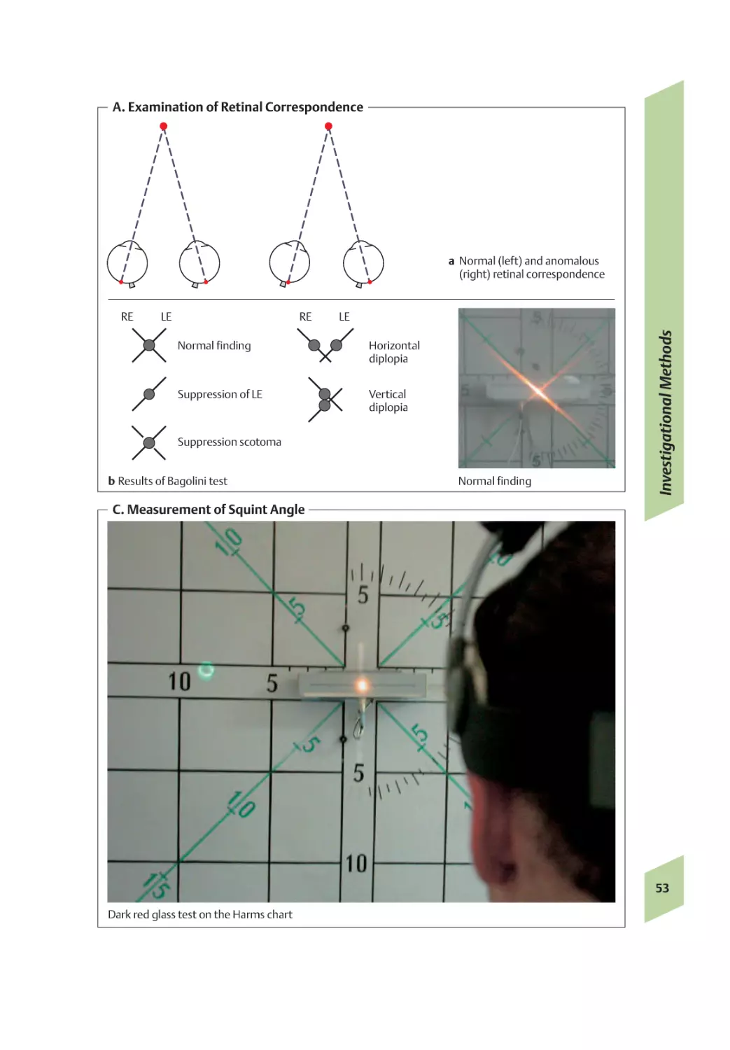
Investigational Methods . . . . . . . . . . . . . . . . . . . . . . . . . . . . . . . . . . . . . . . . . . . . . . . 51
Nonparalytic Squint . . . . . . . . . . . . . . . . . . . . . . . . . . . . . . . . . . . . . . . . . . . . . . . . . . . 57
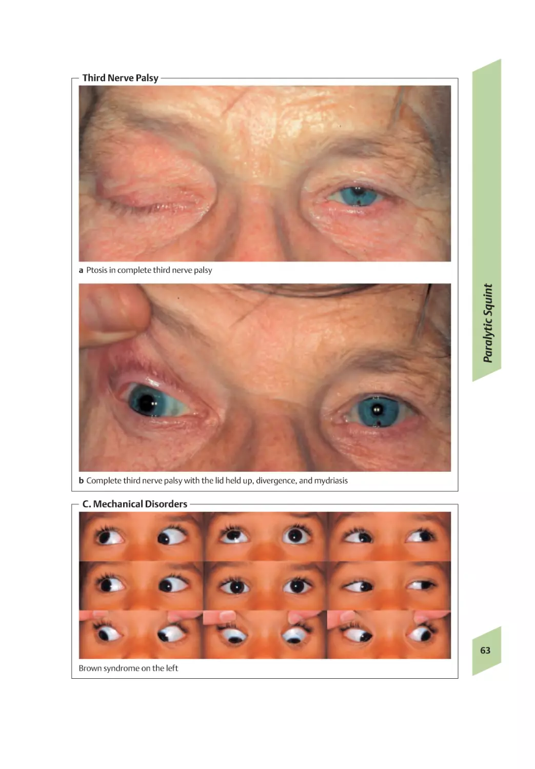
Nonparalytic/Paralytic Squint . . . . . . . . . . . . . . . . . . . . . . . . . . . . . . . . . . . . . . . . . . . 59
Supranuclear Disorders of Eye Movement . . . . . . . . . . . . . . . . . . . . . . . . . . . . . . . . 65
VII
00_Inhaltsverzeichnis
09.06.2006
8:25 Uhr
Seite VIII
Thieme; Langner; Schlote; Pocket Atlas of Ophthalmology; 1. AK, 5/2006
7 Conjunctiva . . . . . . . . . . . . . . . . . . . . . . . . . . . . . . . . . . . . . . . . . . . . . . . . . . . . . . . . 66
J. M. Rohrbach
Malformations/Degenerative Conditions/Various Conditions . . . . . . . . . . . . . . . . 67
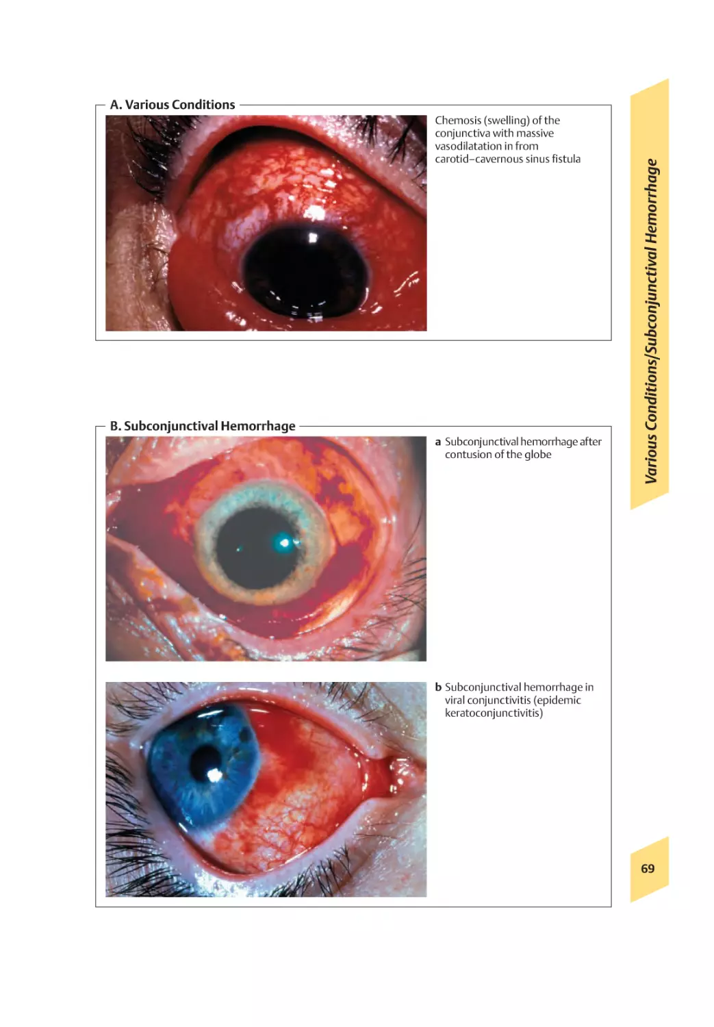
Various Conditions/Subconjunctival Hemorrhage . . . . . . . . . . . . . . . . . . . . . . . . . 69
Conjunctivitis . . . . . . . . . . . . . . . . . . . . . . . . . . . . . . . . . . . . . . . . . . . . . . . . . . . . . . . . 71
Tumors . . . . . . . . . . . . . . . . . . . . . . . . . . . . . . . . . . . . . . . . . . . . . . . . . . . . . . . . . . . . . 83
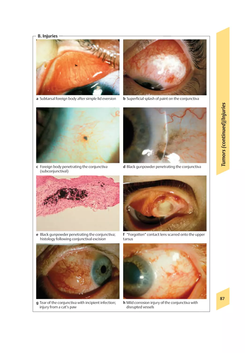
Injuries . . . . . . . . . . . . . . . . . . . . . . . . . . . . . . . . . . . . . . . . . . . . . . . . . . . . . . . . . . . . . . 87
8 Cornea . . . . . . . . . . . . . . . . . . . . . . . . . . . . . . . . . . . . . . . . . . . . . . . . . . . . . . . . . . . . . 88
J. Mielke
Anomalies, Degenerative Conditions . . . . . . . . . . . . . . . . . . . . . . . . . . . . . . . . . . . . 89
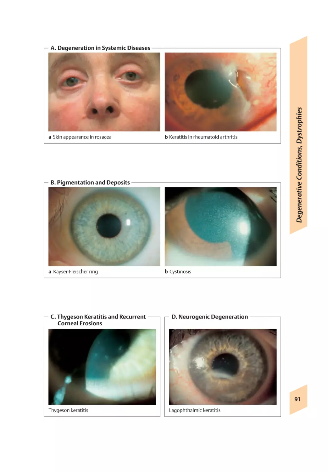
Degenerative Conditions, Dystrophies . . . . . . . . . . . . . . . . . . . . . . . . . . . . . . . . . . . 91
Epithelial and Subepithelial Dystrophies . . . . . . . . . . . . . . . . . . . . . . . . . . . . . . . . . . 93
Stromal Dystrophies, Endothelial Dystrophies . . . . . . . . . . . . . . . . . . . . . . . . . . . . . 95
Keratoconus, Infections . . . . . . . . . . . . . . . . . . . . . . . . . . . . . . . . . . . . . . . . . . . . . . . . 97
Herpes Simplex, Herpes Zoster . . . . . . . . . . . . . . . . . . . . . . . . . . . . . . . . . . . . . . . . . 99
Epidemic Keratitis, Acanthamoeba Keratitis, Fungi . . . . . . . . . . . . . . . . . . . . . . . . 101
UV Injury, Chemical Injury, Burns . . . . . . . . . . . . . . . . . . . . . . . . . . . . . . . . . . . . . . . 103
Injuries, Surgical Changes . . . . . . . . . . . . . . . . . . . . . . . . . . . . . . . . . . . . . . . . . . . . . 105
Keratoplasty, Rejection Reactions . . . . . . . . . . . . . . . . . . . . . . . . . . . . . . . . . . . . . . 107
9 Sclera . . . . . . . . . . . . . . . . . . . . . . . . . . . . . . . . . . . . . . . . . . . . . . . . . . . . . . . . . . . . . 108
T. Schlote
Malformations/Degenerative Conditions/Episcleritis . . . . . . . . . . . . . . . . . . . . . .109
Scleritis . . . . . . . . . . . . . . . . . . . . . . . . . . . . . . . . . . . . . . . . . . . . . . . . . . . . . . . . . . . . . 111
Tumors/Surgical Changes . . . . . . . . . . . . . . . . . . . . . . . . . . . . . . . . . . . . . . . . . . . . . 113
10 Choroid and Iris . . . . . . . . . . . . . . . . . . . . . . . . . . . . . . . . . . . . . . . . . . . . . . . . . . . 114
T. Schlote
Malformations and Anomalies . . . . . . . . . . . . . . . . . . . . . . . . . . . . . . . . . . . . . . . . . 115
Dystrophies/Vascular Changes . . . . . . . . . . . . . . . . . . . . . . . . . . . . . . . . . . . . . . . . . 117
Benign Tumors . . . . . . . . . . . . . . . . . . . . . . . . . . . . . . . . . . . . . . . . . . . . . . . . . . . . . . 119
Malignant Tumors . . . . . . . . . . . . . . . . . . . . . . . . . . . . . . . . . . . . . . . . . . . . . . . . . . . 121
Malignant Tumors . . . . . . . . . . . . . . . . . . . . . . . . . . . . . . . . . . . . . . . . . . . . . . . . . . . .123
Surgery/Injuries . . . . . . . . . . . . . . . . . . . . . . . . . . . . . . . . . . . . . . . . . . . . . . . . . . . . . 125
Uveitis . . . . . . . . . . . . . . . . . . . . . . . . . . . . . . . . . . . . . . . . . . . . . . . . . . . . . . . . . . . . . 127
11 Lens . . . . . . . . . . . . . . . . . . . . . . . . . . . . . . . . . . . . . . . . . . . . . . . . . . . . . . . . . . . . . 134
J. M. Rohrbach
Presbyopia . . . . . . . . . . . . . . . . . . . . . . . . . . . . . . . . . . . . . . . . . . . . . . . . . . . . . . . . . .135
Cataract . . . . . . . . . . . . . . . . . . . . . . . . . . . . . . . . . . . . . . . . . . . . . . . . . . . . . . . . . . . . 139
Lens Dislocation . . . . . . . . . . . . . . . . . . . . . . . . . . . . . . . . . . . . . . . . . . . . . . . . . . . . . 149
Inflammation/Tumors/Trauma/Lens Resorption . . . . . . . . . . . . . . . . . . . . . . . . . . 151
12 Glaucoma . . . . . . . . . . . . . . . . . . . . . . . . . . . . . . . . . . . . . . . . . . . . . . . . . . . . . . . . . 152
M. Grueb
VIII
Definition/Classification/Diagnosis . . . . . . . . . . . . . . . . . . . . . . . . . . . . . . . . . . . . . 153
Primary Congenital Glaucoma, Dysgenetic Glaucoma . . . . . . . . . . . . . . . . . . . . . 155
Primary Open-Angle Glaucoma . . . . . . . . . . . . . . . . . . . . . . . . . . . . . . . . . . . . . . . . 157
00_Inhaltsverzeichnis
09.06.2006
8:25 Uhr
Seite IX
Secondary Open-Angle Glaucoma . . . . . . . . . . . . . . . . . . . . . . . . . . . . . . . . . . . . . . 159
Primary Angle-Closure Glaucoma . . . . . . . . . . . . . . . . . . . . . . . . . . . . . . . . . . . . . . . 163
Secondary Angle-Closure Glaucoma . . . . . . . . . . . . . . . . . . . . . . . . . . . . . . . . . . . . 165
Treatment . . . . . . . . . . . . . . . . . . . . . . . . . . . . . . . . . . . . . . . . . . . . . . . . . . . . . . . . . . 167
13 Retina and Vitreous Body . . . . . . . . . . . . . . . . . . . . . . . . . . . . . . . . . . . . . . . . . . . 168
F. Gelisken
Congenital Anomalies and Tumors . . . . . . . . . . . . . . . . . . . . . . . . . . . . . . . . . . . . . . 169
Degenerative Conditions and Age-related Changes . . . . . . . . . . . . . . . . . . . . . . . . 171
Retinal Detachment . . . . . . . . . . . . . . . . . . . . . . . . . . . . . . . . . . . . . . . . . . . . . . . . . . 173
Vascular Diseases of the Retina . . . . . . . . . . . . . . . . . . . . . . . . . . . . . . . . . . . . . . . . . 179
Diabetic Retinopathy . . . . . . . . . . . . . . . . . . . . . . . . . . . . . . . . . . . . . . . . . . . . . . . . . 185
14 Macula . . . . . . . . . . . . . . . . . . . . . . . . . . . . . . . . . . . . . . . . . . . . . . . . . . . . . . . . . . . 188
U. Schneider
Diagnosis . . . . . . . . . . . . . . . . . . . . . . . . . . . . . . . . . . . . . . . . . . . . . . . . . . . . . . . . . . . 189
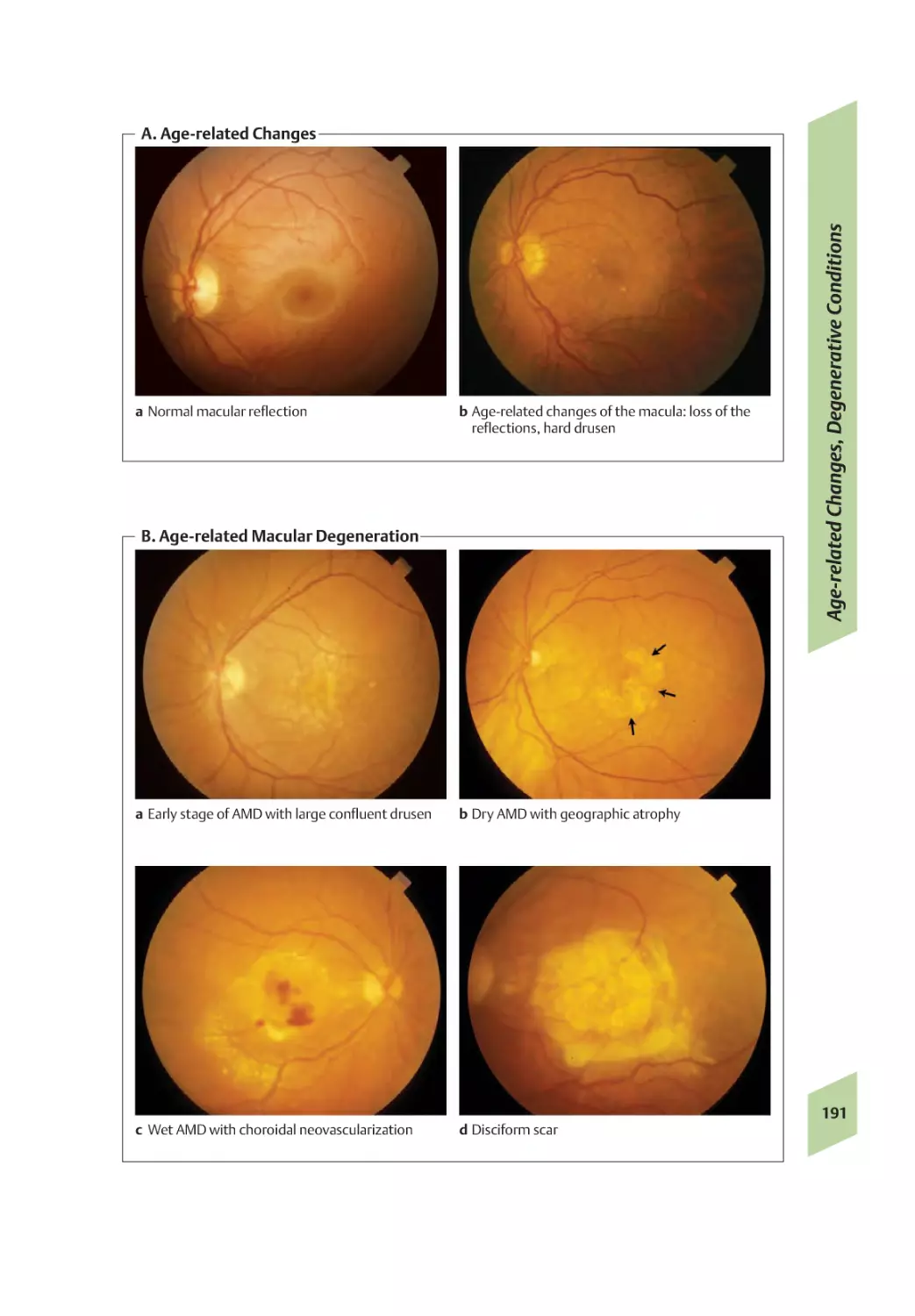
Age-related Changes, Degenerative Conditions . . . . . . . . . . . . . . . . . . . . . . . . . . . 191
Degenerative Conditions . . . . . . . . . . . . . . . . . . . . . . . . . . . . . . . . . . . . . . . . . . . . . . 193
Inflammatory Conditions . . . . . . . . . . . . . . . . . . . . . . . . . . . . . . . . . . . . . . . . . . . . . . 197
Macular Dystrophies . . . . . . . . . . . . . . . . . . . . . . . . . . . . . . . . . . . . . . . . . . . . . . . . . 199
Traumatic and Postoperative Maculopathies . . . . . . . . . . . . . . . . . . . . . . . . . . . . . 201
15 Optic Nerve and Optic Pathway . . . . . . . . . . . . . . . . . . . . . . . . . . . . . . . . . . . . . . 202
P. Weckerle
Malformations and Anomalies . . . . . . . . . . . . . . . . . . . . . . . . . . . . . . . . . . . . . . . . . 203
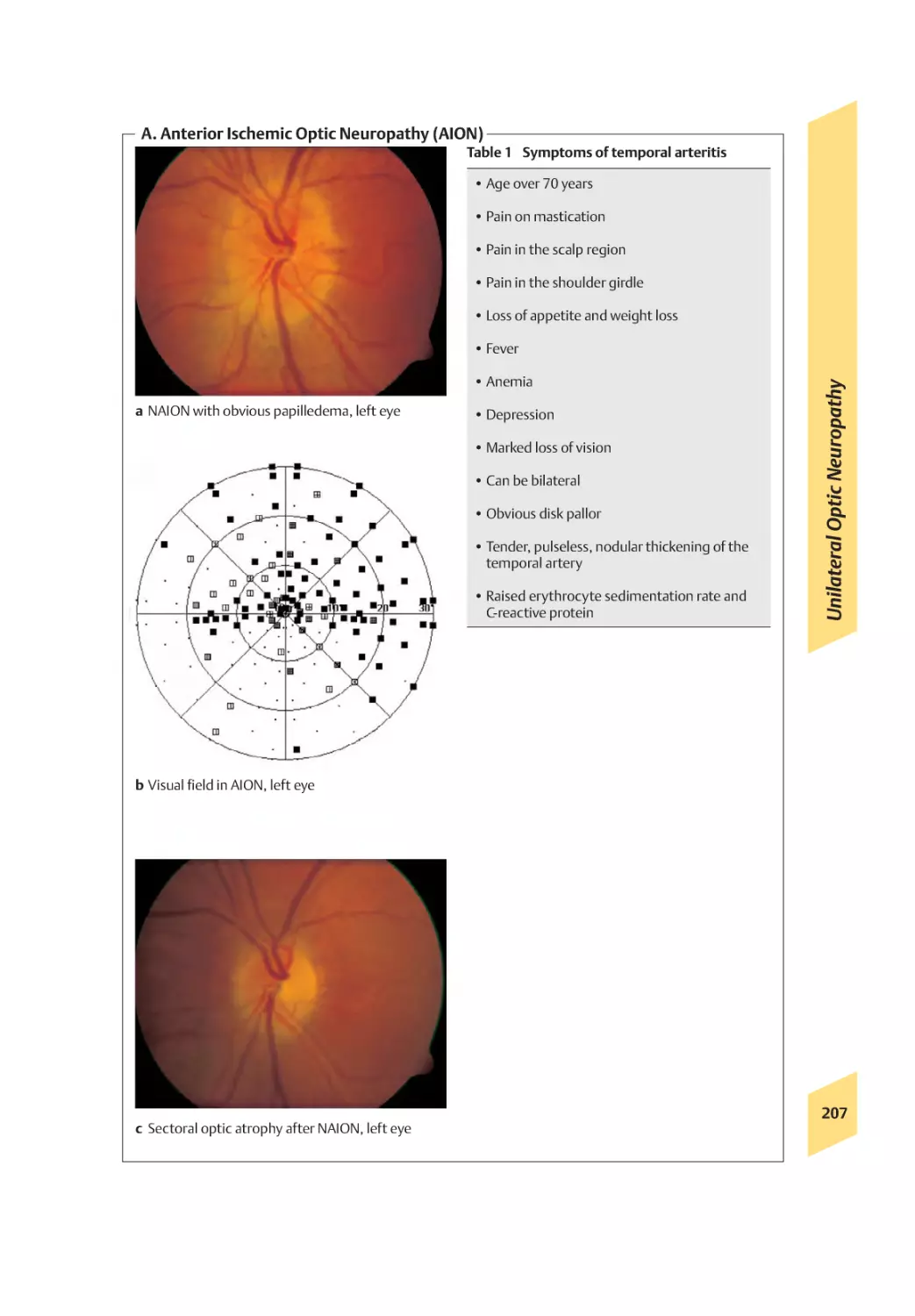
Unilateral Optic Neuropathy . . . . . . . . . . . . . . . . . . . . . . . . . . . . . . . . . . . . . . . . . . . 207
Bilateral Optic Neuropathy . . . . . . . . . . . . . . . . . . . . . . . . . . . . . . . . . . . . . . . . . . . . 209
Inflammation . . . . . . . . . . . . . . . . . . . . . . . . . . . . . . . . . . . . . . . . . . . . . . . . . . . . . . . . 213
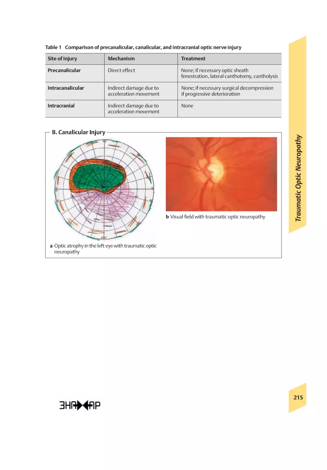
Traumatic Optic Neuropathy . . . . . . . . . . . . . . . . . . . . . . . . . . . . . . . . . . . . . . . . . . . 215
Tumors, Infiltration . . . . . . . . . . . . . . . . . . . . . . . . . . . . . . . . . . . . . . . . . . . . . . . . . . . 217
Papilledema . . . . . . . . . . . . . . . . . . . . . . . . . . . . . . . . . . . . . . . . . . . . . . . . . . . . . . . . . 219
Visual Field . . . . . . . . . . . . . . . . . . . . . . . . . . . . . . . . . . . . . . . . . . . . . . . . . . . . . . . . . 221
16 Drug-induced Ocular Side-Effects . . . . . . . . . . . . . . . . . . . . . . . . . . . . . . . . . . . . 222
T. Schlote
Lid and Conjunctival Changes . . . . . . . . . . . . . . . . . . . . . . . . . . . . . . . . . . . . . . . . . 223
Pemphigoid/Corneal Changes . . . . . . . . . . . . . . . . . . . . . . . . . . . . . . . . . . . . . . . . . 225
Drug-induced Glaucoma . . . . . . . . . . . . . . . . . . . . . . . . . . . . . . . . . . . . . . . . . . . . . . 227
Cataract . . . . . . . . . . . . . . . . . . . . . . . . . . . . . . . . . . . . . . . . . . . . . . . . . . . . . . . . . . . . 229
Retinal Changes . . . . . . . . . . . . . . . . . . . . . . . . . . . . . . . . . . . . . . . . . . . . . . . . . . . . . 231
Optic Neuropathy/Transitory Myopia . . . . . . . . . . . . . . . . . . . . . . . . . . . . . . . . . . . 233
17 Eye Diseases in Developing Countries . . . . . . . . . . . . . . . . . . . . . . . . . . . . . . . . . 234
J. Mielke
Epidemiology/Cataract/Trachoma/Glaucoma . . . . . . . . . . . . . . . . . . . . . . . . . . . . 235
Onchocerciasis/Loa loa/Vitamin A Deficiency . . . . . . . . . . . . . . . . . . . . . . . . . . . . . 237
IX
18 Index . . . . . . . . . . . . . . . . . . . . . . . . . . . . . . . . . . . . . . . . . . . . . . . . . . . . . . . . . . . . 238
00_Inhaltsverzeichnis
09.06.2006
8:25 Uhr
Seite X
00_Inhaltsverzeichnis
09.06.2006
8:25 Uhr
Seite 1
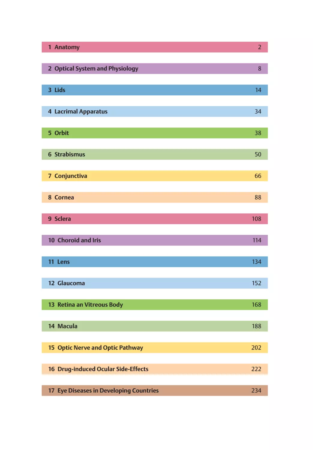
1 Anatomy
2
2 Optical System and Physiology
8
3 Lids
14
4 Lacrimal Apparatus
34
5 Orbit
38
6 Strabismus
50
7 Conjunctiva
66
8 Cornea
88
9 Sclera
108
10 Choroid and Iris
114
11 Lens
134
12 Glaucoma
152
13 Retina an Vitreous Body
168
14 Macula
188
15 Optic Nerve and Optic Pathway
202
16 Drug-induced Ocular Side-Effects
222
17 Eye Diseases in Developing Countries
234
01_Anatomie
08.06.2006
17:02 Uhr
Seite 2
Thieme; Langner; Schlote; Pocket Atlas of Ophthalmology; 1. AK, 5/2006
A. Eye
In addition to the eyeball (bulbus oculi, A), the
visual organ consists of the protective structures of the eye (orbit, lids, conjunctiva, and
lacrimal apparatus) and the movement apparatus consisting of the extrinsic ocular muscles
and Tenon’s capsule. The optic nerve connects
the sensory epithelium (the retina) with the
brain. The eyeball is surrounded by the fatty tissue of the orbit.
1 Anatomy
B. Orbit
The frontal bone (orbital roof), zygomatic bone
(lateral wall and floor), maxilla (floor), lacrimal
bone and ethmoid bone (medial wall), and also
the palatine bone and the sphenoid (blunt tip
are involved in the structure of the orbit (B).
The openings in the orbit are the optic canal
(which contains the optic nerve), the superior
and inferior orbital fissures, the infraorbital,
ethmoidal, and zygomatico-orbital foramina,
and the nasolacrimal canal.
C. Lids
The palpebral fissure is bounded by the upper
and lower lids (palpebrae), the main structure
of which is formed by a dense lid plate (tarsus).
Exteriorly, the lids are covered by stratified keratinized squamous epithelium which becomes
the palpebral conjunctiva at the lid margin.
Lashes (ciliae) are found in 2 to 3 rows along
the lid margin. The holocrine Zeis glands and
the apocrine Moll glands end in the hair follicles. The excretory ducts of the larger Moll
glands open close to the posterior lid margin.
The eyebrow (supercilium) marks the upper
border of the orbit. Blinking and closing of the
lids is performed mainly by the orbicularis
oculi muscle (innervated by the facial nerve).
The levator palpebrae superioris (oculomotor
nerve) and superior and inferior tarsal muscles
(cervical sympathetic nerves) also open the
lids. The sensory innervation of the upper lid is
through branches of the first division of the
trigeminal nerve (V1), that of the lower lid by
branches of the second division (V2).
D. Conjunctiva
2
The conjunctiva covers the posterior surface of
the upper and lower lids as the palpebral conjunctiva. It consists of two or more layers of isoprismatic – to highly prismatic epithelium. At
the upper and lower fornix it changes to the
bulbar conjunctiva, which is slightly mobile
where it lies over the sclera. The conjunctiva
consists of stratified nonkeratinizing epithelium.
E. Lacrimal Apparatus (E)
The lacrimal gland (glandula lacrimalis) lies
above the outer corner of the eye. It is a tubuloalveolar gland whose 6–12 excretory ducts
end in the lateral upper conjunctival fornix. The
secretory parasympathetic innervation follows
the facial nerve and the sympathetic innervation is through the cervical sympathetic. The
tear fluid is low in protein and of low viscosity.
Through blinking, the tears reach the medial
angle of the lids and the fluid drawn into the
lacrimal punctum and into the lacrimal canaliculi (canaliculi lacrimales). These open into the
lacrimal sac (saccus lacrimalis) and from there
the tears flow out through the nasolacrimal
duct into the lower nasal passage (E).
F. Motor Apparatus
The extrinsic ocular muscles (2 horizontal, 2
vertical, and 2 oblique) lie in the fat of the orbit
and move the eyeball. The superior, inferior,
medial, and lateral rectus muscles originate
from the tendinous ring, which forms the tip of
the muscle pyramid at the orbital apex, and
pass over the equator of the eyeball. With the
exception of the lateral rectus muscle, which is
innervated by the abducent nerve (CN VI), and
the superior oblique muscle, which is innervated by the trochlear nerve (CN IV), they are
innervated by the oculomotor nerve (CN III).
The inferior oblique muscle arises from the medial wall of the orbit. The superior oblique muscle passes from the tendinous ring initially to
the medial wall of the orbit, where it changes
its direction at the trochlea.
01_Anatomie
08.06.2006
17:02 Uhr
Seite 3
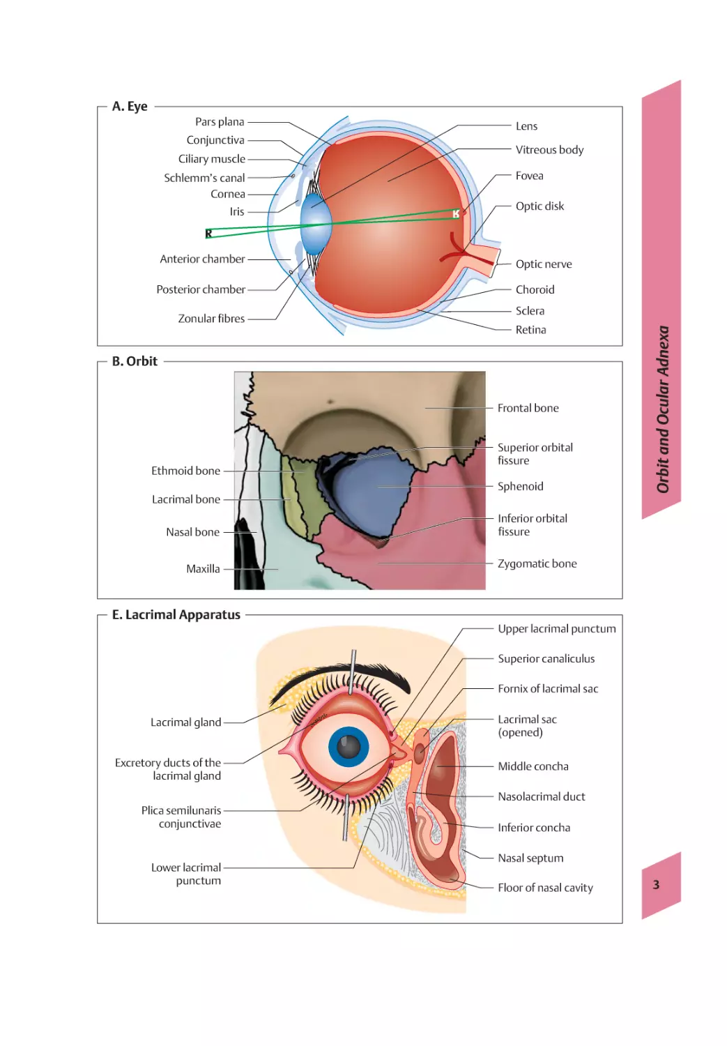
A. Eye
Ciliary muscle
Schlemm’s canal
Cornea
Iris
Lens
Vitreous body
Fovea
Optic disk
Anterior chamber
Optic nerve
Posterior chamber
Choroid
Zonular fibres
Sclera
Retina
B. Orbit
Frontal bone
Ethmoid bone
Superior orbital
fissure
Sphenoid
Orbit and Ocular Adnexa
Pars plana
Conjunctiva
Lacrimal bone
Nasal bone
Maxilla
Inferior orbital
fissure
Zygomatic bone
E. Lacrimal Apparatus
Upper lacrimal punctum
Superior canaliculus
Fornix of lacrimal sac
Lacrimal gland
Excretory ducts of the
lacrimal gland
Lacrimal sac
(opened)
Middle concha
Nasolacrimal duct
Plica semilunaris
conjunctivae
Lower lacrimal
punctum
Inferior concha
Nasal septum
Floor of nasal cavity
3
01_Anatomie
08.06.2006
17:02 Uhr
Seite 4
Thieme; Langner; Schlote; Pocket Atlas of Ophthalmology; 1. AK, 5/2006
1 Anatomy
A. Blood Supply
The ophthalmic artery is a branch of the internal carotid artery and passes into the orbit with
the optic nerve. It then runs forward with the
superior oblique muscle and ends as the dorsal
nasal artery and supratrochlear artery. Before
that it gives off the following branches: the central retinal artery, which travels to the retina in
the optic nerve (A); short and long posterior ciliary arteries to the choroid and ciliary body; the
lacrimal artery to the lacrimal gland; the
supraorbital artery to the forehead; and the anterior and posterior ethmoidal arteries to the
ethmoid air cells. The anterior ciliary arteries
arise from the muscular branches to the extrinsic ocular muscles, which pass through the
sclera to the ciliary body and iris. The superior
ophthalmic vein collects the blood from the
eyeball, upper orbit, lids and ethmoid air cells
and drains into the cavernous sinus. The inferior ophthalmic vein arises on the floor of the
orbit and flows either into the superior ophthalmic vein or into the pterygoid plexus.
B. Eyeball
The eyeball ( B ), bulbus oculi , has an almost
spherical shape with an average diameter of
23 mm. The eyeball is bounded anteriorly by
the cornea. At the posterior pole, the optic
nerve leaves the eye somewhat medial to the
axis of the eye, and the fovea centralis-which
is the site of most acute vision-is somewhat
lateral to this. The circumference at the greatest transverse diameter of the eye is called the
equator. The wall of the eye consists of three
layers: the outer layer (tunica fibrosa) with
sclera and cornea; the middle layer (tunica
vasculosa) with choroid, ciliary body, and iris;
and the inner layer (tunica interna) with the
retina and the retinal pigment epithelium. Inside the eye a distinction is made between the
anterior and posterior chambers of the eye and
the vitreous space. The cornea, aqueous
humor, lens, and vitreous body constitute the
optic media of the eye. The lens, zonular fibers,
and ciliary muscle are called the accommodation apparatus.
C. Sclera
4
The sclera, which is white in adults, consists of
packed lamellae of collagen fibers covering the
posterior 5ßz of the eye. At the corneal limbus it
becomes the substantia propria corneae (stroma).
D. Cornea
The cornea has a diameter of about 12 mm in
adults. The outside of the cornea consists of
stratified nonkeratinized squamous epithelium, which changes to the epithelium of the
bulbar conjunctiva at the corneal limbus. The
inside is formed by a single layer of flat endothelial cells. Bowman’s membrane is situated between the epithelium and stroma and Descemet’s membrane between the endothelium
and stroma (Da). The refractive power of the
cornea is about 42 diopters (Db). The central
thickness is approximately 500 µm.
E. Lens
The lens, with a horizontal diameter of about
10 mm, is situated in the posterior chamber of
the eye. It is about 3–4 mm thick at the center.
It is a biconvex lens, with the anterior surface
less curved than the posterior surface. The lens
shell, which surrounds the nucleus concentrically, lies beneath the lens capsule.
F. Vitreous Body
The vitreous body, which is 95 % water, fills the
vitreous space situated behind the lens. Its gelatinous consistency is due to the presence of
hyaluronic acid, mucopolysaccharides, and collagen fibrils.
G. Choroid
The choroid occupies the major part of the middle layer of the eye. In addition to arteries and
veins, it also carries approximately 15–20 ciliary nerves. It is separated from the retina by
Bruch’s membrane, which is 2 µm thick.
08.06.2006
A. Blood Supply
Angiography
D. Cornea
17:02 Uhr
Seite 5
B. Eyeball
Ultrasound
Blood Supply and Eyeball
01_Anatomie
a PAS stain, ca. 63
5
b Slit lamp photograph
01_Anatomie
08.06.2006
17:02 Uhr
Seite 6
Thieme; Langner; Schlote; Pocket Atlas of Ophthalmology; 1. AK, 5/2006
1 Anatomy
A. Ciliary Body
The ciliary body (Ba) extends from the ora serrata as far as the base of the iris and surrounds
the iris like a ring. A distinction is made between the outer part, the orbiculus ciliaris with
fine meridional folds where the zonular fibers
arise, and the inner part, the corona ciliaris. The
ciliary body is covered by a bilaminar epithelium, which is responsible for the production of
aqueous humor. The anterior and posterior
chambers together contain about 0.2–0.3 ml of
aqueous humor, most of which drains out at the
iridocorneal angle. Part of the ciliary body is the
ciliary muscle, whose smooth muscle fibers are
arranged meridionally, circularly, and radially
(parasympathetic innervation via the oculomotor nerve predominates with some cervical
sympathetic input). Contraction of the muscle
leads to slackening of the zonular fibers and,
through the associated increased curvature of
the lens, to accommodation.
B. Iris and Pupil
The iris, like a diaphragm, forms the pupil. The
iris has no epithelium on its anterior aspect, so
that the iris stroma, which is arranged radially
to the edge of the pupil, is exposed. The iris is
thinnest at the margin of the pupil and allows
the bilaminar pigmented epithelium on the
back to be seen. The pupil is surrounded by the
sphincter pupillae muscle (parasympathetic
innervation via the oculomotor nerve), the innervation of which produces contraction of the
pupil (miosis). At the margin of the pupil, the
iris is widely connected with the ciliary body.
The muscle fibers of the dilatator pupillae muscle (cervical sympathetic) run here, contraction
of which leads to pupil dilatation (mydriasis).
At the iridocorneal angle (Ba), the aqueous
humor flows through gaps in the pectinate ligament of the iris (trabecular meshwork, Bb)
into Schlemm’s canal.
C. Retina
6
The retina forms the inner layer of the eye. It is
divided into a nonsensory part and an optic
part, the boundary of which is formed by the
ora serrata. The anterior part does not have any
sensory epithelium and covers the ciliary body
and iris as a bilaminar epithelium. The optic
part consists of two layers, the outer layer (pigment layer) and the inner layer (cerebral layer),
which lie loosely on one another and are adherent only at the ora serrata and at the entrance of the optic nerve. The central retinal artery and vein unite at the entrance of the optic
nerve (optic disc or papilla). The macula lutea
(yellow spot) is lateral to this with the fovea
centralis at its center, the site of maximum visual acuity (Ca). The pigment layer consists of
a single layer of isoprismatic epithelium (retinal pigment epithelium). The inner retina includes the photoreceptor cells and nine further
identifiable layers of the cerebral layer (Cb and
c). They are primary sensory epithelial cells.
About 120 million rods and 6–7 million cones
are distinguished. There are only cones in the
fovea centralis, with no other layers of the cerebral layer. The perikarya of the bipolar cells,
which are the second neuron of the optic nerve,
are located in the inner nuclear layer. They
maintain synaptic contact with the sensory
cells in the outer plexiform layer and with the
multipolar ganglion cells of the ganglion layer
(third neuron) in the inner plexiform layer,
from where sensory impulses are conducted in
unmyelinated nerve fibers to the optic disc. The
horizontal and amacrine cells of the inner nuclear layer form the association apparatus of
the retina through the parallel connections of
several synapses.
D. Optic Nerve and Optic Tract
The optic nerve is about 45 mm in length, twothirds of which is inside the orbit. At the lamina cribrosa, ca. 1 million nerve fibers leave the
eyeball and from this point are surrounded by
a medullary sheath of oligodenroglia, dura
mater and pia mater. After passing through the
optic canal, it reaches the optic chiasm on the
floor of the third ventricle after running about
10 mm in the middle cranial fossa. Here the
nasal fibers of the retina cross to the opposite
side. The optic nerve fibers run as the optic
tract as far as the lateral geniculate body. The
optic radiation (Gratiolet’s radiating fibers)
runs from here through the posterior crus of
the internal capsule to the primary optic visual cortex, the area striata, area 17.
01_Anatomie
08.06.2006
17:02 Uhr
Seite 7
B. Iris and Pupil
Iris
Anterior chamber
Schlemm’s canal
Iridocorneal angle
Sclera
Zonular fibres
Ciliary body
Ciliary sulcus
Vitreous body
Lens
a Iridocorneal angle
b Trabecular meshwork:
scanning electron-microscopic appearance
C. Retina
a Masson trichrome stain, ca. 150ã
Ciliary Body/Iris/Pupil/Retina/Optic Nerve
Cornea
Posterior chamber
b Photograph of fundus with optic disk
and macula
Internal limiting membrane
Nerve fibre layer
Ganglion cell layer
Inner plexiform layer, amacrine cells
Inner nuclear layer, bipolar cells
Outer plexiform layer, horizontal cells
Outer nuclear layer
External limiting membrane
Rods and cones
Retinal pigment epithelium
c Diagram
Bruch’s membrane
7
02_Physiologie
08.06.2006
17:06 Uhr
Seite 8
Thieme; Langner; Schlote; Pocket Atlas of Ophthalmology; 1. AK, 5/2006
2 Optical System and Physiology
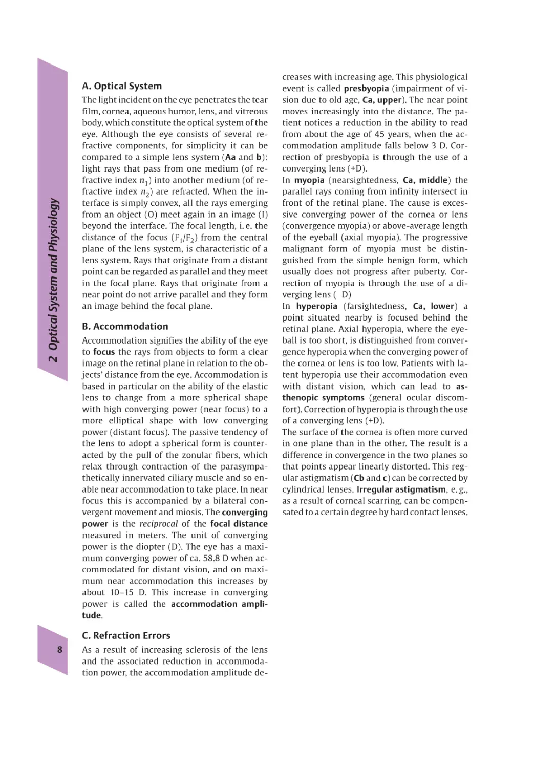
A. Optical System
The light incident on the eye penetrates the tear
film, cornea, aqueous humor, lens, and vitreous
body, which constitute the optical system of the
eye. Although the eye consists of several refractive components, for simplicity it can be
compared to a simple lens system (Aa and b):
light rays that pass from one medium (of refractive index n1) into another medium (of refractive index n2) are refracted. When the interface is simply convex, all the rays emerging
from an object (O) meet again in an image (I)
beyond the interface. The focal length, i. e. the
distance of the focus (F1/F2) from the central
plane of the lens system, is characteristic of a
lens system. Rays that originate from a distant
point can be regarded as parallel and they meet
in the focal plane. Rays that originate from a
near point do not arrive parallel and they form
an image behind the focal plane.
B. Accommodation
Accommodation signifies the ability of the eye
to focus the rays from objects to form a clear
image on the retinal plane in relation to the objects’ distance from the eye. Accommodation is
based in particular on the ability of the elastic
lens to change from a more spherical shape
with high converging power (near focus) to a
more elliptical shape with low converging
power (distant focus). The passive tendency of
the lens to adopt a spherical form is counteracted by the pull of the zonular fibers, which
relax through contraction of the parasympathetically innervated ciliary muscle and so enable near accommodation to take place. In near
focus this is accompanied by a bilateral convergent movement and miosis. The converging
power is the reciprocal of the focal distance
measured in meters. The unit of converging
power is the diopter (D). The eye has a maximum converging power of ca. 58.8 D when accommodated for distant vision, and on maximum near accommodation this increases by
about 10–15 D. This increase in converging
power is called the accommodation amplitude.
C. Refraction Errors
8
As a result of increasing sclerosis of the lens
and the associated reduction in accommodation power, the accommodation amplitude de-
creases with increasing age. This physiological
event is called presbyopia (impairment of vision due to old age, Ca, upper). The near point
moves increasingly into the distance. The patient notices a reduction in the ability to read
from about the age of 45 years, when the accommodation amplitude falls below 3 D. Correction of presbyopia is through the use of a
converging lens (+D).
In myopia (nearsightedness, Ca, middle) the
parallel rays coming from infinity intersect in
front of the retinal plane. The cause is excessive converging power of the cornea or lens
(convergence myopia) or above-average length
of the eyeball (axial myopia). The progressive
malignant form of myopia must be distinguished from the simple benign form, which
usually does not progress after puberty. Correction of myopia is through the use of a diverging lens (–D)
In hyperopia (farsightedness, Ca, lower) a
point situated nearby is focused behind the
retinal plane. Axial hyperopia, where the eyeball is too short, is distinguished from convergence hyperopia when the converging power of
the cornea or lens is too low. Patients with latent hyperopia use their accommodation even
with distant vision, which can lead to asthenopic symptoms (general ocular discomfort). Correction of hyperopia is through the use
of a converging lens (+D).
The surface of the cornea is often more curved
in one plane than in the other. The result is a
difference in convergence in the two planes so
that points appear linearly distorted. This regular astigmatism (Cb and c) can be corrected by
cylindrical lenses. Irregular astigmatism, e. g.,
as a result of corneal scarring, can be compensated to a certain degree by hard contact lenses.
02_Physiologie
08.06.2006
17:06 Uhr
Seite 9
A. Optical System
16,7 mm
n1
F2
F1
H
K
a Simple optical system
I
α
O
K
b Reduced eye
Presbyopia
C. Refraction Errors
∞
Distant
(objekt at ∞)
Near
Myopia
∞
Distant
Near
Hyperopia
∞
I
Optical System/Accommodation/Refraction Errors
n2
O
∞
Distant
Near
∞
a Presbyopia/myopia/hyperopia
9
b Corneal topography with spherical cornea
c Corneal topography with astigmatic cornea
02_Physiologie
08.06.2006
17:06 Uhr
Seite 10
2 Optical System and Physiology
Thieme; Langner; Schlote; Pocket Atlas of Ophthalmology; 1. AK, 5/2006
10
A. Visual Acuity
C. Visual Field
Vision in the eye relates to the overall function
of the visual organ. Apart from pure visual acuity, it also includes the visual field, color vision,
and dark vision. Visual acuity means the resolving ability of the eye with an optimally correcting lens, i.e. the ability of the retina barely
to distinguish two points from one another
(resolution threshold). A normal eye can just
differentiate two points when the rays emerging from them form an angle at the eye of one
minute of arc (1/60 degree). Visual acuity is calculated from the actual distance of the points
from the eye divided by the distance at which
the normal eye can resolve the points, and in
the normal eye it is therefore 1ßq = 1.0. Optotypes
projected into the distance (Landolt’s rings,
block letters, numbers, E hooks, children’s pictures) or vision test tables for near vision (e. g.,
Birkhäuser tables) are used to test vision.
The term visual field describes the area that is
perceived at the same time when the eye is not
moving. A distinction is made between monocular and binocular visual fields. The outer
boundaries depend on adaptation and on the
size, brightness, and color of the object and on
whether the object is mobile or static. The
boundaries are usually 60° nasally, 70° above,
80° below, and 90° temporally. The visual field
is measured by perimetry (C). Two forms are
distinguished:
● Kinetic perimetry: this records the site where
a stimulus of defined brightness is first perceived as it is brought into the visual field.
● Static perimetry: measurement of the minimum brightness that a stimulus must have in
order to be identified at a certain site with defined background brightness.
B. Receptors
Rods and cones constitute the retina’s receptors. While the fovea centralis consists exclusively of cones, which are responsible for color
vision in good lighting (photopic vision), their
density diminishes rapidly toward the periphery. The rods are responsible for vision in poor
light (scotopic vision); their greatest density is
around the fovea centralis but they are also distributed over the entire retina (Ba). The photoreceptors are absent in the region of the optic
disc. Under the effect of light, the rhodopsin located in the outer limbs of the rods and cones
is bleached, leading to the conversion of light
energy into electrical impulses through a
change in the conformation of the pigment part
from 11-cis- to all-trans-retinal (Bb). Rhodopsin is a chromoprotein and is a component of
visual purple. It is composed of the protein
opsin and the vitamin A derivatives 11-cis- and
all-trans-retinal. In the dark, regeneration of
the rhodopsin occurs with expenditure of energy. Absorption of light is required for the
bleaching of the rhodopsin. As the rhodopsin
contained in the rods absorbs light from the entire (visible) wavelength spectrum, different
wavelengths (colors) cannot be distinguished
by the rods. In contrast, the three visual pigments of the cones each absorb only light of a
certain wavelength region, which allows for
color vision.
Defects in the visual field are called scotomas
and can be symptoms of many eye diseases. The
blind spot due to the absence of receptors in
the optic disc is a physiological “scotoma.” In
the binocular visual field, each eye’s blind spot
is compensated by the other side. Temporally
located objects are projected on the nasal half
of the retina and vice versa. Objects in the
upper visual field are imaged on the lower half
of the retina, and objects from the lower region
in the upper half of the retina.
02_Physiologie
08.06.2006
17:06 Uhr
Seite 11
160
140
120
100
80
60
40
20
0
– 90
Distribution of rods
– 75
– 60
– 45
– 30
160
Distribution of cones
140
120
100
80
60
40
20
0
– 90
– 75
– 60
– 45
100
Sensitivity in the dark
90
80
70
60
50
40
30
20
10
0
– 75
– 60
– 45
– 90
1,2
– 15
– 30
– 30
0
– 15
– 15
15
0
0
30
30
15
45
45
30
45
60
60
60
75
75
75
90
90
90
Visual Acuity/Receptors/Visual Field
Sensitivity (%)
Receptor density
(1000/qmm)
Receptor density
(1000/qmm)
B. Receptors
Vision
Vision (1)
1
0,8
0,6
0,4
0,2
0
– 90
– 75
– 60
– 45
– 30
– 15
0
30
45
60
75
90
Retinal localization (degrees)
a Distribution of rods and cones, dark sensitivity and vision
CH3 H
C
C
13
C. Visual Field
+
C
N
H
H
CH3
H
C
BR570 protonated
all-trans-form
O640 protonated
all-trans-form
13
C
C
H
H
+
N
K610 protonated
13-cis-form
M412 deprotonated
13-cis-form
CH3
N520 protonated
13-cis-form
H
C
13
L550
C
10°
30 dB
20° 30°
C
H+
H
N:
H+
b Photocycle of bacteriorhodopsin (BR): numbers
correspond to the wavelength of the absorption
maximum in nanometers
11
02_Physiologie
08.06.2006
17:06 Uhr
Seite 12
Thieme; Langner; Schlote; Pocket Atlas of Ophthalmology; 1. AK, 5/2006
2 Optical System and Physiology
A. Adaptation
Adaptation (A) signifies the adjustment of the
eye to different light levels. This is a complex
process, which comprises a change in pupil
size, a change between rod and cone vision, and
a change in the sensitivity of the retina. According to the duplicity theory of vision, daytime and color vision (photopic vision) is a
function of the cone apparatus, while vision in
dim light and night vision (scotopic vision) are
provided by the rod apparatus. Light adaptation means the transition to photopic vision
and is based on pupil constriction and the transition from rod to cone vision with the breakdown of rhodopsin. The first phase of the transition (alpha adaptation) occurs in about 0.05
seconds, while the second phase (beta adaptation) lasts 6–7 minutes. The transition to scotopic vision (dark adaptation) takes place
much more slowly and is complete after about
30 minutes. The first phase comprises cone
adaptation and ends after about 7–8 minutes
with the Kohlrausch notch, the transition to
cone adaptation with the regeneration of
rhodopsin. Dark adaptation is associated with
pupil dilatation, a loss of color vision, a reduction in visual acuity, and a physiological central
scotoma.
B. Color Vision
Color vision is a function of the cones. The
wavelength spectrum of light perceived by the
eye is between 400 nm and about 700 nm. According to the Young-Helmholtz three-color
theory (Bb), three types of cones are distinguished: those that absorb blue-violet light,
those that absorb green light, and those that
absorb yellow-red light (the trichromatic system, Ba). According to the laws of color mixing,
all other colors (including white) can be mixed
from light of these three colors. The rhodopsin
of the rods absorbs light from the entire visible
wavelength spectrum, which is why it is not
possible to distinguish colors with scotopic vision.
C. Central Processing
12
The stimulus of incident light leads to hyperpolarization of the primary membrane potential in the receptors of the retina. The magnitude of the potential increases with increase in
the stimulus strength. When it crosses the
threshold, this secondary receptor potential
leads to action potentials in the corresponding
ganglion cell, the frequency of which is proportional to the magnitude of the receptor potential. Through cross-connections within the
retina (horizontal cells and amacrine cells), receptive fields occur that exert stimulating and
inhibiting influences on the action potentials.
Such a receptive field consists of a round center and a concentrically ordered periphery. If
the light impulse falls on the center, the frequency of the action potentials rises. Illumination of the periphery leads to a fall in the action potential frequency. If the light stimulus is
absent, excitation occurs in the peripheral part
of the receptive field. Such a receptive field is
called an ON field in contrast to an OFF field
with the opposite reaction. The function of the
receptive fields is to contrast the sensory stimulus.
D. Pupillary Reflex
The pupillary reflex is triggered by the sudden
incidence of light into a pupil. The eye reacts
with miosis (direct reaction). The contralateral
pupil also contracts due to the central crossover
of the stimulus (consensual reaction).
Afferent supply: optic nerve
Efferent supply: parasympathetic fibers via the
oculomotor nerve.
E. Corneal Reflex
The corneal reflex is triggered by touching the
cornea, which leads to reflex lid closure.
Afferent supply: trigeminal nerve
Efferent supply: mainly the facial nerve.
02_Physiologie
08.06.2006
17:06 Uhr
Seite 13
A. Adaptation
Adaptation/Color Vision
Adaptation mechanisms in light and dark adaptation
Relative absorption
B. Color vision
400
450
500
550
600
650
700
Wavelength (nm)
a Absorption spectra of the different cone types
13
b Additive color vision: Young–Helmholtz
three-color theory
c Ishihara plate for testing color vision
03_Lider
08.06.2006
17:07 Uhr
Seite 14
3 Lids
Thieme; Langner; Schlote; Pocket Atlas of Ophthalmology; 1. AK, 5/2006
14
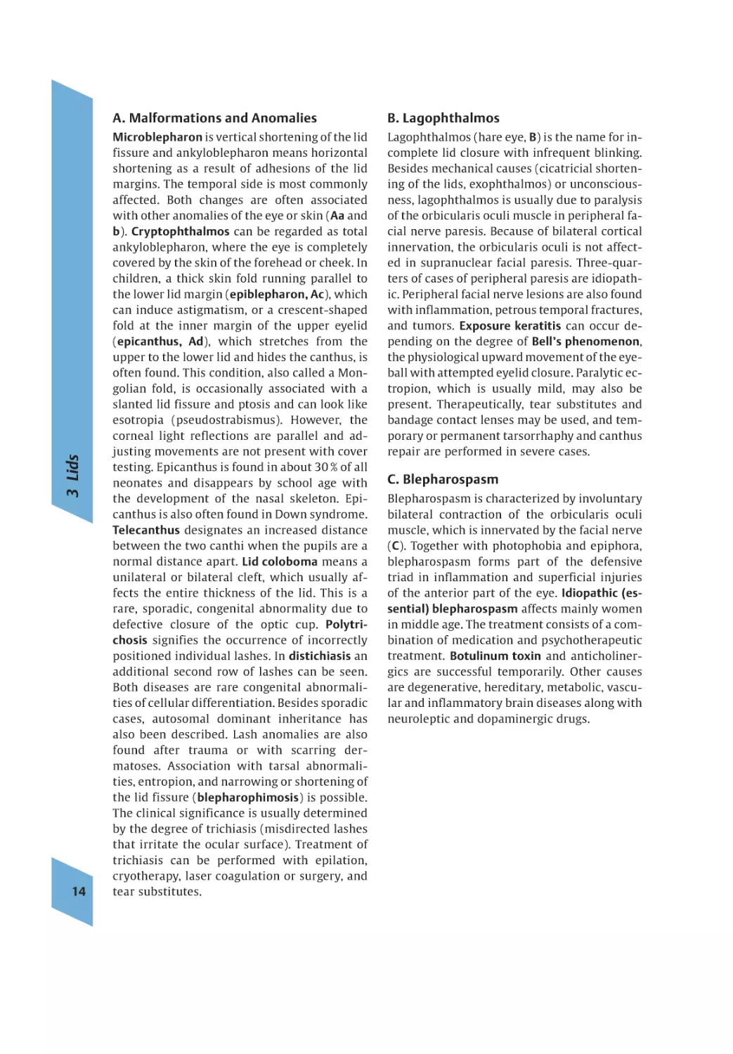
A. Malformations and Anomalies
B. Lagophthalmos
Microblepharon is vertical shortening of the lid
fissure and ankyloblepharon means horizontal
shortening as a result of adhesions of the lid
margins. The temporal side is most commonly
affected. Both changes are often associated
with other anomalies of the eye or skin (Aa and
b). Cryptophthalmos can be regarded as total
ankyloblepharon, where the eye is completely
covered by the skin of the forehead or cheek. In
children, a thick skin fold running parallel to
the lower lid margin (epiblepharon, Ac), which
can induce astigmatism, or a crescent-shaped
fold at the inner margin of the upper eyelid
(epicanthus, Ad), which stretches from the
upper to the lower lid and hides the canthus, is
often found. This condition, also called a Mongolian fold, is occasionally associated with a
slanted lid fissure and ptosis and can look like
esotropia (pseudostrabismus). However, the
corneal light reflections are parallel and adjusting movements are not present with cover
testing. Epicanthus is found in about 30 % of all
neonates and disappears by school age with
the development of the nasal skeleton. Epicanthus is also often found in Down syndrome.
Telecanthus designates an increased distance
between the two canthi when the pupils are a
normal distance apart. Lid coloboma means a
unilateral or bilateral cleft, which usually affects the entire thickness of the lid. This is a
rare, sporadic, congenital abnormality due to
defective closure of the optic cup. Polytrichosis signifies the occurrence of incorrectly
positioned individual lashes. In distichiasis an
additional second row of lashes can be seen.
Both diseases are rare congenital abnormalities of cellular differentiation. Besides sporadic
cases, autosomal dominant inheritance has
also been described. Lash anomalies are also
found after trauma or with scarring dermatoses. Association with tarsal abnormalities, entropion, and narrowing or shortening of
the lid fissure (blepharophimosis) is possible.
The clinical significance is usually determined
by the degree of trichiasis (misdirected lashes
that irritate the ocular surface). Treatment of
trichiasis can be performed with epilation,
cryotherapy, laser coagulation or surgery, and
tear substitutes.
Lagophthalmos (hare eye, B) is the name for incomplete lid closure with infrequent blinking.
Besides mechanical causes (cicatricial shortening of the lids, exophthalmos) or unconsciousness, lagophthalmos is usually due to paralysis
of the orbicularis oculi muscle in peripheral facial nerve paresis. Because of bilateral cortical
innervation, the orbicularis oculi is not affected in supranuclear facial paresis. Three-quarters of cases of peripheral paresis are idiopathic. Peripheral facial nerve lesions are also found
with inflammation, petrous temporal fractures,
and tumors. Exposure keratitis can occur depending on the degree of Bell’s phenomenon,
the physiological upward movement of the eyeball with attempted eyelid closure. Paralytic ectropion, which is usually mild, may also be
present. Therapeutically, tear substitutes and
bandage contact lenses may be used, and temporary or permanent tarsorrhaphy and canthus
repair are performed in severe cases.
C. Blepharospasm
Blepharospasm is characterized by involuntary
bilateral contraction of the orbicularis oculi
muscle, which is innervated by the facial nerve
(C). Together with photophobia and epiphora,
blepharospasm forms part of the defensive
triad in inflammation and superficial injuries
of the anterior part of the eye. Idiopathic (essential) blepharospasm affects mainly women
in middle age. The treatment consists of a combination of medication and psychotherapeutic
treatment. Botulinum toxin and anticholinergics are successful temporarily. Other causes
are degenerative, hereditary, metabolic, vascular and inflammatory brain diseases along with
neuroleptic and dopaminergic drugs.
03_Lider
08.06.2006
17:07 Uhr
Seite 15
a Complex facial deformity
b Complex eye deformity
c Epiblepharon
d Epicanthus
B. Lagophthalmos
Malformations and Anomalies
A. Malformations and Anomalies
C. Blepharospasm
15
Lagophthalmos in facial nerve paresis
03_Lider
08.06.2006
17:07 Uhr
Seite 16
Thieme; Langner; Schlote; Pocket Atlas of Ophthalmology; 1. AK, 5/2006
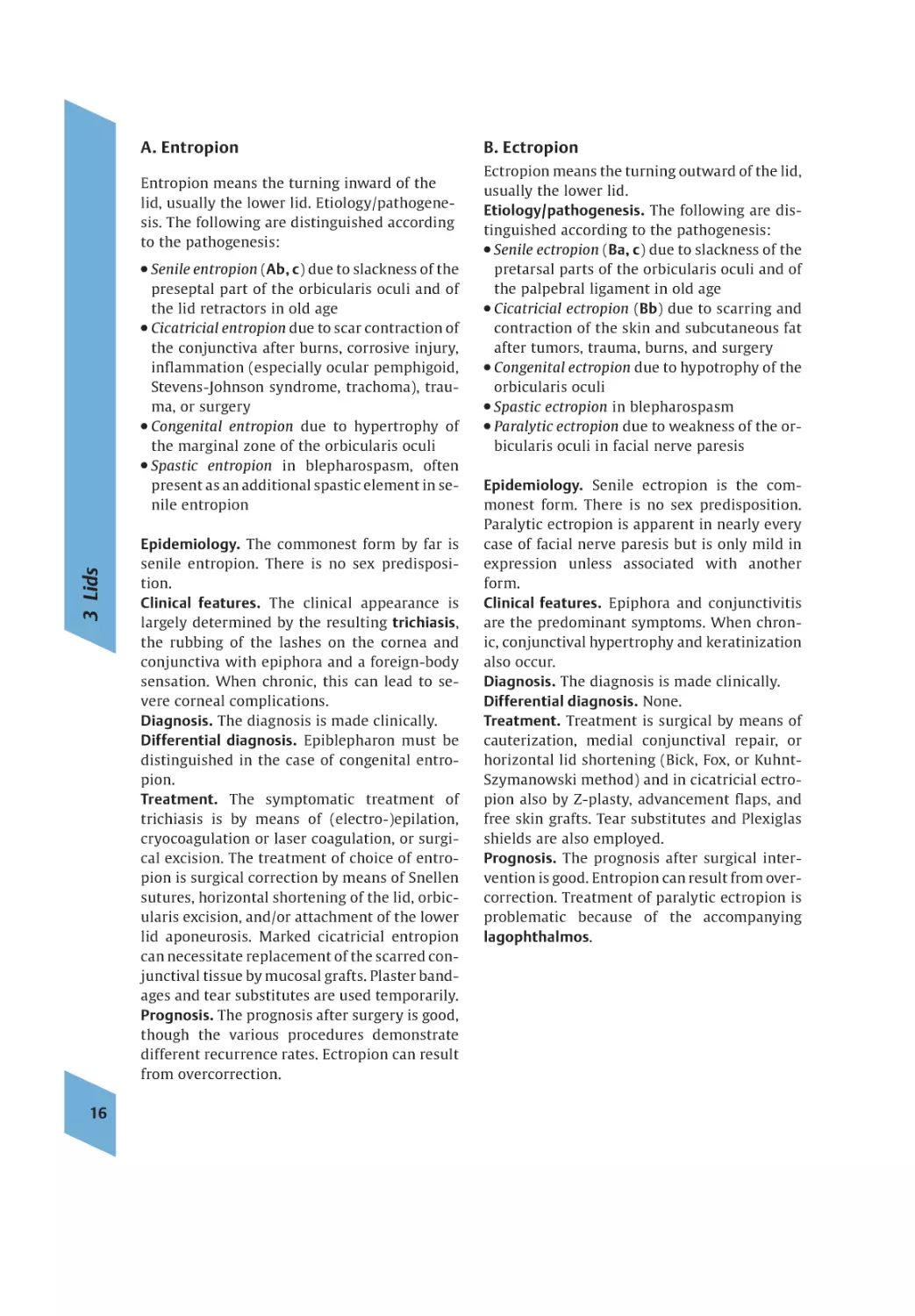
A. Entropion
Entropion means the turning inward of the
lid, usually the lower lid. Etiology/pathogenesis. The following are distinguished according
to the pathogenesis:
Senile entropion (Ab, c) due to slackness of the
preseptal part of the orbicularis oculi and of
the lid retractors in old age
● Cicatricial entropion due to scar contraction of
the conjunctiva after burns, corrosive injury,
inflammation (especially ocular pemphigoid,
Stevens-Johnson syndrome, trachoma), trauma, or surgery
● Congenital entropion due to hypertrophy of
the marginal zone of the orbicularis oculi
● Spastic entropion in blepharospasm, often
present as an additional spastic element in senile entropion
3 Lids
●
16
Epidemiology. The commonest form by far is
senile entropion. There is no sex predisposition.
Clinical features. The clinical appearance is
largely determined by the resulting trichiasis,
the rubbing of the lashes on the cornea and
conjunctiva with epiphora and a foreign-body
sensation. When chronic, this can lead to severe corneal complications.
Diagnosis. The diagnosis is made clinically.
Differential diagnosis. Epiblepharon must be
distinguished in the case of congenital entropion.
Treatment. The symptomatic treatment of
trichiasis is by means of (electro-)epilation,
cryocoagulation or laser coagulation, or surgical excision. The treatment of choice of entropion is surgical correction by means of Snellen
sutures, horizontal shortening of the lid, orbicularis excision, and/or attachment of the lower
lid aponeurosis. Marked cicatricial entropion
can necessitate replacement of the scarred conjunctival tissue by mucosal grafts. Plaster bandages and tear substitutes are used temporarily.
Prognosis. The prognosis after surgery is good,
though the various procedures demonstrate
different recurrence rates. Ectropion can result
from overcorrection.
B. Ectropion
Ectropion means the turning outward of the lid,
usually the lower lid.
Etiology/pathogenesis. The following are distinguished according to the pathogenesis:
● Senile ectropion ( Ba, c ) due to slackness of the
pretarsal parts of the orbicularis oculi and of
the palpebral ligament in old age
● Cicatricial ectropion ( Bb ) due to scarring and
contraction of the skin and subcutaneous fat
after tumors, trauma, burns, and surgery
● Congenital ectropion due to hypotrophy of the
orbicularis oculi
● Spastic ectropion in blepharospasm
● Paralytic ectropion due to weakness of the orbicularis oculi in facial nerve paresis
Epidemiology. Senile ectropion is the commonest form. There is no sex predisposition.
Paralytic ectropion is apparent in nearly every
case of facial nerve paresis but is only mild in
expression unless associated with another
form.
Clinical features. Epiphora and conjunctivitis
are the predominant symptoms. When chronic, conjunctival hypertrophy and keratinization
also occur.
Diagnosis. The diagnosis is made clinically.
Differential diagnosis. None.
Treatment. Treatment is surgical by means of
cauterization, medial conjunctival repair, or
horizontal lid shortening (Bick, Fox, or KuhntSzymanowski method) and in cicatricial ectropion also by Z-plasty, advancement flaps, and
free skin grafts. Tear substitutes and Plexiglas
shields are also employed.
Prognosis. The prognosis after surgical intervention is good. Entropion can result from overcorrection. Treatment of paralytic ectropion is
problematic because of the accompanying
lagophthalmos.
08.06.2006
A. Entropion
17:07 Uhr
Seite 17
B. Ectropion
a Upper lid entropion
a Senile ectropion
b Senile entropion
b Cicatricial ectropion
c Senile entropion
c Senile ectropion, predominantly nasal
Dystrophies, Degenerative Conditions, and Age-related Changes
03_Lider
17
03_Lider
08.06.2006
17:07 Uhr
Seite 18
Thieme; Langner; Schlote; Pocket Atlas of Ophthalmology; 1. AK, 5/2006
A. Ptosis
3 Lids
Ptosis is the term for pathological drooping of
the upper lid.
Etiology/pathogenesis. Ptosis can be caused by
one or more of the following factors:
● Neurogenic disorders such as congenital or
acquired oculomotor paresis (Aa), Horner
syndrome, Marcus-Gunn phenomenon, or
aberrant innervation of the oculomotor nerve
● Congenital or acquired myogenic disorders
such as myasthenia gravis, myotonic dystrophy, ocular myopathy, or oculopharyngeal
muscular dystrophy
● Levator palpebrae muscle aponeurosis slackness or dehiscence as in senile ptosis (Ab) or
postoperative ptosis
● Mechanical disorders due to excessive weight
of the upper lid (tumors) or scarring of the
conjunctiva
18
Epidemiology. Ptosis is a relatively common
finding. The most important causes are acquired oculomotor paresis, Horner syndrome
and myasthenia gravis. There is no age or sex
predisposition in oculomotor paresis and
Horner syndrome. Myasthenia gravis typically
affects women in middle age.
Clinical features. Clinically the drooping of one
or both upper lids predominates. Depending on
the pathogenesis and degree of severity, the patients complain of other symptoms (e.g., restrictions of the visual field) or diminished vision.
In paralysis of the oculomotor nerve, ptosis occurs because of the loss of the levator palpebrae muscle (paralytic ptosis). Disorders of eye
movement with diplopia (external ophthalmoplegia) and paresis of the sphincter pupillae
and ciliary muscle with fixed pupils and accommodation disorders (internal ophthalmoplegia) can be present also. Diabetes mellitus is
one of the commonest causes of peripheral lesions of the third cranial nerve. Recurrent ischaemic lesions with acute unilateral and
sometimes painful paresis of the extrinsic eye
muscles innervated by the oculomotor nerve
are characteristic. Nuclear oculomotor lesions
are usually associated with slight bilateral ptosis as both levator palpebrae muscles are innervated by a single nuclear region.
In Horner syndrome there is a classical triad
consisting of ptosis, miosis, and (pseudo-)
enophthalmos. Since only the circular fibers of
the ciliary muscle are affected in this ocular
sympathetic paresis and the function of the levator palpebrae, which is innervated by the
oculomotor nerve, is not affected, the unilateral ptosis is often slight.
In the Marcus-Gunn phenomenon (Ac–f), congenital anomalous innervation leads to elevation of the ptotic lid during masticatory movements and on mouth opening. Simple
congenital ptosis is the result of dominantly or
recessively inherited dystrophy of the levator
palpebrae muscle, which is usually unilateral.
It is characterized by defective contraction and
relaxation of the muscle. Weakness of the superior rectus muscle is also seen occasionally.
Myasthenia gravis pseudoparalytica is an autoimmune disease and is due to a disorder of
neuromuscular stimulus transmission as a result of blocking of the acetylcholine receptors
of the motor endplate by circulating polyclonal autoantibodies. There is an increased incidence of other autoimmune diseases. Ocular
symptoms (ptosis, diplopia) occur most frequently, followed by speech, chewing, and
swallowing disorders. The muscle weakness
also affects facial expression (myopathic facies). Three-quarters of patients with myasthenia have eye involvement. Purely ocular myasthenia is present in 20 % of cases. The disease
is manifested more often after psychological
stress and worsens during the course of the day
and with fatigue. The usually bilateral, asymmetrical ptosis increases on prolonged upgaze
(Simpson test). The muscle weakness improves
following injection of an acetylcholinesterase
inhibitor (Tensilon or prostigmine test). In 90 %
of patients with generalized myasthenia and in
50 % with purely ocular myasthenia, autoantibodies to acetylcholine receptors can be found
in the serum. There is an increased incidence
of persistent thymus, thymus hyperplasia, or
thymoma.
08.06.2006
17:07 Uhr
Seite 19
A. Ptosis
a Congenital ptosis
b Senile ptosis
c Marcus–Gunn phenomenon
d Marcus–Gunn phenomenon
e Marcus–Gunn phenomenon
f Marcus–Gunn phenomenon
Dystrophies, Degenerative Conditions, and Age-related Changes
03_Lider
19
03_Lider
08.06.2006
17:07 Uhr
Seite 20
Thieme; Langner; Schlote; Pocket Atlas of Ophthalmology; 1. AK, 5/2006
3 Lids
A. Ptosis (continued)
20
Myotonic dystrophy (Curschmann-BattenSteinert syndrome) is an autosomal dominant
inherited myopathy with circumscribed muscle dystrophy, myotonic reaction, and various
concomitant symptoms such as cataract and
gonadal atrophy. Apart from bilateral ptosis,
amimia, and atrophy of the temporal muscles
(myopathic facies), there is weakness particularly of the sternocleidomastoid, brachioradialis, and fibular muscles.
Ocular myopathy (chronic progressive external ophthalmoplegia) is characterized by progressive ptosis with progressive ocular muscle
paresis due to atrophy of the motor nuclear region. As the muscle involvement is strictly
symmetrical, double vision is not found even
in the advanced stage. In the Kearns-Sayre
syndrome (ophthalmoplegia plus), as well as
ocular myopathy, tapetoretinal degeneration,
cardiac conduction disorders, small stature,
and neurological manifestations are also
found.
In the autosomal dominant inherited oculopharyngeal muscular dystrophy there is
paresis of pharyngeal muscles and of the temporalis muscle along with paralysis of the extrinsic ocular muscles.
Diagnosis. The diagnosis of ptosis is made clinically. The Tensilon test (see p. 18) is used to distinguish it from myasthenia gravis.
Differential diagnosis. Pseudoptosis due to an
excessively small eyeball (microphthalmos, phthisis bulbi) or contralateral lid retraction, blepharochalasis, or dermatochalasis.
Treatment. The treatment of ptosis depends on
the etiology. Congenital or myogenic ptosis can
usually be improved by transdermal or
transconjunctival levator resection/folding if
some residual levator function is preserved. If
there is no or only slight levator function, suturing of the tarsus to the frontalis muscle by
means of a loop usually produces better results.
Tarsoconjunctival resection or reinforcement
of the aponeurosis is also employed. Thymectomy demonstrates good results in the treatment of myasthenia gravis. Long-term immunosuppressant treatment with azathioprine
and corticosteroids is recommended. Symptomatic treatment with an acetylcholinesterase
inhibitor (pyridostigmine) usually shows a
rapid improvement in the symptoms but is not
suitable for long-term treatment because of diminishing efficacy.
Prognosis. The course and prognosis differ depending on the etiology. While there is no
change in Horner syndrome and in most forms
of oculomotor paresis, the prognosis is good in
diabetic oculomotor paresis with regression
within three months. Recurrences are possible.
Myasthenia gravis has a chronic progressive
course.
B. Blepharochalasis
Bilateral blepharochalasis is caused by acute lid
edema. Thinning and atrophy of the skin and
stretching and separation of the aponeurosis
lead to ptosis (Ba and b). The function of the levator palpebrae is usually normal and the degree of ptosis is variable. Blepharochalasis is
apparent before the age of 20 years in 60 %, and
women are affected more often. Dominant inheritance has been described. Treatment is surgical.
C. Dermatochalasis
Dermatochalasis is characterized by folds of excessive skin in the upper lid (Ca and b). If the
orbital septum is weakened, this can be associated with a prolapse of the orbital fat. The lid
crease is obliterated. Dermatochalasis occurs
predominantly at a more advanced age. Patients complain of a heavy sensation in the region of the eyes and in severe cases of visual
impairment. Treatment is surgical by an upper
lid lift with excision of the excess skin.
08.06.2006
17:07 Uhr
Seite 21
B. Blepharochalasis
a
b
Dystrophies, Degenerative Conditions, and Age-related Changes
03_Lider
C. Dermatochalasis
21
a
b
03_Lider
08.06.2006
17:07 Uhr
Seite 22
3 Lids
Thieme; Langner; Schlote; Pocket Atlas of Ophthalmology; 1. AK, 5/2006
22
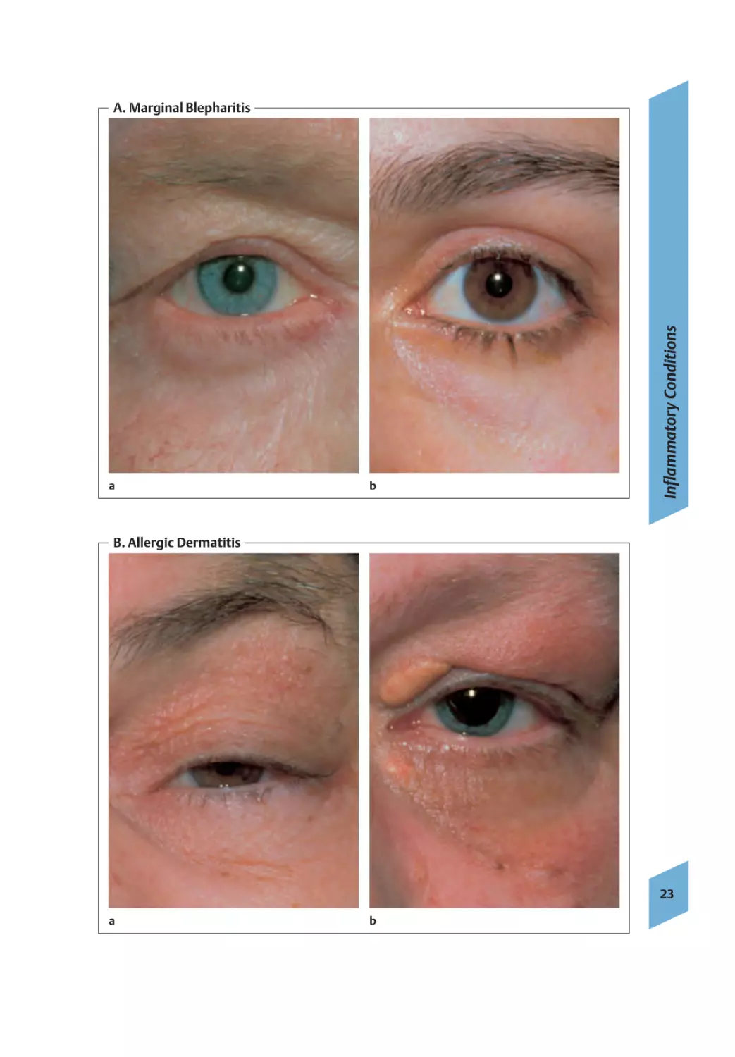
A. Inflammation of the Lid Margin
(Marginal Blepharitis)
B. Allergic Inflammation of the Lid
(Allergic Dermatitis)
Marginal blepharitis (Aa and b) is a chronic inflammation of the lid margin. It is often accompanied by secondary changes in the conjunctiva and cornea.
Etiology/pathogenesis. Staphylococcal infections and seborrhea are the most important
pathogenic factors. In addition, instability of
the tear film is nearly always found.
Epidemiology. This is the commonest disorder
of the outer eye. Marginal blepharitis often begins in childhood and women are affected more
frequently.
Clinical features. The patients complain of
chronic irritation of the eyes with burning and
itching. Mixed forms of staphylococcal (ulcerative blepharitis) and seborrheic (squamous
blepharitis) inflammation are typical. The
staphylococcal form is due to chronic infection
of the lash follicles by staphylococci. As well as
dilated blood vessels (rosettes), encrusted
scales are found at the base of the lashes. Trichiasis, loss (madarosis), or depigmentation (poliosis) of the lashes can also occur. Scarring of
the lid margin (entropion, ectropion) or secondary conjunctival and corneal inflammation
can occur in the late stage. Seborrheic blepharitis is the result of overproduction by the meibomian and Zeis glands. The excess free fatty
acids lead to irritation of the lid margin, conjunctiva, and cornea. Other seborrheic skin
changes are frequently found. The lid margins
demonstrate a typical waxy sheen. The lashes
are greasy and stuck together. Soft greasy scales
are found over the entire lid margin. Small
drops of oil are visible along the openings of
the ducts of the meibomian glands.
Diagnosis. The diagnosis is made clinically.
Differential diagnosis. Rosacea and other dermatoses involving the lids.
Treatment. Lid hygiene is in the foreground of
treatment. Warm compresses and artificial tears
often relieve the symptoms. Antibiotic eyedrops
are additionally used in staphylococcal blepharitis. Severe secondary complications occasionally necessitate the use of corticosteroids.
Prognosis. This is a chronic disorder the treatment of which is often unsatisfactory and
which usually cannot be cured. Secondary
changes are less severe in the seborrheic form
than in staphylococcal blepharitis.
Despite the usually multifactorial pathogenesis, lid dermatitis and eczema (Ba and b) are
usually classified according to the main pathogenesis. Most lid dermatitis and eczema are allergic in origin. Contact allergies of the delayed
type are most common, followed by atopic and
irritant eczema. Most patients are women, and
the average age of affected patients is 45 years.
Apart from cosmetics, ophthalmological preparations and contact lens care products often
have allergenic or irritant effects. Erythema, severe itching, and also scaling in the later stages
are typical clinical signs. Confirmation of contact sensitization is done by skin testing. Apart
from avoidance of the allergen, mast cell stabilizers and antihistamines are used in treatment, with corticosteroids and cyclosporin A
(ciclosporin) in severe and chronic cases. Besides the lichenification of the skin of the lid,
typical stigmata of atopy are often found in
atopic dermatitis, such as a double DennieMorgan crease in the lower lid, loss of the lateral part of the eyebrows (Hertoghe’s sign), and
haloing of the eyes. The lid eczema may be the
only manifestation of atopy. Secondary contact
allergy can also be detected in about one-third
of patients. Marginal blepharitis with marked
instability of the tear film is frequently found
as a complication. Herpetic blepharoconjunctivitis is a rare complication.
03_Lider
08.06.2006
17:07 Uhr
Seite 23
a
b
Inflammatory Conditions
A. Marginal Blepharitis
B. Allergic Dermatitis
23
a
b
03_Lider
08.06.2006
17:07 Uhr
Seite 24
Thieme; Langner; Schlote; Pocket Atlas of Ophthalmology; 1. AK, 5/2006
3 Lids
A. Infectious Inflammation of the Lid
24
In local infection following trauma, insect bites,
hematoma, or spread from purulent sinusitis or
osteomyelitis, severe erythema and swelling of
the lid (pseudoptosis) with fever and the development of a lid abscess (Aa) or lid cellulitis
(Ab) can occur with the risk of intracerebral extension. Typical pathogens are staphylococci
and streptococci, rarely anaerobic bacteria.
Mixed infections are frequent. Apart from establishing the cause, the treatment consists of
giving high-dose broad-spectrum antibiotics. If
resolution or spontaneous perforation do not
occur, incision, irrigation, and drainage is performed. In contrast to orbital cellulitis, eye
movements are always intact.
Lid erysipelas occurs rarely. Together with severe pain and usually a high fever, a sharply demarcated intense erythema develops that does
not necessarily have the typical tongues-offlame shape in the lid. The cause is a wound infection with (-hemolytic streptococci. Highdose antibiotic therapy is necessary to prevent
extension. Involvement of the lids in syphilis,
anthrax, tuberculosis, leprosy, and diphtheria
has also been described rarely.
Lid herpes is an infection of the skin of the lid
with the herpes simplex virus (Ac). This is usually due to reactivation of latent infection, more
rarely to primary infection. After lessening of
maternal antibody protection, the first infection often occurs in early childhood due to direct contact or droplet infection from herpetic
lesions from healthy chronic shedders. Only 1 %
of the initial infections are clinically apparent,
usually as herpetic gingivostomatitis. The neurotropic viruses persist in the body even after
the inflammation has subsided. Recurrences
occur through irritation of latently infected
neurons after febrile infections, sun exposure,
menstruation, trauma, and gastrointestinal disorders, and also due to immunosuppressant,
hormonal, and psychological factors. There is
severe itching and a sensation of tension; the
typical cluster of vesicles develops on a reddened background and heals without scarring
after 8–10 days. Regional lymph node swelling
is possible. Besides the lips and genitals, the
skin of the face, particularly the lid, is typically affected. Involvement of the conjunctiva and
cornea is not infrequent. Treatment is symptomatic. Antiviral ointments hasten healing.
Ophthalmic zoster (shingles), when it affects
the face, is an infection of the gasserian ganglion and the area innervated by the ophthalmic nerve (V1) by the varicella zoster virus
(Ad). The disease is an expression of a reinfection or more often activation of the neurotropic viruses even years after the primary infection. Severe neuralgiform pain occurs initially.
Skin erythema develops with initially clear
tense vesicles, the content of which leaves behind yellowish dried and brownish crusts and
scars. The disease is of great importance because of the potentially serious ophthalmological complications. Treatment consists of local
and systemic administration of virostatic
drugs.
Under conditions of poor hygiene crab lice
(Phthirus pubis) or more rarely head lice
(Pediculus humanus capitis) can infest the lid
margin. They are spread by direct contact. The
lice lodge on the lid margin between the cilia
and lay their nits on the shafts of the eyelashes, where they have the appearance of small
black grains. The patients complain of severe
itching. Marginal blepharitis is often found.
Treatment consists of removal of the lice and
nits and administration of topical parasympathomimetics.
Ticks (Ae) can also occasionally infest the lid
margin. They are removed completely with
tweezers. Transmission of borreliosis or early
summer meningoencephalitis is also possible
on the lid.
03_Lider
08.06.2006
17:07 Uhr
Seite 25
A. Infectious Inflammations of the Lid
b Lid phlegmon
c Herpes simplex
d Herpes zoster
e Tick in the upper row of lashes
f Fly larva in the lower row of lashes
Inflammatory Conditions
a Lid abscess
25
03_Lider
08.06.2006
17:07 Uhr
Seite 26
3 Lids
Thieme; Langner; Schlote; Pocket Atlas of Ophthalmology; 1. AK, 5/2006
A. Hordeolum
C. Molluscum Contagiosum
A stye (external hordeolum, A) is an acute inflammation of the Zeis or Moll glands with pain
and swelling. Similarly painful inflammation of
the meibomian glands is called an internal
hordeolum.
Etiology/pathogenesis. The cause is folliculitis
or an abscess in the Zeis or Moll glands located superficially at the roots of the lashes, or a
staphylococcal infection of the meibomian
glands located in the tarsus.
Epidemiology. There is no age or sex predisposition.
Clinical features. There is localized inflamed
swelling of the lid margin. Several hordeola are
often found adjacent to one another. There can
be general malaise. Swelling of preauricular
lymph nodes and fever are also frequent. Lid abscess and orbital cellulitis are rare.
Diagnosis. The diagnosis is made clinically. Diabetes mellitus must be excluded if there is repeated occurrence.
Differential diagnosis. Marginal blepharitis,
chalazion.
Treatment. Dry heat infrared warming, and
local disinfectant and antibiotic therapy hasten
coalescence. Lancing may become necessary if
spontaneous healing does not occur.
Prognosis. Benign, occasionally recurrent.
The wartlike lesions of molluscum contagiosum (C) are usually numerous and consist of virally induced papules with a central indentation.
Etiology/pathogenesis. Molluscum contagiosum is caused by DNA viruses of the poxvirus
group. Transmission is by direct contact, autoinoculation, perinatally, and by sexual contact. The incubation period varies between 1
week and 6 months.
Epidemiology. The incidence of molluscum
contagiosum is increased in children and immunosuppressed patients. There is no sex predisposition.
Clinical features. There are usually numerous
skin-colored papules ranging in size from a pinhead to pea-sized with an indented center.
With pressure, a cheesy contagious material is
expressed. Concomitant conjunctivitis is often
observed when the lesions are near the lid margin.
Diagnosis. The diagnosis is made clinically.
Differential diagnosis. Common wart, milium,
keratoacanthoma, granuloma, chalazion, basal
cell carcinoma.
Treatment. Molluscum contagiosum lesions
are incised and removed by curetting or excision.
Prognosis. Benign, recurrent. Spontaneous remissions are possible even after years.
B. Chalazion
26
A chalazion (Ba) is the result of obstruction of
the duct of a meibomian gland, which is usually idiopathic, with secondary lipogranulomatous inflammation (Bb).
Epidemiology. There is no age or sex predisposition. There is a higher incidence of chalazion
in seborrheic dermatitis, rosacea, and diabetes
mellitus.
Clinical features. The appearance is of a pale,
round, firm lesion of the lid. With secondary
bacterial infection it is difficult to distinguish
from an internal hordeolum. Astigmatism with
a reduction in vision can be induced by pressure on the eyeball.
Diagnosis. The diagnosis is made clinically.
Differential diagnosis. Hordeolum, pyogenic
granuloma (Bc), sebaceous gland carcinoma.
Treatment. The cysts can be incised and removed by curetting. Steroid injection can initiate remission.
Prognosis. Benign, occasionally recurrent.
D. Common Warts
Common warts are occasionally found on the
lid (D). These pea-sized, hemispherical hard
nodules with a prickly surface are the result of
infection with a papilloma virus. The incubation period is 6 weeks to 20 months. They
sometimes occur in groups as filiform warts.
The therapeutic options available include removal with a curette, cryotherapy, and electrocoagulation.
03_Lider
08.06.2006
17:07 Uhr
Seite 27
C. Molluscum Contagiosum
B. Chalazion
D. Common Wart
Tumors
A. Hordeolum
a
b Histological section, ca. 40: lymphocytes,
epithelioid cells, and foreign-body giant cells
27
c Pyogenic granuloma
03_Lider
08.06.2006
17:07 Uhr
Seite 28
Thieme; Langner; Schlote; Pocket Atlas of Ophthalmology; 1. AK, 5/2006
3 Lids
A. Dermoid cyst
The dermoid cyst (A) is a benign mature teratoma.
Etiology/pathogenesis. The cyst is a congenital lesion due to deep sequestration of epidermal tissue.
Epidemiology. Manifestation extends from
birth to early adulthood. There is no sex predisposition.
Clinical features. An bulging elastic tumor, ca.
1 cm, of variable mobility usually develops in
the upper temporal lid region. An intraorbital
location is possible.
Diagnosis. The diagnosis is made clinically.
Differential diagnosis. Tumors of the lacrimal
gland, mucocele, hemangioma, lymphangioma,
inflammatory processes.
Treatment. Surgical excision is the treatment
of choice. Observation is also possible depending on the size.
Prognosis. Benign, not recurrent, no known
risk of malignant transformation.
B. Capillary Hemangioma
The capillary hemangioma (B) is a benign neoplasm of blood vessels.
Etiology/pathogenesis. Unknown.
Epidemiology. There is a greater incidence in
females.
Clinical features. A usually superficial bright
red to purple lesion is observed usually on the
surface of the upper lid ca. 1 cm in size and of
spongy consistency.
Diagnosis. The diagnosis is made clinically.
Differential diagnosis. Cavernous hemangioma, nevus flammeus, lymphangioma.
Treatment. Steroid induction of spontaneous
regression along with laser and cryocoagulation are possible.
Prognosis. Benign, not recurrent. Regression
with complete remission in up to 90 % in the first
years of life.
C. Xanthelasma
28
Xanthelasmas (C) are flat yellowish tumors in
the region of the medial canthus, which are due
to deposits of cholesterol in macrophages.
Etiology/pathogenesis. Approximately 50 % of
affected patients suffer from hypercholesterolemia, otherwise it is due to a local disorder of lipid metabolism.
Epidemiology. The disease affects older
women more often.
Clinical features. Bilateral, yellowish, flat
plaques are most commonly found at the medial canthus.
Diagnosis. The diagnosis is made clinically.
Differential diagnosis. None.
Treatment. Apart from treatment of the underlying disease, the main treatments are surgical
excision or laser ablation for cosmetic reasons.
Prognosis. Benign, recurrent.
D. Keratoacanthoma
The keratoacanthoma (D) is a spherical epithelial tumor.
Etiology/pathogenesis. Unknown.
Epidemiology. The tumor occurs predominantly in later life. There is no sex predisposition.
Clinical features. Within a few weeks, a broadbased tumor 0.5–1 cm in diameter with a central keratinous crater develops, which resolves
after a few weeks leaving a flat scar.
Diagnosis. The diagnosis is made clinically.
Differential diagnosis. Squamous cell carcinoma, basal cell carcinoma, common wart, actinic
keratosis.
Treatment. Because it is sometimes difficult to
distinguish it from squamous cell carcinoma,
surgical excision with histological examination
is recommended.
Prognosis. Benign, not recurrent, no known
risk of malignant transformation.
E. Other
In addition to the described tumors, benign
lesions of the lid also include seborrheic and
actinic keratoses, nevi (Ea), congenital hemangiomas, lymphangiomas, fibromas and neurofibromas, along with epidermoid cysts. The
cutaneous horn (Eb) occupies a particular position. This is a yellow-brown growth of the
skin of various histogenesis, which is excised
because of the risk of malignant transformation.
03_Lider
08.06.2006
17:07 Uhr
Seite 29
B. Capillary Hemangioma
C. Xanthelasma
D. Keratoacanthoma
Tumors
A. Dermoid Cyst
E. Other
29
a Nevus
b Cutaneous horn
03_Lider
08.06.2006
17:07 Uhr
Seite 30
Thieme; Langner; Schlote; Pocket Atlas of Ophthalmology; 1. AK, 5/2006
A. Basal Cell Carcinoma
3 Lids
The basal cell carcinoma is a semimalignant
skin tumor derived from the epidermal basal
cells and the cells of the outer hair follicle
sheaths.
Etiology/pathogenesis. The etiology has not
been finally elucidated. Chronic light exposure
is important. Precancerous forms are unknown.
Epidemiology. The tumor occurs typically in
later life and accounts for 90 % of malignant and
20 % of all lid tumors. There is no sex predisposition.
Clinical features. Different forms are distinguished according to the growth behavior:
● Nodular (solid) type ( Aa ): 75 %; slow-growing
nontender nodule, central ulceration (Ab),
pearly shimmer, telangiectasias
● Pigmented type ( Ac ): 10 %
● Sclerosing (morphea) type ( Ad )
● Superficial type
Mixed forms are frequent. The preferred location is the medial lower lid. The tumor growth
is infiltrating and destructive.
Diagnosis. The diagnosis is made clinically and
histologically.
Differential diagnosis. Other malignant tumors of the lid, keratoacanthoma, seborrheic
keratosis, common wart.
Treatment. Surgical excision followed by plastic reconstruction is the treatment of choice.
Cryotherapy and radiotherapy are also used occasionally with good results. Prognosis. Recurrence occurs in 5–15%. Metastasis occurs in 0.1%.
B. Squamous Cell Carcinoma
30
Squamous cell carcinoma (B) is a rapidly growing and metastasizing tumor of epithelial origin.
Etiology/pathogenesis. The etiology has not
been finally elucidated. There is a predisposition with light exposure. Development from
precancerous lesions and de novo is known to
occur.
Epidemiology. There is a higher incidence in
the male sex and in later life. The tumor accounts for nearly 10 % of lid malignancies and
2 % of all lid tumors.
Clinical features. Over months, a flat erythematous lesion with crusts and ulceration develops, which later becomes nodular, papillomatous, or cystic.
Diagnosis. The diagnosis is made clinically and
histologically.
Differential diagnosis. Keratoacanthoma, other
malignant tumors and precancerous lesions of
the lid.
Treatment. The treatment of choice is histologically confirmed excision. Prognosis. Recurrence occurs in 10–20 %. Metastasis occurs in
1–21%. Mortality is 10–15 %.
C. Sebaceous Cyst Carcinoma
Sebaceous cyst carcinomas (C) are rapidly
growing and metastasizing tumors that originate predominantly from meibomian and Zeis
glands.
Etiology/pathogenesis. The etiology is unknown.
Epidemiology. Sebaceous cyst carcinomas
occur mainly in later life. Women appear to be
affected slightly more often. The tumor accounts for 0.2–0.7 % of all and about 1 % of malignant lid tumors.
Clinical features. The tumor, which is located
predominantly in the upper lid, commences as
a firm nonpainful tumor, similar to a chalazion.
Late ulceration is possible. There is often a loss
of eyelashes.
Diagnosis. The diagnosis is made clinically and
histologically.
Differential diagnosis. Chalazion, other malignant tumors and precancerous lesions of the
lid. Treatment. Mainly surgical excision.
Prognosis. Recurrence occurs in 10–40 % and
lymphogenous metastasis in 17–28 %. The mortality is 5–15 %.
Other, rare malignant tumors of the lid include
malignant melanoma, Merkel cell carcinoma,
Kaposi sarcoma, chondrosarcoma, and metastases.
03_Lider
08.06.2006
17:07 Uhr
Seite 31
A. Basal Cell Carcinoma
b Basal cell carcinoma, severely ulcerating
c Pigmented type
d Morphea type: medial lower lid and inner canthus
Tumors
a Nodular type
B. Squamous Cell Carcinoma
C. Sebaceous Cyst Carcinoma
31
03_Lider
08.06.2006
17:07 Uhr
Seite 32
Thieme; Langner; Schlote; Pocket Atlas of Ophthalmology; 1. AK, 5/2006
A. Surgical Alterations
A frequent unwanted consequence of surgery is
the development of a scar keloid. This is a tough,
flat or stringy, sometimes itchy proliferation of
connective tissue, which develops depending
on individual and ethnic predisposition weeks
to months after the procedure in the region of
the operation scar. In contrast to hypertrophic
scars, extension is possible even to originally
undamaged skin. The treatment consists of intralesional injection of corticosteroids, radiotherapy, a pressure bandage, and possibly surgical revision in the case of scar contractures. In
the course of postoperative wound healing, lid
deformities can occur, which sometimes necessitate revision operations. These include
mainly ectropion, entropion, and ptosis.
3 Lids
B. Trauma
32
Lid trauma is a frequent injury. Approximately
75 % of the patients are male. The average age
is between 30 and 60 years. Over half of the injuries are due to blunt trauma due to falls, acts
of violence (Ba), road traffic accidents, and
work and sports injuries (Bb). In addition to lacerations and bites (Bc–e), corrosive injuries and
burns (Bf) are also frequent. In about half of all
lid injuries there is also injury of the globe,
which can extend from superficial injury to severe eyeball penetration and rupture. Severe,
extensive and multiple lacerations of the facial
region after road traffic accidents have become
rare as a result of seatbelt laws in cars and the
introduction of shatter-proof glass. Lid injuries
are often found in conjunction with extensive
head and facial injuries. Because of the collateral circulation and the loose structure of the
lid connective tissue, hemorrhage and lid
edema develop rapidly, which can often spread
to the uninjured side. Fractures of the orbit,
midface, and base of the skull should be excluded. Depending on the depth and extent of
the lid injury, abrasions, contusions, lacerations, avulsions, and puncture wounds are distinguished. With full-thickness defects of the
upper lid, the levator palpebrae muscle is often
involved, which can lead to posttraumatic ptosis. With lid injuries in the medial canthus,
there are often injuries of the lacrimal ducts. In
particular, because of the divided insertion of
the medial palpebral ligament, careful repair of
this area is important to prevent posttraumat-
ic lid deformities. The risk of infection after lid
injuries can be regarded as low. In the treatment of lid trauma, a distinction is made between primary management and repair, which
may be required later. The goal of the primary
treatment is restoration of the anatomy and
normal function. Management of the lid injury
depends on the degree of the damage and extends from simple interrupted sutures and suture of the lid margins to large advancement
flaps. Deformities of the lids can occur in the
course of wound healing due to scar formation,
necessitating further revision operations. The
commonest posttraumatic deformities are ectropion, entropion with trichiasis, deformity of
the canthus, and ptosis.
03_Lider
08.06.2006
17:07 Uhr
Seite 33
a Lid hematoma due to a blow from a fist
b Lid hematoma due to impact of a squash ball
c Severe lid trauma with avulsion of lid margin
and loss of tissue
d Multiple lacerations (windshield injury))
e Dog bite injury
f Explosive injury
Surgical Alterations and Trauma
B. Trauma
33
04_Tränenapparat
08.06.2006
17:09 Uhr
Seite 34
Thieme; Langner; Schlote; Pocket Atlas of Ophthalmology; 1. AK, 5/2006
A. Congenital Anomalies of the Lacrimal
Gland
Complete absence of the lacrimal gland (aplasia), displacement (ectopia), lacrimal gland
cysts (dacryops, A), or lacrimal gland fistulas
are rare.
4 Lacrimal Apparatus
B. Dry Eye: Sicca Syndrome
Complex qualitative and quantitative disorders
of the production and surface adhesion of the
tear film.
Etiology/pathogenesis. Multifactorial, caused
by ophthalmological and general diseases and
by exogenous factors (B, Table 1).
Epidemiology. Very common. Symptoms in ca.
22 % of women and 10 % of men between 55 and
60 years. Signs of keratoconjunctivitis sicca in
20 % of women and 15 % of men between 45 and
54 years.
Clinical features. Dryness, sensation of pressure, pain, burning, scratching, foreign-body
sensation, photophobia, lid swelling, corneal
edema, corneal epithelial filaments, conjunctival folds parallel to the lid margins (LIPCOF),
tear meniscus irregular and/or reduced
< 0.2 mm.
Diagnosis. The diagnosis is made clinically and
by means of tests, including the Schirmer test
< 10 mm/5 min, Jones basal secretion test
< 10 mm/5 min, break-up time < 10 s.
Differential diagnosis. Infection, asthenopia.
Treatment. Tear substitute, moisture chamber,
glasses with side protection, punctum plug.
Treatment of the underlying disease. Reduction in exogenous factors (smoke, dust, night
work, etc.).
Prognosis. Rarely major symptoms. Increased
bacterial superinfection.
C. Acute Dacryoadenitis
34
Sudden unilateral tender swelling of the
lacrimal gland.
Etiology/pathogenesis. See C, Table 2.
Epidemiology. Usually middle-aged women.
Clinical features. Erythema, swelling, tenderness of the lateral third of the upper lid (Sshaped curve of the upper lid, C), conjunctival
chemosis, palpable preauricular lymph nodes,
fever, general malaise, leukocytosis.
Diagnosis. The diagnosis is made clinically.
Differential diagnosis. Lid/orbital cellulitis,
hordeolum, lid abscess, sinusitis, erysipelas.
Treatment. Antibiotic therapy. Incision and
drainage.
Prognosis. The disease lasts 8–10 days.
D. Chronic Dacryoadenitis
Painless, slightly inflammatory swelling of the
lacrimal gland, unilateral or bilateral.
Etiology/pathogenesis. Sequela of acute
dacryoadenitis, granulomatous diseases (D,
Table 3).
Epidemiology. Usually men over 40 years.
Clinical features. Painless swelling of the lateral upper lid.
Diagnosis. The diagnosis is made clinically.
Further investigations depending on the underlying disease. Biopsy.
Differential diagnosis. Tumor, dermoid, osteomyelitis, systemic diseases.
Treatment. Treatment of the underlying disease.
Prognosis. Depending on the underlying disease.
E. Pleomorphic Adenoma
(Benign Mixed-Cell Tumor)
Etiology/pathogenesis. Unknown. Benign
tumor of epithelial and myoepithelial cells.
Epidemiology. Commonest epithelial tumor of
the lacrimal gland (50 %). Age peak at 4th–5th
decade. The ratio of men to women is 1.5 : 1 to
2 : 1.
Clinical features. Painless, slowly growing
tumor. Visual changes and double vision are not
reported. Displacement of the eyeball downward and nasally.
Diagnosis. CT shows a sharply demarcated
tumor. Bone atrophy possible. On MRI the
tumor appears hypointense on T1-weighted
imaging and hyperintense on T2-weighted imaging. Eyeball deformation is possible.
Differential diagnosis. Adenoid cystic carcinoma.
Treatment. Complete excision including the
pseudocapsule. Incisional biopsy leads to a
high rate of recurrence.
Prognosis. Five-year survival rate of 99 %. Recurrences possible after incomplete excision,
malignant transformation also possible.
04_Tränenapparat
08.06.2006
17:09 Uhr
A. Congenital Abnormalities of the
Lacrimal Gland
Seite 35
B. Sicca Syndrome
• General diseases
– Diabetes mellitus
– Thyroid diseases: hypothyroidism, endocrine
ophthalmopathy, Hashimoto thyroiditis
– Sjögren syndrome, rheumatoid arthritis,
SLE (systemic lupus erythematosus),
Wegener granulomatosis, sclerodema
– Skin diseases: neurodermatitis, acne rosacea,
allergies
– Pregnancy
Lacrimal gland cyst
C. Acute Dacryoadenitis
S-shaped curve of the upper lid
Table 2 Etiology/pathogenesis of acute
dacryoadenitis
• Bacterial
– Staphylococci
– Streptococci
– Gonococci
• Viral
– Mumps
– Measles
– Scarlet fever
– Mononucleosis
– Herpes zoster
D. Chronic Dacryocystitis
• External influences
– Smoke, dry heating, air conditioning
– Night work
– Contact lenses
– Cosmetics
• Medications
– Analgesics
– Antiarrhythmics
– Antihistamines
– Antihypertensives
– Hormones
– Lipid-lowering drugs
– Psychotropic medications
– Synthetic retinoids
– Virostatic drugs
– Cytostatic drugs
Diseases of the Lacrimal Gland
Table 1 Causes of sicca syndrome
• Diseases of the eye
– Systemic, infectious, tumor, or surgical
damage to the lacrimal gland
– Conditions of the conjunctiva: trachoma,
pemphigoid, allergies, operations,
Stevens–Johnson syndrome, burns, corrosive
injury, radiation
– Conditions of the cornea: dystrophies,
previous keratoplasty
– Lid deformities, lid closure deficit, blepharitis
E. Pleomorphic Adenoma
Table 3 Pathogenesis/pathogenesis of chronic
dacryoadenitis
• Unhealed acute dacryoadenitis
• Chronic conjunctivitis
• Tuberculosis
• Syphilis
• Leprosy
• Sarcoidosis
• Actinomycosis
• Nocardiosis
• Trachoma
35
04_Tränenapparat
08.06.2006
17:09 Uhr
Seite 36
Thieme; Langner; Schlote; Pocket Atlas of Ophthalmology; 1. AK, 5/2006
4 Lacrimal Apparatus
A. Adenoid Cystic Carcinoma
Etiology/pathogenesis. Not known.
Epidemiology. Commonest malignant, second
commonest epithelial lacrimal gland tumor
(25–30 %). Age peak in women about 40 years.
Clinical features. Rapidly growing. Pain with
tumor infiltration of nerves or bone.
Diagnosis. On CT, roundish poorly demarcated
space-occupying lesion with irregular surface
and bone destruction. MRI shows cystic isointense or hyperintense changes in the T1weighted image.
Differential diagnosis. See A, Table 1.
Treatment. Radical resection. Radiotherapy.
Prognosis. Five-year survival rate of 21 %. Recurrences and metastasis to the lungs. The
prognosis depends on the histological grade.
B. Diseases of the Lacrimal Ducts
The hallmark of all diseases of the lacrimal
ducts is epiphora. Epiphora immediately postnatally is due to atresia or stenosis of the
lacrimal ducts, especially of Hasner’s valve.
Signs of inflammation point to infection, which
can be acute or chronic and appear as canaliculitis or dacryocystitis. Tumors are a rarer
cause of epiphora. Signs suggestive of malignancy are:
● Development of mass above the medial
palpebral ligament
● Telangiectasia overlying lacrimal sac swelling
● Bloody secretion, nosebleed, bloody reflux
after irrigation of the lacrimal duct
C. Acute Dacryocystitis
36
Etiology/pathogenesis. Partial or complete
obstruction of the nasolacrimal duct with inflammation due to infection, tumor, foreign
bodies, after trauma or due to granulomatous
diseases.
Epidemiology. Adults between 50 and 60 years.
Clinical features. Epiphora; acute, unilateral,
painful inflammation of the lacrimal sac; pus
emerging from the lacrimal punctum; fever;
general malaise. Pain radiates to the forehead
and teeth.
Diagnosis. Diagnosis is made clinically. Swab
and culture with antibiotic sensitivity.
Differential diagnosis. Orbital cellulitis, inflammation of the paranasal sinuses, erysipelas.
Treatment. Conservative: see C, Table 2. For decompression, freeze the skin (with ethyl chlo-
ride), then make the incision. Dacryocystorhinostomy is often necessary later.
Prognosis. Spontaneous discharge of the pus
into the ethmoid cells, conjunctival sac, and
nose is possible. This can lead to chemosis, lid
edema, and erysipelas, more rarely to orbital
cellulitis.
D. Chronic Dacryocystitis
Etiology/pathogenesis. Congenital or idiopathic obstructions of the nasolacrimal duct,
e. g., due to unhealed acute dacryocystitis,
dacryoliths, foreign bodies, tumors, diseases of
the surrounding structures (sinusitis, tumors),
trauma.
Epidemiology. See acute dacryocystitis.
Clinical features. Epiphora; signs of inflammation are absent; large amounts of mucopurulent secretion drain on pressure over the
lacrimal sac.
Diagnosis. Swab and culture, lacrimal duct
probing.
Differential diagnosis. None.
Treatment. See C, Table 2. Dacryocystorhinostomy.
E. Trauma of the Lacrimal Ducts
These injuries often occur together with lid and
facial injuries, particularly with injuries of the
nasal canthus. The canaliculi are affected in
70 %, more rarely the lacrimal sac (20 %) and the
nasolacrimal duct (10 %). The defect and the
canaliculus can be located by thorough inspection under the operating microscope and irrigation with methylene blue or fluorescein. If
fractures or foreign bodies are suspected, radiography of the orbit in two planes or CT is indicated. Therapeutically, the earliest possible
reconstruction with silastic intubation of the
lacrimal ducts should be attempted. The prognosis depends on the extent of the injury. Lid
deformities and stenosis of the lacrimal duct
system are possible.
08.06.2006
17:09 Uhr
A. Adenoid Cystic Carcinoma
Table 1 Differential diagnosis of tumors in the
upper temporal quadrant of the orbit
• Benign
– Dacryops
– All forms of dacryoadenitis
– Pleomorphic adenoma
– Dermoid cyst
– Benign lymphoid lacrimal gland tumor
– Eosinophilic granuloma
– Aneurysmal bone cyst
– Cholesterol granuloma
• Malignant
– Adenoid cystic carcinoma
– Pleomorphic adenocarcinoma
– Mucoepidermoid carcinoma
– Squamous epithelial carcinoma
– Oncocytoma
– Malignant lymphoma
– Metastases
C. Acute Dacryocystitis
Table 2 Treatment of acute
canaliculitis/dacryocystitis
Seite 37
B. Diseases of the Lacrimal Ducts
Occlusion of the superior
and inferior lacrimal
canaliculus
Occlusion of the
common canaliculus
Occlusion of the
nasolacrimal duct
Stenosis of the lacrimal ducts
D. Chronic Dacryocystitis
Lacrimal Gland Tumors, Lacrimal Ducts
04_Tränenapparat
Lacrimal duct stenosis due to papilloma
E. Trauma of the Lacrimal Ducts
• Bacterial
– Local: gentamicin eye drops 5 times daily and
gentamicin eye ointment at night for 14 days;
disinfectant dressings with Rivanol solution
1:1000; xylometazoline eye drops t.i.d. to
reduce swelling
– Systemic: e.g., dicloxacillin p.o. For 10–14
days
– Actinomycetes: tetracycline eye ointment
t.i.d.
• Chlamydia
– Local: tetracycline, ofloxacin, erythromycin
eye ointment t.i.d. for 6 weeks
– Systemic: tetracycline, erythromycin,
doxycycline, or sulfamethoxazole p.o. for
3 weeks
Avulsion of lacrimal duct
• Fungi
– E.g., natamycin eye ointment 1–2 hourly
• Viruses
– Varicella zoster: aciclovir 800 mg 5 tabs daily
for 7 days, aciclovir eye ointment 5 times daily
continued for 3 days after healing
Acute dacryocystitis with persistent Hasner
membrane
37
05_Augenhöhle
09.06.2006
8:28 Uhr
Seite 38
Thieme; Langner; Schlote; Pocket Atlas of Ophthalmology; 1. AK, 5/2006
5 Orbit
A. Symptoms of Orbital Diseases
38
The orbit has close topographical relations to
the paranasal sinuses and is linked to the interior of the skull through the optic canal and the
superior orbital fissure. Thus, primary orbital
processes can extend into the paranasal sinuses and the region of the chiasm, and conversely processes arising primarily in these regions
can involve the orbit secondarily. Extension of
primary intraocular diseases into the orbit and
of primary intraorbital processes into the eyeball is likewise possible.
The pyramid-shaped orbit is bounded anteriorly by the eyeball and the orbital septum and otherwise by bone. The important diseases of the
orbit, namely inflammation, tumors and injuryrelated hemorrhage, are usually associated with
an increase in the volume and pressure in the
orbit. Orbital symptoms are therefore explained
mostly by displacement and/or compression of
orbital structures and the eyeball.
The symptoms of orbital diseases are:
● Exophthalmos ( Aa–c ), protrusion of the eye
(proptosis), is the main symptom of a spaceoccupying process. Space-occupying lesions
located in the muscle funnel (intraconal),
such as optic glioma and optic sheath meningioma, lead to protrusion axially and forward.
An extraconal increase in volume, in contrast,
usually leads to displacement of the eye in the
direction opposite to that of the pathological
process. As a rule, exophthalmos can be identified when observed from the side. It is
measured with a Hertel exophthalmometer,
which measures the distance of the anterior
pole of the cornea from the lateral margin of
the bony orbit, and comparison of the two
sides is particularly important. A greatly enlarged eye, e. g., in high myopia or buphthalmos, and also enophthalmos of the opposite
side, can mimic exophthalmos (“pseudo-exophthalmos”).
● Enophthalmos (see p. 47, Ac ), the sinking
backward of the eye, is the sign of volume
deficit in the orbit. In old age this is due to atrophy of the orbital fat pad. The cause of unilateral enophthalmos is usually a fracture of
the orbital bones (especially the floor of the
orbit and ethmoid cells) with displacement of
orbital tissue. Processes associated with tissue contraction such as some breast cancer
metastases can produce enophthalmos. The
●
●
enophthalmos in Horner syndrome is usually only slight. The upper lid is often lower in
enophthalmos. Enophthalmos is also measured with the Hertel exophthalmometer. A reduction in the size of the eye as in congenital
microphthalmos or phthisis bulbi can mimic
enophthalmos.
A motility deficit (strabismus, Ac) is caused
by the fact that space-occupying and destructive processes in the orbit often reduce
the mobility of the eyeball by interfering directly with the ocular muscles (mechanical
paresis) or by damaging the motor nerves
(neurogenic paresis). If there is a difference
in the reduction in eyeball motility between
the two sides, strabismus results, usually
with double vision.
Exposure keratitis and sometimes a corneal
ulcer can develop in exophthalmos as blinking and moistening of the surface of the eye
are impaired.
05_Augenhöhle
09.06.2006
8:28 Uhr
Seite 39
A. Symptoms of Orbital Diseases
Symptoms
a Exophthalmos on the right with optic glioma; retraction of the upper lid (pupil dilated by medication)
c Inflammatory orbital tumor on the right with
exophthalmos and dilated episcleral vessels;
massive restriction of eyeball movement on
looking to the right
39
b Exophthalmos seen from the side
05_Augenhöhle
09.06.2006
8:28 Uhr
Seite 40
Thieme; Langner; Schlote; Pocket Atlas of Ophthalmology; 1. AK, 5/2006
5 Orbit
A. Symptoms of Orbital Diseases
(continued)
40
Other symptoms of orbital diseases:
● Disorders of visual function can occur when
an orbital space-occupying lesion compresses the optic nerve directly or indirectly
through an increase in the pressure in the
orbit, thus leading to a loss of visual field or
visual acuity. Lesions of the optic nerve with
loss of visual function also occur not infrequently because of injuries or orbital infections. A reduction in vision also results when
there is central corneal opacification due to
exposure keratitis.
● Swelling of the head of the optic nerve ( papilledema) is a frequent though rather nonspecific finding in orbital processes. In contrast to papilledema due to raised intracranial
pressure, papilledema because of processes
in the orbit is often associated early with a reduction in function.
● More prolonged optic nerve compression
leads to a loss of ganglion cell axons and blood
vessels in the region of the optic disc (optic
atrophy, Aa). The disk becomes pale as a result.
● Anastomoses sometimes develop between
the vessels on the disk and those of the peripapillary choroid due to chronic optic nerve
compression (e. g., with slowly progressing
optic nerve tumors) (opticociliary shunt vessels, Aa).
● Compression of the globe from behind can
produce folding of the choroid (choroid folds,
Ab). These choroid folds are usually arranged
in parallel and are visible on funduscopy, but
are seen even better on angiography. They
tend to reduce visual function only slightly.
● More rarely, orbital tumors indent the globe
(globe indentation), so that the appearance
is that of an intraocular tumor.
● Alterations of the lids , such as retraction of
the upper lid, are typical of endocrine orbitopathy but also occur in all forms of advanced exophthalmos. Swelling of the
lacrimal gland often gives the upper lid lateral ptosis like an S-shape (Ac).
● Orbital diseases are often associated with alterations of the conjunctiva. Inflammatory
conditions of the orbit usually and tumor
processes occasionally cause redness (injection) and swelling (chemosis) of the conjunc-
●
tiva. Carotid-cavernous sinus fistulas are
characterized by dilatation of conjunctival
and episcleral vessels because of the raised
orbital venous pressure.
There can be an increase in intraocular pressure as the aqueous humor is drained into the
orbital veins. Compression of these veins
sometimes leads to congestion of the aqueous humor and so to an increase in ocular
pressure. An abnormality of venous orbital
outflow with ocular hypertension is typical of
carotid-cavernous sinus fistulas. In endocrine
orbitopathy, there can be an increase in pressure depending on the direction of gaze
(“pseudo-glaucoma”).
05_Augenhöhle
09.06.2006
8:28 Uhr
Seite 41
a Temporal pallor of the optic nerve (optic atrophy) and shunt vessels in optic glioma
Symptoms
A. Symptoms of Orbital Diseases (continued)
b Choroid folds running largely parallel in orbital metastasis
c Inflammation of the lacrimal gland (dacryoadenitis) on the left with typical S-shape
41
05_Augenhöhle
09.06.2006
8:28 Uhr
Seite 42
Thieme; Langner; Schlote; Pocket Atlas of Ophthalmology; 1. AK, 5/2006
5 Orbit
A. Investigations of Orbital Processes
42
The diagnosis begins with the history and clinical examination. Depending on these findings,
imaging procedures are employed.
History. Obtaining an accurate history often
provides valuable diagnostic hints. An inquiry
is made about the development (acute or slow),
the duration and nature of the symptoms, pain
(inflammation is usually painful, tumors are
usually not painful), and any previous injury.
Clinical examination. The eye is examined in
bright light and with the slit lamp, and the examination also includes measurement of exophthalmos with the Hertel exophthalmometer. Anteriorly located processes should be
palpated where possible to determine their
consistency. Crackling subcutaneous emphysema on palpation is suggestive of a fracture of
the ethmoid cells.
Examination of function. Simple tests used in
the functional investigation of diseases of the
orbit include measurement of visual acuity, the
position and mobility of the eyes, and examination of pupillary reaction (swinging flashlight test). Examination of the visual field
(perimetry) is usually also necessary. A reduction or loss of sensation in the area of distribution of the infraorbital nerve with a corresponding history indicates a fracture of the
orbital floor. Occasionally, more complex diagnostic procedures are required such as measurement of the visual evoked potentials (VEP).
Imaging procedures (Aa–f). Processes located
in the anterior orbit are readily accessible to ultrasound. This can be used to measure the
thickness of the extrinsic ocular muscles, optic
nerve sheath, and sclera and the extent and
consistency of tumors, and to detect foreign
bodies. Conditions at the apex of the orbit can
also be imaged with computed tomography
(CT) and magnetic resonance imaging (MRI). As
a rule, orbital soft-tissue processes can be imaged better with MRI, and primary or secondary changes in the orbital bone better with CT.
The circulatory relationships in the orbit (e. g.,
in carotid-cavernous sinus fistulas) can be examined using color duplex Doppler ultrasound.
Angiography can be indicated in certain cases.
Biopsy. If an orbital process cannot be classified on the basis of the clinical and imaging investigations, a biopsy should be taken by fineneedle or excisional biopsy, at least when there
is a progressive condition suggestive of malignancy. If a mixed tumor of the lacrimal gland
(pleomorphic adenoma) is suspected, the entire lacrimal gland should be removed as an incisional biopsy in this situation is associated
with an increased risk of recurrence.
B. Malformations
Malformations of the orbit are relatively rare
apart from dermoid cysts. Defects of the sphenoid bone are sometimes found in neurofibromatosis. If the development of the bony orbit
is abnormal, the meninges or brain can prolapse into the orbit (meningocele or encephalomeningocele). In Crouzon disease
(craniofacial dysostosis) there is a narrow, shallow orbit so that the eyes protrude. Papilledema and optic atrophy can occur because of
optic nerve compression. Orbital changes also
form part of the mandibulofacial dysplasias
such as Goldenhar, Rubinstein-Taybi or Hallermann-Streiff syndromes.
05_Augenhöhle
09.06.2006
8:28 Uhr
Seite 43
a CT in endocrine orbitopathy; marked thickening
of the horizontal ocular muscles bilaterally
b CT of a retrobulbar cavernous hemangioma on the
left
c MRI of an optic sheath meningioma in the region
of the orbital apex on the right
d CT of a non-Hodgkin lymphoma (NHL) in the region
of the orbital apex on the left
e MRI of a lacrimal gland tumor on the left
f MRI of a markedly dilated orbital vein (orbital varix)
on the left, exophthalmos
Investigations/Malformations
A. Investigations of Orbital Processes
43
05_Augenhöhle
09.06.2006
8:28 Uhr
Seite 44
Thieme; Langner; Schlote; Pocket Atlas of Ophthalmology; 1. AK, 5/2006
5 Orbit
A. Vascular Anomalies
Carotid-cavernous sinus fistulas are characterized by a pathological shunt between the arterial and venous system. This usually occurs
spontaneously in older persons, and in younger
persons it is mainly due to cranial trauma. The
great increase in orbital venous pressure leads
to congestion of venous blood (sometimes with
reversed flow) and of aqueous humor. The typical findings therefore are dilated vessels and
an increase in ocular pressure. The patient
often notices a sound that is synchronous with
the pulse, which can also be heard by the physician when the stethoscope is placed on the
temple. Exophthalmos synchronous with the
pulse is also typical. The treatment is determined by the extent of the disease. Smaller fistulas often close spontaneously. Fistulas that
threaten vision or even life are closed by the
neuroradiologist through a catheter.
Orbital varices (see p. 43, Af) usually occur
without an identifiable cause. The patients notice exophthalmos, which increases during the
Valsalva maneuver or when the head is lowered. Treatment is not usually necessary.
B. Inflammatory Conditions
44
Orbital cellulitis (Ba and b). This is a bacterial
infection, which usually spreads from the
paranasal sinuses into the orbit, or occurring
more rarely after an open injury or as a result
of a furuncle on the face. The commonest bacteria are staphylococci, streptococci, and
Haemophilus species. The clinical picture is
characterized by severe general malaise, exophthalmos, a motility deficit, and usually considerable lid and conjunctival swelling. Along
with persistent loss of function due to damage
to the optic nerve, there is sometimes a risk of
cavernous sinus thrombosis leading to death.
The treatment consists of prompt broad-spectrum antibiotics and often débridement of the
paranasal sinuses. Subperiosteal abscesses of
the orbit usually have to be drained.
Fungal infections of the orbit (Aspergillus,
Mucor/Mucorales) have a less-fulminant course
than bacterial infections but they are rather
more difficult to treat. People with a compromised immune system are affected almost exclusively.
Orbital pseudotumor (Bc). This acute inflammation of orbital structures, probably of im-
munological origin, often affects the muscles
(myositis). The very painful, usually unilateral
disease responds very quickly and well to longterm corticosteroids. Recurrences and a variable course are not unusual.
Vasculitis such as Wegener disease or polyarteritis nodosa can be associated with orbital
involvement.
Endocrine orbitopathy (Bd; see p. 43, Aa). This
orbital inflammation is usually associated with
autoimmune hyperthyroidism. The classical
symptom triad of exophthalmos, goiter, and
tachycardia (Merseburger triad) was described
by Karl von Basedow/Robert Graves, so that the
disease is also known as Basedow/Graves disease. It is very much more common in women
than in men and commences acutely or subacutely. If it is untreated, there is spontaneous
(partial) remission or transition to a scar stage.
The symptoms are exophthalmos, which is usually bilateral, retraction, especially of the upper
lids, and edema of the lids and conjunctiva. The
commonest lid signs bear the name of their describers, von Graefe (lid lag when looking
downward), Dalrymple (visible sclera at the
upper border of the cornea), and Stellwag (reduction in the rate of blinking). There is often
a motility deficit and strabismus, and exposure
keratitis and optic nerve compression occur
only in severe cases of the disease. The pressure of the thickened muscles on the globe on
looking upward often causes a rise in ocular
pressure (“pseudo-glaucoma”). In treatment,
thyroid function must first be normalized. The
inflammation is treated with corticosteroids.
Immunosuppression is sometimes necessary. If
loss of function due to optic nerve compression
threatens, resection of orbital fat or opening of
the paranasal sinuses must be performed to decompress the optic nerve. Cosmetic correction
is performed in the noninflamed state.
05_Augenhöhle
09.06.2006
8:28 Uhr
Seite 45
a Orbital cellulitis, massive swelling, and erythema
of the upper lid
b Orbital cellulitis, massive swelling of the conjunctiva
with hemorrhage beneath; exophthalmos, limited
mobility of the globe
c Myositis on the left; swelling and erythema of the
upper and lower lids
d Endocrine orbitopathy with exophthalmos particularly marked on the left; retraction of the upper lid
with visible sclera at the upper border of the cornea
(Dalrymple’s sign)
Vascular Anomalies/Inflammatory Conditions
B. Inflammation
45
05_Augenhöhle
09.06.2006
8:28 Uhr
Seite 46
Thieme; Langner; Schlote; Pocket Atlas of Ophthalmology; 1. AK, 5/2006
5 Orbit
A. Tumors
46
The spectrum of orbital tumors is markedly different in children from that in adults, and benign and malignant tumors occur at every age
(A, Table 1). The commonest malignant orbital
tumor in childhood is the rhabdomyosarcoma
(Aa), which is characterized by rapid growth
and an often inflammatory appearance. A biopsy confirms the diagnosis. The chances of cure
are very good after adequate therapy, which
consists of chemotherapy and radiotherapy.
Benign dermoid cysts, which arise from skin
dermis displaced deeply, are located preferentially at the upper temporal entrance of the
orbit. Not infrequently there are strands of tissue between the cyst and bony structures. Dermoid cysts are bordered by stratified keratinized squamous epithelium and filled with
masses of keratin, sometimes containing hair.
There are skin appendages (hairs and sebaceous glands) and not rarely inflammatory infiltrates in the region of the cyst wall. Complete
surgical excision is usually done before the
child starts school. Hemangiomas (Ab) and
lymphangiomas of the orbit in children are
usually operated only when visual function is
endangered due to amblyopia (e. g., due to
tumor-induced ptosis) or optic nerve compression. Optic nerve glioma, occasionally occurring in neurofibromatosis (see p. 39, Aa, and
p. 41, Aa), and optic sheath meningioma (see
p. 43, Ac) very often cause considerable losses
of function despite being biologically benign.
Surgical excision usually leads to deterioration
in function on the affected side but can be indicated to prevent growth of the tumor into the
chiasm and the opposite side. Tumors of the
optic nerve can be irradiated successfully.
In adults, the commonest primary malignant
tumors are non-Hodgkin lymphoma (NHL, see
p. 43, Ad), adenoid cystic lacrimal gland carcinoma, which is often associated with severe
pain because of nerve infiltration, and malignant fibrous histiocytoma.
However, metastases from breast, bronchial,
prostate, and gastrointestinal carcinomas and
from cutaneous melanomas are commoner
than primary malignancies (Ac and p. 41, Ab);
orbital invasion from carcinomas of the
paranasal sinuses, the lacrimal sac, and especially malignant tumors of the lids and conjunctiva (basal cell carcinoma, squamous cell
carcinoma, conjunctival melanoma) also occur.
Both in children (retinoblastoma) and in adults
(malignant choroid melanoma), primary intraocular tumors can perforate the sclera and
mimic an orbital tumor through intraorbital
growth. Occasionally (sphenoid) meningiomas
grow into the orbit from inside the skull. Outpouchings of the paranasal sinus mucosa (mucocele) into the orbit can give the impression
of an orbital tumor.
The treatment of malignant orbital tumors in
adults, depending on the type and extent of the
neoplasm, consists of radiation, surgical excision, or exenteration of the orbit, where the entire orbital contents including the globe and
periosteum are removed (Ad). Benign tumors
of the orbit can be observed if function is normal and progression does not occur.
05_Augenhöhle
09.06.2006
8:28 Uhr
Seite 47
A. Tumors
b Cryoextraction of an orbital hemangioma located
nasally above the globe
Tumors
a Rhabdomyosarcoma of the left orbit with considerable inferior displacement of the eye; slightly
inflammatory appearance of the upper and lower
lid
c Enophthalmos on the right with orbital metastasis d Treatment of a facial defect by an epithesis
from breast cancer
following exenteration of the orbit
Table 1 Orbital tumors in childhood and adulthood
Benign
Childhood
Adulthood
Dermoid cyst, hemangioma
(capillary), lymphangioma,
optic glioma, juvenile
xanthogranuloma (JXG),
eosinophilic granuloma
(rare), Langerhans cell
histiocytosis, orbital
teratoma (very rare), others
Pseudotumor of the orbit, hemangioma (cavernous),
fibrous histiocytoma, fibroma, neurinoma, neurofibroma,
dermoid cyst, pleomorphic adenoma of the lacrimal gland,
optic sheath meningioma, optic glioma, fibrous dysplasia,
osteoma, hemangiopericytoma, intracranial meningioma
with orbital invasion, others
Malignant, Rhabdomyosarcoma,
primary
neuroblastoma
Non-Hodgkin lymphoma (NHL), adenoid cystic carcinoma
of the lacrimal gland, malignant fibrous histiocytoma,
rhabdomyosarcoma (very rare), malignant melanoma
(very rare)
Malignant, Retinoblastoma with orbital
secondary invasion, leukemic infiltrates,
metastases (rather rare)
Tumors of the lids and conjunctiva with orbital invasion
(basal cell carcinoma, melanoma, squamous cell carcinoma), malignant melanoma of the choroid with orbital
invasion, malignancies of the paranasal sinuses with orbital
invasion, plasmacytoma (rare), leukemic infiltrates (rare),
lacrimal sac carcinoma with orbital invasion (very rare)
47
05_Augenhöhle
09.06.2006
8:28 Uhr
Seite 48
Thieme; Langner; Schlote; Pocket Atlas of Ophthalmology; 1. AK, 5/2006
5 Orbit
A. Injuries
48
Blunt force to the eye, e. g., due to sports balls
or a blow from a fist, lead to brief but considerable compression of the orbital soft tissues.
As a result of the increase in intraorbital pressure, fracture of the thin orbital bone in the region of the orbital lamina of the ethmoid and
especially of the orbital floor often occurs
(“blow-out fracture,” Aa and b). Orbital floor
fractures are often associated with a loss of sensation in the area of distribution of the infraorbital nerve. If tissue is incarcerated in the
wound gap, enophthalmos and a motility
deficit are often found. Subcutaneous emphysema, which is usually characterized by the
typical sensation on palpation in the region of
the lids, is typical of fractures in the region of
the ethmoid cells. Fractures of the orbit without significant bone dehiscence, enophthalmos, and motility deficit do not always have to
be operated. However, systemic antibiotics and
a prohibition on sneezing are recommended in
order to prevent penetration of bacteria from
the paranasal sinuses into the orbit. Fractures
of the orbital floor associated with greater tissue prolapse are treated surgically, usually by
insertion of a plate.
Traumatic hematomas of the orbit have to be
decompressed surgically if significant compression of the optic nerve is present (examination of pupil reactions). In these cases there
is usually massively increased lid tension. Midface fractures occur not infrequently in conjunction with head injuries. If the fracture line
passes through the optic canal as in LeFort II
and LeFort III fractures, there is a danger of injury to the optic nerve by bone fragments or
overstretching (traumatic optic neuropathy).
However, stretching of the optic nerve can also
occur in the region of the optic canal in the absence of a fracture. The decision on treatment
is difficult as neither high-dose corticosteroids
nor surgical decompression of the optic nerve
canal has yielded convincing results.
The extent of penetrating injuries of the orbit
(Ac) is often underestimated. There can often
be a major optic nerve injury even when the external wound is small. Penetration of a foreign
body into the orbit and penetration of the globe
must always also be considered. Operative
wound management may be required depending on the situation. Organic foreign bodies in
particular should be removed as they otherwise
usually cause a severe inflammatory reaction.
Foreign bodies made of certain metals (most
forms of steel, silver, gold, aluminum), glass, or
porcelain can sometimes be left in the orbit as
they normally do not cause any significant tissue reaction.
B. Systematic Classification
The flow chart in B opposite shows the systematic classification of all orbital diseases.
05_Augenhöhle
09.06.2006
8:28 Uhr
Seite 49
a Fracture of the floor of the left orbit with lid hema- b CT of a fracture of the right orbital floor and medial
toma and subconjunctival hemorrhage
orbital wall; opacity of the maxillary sinus and
ethmoid cells due to hemorrhage
c Penetrating injury of the orbit due to a tree branch
which had penetrated as far as the region of the
cavernous sinus and had divided the optic nerve
B. Systematic Classification
Injuries/Systematic Classification
A. Injuries
Orbital diseases
Pseudoexophthalmos
High myopia ipsilateral,
high hyperopia contralateral,
lid retraction, paresis,
dyscephalies[21]
Exophthalmos
Acute
Inflammatory
Vascular
Traumatic
Cellulitis,
abscess,
dacryoadenitis
Arterioveno
us fistula,
varices,
hemorrhage
Hematoma,
subcutaneous
emphysema
after orbital
wall fracture
Enophthalmos
Subacute
to slowly
progressive
Inflammatory
Endocrine
orbitopathy,
inflammatory
pseudotumor
Horner syndrome,
orbital floor fracture,
fat atrophy, microphthalmos,
metastasis from
breast cancer, dyscephaly
Tumor
Malignant
rhabdomyosarco
ma, fibrosarcoma,
carcinoma (often
from sinuses),
glioma,
lymphoma,
metastases
Benign dermoids,
lacrimal gland
adenoma,
hemangioma,
lymphangioma,
meningioma,
neurinoma[22],
mucocele
49
06_Strabismus
08.06.2006
17:13 Uhr
Seite 50
Thieme; Langner; Schlote; Pocket Atlas of Ophthalmology; 1. AK, 5/2006
A. Definition
Errors of eye alignment are called strabismus
(squint, heterotropia). Along with eye alignment, visual acuity, refraction, fixation, motility, and stereoscopic vision play a part in diagnosis.
6 Strabismus
B. Tests of Visual Acuity
In children, tests of visual acuity are possible
from the second year of life at the earliest. Prior
to that, the preferential use of one eye and reduced visual acuity of the other can be examined by covering one eye. On occlusion of the
better-seeing eye (occlusion test), a defensive
reaction occurs. Other methods of assessing visual acuity in infants include holding up a 10
cm/m vertical prism, producing optokinetic
nystagmus (OKN) and the preferential looking
test with Teller striated pattern charts. Löhlein
child pictures or the LH test (Lea Hyvärinen
test) can be used in children over 2 years. Near
acuity is tested with Landolt rings at a test distance of 30 cm.
Decreased visual acuity may be evidence of amblyopia if further investigation excludes an organic cause for the reduction in acuity. Amblyopia is regarded as a diagnosis of exclusion. It
is defined as visual weakness in an otherwise
normal eye (B).
C. Examination of Eye Alignment
50
An orientating examination of eye alignment
can be performed using the corneal light reflection (Hirschberg test, Ca). Exact assessment is made possible by the unilateral cover
test and alternating covering and uncovering
(with and without prisms) in which the patient
fixes on a visual object at a defined distance. In
the unilateral cover test, the examiner covers
the apparently fixating eye and observes the
movement of the other eye (Cb). If an adjustment movement occurs, manifest strabismus
(heterotropia) is present. No adjustment movement is observed when there is either no squint
(orthotropia) or if the squinting eye was covered. Adjustment movements during the alternating cover test indicate latent strabismus
(heterophoria). If the eye that was covered last
makes a slow fusional movement on uncovering, that is, during binocular vision, this suggests good function. When the prism is held up
with a strength corresponding to the objective
squint angle, no further adjustment movement
occurs. The tip of the prism points in the direction of the squint. Poor cooperation (lack of
fixation) and eccentric fixation (see below)
limit assessment of the unilateral cover test.
D. Examination of Fixation
The best possible visual acuity is obtained only
with foveal (= central) fixation. If, on the other
hand, a different part of the retina is used for
fixation (eccentric fixation), the visual acuity
falls depending on the distance from the fovea
(D). Fixation is a monocular process, so central
fixation can exist even in the presence of a
squint. During binocular vision central fixation
of one eye is given up and anomalous retinal
correspondence (ARC) results. Central fixation
and ARC are thus not mutually exclusive. Examination of fixation is always monocular. For
instance, the position of the corneal reflection
image can be assessed. It is better to examine
fixation with a visuscope. This is an ophthalmoscope with a 1° sized test mark that is reproduced on the patient’s macula. The patient
is asked to fixate on this mark and the examiner can then assess the position of the mark
on the retina. As well as the location of the fixation point, the type of fixation, e. g., smooth,
unsteady, or nystagmic, is assessed.
06_Strabismus
08.06.2006
17:13 Uhr
Seite 51
B. Test of Visual Acuity
Classification of
functional amblyopia
Strabismus?
Yes
No
No
Amblyopia
associated with
nystagmus?
Ametropic
amblyopia
Amblyopia with
anisometropia
C. Examination of Eye Alignment
Amblyopia with
astigmatism
Investigational Methods
Strabismic
amblyopia
Refractive error?
a Corneal light reflection images
D. Examination of Fixation
Foveal
Parafoveal
Paramacular
Parapapillary
51
Fixation sites
b Unilateral cover test
06_Strabismus
08.06.2006
17:13 Uhr
Seite 52
Thieme; Langner; Schlote; Pocket Atlas of Ophthalmology; 1. AK, 5/2006
6 Strabismus
A. Examination of Retinal
Correspondence
Examination for normal (NRC) or anomalous
retinal correspondence (ARC) is important in
the investigation of binocular vision (Aa). The
Bagolini striated glass test (light trail test) is
most often employed. When one is looking at a
fixation light through fine parallel grooves
scratched or etched onto glass, a streak of light
perpendicular to the grooves on the glass is
perceived because of the light scattering. When
the grooves on both lenses are arranged by
moving them by 90° (e. g. at an angle of 135° on
the right and 45° on the left) the normal patient perceives a light cross with the fixation
light at the site where they cross. Fusion is impaired only slightly in this test as the surroundings continue to be seen through the
lenses. The patient is asked to describe how
they see the light streaks and the light source
(Ab). The Bagolini light streak test investigates
binocular vision and retinal correspondence
can be examined when cooperation is good.
Another test of correspondence is the Hering
afterimage, which can be used in central fixation. With a flashing device, different images
(e. g. a horizontal and a vertical bar) are flashed
into each eye onto the fixation site. The patient
then describes the relationship of the two
afterimages to each other, which in each case
represents the direction of the fovea of each
eye. If the position of the afterimages is reported at the same site (e. g. a cross in the
middle), then both foveas have the same spatial perception independent of the eye alignment. There is thus normal retinal correspondence (NRC). If the afterimages are in different
sites, there is anomalous retinal correspondence (ARC).
B. Examination of Binocular Vision
52
Simultaneous vision, fusion, and stereoscopic
vision are distinguished in binocular vision. If
horizontally disparate images separated by
color or polarization are offered to the two
eyes, these images can be fused into one image
with depth perception if stereoscopic vision is
present. Stereopsis can be quantified by grading the horizontal disparity. Common tests of
stereoscopic vision tests used as sample images
are the Lang stereo test, the Titmus test and
the TNO test. The Lang test does not require
glasses and can therefore be used as a screening test even in small children to exclude a
squint.
C. Measurement of the squint angle
The subjective squint angle can be measured
with the dark red glass test with appropriate
cooperation. When a dark red glass is held in
front of one eye, fusion (merging of two visual
impressions into one image) is abolished. Different images are thus perceived with the two
eyes, and the examiner enquires about their positions relative to each other. Reliable statements are obtained only when there is normal
retinal correspondence (NRC) and central fixation. The patient fixates on a light source that
is located in the middle of a tangential table or
on the Harms tangent wall. When one eye is
covered with the dark red glass, the patient
now perceives only a red point of light with the
covered eye. Through the brain, the patient localizes the red light point to the corresponding
retinal site of the other eye, i. e. to its fovea.
With normal eye alignment, the red light point
and the yellow fixation light can now be perceived alternately (confusion). In the case of
heterotropia or heterophoria, the red light
point is not localized in the center of the tangent table. The patient can establish the subjective squint angle by stating the site where
they perceive the red light point. If instead of a
fixation light a light strip is used, the axis of
which can be adjusted, the deviation of the eyes
relative to each other (cyclotropia) can be
measured.
06_Strabismus
08.06.2006
17:13 Uhr
Seite 53
A. Examination of Retinal Correspondence
a Normal (left) and anomalous
(right) retinal correspondence
LE
RE
LE
Normal finding
Horizontal
diplopia
Suppression of LE
Vertical
diplopia
Suppression scotoma
b Results of Bagolini test
Normal finding
Investigational Methods
RE
C. Measurement of Squint Angle
53
Dark red glass test on the Harms chart
06_Strabismus
08.06.2006
17:13 Uhr
Seite 54
6 Strabismus
Thieme; Langner; Schlote; Pocket Atlas of Ophthalmology; 1. AK, 5/2006
54
A. Examination of Eye Movement
B. Classification
Strabismus is often associated with a disorder
of eye motility. As an orientating test, the position of the eyes relative to each other is examined during slow following movements (examination of eye motility according to pursuit
movements) in the nine diagnostically important directions of gaze (Aa). Deviation in the
final positions of each of these nine gaze directions and a change in the squint angle in different positions of gaze (incomitant strabismus) can be examined using the position of the
corneal reflection images and the cover test
(Ab). When the squint angle is the same in the
different directions of gaze, we speak of concomitant strabismus. The squint angle can be
quantified with the unilateral and alternating
prism cover test and the dark red glass test (see
above).
Other examination methods that are used to
elucidate supranuclear disorders of eye movement are examination of saccades, examination of pursuit movement and optokinetic
nystagmus, and examination of the vestibuloocular reflex (VOR). Saccades are rapid eye
movements by which the fovea is directed
quickly toward certain visual objects. The innervation pattern required for them is generated in the reticular formation of the brainstem. Two visual targets are offered to the
patient (e. g., the ends of a rod ca. 15 cm long)
at a distance of about 50 cm. The patient is then
told to fix the ends of the rod alternately as
quickly as possible on command. Saccades can
be slowed or inaccurate, i. e., too short (hypometric) or too long (hypermetric). Pathological
changes are found with paralysis of the ocular
muscles or in diseases that involve the
supranuclear control of eye movement. Slow
pursuit movements of the eyes serve to produce a continuous foveal image of moving visual objects so that they are always identified
sharply. Optokinetic nystagmus is triggered
when a striped pattern is moved before the patient’s eyes. Looking at the landscape from the
window of a moving train also produces optokinetic nystagmus. The slow phase of the nystagmus is used for keeping the eyes on the target and thus ensuring a sharp image, while the
fast phase brings the eyes back to the starting
position.
The distinguishing characteristics of the different errors of eye alignment are described according to the type of strabismus (convergent
squint, divergent squint, vertical squint), the
cause (paralytic, nonparalytic, secondary, consecutive), the degree (heterophoria, heterotropia), the location (unilateral, alternating), the temporal occurrence (constant,
intermittent), and the size of the squint angle
in different gaze positions (concomitant, incomitant). The most common distinction is between paralytic and nonparalytic squint. Further findings such as amblyopia, anomalous
head posture, and nystagmus are also employed for description. Manifest squint is the
term used when a squint is present constantly.
Latent squint occurs only under certain examination conditions and is not present during
normal conditions of binocular vision. Intermittent squint is present when a squint occurs
only occasionally.
In nonparalytic squint the function of the ocular muscles is not impaired, so that the squint
angle is about the same in all directions (concomitant strabismus). The cause is probably
absent or delayed development of the nerve
cells in the brain that control binocular vision.
A permanent disorder of binocular vision results. In order to suppress double image perception, adaptation mechanisms such as suppression of one image and development of
anomalous retinal correspondence are used by
the child’s brain.
06_Strabismus
08.06.2006
17:13 Uhr
Seite 55
a The nine diagnostic gaze directions
Unilateral
cover test
Adjustment
movement?
Yes
No
Heterotropia
Orthotropia
Investigational Methods
A. Examination of Eye Movement
Alternating
cover test
Adjustment
movement?
Yes
No
Heterotropia
Orthotropia
55
b Examination of eye alignment with the unilateral and alternating cover tests
06_Strabismus
08.06.2006
17:13 Uhr
Seite 56
Thieme; Langner; Schlote; Pocket Atlas of Ophthalmology; 1. AK, 5/2006
6 Strabismus
A. Early infantile Convergent Strabismus
(Esotropia)
Convergent strabismus occurs within the first
six months of life and is the most common form
of squint. The squint angle is often nearly equal
on distant and near vision and is independent
of the direction of gaze. There is convergence
excess when the squint angle is much greater
on near vision than when looking into the distance. If the near angle can be compensated by
plus glasses to provide further near addition,
this is called accommodative convergence excess (Aa, b) Hyperopia is usually present and
amblyopia is also present in the case of unilateral squint. In addition, latent nystagmus develops and a vertical squint (supraduction
and/or dissociated vertical squint) is observed.
The treatment is prescription of glasses when
there is farsightedness and a high degree of
astigmatism. Otherwise, occlusion of the fixating eye is indicated with unilateral squint. After
good visual acuity is achieved, surgery can be
performed on the ocular muscles to improve
eye alignment. Even when treatment commences early, normal binocular vision (highquality stereopsis) cannot be achieved. The aim
of treatment is good bilateral visual acuity and
inconspicuous eye alignment.
B. Normosensorial Essential Late
Strabismus
Normosensorial late strabismus is the term for
a convergent squint that typically occurs only
between the ages of 2 and 4 years. The squint
angle is smaller than in infantile strabismus.
The children may report double vision or may
close one eye. The squint can be intermittent
initially. There is normal retinal correspondence. If a refraction error is present, this is
usually hyperopia, and glasses are prescribed
first therapeutically. If the convergent squint
persists despite the glasses, an ocular muscle
operation should be performed as soon as possible in order to prevent amblyopia and a loss
of binocular vision (stereopsis). The prognosis
in normosensorial strabismus is good when
surgery is performed in time as it can be assumed that there was normal binocular cooperation before the onset of the squint.
56
C. Microstrabismus
Microstrabismus is the term for a convergent
squint with a squint angle below 5°. The squint
angle is not noticeable cosmetically, making it
difficult to discover the eye misalignment and
favoring the development of amblyopia. Often
the squint is only discovered at the school entry
examination due to an absence of stereopsis.
There is anomalous retinal correspondence and
there is also often eccentric fixation of the deviating eye. Thus, amblyopia is also observed
regularly. The treatment consists of occlusion
of the eye with better vision in order to treat
the amblyopia. Normal binocular vision cannot
be achieved even with successful treatment of
the amblyopia. If the microstrabismus is discovered after the age of 6 or 7 years, normal visual acuity usually cannot be achieved in the
deviating eye despite intensive occlusion.
D. Intermittent Divergent Strabismus
(Exotropia)
Intermittent divergent squint (D) is less common than convergent squint. With intermittent
divergent squint the deviation of the eye is usually not noticed by the patient. The intermittent divergent squint becomes more frequent
when the patient is fatigued and in poor general condition. The squint usually starts at the
age of 2 or 3 years, and amblyopia is rare. A
squint operation can be performed if the squint
is frequent and noticeable, but complete elimination of the divergent squint is seldom
achieved and recurrences of a newly noticeable
divergent squint can occur.
E. Consecutive Divergent Strabismus
A previously existing convergent squint
changes into a divergent squint spontaneously
or after an operation. There is no binocular vision. If the squint is noticeable, eye alignment
can be improved with ocular muscle surgery.
06_Strabismus
08.06.2006
17:13 Uhr
Seite 57
A. Early Infantile Convergent Strabismus (Esotropia)
Nonparalytic Squint
a Convergent strabismus with convergence excess (fixation through distance segments)
b Convergent strabismus with convergence excess (fixation through near segments)
D. Intermittent Divergent Strabismus (Exotropia)
57
06_Strabismus
08.06.2006
17:13 Uhr
Seite 58
Thieme; Langner; Schlote; Pocket Atlas of Ophthalmology; 1. AK, 5/2006
A. Secondary Divergent Strabismus
D. Paralytic Strabismus
The divergent squint occurs after prolonged visual impairment of an eye. The squint angle increases gradually over years. Eye alignment can
be improved by ocular muscle surgery.
Paralytic squint is a disorder of ocular muscle
function in the form of palsy or complete paralysis. The cause is located in the orbit, the cavernous sinus, the posterior cranial fossa, or the
supratentorial region. If several cranial nerves
are affected, there is usually a lesion in the cavernous sinus or in the superior orbital fissure.
In paralytic squint, the squint angle and the distance between the double images depends on
the direction of gaze (incomitancy) with the
greatest angle on gaze in the direction of pull
of the affected ocular muscle. The squint angle
is smaller (primary squint angle) on fixation
with the healthy eye than on fixation with the
affected eye (secondary squint angle). Along
with the eye misalignment and the motility disorder, an anomalous head posture is often
adopted to minimize the double vision. The
head is turned in the direction of the main function of the paralytic muscle. Paralytic squint is
more common in adults than in children. The
most important causes are circulatory disorders, head injury, compression due to a spaceoccupying lesion, and inflammation. The differential
diagnosis
includes
congenital
disorders of eye movement and muscle diseases. As palsies can resolve, conservative
therapy is indicated initially. Troublesome
double vision is eliminated by covering with
translucent tape or by prisms. An operation is
performed when the findings remain constant
(usually after a year). The aim of the operation
is the largest possible visual field with simple
binocular vision and without anomalous head
posture.
6 Strabismus
B. Associated Forms of Strabismus
These forms of squint can occur in conjunction
with a horizontal error of eye alignment, usually with early childhood convergent squint. In
A or V pattern strabismus (Ba) the lines traced
by the two eyes when looking up and down
form an A or a V, i. e. the horizontal squint angle
changes on looking up or down. A disorder in
the equilibrium between the tensile forces of
the oblique ocular muscles (e. g., underfunctioning of the inferior oblique in A incomitancy) causes this incomitancy. In dissociated vertical divergence (Bb, c) the eye that is not
fixating drifts upward. If the fixation changes,
then the vertical squint changes to the other
eye. Strabismus sursoadductorius (Bd) is the
term used when the adducting eye is higher on
looking to the side. This form of squint is often
associated with V incomitancy and there is usually overfunctioning of the inferior oblique
muscle. Unilateral strabismus sursoadductorius can also be a sign of fourth nerve palsy.
C. Latent Heterophoria
When fusion is interrupted, e. g., during the alternating cover test, an adjustment movement
of the uncovered eye can be observed in about
80% of the population. Horizontal, or more
rarely vertical, deviation occurs, which is described as heterophoria, e. g., esophoria with
convergent strabismus. When there is binocular vision again, the phoria is compensated by
fusion. Heterophoria becomes pathological
only when asthenopic symptoms occur during
fusion efforts or when the deviation can no
longer be fused. Heterophoria can decompensate particularly when there is physical or mental stress and under the effect of medications
or alcohol. Treatment first involves correction
of any refractive errors. If the symptoms persist, the heterophoria can be improved by prescribing prisms. Squint angles greater than 6°
are corrected by ocular muscle surgery.
58
06_Strabismus
08.06.2006
17:13 Uhr
Seite 59
a A-pattern
Nonparalytic/Paralytic Squint
B. Associated Forms of Strabismus
b Dissociated vertical divergence, right fixation
c Dissociated vertical divergence, left fixation
59
d Strabismus sursoadductorius
06_Strabismus
08.06.2006
17:13 Uhr
Seite 60
Thieme; Langner; Schlote; Pocket Atlas of Ophthalmology; 1. AK, 5/2006
6 Strabismus
A. Sixth Nerve (Abducent Nerve) Palsy
Partial or complete paralysis of the lateral rectus muscle leads to an incomitant convergent
squint (Aa). In adults the palsy often occurs
with cerebral circulatory disorders in diabetes
mellitus and arterial hypertension. Head injury
with stretching of the sixth cranial nerve and a
space-occupying lesion in the course of the
nerve along with inflammatory conditions and
indirect injury because of raised cerebrospinal
fluid pressure are further causes. In children,
sixth nerve palsy is caused more often by an intracerebral space-occupying lesion. Trauma, inflammation and rarely pseudoinfections after
immunization are further causes. The clinical
symptom is sudden-onset double vision with
the images side by side. When the paresis is
very slight, the complaint is often only of indistinct vision. The distance between the double images is greater at a distance than near,
and is greatest on looking toward the affected
side. Sometimes the head is turned toward the
side of the affected eye. The abduction saccade
is markedly slowed. Other neurological symptoms (ipsilateral facial palsy, ipsilateral loss of
sensation in the area innervated by the trigeminal nerve, and/or contralateral hemiparesis)
are signs of a lesion of the abducent nucleus region in the brainstem. In the differential diagnosis, especially in children, the retraction syndrome (Stilling-Türk-Duane, Ab) should be
considered. This is a congenital disorder of ocular movement with a large variety of clinical
variants. The cause is hypoplasia of the abducent nucleus and growth of oculomotor fibers
into the lateral rectus muscle. Abduction is
most commonly affected, but adduction can
also be restricted. Because of the co-innervation of the lateral rectus and medial rectus
muscles, eyeball retraction with narrowing of
the lid fissure occurs on adduction. The disorder is usually unilateral and an anomalous head
posture is adopted.
B. Fourth Nerve (Trochlear Nerve) Palsy
60
Corresponding to the main action of the superior oblique muscle (inward deviation, depression on adduction), vertical oblique double vision with image deviation occurs with
unilateral fourth nerve paresis. The distance
between the double images is particularly great
on looking downward in adduction (e. g., when
reading or climbing stairs). Compensatory head
tilting occurs: the head is tilted to the side of
the healthy eye, the face is turned to the affected side, and the chin is lowered. The affected eye is higher than the healthy one on adduction (supraduction), the hypertropia and
the excyclotropia also become greater on lowering the gaze (B). The convergent deviation
that also occurs becomes greater on looking
down (V incomitancy), because the abducting
action of the superior oblique muscle is missing. The Bielschowsky phenomenon is important diagnostically; when the head is tilted to
the affected side, the higher position of the paralytic eye increases and it diminishes when the
head is tilted to the healthy side.
With bilateral fourth nerve palsy, strabismus is
usually not apparent, but the patient perceives
tilted double images because of the excyclotropy. The outward deviation is greater on looking down than in unilateral paresis (usually
>10°). The V incomitancy is also usually greater
with bilateral palsy than with unilateral. In the
differential diagnosis, fourth nerve palsy must
be distinguished from strabismus sursoadductorius, a congenital disorder. Here, too, the
affected eye is higher on adduction but there is
no increase in the angle on looking down.
06_Strabismus
08.06.2006
17:13 Uhr
Seite 61
A. Sixth Nerve Palsy
Paralytic Squint
a Sixth nerve palsy on the left
b Retraction syndrome on the left
B. Fourth Nerve Palsy
61
Fourth nerve palsy on the right
06_Strabismus
08.06.2006
17:13 Uhr
Seite 62
Thieme; Langner; Schlote; Pocket Atlas of Ophthalmology; 1. AK, 5/2006
6 Strabismus
A. Third Nerve (Oculomotor Nerve) Palsy
Complete oculomotor nerve palsy (Aa) leads to
ptosis, exotropia, and a major reduction in adduction, elevation, and depression. The pupil is
dilated, the pupillary light reaction is reduced
or abolished, and accommodation is paralyzed
(Ab). If the pupil and accommodation are not
affected, this is termed external oculomotor
nerve palsy. It is often observed in circulatory
disorders. Pupil dilatation occurs when the
oculomotor nerve is compressed. Rarely, only a
single oculomotor branch is affected. Particularly after traumatic oculomotor nerve palsy,
aberrant regeneration can occur. For instance,
nerve fibers from the medial rectus muscle can
innervate the levator palpebrae muscle. The
upper lid is then elevated on adduction. If there
is pupillary involvement (mydriasis) or no resolution, a cranial CT or MRI should be performed. Depending on the extent of the affected ocular muscles, therapy with prisms should
be prescribed, and after a healing period of
9–12 months ocular muscle surgery should be
considered. Differential diagnoses include
myasthenia, chronic progressive external ophthalmoplegia, endocrine orbitopathy, and orbital pseudotumor.
B. Complex Paralytic Strabismus
62
This includes diseases that are associated with
paralytic squint. Various diseases at the apex of
the orbit are grouped together as orbital apex
syndrome, which lead to paralysis of all the
nerves to the ocular muscles and the first division of the trigeminal nerve (V1). Important
causes are arteriovenous fistulas, sinus venous
thrombosis, inflammatory orbital diseases (cellulitis, mucormycosis and Tolosa-Hunt syndrome), and tumors. Exophthalmos is often observed with an orbital lesion. Myasthenia
gravis is often first manifested in the eye; ptosis and alternating double vision are observed
in the predominantly elderly patients. The
complaints vary with the time of day, and the
muscle weakness often becomes worse toward
evening. Symptoms on speaking, swallowing,
and breathing are signs of generalized involvement. The muscle function is improved briefly
by intravenous injection of edrophonium chloride (e. g., Tensilon). Chronic progressive
external ophthalmoplegia (CPEO) is a mitochondriopathy. There is slowly progressive
weakness of all the ocular muscles with a variable degree of motility disorders and corresponding double vision, and subsequently
there is also involvement of the limb muscles.
Bilateral ptosis with greatly impaired levator
function is often observed. In addition there is
weakness of the muscles of facial expression,
particularly the orbicularis oculi. A special form
is the Kearns-Sayre syndrome. Along with the
muscle weakness there are changes in the fundus like those of retinitis pigmentosa and also
cardiac arrhythmias, hearing disorders, and
mental retardation. Causal treatment of CPEO
is not possible.
C. Mechanical Disorders
In orbital fractures, orbital structures can become trapped in the fracture and thus lead to
a mechanical disorder of movement. This is
found most often in fractures of the orbital
floor. Elevation and depression are then impaired. There is often also a loss of sensation in
the cheek and upper lip due to injury of the infraorbital nerve. In Brown syndrome the underlying cause is thickening of the superior
oblique tendon and/or trochlea, which prevents
the tendon from gliding through the trochlea
(C). This interferes with the function of the inferior oblique muscle from a certain point of
gaze excursion because of the fixed position of
the superior oblique tendon in the trochlea. The
causes are a congenital anomaly of the muscle
tendon or trochlea or acquired changes after
trauma, operation, and rheumatoid inflammation.
06_Strabismus
08.06.2006
17:13 Uhr
Seite 63
Third Nerve Palsy
Paralytic Squint
a Ptosis in complete third nerve palsy
b Complete third nerve palsy with the lid held up, divergence, and mydriasis
C. Mechanical Disorders
63
Brown syndrome on the left
06_Strabismus
08.06.2006
17:13 Uhr
Seite 64
Thieme; Langner; Schlote; Pocket Atlas of Ophthalmology; 1. AK, 5/2006
6 Strabismus
A. Supranuclear Disorders of Eye
Movement
64
These include disorders of coordinated eye
movement caused by lesions of the neural
structures proximal to the ocular muscle nuclei
or of the tracts that link the ocular muscle nuclei with each other (Aa). Gaze palsy means disorders of movement of both eyes due to damage to the structures that control conjugate eye
movements. Because of the proximity of the
cranial nerve nuclei III, IV, and VI, gaze palsy is
often combined with a paralytic squint. Horizontal gaze palsy can be caused by a lesion in
the abducent nucleus or in the paramedian
pontine reticular formation (PPRF). In a lesion
of the abducent nucleus, all types of conjugate
eye movements to the side of the lesion are disturbed. When there is damage to the PPRF only
rapid ipsilateral directed eye movements are
lost while pursuit movements and the vestibulo-ocular reflex are preserved. In acute lesions
of the PPRF tonic deviation of gaze and spontaneous nystagmus to the opposite side are observed. A lesion in the internal capsule can
cause a gaze palsy that usually disappears after
two weeks. Gaze to the side opposite to the lesion is paralyzed and there is tonic deviation of
gaze to the side of the lesion. Vertical gaze palsy
can be grouped under the Parinaud syndrome.
Isolated paralysis of upward gaze is often
caused by a pineal tumor, which leads to interruption of the fibers that cross in the posterior commissure in the midbrain. The very
rarely occurring isolated paralysis of downward gaze arises from a bilateral lesion of the
RIMLF (rostral interstitial nucleus of the medial longitudinal fasciculus) through occlusion of
the posterior thalamo-subthalamic artery. In
the Parinaud syndrome, convergent nystagmus
often occurs on looking upward. In addition, an
Argyll-Robertson pupil is described in Parinaud syndrome. The light reaction is absent
while the accommodation reaction is preserved.
Congenital oculomotor apraxia is called Cogan
syndrome, in which the patient cannot voluntarily initiate horizontal saccades.
Internuclear ophthalmoplegia (INO) is the
term for prenuclear paralysis of the medial rectus muscle. The affected eye cannot be adducted when the gaze direction is changed but can
be by near accommodation convergence. The
adduction saccade is markedly slowed in INO.
There is often directional nystagmus to the
side opposite the lesion. The cause of INO is a
lesion of the excitatory internuclear neurons,
which originate from the abducent nucleus,
cross to the other side and ascend in the medial longitudinal fasciculus (MLF) to reach the
subnucleus of the medial rectus muscle. Since
there are also fibers in the MLF that are responsible for vertical eye movements, a vertical squint (skew deviation) can occasionally
occur with INO. Moreover, the adjacent ipsilateral PPRF or the abducent nucleus can also be
damaged along with the MLF. This produces the
one and a half syndrome: horizontal motility
in the ipsilateral eye is completely abolished
(no adduction and abduction), but only adduction is paralyzed in the contralateral eye. A frequent cause of INO is multiple sclerosis. Differential diagnosis: myasthenia.
Damage to the graviceptive tracts (from the
otolith apparatus to the ocular muscles and to
the neck muscles and cerebral cortex) leads to
the ocular tilt reaction. The following four abnormalities are observed: tilting of the subjective visual verticals; rotation of both eyes
around the line of gaze (cycloduction); vertical
strabismus; and head tilted to one shoulder
(skew deviation). All four components show
tilting in the same direction (Ab–d) Tilting to
the same side is seen with a lesion in the region of the lower brainstem (medulla oblongata, lower pons) and to the opposite side with
damage to the upper brainstem.
06_Strabismus
08.06.2006
17:13 Uhr
Seite 65
A. Supranuclear Disorders of Eye Movement
Lateral rectus muscle
Superior rectus muscle
Medial rectus muscle
Inferior oblique muscle
b Ocular tilt
Trochlear nerve (IV)
Inferior rectus muscle
Abducent nerve (VI)
Oculomotor nerve (III)
RIMLF
III
IV
Supranuclear Disorders of Eye Movement
Superior oblique muscle
PPRF
VI
VII
c Fundus of right eye: incyclorotation
a Innervation of the extraocular eye muscles
65
d Fundus of left eye: excyclorotation
07_Bindehaut
09.06.2006
8:27 Uhr
Seite 66
Thieme; Langner; Schlote; Pocket Atlas of Ophthalmology; 1. AK, 5/2006
A. Malformations
Microphthalmos (= small eye) and cryptophthalmos (= usually microphthalmic eye concealed by the absence of a palpebral fissure) are
associated with an often massively reduced
conjunctival sac, which makes it considerably
difficult to fit an artificial eye. The limbal dermoid and the lipodermoid (A) that occurs in a
superior temporal location are congenital tumors (choristomas) of the conjunctiva. Both
can be associated with Goldenhar syndrome.
The lipodermoid should be approached surgically with great care, if at all, as injury of the
lacrimal ducts with resulting dry eye is a possible complication.
7 Conjunctiva
B. Degenerative Conditions and Age-related Changes
66
Aneurysms, segmental thickening (varicosities), dilatations (telangiectasias), and increased tortuosity of the bulbar conjunctival
vessels are not uncommon (Ba). They become
more common with increasing age and are
therefore most likely of a degenerative nature.
They are generally without pathological significance but can be correlated with systemic vascular abnormalities. Occasionally, vascular abnormalities are visible in the conjunctiva over
melanomas of the choroid and ciliary body or
following radiation. Decreased density of the
limbal vascular arcades occurs with increasing
age.
The pinguecula (Bb) is a degenerative condition of the conjunctival connective tissue probably caused by chronic UV light exposure,
which becomes very common with increasing
age. The slightly prominent whitish or yellowish and usually triangular lesion is typically
located in the nasal and more rarely in the temporal palpebral fissure at the limbus. Keratinization of the epithelium over the pinguecula (leukoplakia), calcification, or concomitant
inflammation (“pingueculitis”) can occasionally occur due to the permanent mechanical alteration. If it is symptomatic, treatment consists of surgical excision.
The pterygium (“winglike,” Bc and d) is also a
degenerative condition of the conjunctival connective tissue induced by UV light. It is relatively common, especially at an advanced age,
and transition from a pinguecula to a pterygium is common. The pterygium is very well
vascularized compared to the pinguecula and
usually has a larger conjunctival portion (tail);
it is important clinically mainly because of the
corneal involvement, which is why its treatment is discussed there. Neither the pinguecula nor the pterygium undergoes malignant
transformation.
Whitish calcifications in the region of the tarsal
conjunctiva (calcium infarcts) become more
common with increasing age. They are usually
of no pathological significance and have to be
removed (through a stab incision) only when
there is a foreign-body sensation. Particularly
in elderly and overweight persons, prolapse of
orbital fat (Be) under the upper temporal conjunctiva sometimes occurs, giving the impression of a tumor. The presumed cause is dehiscence of the orbital septum. In contrast to the
lipodermoid, the fat can be replaced in the orbit
with a swab or similar object and the fatty tissue prolapse usually increases in size with
pressure on the globe. Surgical reduction by fat
resection or coagulation is required only in exceptional cases and must avoid the lacrimal
ducts.
C. Various Conditions
Yellowish discoloration of the conjunctiva is
observed in icterus, while brownish discoloration occurs, for example, in adrenocortical
insufficiency (Addison disease), alkaptonuria
(ochronosis), or with certain medications.
Working with silver can lead to a grayish conjunctiva (argyrosis), and systemic or local exposure to iron (siderosis), gold (chrysiasis),
copper (chalcosis), or arsenic leads rarely to
conjunctival discoloration.
09.06.2006
A. Malformations
8:27 Uhr
Seite 67
B. Degenerative Conditions and
Age-related Changes
Lipodermoid, temporal
a Conjunctival vascular anomalies
b Pinguecula, nasal
Malformations/Degenerative Conditions/Various Conditions
07_Bindehaut
c Pterygium
e Prolapse of orbital fat under the upper
temporal conjunctiva
d Histology of the pterygium (EvG stain): black fibers
can be identified, which correspond to collagen
fibers altered by UV light (elastoid degeneration)
67
07_Bindehaut
09.06.2006
8:27 Uhr
Seite 68
Thieme; Langner; Schlote; Pocket Atlas of Ophthalmology; 1. AK, 5/2006
7 Conjunctiva
A. Various Conditions (continued)
The circulation of the erythrocytes in the vessels of the conjunctiva can be observed with
the slit lamp. Slowing of flow (“sludging”) is
sometimes seen in diseases associated with increased viscosity of the blood (e. g., plasmacytoma, Waldenström disease, cryoglobulinemia)
or diminished deformability of the red blood
cells (e. g., sickle cell anemia).
Chemosis is the name given to edematous
swelling of the conjunctiva. It occurs predominantly with conjunctival inflammation but can
also be manifested separately, e. g., due to an
abnormality of orbital outflow (A) or certain
medications. Transitory conjunctival chemosis
may be allergic in nature, although a causative
agent often cannot be found. Drying (xerosis)
of the conjunctiva is characterized by a dull
conjunctival surface that shines little or not at
all. Subsequent keratinization of the epithelial
cells may occur. The xerosis usually develops as
a result of long-term exposure (lagophthalmos)
or major tear deficiency. Vitamin A deficiency
leads rarely but typically to xerosis, which is
often accentuated in the palpebral fissure region (Bitot’s spots).
B. Subconjunctival Hemorrhage
68
Subconjunctival hemorrhage (Ba, b) is bleeding
into the subconjunctival connective tissue.
Etiology/pathogenesis. Subconjunctival hemorrhage results from rupture or other opening
of a conjunctival or episcleral blood vessel.
Usually no cause can be found (idiopathic subconjunctival hemorrhage). Sometimes a prior
Valsalva maneuver (e. g., coughing, straining,
vomiting) is reported. Other causes of subconjunctival hemorrhage include arterial hypertension and a reduction in coagulation, e. g.,
due to anticoagulant medication or with
leukemia. In addition, general infections associated with fever, vitamin C deficiency (scurvy),
blunt or sharp eye injury, surgery on the eye,
and viral conjunctivitis are possible causes.
Epidemiology. Subconjunctival hemorrhage
has a high incidence. Traumatic subconjunctival hemorrhages are often observed at a
younger age and idiopathic hemorrhages are
often seen at a more advanced age.
Clinical features. Because of the delicate structure of the conjunctiva, even a little blood can
spread diffusely in the subconjunctival con-
nective tissue and cause diffuse erythema,
which is usually of uniform intensity and conceals the blood vessels. The lower conjunctiva
is affected more often than the upper. The subconjunctival hemorrhage is not very elevated,
but massive hemorrhages with considerable elevation of the conjunctiva occur particularly
with closed globe rupture and severe orbital injuries. The subconjunctival hemorrhage develops acutely and usually appears alarming to affected patients, though in itself it is completely
harmless. Provided there are no associated
(traumatic) conditions of the eye, the visual
acuity is unchanged as the hemorrhage is a
purely extraocular event. There is no pain.
Diagnosis. The diagnosis is made clinically and
the history may assist further. When trauma is
reported, injuries of the eyeball or orbit should
be excluded. When an idiopathic subconjunctival hemorrhage occurs for the first time,
further diagnostic measures are not usually required. In the event of recurrence, arterial hypertension and a coagulation disorder should
be excluded.
Treatment. Subconjunctival hemorrhage usually does not require any treatment. Extreme
forms, which lead to an abnormality of corneal
moistening, can necessitate incision of the conjunctiva with drainage of the hemorrhage.
Prognosis. Spontaneous absorption of the
blood takes place within a few days, and slight
pigmentation persists rarely.
07_Bindehaut
09.06.2006
8:27 Uhr
Seite 69
Chemosis (swelling) of the
conjunctiva with massive
vasodilatation in from
carotid–cavernous sinus fistula
B. Subconjunctival Hemorrhage
a Subconjunctival hemorrhage after
contusion of the globe
Various Conditions/Subconjunctival Hemorrhage
A. Various Conditions
b Subconjunctival hemorrhage in
viral conjunctivitis (epidemic
keratoconjunctivitis)
69
07_Bindehaut
09.06.2006
8:27 Uhr
Seite 70
Thieme; Langner; Schlote; Pocket Atlas of Ophthalmology; 1. AK, 5/2006
7 Conjunctiva
A. Conjunctivitis
70
Conjunctivitis is defined as an infectious or
noninfectious inflammation limited to the conjunctiva. As the conjunctiva is continuous with
the epidermis at the lid margins and with the
corneal epithelium at the limbus, these structures are not infrequently affected together
with the conjunctiva (blepharoconjunctivitis or
keratoconjunctivitis). In principle, the classical
phenomena of inflammation-calor, rubor,
tumor, dolor, and functio laesa (i. e., heat, redness, swelling, pain, and loss of function) -also
apply to conjunctivitis; rubor (“red eye,” Aa)
has by far the greatest importance. However,
the distinction of “genuine” inflammation from
conjunctival hyperemia (Ab) is often difficult
and sometimes somewhat arbitrary. Formally,
primary conjunctivitis and secondary conjunctival irritation, e. g., due to intraocular processes, can be differentiated. The different forms of
conjunctivitis sometimes differ considerably
with regard to the etiology, clinical appearance,
course, and treatment.
Etiology/pathogenesis. The conjunctiva, like
the cornea, is considerably exposed to environmental influences. There is a natural defense against infection as a result of blinking,
tear flow, good perfusion, plentiful local immune cells and antibacterial substances (e.g.
immunoglobulins, interferons, and lysozyme of
the tear film).
The varied causes of conjunctivitis are summarized in Table 1 (p. 72).
The conjunctival surface is not sterile. Rather,
there are normally numerous bacteria (local
flora), which are usually not pathogenic. Under
certain conditions, however, almost every bacterium can cause conjunctivitis. The primary
factors are insufficiency of the tear film (sicca
syndrome) and blinking, along with contact
lenses and other factors that interfere with the
natural mechanical or immunological defenses. Conjunctival infections are primarily contact infections, that is, the germs reach the eye
through contaminated fingers, towels, ophthalmological equipment, etc., or close physical contact.
The allergic type I conjunctivitides (immediate
type) consist particularly of allergies to pollen,
grasses, and animal dander; those of delayed
type usually represent reactions to chronic allergens such as eye drops, cosmetics, house
dust mites, and also contact lenses. The atopic
diseases that are related to the allergies (Ac) are
probably due to hyperreactivity of the immune
system. There is often an association with neurodermatitis or bronchial asthma. The causes of
the common chronic “dry eye” (sicca syndrome
or keratoconjunctivitis sicca) are not completely known. Hormonal disorders (androgen
deficiency or an excess of female sex hormones), involution of the lacrimal glands, and
also inflammatory phenomena are probably
the most important in elderly patients. In children and younger adults, “dry eye” may develop as a postinflammatory event, e. g., after a
viral infection. Tear deficiency states are typical of the collagenoses. They are practically inevitable in Sjögren syndrome. However, conjunctivitis sicca can also arise when tear
production is adequate, or if the quality of the
tear film is reduced by a mucin deficiency (loss
of goblet cells) or lipid deficiency (outflow disorder of the meibomian glands, e. g., in lid margin inflammation due to rosacea). Many patients with “dry eye” report intensive work at
a computer. Staring at the monitor suppresses
the normal blinking reflex and thus the normal
distribution of the tear film, which can then
lead to sicca syndrome. Certain medications
used locally or systemically can also interfere
with the tear film.
07_Bindehaut
09.06.2006
8:27 Uhr
Seite 71
A. Conjunctivitis
b Noninflammatory dilatation of the
conjunctival vessels (hyperemia)
in orbital outflow disorder (carotid–cavernous sinus fistula); in
contrast to the inflammatory
erythema, the vasodilatation is
more marked but is also confined
to fewer vessels
Conjunctivitis
a “Red eye” as the typical sign of
conjunctivitis; diffuse dilatation of
the conjunctival vessels (conjunctival injection)
c Atopic conjunctivitis with thickening of the lid margin and
erythema of the skin of the lid
71
07_Bindehaut
09.06.2006
8:27 Uhr
Seite 72
Thieme; Langner; Schlote; Pocket Atlas of Ophthalmology; 1. AK, 5/2006
Table 1 Causes and treatment of conjunctivitis
Cause
7 Conjunctiva
Bacteria
72
Treatment
●
Staphylococcus aureus
Staphylococcus epidermidis
Streptococcus pneumoniae
Haemophilus influenzae
●
Astringent, antiseptic, or antibiotic eye
drops (aminoglycosides or gyrase inhibitors); débridement of the lid margins and
lacrimal ducts
●
Neisseria gonorrhoeae
(gonoblennorrhea))
●
Local and systemic antibiotic (penicillin),
protection of the unaffected eye
●
Chlamydia trachomatis
serotypes A–C (trachoma)
Chlamydia trachomatis
serotypes D–K (“swimming pool
conjunctivitis”)
●
Tetracycline (not in children) or erythromycin eye drops, possibly systemic treatment
and treatment of the opposite eye
●
Other
●
Herpes simplex virus (HSV)
Varicella zoster virus (VZV)
●
Virustatic agents only exceptionally,
usually spontaneous healing
●
Adenovirus types 8 and 19
(epidemic keratoconjunctivitis)
●
Symptomatic measures (tear substitute),
prevention of spread (hygiene)
●
Molluscum contagiosum near to
the lid margin
●
Excision of molluscum contagiosum
●
Measles, rubella, and
mumps virus
●
None required
●
Others
Fungi
●
Especially Candida albicans
(with immunosuppression)
●
Antimycotic eye drops
Parasites
●
E. g., onchocerciasis or Loa loa
●
Antiparasitic medications, surgical removal
of subconjunctival worms
Allergies and
atopic diseases
●
Avoidance of allergen; local mast cell
stabilizers, antihistamines, anti-inflammatories (corticosteroids), in certain cases
cyclosporine eye drops, tear substitute,
rarely systemic immunosuppression
Contact lensinduced giant
papilla
conjunctivitis
●
Avoidance of contact lenses
Viruses
09.06.2006
8:27 Uhr
Seite 73
Cause
Treatment
Tear film
insufficiency
(sicca syndrome)
●
Tear substitute, cyclosporine eye drops,
débridement of the lid margins; in severe
cases occlusion of the lacrimal punctum,
in future possibly local hormone therapy
(androgens)
Immunological
diseases
●
Ocular pemphigoid
Paraneoplastic pemphigoid
Endocrine orbitopathy
Graft-versus-host reaction after
bone marrow transplantation
Stevens–Johnson syndrome
●
Systemic immunosuppression, tear
substitute (without preservatives), rarely
surgical measures, discontinuation of
causative medications
Vasculitides
●
Wegener granulomatosis
Primary chronic polyarthritis
Kawasaki syndrome
●
Treatment of the underlying disease,
symptomatic measures in the eye
Metabolic
disorders
●
Gout
Plasminogen deficiency
(conjunctivitis lignosa)
●
Treatment of the underlying disease,
symptomatic measures in the eye
Mechanical
effects
●
● (Surgical) Elimination of the cause,
Foreign bodies
Disorder of differentiation of
conjunctival resection in superior limbic
the limbus epithelium (super- keratoconjunctivitis
ior limbic keratoconjunctivitis)
Trichiasis (entropion)
Toxic chemical
and thermal
effects
●
Corrosive injuries
Burns
●
Acute dilution of the causative agent or
cooling; treatment and prevention of
secondary complications
Reaction to
medications
●
Stevens–Johnson syndrome
Lyell syndrome
Toxic conjunctivitis, e. g., in
reaction to aspirin or
antibiotics
●
Avoidance of the causative medication;
treatment and prevention of secondary
complications (e. g., prevention of adhesions (symblephara)
Exposure
●
Nonclosing eyelids
(lagophthalmos)
Lid deformity (ectropion)
●
Tear substitute; surgical reduction of the
palpebral fissure, surgical correction of
the lid deformity
Asthenopia
●
Latent hyperopia
Heterophoria
●
Optic correction
Prism glasses, squint operation
●
●
Conjunctivitis (continued)
07_Bindehaut
73
07_Bindehaut
09.06.2006
8:27 Uhr
Seite 74
Thieme; Langner; Schlote; Pocket Atlas of Ophthalmology; 1. AK, 5/2006
●
7 Conjunctiva
A. Conjunctivitis (continued)
74
Epidemiology. Conjunctivitis is the most frequent disease of the eyes overall. About 10 % of
all neonates develop conjunctivitis for various
reasons (A, Table 1). It has been estimated that
about 1–3 % of all wearers of contact lenses are
affected with giant papillary conjunctivitis.
Conjunctival inflammation occurs at every age,
but certain forms are found more often in certain age groups. For instance, vernal keratoconjunctivitis is primarily a disease of children,
while atopic keratoconjunctivitis and allergic
conjunctivitis are typically diseases of adolescents and young adults. The cicatricial conjunctivitides represent diseases of more advanced age.
Infectious conjunctivitis affects both sexes with
roughly the same incidence. Conjunctivitis due
to chemical or mechanical causes and vernal
keratoconjunctivitis are markedly more common in males, while conjunctivitis sicca occurs
much more often in females.
Clinical features. With the exception of injury,
conjunctivitis usually develops bilaterally,
though the degree of severity is often asymmetrical. It is typical of epidemic keratoconjunctivitis that the second eye is usually affected less than the first, as a result of antibody
production.
Symptoms and signs in conjunctivitis:
● Foreign body sensation (“a feeling like sand
in the eyes”).
● “Red eye” (see p. 71, Aa ). Normally the eye is
white because the conjunctival and episcleral vessels are not visible or are barely visible against the white background of the sclera. The white eye turns into a “red eye” only
through dilatation of the epibulbar vessels.
The (historical) distinction between conjunctival injection (more superficial) and ciliary injection (deeper and more pronounced
at the limbus) is often not helpful in routine
clinical practice as there are often varied presentations.
In conjunctivitis the entire conjunctiva is usually reddened. Toxic conjunctival irritation
prefers the lower parts of the conjunctiva. Superior limbic keratoconjunctivitis (Aa), as the
name suggests, is limited to the upper margin of the cornea. In conjunctivitis sicca and
conjunctivitis due to Chlamydia, the redness
is often only slight or not apparent at all.
●
●
●
Epiphora (abnormal overflow of tears). Tearing is an expression of reflexly increased
tear production, and is more rarely an outflow disorder (due to swelling). It is found
typically in conditions of the cornea but can
also occur in conjunctivitis, particularly epidemic keratoconjunctivitis.
Secretion from the conjunctival sac. This secretion is usually yellowish and creamy purulent in bacterial conjunctivitis (Ab), while it is
usually watery and clear in viral conjunctivitis. In allergic and atopic conjunctivitis and in
conjunctivitis due to Chlamydia, the secretions are usually viscous and mucoid (Ac).
Adhesion of the lids. This is usually found in
the mornings as the mucus or secretion dries
during the night. Adhesion of the lids is very
typical of bacterial conjunctival infections.
Itching. Most forms of conjunctivitis are not
itchy. However, itching is typical of allergic
and atopic conjunctivitis, which also include
contact lens-induced giant papillary conjunctivitis. Parasitic conjunctival infections are
also usually associated with severe itching.
07_Bindehaut
09.06.2006
8:27 Uhr
Seite 75
a Superior limbic keratoconjunctivitis: redness of the b Purulent secretion as evidence of bacterial
upper conjunctiva with whitish epithelial change
conjunctivitis
at the limbus
Conjunctivitis (continued)
A. Conjunctivitis
c Allergic conjunctivitis with secretion of viscous
mucus
Table 1 Causes, time of appearance, and incidence of neonatal conjunctivitis
Disease/cause
Time of appearance
Incidence
Toxic irritation due to Credé
prophylaxis (1% silver nitrate)
A few hours after application
of drops
Has become rare due to antibiotic
prophylaxis today
Gonoblennorhea/intrapartum
infection with Neisseria gonorrhoeae
1 – 4 days postpartum
Has become very rare because of
prophylaxis
Other intrapartum infection with
bacteria (e.g., Streptococcus
pneumoniae, Haemophilus
influenzae, staphylococci,
Pseudomonas aeruginosa, etc.)
4 – 6 days postpartum
Not rare
Intrapartum infection with herpes
simplex virus (HSV)
5 – 7 days postpartum
Not rare
Intrapartum infection with
Chlamydia trachomatis
4 – 15 (usually 7 – 10) days
postpartum
Most frequent cause of neonatal
conjunctivitis
75
07_Bindehaut
09.06.2006
8:27 Uhr
Seite 76
Thieme; Langner; Schlote; Pocket Atlas of Ophthalmology; 1. AK, 5/2006
●
7 Conjunctiva
A. Conjunctivitis (continued)
76
Clinical features (continued). Other symptoms
and signs:
● Pain. Conjunctivitis is usually only slightly
painful or not painful. Pain is reported especially when the cornea is also involved, as in
epidemic keratoconjunctivitis and in conjunctivitis sicca, despite the fact that the clinical signs are often rather slight.
● Photophobia. Photophobia is not a typical
symptom of most conjunctivitides but often
occurs in epidemic keratoconjunctivitis.
● Swelling of the conjunctiva (chemosis). This
is observed particularly in adenoviral epidemic keratoconjunctivitis but is also highly
typical of allergic and parasitic conjunctivitis. Chemosis of the conjunctiva is also found
very often in florid endocrine orbitopathy.
● Subconjunctival hemorrhage. This develops
preferentially in viral conjunctivitis, especially epidemic keratoconjunctivitis, more
rarely in bacterial conjunctivitis.
● Follicle production (Aa). Viral and chlamydial and also medication-induced conjunctivitis leads to the formation of nodules (follicles) that are visible with the slit lamp. They
correspond to accumulations of lymphocytes
and occur particularly in the region of the
fornices (follicular conjunctivitis). Trachoma
was given the name of “Egyptian granular
disease” because of the very large granular
follicles and because of the introduction of
the disease to Western Europe as a result of
the North African expedition of Napoleon I at
the end of the 18th century. Because the
function of their immune system is still limited, infants do not develop follicular conjunctivitis.
● Papilla formation (Ab). In the region of the
lids, the conjunctiva is fixed relatively firmly to the tarsus by strands of connective tissue. The tarsal conjunctiva can therefore
swell only very little compared to the bulbar
conjunctiva. In the case of swelling, the fixing connective tissue strands can lead to contractions, so that “warts” (papillae) develop
that have a vessel at their center (papillary
conjunctivitis). These papillae are usually
small but can become very big, especially in
vernal keratoconjunctivitis and allergic reactions to mainly but not exclusively soft contact lenses (giant papillary conjunctivitis),
and produce a cobblestone-like pattern.
●
●
Membrane production (Ac). Genuine membranes can be distinguished from pseudomembranes. The former lead to bleeding on
removal, while the latter do not. Classically,
diphtheria, which rarely occurs today, leads to
genuine conjunctival membranes. However,
membranes, like pseudomembranes, can also
occur in other bacterial and viral conjunctival
infections (pseudomembranous conjunctivitis). Pseudomembranes are also found in allergic or toxic (medication-induced) conjunctivitis. A special form of membrane formation
is found in conjunctivitis lignosa (“woody
conjunctivitis”), in which the conjunctival deposits usually have a tough consistency (Ad).
Marked redness and swelling of caruncle
and plica semilunaris (Ae). This is particularly characteristic of epidemic keratoconjunctivitis but is also often observed in florid
endocrine orbitopathy.
Adhesions (symblephara, Af). Adhesions of
the tarsal and bulbar conjunctiva are more
frequent in the region of the lower conjunctival sac than in the upper. They are practically inevitable with chronic cicatricial conjunctivitis (ocular and medication-induced
pemphigoid) and advanced trachoma, commonly found with atopic conjunctivitis, and
occasionally occur following pseudomembranous conjunctivitis of various origins and
in dry eye. Scleroderma and ichthyosis are
often associated with conjunctival adhesions. When the symblephara are more severe, there is a reduction in eye motility.
07_Bindehaut
09.06.2006
8:27 Uhr
Seite 77
A. Conjunctivitis
b Papillary conjunctivitis: typical “cobblestones” in
the tarsal conjunctiva in vernal conjunctivitis
Conjunctivitis (continued)
a Follicular conjunctivitis
c Membrane formation in epidemic keratoconjuncti- d Thick “woody” membrane in keratoconjunctivitis
vitis
lignosa (plasminogen deficiency)
e Marked erythema of the caruncle in epidemic
keratoconjunctivitis
f Symblepharon (adhesion of bulbar and tarsal
conjunctiva) in atopic keratoconjunctivitis
77
07_Bindehaut
09.06.2006
8:27 Uhr
Seite 78
Thieme; Langner; Schlote; Pocket Atlas of Ophthalmology; 1. AK, 5/2006
7 Conjunctiva
A. Conjunctivitis (continued)
78
Clinical features (continued). Other symptoms
and signs:
● Lid margin parallel conjunctival folds (LIPCOF). Characteristic of conjunctivitis sicca.
● Lid conditions. A large number of cases of
conjunctivitis lead to accompanying redness
and swelling of the lids. The latter is typical
of epidemic keratoconjunctivitis and particularly of gonococcal conjunctivitis (gonoblennorrhea). The swelling can give the
impression of a drooping upper lid (pseudoptosis). Chronic allergic conjunctivitis is not
infrequently associated with redness and
scaling of the periorbital lid skin (allergic
contact eczema or atopic dermatitis).
● Associated corneal conditions. In epidemic
keratoconjunctivitis, superficial roundish
opacities of the cornea occur usually 5–15
days after the acute manifestation (Aa).
These opacities, known as nummules, are an
expression of an antigen-antibody reaction
and indicate that there is no or only a slight
risk of contagion. Peripheral subepithelial
corneal infiltrates and superficial vascular
growth into the peripheral cornea (pannus)
are sometimes found in the course of
chlamydial conjunctivitis. Vernal keratoconjunctivitis and atopic keratoconjunctivitis in
their tarsal papillary forms are occasionally
associated with shieldlike corneal opacities
and in their limbic forms with whitish opacities at the margin of the cornea. These socalled Trantas dots (Ab) can rarely also be observed in contact lens-associated giant
papillary conjunctivitis. In some marked
forms of “dry eye” threads of mucus are
found on the cornea (filiform keratitis).
Chronic rosacea conjunctivitis is often associated with peripheral corneal vascularization. Chronic cicatricial conjunctivitis such as
ocular pemphigoid or linear IgA dermatosis
(Ac) often leads to considerable vascular proliferation and diffuse corneal opacity. Neisseria gonorrhoeae is extremely dangerous as it
can easily penetrate the intact cornea, and
lead to corneal perforation. Blindness from
gonorrheal conjunctivitis was therefore very
common until the end of the 19th century.
● Swelling of regional lymph nodes. The lymphatic vessels of the conjunctiva drain into
the preauricular and submandibular lymph
●
●
nodes. Swelling of the lymph nodes is typical of epidemic keratoconjunctivitis, but
sometimes also occurs in chlamydial and
some other bacterial conjunctivitides. Parinaud oculoglandular syndrome is an etiologically diverse but usually bacterial and granulomatous unilateral conjunctivitis with
associated swelling of the regional lymph
nodes.
General symptoms. Epidemic keratoconjunctivitis is occasionally associated with
general (flulike) symptoms, while conjunctivitis of other causation is not commonly associated with systemic symptoms.
Intraocular conditions. Pure conjunctivitis is
almost never associated with intraocular inflammation, alteration of the pupil, or change
in ocular pressure. Chronic cicatricial conjunctivitis is an exception, in which glaucoma can occur because of displacement of the
episcleral veins. Rarely, inflammatory exudates on the corneal endothelium are found
in epidemic keratoconjunctivitis. Visual acuity is usually not affected by conjunctivitis or,
if it is, acuity is reduced only slightly (by the
epiphora) unless secondary corneal complications have developed. A reduction in vision, an irregular pupil, or increased ocular
pressure in combination with a “red eye”
argue against the diagnosis of primary conjunctivitis.
07_Bindehaut
09.06.2006
8:27 Uhr
Seite 79
A. Conjunctivitis
b Whitish infiltrates (Trantas dots)
in vernal keratoconjunctivitis
Conjunctivitis (continued)
a Roundish superficial corneal
opacities (nummules) in
epidemic keratoconjunctivitis
c Corneal opacities in chronic
cicatricial conjunctivitis
(linear IgA dermatosis)
79
07_Bindehaut
09.06.2006
8:27 Uhr
Seite 80
Thieme; Langner; Schlote; Pocket Atlas of Ophthalmology; 1. AK, 5/2006
7 Conjunctiva
A. Conjunctivitis (continued)
80
Diagnosis. Diagnosis begins with the history.
The course (acute/chronic) gives information
about the cause. The season also provides evidence for the origin of the conjunctivitis. For
instance, allergic rhinoconjunctivitis is manifested in the spring from increased pollen,
while vernal keratoconjunctivitis is exacerbated primarily in spring until fall (“spring catarrh”). Fall and winter are the periods of
predilection for epidemic keratoconjunctivitis.
Known allergies such as hay fever or asthma
point to allergic or atopic conjunctivitis and
certain activities suggest possible foreign
body-induced conjunctivitis. Giant papillary
conjunctivitis usually develops about ten
months after starting to wear contact lenses. If
associated with swimming, the conjunctivitis
might be chlamydial (“swimming-pool conjunctivitis”). Certain systemic diseases such as
rheumatoid arthritis, Sjögren syndrome, Wegener granulomatosis, Kawasaki syndrome, or
graft-versus-host (GVH) reaction, may have
associated conjunctivitis. However, the conjunctivitis sometimes precedes the systemic
manifestations. In the case of infectious conjunctivitis, the time of inoculation is often unknown. An exception is the highly infectious
epidemic keratoconjunctivitis, which has an incubation period of about 5–10 days. The history often reveals that relatives or friends have
had a “red eye” or there has been a visit to an
eye specialist (infection from another patient
or measurement of ocular pressure with contaminated instruments). In bacterial conjunctivitis, the second eye is usually affected 2–3
days after the first. Neonatal conjunctivitis has
various causes, which can be suspected from
the time at which they occur (see p. 75, Table 1).
Following the history, the diagnosis is made
from the symptoms and the clinical appearance. This often allows etiological classification
of the conjunctivitis. The entire conjunctival
sac should always be inspected (single and if
necessary double eversion). Other investigations include:
● Swab
for microbiological examination
(smear or bacterial culture).
● Swab for detection of molecular genetic
pathogens (bacteria or viruses) by the polymerase chain reaction (PCR).
● Smear of a superficial scrape of the conjunc-
tiva. Besides conjunctival epithelial cells,
neutrophil granulocytes are found particularly in bacterial and mycotic conjunctivitis,
mainly lymphocytes in viral conjunctivitis,
and a mixed picture of neutrophils and lymphocytes in chlamydial infections, sometimes with intracytoplasmic inclusion bodies
in the epithelial cells. Eosinophilic and sometimes basophilic granulocytes are usually
found in allergic, atopic, and giant papillary
conjunctivitis.
● Measurement of tear secretion using strips of
filter paper (Schirmer’s test I, II) and measurement of tear quality by determining the
tear film break-up time.
● Morphological diagnosis using impression
cytology or conjunctival biopsy is employed
especially to confirm the diagnosis of cicatricial conjunctivitis (ocular pemphigoid) or
to exclude a diffuse (pagetoid) tumor imitating conjunctivitis. In massive sicca syndrome
and vitamin A deficiency, keratinization of
the epithelium can often be identified.
● Allergy testing.
● Measurement of autoantibodies, e. g., when
Wegener disease, lupus erythematosus, or
Sjögren syndrome is suspected.
● Consultation with other specialists. When
conjunctivitis is suspected to be due to a systemic disease, consultation may be necessary. Sjögren syndrome is usually confirmed
by biopsy of the oral mucosa. Chronic cicatricial conjunctivitis such as ocular pemphigoid, atopic diseases, and some allergies
require collaboration with a dermatologist.
Differential diagnosis. The symptom of “red
eye” is not diagnostic of conjunctivitis. The differential diagnosis must therefore consider all
diseases that can be associated with secondary
reddening of the conjunctiva. These include, in
particular, keratitis, anterior uveitis (iritis or
iridocyclitis), and acute narrow-angle glaucoma, but previous surgery, an immunological
corneal graft reaction, neovascular diseases,
disorders of orbital outflow (e. g., carotid-cavernous sinus fistula), or tumors should also be
considered. It is often difficult to distinguish
conjunctivitis from episcleritis, which is usually unilateral and has few symptoms. The latter
is usually less diffuse or more circumscribed
and is associated with dilatation of deeper vessels. The distinction is often made by manual
displacement of the conjunctiva against the
09.06.2006
8:27 Uhr
sclera (if the dilated vessels can be displaced
easily, they are more likely to be in the conjunctiva than in the episclera) or local application of a sympathomimetic drop, which leads
to constriction of the superficial (conjunctival)
but less of the deeper (episcleral) vessels. It is
often difficult clinically to distinguish hyperemia of the conjunctiva, such as that which occurs in chronic alcoholism, in certain metabolic disorders, or after prolonged use of various
medications, from conjunctivitis.
Treatment. Treatment is guided by the (suspected) cause of the conjunctivitis (see p. 72,
Table 1). The majority of cases of conjunctivitis
heal fully without treatment. Since complications of conjunctivitis occur only rarely, there
is usually a risk of overtreatment rather than
undertreatment. In general, local antibiotics
and corticosteroids should be used selectively
because of their side-effects (risk of developing antibiotic resistance and allergies; cost in
the case of antibiotics, and risk of inducing or
exacerbating a herpetic, bacterial, or mycotic
infection; glaucoma and cataract with more
prolonged use of corticosteroids). In many
cases, symptomatic measures (cleansing of the
conjunctival sac and lid margins with tap
water) are sufficient. “Household remedies”
such as chamomile solutions should be avoided as these increase rather than improve the
conjunctival irritation. Antiseptic or astringent
eye drops often shorten or alleviate the symptoms, the latter by constricting the vessels
(“whiteners”). In bacterial conjunctivitis, examination of the cornea is necessary so that any
ulceration can be identified promptly. Use of an
eye patch increases the temperature over the
eye and thus may promote bacterial growth.
Prognosis. The prognosis in most types of conjunctivitis is generally very good. Complete
healing usually occurs within a few days,
though epidemic keratoconjunctivitis can
cause symptoms for several weeks or even
months because of the nummular corneal infiltrates or “dry eye”. Loss of function due to
corneal complications occurs particularly in
the chronic forms such as atopic conjunctivitis,
in vernal keratoconjunctivitis, and above all in
cicatricial forms of conjunctivitis. Since infectious (bacterial or viral) conjunctivitis usually
does not lead to permanent immunity, recurrences are possible. Repeated episodes are
characteristic of the allergic and atopic types of
Seite 81
conjunctivitis. Sicca syndrome occasionally resolves spontaneously, especially when it is of a
postinfectious nature. However, it often persists indefinitely.
Particular caution is indicated with neonatal
bacterial conjunctivitis since systemic complications such as sepsis, orbital cellulitis, or
meningitis are possible because of the physiological immune incompetence.
Conjunctivitis (continued)
07_Bindehaut
81
07_Bindehaut
09.06.2006
8:27 Uhr
Seite 82
Thieme; Langner; Schlote; Pocket Atlas of Ophthalmology; 1. AK, 5/2006
7 Conjunctiva
A. Tumors
82
Numerous benign and malignant tumors occur
in the conjunctiva, but the spectrum is much
narrower than in the lids, for example. Conjunctival neoplasms are predominantly of epithelial and melanocytic origin. In addition,
there are the much rarer lymphatic, vascular,
neurogenic, myogenic, and metastatic neoplasms. Not a few tumors of the conjunctiva are
of a degenerative or reactive nature. Some are
congenital hamartomas (= tumors of cells from
that site) or choristomas (= tumors with parts
that do not normally occur at that site). Overall, the bulbar conjunctiva is affected much
more often by tumors than the tarsal conjunctiva. Most conjunctival neoplasms are noticed
because of their visibility. Tumors growing on
the cornea can lead to a reduction in vision by
inducing astigmatism or altering the tear film.
Very large neoplasms can sometimes interfere
with globe motility. Tumors of the tarsal conjunctiva sometimes displace the lacrimal punctum so that epiphora occurs.
Benign tumors. Squamous epithelial papillomas often have a cauliflower-like surface (Aa).
Numerous vessels sprouting within the subepithelial connective tissue are typical. The tumors are broad-based (sessile) or narrowbased (pedunculated). They are often caused by
human papilloma viruses (HPVs), especially in
younger persons. Epithelial dysplasia and carcinoma in situ, which should be regarded as
precancerous, are intraepithelial in location
and usually not prominent. A whitish color,
termed leukoplakia (Ab), is often seen as an expression of superficial keratinization (leukoplakia tends to suggest a benign lesion but can
also occur in malignant epithelial processes).
The commonest melanocytic conjunctival
tumor is the nevus (Ac). This is considered to
be a (congenital) hamartoma and arises from
proliferation of melanocytes in the region of
the basal epithelium (junctional nevus).
Through “dripping” and “maturation” of the
melanocytes, the nevus subsequently involves
the subepithelial connective tissue (compound
nevus, ultimately subepithelial nevus). Conjunctival nevi, which are often only slightly pigmented, are found in children and adolescents
in the bulbar conjunctiva. In times of hormonal change (such as puberty), these nevi can enlarge and change color. “Nevi” developing at an
advanced age or on the tarsal conjunctiva are
usually malignant melanomas. On slit-lamp examination and histology, small cysts are often
seen within the nevi (Ad).
At an advanced age, flat areas of pigmentation
are sometimes found, which are known as acquired epithelial melanosis (Ae). Histologically, a benign type can be distinguished from a
type tending to malignant degeneration (= premelanoma). Congenital conjunctival choristomas (limbal dermoid and lipodermoid) and
the tumorlike conditions due to degeneration
of the conjunctival connective tissue (pinguecula and pterygium) have already been mentioned. Conjunctival cysts can usually be transilluminated. Reactive tumors include foreignbody granuloma (e. g., as a reaction to suture
material used in ocular muscle surgery, Af) and
pyogenic granuloma (Ag). Pyogenic granulomas correspond histologically to highly vascular granulation tissue, which gives the often
rapidly growing tumor a reddish color. They
usually develop after an operation or a (trivial)
injury; more often they occur with a chalazion.
Lymphangiomas often have bleeding into the
tumor. Fibromas, neurofibromas, neurinomas,
lipomas, leiomyomas, hemangiomas, and benign lymphatic tumors are rarities in the conjunctiva.
The caruncle has a different morphological
structure from the conjunctiva. It is only here
that skin appendages such as hairs and sebaceous glands are found. This explains why the
range of tumors at the caruncle differs from
that of the remainder of the conjunctiva. Thus,
the usually reddish or brownish oncocytoma
(Ah), which probably arises from degeneration
of glandular cells, occurs almost only in the
caruncle.
07_Bindehaut
09.06.2006
8:27 Uhr
Seite 83
A. Tumors
b Leukoplakia (“white spot”) of the conjunctiva
c Nonpigmented nevus of the conjunctiva with focal d Histology of an excised conjunctival nevus
pigmentation; the typical clear cysts can just be
seen
e Primary acquired melanosis of the conjunctiva
f Foreign body granuloma
g Pyogenic granuloma of the conjunctiva
h Oncocytoma of the caruncle
Tumors
a Papilloma of the conjunctiva
83
07_Bindehaut
09.06.2006
8:27 Uhr
Seite 84
Thieme; Langner; Schlote; Pocket Atlas of Ophthalmology; 1. AK, 5/2006
7 Conjunctiva
A. Tumors (continued)
84
Malignant tumors. Malignant tumors are rare
in the conjunctiva. Conjunctival squamous cell
carcinoma, which can develop from a papilloma or epithelial dysplasia, occurs at a later age
and usually has a glassy gelatinous appearance
(Aa). Particularly in the so-called mucoepidermoid variant, it can penetrate the sclera along
preformed channels (emissaries) and extend
into the inside of the eye.
Malignant melanomas of the conjunctiva (Ab)
arise predominantly from acquired epithelial
melanosis, more rarely from normal conjunctiva (de novo) or from a nevus. These tumors are
usually not quite black but rather brownish and
are situated preferentially in the bulbar conjunctiva. Middle-aged and elderly people are
affected. Conjunctival malignant melanomas in
childhood are extremely rare. Malignant nonHodgkin lymphoma (NHL) of the conjunctiva
(Ac) usually has a characteristic salmon-yellow
color. It is one of the so-called MALT (mucosaassociated lymphoid tissue) lymphomas, which
are characterized by persistence for years without significant progression. In AIDS, Kaposi sarcoma can occur on the conjunctiva. Malignant
neoplasms of the lids such as basal cell carcinoma, squamous cell carcinoma, and sebaceous
gland carcinoma can extend to the conjunctiva
by direct growth. Primary intraocular tumors,
usually choroid malignant melanoma, sometimes grow through the sclera and may then
appear to be conjunctival tumors (Ad). Metastases from tumors distant from the eye are very
unusual in the conjunctiva but are possible
(Ae). The primary tumors are mainly lung cancer, breast cancer, and cutaneous malignant
melanoma.
Diagnosis. The diagnosis is usually made from
the clinical appearance, the history (rate of
growth), and, as the range of tumors differs at
different ages, from the age of the patient. By
means of transillumination (diaphanoscopy)
and high-resolution ultrasound (ultrasonic biomicroscopy), a distinction can be made between a solid and a cystic neoplasm. In case of
doubt, a biopsy of the conjunctiva can be performed readily to allow histological tumor diagnosis.
Differential diagnosis. The differential diagnosis includes inflammatory conditions in particular. For instance, nodular scleritis can look like
a conjunctival tumor. Conversely, it is also possible for a tumor such as a (pagetoid) sebaceous
gland carcinoma growing diffusely in the conjunctival epithelium to look like inflammation
(“masquerade syndrome”). Foreign bodies that
have penetrated the conjunctiva and have become secondarily encapsulated can occasionally mimic a tumor.
With malignant conjunctival tumors capable of
metastasis, such as squamous cell carcinoma
and in particular malignant melanoma, palpation and possibly ultrasound examination of
the regional, preauricular, and submandibular
lymph nodes should not be omitted. Conjunctival melanoma and conjunctival NHL in particular require staging by an internist to exclude systemic manifestation.
Treatment. Tumors without suspected metastasis can be observed (“watchful waiting”). A
photographic record is useful in monitoring the
course. Troublesome tumors and those suspected of malignancy should be excised completely (excisional biopsy). This can usually be
done relatively easily under local anesthesia.
Sometimes, only a small piece is taken from
large tumors (incisional biopsy) in order to obtain a diagnosis initially. In the case of squamous cell carcinoma and malignant melanoma,
some healthy tissue must also be removed from
the margins of the tumor in order to remove
any residual tumor cells.
07_Bindehaut
09.06.2006
8:27 Uhr
Seite 85
A. Tumors
b Malignant melanoma of the conjunctiva, arising
from primary acquired melanosis
Tumors (continued)
a Squamous cell carcinoma of the conjunctiva
c Non-Hodgkin lymphoma (NHL) of the conjunctiva d Malignant melanoma of the choroid, growing
with typical salmon-colored appearance
outward through the sclera, mimics primary
conjunctival melanoma
85
e Conjunctival metastasis from a squamous cell
carcinoma
07_Bindehaut
09.06.2006
8:27 Uhr
Seite 86
Thieme; Langner; Schlote; Pocket Atlas of Ophthalmology; 1. AK, 5/2006
7 Conjunctiva
A. Tumors (continued)
Treatment (continued). Adjuvant cryotherapy
or adjuvant radiotherapy can sometimes be required. Apart from NHL of the conjunctiva, radiotherapy does not play an important part in
the primary treatment of conjunctival neoplasms. Conditions situated within the epithelium that can change into an invasive tumor
(e. g., carcinoma in situ and primary acquired
melanosis) are accessible to cytostatic treatment with mitomycin C drops (0.02 %). The conjunctival defect remaining after tumor excision
usually closes spontaneously. It should be ensured that adhesion of the bulbar and tarsal
conjunctiva (symblepharon) does not occur.
The conjunctival sac must sometimes be wiped
repeatedly or an Illig shell must be inserted.
Very large conjunctival defects require reconstructive treatment in the form of an oral mucosa graft, an amniotic membrane graft, or a
conjunctival graft from the other eye.
When malignant conjunctival tumors have
penetrated into the orbit, the entire orbital contents usually have to be removed (exenteration
of the orbit). Metastasis from squamous cell
carcinoma and malignant melanoma of the
conjunctiva is usually lymphatogenous at first.
Surgical clearance of the regional lymph nodes
(neck dissection) can therefore still be curative.
At the stage of hematogenous distant metastasis, particularly with conjunctival melanoma,
all the currently available therapies can at best
increase life expectancy but cannot produce
cure.
Prognosis. The prognosis of conjunctival tumors is generally very good. However, recurrences of the malignant neoplasms and even of
the biologically benign pterygium are frequent.
A fatal outcome is very rare with squamous cell
carcinoma. In contrast, the mortality of conjunctival NHL is about 5–10 %, and is about
20–30 % in conjunctival melanoma, where the
prognosis depends mainly on the location and
thickness of the tumor. For the very unusual
conjunctival metastasis, survival is usually only
a few months.
B. Injuries
86
Injuries of the conjunctiva affecting mainly
children and young men are very frequent.
They include subconjunctival hemorrhage, superficial abrasions, foreign bodies, and lacera-
tions. Foreign bodies penetrate the conjunctiva
in the course of occupational or leisure activities. They usually consist of metal, glass (including broken glasses), sand, or organic material (wood, insects). Removing them from the
lower conjunctival sac with a cotton tipped applicator or tweezers is usually unproblematic.
If they are in the upper conjunctival sac, irrigation of the conjunctival sac and single or double eversion of the upper lid is occasionally required for their removal (Ba and b). Scratches
on the cornea often provide indirect evidence
of a subtarsal foreign body. Rarely, foreign bodies that are not removed are incorporated into
the conjunctiva or subconjunctival connective
tissue (Bc–f). Lacerations of the conjunctiva
occur due to sharp and less often due to blunt
injuries (Bg). Smaller tears do not require any
treatment. Larger tears are usually sutured, especially when the more deeply located connective tissue of Tenon’s capsule is also involved. Traumatic defects of conjunctival
substance occur very rarely. Corrosive injuries
(Bh) will be discussed in Chapter 8, “Cornea”.
Subconjunctival air (emphysema) can occur
with fractures of the paranasal cavities.
Apart from the sometimes harmful corrosive
injuries, conjunctival injuries very rarely cause
persistent functional damage. However, in the
case of lacerations and massive subconjunctival bleeding, involvement of the globe (e. g., in
the form of a scleral perforation or an intraocular foreign body) should be ruled out. In case
of doubt, inspection of the sclera and funduscopy with the pupil dilated are indicated.
07_Bindehaut
09.06.2006
8:27 Uhr
Seite 87
a Subtarsal foreign body after simple lid eversion
b Superficial splash of paint on the conjunctiva
c Foreign body penetrating the conjunctiva
(subconjunctival)
d Black gunpowder penetrating the conjunctiva
e Black gunpowder penetrating the conjunctiva;
histology following conjunctival excision
f “Forgotten” contact lens scarred onto the upper
tarsus
g Tear of the conjunctiva with incipient infection;
injury from a cat’s paw
h Mild corrosion injury of the conjunctiva with
disrupted vessels
Tumors (continued)/Injuries
B. Injuries
87
08_Hornhaut
08.06.2006
17:16 Uhr
Seite 88
8 Cornea
Thieme; Langner; Schlote; Pocket Atlas of Ophthalmology; 1. AK, 5/2006
A. Malformations and Anomalies
D. Central Degenerations
Complete absence of the cornea is a very rare
condition and is usually associated with other
anomalies of the eye. In microcornea the
corneal diameter in adults is >10 mm. In megalocornea the corneal diameter is >13 mm. Microcornea and megalocornea can be associated
with systemic malformations and generalized
diseases. Tumors of the cornea are rare and are
often only secondary to conjunctival tumors.
The commonest tumor of the cornea is the
epibulbar dermoid situated at the limbus. The
tumor occurs as part of Goldenhar syndrome
(facioauriculovertebral syndrome) (A). This is a
noninherited embryogenetic malformation of
the structures of the first branchial arch of unknown cause. About two-thirds are male. The
incidence is 1 : 3000 births. The unilateral malformation is associated with involvement of
the face and neck region. Additional findings
are ocular colobomas, hypoplasia of the auricle, preauricular appendages, strabismus, and
spinal malformations. The limbal dermoid
should be excised if there is a risk of amblyopia,
astigmatism, or exposure keratitis. Congenital
conditions of the cornea associated with glaucoma are dealt with in Chapter 12.
Lipid keratopathy occurs after corneal trauma,
herpes simplex, or herpes zoster keratitis (secondary form) and shows homogenous, large
yellow-white vascularized stromal deposits.
The primary form is avascular. Band keratopathy (Da) arises from deposition of calcium in
the region of the lid fissure at the level of Bowman’s membrane in association with chronic
eye diseases (e. g., chronic iridocyclitis, phthisis, or hypercalcemia). A possible therapy is application of EDTA plus abrasion. Nodular Salzmann degeneration (Db) occurs after chronic
keratitis. Clinically there are circular, subepithelial, whitish stromal nodules of the middle
of the corneal periphery.
B. Corneal Degeneration
Corneal degeneration is a nonhereditary, secondary, progressive change in association with
age and ocular and systemic diseases.
C. Age-related Degenerative Conditions
88
Age-related degenerative conditions often do
not require treatment. Arcus senilis (C) is seen
in nearly all persons over 80 years, but when it
occurs in young persons a disorder of lipid metabolism must be considered. Clinically there is
a bilateral, ring-shaped, whitish, peripheral
stromal opacity with a clear gap between it and
the limbus. The Vogt limbus band is a halfmoon shaped, whitish, limbally located opacity in the region of the lid fissure. Crocodile shagreen are gray-white polygonal opacities in the
anterior or posterior stroma, which are separated by clear gaps. Peripherally located collagen formations of the endothelial cells are
called Hassall-Henle bodies, and when centrally located are called cornea guttata. They can
be the first stage of Fuchs endothelial dystrophy.
E. Peripheral Degeneration
In staphylococcal-induced marginal keratitis
(Ea), subepithelial infiltrates are apparent in
the 2-, 4-, 8-, and 10-o’clock positions, which
are separated from the limbus by a clear gap.
There is a good response to treatment with
local steroids. Terrien’s marginal degeneration
(Eb) is a progressive, bilateral, usually asymptomatic thinning of the corneal stroma, which
begins in the superior cornea; the origin is unclear and 75 % of cases occur in men. Perforation occurs in up to 15 % of cases. Mooren’s
ulcer is an autoimmune process that in elderly
patients leads to unilateral, painful infiltration
at the limbus that extends circularly and centrally. Dellen are round epithelial defects and
corneal thinning associated with local corneal
dehydration.
08.06.2006
17:16 Uhr
A. Malformations and Anomalies
Epidermoid in Goldenhar syndrome
Seite 89
C. Age-related Degeneration
Arcus senilis
D. Central Degenerations
a Band keratopathy
Anomalies, Degenerative Conditions
08_Hornhaut
b Salzmann degeneration
E. Peripheral Degeneration
89
a Marginal keratitis
b Terrien marginal degeneration
08_Hornhaut
08.06.2006
17:16 Uhr
Seite 90
Thieme; Langner; Schlote; Pocket Atlas of Ophthalmology; 1. AK, 5/2006
A. Degeneration in Systemic Diseases
Epithelial keratitis, inflammation, and vascularization of the inferior cornea with subepithelial infiltrates and corneal thinning occur in
5 % of rosacea (Aa). This predominantly affects
female patients between 30 and 40 years of age.
Dermatological signs include papules, pustules, telangiectasias, and hypertrophic sebaceous glands. Treatment often includes local
steroids and systemic tetracyclines. Keratitis
can also occur in the collagenoses. In rheumatoid arthritis (Ab) there can be peripheral
corneal alterations such as sclerosing keratitis,
limbal corneal ulceration, and thinning, which
typically occur with “burnt-out” joint disease
and require systemic immunosuppression.
8 Cornea
B. Pigmentation and Deposits
Epithelial, linear iron deposits are an age-related finding known as the Hudson-Stähli line
when horizontal in the lower third of the
cornea, or in keratoconus running circularly
around the base of the cone (Fleischer ring), as
a brown, vertical line in front of a pterygium
(Stocker line) or directly in front of a filtering
bleb (Ferry line). A circular, peripherally located golden-brown corneal ring at the level of Descemet’s membrane (Kayser-Fleischer ring, Ba)
is pathognomonic of copper deposition in Wilson disease. Corneal opacities due to dermatan,
keratan, heparan, and keratan sulfate are found
to varying degrees in young patients affected
with the mucopolysaccharidoses. Treatment
with chloroquine and amiodarone, as in Fabry
disease, can lead to whorl-shaped deposits
(cornea verticillata). Corneal crystals develop
because of gold therapy (chrysiasis), in cystinosis (Bb), and in monoclonal gammopathies.
C. Thygeson Keratitis and Recurrent Corneal Erosions
90
Thygeson keratitis (C) shows bilateral, superficial, snowflake-like, stainable epithelial opacities of unknown cause. Treatment is prolonged
with local steroids, tear substitutes, or cyclosporin A eye drops. The disease resolves
spontaneously. Recurrent corneal erosions
may occur from local corneal disease or after
superficial corneal trauma. Clinically there is
sudden unilateral pain, blepharospasm, photophobia, and epiphora. Slit-lamp examination
shows a noninflamed corneal epithelial defect.
Tear substitutes and contact lenses are used
therapeutically, and possibly also phototherapeutic keratectomy, corneal needling, or epithelial débridement.
D. Neurogenic Degeneration
Diseases of the trigeminal nerve lead to anesthesia of the corneal epithelium with the development of neuroparalytic keratitis. Clinically, punctate keratitis occurs first, with slow
development of corneal edema. Lagophthalmic
keratitis (D) is exposure keratitis due to incomplete lid closure in facial palsy.
E. Corneal Dystrophies
The corneal dystrophies are bilateral, symmetrical, progressive disorders of cell function and
morphology. They are usually due to dominant
inheritance and commence in youth. They are
classified according to the preferred corneal
layer into epithelial, Bowman’s membrane,
stromal, and endothelial dystrophies, or according to the gene locus, which is usually
known today. Glare and reduced vision are the
clinically predominant symptoms. Recurrent
painful erosions occur in the epithelial dystrophies. These dystrophies are treated with tear
substitutes and therapeutic contact lenses.
With increasing loss of vision, lamellar or penetrating keratoplasty is indicated in all of the
dystrophies.
08_Hornhaut
08.06.2006
17:16 Uhr
Seite 91
a Skin appearance in rosacea
b Keratitis in rheumatoid arthritis
B. Pigmentation and Deposits
a Kayser-Fleischer ring
C. Thygeson Keratitis and Recurrent
Corneal Erosions
Degenerative Conditions, Dystrophies
A. Degeneration in Systemic Diseases
b Cystinosis
D. Neurogenic Degeneration
91
Thygeson keratitis
Lagophthalmic keratitis
08_Hornhaut
08.06.2006
17:16 Uhr
Seite 92
Thieme; Langner; Schlote; Pocket Atlas of Ophthalmology; 1. AK, 5/2006
A. Meesmann Dystrophy
Etiology/pathogenesis. Mutations of the keratin genes 3 and 12 on chromosomes 12q13 and
17q12 lead to tiny 10–50 μm keratin-filled microcysts.
Clinical features. Bilateral, multiple, whitish,
transparent, very delicate epithelial vesicles
sharply demarcated from the uninvolved epithelium. Vision is preserved for a long time.
Erosions.
Differential diagnosis. Microcystic dystrophy,
band-shaped or whorl-shaped dystrophy. Prognosis. Slow course. Rapid recurrence after surgical treatment.
8 Cornea
B. Microcystic Epithelial Dystrophy
(Cogan, Map-Dot-Fingerprint Dystrophy)
Etiology/pathogenesis. Thickening of the basement membrane. PAS-positive cysts migrate
from the basement membrane to the surface.
Epidemiology. Women > men.
Clinical features. Four different forms of epithelial opacity are described (maps, dots, fingerprints, vesicles). The clinical appearance is
variable. The condition is usually asymptomatic.
Astigmatism, corneal erosions, blurred vision,
and foreign-body sensation occur (Ba, b).
Differential diagnosis. Meesmann dystrophy,
band-shaped or whorl-shaped dystrophy.
Treatment. Phototherapeutic keratectomy.
Prognosis. Recurrences.
C. Honeycomb Dystrophy (Thiel-Behnke)
Etiology/pathogenesis. Mutation of the keratoepithelin gene on chromosome 5q31 or on
chromosome 10q23–24. Dystrophy of Bowman’s membrane.
Clinical features. In the early stage, diffuse
subepithelial opacity; later honeycomb or fishnet pattern. Opacity-free zone ca. 1 mm toward
the limbus. Recurrent painful erosions in childhood. Reduction in vision in adulthood.
Differential diagnosis. Reis-Bücklers dystrophy.
Prognosis. Recurrences in the graft within five
years.
D. Reis-Bücklers Dystrophy (Granular
Corneal Dystrophy Type III)
92
Etiology/pathogenesis. Mutation of the keratoepithelin gene on chromosome 5q31, which
leads to subepithelial hyaline deposits and destruction of Bowman’s membrane.
Epidemiology. Very rare. Full manifestation of
the disease in the second decade of life.
Clinical features. Dense, gray-white, sharply
demarcated subepithelial, maplike opacities
with lighter and denser areas. “Lunar landscape.” Periphery free. Painful erosions in
childhood, which persist with aging. Severe reduction in vision.
Differential diagnosis. Honeycomb dystrophy.
Prognosis. Recurrences in the graft within five
years, usually above Bowman’s membrane.
E. Granular Dystrophy Type I
(Groenouw I)
Epidemiology. Most frequent form of granular
dystrophy.
Clinical features. Small (<300 μm), gray-white,
sharply demarcated granules in a fan shape
under the epithelium in otherwise clear stroma. The limbus remains free. Photophobia, erosions in 50 % of adults, reduction in vision. Can
have a long asymptomatic course (Ea, b).
Differential diagnosis. Reis-Bücklers dystrophy.
Prognosis. Recurrences in the graft after 1–2
years. They are then situated between the epithelium and Bowman’s membrane.
F. Lattice Dystrophy Type I
Etiology/pathogenesis. There are five known
different mutations of the keratoepithelin gene
on chromosome 5q31. Pathological keratoepithelin is deposited subepithelially and in the
anterior stroma.
Epidemiology. Manifestation in the first decade.
Clinical features. Irregular, fine, semitransparent, radial lines forming lattice patterns. Point
and diffuse opacities beside them. The patient
complains of recurrent erosions. Development
of astigmatism and reduction in vision are possible. Variable severity.
Prognosis. Recurrences in the graft always
occur.
08.06.2006
17:16 Uhr
A. Meesmann Dystrophy
Seite 93
B. Microcystic Epithelial Dystrophy
a Cogan dot dystrophy
E. Granular Dystrophy Type I
a Granular dystrophy, early form
Epithelial and Subepithelial Dystrophies
08_Hornhaut
b Cogan fingerprint dystrophy
F. Lattice Dystrophy Type I
93
b Granular dystrophy, late form
Lattice dystrophy
08_Hornhaut
08.06.2006
17:16 Uhr
Seite 94
Thieme; Langner; Schlote; Pocket Atlas of Ophthalmology; 1. AK, 5/2006
A. Macular Dystrophy
Etiology/pathogenesis. A mutation on chromosome 16q22 is suspected. Autosomal-recessive inheritance. Disorder of keratan sulfate
synthesis.
Clinical features. Bilateral, blotchy corneal
opacities involving several corneal layers and
the limbus. Diffuse opacities and pseudoguttae.
In the full condition there is complete opacity
of the stroma. Signs occur between the ages of
5 and 10 years. Reduction in vision even at a
young age.
Prognosis. Recurrences in the graft only after
years.
8 Cornea
B. Central Crystalline Dystrophy of
Schnyder
Etiology/pathogenesis. Unclear. Gene locus
1p34.1–p36. Inflammation-free deposition of
cholesterol and phospholipids in the basement
membrane, Bowman’s membrane, and stroma.
Epidemiology. Very rare.
Clinical features. Bilateral, crystalline, polychromatic corneal deposits in the anterior stroma, subsequently central discoid opacities.
Arcus senilis. Association with disorders of
lipid metabolism not certain.
Differential diagnosis. Lecithin cholesterol
acetyltransferase deficiency, fish-eye disease,
Tangier disease.
Treatment. Penetrating keratoplasty necessary
very rarely.
Prognosis. Good overall.
C. Cornea Guttata/Fuchs Endothelial
Dystrophy
94
Etiology/pathogenesis. Autosomal dominant
disease of the pump function of the endothelial cells is assumed; reduced endothelial cell
density.
Epidemiology. Women are affected four times
more often than men. The disease commences
after the age of 40 years.
Clinical features. Wartlike thickenings of Descemet’s membrane. In regressive light the
back of the cornea can have a “beaten metal”
appearance. Depending on the stage, reduction
in vision, photophobia, and pain when vesicles
burst. Often improvement in symptoms in the
course of the day because of physiological
evaporation.
Course in 4 stages:
1. Cornea guttata
2. Epithelial and stromal edema (C)
3. Bullous keratopathy
4. Vascularization, scarring, superinfection
Differential diagnosis. In stage 1, HassallHenle corpuscles.
D. Congenital Hereditary Endothelial
Dystrophy (CHED)
Etiology/pathogenesis. Mutation in the pericentric region of chromosome 20p11.2–q11.2.
Clinical features. Occurs in childhood. Bilateral whitish corneal opacity, no vascularization
or signs of inflammation. Band degeneration
and development of pannus. Thin, defective
Descemet’s membrane with absence of endothelial cells, corneal edema, and loss of vision. The dominant form (CHED I) shows a clear
cornea at birth; the recessive form (CHED II)
shows early corneal opacities.
Differential diagnosis. Posterior polymorphic
dystrophy of Schlichting.
E. Posterior Polymorphic Dystrophy
(Schlichting Corneal Dystrophy
Etiology/pathogenesis. Mutation in the pericentromeric region of chromosome 20. It is suspected that this is a different phenotype of congenital hereditary endothelial dystrophy.
Clinical features. Circumscribed, bilateral alterations that are subdivided into three forms
biomicroscopically: vesicular, geographic, and
curvilinear opacities in the endothelium. Descemet’s membrane is thickened and may be
opacified and gray. Usually asymptomatic coincidental finding. Corneal decompensation
with severe endothelial involvement; there is
then also a frequent association with glaucoma
or anterior chamber angle anomalies.
Differential diagnosis. Congenital hereditary
endothelial dystrophy.
Treatment. Almost never requires treatment.
Prognosis. Good. The disease can recur in a
graft. Spontaneous remission is possible.
08_Hornhaut
08.06.2006
17:16 Uhr
Seite 95
B. Central Crystalline Dystrophy
of Schnyder
C. Cornea Guttata/Fuchs
Endothelial Dystrophy
E. Posterior Polymorphic Dystrophy
(Schlichting Corneal Dystrophy)
Stromal Dystrophies, Endothelial Dystrophies
A. Macular Dystrophy
95
Fuchs endothelial dystrophy
08_Hornhaut
08.06.2006
17:16 Uhr
Seite 96
Thieme; Langner; Schlote; Pocket Atlas of Ophthalmology; 1. AK, 5/2006
8 Cornea
A. Keratoconus
Etiology/pathogenesis. Dystrophy with development of a defect of the collagen in Bowman’s
membrane and central corneal thinning.
Epidemiology. The disease has an increased familial incidence and can be associated with
neurodermatitis, Down syndrome, Turner syndrome, Marfan syndrome, and Ehlers-Danlos
syndrome.
Clinical features. The corneal surface has an
asymmetrical, pointed-spherical shape, bilateral (Aa) with vertical lines in the stroma (Vogt
lines). Tears of Descemet’s membrane can lead
to sudden corneal decompensation (“acute keratoconus,” Ab). Reduction in vision and
changes in refraction occur episodically with
the development of myopic irregular astigmatism. Slit-lamp examination shows a hemosiderin ring (Fleischer ring) around the base of
the keratoconus.
Diagnosis. The protrusion is seen from the side
or from above (Munson’s sign, Ac). Corneal topography is used to make the diagnosis and
monitor progress (Ad). It is classified as slight
(>48 D), moderate (>54 D), or severe (>54 D).
Differential diagnosis. Megalocornea, buphthalmos.
Treatment. Fitting of rigid contact lenses. If
correction of the astigmatism is no longer possible, penetrating keratoplasty is indicated
when there is increasing reduction in vision.
Prognosis. The disease progresses but then
comes to a halt in two-thirds of cases. It is argued that the disease recurs after penetrating
keratoplasty.
B. Other Ectatic Dystrophies
Keratoglobus (B) is a severe disease already
present at birth. There is thinning of the entire
cornea, which is greatest peripherally. There is
a risk of rupture. The back of the cornea projects spherically. In the rare pellucid marginal
degeneration, a paracentral inferior area of
cornea becomes thinned. The cornea above it
protrudes.
C. Infections of the Cornea
96
Infections due to viruses, bacteria, fungi, and
protozoa are the most frequent form of keratitis. In contrast, mechanical, toxic, radiological,
degenerative, dystrophic, and neurotrophic
keratitides are rarer. Scars develop in ulcerat-
ing keratitis when Bowman’s membrane is destroyed. Purely epithelial keratitis heals without scarring. In nonulcerating processes and
when the epithelium is intact, scars arise
through neovascularization, resolution of collagen fibers, or endothelial decompensation.
D. Bacterial Keratitis
Etiology/pathogenesis. Staphylococcus aureus,
Staphylococcus epidermidis, Streptococcus pneumoniae, Pseudomonas aeruginosa, Moraxella.
Epidemiology. Wearers of contact lenses or patients with diseases of the corneal surface (previous trauma, sicca syndrome, lid deformities,
etc.) are particularly at risk.
Clinical features. Pain, photophobia, epiphora,
blepharospasm,
mucopurulent
secretion,
corneal ulcer (D), corneal infiltrate, reduction
in vision, hypopyon. Although the clinical signs
are not typical of the pathogen, staphylococcal
and streptococcal infections often show dense,
oval, yellow-white opacities. Pseudomonas infections lead to sharply demarcated, rapidly
progressive ulcers.
Diagnosis. Clinical appearance, conjunctival
swab with antibiotic sensitivity, scrapings. Differential diagnosis. Keratitis from other causes.
Treatment. Treatment according to antibiotic
sensitivity. Not longer than 10 days, as otherwise no epithelial closure will occur. In severe
cases, it is necessary to make up highly concentrated antibiotics for topical/subconjunctival use. “Hot” keratoplasty if perforationis
threatened. If there is concomitant iritis, a mydriatic should be used.
Prognosis. Perforation is possible within 48 h
with Pseudomonas aeruginosa. Depending on
the size and position of the defect, a reduction
in vision, scarring, and astigmatism are possible.
08_Hornhaut
08.06.2006
17:16 Uhr
Seite 97
A. Keratoconus
b Acute keratoconus
c Munson’s sign
d Corneal topography in keratoconus
Keratoconus, Infections
a Keratoconus
B. Other Ectatic Dystrophies
D. Bacterial Keratitis
97
Keratoglobus
Bacterial corneal ulcer
08_Hornhaut
08.06.2006
17:16 Uhr
Seite 98
Thieme; Langner; Schlote; Pocket Atlas of Ophthalmology; 1. AK, 5/2006
8 Cornea
A. Herpes Simplex Keratitis
98
Etiology/pathogenesis. Herpes simplex virus
(HSV) type 1. Local recurrences occur, e. g., with
general infections, pyrexia, sun exposure, immunosuppression, and menstruation.
Epidemiology. Very common infection. The
primary infection occurs from the age of 6
months. Infection is present in about 90 % of the
population.
Clinical features. The primary infection in the
eye is similar to unilateral follicular conjunctivitis with swelling of the preauricular lymph
nodes. Rapidly crusting vesicles are found on
the lids and periorbital skin.
Keratitis is fundamentally a recurrence. HSV infection can have the appearance of epithelial,
endothelial or stromal keratitis. The typical appearance is of dendritic or geographic keratitis
(Aa). Corneal sensitivity is reduced. Rarely, the
disease leads to necrotic stromal infiltration
with folds in Descemet’s membrane, edema,
and inflammation of the anterior segment of
the eye with secondary glaucoma (Ab). Retinitis occurs predominantly in immunosuppressed patients (see Chapter 13). Disciform
keratitis (metaherpetic keratitis, endotheliitis,
Ac) is a collective term for immune complex reactions that are not viral in origin (corneal
edema, folds in Descemet’s membrane, infiltrations).
Diagnosis. Clinical appearance, esthesiometry,
fluorescein staining, measurement of ocular
pressure.
Differential diagnosis. Red eye, keratitis from
other causes.
Treatment. Aciclovir eye ointment 5 times a
day, atropine 1 % twice a day, continued until
three days after healing. Partial scraping when
there is purely epithelial involvement. In disciform keratitis, local treatment; with simultaneous kerato-uveitis or inflammatory secondary glaucoma, aciclovir 800 mg 5 times a day
for three weeks. Systemic therapy for a period
of up to 12 months is recommended. Steroids
should be given only after epithelial closure
and only for a short time; they reduce scarring
in stromal keratitis. Penetrating keratoplasty
should be performed with aciclovir cover only
if there has previously been freedom from recurrence for 6–12 months.
Prognosis. The primary infection is often selflimiting. Recurrences also heal spontaneously
after 2–3 weeks. Antiviral therapy shortens the
duration of the disease.
B. Herpes Zoster Keratitis
Etiology/pathogenesis. The cause of the disease is human herpes virus type 3, which causes both chickenpox and zoster. Due to the
viremia in chickenpox, a few virus particles
persist in the nerve ganglia. Endogenous reinfection originating in the gasserian ganglion
occurs in the territory of the trigeminal nerve.
Epidemiology. The disease can occur at any
age, but elderly persons and immunosuppressed patients are mostly affected. Shingles
occurs in about 20 % of the population, with eye
involvement in 15–20 %.
Clinical features. Sudden, very painful onset of
the disease, usually before the visible exanthem. All the divisions of the trigeminal nerve
can be affected. The conjunctiva is often also
involved. However, involvement of the lids,
cornea, sclera, uveal tract, and retina is possible. Corneal disease is manifested primarily as
punctate keratitis or as dendritic ulcers.
Corneal reactions can occur such as those of
herpes simplex keratitis. Anterior stromal infiltrates also appear in one-third of cases about
10 days after the start of the disease. Disciform
keratitis occurs three weeks after the onset of
the disease. Corneal sensation is abolished.
Diagnosis. Clinical appearance, tonometry, examination of motility.
Differential diagnosis. Herpes simplex keratitis, conjunctivitis.
Treatment. Oral aciclovir; alternatively, valaciclovir or famciclovir.
Prognosis. If scarring occurs, lid deformities
are possible. A few patients suffer from severe
chronic pain. Not infrequently, lipid keratopathy develops later.
08_Hornhaut
08.06.2006
17:16 Uhr
Seite 99
a Dendritic keratitis and geographic keratitis
b Corneal ulcer in herpes simplex
Herpes Simplex, Herpes Zoster
A. Herpes Simplex Keratitis
B. Herpes Zoster Keratitis
99
c Metaherpetic keratitis
Lipid keratopathy in herpes zoster
08_Hornhaut
08.06.2006
17:16 Uhr
Seite 100
Thieme; Langner; Schlote; Pocket Atlas of Ophthalmology; 1. AK, 5/2006
A. Epidemic Keratitis
8 Cornea
Etiology/pathogenesis. Infection with highly
infectious adenoviruses type 8 and 19. Route of
transmission: hand-eye.
Epidemiology. No age or sex predisposition.
Clinical features. Rapid unilateral onset with
epiphora, follicular conjunctivitis, swelling of
plica and caruncle, chemosis, blepharospasm,
sticky eyes, lymph node swelling. Within a
short time the second eye is affected also. Typically, the following course is seen in the
cornea:
● Diffuse epithelial keratitis
● Punctate keratitis
● Stromal opacities
● Subepithelial infiltrates ( A ) with reduction in
vision and photophobia
Diagnosis. Clinical appearance. Rapid testing.
Differential diagnosis. Herpes simplex keratoconjunctivitis, Thygeson superficial keratitis,
chlamydial infection, molluscum contagiosum.
Treatment. Further spread must be avoided by
handwashing and disinfection. Artificial tears.
Antibiotic eye drops only in superinfection.
Prognosis. The incubation period is 8–9 days.
Patients are still infectious 14 days after the
onset of the disease. The conjunctivitis lasts for
about two weeks. It takes months before the
disease is cured and persistence of the corneal
infiltrates for years is possible.
B. Acanthamoeba Keratitis
100
Etiology/pathogenesis. Acanthamoebas are
ubiquitous protozoa. They are present in the
form of a replicating trophozoite that is motile
and as a resting cyst. The cystic form in particular is very resistant and can survive in swimming pools, hot water, and also in ice. Epidemiology. Wearers of soft contact lenses or
patients after corneal trauma are frequently affected.
Clinical features. Glare, reduction in vision, severe pain. Recurrent, fluorescein-positive epithelial defects, multilocular stromal infiltrates
(ring infiltrates). The infiltrates have a dendritic appearance but are subepithelial in location.
Diffuse anterior scleritis or uveitis.
Diagnosis. Smear. Scrapings. Culture on agar
enriched with E. coli. Double-walled polygonal
cysts on H/E or PAS staining. Examine contact
lenses at the same time.
Differential diagnosis. The disease is easily
confused with herpes simplex keratitis or with
fungal keratitis.
Treatment. Intensive local antibiotics or
biguanides.
Prognosis. Protracted course with remissions
and recurrences.
C. Fungal Keratitis
Etiology/pathogenesis. Fungal keratitides occurs after ocular trauma due to the introduction of plant materials into the eye. These are
usually Aspergillus fusarium and Cephalosporium species. In debilitated or immunosuppressed patients fungal infections tend rather
to be caused by Candida and other yeasts.
Epidemiology. These are rare diseases.
Clinical features. The clinical appearance resembles bacterial keratitis. There is a graywhite infiltrate with fine “outliers” in the stroma (satellite lesions, Ca, b). Hypopyon is often
found. The condition worsens when steroids
are given.
Diagnosis. The history is instructive. If there is
no response to antibiotic therapy, fungal keratitis should be considered. Corneal sensitivity
is reduced. Scrapings from the margin of the
ulcer can be examined histologically.
Differential diagnosis. Bacterial keratitis.
Treatment. Local administration of natamycin
eye ointment. Change to alternative preparations if necessary. Mydriatics if there is anterior chamber irritation. Systemic treatment with
ketoconazole is occasionally indicated.
Prognosis. Usually a slow healing process. In
some cases a penetrating keratoplasty must be
performed.
08_Hornhaut
08.06.2006
17:16 Uhr
Seite 101
B. Acanthamoeba Keratitis
Epidemic Keratitis, Acanthamoeba Keratitis, Fungi
A. Epidemic Keratitis
Subepithelial infiltrates
C. Fungal Keratitis
101
a
b
08_Hornhaut
08.06.2006
17:16 Uhr
Seite 102
Thieme; Langner; Schlote; Pocket Atlas of Ophthalmology; 1. AK, 5/2006
A. Corneal Trauma
A distinction can be made according to the
source of injury into mechanical, electromagnetic (UV) radiation, thermal, or chemical trauma. The most common forms of trauma are
corneal foreign bodies and traumatic corneal
erosions/abrasions. The treatment and prognosis depend on the extent of the trauma. Penetrating and perforating injuries require thorough investigation and mainly surgical
management.
8 Cornea
B. UV keratopathy
This is injury due to UV radiation, e. g., during
welding or high-altitude exposure. After a latency of 6–8 hours, patients develop a reduction in vision with pain, blepharospasm,
epiphora, and a foreign-body sensation. Clinically, unilateral or bilateral superficial punctate keratitis (B) is seen in the region of the
palpebral fissure. Depending on the history, a
corneal foreign body must be considered in the
differential diagnosis. Tear substitutes and gentamicin eye drops t. i. d. are helpful in treatment. Despite the dramatic clinical picture
(“sunburn of the eye”), it is a harmless superficial injury that heals without sequelae.
C. Chemical Injury/Burns
102
Etiology/pathogenesis. Injury due to alkali,
acid, or heat (Ca, b). The severity of the injury
depends on the agent, its concentration, and
the duration of exposure. Alkali burns cause
greater damage than acid burns. The coagulation necrosis caused by an acid burn prevents
further penetration of the acid. The colliquation necrosis of an alkali burn leads to rapid
penetration of the alkali into the anterior segment of the eye.
Epidemiology. Frequent occupational injury;
7–10 % of all eye injuries. A large proportion of
the patients are young working men between
20 and 40 years of age. The ratio of acid to alkali burn is about 1 : 2 to 1 : 3.
Clinical features. The acute phase is associated
initially with a varying degree of loss of vision,
pain, epiphora, and blepharospasm.
Clinically, the extent of the burn can be divided into four stages (after Reim):
I Corneal erosion, clear cornea, hyperemia, intact perilimbal vessels
II Corneal erosion, slight clouding of the
cornea, ischemia of the limbus region > 1ße of
the circumference, chemosis (Cc).
III Corneal erosion, clouding of the cornea, ischemia of the limbus region > á of the circumference, ulceration, development of
pannus, stromal neovascularization, scarring (Cd).
IV Corneal erosion, ischemia of the limbus region > à of the circumference, necrosis, ulcers, symblephara, scarring, lid deformities,
secondary glaucoma, cataract, hypotension,
phthisis bulbi.
Diagnosis. Diagnosis is made from the history.
The staging is guided by the clinical picture. Intraocular structures in the anterior segment of
the eye can also be involved and can be associated with lens opacities and secondary glaucoma.
Treatment. Immediate irrigation of the eye is
the most important first aid measure.
Prognosis. The prognosis of the disease depends critically on how rapidly irrigation is
performed. Regeneration of the corneal epithelium depends on the extent of the damage
to limbal stem cells. Epithelialization by the
stem cells does not take place if more than half
of the circumference of the limbus is damaged.
Rather, conjunctival tissue grows over the defect.
D. Superglue (Cyanoacrylate) Injuries
If rapid-setting adhesive gets into the eye, it
hardens on the cornea and falls off (D). The resulting corneal erosion is treated with tear substitutes and local antibiotics. The condition
usually heals without sequelae.
08.06.2006
17:16 Uhr
B. UV keratopathy
Seite 103
D. Superglue Injury
UV Injury, Chemical Injury, Burns
08_Hornhaut
Superficial punctate keratitis (stained with
fluorescein)
C. Chemical Injury/Burns
a Following burn from hot aluminum:
conjunctivalization of the corneal surface
b Acid burn with corneal erosion below
c Alkali burn stage II
d Alkali burn stage III
103
08_Hornhaut
08.06.2006
17:16 Uhr
Seite 104
Thieme; Langner; Schlote; Pocket Atlas of Ophthalmology; 1. AK, 5/2006
8 Cornea
A. Superficial/Penetrating/Perforating
Injuries
Etiology/pathogenesis. The pattern of injury
varies and ranges from erosion, a superficial
loss of epithelium, through lamellar injuries
and foreign-body penetration (Aa–c) to perforating injuries with involvement of the inner
structures of the eye or even loss of the eye.
Epidemiology. The injuries usually occur in association with occupational or leisure activities. Men aged between 20 and 40 years are predominantly affected. Corneal foreign bodies are
the most frequent ocular injuries and the greatest proportion of corneal foreign bodies consists of metal. Only about 5 % are of an organic
nature.
Clinical features. Usually pain, epiphora, blepharospasm, conjunctivitis, upper lid swelling,
and reduction in vision. However, the symptoms can also sometimes be very slight and visual acuity is preserved fully. If there is hyphema and a rise in intraocular pressure, the
stromal part of the cornea becomes stained
after 48 hours. The cause of hematocornea (Ad)
is hemoglobin and other breakdown products
of the erythrocytes.
Diagnosis. Diagnosis is made from the history
and thorough examination with lid eversion,
staining of the cornea, inspection of the wound
with the operating microscope, and if necessary radiography of the orbit in two planes or
CT.
Treatment. Operative removal of the foreign
body and wound closure. Local or systemic antibiotics. If necessary, second operation for injuries of intraocular structures.
Prognosis. Depends on type of injury.
B. Corneal Changes after Surgery
104
Corneal changes following phacoemulsification include folds in Descemet’s membrane,
lysis of Descemet’s membrane, and “snail
tracks.” Injuries of the endothelium can lead to
endothelial decompensation with development of bullous keratopathy. Penetrating keratoplasty is then indicated.
Silicone oil keratopathy occurs in vitreoretinal
surgery when there is prolonged contact between silicone oil and corneal endothelium.
The keratopathy develops within months and
is usually associated with secondary glaucoma.
Clinically the appearance is of increasingly
dense whitish stromal opacification of the
cornea.
C. Corneal Changes after Refractive
Surgery
Corneal operations to correct refractive errors
lead to typical corneal changes depending on
the procedure. In radial keratotomy (Ca),
80–90 % of the thickness of the cornea is incised
radially. This procedure was introduced to correct myopia but has now been replaced by laser
refractive correction methods. Photorefractive
keratectomy (PRK) removes superficial layers
of the cornea with the excimer laser and produces correction of myopia when the central
cornea is flattened or correction of hyperopia
when the curvature of the cornea is increased.
A possible side-effect is the development of a
central corneal scar (haze, Cb). In laser in situ
keratomileusis (LASIK) a layer of ca. 130 μm of
cornea (flap) is raised. The underlying cornea
is flattened with the excimer laser and the flap
is replaced. Loss of the flap, clouding or dislocation of the flap, ingrowth of epithelium, or
infectious keratitis are rare adverse effects of
the procedure. Laser epithelial keratomileusis
(LASEK) combines the techniques of both of
these procedures. A so-called epithelial flap is
raised and an excimer laser ablation is performed underneath this. The epithelium is then
replaced. All of the procedures lead to falsely
low measurements of the intraocular pressure
and effect the calculation of an artificial lens.
08_Hornhaut
08.06.2006
17:16 Uhr
Seite 105
a Explosive injury
b Corneal siderosis due to iron-containing foreign
body
c Lamellar injury due to glass splinter
d Hematocornea
Injuries, Surgical Changes
A. Superficial/Penetrating/Perforating Injuries
C. Corneal Changes after Refractive Surgery
a Radial keratotomy
b Haze after photorefractive keratectomy
105
08_Hornhaut
08.06.2006
17:16 Uhr
Seite 106
Thieme; Langner; Schlote; Pocket Atlas of Ophthalmology; 1. AK, 5/2006
8 Cornea
A. Keratoplasty
In penetrating keratoplasty all the layers of the
cornea are transplanted from a donor to a recipient, whereas in lamellar keratoplasty the
deep stroma, Descemet’s membrane, and the
endothelium are left in the recipient.
The most frequent indications for penetrating
keratoplasty are bullous keratopathy, keratoconus, corneal dystrophy, corneal scarring that
has occurred after infectious keratitis or trauma, and re-keratoplasties. The circular graft is
fitted into a correspondingly sized trepanned
recipient bed, typically with two continuous
cross sutures (Aa).
In tectonic keratoplasty (Ab), restoration of visual function is not the principal aim but rather
preservation of the globe. Alternatively, threatened perforation can be covered by amniotic
membrane. Keratoplasty à chaud means keratoplasty performed as an emergency. As the
cornea is a bradytrophic, avascular tissue, there
is a particular immunological situation. Prior to
transplantation, it is not necessary to ensure
strict tissue compatibility as in the case of other
organs. Nevertheless, HLA typing should be
performed in high-risk keratoplasties. In order
to avoid the risk of graft failure, keratoplasty
must be strictly indicated. Lid deformities, dry
eye, glaucoma, and poor compliance are contraindications.
B. Complications of Keratoplasty
106
Immunological transplant reactions can occur
even years after the operation. The rejection reactions are classified according to their location
in the corneal tissue. The epithelial rejection
reaction consists of a slightly raised stainable
line of cytotoxic lymphocytes running centrally from the edge of the graft (epithelial Khodadoust line). This form of immune reaction is
usually not dangerous. Subepithelial changes
often occur months after the transplantation.
These appear as irregular subepithelial infiltrates on the graft and appear similar to the infiltrates in adenoviral keratoconjunctivitis. The
eye is otherwise completely uninflamed. They
are often a coincidental finding, which is reversible provided deeper layers of the cornea
are not involved. With stromal changes, superficial graft coalescence can occur, and acute
stromal necrosis occurs in 1–2 %. This is a dense
whitish infiltration by leukocytes, lympho-
cytes, and plasma cells affecting all the layers
of the cornea. An infectious corneal ulcer must
be considered in the differential diagnosis. Endothelial changes have a heterogeneous appearance clinically. As in the epithelium, a line
running from the edge of the graft to the center of cytotoxic lymphocytes can be seen in the
focally progressive endothelial reaction. This
so-called endothelial Khodadoust line moves
at a varying rate and separates the endothelium that has been destroyed by lymphocytes
from the still intact graft (Ba). In the diffuse immune reaction, disseminated precipitates are
found on the endothelium. The endothelial immune reactions lead to corneal edema. In addition, nonimmunological causes can lead to
graft failure (Bb). These include poor wound
apposition and delayed wound healing at the
edge of the graft, dry eye, infections (Bc), toxic
medication damage, ingrowth of epithelial vessels, and recurrences of the underlying disease.
The last has been described with herpes simplex keratitis (Bd), keratoconus, and the
corneal dystrophies. Surgical procedures can
also trigger a rejection reaction. The treatment
is guided by the cause of the graft damage and
includes tear substitutes, contact lenses, antibiotics, antiviral drugs or immunosuppressants, or re-keratoplasty. Before instituting
therapy, the epithelial toxicity of the individual
substances should be considered.
08_Hornhaut
08.06.2006
17:16 Uhr
Seite 107
a Continuous running suture in keratoplasty
b Opacified tectonic keratoplasty
B. Complications of Keratoplasty
a Endothelial Khodadoust line
b Graft failure
c Suture infection
d Herpes simplex recurrence on graft
Keratoplasty, Rejection Reactions
A. Keratoplasty
107
09_Lederhaut
09.06.2006
8:29 Uhr
Seite 108
Thieme; Langner; Schlote; Pocket Atlas of Ophthalmology; 1. AK, 5/2006
9 Sclera
A. Malformations and Anomalies
A blue sclera can occur in isolation as an inherited autosomal dominant or recessive
anomaly. It is seen in keratoconus and keratoglobus in association with other ocular diseases and is also a partial aspect of various
hereditary systemic diseases such as osteogenesis imperfecta (types 1–4), Ehlers-Danlos syndrome, Marfan syndrome, and Turner syndrome. The normal fetal transparency of the
sclera is preserved.
Alkaptonuria (ochronosis) is due to an inherited autosomal recessive deficiency of the enzyme homogentisate oxidase and leads to deposition of homogentisic acid especially in
cartilage and other avascular connective tissue.
Spotty black deposits are seen in the conjunctiva, cornea, and sclera in about 70 % of patients.
Sclerectasia and scleral staphylomas can occur
as isolated congenital malformations. Whereas
sclerectasia is purely a bulging out of the sclera, scleral staphylomas are local areas of thinning that reveal underlying uveal tissue.
B. Degenerative Conditions and
Age-related Changes
Age-related scleral atrophy with bluish uveal
tissue showing through can be observed in
many people at an advanced age and is of no
pathological significance. This includes senile
hyaline scleral plaques (Ba), which become visible as oval alterations in front of the attachments of the horizontal extraocular muscles
and are the result of local calcification. Treatment is not necessary.
The causes of acquired sclerectasia or staphyloma are congenital glaucoma/buphthalmos,
injury, scleritis, uveitis, degenerative myopia,
or surgical procedures. Excessively progressive
degenerative myopia leads to sclerectasia circumscribed behind or posterior staphyloma
(Bb). This is a sharply demarcated bulging of
the entire posterior pole, which is associated
with massive chorioretinal atrophy.
C. Episcleritis
108
Episcleritis is an inflammation of the episclera,
a loose, highly vascularized connective tissue
immediately on the surface of the sclera. Diffuse (Ca) and nodular forms (Cb) are distinguished.
Etiology/pathogenesis. The majority of cases
of episcleritis are idiopathic. A systemic disease, usually of an autoimmune nature, it is
present in ca. 35 % (C, Table 1). Local infections
or corrosive injuries are less common.
Epidemiology. Not uncommon. Young and
middle-aged adults and especially women are
mainly affected. Bilateral in 30–35 %.
Clinical features. The onset is usually acute,
with red eye, epiphora, and mild to moderate
pain. There is less tenderness than with scleritis. The appearance of diffuse episcleritis is of
a variable, extensive, and edematous episclera
with congestion of the superficial episcleral
vascular plexus. Nodular episcleritis shows one
or more circumscribed nodular thickenings of
the episclera with increased vascular injection.
The nodules are mobile on the sclera.
Diagnosis. The diagnosis is made clinically. A
full medical and dermatological examination
should be performed, especially when there is
systemic disease or recurrence (C, Table 1). Infections (syphilis) and metabolic disorders
(gout) should be excluded.
Differential diagnosis. Conjunctivitis, scleritis.
Treatment. Treat any associated systemic disease. Mild, idiopathic episcleritis without
symptoms can be observed. More severe
disease can be treated with short-term local
corticosteroids and systemic nonsteroidal antiinflammatory drugs. High-dose oral corticosteroid therapy is necessary only rarely.
Prognosis. Usually mild, self-limiting course
(1–2 weeks). Frequency of recurrence ca. 30 %.
Ocular complications are rarer than with scleritis: anterior uveitis (12 %), corneal involvement (15 %), secondary glaucoma (4–8 %).
09_Lederhaut
09.06.2006
8:29 Uhr
Seite 109
a Senile scleral plaques
b Posterior staphyloma
C. Episcleritis
a Diffuse episcleritis
Malformations/Degenerative Conditions/Episcleritis
B. Degenerative Conditions and Age-related Changes
b Nodular episcleritis
Table 1 Systemic diseases with episcleritis
Joints
Rheumatoid arthritis
Seronegative spondyloarthropathies
Connective tissue
Systemic lupus erythematosus
Recurrent polychondritis
Vasculitis
Behçet disease
Polyarteritis nodosa
Giant-cell arteritis
Wegener granulomatosis
Cogan syndrome II
Infectious/granulomatous
Syphilis
Tuberculosis
Herpes simplex
Sarcoidosis
Intestine
Ulcerative colitis
Crohn disease
Skin
Rosacea
Atopic dermatitis
Ophthalmic zoster
Metabolic
Gout
109
09_Lederhaut
09.06.2006
8:29 Uhr
Seite 110
Thieme; Langner; Schlote; Pocket Atlas of Ophthalmology; 1. AK, 5/2006
9 Sclera
A. Scleritis
110
Scleritis is a severe inflammation of the wall of
the globe (sclera). A distinction is made between anterior scleritis (involvement of the anterior part of the sclera) and posterior scleritis.
Anterior scleritis is classified as diffuse (Aa),
nodular (Ab), necrotizing scleritis with inflammation (Ac), and necrotizing scleritis without
inflammation (scleromalacia perforans, Ad).
Etiology/pathogenesis. Infectious scleritis
(bacteria, viruses, fungi), necrotizing scleritis
after ocular surgery, scleritis associated with
vasculitis/systemic diseases, and idiopathic
scleritis can be distinguished (A, Table 1). The
last two are primary immune-mediated vasculitides.
Epidemiology. Scleritis is rare; women are affected slightly more often than men. It can
occur at any age but there is an increased incidence between 40 and 60 years. Anterior scleritis is much more common (95 %) than posterior scleritis (5 %). Anterior scleritis is bilateral
in 30–50 %, but in scleromalacia perforans bilateral involvement is seen in up to 80 %. One
form of scleritis does not usually transform into
a different form once it has become manifest.
In about 57 % of all patients with scleritis an
underlying systemic disease can be found; this
is a collagenosis or vasculitis in 48 %. The most
common associated systemic disease is rheumatoid arthritis.
Clinical features. Usually acute onset with severe lancinating eye pain, pain on movement,
photophobia, epiphora, and redness of the eye.
In diffuse anterior scleritis, a sectorally limited
livid discoloration of the sclera with increased
filling of deep and superficial episcleral and
conjunctival vessels can be seen. The inflamed
area is very tender. In anterior nodular scleritis there is circumscribed granulomatous thickening of the sclera with a surrounding concomitant inflammatory reaction. Anterior
necrotizing scleritis with inflammation leads to
progressive destruction of scleral tissue, accompanied by obvious inflammatory changes
such as those that are also found in diffuse anterior scleritis. In contrast, scleromalacia perforans has the appearance of necrotizing scleritis, in which inflammatory phenomena such
as redness and swelling of surrounding tissue
or pain are hardly observed. The thinning of the
sclera can progress to obvious sclerectasia.
Scleral perforation is rare. The clinical signs of
posterior scleritis, along with the pain in the
eye, are loss of vision, papilledema, retinal and
choroid folds (Ae), serous retinal detachment,
subretinal granuloma and choroid detachment.
Diffuse anterior scleritis is often present at the
same time.
Diagnosis. The diagnosis is made clinically. The
thickening of the posterior sclera especially in
posterior scleritis can be imaged using ultrasound. An underlying systemic disease/vasculitis should be excluded in every scleritis of
unclear origin. Sarcoidosis, borreliosis, tuberculosis, and syphilis should also be excluded.
Differential diagnosis. Conjunctivitis, episcleritis, myositis, orbital pseudotumor. Rarely
masquerade
syndrome
with
malignant
melanoma of the choroid, lymphoma, and multiple myeloma (posterior scleritis) or invasive
squamous cell carcinoma of the conjunctiva
(necrotizing anterior scleritis).
Treatment. This depends on the cause, the
severity of the scleritis, and the presence of a
systemic disease. Local therapy alone is not sufficient. Mild idiopathic scleritis can be treated
initially with nonsteroidal anti-inflammatory
drugs. If this is not enough, high-dose corticosteroids are given orally. If necessary, other
immunosuppressants can be used (e. g.,
methotrexate). If there is a systemic disease, its
treatment predominates.
Prognosis. Variable. Often chronic recurrent
course. Accordingly, ocular complications are
frequent, especially with necrotizing scleritis:
loss of vision (40 %), anterior uveitis (40 %), peripheral ulcerative keratitis, glaucoma and
cataract (each 15–20 %). Wegener granulomatosis in particular is associated with severe
necrotizing scleritis.
09_Lederhaut
09.06.2006
8:29 Uhr
Seite 111
A. Scleritis
Table 1 Underlying systemic diseases
Joints
– Rheumatoid arthritis
– Polyarticular juvenile idiopathic
arthritis (seropositive)
– Seronegative spondyloarthropathies
Connective – Systemic lupus erythematosus
tissue
– Recurrent polychondritis
– IgA nephropathy
–
–
–
–
–
–
Behçet disease
Polyarteritis nodosa
Giant-cell arteritis
Wegener granulomatosis
Cogan syndrome II
Takayasu disease
Infectious/
granulomatous
–
–
–
–
Syphilis
Tuberculosis
Borreliosis
Sarcoidosis
Intestine
– Ulcerative colitis
– Crohn disease
Skin
– Ophthalmic zoster
– Porphyria
– Pyoderma gangrenosum
Metabolic
a Diffuse anterior scleritis
b Nodular anterior scleritis
Scleritis
Vasculitis
– Gout
c Necrotizing scleritis with inflammation
111
d Scleromalacia perforans
e Fundus appearance in posterior scleritis
09_Lederhaut
09.06.2006
8:29 Uhr
Seite 112
Thieme; Langner; Schlote; Pocket Atlas of Ophthalmology; 1. AK, 5/2006
9 Sclera
A. Tumors
Primary tumors of the sclera are very rare.
These include fibromas, fibrosarcomas, hemangiomas, epidermoids (Aa), neurofibromas,
schwannomas, or ectopic lacrimal gland tissue.
Benign episcleral osseous choristoma arises
from ectopic bone tissue and is situated temporally above between the lateral and superior
rectus muscles.
The sclera has only very slight pigmentation in
caucasians (Ab). Pigment spots are seen especially with a dark iris and consist of episcleral
collections of uveal melanocytes about 3–4 mm
from the limbus, which have migrated from the
uveal tract through scleral channels (ciliary arteries, intrascleral nerve loop of Axenfeld). They
are of no pathological significance. Blue nevi of
the sclera can occur in isolation. Conspicuous
pigmentation of the sclera is seen together with
increased pigmentation of other ocular structures (lids, uvea) in oculodermal melanocytosis
(Ota’s nevus, Ac) (see Chapter 10).
Secondary involvement of the sclera by ingrowth of malignant tumors of the conjunctiva (malignant melanoma, squamous cell carcinoma) or of the choroid (malignant melanoma)
is more common than primary scleral tumors.
B. Surgical Changes and Trauma
112
Surgically induced necrotizing sclerokeratitis
is a rare serious complication after ocular surgery. Patients who have had several ocular operations (cataract surgery, strabismus surgery, trabeculectomy, vitrectomy, buckle
surgery) are affected in particular. It usually
consists of necrotizing anterior scleritis, more
rarely peripheral posterior scleritis, which is
usually limited to the original area of operation. The scleritis manifests itself usually several months after the operation, more rarely
after years. The etiology is unclear, but a hypersensitivity reaction to a scleral antigen released during operation and local ischemia
have been suggested as initiating factors for
an autoimmune reaction. In fact, a predisposing systemic disease (especially collagenosis)
can be found in the majority of patients. Investigation must therefore be directed toward
detecting a predisposing systemic disease
(see p. 111, Table 1 ). The treatment should
consist of oral administration of high-dose
corticosteroids and other immunosuppres-
sants if necessary. The tendency to recurrence
is over 30 %.
Besides the rather rare inflammatory conditions, many operations on the sclera lead to circumscribed scleral atrophy (Ba), which is usually without pathological significance but
occasionally leads to sclerectasia. About 5 % of
all ocular traumas are associated with involvement of the sclera. This includes superficial and
penetrating foreign bodies, sharp penetrating
injury, and blunt trauma. Blunt trauma can lead
to scleral rupture, where the tears occur mainly at the corneal limbus, the region of attachment of the external ocular muscles, or in the
region of previous operative procedures. This
applies particularly to cataract operated eyes,
in which about 0.3 % suffer traumatic wound
dehiscence (Bb). With the corneoscleral approach to cataract operation, there is prolonged
diminished postoperative wound strength:
after one month it is about 30 % of the initial
strength and 75 % after two years.
With every severe traumatic subconjunctival
hemorrhage, an underlying scleral rupture
must be considered (Bc). Indirect evidence consists of low ocular pressure, flattening or depression of the anterior chamber (compare
sides), and the details of the accident. Foreign
bodies and full-thickness defects can be imaged
using ultrasound or CT. Scleral ruptures and
tears require surgical management in order to
restore stability to the globe, prevent infection
inside the eye, and enable (later) internal reconstruction.
09_Lederhaut
09.06.2006
8:29 Uhr
A. Tumors
Seite 113
B. Surgical changes and trauma
a Scleral atrophy after cyclocryocoagulation
b Normal scleral pigmentation (pigment spots)
b Traumatic wound dehiscence after cataract
extraction at the corneoscleral incision
c Oculodermal melanocytosis
c Rupture of the globe after blunt injury
Tumors/Surgical Changes
a Epidermoid
113
10_Aderhaut
09.06.2006
8:31 Uhr
Seite 114
Thieme; Langner; Schlote; Pocket Atlas of Ophthalmology; 1. AK, 5/2006
10 Choroid and Iris
A. Malformations and Anomalies
114
Complete absence of the iris is called aniridia
(Aa), though this is usually extreme hypoplasia. Congenital aniridia is bilateral, of autosomal-dominant inheritance (mutations of the
PAX6 gene) and often occurs together with
other ocular malformations. The rarer sporadic
aniridia can be associated with Wilms tumor
(more rarely testicular tumors), mental retardation, and malformations of the urogenital
tract (Miller syndrome). The syndrome is due
to mutations and major deletions of the short
arm of chromosome 11, which includes the
Wilms tumor gene (WT1) as well as the PAX6
gene. Children with sporadic aniridia must
therefore undergo regular pediatric examination. Symptomatic therapy for aniridia includes
iris-colored contact lenses or an intraocular iris
diaphragm in cataract surgery.
Persistent pupillary membrane (Ab) arises
from incomplete resolution of the tunica vasculosa lentis and mesodermal tissue of the anterior chamber. The degree is very variable.
Dense membranes restricting vision are rare
and can be excised.
Corectopia describes a usually bilateral, symmetrical displacement of the pupil (especially
upward and outward) and is often associated
with other ocular malformations.
Heterochromia is a congenital or acquired difference between the two eyes in the color of
the iris. Congenital heterochromia can have different causes: simple heterochromia is an isolated anomaly of unknown origin (the affected
side has a lighter iris). Sympathetic heterochromia usually arises because of a birth injury of the cervical sympathetic nerves and results in progressive fading and hypoplasia of
the iris on the affected side (often combined
with Horner syndrome). In addition, heterochromia occurs in melanosis of the iris as part
of congenital ocular (Ac) or oculodermal
melanosis (Ota’s nevus). No primary therapy is
available.
Oculodermal melanosis (Ota’s nevus) is characterized by unilateral (very rarely bilateral) increased melanocytic pigmentation of the lids,
conjunctiva, sclera, iris, ciliary body, and
choroid (region of distribution of the first and
second divisions of the trigeminal nerve). In the
rarer purely ocular melanosis there is no skin
involvement. Oculodermal melanosis is more
common in black people and Asians and is rare
in caucasians. There is an increased risk of malignant degeneration only in caucasians (conjunctival and choroidal melanoma). Secondary
glaucoma often occurs in the affected eye. Regular follow-up of these patients is necessary.
Iris bicolor (Ad) is a rare, unilateral or bilateral, circumscribed hyperpigmentation or hypopigmentation of the iris and does not require
any treatment. This also applies to congenital
iris cysts located in the iris stroma or at the
margin of the pupil.
Congenital ectropion uveae is a rare malformation of the pupillary margin, which occurs
due to circumscribed protrusion of the pigment
layer of the iris and itself does not require any
treatment.
Colobomas can be limited to part of the uveal
tract (iris, Ae; choroid, Af) or can involve the
entire uveal tract. Colobomas are tissue defects
that are due to defective closure of the optic fissure during embryogenesis (5th–8th week) and
therefore occur in the lower nasal quadrant. Iris
colobomas may require treatment because of
increased sensitivity to glare (operative closure
of the iris, iris-colored contact lens). Choroidal
colobomas can vary between small defects
about the diameter of the disk and large defects
involving the entire quadrant. No functioning
retina can develop in the region of the choroidal
coloboma because of the absence of retinal pigment epithelium. If the macula is not affected,
visual acuity is normal. The risk of retinal detachment is markedly increased because of a
high tendency to retinal tears at the margin of
the choroidal coloboma.
10_Aderhaut
09.06.2006
8:31 Uhr
Seite 115
a Aniridia
b Persistent pupillary membrane
c Ocular melanosis on the right
d Iris bicolor
e Iris coloboma
f Retinochoroidal coloboma
Malformations and Anomalies
A. Malformations and Anomalies
115
10_Aderhaut
09.06.2006
8:31 Uhr
Seite 116
Thieme; Langner; Schlote; Pocket Atlas of Ophthalmology; 1. AK, 5/2006
10 Choroid and Iris
A. Dystrophies, Degenerative
Conditions, and Age-related Changes
116
Typical age-related changes of the choroid and
iris (uveal tract) include atrophy of the pupillary margin, thinning of the iris stroma with atrophy of the sphincter and dilator pupillae
muscles, and correspondingly sluggish pupil
reaction. Iridoschisis (Aa) is rare, where separation of the iris stroma occurs, usually inferiorly. The ciliary body villi and stroma undergo
increasing hyalinization in old age. The proportion of connective tissue increases continuously and is accompanied by compensatory hypertrophy of the ciliary muscle, which itself is
subject to fibrosis. However, the contractility of
the ciliary muscle is preserved throughout life.
The atrophic changes also include the ciliary
body epithelium. Senile choroidal sclerosis is
the name for the increase in the choroidal vascular pattern, which is due particularly to thinning of the retinal pigment epithelium.
Uveal degeneration can be a concomitant phenomenon of numerous disease processes with
intraocular involvement, including those of a
traumatic, inflammatory, postoperative, or ischemic nature. The changes sometimes have a
characteristic pattern. Marked degenerative
choroidal and retinal changes can be observed
especially in severe progressive myopia (Ab).
The posterior pole including the macula is involved in particular. Oval, round, or geographic zones of atrophy develop in which the retinal pigment epithelium is lacking and the
sclera and choroidal vessels become visible.
Dystrophies of the choroid are characterized
by progressive destruction of the choriocapillary layer with secondary atrophy of the retinal pigment epithelium (A, Table 1).
Central areolar choroid dystrophy usually becomes apparent in the third to fifth decades in
the form of exudative, edematous maculopathy. In its subsequent course, symmetrical circumscribed and slowly progressive atrophy of
the choroid and retinal pigment epithelium occurs bilaterally in the region of the macula. The
loss of function results in a central scotoma. The
retinal periphery is not involved.
Gyrate atrophy (Ac) is due to a deficiency of the
enzyme ornithine ketoaminotransferase (OAT),
which leads to raised ornithine concentrations.
The disease becomes manifest between the
first and second decades. The great majority of
patients have high myopia at the same time.
The degenerative process begins in the middle
retinal periphery and gradually involves the entire retina and choroid. Central visual acuity is
preserved for a relatively long time before the
process involves the macula. The atrophy at the
late stage also extends to the optic disc, retinal
vessels, ciliary body, and iris. A secondary
cataract can often be observed at this time.
Therapeutically, when there is partial OAT
function (heterozygosity with 50 % functioning
OAT) the activity of the enzyme can be increased by adding the coenzyme vitamin B6
(pyridoxine). The prognosis thus depends substantially on the success of such vitamin supplementation. The ornithine concentration can
also be affected by a low-arginine diet or increased renal excretion of arginine.
B. Vascular Changes
Vascular changes in the choroid and iris include
hyperemia, hemorrhages, neovascularization,
and ischemic conditions. Rubeosis iridis (Ba) is
the term for neovascularization of the iris. The
causes are numerous:
● Vascular hypoxia (e. g., central retinal vein
occlusion)
● Neoplastic (e. g., choroid melanoma)
● Inflammatory (e. g., uveitis)
● Neuronal/retinal
diseases (e. g., diabetic
retinopathy)
The rubeosis usually commences at the margin
of the pupil and the root of the iris. While the
dilated vessels always run radially in pure iris
hyperemia, there is irregular, diffuse growth in
rubeosis. There is a clearly increased bleeding
tendency. Invasion of the anterior chamber
angle/trabecular meshwork leads to the development of secondary angle-closure glaucoma
due to the formation of anterior synechiae
(neovascularization glaucoma, Bb).
10_Aderhaut
09.06.2006
8:31 Uhr
Seite 117
a Iridoschisis
b High myopia
Table 1 Dystrophies of the choroid
Dystrophy
Circumscribed/
diffuse
Inheritance
Central
areolar
choroidal
dystrophy
Diffuse
AD
Choroideremia
Diffuse
XR
Gyrate
atrophy
Diffuse
AR
Generalized
choroidal
atrophy
Diffuse
AD,
rarely AR
Crystalline
retinopathy
Diffuse
AD/AR
Dystrophies/Vascular Changes
A. Dystrophies, Degenerative Conditions, and Age-related Changes
c Gyrate atrophy
Inheritance: AD = autosomal dominant
AR = autosomal recessive
XR = X-linked recessive
B. Vascular Changes
a Rubeosis iridis
b Peripheral anterior synechiae in neovascular
glaucoma (HE stain, 63)
117
10_Aderhaut
09.06.2006
8:31 Uhr
Seite 118
Thieme; Langner; Schlote; Pocket Atlas of Ophthalmology; 1. AK, 5/2006
10 Choroid and Iris
A. Benign Tumors of the Uveal Tract
118
The most common tumor of the iris is the
nevus. Nevi originate from iris melanocytes and
usually become visible from around puberty
until early adulthood. A distinction is made between circumscribed (Aa) and diffuse (Ab) iris
nevi. Circumscribed nevi can be very varied in
position, size, degree of pigmentation, and
shape. There is not usually an increase in size.
If the nevus located at the pupillary margin,
there can be pupil irregularity and ectropion
uveae. Diffuse iris nevi are flatter and involve
an entire sector or even the entire iris. The diagnosis is made clinically and should be confirmed by monitoring progress. Malignant
transformation is rare.
Iris pigment epithelial cysts (Ac) are usually idiopathic in nature; more rarely, they occur after
medical therapy (e. g., miotics). There is an increased incidence in middle age. They are
brownish, roundish tumors at the pupillary
margin or in the center of the iris stroma. When
they are located peripherally they are not visible directly and are apparent because of protrusion of the iris stroma. The diagnosis is made
on the basis of the clinical picture and with ultrasonic biomicroscopy. No treatment necessary.
Varix nodes of the iris are very rare, convexly
rounded, brown-black tumors with a clear tendency to bleeding. There is usually spontaneous regression.
Primary benign tumors of the ciliary body are
rare (leiomyomas, adenomas, cysts). The
medulloepithelioma of the ciliary body usually becomes apparent in childhood as a unilateral, unifocal tumor of the ciliary body. Benign
and malignant forms are distinguished. Extraocular growth and metastasis are possible.
With locally limited growth, treatment is block
excision of the tumor, otherwise enucleation.
Nevi of the choroid (Ad) are the most common
intraocular tumors. The etiology is unclear.
They are located mainly at the posterior pole
and are quite often multifocal or bilateral. In
the great majority of cases they remain asymptomatic. Approximately 90 % of the nevi are between 0.5 and 6 mm in size. Clinically they are
round to oval grayish brown lesions, with their
edges not completely demarcated and without
significant height (< 1 mm). In contrast to
choroidal melanoma, the nevus usually disap-
pears in light free of red wavelengths. Up to
50 % of the nevi have drusen on the surface as
an expression of a chronic stationary process.
Treatment is required only rarely. Malignant
transformation occurs in about 1 : 5000 per
year.
Hemangiomas of the choroid (Ae) are rare. Diffuse and circumscribed forms are distinguished. The diffuse hemangiomas are usually
associated with Sturge-Weber syndrome and
often infiltrate more than half of the entire
uveal tract. They are diagnosed within the first
decade. The localized hemangiomas usually
become symptomatic only around the fourth or
fifth decades and are located mainly at the posterior pole. Clinically the appearance of
choroidal hemangiomas is of a reddish-orange
color. There is no tendency to growth. Independent of the form, they lead in up to 80 % to
exudative retinal detachment and retinal degeneration. Secondary glaucoma can be expected particularly with the diffuse hemangioma. Asymptomatic hemangiomas do not
require any treatment. If there are complications, laser coagulation or photodynamic therapy can be used.
Choroidal osteoma (Af) is an ossified lesion occurring in the second to third decade, mainly
in women. It is bilateral in about 25 %. A juxtapapillary, well-demarcated, irregular whitish
yellow elevation is characteristic. The diagnosis is confirmed by echography (maximum reflectivity). Histologically mature bone tissue
with medullary spaces is seen. No treatment
available.
Bilateral uveal hyperplasia is very rare and is
characterized by an increasing number of bilateral, nevuslike choroidal lesions. Ocular
complications occur rapidly (cataract, uveitis,
retinal detachment). The lesions probably consist of uveal melanocytes, which are stimulated to proliferation by growth factors from malignant tumors. The uveal changes precede the
actual tumor manifestation by months. The primary tumors are tumors of the gastrointestinal
tract, the lung, and the female reproductive organs. The search for and treatment of the primary tumor take priority.
10_Aderhaut
09.06.2006
8:31 Uhr
Seite 119
A. Benign Tumors of the Uveal Tract
b Diffuse iris nevus
c Iris pigment epithelial cysts
d Choroidal nevus
e Choroidal hemangioma
f Choroidal osteoma
Benign Tumors
a Circumscribed iris nevus
119
10_Aderhaut
09.06.2006
8:31 Uhr
Seite 120
Thieme; Langner; Schlote; Pocket Atlas of Ophthalmology; 1. AK, 5/2006
10 Choroid and Iris
A. Malignant Melanoma of the Iris and
Ciliary Body
120
The malignant tumors of the uveal tract are
predominantly malignant melanoma and
metastases from primary extraocular tumors.
Etiology/pathogenesis. Unknown.
Epidemiology. Malignant melanomas of the
iris account for about 3–10 % and ciliary body
melanomas for about 9 % of all uveal melanomas. Both forms are rare overall.
Clinical features. Circumscribed iris melanomas are distinguished clinically from diffuse
ones (Aa). In both cases feeder vessels from the
circulus arteriosus iridis major are often found.
The degree of pigmentation of the tumor can
be very variable. With the more frequent circumscribed tumors, distinction from an iris
nevus is usually possible only by a proven tendency to growth. Prominence of the tumor,
corneal edema, and incipient bands of corneal
degeneration often indicate an active growth
process clinically. Diffuse iris melanomas are
rarer and often become apparent because of the
combination of heterochromia (affected side
darker) and increased intraocular pressure.
Clinically the thickened, faded, or often darker
iris surface and a more pigmented chamber
angle are apparent when the sides are compared.
The ciliary body melanoma (Ab–e) is often only
discovered late because of its location. Usually
rather nonspecific symptoms such as localized
cataract, pigment dispersion with or without
secondary glaucoma, vitreous hemorrhage,
retinal detachment, circumscribed episcleritis,
or uveitis lead to the diagnosis. Besides localized tumors, annular diffuse growth (ring
melanoma) with early invasion of the chamber
angle (“unilateral pigment glaucoma”) can
occur very rarely, where the tumor can only be
demonstrated with difficulty and therefore is
often diagnosed late.
Diagnosis. With iris melanomas, extension of
the tumor in the posterior direction must be
excluded. With ciliary body melanoma, fineneedle biopsy may be required to detect the
tumor. To exclude an extraocular primary
tumor/metastasis, a chest radiograph, abdominal ultrasound, and dermatological and urological or gynecological investigations should
be performed.
Differential diagnosis. Iris nevus.
Treatment. If there is a clear tendency to
growth, the treatment of circumscribed iris
melanomas usually consists of a primary globepreserving surgical procedure (iridectomy, iridocyclectomy). If excision is successful, the
prognosis for vision is often good. Treatment of
diffuse iris melanomas, which are associated
with a much worse prognosis for vision because
of the secondary glaucoma, is much more difficult and often leads to enucleation in the advanced stage. This also applies for the diffuse
form of ciliary body melanoma.
Prognosis. The prognosis in terms of survival
is good for iris melanomas, and metastasis is
very rare. Ciliary body melanomas, like all uveal
melanomas, have a poor prognosis because
they are diagnosed late.
B. Metastases of the Iris and Ciliary Body
Iris metastases are rare and account for about
5–10 % of all ocular metastases. There is often
involvement of the ciliary body at the same
time. They arise as a result of hematogenous
metastasis from extraocular tumors. Elderly
patients are mainly affected. The primary tumors are especially breast (B) and lung cancer,
more rarely cutaneous melanoma, gastrointestinal carcinoma, and bronchial carcinoids.
Clinically, a whitish or flesh-colored tumor
(pigmentation in the case of metastatic
melanoma) with an irregular surface can be observed. Usually unilocular, occasionally multilocular, rarely bilateral, ipsilateral or contralateral metastasis to the choroid at the same time
in about one-third. The suspected diagnosis is
made clinically; usually the primary tumor is
already known. The differential diagnosis includes amelanotic melanoma, granulomas,
xanthogranulomas, or leiomyomas. Treatment
of the primary tumor is primary treatment
(with interdisciplinary collaboration); local external radiation or brachytherapy are also possible, more rarely surgical excision of the
metastasis.
10_Aderhaut
09.06.2006
8:31 Uhr
Seite 121
A. Malignant Melanoma of the Iris and Ciliary Body
b Ciliary body melanoma
c Ciliary body melanoma penetrating anteriorly
d Macroscopic appearance of a ciliary body
melanoma
Malignant Tumors
a Iris melanoma
B. Metastases of Iris and
Ciliary Body
e Penetration of a ciliary body melanoma through
the sclera along an emissary vein
121
Metastasis from a breast cancer
10_Aderhaut
09.06.2006
8:31 Uhr
Seite 122
Thieme; Langner; Schlote; Pocket Atlas of Ophthalmology; 1. AK, 5/2006
10 Choroid and Iris
A. Malignant Melanoma of the Choroid
122
Malignant melanoma of the choroid is the most
common primary malignant intraocular tumor.
Etiology/pathogenesis. Unclear.
Epidemiology. Incidence of uveal melanoma is
about 6 per 1 million population per year. Of
these, 80–90 % are melanomas of the choroid.
Peak incidence is in the sixth and seventh
decades.
Clinical features. The symptoms are a reduction in vision and visual field, more rarely
changes in the iris and pupil following tumor
extension into the anterior chamber. Clinical
examination shows a usually unilocular tumor,
mainly in the region of the posterior uveal tract.
Occasionally there is increased episcleral vascular filling over the tumor (“sentinel vessels”).
The tumor has a smooth surface with a brown,
gray, or black color (white in the case of amelanotic melanoma). Superficial deposits of lipofuscin (“orange pigment”, Aa), tumor-induced,
exudative detachment at the lower edge of the
tumor, and a mushroom tumor shape (Ab and
c) are highly suggestive of melanoma. With
necrotic melanomas, intraocular inflammation
is frequent (about 5 % of all choroidal melanomas appear initially as inflammation). Occasionally secondary glaucoma with rubeosis
iridis or direct chamber angle invasion are seen.
Choroidal melanomas grow slowly with a
tumor doubling time of about 30 days (epitheloid cell melanoma, Ad) to 350 days (spindle
cell melanoma).
Diagnosis. The diagnosis is made from the clinical
appearance,
echography,
and
diaphanoscopy, and possibly fine-needle biopsy.
Exclusion of a primary extraocular tumor/
metastasis by chest radiography (or thoracic
CT), upper abdominal ultrasound, consultation
with
dermatology,
gynecology,
urology
(whole-body MRI if necessary).
Differential diagnosis. Choroidal nevus and
metastases, disciform macular degeneration,
choroidal hemorrhage and detachment, hypertrophy of the retinal pigment epithelium,
hamartomas.
Treatment. Depends on the patient’s age and
general condition, tumor size and location, and
accompanying ocular changes. A range of therapeutic options is available: observation without intervention; local radiotherapy through
applicators (brachytherapy); direct destruction
of the tumor tissue by laser energy (transpupillary thermotherapy [TTT]); endoresection of
the tumor; transscleral tumor resection and
enucleation. Overall, there is a trend toward
primary globe-preserving procedures.
Prognosis. The mortality rate remains the same
across the different forms of therapy. About
40 % of patients develop liver metastases within 10 years after the diagnosis is made. The
mean survival time after liver metastases have
occurred is between 2 and 7 months. Prognostic parameters for the development of metastases are tumor location (especially ciliary
body involvement), tumor size (greatest tumor
diameter), histological cell type (epitheloid
cells), tumor invasion (deep sclera, extraocular
growth), and certain vessel patterns and monosomy 3 in the tumor.
B. Metastases of the Choroid
Metastases are the most common intraocular
malignancies overall and about 80–90 % of
them are located in the choroid. They occur
mainly at a later age as a result of hematogenous metastasis of extraocular tumors. Most of
them are carcinomas, especially breast cancer
(Ba, 40–50 % overall) and lung cancer (Bb,
20–30 %), more rarely tumors of the gastrointestinal tract or kidney, or carcinoids. Of the
nonepithelial tumors, malignant cutaneous
melanoma is the most common tumor metastasizing to the choroid.
Most choroidal metastases are located at the
posterior pole. The main symptom is painless
deterioration of vision. The clinical picture is
characterized by unilocular or multilocular,
yellowish, grayish or gray-red infiltrations,
which are not very elevated. There is often an
accompanying exudative retinal detachment.
The treatment of the primary tumor is also the
treatment of the choroidal metastases
(chemotherapy or hormone therapy). This,
along with adjuvant local irradiation, often
produces regression of the ocular metastases.
The prognosis with regard to life expectancy is
poor because of generalized tumor spread.
10_Aderhaut
09.06.2006
8:31 Uhr
Seite 123
A. Malignant Melanoma of the Choroid
b Ultrasound appearance of a choroidal melanoma
with typical mushroom shape
Malignant Tumors
a Choroidal melanoma with orange pigment
c Choroidal melanoma with “collar-stud” appearance d Histology of an epithelioid cell type of choroidal
on penetration of Bruch’s membrane
melanoma
B. Metastases of the Choroid
123
a Multilocular metastases from breast cancer
b Metastasis from a bronchial carcinoma
10_Aderhaut
09.06.2006
8:31 Uhr
Seite 124
Thieme; Langner; Schlote; Pocket Atlas of Ophthalmology; 1. AK, 5/2006
10 Choroid and Iris
A. Surgical Changes
The well-perfused uveal tract is always subject
to a particular risk of bleeding during surgery
and has a tendency to inflammatory conditions.
An alteration of the iris, e. g., after cataract surgery, can lead rapidly to the development of a
fibrin reaction of the anterior chamber, and
wide panlaser coagulation of the retina can
lead to consecutive choroidal swelling. The
choroidal effusion syndrome is a very rare
complication, where massive bleeding into the
choroid can be triggered by a relatively sudden
drop in intraocular pressure. Occasionally, particularly after pressure-lowering procedures,
choroidal detachment (A), which is due to the
low intraocular pressure, occurs postoperatively and resolves spontaneously when the intraocular pressure rises again.
B. Changes in the Uveal Tract with
Penetrating Injuries
Penetrating injuries of the eye are open-eye injuries that are caused by the penetration of
pointed or sharp objects into the eye. Younger
men and children are mainly affected. The
uveal tract is nearly always involved. There can
be a direct tear of uveal tissue, or it can be damaged by prolapsing into the wound gap (B). The
diagnosis is usually straightforward. When the
history warrants, imaging procedures should
be performed to visualize intraocular foreign
bodies. The primary management is watertight
closure of the wound. In the further management, correction of tissue defects (e. g., pupillary defect due to a tear of the iris) can be performed. Sympathetic ophthalmia is one of the
late complications (see below).
C. Contusion of the Globe
124
Contusion of the globe describes a contusion
of the eye due to the effect of blunt force.
Etiology/pathogenesis. The injuries are due to:
● Compression of the anterior segment of the
eye with expansion of the eyeball equator
● The expansion phase following the compression phase with longitudinal stretching of the
globe (injuries of the posterior segment of the
eye)
● Propagation of the contusion wave (coupcontrecoup effect)
With extreme compression forces exceeding
the resistance of the wall of the eyeball, rupture of the globe occurs, often with prolapse of
uveal tissue.
Epidemiology. 1–7 % of all ocular injuries.
Mainly younger men (> 80 %).
Clinical features. A number of ocular structures
can be affected with a variable degree of severity. Frequent nonuveal changes are lid edema
or hematoma, subconjunctival hemorrhage,
cataract, and Berlin’s edema of the retina. The
uveal changes include iritis (100 %), anterior
chamber angle recession (50–80 %), tear of the
root of the iris (iridodialysis, 4–30 %, Ca), hyphema (50–60 %, Cb), mydriasis (up to 50 %),
and rarely tears of the iris sphincter (Cc), cyclodialysis, choroidal hemorrhage, choroidal
rupture (Cd), chorioretinitis sclopetaria (rupture of the retina and choroid), and subsequent
choroidal atrophy.
Diagnosis. The diagnosis is made from the clinical appearance. CT is used if globe rupture or
orbital fracture is suspected.
Differential diagnosis. Closed-globe rupture,
penetrating injury with or without intraocular
foreign body.
Treatment. Globe contusion is treated conservatively: physical rest, no reading, local corticosteroids depending on the degree of inflammation, and pressure-lowering medications as
needed. If an anterior chamber hemorrhage
fails to reabsorb, a hematocornea develops, or
the rise in intraocular pressure cannot be controlled medically, surgical removal of the hyphema is performed. Operative closure should
be performed if the globe is ruptured.
Prognosis. There is usually a very good prognosis for vision. The prevalence of traumatic
secondary glaucoma is about 0.5–9 %. The vision prognosis is much worse if there is a
choroidal rupture at the posterior pole, globe
rupture, hemophthalmos, retinal detachment,
or traumatic optic neuropathy.
09.06.2006
8:31 Uhr
A. Surgical Changes
Choroidal detachment
Seite 125
B. Penetrating Injuries
Corneal wound with iris prolapse
Surgery/Injuries
10_Aderhaut
C. Blunt Ocular Trauma (Contusion)
a Iris dialysis
b Hyphema
c Iris sphincter defect and traumatic rosette cataract
d Choroidal rupture
125
10_Aderhaut
09.06.2006
8:31 Uhr
Seite 126
Thieme; Langner; Schlote; Pocket Atlas of Ophthalmology; 1. AK, 5/2006
A. Definition and classification of uveal
inflammation
10 Choroid and Iris
Uveitis is the term for primary inflammation of
the uveal tract of various causes. From the
anatomical aspect, differentiation is made into:
● Anterior uveitis: iritis, iridocyclitis
● Intermediate uveitis
● Posterior uveitis: choroiditis, chorioretinitis,
retinochoroiditis
● Panuveitis: anterior and posterior uveitis
126
The course can be acute, acute-recurrent, or
chronic (inflammation persisting longer than
three months). With regard to the etiology, infectious, traumatic, iatrogenic (postoperative,
drug-related),
immunologically
mediated
(with or without systemic disease), and idiopathic forms can be distinguished.
B. Anterior Uveitis
Anterior uveitis is inflammation that is limited
to the iris (iritis) or to the iris and ciliary body
(iridocyclitis).
Etiology/pathogenesis. The most common
causes of infectious anterior uveitis in Western
Europe are borreliosis, tuberculosis, syphilis,
and herpes viruses. A large proportion is noninfectious and can be interpreted as a primary
autoimmune event (B, Table 1). In 40–60 % of
the acute forms there is an association with
HLA–B27. Traumatic origins may follow contusion, intraocular injuries, and surgery; it is
rarely due to medications or contact lenses.
Epidemiology. The most common form of
uveitis (ca. 60 %). An incidence of 17/100 000
and prevalence of 34/100 000 are assumed for
all forms of uveitis.
Clinical features. Acute anterior uveitis is characterized by sudden onset with pain, red eye,
epiphora, sensitivity to glare, and deterioration
of vision. Mixed injection (conjunctival and ciliary, Ba), deposits on the back of the cornea
(Bb), severe inflammation of the anterior
chamber (cells, Tyndall phenomenon) ranging
up to a fibrin reaction, inflammatory miosis,
and adhesions between the iris and lens are
often found. Particularly severe disease is associated with hypopyon (a deposit of leukocytes on the floor of the anterior chamber).
Chronic anterior uveitis (Bc) has a slowly progressive course with an externally noninflamed
eye. There is no pain, so that deterioration in
vision is noticed as the main symptom. Because
of the initially slight symptoms, the diagnosis
is often made late, especially in children with
juvenile idiopathic arthritis. When the course
is prolonged, secondary changes then occur,
such as bandlike corneal degeneration (Bd),
secondary cataract (posterior subcapsular
cataract), and secondary glaucoma.
Diagnosis. A careful general history is required
since anterior uveitis is often a concomitant
phenomenon with systemic diseases. Sometimes a typical clinical picture (heterochromic
cyclitis, herpes simplex keratouveitis) leads directly to the diagnosis. When examining the
patient, particular attention should be paid to
complications of anterior uveitis (macular
edema, cataract, glaucoma). Chest radiography
to exclude active tuberculosis and sarcoidosis.
Serological exclusion of syphilis and borreliosis. In addition, angiotensin-converting enzyme (ACE) as a marker of sarcoidosis and possibly HLA–B27 in acute-recurrent anterior
uveitis. In the case of general symptoms, investigation should always be demanded. In
children, look for antinuclear antibodies (ANA)
and perform pediatric rheumatology investigation for juvenile idiopathic arthritis.
Differential diagnosis. Old retinal detachment,
pigment dispersion syndrome, intraocular
hemorrhage, endophthalmitis.
Treatment. With infectious origin, appropriate
treatment with antiviral agents or antibiotics
(e.g., in borreliosis). Noninfectious uveitis is
treated symptomatically. Local therapy consists of corticosteroid eye drops and mydriatics/cycloplegics to avoid posterior synechiae
between the iris and lens and to suppress pain
by resting the ciliary body. If necessary, supplement with subconjunctival, parabulbar, or
oral administration of corticosteroids. Immunosuppression is rarely needed in chronic or
frequently recurring uveitis.
10_Aderhaut
09.06.2006
8:31 Uhr
Seite 127
B. Anterior Uveitis
Joints
Seronegative spondylarthropathies
Juvenile idiopathic arthritis
Connective tissue
Systemic lupus erythematosus
Recurrent polychondritis
Dermatomyositis
Vasculitis
Behçet disease
Polyarteritis nodosa
Wegener granulomatosis
Cogan syndrome II
Infectious/granulomatous
Syphilis
Tuberculosis
Borreliosis
Leprosy
Herpes simplex
Sarcoidosis
Intestine
Ulcerative colitis
Crohn disease
Kidney
tubulointerstitial nephritis and uveitis (TINU) syndrome
Skin
Ophthalmic zoster
a Ciliary injection
b Large keratic precipitates (“greasy” appearing
deposits on the corneal endothelium) in the lower
half of the cornea
c Chronic anterior uveitis with posterior synechiae
and secondary cataract
d Bandlike corneal degeneration
Uveitis
Table 1 Systemic diseases in anterior uveitis
127
10_Aderhaut
09.06.2006
8:31 Uhr
Seite 128
Thieme; Langner; Schlote; Pocket Atlas of Ophthalmology; 1. AK, 5/2006
A. Herpes Simplex Keratouveitis
The acute-recurrent keratouveitis caused by reactivation of HSV-1 is characterized by decreased corneal sensitivity, keratitis (often preexisting corneal stromal scarring with
neovascularization, A), anterior chamber inflammation, adhesions between the iris and
lens, and diffuse iris pigment epithelium defects. Secondary cataract and glaucoma (trabeculitis) are common. Local anti-inflammatory
and antiviral therapy and systemic antiviral
therapy for a prolonged period is required and
also to prevent recurrence.
10 Choroid and Iris
B. Heterochromic Iridocyclitis (Fuchs)
Fuchs heterochromic cyclitis is a rare, chronic
iridocyclitis of unknown origin, unilateral in
90 %, without ethnic, age, or sex predilection.
There are usually few symptoms, but occasionally there is recurrent anterior chamber hemorrhage. In its classical form, it is characterized
by an externally uninflamed eye; heterochromia (affected eye lighter, Ba); fine, sometimes
stellate posterior deposits distributed diffusely over the entire corneal endothelium; moderate anterior chamber inflammation; iris stromal atrophy (Bb); absence of adhesions
between the iris and lens; secondary cataract
(posterior capsule opacity); and cellular infiltration of the severely degenerative vitreous
body. There are frequently vascular anomalies
in the anterior chamber angle. There is no retinal involvement. It is usually mild, so that treatment is not necessary. Moreover, the inflammation is hardly affected by corticosteroids.
The main problem is the development of secondary glaucoma in 5–60 % of all patients,
which is difficult to treat.
C. Intermediate Uveitis
128
Intermediate uveitis is the term for intraocular
inflammation characterized by inflammatory
changes in the vitreous body, retinal vascular
sheathing, and/or predominant infiltration of
the pars plana of the ciliary body (pars planitis). Infiltration of the choroid is not a feature
of the disease (cf. posterior uveitis).
Etiology/pathogenesis. The etiology usually
remains unclear. Known causes are borreliosis,
syphilis, sarcoidosis. Association with disseminated encephalomyelitis.
Epidemiology. 5–20 % of all uveitides, one-
third of pediatric uveitides. Bilateral in
70–90 %.
Clinical features. The main symptom is painless deterioration of vision (blurred vision,
floaters). It is often an incidental finding as the
eye may not be inflamed externally. Visual acuity varies depending on the degree of inflammation. Anterior chamber inflammation can be
present. There are usually numerous cells in the
vitreous body, and the vitreous body skeleton/framework often also has a streaky appearance (Ca, b). There may be whitish, fluffy
to sharply demarcated accumulations of inflammatory cells (“snowballs”, Cc) peripherally, mainly in the lower circumference. In pars
planitis the changes can be limited to dense
whitish membranes (“snowbanks”) lying on
the pars plana. There is often inflammatory
sheathing, particularly of venous vessels and
especially peripherally. There are often complications such as macular edema, secondary
cataract, and secondary glaucoma with chronic persistent inflammation. There is risk of
hemorrhage and development of holes with
retinal detachment through contraction of the
vitreous body.
Diagnosis. The diagnosis is made clinically.
Borreliosis, syphilis, and sarcoidosis should be
excluded (serology, chest radiography). Neurological investigation is called for with symptoms of disseminated encephalomyelitis.
Differential diagnosis. Endophthalmitis, masquerade syndrome (intraocular lymphoma, old
retinal detachment).
Treatment. Antibiotics when the origin is
infectious. Monitoring without therapy in idiopathic cases with good visual acuity, low inflammatory activity, and absence of complications. With slight inflammatory activity,
possibly parabulbar corticosteroid administration. More severe inflammation with macular
edema requires systemic therapy with oral corticosteroids (initially 1–2 mg prednisolone
equivalent/kg/day); in macular edema this
might be combined with carbonic anhydrase
inhibitors (acetazolamide). Immunosuppression additionally in severe disease (e. g., cyclosporine A).
Prognosis. Mainly good prognosis for vision.
10_Aderhaut
09.06.2006
8:31 Uhr
A. Herpes Simplex Keratouveitis
Seite 129
C. Intermediate Uveitis
a Cellular infiltration of the anterior vitreous body
Uveitis
B. Heterochromic Cyclitis
a Heterochromia and cataract in the right eye
b Retinal vascular sheathing and vitreous
membranes
b Retroillumination of the iris with diffuse defects
of the iris pigment epithelium
c “Snowballs” in the vitreous body
129
10_Aderhaut
09.06.2006
8:31 Uhr
Seite 130
Thieme; Langner; Schlote; Pocket Atlas of Ophthalmology; 1. AK, 5/2006
10 Choroid and Iris
A. Posterior Uveitis
130
Posterior uveitis is a blanket term for a heterogeneous group of diseases that are associated
primarily with inflammatory changes of the
choroid (often with secondary involvement of
the retina).
Etiology/pathogenesis. Very heterogeneous
and unclarified in many respects (T cell-mediated reaction to uveal antigens; direct inflammatory reaction to pathogens that have migrated into the uveal tract). Infectious causes
(e. g., tuberculosis), autoimmune diseases (e. g.,
Cogan syndrome), or idiopathic causes are possible (A, Table 1).
Epidemiology. Approximately 20 % of all
uveitides. Most commonly due to toxoplasmosis or sarcoidosis (Aa).
Clinical features. Nearly always deterioration
in vision. Distorted vision, with and without
pain or red eye. Often severe inflammatory
changes in the vitreous body, which can prevent a view of the retina and choroid. Vasculitis of retinal vessels is possible. Involvement of
the choroid in the form of individual or multiple inflammatory lesions (Ab). Characteristic
changes in individual diseases at times, which
allow diagnosis.
Diagnosis. With the typical clinical picture
(e. g., toxoplasmosis), no further investigations
are required. Otherwise chest radiography to
exclude sarcoidosis/tuberculosis; serology for
borreliosis, syphilis, angiotensin-converting
enzyme, toxocariasis in childhood. Further interdisciplinary investigation if systemic disease
is suspected. Aspiration of vitreous body when
the course is resistant to treatment.
Differential diagnosis. Masquerade syndrome
(old retinal detachment, intraocular lymphoma), endophthalmitis.
Treatment. With infectious cause, treatment is
primarily curative and anti-infectious. With idiopathic or autoimmune disease, treatment is
symptomatic, usually with oral corticosteroids
(initially 1–2 mg prednisolone equivalent/kg/
day). Immunosuppression should be given if
necessary in severe disease.
Prognosis. Depends on the underlying disease
and severity of the disease.
B. Toxoplasmosis Retinochoroiditis
Toxoplasmosis retinochoroiditis is the most
common form of posterior uveitis.
Etiology/pathogenesis. The disease is caused
by infection with the protozoon Toxoplasma
gondii. Intrauterine infection can occur if the
mother is infected during pregnancy (congenital toxoplasmosis) or infection can occur postnatally (acquired toxoplasmosis).
Epidemiology. Serological evidence of prior
toxoplasmosis has a high prevalence and
70–90 % of cases of congenital toxoplasmosis
and about 2–4 % with acquired toxoplasmosis
develop toxoplasmosis retinochoroiditis. Immunosuppressed and HIV patients have a
markedly increased risk of toxoplasmosis.
Clinical features. The primary intrauterine infection usually passes unnoticed (subclinical).
Recurrences of congenital toxoplasmosis often
lead to bilateral (30 %) chorioretinal scarring
(Ba, b) with a preference for the posterior pole,
including the macula. Late ocular damage develops due to further recurrences of the
retinochoroiditis, which originates in the marginal area of scars that are already present.
Other ocular manifestations are anterior chamber inflammation, retinal vasculitis, papillitis
(Jensen’s juxtapapillary retinitis) and neuroophthalmological manifestations with CNS infection.
Diagnosis. With a good view of the fundus,
retinochoroiditis is diagnosed clinically because of the typical appearance.
Differential diagnosis. With a poor view of the
fundus, all other forms of posterior uveitis,
panuveitis, endophthalmitis.
Treatment. Treatment is with a combination of
corticosteroids and pyrimethamine/sulfadiazine combination or clindamycin when there
is a chorioretinal infiltrate within the large vascular arcades with involvement of or a threat
to the macula or massive vitreous infiltration.
It is not curative and serves primarily to protect ocular structures. Peripheral infiltrates can
be observed in immunocompetent patients.
Prognosis. The active stage is self-limiting. The
prognosis for vision is generally good, depending on the location. Recurrences are possible at
any time.
10_Aderhaut
09.06.2006
8:31 Uhr
Seite 131
A. Posterior Uveitis
Table 1 Differential diagnosis of posterior uveitis
Serpiginous chorioretinitis
Multifocal chorioretinitis
White dot syndrome (APMPPE, MEWDS, and others).
Granulomatous
Sarcoidosis
Infectious diseases
Histoplasmosis
Tuberculosis
Syphilis
Borreliosis
Toxoplasmosis
Toxocariasis
Autoimmune diseases/vasculitis
Behçet disease
Cogan syndrome II
Vogt-Koyanagi–Harada syndrome
Uveitis
Idiopathic
a Central choroidal granuloma in sarcoidosis
b Multifocal choroiditis
B. Toxoplasmosis Retinochoroiditis
a Typical central scarring
b Large choroidal scar with old pigmentation above
and new infiltrate below
131
10_Aderhaut
09.06.2006
8:31 Uhr
Seite 132
Thieme; Langner; Schlote; Pocket Atlas of Ophthalmology; 1. AK, 5/2006
10 Choroid and Iris
A. Serpiginous (Geographic)
Chorioretinopathy
Serpiginous chorioretinopathy is a rare bilateral, asymmetric to symmetric, slowly progressive disease that leads to gradual loss of the
retinal pigment epithelium and the choriocapillary layer.
Etiology/pathogenesis. Unknown.
Epidemiology. Whites in the fouthth to sixth
decades are affected predominantly. Clinical
features. The course is variable and years can
pass between individual periods of activity. The
clinical picture is characterized initially by
peripapillary infiltrates and scarring, from
which there is finger-shaped spread in the direction of the macula (Aa). Isolated foci can
occur at the posterior pole as far as the middle
of the periphery.
Diagnosis. Diagnosis is made from the clinical
appearance and fluorescence angiography (Ab,
c). On fluorescence angiography, fresh foci typically show early block and late hyperfluorescence.
Differential diagnosis. Posterior uveitis from
other causes.
Treatment. Systemic therapy with corticosteroids alone cannot usually control the disease
activity sufficiently. Early systemic immunosuppression (cyclosporin A, alkylating agents)
is therefore recommended.
Prognosis. Because of the progressive scarring
of the macula and frequent macular choroidal
neovascularization, the prognosis for vision is
poor if not treated. The prognosis appears to be
better with immunosuppressant treatment.
B. Panuveitis
132
Panuveitis is the term for simultaneous anterior and posterior uveitis (B). However, panuveitis is rare overall and can be due to infection
(borreliosis), granulomatous disease (sarcoidosis), or autoimmune disease (sympathetic ophthalmia), or can form part of severe immunological systemic diseases (Behçet disease,
Vogt-Koyanagi-Harada syndrome). Curative
treatment is given in the case of an infectious
cause and the treatment is symptomatic when
the cause is idiopathic or immunological (see
treatment of anterior and posterior uveitis on
pp. 126 and 130). The prognosis for vision depends on the disease in each case, but is often
poor.
C. Sympathetic Ophthalmia
Sympathetic ophthalmia is a bilateral, granulomatous panuveitis that classically occurs following a penetrating injury of the eye with
uveal involvement. It can also result from ocular surgery.
Etiology/pathogenesis. T cell-mediated autoimmune reaction, caused by release of uveal
antigens (probably from melanocytes). Genetic predisposition (association with HLADRB1*04 and DQA1*03).
Epidemiology. Very rare. Manifestation may be
days to many years after the causative trauma
(90 % within one year).
Clinical features. The visual acuity is variable.
Greasy-appearing deposits on the back of the
cornea, variable anterior chamber inflammation, inflammatory cells in the vitreous.
Choroidal thickening, papillitis, retinal vascular sheathing, Fuchs-Dalen nodules (small
white infiltrates in the middle periphery of the
fundus) (C). Macular edema, exudative retinal
detachment, and optic atrophy are also possible.
Diagnosis. Diagnosis is made clinically when
panuveitis from other causes is excluded.
Differential diagnosis. Endophthalmitis, other
forms of panuveitis, masquerade syndromes
(intraocular lymphoma).
Treatment. There is high risk of blindness
without adequate treatment if bilateral. Administration of local and systemic corticosteroids constitutes the basic therapy. Use of
immunosuppressants (e. g. cyclosporine A) is
often also required.
Prognosis. Recurrences are frequent following
reduction or cessation of therapy. Long-term or
even lifetime treatment is therefore necessary.
The most common complications interfering
with vision are macular edema, secondary
cataract, and secondary glaucoma.
10_Aderhaut
09.06.2006
8:31 Uhr
A. Serpiginous Chorioretinopathy
B. Panuveitis
Panuveitis due to tuberculosis
Uveitis
a Active lesion (arrow) adjacent to scarred areas
Seite 133
b Fluorescence angiography in the early phase
C. Sympathetic Ophthalmia
c Fluorescence angiography: diffuse leakage
(arrow) in the late phase
133
11_Linse
09.06.2006
8:46 Uhr
Seite 134
Thieme; Langner; Schlote; Pocket Atlas of Ophthalmology; 1. AK, 5/2006
The crystalline lens, along with the vitreous
body, is the most centrally located structure of
the eye and forms a functional unit with the
zonular apparatus and the ciliary body. Compared to the cornea, it has a much lower converging power of ca. 17 diopters because of the
relatively similar refractive indices of aqueous
humor, lens, and vitreous body (cornea ca. 42 D).
However, the lens is the only part of the refractive apparatus that is adjustable. Adjustment to near vision (accommodation) therefore
involves only the lens. In addition, the lens
functions as a UV filter for wavelengths between 300 and 400 nm and thus has a protective function for the macula.
The lens reacts to harmful influences in a limited fashion and may display opacification
(cataract) and discoloration, restriction of accommodation, and displacement (subluxation
or dislocation).
11 Lens
A. Malformations and Anomalies
134
A genuine fissure of the lens (coloboma) occurs
rarely. However, fissures of the uveal tract can
be associated with a coloboma of the zonular
fibers (Aa) and then lead to notching of the lens
(“pseudocoloboma”) due to the lack of tension
in this region.
Deviations from the normal lens shape include
a spherical lens (spherophakia, Ab) and lenticonus or lentiglobus, in which the lens “bulges
out” in front or behind. Spherophakia can cause
glaucoma by occluding the pupil, while lenticonus and lentiglobus usually remain asymptomatic, especially when they are mild.
Congenital absence of the lens (congenital aphakia) in an otherwise normal and functioning eye
has not been observed hitherto. Probably the
lens functions as an inducer for further eye development, so that absence of the lens is not
compatible with normal eye development. Lens
duplication (biphakia) is very unusual.
The congenital lens opacities, which can be interpreted as malformations, are discussed below.
B. Deposits on the Lens and
Intralenticular Deposits
Remnants of the pupillary membrane (persistent pupillary membrane) can lie on the anterior lens surface as pigment cells or as strands
of connective tissue arising from the iris collarette (Ba). They are only rarely of clinical significance. With increasing age, deposits of
pseudoexfoliation material (PEX; Bb) may
occur. These often become visible only when
the pupil is dilated and predispose to glaucoma and lens subluxation (q. v.).
Intraocular foreign bodies of iron can cause
rusting of the lens (siderosis lentis, see below),
while corresponding copper foreign bodies or
Wilson disease can induce a yellow-green discoloration of the lens (chalcosis lentis) also
known as sunflower cataract. Granular gold
deposits located under the lens (chrysiasis
lentis) can occasionally occur after prolonged
treatment with gold preparations (e. g. in primary chronic polyarthritis [PCP]). Other medications, e. g., amiodarone or chlorpromazine,
are sometimes deposited in the lens. In patients
with cataract, glittering cholesterol crystals are
not infrequently found in the lens (so-called
Christmas tree decoration cataract, Bc). However, there does not appear to be an association
with blood lipid levels.
11_Linse
09.06.2006
8:46 Uhr
Seite 135
A. Malformations and Anomalies
B. Deposits on the Lens and
Intralenticular Deposits
a Remnants of the pupillary membrane (persistent
pupillary membrane) on the anterior surface of
the lens in a baby; typically, the connective tissue
strands attach not at the margin of the pupil but
at the iris collarette
Presbyopia
a Iris-zonule coloboma (diagram): retraction of
the lens in the region of the zonular defect
(“pseudo lens coloboma”)
b Spherical lens (spherophakia) in homocystinuria;
the lens equator is visible at the edge of the pupil;
risk of pupillary block glaucoma
b Deposits of pseudoexfoliation (PEX) material on
the lens; the anterior pole of the lens is usually
spared
c Cholesterol crystals in an opacified lens
(Christmas tree cataract)
135
11_Linse
09.06.2006
8:46 Uhr
Seite 136
Thieme; Langner; Schlote; Pocket Atlas of Ophthalmology; 1. AK, 5/2006
11 Lens
A. Presbyopia
136
Presbyopia (impairment of vision due to old
age) is present when the accommodation ability of the lens is no longer sufficient to enable
sharp and comfortable near vision.
Etiology/pathogenesis. The mechanisms of accommodation and the changes that lead to presbyopia have not been fully elucidated. Current
theories of accommodation assume that the
ciliary body is relaxed when looking into the
distance (disaccommodation), so that the zonular fibers are tensed and the lens adopts a
flatter shape (with lower converging power).
During accommodation there is contraction of
the parasympathetically innervated ciliary
muscle, which is composed of different muscle
portions, and it protrudes inward as a result.
This relieves the zonular fibers so that the lens
can deform (become thicker) because of its innate elasticity, with the curvature of the posterior lens surface increasing in particular (Aa).
The now more spherical lens has greater converging power and thus allows sharp near vision. On looking into the distance again, the
lens again adopts the flat disaccommodated
shape through slackening of the ciliary muscle
and tension of the zonular fibers (Aa).
With increasing age, the elasticity of the lens
diminishes as a result of the continuing appositional growth of lens fibers. The contractility
of the ciliary muscle also diminishes as structural changes occur in the muscle itself. Even if
the lens changes are the most important, presbyopia is therefore due both to the changes in
the lens and to those in the ciliary body.
Epidemiology. Presbyopia affects every person
who reaches a more advanced age and is thus
the most important age-related phenomenon
in the eye overall.
Clinical features. Independent of sex, the ability for near focusing (accommodation distance)
of the ocular lens steadily diminishes, practically from early childhood (Ab). It usually
reaches the “critical limit” of about 4 to 5 D,
which is required for comfortable near vision,
in middle age (around the age of 45 years). If
the ability for near focusing diminishes further,
near vision becomes unclear or the near point
has to be moved into the distance by moving
the observed object (e. g., the newspaper is held
farther and farther away, until the required distance exceeds the length of the arm). A certain
residual accommodation usually remains until
an advanced age. Farsighted (hyperopic) persons usually develop presbyopia sooner than
shortsighted (myopic) ones. Presbyopia has no
effect on distant visual acuity.
Diagnosis.
The diagnosis is derived primarily from the history. The remaining near vision accommodation can be measured with various devices
(e. g., the accommodometer).
Differential diagnosis.
The differential diagnosis occasionally involves
accommodation disorders from other causes
(due to diseases of the ciliary body or lens, diabetes mellitus, or certain medications).
Treatment.
Presbyopia is corrected optically by near addition in the form of a converging lens (plus lens,
or weakening of any minus correction already
present). This usually commences with a near
addition of +1 D, which is then increased to +
3 D with increasing age. As the range of sharp
vision gets smaller the stronger the glasses, the
near addition should be as weak as possible. It
is also important to ask about the working or
reading distance as the required glasses
lens strength depends on this. Separate reading glasses are prescribed for emmetropes.
With ametropic, the near addition can be integrated in the distance glasses (bifocal or multifocal lenses). Shortsighted patients sometimes only have to take off their distance
glasses to correct their presbyopia. Contact
lenses with integrated near addition or surgical procedures on the cornea or with implanted lenses to correct presbyopia have not so far
become popular. Medical measures or “eye
training” to slow the presbyopia are of no benefit.
11_Linse
09.06.2006
8:46 Uhr
Seite 137
A. Presbyopia
Distant
a Upper half of the diagram: eye in condition of disaccommodation (looking into the distance); the ciliary
muscle is slack, the zonular fibers are tensed, the lens is flattened (low converging power)
Lower half of the diagram: eye in condition of accommodation (looking at a near object); the contracted
ciliary muscle bulges forward in a ring, leading to relaxation of the zonular fibers and thus to increased
curvature of the lens because of its intrinsic elasticity (increase in converging power)
Presbyopia
Near
14
12
AD (diopters)
10
8
6
4
2
0
0
10
20
30
40
Age (years)
50
60
70
b Reduction in accommodation power (AD) with age (Duane curve)
137
11_Linse
09.06.2006
8:46 Uhr
Seite 138
Thieme; Langner; Schlote; Pocket Atlas of Ophthalmology; 1. AK, 5/2006
11 Lens
A. Cataract
138
Any opacity of the lens beyond the age norm
can be called a cataract. The term derives from
the fact that the ancient Egyptians are said to
have believed that the lens opacities were
caused by a “membrane” lying over the lens like
a waterfall (cataract).
Cataract classification terminology is very varied (see p. 140, Table 1). In clinical usage, the
different classifications are often combined,
e.g., congenital polar cataract, brown nuclear
cataract, or mature senile cataract.
Etiology/pathogenesis. The causes of the loss
of transparency of the lens are extremely varied (see p. 141, Table 2) and largely still not understood. Photo-oxidative damage to the lens
fiber membranes and the lens proteins is assumed to be the most likely cause of senile
cataract – with a protein content of 35 %, the lens
is the most protein-rich organ in the human
body. In addition there is increasing pigment
accumulation in the lens with aging, so that the
light absorption maximum shifts to the blue region. High myopia, smoking, heavy alcohol consumption, and high UV light exposure appear
to be risk factors for development of senile
cataract.
Contusion cataracts are due to contusion-related destruction of lens fibers, while perforations of the capsular sac lead to loss of transparency due to an influx of aqueous humor and
swelling of the lens material. The subcapsular,
whitish opacities (“glaukomflecken”), which
occur after acute angle-closure glaucoma, are
interpreted as focal pressure-related necrosis
of the lens epithelium.
Secondary cataracts (Ae) are found predominantly in uveitis, retinitis pigmentosa, intraocular tumors, previous irradiation and corticosteroid therapy. It is suspected that compromise
of the equatorial lens epithelium occurs under
the influence of the harmful agent, with the
consequence that normal lens fiber differentiation ceases and the proliferating lens epithelium migrates in the direction of the (normally epithelium-free) posterior pole, where the
cells take on a bubblelike shape (Wedl bubble
cells).
Congenital cataracts (Af) are an expression of
genetic changes, ocular malformations, intrauterine damage (e. g., by infection) or metabolic
disorders. In about 25 % of congenital cataracts,
family members are affected (usually autosomal-dominant inheritance). The cause of the
majority of congenital lens opacities remains
unclear.
Epidemiology. Probably more than 90 % of all
cataracts are of the senile type (senile cataract).
The cataract is therefore a disease mainly of advanced and old age. The transitions between
normal age-related loss of transparency of the
lens and cataract are fluid, so that figures for
the incidence of cataract vary. It can be assumed that about 20–40 % of 60-year-old and
60–80 % of 80-year-old persons have vision-reducing lens opacities, i. e., cataracts. The prevalence of congenital cataract in developed countries is about 2–4 per 10 000 births. All forms
of cataract affect both sexes with about the
same frequency. The only exception is traumatic lens opacities, which show a marked
male predominance. Worldwide, about 20 million persons are blind because of cataract.
Cataract is thus the leading cause of blindness.
An important task of the world community is
to increase the number of cataract operations
in the less-developed countries to meet the need.
11_Linse
09.06.2006
8:46 Uhr
Seite 139
A. Cataract
b Hypermature cataract of Morgagni; the brownish
nucleus of the lens has sunk downward inside the
white liquefied cortical material
c Intralental water clefts in diabetes mellitus; such
lens opacities are sometimes reversible
d Syndermatotic cataract: tortoiseshell opacification
of the anterior subcapsular parts of the cortex in a
patient with atopic dermatitis
Cataract
a Mature cataract; the pupil is white
e Opacification of the posterior subcapsular parts of f Opacification of the embryonic lens nucleus in
the lens, typically found in secondary cataract
congenital cataract
139
11_Linse
09.06.2006
8:46 Uhr
Seite 140
Thieme; Langner; Schlote; Pocket Atlas of Ophthalmology; 1. AK, 5/2006
Table 1 Classification (terminology) of lens opacities
●
Severity
11 Lens
– Incipient cataract
– Advanced cataract
– Immature cataract
– Mature cataract (see p. 139, Aa)
– Hypermature cataract with nucleus sunk in the capsular sac (hypermature morgagnian cataract) (see p. 139, Ab)
– Intumescent cataract (with swelling of the lens)
140
●
Location
– Nuclear cataract (see p. 143, Ab)
– Cortical cataract (anterior or posterior) (see p. 143, Aa)
– Opacities of the outer (subcapsular) parts of the cortex (subcapsular cataract)
– Opacities of the anterior or posterior pole of the lens (anterior or posterior polar cataract)
(see p. 139, Ad; see p. 143, Ac)
– Opacities of different layers, (e.g., corticonuclear cataract)
●
Shape of the lens opacity, e. g.
– Wedge-shaped (cuneiform) cataract (see p. 143, Aa)
– Fish-shaped (pisciform) cataract
– Powdery (pulverulent) cataract
– Star-shaped (stellate) cataract
●
Color
– Brunescent (brown) cataract (see p. 143, Ab)
– Black cataract
●
Time of manifestation
– Congenital cataract (see p. 139, Af; see p. 143, Ac)
– Infantile cataract
– Juvenile cataract
– Presenile cataract (in adults under 40 years)
– Senile cataract (see p. 143, Aa and b)
●
Origin
– Traumatic cataract
– Cataract associated with skin diseases (syndermatotic cataract) (see p. 139, Ad)
– Cataract due to other (eye) diseases (secondary cataract) (see p. 139, Ae)
11_Linse
09.06.2006
8:46 Uhr
Seite 141
Table 2 Causes of lens opacity
●
Age (photo-oxidative changes/senile cataract)
●
Ocular (mechanical) trauma: blunt (globe contusion) or sharp (penetrating) injury
●
Ocular surgery
a
●
Intraocular diseases
– Inflammation: chronic uveitis, (infectious) endophthalmitis, rubella embryopathy (Gregg
syndrome), syphilis, toxoplasmosis, other
– Tumors: (anterior) choroidal melanoma, other
– Degenerative conditions/dystrophies: retinitis pigmentosa
– Primary intraocular ischemia: following cerclage operation (string syndrome)
– Acute angle-closure glaucoma (“glaukomflecken”)
– Malformations: microphthalmos, PHPVa, Peters’ anomaly, aniridia, other
●
Syndromes
– Trisomy 13
– Trisomy 18
– Trisomy 21
– Turner syndrome
– Lowe syndrome
– Alport syndrome, other
●
General diseases
– Metabolic disorders: diabetes mellitus, galactosemia and galactokinase deficiency,
α-galactosidase deficiency (Fabry disease), tetany, myotonic dystrophy (CurschmannSteinert disease), Refsum syndrome, hepatolenticular degeneration (Wilson disease),
defective nutrition, dialysis, other
– Circulatory disorders: carotid stenosis (ischemic ophthalmopathy), pulseless disease
(Takayasu disease)
– Skin diseases (syndermatotic cataract): atopic dermatitis, Werner syndrome (adult progeria),
other
– Other: neurofibromatosis (NF) type II, premature birth, other
●
Medications
– Corticosteroids
– Certain cytostatics
– Chlorpromazine
– Local parasympathomimetics, other
●
Radiation
– Ionizing: X-rays, β-rays, γ-rays
– Nonionizing: UV light, infrared rays (“glassblower’s cataract”), microwaves, high-voltage
current (electric cataract)
PHPV = persistent hyperplastic primary vitreous body
Cataract (continued)
– Pars plana vitrectomy
– Fistulating operations
– Peripheral iridectomy
141
11_Linse
09.06.2006
8:46 Uhr
Seite 142
Thieme; Langner; Schlote; Pocket Atlas of Ophthalmology; 1. AK, 5/2006
11 Lens
A. Cataract (continued)
142
Clinical features. The senile cataract is usually
characterized by wedge-shaped opacities of
the lens cortex (cuneiform cataract, Aa) and/or
initially whitish and subsequently brownish
opacities of the lens nucleus ([brunescent] nuclear cataract, Ab). Corresponding to the loss of
transparency, patients report cloudy or blurred
vision, reduced contrast, increased glare (scattered light), and changes in color perception
(usually a yellowish tinge). Double vision (monocular diplopia) can also occur sometimes because of different refractive indices in the lens.
Since it is rather more possible to see past the
lens opacities with a dilated pupil, not a few
patients complain of more severe symptoms in
bright light (sunshine) or when reading, both
of which are situations associated with pupil
constriction. Overall, cataract-related symptoms are relatively individual and do not correlate absolutely with vision. Some patients already feel markedly disturbed with only slight
reductions in lens transparency, while others
can still get by well subjectively even with an
advanced cataract. Senile cataract is usually bilateral but can be asymmetrical in its severity.
Congenital cataracts are bilateral in ca. 65 %.
Typical patterns of opacity are the loss of transparency of the embryonic lens nucleus (nuclear
cataract, see p. 139, Af), opacities of the anterior or posterior pole of the lens (polar cataract,
Ac), and opacity of the entire lens (mature
cataract). As babies and infants cannot report
loss of vision, the gray or white pupil (leukocoria) and secondary strabismus are the leading symptoms.
Traumatic lens opacities are usually unilateral.
Rosettelike opacities (contusion or perforation
rosette) are often found. After sharp injuries,
defects or (later) scarring of the lens capsule
are visible.
Most lens opacities, and particularly the senile
forms, usually have a slow progressive course
over months and years. Some secondary cataracts may develop relatively quickly. The fastest
loss of transparency is found in traumatic openings of the capsular sac (traumatic cataract).
Under certain special conditions, lens opacities
can regress, e. g., in premature infants (maturing of the lens) or after improvement of the
metabolic status in diabetes mellitus or galactosemia. Rarely, an entire opacified lens with
the exception of the capsular sac undergoes
spontaneous resorption (e. g., after trauma,
with rubella cataract, or cataract as part of the
Hallermann-Streiff syndrome).
Diagnosis.
The diagnosis is made from the history and slitlamp appearance, and a mydriatic should be
employed to allow assessment of the lens. If the
fundus cannot be seen with a very advanced
lens opacity, an ultrasound examination of the
globe should be performed, at least in the case
of unilateral cataract, in order to exclude concomitant ocular diseases such as a tumor or
retinal detachment.
Differential diagnosis.
There is no true differential diagnosis. However, very frequently there are additional changes in the eye (e. g., glaucoma, age-related
macular degeneration) and it is not always possible to decide how much the cataract contributes to the loss of vision. In the case of congenital cataract, other diseases associated with
leukocoria should be considered in the differential diagnosis. The main conditions to be considered are retinoblastoma, Coats disease, Toxocara infection, large choroidal colobomas,
persistent hyperplastic primary vitreous body
(PHPV), and persistent pupillary membrane
(see p. 135, Ba).
11_Linse
09.06.2006
8:46 Uhr
Seite 143
A. Cataract
b Brunescent (brown) nuclear
c Anterior polar cataract; these
cataract; typical senile cataract
lens opacities are usually
congenital
Cataract (continued)
a Cuneiform (cortical) cataract;
typical senile cataract
d Scheme of cataract extraction
d1 Entering the anterior chamber with a keratome d2 Circular opening of the anterior lens capsule
(from temporal side)
(capsulorhexis)
d3 Ultrasonic destruction of the lens nucleus in
the capsular sac (phacoemulsification)
d4 Clinical appearance one day after the operation;
the round opening in the anterior lens capsule
can be identified with the artificial lens behind
it in the capsular sac, the upper edge of which
is visible below the pupillary margin
143
11_Linse
09.06.2006
8:46 Uhr
Seite 144
Thieme; Langner; Schlote; Pocket Atlas of Ophthalmology; 1. AK, 5/2006
11 Lens
A. Cataract (continued)
144
Treatment. Free-radical scavengers (e. g., vitamin C) are thought to delay cataract development; however, the data in this regard are still
very scanty. There are no medications that reverse lens opacities that already are present. As
an exception, long-term medical mydriasis may
be used (in children) when the paracentral
parts of the lens are clear. Treatment of cataract
is nearly always surgical.
Cataract extraction is performed when visual
acuity has diminished so much that daily life is
impaired. From time to time, a lens has to be
removed in order to allow better examination
and possibly treatment of the ocular fundus
(e. g., in diabetic retinopathy). Cataract extraction is usually an elective procedure, but lensrelated complications such as lentogenic (phacomorphic) glaucoma or inflammation due to
lens destruction (phacolytic uveitis) can require prompt action. The congenital and infantile acquired cataracts that markedly impair
function also require prompt extraction as otherwise underdevelopment of the retina and visual cortex (amblyopia ex anopsia) threatens.
The most popular method of operation today is
extracapsular cataract extraction, that is, preserving the capsular sac with ultrasonic destruction of the lens nucleus (phacoemulsification) and implantation of an intraocular lens
(IOL). The basic sequence of this operation is
shown in Table 1 on p. 145 and is illustrated on
p, 143 (A d1–d4). When the lens nucleus is very
hard, this is slid out from the eye after a larger
incision and larger capsule opening by slight
pressure (nucleus expression). The lens is removed inside the capsular sac (intracapsular
cataract extraction) only in exceptional cases,
usually when the zonular apparatus is insufficient. Since implantation of an artificial lens in
or on the capsular sac is then no longer possible, the lack of a lens (aphakia) must be corrected by an anterior chamber lens, a lens fixed
to the iris, or a posterior chamber lens sutured
to the sclera.
Congenital and infantile cataracts can simply
be suctioned out as the lens is very soft early
in life. The approach is from the front or
through the pars plana. Opening the posterior
lens capsule (primary posterior capsulotomy)
and anterior vitrectomy are usually performed
in children because of the large risk of second-
ary cataract and thus risk of amblyopia. Intraocular lenses are inserted with caution in
children and usually only after the age of 3 or
4 years when the globe has reached its final
length and refraction is thus more stable. Correction of aphakia beforehand is done mostly
with contact lenses.
Artificial intraocular lenses are manufactured
in varied designs from rigid Plexiglas (polymethylmethacrylate, PMMA) or from soft materials such as acrylates or silicone, which allow
the lens to be folded so that it can be inserted
through a smaller incision. Most lenses consist
of a central optic and usually two footplates
(haptics), which serve to center the lens in the
capsular sac. Loss of the natural lens is associated with loss of accommodation. Operated patients therefore normally need at least distance
or reading glasses. So-called “accommodating”
artificial lenses provide only very little accommodation, if any.
“Pseudoaccommodation” can be achieved by
multifocal artificial lenses. These lenses are implanted only occasionally and have the disadvantage of somewhat lesser acuity and reduced
contrast vision.
In experienced hands, the technical success
rate of cataract extraction in otherwise normal
eyes is 95–99 %, and the great majority of patients experience a clear gain in function. However, in the usually elderly patient there are frequently concomitant conditions such as macular degeneration, diabetic retinopathy, or glaucomatous optic atrophy, which can limit the
improvement in visual acuity. Despite very mature surgical techniques, complications may
occur during the operation and also in the
short- to long-term postoperative course
(p. 146, Table 2).
11_Linse
09.06.2006
Table 1
8:46 Uhr
Seite 145
Basic course of modern phacoemulsification with intraocular lens implantation
1. Preoperative
●
●
●
Explanation of the course without operation, the operation and possible risks.
Determine whether the operation will be performed as an ambulatory or inpatient procedure. Concomitant ocular diseases, the patient’s general condition, and domestic circumstances should be taken into account.
Determine the anesthesia method. The operation is usually performed under local anesthesia
(parabulbar anesthesia) or with anesthetic drops (particularly if the patient is on anticoagulant therapy). General anesthesia is necessary in uncooperative patients (e.g., children) or
when the initial conditions are very difficult.
Echographic measurement of globe length (biometry) and optical measurement of the corneal converging power, followed by calculation of the strength of the artificial lens to be implanted.
2. Intraoperative
● Administer anesthesia.
● Exert pressure on the eye (oculopression) to reduce the intraocular pressure and achieve
better distribution of the anesthetic (optional).
● Sterile draping and insertion of a lid speculum.
● Enter the anterior chamber through the cornea or, after resecting the conjunctiva, through
the corneosclera, dissecting a tunnel.
● Stabilize the anterior chamber by adding a viscous substance (viscoelastic agent).
● Circular tear of the anterior lens capsule with a curved needle or forceps (capsulorhexis).
● Injection of liquid between capsule and cortex and between cortex and nucleus (hydrodissection and hydrodelineation).
● Placement of two stab incisions (paracentesis) to enable bimanual working.
● Destruction of the lens nucleus in the capsular sac (phacoemulsification).
● Suction of the soft cortical material with a suction-irrigation system, polishing the capsular
sac if necessary.
● Unfolding of the capsular sac with viscoelastic agent.
● Implantation of the artificial lens.
● Suction of the viscoelastic agent.
● Wound closure by suture provided a self-sealing tunnel was not created.
● Subconjunctival injection of an antibiotic and corticosteroid for prophylaxis of infection and
inflammation.
● Bandage for a maximum of 1–2 days.
Cataract (continued)
●
3. Postoperative
● Ophthalmological examination on first day postoperatively, further examinations as
indicated.
● Inform patient of required behavior (avoidance of direct pressure on the eye).
● Local therapy until signs of inflammation have subsided.
● Fitting of new glasses after ca. 2–4 weeks.
145
11_Linse
09.06.2006
8:46 Uhr
Seite 146
Thieme; Langner; Schlote; Pocket Atlas of Ophthalmology; 1. AK, 5/2006
Table 2 Complications of cataract surgery
1. Intraoperative
●
●
●
11 Lens
●
146
Radial tear of the capsulorrhexis or rupture of the posterior lens capsule (incidence ca.
1–3 %). In these cases vitreous prolapse or dislocation of lens material into the vitreous
space often occurs, which then necessitates a vitrectomy and makes implantation of an
artificial lens difficult.
Massive hemorrhage into the choroid or beneath the retina (expulsive hemorrhage) (very
rare but often deleterious).
Injury to the iris by the phaco handpiece (particularly when the iris is atrophic).
Tear of Descemet’s membrane.
2. Postoperative
● Retinal detachment (incidence ca. 1 % with uncomplicated surgery; higher incidence after
capsule rupture and with high myopia). Most retinal detachments occur within three years
after the cataract extraction.
● Increase in ocular pressure/glaucoma. In the immediate postoperative period, viscoelastic
agent remaining in the eye is the usual cause; this pressure increase is reversible. More prolonged increases in pressure after cataract extraction are rare in adults—lens removal tends
to lead rather to a decrease in pressure, but is very common in the long term after lensectomy in childhood (20–40 %).
● Subacute, acute or hyperacute infection (usually due to bacteria, incidence ca. 1 %) (Af).
● Sterile inflammation with production of fibrin and risk of adhesions (synechiae).
● Edema of the macula (Irvine–Gass syndrome).
● Opacity of the cornea (particularly if the endothelial cell density is already reduced preoperatively).
● Astigmatism (corneal distortion).
● Wound leakage with reduction in pressure (hypotony) and risk of development of anterior
synechia.
● Trapping of the iris in the wound with irregularity of the pupil.
● Displacement of the artificial lens, sometimes with iris capture (Ad and e).
● Opacity of the artificial lens due to influx of calcium (particularly with soft hydrophilic acrylic lenses) (Ag).
● Intraocular, tumorlike growth of conjunctival or corneal epithelium (very rare).
● Deterioration of a disease existing before the operation (e. g., diabetic retinopathy, age-related macular degeneration).
● Opacity of the anterior or, more importantly, the posterior lens capsule due to proliferation
of remaining lens epithelium. This secondary cataract occurs in a so-called regeneration variant (with frog’s egg-like spheres, Aa) and a fibrotic variant (Ab) and leads to a late reduction in the initially good visual acuity. The incidence of secondary cataract is between 3 %
and 30 % depending on the lens type and operation technique. The treatment of the secondary cataract usually consists of opening the posterior lens capsule by means of a laser (YAG
laser capsulotomy, Ac). However, retinal detachment is produced in ca. 2% of cases. Surgical
procedures can be used sometimes (e. g., suction of secondary cataract).
11_Linse
09.06.2006
8:46 Uhr
Seite 147
a Secondary cataract of regenerative type
(proliferation of the lens epithelium)
b Secondary cataract of fibrotic type
c Appearance after opening the posterior lens
capsule (capsulotomy) using a neodymium-YAG
laser because of a secondary cataract
d Considerable downward dislocation of an older
artificial lens
e Anterior displacement of an artificial lens implanted
in a child with iris capture
f Infection of the capsular sac after artificial lens
implantation
g Opacification of a soft artificial lens (due to influx
of calcium)
Cataract (continued)
A. Cataract
147
11_Linse
09.06.2006
8:46 Uhr
Seite 148
Thieme; Langner; Schlote; Pocket Atlas of Ophthalmology; 1. AK, 5/2006
11 Lens
A. Lens Dislocation
148
Displacement of the lens within the retropupillary plane is called lens subluxation. If the
lens is loosened so much that it lies in the posterior vitreous space or in the anterior chamber, this is called luxation of the lens. The lens
displacement overall is also described by the
term dislocation.
Etiology/pathogenesis. Subluxation and luxation of the lens are due to partial or total destruction of the zonular apparatus. The cause is
mainly trauma and also various metabolic disorders and eye diseases (A, Table 1).
Epidemiology. Subluxations and dislocations
of the lens are not uncommon and are manifested mainly at a younger age, with the exception of lens displacements due to pseudoexfoliation (PEX). Traumatic subluxation/ dislocation affects men predominantly, while the
metabolically related variants affect the two
sexes about equally (A, Table 2). The incidence
of lens subluxation/luxation in Marfan syndrome, Weil-Marchesani syndrome, and homocystinuria is ca. 80–90 %.
Clinical features. The patient’s symptoms depend particularly on the degree of lens displacement. Slight dislocations often remain
asymptomatic. If the lens is displaced so far
that the optical axis no longer passes through
the lens, then in optical terms the lens is absent (aphakia), with a corresponding deficit in
converging power. Typical symptoms of lens
displacement are restriction of accommodation, a reduction in visual acuity, and double vision (diplopia), particularly when the optical
axis passes through the periphery of the lens.
In many cases, the lens dislocation is associated with a trembling lens (phacodonesis), visible with the slit lamp.
Diagnosis. The lens displacement can usually
be seen readily with the slit lamp (Aa). Slight
forms of lens dislocation often only become
visible when the pupil is dilated. If it cannot be
visualized, the position of the lens can be
shown by ultrasound.
Differential diagnosis. There is virtually no differential diagnosis. Lens dislocation into the
vitreous chamber should be distinguished from
surgical or traumatic loss of the lens.
Treatment. The treatment is guided by the symptoms. When the condition is asymptomatic, occasional reexamination is all that is needed. As
“repair” of the zonular fibers is not possible, the
only possibility to consider when the symptoms are more severe is removal of the lens. As
a rule, the capsular sac has to be removed at
the same time (intracapsular extraction). The
lens can be approached from the anterior
chamber or through the pars plana. Correction
of the aphakia is preferably by use of an anterior chamber lens or a sclera-fixed posterior
chamber lens, sometimes also by contact lenses or cataract glasses. Removal of loosened
lenses is associated with an increased postoperative risk of retinal detachment (particularly in Marfan syndrome and high myopia and
following trauma). Lenses lying asymptomatically in the vitreous body can be left because
they often cause no problems for years or
decades. However, complications can arise due
to decomposition (phacolysis), so that extraction of such lenses is usually advised in the era
of modern vitreoretinal surgical techniques.
Lenses dislocated into the anterior chamber
(Ab) usually cause secondary glaucoma and are
therefore an indication for surgery.
09.06.2006
8:46 Uhr
Seite 149
Lens Dislocation
11_Linse
149
11_Linse
09.06.2006
8:46 Uhr
Seite 150
Thieme; Langner; Schlote; Pocket Atlas of Ophthalmology; 1. AK, 5/2006
A. Inflammation
Inflammation of an intact lens is unknown.
Lens protein released by trauma or surgery can
provoke phacogenic uveitis, where the inflammatory changes (lymphocytes, plasma cells,
epitheloid cells, giant cells) are typically most
marked in the immediate vicinity of the capsular sac. A lens abscess after (traumatic) introduction of bacteria into the capsular sac is
extremely rare.
B. Tumors
The lens is one of the very few tissues in the
human body where development of a tumor
has never been observed. The lack of malignant
transformation capacity of the lens epithelium
is probably due to the avascularity of the lens
and its position largely isolated from normal
metabolism.
11 Lens
C. Trauma
Blunt or sharp injuries of the eye are often responsible for a lens opacity (traumatic cataract,
Ca and b) or lens displacement (see Lens Dislocation, p. 148). About 5 % of all intraocular
penetrating foreign bodies are in the lens. They
are usually removed together with the opacified lens. In certain cases, intralental foreign
bodies (with a clear lens center) can be left or
extracted with lens preservation. The presence
of iron foreign bodies in the lens for a prolonged
period may cause rusting of the lens (siderosis
lentis, Cc). In this case there are typically
brownish deposits behind the iris, which only
become visible when the pupil is dilated. Copper foreign bodies lead rather to a yellowishgreen cataract, which is also called a sunflower
cataract because of the radially arranged opacities.
D. Spontaneous Lens Resorption
and Lens Metaplasia
150
Under certain conditions a lens can be resorbed
spontaneously, especially when it is of a soft
consistency. The causes are injuries, inflammation (e. g., rubella embryopathy), certain syndrome-associated forms of cataract (e. g., cataract
in Hallermann-Streiff syndrome) or persistent
hyperplastic primary vitreous body (PHPV).
Usually more or less transparent parts of the
lens capsule persist.
Fibrous metaplasia of the lens epithelium
sometimes occurs, in which the epithelial cells
producing the lens fibers change after inflammation, injury, or surgery into fibrocyte-like
contractile cells. In this way, the lens can turn
into a tough connective tissue membrane. In
PHPV there is replacement of the lens material by fatty tissue in rare cases (pseudophakia
lipomatosa, D). Ossification of the lens (osseous
cataract) is extremely rare. This is usually due
to trauma in the distant past that has initiated
considerable intraocular transformation processes.
11_Linse
09.06.2006
8:46 Uhr
Seite 151
a Opacity and swelling of the lens material after sharp b Rosette-shaped lens opacity after globe contusion
penetrating globe injury
(contusion rosette)
c Circular rust spots beneath the lens capsule
(siderosis lentis)
Inflammation/Tumors/Trauma/Lens Resorption
C. Trauma
D. Spontaneous Lens Resorption and Lens Metaplasia
151
Replacement of the lens by fatty tissue in PHPV with advanced degeneration of the globe; iris remnants at
the upper edge of the picture
12_Glaukom
09.06.2006
8:07 Uhr
Seite 152
Thieme; Langner; Schlote; Pocket Atlas of Ophthalmology; 1. AK, 5/2006
A. Definition and Classification
12 Glaucoma
For decades glaucoma has been defined primarily as a disorder of aqueous humor dynamics
with consequent raised intraocular pressure.
More recent discoveries have qualified the significance of the intraocular pressure alone as a
pathogenetic factor in the development of
glaucoma. According to these, glaucoma must
today be defined as an optic neuropathy, which
is diagnosed clinically from its characteristic
disk appearance and typical changes in the visual field and which can be caused by various
risk factors including raised intraocular pressure. It is classified according to etiological and
pathogenetic aspects and the classification is
usually expanded further by its characteristic
course (A, Table 1).
152
B. Diagnosis
Measurement of the intraocular pressure is
usually performed by applanation tonometry
(Goldmann). This measures the force required
to flatten the central 7.35 mm2 of the cornea.
The average intraocular pressure is 16.5 mmHg
and is subject to a circadian rhythm of ± 5
mmHg. Pressures between 25 and 35 mmHg
and greater diurnal fluctuations are typical of
glaucoma. Thus, a single finding of a pressure
of 21 mmHg or less does not exclude glaucoma.
Since the thickness of the cornea and other
corneal factors has an influence on the intraocular pressure values obtained by applanation tonometry, measurement of corneal thickness is being used increasingly for critical
scrutiny of the results. The correction factor is
0.2–0.7 mmHg/10 µm (normal 545 µm). Indentation tonometry (Schiötz) and other methods
are less used clinically.
Gonioscopy is important in developing the differential diagnosis. Assessment of the chamber
angle is made (Spaeth method) according to the
width of the chamber angle (10–40°), the shape
of the iris curvature (flat, normal, steep) and
the level of the iris insertion in the chamber
angle (A = high to E = low). In addition, the degree of trabecular pigmentation and anterior
synechiae is noted. A range of other classifications is used clinically. Using ultrasonic biomicroscopy (UBM), the iris, root of the iris, chamber angle, ciliary body, and lens can also be
imaged ultrasonically.
Assessment of the optic disc includes noting
the size and shape of the disk, the neuroretinal
margin and parapapillary, chorioretinal atrophy (zone α and β), and also the depth of cupping, visibility of retinal nerve fibers, diameter
of retinal vessels, and the presence of marginal hemorrhages. Glaucomatous optic neuropathy is characterized by the loss of retinal ganglion cells and astrocyte support tissue within
the disk, which is shown as an increase in the
depth and area of the cupping. Examination is
by direct or indirect ophthalmoscopy. Quantification of the loss of neuroretinal tissue is
possible by various morphometric methods.
Photographic documentation of the disk and
nerve fiber layer facilitates assessment of the
disease progress. Examination of the visual
fields is a crucial part of the investigation. The
earliest clinically significant defect is a scotoma, which manifests itself between 10° and
20° from the fixating point (Bjerrum area). As
the glaucoma progresses, the scotomas become
connected to the blind spot and become confluent. In the late stage, the defects extend to
the periphery. Usually the temporal visual field
remnant is the last to disappear before the loss
of central visual acuity. Static threshold perimetry is used particularly for examining the visual field. Kinetic methods are also used for
special problems and in advanced disease.
Comprehensive investigation also includes recording the age, medical history (particularly
vascular risk factors), and also the family history (see also “Primary open-angle glaucoma,”
p. 156).
09.06.2006
8:07 Uhr
A. Definition and Classification
Table 1 Glaucoma classification proposed by
the European Glaucoma Society
Seite 153
B. Diagnosis
LE
30° visual field
• Primary congenital glaucoma/dysgenetic
glaucoma
• Primary open-angle glaucoma
– Primary open-angle glaucoma with high
pressure/chronic simple glaucoma
– Primary open-angle glaucoma without high
pressure/normal-pressure glaucoma
– Primary juvenile glaucoma
– Ocular hypertension
• Secondary open-angle glaucoma
– Secondary open-angle glaucoma as a result
of ocular diseases
– Pseudoexfoliation glaucoma
– Pigment dispersion glaucoma
– Lens-induced secondary open-angle
glaucoma (phacolytic glaucoma, lens
particle glaucoma, phacoanaphylactic
glaucoma)
– Glaucoma with intraocular hemorrhage
(glaucoma with hyphema, hemolytic
glaucoma, ghost cell glaucoma)
– Glaucoma with intraocular inflammation
– Glaucoma with intraocular tumors
– Glaucoma with retinal detachment
– Traumatic glaucoma
– Iatrogenic secondary open-angle glaucoma
– Steroid glaucoma
– Glaucoma following intraocular surgery
and laser therapy
– Secondary open-angle glaucoma as a result
of extraocular diseases
– Glaucoma with raised episcleral venous
pressure
• Primary angle-closure glaucoma
– Primary angle-closure glaucoma
• Secondary angle-closure glaucoma
– Secondary angle-closure glaucoma with
pupillary block
– Secondary angle-closure glaucoma without
pupillary block with anterior pulling mechanism
– Secondary angle-closure glaucoma without
pupillary block with posterior pushing mechanism
30°
Stage I
30°
Stage II
30°
Stage III
30°
Stage IV
Definition/Classification/Diagnosis
12_Glaukom
90° visual field
30°
90°
Stage V
Stages of glaucomatous visual fields according
Aulhorn
153
12_Glaukom
09.06.2006
8:07 Uhr
Seite 154
Thieme; Langner; Schlote; Pocket Atlas of Ophthalmology; 1. AK, 5/2006
12 Glaucoma
A. Primary Congenital Glaucoma,
Dysgenetic Glaucoma
154
The dysgenetic or developmental forms of glaucoma are characterized by maldevelopment of
the aqueous humor outflow structures, frequently combined with other ocular or systemic conditions.
The cause is disorders of differentiation of the
cranial neural tube during the embryonic period. Glaucoma in childhood has an incidence of
1:10 000. The degree of severity and age at
manifestation vary greatly. When it is manifested before the age of 3 years it is called congenital glaucoma, and with later manifestation
it is called juvenile glaucoma (up to 35 years).
The pathologically raised ocular pressure can
induce enlargement of the entire eyeball due to
the elasticity of the globe that is still present
(buphthalmos). The diagnostic lead symptom
is increased corneal diameter (> 13 mm). Tears
of Descemet’s membrane and subsequent repair mechanisms lead to typical corneal scars
(Haab bars). Other symptoms are epiphora and
photophobia. It is classified according to syndromatological or morphological aspects.
In about 50 % of glaucomas in childhood and
youth, there is isolated trabecular dysgenesis,
in which there is an obstruction to aqueous
humor outflow without further ocular and systemic anomalies (Aa). Three chamber angle formations are distinguished: posterior, anterior,
and concave iris insertion. Schlemm’s canal is
usually patent. The morphological and functional severity is highly variable. Spontaneous
remissions in the form of secondary maturation
of the chamber angle have been described.
Iridotrabecular dysgenesis is explained by the
common origin in an association of cells in the
cranial neural tube. Dysgenetic findings in the iris
can be represented by iris stromal hypoplasia,
persistent pupillary membrane, iris hyperplasia (including ectropion uveae), or pathological
iris vessels (irregular, superficial iris vessels,
persistence of the vascular tunic of the lens), as
holes, as iris colobomas or as aniridia (Ab–d).
Peripheral, midperipheral, and central forms of
iridocorneotrabecular dysgenesis (Ae and f) can
be distinguished; posterior embryotoxon is the
name of the prominent margin of Descemet’s
membrane, which is found as a fine whitish
ring in the peripheral cornea in 15–30 % of
normal eyes. If peripheral, mesodermal strands
from the iris stroma to Schwalbe’s line also
occur; this is called Axenfeld’s anomaly. In 50 %
of cases, glaucoma occurs due to displacement
of the chamber angle (Axenfeld syndrome). In
Rieger’s anomaly, midperipheral anterior synechiae and iris stromal hypoplasia are characteristic in addition to posterior embryotoxon.
Pupillary ectopia, corneal opacities, and glaucoma (50 % of cases) can also occur. Additional
dental or skeletal anomalies characterize Rieger syndrome. As the transition between Axenfeld’s and Rieger’s anomalies is fluid and the
two conditions often occur together, it is assumed that they are variants of the same malformation complex, collectively termed the
Axenfeld-Rieger syndrome. The central form of
iridocorneotrabecular dysgenesis is called
Peters’ anomaly. This is a rare and usually bilateral anomaly. Dysgenetic glaucoma occurs in
50–70 % of eyes. There are also central corneal
opacities, absence or thinning of individual
corneal layers, anterior synechiae of iris and
lens, anterior polar cataract, and microphthalmos. Additional malformations of the CNS,
heart, circulation, or facial skeleton are called
Peters-plus syndrome.
Typical systemic malformations, in which development of dysgenetic glaucoma occurs, are
Lowe syndrome, phakomatoses (especially
Sturge-Weber syndrome), Rubinstein-Taybi syndrome, Pierre-Robin syndrome, chromosome
aberrations, Marfan syndrome, Weil-Marchesani syndrome, rubella embryopathy, and
homocystinuria.
12_Glaukom
09.06.2006
8:07 Uhr
Seite 155
a Trabecular dysgenesis
b Iridotrabecular dysgenesis
c Iridotrabecular dysgenesis
d Aniridia
e Iridocorneotrabecular dysgenesis
f Iridocorneotrabecular dysgenesis
Primary Congenital Glaucoma, Dysgenetic Glaucoma
A. Primary Congenital Glaucoma, Dysgenetic Glaucoma
155
12_Glaukom
09.06.2006
8:07 Uhr
Seite 156
Thieme; Langner; Schlote; Pocket Atlas of Ophthalmology; 1. AK, 5/2006
12 Glaucoma
A. Primary Open-Angle Glaucoma
Simple chronic glaucoma (primary open-angle
glaucoma with high pressure, primary chronic
open-angle glaucoma) is characterized by an
intraocular pressure greater than 21 mmHg (Aa),
an open chamber angle (Ab), characteristic disk
cupping (Ac–e) with visual field defects (Af),
and the absence of a known secondary cause of
glaucoma. The rise in the intraocular pressure
is caused by an increased resistance to outflow
in the juxtacanalicular trabecular meshwork.
Associated gene mutations (MYOC/TIGR) have
been found increasingly in recent years. Primary chronic open-angle glaucoma is the most
common form of glaucoma. The prevalence is
about 1.4 % in the population over 40 years of
age. There is no sex predisposition. Twenty percent of all cases of blindness in the Western
world are caused by simple glaucoma. The disease begins insidiously and has no symptoms
initially until already obvious visual field defects have occurred. In most cases suspicion is
aroused by a routinely detected abnormal appearance of the disk. Without treatment, the
intraocular pressure is usually between 25 and
35 mmHg. The chamber angle is widely open.
The disease is chronic and slowly progressive.
Untreated it leads to gradual loss of ocular
function.
B. Normal-Pressure Glaucoma
156
The important feature of normal-pressure glaucoma is the presence of glaucomatous optic
neuropathy and visual field defects with intraocular pressure in the normal range, an open
chamber angle, and the absence of a known
secondary cause of glaucoma. The term includes an entire group of glaucomas. Numerous risk factors (compression of the optic
nerve, vascular dysregulation, arterial hypertension and hypotension, sleep apnea syndrome, and alterations in blood rheology, viscosity, and coagulability) can influence its
development and/or course. Furthermore, genetic associations (optineurin) are increasingly coming to light, which allow a better distinction from primary chronic open-angle
glaucoma. The prevalence in Western Europe is
about 0.6%. The diagnosis is made mainly as a
diagnosis of exclusion of other forms of glaucoma and nonglaucomatous optic neuropathies. Nevertheless, the intraocular pressure
plays a part in the pathogenetic events. A reduction in the pressure by 30 % or more can prevent or at least slow further progression of the
disease. Progression of the glaucomatous damage despite effective pressure lowering should
suggest a major nocturnal drop in blood pressure. If there is persistent evidence that a vasospastic syndrome is present, systemic therapy with magnesium in mild cases and with
low-dose calcium-channel blockers in more severe forms can be given. Cooperation with the
primary care doctor is advisable in any case.
C. Primary Juvenile Glaucoma
Primary juvenile glaucoma occurs between the
ages of 10 and 35 years. While it follows a similar clinical course, it shows a greater genetic
dependence than primary chronic open-angle
glaucoma. Associated genes have been found
on chromosome 1 and MYOC. There is often a
positive family history.
D. Ocular Hypertension
Ocular hypertension is characterized by intraocular pressure levels greater than 21 mmHg
without characteristic glaucomatous optic neuropathy. Measurement of corneal thickness is
required for critical evaluation of pressures
measured by applanation tonometry. Only a
minority of persons develop glaucomatous
damage in the course of years. The five-year
conversion rate is about 10 %. Treatment is advisable if there are additional risk factors.
12_Glaukom
09.06.2006
8:07 Uhr
Seite 157
A. Primary Open-Angle Glaucoma
Intraocular pressure
32
RE
LE
(mmHg)
30
28
26
24
22
20
18
2
4
6
8 10 12 14 16 18 20 22 24
Time (h)
a Pressure profile in primary open-angle glaucoma
b Open chamber angle in primary open-angle
glaucoma
Primary Open-Angle Glaucoma
0
c Advanced optic neuropathy in primary open-angle d Defects in the nerve fiber layer in primary openglaucoma
angle glaucoma
e Scanning laser ophthalmoscopy (Heidelberg retina f Visual field defects in primary open-angle glaucoma
tomography) in primary open-angle glaucoma
157
12_Glaukom
09.06.2006
8:07 Uhr
Seite 158
Thieme; Langner; Schlote; Pocket Atlas of Ophthalmology; 1. AK, 5/2006
12 Glaucoma
A. Secondary Open-Angle Glaucoma
158
Secondary open-angle glaucoma is characterized by increased intraocular pressure with
progressive, typically glaucomatous optic neuropathy and visual field defects; it occurs as a
consequence of an ocular or extraocular disease and/or treatment. The chamber angle is
open more than 270°. Transition to secondary
angle-closure glaucoma is frequent.
Pseudoexfoliative syndrome is characterized
by deposition of pseudoexfoliation material in
the anterior segment of the eye and in various
extraocular tissues. About 20–60 % of the patients develop glaucoma (pseudoexfoliative
glaucoma (PEX glaucoma) through obstruction
of the trabecular meshwork. The pseudoexfoliation material is a fibrillary basement membrane protein, for the origin of which genetic
and nongenetic causes (UV radiation, autoimmune factors, slow virus infection, trauma) are
believed responsible. Patients in the 5th to 6th
decades are mainly affected. There is an increased prevalence in persons of Scandinavian
ancestry. An increased familial incidence has
been described. PEX glaucoma usually manifests itself with markedly higher pressure levels, more rapid progression, and lower pressure
tolerance than in primary chronic open-angle
glaucoma. The PEX material appears as graywhite flakes and is found on the corneal endothelium, the iris, lens, zonular fibers, and ciliary
body processes as well as in the intratrabecular and intertrabecular spaces of the trabecular
meshwork (Aa, b). Occasionally, phacodonesis
or spontaneous dislocation of the lens can be
seen. Gonioscopically, increased pigmentation
of the chamber angle can be seen and sometimes there is a wavy line anterior to Schwalbe’s
line (Sampaolesi line). Glaucomatous optic neuropathy is also found corresponding with increased intraocular pressure levels. As in primary chronic open-angle glaucoma, this is a
chronic progressive disease, but it shows a reduced response to conservative therapy, so it
usually leads to early surgical intervention.
Pigment dispersion syndrome is characterized
by pigment deposition in the anterior segment
of the eye. Through overloading of the trabecular meshwork, with a subsequent alteration in
the trabecular lamellae, about 25–35 % of patients develop glaucoma (pigment dispersion
glaucoma). According to the theory of inverse
pupillary block, the iris acts as a valve, causing
the intraocular pressure in the anterior chamber to be higher than in the posterior chamber.
This leads to the peripheral iris being pressed
back against the lens. Release of melanin granules occurs because of friction between zonular fibers and the back of the iris. Myopic men
aged 30–40 years are predominantly affected.
Pigment glaucoma usually develops within the
first 15 years after the pigment dispersion is
found. In some cases there is a rise in intraocular pressure on physical exertion or after
pupil dilatation, presumably as a result of increased pigment dispersion. Typically, spindleshaped to triangular pigmentation of the
corneal endothelium with its base inferior is
seen (Krukenberg spindle, Ac). The trabecular
meshwork is highly pigmented (Ad). Because of
the loss of pigment from the iris pigment epithelium, radial transillumination defects occur
(church window phenomenon, Ae). Dustlike
pigment deposits can also be apparent on the
anterior surface of the iris, lens, zonular fibers,
and retinal periphery. If a rise in intraocular
pressure also occurs, characteristic glaucomatous optic neuropathy can develop. Ultrasonic
biomicroscopy is helpful in diagnosing inverse
pupillary block (Af). Like primary chronic openangle glaucoma, this is a chronic progressive
disease. Besides local pressure-lowering therapy, laser iridotomy or surgical iridectomy to
abolish the inverse pupillary block is recommended. Spontaneous remissions are possible.
12_Glaukom
09.06.2006
8:07 Uhr
Seite 159
a Pseudoexfoliative glaucoma
b Pseudoexfoliative glaucoma
c Krukenberg spindles in pigment dispersion
glaucoma
d Appearance of chamber angle in pigment
dispersion glaucoma
e Church window phenomenon (transillumination
defects) in pigment dispersion glaucoma
f Ultrasonographic biomicroscopic appearance in
pigment dispersion glaucoma
Secondary Open-Angle Glaucoma
A. Secondary Open-Angle Glaucoma
159
12_Glaukom
09.06.2006
8:07 Uhr
Seite 160
Thieme; Langner; Schlote; Pocket Atlas of Ophthalmology; 1. AK, 5/2006
12 Glaucoma
A. Secondary Open-Angle Glaucoma
(continued)
160
Phacolytic glaucoma is occasionally observed
with mature or hypermature cataract (Aa). Denatured lens protein passes through the intact
lens capsule and is phagocytosed. Obstruction
of the trabecular meshwork by macrophages or
lens protein leads to an increase in outflow resistance with a subsequent rise in intraocular
pressure. In lens particle glaucoma the obstruction of the trabecular meshwork is due to
lens particles from a lens injured by trauma or
surgery. Phacoanaphylactic glaucoma is characterized by granulomatous inflammation of
the trabecular meshwork after complicationfree extracapsular cataract extraction because
of an existing sensitization to released lens protein.
Anterior chamber hemorrhage (hyphema) leads
to an increase in intraocular pressure through
obstruction of the trabecular meshwork (Ab).
Hemolytic glaucoma arises from obstruction of
the trabecular meshwork by macrophages,
which phagocytose the erythrocyte degradation products. After vitreous hemorrhages, the
erythrocytes degenerate into so-called ghost
cells, which enter the anterior chamber through
defects in the anterior vitreous limiting membrane, where, and because of the loss of their
natural deformability, they can displace the
trabecular meshwork and increase outflow resistance.
During the acute phase of anterior or intermediate uveitis, the intraocular pressure can fall
to subnormal levels as the inflammation of the
secretory ciliary epithelium leads to a reduction in aqueous humor production. In the further course, however, inflammation-induced
glaucoma frequently develops due to an increase in the viscosity of the aqueous humor as
a result of increased protein content, obstruction of the trabecular meshwork by inflammatory cells, or swelling of the trabecular meshwork cells in the course of trabeculitis. The
increased outflow resistance leads to an increase in intraocular pressure. With chronic inflammation (Ac), neovascularization, adhesions,
and scarring can occur. Secondary angleclosure glaucoma develops as a result of anterior synechiae. Important forms that are associated with an increase in intraocular pressure
are Fuchs heterochromic cyclitis and Posner-
Schlossman syndrome, and also uveitis in juvenile idiopathic arthritis, herpes simplex, herpes zoster, or Behçet disease.
Glaucoma with intraocular tumors arises because of tumor extension into the trabecular
meshwork and/or obstruction of the trabecular
meshwork by tumor-related neovascularization, inflammation, necrosis, hemorrhage, or
pigment dispersion (Ad). The clinical features
and course are very variable.
Although retinal detachment is usually associated with a drop in intraocular pressure, secondary reorganization processes can also cause
a rise in intraocular pressure.
The traumatic forms of glaucoma are a heterogeneous group of glaucoma diseases, in
which not only the type of ocular trauma is important but numerous different pathogenetic
processes are also involved. Treatment is difficult and the results are often unsatisfactory.
Sometimes, glaucoma develops only years later.
Transient rises in intraocular pressure occasionally occur following intraocular surgery
and laser treatment due to persistence of viscoelastic substances, lens particles, vitreous
prolapse, or prostaglandin secretion. With the
increasing use of silicone oil in vitreoretinal
surgery, the number of cases of silicone oil
glaucoma due to displacement of the trabecular meshwork is increasing (Ae).
Glaucoma with raised episcleral venous pressure (Af) arises as a result of intravascular outflow obstruction or because of arteriovenous
shunts. In contrast to other forms of glaucoma,
the cause of the increase in outflow obstruction
is distal to Schlemm’s canal.
12_Glaukom
09.06.2006
8:07 Uhr
Seite 161
a Phacolytic glaucoma with mature cataract
b Glaucoma with hyphema and hematocornea
c Glaucoma with chronic uveitis
d Glaucoma with malignant melanoma of the iris
e Silicone oil glaucoma with emulsified silicone oil
bubbles in the anterior chamber
f Glaucoma with raised episcleral venous pressure
Secondary Open-Angle Glaucoma (continued)
A. Secondary Open-Angle Glaucoma
161
12_Glaukom
09.06.2006
8:07 Uhr
Seite 162
Thieme; Langner; Schlote; Pocket Atlas of Ophthalmology; 1. AK, 5/2006
12 Glaucoma
A. Primary Angle-Closure Glaucoma
162
Primary angle-closure glaucoma is a collective
term for closure of the chamber angle by a
range of different mechanisms in the absence
of a known secondary cause of the rise in intraocular pressure. The shift in aqueous humor
outflow leads to structural changes in the eye
as well as a rise in intraocular pressure. Acute
angle closure is characterized by reversible iridocorneal apposition; in contrast, chronic
angle closure arises on the basis of irreversible
anterior synechiae.
The most common pathological mechanism is
pupillary block (Aa); the shift in the transpupillary aqueous humor outflow produces a rise in
pressure in the posterior chamber. Because of
this, the peripheral iris is pressed against the
trabecular meshwork and Schwalbe’s line. The
circular obstruction of the trabecular outflow
causes a rise in intraocular pressure to levels
up to 80 mmHg. Anatomical (lens size, corneal
diameter, axis length) and physiological (pupil
size) factors are predisposing. In the rare plateau iris mechanism (Ab) when there is a very
thick and/or flat peripheral iris and with anterior iris insertion, the iris root can obstruct the
trabecular aqueous humor outflow in mydriasis. The central anterior chamber depth is often
not reduced. The diagnosis is made by ultrasonic biomicroscopy. Iridotomy and iridectomy
are often ineffective for plateau iris. Improvement can be achieved by giving strong miotics.
Further systemic risk factors for pupillary block
include female sex, a positive family history,
and sympathomimetic and sympatholytic therapy.
Acute angle-closure glaucoma (glaucoma attack, Ac) is the result of a reversible, sudden
and complete closure of the chamber angle,
usually as a consequence of a pupillary block
mechanism. The patients complain of rapidly
progressive deterioration in vision and pain
and redness of the affected eye. Nausea, vomiting, and cardiac arrhythmias can also occur.
Morphologically there is mixed injection with
corneal edema, a shallow anterior chamber,
and positive Tyndall effect; the medium-sized
to dilated pupil is often oval in shape and does
not react to light and the disk is hyperemic. The
attack can be interrupted by miotics, carbonic
anhydrase inhibitors, and hyperosmotics.
Causal treatment (both eyes) is given in the at-
tack-free interval by means of laser iridotomy
or surgical iridectomy (Ad, e). In most cases, the
intraocular pressure can be reduced to normal
levels by this means permanently and without
medication. If more than 50 % of the chamber
angle is closed, further therapy with local
antiglaucoma agents is occasionally necessary.
The cumulative risk of a glaucoma attack in the
other eye is 10 % per year.
Intermittent angle-closure glaucoma describes
a form of angle-closure glaucoma in which the
symptoms of acute angle closure are milder but
occur recurrently.
Chronic angle-closure glaucoma is characterized by irreversible, slowly progressive obstruction of the chamber angle. Obstruction of
the trabecular outflow usually begins above
and spreads downward gradually. The intraocular pressure is usually only moderately
raised, and disk changes and visual field findings resemble those of primary chronic openangle glaucoma.
As a consequence of the mechanical damage
during the acute rise in pressure, foci of iris atrophy and pupil irregularity are found after
acute angle closure. The chamber angle can be
open or closed to a varying degree. Small, graywhite subcapsular lens opacities (glaukomflecken, Af) are evidence of previous acute
angle closure. The pressure damage can lead to
pale optic atrophy. Untreated or treated too
late, angle-closure glaucoma leads to permanent impairment of ocular function or even
painful blindness.
12_Glaukom
09.06.2006
8:07 Uhr
Seite 163
a Ultrasonographic biomicroscopic appearance
with narrow anterior chamber
b Ultrasonographic biomicroscopic appearance
with plateau iris
c Acute angle closure
d Pupillary block before laser iridotomy
e Pupillary block after laser iridotomy
f Glaukomflecken
Primary Angle-Closure Glaucoma
A. Primary Angle-closure Glaucoma
163
12_Glaukom
09.06.2006
8:07 Uhr
Seite 164
Thieme; Langner; Schlote; Pocket Atlas of Ophthalmology; 1. AK, 5/2006
12 Glaucoma
A. Secondary Angle-Closure Glaucoma
164
Secondary angle-closure glaucoma is further
classified according to its pathomechanism.
Pupillary block signifies obstruction of trabecular outflow by the peripheral iris due to displacement of the transpupillary aqueous
humor outflow with a consequent rise in the
pressure in the posterior chamber of the eye.
Secondary angle-closure glaucoma with pupillary block occurs with lens swelling, anterior
lens dislocation, posterior synechiae, vitreous
protrusion in aphakia (Aa), microspherophakia,
and intraocular lenses (Ab). Secondary angleclosure glaucoma with anterior (“pulling”)
mechanism is found after trauma, surgical procedures, and inflammation, as well as neovascularization glaucoma and ICE (iridocorneal
endothelial) syndrome. The trabecular meshwork is displaced by iris tissue or a membrane,
which gradually narrows the chamber angle by
contraction and finally closes it.
Neovascularization glaucoma (Ac) is a typical
complication of retinal hypoxia, such as that
which occurs in proliferative diabetic retinopathy, central retinal vein or central retinal artery
occlusion, Eales disease, untreated retinal detachment, severe intraocular inflammation, or
tumors, leading to the formation of new vessels. The neovascularization proliferation commences typically at the pupillary margin and
continues from there radially toward the chamber angle (rubeosis iridis) until the new vessels
join up with the circulus arteriosus iridis major.
From there, proliferation extends over the surface of the ciliary body and the scleral spur
deep into the trabecular meshwork (Ad). Peripheral anterior synechiae occur early through
contraction of the newly formed fibrovascular
tissue. The ocular pressure begins to rise.
Through progressive contraction of the fibrovascular tissue there is gradually complete obstruction of the chamber angle. The iris is pulled
over the entire trabecular meshwork. Pupil distortion and ectropion uveae are the result. The
intraocular pressure rises further and markedly. Pain develops along with congestive hyperemia of the entire globe. Neovascularization
glaucoma is characterized by a sometimes fulminant course. Along with the obligatory treatment of the underlying disease, early surgical
intervention is often necessary. The prognosis
is poor. Enucleation is regarded as a last resort.
Iridocorneal endothelial (ICE) syndrome (Ae)
is the collective term for essential (or progressive) iris atrophy, iris nevus (or Cogan-Reese)
syndrome, and Chandler syndrome, which are
different manifestations of the same disease.
Starting from a primary corneal endothelial
change, a membrane consisting of endothelial
cells and Descemet-like cells grows out over
the open chamber angle and onto the anterior
surface of the iris. Contraction of the membrane leads to the development of anterior
synechiae, pulling the pupil toward the
synechiae, and to iris atrophy and the development of iris holes in the opposite quadrant.
Glaucoma develops because of obstruction of
the chamber angle (Af). The loss of regular endothelial cells leads secondarily to corneal decompensation with painful restriction of vision. A viral etiology is suspected. Middle-aged
women are affected predominantly. The disease is unilateral and there is no increased familial incidence. Therapeutically, along with
conservative and surgical treatment of the
glaucoma, penetrating keratoplasty is the primary treatment when there is severe reduction
in vision.
Secondary angle-closure glaucoma due to posterior (“pushing”) mechanisms occurs because
of forward displacement of the iris-lens diaphragm and the associated narrowing of the
trabecular meshwork. Posterior mechanisms
are found in malignant glaucoma, iris and ciliary body cysts, intraocular tumors, silicone oil
or gas in the vitreous cavity, uveal effusion,
retinopathy of prematurity, and various congenital anomalies.
12_Glaukom
09.06.2006
8:07 Uhr
Seite 165
a Aphakia glaucoma
b Glaucoma with anterior chamber lens
c Neovascularization glaucoma
d Appearance of chamber angle in neovascularization glaucoma (secondary angle-closure
glaucoma)
e ICE syndrome
f Appearance of chamber angle in ICE syndrome
Secondary Angle-Closure Glaucoma
A. Secondary Angle-Closure Glaucoma
165
12_Glaukom
09.06.2006
8:07 Uhr
Seite 166
Thieme; Langner; Schlote; Pocket Atlas of Ophthalmology; 1. AK, 5/2006
A. Drug Treatment
The goal of glaucoma treatment is avoidance of
further progression of the disease by lowering
the intraocular pressure by at least 30 % and to
levels below 18 mmHg. The first-line therapy is
usually drug treatment. Various types of drugs
are currently in use, some of which are available as combined preparations. All locally applied antiglaucoma drops share local intolerance reactions and local cytotoxic effects.
Sympathomimetics (nonselective/α -selective)
lower the intraocular pressure by reducing the
production of aqueous humor and improving
outflow. Sympatholytics (β-selective) lower
the intraocular pressure by reducing aqueous
humor production. The effect commences after
two hours and lasts about 24 hours. Systemic
side-effects include in particular cardiac arrhythmias and bronchospasm, which is why
preexisting arrhythmias and chronic obstructive lung diseases are contraindications. Carbonic anhydrase inhibitors also lower the intraocular pressure by reducing the production
of aqueous humor. The effect commences after
only an hour and lasts up to 12 hours. A typical systemic side-effect with oral administration is hypokalemia due to diuretic loss of
potassium. With systemic therapy, potassium
should therefore be replaced. Parasympathomimetics (cholinergics) lower the intraocular
pressure by reducing outflow resistance. In addition, they have a pupil-constricting effect.
Prostaglandin derivatives lower the intraocular pressure by improving the uveoscleral outflow path. Typical local side-effects are dark
coloration of the eyelashes and iris, increased
growth of the lashes, and conjunctival hyperemia. Osmotic agents are the most effective reducers of intraocular pressure. They are given
intravenously in acute angle closure.
12 Glaucoma
2
B. Surgical Treatment
166
A surgical procedure is indicated when the medical reduction in pressure is inadequate or
progression occurs despite apparently wellcontrolled ocular pressure. In neodymium-YAG
laser iridotomy a small hole is created in the
iris without opening the eye as in surgical iridectomy. By this means an equalization of
pressure between the posterior and anterior
chamber can be created in acute angle closure;
the anterior chamber becomes deeper and out-
flow through the trabecular meshwork is again
possible. Argon laser trabeculoplasty creates a
cicatricial contraction in the vicinity of the trabecular meshwork, thus widening the outflow
passages. The effect is usually temporary, similar to cyclophotocoagulation, in which atrophy of parts of the aqueous-producing ciliary
body is achieved using a laser. The effect usually commences within the first few weeks. The
treatment can also be carried out with heat (cyclodiathermy), high-frequency ultrasound, or
cold (cyclocryocoagulation). By the creation of
a corneoscleral tunnel, the aqueous humor is
diverted under the conjunctiva in trabeculectomy, where a filtering bleb develops (Ba). The
success rate has been markedly improved by
use of antimetabolites (mitomycin, 5-fluorouracil) that influence postoperative wound
healing. There is a range of variations of this
most common surgery of glaucoma therapy. In
contrast, deep sclerectomy and viscocanalostomy are not perforating, penetrating operations since parts of Schlemm’s canal and the
trabecular meshwork remain intact. Cyclodialysis, goniocurettage, and trabecular aspiration have not so far become accepted clinically. The status of drainage implants (Bb) and
retinectomy in the most severe and treatmentrefractory forms of glaucoma has not yet been
finally clarified. In congenital glaucoma, goniotomy or trabeculotomy is performed.
12_Glaukom
09.06.2006
8:07 Uhr
Seite 167
A. Drug Treatment
Monotherapy
– α2 Agonists
– β-Blockers
– Carbonic anhydrase inhibitors
– Prostaglandin derivatives
No reduction
in IOP
Reduction
in IOP
Target
pressure
not reached
Continue
Combination
therapy
Maintain
target pressure
Target
pressure
reached
Regular
monitoring of:
– visual field
– disk
– IOD
Change
monotherapy
Target pressure
not reached
Change
combination
therapy
Reduction
in IOP
No reduction
in IOP
Treatment
Target
pressure
reached
Laser,
trabeculectomy,
etc.
Glaucoma therapy
B. Surgical Treatment
a Condition following trabeculectomy
b Condition following implantation of a drainage
implant
167
13_Netzhaut
09.06.2006
8:08 Uhr
Seite 168
13 Retina and Vitreous Body
Thieme; Langner; Schlote; Pocket Atlas of Ophthalmology; 1. AK, 5/2006
A. Coloboma
D. Retinoblastoma
A retinochoroidal coloboma is characterized by
a cleft directed downward. The cause of this
congenital anomaly is incomplete closure of
the fetal optic cup cleft. Colobomas are bilateral in over 60 % of patients. A well-demarcated,
oval, yellow-white lesion is visible usually in
the lower temporal half of the fundus (A). It can
extend as far as the disk and macula. The iris or
lens can be involved. Microphthalmos, high
myopia, cataract, and phthisis bulbi can be associated. The visual prognosis depends on the
extent of the coloboma and the presence of
other anomalies. Amblyopia, nystagmus, and
strabismus are common. Secondary choroidal
neovascularization and retinal detachment are
complications of chorioretinal coloboma.
Retinoblastoma is the most common malignant
ocular tumor in childhood. It originates from
primitive retinoblasts. The gene responsible for
retinoblastoma is located on chromosome
13q14. The incidence is 1:14 000 live births. The
average age at diagnosis is 18 months and 67 %
of cases are unilateral. The most common presentation of retinoblastoma is leukokoria
(50 %), strabismus (20 %), reduced vision, red
eye, glaucoma, and an orbital cellulitis-like picture. The retinoblastoma grows endophytically
(subretinally) (Da) or exophytically (into the
vitreous space (Db). On ultrasound a solid mass
with high internal echoes is seen. The classification in the tumor tissues can be shown ultrasonographically and by CT, and the differential
diagnosis of other causes of leukokoria is important (see below). On histopathology, rosettes (Flexner-Wintersteiner, Dc, and HomerWright) and fleurettes confirm the diagnosis.
The therapy options are laser photocoagulation, thermotherapy, cryotherapy, external
radiotherapy, brachytherapy, chemotherapy,
and enucleation. The affected families require
detailed genetic counseling. The prognosis of
retinoblastoma depends on invasion of the
optic nerve and choroid. The mortality is ca. 8 %
in the absence of optic nerve invasion; this rate
increases to 45 % when tumor invasion extends
to behind the lamina cribrosa.
B. Myelinated Nerve Fibers
Myelinated nerve fibers are congenital intraretinal nerve fibers containing myelin; 80 % of
cases are unilateral. A juxtapapillary bright
white feathered lesion is often apparent (B).
They are located in the inner layers of the retina, and usually occur as isolated lesions at the
posterior pole or mid-periphery. Papilledema,
papillitis, retinal inflammation, and cotton
wool spots should be excluded in the differential diagnosis. Central visual acuity is rarely affected.
C. Capillary Hemangioma of the Retina
(von Hippel Disease,
Retinal Angiomatosis)
A retinal capillary hemangioma can present at
the periphery of the retina or at the edge of the
disk. The tumor is a reddish or whitish, prominent, round lesion with feeder vessels (C). Hard
exudates, exudative detachment, fibrovascular
proliferation, and hemorrhages can be visible
depending on whether there is leakage from
the tumor. If other organs (cerebellum, kidney,
adrenal, pancreas) are affected, it represents
hereditary von Hippel-Lindau disease. A general examination of the patient and family
screening are therefore essential. Treatment
consists of laser photocoagulation, cryotherapy, photodynamic therapy, brachytherapy, and
vitrectomy.
168
E. Leukokoria
Leukokoria is the name for a white reflex seen
in the pupillary aperture. Leukokoria requires
precise differential diagnosis. Possible causes
are primary hyperplastic persistent vitreous
body, Coats disease, ocular toxoplasmosis,
retinoblastoma, retinal detachment, cataract,
and endophthalmitis.
09.06.2006
8:08 Uhr
A. Coloboma
Retinochoroidal coloboma in the lower half of the
fundus
C. Capillary Hemangioma
Seite 169
B. Myelinated Nerve Fibers
Myelinated nerve fibers below the disk
D. Retinoblastoma
Capillary hemangioma of the retina with feeder
vessels
a Calcified retinoma (possibly regressed
retinoblastoma)
b Exophytic retinoblastoma (macroscopic
appearance following enucleation)
c Flexner–Wintersteiner rosettes in retinoblastoma
Congenital Anomalies and Tumors
13_Netzhaut
169
13_Netzhaut
09.06.2006
8:08 Uhr
Seite 170
Thieme; Langner; Schlote; Pocket Atlas of Ophthalmology; 1. AK, 5/2006
13 Retina and Vitreous Body
A. Degenerative Conditions and
Age-related Changes
170
Vitreous liquefaction. With increasing age, liquefaction of the gel-like vitreous body takes
place. This begins in front of the macula or in
the center of the vitreous. It is found in over
90% of those over 40 years. Earlier occurrence
is observed with myopia, uveitis, trauma, and
surgery, and in some hereditary syndromes.
Posterior vitreous detachment. Following vitreous liquefaction, the posterior vitreous body
limiting membrane becomes detached from
the retina (Aa). The membrane is attached at
the disk by a ring-shaped structure, which is
visible in the vitreous space as a mobile ring
(Weiss ring) following detachment. This is regarded as certain evidence of vitreous detachment. Patients often perceive this structure as
a transparent mobile object in front of the eye.
A posterior vitreous detachment is apparent in
25 % of 60- to 69-year-olds. This rate increases
to over 60 % between 70 and 79 years. Premature vitreous detachment can also develop with
high myopia and following cataract surgery, intraocular inflammation, and trauma.
Vitreous hemorrhage. Following an acute posterior vitreous detachment, separation of the
vitreous gel often occurs at the base of the vitreous in the upper quadrants of the retina. If
the pull of the vitreous leads to bleeding into
the vitreous space, a mirror-image hemorrhage
is often seen in the lower periphery at the margin of the vitreous where it is still attached
(Ab).
Equatorial degeneration (palisades). Equatorial degeneration is seen in ca. 4.5 % of emmetropic and ca. 17 % of myopic eyes over 6
diopters. These lesions can appear circular, in
the region of the posterior equator, or else radial along a peripheral retinal vessel. Multiple
atrophic round holes often become visible in
the palisade. Nevertheless, so-called horseshoe
holes, which occur at the edge of the palisade,
are the usual cause clinically of rhegmatogenous retinal detachment.
White with pressure/white without pressure.
These terms refer to a well-demarcated whitish
lesion in the retinal periphery. If it can be
demonstrated by the examiner only by depressing the retina, it is called “white with
pressure.” If it is visible without additional manipulation, it is called “white without pres-
sure.” There is no increased risk of retinal detachment. Nevertheless, prophylactic laser
treatment is recommended if the other eye has
previously developed a giant tear of the retina.
Pavingstone degeneration. This is the term for
well-demarcated areas of pigment epithelium
atrophy located in the retinal periphery. There
is no association with the development of a primary retinal tear.
Retinoschisis. In retinoschisis there is a split in
the neurosensory retina. Typical retinoschisis
(splitting in the external plexiform layer) commences in the ora serrata as cystoid degeneration (Ac). In the reticular type of retinoschisis,
splitting occurs in the innermost layers of the
neurosensory retina. Retinoschisis is commonly bilateral and located in the lower quadrants
of the retinal periphery. The incidence is
4–10 %. Holes are seen in the external layers in
over 20 % of eyes with retinoschisis. In contrast,
holes in the internal layers are very rare.
Retinoschisis is often benign and does not require any treatment. The risk of progressive
schisis detachment is 1 %. In contrast to retinal
detachment, retinoschisis shows a transparent,
rigid surface and does not move with ocular
movements, and there are white spots within
the schisis cavity on laser application. Progressive schisis detachment is treated by buckle
surgery or pars plana vitrectomy.
Asteroid hyalosis. In this condition there are
numerous yellowish round particles in the vitreous, which move together with ocular movements (Ad). It is unilateral in 70 % and of no
pathological significance. Vision is not impaired.
Synchysis scintillans. This involves numerous
small yellow structures in the vitreous space.
The condition is often bilateral. After eye movements, the particles sink; they have no negative effect on visual acuity.
13_Netzhaut
09.06.2006
8:08 Uhr
Seite 171
a Vitreous detachment with avulsed retinal vessels
and retinal break; laser spots visible around the
break
b Vitreous hemorrhage with posterior vitreous
detachment
c Retinoschisis with breaks in the outer layer
d Asteroid hyalosis
Degenerative Conditions and Age-related Changes
A. Degenerative Conditions and Age-related Changes
171
13_Netzhaut
09.06.2006
8:08 Uhr
Seite 172
Thieme; Langner; Schlote; Pocket Atlas of Ophthalmology; 1. AK, 5/2006
A. Retinal Detachment
Retinal detachment is the term for detachment
of the neurosensory retina from the retinal pigment epithelium through influx of fluid into
the subretinal space. Different forms (rhegmatogenous, exudative, tractional) are distinguished.
13 Retina and Vitreous Body
B. Rhegmatogenous Retinal Detachment
172
Rhegmatogenous (= break-induced) retinal detachment is produced by a full-thickness defect
(hole). It is an ophthalmological emergency,
which can lead to blindness if untreated.
Etiology/pathogenesis. The most common
causes are age-related destruction and liquefaction of the vitreous body. The vitreous detachment following degeneration can cause
holes and tears through traction (vitreous pull)
on the peripheral retina, through which the liquid part of the vitreous can penetrate and separate the neurosensory layer from the pigment
epithelial layer. Other predisposing factors are
high myopia, cataract surgery, and ocular trauma.
Epidemiology. The incidence of rhegmatogenous retinal detachment is 0.01 %. It is diagnosed most frequently between the ages of 50
and 70 years. The risk of rhegmatogenous retinal detachment in the opposite eye is 10 %.
Clinical features. The most important symptoms are photopsia (flashes in the eye), floaters
(“mouches volantes”), and an absolute scotoma
(“shadow”). If the macula is not affected (Ba),
visual acuity can be relatively good. Clinically,
the detached retina shows a whitish and
creased surface (Ba–c). Retinal defects, pigment cells, and erythrocytes are found in the
vitreous body often with subnormal intraocular pressure.
Diagnosis. The diagnosis is made clinically by
indirect ophthalmoscopy with mydriasis. The
goal of the examination is to detect all causative
retinal breaks. If no retinal break is seen, ultrasonography is necessary to exclude possible exudative retinal detachment. Types of retinal
tears:
● Horseshoe tear ( Bd ). The anterior part of the
retina is elevated by vitreous traction. A bridging vessel may be seen. The risk of retinal detachment is high.
● Round hole with operculum. The operculum
lies free above the retinal hole. There is no vit-
reous pull at the edges of the hole, so the risk
of detachment is low.
● Atrophic hole. Often a round retinal hole is seen
in the middle of a palisade without vitreous
pull. The risk of retinal detachment is low.
● Dialysis. The retinal defect is seen in the region of the ora serrata. The cause is often
trauma (see “Retinal detachment with dialysis of the ora,” p. 174).
C. Exudative Retinal Detachment
In exudative retinal detachment, the separation
of the neurosensory retina from the pigment
epithelium is the result of increased leakage of
fluid from intraocular tumors, of a disturbance
of the blood-retina barrier, or of mechanical
block of the venous outflow of the retina.
Etiology/pathogenesis. Choroidal or retinal
tumors (hemangioma, malignant melanoma,
retinoblastoma, retinal angiomatosis, metastases, peripheral vasoproliferative retinal tumors); systemic diseases (leukemia, advanced
arterial hypertension, renal failure, preeclampsia); vascular retinal diseases (ischemic central
vein occlusion, Coats disease, choroid ischemia); inflammation (scleritis, Harada disease, orbital Echinococcus). A systemic screening should be performed to exclude distant
metastases if a tumor is suspected.
Diagnosis. The boundaries of the exudative detachment change with position (in contrast to
rhegmatogenous and tractional detachment)
and the surface is smooth. It is often seen in the
lower half of the fundus (C). A prominent mass,
strikingly dilated retinal veins, or other signs of
inflammation are symptoms indicative of exudative detachment.
13_Netzhaut
09.06.2006
8:08 Uhr
Seite 173
B. Rhegmatogenous Retinal Detachment
Retinal Detachment
a Retinal detachment in the temporal mid-periphery b Subtotal retinal detachment with macular involvewithout macular involvement
ment
c Total retinal detachment
d Horseshoe-like break with bridging vessel
surrounded by laser spots
C. Exudative Retinal Detachment
Inferior exudative retinal
detachment with retinal
vascular abnormality
173
13_Netzhaut
09.06.2006
8:08 Uhr
Seite 174
Thieme; Langner; Schlote; Pocket Atlas of Ophthalmology; 1. AK, 5/2006
A. Tractional Retinal Detachment
13 Retina and Vitreous Body
In tractional retinal detachment, the detached
retina shows a rigid, concave surface. Aftermovements of the retina are absent. Epiretinal
membranes and star folds of the retina are additional characteristic findings.
The most common causes are proliferative vitreoretinopathy (diabetes mellitus, vascular occlusions of the retina, detachment operation,
trauma), persistent vitreous traction, and
hereditary vitreoretinal diseases.
174
B. Special Forms of Retinal Detachment
Retinal detachment with dialysis of the ora. In
85 % of retinal detachments after blunt ocular
trauma, there is a tear of the ora serrata. Interruption of the insertion of the retina in the ora
serrata develops. Eighty percent of the detachments are diagnosed within two years after the
trauma. The superonasal or inferotemporal
quadrants are affected most frequently. Progression of the ora dialysis is insidious because
of the firm consistency of the vitreous body in
young patients. The treatment of choice is
buckling parallel to the limbus combined with
cryosurgery (see below).
Giant tear detachment. A circular tear of the
retina over more than 90° of the circumference
is called a giant tear. It is mostly idiopathic or
develops after blunt or surgical ocular trauma.
Other causes are vitreoretinal dystrophy (Wagner-Stickler disease) and acute retinal necrosis.
Men are affected more often. The disease is often bilateral. Various procedures (buckle surgery
with or without intraocular gas injection, pars
plana vitrectomy with silicone oil endotamponade) are possible. The other eye must be
monitored regularly. Prophylactic treatment of
abnormal peripheral areas of the retina in the
other eye is recommended, particularly in the
presence of high myopia and vitreous traction.
Retinopathy of prematurity (retrolental fibroplasia, ROP). This is a vasoproliferative vitreoretinopathy, which in those affected can lead to
detachment even in childhood and youth. ROP
affects preterm infants who are exposed to high
oxygen concentrations. This can interrupt the
normal vascular development of the immature
retina. As a result, undesirable vasoproliferative processes occur (active ROP), which turn
into a scarring stage with vitreous and epiretinal traction.
Preterm infants with a birth weight of less than
1500 g or with a gestational age of less than 32
weeks are at high risk of developing ROP. Retinal detachments in the first month of life are
usually tractional or exudative. Rhegmatogenous detachment develops only after a few
years, with changes in the gel-like vitreous
body.
The stages of ROP have been defined in an international classification (Table 1, Ba–c). Although the majority of affected children show
spontaneous regression, progression of the ROP
can lead to blindness. The treatment depends
on the stage, severity, and location of the
pathological changes. Therapeutic options include cryotherapy, laser coagulation, buckle
surgery, and vitrectomy.
Proliferative vitreoretinopathy (PVR). PVR develops following migration and proliferation of
pigment cells, glial cells, macrophages, and fibroblasts in the vitreous body and on the retinal surface (epiretinal and subretinal). The resulting membrane structures lead to tractional
retinal detachment (Bd). It is frequent with
long-standing retinal detachments and after
failed surgery for detachment. Other risk factors are the number, size, and location of the
retinal tears, previous laser coagulation or
cryotherapy of the retinal tears, and repeated
intraocular surgery. Depending on the severity
of the PVR, a denting procedure can produce
anatomical success in ca. 40 % of the patients.
A success rate of over 75 % can be achieved by
combination with vitrectomy. The functional
results are very modest even after retinal attachment is achieved.
13_Netzhaut
09.06.2006
8:08 Uhr
Seite 175
B. Special Forms of Retinal Detachment
Table 1 Stages of retinopathy of prematurity
Stage
Clinical features
Demarcation line (between the healthy and avascular retina)
II
Ridged demarcation line
III
Ridged demarcation line with extraretinal fibrovascular proliferation
IV
a
b
Incomplete retinal detachment
– Without macula involvement
– With macula involvement
V
Total tractional retinal detachment; may be funnel-shaped
Retinal Detachment
I
a Demarcation line with extraretinal neovasculariza- b Ultrasonic funnel-shaped retinal detachment in
tion in ROP, stage III
ROP, stage V
175
c Retinal detachment in spontaneously regressing
ROP
d Tractional retinal detachment in PVR
13_Netzhaut
09.06.2006
8:08 Uhr
Seite 176
Thieme; Langner; Schlote; Pocket Atlas of Ophthalmology; 1. AK, 5/2006
13 Retina and Vitreous Body
A. Treatment of Rhegmatogenous and
Tractional Retinal Detachment
The general aims of treatment are induction of
an adhesion around the retinal tear, restoration
of contact between the detached neurosensory
retina and the retinal pigment epithelium, and
elimination of tractional forces.
Various methods are employed depending on
the location of the hole, the age of the patient
and of the detachment, the appearance of the
fundus, and the surgeon’s experience. Rhegmatogenous retinal detachment is an emergency. Following the diagnosis, the patient
should remain fasting so that surgery can be
performed as soon as possible. A ban on reading until the operation and placing the patient
on the side of the hole (e. g., flat and on the right
with a horseshoe tear at the 10-o’clock position
in the right eye) is recommended as this can
counteract further progression of the detachment until the time of surgery.
B. Scleral Buckling Procedure
After marking the retinal defect on the outer
sclera, cryosurgery is performed around the lesion. This is followed by approximation of the
globe wall to the detached retina through a depression of the globe wall by fixing a segmental buckle or a circular band (cerclage over
360°) to the sclera. The advantages of buckle
surgery are the basic technical equipment, the
short rehabilitation time, the low risk of iatrogenically induced lens opacity, and avoidance
of intraocular complications such as hemorrhage or inflammation (particularly when no
aspiration is performed to drain the subretinal
fluid). The limitations are a reduced view of the
fundus (e. g., with miosis), multiple retinal
holes in different quadrants and limbus distances, large and central retinal tears, and scleral thinning.
C. Pneumatic Retinopexy
176
In pneumatic retinopexy (C, Table 1) an inert
expanding gas or air is injected into the vitreous. In this way, the retina is reattached.
Cryosurgery is performed before or after the
gas injection or laser coagulation is done
around the retinal defect after attachment of
the retina. Detachments with a single retinal
tear in the upper periphery of the fundus (10o’clock to 2-o’clock positions) are the best precondition for this procedure.
D. Pars Plana Vitrectomy (PPV)
Under the operating microscope, the vitreous
body and all epiretinal and subretinal tractional components are removed. The retina is then
reattached by application of liquid perfluorocarbon. The retinal defects are sealed with
endolaser or exocryoapplication. The perfluorocarbon is then exchanged for silicone oil or
expanding gases to tamponade the retina. A
second operation is required to remove the silicone oil.
Advantages of PPV are the precise location of
all retinal holes, elimination of media opacities,
the fact that it can be combined with cataract
extraction, direct relief of vitreous traction, and
removal of epiretinal and subretinal strands.
Disadvantages of PPV are the expensive equipment and experienced team needed, slow
opacification of the lens (cataract), the possibility of a second operation to remove the silicone oil, and the necessity for early postoperative follow-up (fibrin reaction in the anterior
chamber, increased intraocular pressure).
Expanded indications for PPV. Primary procedure in rhegmatogenous pseudophakia and
PVR detachment; macular diseases (macular
hole, choroidal neovascularization, macular
edema); to obtain material in inflammatory
and infiltrative diseases; endophthalmitis;
retinal vascular occlusion (e. g., arteriovenous
decompression in branch vein occlusion);
tumor endoresection; therapy-refractory glaucoma; internal reconstruction after globe trauma.
13_Netzhaut
09.06.2006
8:08 Uhr
Seite 177
C. Pneumatic Retinopexy
Table 1 Characteristics of expanding gases used within the eye
Gas
Expansion
Intraocular use
Intraocular
duration
SF6, sulfur hexafluoride
2
20 %
10 – 14 days
C2F6, perfluoroethane
3,5
16 %
30 – 35 days
C3F8, perfluoropropane
4
16 %
55 – 65 days
Retinal Detachment
D. Pars Plana Vitrectomy
PVR ablation with
subretinal disturbance
Table 2 Classification of proliferative vitreoretinopathy
Grade Location
Characteristics
A
Vitreous body
Opacities and pigment cell clumps in the vitreous body (mainly inferior)
B
Retinal surface Wrinkling of the inner retinal surface, rolled retinal hole edges, increase in vessel
tortuosity; rigid retina; decrease in vitreous body motility
C
Posterior
1 – Focal
Stellate fold
2 –Diffuse
Confluent stellate folds; optic nerve often not visible
Posterior or
anterior
3 – Subretinal
Subretinal proliferation; “napkin ring” strands around the
optic nerve; dendritic “washing lines,” “motheaten” plaques
Anterior
4 – Circular
Contraction along the posterior edge of the base of the vitreous body; retina pulled in the direction of the vitreous body;
posterior radial folds in the retina
Anterior
5 –Anterior
displacement
Vitreous body pulled forward; stretching of the ciliary body;
iris retraction; hypotony
177
13_Netzhaut
09.06.2006
8:08 Uhr
Seite 178
Thieme; Langner; Schlote; Pocket Atlas of Ophthalmology; 1. AK, 5/2006
13 Retina and Vitreous Body
A. Central Retinal Vein Occlusion (CRVO)
178
CRVO is the second most common vascular disease of the retina after diabetic retinopathy.
Retinal hemorrhages, dilatation, and increased
tortuosity of the retinal veins are seen in all
four quadrants.
Etiology/pathogenesis. Arterial hypertension
is the most important predisposing systemic
disease; diabetes mellitus, cardiovascular diseases, hyperviscosity syndromes, and increased intraocular pressure are other risk factors. On histopathology, thrombosis is seen
within the central retinal vein at the level of the
lamina cribrosa.
Epidemiology. There is an increased incidence
of CRVO between the fifth and seventh decades.
Clinical features. The perfusion of the retina
determines the clinical features of the CRVO
(see below).
Nonischemic CRVO (venous stasis retinopathy,
nonhemorrhagic CRVO, perfused CRVO, Aa and
b). The capillary nonperfused area of the retina is smaller than 10 disk areas. The reduction
in vision is caused by the edema or hemorrhages in the macula. When the central visual
acuity on first presentation is 0.5 or better, the
visual prognosis is good in over 67 % of patients.
Nevertheless, up to 34 % of nonischemic CRVO
can convert within 32 months to ischemic CRVO.
Ischemic CRVO (hemorrhagic CRVO, nonperfused CRVO, Ac). The capillary nonperfused
area is greater than 10 disk areas. Extended
retinal hemorrhages and cottonwool spots are
found at the disk margin and at the posterior
pole. Marked disk swelling is common in the
acute stage. The causes of reduced vision are
edema, hemorrhage, and ischemia in the macula, neovascular glaucoma, vitreous hemorrhage, and exudative or tractional retinal detachment. Two major complications of
ischemic CRVO are rubeosis iridis and neovascular glaucoma.
Diagnosis. The clinical appearance is often sufficient for diagnosis. Fluorescence angiography
demonstrates the area of retinal ischemia to be
measured so that classification of the CRVO is
possible. Afferent pupillary defects and electroretinography are additional diagnostic
methods for assessing the retinal ischemia in
the acute stage of CRVO.
Treatment. Observation: In the case of nonischemic CRVO without macular edema, there
should be monthly follow-up to allow early diagnosis of the occurrence or increase of retinal
ischemia. Besides ophthalmoscopy, slit-lamp
examination of the anterior segment and
tonometry are essential to promptly identify
the most feared complications (rubeosis iridis,
chamber angle neovascularization, neovascular
glaucoma).
Medical: Regulation of high blood pressure and
the treatment of risk factors have a beneficial
effect on the prognosis. The acceptance of
platelet aggregation inhibitors, isovolemic hemodilution, and systemic corticosteroids
varies.
Laser photocoagulation: Panretinal laser photocoagulation is recommended according to the
“Central Retinal Vein Occlusion Study Group” in
the presence of the following findings:
● Retinal ischemia over 10 disk areas
● Neovascularization of the retina
● Neovascularization of the iris greater than two
clock hours
● Chamber angle neovascularization
Surgical: Radial optic neurotomy, intravitreal
steroid injection. These new surgical procedures have not yet been evaluated by prospective randomized studies.
B. Branch Retinal Vein Occlusion (BRVO)
Hemorrhages and edema of the retina are
found in the region of the affected retinal veins
(Ba). The occluded retinal vein is dilated peripheral to the site of arteriovenous crossing.
Reduced vision and sectoral visual field defects
result from hemorrhages, edema, or retinal ischemia (Bb, c). If there is a perfused macula
with edema, grid laser photocoagulation is indicated. Sectoral laser coagulation is recommended in retinal neovascularization, ischemia
over five disk areas, or rubeosis iridis.
09.06.2006
8:08 Uhr
A. Central Retinal Vein Occlusion
(CRVO)
Seite 179
B. Branch Retinal Vein Occlusion
(BRVO)
a Venous stasis retinopathy (nonischemic CRVO)
a Occlusion of the inferior temporal vein
b Venous stasis retinopathy after 12 months with
regression of the hemorrhages and edema
b Fluorescence angiography shows perfused
macular edema (acute stage) with occlusion
of the superior temporal vein
c Ischemic CRVO (hemorrhagic CRVO) with
marked hemorrhages and edema
c Fluorescence angiography shows retinal ischemia
below the macula with occlusion of the inferior
temporal vein
Vascular Diseases of the Retina
13_Netzhaut
179
13_Netzhaut
09.06.2006
8:08 Uhr
Seite 180
13 Retina and Vitreous Body
Thieme; Langner; Schlote; Pocket Atlas of Ophthalmology; 1. AK, 5/2006
180
A. Central Retinal Artery Occlusion
(CRAO)
B. Branch Retinal Artery Occlusion
(BRAO)
CRAO is caused by occlusion of the central artery of the retina. The lead symptom is painless
and sudden reduction in vision.
Etiology/pathogenesis. There is usually an embolus or thrombus. A yellow cholesterol embolus (Hollenhorst plaque) on the head of the
optic nerve or in a branch retinal artery confirms the diagnosis. The most important causes of emboli are atherosclerotic plaques of the
carotid arteries, arterial hypertension, and cardiac valve lesions.
Epidemiology. The prevalence of CRAO is 0.85
per 100 000 population in one year. It is a disease of adulthood (in the sixth decade of life on
average). Involvement is bilateral in 1–2 %, with
the exception of temporal arteritis and other
systemic vasculitides.
Clinical features. Visible emboli in the retinal
arteries and cherry-red reflection (dark red
foveal appearance surrounded by whitish retinal edema at the posterior pole, Aa, b) is
pathognomonic of central retinal artery occlusion. The arteries are thin and the segmented
blood flow in the retinal arteries can be observed in acute stages of the disease (Ac).
Diagnosis. Diagnosis is made clinically (see below). The diagnosis can be confirmed by fluorescence angiography.
Differential diagnosis. Differentiation between
arteritic (Horton disease) and nonarteritic CRAO
is very important because in Horton disease the
fellow eye can become involved within a few
days without appropriate treatment.
Treatment. There is no consensus currently
about the efficacy of different forms of treatment.
● Conservative treatment: globe massage, anterior chamber paracentesis, infusion treatment with pentoxifylline, hyperbaric oxygen,
intravenous rTPA, or corticosteroid injection.
● Invasive treatment: selective catheterization
of the ophthalmic artery with administration
of fibrinolytic drugs.
Prognosis. The visual acuity on first presentation generally decides the prognosis. Spontaneous improvements are observed in up to 15 %.
Corresponding to the occluded arterial branch,
retinal edema is more apparent at the posterior pole (Ba). Patients report a sector-shaped visual field defect. The reduction in central visual acuity depends on the perfusion of the fovea
(Bb). Besides the whitish retinal edema, yellowish intra-arterial emboli and narrowing of
the arteries are important clinical findings.
Screening for cardiovascular risk factors and
conservative therapy (see CRVO, p. 178) are recommended. The prognosis for central visual
acuity is good.; however, visual field defects
can persist.
C. Ocular Ischemic Syndrome
This is chronic insufficiency of the ophthalmic
artery leading to reduced perfusion of the entire eye. The most common cause is carotid
stenosis. Central visual acuity is reduced in the
advanced stages. Clinically, narrowed retinal
arteries and dot-and-blot hemorrhages in the
periphery of the retina are apparent. Dilatation
and tortuosity of the retinal veins are absent
(differential diagnosis: venous stasis retinopathy). Rubeosis iridis and a Tyndall effect in the
anterior chamber are common. The retinal neovascularization often develops following rubeosis iridis. The pathologically prolonged armretina filling time on fluorescence angiography
is pathognomonic. Carotid Doppler examination can show the extent of the stenosis. Affected patients usually show a high-risk profile
with regard to cardiovascular diseases. The
mortality is ca. 50 % within five years after the
diagnosis is made. Evaluation by an internist is
therefore strongly recommended. Panretinal
laser photocoagulation is performed in the case
of extensive retinal ischemia and in the presence of rubeosis iridis and retinal neovascularization.
09.06.2006
8:08 Uhr
A. Central Retinal Artery Occlusion
(CRAO)
Seite 181
B. Branch Retinal Artery Occlusion
(BRAO)
a CRAO with retinal edema due to ischemia; intact
retinal perfusion temporal to the disk is due to
the patent cilioretinal artery
a BRAO with retinal edema corresponding to the
sector supplied by the superior temporal artery;
retinal hemorrhage at the edge of the disk,
dilatation and tortuosity of the veins indicates
combined arteriovenous occlusion of the retina
b CRAO with cherry-red spot in the fovea
b Fluorescence angiography shows the nonperfusion of the retina
c Fluorescence angiography with nonperfused area
of the retina and segmented blood flow
Vascular Diseases of the Retina
13_Netzhaut
181
13_Netzhaut
09.06.2006
8:08 Uhr
Seite 182
Thieme; Langner; Schlote; Pocket Atlas of Ophthalmology; 1. AK, 5/2006
13 Retina and Vitreous Body
A. Arterial Macroaneurysm of the Retina
Round dilatations of the retinal arterioles within the first three orders of arteriolar bifurcation
are called acquired arterial macroaneurysms.
Arterial hypertension and atherosclerosis are
the most important causes. The condition is
usually unilateral and is more frequent in
women. A reduction in vision or a visual field
defect occurs when there is leakage or hemorrhage in the region of the macula. Hemorrhages
can occur in different layers of the retina and
in the vitreous space. Subpigment epithelial
hemorrhages show as dark red lesions and require differential diagnosis from pigmented
choroid tumors. If multiple macroaneurysms,
inflammatory cells in the vitreous, or collateral vessels are found, a precise diagnosis is important because the prognosis and the treatment of the arterial macroaneurysms can vary
(see Coats disease below; branch retinal vein
occlusion, p. 178). Arterial macroaneurysms can
often be shown by indocyanine green angiography despite epiretinal hemorrhages. The
hemorrhages are nearly always reversible. If
there is a reduction in vision following resorption of the hemorrhages, fluorescence angiography is recommended to show leakage from
the macroaneurysm. Laser photocoagulation of
the macroaneurysm can then lead to resolution
of the leakage.
B. Coats Disease
182
Coats disease is a congenital retinal vascular
disease characterized by retinal telangiectases,
microaneurysms, and capillary hypoperfusion
areas in the periphery of the retina. Miliary
aneurysms of the liver and certain forms of idiopathic juxtafoveal telangiectases are regarded as variants of Coats disease. Histopathologically there is a loss of endothelial cells and pericytes of the retinal vessel walls. Male are affected predominantly. Eighty percent of cases
are unilateral. There is an increased incidence
of the disease in the first and fifth decades of
life. Peripheral retinal vascular anomalies cause
leakage, which presents at the posterior pole in
the advanced stage as macular edema and subretinal lipid deposits (Ba, b). Coats disease is a
chronic, slowly progressive disease. The diagnosis is usually made by the exudation at the
posterior pole. Complications are retinal detachment, vasoproliferative tumor of the retina, cataract, glaucoma, and phthisis bulbi.
The diagnosis is made clinically and by the demographic features (sex, age) and the fluorescence angiographic appearance in the retinal
periphery (Bc). The differential diagnosis includes other causes of leukokoria in childhood
and peripheral retinal vasculopathies in adults.
The treatment of Coats disease is based on destruction of the avascular areas of the retina
and leaking microaneurysms. The treatment of
choice is laser photocoagulation. Cryotherapy
is employed if laser therapy fails, if the lesions
are located at the extreme periphery of the retina, or if there is an increase in the subretinal
exudations. In the advanced stage of Coats disease (exudative retinal detachment, vasoproliferative tumor formation), vitreoretinal procedures are also possible.
C. Hypertensive Retinopathy
Chronic arterial hypertension leads to narrowing of retinal arterioles and a disturbance of the
internal blood-retina barrier. As a result, exudates, hemorrhages, cottonwool spots, and
lipid deposits develop with a preference for the
posterior pole of the retina. The stages of hypertensive retinopathy (according to Scheie)
are:
Stage 0: Normal retinal appearance.
Stage I: Diffuse arteriolar narrowing. No focal
change in the caliber of the arterioles.
Stage II: Marked arteriolar narrowing with additional focal constrictions (Ca).
Stage III: Marked diffuse and focal arteriolar
narrowing with retinal hemorrhages.
Stage IV: In addition to the listed findings, retinal edema, hard exudates, and papilledema occur (Cb).
09.06.2006
8:08 Uhr
A. Arterial macroaneurysm of
the Retina
Seite 183
B. Coats Disease
Arterial macroaneurysm in the course of the
superior temporal artery; retinal, subretinal, and
subretinal pigment epithelial hemorrhages can be
seen
a Macular edema with lipid deposits caused by
arterial microaneurysms
b Submacular cholesterol plaque in Coats disease
c Fluorescence angiography in Coats disease:
retinal telangiectases, microaneurysms, capillary
breakages with leakage at the periphery
Vascular Diseases of the Retina
13_Netzhaut
C. Hypertensive Retinopathy
183
a Stage II hypertensive retinopathy
b Stage IV hypertensive retinopathy with
papilledema and macular star
13_Netzhaut
09.06.2006
8:08 Uhr
Seite 184
Thieme; Langner; Schlote; Pocket Atlas of Ophthalmology; 1. AK, 5/2006
13 Retina and Vitreous Body
A. Diabetic Retinopathy
Diabetic retinopathy (DR) is a microangiopathy
of the retina caused by insulin deficiency.
Etiology/pathogenesis. The following metabolic disorders are produced in diabetes mellitus by hyperglycemia: sorbitol cycle, nonenzymatic glycation and production of AGEs (advanced glycation end products), activation of
DAG/PKC (diacyl glycerol/protein kinase C).
Diabetic retinopathy is characterized by microvasculopathy. Microvascular changes seen
are: thickening of the basement membrane,
loss of pericytes, and reduction in extracellular
matrix. As a result of these changes, microaneurysms, retinal edema, retinal ischemia, and
retinal neovascularization can develop.
The cytokine VEGF (vascular endothelial growth
factor) is an important factor in the pathogenesis of diabetic retinopathy. The occlusion of
retinal capillary vessels and the resulting hypoxia lead to increased release of VEGF. VEGF
then induces retinal neovascularization and
leakage from retinal vessels.
Epidemiology. In developed Western countries, the rate of blindness in diabetic retinopathy is ca. 8 % in individuals under 65 years old.
It is almost twice as high (14.5 %) among 65- to
74-year-old patients.
Diagnosis. Funduscopic examination with mydriasis is crucial.
Stage classification of DR.
Nonproliferative DR (NPDR, Aa and b):
● Mild form: isolated microaneurysms
● Moderate form: retinal hemorrhages and/or
microaneurysms at the posterior pole and/or
hard exudates
● Severe form: in addition to the mild or moderate forms, retinal hemorrhages and microaneurysms in four quadrants, venous beading
in two quadrants, or intraretinal microvascular abnormality (IRMA) in one quadrant
Proliferative DR (PDR):
● Neovascularization of the disk (NVD): new vessels on or within one disk diameter of the disk
● Neovascularization elsewhere (NVE); Ac–e):
New retinal vessels more than one disk diameter distant from the disk
184
Clinically significant macular edema. Retinal
thickening within 500 µm of the center of the
macula, or lipid deposits within 500 µm of the
center of the macula associated with a thickening of the adjacent retina; thickening of the
retina over at least one disk area size and located within one disk diameter from the center of the macula. 4:2:1 rule. Numerous microaneurysms in all four quadrants or venous
beading in at least two quadrants or IRMA in at
least one quadrant. The changes indicate severe
diabetic retinopathy.
Treatment.
Laser therapy: The concept of laser photocoagulation (LC) in PDR is based on downregulation
of VEGF production by ablative therapy of ischemic areas of the retina. By converting the
hypoxia to anoxia, regression of retinal neovascularization can be achieved. Focal laser
photocoagulation is employed in nonischemic
forms of diabetic macular edema. The indications are:
● NPDR (severe form): panretinal LC
● PDR: panretinal LC
● Clinically significant macular edema: focal LC
Through prompt and effective laser therapy, the
risk of severe loss of vision can be prevented in
up to 60 %. In clinical routine, focal laser photocoagulation of clinically significant macular
edema is performed before the panretinal laser
photocoagulation (see p. 187, Tables 1 and 2,
Aa–c).
The complications of panretinal laser photocoagulation are visual field defects, nyctalopia,
disorders of contrast and light sensitivity, and
an increase in macular edema.
13_Netzhaut
09.06.2006
8:08 Uhr
Seite 185
A. Diabetic Retinopathy
b Fluorescence angiography shows numerous
microaneurysms in the macula
c Venous looping and NVE with preretinal
hemorrhage
d NVE with glial proliferation
Diabetic Retinopathy
a Clinically significant macular edema at the
posterior pole in NPDR
e Fluorescence angiography shows leakage at the
actively proliferating margin of the NVE and
retinal ischemia below the NVE
185
13_Netzhaut
09.06.2006
8:08 Uhr
Seite 186
Thieme; Langner; Schlote; Pocket Atlas of Ophthalmology; 1. AK, 5/2006
A. Diabetic Retinopathy (continued)
13 Retina and Vitreous Body
Pars plana vitrectomy: Indications for vitrectomy:
● Tractional retinal detachment threatening the
macula or showing progression
● Vitreous hemorrhage without resorption
● Recurrent vitreous hemorrhage
● Treatment-refractory neovascularization glaucoma
● Chronic macular edema without a positive effect after focal laser treatment
186
Prognosis. The following risk factors correlate
positively with the start and progression of diabetic retinopathy.
Duration of the diabetes: The risks of development and progression of diabetic retinopathy increase with increasing duration of the diabetes.
In type 1 diabetics, the incidence of diabetic retinopathy (NPDR and PDR) is 13 %, with a disease
duration of up to five years after diagnosis. The
proportion of patients with diabetic retinopathy
rises to 90 % with a disease duration of 10–15
years. While the risk of PDR is 2 % in type 2 diabetics with a disease duration of five years, this
proportion increases to 25 % with a diabetes duration of 25 years or more.
Blood sugar: Numerous studies have confirmed
the negative effect of insufficiently controlled
blood sugar on the progression of diabetic
retinopathy. In type 1 diabetics without visible
retinal changes, strict blood sugar control by
means of insulin therapy diminishes the risk of
developing retinopathy by up to 75 % compared
to conventionally treated patients. In 50 % of
treated type 1 diabetics with existing retinopathy, this therapy was able to prevent progression
of the retinopathy compared to the control
group. It should be borne in mind that strict control of the blood sugar in the first 6–12 months
can lead to progression of existing retinopathy
as a side-effect. In type 2 diabetics, the development and progression of diabetic retinopathy are
reduced by strict blood sugar control compared
to control patients (conservative treatment
group). As a result, the need for laser therapy is
markedly reduced.
Other risk factors: Arterial hypertension, hyperlipidemia, nephropathy.
Differential diagnosis.
Ocular ischemic syndrome: Can occur unilaterally. The retinal arteries are narrowed, and ischemic retinal hemorrhages are seen typically in
the mid-periphery of the retina. Rubeosis iridis
and anterior chamber inflammation are not rare
as initial findings. Prolonged arm-retina filling
time on fluorescence angiography. Carotid Doppler ultrasound is a crucial investigation.
Branch vein occlusion: Sectoral distribution of the
hemorrhages and retinal thickening. A dilated
retinal vein can be seen in the affected area of
the retina.
Hypertensive retinopathy: Superficial and flameshaped retinal hemorrhages, especially at the
posterior pole. Depending on the severity of the
hypertension, soft exudates and papilledema can
be visible.
Radiation retinopathy: Ischemic retinal hemorrhages in the mid-periphery. The history of previous radiotherapy (usually for uveal melanoma)
is crucial for the diagnosis.
Pregnancy and diabetic retinopathy. Pregnancy
is regarded as a risk factor for the development
or progression of diabetic retinopathy. If NPDR
(mild to moderate) is present at the start of the
pregnancy, ophthalmological review in each
trimester is recommended. If progression or a severe form of NPDR/PDR is found, panretinal laser
photocoagulation and monthly follow-up are
recommended.
Cataract and diabetic retinopathy. Following
cataract extraction, diabetic retinopathy can increase. Prior to cataract extraction, adequate
treatment of the retinopathy according to the
criteria given above should be provided.
13_Netzhaut
09.06.2006
8:08 Uhr
Seite 187
A. Diabetic Retinopathy
Type of diabetic
retinopathy
Follow-up
interval
No diabetic retinopathy
12 months
Mild or moderate NPDR
without macular edema
6 months
Mild or moderate NPDR
with macular edema
3 months
Severe NPDR
3 months
PDR
1 – 3 months
NPDR = nonproliferative diabetic retinopathy;
PDR = proliferative diabetic retinopathy
b Atrophic and partially pigmented laser spots
following panretinal laser photocoagulation
a Fresh exudates in the nasal half of the fundus with
panretinal laser photocoagulation
Diabetic Retinopathy (continued)
Table 1 Ophthalmological examination
in diabetic patients
c PDR with marked traction at the posterior pole
Table 2 Laser photocoagulation of diabetic retinopathy
Stage of diabetic retinopathy
Technique
Laser spots
NPDR (severe)
Panretinal
2000 – 3000
PDR
Panretinal
2000 – 3000
CSME
Focal
Variable
NPDR = nonproliferative diabetic retinopathy;
PDR = proliferative diabetic retinopathy;
CSME = clinically significant macular edema
187
14_Makula
09.06.2006
8:09 Uhr
Seite 188
14 Macula
Thieme; Langner; Schlote; Pocket Atlas of Ophthalmology; 1. AK, 5/2006
188
A. Fluorescence Angiography
B. Optical Coherence Tomography
In fluorescence angiography (FA) (A) a fluorescent dye is injected intravenously and its passage through the vessels in the optic fundus is
recorded photographically. Upon irradiation of
the dye in the vessels of the fundus with light
of short wavelengths, light of longer wavelengths is emitted (fluorescence) . Using excitatory and blocking filters, which are incorporated in the radiation path of a fundus camera,
the emitted light can be shown selectively and
a fluorescence picture of the vascular structures of the optic fundus is produced.
The dyes used cannot pass through the walls of
the retinal vessels (inner blood-retina barrier)
or through the pigment epithelium (outer
blood-retina barrier) when the fundus is normal, so leakage of dye is pathological. When interpreting the angiograms, a distinction has to
be made between areas of increased fluorescence (hyperfluorescence) and those with diminished fluorescence (hypofluorescence). Hypofluorescence is caused by blockage (blood,
pigment, etc.) or hypoperfusion, and hyperfluorescence by anomalous vessels, pigment epithelium atrophy (fenestration), or leakage.
Angiography with fluorescein (FA, Aa, b). FA is
used particularly in macular diseases to
demonstrate and classify choroidal neovascularization, and also to record perfusion disorders, the permeability of the retinal vessels and
pigment epithelium, and pigment epithelium
atrophy. Side-effects in the form of allergic reactions are rare. Angiography with indocyanine
green (ICGA, Ac). In ICGA, the absorption and
emission wavelengths are longer than in FA. In
this infrared range, blockage of choroidal fluorescence by the pigment epithelium is slight. In
addition, ICG binds more strongly to plasma
proteins and thus leads to lower leakage activity than does fluorescein. This has advantages
when imaging choroidal vessels and their
pathologies, especially occult choroidal neovascularization, retinochoroidal anastomoses,
and tumor vessels. Unlike fluorescein, ICG contains iodine.
Optical coherence tomography (OCT) (B) offers
the possibility of noninvasive imaging of morphological structures in the fundus with high
spatial resolution in the micrometer range. By
means of reflection of coherent light, images of
tissue structures are produced on the basis of
their different light-scattering characteristics.
The OCT picture of the healthy human retina
shows two hyperreflective layers: the nerve
fiber layer and, as the thickest band, the pigment epithelium-choriocapillary layer complex. OCT is used especially in the classification
of macular holes, imaging of vitreoretinal tractions, pigment epithelium detachments, and
macular edema, and to monitor treatment.
C. Multifocal Electroretinogram (MFERG)
The multifocal electroretinogram (MFERG) (C)
allows objective measurement of the function
of the photoreceptors and bipolar cells at the
posterior pole and thus enables functional
mapping of the macula. The diagnostic value of
MFERG lies in early identification of Stargardt
disease and toxic maculopathies. In addition,
MFERG is an important supplement in the localization of unclear visual disorders.
D. Fundus-controlled Perimetry
Examinations of retinal function, with a simultaneous view of the fundus, are possible with
a scanning laser ophthalmoscope (D). Kinetic
perimetry and fixation can be measured along
with automatic threshold perimetry. The direct
correlation of morphology and function enables demonstration of small scotomas, which
are particularly associated with macular diseases. Investigations of reading behavior with
exact observation of fixation are also possible.
14_Makula
09.06.2006
8:09 Uhr
A. Fluorescence Angiography
Seite 189
B. Optical Coherence Tomography
Cystoid macular edema
a Fluorescein angiogram: classical choroidal
neovascularization; early phase
Diagnosis
C. Multifocal Electroretinogram
b Fluorescein angiogram: classical choroidal
neovascularization; late phase
D. Fundus-controlled Perimetry
c Indocyanine green angiogram: choroidal
neovascularization with feeder vessel from
the choroidal vascular system
189
14_Makula
09.06.2006
8:09 Uhr
Seite 190
Thieme; Langner; Schlote; Pocket Atlas of Ophthalmology; 1. AK, 5/2006
A. Age-related Changes
At a more advanced age, the macula shows a diminished oval light reflection and absent foveal
reflection. The retinal pigment epithelium
shows fine granulation, and a small number of
small hard drusen can be observed, although
these have no pathological significance (Aa, b).
14 Macula
B. Age-related Macular Degeneration
(AMD)
190
Age-related macular degeneration (AMD) is now
the most common cause of irreversible loss of
central visual acuity and legal blindness in
Western industrialized countries. Although
there are usually no significant impairments of
function in the early stage of AMD (Ba) when
large drusen and pigment alterations are seen,
a considerable loss of vision occurs in the late
stages of the disease due to retinal pigment epithelium atrophy (“dry” AMD, Bb) or choroidal
neovascularization (“wet” AMD, Bc).
Etiology/pathogenesis. A disorder in the metabolism of the retinal pigment epithelium
leads to the formation of drusen (deposits between Bruch’s membrane and the pigment epithelium). Ultimately, there is destruction of
the retinal pigment epithelium, a chronic inflammatory process, and release of growth factors with the development of choroidal neovascularization (CNV). Apart from the
confirmed age dependency, possible risk factors include female sex, nicotine abuse, intensive sun exposure, cardiovascular factors, and
genetic components.
Epidemiology. The incidence of the late stages
of AMD rises from 0.8 % in the 65- to 69-yearold age group to 6.8 % in the 80-years and older
age group.
Clinical features. “Dry” AMD is characterized
by increasing atrophy of the retinal pigment
epithelium and the choriocapillary layer. Multiple small areas of atrophy develop: these are
initially grouped around the fovea, and later extend into the fovea. The dry form usually develops slowly; as soon as the fovea is involved,
there is a major reduction in vision. “Wet” AMD
is characterized by the ingrowth of new
choroidal vessels through Bruch’s membrane
into the subpigment epithelial and subretinal
space. These vessels are fragile and are often
associated with hemorrhages. Ultimately, CNV
leads to extensive fibrous scarring of the mac-
ula (Bd). Metamorphopsia is the characteristic
symptom. Clinically, CNV appears as a grayish
subretinal lesion, surrounded by subretinal
hemorrhages and edema.
Diagnosis. CNV can be imaged and classified by
fluorescence angiography. Classical, occult, and
mixed CNV pattern can be distinguished.
Differential diagnosis. Chronic recurrent central serous retinopathy, branch retinal vein occlusion of the macula, CNV in other diseases.
Treatment. The currently available therapeutic
possibilities can usually only slow the progressive loss of function; there is so far no causal
therapy. In “wet” AMD the treatment options
depend on angiographic type of the CNV. Classical extrafoveal CNV can be treated by laser coagulation. When the CNV is subfoveal, photodynamic therapy is often used. Alternative
treatment concepts include macula translocation, intravitreal injection of angiogenesis inhibitors, transpupillary thermotherapy, surgical excision of the CNV, and transplantation of
pigment epithelium cells. Magnifying visual
aids continue to be very important in both dry
and wet AMD.
Prognosis. The spontaneous course of the various forms of AMD is very unfavorable. The ability to read is nearly always lost, but complete
blindness is rare as the peripheral visual field
is preserved. The five-year incidence of “wet”
AMD in the second eye is 40 %.
14_Makula
09.06.2006
8:09 Uhr
Seite 191
a Normal macular reflection
b Age-related changes of the macula: loss of the
reflections, hard drusen
B. Age-related Macular Degeneration
a Early stage of AMD with large confluent drusen
b Dry AMD with geographic atrophy
c Wet AMD with choroidal neovascularization
d Disciform scar
Age-related Changes, Degenerative Conditions
A. Age-related Changes
191
14_Makula
09.06.2006
8:09 Uhr
Seite 192
Thieme; Langner; Schlote; Pocket Atlas of Ophthalmology; 1. AK, 5/2006
14 Macula
A. Central Serous Retinopathy (CSR)
Etiology/pathogenesis. A breakdown of the barrier in the region of the retinal pigment epithelium and the choriocapillary layer leads to
a circumscribed serous retinal detachment.
Epidemiology. Mainly men in middle age; more
rarely women.
Clinical features. The affected patients complain
of a reduction in vision and metamorphopsia.
The visual acuity can often be improved by providing glasses. If the vitreous is clear, a round
to oval serous detachment of the retina can be
seen in the fundus. Small detachments of the
retinal pigment epithelium as well as atrophic
changes can also be observed.
Diagnosis. The diagnosis is made by ophthalmoscopy, and can be confirmed by fluorescein
angiography if necessary. Angiographically, one
or more hot spots are seen, which appear like
a smokestack initially and enlarge during the
course of the fluorescein angiography, resulting in diffuse fluorescein leakage. Recurrent
forms of the disease show multiple extensive
areas of hyperfluorescence along with extensive atrophy of the retinal pigment epithelium.
Differential diagnosis. “Wet” AMD, optic disc pit.
Treatment. In most cases, spontaneous healing
with resolution occurs within 2–6 months. If
regression fails to occur or the condition is recurrent, focal laser coagulation can be considered.
Prognosis. The rate of recurrence is between
20 % and 30 %. When the condition is recurrent,
extensive atrophy of the retinal pigment epithelium can occur with persistent visual impairment.
B. Epiretinal Gliosis (Macular Pucker)
192
Etiology/pathogenesis. Epiretinal membranes
develop as a result of retinal vascular diseases or
intraocular inflammation and following trauma
and intraocular surgical procedures. The condition is usually idiopathic after vitreous hemorrhage.
Epidemiology. Common; correlation with age.
Clinical features. The clinical appearance is
very variable, from an asymptomatic incidental
finding to marked macular pucker with metamorphopsia and reduced vision. The macula
initially shows glistening points, and later
puckering with or without a visible membrane
and distortion of the perimacular vessels (Ba, b).
Diagnosis. The diagnosis is essentially made on
the basis of the funduscopy. Leakage from retinal vessels, possibly with the development of
cystoid macular edema, can be observed on fluorescein angiography in the advanced stage.
Treatment. Pars plana vitrectomy with membrane peeling if the vision is reduced, or if the
patient suffers from metamorphopsia.
C. Idiopathic Macular Hole
Etiology/pathogenesis. Tangential traction plays
a central part in the development of a macular
hole. If the vitreous is attached, detachment of
the fovea occurs in stage I (“threatened” macular hole). Stage I can be reversed by vitreous
detachment. In the course of the disease, there
is usually an eccentrically located small tear of
the retina (stage II). Ultimately, a fully formed
macular hole develops (stage III) with or without an operculum . In stage IV, posterior vitreous detachment is also present (Ca, b).
Epidemiology. Women in the sixth to eighth
decades of life are usually affected. Prevalence
0.05 %. Bilateral in ca. 20 %.
Clinical features. Besides the loss of vision, metamorphopsia and/or a central scotoma are reported. Stage I can be identified clinically by a
yellow spot or yellow ring with attached vitreous limiting membrane. A complete retinal defect with yellow-white pigment deposits on the
bottom of the hole and with elevation of the
margins of the hole are characteristic in stage III.
Diagnosis. Optical coherence tomography (OCT)
enables improved diagnosis of the early stages
of the disease.
Differential diagnosis. Pseudoforamen in
epiretinal gliosis, central serous retinopathy,
cystoid macular edema.
Treatment. Pars plana vitrectomy with endotamponade for stages II–IV.
14_Makula
09.06.2006
8:09 Uhr
Seite 193
a Neurosensory edema
b Fluorescein angiogram with hot spot, smokestack
phenomenon, and neurosensory edema
B. Epiretinal Gliosis (Macular Pucker)
Degenerative Conditions
A. Central Serous Retinopathy
a Epiretinal gliosis (macular pucker) with distortion b Fluorescein angiography
of the perimacular vessels
C. Idiopathic Macular Hole
a and b Idiopathic macular hole (stage IV) with elevation of the hole margins and operculum on optical
coherence tomography
193
14_Makula
09.06.2006
8:09 Uhr
Seite 194
Thieme; Langner; Schlote; Pocket Atlas of Ophthalmology; 1. AK, 5/2006
A. Choroidal Folds
D. Angioid Streaks
Etiology/pathogenesis. Choroidal folds arise
when there is deformation of the globe wall
due to a retrobulbar lesion or one within the
globe wall, as well as in the case of hypotony.
Clinical features. The clinical and angiographic appearance is characterized by folds usually
running horizontally (A).
Diagnosis. Choroidal folds are easily overlooked on ophthalmoscopy, but they are usually seen more clearly on fluorescein angiography.
Treatment. Treatment of the underlying disease.
Etiology/pathogenesis. A rupture develops in
Bruch’s membrane, which shows calcifying degeneration. Angioid streaks can occur in isolation or can be associated with a number of systemic diseases.
Clinical features. At the posterior pole, grayish
to dark red “vessel-like” streaks, which run radially from the disk to the periphery, are seen
(D). The retinal pigment epithelium over these
tears is atrophic or demonstrates proliferation.
Diffuse retinal pigment epitheliopathy with orange-colored granulation (“peau d’orange”) at
the posterior pole and peripapillary atrophy are
also characteristic of this condition.
Diagnosis. Angioid streaks show hyperfluorescence on fluorescein angiography.
Differential diagnosis. Choroidal rupture.
Treatment. In the presence of CNV, treatment
by laser coagulation or photodynamic therapy,
depending on the location of the CNV.
14 Macula
B. Diseases with Choroidal
Neovascularization (CNV)
Diseases that lead to inflammatory and degenerative changes in the macula or rupture of
Bruch’s membrane can cause a vasoproliferative process in which vessels from the choroid
proliferate through Bruch’s membrane under
the retina. After an exudative phase with subretinal hemorrhages and neurosensory edema,
this CNV undergoes a self-limiting scarring
process (disciform scar) leading to metamorphopsia and considerable visual impairment
(B).
C. Degenerative Myopia
Epidemiology. Higher incidence in women and
in Asia.
Clinical features. The fundus usually appears
quite pale with characteristic streaks at the
posterior pole in which extended larger
choroidal vessels are visible. Extensive chorioretinal atrophy (conus myopicus) is visible in
the peripapillary region. Thinning of the sclera
can lead to bulging of the ocular wall at the posterior pole (staphyloma), resulting in an increase in the myopia or in a circumscribed retinal detachment.
Ruptures in Bruch’s membrane (lacquer cracks)
are visible as yellowish atrophic streaks. These
can lead to the development of CNV with severe visual loss. Finally, a pigmented scar remains, which is called Fuchs’ spot (B).
Treatment. Photodynamic treatment of the CNV.
194
E. Parafoveal Telangiectases
Etiology/pathogenesis. Not known.
Epidemiology. Rare, affects mainly patients in
the fifth to sixth decades.
Clinical features. The ectatic capillaries are usually situated temporal to the fovea in a symmetric fashion. Should macular edema develop, metamorphopsia and a reduction in vision
can occur.
Diagnosis. The telangiectases are demonstrated well by fluorescence angiography.
Differential diagnosis. Diabetic maculopathy.
Treatment. Because of the central position of
the ectatic capillaries, laser coagulation is usually not possible; photodynamic therapy of the
CNV.
Prognosis. Good prognosis for vision; the loss of
vision occurs very slowly as long as no secondary CNV develops (E).
09.06.2006
8:09 Uhr
A. Choroidal Folds
Seite 195
B. Diseases with Choroidal
Neovascularization
High myopia with choroidal neovascularization and
subretinal hemorrhages (Fuchs spot)
Table 1 Diseases with choroidal
neovascularization
D. Angioid Streaks
• Age-related macular degeneration
• Myopia
• Angioid streaks
• Perifoveal telangiectases
• Traumatic choroidal rupture
• Hereditary drusen
• Vitelliform degeneration
• Disc drusen
• Multifocal choroiditis
• Presumed ocular histoplasmosis syndrome
(POHS)
• Serpiginous choriopathy
• Birdshot chorioretinopathy
• Toxoplasmosis chorioretinopathy
• Choroidal coloboma
• Choroidal nevus
• Idiopathic
Degenerative Conditions
14_Makula
E. Parafoveal Telangiectases
Table 2 Diseases with angioid streaks
• Pseudoxanthoma elasticum
(Grönblad–Strandberg syndrome)
• Ehlers–Danlos syndrome
• Senile elastosis
• Paget disease
• Sickle cell anemia
• Thalassemia
• Acromegaly
• Idiopathic
Perifoveal telangiectases with choroidal
neovascularization (arrows)
195
14_Makula
09.06.2006
8:09 Uhr
Seite 196
Thieme; Langner; Schlote; Pocket Atlas of Ophthalmology; 1. AK, 5/2006
A. White Dot Syndromes
Inflammatory diseases that preferentially affect the macula are collectively called “white
dot” syndromes. They are characterized by
multiple yellow-white spots at the posterior
pole, and very often demonstrate the development of CNV as a secondary complication with
an accompanying deterioration in vision.
14 Macula
B. Presumed Ocular Histoplasmosis
Syndrome (OHS)
Etiology/pathogenesis. A disease in the USA
which is ascribed in endemic regions to the
fungus Histoplasma capsulatum; similar syndrome in Europe but without evidence of fungal infection.
Epidemiology. Healthy adults between the ages
of 20 and 50 years.
Clinical features. Punched out, atrophic lesions
(“histo” spots) in combination with peripapillary atrophy, peripheral pigmented striped lesions and noninflamed vitreous (B).
Prognosis. Tendency to recur; frequently CNV.
C. Multifocal Choroiditis
Epidemiology. Rare, usually young women.
Clinical features. At the posterior pole and in
the mid-periphery of the retina, there are multiple whitish active spots and atrophic scars (C).
Concomitant cellular vitreous infiltration distinguishes the condition from POHS.
Prognosis. Tendency to recur and to develop
subretinal fibrosis; frequently CNV.
D. Punctate Internal Choriopathy (PIC)
Epidemiology. Rare, usually young women.
Clinical features. Usually bilateral disease with
small indistinctly demarcated spots at the posterior pole, which develop simultaneously and
heal with associated scarring (D).
Prognosis. Frequently CNV.
E. Multiple Evanescent White Dot
Syndrome (MEWDS)
196
Epidemiology. Rare, usually young women.
Clinical features. There is usually unilateral
loss of vision, which can be accompanied by enlargement of the blind spot. On funduscopy, the
numerous very small white spots can be identified with difficulty in the outer retina (E). Orange granulation of the macular pigment is typical.
Diagnosis. Characteristic of this disease are its
unilaterality and the fluorescence angiographic appearance with early hyperfluorescence of
the spots, which are composed of numerous
small hyperfluorescent dots.
Prognosis. Very good; the spots can resolve
without scarring. Rarely CNV.
F. Acute Posterior Multifocal Placoid
Pigment Epitheliopathy (APMPPE)
Epidemiology. Young adults, often following a
viral infection.
Clinical features. The fundus appearance is characterized by bilateral, cream-colored lesions,
which are usually larger than in the other
“white dot” diseases (F). Concomitant mild vitritis is common. Systemic disease manifestations (erythema nodosum, cerebral vasculitis)
are possible.
Diagnosis. The fluorescein angiographic appearance is indicative of the diagnosis. The
acute lesions show early hypofluorescence and
late hyperfluorescence.
Prognosis. Favorable spontaneous course with
healing and mild scarring of the lesions. Rarely
CNV.
G. Birdshot Chorioretinopathy
Epidemiology. Commences in middle age.
Clinical features. On funduscopy, mediumsized creamy-yellow choroidal infiltrates nasal
to the optic disc and outside the vascular arcades with vitreous infiltration are seen. The
characteristic depigmented scars develop in
the subsequent course of the disease (G). Further deterioration of vision due to cystoid macular edema is possible.
Diagnosis. HLA-A29 association. Intraretinal leakage can be seen on fluorescence angiography.
Prognosis. Chronic recurrent course. Rarely CNV.
09.06.2006
8:09 Uhr
Seite 197
B. Presumed Ocular Histoplasmosis
Syndrome (OHS)
C. Multifocal Choroiditis
Presumed ocular histoplasmosis syndrome with
choroidal neovascularization (arrow) and “histo”
spots
D. Punctate Inner Choroidopathy
E. Multiple Evanescent White
Dot Syndrome
F. Acute Posterior Multifocal
Placoid Pigment Epitheliopathy
G. Birdshot Chorioretinopathy
Inflammatory Conditions
14_Makula
197
14_Makula
09.06.2006
8:09 Uhr
Seite 198
Thieme; Langner; Schlote; Pocket Atlas of Ophthalmology; 1. AK, 5/2006
14 Macula
A. Stargardt Disease,
Fundus Flavimaculatus
Etiology/pathogenesis. This is due to a mutation in the ABCA4 gene. Inheritance is autosomal recessive.
Epidemiology. The most common macular dystrophy.
Clinical features. Typically Stargardt disease
begins in the first to second decades of life with
rapid deterioration of central vision, and the
disease then progresses more slowly. In the
fundus, a loss of the light reflection is found initially. An oval area of pigment epithelium atrophy develops, with an appearance that is reminiscent of hammered metal (Aa). Later in the
course of the disease, this zone of atrophy can
be surrounded by yellowish fishtail-shaped
spots (fundus flavimaculatus), which can also
occur in isolation.
Diagnosis. In the early stage of the disease, the
multifocal electroretinogram (ERG) is indicative of the diagnosis (Ab). The electro-oculogram (EOG) is in the subnormal range. An extensive blockage of choroidal fluorescence is
characteristic on fluorescence angiography.
Treatment. Optic rehabilitation.
Prognosis. Reduction in vision to 0.1 or worse.
B. Vitelliform Macular Dystrophy
(Best Disease)
Etiology/pathogenesis. This is due to a mutation in the VMD2 gene. Inheritance is autosomal dominant.
Clinical features. Around the first to second
decades of life, a characteristic prominent, yellow cystic lesion develops (vitelliform stage).
Vision at this stage is usually not impaired.
Later in the disease, the cyst content liquefies
and sinks (pseudohypopyon, Ba), and the cyst
ruptures (vitelliruptive stage, Bb). Finally, an
atrophic macular scar develops, with increasing deterioration in vision.
Diagnosis. The EOG is diagnostic, demonstrating a reduced or abolished increase with increasing brightness, in the presence of a normal ERG.
Prognosis. Relatively good, many patients maintain reading vision in the better eye.
Treatment. Optic rehabilitation.
198
C. Cone Dystrophy
Etiology/pathogenesis. Cone dystrophy is the
collective name for a heterogeneous group of
diseases that are predominantly associated
with abnormal function of the retinal cones. It
often occurs sporadically or with autosomal
dominant, autosomal recessive, and X-linked
inheritance.
Clinical features. A slow reduction in vision
with photophobia and a disorder of color vision
manifests itself usually in the first two decades
of life. Bull’s eye maculopathy, in which a central island of intact pigment epithelium is surrounded by a ring-shaped zone of pigment epithelium atrophy, is a characteristic change on
funduscopy (C). Commonly only a diffuse
clumping of pigment in the region of the macula with temporal disk pallor is observed.
Diagnosis. An early disturbance in color vision
is characteristic. The photopic ERG and the
multifocal ERG are reduced; the EOG is unchanged.
Differential diagnosis. Rod-cone dystrophy, hereditary optic atrophy.
Treatment. Optic rehabilitation.
Prognosis. Reduction in vision to 0.1 or worse. D.
Pattern Dystrophy
Etiology/pathogenesis. Usually autosomal
dominant inheritance.
Clinical features. Pattern dystrophies lead to
only slight visual impairment in middle age.
Diagnosis. On fluorescence angiography, the
typically butterfly-shaped, reticular, or granular “patterns” due to fenestration and blockade
phenomena can be seen (D). The EOG can be
subnormal, and the ERG is normal.
Prognosis. Good prognosis for vision; good vision is often preserved in one eye until an advanced age.
14_Makula
09.06.2006
8:09 Uhr
Seite 199
A. Stargardt Disease
b Multifocal ERG in Stargardt disease
Macular Dystrophies
a
B. Best disease
a Pseudohypopyon stage
C. Cone Dystrophy
b Vitelliruptive stage
D. Pattern Dystrophy
199
14_Makula
09.06.2006
8:09 Uhr
Seite 200
Thieme; Langner; Schlote; Pocket Atlas of Ophthalmology; 1. AK, 5/2006
A. Berlin Edema, Commotio Retinae
14 Macula
Etiology/pathogenesis. After blunt injury to
the globe, so-called Berlin edema can develop
at the posterior pole indirectly as a result of a
contre-coup effect. This is not retinal edema
but more likely a fragmentation of the outer
segments of the photoreceptors.
Clinical features. On funduscopy, a circumscribed region of gray-white discoloration of
the retina can be observed (Aa). This can be accompanied by subretinal and intraretinal hemorrhages.
Treatment. Corticosteroids. Prognosis. Although
there is a good tendency to regression, secondary changes such as gliosis, retinal pigment epithelium atrophy (Ab), or development of a
macular hole can be induced. Relative and absolute scotomas can persist.
B. Choroidal rupture
Etiology/pathogenesis. After blunt injury to
the globe, rupture of the choroid can develop
indirectly as a result of a contre-coup mechanism.
Clinical features. After resorption of concomitant hemorrhages, the choroidal rupture is visible as a yellowish crescent-shaped line running concentrically around the disk (B).
Prognosis. During the further course of the
condition, secondary changes such as epiretinal gliosis, retinal pigment epithelium atrophy,
and choroidal neovascularization (CNV) can develop.
C. Purtscher Retinopathy
200
Etiology/pathogenesis. Purtscher retinopathy
can lead to a disorder of the microcirculation
as an indirect complication following abdominal or thoracic trauma. It has been suggested
that the cause is a complex event consisting of
traumatic vessel injury and formation of granulocyte aggregates.
Clinical features. The characteristic appearance is seen at the posterior pole with cottonwool-like spots and intraretinal hemorrhages,
grouped mainly around the disk.
Diagnosis. The capillary occlusions are visible
on fluorescein angiography.
Differential diagnosis. Berlin edema, fat embolism from various causes.
Prognosis. Usually spontaneous recovery of vision.
D. Macular Hemorrhage,
Valsalva Retinopathy
During a Valsalva maneuver, hemorrhages can
occur beneath the internal limiting layer due to
the increased venous pressure (D). Differential
diagnosis. Hemorrhages due to coagulation disorders or hypoxia, hemorrhage from macroaneurysms.
Prognosis. Usually spontaneous resolution.
E. Solar Retinopathy
Etiology/pathogenesis. Light injury occurs due
to phototoxic and thermal processes after looking directly at the sun or as a result of exposure
to an operating microscope light.
Clinical features. The condition becomes apparent in the acute stage through an afterimage, blurred vision, and a central scotoma. On
funduscopy, a small yellow-white foveal lesion
can be observed, which regresses after a few
weeks, leaving behind a small, reddish, sharply
demarcated lamellar hole or pit (E).
Treatment. Corticosteroids.
F. Cystoid Macular Edema
(CME, Irvine-Gass Syndrome)
Etiology/pathogenesis. Originally described following cataract extraction, CME can occur after
any intraocular procedure. The prostaglandins
released during intraoperative procedures are
considered to be responsible for the development of edema.
Clinical features. Absent foveal reflection and
cystoid breakup of the fovea with reduction in
vision.
Diagnosis. Fluorescein angiography shows intraretinal leakage in the late phase from the
parafoveal capillaries into the characteristic
cysts (F). Optical coherence tomography (OCT).
Treatment. Corticosteroids; nonsteroidal antiinflammatory drugs.
Prognosis. Although there is a good tendency
to regression, a permanent loss of vision can
occur.
14_Makula
09.06.2006
8:09 Uhr
Seite 201
a Fresh Berlin edema
B. Choroidal Rupture
Fresh choroidal rupture
E. Solar Retinopathy
Solar retinopathy
b Extended pigment epithelial atrophy following
Berlin edema
D. Macular Hemorrhage,
Valsalva Retinopathy
Traumatic and Postoperative Maculopathies
A. Berlin Edema
Macular hemorrhage following Valsalva maneuver
F. Cystoid Macular Edema
Fluorescein angiogram: cystoid macular edema
201
15_Sehnerv
09.06.2006
8:35 Uhr
Seite 202
15 Optic Nerve and Optic Pathway
Thieme; Langner; Schlote; Pocket Atlas of Ophthalmology; 1. AK, 5/2006
202
A. “Pseudopapilledema”
B. Disk Anomaly with Cupping
The most common congenital disk anomaly is
an incidental finding of a prominent disk in hyperopia (Ab). It can be present unilaterally or
bilaterally. There is no impairment of vision. In
the fundus an often relatively small disk is
found that is not cupped and that has an indistinct nasal or circular margin, however seldom
more prominent than 3 diopters. The vessels
are not concealed by edematous nerve tissue.
The peripapillary zone is free from edema.
Anomalous vessel origins from the arteries are
often found with looping or trifurcations. If a
spontaneous venous pulse can be observed,
this argues against papilledema. No treatment
is required. The prognosis is good, though there
is an increased risk of anterior ischemic optic
neuropathy (AION) (disk at risk). Drusen (Ac),
which cause diagnostic difficulties particularly
in children, are due to deposition of hyaline
bodies, which become calcified during the
course of life. The cause is a narrowing in the
scleral canal, producing a disorder of axoplasmic flow. The condition is usually bilateral
(66 %). Since autosomal dominant inheritance
is the rule, the parents should be examined if
the diagnosis is unclear. There is no impairment
of vision, but defects in the visual field corresponding to the course of nerve fibers can
sometimes be seen. On ultrasound, calcified
drusen are highly echoic with a subsequent
acoustic shadow and can be seen even at low
ultrasound power (Ad). Calcified drusen develop only by school age. Prominent disks are then
seen, which are reminiscent of chronic papilledema but without edematous swelling of
the peripapillary zone. With increasing age, the
drusen come to the surface and are then visible as whitish shining bodies. Hemorrhages are
possible in the superficial nerve fiber layer.
During ophthalmoscopy, the light is shone beside the drusen, which makes them more clearly visible. The disk appears paler because of the
drusen, which suggests atrophic papilledema
or optic atrophy following anterior ischemic
optic neuropathy. There is no treatment. The
prognosis depends on the extent of the visual
field defects, but these are usually not perceived subjectively. The risk of anterior ischemic optic neuropathy, like that of juxtapapillary retinal vein occlusion, is increased.
A disk coloboma (Ba) is an incomplete closure
of the optic cup in the region of the optic nerve,
usually inferonasally. The coloboma is unilateral or bilateral. It can continue into the retina
and choroid and can also be observed in the anterior segments of the eye. Microphthalmos is
possible. Spontaneous occurrence or autosomal dominant inheritance occur. Cranial midline defects of the face or skull such as hypertelorism, broad flat root of the nose, cleft lip,
and basal encephalocele may be associated.
Renal hypoplasia also occurs in combination
with disk colobomas (papillorenal syndrome).
The disk is enlarged and shows greater cupping
below. Vision is affected to a varying degree.
Serous macular detachment can occur as a rare
complication. Because of the possible cerebral
involvement, an MRI scan should be performed.
There is no treatment except that of any amblyopia (e. g., due to anisometropia or squint)
that is present.
Optic pits (Bb) consist of small, usually inferotemporal hollows in the disk, which correspond to hernias in the lamina cribrosa. They
sometimes communicate with the subretinal
space. They are unilateral, sporadic, and not associated with other systemic anomalies.
Arched visual field defects are often seen depending on the location. Central visual acuity
is normal unless serous macular detachment is
present, which occurs in about half of cases in
the course of life. The prognosis depends on the
macular involvement.
15_Sehnerv
09.06.2006
8:36 Uhr
Seite 203
a Normal disk, right eye
b Abnormal disk without cupping with
indistinct margins in hyperopia, left eye
c Disk with drusen, left eye
d Ultrasound with drusen
Malformations and Anomalies
A. “Pseudopapilledema”
B. Disk Anomaly with Cupping
203
a Disk coloboma, left eye
b Optic pit, temporoinferior, right eye
15_Sehnerv
09.06.2006
8:36 Uhr
Seite 204
Thieme; Langner; Schlote; Pocket Atlas of Ophthalmology; 1. AK, 5/2006
15 Optic Nerve and Optic Pathway
A. Disk Anomaly with Cupping
(continued)
Tilted disk (Aa) is a congenital anomaly in
which the optic nerve enters the globe obliquely. The disk appears oval and has a depression
below and a relative elevation of the disk margin above. The vessels are often displaced
nasally and scleroconus is visible below. The inferonasal fundus can be hypopigmented. If
there is staphyloma in this region, the refraction differs from that of the macula. This leads
to relative temporal scotomas in the visual
field, which do not respect the midline. These
scotomas disappear when refraction is corrected appropriately (usually myopic astigmatism).
Morning glory disk (Ab) is a funnel-shaped
cupped optic disc with a central depression and
a circular change in the retinal pigment epithelium. It is usually unilateral. Vision is below
0.1 in 90 % of patients. The disk is well colored
and enlarged. Centrally the disk is filled with
glial tissue. The vessels divide too early. Serous
retinal detachment can occur. Encephaloceles
occur and must not be mistaken for tumors. Facial anomalies are possible. There is no therapeutic option. Deterioration in vision depends
on the retinal involvement.
In macrodisk (Ac) the disk diameter is greater
than normal. Major disk cupping is apparent,
which according to the ISNT rule (inferior superior nasal temporal) has the thickest group
of nerve fibers below and the thinnest one temporally, with no notching. Function is not impaired. Glaucoma must be considered in the
differential diagnosis.
B. Disk Anomaly without Cupping
204
The more frequent microdisk is smaller than a
normal disk with normal function and there is
little or no cupping.
Optic nerve hypoplasia (Ba) is distinguished by
the deficiency of nerve fibers, which leads to a
varying degree of functional impairment. It can
be unilateral or bilateral. The cause is unclear,
but risk factors are young maternal age, anticonvulsant drugs, alcohol or hallucinogens in
pregnancy, and maternal diabetes mellitus.
Optic nerve hypoplasia due to diabetes is typically limited to the upper pole of the disk and
is associated with corresponding inferior visual field defects. Vision and the visual fields are
usually markedly impaired, but normal vision
has also been described. When the hypoplasia
is bilateral, nystagmus can be present depending on visual function, and a convergent squint
is possible when it is unilateral. This can additionally lead to amblyopia, which is why occlusion therapy should be attempted in case of
doubt. When the disk is clearly hypoplastic, an
excessively small optic nerve is found in a normal scleral canal so that a pigmented double
ring structure can be seen around the optic
nerve. Disk photography is useful to compare
the sizes of the two disks. Since optic nerve hypoplasia can be associated with cerebral malformations, such as de Morsier syndrome,
which is characterized by absence of the septum pellucidum, chiasm hypoplasia, and endocrine abnormalities, or with occipital porencephaly, MRI and endocrine investigations
should be performed in these children. There is
no therapeutic option. Deterioration is not to
be expected. The very rare, unilateral or bilateral optic aplasia can be regarded as an extreme variant. In this, nerve fibers are entirely
absent, and the affected eyes are blind.
Medullary fibers (Bb) are myelin-containing
fibers beside or on the disk and in the peripheral retina. This harmless condition is found in
ca. 1 % of the population.
In Bergmeister’s disk the hyaloid artery has not
regressed fully. A remnant runs from the disk
as a white strand toward the back of the lens,
where a white Mittendorf spot may be seen.
Melanocytomas (Bc) of the disk are deep black,
prominent abnormalities in the region of the
disk and extending beyond it. This is a benign
melanocytic tumor.
09.06.2006
8:36 Uhr
A. Disk Anomaly with Cupping
Seite 205
B. Disk Anomaly without Cupping
a Tilted disk, right eye
a Disk hypoplasia, left eye
b Morning glory disk, right eye
b Medullary fibers, left eye
c Macrodisk, right eye
c Melanocytoma of the disk, left eye
Malformations and Anomalies
15_Sehnerv
205
15_Sehnerv
09.06.2006
8:36 Uhr
Seite 206
Thieme; Langner; Schlote; Pocket Atlas of Ophthalmology; 1. AK, 5/2006
15 Optic Nerve and Optic Pathway
A. Ischemic Optic Neuropathy
206
Nonarteritic anterior ischemic optic neuropathy (NAION, Aa is an infarction of the short ciliary arteries supplying the optic nerve. It is associated with sudden, painless loss of vision,
which is usually noticed in the morning. It can
worsen somewhat in the days following. Vision
is between 1.0 and zero. In the visual field there
are altitudinal nerve fiber defects, often in the
lower half of the field. A relative pupillary afferent defect is always found. The disk margins
are indistinct and prominent. There is diffuse
or segmental disk swelling corresponding to
the visual field defects (Ab). Hemorrhages at
the disk margin and cottonwool spots occur.
The patients are usually over 45 years old and
often have cardiovascular or hemorheological
risk factors. Small disks or disks with drusen
have an increased risk of NAION. The other eye
will be affected within 5 years in up to 20%.
Within 6 to 12 weeks optic atrophy develops
with segmental pallor of the disk in the affected area (Ac). There is no therapeutic option. Antiplatelet drugs are given prophylactically to
protect the other eye. Investigation and treatment of risk factors is necessary.
An important differential diagnosis of NAION is
arteritic AION. The cause of the optic nerve infarction is Horton’s giant cell arteritis. The
ophthalmological clinical findings and course
do not differ from those of NAION but are usually more severe. Elderly patients are affected.
The incidence among those over 70 year old is
27/100 000. In 10 % of cases, temporary deteriorations in vision precede the event by 48
hours. Often, other symptoms can be elicited
from the patients such as pain on mastication,
headache, involuntary loss of appetite and general malaise. The pulseless temporal artery is
thickened and nodular. The erythrocyte sedimentation rate (ESR) and C-reactive protein are
usually markedly raised in these patients (A,
Table 1). Since the risk for the other eye is very
high, especially in the first 10 days after the initial event, high-dose steroid therapy must be
instituted immediately, up to 1500 mg/day
methylprednisolone i. v. for three to five days.
This is followed by careful reduction (initially
1 mg/kg prednisolone) with ESR monitoring. A
maintenance dose is given for approximately a
year and, even after stopping the steroids, the
ESR should be checked regularly as recurrences
are possible. An improvement in vision cannot
be expected. Temporal artery biopsy is still possible a few days after the start of the steroid
therapy. Only a minimal improvement in function can be expected in both forms of anterior
ischemic optic neuropathy (AION).
Posterior ischemic optic neuropathy (PION) is
a very rare variant of optic nerve infarction, in
which painless loss of vision occurs without
swelling of the disk becoming apparent. In this
condition, also, there is always a relative pupillary afferent deficit and corresponding visual
field defects. The disk becomes atrophic after
6–12 weeks. This form has been described with
severe blood loss (when it is often bilateral) and
in systemic diseases of the vascular system
such as Wegener disease.
B. Radiation Optic Neuropathy
Radiation optic neuropathy can occur 6–9
months after irradiation of the paranasal sinuses, the nasopharynx, or the middle cranial
fossa with a cumulative radiation dose usually
above 6000 cGy and a daily fractionated dose
above 180 cGy. The critical threshold can be
lower if the nerves have been damaged previously, in elderly patients, or in those with diabetes mellitus or arteriosclerosis. Pathophysiologically, there is damage to the vascular
endothelium with consequent ischemia of the
nerve tissue. The patient notices a deterioration
in vision while the fundus appears unremarkable. There is a relative pupillary afferent defect and nerve fiber bundle deficits are found
in the visual field. On MRI, contrast enhancement is seen in the affected area. A therapeutic trial of steroids and full heparinization can
be attempted, with subsequent changeover to
coumarin derivatives with PTT and INR monitoring.
15_Sehnerv
09.06.2006
8:36 Uhr
Seite 207
A. Anterior Ischemic Optic Neuropathy (AION)
Table 1 Symptoms of temporal arteritis
• Age over 70 years
• Pain on mastication
• Pain in the scalp region
• Pain in the shoulder girdle
• Loss of appetite and weight loss
• Anemia
a NAION with obvious papilledema, left eye
• Depression
• Marked loss of vision
• Can be bilateral
• Obvious disk pallor
• Tender, pulseless, nodular thickening of the
temporal artery
• Raised erythrocyte sedimentation rate and
C-reactive protein
Unilateral Optic Neuropathy
• Fever
b Visual field in AION, left eye
207
c Sectoral optic atrophy after NAION, left eye
15_Sehnerv
09.06.2006
8:36 Uhr
Seite 208
Thieme; Langner; Schlote; Pocket Atlas of Ophthalmology; 1. AK, 5/2006
In patients with bilateral slow visual deterioration, abnormal color vision, and central or
centrocecal scotomas, a hereditary, toxic, or
malnutritional cause should be sought.
15 Optic Nerve and Optic Pathway
A. Hereditary Optic Neuropathy
208
The most common form of hereditary optic
neuropathy is autosomal dominant optic atrophy (ADOA). Mild symmetrical loss of vision
occurs within the first decade of life. This is due
to the destruction of retinal ganglion cells and
their axons in the papillomacular bundle. The
visual acuity is between 0.025 and 0.8. The ability to read is usually not lost. Temporal pallor
is seen on funduscopy (Aa). Central or centrocecal scotomas are found in the visual fields
(Ab). Deficits along the tritan axis in the panel
D15 test are typical (Ac). Because both sides are
affected equally, there is no relative pupillary
afferent defect. Nystagmus can be present depending on the visual acuity and if the condition commences very early. As different degrees of severity within the family are possible,
specific investigation of family members is necessary. The causative mutation is on chromosome 3 q. A molecular genetic test is available.
There are no therapeutic possibilities. Over
time, slight deterioration can occur but the
ability to read is usually preserved.
Recessively inherited optic neuropathy occurs
very rarely. The parents are usually consanguineous. Even at birth or in the first years of
life, severe reduction in vision with pendular
nystagmus and pale but otherwise normal
disks and prominent retinal vessels are seen.
However, electrophysiological investigations
for the differential diagnosis of Leber congenital amaurosis are normal. Imaging of the chiasm region should always be performed to exclude compression in this area. Other systemic
abnormalities are frequent. There is no therapeutic option.
Leber hereditary optic neuropathy (LHON, Ad)
is a mitochondrial inherited optic neuropathy
that is inherited from the mother. There are
four known mitochondrial gene mutations:
11778 (50 % of cases), 3460, 14484 and 14459.
The mutated mitochondrial DNA cannot code
the enzymes required for the respiratory chain
and energy production is disturbed as a result.
The retinal ganglion cells are affected selectively. The ratio of men to women is 8:1 with
the 14484 mutation and 4:1 with the 11778 and
3460 mutations. The diagnosis is made biochemically from whole blood. However, the
presence of a mutation does not signify clinical disease in the affected person (heteroplasmy). Before vision diminishes, telangiectases
(Ad) at the disk can be observed in affected persons. Painless acute or subacute monocular deterioration in vision usually occurs between the
15th and 40th years of life. A relative pupillary
afferent defect is seen. A central or centrocecal
scotoma is seen in the visual field (Ae). The
peripapillary telangiectases become more obvious, and the nerve fiber layer is thickened and
slightly swollen. No leakage is seen on fluorescence angiography. Vision declines over days to
weeks to 0.1 or worse. In the subsequent course
the telangiectases disappear and the disk becomes diffusely pale (Af). Within weeks to
months, the second eye follows. In 5 % of the
patients with the 11778 mutation, 22 % with the
3460 mutation, and 37 % with the 14484 mutation an improvement in vision can occur even
years later. Systemic involvement with ataxia,
peripheral neuropathy, or cardiac conduction
disorders has been described. There is no therapeutic option. However, carriers should avoid
tobacco and foodstuffs containing cyanide. Taking vitamins C and E, along with coenzyme Q,
appears useful but there are no studies to confirm an effect.
15_Sehnerv
09.06.2006
8:36 Uhr
Seite 209
a Temporal pallor in autosomal dominant optic
atrophy (ADOA), left eye
b Relative central scotoma in ADOA, left eye
Bilateral Optic Neuropathy
A. Hereditary Optic Neuropathy
d Acute change in Leber hereditary optic neuropathy
with vasodilatation and telangiectases, left eye
c Panel D15 color test (desaturated) in ADOA
e Visual field with central scotoma in Leber hereditary
optic atrophy
f Optic atrophy with long-standing Leber hereditary
optic neuropathy, left eye
209
15_Sehnerv
09.06.2006
8:36 Uhr
Seite 210
Thieme; Langner; Schlote; Pocket Atlas of Ophthalmology; 1. AK, 5/2006
15 Optic Nerve and Optic Pathway
A. Toxic Optic Neuropathy
210
The toxic optic neuropathies (A, Table 1) affect
the papillomacular bundle. They are associated
with simultaneous binocular loss of vision and
central or centrocecal scotomas (Aa). There is
no relative pupillary afferent defect when both
sides are affected equally. The deterioration in
vision is often preceded by disturbances of
color perception (Ab) and should be used for
monitoring, particularly in the case of antituberculosis therapy. Examination should be performed prior to commencing treatment and repeated at regular intervals. If changes occur, the
medication should be stopped. Avoidance of
the causative toxin is the only possible treatment but is often not curative.
In methanol optic neuropathy, methanol is metabolized to formic acid, which leads to severe
acidosis. Anoxia occurs in the area of the optic
nerve, the basal ganglia, and parieto-occipital
white matter. Vomiting, loss of consciousness,
delirium, and parkinsonism occur in addition
to the loss of vision. On ophthalmoscopy, the
appearance is normal or there may be slight
optic disc edema. After some time, the disk becomes pale. Therapeutically, the acidosis must
be corrected first, but with severe intoxication
the loss of vision, parkinsonism, and memory
loss persist.
Ethambutol is used to treat tuberculosis. It leads
to slowly progressive, bilateral optic neuropathy. Here, too, the fibers of the papillomacular
bundle are preferred, so that a central scotoma
and poor color vision occur. Chiasm involvement with bitemporal hemianopia is possible.
At a dose of 15–25 mg/kg the incidence is below
3 %. However, caution is needed in patients
with renal insufficiency. After 2–8 months a
disturbance of color vision and loss of vision
occur. Because of the binocular involvement,
there is no relative pupillary afferent defect. At
the start there are no visible changes on ophthalmoscopy. Visual evoked potentials can be
helpful. If the medication is not stopped, further loss of vision and optic atrophy occur. If
optic atrophy is already advanced, no further
improvement can be expected even if the medication is stopped.
Isoniazid leads to similar symptoms as ethambutol, but in this case optic disc edema can be
seen.
Tobacco-alcohol optic neuropathy (Ac) is a
binocular, slowly progressive optic neuropathy
in patients who consume large quantities of tobacco and alcohol and have no other cause of
disease of the optic nerve. Tobacco leads to an
accumulation of cyanide compounds. A low
cobalamin (vitamin B12) level, which often occurs in alcoholics, can further increase this. Alcohol leads to direct neurotoxicity. A genetic
predisposition and vitamin deficiency, particularly of the B complex, appear to be needed for
this neurotoxicity. The optic neuropathy can be
improved with thiamin and cobalamin replacement. However, intramuscular injection
of vitamin B12 in the form of hydroxycobalamin should be employed as cyano compounds
can lead to impairment of the respiratory chain
(see above).
B. Malnutritional Optic Neuropathy
Most nutritional deficiency states that include
optic neuropathy involve the vitamin B complex. Thiamin (B1) is a coenzyme involved in
energy production in the breakdown of glucose
to acetylcoenzyme A. Folic acid and vitamin
B12 detoxify cyanide and formic acid, which interfere with mitochondrial metabolism. Megaloblastic anemia indicates such a deficiency.
When there is a deficiency of these vitamins,
bilateral visual deterioration with central or
centrocecal scotomas occurs. Optic atrophy develops in the subsequent course. In vitamin B12
deficiency, other neuropathies can occur as
well as the bilateral optic neuropathy. Such a
deficiency arises in pernicious anemia, after
gastrointestinal surgery, or in fish tapeworm
infestations. Replacement of the deficiency can
lead to a certain improvement in the optic neuropathy. Use of hydroxycobalamin intramuscularly should be ensured in this case also.
15_Sehnerv
09.06.2006
8:36 Uhr
Seite 211
A. Toxic Optic Neuropathy
Bilateral Optic Neuropathy
a Visual field in toxic optic neuropathy
b Color vision in toxic optic neuropathy
Table 1 Most important causes of toxic optic
neuropathy
• Ethambutol/myambutol
• Isoniazid
• Chloramphenicol
• Streptomycin
• Cytostatics
• Digitalis glycosides
• Penicillamine
• Tamoxifen
• Methanol
• Combination of tobacco and ethanol
• Lead
c Fundus appearance of tobacco–alcohol
amblyopia
211
15_Sehnerv
09.06.2006
8:36 Uhr
Seite 212
Thieme; Langner; Schlote; Pocket Atlas of Ophthalmology; 1. AK, 5/2006
15 Optic Nerve and Optic Pathway
A. Typical Optic Neuritis
212
Optic neuritis is inflammation of the optic
nerve from different causes. A rough classification into typical and atypical optic neuritis is
made. Typical optic neuritis occurs idiopathically or in association with multiple sclerosis.
In this case the myelin sheath is attacked. The
picture of typical optic neuritis (A, Table 1)
comprises unilateral deterioration of vision
with pain on globe movement in patients between 15 and 45 years of age. Seventy percent
of the patients are women. The vision fluctuates between normal and an absence of light
perception. A relative pupillary afferent defect
is always present unless the other eye has been
previously damaged. Patients often report color
desaturation. Nerve fiber bundle defects are
found in the visual field. Temporary deterioration in vision in association with physical exertion (Uhthoff phenomenon) is also possible.
optic disc edema is found in only one-third of
patients, which rarely is severe (papillitis). In
most patients the disk is unremarkable (retrobulbar neuritis). Changes in the retina, choroid,
or vitreous argue against typical optic neuritis.
Deterioration in vision can still occur within
the first week after the onset of the disease, and
then slow improvement begins within another
four weeks. Temporal optic nerve pallor rarely
begins to develop after about five weeks. Delayed latency is found in the visual evoked potential (VEP) in optic neuritis. However, as this
is also observed in other optic neuropathies,
this is a very unreliable feature. When the clinical findings are clear, VEP is not needed to confirm the diagnosis. An MRI scan of the brain
should be performed in all patients in order to
estimate the risk of multiple sclerosis (MS). Approximately 50 % of patients with a first
episode of optic neuritis show demyelination
on MRI (A). If no foci of demyelination are
found, the risk of developing MS within the
next five years is 16% and is 22 % at 10 years (A,
Table 2). The risk at 10 years is 56 % if there is
one demyelination focus when the optic neuritis is diagnosed. If MS is suspected from the MRI
scan, lumbar puncture can corroborate the diagnosis if increased immunoglobulins and
oligoclonal bands are found. Typical optic neuritis is treated with an infusion of prednisolone
1000 mg/day for three days. In nearly all patients (80 %) there is a marked improvement in
vision within the first 30 days, but ultimate recovery can take up to six months. Ninety percent achieve a visual acuity above 0.8 within
one year after the onset of the optic neuritis.
The further prognosis depends on the frequency of the optic neuritis as this inflammation can
recur, especially in MS.
B. Atypical Optic Neuritis
Atypical optic neuritis is much more commonly associated with other diseases than with MS.
The symptoms do not correspond to those in
Table 1. The patients are older or younger than
in typical optic neuritis and there may be no
pain on eye movement. On ophthalmoscopy,
optic disc edema can often be found (papillitis). Investigations should then be performed
with regard to inflammatory causes (B, Table
3). As there can be a specific cause, this investigation should also be done when there is a
mixed picture of typical and atypical optic neuritis. The infectious causes include infectious
meningitis/encephalitis, syphilis, toxoplasmosis, herpes simplex, herpes zoster, tuberculosis,
borreliosis, bartonellosis, and bacterial or fungal sinusitis. Among the noninfectious causes,
acute disseminated encephalomyelitis, Guillain-Barré syndrome, Crohn disease, ulcerative
colitis, Reiter disease, Sjögren syndrome, Behçet disease, Wegener disease, lupus erythematosus, and sarcoidosis should be considered.
As these are diseases that require treatment
and the prognosis can often depend on early diagnosis and specific therapy, serological investigation is useful in atypical optic neuritis.
15_Sehnerv
09.06.2006
8:36 Uhr
Seite 213
A. Typical Optic Neuritis
Table 1 Symptoms of typical optic neuritis
• Unilateral
Table 2 Risk of manifest multiple sclerosis (MS)
according to MRI foci (Optic Neuritis Study
Group [multiple authors]. High- and lowrisk profiles for the development of multiple
sclerosis within 10 years after optic neuritis:
experience of the optic neuritis treatment
trial. Arch Ophthalmol. 2003) Jul;121(7): 944–9.
• Pain on movement
• Color desaturation
• Afferent pupillary defect
• Age between 18 and 45 years
Number of
demyelination
foci on MRI
MS risk in
the next
5 years
MS risk in
the next
10 years
0
16 %
22 %
• Unremarkable or slightly edematous disk
≥1
56 %
≥3
51 %
• No systemic disease apart from multiple sclerosis
Total
30 %
38 %
Inflammation
• Begins within a week, then improves within
four weeks
B. Atypical Optic Neuritis
Table 3 Causes of atypical optic neuritis
Infectious – Infectious meningitis/
encephalitis
– Syphilis
– Toxoplasmosis
– Herpes simplex, herpes zoster
– Tuberculosis
– Borreliosis
– Bartonellosis
– Bacterial sinusitis
– Fungal sinusitis
Immuno-
logical
MRI scan in patient with typical MS foci of
demyelination close to the medullary center
of the cerebellum
– Postviral
– Postvaccinal
– Acute disseminated
encephalomyelitis
– Guillain–Barré syndrome
– Posterior uveitis
– Retinitis
– Crohn disease
– Ulcerative colitis
– Sjögren syndrome
– Behçet disease
– Wegener disease
– Lupus erythematosus
– Infectious optic neuritis
– Sarcoidosis
213
15_Sehnerv
09.06.2006
8:36 Uhr
Seite 214
Thieme; Langner; Schlote; Pocket Atlas of Ophthalmology; 1. AK, 5/2006
The optic nerve is involved in 0.5–5 % of closed
head injuries. A distinction is made between
precanalicular, canalicular, and intracranial optic nerve injuries (Table 1).
15 Optic Nerve and Optic Pathway
A. Precanalicular Injury
Because of its redundant course in the orbit, the
intraorbital optic nerve is only rarely injured by
trauma, as it is mobile to some extent. Nevertheless, injuries can occur especially due to direct injury (e. g., foreign body). In the worst case
the nerve is avulsed from the globe, in which
case the eye is blinded immediately. If bleeding does not prevent visualization, a gray hole
is seen surrounded by a hematoma. There is no
treatment. If the optic nerve is injured within
one centimeter behind the globe, central retinal artery occlusion may also be seen. If the injury lies further posteriorly, no changes are
seen in the fundus. Vision can deteriorate further after the injury due to compression as a
result of a hematoma in the optic nerve sheath.
Increased intraocular pressure and proptosis
suggest compression by a hematoma outside
the optic nerve sheath. If the intraocular pressure is over 40 mmHg, lateral canthotomy and
cantholysis are indicated. When there is partial
injury of the optic nerve, nerve fiber bundle defects are found in the visual field corresponding
to the affected parts of the optic nerve. An afferent pupillary defect is present. After a few
weeks, partial optic atrophy develops. There
have been only isolated reports of optic nerve
sheath fenestration for decompression of a
hematoma located in the optic nerve sheath.
There have also been only isolated reports of
removal of foreign bodies compressing the
optic nerve in the vicinity of its entry into the
optic canal. Controlled studies are lacking.
B. Canalicular Injury
214
In closed head injuries, injuries of the optic
nerve are usually canalicular. This type of injury is often found following blows to the head,
even those not leading to loss of consciousness.
A fracture of the sphenoid is found in only half
of the cases; fractures of the optic canal are unusual and resulting injury of the optic nerve by
bone fragments is rare. The pathomechanism is
presumably ischemic damage after compression or shearing of the vessels supplying the
optic nerve. Loss of vision occurs immediately
and is only rarely progressive. Nerve fiber bun-
dle defects are found in the visual field (Ba).
The globe appears normal; only a relative
pupillary afferent defect is found. After two to
four weeks the optic atrophy (Bb) becomes visible. Treatment is controversial, and hitherto
has usually consisted of high-dose steroids and/or decompression of the optic canal. However,
up to 57 % of functional deficits improve spontaneously. High-dose steroid therapy is based
on the results of therapy in spinal injuries. Surgical decompression of the optic canal can be
performed transcranially or transethmoidally.
The only prospective, nonrandomized study to
compare the different treatment modalities in
canalicular optic nerve injury-observation,
high-dose therapy with 100–5400 mg/day
methylprednisolone, and surgical decompression of the optic nerve canal-found no positive
effect of steroid therapy or surgical therapy
compared to observation alone. As a result, a
recommendation on treatment cannot be
given. Decompression of the optic nerve canal
can be attempted if there is progressive deterioration in visual acuity.
C. Intracranial Injury
As a result of the sudden movement of the brain
with severe blows to the head, axonal shearing
of the intracranial part of the optic nerve can
occur (similar to that which occurs with intracanalicular injury). Usually there is loss of consciousness. There is often a relative pupillary
afferent defect with an unremarkable fundus.
Optic atrophy develops after several weeks.
15_Sehnerv
09.06.2006
8:36 Uhr
Seite 215
Site of injury
Mechanism
Treatment
Precanalicular
Direct effect
None; if necessary optic sheath
fenestration, lateral canthotomy, cantholysis
Intracanalicular
Indirect damage due to
acceleration movement
None; if necessary surgical decompression
if progressive deterioration
Intracranial
Indirect damage due to
acceleration movement
None
B. Canalicular Injury
b Visual field with traumatic optic neuropathy
Traumatic Optic Neuropathy
Table 1 Comparison of precanalicular, canalicular, and intracranial optic nerve injury
a Optic atrophy in the left eye with traumatic optic
neuropathy
215
15_Sehnerv
09.06.2006
8:36 Uhr
Seite 216
Thieme; Langner; Schlote; Pocket Atlas of Ophthalmology; 1. AK, 5/2006
15 Optic Nerve and Optic Pathway
A. Compressive Optic Neuropathy
216
Up to 20 % of brain tumors are situated in the
region of the chiasm and so lead to alterations
in the optic nerve. The prevalence of certain tumors changes with age. Pilocytic astrocytomas
predominate in children. Gliomas indicate neurofibromatosis. Meningiomas occur more often
in middle age. In old age, glioblastomas and
metastases must be considered (A, Table 1).
Since the only symptoms may be ophthalmological, the possibility of a brain tumor should
always be considered. The visual acuity, relative pupillary afferent defect, visual field of
both eyes, color desaturation, motility, and fundus appearance must be examined and the
sides compared. If a tumor is suspected, imaging-MRI if possible-is urgently required.
Prechiasmatic tumor. When the optic nerve is
damaged by compression, the presentation
varies from patient to patient. The visual acuity and visual field can remain normal or be
only slightly impaired for a long time with compression. A relative pupillary afferent defect is
a very sensitive sign. If the tumor is in the orbit,
exophthalmos, chemosis, motility, and sensation disorders and retinal folds are associated.
When it is located in the apex of the orbit,
chronic papilledema is possible (Aa). The disk
can appear normal if the optic atrophy has not
yet reached the disk; with slow growth, the disk
can appear pale initially. Optociliary shunt vessels are possible (Ab). In the visual field, nerve
fiber bundle defects can be anticipated. In the
optic canal, the defects are nonspecific. The
closer to the chiasm the tumor is located, the
more the defect is oriented to the vertical midline. When there is a marked visual field defect
of one eye and coinvolvement of the temporal
visual field of the other eye (anterior junction
lesion), a location close to the chiasm can be
assumed. Slowly growing meningiomas can
easily be mistaken for normal-pressure glaucoma. A tumor must be excluded with chronically progressive visual deterioration. These tumors may be difficult to diagnose, particularly
with small meningiomas in the optic canal, and
diagnosis is often possible only after repeating
the MRI scan several times (Ac).
Chiasmatic tumor. Chiasmatic tumor presents
with bilateral optic atrophy, which is often not
symmetrical. Bitemporal defects are typical in
hypophyseal adenomas but they do not have to
be distributed equally (Ad, e).
Postchiasmatic tumors. Tumors in the optic
tract are distinguished by homonymous, slightly symmetrical visual field defects with a relative afferent pupillary defect on the side of the
temporal visual field defect, and asymmetric
optic atrophy is common. The optic atrophy is
in the form of horizontal bands on the side opposite to the lesion and is more marked temporally on the ipsilateral side. Neurological
focal signs are possible.
Therapeutic possibilities. Therapy consists of
irradiation or surgical removal of the tumors .
A reduction in size can be achieved with
bromocriptine only in the case of some hypophyseal adenomas. Close monitoring of
these adenomas is required during pregnancy
as they then have a tendency to growth. In the
case of primarily inoperable tumors, radiation
is given if possible. Gliomas in children are
treated only if there is a tendency to growth.
Radiation can be given over the age of 5 years.
Halting of tumor growth is also attempted with
chemotherapy. In all patients, regular visual
field monitoring is indicated for prompt identification of recurrences.
B. Infiltrative Optic Neuropathy
Infiltration of the optic nerve occurs with carcinomas, lymphomas, leukemia, or intraorbital
inflammation. The patients notice a deterioration in vision. The disk is normal or edematous.
Imaging and possibly lumbar puncture may aid
in diagnosis. Steroids are helpful therapeutically (1 mg/kg). With bacterial causes, antibiotic treatment is given initially. Optic neuritis
must be considered in the differential diagnosis.
15_Sehnerv
09.06.2006
8:36 Uhr
Seite 217
A. Compressive Optic Neuropathy
Table 1 Summary of tumors of the pregeniculate
optic pathway
Arten
– Pilocytic astrocytoma (optic glioma)
– Schwannoma
– Cavernous hemangioma, meningioma
– Hypophyseal adenoma
– Craniopharyngioma
a Optic meningioma in the apex of the orbit with
chronic optic disk edema, right eye
Tumors, Infiltration
– Metastases (depending on primary tumor)
c MRI of an optic nerve sheath meningioma
b Optic meningioma with disk pallor and dilated
opticociliary shunt vessel, left eye
217
d Asymmetric bitemporal hemianopia with
hypophyseal adenoma
e CT of a hypophyseal adenoma
15_Sehnerv
09.06.2006
8:36 Uhr
Seite 218
Thieme; Langner; Schlote; Pocket Atlas of Ophthalmology; 1. AK, 5/2006
15 Optic Nerve and Optic Pathway
A. Papilledema
218
When papilledema is bilateral in raised intracranial pressure, it is called swollen (or
choked) disks. The axons are swollen. The
raised intracranial pressure is transmitted
along the optic sheath. In consequence, there is
a disturbance of the axoplasmic flow at the
lamina cribrosa. Swelling of the optic nerve and
opacification of the retinal nerve fibers result.
When the optic nerve is otherwise healthy, this
phenomenon is always bilateral. When there is
unilateral optic atrophy, the atrophic disk cannot swell and the papilledema is unilateral
(Foster-Kennedy syndrome). Four stages are
distinguished ophthalmologically: early papilledema is slightly hyperemic (Aa), the nerve
fibers are only slightly opaque, the venous
pulse is absent, and cupping can still be seen.
With fully developed papilledema, hemorrhages and cottonwool spots are also found and
the cupping has disappeared (Ab). The patient
may notice transient blurring of vision. In
chronic papilledema capillary ectasia is apparent, and the hemorrhages and exudates become fewer (Ac). Drusenlike bodies develop on
the surface. Optociliary shunt vessels can develop. Atrophic papilledema is characterized
by atrophy of the nerve fibers and corresponding visual field defects (Ad). Visual acuity is
normal in acute papilledema and only alters
late, except when macular edema with a star
develops. In the visual field an enlarged blind
spot is found (Ae). In the subsequent course,
nerve fiber bundle defects are found initially
nasally and inferiorly (Af).
Papilledema is a medical emergency. Imaging
is indicated immediately, if possible MRI. If
there is a space-occupying lesion, the further
procedure is guided by the tumor entity. If a
space-occupying lesion is not found and the
ventricular system is not dilated, inflammatory and infiltrative causes must be excluded and
the intracranial pressure measured by lumbar
puncture. Changes in the cerebrospinal fluid
are treated according to the cause. If the cerebrospinal fluid is normal and the intracranial
pressure is raised, the cause is pseudotumor
cerebri (PTC). Apart from abducent nerve palsy,
there may be no other neurological symptoms.
In 25% of cases an empty sella is found on MRI.
PTC is usually idiopathic (56–90 %). Overweight
women (8:1 compared to men) in the third
decade of life are often affected. The incidence
is 15/100 000. Idiopathic PTC is self-limiting
but can recur. It is assumed that there is increased resistance of arachnoid cells to cerebrospinal fluid. The patients complain of
headache, double vision, and blurring of vision.
Visual field defects are found in 90 % of patients. There is usually an enlarged blind spot,
and concentric narrowing or nerve fiber bundle defects inferonasally. In addition to repeated lumbar punctures, treatment is with acetazolamide. Starting with 1 g/day, this can be
increased up to 2 g/day and possibly more.
Obese patients should lose weight. The surgical procedure depends on the symptoms; if the
prescribed treatment is ineffective and the
headaches persist, a ventriculoperitoneal shunt
can be considered, and optic sheath fenestration with predominantly visual field defects.
The prognosis is usually good. A persistent visual field defect after variable periods of time
(weeks to years) is rare. Systemic hypertension,
blurring of vision, multiple peripapillary hemorrhages, and advanced age are prognostically
unfavorable with regard to the visual field.
Nonidiopathic PTC occurs with sinus vein
thrombosis; use of exogenous substances such
as tetracyclines, nalidixic acid, hypervitaminosis A, steroids, etc., or their withdrawal;
and endocrinological disorders or systemic diseases such as lupus erythematosus, iron deficiency anemia, systemic hypertension with encephalopathy, uremia, or Lyme borreliosis.
Nonidiopathic PTC must be treated according
to the cause. Other causes of bilateral disk
swelling are diabetes mellitus (incidental finding) and toxic or hereditary damage. However,
in the latter, centrocecal scotomas are present.
15_Sehnerv
09.06.2006
8:36 Uhr
Seite 219
A. Papilledema
b Acute papilledema with hemorrhages and cotton
wool spots, left eye
c Chronic papilledema, right eye
d Atrophic papilledema, left eye
e Enlarged blind spot in papilledema
f Visual field defects inferonasally; pronounced and
enlarged blind spot in papilledema
Papilledema
a Incipient papilledema, right eye
219
15_Sehnerv
09.06.2006
8:36 Uhr
Seite 220
Thieme; Langner; Schlote; Pocket Atlas of Ophthalmology; 1. AK, 5/2006
15 Optic Nerve and Optic Pathway
A. Optic Pathway
220
The optic pathway begins in the retina with the
photoreceptors (ca. 125 million, first neurons).
Still in the retina, the changeover to the second
neurons, the bipolar cells (ca. 10 million) takes
place. The impulse is then conducted through
the ganglion cells (ca. 1.2 million) as far as the
lateral geniculate body (LGB) (third neurons).
In the LGB, changeover to the fourth neurons
(ca. 5 million) takes place, which carry the impulse through the optic radiation to the visual
cortex. Optic nerve. The axons of the ganglion
cells run toward the disk in an arc in the temporal region of the retina, without crossing the
horizontal raphe. Nasally they run straight toward the disk. After entering the disk, the peripheral nerve fibers remain on the outside of
the optic nerve, while the fibers representing
the central parts of the visual field run in the
center of the optic nerve. If there is damage to
the optic nerve during its orbital, intracanalicular, and intracranial course, unilateral nerve
fiber bundle defects are found nasally in an arc
(e. g., compressive) and temporally in a wedge
shape (A1). A relative afferent pupillary defect
(RAPD) is always seen on the affected side.
After 6–12 weeks, optic atrophy is seen corresponding to the visual field defects. The second
eye is not affected.
Anterior junction syndrome. Corresponding to
the new order of the nerve fibers, visual field
defects oriented to the vertical midline are
demonstrated from damage to the prechiasmatic region. In the anterior junction syndrome
there are marked defects in the visual field in
one eye, while a deficit in the temporal half of
the visual field of the other eye may be only
suggested (A2). The cause lies in the fact that
the nasal nerve fibers run in the same direction
as the opposite optic nerve toward the opposite side before they reach the optic tract. Because of this asymmetry in the involvement of
the eyes, RAPD can often be observed with lesions in this area.
Chiasm. Lesions in the chiasm are bilateral due
to the crossing of the nasal nerve fibers to the
opposite side, but are not symmetrical (A3).
The RAPD is on the more severely affected side.
Since it is limited on both sides by the carotids
and cavernous sinus, by the hypophysis below,
by the ethmoidal cells anteriorly, and by the
floor of the third ventricle above, damage can
lead to blindness in both eyes due to the spatial narrowing and the high density of the visual information in this region. In general, patients with visual field defects that only suggest
a vertical separation line should have immediate imaging.
Optic tract. Tract lesions are rare. They are
characterized by homonymous visual field defects on the side opposite to the lesion, which
are limited by a vertical midline. Postchiasmatically they are still asymmetrical because of
the disorder of the nerve fibers. The more posterior the lesion in the optic pathway, the more
symmetrical the homonymous defects become
(A4). The RAPD can be on the side of the temporal defect. After weeks, asymmetrical optic
atrophy is found bilaterally.
Lateral geniculate body (LGB). Wedge-shaped,
homonymous visual field defects occur with lesions in the LGB. If the superior geniculate body
is affected, the defect is directly above and
below the horizontal midline (A5), while the
two wedge-shaped defects are oriented to the
upper and lower vertical midline with involvement of the inferior geniculate body (A6). Up
to shortly after the LGB, an RAPD can be found
on the side opposite the lesion.
Optic radiation. Following the LGB, the neurons fan out to the optic radiation. The inferior
parts of the visual field are supplied by the superior parietal neurons (A7), the superior parts
of the visual field by the inferior temporal neurons (A8). The far peripheral temporal crescent
is represented anteriorly in Meyer’s loop,
which has no homonymous part in the opposite side (A9 in 90° visual field). Lesions here
are located in the temporal lobe at the anterior horn of the lateral ventricle. Visual cortex.
The visual cortex is divided by the calcarine fissure into an upper and a lower part. The lower
visual field is represented above and the upper
visual field below the calcarine fissure. Half of
the visual cortex represents the central 5° of
the visual field (A10). The more peripheral
parts of the visual field are represented more
anteriorly (A11). The temporal crescent is represented furthest anteriorly.
15_Sehnerv
09.06.2006
8:36 Uhr
Seite 221
A. Optic Pathway
1st and 2nd
neurons
Retina
3rd neuron
Optic
nerve,
chiasm,
optic tract
1
2
3
4
LGB
9
Optic
radiation
6
Visual
cortex
(area 17/V1)
7
Visual Field
4th neuron
5
8
10
11
Diagram of the human optic pathway and its neuronal segments with visual field defects
221
16_Med. Nebenwirkungen
09.06.2006
9:13 Uhr
Seite 222
Thieme; Langner; Schlote; Pocket Atlas of Ophthalmology; 1. AK, 5/2006
16 Drug-induced Ocular Side-Effects
A. Evidence of Causality
222
The medical literature constantly reports adverse ocular drug effects. It is certain that adverse drug effects on the eye are frequent even
if a large number of them have not been adequately researched. Clear evidence of causality
is lacking for many reported side-effects because they are very rare and thus only become
apparent in isolated cases with long-term clinical use after a medication has been approved.
However, knowledge of confirmed adverse
side-effects of medications is undoubtedly one
of the fundamental obligations of the clinical
physician.
B. Changes in the Conjunctiva and Lids
The conjunctiva and lids can show numerous
drug-induced changes:
● Conjunctival hemorrhages, e. g., due to anticoagulants, antiplatelet drugs, corticosteroids
● Conjunctival hyperemia, e. g., due to topically employed parasympatholytics (atropine),
parasympathomimetics (pilocarpine), β-receptor blockers, prostaglandin analogues, cromoglycic acid
● Toxic conjunctivitis , e. g., due to topically and
systemically employed antibiotics and eye
drops containing preservatives
● Conjunctivitis sicca, e. g., systemically and
topically employed (β-receptor blockers, eye
drops containing preservatives (benzalkonium chloride)
● Pseudomembranous conjunctivitis (Ba) in
Stevens-Johnson syndrome/Lyell syndrome,
e. g., due to salicylates, antibiotics
● Drug-induced ocular pemphigoid (see below)
● Pigmentation and deposits, e. g., due to amiodarone, gentamicin, chloroquine, and derivatives
● Necrosis of the conjunctiva, e. g., due to anticoagulants
● Allergic blepharoconjunctivitis (Bb), produced by numerous topically and systemically administered drugs, particularly local mydriatics, miotics, and systemic antibiotics
(especially sulfonamides)
● Anesthesia of the conjunctiva, due to topically employed (β-receptor blockers and nonsteroidal anti-inflammatory drugs
● Lid edema, lid dermatitis, e. g., due to gold
preparations, synthetic retinoids
● Photosensitivity reactions, e. g., due to syn-
thetic retinoids and nonsteroidal anti-inflammatory drugs
● Depigmentation of the lids, very rare, e. g., due
to cytostatics (methotrexate)
● Increased pigmentation of the lids, e. g., due
to topically employed prostaglandin derivatives in glaucoma therapy
● Loss of lashes (madarosis), e. g., due to cytostatics, amiodarone, chloroquine, and derivatives, acetazolamide, lithium, allopurinol,
β-receptor blockers (topically)
● Lash growth (Bc), due to prostaglandin analogues in glaucoma therapy
C. Effects of Long-term Therapy with
Antiglaucoma Drugs on the
Conjunctiva and Lids
Topically employed antiglaucoma drugs lead to
adverse effects particularly often because of
their long-term use; these are caused not only
by the drugs but often also by preservatives
(usually benzalkonium chloride) (C, Table 1).
16_Med. Nebenwirkungen
09.06.2006
9:13 Uhr
Seite 223
a Pseudomembranous conjunctivitis during gold
therapy
b Acute allergic blepharitis
C. Effects of Long-term Treatment
with Antiglaucoma Drugs on the
Conjunctiva and Lids
c Lash growth with prostaglandin derivatives
Lid and Conjunctival Changes
B. Changes in the Conjunctiva and Lids
Occlusion of the inferior lacrimal punctum with
use of miotics
Table 1
Beta-receptor blockers
Allergic blepharoconjunctivitis, conjunctival hyperemia*, corneo-conjunctival
anesthesia*, loss of lashes (isolated cases), keratoconjunctivitis sicca*, contact
lens intolerance, myasthenic syndrome (timolol), ocular pseudopemphigoid
Pilocarpine, carbachol
Allergic blepharoconjunctivitis, conjunctival hyperemia, ocular
pseudopemphigoid, occlusion of tear ducts, superficial punctate keratitis,
accommodation spasm*, narrowing of visual field, miosis*, transitory
myopia*, vitreous hemorrhage, iris/ciliary body cysts, retinal detachment,
pupillary block glaucoma, posterior synechiae
Prostaglandin analogues
Allergic blepharoconjunctivitis, conjunctival hyperemia*, lid margin
pigmentation, lash growth and pigmentation*, iris pigmentation*
Carbonic anhydrase
inhibitors
Allergic blepharoconjunctivitis, periorbital dermatitis, conjunctival
hyperemia
Alpha-receptor agonists
Allergic blepharoconjunctivitis, periorbital dermatitis, conjunctival
hyperemia, lid retraction (apraclonidine)
* = Common
223
16_Med. Nebenwirkungen
09.06.2006
9:13 Uhr
Seite 224
Thieme; Langner; Schlote; Pocket Atlas of Ophthalmology; 1. AK, 5/2006
16 Drug-induced Ocular Side-Effects
A. Drug-induced Ocular Pemphigoid
Drug-induced ocular pemphigoid is rare and
can be caused by topically or systemically
administered drugs (A, Table 1). The pathogenesis is unclear. This may be an independent
syndrome or a variant of classical ocular pemphigoid.
The symptoms include chronic conjunctival irritation and epiphora. The clinical picture in the
early stage is characterized by chronic conjunctivitis and shortening of the lower fornix.
In the late stage, the clinical picture corresponds to that of classical ocular pemphigoid
(corneal pannus, development of symblephara,
restricted motility). The diagnosis is based on
the history, clinical picture, and immunohistological testing of a conjunctival biopsy. On immunohistochemistry, deposits of immunoglobulins (usually IgG) can characteristically be
found on the basement membrane of the conjunctival epithelium but more seldom than in
classical pemphigoid.
If drug-induced pemphigoid is suspected, the
treatment consists primarily of stopping the
suspected causative agents. This should be followed by close monitoring. If there is progression despite cessation of treatment, systemic
immunosuppression is indicated as in classical
pemphigoid (dapsone, mycophenolate mofetil).
The prognosis is better than in classical ocular
pemphigoid. The process often comes to a halt
when the causative agents are discontinued.
B. Changes in the Cornea
224
Numerous medications can lead to degeneration and deposits in the cornea (B, Table 2).
Amiodarone is the most common cause of
cornea verticillata (whorl-shaped corneal degeneration, vortex keratopathy, Ba). At a dosage
of 100–400 mg/day the first deposits are observed in the great majority of patients after
only 1–4 months, and the full appearance is
seen in practically all patients within 6–12
months. Symptoms (haloes) develop rarely. The
clinical picture consists typically of bilateral,
golden-brown, whorl-shaped lines of opacification within the corneal epithelium. Treatment is usually not required and the amiodarone does not have to be stopped because of
vortex keratopathy. The corneal changes are
completely reversible after the conclusion of
therapy.
Cornea verticillata caused by chloroquine or
hydroxychloroquine occurs in 30–75 % of those
treated for several months. Initially, diffuse
punctate deposits can often be seen, which subsequently change into the full picture of vortex
keratopathy. Treatment of the corneal changes
is not required. Vortex keratopathy during
treatment with chloroquine or hydroxychloroquine without other ocular involvement is not
a reason for ending the treatment. However, the
dose should be reviewed as the keratopathy can
also be a sign of overdosage. Examination and
monitoring of retinal function are more critical
as retinal involvement can lead to irreversible
loss of vision.
Corneal changes such as superficial punctate
keratitis, corneal erosion, and ulcer can be the
direct or indirect consequence of medication
use (Bb). Direct toxic corneal damage with topical use depends on the dose and toxicity of the
substance, and, in the case of systemic administration, arises additionally from its secretion
in tear fluid. Topical agents include neomycin,
antiviral agents, benzalkonium chloride, corticosteroids, and the cytostatics 5-fluorouracil
and mitomycin C, which are used topically in
the course of glaucoma operations. The abuse
of superficial anesthetics after corneal injury
can lead to massive corneal damage. Production of lagophthalmos after periocular injection of botulinum toxin can induce exposure
keratitis.
16_Med. Nebenwirkungen
09.06.2006
9:13 Uhr
Seite 225
A. Drug-induced Ocular Pemphigoid
Table 1 Drugs producing pemphigoid
• With topical use
– Beta-receptor blockers: betaxolol, carteolol,
levobunolol, metipranolol, pindolol, timolol
– Preservatives: benzalkonium chloride
– Miotics: carbachol, pilocarpine
– Sympathomimetics: dipivefrine, epinephrine
– Antiviral drugs: idoxuridine, trifluridine, vidarabine
Fornix shortening/symblephara
B. Changes in the Cornea
Table 2 Drugs producing corneal degeneration and deposits
Drug
Characteristics
Amiodarone
Common, reversible after cessation of treatment
Atavaquone
Isolated case
Chloroquine and derivatives
Common, reversible after cessation of treatment
Indomethacin
Rare with long-term treatment, reversible
Amantadine
Rare, reversible after cessation of treatment
Chlorpromazine
Common with long-term treatment,
reversible after cessation of treatment
Ciprofloxacin, norfloxacin
(local)
Isolated reports during treatment
of keratitis, reversible
Gold
Common with long-term treatment and
total dose > 1 g, reversible after stopping
Isotretinoin
Incidence not known, reversible
Prednisolone phosphate
eye drops
Isolated cases with preexisting
corneal surface defects
Vitamin D
Signs of vitamin D intoxication
a Cornea verticillata with amiodarone
b Corneal erosion with 5-fluorouracil
Vortex keratopathy
Pigmentation/deposits
(without vortex
keratopathy)
Bandlike degeneration
Pemphigoid/Corneal Changes
•With systemic use
– ACE inhibitors: captopril, enalapril
– Antibiotics: rifampicin, sulfonamides
– Beta-receptor blockers: propranolol
– Chemotherapeutic agent: sulfadiazine
225
16_Med. Nebenwirkungen
09.06.2006
9:13 Uhr
Seite 226
Thieme; Langner; Schlote; Pocket Atlas of Ophthalmology; 1. AK, 5/2006
16 Drug-induced Ocular Side-Effects
A. Corticosteroid-induced Ocular
Hypertension/Steroid Glaucoma
226
Steroid glaucoma is a secondary open-angle
glaucoma.
Etiology/pathogenesis. The rise in the intraocular pressure is due to an increase in trabecular outflow resistance as a result of biochemical and morphological changes in the trabecular meshwork cells. These can include changes
in cell morphology (cytoskeleton, cell nucleus
size), in cell function (phagocytosis, cell migration, cell proliferation), and in the composition of the extracellular matrix. However, the
precise pathogenesis is still unclear.
Epidemiology. Approximately 5 % of the
healthy general population show a marked rise
in intraocular pressure within 4–6 weeks of
local corticosteroid administration (so-called
high responders; A, Table 1). In addition to individual sensitivity, the likelihood of a rise in
intraocular pressure is influenced by additional risk factors (A, Table 2) and by the duration,
route of administration, dosage, and potency of
the corticosteroid.
Clinical features. An increase in pressure occurs at the earliest 1–2 weeks after the start of
the corticosteroid therapy, up to months to
years later with continued therapy. In early
childhood, a clinical picture similar to that of
congenital/juvenile glaucoma can develop (see
Chapter 12). In late childhood and adulthood,
the clinical picture is then similar to that of
primary chronic open-angle glaucoma, i. e.,
usually an asymptomatic insidious start with a
noninflamed eye, increased intraocular pressure, and possibly disk and visual field changes
(A). Diagnosis. The diagnosis is made from a
careful history of previous or current use of corticosteroids along with the usual investigations
in glaucoma (see Chapter 12). Other visible corticosteroid-induced changes (e. g., secondary
cataract) can support the diagnosis.
Treatment. Treatment consists mainly of stopping the corticosteroid therapy if at all possible. Otherwise it is similar to that of primary
chronic open-angle glaucoma.
Prognosis. Prognosis is good because normalization of the intraocular pressure usually
occurs within a few weeks after stopping the
corticosteroid therapy. In many patients, therefore, steroids cause transient ocular hypertension without lasting optic nerve damage. If it
does occur, glaucomatous optic neuropathy is
irreversible.
B. Other Forms of Drug-induced Increase
in the Intraocular Pressure
Mydriatics/cycloplegics rarely cause acute
angle-closure glaucoma. This occurs in eyes
with preexisting narrow anterior chamber angles. The risk of a rise in pressure is much higher when glaucoma has already been diagnosed:
> 30 % of patients with primary chronic openangle glaucoma show a rise in the intraocular
pressure of > 5 mmHg with use of mydriatics.
Many psychotropic medications can also cause
acute angle-closure glaucoma because of their
simultaneous anticholinergic effect. This is a
very rare side-effect. A range of different drugs
(especially sulfonamides and derivatives) can
in very rare cases cause an acute rise in intraocular pressure through anterior displacement of the iris-lens diaphragm (angle-closure
glaucoma without pupillary block) because of
ciliary body edema. Secondary glaucoma as a
result of an intraocular hemorrhage during
treatment with coumarin derivatives is an extremely serious side-effect (Ba, b). Patients
with exudative age-related macular degeneration are particularly predisposed. The condition is very difficult to control therapeutically
and leads to loss of the eye or to blindness in
> 50% of cases.
Paradoxical rises in pressure can occur as a result of topical administration of antiglaucoma
agents. These include an increase in pupillary
block due to pilocarpine or a rise in pressure
during combined therapy with prostaglandin
derivatives.
09.06.2006
9:13 Uhr
Seite 227
Drug-induced Glaucoma
16_Med. Nebenwirkungen
227
16_Med. Nebenwirkungen
09.06.2006
9:13 Uhr
Seite 228
Thieme; Langner; Schlote; Pocket Atlas of Ophthalmology; 1. AK, 5/2006
16 Drug-induced Ocular Side-Effects
A. Drug-induced Cataract
The number of medications known to produce
cataract in clinical use is limited (A, Table 1).
Undoubtedly,
glucocorticosteroid-induced
cataract (Aa) is by far the most common druginduced lens opacity overall and is also the
most common ocular complication of such
therapy. The precise pathogenesis is not
known. The lens opacity can be produced by
any form of steroid therapy (oral, topical, inhalation). With oral administration of 10 mg
prednisolone equivalent daily for more than a
year there is a relatively high risk of cataract
development, but there is no safe threshold
dose for avoiding it. Children’s lenses are more
sensitive in principle than those of adults but
are believed to have a limited capacity for regression of the opacities. Clinically there is typically a posterior capsule opacity of variable
severity, which can cause marked glare sensitivity and myopia to develop along with the deterioration in vision. In advanced cases there is
complete opacification of the full thickness of
the lens, which then does not differ morphologically from a senile cataract. Cataract extraction can be performed without an increased
risk
of
intraoperative
and
postoperative complications.
B. Changes in the Sclera
Drug side-effects in the sclera are very rare
(bradytrophic metabolism). Episcleritis and
scleritis have been described in conjunction
with bisphosphonate use (palmidronic acid)
and after topical use of mitomycin C; scleral atrophy with mitomycin C and corticosteroids;
orange discoloration of the sclera with systemic administration of rifabutin; and scleral
calcification with vitamin D overdose.
C. Changes in the Lacrimal Apparatus
228
The keratoconjunctivitis sicca that can be produced by numerous topically or systemically
administered medications is due in many cases
to hyposecretion of the lacrimal gland, which
can be attributed, for example, to an anticholinergic component of the effect (psychotropic drugs) or to direct alterations in the
tissues of the lacrimal gland (lipofuscin deposition due to amiodarone). Stenosis of the
lacrimal ducts can develop with systemic administration of quinacrine or 5-fluorouracil
and with prolonged local therapy with
antiglaucoma agents (parasympathomimetics,
epinephrine) and antiviral drugs.
D. Changes in the Orbit and Ocular
Muscles
Among the most important orbital changes
during medication use are the rare spontaneous hemorrhages, which can be produced as
a result of intravenous thrombolytic therapy
with streptokinase, urokinase, and tissue plasminogen activator (TPA) and by various heparins. A possible cause of heparin-induced
bleeding is IgG antibody-mediated thrombocytopenia, which should therefore always be excluded in every case.
A range of medications can lead to exophthalmos by affecting thyroid metabolism (antithyroid drugs, vitamin A, lithium). Oral long-term
therapy with corticosteroids can lead to reversible exophthalmos by increasing the intraorbital fat tissue.
Numerous medications can lead in various
ways to an impairment of the ocular muscles;
these include ocular muscle palsies (e. g., botulinum toxin in essential blepharospasm), triggering of a myasthenic syndrome or deterioration of myasthenia (e. g., numerous antibiotics),
total
external
ophthalmoplegia
(e. g.,
amitriptyline), internuclear ophthalmoplegia
(e. g., cimetidine), nystagmus (e. g., aminoglycosides, antiepileptic drugs), and oculogyric
crisis (e. g., antidepressants).
16_Med. Nebenwirkungen
09.06.2006
9:13 Uhr
Seite 229
A. Drug-induced Cataract
Drug
Type of lens opacity
Dose-dependency, course
Amiodarone
Anterior, subcapsular,
white-yellow opacities
Dose-dependent, in 50–60 % of
patients on long-term treatment,
not visually significant, cessation
of treatment not necessary
Busulfan
Posterior subcapsular opacity,
polychromatic reflexes
Dose-dependent in 10–30 % of
patients
Chlorpromazine
(Ab)
Yellow-brown central subcapsular
deposits with isolated peripheral
extensions
Only with long-term treatment for several years (cumulative dose > 500 g),
in 40% at a dose of 300 mg/day for
more than 3 years, not visually significant,
cessation of treatment not necessary
Glucocorticosteroids
Unilateral or bilateral, posterior
subcapsular opacity
Slowly progressive, no definite
dose dependency
Gold salts
Chrysiasis lentis: fine granular
yellowish deposits in the central
anterior capsule and in the
suture system of the lens
With long-term treatment and a cumulative dose > 2.5 g in over 50% of patients,
no deterioration in vision, discontinuation
of treatment not required
Methoxsalen/PUVA
therapy
Anterior, subcapsular opacity
Lens opacities avoidable by wearing
UV protective glasses on the day of
irradiation
Cataract
Table 1 Drugs confirmed to induce cataract in clinical use
229
a Steroid cataract
b Chlorpromazine-induced lens opacity
16_Med. Nebenwirkungen
09.06.2006
9:13 Uhr
Seite 230
Thieme; Langner; Schlote; Pocket Atlas of Ophthalmology; 1. AK, 5/2006
16 Drug-induced Ocular Side-Effects
A. Changes in the Retina
230
The adrenergic drugs epinephrine and its prodrug dipivefrine, which are rarely used today as
antiglaucoma agents, are contraindicated in
aphakic patients because they cause macular
edema. There is no confirmed connection between the prostaglandin analogues also used
topically in glaucoma therapy and the production of macular edema. However, close monitoring is recommended in aphakic/pseudophakic patients. The miotics pilocarpine and
carbachol can very rarely cause rhegmatogenous retinal detachment, especially when there
is preexisting retinal pathology. In patients
with risk factors for retinal detachment, miotics should therefore be used cautiously.
Long-term use of chloroquine and hydroxychloroquine in the treatment of collagenoses
and rheumatic diseases can lead to irreversible
retinal damage. This side-effect is rare with average dosage (chloroquine 250 mg/day or less
than 3.5–4.0 mg/kg/day; hydroxychloroquine
400 mg/day or less than 6.0–6.5 mg/kg/day) but
is more commonly due to chloroquine. In the
early stage, fine pigment changes of the macula are found, which are not yet associated with
changes in visual acuity or the visual fields. In
the late stage, marked pigment changes occur
in the macular region (bull’s eye maculopathy)
with a reduction in visual acuity and visual field
defects. The condition is not reversible even
after stopping the treatment and can deteriorate further. Treatment consists of stopping the
medication in consultation with the treating
internist. In principle, patients on this treatment should be screened (vision, color vision,
macular appearance) with a baseline examination and review every six months. The antiestrogen tamoxifen, which is used in the treatment of breast cancer, can (in up to 6 %) lead to
yellow-white, partially reflective, intraretinal
deposits with a preference for the macula,
which can be associated with macular edema
(Aa). A corresponding reduction in visual acuity can occur. In consultation with the treating
oncologist, a reduction in dose or cessation of
therapy should be encouraged.
Macular changes are known to occur with a range
of other medications such as quinine, hydrochlorothiazide, indomethacin, or mitomycin C (hypotony maculopathy after glaucoma surgery). Patients with wet age-related
macular degeneration in particular have a high
risk of developing massive intraocular hemorrhage on anticoagulant therapy. Such an increased risk has not been confirmed for antiplatelet drugs.
The use of interferon-alfa, especially to treat
chronic hepatitis B and C, can be associated
with the development of retinal vascular
changes (cottonwool spots, punctate hemorrhages, central retinal vein, branch retinal vein,
and branch retinal artery occlusions) (Ab). The
incidence and pathogenesis are unclear.
The
phenothiazines
thioridazine
and
fluphenazine can cause changes in the retinal
pigment epithelium, which lead to a reduction
in visual acuity and disorders of color perception and dark adaptation. Usually symptoms
regress completely after conclusion of treatment. Six-month screening during treatment is
recommended. The antiepileptic drug vigabatrin, used in severe forms of childhood epilepsy, frequently causes irreversible, concentric
visual field defects (Ac). Ocular side-effects
must be weighed against the benefit of treatment.
B. Changes in the Uveal Tract
A range of medications are suspected to be
causes of uveitis. These include the topically
used antiglaucoma agents metipranolol (β-receptor blocker), prostaglandin analogues, and
(in glaucoma surgery) the cytostatics mitomycin C and 5-fluorouracil. The medications
used systemically include bisphosphonates,
quinine, rifabutin, sulfonamides, and vaccines.
For the majority of the listed drugs, there is no
confirmed association between administration
of the medication and uveitis.
16_Med. Nebenwirkungen
09.06.2006
9:13 Uhr
Seite 231
A. Changes in the Retina
120
1
105
90
75
70
60
135
60
120
2
45
105
90
150
150
15
20
50
40
30
20
10
165
10
20
30
40
50
60
70
80 90 0
180 90 80 70 60
50
40
30
20
10
345
3
30
40
40
330
210
315
240
255
270
285
300
120
105
90
75
60
70
60
4
45
30
20
10
285
300
120
105
90
75
60
70
60
135
15
165
10
20
30
40
50
60
70
80 90 0
180 90 80 70 60
50
40
30
20
255
10
10
345
30
40
50
60
70
80 90 0
20
195
345
30
330
40
210
50
50
60
70
60
70
270
20
10
40
240
15
20
10
30
225
30
30
20
210
45
40
10
195
80 90 0
330
270
150
30
20
40
70
315
255
10
50
60
345
240
30
180 90 80 70 60
50
50
40
165
40
60
70
225
50
150
30
50
60
70
135
20
20
195
30
50
225
10
10
20
210
15
20
10
10
195
30
30
10
180 90 80 70 60
45
40
30
165
60
50
30
40
75
70
60
135
50
Retinal Changes
b α-Interferon-induced retinopathy
a Tamoxifen-induced retinopathy
315
285
300
225
240
255
270
330
315
285
300
c Stage-dependent vigabatrin-induced visual field restriction (1, normal; 2, slight restriction; 3, moderate
restriction; 4, severe restriction)
231
16_Med. Nebenwirkungen
09.06.2006
9:13 Uhr
Seite 232
Thieme; Langner; Schlote; Pocket Atlas of Ophthalmology; 1. AK, 5/2006
A. Changes in the Optic Nerve and Optic
Pathway
16 Drug-induced Ocular Side-Effects
The following drug-induced changes can be distinguished:
● Anterior ischemic optic neuropathy
● Optic neuritis-like neuropathy
● Papilledema/optic atrophy
232
Anterior ischemic optic neuropathy (AION) can
rarely be caused by amiodarone, ergotamine,
interferon alpha, and oral contraceptives. The
extent of the reduction in vision is variable and,
as in other forms of AION, it is usually irreversible. If amiodarone-induced AION occurs,
cessation of treatment must be considered in
order to avoid manifestation in the other eye.
However, this depends on the often-vital indication for amiodarone therapy and must therefore be evaluated in each individual case. AION
with the use of modern contraceptives can be
classified as very rare.
Optic neuritis-like neuropathy is a drug-induced toxic neuropathy that resembles optic
neuritis clinically and is associated with recovery of visual acuity if the causative medication
is discontinued early. Besides numerous other
medications, the tuberculostatic drug ethambutol should be mentioned in particular, which
can cause an axial (reduction in vision, central
scotoma, decreased green vision sensitivity) or
paraxial optic neuropathy (peripheral or paracentral visual field defects, normal visual acuity, normal color vision) only days to a few
weeks after the start of treatment (Aa and b).
An examination of vision, the visual fields, and
color vision should be performed within the
first two weeks after the start of treatment and
then continued at intervals of six weeks. If ocular changes are discovered early, there is a
good prospect of a gradual regression of the
changes after stopping the ethambutol treatment. If the treatment is continued, severe, irreversible damage to vision as a result of optic
atrophy can be expected. Isoniazid, which is
usually used in combination with ethambutol,
also has a neurotoxic effect, but this appears to
be much less than that of ethambutol.
Optic atrophy and optic neuritis-like neuropathy and also cortical blindness and homonymous hemianopia have also been described in
conjunction with intra-arterial or intrathecal
injection of cytostatics such as methotrexate,
cisplatin, or carmustine. Cortical blindness and
hemianopia have also been observed in isolated cases with the use of vinca alkaloids, interleukin 2, interferon alpha, tacrolimus, and cyclosporin A. The cause of the cortical blindness
with cyclosporin A and tacrolimus is neurotoxically induced posterior leukoencephalopathy.
B. Changes in Pupillary Function,
Refraction, and Accommodation
Undesirable pupil dilatation after systemic administration of medication is very rare and is
due mainly to abuse (amphetamines) or overdosage (barbiturates, carbamazepine, phenothiazines, tricyclic antidepressants, chloroquine, quinine, clonidine). The parasympatholytics used topically for the purpose of mydriasis (atropine, tropicamide) additionally
cause paralysis of accommodation that varies
in duration. Pupil constriction is a side-effect
observed in all glaucoma patients after the use
of the direct parasympathomimetics pilocarpine and carbachol. This can cause additional visual field restriction. As well as the
miosis, transitory myopia is the most common
side-effect produced by miotics (duration of
2–3 hours with a single use). The cause is ciliary body spasm. Other medications (sulfonamides, carbonic anhydrase inhibitors) can
rarely cause transitory myopia as a result of ciliary body edema (B). The side-effect is completely reversible within a few days after stopping the medication.
16_Med. Nebenwirkungen
09.06.2006
9:13 Uhr
Seite 233
A. Changes in the Optic Nerve and Optic Pathway
21
10°
30
20°
30°
10°
12
30° visual field
(Tuebinger automatic perimetry)
Relative defect
Absolute defect
Vision 1.25
Left
a
20°
Vision 0.20
Right
9
30°
10°
19
30° visual field
(Tuebinger automatic perimetry)
Relative defect
Absolute defect
Vision 0.03
Left
b
30°
12
13
10°
20°
20°
Optic Neuropathy/Transitory Myopia
18
30°
16
Vision 0.06
Right
Visual field changes with ethambutol treatment (a paraxial optic neuropathy; b axial optic neuropathy)
B. Changes in Pupillary Function, Refraction, and Accommodation
Transitory myopia
Myopia
resolved
Administration of cycloplegics
Ciliary
body spasm
Yes
Yes
Pain possible
No
Ciliary body edema
Differential diagnosis of transitory myopia
Myopia
not resolved
Anterior
displacement
of the lens
No
Lens swelling
233
17_Augenerkrankungen
09.06.2006
8:12 Uhr
Seite 234
Thieme; Langner; Schlote; Pocket Atlas of Ophthalmology; 1. AK, 5/2006
17 Eye Diseases in Developing Countries
A. Epidemiology
The World Health Organization (WHO) in 1998
estimated the number of blind persons worldwide at 45 million and the number of persons
requiring assistance because of impaired visual function at 150–180 million. Nine out of ten
blind people live in developing countries; 60 %
of those affected are in Africa (south of the Sahara), China, and India. Eighty percent of cases
of blindness are regarded by the WHO as avoidable. The most common cause of blindness is
cataract, accounting for ca. 40–50 %. Other diseases are trachoma, glaucoma, vitamin A deficiency, and onchocerciasis (A).
Diagnosis. Clinical appearance. Histological evidence of the pathogen; culture or DNA evidence of the pathogen.
Treatment. Azithromycin.
Prognosis. The disease does not produce lasting immunity.
B. Cataract
D. Glaucoma Diseases
The WHO estimates the number of persons
blinded by cataract at 16 million. Age-dependent mature cataract (B) is the most common
form of cataract. The fact that no surgical treatment is available to those affected or that the
operation is not within their means is responsible for the large proportion of persons blind
because of cataract in the developing countries.
Various organizations have set themselves the
goal of reducing the number of cases of blindness worldwide by half from 1999 to 2020
through the Vision 2020 project. This means
primarily increasing the number of cataract operations in these countries.
Glaucoma comes third in the causes of blindness, after cataract and trachoma. Approximately 5.2 million persons worldwide are blind
because of glaucoma, and glaucoma is suspected in about 105 million persons. Precise estimates are rendered difficult by the lack of a uniform definition of glaucoma and glaucoma
diagnosis. Persons of African and Asian origin
appear to develop open-angle glaucoma more
often compared to caucasians. The disease is
difficult to identify in the early stage because
of the lack of symptoms. The treatment of
open-angle glaucoma requires regular, often
lifelong treatment with pressure-lowering eye
drops. Alternatively there are various pressurelowering operations (see Chapter 12). However, both conservative and surgical treatment are
not accessible by or affordable for the majority of affected persons in developing countries.
C. Trachoma
234
III Scarring after bursting of the follicles on the
tarsal conjunctiva (Ca).
IV Lid deformity because of the scarring.
Trichiasis, with at least one lash rubbing on
the eye.
V Corneal opacities and reduction in vision
(Cb). Severe scarring of the upper conjunctival fornix. Pannus formation.
Etiology/pathogenesis. Trachoma is an infection due to Chlamydia trachomatis.
Epidemiology. The pathogen is distributed
worldwide and causes 15 % of all cases of blindness. About 146 million persons require treatment. Trachoma occurs mainly in poor rural
regions in Africa, where the pathogens are
transmitted within families (finger-eye-finger).
Clinical features. Trachoma manifests itself
primarily as keratoconjunctivitis with reddening of the conjunctiva, itching, and epiphora.
The subsequent course of the disease is divided into five stages:
I Follicular inflammation: there are at least
five follicles on the upper tarsal conjunctiva
with a diameter of at least 0.5 mm. Serous
conjunctivitis.
II Marked follicular inflammation: there are
innumerable follicles on the upper tarsal conjunctiva. The conjunctiva is severely reddened, rough, and thickened.
17_Augenerkrankungen
09.06.2006
A. Epidemiology
14 %
8:12 Uhr
Seite 235
B. Cataract
5%
42 %
16 %
Cataract
Degenerative conditions
and metabolic diseases
Trachoma
Glaucoma
Onchocerciasis and
vitamin A deficiency
Most common causes of bilateral blindness
worldwide (after WHO)
Mature cataract
Epidemiology/Cataract/Trachoma/Glaucoma
23 %
C. Trachoma
235
a Trachoma stage III
b Trachoma stage V
17_Augenerkrankungen
09.06.2006
8:12 Uhr
Seite 236
Thieme; Langner; Schlote; Pocket Atlas of Ophthalmology; 1. AK, 5/2006
17 Eye Diseases in Developing Countries
A. Onchocerciasis (River Blindness)
Etiology/pathogenesis. Worm infection due to
Onchocerca volvulus. Female worms live parasitically for up to 14 years in connective-tissue
nodules under the skin. They release microfilariae ca. 300 µm in size, which are taken up by
the blood-sucking buffalo gnats and are transmitted to another human after formation of a
larva.
Epidemiology. The pathogen is endemic in
Africa and Latin America. It is estimated that ca.
18 million people are infected worldwide. Approximately 270 000 persons are blind from
this cause, 99 % of them in Africa.
Clinical features. The microfilariae migrating
through the body cause the clinical and especially dermatological symptoms. Adult worms
lead to nodule formation. Blindness is the most
severe consequence of the disease. If the microfilariae penetrate into the cornea or into the
anterior segment of the eye, “snowflake” keratitis, iridocyclitis, neovascularization of the
cornea, sclerosing keratitis with loss of vision,
and blindness are seen.
Diagnosis. Clinical appearance. Microscopic
detection in biopsies.
Treatment. Insecticides to control the buffalo
gnat, which lays its eggs in rapidly flowing
water. If the circulation of the disease is interrupted by 14 years, the reservoir of adult worms
in the human population dies out during this
period. Systemic treatment with ivermectin
once a year.
Prognosis. Adult worms are not killed by ivermectin.
B. Loiasis/Loa loa
236
This is a form of filariasis caused by the nematode Loa loa. The disease is due to subcutaneous
worms. The female subcutaneous worms are up
to 7 cm long and the male worms up to 3 cm.
The microfilariae released by the female worms
are found in the peripheral blood and from
there are transmitted to other persons by the
sucking action of the horsefly (Chrysops). The
disease is endemic in the coastal countries of
West Africa, especially in Nigeria and Cameroon. The principal manifestation is painful
subcutaneous swellings on the distal limbs
lasting 1–2 days (Calabar swellings) or involvement of periocular tissue. The symptoms
are caused by the migrating worms. Subcon-
junctival motile worms can be seen under the
slit lamp. The disease does not lead to blindness. Therapeutically the worm can be removed
surgically. Diethylcarbamazine is available as a
medication.
C. Leprosy
Leprosy is caused by Mycobacterium leprae. M.
leprae is a very slowly replicating organism
with a low risk of contagion. The disease occurs
in tropical countries but is not linked to a warm
climate. In 1996 the number of persons with
leprosy was estimated by the WHO to be
1.3 million. The disease leads to blindness in ca.
50 000–100 000 persons. The clinical picture
has two main forms, tuberculoid leprosy and
lepromatous leprosy (transitional forms: borderline leprosy). In the tuberculoid form of leprosy, infection of peripheral nerves leads to the
development of lagophthalmic keratitis with
disorders of moistening and ulceration and
thus to blindness. Keratitis and iridocyclitis are
found in the lepromatous form. Systemic treatment is with dapsone.
D. Vitamin A Deficiency
Vitamin A deficiency leads to blindness in ca.
350 000 children annually. Vitamin A together
with opsin produces the visual pigment rhodopsin and the cause of the disease is a dietary
lack of the vitamin. Deficiency states lead to night
blindness, dry conjunctivae (xerophthalmia),
and the development of small gray-white
plaques in the region of the palpebral fissure
(Bitot’s spots), keratomalacia, and ulceration.
The deficiency can be eliminated by a highvitamin diet or vitamin supplementation.
09.06.2006
A. Onchocerciasis
Onchocerca volvulus
C. Leprosy
Lepromatous change in the face
8:12 Uhr
Seite 237
B. Loiasis
Loa loa, subconjunctival worm
Onchocerciasis/Loa loa/Vitamin A Deficiency
17_Augenerkrankungen
D. Vitamin A Deficiency
Corneal changes in vitamin A deficiency
237
18_Register
09.06.2006
8:12 Uhr
Seite 238
Thieme; Zeller; Schlote; TA der Augenheilkunde; 6/2006 1. Umbruch
Index
A
238
abducent nerve palsy, 60, 61
abscess, lid, 24, 25
Acanthamoeba keratitis, 100, 101
accommodation, 8, 136, 137
amplitude, 8
drug-induced changes, 232, 233
restriction, 148
action potentials, 12
acuity, visual, 10
acute posterior multifocal placoid
pigment epitheliopathy (APMPPE),
196, 197
adaptation, 12, 13
addition, 136
adenoid cystic carcinoma, 36, 37
adenoma
hypophyseal, 217
pleomorphic, 34, 35, 42
ADOA, see autosomal dominant optic
atrophy
adulthood, orbital tumors, 47
age-related changes
conjunctiva, 66, 67
cornea, 88, 89
lids, 17–21
macula, 190, 191
retina, 170, 171
sclera, 108, 109
vitreous body, 170, 171
see also presbyopia
age-related macular degeneration
(AMD), 190, 191
AION, see anterior ischemic optic
neuropathy
alkaptonuria, 108
allergic blepharoconjunctivitis, 222,
223
allergic dermatitis, 22, 23
allergies, contact, delayed, 22
alternating cover test, 50
amblyopia, 50, 51
AMD, see age-related macular degeneration
ametropia, 136
amiodarone
and AION, 232
and cataract, 229
and corneal degeneration, 224, 225
amplitude, accommodation, 8
anatomy, 2–7
angiography, 5
angioid streaks, 194, 195
angiomatosis, retinal, 168
angle-closure glaucoma
primary, 162, 163
secondary, 164, 165
aniridia, 114, 115
anomalies
Axenfeld’s, 154
cornea, 88, 89
disc, with/without cupping, 202,
203, 204, 205
lacrimal gland, 34, 35
lens, 134, 135
optic nerve, 202–205
orbit, vascular, 44
Peters, 44
retinal correspondence, 50, 52
Rieger, 44
sclera, 108
see also malformations
anomalous retinal correspondence
(ARC), 50, 52
anterior ischemic optic neuropathy
(AION), 206, 207
drug-induced, 232
anterior junction syndrome, 220
antiglaucoma agents
effects, 222, 223
and intraocular pressure, 226
and retinal changes, 230
aphakia, 144, 148
congenital, 134
aphakia glaucoma, 164, 165
aplasia, lacrimal gland, 34
ARC, see anomalous retinal correspondence
arcus senilis, 88, 89
area striata, 6
argon laser trabeculoplasty, 166
Argyll Robertson pupil, 64
arteritis, and optic neuropathy, 206,
207
artificial intraocular lens, 144, 145
displacement, 146, 147
opacification, 146, 147
Aspergillus spp., orbital infection, 44
asteroid hyalosis, 170, 171
asthenopic symptoms, 8
astigmatism, irregular, 8
atopic dermatitis, 22
atrophic papilledema, 218, 219
atrophy, scleral, 112, 113
atypical optic neuritis, 212, 213
autosomal dominant optic atrophy
(ADOA), 208, 209
Axenfeld’s anomaly, 154
B
bacterial keratitis, 96, 97
bacteriorhodopsin, 11
Bagolini striated glass test, 52
bandlike degeneration, drug-induced,
225
basal cell carcinoma, lid, 30, 31
Basedow–Graves disease, 44
Bell’s phenomenon, 14
Bergmeister’s disc, 204
Berlin edema, 200, 201
Best disease, 198, 199
Bielschowsky sign, 60
binocular vision, examination, 52
biomicroscopy, ultrasonic, 152
biopsy, orbit, 42
birdshot chorioretinopathy, 196, 197
Bitot’s spots, 236
blepharitis, marginal, 22, 23
blepharochalasis, 20, 21
blepharoconjunctivitis, 70
allergic, 222, 223
blepharophimosis, 14
blepharospasm, 14, 15
blind spot, and papilledema, 219
blood supply, 4, 5
see also vascular
blow-out fracture, orbit, 48
blurred vision, 142
botulinum toxin, and blepharospasm,
14
branch retinal artery occlusion
(BRAO), 180, 181
branch retinal vein occlusion (BRVO),
178, 179
breast cancer, choroid metastases,
123
bronchial carcinoma, choroid metastases, 123
Brown syndrome, 62, 63
Bruch’s membrane, 194
bulbar conjunctiva, 2
bulbus oculi, 2, 4
buphthalmos, 154
busulfan, and cataract, 229
C
calcifications, conjunctiva, 66
canalicular injury, optic nerve, 214,
215
canaliculitis, 36
capillary hemangioma, 28, 29
retina, 168, 169
capsulotomy, 147
primary posterior, 144
carotid–cavernous sinus fistulas, 44
carotid stenosis, 180
caruncle
oncocytoma, 82, 83
redness, 76, 77
cataract, 138–147
anterior polar, 143
brunescent, 143
causes, 140
Christmas tree, 134, 135
classification, 140
clinical features, 142
congenital, 138, 139
contusion, 138
cuneiform, 143
in developing countries, 234, 235
and diabetic retinopathy, 186
diagnosis, 142
drug-induced, 228, 229
epidemiology, 138
etiology/pathogenesis, 138
extraction, see cataract extraction
hypermature, 139
mature, 139, 234, 235
Morgagni, 139
rosette, 125
secondary, 138, 139, 146, 147
sunflower, 134, 150
surgery, and wound dehiscence,
112, 113
syndermatotic, 139
traumatic, 150, 151
treatment, 143, 144
see also lens opacity
cataract extraction, 144
complications, 146, 147
intracapsular, 144
scheme, 143
and wound dehiscence, 112, 113
cavernous hemangioma, retrobulbar,
43
09.06.2006
cellulitis
lid, 24
orbit, 44, 45
central crystalline dystrophy, Schnyder, 94, 95
central processing, 12
central retinal artery occlusion
(CRAO), 180, 181
central retinal vein occlusion (CRVO),
178, 179
central serous retinopathy (CSR), 192,
193
chalazion, 26, 27
CHED, see congenital hereditary endothelial dystrophy
chemical injury/burns, cornea, 102,
103
chemosis, 68, 69, 76
cherry-red spot, fovea, 181
chiasm, 220
chiasmatic tumor, 216
childhood, orbital tumors, 47
Chlamydia spp., and allergic conjunctivitis, 74
chloroquine
and cornea verticillata, 224
and retinal changes, 230
chlorpromazine, and cataract, 229
cholesterol deposits, 134, 135
Coats disease, 183
chorioretinopathy
birdshot, 196, 197
geographic, 132, 133
serpiginous, 132, 133
choroid, 4, 114–133
benign tumors, 118, 119
detachment, 124, 125
dystrophy, 116, 117
effusion syndrome, 124
hemangioma, 118, 119
melanoma, malignant, 84, 85, 122,
123
metastases, 122, 123
nevi, 118, 119
osteoma, 118, 119
rupture, 124, 125, 200, 201
surgical changes, 124, 125
see also uveal tract
choroidal folds, 40, 41, 194, 195
choroidal neovascularization (CNV),
189, 190
associated diseases, 194, 195
choroiditis, multifocal, 196, 197
choroidopathy, punctate internal,
196, 197
Christmas tree cataract, 134, 135
chronic progressive external ophthalmoplegia (CPEO), 62
chrysiasis lentis, 229
church window phenomenon, 158,
159
cicatricial ectropion, 16, 17
cicatricial entropion, 16, 17
cilia, 2
see also eyelashes
ciliary body, 6, 7
edema, 232, 233
malignant tumors, 120, 121
medulloepithelioma, 118
melanoma, 120, 121
8:12 Uhr
Seite 239
metastases, 120, 121
see also uveal tract
ciliary injection, 127
classifications
cataract, 140
diabetic retinopathy, staging, 184
eye alignment, 54
functional amblyopia, 51
glaucoma, 152, 153
lens opacity, 140
orbital diseases, 49
proliferative vitreoretinopathy, 177
retinopathy of prematurity, 174, 175
clinical examination, orbit, 42
cloudy vision, 142
CNV, see choroidal neovascularization
Coats disease, 182, 183
Cogan dystrophy, 93
Cogan syndrome, 64
collar-stud appearance, choroidal
melanoma, 123
coloboma
disc, 202, 203
iris, 114, 115
lid, 14
retinochoroidal, 114, 115, 168, 169
color vision, 12, 13
in toxic optic neuropathy, 211
common warts, 26, 27
commotio retinae, 200
computed tomography (CT), orbit, 42,
43
cone dystrophy, 198, 199
cones, 10
confusion, 52
congenital anomalies, lacrimal gland,
34, 35
congenital aphakia, 134
congenital cataracts, 138, 139
congenital ectropion, 16
congenital entropion, 16, 17
congenital glaucoma, primary, 154,
155
congenital hereditary endothelial
dystrophy (CHED), 94
congenital ptosis, 18, 19
conjunctiva, 66–87
adhesions, 76, 77
alterations, 40
anatomy, 2, 3
anesthesia, 222
and antiglaucoma drugs, 222, 223
blunt injuries, 86, 87
bulbar, 2
chemosis, 68, 69
conditions, various, 66–69
corrosion injury, 86, 87
degenerative conditions, 66, 67
discoloration, 66
drug-related changes, 222, 223
dry, 236
granuloma, pyogenic, 82, 83
gunpowder, 86, 87
hemorrhage, 68, 69, 222
hyperemia, 70, 71, 222
injuries, 86, 87
leukoplakia, 82, 83
malformations, 66, 67
melanoma, malignant, 84, 85
melanosis, 82, 83
necrosis, 222
nevus, 82, 83
non-Hodgkin lymphoma, 84, 85
palpebral, 2
papilloma, 82, 83
squamous cell carcinoma, 84, 85
swelling, 76
tumors, see conjunctival tumors
xerosis, 68
conjunctival sac, secretion, purulent,
74, 75
conjunctival tumors, 82–86
differential diagnosis, 84
prognosis, 86
treatment, 84, 86
conjunctivitis, 70–81
allergic, 74, 75
atopic, 70, 71
causes, 72–73
clinical features, 74–79
and corneal conditions, 78, 79
diagnosis, 80–81
epidemiology, 74
etiology/pathogenesis, 70
follicular, 76, 77
and lid conditions, 78
neonatal, 74, 75, 80
papillary, 76, 77
prognosis, 81
pseudomembranous, 76, 222
sicca, 222
toxic, 222
treatment, 72–73, 81
consecutive divergent strabismus, 56
contact allergies, delayed, 22
contact lenses, and presbyopia, 136
contusion, 124, 125
cataract, 138
rosette, 142, 151
converging power, 8, 134
corectopia, 114
cornea, 4, 5, 88–107
age-related degeneration, 88, 89
anomalies, 88, 89
chemical injury/burns, 102, 103
and conjunctivitis, 78, 79
crystals, 90
degeneration, 88, 89, 127
degenerative conditions, 88–91
drug-induced changes, 224, 225
dystrophies, 90–95
foreign body, 104, 105
glass splinter, 105
granular dystrophy, 92, 93
guttata, 88, 94, 95
herpes simplex keratitis, 98, 99
herpes zoster keratitis, 98, 99
infections, 96
injuries, 104, 105
keratic precipitates, 127
keratoconus, 96, 97
keratoplasty, 106, 107
lattice dystrophy, 92, 93
malformations, 88, 89
neurogenic degeneration, 90, 91
pigmentation, 90, 91
postoperative changes, 104, 105
recurrent erosions, 90, 91
siderosis, 105
Index
18_Register
239
18_Register
09.06.2006
8:12 Uhr
Seite 240
Index
Thieme; Zeller; Schlote; TA der Augenheilkunde; 6/2006 1. Umbruch
superglue injury, 102, 103
thickness, measurement, 152
trauma, 102
ulcer, see corneal ulcer
verticillata, 90
corneal reflex, 12
corneal ulcer
bacterial, 96, 97
in herpes simplex, 99
correspondence, see retinal correspondence
corrosion injury, conjunctiva, 86, 87
corticosteroids
and glaucoma, 226, 227
and ocular hypertension, 226, 227
coumarin derivatives, and intraocular
pressure, 226
cover tests, eye alignment, 55
alternating, 50
unilateral, 50, 51
CPEO, see chronic progressive external ophthalmoplegia
crab lice, 24
craniofacial dysostosis, 42
CRAO, see central retinal artery occlusion
crocodile shagreen, 88
Crouzon disease, 42
CRVO, see central retinal vein occlusion
cryptophthalmos, 14
CSR, see central serous retinopathy
CT, see computed tomography
cupping, 202–205
cutaneous horn, eyelid, 28, 29
cyanoacrylate injury, cornea, 102
cyclitis, heterochromic, 128, 129
cyclodialysis, 166
cyclophotocoagulation, glaucoma
treatment, 166
cycloplegics
and drug-induced changes, 232,
233
and intraocular pressure, 226
cyclotropia, 52
cyst, lacrimal gland, 35
cystinosis, 90, 91
cystoid macular edema, 189, 200, 201
D
240
dacryoadenitis, 41
acute, 34, 35
chronic, 34, 35
dacryocystitis
acute, 36, 37
chronic, 36, 37
dacryops, 34
Dalrymple sign, 44, 45
dark, sensitivity, 11
dark adaptation, 12, 13
dark red glass test, 52, 53
defects, visual field, 220, 221
deformity, eyelid, 32
degeneration
bandlike, drug-induced, 225
uveal, 116
degenerative conditions
conjunctiva, 66, 67
cornea, 88–91
eyelids, 17–21
macula, 190–195
retina, 170, 171
sclera, 108, 109
vitreous body, 170, 171
degenerative myopia, 194
dellen, 88
demyelination, and multiple sclerosis, 212, 213
depigmentation, lids, 222
deposits
drug-induced, 225
drug-related, 222
intralenticular, 134, 135
lens, 134, 135
dermatitis
atopic, 22
led, 222
dermatochalasis, 20, 21
dermoid
epibulbar, 88
limbal, 66
dermoid cysts, 28, 29, 46
orbit, 42
developing countries, eye diseases,
234–237
diabetes mellitus
intralenticular water cleft, 139
and retinal detachment, 174
diabetic retinopathy, 184–187
and cataract, 186
differential diagnosis, 186
epidemiology, 184
etiology/pathogenesis, 184
examination, 187
laser photocoagulation, 187
and pregnancy, 186
prognosis, 186
proliferative, 184
stage classification, 184
dialysis
ora serrata, 174
retina, 172
directional nystagmus, 64
disaccommodation, 136, 137
disc, see optic disc
disc anomaly
with cupping, 202, 203, 204, 205
without cupping, 204, 205
distichiasis, 14
divergence, dissociated vertical, 58,
59
dog bite injury, eyelid, 33
drainage implant, glaucoma treatment, 166, 167
drug-induced cataract, 228, 229
drug treatment
antiglaucoma, 166, 167, 222, 223
and intraocular pressure, 226, 227
ocular side effects, 222–233
side effects, causality, 222
drusen, disc, 202, 203
dry AMD, 190, 191
dry eye, 34
duplicity theory of vision, 12
dysgenesis
iridocorneotrabecular, 154, 155
iridotrabecular, 154, 155
trabecular, 154
dysgenetic glaucoma, 154, 155
dysostosis, craniofacial, 42
dystrophy
central crystalline, 94, 95
choroid, 116, 117
Cogan, 93
cone, 198, 199
congenital hereditary endothelial,
94
cornea, 90–95
eyelids, 17–21
Fuchs endothelial, 94, 95
granular, cornea, 94, 95
Groenouw, 92
honeycomb, 92
lattice, cornea, 92, 93
macula, 94, 95, 198, 199
map–dot–fingerprint, 92
Meesmann, 92, 93
microcystic epithelial, 92, 93
myotonic, 20
oculopharyngeal muscular, 20
pattern, 198, 199
posterior polymorphic, 94, 95
Reis–Bücklers, 92
Schlichting corneal, 94, 95
Schnyder central crystalline, 94, 95
Thiel–Behnke, 92
vitelliform macular, 198
E
early infantile convergent strabismus,
56, 57
ectopia, lacrimal gland, 34
ectropion, 16, 17
uveae, 114
edema
Berlin, 200, 201
cystoid macular, 200, 201
eyelid, 222
neurosensory, central serous
retinopathy, 193
retinal, 181
electro-oculogram, macular dystrophies, 198
electroretinogram, macular dystrophies, 198
emmetropia, 136
encephalomeningocele, 42
endocrine orbitopathy, 43, 44, 45
endothelial Khodadoust line, 106, 107
enophthalmos, 38
orbital metastasis, 47
entropion, 16, 17
epiblepharon, 14, 15
epibulbar dermoid, 88
epicanthus, 14, 15
epidemic keratitis, 100, 101
epidemiology
cataract, 138
conjunctivitis, 74
developing countries, 234, 235
diabetic retinopathy, 184
epidermoid, 89, 112, 113
epiphora, 37, 74
epiretinal gliosis, 192, 193
episcleritis, 108, 109
epithesis, orbit, 47
09.06.2006
equatorial degeneration, 170
erosions, recurrent, cornea, 90, 91
erysipelas, lid, 24
erythrocytes, circulation, 68
esotropia, 56
ethambutol, and optic neuropathy,
210, 232, 233
excyclorotation, 65
exophthalmos, 38, 39
and endocrine orbitopathy, 44, 45
and orbital cellulitis, 44
and thyroid metabolism, 228
exotropia, 56, 57
explosive injury
cornea, 105
lid, 33
exposure keratitis, 14, 38
eye
anatomy, 2, 3
diseases, developing countries,
234–237
training, 136
eye alignment
classification, 54
examination, 50, 51
eyeball, 4, 5
see also globe
eyelashes, 222, 223
eyelids, see lids
eye movement
examination, 54, 55
supranuclear disorders, 64, 65
F
farsightedness, 8
Ferry line, 90
fibrin reaction, choroid, 124
fixation, examination, 50, 51
Fleischer ring, 90
Flexner–Wintersteiner rosettes,
retinoblastoma, 168, 169
fluorescence angiography
branch retinal artery occlusion, 181
branch retinal vein occlusion, 179
central retinal artery occlusion, 181
central serous retinopathy, 193
Coats disease, 183
cystoid macular edema, 201
diabetic retinopathy, 185
epiretinal gliosis, 193
macula, 188, 189
serpiginous chorioretinopathy, 133
fly larva, 25
focal distance, 8
focus, 8
follicle production, 76
follicular conjunctivitis, 76, 77
foreign body
conjunctiva, 86, 87
cornea, 104, 105
granuloma, conjunctiva, 82, 83
orbit, 48
fornix shortening, 224, 225
fourth nerve palsy, 60, 61
fovea
cherry-red spot, 181
perfusion, 180
Fuchs–Dalen nodules, 132
8:12 Uhr
Seite 241
Fuchs endothelial dystrophy, 94, 95
Fuchs heterochromic iridocyclitis,
128, 129
functional examination, orbit, 42
fundus, appearance in posterior scleritis, 111
fundus flavimaculatus, 198
fungal keratitis, 100, 101
fusion, 52
G
ganglion cells, 220
gaze directions, nine diagnostic, 55
gaze palsy, 64
geniculate body, lateral, 220
geographic atrophy, in AMD, 191
geographic chorioretinopathy, 132,
133
ghost cells, 160
giant cell arteritis, 206
giant tear detachment, 174
glasses, and presbyopia, 136
glass splinter, cornea, 105
glaucoma, 88, 152–167
angle-closure, see angle-closure
glaucoma
aphakia, 164, 165
with chronic uveitis, 160, 161
church window phenomenon, 158,
159
classification, 152, 153
and corticosteroids, 226, 227
definition, 152, 153
in developing countries, 234, 235
diagnosis, 152, 153
drug-induced, 226, 227
dysgenetic, 154, 155
hemolytic, 160
juvenile, primary, 156
Krukenberg spindle, 158, 159
lens particle, 160
lentogenic, 144
neovascularization, 116, 117, 164,
165
normal-pressure, 156
open-angle, see open-angle glaucoma
phacoanaphylactic, 160
phacolytic, 160, 161
phacomorphic, 144
pigment dispersion, 158, 159
primary congenital, 154, 155
pseudoexfoliative, 158, 159
and raised episcleral venous pressure, 160, 161
silicone oil, 160, 161
traumatic, 160
treatment, 166, 167
“glaukomflecken,” 138, 162, 163
glioblastoma, 216
glioma, optic nerve, 41, 46
gliosis, epiretinal, 192, 193
globe
contusion, 124, 125
indentation, 40
see also eyeball
glucocorticosteroid-induced cataract,
228, 229
goiter, 44
Goldenhar syndrome, 42, 88, 89
gold salts, and cataract, 229
goniocurettage, 166
gonioscopy, 152
goniotomy, 166
Graefe sign, 44
graft failure, keratoplasty, 106, 107
granular dystrophy, cornea, 92, 93
granuloma
foreign body, 82, 83
pyogenic, 27, 82, 83
Groenouw dystrophy, 92
gunpowder, in conjunctiva, 86, 87
gyrate atrophy, 116, 117
H
Haab bars, 154
Haemophilus spp., and orbital cellulitis, 44
Hallermann–Streiff syndrome, 42,
150
Harms tangent wall, 52, 53
Hasner membrane, persistent, 37
Hassall–Henle bodies, 88
haze, and photorefractive keratectomy, 104, 105
head lice, 24
Heidelberg retina tomography, in
open-angle glaucoma, 157
hemangioma, 46, 47
capillary, 28, 29, 168, 169
cavernous, retrobulbar, 43
choroid, 118, 119
orbit, 47
retina, 168, 169
hematocornea, 105
hematoma
eyelid, 33
traumatic, orbit, 48
hemianopia, asymmetric bitemporal,
217
hemolytic glaucoma, 160
hemorrhage
conjunctival, 222
intraocular, and anticoagulant
treatment, 226, 227
macular, 200, 201
subconjunctival, 68, 69, 76
vitreous, 170, 171
hereditary optic neuropathy, 208,
209
Hering afterimage, 52
herpes simplex
keratitis, 98, 99, 106, 107
keratouveitis, 128, 129
lid, 24, 25
herpes zoster
eyelid, 24, 25
keratitis, 98, 99
heterochromia, 114, 128, 129
heterochromic iridocyclitis, 128, 129
heterophoria, 50
latent, 58
heterotropia, 50
see also strabismus
Hirschberg test, 50
Index
18_Register
241
18_Register
09.06.2006
8:12 Uhr
Seite 242
Index
Thieme; Zeller; Schlote; TA der Augenheilkunde; 6/2006 1. Umbruch
histoplasmosis syndrome, ocular,
196, 197
history, orbital processes, 42
histo spots, 196, 197
hole, atrophic, retina, 172
Homer–Wright rosettes, 168
homocystinuria, and lens dislocation,
149
honeycomb dystrophy, 92
hordeolum, 26, 27
Horner syndrome, 18
horseshoe tear, retina, 172, 173
Horton’s giant cell arteritis, 206
hot spots, in central serous retinopathy, 192, 193
Hudson–Stähli line, 90
hyaline scleral plaques, senile, 108,
109
hyalosis, asteroid, 170, 171
hydroxychloroquine
and cornea verticillata, 224
and retinal changes, 230
hyperemia, conjunctival, 70, 71, 222
hyperopia, 8, 202
hyperpolarization, 12
hypertension
and corticosteroids, 226, 227
ocular, 156
systemic, 218
hypertensive retinopathy, 182, 183
hypertrophic scar, lid, 32
hyphema, 124, 125, 160, 161
hypophyseal adenoma, 217
I
242
ICE, see iridocorneal endothelial syndrome
idiopathic macular hole, 192, 193
imaging procedures, orbital processes, 42
implant, drainage, glaucoma treatment, 166, 167
incyclorotation, 65
indocyanine green angiography,
macula, 188, 189
infection
cornea, 96
postoperative, cataract surgery, 146,
147
inferior ophthalmic vein, 4
inflammation/inflammatory conditions
lacrimal gland, 41
lens, 150
lids, 22–25
macula, 196, 197
optic nerve, 212, 213
uveal, 126, 127
INO, see internuclear ophthalmoplegia
interferon-alfa, and retinal changes,
230, 231
intermittent divergent strabismus,
56, 57
internuclear ophthalmoplegia (INO),
64
intracranial injury, optic nerve, 214,
215
intralenticular deposits, 134, 135
intraocular hemorrhage, and anticoagulant treatment, 226, 227
intraocular lenses, 144
intraocular pressure
drug-induced increase, 226, 227
and glaucoma, 152
increase, 40
intraocular tumors, and glaucoma,
160, 161
iridectomy, 166
iridocorneal angle, 6
iridocorneal endothelial (ICE) syndrome, 164, 165
iridocorneotrabecular dysgenesis,
154, 155
iridocyclitis, heterochromic, 128, 129
iridodialysis, 124, 125
iridoschisis, 116, 117
iridotomy, laser, glaucoma treatment,
166
iridotrabecular dysgenesis, 154, 155
iris, 6, 7, 114–133
benign tumors, 118, 119
bicolor, 114, 115
malignant tumors, 120, 121
melanoma, 120, 121
metastases, 120, 121
nevi, 118, 119
pigment epithelial cysts, 118, 119
sphincter defect, 124, 125
surgical changes, 124, 125
varix nodes, 118, 119
see also uveal tract
Irvine–Gass syndrome, 200, 201
ischemia, retinal, 178, 179
ischemic syndrome, ocular, 180
Ishihara plate, 13
isoniazid, and optic neuropathy, 210
itching, conjunctivitis, 74
J
junction syndrome, anterior, 220
juvenile glaucoma, primary, 156
K
Kayser–Fleischer ring, 90, 91
Kearns–Sayre syndrome, 62
keloid, lid, 32
keratectomy, photorefractive (PRK),
104, 105
keratitis
Acanthamoeba, 100, 101
bacterial, 96, 97
dendritic, 99
epidemic, 100, 101
exposure, 14, 38
fungal, 100, 101
lagophthalmic, 90, 91
marginal, 88, 89
metaherpetic, 99
in rheumatoid arthritis, 90, 91
superficial punctate, 102, 103
keratoacanthoma, 28, 29
keratoconjunctivitis, 69, 70, 74, 75, 77
epidemic, 76, 77, 78, 79
lignosa, 77
vernal, 76, 77, 78, 79
keratoconus, 96, 97
keratoglobus, 96, 97
keratomalacia, 236
keratomileusis, 104
keratopathy
band, 88, 89
lipid, 88, 99
silicone oil, 104
UV, 102, 103
vortex, drug-induced, 225
keratoplasty, 106, 107
keratotomy, radial, 104, 105
keratouveitis, herpes simplex, 128,
129
Khodadoust line, endothelial, 106,
107
kinetic perimetry, 10
Krukenberg spindle, and glaucoma,
158, 159
L
lacquer cracks, 194
lacrimal apparatus, 34–37
anatomy, 2, 3
drug-induced changes, 228
lacrimal ducts
avulsion, 37
diseases, 36, 37
stenosis, 37
trauma, 36, 37
lacrimal gland
congenital anomalies, 34, 35
cyst, 35
diseases, 34, 35
inflammation, 41
tumor, 42, 43
tumors, 36, 37
lagophthalmic keratitis, 90, 91
lagophthalmos, 14, 15
and ectropion, 16
Lang stereo test, 52
laser iridotomy, glaucoma treatment,
166
laser keratomileusis, 104
laser treatment
diabetic retinopathy, 184, 187
glaucoma, 160, 166
retinal vein occlusion, 178
lashes, 222, 223
see also cilia
lattice dystrophy, cornea, 92, 93
Lea Hyvärinen (LH) test, 50
Leber hereditary optic neuropathy
(LHON), 208, 209
led, dermatitis, 222
lens, 4, 134–151
anomalies, 134, 135
artificial intraocular, 144
deposits, 134, 135
dislocation, 148, 149
epithelium, fibrous metaplasia, 150
inflammation, 150
intraocular, and glaucoma, 164, 165
luxation, 148
malformations, 134, 135
metaplasia, 150, 151
09.06.2006
opacity, see lens opacity
resorption, spontaneous, 150, 151
siderosis, 150, 151
spherical, 134, 135
subluxation, 148, 149
trauma, 150, 151
trembling, 148
tumors, 150
lens opacity
causes, 140
chlorpromazine-induced, 229
classification, 140
see also cataract
lens particle glaucoma, 160
lentogenic glaucoma, 144
leprosy, 236, 237
leukocoria, 142, 168
leukoplakia, conjunctiva, 82, 83
LHON, see Leber hereditary optic
neuropathy
lice, 24
lids, 14–33
abscess, 24, 25
adhesion, 74
age-related changes, 17–21
allergic inflammation, 22
alterations, 40
anatomy, 2, 3
and antiglaucoma drugs, 222, 223
basal cell carcinoma, 30, 31
benign lesions, 28, 29
cellulitis, 24
coloboma, 14
and conjunctivitis, 78
cutaneous horn, 28, 29
deformity, 32
degenerative conditions, 17–21
depigmentation, 222
drug-related changes, 222, 223
dystrophies, 17–21
edema, 222
erysipelas, 24
Graefe sign, 44
hematoma, 33
herpes, 24, 25
hypertrophic scar, 32
infectious inflammation, 24, 25
inflammatory conditions, 22–25
malformations, 14, 15
margin, inflammation, 22
nevus, 28, 29
phlegmon, 24, 25
pigmentation, increased, 222
sebaceous cyst carcinoma, 30, 31
squamous cell carcinoma, 30, 31
S-shaped curve, 34, 35
surgical alterations, 32
swelling, 45
ticks, 24, 25
trauma, 32, 33
tumors, 26–31
light adaptation, 12, 13
light trail test, 52
lipid keratopathy, in herpes zoster, 99
lipodermoid, 66, 67
Loa loa, 236, 237
Löhlein child pictures, 50
loiasis, 236, 237
looking test, preferential, 50
lung cancer, choroid metastases, 123
8:12 Uhr
Seite 243
Lyell syndrome, 222
lymphangiomas, 46
lymph nodes, and conjunctivitis, 78
lymphoma, non-Hodgkin, orbital
apex, 43
M
macroaneurysm, arterial, retina, 182,
183
macrodisc, 204, 205
macula, 188–201
age-related changes, 190, 191
degenerative conditions, 190–195
diagnosis, 189, 190
dystrophy, 94, 95, 198, 199
edema, see macular edema
fluorescence angiography, 188, 189
fundus-controlled perimetry, 188,
189
hemorrhage, 200, 201
hole, idiopathic, 192, 193
indocyanine green angiography,
188, 189
inflammatory conditions, 196, 197
multifocal electroretinogram, 188,
189
optical coherence tomography, 188,
189
postoperative conditions, 200, 201
pucker, 192, 193
trauma, 200, 201
traumatic maculopathies, 200, 201
macular edema, 179
cystoid, 189, 200, 201
and diabetic retinopathy, 184, 185
madarosis, 222
magnetic resonance imaging (MRI),
orbit, 42, 43
malformations
conjunctiva, 66, 67
cornea, 88, 89
lens, 134, 135
lids, 14, 15
optic nerve, 202–205
orbit, 42
sclera, 108
systemic, and glaucoma, 154
see also anomalies
malignant melanoma
choroid, 84, 85, 122, 123
conjunctiva, 84, 85
malnutrition, and optic neuropathy,
211
map-dot-fingerprint dystrophy, 92
Marcus Gunn phenomenon, 18, 19
Marfan syndrome, 148, 149
marginal blepharitis, 22, 23
marginal degeneration, pellucid, 96
mature cataract, 234, 235
mechanical disorders, 62, 63
medullary fibers, 204, 205
medulloepithelioma, ciliary body, 118
Meesmann dystrophy, 92, 93
melanocytoma, optic disc, 204, 205
melanocytosis, oculodermal, 112, 113
melanoma
ciliary body, 120, 121
and glaucoma, 161
iris, 120, 121, 161
malignant, see malignant
melanoma
melanosis
conjunctiva, 82, 83
ocular, 114, 115
membrane production, 76
meningioma, optic, 43, 46, 217
meningocele, 42
Merseburger triad, 44
metaherpetic keratitis, 99
metastases
choroid, 122, 123
ciliary body and iris, 120, 121
orbit, 46, 47
methanol optic neuropathy, 210
methoxsalen, and cataract, 229
MEWDS, see multiple evanescent
white dot syndrome
microcornea, 88
microcystic epithelial dystrophy, 92,
93
microstrabismus, 56
Miller syndrome, 114
miotics, drug-induced changes, 232
mixed-cell tumor, benign, 34
molluscum contagiosum, 26, 27
Mooren’s ulcer, 88
Morgagni cataract, 139
morning glory disc, 204, 205
morphometric methods, 152
Morsier syndrome, 204
motility, examination, 54
motility deficit, orbit, 38
motor apparatus, anatomy, 2, 3
mouches volantes, 172
MRI, see magnetic resonance imaging
Mucor/Mucorales spp., orbital infection, 44
multifocal choroiditis, 196, 197
multifocal electroretinogram, macula,
188, 189
multiple evanescent white dot syndrome (MEWDS), 196, 197
multiple sclerosis, and typical optic
neuritis, 212, 213
muscles, ocular, 2
muscular dystrophy, oculopharyngeal, 20
myasthenia gravis, 62
myasthenia gravis pseudoparalytica,
18
Mycobacterium leprae, 236
mydriatics, and intraocular pressure,
226
myelinated nerve fibers, 168, 169
myopathy, ocular, 20
myopia, 8, 116, 117
degenerative, 194
and lens dislocation, 149
transitory, 233
myositis, orbit, 45
myotonic dystrophy, 20
Index
18_Register
N
NAION, see nonarteritic anterior
ischemic optic neuropathy
near addition, 136
243
18_Register
09.06.2006
8:12 Uhr
Seite 244
Index
Thieme; Zeller; Schlote; TA der Augenheilkunde; 6/2006 1. Umbruch
nearsightedness, 8
necrosis, conjunctiva, 222
neonatal conjunctivitis, 74, 75, 80
neovascularization
choroidal (CNV), 189, 190, 194, 195
of the disc (NVD), 184
elsewhere (NVE), 184, 185
glaucoma, 116, 117, 164, 165
nerve fibers, myelinated, 168, 169
neurofibromatosis, 42
neuropathy, optic, traumatic, 48
neurosensory edema, central serous
retinopathy, 193
nevus
choroid, 118, 119
conjunctiva, 82, 83
eyelid, 28, 29
iris, 118, 119
night blindness, 236
nonarteritic anterior ischemic optic
neuropathy (NAION), 206, 207
non-Hodgkin lymphoma, 46
conjunctiva, 84, 85
orbital apex, 43
nonparalytic squint, 56–59
nonproliferative diabetic retinopathy,
184
normal-pressure glaucoma, 156
normosensorial essential late strabismus, 56
nutritional deficiency, and optic
neuropathy, 211
NVD, NVE, see neovascularization
nystagmus, 56
directional, 64
optokinetic, 50
O
244
occipital porencephaly, 204
occlusion test, 50
ochronosis, 108
ocular histoplasmosis syndrome,
presumed, 196, 197
ocular hypertension, 156
and corticosteroids, 226, 227
ocular ischemic syndrome, 180
ocular melanosis, 114, 115
ocular muscles, drug-induced
changes, 228
ocular myopathy, 20
ocular pemphigoid, 222
drug-induced, 224, 225
ocular tilt reaction, 64, 65
ocular trauma, blunt, 124, 125
oculodermal melanocytosis, 112, 113
oculodermal melanosis, 114
oculomotor apraxia, 64
oculomotor nerve palsy, 62, 63
oculopharyngeal muscular dystrophy,
20
old age, 8
Onchocerca volvulus, 236, 237
onchocerciasis, 236, 237
oncocytoma, caruncle, 82, 83
one and a half syndrome, 64
opacification, artificial lens, 146, 147
open-angle glaucoma
primary, 156, 157
secondary, 158–161
ophthalmia, sympathetic, 132, 133
ophthalmic artery, 4
insufficiency, 180
ophthalmoscopy, scanning laser, in
open-angle glaucoma, 157
optical coherence tomography
idiopathic macular hole, 193
macula, 188, 189
optical system, 8–13, 9
optic atrophy, 40, 41, 42
after AION, 207
autosomal dominant, 208, 209
Leber hereditary, 209
and traumatic optic neuropathy,
215
optic chiasm, 6
optic disc
anomaly, with/without cupping,
202, 203, 204, 205
assessment, 152
coloboma, 202, 203
melanocytoma, 204, 205
morning glory, 204, 205
swollen, 218
tilted, 204, 205
optic glioma, 41
optic meningioma, 43, 46, 217
optic nerve, 6, 202–221
drug-induced changes, 232, 233
glioma, 41, 46
hypoplasia, 204, 205
inflammation, 212, 213
malformations, 202–205
papilledema, 218, 219
swelling, 40
trauma, 214, 215
tumors, 216, 217
visual field, 220, 221
optic neuritis, 212, 213
optic neuropathy
bilateral, 208–211
compressive, 216, 217
hereditary, 208, 209
infiltrative, 216
ischemic, 206, 207
malnutritional, 211
radiation, 206
recessively inherited, 208
toxic, 210, 211
traumatic, 48, 214, 215
unilateral, 206, 207
opticociliary shunt vessels, 40
optic pathway, 202–221
drug-induced changes, 232, 233
pregeniculate, tumors, 217
optic pits, 202, 203
optic radiation, 6, 220
optic tract, 6, 220
optokinetic nystagmus, 50
ora serrata, dialysis, 174
orbit, 38–49
anatomy, 2, 3
blow-out fracture, 48
diseases, see orbital diseases
drug-induced changes, 228
epithesis, 47
fracture, 49
hemangioma, 47
injuries, 48, 49
malformations, 42
motility deficit, 38
penetrating injury, 49
traumatic hematoma, 48
tumors, 46, 47
varix, 43, 44
vascular anomalies, 44
orbital apex syndrome, 62
orbital cellulitis, 44
orbital diseases
cellulitis, 45
classification, 49
fungal infections, 44
metastasis, 41
pseudotumor, 44
symptoms, 38, 39, 40, 41
varix, 43
orbital fat, prolapse, 66, 67
orbital processes, diagnosis, 42, 43
orbital vein, dilated, 43
orbitopathy, endocrine, 43, 44, 45
orthotropia, 50
osteoma, choroid, 118, 119
Ota’s nevus, 112, 113, 114
P
pain, in conjunctivitis, 76
palisades, 170
palpebrae, 2
palpebral conjunctiva, 2
panuveitis, 132, 133
papilla formation, 76
papillary conjunctivitis, 76, 77
papilledema, 40, 42
optic nerve, 218, 219
papilloma
conjunctiva, 82, 83
lacrimal duct, 37
parafoveal telangiectases, 194, 195
paralytic ectropion, 16
paralytic ptosis, 18
paralytic squint, 59–63
paralytic strabismus, 58
paramedian pontine reticular formation (PPRF), 64
Parinaud syndrome, 64
pars plana vitrectomy (PPV), 176, 177
and diabetic retinopathy, 186
pattern dystrophy, 198, 199
paving-stone degeneration, 170
Pediculus humanus capitis, 24
pellucid marginal degeneration, 96
pemphigoid, ocular, 222
drug-induced, 224, 225
perforation rosette, 142
perimetry, 10, 42
fundus-controlled, macula, 188, 189
persistent hyperplastic primary vitreous (PHPV), 150, 151
persistent pupillary membrane, 114,
115
Peters’ anomaly, 154
phacoanaphylactic glaucoma, 160
phacodonesis, 148
phacoemulsification, 143, 144, 145
phacogenic uveitis, 150
phacolytic glaucoma, 160, 161
phacolytic uveitis, 144
09.06.2006
phacomorphic glaucoma, 144
phenothiazines, and retinal changes,
230
phlegmon, lid, 24, 25
photo-oxidative damage, 138
photophobia, 76
photoreceptors, 220
photorefractive keratectomy (PRK),
104, 105
photosensitivity reactions, 222
PHPV, see persistent hyperplastic
primary vitreous
Phthirus pubis, 24
physiology, 8–13
pigmentation
cornea, 90, 91
drug-related, 222, 225
lids, 222
sclera, 112, 113
pigment dispersion glaucoma, 158,
159
pigment dispersion syndrome, 158
pigment epithelial cysts, iris, 118, 119
pinguecula, 66, 67
pleomorphic adenoma, 34, 35
lacrimal gland, 42
plica semilunaris, redness, 76, 77
pneumatic retinopexy, 176, 177
polyarteritis nodosa, 44
polytrichosis, 14
porencephaly, occipital, 204
postchiasmatic tumors, 216
posterior ischemic optic neuropathy
(PION), 206
posterior polymorphic dystrophy, 94,
95
PPRF, see paramedian pontine reticular formation
PPV, see pars plana vitrectomy
precanalicular injury, optic nerve,
214, 215
prechiasmatic tumor, 216
preferential looking test, 50
pregnancy, and diabetic retinopathy,
186
presbyopia, 8, 134–137, 136, 137
pressure, intraocular, drug-induced
increase, 226, 227
presumed ocular histoplasmosis
syndrome, 196, 197
PRK, see photorefractive keratectomy
prolapse, orbital fat, 66, 67
proliferative diabetic retinopathy, 184
proliferative vitreoretinopathy (PVR),
174
classification, 177
and tractional retinal detachment,
174, 175
pseudoaccommodation, 144
pseudocoloboma, 134, 135
pseudoexfoliation (PEX)
and glaucoma, 158, 159
and lens dislocation, 148, 149
material, 134, 135
pseudo-glaucoma, 40, 44
pseudohypopyon stage, Best disease,
198, 199
pseudomembranous conjunctivitis,
222, 223
pseudopapilledema, 202, 203
8:12 Uhr
Seite 245
pseudotumor, orbital, 44
pseudotumor cerebri, 218
psychotropic medications, and
intraocular pressure, 226
pterygium, 66, 67
ptosis, 18–20
punctate internal choroidopathy, 196,
197
punctate keratitis, superficial, 102,
103
pupil, 6, 7
pupillary defect, relative afferent, 220
pupillary function, drug-induced
changes, 232, 233
pupillary membrane, persistent, 114,
115, 134, 135
pupillary reflex, 12
pursuit movements, 54
Purtscher retinopathy, 200
pyogenic granuloma, 27
conjunctiva, 82, 83
R
radial keratotomy, 104, 105
radiation optic neuropathy, 206
RAPD, see relative afferent pupillary
defect
reading glasses, and presbyopia, 136
receptor potential, secondary, 12
receptors, 10, 11
red eye, 70, 71, 74
differential diagnosis, 80
redness, caruncle, 76, 77
refraction
drug-induced changes, 232, 233
errors, 8, 9
refractive surgery, corneal changes
after, 104, 105
Reis–Bücklers dystrophy, 92
rejection reactions, keratoplasty, 106
relative afferent pupillary defect
(RAPD), 220
retina, 6, 7, 168–187
age-related changes, 170, 171
angiomatosis, 168
arterial macroaneurysm, 182, 183
atrophic hole, 172
capillary hemangioma, 168, 169
correspondence, see retinal correspondence
degenerative conditions, 170, 171
detachment, see retinal detachment
dialysis, 172
drug-induced changes, 230, 231
giant tear detachment, 174
horseshoe tear, 172, 173
ischemia, 178, 179
round hole with operculum, 172
vascular disease, 178–183
retinal correspondence
anomalous (ARC), 50, 52
examination, 52, 53
normal (NRC), 52, 53
retinal detachment, 172–177
exudative, 172, 173
and glaucoma, 160
rhegmatogenous, 172, 173
serous, 110
special forms, 174, 175
tractional, 174
treatment, 176
retinectomy, 166
retinoblastoma, 168, 169
retinochoroidal coloboma, 168, 169
retinopathy
central serous, 192, 193
diabetic, 184, 185
hypertensive, 182, 183
Purtscher, 200
solar, 200, 201
Valsalva, 200, 201
venous stasis, 178, 179
retinopathy of prematurity, 174
stages, 175
retinopexy, pneumatic, 176, 177
retinoschisis, 170, 171
retraction syndrome, 60, 61
rhabdomyosarcoma, 46, 47
orbit, 47
rhegmatogenous retinal detachment,
treatment, 176
rheumatoid arthritis, and keratitis,
90, 91
rhodopsin, 10
Rieger’s anomaly, 154
RIMLF, 64
river blindness, 236, 237
rods, 10
rosacea, 90, 91
rosette
cataract, 125
contusion, 142, 151
in retinoblastoma, 168, 169
rostral interstitial nucleus of the medial longitudinal fasciculus (RIMLF),
64
round hole with operculum, retina,
172
rubeosis iridis, 116, 117, 164
Rubinstein–Taybi syndrome, 42
rubor, 70
rupture, choroidal, 200, 201
Index
18_Register
S
saccades, 54
Salzmann degeneration, 88, 89
scar keloid, lid, 32
Schlichting corneal dystrophy, 94, 95
Schnyder central crystalline
dystrophy, 94, 95
sclera, 4, 5, 108–113
anomalies, 108
degenerative conditions, 108
drug-induced changes, 228
episcleritis, 108, 109
malformations, 108
rupture, 112, 113
staphyloma, 108
surgical changes, 112, 113
trauma, 112, 113
tumors, 112, 113
scleral atrophy, 112, 113
scleral buckling, 176
sclerectasia, 108
scleritis, 110, 111
sclerokeratitis, 112
245
18_Register
09.06.2006
8:12 Uhr
Seite 246
Index
Thieme; Zeller; Schlote; TA der Augenheilkunde; 6/2006 1. Umbruch
246
scleromalacia perforans, 110, 111
scotomas, 10
sebaceous cyst carcinoma, lid, 30, 31
secondary divergent strabismus, 58
secondary receptor potential, 12
semilunar fold, redness, 76, 77
senile ectropion, 16, 17
senile entropion, 16, 17
senile hyaline scleral plaques, 108,
109
senile ptosis, 18, 19
serous retinal detachment, 110
serpiginous chorioretinopathy, 132,
133
shagreen, crocodile, 88
sicca syndrome, 34, 35
side effects, ocular, drugs, 222–233
siderosis, corneal, 105
siderosis lentis, 134, 150, 151
silicone oil
glaucoma, 160, 161
keratopathy, 104
sinus vein thrombosis, 218
sixth nerve palsy, 60, 61
smokestack phenomenon, 193
snowballs, vitreous body, 128, 129
snowbanks, uveitis, 128
solar retinopathy, 200, 201
spastic ectropion, 16
spastic entropion, 16
sphenoid bone, defects, 42
spherophakia, 134, 135
squamous cell carcinoma
conjunctiva, 84, 85
lid, 30, 31
squint, 50, 54
angle, 52, 53, 58
nonparalytic, 56–59
paralytic, 59–63
see also strabismus
S shape, lid, 35, 41
staphyloma
posterior, 108, 109
scleral, 108
Stargardt disease, 198, 199
static perimetry, 10
Stellwag sign, 44
stereopsis, 52
steroid cataract, 228, 229
steroid glaucoma, 226, 227
Stevens–Johnson syndrome, 222
Stilling–Türk–Duane syndrome, 60
Stocker line, 90
strabismus, 38, 50–65
associated forms, 58, 59
complex paralytic, 62
concomitant, 54
consecutive divergent, 56
early infantile convergent, 56, 57
incomitant, 54
intermittent divergent, 56, 57
investigational methods, 50–55
latent, 50
manifest, 50
mechanical disorders, 62, 63
and nerve palsies, 60, 61
nonparalytic squint, 56–59
normosensorial essential late, 56
paralytic, 59–63
A pattern, 58, 59
secondary divergent, 58
supranuclear disorders, 64, 65
sursoadductorius, 58, 59, 60
V pattern, 58
see also heterotropia; microstrabismus; squint
subconjunctival hemorrhage, 76
sulfonamides, and intraocular pressure, 226
sunflower cataract, 134, 150
supercilium, 2
superglue injury, cornea, 102, 103
superior ophthalmic vein, 4
supranuclear disorders, eye movement, 64, 65
surgery
cataract extraction, complications,
146, 147
corneal changes after, 104, 105
glaucoma, 166, 167
intraocular, and glaucoma, 160
and sclera, 112, 113
sutures, in keratoplasty, 106, 107
symblepharon, 76, 77, 224, 225
sympathetic ophthalmia, 132, 133
syndermatotic cataract, 139
systemic diseases
in anterior uveitis, 127
and episcleritis, 109
and glaucoma, 154
and panuveitis, 132
and scleritis, 110, 111
T
tachycardia, 44
tamoxifen, and retinal changes, 230,
231
tectonic keratoplasty, 106, 107
telangiectases, parafoveal, 194, 195
telecanthus, 14
temporal arteritis, symptoms, 207
Terrien marginal degeneration, 88, 89
Thiel–Behnke dystrophy, 92
third nerve palsy, 62, 63
Thygeson keratitis, 90, 91
thyroid metabolism, and exophthalmos, 228
tilted disc, 204, 205
Titmus test, 52
TNO test, 52
tobacco–alcohol optic neuropathy,
210, 211
toxic conjunctivitis, 222
toxic optic neuropathy, 210, 211
toxoplasmosis retinochoroiditis, 130,
131
trabecular aspiration, 166
trabecular dysgenesis, 154, 155
trabecular meshwork, 6, 7
trabeculoplasty, laser, 166
trabeculotomy, glaucoma treatment,
166, 167
trachoma, in developing countries,
234, 235
tractional retinal detachment, 174
treatment, 176
transitory myopia, 233
transplant reactions, 106
trauma
blunt, 124, 125
cornea, 102
lens, 150, 151
and maculopathy, 200, 201
ocular, and lens dislocation, 149
and optic neuropathy, 214, 215
orbit, 48
sclera, 112, 113
trembling lens, 148
trichiasis, and entropion, 16
trichromatic system, 12
trochlear nerve palsy, 60, 61
tuberculosis, and panuveitis, 133
tumors
benign, conjunctiva, 82, 83
chiasmatic, 216
conjunctiva, 82–86
intraocular, and glaucoma, 160, 161
lids, 26–31
malignant, conjunctiva, 84, 85
optic nerve, 216, 217
optic pathway, pregeniculate, 217
orbit, 46, 47
postchiasmatic, 216
prechiasmatic, 216
sclera, 112, 113
typical optic neuritis, 212, 213
U
ultrasonic biomicroscopy (UBM), 152
pigment dispersion glaucoma, 158,
159
ultrasound
in cataract treatment, 143
eyeball, 5
retinopathy of prematurity, 175
ultraviolet (UV) filter, lens, 134
ultraviolet (UV) keratopathy, 102, 103
uveal hyperplasia, bilateral, 118
uveal tract, 114, 116
benign tumors, 118, 119
drug-induced changes, 230
inflammation, 126, 127
malignant tumors, 120–123
and penetrating injuries, 124, 125
see also choroid; ciliary body; iris
uveitis, 126–133
anterior, 126, 127
chronic, and glaucoma, 160, 161
chronic anterior, 126, 127
drug-induced, 230
and glaucoma, 160
intermediate, 128, 129
phacogenic, 150
phacolytic, 144
posterior, 130, 131
V
Valsalva retinopathy, 200, 201
varicella zoster, 24
varix
nodes, iris, 118, 119
09.06.2006
orbital, 43, 44
vascular anomalies, orbit, 44
vascular changes, choroid and iris,
116, 117
vascular disease, retina, 178–183
vascular endothelial growth factor
(VEGF), 184
vasculitis, orbit, 44
venous pressure, raised, and glaucoma, 160, 161
venous stasis retinopathy, 178, 179
vigabatrin, and retinal changes, 230,
231
visual acuity, 10, 152
tests, 50, 51
visual cortex, 220
visual evoked potentials (VEP), 42
visual field, 10, 11
in AION, 207
defects, 157, 220, 221
and glaucoma, 152, 153
in Leber hereditary optic atrophy,
209
and optic nerve, 220, 221
and papilledema, 219
in toxic optic neuropathy, 211
with traumatic optic neuropathy,
214, 215
visual function, disorders, 40
visuscope, 50
vitamin A deficiency, 236, 237
vitelliform macular dystrophy, 198
vitelliruptive stage, Best disease, 198,
199
vitrectomy, pars plana, 176, 177
and diabetic retinopathy, 186
vitreoretinopathy, proliferative, see
proliferative vitreoretinopathy
vitreous body, 4, 168–187
age-related changes, 170, 171
degenerative conditions, 170, 171
snowballs, 128, 129
vitreous detachment, posterior, 170,
171
vitreous hemorrhage, 170, 171
Vogt limbus band, 88
von Hippel disease, 168
8:12 Uhr
Seite 247
Y
Young–Helmholtz three-color theory,
12, 13
Z
zoster, ophthalmic, 24
Index
18_Register
W
wart, common, 26, 27
water cleft, intralenticular, 139
Wegener disease, 44
Well–Marchesani syndrome, 148, 149
wet AMD, 190, 191
white dot syndromes, 196
white without pressure, 170
white with pressure, 170
wound dehiscence, traumatic, sclera,
112, 113
X
xanthelasma, 28, 29
xerophthalmia, 236
xerosis, 68
247